Note: Descriptions are shown in the official language in which they were submitted.
WO 2022/069755
PCT/EP20211077205
Device and method for applying photobiomodulation
Corresponding applications
The present application claims priorities to the earlier European application
N 2199710.3 filed on
October 1, 2020, and international application PCT/EP2021059842 filed on April
15, 2021, the
content of those earlier applications being incorporated by reference in their
entirety in the present
application.
Field of invention
The present invention generally relates to photobiomodulation (PBM) and more
precisely to
devices and methods for applying photobiomodulation therapy (PBMT).
State of the art
Definitions
The following definitions apply to the present document.
- Light: Electromagnetic radiations with wavelengths ranging between 250 nm
and 3 pm.
- Irradiance or primary incidence E [W/m2]: The irradiance describes the power
per unit surface
directly received from a source.
- Radiance L [W/(m2.sr)]: The radiance is the power of light that passes
through or is emitted from
a unit surface area and propagates within a unit solid angle in a specified
direction.
- Fluence rate F [W/m2]: The fluence rate is the power entering a sphere
presenting a unit cross-
section. It takes into account diffusion and/or scattering effects in the
target environment. The
fluence rate is measured with an isotropic power meter. It takes into account
the direct flux (the
irradiance) as well as the scattering and diffusion contributions. Like the
Fluence (see below) this
1
CA 03194392 2023- 3- 30
WO 2022/069755
PCT/EP2021/077205
term is of fundamental importance in dosimetry where multiple scattering and
diffusion in the
target tissue are of great importance.
- Fluence or light dose 41 [J/m2]: Is the time integral of the fluence
rate. Therefore, the fluence is
the energy entering a sphere presenting a unit cross-section.
- Absorption coefficient pa [m4]: Inverse of the mean free path before
photon absorption
- Scattering coefficient Is [m4]: Inverse of the mean free path between photon
scattering
- Reduced scattering coefficient pt' [m4]: 1.ts' = t(1-g)
- Anisotropy factor g [--]: g is equal to the mean value of cos "theta",
where "theta" is the deflection
angle of a photon scattered by a particle.
- Effective attenuation coefficient pleff [111-1: pelf = (3 pa(tia +110)1/2
Photobiomodulation, named PBM in the present document, refers to the treatment
of biological
objects, such as a tissue or an organ, with certain wavelength(s) of light.
This treatment may
facilitate tissue or nerve regeneration and remodeling, resolve inflammation,
reduce edema, relieve
pain, modulate the immune system and the metabolism. It positively acts on age
related macular
degeneration, blood treatment, wound healing, immunomodulation, and possibly
even viral and
bacterial infections.
Many conditions are associated with perturbations of the metabolism, including
deficiencies of the
mitochondrial respiration. These conditions include neurodegenerative diseases
(Parkinson's,
Alzheimer's and Hunti ngton' s diseases), atherosclerosis, certain forms of
diabetes, autoi mmune
diseases, cancer, chronic wounds, damages resulting from ischemia-reperfusions
and chronic or
acute inflammation like the acute respiratory distress syndrome (ARDS). It is
also well known that
the metabolism is significantly altered in the cases of stroke, heart attack,
grafts or ischemic
wounds, among other. As an example, it has been shown that the mitochondrial
respiration plays
an important role in the heart remodeling [Kindo, 2016], and that the cardiac
metabolism reacts to
a parietal stress by a mitochondrial dysfunction [Kindo, 2012].
Therefore, strategies to normalize, restore and/or increase the metabolism are
of high interest to
treat and characterize numerous conditions. PBM therapy is one of these
strategies [Hamblin 2017;
Hamblin 2018].
PBM therapy is based on the administration of light at low (sub-thermal)
irradiance, mostly at
wavelengths ranging between 600 and 900 nm, a spectral window corresponding to
the maximal
2
CA 03194392 2023- 3- 30
WO 2022/069755
PCT/EP2021/077205
light penetration depth in most soft tissues. PBM has a broad range of
molecular, cellular, and
tissular effects [Hamblin 2017; Hamblin 2018].
However, its mechanisms are not yet fully understood. Moreover, PBM treatment
parameters are
very rarely optimized and/or mastered. Based on the studies conducted by
several groups [Hamblin
2017; Hamblin 2018] and, most importantly, in vitro and in vivo observations
carried out by the
inventors, one can conclude that PBM generates several positive effects, in
particular:
a) An increase of the tissue oxygen (02) consumption following or during
hypoxia,
b) A stimulation of angiogenesis,
c) A stimulation of regeneration processes at the cellular level,
d) An increase, following an application of 5-aminolevulinic acid (MA), of
the endogenous
production of protoporphyrin IX (PplX) [Sachar,2016], which can be used as an
02 sensor.
It should be noted that several formulations of ALA to induced PDX as
photosensitizer
and fluorescing markers are approved for cancer therapy and detection,
respectively.
e) An increase of the ATP production, indicating an improved metabolic
activity,
A modulation of reactive oxygen species (ROS)
g) A modulation of reactive nitrogen species (RNS)
h) A rescuing of cells subject to an intoxication.
i) An increase of the survival rate of embryos subject to
anoxia/reoxygenation events.
.1) An increase of circulating nitric oxide (NO) during long hypoxia
or hypoxemia event.
k) A sustained homeostasis (based on hem odyn am i cs variables,
blood gas measurement as
glycemia) during long hypoxia or hypoxemia event.
These observations probably result from a stimulation of the metabolic
activities and are of high
interest for numerous medical applications, including those mentioned above [I
iambi in 2017;
Hamblin 2018].
PBM is, in particular, of interest for the treatment of myocardial infarction
(MI) [Liebert, 2017],
which is one the most common acute pathologies. It represents a major cause of
death worldwide.
At present, the treatments of choice for patients suffering from MI to limit
its size and reduce acute
myocardial ischemic injury are time consuming, have side effects and limited
efficacies. They
consist of either primary percutaneous coronary intervention or thrombolytic
therapy. Moreover,
3
CA 03194392 2023- 3- 30
WO 2022/069755
PCT/EP2021/077205
the treatment itself (process of reperfusion) can be the cause of death of
cardiomyocytes until days
after the treatment, a process also known as myocardial reperfusion injury,
for which, up to this
date, there is still no effective treatment [Chouchani, 2016; Ferrari, 2017;
Kalogeris, 2017].
PBMT is also of interest for the treatment of systemic inflammation as it is
the case for
fibromyalgia, rheumatology-related arthritis or auto immune disease, in
particular when the
circulating blood is directly illuminated. PBMT can also help to avoid
consequences of SARS-
Cov2 in acute phase where a strong immune response through a cytokinic storm
induces acute
respiratory distress syndrome (ARDS) or during chronic phases resulting from
long SARS-Cov2
effect.
US 2007/219604 Al discloses a method for applying PBM on a biological object
wherein light is
delivered with an adequate temporal evolution of the optical power, the power
being determined
on the basis of the biological object optical coefficients and the light
delivery geometry on/in the
biological object. In this patent application US 2007/219604 Al, the PBM
effects are induced by
the generation of a fluence rate, successively in each parts of the volume of
the biological object.
It should be noted that specific values of the fluence rates and illumination
times are not mentioned
in this application.
Existing methods for applying PBM are however not efficient enough, in
particular because of the
bimodal effects of PBM, as explained below.
The limited use of PBMT can also be explained by the absence of methods to
monitor the
metabolic activity of biological tissues. This statement is supported by
another discovery of the
inventors demonstrating that the importance of the PBM effects depends on the
time at which light
is applied relative the metabolic activity, as determined, for example, by the
oxygen consumption.
There is therefore a need to improve the use of PBM for the treatment of
biological objects.
List of figures
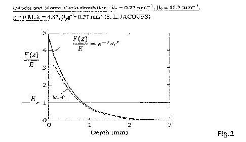
Figure 1: Evolution of the fluence rate/irradiance ratio versus the depth in a
semi-infinite tissue
for a "broad", collimated and perpendicular illumination of the air-tissue
interface. The continuous
4
CA 03194392 2023- 3- 30
WO 2022/069755
PCT/EP2021/077205
curve is the solution of the diffusion approximation, whereas the dashed curve
is the solution of a
Monte-Carlo computer-based simulation, where a and IA s are the absorption
and scattering
coefficients, respectively. Lteff is the effective attenuation coefficient, g
is the anisotropy factor,
whereas k is the pre-exponential factor resulting from the backscattering of
light. Derived from
[Jacques, 2010].
Figure 2: Temporal evolution of the p02 (black curve; mmHg) and temperature
(grey curve; C)
measured above a monolayer of HCM cells subject to metabolic oscillations.
Figure 3: Synergic effect of STS and PBM on angiogenesis.
Figures 4a, 4b,: Various PBM conditions presented as a ratios of the PpIX
fluorescence intensity
of PBM/no PBM reflecting in particular the metabolic activity, observed in
human cardiomyocytes
(HCM) at 689nm (Figure 4a) and 652nm (Figure 4b). The values of the ratio" PBM
/ no PBM"
are given by the monochrome bar.
Figure 4c : Left The fluence rate dependence effect (fluence rate ranging from
0.5 ¨ 15 mW/cm2)
at 689nm ( a potent wavelength) and 730nm (a non-potent wavelength) . Right :
The combination
of the potent wavelength using a not effective fluence rate (9mW/cm2) with a
nonpotent
wavelength inducing a significant effect in the relative increase of the PpIX
fluorescence.
Figure 5: Evolution of the fluence rate/irradiance ratio (F/E) versus the
depth in a semi-infinite
tissue for a "broad", collimated and perpendicular illumination of the air-
tissue interface. The
continuous curve is the solution of the diffusion approximation, whereas the
dashed curve is the
solution of a Monte-Carlo computer-based simulation, where pa and p.s are the
absorption and
scattering coefficients, respectively. Lteff is the effective attenuation
coefficient, g is the anisotropy
factor, whereas k, which depends on the refractive index matching conditions
(Mime/flair = 1.37) as
well as the optical coefficients, is the pre-exponential factor resulting from
the backscattering of
light. Derived from [Jacques, 2010].
Figure 6: Frequency analysis of the p02 in the CAM at EDD 7. Left up: Image of
the experiment
showing the metallic Clark's probe applied against a blood vessel. Left
middle. p02 signal
(acquired at 50 Hz). Left down: the associated spectrum based on a wavelet
analysis (the vertical
scale is the frequency, and the monochrome level represents the oscillation
amplitude, the
horizontal scale is the time in seconds). Right bottom: p02 signal (acquired
at 1 Hz) and right up:
the monochrome level represents the oscillation amplitude) associated spectrum
resulting from the
wavelets analysis. The horizontal axis is the time given in minutes. A strong
activation of the p02
CA 03194392 2023- 3- 30
WO 2022/069755
PCT/EP2021/077205
tone is observed 25 minutes (time out of the scale) after a topical
application of NaHS (10 I ¨
1 M), which is deactivated by PBM (850 nm, 7 mW.cm2, 30 s) at time 105 min
(see myogenic
signal). The horizontal lines reported on the spectrum indicate specific
frequencies. 1 ¨ Cardiac, 2
¨ Respiratory, 3 ¨ Myogenic, 4 ¨ Neurogenic, 5 ¨ eNOS dept, 6 ¨ eNOS indept
(probably
prostaglandin [Shiogai and all). Cardiac frequency cannot be resolved within
the sampling.
Respiratory and neurogenic bands do not exist at this EDD.
Figure 7: Enlargement of the experiment presented in figure 6. The horizontal
axis gives the time
in minutes.
Figure 8: Left: image of the in ovo anoxia reoxygenation experiment. Right:
p02 signal (OX-
100 pm Unisense0) recorded during the whole duration of the experiment
(Vertical axis:
mmHg). The hypoxia due to the flushing of N2 (up to 60 minutes) is followed by
a
reoxygenation.
Figure 9: Stimulation of a chicken embryo's heart at EDD 5 by PBM during an
anoxic
cardioplegia. Left: Time course measurement of the heartbeat (assessed by the
change of the heart
reflectivity in the region of interest defined by the rectangle presented on
the image showing the
embryo (right). The heartbeat is stopped between 0 and 45 s. Then, a PBM
illumination during 7
seconds re-activated the heart beat for more than 1 min.
Figure 10: Spatial evolution of the fluence rate (E) in a semi-infinite tissue
for a "broad",
collimated and perpendicular illumination of the air-tissue interface. 62=0,3
mm; n=10; nussueinair
= 1.37;
T=180 s; 6.F= 1.6 mW/cm2; The dotted curve corresponds to the continuous fit
of the "step-based"
evolution of the fluence rate. The analytical expression of this fit is
presented as an insert in this
figure. This illustrates that E may be changed continuously instead of
incrementally.
Figure 11: Temporal evolution of the fluence rate (E) in a semi-infinite
tissue for a "broad",
collimated and perpendicular illumination of the air-tissue interface. 62=0,3
mm; n=10; niissueinair
= 1.37; T=180 s; 6,F'= 1.6 mW/cm2; The dotted curve corresponds to the
continuous fit of the
"step-based" evolution of the fluence rate. The analytical expression of this
fit is presented as an
insert in this figure. This illustrates that E may be changed continuously
instead of incrementally.
Figure 12: Overview of a trans-myocardial implantation at 90 during coronary
thrombosis (b) of
cylindrical distributors (d) connected to an optical source (a) that are
placed trans-myocardially (c)
with a predefined pattern and spacing through a mask. (e) the light
distributor, the iCATS catheter
6
CA 03194392 2023- 3- 30
WO 2022/069755
PCT/EP2021/077205
and the guide to be inserted into the catheter for transfixion. (I) image
illustrating the propagation
of light from an isotropic distributor (sphere of 0.8 mm diameter) implanted
in the center of an ex
vivo swine left ventricular myocardium (652 nm ¨ 100mW).
Figure 13: implantation of an iCATS catheter all along the damage left
ventricle of the swine
heart in situ after a sternotomy. A) Catheter was introducing from the apex
side until it come out
of the top of left ventricle B). C) Heart cross section after the heart
excision without removing the
catheter. The catheter, here is well placed close to the middle of the
myocardium thickness.
Figure 14a: Visual localization of the ischemic area (1Z) after the occlusion
of the descending left
anterior coronary artery (LAD) partially perfusing the left ventricle during
an open-heart surgery
on pig. The IZ is easily differentiated from non-ischemic area (NZ) An
electrical impedance
sensor can also be used to characterize the ischemic area.
AJP- Heart Circ Physiol
Figure 14b: Visual representation of the transmyocardial photoconditioning
described in example
1, after the occlusion of the LAD during an open-heart surgery on pig. During
the ischemic phase,
interstitial catheters (iCATO - 0.89mm) were inserted into the left myocardium
from the apex to
left atrium with an optimal distance between them in order to maximize the
treatment area.
Cylindrical distributors (RD2500 - stick length 7cm) were then placed in each
iCATO. PBM
treatment was launched few seconds to few minutes depending on the
illumination time before the
reperfusion. A 670nm and 808nm illumination were used from two distributors
few seconds before
the reperfusi on of the ventricle. Catheters were then removed just after the
reperfusi on. Eventually,
it is possible for them to be to let in place after the surgery for further
regenerative illuminations.
Figure 15: Simplified diagram illustrating an example of a part of the device
supplying a treatment.
Depending on the disease and the treatment method, interstitial or systemic
for instance, the device
can be modular with combination of different elementary communicated blocks.
Some blocks can
be a sensor based on observables described in the present document, or an
interface to acquire data
from usual clinical system. Some blocks can characterize the spectroscopy of
the illumination
whereas others can be dedicated to control exogenous agent perfusion and
perfusion temperature
into a catheter. Others actuators can also be added to combined exogenous
stimulus like
mechanical pressure or temperature change with PBM. In case of the use of
multi-lumens balloon
catheter, as shown here, some blocks can control the time and/or the period
and/ or the level of
inflation /deflation with or without taking into account the monitoring of the
change. Part of the
7
CA 03194392 2023- 3- 30
WO 2022/069755
PCT/EP2021/077205
device also integrates specific balloon size and shape to optimally treat the
biological objects. For
instance, as represented in the figure, a balloon centered into the right
atrium which present two
opposites conic shapes can optimally treat circulating objects coming from the
inferior and the
superior part of the vena cava. Moreover, since PBM strongly influences the
biological rhythms,
particularly during the hypoxia or hypoxemia, balloon can be shaped in order
to be in contact to
the sino-atrial node located on the top of the right atrium or to be in
contact of the atrial ¨ ventricle
node. Moreover, using a multi-lumens catheter, exogenous agents can be
injected through the
balloon upstream or downstream the illumination function of the agent.
Obviously the device can
also be used to photo-activate the photo-sensitive agent. Blocks of these
device can also be
implemented directly through various implants to minimize platelet aggregate
and coagulation on
heart valve. It can also be implemented on artificial heart or pancreatic
chambers for instance, to
increase the biocompatibility / biostability, and/or can be implemented to
reduce inflammation
and immune response of implant chamber used in chemotherapy as well as
activating
endothelialization of hip prosthesis or vascular stent as other examples.
Figure 16: Examples of the irradiation geometries used in PBM. The stippled
areas represent
schematically the pattern of fluence rate in tissue. (a), (b) Surface
irradiation from broad beam or
lens-tipped fiber. (c)-(e) Interstitial irradiation with cut-end or
cylindrical fibers. (f)-(h)
Intracavitary and intralumenal irradiation. (i), (j) Intracavitary whole-
surface irradiation using an
isotropically-tipped fiber or a light-diffusing liquid (shaded).
Source: Wilson, 1986.
Figure 17: Spatial evolution of the fluence rate (E) in a semi-infinite tissue
for a "broad",
collimated and perpendicular illumination of the air-tissue interface. The
spatial evolution of E is
optimized in such a way that its value never exceeds 100 mW/cm2, while
minimizing the total
illumination time. The idea is to illuminate the sample exploiting two PBM hot
spots visible in
figure 4, i.e. generating fluence rates of 15 and 3 mW/cm2 during 40 and 180 s
(values of T),
respectively. The corresponding values for AF', Az and n (the number of steps)
are respectively:
- 4 mW/cm2, 0,15 mm and 13 for T = 40s
- 1.6 mW/cm2, 0.3 mm and 4 for T = 180 s.
ntissuehlair = 1.37.
Figure 18: Temporal evolution of the fluence rate (E) in a semi-infinite
tissue for a "broad",
collimated and perpendicular illumination of the air-tissue interface. The
spatial evolution of E is
8
CA 03194392 2023- 3- 30
WO 2022/069755
PCT/EP2021/077205
optimized in such a way that its value never exceeds 100 mW/cm2, while
minimizing the total
illumination time. The idea is to illuminate the sample exploiting two PBM hot
spots visible in
figure 4, i.e. generating fluence rates of 15 and 3 mW/cm2 during 40 and 180 s
(values of T),
respectively. The corresponding values for AF', Az and n (the number of steps)
are respectively:
-4 mW/cm2, 0,15 mm and 13 for T ¨40 s
- 1.6 mW/cm2, 0.3 mm and 4 for T = 180 s.
ritissueillair = 1.37.
The "step-based" evolution of the fluence rate can be fitted by the analytical
expression presented
as insert in figure 11.
Figure 19a: A non-uniform light emittance illustrated with a non-uniform
longitudinal emittance
from a cylindrical distributor. The distributor (2) is placed into a vessel
delimited by the wall vessel
(1). The emittance of the distributor is shaped in order to create a light
gradient all along the stick
(part of the distributor which emits the light), represented here by iso-
curves of the fluence rate
which are not parallel with the stick. Considering the two identical
circulating objects passing all
along the distributor at a certain distance hi and hi within a certain speed
i; will find at particular
place, Ai(hi) and A(h) respectively, the optimal fluence rate in respect to
the illumination time
defining by 13 in order to address a particular hot spot Qi,k. Obviously, the
light gradient is adapted
on the basis of optical coefficient of the circulating medium, the geometry of
the vessel as well as
the level of speed and the nature of the flow (laminar, turbulent, pulsatile).
Figure 19b: Wavelength combination of non-uniform emittance illustrated with a
sequential non-
uniform longitudinal emittance from a cylindrical distributor. in certain
scenario where particular
hot spots cannot be selected, due to for instance, a mismatch between the
constraint illumination
time and accessible fluence rate, illumination combination of a potent (?4)
and a non-potent (tj )
wavelengths can be used to overpass the issue. In the scheme, where any hot
spot for any kind of
circulating can be selected, the iso-dose can be in parallel for both
wavelengths, then, a sequential
or simultaneous uniform illumination can be used to obtain a potent PBM
effect. In contrary, if
hot spots for certain kind of circulating object can be selected, a non-
uniform illumination must
be considered to optimally treat all kind of circulating object.
Figurel9c: Modulation of the emittance of a uniform longitudinal distributor
within a balloon.
Encapsulation of a RD (stick of 2cm delimited by the radiomarkers) into a
balloon based catheter.
The shape and the size of the balloon is defined by the shape of the emittance
of the RD. In this
9
CA 03194392 2023- 3- 30
WO 2022/069755
PCT/EP2021/077205
case, circulating object passing in the vicinity of the balloon will be
exposed to different fluence
rate since the distance within the distributor is modulated by the modulation
of the diameter of the
balloon.
Figure20: Fluoroscopy of a pig chest showing a cylindrical distributor (RD070)
where arrows
locate radiomarkers which delimit the stick (7cm). The stick starts from the
superior vena cava
then passes through the right atrium, and finishes in the inferior vena cava
as shown in figure 15.
The distributor is placed under the procedure described in example 13 with the
difference that the
used catheter is a peelable one enabling to remove it and only let the optical
distributor in the
central venous line. The optical distributor can be let during days, for
chronic illumination.
Figure21: Illumination scheme protocol exploiting a hot spot "line" at 689nm.
In figure 4, it
appears a relative potent line at 40s for fluence rate comprising between 0.5
to 20mW.cm-2. This
could be used to optimize the illumination treatment time by an increase of
the treated depth per
illumination time. It is known that the fluence rate emitted from an
interstitial longitudinal uniform
distributor placed into a semi-infinite medium can be approximated by an
analytical expression
based on Bessel function of second kind. Figure 21 shows the treatment
protocol on the basis of
the evolution of fluence rate perpendicular to the distributor axis defined
within optical coefficient
(ua = 0.17mm-1: ueff = 0.88mm-1). Between 0 to 40s, an optical power of 2.8
mW.cm4 is coupled
to the distributor which induces a fluence rate of 20m W.cm-2 in the vicinity
of the distributor
surface to a fluence rate of approximately 0.5mW.cm-2 at ¨3.5 mm. Between 40s
to 80s, the optical
power is adjusted (multiplication factor P) at 100 mW.cm-1 in order to obtain
a fluence rate of
¨20 mW.cm-2 at 3.5 mm whereas the fluence rate reach 0.5 mW.cm-2 at ¨7 mm.
Therefore, using
this hot spot line, in this case, the depth of treatment is 7 mm within a
treatment time of 80 s.
Figure22: Graphic illustration of the use of selection of combined hot spots
of the same
wavelength to treat simultaneously different parts of the biological object,
within its specific
illumination time. Based on the same simulation described in figure 21, where
longitudinal
distributor is placed into the myocardium, the figure shows the evolution of
the fluence rate in the
depth of the tissue for successive optical power applied within the time
presented in inset. Using a
particular combination of an hotspot spot ai,689 = (20 1 mW.cm-2 ; 60 ls )
within another one
which presents a lower fluence rate but a multiple of illumination time n1,689
= ( 3 1.6 mW.cm-2
; 180 ls ) for instance, to successfully treat conjoint superficial layers
(using Q1,689 ) each 60 s,
whereas in parallel a deeply second zone is treating cumulatively (part of the
fluence rate which is
CA 03194392 2023- 3- 30
WO 2022/069755
PCT/EP2021/077205
underlined in black). During the session A, three superficial layers (by
increasing the optical power
every 60 s in respect to gli,689 ) is treated (illumination time is projected
on a lower graph) whereas
a deeply zone will receive a succession of 3 x 60s in the range of 3 1.6
mW.cm-2. Since a part
of the deep area have then received 180s, the optical power of the session is
defined to avoid to
illuminate parts which have already received 180s. Then as it is shown the
starting optical power
of session B is defined to continue the Clj,689which induces another part of
the object will be subject
to ai,689.
Figure20: Fluoroscopy of a pig chest showing a cylindrical distributeur
(RDS70) where arrows
locate radiomarkers which delimite the stick (7cm). The stick starts from the
superior vena cava
then passes through the right atrium, and finishes in the inferior vena cava
as shown in figure 15.
The distributor is placed under the procedure described in exemple 13 with the
difference that the
used catheter is a peelable one enabling to remove it and only let the optical
distributor in the
central venous line. The optical distributor can be let during days, for
chronic illumination.
Figure21: Illumination scheme protocol illustrating how the "line" hot spot
visible in Figure 4a
can be exploited to minimize the illumination time with a "long" cylindrical
light distributor
inserted in a "large" biological object. In this geometry the evolution of the
fluence rate as a
function of the distance from the surface of the light distributor can be
modeled by an analytical
expression containing Bessel functions of the second kind. The evolution of
the fluence rate as a
function of the distance mentioned above is shown for the following optical
coefficients of the
biological object (i.ia = 0.17 mm4: peff = 0.88 mm') for two different linear
power densities
expressed in mW/cm of the light distributor length. The first one (2.8 mW/cm),
applied for 40 s,
induces the fluence rate of 20 mW/cm2 at the light distributor surface,
whereas this fluence rate is
about 0.5 mW/cm2 at a distance of 3.5 mm. The second linear power density (100
mW/cm) is
applied between 40 and 80 s, thus resulting in a fluence rate of 20 mW/cm2 at
3.5 mm whereas its
value is 0.5 mW/cm2 at 7 mm. Therefore, using this hot spot line the depth of
treatment is 7 mm
for a treatment lasting 80 s.
Figure22: Graphic illustration of the combined use of two hot spots,
corresponding to the surfaces
Qi,689 and C2j,689, to treat simultaneously different depths in the biological
object, with one
wavelength. Considering the geometric and optical conditions corresponding to
Figure 21, the
evolution of the fluence rate with depth is shown for different linear power
densities that are
11
CA 03194392 2023- 3- 30
WO 2022/069755
PCT/EP2021/077205
applied sequentially. Using a particular combination of hot spots (Q,689: 20 1
mW.cm-2; 60 1s),
(i2j,689: 3 1.6 mW.cm-2; 180 1s) four layers are treated in two illumination
sessions (A and B).
During the session A, two layers are treated by increasing the linear power
density by steps of 60
s until 180 s in order to use the hot spot a1,689, whereas the remaining two
layers are treated at the
while using the hot spot S2j,689. As depicted in the insert located in the
upper right corner of Figure
22, the total treatment time is 360 s. It should be noted, that the linear
power density used for the
session B is defined in such a way that the two treated layers located between
1 and about 3 mm
are contiguous.
Figure 23: This figure is a generalization of Figure 22, when two wavelengths,
presenting different
penetration depths in the tissue, are applied synchronously. The combined use
of these two
wavelengths enables, as a consequence, to reduce the treatment time mentioned
in Figure 22 by
factor of 2.
Figure 24a This figure presents the normalized fluence rate, expressed in
(mW/cm2)/(mW/cm),
around a conical light distributor presenting a length of 7 cm, surrounded by
a fluid with optical
properties corresponding to the blood (Lta = 0.25 mm4, geff =1.07 mm'). The
arrow represents a
blood volume element propagating according to a trajectory that is parallel to
the light distributor
axis at a distance of 4 mm from this axis. This figure illustrates that the
blood volume element will
be exposed to the desired normalized fluence rate independently of the
position of the blood
volume element.
Figure 24b: This figure represents the same situation as Figure 24a but the
blood volume
element propagates at the surface of the conical light distributor.
Figure 25a Glycaemia ration between the beginning and the end of hypoxemia
event. PBM
illumination in deoxygenated blood during hypoxemia significantly reduces the
level of
glycaemia in blood.
Figure 25b: Monitoring of the Clark probe in the aorta of the arterial partial
pressure during the
PBM illumination in the lung arteries in normoxia.
12
CA 03194392 2023- 3- 30
WO 2(122/(169755
PCT/EP2021/077205
General description of the invention
The inventors have shown that the control of the light dosimetry (fluence rate
[mW/cm2]; light
dose [J/cm2]) and spectroscopy (wavelength(s)) as well as the illumination
duration and the time
of illumination are crucial to induce optimal PBM effects. This observation is
very important since
the PBM effects are known to be bimodal (sometimes qualified as biphasic),
i.e. too high or too
low fluence rates and/or light doses significantly reduce the PBM effects and
are therefore
frequently associated to the Arndt-Schultz rule observed in pharmacology. This
bimodal response
has been reported by numerous groups looking at various "standard" effects
(mitochondria
membrane potential; ATP production; etc) [Huang 2009; Hamblin 2017; Hamblin
2018].
Looking at the PBM effects on the endogenous production of PpDC in different
cell lines, including
glioma cells and human cardiomyocytes (HCM), the inventors found that both the
fluence rate and
the illumination time must be applied in a controlled manner. These two
parameters must be
applied with specific values, for a given illumination in each parts of the
volume of the biological
object to optimize PBM effects. In contradiction to what is reported in this
field, the inventors have
discovered that the bimodal effects of PBM are only observed for a specific
set of these parameters.
These sets of parameters are defined as "hot spots" (figure 4a and 4b) in this
document. Moreover,
the inventors have shown that some of these hot spots are wavelength
independent.
It is also established that the optical properties of biological tissues,
described mostly by their
absorption and scattering coefficients, have an important impact on the
propagation of the light
around a light source [Tuchin, 2015; Hamblin 2017; Hamblin 2018]. In general,
the fluence rate
(and the light dose) decreases with the distance from the light source due to
the absorption and
scattering of the light in the tissue (see Figure 1). Therefore, the fluence
rate and/or the light dose
in the tissues are, in most situations, never optimal at the same time in
different locations in the
tissues treated by PBM. The existence of the parameters hot spots mentioned
above combined
with: i) the heterogeneous distribution of the light in the tissues treated by
PBM, and ii) the very
limited control of the light delivery and dosimetry by the vast majority of
the research or clinical
groups active in this field explain the limited and contradictory outcomes
reported in the literature
[Chung 2012]. This situation also explains why PBMT is poorly used at present.
13
CA 03194392 2023- 3- 30
WO 2022/069755
PCT/EP2021/077205
An object of the present invention is to provide an improved PBM for the
treatment of biological
objects, such as tissues, circulating blood and/or the lymph.
Another object of the present invention is to provide an efficient treatment
of ischemia reperfusion
injuries, such as myocardial infarction (MI), by PBM applied with the
conditions and methods
mentioned above and below.
Another object of the present invention is to provide an efficient treatment
of fibrillations,
including atrial fibrillations, by PBM applied with the conditions and methods
mentioned above
and below.
Another object of the present invention is to provide an efficient PBM-based
treatment of
metabolic disorders such as type 2 diabetes, hepatic diseases or hormones
secretion with the
conditions and methods mentioned above and below.
Another object of the present invention is to provide an efficient treatment
of systemic
inflammation or exacerbated systemic immune response by PBM applied with the
conditions and
methods mentioned above and below.
Another object of the present invention is to provide an efficient PBM-based
treatment to maintain
systemic homeostasis during hypoxemia and or hypoxia with the conditions and
methods
mentioned above and below.
Another object of the present invention is to provide efficient methods in
cells-based therapy
notably to increase the proliferation rate of stem cells as well as to trig
cells differentiation.
Another object of the present invention is to provide an efficient
treatment/diagnosis of PDX-
based methods, for instance in photodynamic therapy or in cancer detection by
imaging the PpIX
fluorescence. Embodiments of this invention involves: the use of a helmet,
integrating light
emitting diodes, which induce a PBM illumination through the skull on a
specific area of the brain
14
CA 03194392 2023- 3- 30
WO 2022/069755
PCT/EP2021/077205
before the PhotoDynamic Detection (PDD) or PhotoDynamic Therapy
(PDT)procedures used to
manage cancers.
Another object of the present invention is to increasing and homogenizing the
endogenous
production of PDX in plants and larvae. One embodiment of this approach is to
increase the
efficacy of the phototoxic effects induced in weed/larvae.
Another object of the present invention is to provide an efficient treatment
of conditions by PBMT
based on the monitoring of the metabolic activity. This monitoring, based on a
frequency analysis
of parameters reflecting the metabolic activity, enables to adjust the
radiometric (fluence rate,
illumination time, light dose, ...) and spectral (wavelength(s)) parameters in
such a way that the
PBM effects are maximized. This monitoring can also be used to assess the
status of the metabolic
activity to determine the optimal light application moment. Embodiments of the
present invention
involve the use of standard probes to measure physiological of biochemical
parameters reflecting
the metabolic activity. As mentioned below, such probes include, thermocouple,
Clark's p02
probes or optical fiber-based probes to measure these parameters. The signals
delivered by these
probes are then processed by a dedicated unit to perform the frequency
analysis enabling to extract
parameters providing information on the PBM effects and metabolic activity.
The above objects are achieved with the device and methods of the invention as
defined in the
claims.
Advantageously the device and method according to the invention are
characterized by the fact
that the PBM effects are induced by the generation of a specific fluence rate
during a specific time
corresponding to specific "hot spots" as selection conditions (see below),
successively in each
parts of the volume of the biological object.
An illustrative embodiment of the present invention consists to use one or
several light source(s)
coupled to one or several light distributor(s) applying a specific fluence
rate during a specific time
CA 03194392 2023- 3- 30
WO 2022/069755
PCT/EP2021/077205
corresponding to one or several "hot spots" presented in figure 4a and 4b to
increase the
metabolism in the tissues/conditions of interest mentioned below.
The invention optionally al so encompasses devices and methods predicting the
ti me for applying
PBM on a biological object, based on a frequency analysis (Differential
analysis of temporal signal
(integration of the past or derivation of the present)) of fluctuations of
parameters reflecting the
metabolic activity or predictive methods based on artificial intelligence of
one or several
parameters reflecting the PBM effects or the metabolic activity of the
biological object.
Optionally, the light power delivered by the device, the illumination time and
the moment of PBM
application relative to variations of the metabolic activity is adapted on the
basis of feedback
observables (see the list given below) to optimize the PBM effects.
16
CA 03194392 2023- 3- 30
WO 2(122/(169755
PCT/EP2021/077205
Observable feedbacks:
a) The temperature.
Figure 2 presents the evolution of the temperature measured with a
thermocouple and a probe used
to measure the oxygen consumption rate (OCR), which reflects the metabolic
activity, in an
experimental setup developed by the inventors. This setup consists in a
monolayer of Human
CardioMiocytes (HCM) positioned at the bottom of a petri dish which was
covered with a 15 mm
thick layer of physiologic water. The thermocouple and the probe to measure
the OCR as well as
the partial pressure of oxygen (p02) were both positioned 1 mm above the cell
monolayer.
As can be seen on figure 2, significant and easily measurable changes of the
temperature are
observed while the p02, and hence the OCR, are changing. The temperature
increases with an
increasing activity of the OCR. Interestingly, the high-frequency oscillations
observed for the OCR
and the temperature after a maximal value of the p02 are in phase and
synchronous.
Therefore, measuring the temperature provides an observable feedback to
monitor and/or adapt
the light dose used for PBM. Measuring the temperature also enables to
determine the optimal
PBM illumination time relative to variations of the metabolisms.
b) The tissue autofluorescence reflecting the redox state of enzymes involved
in the metabolism
Oxidation is the main process producing the necessary energy in cells. It can
occur in aerobic or
anaerobic conditions. In many situations, biological oxidation starts with
substrate
dehydrogenation, i.e. the displacement of two hydrogen atoms, whereas
coenzymes such as
NAD+, NADP+ and FAD serve as acceptors of these atoms. Since the cellular
concentration of
these coenzymes is low, they must be recycled by re-oxidization. Therefore,
these coenzymes
serve as primary donor and acceptor in the process of oxidative
phosphorylation (OXPHOS)
[Ferraresi, 2012]. Since NADH and FAD are bound to many enzymes involved in
metabolic
pathways [Alberts, 2002], the relative ratio between the NADH and FAD binding
sites changes as
well when the cells are switching their metabolism [Banerjee, 1989]. Hence,
cell responses to
changes of the 02 level (change of metabolic activity) resulting from PBM can
be monitored
looking at their effects on FAD and NADH.
17
CA 03194392 2023- 3- 30
WO 2022/069755
PCT/EP2021/077205
These coenzymes can be studied non-destructively looking at their
autofluorescence, i.e. without
the addition of exogenous probes [Ramanujam, 2001]. One of the most common
optical techniques
giving information about the metabolic state of cells is based on the
determination of the redox
ratio of FAD and NADH by fluorescence spectroscopy [Chance, 1979; Walsh, 2012;
Blacker,
2016], in particular time-resolved fluorescence spectroscopy [Skala, 2007;
Skala, 2010; Kalinina,
2016; Walsh, 2013], a field corresponding to the expertise of the inventors
since more than two
decades [Wagnieres, 1998]. For instance, in cancer cells, an increase of
cellular metabolism is
usually indicated by a decrease of the redox ratio [Chance, 1989].
Therefore, steady-state and/or time-resolved fluorescence spectroscopy (or
imaging) of the tissue
autofluorescence is an interesting feedback observable to monitor or adapt the
light dose used for
PBM. Interestingly, the combined use of this approach with direct 02 sensing
based on the time-
resolved luminescence spectroscopy of molecular probes (PPDC or exogenous p02
probes as
proposed by Kalinina et al. [Kalinina, 2016]) or interstitial Clark's probes,
provide unique
information on the PBM effects. Monitoring these parameters is minimally
invasive and fast.
c) The assessment of the hemoglobin saturation.
In normal conditions, the body maintains a stable level of oxygen saturation
for the most part by
chemical processes of aerobic metabolism associated with breathing. However,
it is well known
that the hemoglobin saturation can change for different metabolic activities.
Since many methods are well established to measure the hemoglobin saturation,
notably the
peripheral or central venous saturation which is known to reflect the cardiac
output excepting in
sepsis shocks, applying this approach to monitor the changes of metabolic
activities induced by
PBM is of high interest.
In addition, since several gazes can be endogenously produced and diffused
within the tissue and
the circulating blood, and can bind to various metalloproteins, which present
strong optical
absorption bands, for instance, NO or H2S can bind deoxyhemoglobin to create
nitrosyl
hemoglobin or sulfhemoglobin or carboxyhemoglobin which decrease the level of
available
deoxyhemoglobin, and since PBM can induce photodissociation (Photolysis) of
metalloproteins
as hemoglobin, especially nitrosyl [Lohr, 2009] with a simultaneous formation
of methemoglobin,
and since the changes of these different forms of "hemoglobin" can be measured
(via optical
absorption measurement [Van leeuwen, 2017] ), monitoring the changes of the
metabolic activities
18
CA 03194392 2023- 3- 30
WO 2022/069755
PCT/EP2021/077205
induced by PBM through the assessment of these various metalloproteins
complexes is of high
interest.
d) The pH and or the level of bicarbonate (HC0f).
It is well known that glycolysis, which is the metabolic pathway that converts
glucose into
pyruvate, leads to an acidification of the extracellular surrounding media.
This acidification
frequently results from the excretion of lactic acid after its conversion from
pyruvate [Wu, 2007].
Since many methods are well established to measure the pH or the level of
bicarbonate in tissues
or in the circulating blood, applying this approach to monitor the changes of
metabolic activities
induced by PBM is of high interest.
As tissular (or saliva) lactate concentration can be constantly assess with
minimally invasive
device such subcutaneous microneedle [Tsurukoa, 2016] or in mouth, this
approach can be used
to monitor the changes of metabolic activities induced by PBM.
e) The concentration of ROS.
Reactive oxygen species (ROS) are chemically reactive chemical species
containing oxygen.
Examples include peroxides, superoxide, hydroxyl radical, singlet oxygen,
[Hayyan, 2016]
and alpha-oxygen. In a biological context, ROS are formed as a natural
byproduct of the normal
metabolism of oxygen and have important roles in cell signaling and
homeostasis [Devasagayam,
2004]. ROS are produced during a variety of biochemical reactions within the
cell and within
organelles such as mitochondria, peroxisomes, and endoplasmic reticulum.
Effects of ROS on cell metabolism are well documented in a variety of species
[Nachiappan,
2010]. These include not only roles in apoptosis (programmed cell death) but
also positive effects
such as the induction of host defense genes and mobilization of ion transport
systems. This
implicates them in control of cellular function. In particular, platelets
involved in wound repair
and blood homeostasis release ROS to recruit additional platelets to sites of
injury. These also
provide a link to the adaptive immune system via the recruitment of
leukocytes.
Abnormal levels of ROS are implicated in numerous pathologies through a strong
modulation of
various biological cascades [Sies, 2020]. Interestingly ROS level are also
primordial for cell
reprograming [Bigarella, 2014; Zhou, 2016] and tissular remodeling. For
instance, their production
kinetics depend on a broad spectrum of extrinsic or intrinsic repetitive
stimulus such as hormones
19
CA 03194392 2023- 3- 30
WO 2022/069755
PCT/EP2021/077205
secretion or mechanical forces (like vascular shear stress), which influence
directly the tissular
behavior and properties [Hwang, 2003; Brandes, 2014], and finally phenotypic
aspects.
Since many methods are well established to measure the ROS in tissues,
applying this approach to
monitor the changes of metabolic activities induced by PBM is of high
interest.
As ROS and reactive nitrogen species (RNS) are intricate by nature
[Moldogazieva, 2018] and
since many methods to assess to RNS are well established [Griendling, 2016],
the termed of
reactive oxygen and nitrogen species RONS should be used here. Actually, in a
full extends, the
term should be reactive oxygen and nitrogen and sulfur species (RONSS).
0 The level of H2S.
Hydrogen sulfide (H2S) exert a wide range of actions on the whole organism. It
is an epigenetic
modulator inducing histone modification particularly via DNA demethylation, a
process which
permit cell differentiation [Yang, 2015]. It is fundamental in aging process
of aerobic living
organism by maintaining a high level of copy number of mitochondrial DNA [Li,
2015], as well
in senescence process through sirtuin 1 activation. Interestingly, H2S is the
only species which is
both substrate and inhibitor of the OXPHOS inside the mitochondria depending
of its
concentration [Szabo, 2014], which may to be in parallel to the famous
observation that exogenous
H2S inhalation induce a suspended animation-like state in small mammals, known
as artificial
hibernation, or hypometabolism [Blackstone, 2005]. H25 is known to protects
against many
cardiac conditions, including pressure overload-induced heart failure [Snij
der, 2015]. This
supports the hypothesis that endogenous H2S is a regulator of energy
production in mammalian
cells particularly during stress conditions, which enables cells to cope with
energy demand when
oxygen supply is insufficient [Fu, 2012].
Moreover, we observed in fertilized chick's eggs an in-ovo anoxia
reoxygenation This study was
performed by gently placing an H2S microprobe above the ventricle of the
chicken or the dorsal
aorta. We observed that the H2S level increased significatively within the
anoxic (transient)
cardioplegia (or when the heart beat extremely slowly) and decrease when it
beat again. Therefore,
it appears that the blood flow plays a role by removing endogenous production
of H2S which could
bind deoxyhemoglobin to form sultlemoglobin and propagate it.
Since many methods are well established to measure H2S in tissues [Olson,
2012], applying this
approach to monitor the changes of metabolic activities induced by PBM is of
high interest.
CA 03194392 2023- 3- 30
WO 2022/069755
PCT/EP2021/077205
g) The level of hydrogen selenide (H2Se).
Alongside oxygen and sulfur, selenium, the constitutive element of H2Se
belongs to the chalcogens
group and have similar excretory and metabolic pathways. Analog to H2S, H2Se,
is an endogenous
small gaseous molecule which can induce a suspended animation like state and
show reperfusion
injury protection [Iwata, 2015]. It reversibly binds COX, which inhibits the
mitochondrial
respiration and argued to be the fourth gasotransmitors with H2S, NO and
carbon monoxide (CO)
[Kuganesan, 2019]. Moreover, incorporated into numerous selenoprotein
oxidoreductase enzymes
as glutathi one peroxidase, it is essential for maintaining redox-status
homeostasis in health and
diseases, and its deficiency induces a substantial increase of ROS, which is
suspected to be one
important cause of cancer and CVD [Bleys, 2008].
Since many methods are well established to measure H2Se in tissues or Selenium
in serum,
applying this approach to monitor the changes of metabolic activities induced
by PBM is of high
interest.
h) The concentration of ions.
Ions play an important role in the metabolism of all organisms as reflected by
the wide variety of
chemical reactions in which they take part [van Vliet, 2001]. Ions are
cofactors of enzymes,
catalyzing basic functions such as electron transport, redox reactions, and
energy metabolism; and
they also are essential for maintaining the osmotic pressure of cells. Because
both ions limitation
and ions overload delay growth and can cause cell death, ion homeostasis is of
critical importance
to all living organisms.
Since many methods are well established to measure ions in tissues (in
particular calcium,
potassium, chloric and/or hydrosulfi de i on s), applying this approach to
monitor the changes of
metabolic activities induced by PBM is of high interest.
i) The level of cytochrome, including cytochrome c oxidase, by MRS.
It is well established that broadband Near InfraRed Spectroscopy (MRS) can be
used to monitor
concentration changes of the oxidation state of cytochromes as cytochrome-c-
oxidase (AoxCCO)
which plays a key role in the mitochondrial respiration [Roever, 2017].
21
CA 03194392 2023- 3- 30
WO 2022/069755
PCT/EP2021/077205
Since different methods are well established to measure AoxCCO in tissues (or
other cytochromes
involved in the metabolism), applying these methods, including NIRS to monitor
the changes of
metabolic activities induced by PBM is of high interest.
i) The use of medical (functional) imaging techniques (MRS "Magnetic Resonance
Spectroscopy",
MRI "Magnetic Resonance Imaging": NMR "Nuclear Magnetic Resonance": PET
"positron
emission tomography": EPR "Electron Paramagnetic Resonance": SPECT "single
photon
emission computed tomography" BOLD "Blood ox venation level dependent" NIRS
"Near
infrared Spectroscopy").
Metabolic imaging focuses and targets changes in metabolic pathways for the
characterization of
various clinical conditions. Most molecular imaging techniques are based on
PET and MRS,
including conventional 'H and '3C MRS at thermal equilibrium and
hyperpolarized magnetic
resonance imaging (HP MRI). The metabolic pathways that are altered in many
pathological
conditions and the corresponding probes and techniques used to study those
alterations have been
reviewed by Di Gialleonardo et al. [Di Gialleonardo, 2016]. In addition, Fuss
et al. [Fuss, 2016]
described the use of medical imaging to address various conditions in humans.
Since many metabolic imaging-based methods are well established to assess the
metabolism,
applying these methods, including functional metabolic imaging, to monitor the
changes of
metabolic activities induced by PBM is of high interest.
k) The vascular tone and the vasomotion.
Vascular tone refers to the degree of constriction experienced by a blood
vessel relative to its
maximally dilated state. All arterial and venous vessels under basal
conditions present some degree
of smooth muscle contraction between balance of constrictor and dilatator
influences that
determines the diameter of the vessel, e g the vascular resistance to adapt!
regulate blood flow and
pressure. Basal vascular tone differs among macro and micro-circulation and
organs. Certain
organs have a large vasodilatory capacity (e.g., myocardium, skeletal muscle,
skin, splanchnic
circulation) hence a high vascular tone, whereas others organs have relatively
low vasodilatory
capacity (e.g., cerebral and renal circulations), hence a low vascular tone.
The vascular tone regulation differs among the macro (arteries, veins) and the
micro (arterioles,
venules, capillaries). Notably, even if the tone can be modulated via
extrinsic factor (nerves,
22
CA 03194392 2023- 3- 30
WO 2022/069755
PCT/EP2021/077205
circulating metabolites), blood vessel can exhibit spontaneous oscillations
(vasomotion) which
give rise to flow motion [Aalkaejer, 2011]. Therefore, through the dependence
of the vascular tone
in a multiplicity of actuator from local to systemic, the analysis of this
tone, give insight on the
metabolic activity, and can reflect the degree of aging [Bentov, 2015] and
many
pathophysiological conditions, as ulcer risk, type 2 diabetes [Smirnova,2013],
endothelial
dysfunction or hypertension [Ticcinelli, 2017], renal diseases [Loutzenhiser,
2002] [Carlstrom,
2015] or metabolic syndrome [Walther, 2015]. Moreover, the assessment of the
skin microvascular
endothelial function is used as diagnosis as well as prognostic of CVD
[Hellman, 2015].
Since many methods are well established to assess to the vascular tone and the
vasomotion, such
as videocapillaroscopy, plethysmography [Tamura, 2019], laser doppler
flowmetry, pressures
measurement via cutaneous vascular conductance (CVC) or time frequency
analysis as example,
and since all the cardiovascular system is argued to be a single entity of
coupled oscillators in a
dynamic point of view [Shiogai, 2010], any methods which enable to assess to
the change of
metabolic activities induced by PBM (including heart rate variability (HRV)
which give
information on the autonomic nervous system via ECG or heart sound measurement
[Alvarez,
2018] via phonocardiogram (PCG) [Patidar, 2014] are of high interest. As
observe by the inventors
along surgical procedure respiratory frequency variability (RFD
[stevanovska,2007] or
ballistocardiography to monitor the changes of metabolic activities induced by
PBM is of high
interest.
1) The use of electromagnetic endogenous signals.
Electrocardiogram (ECG), electroencephalogram (EEG) and electromyogram (EMG)
are standard
measurements of electrical activity of the metabolism of the heart, the brain
and the muscle
respectively. Novel ECG analysis based on signal computational classification
[Patidar, 2015] are
promised tools in heart diagnosis notably giving insight in coronary artery
disease [Kumar, 2017]
[Acharia, 2017], arrhythmia and ischemia disorders [Bhoi, 2017]. Same kind of
analysis have been
performed on EEG which show interesting outcomes in epileptic seizures, the
most common brain
disorders [Bhattacharyya, 2018].
Since many methods are well established to monitor these electromagnetic
endogenous signals,
methods which enable to assess to the change of metabolic activities induced
by PBM are of high
interest.
23
CA 03194392 2023- 3- 30
WO 2022/069755
PCT/EP2021/077205
m) The use of bio impedance signals.
It can be used in the clinic for measuring various physiologic parameters
[Petterson, 2016]. This
approach is used for pacemakers as En site from St Jude Medical, Opti Vol from
Medtronic and
closed loop stimulation from biotronik.
Since many methods are well established to measure electrical bioimpedance in
tissue or directly
on the skin, any these methods which enable to assess to the change of
metabolic activities induced
by PBM is oh high interest.
n) The presence of markers in the circulating blood.
A long list of circulating markers of interest to monitor the light dose
during PBM includes
metabolites (succinate, pyruvate, etc.), coagulation factors, apoptotic
factors, (pro and anti)
inflammatory factors, as well as hepatic factors, mitoldnes, or level of
isolated mitochondria for
instance. It should be noted that only a few of them is enumerated here.
Glucose level: Many pathologies are associated with a dysregulation of
circulating glucose level,
which directly induces systemic metabolic disorders, as it is the case for
diabetes. Hence,
monitoring the change of the metabolic activities through the assessment of
the glycaemia is of
high interest.
Succinate: Succinate is a key intermediate of the tricarboxylic acid cycle
(TCA) cycle which plays
an essential role in anabolic and catabolic pathways. Moreover, it is notably
associated with
reperfusion injuries [Chouchani 2014]. Mitochondria are the physiological
source of succinate,
however accumulated succinate can be transported into the cytosol and then in
the circulating
blood. This TCA cycle intermediate connects intracellular metabolic status and
intercellular
signaling [Tretter, 2016]. Level of succinate in blood can vary from 2 to 20
p.M, where this
concentration can increase, with hypoxic stress, pro-inflammatory stimuli,
exercise, or with
pathological conditions such as type 2 diabetes, obesity or ischemia
reperfusion injury [Grimolizzi,
2018]. Since circulating level of succinate can be monitored via
bioluminescent assay, or Raman
spectroscopy the assessment of the change of metabolic activities induced by
PBM via the
circulating level of succinate is of high interest.
Lactate and lactate dehydrogenase (LDH): LDH is a common marker of cell damage
and cell death.
In addition, LDH produced during anaerobic exercise can be reduced by PBM
[Park, 2017].
24
CA 03194392 2023- 3- 30
WO 2022/069755
PCT/EP2021/077205
Hence, using these markers to assess the change of metabolic activities
induced by PBM is of high
interest. Moreover, combination of LDH level with aspartate aminotransferase
(AST) level serves
as a potent indicator of the damage to the body's tissues. It should be noted
that lactate levels can
be assessed with new aerometric method directly on the saliva [Tamura, 2018].
Since the high level of metabolism activites in inflammatory or immune
response, notably by the
capacity of immune cells to change their phenotype, serum/plasma level of
immune/inflammatory
markers, such as: mtDNA copies, number of leucocytes, total antioxidant
capacity, bicarbonate,
malondialdehyde (MDA), uric acid, bilirubin, level of cytokines or chemokine
markers such as
inter] eukins IL2, IL6,1L7, ILI 0, 1L18 or TNFa for instance, as well as
macrophage inflammatory
protein 1-a, 1P10, MCP1, as well as activation of lymphocytes T and / or
monocytes M2 through
flux cytometryare are also of high interest.
Serum/plasma levels of thioredoxin: The level of this enzyme is elevated in
infection, ischemia¨
reperfusion, and other oxidative stresses. Therefore, they are good markers
for monitoring of the
oxidative stresses. Plasma levels of thioredoxin are also elevated in patients
with coronary spastic
angina and other cardiovascular diseases [Nakamura, 2004].
Cardiac markers: Several established markers (myoglobin, creatine ldnase
isoenzyme, troponin I
and T, B-type natriuretic peptide, transaminase) are clinically used for
cardiac infarction diagnosis
and also for other organs injuries. To a lesser extent, LDH, glycogen
phosphorylase and recently
ischemia-modified albumin can be used in diagnosis within 30-minute assay
[Dasgupta, 2014].
This is also the case for thioredoxin level [Jekell, 2004].
Level of circulating eNOS as well NO or nitrite or nitrate: The levels of
these compounds are
essential, notably for the regulation of systemic blood pressure and systemic
homeostasis [Wood,
2013]. By extension, also levels of circulating H2S or sulfite or sulfate
level can be assessed to
monitor the change of the metabolic activities induced by PBM.
Circulating mitochondria: It has been recently shown that cell free functional
mitochondria are
present in the circulating blood. Moreover, mitolcynes are important in the
metabolic remodeling,
especially in the heart failure [Duan, 2019]. Therefore, monitoring of the
change of the metabolic
activities through the assessment of the circulating mitochondria level or
mitokynes level is of
high interest.
CA 03194392 2023- 3- 30
WO 2022/069755
PCT/EP2021/077205
o) The use of voltage-sensitive fluorescent dyes for the optical mapping of
cardiac electrical
signals.
Although, the optical imaging of cardiac electrical signals using voltage-
sensitive fluorochromes
(VSF) has only been performed in experimental studies because these VSFs are
not yet approved
for clinical use, FDA approved dyes, such as Indo Cyanine Green (ICG)
[Martisiene, 2016],
exhibits voltage sensitivity in various tissues, thus raising hopes that
electrical activity of cardiac
tissues could be optically mapped in the clinic. Therefore, methods based on
the use of voltage-
sensitive dyes to map (or to assess locally with a "point measurement" system)
the cardiac
electrical signal to monitor/adapt the light dose during PBM is of high
interest.
p) The use of redox sensors to assess of the status of various tissular redox
states. Since metabolic
and redox reactions are intricated and since many methods are based on the
measurement of redox
sensors proteins to asses to metabolic activities, monitoring changes of
metabolic activities
induced by PBM with redox indicators probes is of high interest.
q) The use of ultrasounds to assess the status of various tissues, including
cardiac tissues.
Ultrasonography is a well-established method to investigate cardiac tissue.
Many parameters
characterizing the heart tissues as well as the blood flow are routinely
obtained during
ultrasonography.
Therefore, methods based on the use of ultrasonography to monitor the light
dose during PBM are
of high interest.
r) The use of the p02 (and / or the OCR) to reflect the metabolic activity of
various tissue and / or
of the whole body through the measurement of the partial pressure of arterial
oxygen (Pa02) and
the fraction inspired oxygen (Fi02).
p02 can be easily measured within exogenous or endogenous probes in different
compartment
(tissular or organ) or different organelles within the cell. For instance,
such probes can be optically
detected. Other techniques, such as EPR oximetry, polarographic electrodes or
BOLD imaging are
of high interest to assess changes of metabolic activities induced by PBM.
s) The use of hemodynamic variables.
26
CA 03194392 2023- 3- 30
WO 2022/069755
PCT/EP2021/077205
In the clinic, real time assessment of these variables is a must to monitor
the metabolic activity,
especially in case of cardiovascular injuries. Such measurements notably
involve, arterial and
venous gases pressures, cardiac output, stroke volumes, capillary pressure, as
well as systemic and
pulmonary resistance. Therefore, the use of these methods to assess changes of
metabolic activities
induced by PBM are of high interest.
t) The use of Krebs cycle enzymes kinetics.
It is well known that Krebs cycle enzymes kinetics are good markers of
metabolism notably to
assess to the level of mitochondria] proteins. Since, for instance, acotinase
or succinate
dehydrogenase activities are commonly measured in clinics, the use of these
methods to assess
changes of metabolic activities induced by PBM are of high interest.
w) The level of PpIX
Protoporphyrin IX, is a precursor of numerous organometallic proteins, such as
hemoglobin and
chlorophyll. The inventors have shown that cells treated by PBM tend to
increase their endogenous
production of PpIX. Therefore, the use of methods based on the detection of
the PpIX level to
assess changes of metabolic activities induced by PBM are of high interest.
Since the heme
concentration is a feedback parameter in the PDX endogenous production
pathway, by extension,
measuring the level of circulating hematocrits to assess changes of the
metabolic activities induced
by PBM is of high of interest.
x) Monitoring of the metabolomics and lipidomics , in particular oxylipines.
Oxylipines are bioactive metabolites derived from the oxygenation of
polyunsaturated fatty acids.
Furthermore, they play a key role in the progression of cardiovascular disease
thrombosis and risk
factors. Hence, their monitoring is of high interest.
y) The level of glycoproteins
It is well known that glycoproteins, comprising of protein and carbohydrate
chains, are involved
in many physiological functions, including immunity. They possess receptors
signaling domains
that recruit signaling molecules.
27
CA 03194392 2023- 3- 30
WO 2022/069755
PCT/EP2021/077205
z) Level of glycerol
Glycerol can serve as a marker of apoptosis. One of the function of glycerol
is that is serves as a
chemical chaperone. In particular, it possesses an ability to enhance the
expression of apoptotic
regulators (Mx).
aa) The assessment of the immunomodulatory effects induced by PBM can be
monitored by pro-
inflammatory circulating monocytes like CD14, CD16 which can differentiate to
the dendritic
cells. It can be also assessed by the cytokines profiles of macrophages.
ab) Monitoring of the basis of the level of oxytocin. For the latter, it has
been shown that
monitoring of the oxytocin levels in the intensive care unit in the premature
infants serves as a
relevant marker of pain.
Optionally, the light power the illumination duration and the application time
of PBM define by
this device, is to be combined with the administration of exogenous stimulus
wherein the stimulus
could be an agents (see the list given below) to increase the PBM effects. It
should be note that the
time between the administration of exogenous agents and the PBM illumination
may take into
account the assimilation duration as well as activation kinetics of the agent.
The inventors demonstrated a svnernv resulting from the combined
administration of PBM with
notably exogenous agents (sulfur donors) and nitric oxide donors.
As already shown by the inventors, an administration of ALA combined with PBM
increases the
PplX build up and, consequently, the level of endogenous PpIX. Therefore, the
coadministration
of ALA and light is of high interest to increase PBM effects. It should be
noted that other
exogenous agents can be combined within PBM to increase its effects, as
indicated below.
As presented in figure 3, the inventors recently observed that when PBM is
combined with an FDA
approved exogenous agent, sodium thiosulfate (STS), which are sulfur donors,
the angiogenesis
observed on the chick's Embryo Chorioallantoic Membrane (CAM) is even more
stimulated. This
strongly suggests that the combination of PBM with exogenous such agents
stimulates even more
angiogenesis, or the metabolic activity, than PBM or such agents applied
separately.
28
CA 03194392 2023- 3- 30
WO 2022/069755
PCT/EP2021/077205
The assessments of these PBM effects on angiogenesis were performed using an
approach based
on fluorescence angiographies performed on the CAM several days after PBM.
Fig. 3 presents a
typical CAM fluorescence angiogram (left image) characterized quantitatively
with an image
processing and analysis software developed in our laboratory [Nowak-Sliwinska,
2010]. The main
objective of this development was to characterize dynamic changes taking place
in the
capillary/vessels network of the CAM between embryo development day (EDD) 6
and 12, when
the CAM vessels are monitored using an epi-fluorescence microscope equipped
with a scientific
camera following the intravenous (iv) injection of a fluorescent agent [Nowak-
Sliwinska, 2010].
From the resulting angiogram, 3 descriptors are extracted: the number of
branching points/mu'',
the mean area of the vessels network meshes, and the mean of the 3' quartile
of the mesh area
histogram. As presented in Figure 3, our proof-of-concept results demonstrates
that PBM
significantly stimulates the CAM angiogenesis. This effect is even more
pronounced if PBM is
combined with an exogenous application of 175 mM STS.
Interestingly, in parallel, the inventors observed in-ovo, through the
monitoring of the H2S and NO
level on the chicken embryo, that a topical application of STS induced a
significant increase of
NO after a long time (6 at 12 hours), whereas, when performing a PBM
irradiation 1 -2 hours after
the STS application, the time when NO was produced was significantly reduces,
typically down
to one hour.
Since STS is a clinically approved H25 donor [Snijder, 2015] to protects
against many cardiac
conditions, as also already reported for H2S [Yu, 2014] , including pressure
overload-induced heart
failure via upregulation of endothelial nitric oxide (NO) synthase [Kondo,
2013] as well as renal
ischemia / reperfusion injury [Bos, 2009], the combined use of PBM, applied
with the
device/protocol mentioned above, with the administration of H2S donors (such
as STS or
methylsulfonylmethane (MSM), or dithiolthiones for instance, or other donors
presenting different
H2S kinetics release) and/or NO donor substances, as for instance arginine,
including NO itself, is
of high interest.
Since Cysteine is an important source of sulfide in the human metabolism,
combining the
administration of this proteinogenic amino acid, or derivatives thereof, such
as selenocysteine, or
synthetic form as N-acetylcysteine is of high interest to potentiate the
effects of PBM.
29
CA 03194392 2023- 3- 30
WO 2022/069755
PCT/EP2021/077205
MSM, a naturally occurring organosulfur compound, is utilized as an
alternative source of
biologically active sulfur. It is mostly used for anti-inflammatory
treatments. It has been
investigated in animal models, as well as in many human clinical trials
[Butawan, 2017]. MSM is
also recognized for its antioxidant capacity and it was proposed that the
antioxidant mechanism
acts indirectly via the mitochondria rather than directly at the chemical
level [Beilke, 1987]. As an
FDA approved substance, MSM, is well-tolerated by most individuals at dosages
of up to four
grams daily, with very few side effects [Butawan, 2017]. Results from in vivo
and in vitro studies
indicate that MSM actions are at the crosstalk of oxidative stress and
inflammation at the
transcription and sub-cellular levels [Butawan, 2017]. Interestingly, Kim et
al. [Kim, 2009]
demonstrated that MSM can also diminish the expression of inducible nitric
oxide (NO) synthase
(iNOS) and cyclooxygenase-2 (COX-2) through suppression of the nuclear factor-
kappa B (NF-
x.13), a transcription factor involved in the immune and cellular responses to
stress. This
observation is highly interesting since NO is a powerful vasodilator involved
in many metabolic
functions. As some other gas transmitters, called gasotransmitors [Donald,
2016], NO can have
differential effects depending on its local concentration and microenvironment
[Thomas, 2015]
which can impact many different processes rRapozzi, 2013; Reeves, 20091 It has
also been
suggested that PBM causes NO photodissociation from COX [Karu, 2005; Lane,
2006].
Concomitantly, NO photodissociation from other intracellular "reservoirs" such
as nitrosylated
forms of myoglobin and hemoglobin have also been hypothesized [Lohr, 2009]. It
is well
established that cell respiration is down regulated by the NO production by
mitochondria] NO
synthase. The 02 displacement from COX by NO inhibits cellular respiration,
and ATP production
[Antunes, 2004; Cooper, 2008]. Therefore, it is believed that PBM increases
ATP production. An
alternative and, possibly, parallel mechanism to explain the release and/or
increase of NO
bi oavailability by PBM could be linked to an action of COX as a nitrite
reductase enzyme (a one-
electron reduction of nitrite gives NO), in particular when the 02 partial
pressure is low [Ball,
2011].
All together, these observations indicate that MSM has an indirect effect on
the mitochondrial
electron transport chain (ETC) through its NO modulation. In addition, this
analysis of the
literature indicates that the combined use of PBM with NO donors, such as S-
Nitrosothiols or alkyl
nitrites, including NO itself, induces a potent synergetic effect.
CA 03194392 2023- 3- 30
WO 2022/069755
PCT/EP2021/077205
Moreover, since interaction between H2S and NO can produce nitroxyl (HNO),
which plays an
effective role within the cardiovascular system about oxidative stress and
cardioprotection, heart
contractility, vascular tone as well as angiogenesis [Nagpure, 2016; Wu 2018],
the co-
administration of (H)NO as well as nitroxyl donor, cimlanod or 1-Nitrosocyclo
Hexyl Acetate for
instance, is of high interest to increase PBM effects.
Ebselen, an FDA approved H2Se donor is of high interest to increases PBM
effects as already
discuss by the inventor (page 13, point g).
As already mention, NAD+ is required for redox reactions and control hundreds
of key process of
energy metabolism to cell survival, rising and falling depending on food
uptake, exercise, and ti me
of the days. Therefore, administration of NAD+ donor, as vitamin B3 within PBM
is of high of
interest to increase PBM effect.
Other exogenous agents of interest for their combined use with PBM are:
- Curcumin, a major active component of turmeric (Curcuma longa, L.), is known
to have various
effects on both healthy and cancerous tissues. Notably, curcumin induces ER
stress, thereby
causing an unfolded protein response, the major retrograde signaling, and
calcium release, which
destabilizes the mitochondrial compartment and induce apoptosis.
- Dexmedetomidine, a well-known a2 agonist agent used in anesthesia, has
gained of interest
recently since it is suggested that dexmedetomidine preconditioning mitigates
myocardial
schemi a/reperfusi on injury via inhibition of mast cell degranul ati on .
Similarly, EPO have shown
positive effect in ischemia reperfusion injuries into the kidney as well as
lipopolysaccharide in
arterial vascular damages.
- Ivermectin are approved by the FDA to treat people with intestinal
strongyloidiasis and
onchocerciasis, two conditions caused by parasitic worms. Moreover, clinical
evidence supports
the use of ivermectin in decreasing mortality in patient with SARS-CoV2
infection. The
combination of ivermectin and PBM can reduce the dose of ivermectin, in
particular to reduce side
effects.
- Viagra as a source of nitrite in various forms (as alkyl nitrites)
31
CA 03194392 2023- 3- 30
WO 2022/069755
PCT/EP2021/077205
By extension, any exogenous agents which are known to modulate the metabolism,
especially
within the mitochondria through the modulation of the ETC or ROS modulator for
instance, are of
high interest to increase PBM effects. It is the case for adenosine
diphosphate (ADP) which is
known to increase the OCR, or for vitamin K, ketami ne suxam ethon i um ,
acetyl chol i ne and
atropine, as well as bradykinin. Additional agents include, catecholamines
like adrenaline
noradrenaline or dopamine, opioids which activate various G proteins, or
various ldnases
modulator or various anti-oxidant and/or anti-inflammatory donor such as
resveratrol. Finally,
targets of the rapamycin or sirtuin modulators are of high of interest to
increase PBM effects.
By extension, since temperature, exogenous or endogenous mechanical pressure
[Li, 2005; Hwang
2003], physical exercise as well as electrostimulation, hyperoxia, hemostasis
(remote
preconditioning) are known to modulate the metabolism, any exogenous stimulus,
or combination
of, like environmental/physical /electrical or electromagnetic stimulus
applied on the biological
object is of high interest to increase PBM effect. For instance, it is known
that the level of
endogenous H2S is inversely correlated within the temperature. An increase of
the temperature in
situ can be viewed as an indirect endogenous H2S donor and, reciprocally, an
increase of the in
situ temperature can be viewed as an endogenous H2S inhibitor.
As observed by the inventors, the potency of PBM not only depends on the light
dose [J/m2] and
spectroscopy (wavelengths) but, surprisingly, also on the fluence rate for
specific illumination
times. For example, the inventors have observed for a specific case, as
indicated in figure 4a and
4b, that cells must be illuminated during 3 minutes with an irradiance (which
is equal to the fluence
rate in this specific setting consisting of cell cultures) of 3 mW/cm2 (i.e. a
dose of 0.54 J/cm2).
Different illumination times and/or irradiances do not induce any PBM effects,
excepting "hot
spots" observed at, for instance, an irradiance of 15 mW/cm2 applied for 40
seconds, or 25
mW/cm2 applied during 22 seconds. Interestingly some of these hot spots are
wavelength
independent, as one can conclude comparing figures 4a and 4b where hot spots
are present in the
same place in terms of irradiance and illumination time.
This is an illustration of a more complex PBM response compare to the well-
known bimodal
effects of PBM, i.e. too high or too low fluence rates and/or light doses
significantly reduce the
PBM effects. The inventors have also demonstrated, in certain conditions, the
absence of
32
CA 03194392 2023- 3- 30
WO 2022/069755
PCT/EP2021/077205
"neutralization" of the PBM effects by an over-dose/irradiance before and/or
after PBM applied
with optimal conditions.
The inventors have also observed surprising results when performing PBM with a
combination of
wavelengths, one of them being ineffective when used alone. This is the case
for 730 nm which is
not potent when used alone, as it can be seen in the figure 4c. It can be seen
from this figure that
the wavelength of 689 nm, which is effective for an illumination time of 180 s
when applied with
an irradiance of 3 mW/cm2 is ineffective if it is applied with 9 mW/cm2.
Surprisingly, combining
synchronously or sequentially illuminations at 730 nm (with an irradiance of 3
mW/cm2 for 180
s) and 689 nm (with an irradiance of 9 mW/cm2 for 180 s), resulted in a marked
PBM effect,
evidenced by the PpIX fluorescence intensity ratio (PBM/no PBM). These effects
were
comparable to those corresponding to the "hot spots" presented in figure 4a
and 4b. Therefore, the
synchronous or sequential combination of wavelengths in such a way that at
least one of them is
not (or poorly) potent is of high of interest to increase PBM effect, in
particular when specific hot
spots of the potent wavelength are difficult to reach due to optical or
geometric constraints.
Indeed, it is well established that the optical properties of biological
tissues, described mostly by
their absorption a and reduced scattering a' coefficients, have an important
impact on the
propagation of the light around a light distributor. In general, the fluence
rate (and the light dose)
decreases with the distance from the light source due to the absorption and
scattering of the light
in the tissue for a given power (and illumination time). Figure 5 illustrates
how the fluence rate F
(normalized by the irradiance E) decreases with the distance from the surface
on the specific case
of a "broad" (much larger than eff4), collimated illumination of a semi-
infinite sample. Therefore,
the fluence rate and the light dose in the tissues are never optimal at the
same time in different
locations of the tissues treated by PBM.
It should be noted that the geometry of the light distributor (illumination
geometry) is adapted to
the specific organs to be treated. For example, frontal (broad field), balloon-
based, or interstitial
illuminations, with one or several fibers, are considered, in particular (see
the products
commercialized by Medlight SA "http://www.medlight.com/e as illustrative
examples).
33
CA 03194392 2023- 3- 30
WO 2022/069755
PCT/EP2021/077205
The inventors have also established an innovation to adjust the radiometric
and spectral conditions
used in PBMT based on frequency analysis (in particular using the wavelet
theory) of parameters
reflecting the metabolic activity. More precisely, they have conducted a time
frequency analysis
of the partial pressure of oxygen (p02) of the chick's embryo chorioallantoic
membrane (CAM)
during PBM.
It is well known that arterioles, particularly in the peripheral
microcirculation, strongly respond to
the surrounding tissue p02 [Jackson, 2016] through complex metabolic
regulation mechanisms
[Reglin, 2014] where low frequency oscillations of the blood perfusion exist
[Kvandal, 2006].
Based on local measurements of the p02 performed in the CAM during a "long"
(several hours)
time using commercially available Clark's probes (Unisense , OX-needle, OX100-
Fast) we
calculated frequency spectra resulting from a wavelet-based analysis of the
p02. Wavelet analysis
is a well-known mathematical transform which enables to characterize
nonstationary frequencies
during the measurement time. Figure 6 (middle left) shows a typical
measurement of the PO2 close
to a new CAM arteriole at embryo development day (EDD) 7. Here, the time
signal (measurement
time: ¨180 s) mostly results from the superposition of 2 frequencies (see
figure 6, lower left; the
vertical axis is the frequency): the heartbeat (-1 Hz) and the myogenic tone (-
0,1 Hz) that
represents the intrinsic activity of the vascular smooth muscle.
H2S is a potent regulator of the vascular tone [Kohn, 2012] which can be
induced by the
administration of NaSH. We measured with our Clark's probe that a H2S
stimulation of a CAM
arteriole induced by the topical application of NaSH (10 Ltl, 1 LIM in
physiologic serum) generates
a strong modulation of the p02 around 60 mmHg. This modulation is observed at
least for the
myogenic (0,05Hz - 0,15Hz), the endothelial nitric oxide synthase dependent
(0,01 Hz ¨ 0,02 Hz)
and the endothelial nitric oxide synthase independent (0,005 Hz ¨ 0,01 Hz)
bands. Other lower
frequencies bands are also activated where it was notably suggesting that some
of them are
correlated within prostaglandin or prostacyclin release from the endothelium.
Our innovation results from PBM irradiations of the CAM we have performed with
a frontal light
distributor (850 nm, 7 mW.cm-2, 30 s) at t=80 min and t = 105 min (see figures
7). A modulation
("dimming") of these frequencies (figure 6 (top right) and figure 7) is
clearly induced by PBM.
34
CA 03194392 2023- 3- 30
WO 2022/069755
PCT/EP2021/077205
It appears, in particular, that the bands 3, 5 and 6, as well as those
corresponding to undefined
lower frequencies, are fully or partially inhibited by PBM (see figure 6).
Inhibition of band 3 (the
myogenic one) indicates that, since this band is due to arteriolar smooth
muscle cells via activation
of NADPH oxidase (NOX) and subsequent ROS generation [Nowicki, 2001; Li,
2017], PBM
inhibits the NOX activity. Since the NOX superfamily plays a fundamental role
in inflammatory
and immune responses where notably NOX are implicated in the metabolic switch
of leucocytes
activation our observations reveal an important pathway of PBM to modulate
inflammations and
be potent as an important immunomodulator. Moreover, modulate the myogenic
tone of the
biological object by PBM can prevent numerous hypertension injuries as it is
the case for instance
in renal pathologies [Loutzenhiser, 2002; Moss, 2016]. Altogether, our results
indicate that a
frequency analysis of a parameter reflecting the metabolic activity, such as
the pO2, enables to
monitor the light dose and/or the fluence rate used for PBM. It should be
noted that the oscillations
observed in figures 6 and 7 were induced by H2S for illustrative purpose, i.e.
to increase the signal
to noise ratio. Indeed, such oscillations are present even in the absence of
H2S.
Therefore, adapting the radiometric and spectral conditions used in PBM
therapy based on the
frequency analysis of parameters reflecting the metabolic activity is of high
interest.
This specific type of monitoring can be performed for two main purposes: i) to
apply the PBM
light at an optimal time relative to the metabolic "oscillations" or, ii) to
assess the level of change
of the metabolism induced by PBM in such a way that it is optimal (to adapt
the radiometry).
The p02, as presented just above, is not the only parameter to be analyzed
using the wavelets, or
frequency-based analysis to monitor the light dosimetry during PBM. The list
presented above
(List of feedback observables) describes other parameters of interest:
The inventors have al so shown that application(s) ti me of light irradiation
within the biological
object is crucial to induce significant PBM effects. These observations are
very important since
biological objects are dynamic within a wide frequency scale of metabolic
activity triggered from
transient or regular endogenous or exogenous factors. Notably, the inventors
have shown that,
when light is applied at a specific time during the metabolic activity of
glioma cells or HCM, the
metabolic response of cells is significatively modulate differently which
modulate accordingly
phenotypical long-time response. Inventors have also shown, using the in-ovo
chicken embryo
heart models during anoxia / reoxygenation studies, that the survival rate is
significantly higher
CA 03194392 2023- 3- 30
WO 2022/069755
PCT/EP2021/077205
when the PBM irradiations start just before the reoxygenation, compare to when
PBM is performed
during ischemia long time before or long time after the reoxygenation.
Interestingly, it is observed
in this condition, that reoxygenation induces an arrest of the heart beating
during a time ranging
between one second and several minutes. PBM conditioning prior to
reoxygenation si gnifi catively
avoid this arrest. Therefore, the inventors have shown that PBM restarts or
modulates the heart
beat following an anoxic cardioplegia or presenting bradychardia or
tachycardia, whereas no
influence is observed on healthy beating hearts. These in ovo observations
have been confirmed in
vivo by the inventors during ischemia/reperfiision events induced by the
ligation of swine hearts
coronary.
The inventors have also shown that PBM can be used for the conditioning of the
heart chicken
embryo during hypoxia reoxygenation events.
One aspect of the present invention is the application of the device or method
to treat ischemia
reperfiision injury, in particular those affecting the myocardium to reduce
the infarction size
following acute myocardium infarction (MI).
Based on the chicken embryo heart the inventors developed an anoxia
reoxygenation experiment
in ovo where eggs were placed in a thermoregulated gas chamber with a
continuous monitoring of
the environmental and embryonic temperature and p02. For some experiments,
small H2S, NO and
pH probes were also positioned around the embryonic heart or at different
location of the
embryonic tissue. This chamber was placed under a microscope for image
recording. After a
stabilization time (temperature stabilization), an anoxic environment was
created by flushing
nitrogen all around the egg during tens of minutes without any change of the
environmental
temperature, followed by a reoxygenation of the egg as depicted on the figure
8.
At Embryonic Development Day (EDD) 3, flushing pure N2 during 45 min before a
reoxygenating
of the embryos induced a mortality rate larger than 50 % 48h after the end of
the experiment. This
experiment supports one aspect of the "reperfusion injury" mentioned as the
"oxygen paradox" in
the article published by Latham et al. (Latham, 1951), i.e. that reperfusions
could be, in certain
cases, lethal (Piper, 2000). In our case, as well for isolated chicken heart
embryo (Raddatz, 2010),
reoxygenation induce a burst of Reactive Oxygen Species (ROS) and a permanent
or transient
cardioplegia followed by irregular heartbeat (bradycardia, tachycardia). In
our experiment, when
36
CA 03194392 2023- 3- 30
WO 2022/069755
PCT/EP2021/077205
embryo at EDD3 undergo a 45 min anoxia, we observed that a photoconditioning
(671 or 808 nm,
mW.cm2, 30 s) of the embryo just before the reperfusion significantly avoid
cardioplegia and,
importantly, increase the survival rate at 48 h. Interestingly, this positive
effect of PBM is not
observed if light is applied too early or too late after the reoxygenati on.
This last observation
clearly suggests that the time at which the light is applied relative to the
reoxygenation is critical
to produce a beneficial outcome.
Therefore, one application of high interest for the invention consists to use
PBM delivered by our
original medical device and method to treat damages resulting from hypoxia
reoxygenation events
and by extension for i schemi a reperfusi on events.
The inventors have also shown that the heart beat can be stimulated by PBM
after an anoxic non-
permanent cardioplegia of the chicken embryo heart.
Before Embryonic Development Day (EDD) 7, oxygen supply of the chick's embryo
was mainly
performed by diffusion across the shell, then through the embryo. Heart beat
and blood flow, which
are observable from EDD 2, mainly act as stimulus for cardiovascular
development. Embryo, up
to day 5 are flattened on the "surface" located just below an albumin layer.
Therefore, it is easily
accessible after removing a part of the shell. This is why, in parallel to the
chicken embryo
ontogeny, embryo from EDD 2 up to EDD 5 are used since decades as excellent
models for
developmental biology and, in particular, in cardiogenesis and rythmogenesis.
This model is also
used for anoxia-reoxygenation studies [Sedmera, 2002] where their behaviors
are studied during
and following hypoxia or anoxia, but also during hypoxic induced tachycardia,
bradycardia or for
fibrillation studies.
One interesting PBM effects observed by the inventors during an anoxic in ovo
experiment is in
relation with the positive effect(s) of light which enable to restart the
heard after a cardioplegia.
Indeed, a prolonged anoxia leads to a stop of the heart beat which, sometimes,
restarts to beat again
transiently until an irreversible and total cardioplegia takes place. The
inventors observed, in most
cases, that a PBM irradiation often restart the beating heart (Figure 9) after
such a cardioplegia in
parallel to the resuscitation observations of ischemic swine. Moreover, based
on frequency and
phase shift analysis of sub-compartments of embryonic heart (Atrium,
ventricle, outflow tract)
beats, PBM illumination stimulated the recovery of this frequency (especially
harmonics) as well
as the phase shift of contraction between sub-compartments after hypoxia. This
is of particular
37
CA 03194392 2023- 3- 30
WO 2022/069755
PCT/EP2021/077205
interest since this phase shift must be preserved to maintain the efficacy of
the pump functionality
(cardiac output). Therefore, through the monitoring of the frequency and the
phase shift of the
heart sub-compartments movements, a local illumination of the sub-compartment,
notably atrium,
which is known, as shown by the inventor, to be very sensitive to hypoxi a, is
of high of interest to
maintain the cardiac output.
This surprising positive effect of PBM strongly suggests that it triggers the
metabolic activities
involved in the heart beat, including after an anoxic cardioplegia. Since PBM
is known to reduce
inflammations and to boost the metabolic activity, PBM is of high interest to
treat conditions such
as fibrillations, including atrial fibrillations, for instance
By extension, since the metabolism is subject to autonomous (i.e. independent
of the cell cycle
[Papagiannalds, 2017]) and non-autonomous rhythms of various frequencies, as
it is the case, for
instance, for the circadian rhythm [Bailey, 2014], applying PBM light at
specific times and/or
frequencies to lock, trig and/or (re)synchronize metabolic oscillations is of
high of interest, in
particular to treat various metabolic disorders, such as type 2 diabetes
]Petrenko, 2020], metabolic
switch as aerobic glycolysis (Warbugg effect) in cancer cells [Gatenby, 2018],
or within hepatic
disorders and diseases [Zhong, 2018].
The inventors have also shown that PBM light delivered directly in the blood
perfusing large
vessels (pulmonary artery, vena cava of pigs), or in the right atrium, which
contain deoxygenated
blood, can be used to modulate systemic hemodynamics and oxygen tension,
generate anti-
inflammatory, immunomodulatory, anti-aggregation, endothelial and epithelial
cells protection.
Surprisingly, such illuminations of deoxygenated blood during long hypoxi a
event in swine
maintain homeostasis according to gas measurements performed in arterial and
venous blood
(using cobas b 123 POC System Roche diagnostics ). In addition, these
illuminations maintained
and stabilized functional hemodynamic variables, such as the cardiac output,
concomitantly to an
increase and a stabilization of the systemic labile NO level, as measured
within a heparinated NO
probe (NO-NP Unisensee placed into pulmonary arteries or the atrium during
tens of minutes
after the PBM Illumination. This effect is unexpected since it is usually well
accepted that the
lifetime of labile NO in blood is ten to hundred times shorter. Interestingly,
without a concomitant
38
CA 03194392 2023- 3- 30
WO 2022/069755
PCT/EP2021/077205
increase of methhemoglobin assess in venous or arterial gas during the
experiment. In addition,
the inventors have shown that PBM light delivered directly in the blood
perfusing large vessels
(pulmonary artery), or in the right atrium, which contain deoxygenated blood,
can be used to
control hypoxemia, hemoglobin saturation, arterial and venous oxygen partial
pressure associated
to a hypoxia. In addition, this approach can be used to maintain the glycaemia
level (figure 25a),
in order, notably, to reduce the probability to induce systemic tissular
damage as it is the case for
multiple organ failure (MOF) resulting from glycaemia deregulation.
Detailed description of the ine ention:
According to the results presented in Figure 4a and 4b that shows PBM effects
on the ability of
HCM cells to produce PDX, a fluence rate (or intensity) of 3 mW/cm2 must be
applied during 3
minutes to maximize this effect. Since the fluence rate is not uniform in bulk
(or 3D) tissues for a
fixed irradiance, as illustrated in figure 5 for a particular geometry and
specific optical coefficients,
the medical device according to the invention preferably delivers an
irradiance which changes with
time in such a way that all cells receive the optimal fluence rate during 3
minutes. In most
situations, the illumination geometry is not changed during the illumination.
Therefore, the
irradiance is simply given by the power [W] illuminating the tissues divided
by the surface [m2]
of the illumination spot. Although Yaroslaysky et al. [reference] already
described similar
concepts, one finding of the inventors mentioned in the present document is to
identify specific
values of the fluence rate and illumination times which must be both applied
at the same time.
Let's consider the specific situation corresponding to the geometry and the
optical coefficients
mentioned in figure 5. Since the fluence rate F can be determined by the
solution of the diffusion
approximation (n.b.: the hypothesis that are at the basis of this diffusion
approximation must be
fulfilled, i.e.: i) a << as'; and ii) we are looking at the fluence rate at
locations "z" which are "far"
(i.e. z> 1/ :) from the light source(s) and boundaries; "k" is a factor
resulting from the light
backscattered by the tissue which increases the fluence rate under the
interface, as described by
Jacques [Jacques, 2010])
39
CA 03194392 2023- 3- 30
WO 2022/069755
PCT/EP2021/077205
F = k.E.elieff z
Equation 1)
, the only way to maintain F constant, called F' thereafter, for increasing
values of z is to increase
E. This statement derives from the inversion of the previous expression which
writes:
E = (F'/k). e"ff z
Equation 2)
Since each HCM cell must be illuminated during 3 minutes with F' = 3 mW/cm2,
another concept
of important has to do with the tolerance affecting this fluence rate. if
cells must be irradiated with
exactly 3 mW/cm2 the total treatment of the whole sample would take an
infinite time since the
volume corresponding to these cells is equal to 0 mm3 (they are confined in a
plane located at
depth "z" which has a volume equal to 0 min3). However, looking at figure 4a
indicates that the
irradiance full width half maximum (FWHM) of the "peak" located at 3 mW/cm2
and 180 s is
about 3.2 mW/cm2, i.e. the fluence rate F' must range within 3 + 1.6 mW/cm2
(written F'+ AF'
thereafter) to induce optimal PBM effects after 180 s. It means that the HCM
cells in which an
optimal PBM effect is induced are not in a plane but in a slice of thickness
Az. The thickness Az
of this slice is given by:
Az = AF'/(dF/dz) = AF'/ k.E. Reif z.
Equation 3)
Since E = (F'/k). &tell', we have
Az = AF'/[teff .F'.
Equation 4)
Therefore, Az only depends on F', AF' and cif.
If the tissue volume to be treated ranges between zi(proximal position) and z2
(distal position), the
number "n" of different irradiances to apply during 180 s (thereafter called
T) is equal to: z2 ¨ zi
/Az.
Consequently, the spatial evolution of the irradiance E is as presented in
figure 10.
CA 03194392 2023- 3- 30
WO 2022/069755
PCT/EP2021/077205
The temporal evolution of the irradiance E(t) is (see figure 11):
E(t) ¨ (F'/k). &Jeff Az.t/T (FA). eAF'.t/F.T
Equation 5)
where E will be equal (providing that the diffusion approximation equation is
valid) to
F'/k.eAFV2F' when 0<t<T, F'/k.e3AFV2F' when T<t<2T, F'/k.e5AF'/2F' when
2T<K3T,
Fik.e(2n+1) AFV2F'
when nT<t<(n+1)T, with n = peff .F'.(z2 ¨ z].)/ AF' (see equation 6 below).
Therefore, the total time "ttot" it takes to treat a volume of tissue ranging
between zi and z2 is:
T.(z2 ¨ zi)/ Az. With the explicit expression of Az (Equation 4) we have:
ttot = T(z2 ¨ zi)/ (AF'/Lter.F') =T.L.Leff .F'.(z2 ¨ zi)/ AF'.
Equation 6)
Finally, it should be noted that, similarly to F', T can be applied with a
certain tolerance due to the
FWHM of the PBM peak along the illumination time axis (see figure 4a).
In summary, in this example the device according to the invention applies an
irradiance E(t), during
AF'.t/F'.T
a time which ranges between 0 and ttot, given by: E(t) = (F 1k). e
Since the device illuminates a certain area of surface S [m2] with a certain
power P [W], we have
that E(t) = P(t)/S.
Therefore, the device delivers an optical power P(t), during a time which
ranges between 0 and ttot,
'..T
given by: P(t) = (S.F'/k). eAF
Equation 7)
All parameters involved in the expressions of E(t) and ttot are determined for
a specific organ (of
known thickness z2 ¨ zi) and illumination geometry (of surface S): Indeed, F',
AF' and T are
41
CA 03194392 2023- 3- 30
WO 2022/069755
PCT/EP2021/077205
derived from the figure 4a (for HCM), whereas k is derived from the tissue
optical coefficients
(which are known for the tissue of interest).
This detailed description, for an optimal PBM effect on the whole volume
addressing a specific
hot spot of figures 4a and 4b can be generalized to "hot spot line" or "hot
spot surfaces" Oa, where
i represents a specific hot surface on the map presented in figure 4d at the
wavelength X. Since, the
points presented in figures 4a and 4b represent specific coordinates (fluence
rate; illumination
time) and since points around the maximum of hot spots can have a reasonable
efficacy, hot spots
can be described by an area defined by a rectangle Oa (MPia.; ATO: the
dimensions of the hot spot
surface S20.), with a typical potency equal to the mean or the barycenter of
the points inside S20., as
represented in figure 4d. The selection of illumination conditions
corresponding to specific Oa.,
which have their specific dimensions AF'D. and ATix, can be defined in order
to minimize the total
treatment time as described in more details below, within a concomitant
reduction of the expected
PBM effect. Then function of the treatment case, acute, chronic, severe,
moderate, and the
geometry of the treated area, a minimization cost algorithm can define the
best hot spots (point,
line or surface) one the basis of parameter define by the users notably by
defining the maximum
treatment time value as well as the minimal level of efficacy expected. For
instance, in order to
treat a superficial wound, optimally, a hot spot point can be used, where in
this case, time of
treatment will not be to long since the small depth of the wound. However, in
case of acute
infarction, time is critical and the treated volume is relatively important,
then the hot spot point
can be translated to an hot spot surface with relatively higher AF'ix. and or
ATix, in order to treat
as much as possible the total volume in a minimum time. It should to be noted
that most of the hot
spots are located in the dose range of 0.2J.cm' to 1J.cm' but others hotspots
may exist in the
illumination time range of seconds within corresponding fluence rates of
hundreds of mW per cm
-
2.
In the description given above, the tissue is considered to be static while
the power of the light
source is changed with time to generate optimal fluence rate during an optimal
time in the targeted
tissues. However, there are situations, for example in fluids, including the
blood, where the
geometry is dynamic, due to the blood flow for example. In such situations,
the power delivered
by the light distributor can be stable (no time evolution), but the light
pattern produced by the light
42
CA 03194392 2023-3-30
WO 2022/069755
PCT/EP2021/077205
distributor, combined with the fluid optical properties, can be such that the
fluence rate is optimal
in some volume elements due to the fluid flow. Therefore, longitudinal
variation of the emittance
in such a way that the light dose and/or fluence rate is optimal to induce PBM
effects at different
locations of a moving fluid, such as blood must be introducing. An
illustrative example is presented
in figure 19a where a non-uniform longitudinal light distributor is positioned
at the center of a
blood vessel. Since the light distributor emittance (W/cm2) increases along
its illuminating
window, blood volume elements located close to the surface of the distributor
will get an
appropriated irradiance on the left of the image, whereas blood volume
elements located far from
this surface will get an appropriated irradiance on the right of the image.
Therefore, the whole
blood volume will receive an optimal dosimetry since it is moving along the
cylindrical light
distributor.
Based on the surprising results obtained by the inventors indicating that the
effects resulting from
the use of PBM-potent wavelengths applied in sub-optimal radiometric
conditions can be optimize
by the combined application of a non-potent wavelength, another PBMT protocol
can be defined.
An illustrative example (Figure 19b) involves PBMT illuminations where both
potent and not
potent wavelengths are delivered successively. It also can be delivered at the
same location of the
distributor. The optimal optical longitudinal profile depends on many
parameters, including the
vessel diameter (geometry), the blood flow, its regime (laminal, turbulent,
pulsatile aspect, ...), and
the blood optical properties. The device can easily be produced, with minor
changes of the
processes used to realize uniform cylindrical light distributor, or by
designing specific balloon
catheter shape/size, as presented in figure 19c. The use (inflation,
illumination, deflation) of the
balloon can be static or dynamic, as it is the case for counter pulsatile
balloons used in the aorta,
which are inflated or deflated synchronously with the heart beat during weeks.
The time of flight
(time during which the object is around the stick) can also be modulated to
optimize the PBM
treatment. This could be done on the basis of the modulation of the cardiac
output (with NG-
monomethyl-L-arginine (L-NM:MA) for instance, or by the level of inflation or
rhythm of
inflation/deflation of a balloon-based catheter surrounding the light
distributor. Obviously, this
concept can also be applied on extracorporeal circulation or circulatory
assistance devices.
43
CA 03194392 2023- 3- 30
WO 2022/069755
PCT/EP2021/077205
Example 1: Treatment of ischemia-reperfusion of heart muscle
A detailed example of the device according to the invention is presented in
this example.
This treatment of ischemia-reperfusion of heart muscle can be performed:
L During acute or chronic myocardial infarction (MO.
2. During aortic clamping and cardioplegia induced by post extracorporeal
circulation.
3. During organ transplants (heart, lung or other).
The optical distribution routes considered are:
1. Interstitial (trans-myocardial)
2. Endocavitary (endoventricular)
3. Endovascular (endocoronary)
Trans-myocardial medical device
This involves implanting light distributors, preferably cylindrical and based
on one or more optical
fibers, through the heart (Figure 12 and 13 and 14b) by a cardiac surgeon
during myocardial
revascularization consecutive to the acute phase of a Ml or following an
aortic clamping of more
than 120 minutes under extracorporeal circulation, or during a heart
transplant.
These light distributors are placed in the suffering cardiac area (ischemic
area for example) at the
end of the surgical procedure before or during reperfusing the coronary
arteries.
These light distributors are placed according to the procedure described
below:
1) Localization and estimation of the area to be treated by:
a. Macroscopic assessment of the area suffering (Figure 14a) (visual
indicators:
edema, aldnesia, dysldnesia, vermilion color chromatic appearance and / or use
of
characterization apparatus)
b. Correlation to the coronarography and ultrasound data corresponding to the
ischemic myocardial anatomical area (for example for the left ventricle):
i. Anterior territory
ii. Lateral territory
iii. Inferior territory
44
CA 03194392 2023- 3- 30
WO 2022/069755
PCT/EP2021/077205
2) Determination of the number of light distributors and their relative
location necessary for
optical delivery on the basis of the extension and accessibility of the
ischemic area:
a. In situ consideration of the distribution of the coronary arteries to
avoid transfixing
them
b. The light distributors are transfixing and implanted at angles ranging
between
horizontal to normal to the surface, in the thickness of the myocardium
(according
to ultrasound data in time-movement mode) both to maximize the volume to be
treated by distributors and allow their fixation as close as possible to the
epicardium
c. The light distributors have a length (5cm) greater than their maximum
length in the
myocardium to distribute the light throughout the myocardium thickness.
3) Placement and fixation of a semi-rigid and transparent silicone-type or
biodegradable mask
by 4 points (single-strand 5.0 wire) on the epicardial surface of the heart to
be treated. This
mask serves both as a guide / template for the transfixion (pre-drilled at the
correct
implantation angles and whose holes follow geometric patterns and predefined
spacings
taking into account the propagation of light in the tissue depending on the
light
wavelength(s) used). This mask is also used to maintain the transfixion
catheters into which
the light distributors are inserted.
4) Transfixion of the myocardial wall through the mask with, for instance, a
iCATO type
catheters whose characteristics are:
a. less than 20 gauges (to minimize hemorrhages and tissue damage),
b. hollow,
c. transparent at the used wavelengths,
d. closed end cap,
e. visual markings on the catheter indicating the length of catheter
placement.
5) Connection to the light distributor device followed by an optical
calibration step
6) Introduction of a light distributor into each catheter (similar to the
interstitial procedure
used in TOOKADO-type photodynamic therapy). Attaching the distributors to the
catheter
via a "luer lock". Attachment of all light distributors to the mask and / or
to the patient's
skin.
7) Determination of the temporal evolution of the light power emitted by the
light distributors
so that the treated cells receive, for an optimal time, a spatial irradiance
("Fluence rate")
CA 03194392 2023- 3- 30
WO 2022/069755
PCT/EP2021/077205
which is also optimal. This determination is made on the basis of the number
and
positioning of the light distributors determined by the user. It consists, for
example, in
deriving the temporal evolution of the light power emitted by the light
distributors from
the preset weighting matrix. These matrices are generated on the basis of a
Monte-Carlo
type simulation of the propagation of light in the tissues of interest and
cost reduction
algorithms taking into account the specificities (optical and biological) of
each wavelength
used to optimize processing time.
Optical delivery in such a way that the time evolution of the light power of
the light source is done
according to the determination described in the previous step. it can be
modulated by monitoring
methods based on various instrumental or biological data previously described.
A simplified
diagram illustrating an example of a part of the device supplying an optical
distributor is presented
in figure 15.
In the case of a single irradiation:
i. Surgical reperfusion procedure
ii. Removal of catheters and mask
Hemostasis if necessary, at transfixion points
iv. Validation of the calibration of light distributors
v. Continuation of the surgical intervention as usual.
vi. Normal closing procedure as for any sternotomy.
In the case of multiple irradiations:
i. Surgical reperfusion procedure
ii. The catheters and light distributors are let in place and attached to
the
biodegradable mask. The light distributors can be tunneled to the skin,
fixed and let in place for 8 to 10 days in order to repeat the procedure
remotely.
iii. Continuation of the surgical intervention as usual.
iv. Normal closing procedure as for any sternotomy.
v. The removal of the light distributors is done with chest drain in place,
by
a simple manual removal with an ultrasound check at the second hour.
46
CA 03194392 2023- 3- 30
WO 2022/069755
PCT/EP2021/077205
Endoventricular medical device
This involves the placement of one or more light distributors percutaneously
into the left ventricle
by an interventional cardiologist under fluoroscopy during myocardial
revascularization in the
acute phase of a MT in pre, per or post conditioning.
These light distributors are placed at the start of the procedure before
revascularizing the occluded
coronary artery(ies).
These light distributors are implanted according to the procedure described
below:
1) Access to the radial or femoral artery by ultrasound puncture.
2) introduction of a Radiofocus-type guiding sheath (5F-6F), using the
Seldinger method.
3) Under fluoroscopy, navigation with a Radiofocus-type diagnostic guide
catheter to the
left ventricle after crossing the aortic valve. Removal of the guide and
placement, via
the diagnostic catheter of the light distributor with or without a self-
inflating systolo-
diastolic balloon in the left ventricle, or directly fixed in the wall of the
ischemic left
ventricle.
4) Optical delivery. The determination of the temporal evolution of the light
power
emitted by the light distributors and the optical delivery is performed as
described
above.
5) Removal of the catheter, light distributors and sheath; possibility to let
the device in
place for 8 to 10 days.
6) Usual procedure for reperfusion by interventional radiology of the occluded
coronary
arteries.
Intravascular medical device
This involves the placement of one or more light distributors percutaneously
in the arteries,
whatever they may be, under fluoroscopy during a revascularization process
after an ischemic
phenomenon in interventional radiology, in pre, per or post conditioning,
during a ME, a lung or
other organ transplantations submitted to ischemia reperfusion phenomena.
These light distributors are placed at the beginning or at the end of the
procedure before
reperfusion.
These light distributors, in the case of the coronary arteries, are implanted
according to the
procedure described below:
1) Radial or femoral arterial access with I.V catheter (Surflo type).
47
CA 03194392 2023- 3- 30
WO 2022/069755
PCT/EP2021/077205
2) Introduction of a Radiofocus-type sheath (5F-6F), using the Seldinger
method.
3) Under fluoroscopy, navigation with a diagnostic catheter (4F, type 1R4,)
and a 0.035"
guide (Radiofocus type), up to the coronary artery.
4) Removal of the guide and placement, via the diagnostic catheter, of the
light distributor.
5) Optical delivery. The determination of the temporal evolution of the light
power emitted
by the light distributors and the optical delivery are performed as described
above.
6) Removal of the catheter, light distributors and sheath.
7) Procedure for reperfusion of the coronary artery as usual.
Translation of these procedures can be performed to heart transplant since it
is known that
ischemic reperfinion injuries is a major issue during organ transplant, this
issue being the main
cause of graft rejections.
By extension, the procedures described above can also be applied to other
organs subject to
ischemia reperfusion injury, as it is the case for kidney, liver, spleen, or
brain for instance. These
procedures can be combined with other procedures in order to illuminate
simultaneously
different part of the body, the thyroid for instance, to control a possible
negative systemic
response induce by the organ subject to I/R.
Example 2 : continuous temporal change of the irradiance (or power) emitted by
the light source
Since AF', defined in the detailed description, is larger than zero, the
temporal evolution of the
irradiance (or power) delivered by the light source can be continuous instead
of incremental (as
presented by the dotted lines fitting the histograms in figure 10 and 11). In
this case, the temporal
evolutions of the irradiance or the power delivered by the light di
stributor(s) is given by equations
and 7, respectively.
Example 3: temporal decrease, instead of an increase, of the irradiance (or
power) emitted by the
light source.
The different tissue layers of thickness Az can be illuminated with the
appropriated fluence rate
while increasing or decreasing (incrementally and/or continuously) the
irradiance (or the power).
48
CA 03194392 2023- 3- 30
WO 2022/069755
PCT/EP2021/077205
In this case, ttot is the same, but the temporal evolution of the irradiance
(or the power), are given
by the expression E(t) = (F/k).em".(ttot-tyr.Tor P( t) t) (S.F'/k).eAF".(tt0t-
t)/F'.1', respectively (0<t< ttot).
Example 4: different illumination (light delivery) geometries.
Figure 5 shows the spatial distribution of the fluence rate for a specific
geometry, namely in a
semi-infinite tissue illuminated with a broad, collimated and perpendicular
light beam at the air-
tissue interface. It is well known in the field of photomedicine, in
particular in photobiomodulation
or LLLT, that different illumination geometries are considered depending on
the tissue/organ
structure(s) and access. Figure 6 illustrates some of the most common
illumination geometries.
Solutions of the diffusion approximation exist for many of these geometries to
determine the
fluence rate. Therefore, a general expression of equations 5 for other
illuminations, organs and/or
light delivery geometries can be written as: E(t) = FE (pa, s, g, next,
nussue, F', AF', S, T, t), where
FE is a function which depends on the tissue optical parameters, the organ and
illumination
geometries, the fluence rate, as well as its FWHM, and the illumination
time(s) generating local
maxima of the PBM effects. Numerous different approaches are known to
determine FE, as
described below in example 6.
Similarly, a general expression of equations 7 for other illuminations, organs
and/or light delivery
geometries can be written as: P(t) = Fp (Pa, Lts, g, next, ntissue, F', AF',
S, T, t), where Fp is a function
which depends on the tissue optical parameters, the organ and illumination
geometries, the fluence
rate, as well as its FWHM, and the illumination time(s) generating local
maxima of the PBM
effects. Numerous different approaches are known to determine Fp, as described
below in example
6.
A combination of the light delivery geometries presented in figure 16 may also
be used. Obviously,
use of optical sources, as LED or VCSEL ex situ or in situ for instance, in
direct contact or in quasi
contact must also be envisaged.
Finally, heterogeneous tissues, in particular layered tissues structures, must
also be envisaged.
Example 5: different tissue optical properties.
Since different types of tissues have different optical properties, the
formalism described above is
valid for different values of Pa, tts, g, next, ntissue, in particular if
these optical properties are subject
to changes for a given tissue during the illumination.
49
CA 03194392 2023- 3- 30
WO 2022/069755
PCT/EP2021/077205
Example 6: Assessment of the fluence rate using other approaches but those
based on the diffusion
approximation.
Different approaches are well established to model the propagation of light in
biological tissues
[Martelli, 2009]. Therefore, these approaches can be used instead of, or in
combination with, the
formalism presented above, which is based on the diffusion approximation of
the light transport
equation, to determine the temporal evolution of the light source to generate
optimal PBM effects.
These approaches, which have been mostly developed in photomedicine to master
the dosimetry
of light in tissues, are classified in two categories:
1) The analytical approaches: Excepting the well-known diffusion approximation
of the light
transport theory which was used to establish the formalism presented above in
the detailed
description of the invention, other analytical approaches are well established
in this field, including
but not limited to: The Kubelka-Munk theory, delta-Eddington radiative
transfer equation, ...
2) The computer-based approaches: numerous computer-based approaches have been
proposed
since decades to simulate the propagation of light in biological tissues.
These approaches include,
but are not limited to: Monte-Carlo simulations, Finite elements simulations,
...
Example 7: The taking into account of the different tissue responses to PBM
depending on the cell
types. wavelengths (spectral desi gn ) and metabol i c activities.
The specific values given above for F'(3 mW/cm2), AF'(1.6 mW/cm2) and T (180
s) result from
our experimental observations obtained in specific conditions in terms of
sample (human
cardiomyocytes: HCM), environment (medium, temperatures, p02, ...) and
spectral design (only
one illumination performed at 689 nm). However, changing one, or a
combination, of these
conditions would lead to different values for F', AF' and T, in particular.
This is, in particular, the
case if the chronogram of the application(s) of light is changed.
Therefore, the concepts presented above may be generalized for different
conditions and cell types.
Example 8: Illumination of the tissues with radiometric conditions
(irradiance/fluence rate,
duration/dose) corresponding to multiple "hot spots" in figure 4a and 4b.
CA 03194392 2023- 3- 30
WO 2022/069755
PCT/EP2021/077205
It should be underlined that figure 4a and 4b presents several "hot spots".
Two "hot spots", namely
the one, mentioned above, which corresponds to an irradiance of 3 mW/cm2 and
an illumination
time of 180 s (hot spot 1), and a second one at 15 mW/cm2 and 40 s for the
irradiance and the
illumination time, respectively (hot spot 2).
This is important, in particular to minimize the total treatment time.
Indeed, since "high" irradiances cannot be applied without damaging the
tissues, only tissues
located "close" to the illumination surface can be treated with the "high"
irradiance of 15 mW/cm2.
Otherwise, cells close to the light source would experience thermal damages
when distant cells
receive a relatively high fluence rate.
It is well accepted by the scientific community of this field that thermal
effects start to be
significant if an irradiance of several hundreds of mW/cm2 of red (or NIR)
light is applied during
more than several seconds over a broad (diameter larger than fled') spot.
Figure 17 and 18 below illustrate how to take profit of the presence of the
"hot spot 2" to minimize
the treatment time if the irradiance must be less than 100 mW/cm2 (the other
conditions are
identical to those considered for figures 10 and 11).
In this case, the treatment algorithm looks as follows:
As long as E < 100 mW/cm2, the temporal evolution of this irradiance (or
power) is given by the
equations 5 (or 7) with: F' = 15 mW/cm2; AF' = 4 mW/cm2 and T = 40s.
Otherwise, the values of
F' = 3 mW/cm2; = 1.6 mW/cm2 and T = 180 s must be used.
This example can be enlarged by the use of the combination of wavelength(s)
within their own hot
spot(s).
Example 9: Use of a passive attenuator to generate a temporal evolution of the
irradiance (or
power) with continuous wave (CW) light sources according to equations 5 and 7.
Numerous light sources are commercially available nowadays to treat tissues by
PBM. Needless
to mention that none of them generate an irradiance (or power) according to
equations 5 (or 7).
However, since many of these commercially available light sources emit CW
light and generate
an irradiance larger than 0.62 mW/cm2 (3 mW/cm2 divided by k = 4.87 in our
specific conditions),
they may be combined with an attenuator which would change its transmission
with time in such
a way that the irradiance would correspond to the value given by equation 5.
51
CA 03194392 2023- 3- 30
WO 2022/069755
PCT/EP2021/077205
More precisely, if E' is the irradiance produced at 689 nm by such a source
without attenuator, the
temporal evolution of the attenuator transmission (Tr) would be given by:
Tr(t) = E(t)/E', where
E(t) would be given by equation 5.
In summary, a particular design of the device according to the invention must
integrate CW light
sources combined with one or several attenuators to end up with an irradiance
corresponding to
that given in equation 5.
The generalizations mentioned above in Examples 1 to 8 also apply to this
example.
Example 10: Use of high frequency modulation of light superposed to the
temporal evolution of
the irradiance or power given in example 4
Since biological objects have dynamic optical absorption and responses to
light, in part due to
dynamic changes of their redox states, wavelength or multiplexed wavelengths
used for PBM can
be synchronized/modulated at higher frequencies than the temporal variation
defining in equation
taking into account the kinetics/dynamic of the oxidative metabolic redox
states.
Example 11: Use of pulse duration changes to modify the irradiance (or power)
with pulsed light
sources, according to equations 5 and 7.
As already mention in example 10, light can be modulated at higher frequency
than that used for
the variations of the irradiance (or power or fluence rate) according to
equations 5 and 7. Since the
average power P(t) is the time average of pulsed optical power p(t):
P(t) = p(t)dt
The temporal evolution of P(t) can be changed by the modulation of the duty
cycle of p(t), for a
given frequency and peak power.
Example 12: Induction of a bystander or abscopal effect.
As observed by the inventors under total blood volume illumination performed
in the central
venous line, transient or middle but significative modulations of the pa02 and
other arterial gas
such as chloric ion, took place in arterial blood only. Indeed, these
modulations were not observed
in the central venous blood (See figure 25 b and c). Since it is known that
PBM induces, in certain
52
CA 03194392 2023- 3- 30
WO 2022/069755
PCT/EP2021/077205
conditions, bystander or abscopal effects, illuminating simultaneously or
sequentially different
parts of the biological object, including circulating objects such as the
blood, is of high interest.
Example 13: Treatment by PBM of circulating biological object into blood or
lymphatic vessels.
I) A temporal variation of the light power or irradiance can be performed to
illuminate, within
a range of fluence rates, circulating biologic object passing in the vicinity
of the light
distributor. The expressions of the irradiance and power given in example 4
can be adapted
to take into account the different speeds of the biologic object into blood or
lymphatic
vessels targeted by PBM.
2) The power of light is synchronized with hemodynamic variables, such as the
changes of
flow due to the heart beat and the vasomotion, to irradiate optimally the
targeted biologic
object within the blood flow.
For instance, in order to overpass negative outcomes of the SARS-CoV-2 and,
more generally, in
the case of ARDS, optimizing the immune response, the hemoglobin oxygen
affinity, the
thrombogenesis processes and to promote the tissue regeneration using notably
the bystander
effects of PBM by inserting one or several light distributors in blood
vessels, such as lung arteries,
has shown impressive positive effects, as demonstrated by the inventors.
In particular, an example of a clinical procedure ("Seldinger method") to
position light distributors
in the right and left pulmonary arteries is described below:
1. Venous access by puncture of the right jugular vein under ultrasound
imaging using an l.V
catheter (Surflo, Terumo).
2. Introduction of a 7 Fr sheath (Radiofocus, Terumo) using the Seldinger
method.
3. Under fluoroscopic guidance, use a 4 Fr JR 4 guiding catheter (Cordis) in
order to engage
the osti um of the right pulmonary artery, using a non-hydrophilic 0.035-inch
gui dewi re
(Radiofocus, Terumo).
4. Removal of the 0.035-inch guidewire and connection of a hemostasis valve
Y connector to
the 4 Fr JR 4 guiding catheter.
5. Placement of the optical distributor through the 4 Fr JR 4 guiding catheter
between the
entry of the upper and lower right lobar pulmonary artery.
6. Total removal of the 4 Fr JR 4 guiding catheter from the 7 Fr sheath.
7. Flush the 4 Fr JR 4 guiding catheter with saline through the hemostasis
valve Y connector.
53
CA 03194392 2023- 3- 30
WO 2022/069755
PCT/EP2021/077205
8. Use the same procedure as described in 3, 4, 5 and 6 with a second 4 Fr JR
4 guiding
catheter in order to place a second optical distributor between the entry of
the upper and
lower left lobar pulmonary artery.
9. Attachment of the 2 optical distributors (intubated in the 4 Fr JR 4
guiding catheter) to the
skin with an adhesive system (Grip Lock, Vygon).
This protocol can be adapted to performed a PBM illumination with one optical
distributor
place into the atrium as well as in the inferior and superior vena cava, as
shown in figure 20.
The optical distributor can be placed during days or weeks to repeat the
treatment periodically.
Example 14): Combination of different illumination scheme.
The PBM effects result from the absorption of light by different primary
photoacceptors leading,
in particular, to changes of numerous signaling and transcription factors. PBM
light is also known
to photodissociate NO from nitroso-hemoglobin and to influence the nitrate
reductase activity
(NRA) involving certain metalloproteins, which also release labile NO at low
oxygen tension and
the presence of nitrite. Since NO is preconize within mild or deep hypoxemia,
therefore different
illuminations scheme must be considered to activate different mechanisms For
instance, in
circulating blood, the first illumination scheme can consist in the delivery
of a constant or pulsed
irradiance/fluence rate (higher power as possible while avoiding thermal
effects, i.e. typically
hundreds of mW.cm-2) applied during an optimal time, ranging between seconds
and minutes, at
an appropriate wavelength to target the photodissociation of, for instance,
nitrosyl-hemoglobin or
sulfhemoglobin. This first illumination scheme must be combined with a second
scheme based on
the concept of hot spots mentioned above.
Example 15: The illumination of the biological object can be performed by a
particular selection
of hot spots. notably to optimize the treatment in term of time of duration.
Figure 21, show the
utilization of an hot spot line (10+9,5 mW.cm2; 40 s). The figure depicts the
fluence rate from a
cylindrical distributor into the myocardium illuminate at 689 nm, using a
Bessel function of the
second kind. Using this hot spot line, a first session of 40 s using a power
into the cylindrical
distributor of 2,8 mW.cm-1 (which correspond to an fluence rate at the surface
of the distributor
equal to 20 mW.cm-2 which enable to treat the first 3,5 mm. Then a second
session of 40 s using a
power of 100 mW.cm4 is used to treat tissue between 3,5 to 7 mm.
54
CA 03194392 2023- 3- 30
WO 2022/069755
PCT/EP2021/077205
Example 16: The illumination of the biologic object can be performed
synchronously with
parameters corresponding to several hot spots. As shown in figure 22 since the
fluence rate
decreases with the distance from the light distributor, distant or deep-seated
tissues are exposed to
a low fluence rate (for example 3 mW/cm2) when tissues close to the light di
stributor(s) are treated
with a high fluence rate (for example 15 or 25 mW/cm2)). This can be used to
optimize the
treatment time as well to target specific part of the biological object.
Example 17: A treatment protocol based on the synchronous or sequential use of
several P13M-
potent wavelengths presenting different penetration depths in tissues. As
shown in figure 23 since
the fluence rate decreases with the distance from the light distributor,
distant or deep-seated tissues
are treated with a penetrating wavelength with a fluence rate and duration
corresponding to a "hot
spot" presented in figure 4a and b, whereas tissues closer to the surface are
treated with conditions
corresponding to the same "hot spot" but with less penetrating wavelength(s).
This can be used to
optimize the treatment time as well to target specific part of the biological
object.
Example 18: is a medical device for treating bone marrow, and inducing cell
lines, designed to be
introduced Dercutaneously into the bone marrow, femur, tibia, iliac crest or
other medullary areas.
It consists of one or more optical fibers. These fibers are put in a catheter
sheath that can be
attached to the skin. The distal ends of the fibers are hermetically connected
to the catheter via an
SMA connector which will be connected to the source. The proximal end is
adjustable (between 2
and 8 cm) by retracting the catheter sheath, allowing the optical fibers to
deploy, which are
reinforced by a rigid material introduced into the spinal cord.
Example 19 Increasing and homogenizing the endogenous production of P IX to
improve the
performances of PhotoDynamic Detection (PDD) and PDT. Embodiments of this
surprising effect
consists to:
1) Use of a helmet, integrating light emitting diodes, which induce a AIM
illumination through
the skull on a specific area of the brain between 6 and 72 hours before the
POD or PDT procedures
to manage brain cancers, including glioblastoma.
2) increasing and homogenizing the endogenous production of PDX in plants and
larvae. One
embodiment of this approach is to increase the efficacy of the phototoxic
effects induced in
weed/larvae in the agriculture field. Which can be adapt to many agriculture
engine.
CA 03194392 2023- 3- 30
WO 2022/069755
PCT/EP2021/077205
Exemple20: Triggering or spatial resynchronization of metabolic activity of a
biological object.
Spatial synchronization of metabolic activities is a must to sustains local or
systemic homeostasis
as well as to enable blood flow in arterial or venous capillaries. Notably,
synchronized local
contraction of a vessel from place to place induce vasomotion These
contractions can be seen as
a spatial wavefront which move all along the vessel. A disruption of these
synchronized
contractions from a injured myogenic conduction for instance can be the cause
of many vascular
pathologies. Since it has been shown that different parts of the biological
object can be targeted
sequentially or successively by the selection of particular hotspots and since
PBM can modulate
or trig specific metabolic activities notably in the myogenic frequency range,
illumination of a
injured vessel at specific distance for instance, equal to the length define
by the spatial period of
the contraction wave can sustain the synchronization of the contraction from
place to place in case
of myogenic desynchronization or trig contractions from place to place to
restore the blood flow.
56
CA 03194392 2023- 3- 30
WO 2022/069755
PCT/EP2021/077205
References
Aalkja3r, C., Boedtkjer, D. & Matchkov, V., 2011. Vasomotion ¨ what is
currently thought? Acta
Physi ol ogi ca, 202(3), pp.253-269.
Acharya, U.R. et al., 2017. Application of higher-order spectra for the
characterization of Coronary
artery disease using electrocardiogram signals. Biomedical Signal Processing
and Control, 31,
pp.31-43. Available at: http://dx.doi.org/10.1016/j.bspc.2016.07.003
B. Alberts, A. Johnson, J. Lewis, M. Raff, K. Roberts, and P. Walter,
Molecular Biology of the
Cell, 4th ed. Garland Science, 2002.
Alvarez, R.C. et al., 2018. Cardiac Hemodynamic Monitoring in the Subcutaneous
Space: A pre-
clinical proof-of-concept. MeMeA 2018 - 2018 IEEE International Symposium on
Medical
Measurements and Applications, Proceedings.
F. Antunes, A. Boveris, and E. Cadenas, "On the mechanism and biology of
cytochrome oxidase
inhibition by nitric oxide," Proc. Natl. Acad. Sci. U. S. A., vol. 101, no.
48, pp. 16774-16779,
Nov. 2004.
Bailey, S.M., Udoh, U.S. & Young, M.E., 2014. Circadian regulation of
metabolism. Journal of
Endocrinology, 222(2). DO!: https://doi .org/10.1530/J 0E-14-0200.
K. A. Ball, P. R. Castello, and R. 0. Poyton, "Low intensity light stimulates
nitrite-dependent
nitric oxide synthesis but not oxygen consumption by cytochrome c oxidase:
Implications for
phototherapy," J. Photochem. Photobiol. B, vol. 102, no. 3, pp. 182-191, Mar.
2011.
S. Banerjee and D. K. Bhatt, "Histochemical studies on the distribution of
certain dehydrogenases
in squamous cell carcinoma of cheek," Indian J. Cancer, vol. 26, no. 1, pp. 21-
30, Mar. 1989.
57
CA 03194392 2023- 3- 30
WO 2022/069755
PCT/EP2021/077205
M. A. Beilke, C. Collins-Lech, and P. G. Sohnle, "Effects of dimethyl
sulfoxide on the oxidative
function of human neutrophils," J. Lab. Clin. Med., vol. 110, no. 1, pp. 91-
96, Jul. 1987.
T. Bentov and M. J. Reed, "The effect of aging on the cutaneous
microvasculature," Mierovase.
Res., vol. 100, pp. 25-31,2015
Bhattacharyya, A. et al., 2018. A novel approach for automated detection of
focal EEG signals
using empirical wavelet transform. Neural Computing and Applications, 29(8),
pp.47-57.
Bhoi, A.K., Sherpa, K.S. & Khandelwal, B., 2017. Arrhythmia and ischemia
classification and
clustering using QRS-ST-T (QT) analysis of electrocardiogram. Cluster
Computing, 21(1),
pp.1033-1044.
Bigarella, C.L., Liang, R. & Ghaffari, S., 2014. Stem cells and the impact of
ROS signaling.
Development (Cambridge), 141(22), pp.4206-4218.
T. S. Blacker and M. R. Duchen, "Investigating mitochondrial redox state using
NADH and
NADPH autofluorescence," Free Radic. Biol. Med., vol. 100, no. Supplement C,
pp. 53-65, Nov.
2016.
Blackstone, E., Morrison, M. & Roth, M.B., 2005. H2S induces a suspended
animation-like state
in mice. Science, 308(5721), p.518.
Bl eys, J., Navas-A ci en, A. & Guallar, E., 2008. Serum selenium levels and
al l -cause, cancer, and
cardiovascular mortality among US adults. Archives of Internal Medicine,
168(4), pp.404-410.
Brandes, R.P., Weissmann, N. & Schroder, K., 2014. Nox family NADPH oxidases
in mechano-
transduction: Mechanisms and consequences. Antioxidants and Redox Signaling,
20(6), pp.887-
898.
58
CA 03194392 2023- 3- 30
WO 2022/069755
PCT/EP2021/077205
M. Butawan, R. L. Benjamin, and R. J. Bloomer, "Methylsulfonylmethane:
Applications and
Safety of a Novel Dietary Supplement," Nutrients, vol. 9, no. 3, Mar. 2017.
Bos, E.M. et al., 2009. Hydrogen sulfide-induced hypometabolism prevents renal
ischemia/reperfusion injury. Journal of the American Society of Nephrology,
20(9), pp.1901-
1905.
Carlstrom, M., Wilcox, C.S. & Arendshorst, W.J., 2015. Renal autoregulation in
health and
disease. Physiological Reviews, 95(2), pp.405-511.
B. Chance, B. Schoener, R. Oshino, F. Itshak, and Y. Nakase, "Oxidation-
reduction ratio studies
of mitochondria in freeze-trapped samples. NADH and flavoprotein fluorescence
signals," J. Biol.
Chem., vol. 254, no. 11, pp. 4764-4771, Jun. 1979.
B. Chance, "Metabolic Heterogeneities in Rapidly Metabolizing Tissues," J.
Appl. Cardiol., vol.
4, no. 4, pp. 207-221, 1989.
Chouchani E. T.,1,2 Victoria R. Pe11,3 Andrew M. James,4 Lorraine M. Work,5
Kourosh Saeb-
Parsy,6 Christian Frezza,7 Thomas Krieg,3 and Michael P. Murphy4, "A Unifying
Mechanism for
Mi toch on dri a] Superoxide Production during ischemia-Reperfusi on injury",
Cell Metabolism 23,
February 9, 2016.
Chouchani, E.T. et al., 2014. Ischaemic accumulation of succinate controls
reperfusion injury
through mitochondria] ROS. Nature, 515(7527), pp.431-435.
Chung H, Dai T, Sharma S. Huang Y, Carroll J, Hamblin M. The Nuts and Bolts of
Low-level
Laser (Light) Therapy. Ann Biomed Eng. 2012;40(2):516-533. doi:10.1007/s10439-
011-0454-7.
C. E. Cooper and G. C. Brown, "The inhibition of mitochondrial cytochrome
oxidase by the gases
carbon monoxide, nitric oxide, hydrogen cyanide and hydrogen sulfide: chemical
mechanism and
physiological significance," J. Bioenerg. Biomembr., vol. 40, no. 5, pp. 533-
539, Oct. 2008.
59
CA 03194392 2023- 3- 30
WO 2022/069755
PCT/EP2021/077205
Dasgupta, A., 2014. Cardiac Markers. Clinical Chemistry, Immunology and
Laboratory Quality
Control, pp.127-144. https://doi.org/10.1016/B978-0-12-407821-5.00008-5.
Devasagayam TP, Tilak JC, Boloor KK, Sane KS, Ghaskadbi SS, Lele RD, "Free
radicals and
antioxidants in human health: current status and future prospects". The
Journal of the Association
of Physicians of India. 52: 794-804, 2004.
Di Gialleonardo V., David M. Wilson, and Kayvan R. Keshari, "The Potential of
Metabolic
Imaging", Semin. Nucl. Med., 46(1): 28-39, 2016.
Donald JA. Gasotransmitter Family. In: Handbook of Hormones. Elsevier;
2016:601.
doi : doi .org/10.1016/B978-0-12-801028-0.00103 -3 .
Duan, J. et al.. Metabolic remodeling induced by mitokines in heart failure.
Aging (Albany NY).
2019 Sep 15; 11(17): 7307-7327.
Ferraresi C., M. R. Hamblin, and N. A. Parizotto, "Low-level laser (light)
therapy (LLLT) on
muscle tissue: performance, fatigue and repair benefited by the power of
light," Photonics Lasers
Med., vol. 1, no. 4, pp. 267-286, Nov. 2012.
Ferrari R., MD, PhD; Cristina Balla, MD; Michele Malaga, MD; Gabriele
Guardigli, MD;
Giampaolo Morciano, MD, PhD; Matteo Bertini, MD; Simone Biscaglia, MD;
Gianluca Campo,
MD, "Reperfusion Damage ¨ A Story of Success, Failure, and Hope", Circ J, 81:
131-141, 2017.
Fu M, Zhang W, Wu L, Yang G, Li H, Wang R., "Hydrogen sulfide (1-I2S)
metabolism in
mitochondria and its regulatory role in energy production", Proc Natl Acad Sci
U S A.,
109(8):2943-8, 2012.
Fuss T. L., B.A. and Leo L. Cheng, "Metabolic Imaging in Humans", Top Magn
Reson Imaging,
25(5): 223-235, 2016.
CA 03194392 2023- 3- 30
WO 2022/069755
PCT/EP2021/077205
R. A. Gatenby and R. J. Gillies, "Why do cancers have high aerobic
glycolysis?," Nat. Rev.
Cancer, vol. 4, no. 11, pp. 891-899, Nov. 2004.
Grimolizzi, F. & Arranz, L., 2018. Multiple faces of succinate beyond
metabolism in blood.
Haematologica, 103(10), pp.1586-1592.
Griendling, K.K. et al., 2016. Measurement of Reactive Oxygen Species,
Reactive Nitrogen
Species, and Redox-Dependent Signaling in the Cardiovascular System: A
Scientific Statement
from the American Heart Association, Circulation journal, 119,5,39-65.
Hamblin M., M. Victor Pires de Sousa and T. Agrawal, "Handbook of Low-Level
Laser Therapy",
Pan Stanford Publishing Pte. Ltd., Singapore, 2017.
Hamblin M., C. Ferraresi, Y.-Y. Huang, L. F. de Freitas, and J. Carroll, "Low-
level light therapy:
photobiomodulation", Editor: SPIE Press, Bellingham, Washington, USA, 2018.
Hayyan M, Hashim MA, AlNashef EvI (March 2016). "Superoxide Ion: Generation
and Chemical
Implications". Chemical Reviews. 116 (5): 3029-85.
Hellmann, M., Roustit, M. & Cracowski, J.L., 2015. Skin microvascular
endothelial function as a
biomarker in cardiovascular diseases? Pharmacological Reports, 67(4), pp.803-
810.
Huang, Ying-Ying, Carroll JD, H.M., 2009. Biphasic dose response. Dose
response, pp.358-383.
http:// dx.doi.org/10.2203/dose-response.09-027.Hamblin
Hwang, J. et al., 2003. Pulsatile Versus Oscillatory Shear Stress Regulates
NADPH Oxidase
Subunit Expression: Implication for Native LDL Oxidation. Circulation
Research, 93(12),
pp.1225-1232.
61
CA 03194392 2023- 3- 30
WO 2022/069755
PCT/EP2021/077205
Iwata, A. et al., 2015. Selenide Targets to Reperfusing Tissue and Protects It
From Injury*. Read
Online: Critical Care Medicine I Society of Critical Care Medicine, 43(7).
Jacques S., "How tissue optics affect dosimetry of photodynamic therapy",
Journal of Biomedical
Optics 15(5), 051608, 2010.
Jekell, A. et al., 2004. Elevated circulating levels of thioredoxin and stress
in chronic heart failure.
The european Journal of Heart failure, 6, pp.883-890.
Jackson, W.F., 2016. Arteriolar oxygen reactivity: where is the sensor and
what is the mechanism
of action? Journal of Physiology, 594(18), pp.5055-5077.
S. Kalinina et al., "Correlative NAD(P)H-FLEVI and oxygen sensing-PLIM for
metabolic
mapping," J. Biophotonics, vol. 9, no. 8, pp. 800-811, Aug. 2016.
Kalogeris T., Christopher P. Baines1,2,3, Maike Krenz1,2, and Ronald J.
Korthuis
Ischemia/Reperfusion, Compr Physiol. ; 7(1): 113-170, 2017.
T. T. Karu, L. V. Pyatibrat, and N. I. Afanasyeva, "Cellular effects of low
power laser therapy can
be mediated by nitric oxide," Lasers Surg. Med., vol. 36, no. 4, pp. 307-314,
Apr. 2005.
Kennedy R. T., et al., Metabolic oscillations in 11-cells , Diabetes,
51(S1), 2002.
Y. H. Kim, D. H. Kim, H. Lim, D.-Y. Baek, H.-K. Shin, and J.-K. Kim, "The anti-
inflammatory
effects of methylsulfonylmethane on lipopolysaccharide-induced inflammatory
responses in
murine macrophages," Biol. Pharm. Bull., vol. 32, no. 4, pp. 651-656, Apr.
2009.
Kindo, M. et al., 2016. Left Ventricular Transmural Gradient in Mitochondria!
Respiration Is
Associated with Increased Sub-Endocardium Nitric Oxide and Reactive Oxygen
Species
Productions. Frontiers in physiology, 7(August), pp.1-8.
62
CA 03194392 2023- 3- 30
WO 2022/069755
PCT/EP2021/077205
Kindo, M. et al., 2012. Pressure overload-induced mild cardiac hypertrophy
reduces left
ventricular transmural differences in mitochondrial respiratory chain activity
and increases
oxidative stress. Frontier in physiology, 3(August), pp. 1-14 .
Kondo K, Bhushan S, King AL, Prabhu SD, Hamid T, Koenig S. et al. "H2S
protects against
pressure overload-induced heart failure via upregulation of endothelial nitric
oxide synthase",
Circulation, 127:1116-1127,2013.
Kohn, C. et al., 2012. Hydrogen sulfide: Potent regulator of vascular tone and
stimulator of
angiogenesis. International Journal of Biomedical Science, 8(2), pp.81-86.
Kuganesan, M. et al., 2019. Selenium and hydrogen selenide: essential
micronutrient and the
fourth gasotransmitter? Intensive Care Medicine
Experimental, 7(1).
http://dx.doi.org/10.1186/s40635-019-0281-y.
Kumar, M., Pachori, R.B. & Acharya, U.R., 2017. Characterization of coronary
artery disease
using flexible analytic wavelet transform applied on ECG signals. Biomedical
Signal Processing
and Control, 31, pp.301-308.
Kvandal, P. et al., 2006. Low-frequency oscillations of the laser Doppler
perfusion signal in human
skin. Microvascular Research, 72(3), pp.120-127.
N. Lane, "Cell biology: power games," Nature, vol. 443, no. 7114, pp. 901-903,
Oct. 2006.
Latham, F., 1951. THE OXYGEN PARADOX EXPERIMENTS ON THE EFFECTS OF
OXYGEN IN HUMAN ANOXIA. The Lancet, 257(6646), pp.77-81
Li, S. & Yang, G., 2015. Hydrogen Sulfide Maintains Mitochondrial DNA
Replication via
Demethylation of TFAM. Antioxidants and Redox Signaling, 23(7), pp.630-642.
63
CA 03194392 2023- 3- 30
WO 2022/069755
PCT/EP2021/077205
Li, Y.S.J., Haga, J.H. & Chien, S., 2005. Molecular basis of the effects of
shear stress on vascular
endothelial cells. Journal of Biomechanics, 38(10), pp.1949-1971.
Li, Y and Patrick J. Pagano, 2017. Microvascular NADPH oxidase in health and
disease Free
Radic Biol Med., 109(1), pp.33-47.
Liebert, A. et al., 2017. A Role for Photobiomodulation in the Prevention of
Myocardial Ischemic
Reperfusion Injury: A Systematic Review and Potential Molecular Mechanisms.
Scientific
Reports, 7(February), pp.1-13. Available at: http://dx.doi
.org/10.1038/srep42386.
N. L. Lohr, A. Keszler, P. Pratt, M. Bienengraber, D. C. Warltier, and N.
Hogg, "Enhancement of
nitric oxide release from nitrosyl hemoglobin and nitrosyl myoglobin by
red/near infrared
radiation: potential role in cardioprotection," J. Mol. Cell. Cardiol., vol.
47, no. 2, pp. 256-263,
Aug. 2009.
Loutzenhiser, R., Bidani, A. & Chilton, L., 2002. Renal myogenic response:
Kinetic attributes and
physiological role. Circulation Research, 90(12), pp.1316-1324.
Martelli F., Del Bianco S., Ismaelli A., "Light propagation through biological
tissue and other
diffusive media: theory, solutions, and software", Bellingham: Society of
Photo-Optical
Instrumentation Engineers, 2009.
Irma Martigiene, Regina Mthanskiene, Voltage-Sensitive Fluorescence of
Indocyanine Green in
the heart, Biophysical Journal, Volume 110, Issue 3, 2016, Pages 723-732, ISSN
0006-3495.
Moldogazieva, N.T. et al., 2018. ROS and RNS signalling: adaptive redox
switches through
oxidative/nitrosative protein modifications. Free Radical Research, 52(5),
pp.507-543. Available
at: https://doi.org/10.1080/10715762.2018.1457217.
Moss, N.G., Gentle, T.K. & Arendshorst, W.J., 2016. Modulation of the myogenic
mechanism:
Concordant effects of NO synthesis inhibition and 0-2 dismutation on renal
autoregulation in the
64
CA 03194392 2023- 3- 30
WO 2022/069755
PCT/EP2021/077205
time and frequency domains. American Journal of Physiology - Renal Physiology,
310(9),
pp.F832¨F845.
Nachiappan, Vasanthi ; Muthulcumar, K ann an , "Cad m ium nduced oxidative
stress in
Saccharomyces cerevisiae". Indian Journal of Biochemistry and Biophysics.
47(6), 2010.
Nagpure, B. V. & Bian, J.S., 2016. Interaction of Hydrogen Sulfide with Nitric
Oxide in the
Cardiovascular System. Oxidative Medicine and Cellular Longevity, 2016.
Nakamura, H., 2004. Thioredoxin as a Key Molecule in Redox Signaling.
Antioxidants and Redox
Signaling, 6(1), pp.15-17.
Nowak-Sliwinska P., J.-P. Ballini, G. Wagnieres, and H. van den Bergh,
"Processing of
fluorescence angiograms for the quantification of vascular effects induced by
anti-angiogenic
agents in the CAM model," Microvasc. Res., vol. 79, no. 1, pp. 21-28, Jan.
2010.
P.T. Nowicki, S. Flavahan, H. Hassanain, S. Mitra, S. Holland, P.J.
Goldschmidt-Clermont,
N.A.F., 2001. Redox Signaling of the Arteriolar Myogenic Response. Circulation
research, 89,
pp.114-116.
Olson, K.R., 2012. A practical look at the chemistry and biology of hydrogen
sulfide. Antioxidants
and Redox Signaling, 17(1), pp.32-44.
Papagi an n aki s A, Ni ebel B, Wit E, Heinemann M, Autonomous Metabolic
Oscillations Robustly
Gate the Early and Late Cell Cycle. Molecular Cell, Volume, 65, Issue 2, 19
January 2017, Pages
285-295, https://doi.org/10.1016/j .molce1.2016.11.018.
Park, H.C. et al., 2017. Effect of LED phototherapy on blood lactate level in
Taekwondo contest.
Mechanisms of Photobiomodulation Therapy XII, 10048(February 2017), p.100480I.
CA 03194392 2023- 3- 30
WO 2022/069755
PCT/EP2021/077205
Patidar, S. & Pachori, R.B., 2014. Classification of cardiac sound signals
using constrained
tunable-Q wavelet transform. Expert Systems with Applications, 41(16), pp.7161-
7170. Available
at: http://dx.doi.org/10.1016/j.eswa.2014.05.052.
Patidar, S., Pachori, R.B. & Rajendra Acharya, U., 2015. Automated diagnosis
of coronary artery
disease using tunable-Q wavelet transform applied on heart rate signals.
Knowledge-Based
Systems, 82, pp.1-10. Available at:
http://dx.doi.org/10.1016/j.knosys.2015.02.011.
Pettersen, F.J. et al., 2016. Bioimpedance measurements of temporal changes in
beating hearts.
Biomedical Physics and Engineering Express, 2(6).
Petrenko, V. et al., 2020. In pancreatic islets from type 2 diabetes patients,
the dampened circadian
oscillators lead to reduced insulin and glucagon exocytosis. Proceedings of
the National Academy
of Sciences of the United States of America, 117(5), pp.2484-2495
Piper, H.M., 2000. The calcium paradox revisited: An artefact of great
heuristic value.
Cardiovascular Research, 45(1), pp.123-127
N. Ramanujam, R. Richards-Kortum, S. Thomsen, A. Mahadevan-Jansen, M. Follen,
and B.
Chance, "Low Temperature Fluorescence Imaging of Freeze-trapped Human Cervical
Tissues,"
Opt. Express, vol. 8, no. 6, pp. 335-343, Mar. 2001.
V. Rapozzi, E. Della Pietra, S. Zorzet, M. Zacchigna, B. Bonavida, and L. E.
Xodo, "Nitric oxide-
mediated activity in anti-cancer photodynamic therapy," Nitric Oxide Biol.
Chem., vol. 30, pp.
26-35, Apr. 2013.
Raddatz E, Thomas AC and al. Differential contribution of mitochondria, NADPH
oxidases, and
glycolysis to region-specific oxidant stress in the anoxic-reoxygenated
embiyonic heart. Am J
Physiol Heart Circ Physiol 300: 11820¨H835, 2011.
66
CA 03194392 2023- 3- 30
WO 2022/069755
PCT/EP2021/077205
K. J. Reeves, M. W. R. Reed, and N. J. Brown, "Is nitric oxide important in
photodynamic
therapy?," J. Photochem. Photobiol. B, vol. 95, no. 3, pp. 141-147, Jun. 2009.
Regl in, B. & Piles, A .R., 2014. Metabolic control of mi crovascular
networks: Oxygen sensing and
beyond. Journal of Vascular Research, 51(5), pp.376-392.
De Roever I, Bale G, Cooper RJ, Tachtsidis I." Functional NIRS Measurement of
Cytochrome-C-
Oxidase Demonstrates a More Brain-Specific Marker of Frontal Lobe Activation
Compared to the
Haemogl obi ns", Adv Exp Med Biol. 2017;977:141-147, 2017.
Sachar M. et al. "Protoporphyrin IX: the Good, the Bad, and the Ugly." The
Journal of
pharmacology and experimental therapeutics vol. 356,2
(2016): 267-75.
doi:10.1124/jpet.115.228130.
Sedmera, D., Kucera, P. & Raddatz, E., 2002. Developmental changes in cardiac
recovery from
anoxia-reoxygenation. American Journal of Physiology - Regulatory Integrative
and Comparative
Physiology, 283(2 52-2), pp.379-388.
Shi ogai , Y., Stefan ovska, A. & McClintock, P. V.E. , 2010. Nonlinear
dynamics of cardiovascular
ageing. Physics Reports, 488(2--3), pp.51-110.
Sies, H. & Jones, D.P., Reactive oxygen species (ROS) as pleiotropic
physiological signalling
agents. Nature Reviews Molecular Cell Biology. 2020 Available at:
http://dx.doi.org/10.1038/s41580-020-0230-3.
M. C. Skala et al., "In vivo multiphoton microscopy of NADH and FAD redox
states, fluorescence
lifetimes, and cellular morphology in precancerous epithelia," Proc. Natl.
Acad. Sci., vol. 104, no.
49, pp. 19494-19499, Dec. 2007.
67
CA 03194392 2023- 3- 30
WO 2022/069755
PCT/EP2021/077205
M. Skala and N. Ramanujam, "Multiphoton Redox Ratio Imaging for Metabolic
Monitoring in
vivo," Methods Mol. Biol. Clifton NJ, vol. 594, pp. 155-162,2010.
Smi rn ova, E. et al., 2013. Assessment of endothelial dysfunction in patients
with impaired glucose
tolerance during a cold pressor test. Diabetes and Vascular Disease Research,
10(6), pp.489-497.
Snijder P. M., A R Frenay, R A de Boer, A Pasch, J L Hillebrands, H G D
Leuvenink and H van
Goor, "Exogenous administration of thiosulfate, a donor of hydrogen sulfide,
attenuates
angiotensin TT-induced hypertensive heart disease in rats", Br J Pharmacol.
2015 Mar; 172(6):
1494-1504.
Stefanovska, A., 2006. Coupled oscillators: Complex but not complicated
cardiovascular and brain
interactions. Annual International Conference of the IEEE Engineering in
Medicine and Biology
- Proceedings, pp.437-440
Szabo, C. et al., 2014. Regulation of mitochondrial bioenergetic function by
hydrogen sulfide. Part
I. Biochemical and physiological mechanisms. British Journal of Pharmacology,
171(8), pp.2099-
2122.
Tamura, T., 2019. Current progress of photoplethysmography and SPO2 for health
monitoring.
Biomedical Engineering Letters, 9(1), pp.21-36. Available at:
https://doi.org/10.1007/s13534-
019-00097-w.
Tamura, T., 2018. Seamless Healthcare Monitoring, https://doi.org/10. 1007/978-
3-319-69362-0
Thomas DD. Breathing new life into nitric oxide signaling: A brief overview of
the interplay
between oxygen and nitric oxide. Redox Biol. 2015;5:225-233.
doi:10.1016/j.redox.2015.05.002.
Ticcinelli, V. et al., 2017. Coherence and coupling functions reveal
microvascular impairment in
treated hypertension. Frontiers in Physiology, 8(OCT), pp.1-20.
68
CA 03194392 2023- 3- 30
WO 2022/069755
PCT/EP2021/077205
Tretter, L., Patocs, A. & Chinopoulos, C., 2016. Succinate, an intermediate in
metabolism, signal
transduction, ROS, hypoxia, and tumorigenesis. Biochimica et Biophysica Acta -
Bioenergetics,
1857(8), pp.1086-1101. Available at:
http://dx.doi.org/10.1016/ibbabio.2016.03.012.
Tsuruoka, N. et al., 2016. Lactate and glucose measurement in subepidermal
tissue using
minimally invasive microperfiision needle. Biomedical microdevices, (18:19),
pp.2-9.
http://dx.doi.org/10.1007/s10544-016-0049-z.
Tuchi n V., "Tissue Optics and Ph oton i cs: Light-Tissue Interaction", J of
Biomedical Ph oton cs &
Eng 1(2), pp 98¨ 134, 2015.
Van Leeuwen, S.R., Baranoslci, G.V.G. & Kimmel, B.W., 2017. Three-wavelength
method for the
optical differentiation of methemoglobin and sulfhemoglobin in oxygenated
blood. Proceedings
of the Annual International Conference of the IEEE Engineering in Medicine and
Biology Society,
EMBS, pp.4570-4573.
Arnoud H. M. van Vliet, Stefan Bereswill, and Johannes G. Kusters, "Ion
Metabolism and
Transport", in: Helicobacter pylori: Physiology and Genetics, Mobley HLT,
Mendz GL, Hazell
SL, editors, Washington (DC): ASM Press; 2001.
Wagnieres G. et al., "In Vivo Fluorescence Spectroscopy and Imaging for
Oncological
Applications", Photochemistry and Photobiology, 68(5): 603-632, 1998.
A. Walsh, R. S. Cook, B. Rexer, C. L. Arteaga, and M. C. Skala, "Optical
imaging of metabolism
in HERZ overexpressing breast cancer cells," Biomed. Opt. Express, vol. 3, no.
1, pp. 75-85, Jan.
2012.
A. J. Walsh et al., "Optical metabolic imaging identifies glycolytic levels,
subtypes, and early-
treatment response in breast cancer," Cancer Res., vol. 73, no. 20, pp. 6164-
6174, Oct. 2013.
69
CA 03194392 2023- 3- 30
WO 2022/069755
PCT/EP2021/077205
G. Walther et al., "Metabolic syndrome individuals with and without type 2
diabetes mellitus
present generalized vascular dysfunction: Cross-sectional study,"
Arterioscler. Thromb. Vase.
Biol., vol. 35, no. 4, pp. 1022-1029,2015.
Wilson B., Patterson M., "The physics of photodynamic therapy", Phys. Med.
Biol., (31)4, pp
327-360,1986.
Wood K, Cortese-Krott M and al. Circulating Blood Endothelial Nitric Oxide
Synthase
Contributes to the Regulation of Systemic Blood Pressure and Nitrite
Homeostasis. Arteri oscl er
Thromb Vase Biol. 2013;33:1861-1871.
Wu, M., Neilson, A., Swift, A.L., Moran, R., Tamagnine, J., Parslow, D.,
Armistead S., Lemire,
K., Orrell, J., Teich, J., Chomicz, S., Ferrick, D.A., Multiparameter
metabolic analysis reveals a
close link between attenuated mitochondrial bioenergetic function and enhanced
glycolysis
dependency in human tumor cells. Am J Physiol Cell Physiol. 292, C125-
C136,2007.
Wu, D., Hu, Q. & Zhu, D., 2018. An update on hydrogen sulfide and nitric oxide
interactions in
the cardiovascular system. Oxidative Medicine and Cellular Longevity, 2018.
Yang, G., 2015. H2S epi gen eti c regulation of vascular cell functions.
Cardiovascular Regenerative
Medicine, pp.2-5.
Yu XH, Cui LB, Wu K, et al. Hydrogen sulfide as a potent cardiovascular
protective agent. Clin
Chim Acta. 2014;437:78-87. doi:10.1016/j.cca.2014.07.012.
Zhong, X. et al., 2018. Circadian Clock Regulation of Hepatic Lipid Metabolism
by Modulation
of m6A mRNA Methylation. Cell Reports, 25(7), p.1816-1828.e4.
Zhou, G. et al., 2016. Optimal ROS Signaling Is Critical for Nuclear
Reprogramming. Cell
Reports, 15(5), pp.919-925. Available at:
http://dx.doi.org/10.1016/j.celrep.2016.03.084.
CA 03194392 2023- 3- 30