Note: Descriptions are shown in the official language in which they were submitted.
WO 2022/087396
PCT/US2021/056231
PEPTIDE FORMULATIONS AND OPHTHALMIC USES THEREOF
CROSS REFERENCE TO RELATED APPLICATIONS
[0001] This reference claims the benefit of priority of U.S
Provisional Application No.
63/104,086, filed on October 22, 2020, the entire contents of which are hereby
incorporated by
reference.
DESCRIPTION OF THE TEXT FILE SUBMITTED ELECTRONICALLY
[0002] The contents of the text file submitted electronically
herewith are incorporated
herein by reference in their entirety: A computer readable format copy of the
Sequence Listing
(filename: FIRS 012 01W0 SeqList.txt, date recorded: October 22, 2021, file
size 34
kilobytes).
BACKGROUND OF THE INVENTION
[0003] Corneal injuries and ocular trauma have the potential to
instigate ocular morbidity,
which can span in severity to include vision loss. Possible insults to the
cornea are limitless,
but significant efforts to address burn and blast injuries in combat soldiers
along with the
incidence of secondary corneal damage due to diseases, such as diabetes,
exemplify the need
for biotherapeutics that address the multifaceted and complex wound healing
process of the
eye. In order to maintain visual acuity, corneal injury treatment must promote
rapid corneal
reepithelization, mitigate injury progression/persistence, and, depending on
the affected
corneal cell types/tissue layers, also encourage regeneration of the other
affected tissue layers.
Significantly, if the corneal stroma is penetrated and damaged, the ocular
treatment must allow
for proper healing through the transformation of keratocytes to fibroblasts
and myofibroblasts
but preclude excessive actions by myofibroblasts that can cause corneal
opacification and
scarring. Importantly, inflammatory cell infiltrates also require calculated
consideration as
disproportionate inflammation can have detrimental effects. Suppressed immune
actions can
lead to infection, while excessive inflammation disrupts normal wound healing
and
regeneration. Therefore, an injury to the cornea, where distinct cellular
layers and structural
uniformity and composition of extracellular matrices are essential to proper
corneal
biomechanics and functionality, requires a biotherapeutic with specific
biological effects on
several different cell types present following tissue damage.
1
CA 03196342 2023- 4- 20
WO 2022/087396
PCT/US2021/056231
[0004] The current standard of care (SOC) for corneal injuries
includes ocular irrigation,
lubricants, artificial tears, antibiotics, bandage contact lenses,
tarsorrhaphy, or construction of
a conjunctival flap. These therapeutic approaches have two significant
limitations. First, they
do not address the fundamental biological and molecular processes in corneal
wound healing,
where therapeutic failure is associated with severe impairment or loss of
vision. Second, as
epitomized by corneal injury and trauma caused by explosive or incendiary
devices in combat
situations, these SOC treatments are either not possible or probable to occur
in timely manner
where medical facilities are limited and ocular wounds are treated
secondarily.
[0005] In addition, there is a clear need for topical therapeutic
formulations that have the
characteristics necessary to provide safe and effective treatment of the
sensitive tissues of the
eye. In particular, the development of peptide containing formulations for
ocular use presents
unique challenges; the poor chemical and physical stability of peptides in
solution limits
formulation options. Therapeutics to be used for ophthalmic delivery must meet
International
Council on Harmonization (ICH) and United States Pharmacopeia (USP) guidelines
governing
formulation heterogeneity, stability, viscosity, and pH to ensure safety as
well as effective
delivery of the active pharmaceutical ingredient to the surface of the eye.
Moreover,
macromolecules such as proteins, antibodies, and small peptides exhibit poor
bioavailability
when delivered topically to the eye in traditional eye drop vehicles.
[0006] Thus, there is a significant need for eye drop
biotherapeutics that expedite wound
healing while mitigating the dysregulated biological processes that cause
corneal opacity and
vision loss. This disclosure addresses this and other needs.
BRIEF SUMMARY OF THE INVENTION
[0007] In an aspect, the present disclosure provides formulations
comprising one or more
peptides, wherein the formulations are suitable for topical administration to
the eye. For
example, the provided formulations are eye drop formulations. In an aspect,
the present
disclosure provides formulations for use in treating corneal injuries.
[0008] In embodiments, the present disclosure provides a
formulation comprising an active
peptide having a molecular weight of about 1.0 kDa to about 10.0 kDa and
hydroxypropyl
methylcellulose (1-IPMC), wherein the formulation is suitable for topical
ocular delivery. In
embodiments, the HPMC is present in the formulation at a concentration of
about 0.01% (w/w)
2
CA 03196342 2023- 4- 20
WO 2022/087396
PCT/US2021/056231
to about 2.0% (w/w), or at a concentration of about 0.1% (w/w) to about 0.19%
(w/w). In
embodiments, the HPMC is present in the formulation at a concentration of
about 0.1% or
about 0.2% or about 0.3% or about 0.5 % or about 1.0% (w/w). In embodiments,
the
formulation further comprises sodium chloride (NaCl). In embodiments, the NaCl
is present at
a concentration from about 0.5% to about 2.0%, or about 0.7% to about 1.5%. In
embodiments,
the NaCl is present at a concentration from about 0.25% to about 0.9%. In
embodiments, the
NaCl is present at a concentration of about 0.9% (w/w).
100091 In embodiments, the formulation further comprises a tonicity
modifier. For
example, in embodiments, the formulation further comprises dextrose, glycerin,
mannitol,
potassium chloride, or magnesium chloride.
[0010] In embodiments, the active peptide is present in the
composition at a concentration
of about 0.005% (w/w) to about 5% (w/w), or about 0.035% (w/w) to about 3.5%
(w/w). In
embodiments, the active peptide is present in the composition at a
concentration of about
0.035% (w/w) to about 3.0% (w/w). In embodiments, the active peptide is
present in the
composition at a concentration of about 0.05% (w/w) to about 2.5 % (w/w). In
embodiments,
the active peptide is present in the composition at a concentration of about
0.1% (w/w) to about
2.0 % (w/w). In embodiments, the active peptide is present in the composition
at a
concentration of about 0.5% (w/w) to about 1.5 % (w/w). In embodiments, the
formulation has
a viscosity between about 18 mPaS and about 28 mPaS. In embodiments, the
formulation has
a viscosity of about 18 mPaS, about 19 mPaS, about 20 mPaS, about 21 mPaS,
about 22 mPaS,
about 23 mPaS, about 24 mPaS, about 25 mPaS, about 26 mPaS, about 27 mPaS, or
about 28
mPaS. In embodiments, the formulation has a pH of about 5 to about 8, or about
5 to about 7,
or about 5, about 6, about 7, or about 8. In embodiments, the formulation has
a pH of about
6.5. In embodiments, the formulation has a pH of between about 6.5 and about
7.5. In
embodiments, the formulation has a pH of between about 6.5 and about 7Ø In
embodiments,
the formulation has an osmolality of about 200 to about 350 mOsm/kg, e.g.,
about 280 to about
350 mOsm/kg, e.g., about 288 mOsm/kg. In embodiments, the formulation has a
density of
about 0.5 g/mL to about 2.0 g/mL. In embodiments, the formulation has a
density of about 0.5
g/mL, about 0.6 g/mL, about 0.7 g/mL, about 0.8 g/mL, about 0.9 g/mL, about
1.0 g/mL, about
1.1 g/mL, about 1.2 g/mL, about 1.3 g/mL, about 1.4 g/mL, about 1.5 g/mL,
about 1.6 g/mL,
3
CA 03196342 2023- 4- 20
WO 2022/087396
PCT/US2021/056231
about 1.7 g/mL, about 1.8 g/mL, about 1.9 g/mL, or about 2.0 g/mL. For
example, in
embodiments, the formulation has a density of about 0.99 g/mL.
[0011] In embodiments, the active agent in the formulations
provided herein is an alpha
connexin peptide, or an active fragment thereof. For example, in embodiments,
the polypeptide
comprises the carboxy terminal-most 4 to 30 contiguous amino acids of the
alpha Connexin.
In embodiments, the polypeptide consists of the carboxy terminal-most 4 to 30
contiguous
amino acids of an alpha connexin. In embodiments, the alpha Connexin is
Connexin 37,
Connexin 40, Connexin 43, or Connexin 45. In embodiments, the polypeptide
comprises an
amino acid sequence selected from the group consisting of SEQ ID NO: 1, SEQ ID
NO: 2,
SEQ ID NO: 3, SEQ ID NO: 4, and SEQ ID NO: 5. In embodiments, the polypeptide
comprises
the amino sequence of SEQ ID NO: 2. In embodiments, the polypeptide further
comprises a
cellular internalization sequence. In embodiments, the cellular
internalization sequence
comprises an amino acid sequence of a protein selected from a group consisting
of
Antennapedia, TAT, HIV-Tat, Penetratin, Antp-3A (Antp mutant), Buforin II,
Transportan,
MAP (model amphipathic peptide), K-FGF, Ku70, Prion, pVEC, Pep-1, SynB 1, Pep-
7, HN-
1, BGSC (Bis-Guanidinium-Spermidine-Cholesterol) and BGTC (Bi s-Guanidinium-
Tren-
Cholesterol). In embodiments, the cellular internalization sequence is
Antennapedia, and
wherein the sequence comprises the amino acid sequence of SEQ ID NO: 7. In
embodiments,
the polypeptide comprises an amino acid sequence selected from the group
consisting of SEQ
ID NO: 8, SEQ ID NO: 9, SEQ ID NO: 10, SEQ ID NO: 11, and SEQ ID NO: 12. In
certain
embodiments, the polypeptide comprises the amino acid sequence of SEQ ID NO:
9.
[0012] In embodiments, the formulations provided herein are
suitable for topical ocular
administration. In embodiments, the administration is via eye drop
administration.
[0013] In embodiments, the present disclosure provides methods of
treating or preventing
an ocular injury in a subject in need thereof, comprising topically
administering a formulation
provided herein. In embodiments, the present disclosure provides formulations
and methods
for accelerating corneal reepithelialization following an ocular injury in a
subject, the method
comprising topically administering a formulation provided herein to the eye of
the subject. In
embodiments, the formulation is administered to the eye immediately after the
event that
caused the ocular injury. In embodiments, the polypeptide is administered to
the subject within
about 1 hour, within about 2 hours, within about 5 hours, or within about 12
hours of the event
4
CA 03196342 2023- 4- 20
WO 2022/087396
PCT/US2021/056231
that caused the ocular injury. In embodiments, the polypeptide is administered
to the subject at
least about 2 hours following the event that caused the ocular injury. In
embodiments, the
polypeptide is administered to the eye of the subject twice per day, or about
every 8 hours, or
about every 12 hours, until ocular healing is observed. In embodiments, the
ocular injury is a
corneal injury. In embodiments, the ocular injury is a retinal injury. In
embodiments, the ocular
injury is caused by a burn, explosion, or laceration. In embodiments, the
ocular injury is a
chemical or thermal burn injury. In embodiments, the ocular injury is caused
by contact of the
eye with a vesicating agent, such as mustard gas or the like. In embodiments,
the ocular injury
is caused by a chronic disease. In embodiments, the chronic disease is
diabetes or diabetic
keratopathy. In embodiments, the chronic disease is retinal disease. In
embodiments, the
subject has dry eye disease. In embodiments, the subject has a persistent
corneal epithelial
defect, such as may be caused by dry eye disease. In embodiments, the injury
is secondary to
an ocular surgery, a chemical or thermal burn injury, or a corneal laceration
injury.
[0014] In embodiments, the present disclosure provides formulations
for use in treating or
preventing an ocular injury in a subject in need thereof, and/or formulations
for use in
accelerating corneal reepithelialization following an ocular injury in a
subject.
BRIEF DESCRIPTION OF THE DRAWINGS
[0015] FIG. 1 shows corneal staining using 1% fluorescein at 10,
24, 36, 48, 72, 96, or 120
hrs following corneal chemical burn, and with eye drop administration of aCT1
(200 iM or 5
mM) or vehicle control twice daily for two days following chemical injury.
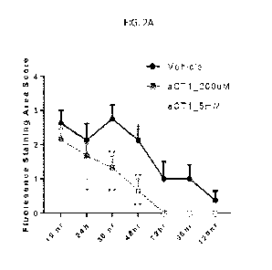
[0016] FIG. 2A is a bar graph showing the quantified fluorescence
staining of FIG. 1 (n=6
per treatment group; *p<0.05, **p<0.01 ***p<0.001; SEM).
[0017] FIG. 2B shows central corneal thickness (t,1m) in rabbit
eyes pre-dose and at day 1,
day 2, and day 3 following corneal chemical burn, with eye drop administration
of aCT1
peptide (200 1.1M or 5 mM) or vehicle control.
[0018] FIG. 3 shows 1% fluorescein staining of rabbit eyes after
bilateral central
transepithelial phototherapeutic keratectomy (PTK) surgery and treatment with
aCT1 peptide
(150 [iM) or vehicle control.
[0019] FIGS. 4A-4B show that treatment with aCT1 peptide decreases
corneal thickening
that occurs following corneal exposure to nitrogen mustard (NM). FIG. 4A shows
corneal
CA 03196342 2023- 4- 20
WO 2022/087396
PCT/US2021/056231
thickness and FIG. 4B provides quantification of the same in the indicated
groups. n = 3 per
treatment group.
[0020] FIGS. 5A-5D show that treatment with aCT1 peptide decreases
inflammatory
responses in nitrogen mustard (NM)-exposed cornea. FIG. 5A shows H&E staining
and FIG.
5B shows inflammatory cell infiltration was significantly reduced in aCT1
treated groups. FIG.
5C shows pro-inflammatory enzyme COX2 staining and FIG. 5D provides
quantification of
COX2 in corneal tissues. (n = 3 per treatment group; one- way ANOVA, "p<0.01;
-'"p<0.001; SD).
[0021] FIGS. 6A-6D show that aCT1 treatment of NM-exposed corneas
may protect
corneal fibroblasts and keratocytes. FIG. 6A provides H&E staining and FIG. 6B
shows the
corneal fibroblasts cell counts in treated corneas in the indicated groups.
FIG. 6C shows IHC
staining for matrix metalloproteinase-9 (MA4P-9), which leads to degradation
of the corneal
stroma. FIG. 5D provides quantification of MMP-9 positivity in the corneal
stroma. n= 3 per
treatment group.
[0022] FIGS. 7A-7D show that treatment with aCT1 peptide reduces
corneal
neovascularization in NM-exposed corneas. FIG. 7A shows blood vessels in the
cornea in each
group by H&E staining and FIG. 7B provides a quantification of blood vessel
count in each
group. Vascular endothelial growth factor (VEGF) is a signaling protein that
stimulates
neovascularization. FIG. 7C shows staining for VEGF and FIG. 7D provides
quantification of
the VEGF positivity score. n= 3 per treatment group.
DETAILED DESCRIPTION
[0023] Provided herein are formulations for topical delivery of
peptide compositions to the
eye, and methods for treating or preventing eye disorders and conditions, such
as corneal
injuries.
[0024] Therapeutics to be used for ophthalmic delivery must meet
ICH and USP guidelines
governing formulation heterogeneity, stability, viscosity, and pH to ensure
safety as well as
effective delivery of the active pharmaceutical ingredient to the relevant
tissues of the eye. The
development of peptide containing formulations for ocular use presents unique
challenges
including the poor chemical and physical stability of peptides in solution,
particularly in the
type of solution that provides sufficient stability and viscosity for topical
administration to the
6
CA 03196342 2023- 4- 20
WO 2022/087396
PCT/US2021/056231
eye. Failure to develop peptide containing formulations that exhibit
sufficient bioavailability
for treatment of ocular disorders is a likely explanation for the lack of
peptide based ocular
therapeutics that have obtained FDA approval. Few peptide containing ocular
formulations
have been FDA approved. For those that are approved, the route of
administration for these
peptide containing formulations is intravitreal injection (Mandal et al.
2018), instead of the
safer and less invasive topical route of administration.
[0025] A viscoelastic polymer such as hydroxypropyl methylcellulose
(HPMC) has not
been used in combination with a peptide in a formulation appropriate for eye
drop delivery. A
formulation with appropriate viscosity, surface tension, and other physical
properties is
necessary for an eye drop to achieve sufficient contact time with the ocular
surface necessary
to ensure peptide delivery. Small peptides are expected to exhibit poor
solubility in
conventional excipients employed to modify eye drop viscosity such as HPMC,
carboxymethylcellulose (CMC), hydroxyethyl cellulose (HEC), and PF-127.
Peptide
aggregation in combination with these ingredients results in precipitate
formation that makes
the formulation unsuitable for ocular delivery. Thus, conventional teaching in
the art is away
from a formulation which utilizes a viscoelastic polymer such as HPMC in
combination with
a small peptide active pharmaceutical ingredient. Instead, current
formulations for delivery of
small peptide therapeutics involve admixture of non-reducing sugars, amino
acids, and
surfactants with the peptide or other macromolecules to achieve formulations
suitable for
ocular delivery (Giannos et al. 2018, Kamerzell et al. 2011).
[0026] The present inventors unexpectedly discovered that a
combination of a viscoelastic
polymer with a small peptide active ingredient resulted in a formulation that
maintains peptide
solubility and with a kinematic viscosity appropriate for topical delivery of
the peptide to the
ocular surface. The formulation surprisingly achieves a stable solution state
of the peptide in a
formulation for eye drop delivery. Peptide stability in solution is an
important performance
characteristic differentiating the present invention from conventional peptide
delivery systems.
Due to poor solubility, peptides are known to precipitate out in conventional
eye drop delivery
systems. It is well known that solutions with high viscosity cannot be filter
sterilized;
conventional formulations for ocular delivery of macromolecules have used
aqueous excipients
that do not include viscoelastic polymers to achieve a solution with low
viscosity that can be
7
CA 03196342 2023- 4- 20
WO 2022/087396
PCT/US2021/056231
sterilized by passage through a sterile filter. Thus, there are no examples of
HPMC admixed
with therapeutic peptides to achieve a formulation suitable for topical
delivery to the eye.
[0027] However, the present inventors surprisingly achieved a
stable formulation
comprising aCT1 peptide and HPMC, that was suitable for topical delivery to
the eye and
effective in treatment of ocular disorders. This formulation was unpredictably
superior to
formulations comprising CMC, HEC, or pluronic gel (PF-127) instead of HPMC.
Unexpectedly, the use of the viscoelastic polymer HPMC with the peptide
yielded a
formulation having a viscosity sufficient to enable contact time necessary for
peptide delivery
to the ocular surface, yet that can be sterilized through passage of the
solution through a 0.22
tM PVDF or PES membrane filter. Formulation sterility is necessary for
delivery of
therapeutic peptides to sensitive tissues such as the eye. Passage of
formulations through a 0.22
1.4M PVDF or PES membrane filter produces a formulation with sterility
suitable for the
delivery of medication to sensitive ocular tissues.
[0028] In embodiments, the formulation further comprises sodium
chloride (NaCl),
potassium chloride (KCl), sodium iodide (NaI), magnesium chloride (MgCl2),
potassium
fluoride (KF), calcium chloride (CaCl2), sodium tetrafluoroborate (NaBF4),
and/or sodium
bromide (NaBr). In embodiments, the formulation comprises NaCl. In
embodiments, the NaCl
surprisingly provides greater stability relative to a formulation that does
not comprise NaCl. In
embodiments, the NaCl is present at a concentration from about 0.5% to about
2.0%, or about
0.7% to about 1.5%. In embodiments, the NaCl is present at a concentration
from about 0.25%
to about 0.9%. In embodiments, the NaCl is present at a concentration of about
0.9% (w/w).
[0029] In embodiments, the formulations provided herein exhibit
stability over time at a
range of temperatures. For example, the formulations provide peptide stability
for at least 1
week, at least 2 weeks, at least 3 weeks, at least 4 weeks, at least 5 weeks,
at least 6 weeks, at
least two months, at least 3 months, at least 4 months, at least 5 months, at
least 6 months, at
least 8 months, at least 12 months, at least 18 months, at least 2 years, at
least 3 years, or at
least 6 years. In embodiments, the formulations provide peptide stability at
about -200, about
C, about 25 C, and any temperature therebetween. In certain embodiments, the
formulations
provide peptide stability at about -200 for at least 6 months, at least 8
months, at least 12
months, at least 18 months, at least 2 years, at least 3 years, at least 4
years, at least 5 years, or
at least 6 years. In embodiments, the formulations provided herein comprise a
peptide (e.g.,
8
CA 03196342 2023- 4- 20
WO 2022/087396
PCT/US2021/056231
an alpha connexin peptide), wherein the peptide remains at least about 80%, at
least about 81%,
at least about 82%, at least about 83%, at least about 84%, at least about
85%, at least about
86%, at least about 87%, at least about 88%, at least about 89%, at least
about 90%, at least
about 91%, at least about 92%, at least about 93%, at least about 94%, or at
least about 95%
stable over at least about 1 month. In embodiments, the formulations provided
herein comprise
a peptide (e.g., an alpha connexin peptide), wherein the peptide remains at
least about 80%, at
least about 81%, at least about 82%, at least about 83%, at least about 84%,
at least about 85%,
at least about 86%, at least about 87%, at least about 88%, at least about
89%, at least about
90%, at least about 91%, at least about 92%, at least about 93%, at least
about 94%, or at least
about 95% stable over at least about 3 months. In embodiments, the
formulations provided
herein comprise a peptide (e.g., an alpha connexin peptide), wherein the
peptide remains at
least about 80%, at least about 81%, at least about 82%, at least about 83%,
at least about 84%,
at least about 85%, at least about 86%, at least about 87%, at least about
88%, at least about
89%, at least about 90%, at least about 91%, at least about 92%, at least
about 93%, at least
about 94%, or at least about 95% stable over at least about 1 year, at least
about 2 years, at least
about 3 years, at least about 4 years, at least about 5 years, or at least
about 6 years. Such
stability is achieved at about -20 , about 5 C, about 25 C, and any
temperature therebetween
when the formulations provided herein are utilized.
[0030] In embodiments, the formulations provided herein exhibit no
impurities or
negligible impurities or an acceptable level of impurities over time at a
range of storage
temperatures. For example, the formulations exhibit no impurities or
negligible impurities or
an acceptable level of impurities for at least 1 week, at least 2 weeks, at
least 3 weeks, at least
4 weeks, at least 5 weeks, at least 6 weeks, at least two months, at least 3
months, at least 4
months, at least 5 months, at least 6 months, at least 1 year, at least 2
years, at least 3 years, at
least 4 years, at least 5 years, or at least 6 years. In embodiments, the
formulations exhibit no
impurities or negligible impurities or an acceptable level of impurities at
about -20 , about 5 C,
about 25 C, and any temperature therebetween. In embodiments, negligible
levels of impurities
in the formulation may be less than 0.1%. In embodiments, acceptable levels of
impurities in
the formulation may be less than about 5%, less than about 4%, less than about
3%, less than
about 2%, less than about 1%, less than about 0.5%.
9
CA 03196342 2023- 4- 20
WO 2022/087396
PCT/US2021/056231
[0031] In embodiments, the formulations provided herein are readily
filterable (e.g.,
filterable through a 0.2 psn PES filter). In embodiments, the formulations
provided herein are
more filterable compared to formulations previously used for ocular
administration of peptides.
In embodiments, the formulations provided herein comprising HPLC, a peptide
(e.g., an alpha
connexin peptide), NaCl, and does not require an additional vehicle, buffer,
or excipient, to
have formulation properties (e.g., viscosity, osmolality, density, pH,
filterability) as well as
purity and stability profiles suitable for ocular delivery.
[0032] In embodiments, the present disclosure provides an eye drop
carrier containing a
therapeutic peptide (e.g., aCT1 peptide) that is non-irritating, stable, and
of appropriate
characteristics for topical use in the eye. Thus, in embodiments, the present
disclosure provides
therapeutic eye drop compositions comprising alpha connexin polypeptides for
the treatment
of ocular injury or disease. In embodiments, the eye drop formulation further
comprises
HIPMC. In embodiments, the formulation further comprises a buffer and/or
excipient which
stabilizes the alpha connexin polypeptide during storage. In embodiments, the
alpha connexin
polypeptide comprises a carboxyl terminal amino acid sequence of alpha
connexin. The alpha
connexin polypeptides of the present invention may comprise, consist, or
include the carboxy-
terminal most 4 to 30 contiguous amino acids of an alpha connexin protein or
conservative
variant thereof. In embodiments, the said at least one alpha connexin
polypeptide is linked at
its amino terminus to a cellular internalization transporter.
100331 In embodiments, the present disclosure provides a
formulation of a stable eye drop
carrier that contains aCT1 for therapeutic application in ophthalmic
indications, and methods
for making the same. In embodiments, the ophthalmic indications include wound
healing,
inflammatory and immune modulation, tissue regeneration, biomechanical
restoration, or
treatment of other physiological conditions affecting any part of the cornea
or other ocular
tissue. The formulations provided herein may be administered to treat acute
and chronic
injuries and wounds, including military or civilian chemical injuries or
corneal lacerations,
surgery-related conditions, and acute and chronic manifestations of any
primary ocular disorder
or other condition causing a secondary ocular condition manifesting or
necessitating medical
attention. The formulations possess physicochemical, biochemical, and
rheological properties
that enable its ability to provide a therapeutic and effective amount of aCT1
peptide when
applied to injuries, wounds, and conditions affecting proper eye function.
CA 03196342 2023- 4- 20
WO 2022/087396
PCT/US2021/056231
[0034] In embodiments, one or more buffering agents in any form
added to sterile water
may be used to maintain a physiologically relevant pH or to maintain a pH
where the addition
of pH modulators will result in a physiological-relevant pH. In some
embodiments, one or more
pH modulators such as sodium borate, citric acid, sodium nitrate, histidine,
hydrochloric acid
or sodium hydroxide may be added to adjust within the desire therapeutic range
of pH 5 to 8.
Preferably, buffering agents are non-irritating, non-staining, and non-
immunogenic. In
embodiments, a preferred buffer is histidine. In embodiments, the histidine is
present at a
concentration of about 20 mM to about 80 mM. In embodiments, the histidine is
present at a
concentration of about 40 mM.
100351 In embodiments, the formulation further comprises a tonicity
modifier. For
example, in embodiments, the formulation further comprises dextrose, glycerin,
mannitol,
potassium chloride, or magnesium chloride. In embodiments, the formulation
further
comprises an antioxidant, such as methionine.
[0036] In embodiments, the formulations provided herein do not
include a buffering agent.
In embodiments, additional excipients are excluded from the formulation, such
that the
formulation does not comprise an excipient. In embodiments, the formulation
comprises the
active agent peptide, HPMC, and no added excipients. In embodiments, the
formulations
provided herein do not include any added sugars, amino acids, and/or
surfactants. In
embodiments, the formulation comprises, consists essentially of, or consists
of the active agent
(e.g., a connexin peptide), HPMC, NaCl, and water. In embodiments, the HPMC is
present in
the formulation at a concentration of about 0.2% w/w to about 1.0% w/w. In
embodiments, the
HPMC is present in the formulation at a concentration of about 0.5% w/w. In
embodiments,
the HPMC is present in the formulation at a concentration of about 1.0% w/w.
[0037] In embodiments, one or more polymers such as HPMC is
included in the
formulation to stabilize the isolated polypeptide. Preferably, the formulation
comprises a
stabilizer that is non-irritating, non-staining, and non-immunogenic. The
addition of stabilizers
enable long-term (i.e., for 3 months, for 6 months, for 9 months, for 12
months, for 18 months,
or for 24 months) storage of the drug product under a variety of temperature
conditions (e.g.,
at about 5 C., at about 10 C., at about 15 C., at about 20 C., at about 25
C., at about 30
C., at about 35 C., or at about 40 C) and under a range of relative
humidities (e.g., at about
0% relative humidity, at about 10% relative humidity, at about 20% relative
humidity, at about
11
CA 03196342 2023- 4- 20
WO 2022/087396
PCT/US2021/056231
30% relative humidity, at about 40% relative humidity, at about 50% relative
humidity, at about
60% relative humidity, at about 70% relative humidity, at about 80% relative
humidity, at about
90% relative humidity, or at about 100% relative humidity). In embodiments,
the present
invention may also include a preservative to further maintain the described
long-term storage
under the stated variety of temperatures and relative humidities.
[0038] Exemplary formulations are provided below in Table 1.
Form His NaC1 MgC12 mannitol Met
HPMC
No peptide pH (mM) (mM) (mM) (mM) (mM) (0/0)
1 20 6.5 30 130 0 0 0 0.19
2 20 6.0 20 65 50 0 10 0.15
3 20 5.5 10 65 0 150 0 0.11
4 20 6.0 40 100 25 0 0 0
10 6.5 40 65 50 0 5 0.19
6 10 6.0 30 130 0 0 0 0.15
7 10 6.0 20 100 25 0 0 0.11
8 10 7.0 10 65 0 150 20 0
9 20 7.0 30 130 0 0 0 0.19
20 6.5 20 65 0 150 0 0.15
11 20 7.0 20 0 0 270 10 0.11
12 20 6.0 20 130 0 0 0 0
13 5 5.5 20 0 0 270 0 0.15
14 5 6.0 10 130 0 0 10 0
20 6.6 0 150 0 0 0 0.5
16 20 5.0 0 150 0 0 0 0.5
100391 In embodiments, the formulations provided herein may be contained in
plastic eye
dropper or glass vial containing a single dose or multiple doses for
therapeutic administration
to a subject in need thereof a topical ophthalmic formulation comprising of at
least one aCT
polypeptide. In embodiments, the formulation may be contained in a glass
container, and may
be more stable in glass containers compared to containers made of other
materials (e.g.,
plastic). In embodiments, the formulation may be contained in a plastic
container, e.g., a plastic
eye dropper. In embodiments, the topical ophthalmic formulation comprises
HPMC. In
embodiments, the formulations provided herein are in a sterile, ready-to-use
eye drop
formulation in an administration-appropriate and -designed eye dropper bottle
or vial.
[0040] In embodiments, the present disclosure provides methods for treating
and
preventing corneal injuries and ocular trauma. In embodiments, the methods
include topical
administration to the eye of a formulation provided herein comprising an alpha
connexin
12
CA 03196342 2023- 4- 20
WO 2022/087396
PCT/US2021/056231
polypeptide. In embodiments, the injury or trauma is a closed globe ocular
injury or wound
where damage to the cornea has occurred. The cause of the corneal injury or
wound is not
limited to and may include blast injuries, chemical and thermal burns, and
other insults or
conditions causing acute or chronic injury, as either a primary and secondary
manifestation of
a disorder or disease. In embodiments, the cause of the corneal injury is
exposure to a vesicant,
or blister agent, such as nitrogen mustard or sulfur mustard (e.g., mustard
gas). In
embodiments, the disorder or disease is diabetes. In embodiments, the disorder
or disease is
diabetic keratopathy. In embodiments, the chronic disease is retinal disease.
In embodiments,
present disclosure provides methods for treating and preventing retinal
diseases. For example,
in embodiments, the retinal disease is selected from macular degeneration
(e.g., age-related
macular degeneration (AMD), neovascular age-related macular degeneration
(nAIVID)),
retinitis pigmentosa (RP), retinal detachment, diabetic retinopathy, macular
edema, diabetic
macular edema (DME), and macular edema occurring after retinal vein occlusion
(RVO). In
embodiments, the disease or disorder involves corneal defects that occur in a
subject when
treatment for an ocular disease or disorder (e.g., a retinal disorder)
involves vitrectomy and/or
one or more intravitreal injections.
[0041] In embodiments, the methods provided herein includes
treatment and/or prevention
of any diseases or disorders leading to corneal scarring or excessive and
dysregulated
inflammation or an immune response. In embodiments, the subject is a human
subject that has
a persistent corneal epithelial defect (PED or PCED), which results from the
failure of rapid
reepithelialization and closure after corneal injury (e.g., within about 2
weeks), even with
standard of care supportive treatment. PEDs can result in serious
complications including
infection and vision loss. In embodiments, the PED is caused by dry eye
disease. Accordingly,
in embodiments, the formulations and methods provided herein treat a subject
suffering from
PEDs or otherwise suffering from corneal injury by enhancing the rate of
reepithelization
following corneal injury. In embodiments, administration of the provided
formulations
enhances the rate of reepithelialization by about 10%, about 25%, about 50%,
about 75%, about
100%, or more. In embodiments, the administration of the provided formulations
enhances the
rate of reepithelialization compared to the rate of reepithelialization in a
control wherein
standard of care or no treatment is administered to the eye of the subject.
The rate of
reepithelialization may be enhanced such that corneal healing occurs within
about 2, about 3,
13
CA 03196342 2023- 4- 20
WO 2022/087396
PCT/US2021/056231
about 4, about 5, about 6, about 7, about 8, about 9, about 10, about 11,
about 12, about 13, or
about 14 days after injury. The formulations and methods provided herein are
not limited to
exclusive treatment alone and may be used in conjunction with other standard
of care
treatment(s).
[0042] The polypeptides useful in the formulations and methods
provided herein may be
any polypeptide with properties such as wound healing properties, anti-
inflammatory
properties, properties relating to protection or regeneration of the corneal
stroma, and/or anti-
neovascular properties. In embodiments, the polypeptides can be any suitable
polypeptide
having a molecular weight of about 1,0 kDa to about 10.0 kDa. In embodiments,
the
polypeptide can be any suitable polypeptide having a molecular weight of about
1.0 kDa, about
2.0 kDa, about 3.0 kDa, about 4.0 kDa, about 5.0 kDa, about 6.0 kDa, about 7.0
kDa, about 8.0
kDa, about 9.0 kDa, or about 10.0 kDa.
[0043] In embodiments, the polypeptides can be any polypeptide
comprising the carboxy-
terminal most amino acids of an alpha Connexin, wherein the polypeptide does
not comprise
the full-length alpha Connexin protein. Thus, in embodiments, the provided
polypeptide does
not comprise the cytoplasmic N-terminal domain of the alpha Connexin. In
embodiments, the
provided polypeptide does not comprise the two extracellular domains of the
alpha Connexin.
In embodiments, the provided polypeptide does not comprise the four
transmembrane domains
of the alpha Connexin. In embodiments, the provided polypeptide does not
comprise the
cytoplasmic loop domain of the alpha Connexin. In embodiments, the provided
polypeptide
does not comprise that part of the sequence of the cytoplasmic carboxyl
terminal domain of the
alpha Connexin proximal to the fourth transmembrane domain. There is a
conserved proline or
glycine residue in alpha Connexins consistently positioned some 17 to 30 amino
acids from the
carboxyl terminal-most amino acid For example, for human Cx43 a proline
residue at amino
acid 363 is positioned 19 amino acids back from the carboxyl terminal most
isoleucine. In
another example, for chick Cx43 a proline residue at amino acid 362 is
positioned 18 amino
acids back from the carboxyl terminal-most isoleucine. In another example, for
human Cx45 a
glycine residue at amino acid 377 is positioned 19 amino acids back from the
carboxyl terminal
most isoleucine. In another example for rat Cx33, a proline residue at amino
acid 258 is
positioned 28 amino acids back from the carboxyl terminal most methionine.
Thus, in
embodiments, the provided polypeptide does not comprise amino acids proximal
to said
14
CA 03196342 2023- 4- 20
WO 2022/087396
PCT/US2021/056231
conserved proline or glycine residue of the alpha Connexin. Thus, the provided
polypeptide
can comprise the c-terminal-most 4 to 30 amino acids of the alpha Connexin,
including the c-
terminal most 4, 5, 6, 7, 8, 9, 10, 11, 12, 13, 14, 15, 16, 17, 18, 19, 20,
21, 22, 23, 24, 25, 26,
27, 28, 29, 30 amino acids of the alpha Connexin. Exemplary alpha Connexin
polypeptides are
disclosed in U.S. Patent Nos. 7,786,074; 7,888,319; 8,357,668; 8,809,257;
8,916,515;
8,859,733; 8,846,605; 9,161,984; 9,394,351; 9,408,381; 9,844,214; 9,855,313;
10,398,140;
and 10,398,757, and/or International Patent Application No. PCT/US2018/000035,
the entire
contents of each of which are hereby incorporated by reference.
[0044] Connexins are the sub-unit protein of the gap junction
channel, which is responsible
for intercellular communication (Goodenough and Paul, 2003). Based on patterns
of
conservation of nucleotide sequence, the genes encoding Connexin proteins are
divided into
two families termed the alpha and beta Connexin genes. The carboxy-terminal-
most amino
acid sequences of alpha Connexins are characterized by multiple distinctive
and conserved
features. This conservation of organization is consistent with the ability of
aCT peptides to
form distinctive 3D structures, interact with multiple partnering proteins,
mediate interactions
with lipids and membranes, interact with nucleic acids including DNA, transit
and/or block
membrane channels and provide consensus motifs for proteolytic cleavage,
protein cross-
linking, ADP-ribosylation, glycosylation and phosphorylation. Thus, the
provided polypeptide
interacts with a domain of a protein that normally mediates the binding of
said protein to the
carboxy-terminus of an alpha Connexin. For example, nephroblastoma
overexpressed protein
(NOV) interacts with a Cx43 c-terminal domain (Fu et al., J Biol. Chem. 2004
279(35):36943-
50). It is considered that this and other proteins interact with the carboxy-
terminus of alpha
Connexins and further interact with other proteins forming a macromolecular
complex. Thus,
the provided polypeptide can inhibit the operation of a molecular machine,
such as, for
example, one involved in regulating the aggregation of Cx43 gap junction
channels.
[0045] The polypeptides provided herein comprise a carboxy-terminal
amino acid
sequence of an alpha Connexin, or a conservative variant thereof. In
embodiments, the
polypeptide comprises or consists of the amino acid sequence RPRPDDLEI (SEQ ID
NO: 2).
In embodiments, the polypeptide is aCT1, as described herein. The term "aCT1"
is used
interchangeably herein with "aCT1," "aCT", "aCT-1", "ACT," and "ACT-1". aCT1
is a 25 aa
peptide having a molecular weight of 3597.33 Da that has a compact 2-domain
design based
CA 03196342 2023- 4- 20
WO 2022/087396
PCT/US2021/056231
on linkage of an Antennapedia cell internalization domain (1-16aa;
RQPKIWFPNRRKPWKK;
SEQ ID NO: 7) to the C-terminal PDZ binding domain of the transmembrane gap
junction
protein Cx43 (17-25aa; RPRPDDLEI; SEQ ID NO:2). Accordingly, the full aCT1
sequence is
RQPKIWFPNRRKPWKK RPRPDDLEI (SEQ ID NO: 9). aCT1 and related peptides increase
the size and stability of gap junctions by modulating the molecular
interaction between Cx43
and its C-terminal binding partners, including the tight junction protein
zonula occludens-1
(ZO-1). This leads to phosphorylation of the serine 368 (S368) amino acid on
Cx43 and favors
a transition of cell-surface Cx43 from hemichannels to gap junction
intercellular channels.
Phosphorylation of S368 prevents the binding of ZO-1 to the C-terminus of Cx43
long after
aCT1 has degraded, permitting therapeutic longevity. Concomitantly, aCT1
stabilizes ZO-1 at
the cell membrane, preventing junctional degradation in response to injury and
preserving
barrier function of epithelial cells. The result is stabilization of gap
junctions (intercellular
communication) as well as tight junctions (intercellular junctions) leading to
a variety of
beneficial effects including increased cellular communication, dampened
inflammatory
responses, and reduction in the infiltration and proliferation of profibrotic
cells. Collectively,
the molecular and cellular events facilitated by aCT1 preserves tissue
integrity, reduces injury
spread, dampens pathological inflammation, and accelerates healing and tissue
regeneration
[0046] In embodiments, the compositions and methods provided herein
are related to
preventing, treating, and/or mitigating the progression of corneal injuries.
In embodiments, the
compositions and methods provided herein are related to preventing, treating,
and/or mitigating
the progression of corneal injuries. In embodiments, the formulations provided
herein are for
use in preventing, treating, and/or mitigating the progression of corneal
injuries. In
embodiments, provided herein are uses of aCT1 in the manufacture of a
medicament for
preventing or treating corneal injuries.
[0047] The aCT sequence of the provided polypeptide can be from any
alpha Connexin.
Thus, the alpha Connexin component of the provided polypeptide can be from a
human,
murine, bovine, monotrene, marsupial, primate, rodent, cetacean, mammalian,
avian, reptilian,
amphibian, piscine, chordate, protochordate or other alpha Connexin. Thus, the
provided
polypeptide can comprise an ACT of a Connexin selected from the group
consisting of mouse
Connexin 47, human Connexin 47, Human Connexin 46.6, Cow Connexin 46.6, Mouse
Connexin 30.2, Rat Connexin 30.2, Human Connexin 31.9, Dog Connexin 31.9,
Sheep
16
CA 03196342 2023- 4- 20
WO 2022/087396
PCT/US2021/056231
Connexin 44, Cow Connexin 44, Rat Connexin 33, Mouse Connexin 33, Human
Connexin 36,
mouse Connexin 36, rat Connexin 36, dog Connexin 36, chick Connexin 36,
zebrafish
Connexin 36, morone Connexin 35, morone Connexin 35, Cynops Connexin 35,
Tetraodon
Connexin 36, human Connexin 37, chimp Connexin 37, dog Connexin 37, Cricetulus
Connexin
37, Mouse Connexin 37, Mesocricetus Connexin 37, Rat Connexin 37, mouse
Connexin 39,
rat Connexin 39, human Connexin 40.1, Xenopus Connexin 38, Zebrafish Connexin
39.9,
Human Connexin 40, Chimp Connexin 40, dog Connexin 40, cow Connexin 40, mouse
Connexin 40, rat Connexin 40, Cricetulus Connexin 40, Chick Connexin 40, human
Connexin
43, Cercopithecus Connexin 43, Oryctolagus Connexin 43, Spermophilus Connexin
43,
Cricetulus Connexin 43, Phodopus Connexin 43, Rat Connexin 43, Sus Connexin
43,
Mesocricetus Connexin 43, Mouse Connexin 43, Cavia Connexin 43, Cow Connexin
43,
Erinaceus Connexin 43, Chick Connexin 43, Xenopus Connexin 43, Oryctolagus
Connexin 43,
Cyprinus Connexin 43, Zebrafish Connexin 43, Danio aequipinnatus Connexin 43,
Zebrafish
Connexin 43.4, Zebrafish Connexin 44.2, Zebrafish Connexin 44.1, human
Connexin 45,
chimp Connexin 45, dog Connexin 45, mouse Connexin 45, cow Connexin 45, rat
Connexin
45, chick Connexin 45, Tetraodon Connexin 45, chick Connexin 45, human
Connexin 46,
chimp Connexin 46, mouse Connexin 46, dog Connexin 46, rat Connexin 46,
Mesocricetus
Connexin 46, Cricetulus Connexin 46, Chick Connexin 56, Zebrafish Connexin
39.9 cow
Connexin 49, human Connexin 50, chimp Connexin 50, rat Connexin 50, mouse
Connexin 50,
dog Connexin 50, sheep Connexin 49, Mesocricetus Connexin 50, Cricetulus
Connexin 50,
Chick Connexin 50, human Connexin 59, or other alpha Connexin.
[0048] The 20-30 carboxy-terminal-most amino acid sequence of alpha
Connexins are
characterized by a distinctive and conserved organization. This distinctive
and conserved
organization includes a type II PDZ binding motif (41)-x-41); wherein x = any
amino acid and (I)
=a Hydrophobic amino acid) and proximal to this motif, Proline (P) and/or
Glycine (G) hinge
residues; a high frequency phospho-Serine (S) and/or phospho-Threonine (T)
residues; and a
high frequency of positively charged Arginine (R), Lysine (K) and negatively
charged Aspartic
acid (D) or Glutamic acid (E) amino acids. For many alpha Connexins, the P and
G residues
occur in clustered motifs proximal to the carboxy-terminal type II PDZ binding
motif The S
and T phosphor-amino acids of most alpha Connexins also are typically
organized in clustered,
repeat-like motifs. This organization is particularly the case for Cx43, where
90% of 20
17
CA 03196342 2023- 4- 20
WO 2022/087396
PCT/US2021/056231
carboxyl terminal-most amino acids are comprised of the latter seven amino
acids. In a further
example of the high conservation of the sequence, ACT peptide organization of
Cx43 is highly
conserved from humans to fish.
[0049] Thus, in one aspect, the provided polypeptide comprises one,
two, three or all of the
amino acid motifs selected from the group consisting of 1) a type II PDZ
binding motif, 2)
Proline (P) and/or Glycine (G) hinge residues; 3) clusters of phospho-Serine
(S) and/or
phospho-Threonine (T) residues; and 4) a high frequency of positively charged
Arginine (R)
and Lysine (K) and negatively charged Aspartic acid (D) and/or Glutamic acid
(E) amino
acids). In another aspect, the provided polypeptide comprises a type II PDZ
binding motif at
the carboxy-terminus, Proline (P) and/or Glycine (G) hinge residues proximal
to the PDZ
binding motif, and positively charged residues (K, R, D, E) proximal to the
hinge residues.
[0050] PDZ domains were originally identified as conserved sequence
elements within the
postsynaptic density protein PSD95/SAP90, the Drosophila tumor suppressor dig-
A, and the
tight junction protein ZO-1. Although originally referred to as GLGF or DHR
motifs, they are
now known by an acronym representing these first three PDZ-containing proteins
(PSD95/DLG/Z0-1). These 80-90 amino acid sequences have now been identified in
well over
75 proteins and are characteristically expressed in multiple copies within a
single protein. Thus,
in one aspect, the provided polypeptide can inhibit the binding of an alpha
Connexin to a
protein comprising a PDZ domain. The PDZ domain is a specific type of protein-
interaction
module that has a structurally well-defined interaction 'pocket' that can be
filled by a PDZ-
binding motif, referred to herein as a "PDZ motif'. PDZ motifs are consensus
sequences that
are normally, but not always, located at the extreme intracellular carboxyl
terminus. Four types
of PDZ motifs have been classified: type I (S/T-x-43), type II (4)-x-43), type
III ('-11-x-4:1)) and
type IV (D-x-V), where x is any amino acid, 41) is a hydrophobic residue (V,
I, L, A, G, W, C,
M, F) and 'Pis a basic, hydrophilic residue (H, R, K). (Songyang, Z., et al.
1997. Science 275,
73-77). Thus, in one aspect, the provided polypeptide comprises a type II PDZ
binding motif
[0051] When specific proteins are referred to herein, variants,
derivatives, and fragments
are contemplated. Protein variants and derivatives are well understood to
those of skill in the
art and in can involve amino acid sequence modifications. For example, amino
acid sequence
modifications typically fall into one or more of three classes:
substitutional, insertional or
deletional variants. Insertions include amino and/or carboxyl terminal fusions
as well as
18
CA 03196342 2023- 4- 20
WO 2022/087396
PCT/US2021/056231
intrasequence insertions of single or multiple amino acid residues. Insertions
ordinarily will be
smaller insertions than those of amino or carboxyl terminal fusions, for
example, on the order
of one to four residues. Deletions are characterized by the removal of one or
more amino acid
residues from the protein sequence. These variants ordinarily are prepared by
site specific
mutagenesis of nucleotides in the DNA encoding the protein, thereby producing
DNA encoding
the variant, and thereafter expressing the DNA in recombinant cell culture.
Techniques for
making substitution mutations at predetermined sites in DNA having a known
sequence are
well known and include, for example, M13 primer mutagenesis and PCR
mutagenesis. Amino
acid substitutions are typically of single residues, but can occur at a number
of different
locations at once; insertions usually will be on the order of about from 1 to
10 amino acid
residues. Substitutions, deletions, insertions or any combination thereof may
be combined to
arrive at a final construct. Substitutional variants are those in which at
least one residue has
been removed and a different residue inserted in its place.
[0052] For example, the replacement of one amino acid residue with
another that is
biologically and/or chemically similar is known to those skilled in the art as
a conservative
substitution. For example, a conservative substitution would be replacing one
hydrophobic
residue for another, or one polar residue for another. Conservatively
substituted variations of
each explicitly disclosed sequence are included within the polypeptides
provided herein.
[0053] Typically, conservative substitutions have little to no
impact on the biological
activity of a resulting polypeptide. In a particular example, a conservative
substitution is an
amino acid substitution in a peptide that does not substantially affect the
biological function of
the peptide. A peptide can include one or more amino acid substitutions, for
example 2-10
conservative substitutions, 2-5 conservative substitutions, 4-9 conservative
substitutions, such
as 2, 5 or 10 conservative substitutions.
[0054] A polypeptide can be produced to contain one or more
conservative substitutions
by manipulating the nucleotide sequence that encodes that polypeptide using,
for example,
standard procedures such as site-directed mutagenesis or PCR. Alternatively, a
polypeptide can
be produced to contain one or more conservative substitutions by using
standard peptide
synthesis methods. An alanine scan can be used to identify which amino acid
residues in a
protein can tolerate an amino acid substitution. In one example, the
biological activity of the
protein is not decreased by more than 25%, for example not more than 20%, for
example not
19
CA 03196342 2023- 4- 20
WO 2022/087396
PCT/US2021/056231
more than 10%, when an alanine, or other conservative amino acid (such as
those listed below),
is substituted for one or more native amino acids.
[0055] It is understood that there are numerous amino acid and
peptide analogs which can
be incorporated into the disclosed compositions. For example, there are
numerous D amino
acids. The opposite stereoisomers of naturally occurring peptides are
disclosed, as well as the
stereoisomers of peptide analogs. These amino acids can readily be
incorporated into
polypeptide chains by charging tRNA molecules with the amino acid of choice
and engineering
genetic constructs that utilize, for example, amber codons, to insert the
analog amino acid into
a peptide chain in a site specific way (Thorson et al., Methods in Molec. Biol
77:43-73 (1991),
Zoller, Current Opinion in Biotechnology, 3:348-354 (1992); Ibba,
Biotechnology & Genetic
Engineering Reviews 13:197-216 (1995), Cahill et al., TIBS, 14(10):400-403
(1989); Benner,
TM Tech, 12:158-163 (1994); Ibba and Hennecke, Bio/technology, 12:678-682
(1994), all of
which are herein incorporated by reference at least for material related to
amino acid analogs).
[0056] D-amino acids can be used to generate more stable peptides,
because D amino acids
are not recognized by peptidases and such. Systematic substitution of one or
more amino acids
of a consensus sequence with a D-amino acid of the same type (e.g., D-lysine
in place of L-
lysine) can be used to generate more stable peptides. Cysteine residues can be
used to cyclize
or attach two or more peptides together. This can be beneficial to constrain
peptides into
particular conformations. (Rizo and Gierasch Ann. Rev. Biochem. 61:387 (1992),
incorporated
herein by reference).
[0057] Thus, the provided polypeptide can comprise a conservative
variant of the c-
terminus of an alpha Connexin (ACT). It is understood that one way to define
any variants,
modifications, or derivatives of the disclosed genes and proteins herein is
through defining the
variants, modification, and derivatives in terms of sequence identity (also
referred to herein as
homology) to specific known sequences. Specifically disclosed are variants of
the nucleic acids
and polypeptides herein disclosed which have at least 65, 66, 67, 68, 69, 70,
71, 72, 73, 74, 75,
76, 77, 78, 79, 80, 81, 82, 83, 84, 85, 86, 87, 88, 89, 90, 91, 92, 93, 94,
95, 96, 97, 98, 99 percent
sequence identity to the stated or known sequence. Those of skill in the art
readily understand
how to determine the sequence identity of two proteins or nucleic acids. For
example, the
sequence identity can be calculated after aligning the two sequences so that
the sequence
CA 03196342 2023- 4- 20
WO 2022/087396
PCT/US2021/056231
identity is at its highest level. Another way of calculating sequence identity
can be performed
by published algorithms.
[0058] Thus, the provided polypeptide can comprise an amino acid
sequence with at least
65, 66, 67, 68, 69, 70, 71, 72, 73, 74, 75, 76, 77, 78, 79, 80, 81, 82, 83,
84, 85, 86, 87, 88, 89,
90, 91, 92, 93, 94, 95, 96, 97, 98, 99 percent sequence identity to the c-
terminus of an alpha
Connexin (ACT). Thus, in one aspect, the provided polypeptide comprises an
amino acid
sequence with at least 65, 66, 67, 68, 69, 70, 71, 72, 73, 74, 75, 76, 77, 78,
79, 80, 81, 82, 83,
84, 85, 86, 87, 88, 89, 90, 91, 92, 93, 94, 95, 96, 97, 98, 99 percent
sequence identity to SEQ
ID NO:1, SEQ ID NO: 2, or any sequence provided herein.
100591 In embodiments, the polypeptide comprises a cellular
internalization transporter or
sequence. The cellular internalization sequence can be any internalization
sequence known or
newly discovered in the art, or conservative variants thereof. Non-limiting
examples of cellular
internalization transporters and sequences include Antennapedia sequences,
TAT, HIV-Tat,
Penetratin, Antp-3A (Antp mutant), Buforin II, Transportan, MAP (model
amphipathic
peptide), K-FGF, Ku70, Prion, pVEC, Pep-1, SynBl, Pep-7, HN-1, BGSC (Bis-
Guanidinium-
Spermidine-Cholesterol, and BGTC (Bis-Guanidinium-Tren-Cholesterol). Exemplary
cell
internalization transporters are provided in Table 2A.
Table 2A. Exemplary cell internalization sequences
Name Sequence SEQ ID NO
Antp RQPKIWFPNRRKPWKK (SEQ ID NO:
7)
HIV-Tat GRKKRRQRPPQ (SEQ ID NO:
14)
Penetratm RQIKIWF QNRRMKWKK (SEQ ID NO:
15)
Antp-3A RQIAIWFQNRRMKWAA (SEQ ID NO:
16)
Tat RKKRRQRRR (SEQ ID NO:
17)
Buforin II TRS SRAGLQFPVGRVHRLLRK (SEQ ID NO:
18)
Transportan GWTLNSAGYLLGKINKALAALA (SEQ ID NO:
19)
KKIL
model amphipathic peptide KLALKLALKALKAALKLA (SEQ ID NO:
20)
(MAP)
K-FGF AAVALLPAVLLALLAP (SEQ ID NO:
21)
Ku70 VPMLK-PMLKE (SEQ ID NO:
22)
21
CA 03196342 2023- 4- 20
WO 2022/087396
PCT/US2021/056231
Prion MANLGYWLLALFVTMWTDVGL (SEQ ID NO:
23)
CKKRPKP
pVEC LLIILRRRIRKQAHAHSK (SEQ ID NO:
24)
Pep-1 KETWWETWWTEWSQPKKKRKV (SEQ ID NO:
25)
SynB1 RGGRLSYSRRRFSTSTGR (SEQ ID NO:
26)
Pep-7 SDLWEMMMVSLACQY (SEQ ID NO:
27)
I-IN-1 TSPLNIHNGQKL (SEQ ID NO:
28)
BGSC (Bis- Guanidinium- (n/a)
Spermidine- Cholesterol)
BGTC (Bis- Guanidinium- (n/a)
Tren- Cholesterol)
[0060] Any other internalization sequences now known or later
identified can be combined
with a peptide of the invention.
[0061] The provided polypeptide can comprise any aCT sequence (e.g,
any of the aCT
peptides disclosed herein) in combination with any of the herein provided cell
internalization
sequences. Examples of said combinations are provided in Table 2B. Thus, the
provided
polypeptide can comprise an Antennapedia sequence comprising amino acid
sequence SEQ ID
NO:7. Thus, the provided polypeptide can comprise the amino acid sequence SEQ
ID NO:8,
SEQ ID NO:9, SEQ ID NO:10, SEQ ID NO:11, or SEQ ID NO: 12.
Table 2B. ACT Polypeptides with Cell Internalization Sequences (CIS) aCT
Polypeptides with Cell Internalization Sequences (CIS)
CIS/ACT Sequence
SEQ ID NO
Antp/ RQPKIWFPNRRKPWKK PSSRASSRASSRPRPDDLEI
SEQ ID NO:8
ACT 2
Antp/ RQPKIWFPNRRKPWKK RPRPDDLEI
SEQ ID NO:9
ACT 1
Antp/ RQPKIWFPNRRKPWKK RPRPDDLEV
SEQ ID NO: 10
ACT 3
Antp/ RQPKIWFPNRRKPWKK RPRPDDVPV
SEQ ID NO: ii
ACT 4
Antp/ RQPKIWFPNRRKPWKK KARSDDLSV
SEQ ID NO: i2
ACT 5
22
CA 03196342 2023- 4- 20
WO 2022/087396
PCT/US2021/056231
HIV-Tat/ GRKKRRQRPPQ RPRPDDLEI
SEQ ID NO:56
ACT 1
Penctratin/ RQIKIWFQNRRMKWKK RPRPDDLEI
SEQ ID NO:57
ACT 1
Antp-3A/ RQIAIWFQNRRMKWAA RPRPDDLEI
SEQ ID NO:58
ACT 1
Tat/ RKKRRQRRR RPRPDDLEI
SEQ ID NO:59
ACT1
Buforin II/ TRSSRAGLQFPVGRVHRLLRK RPRPDDLEI
SEQ ID NO:60
ACT 1
Transportan/ GWTLNS A GYLLGKINK A LA A LA KKIL RPRPDDLEI
SEQ ID NO:61
ACT 1
MAP/ KLALKLALKALKAALKLA RPRPDDLEI
SEQ ID NO:62
ACT 1
K-FGF/ AAVALLPAVLLALLAP RPRPDDLEI
SEQ ID NO:63
ACT 1
Ku70/ VPMLKPMLKE RPRPDDLEI
SEQ ID NO:64
ACT 1
Prion/ MANLGYWLLALFVTMWTDVGLCKKRPKP
SEQ ID NO: 65
ACT 1 RPRPDDLEI
pVEC/ LLIILRRRIRKQAHAHSK RPRPDDLEI
SEQ ID NO:66
ACT 1
Pep-1/ KETWWETWWTEWSQPKKKRKV RPRPDDLEI
SEQ ID NO:67
ACT 1
SynB1/ RGGRLSYSRRRFSTSTGR RPRPDDLEI
SEQ D NO:68
ACT 1
Pep-7/ SDLWEMMMVSLACQY RPRPDDLEI
SEQ ID NO:69
ACT 1
FIN-1/ TSPLNIHNGQKL RPRPDDLEI
SEQ ID NO:70
ACT 1
[0062] Also provided are isolated nucleic acids encoding the
polypeptides provided herein.
The disclosed nucleic acids are made up of for example, nucleotides,
nucleotide analogs, or
nucleotide substitutes. Non-limiting examples of these and other molecules are
discussed
herein. It is understood that for example, when a vector is expressed in a
cell, the expressed
mRNA will typically be made up of A, C, G, and U. Thus, provided is an
isolated nucleic acid
23
CA 03196342 2023- 4- 20
WO 2022/087396
PCT/US2021/056231
encoding a polypeptide comprising the amino acid sequence SEQ ID NO:1, SEQ ID
NO:2,
SEQ ID NO:3, SEQ ID NO:4, SEQ ID NO:5, SEQ ID NO:7, SEQ ID NO:8, SEQ ID NO:9,
SEQ ID NO:10, SEQ ID NO:11, or SEQ ID NO:12.
[0063] In embodiments, provided herein is a composition comprising
one or more of the
herein provided polypeptides, nucleic acids, or vectors in a pharmaceutically
acceptable carrier.
For example, provided is a composition comprising SEQ ID NO:2 or SEQ ID NO:9
in a
pharmaceutically acceptable carrier. In embodiments, the composition comprises
one or more
of the herein provided polypeptides encapsulated in a microcarrier. For
example, in
embodiments, the composition comprises one or more of the herein provided
polypeptides,
wherein the polypeptides are in a nanoparticle or exosome.
[0064] In embodiments, the compositions provided herein comprise
drug loaded
microcarrier formulations comprising nanoparticles or exosomes. In
embodiments, the size of
the nanoparticles is from about 100 nm to about 1000 nm, or about 100 nm to
about 500 nm,
or about 200 nm to about 250 nm, or about 100 nm to about 200 nm.
[0065] In embodiments, formulation comprises about 0.01% w/w to
about 3.5% w/w, or
about 0.05% w/w to about 3.0% w/w, or about 0.07% w/w to about 2.0% w/w, or
about 0.1%
w/w to about 1.0% w/w, or about 0.1% w/w to about 0.5% w/w, or about 0.2% w/w
to about
0.8% w/w, or about 0.03% w/w to about 0.07% w/w, or about 0.05% w/w, of the
polypeptide.
In embodiments, the formulation comprises about 0.035% w/w of the polypeptide
or about
0.07% w/w of the polypeptide or about 0.1% w/w of the polypeptide, or about
1.0% w/w of
the peptide, or about 3.5% w/w of the polypeptide.
[0066] In embodiments, the formulation comprises about 0.00035
mg/mL to about 35
mg/mL, or about 0.001 mg/mL to about 20 mg/mL, or about 0.01 mg/mL to about
3.5 mg/mL,
or about 0.1 mg/mL to about 1.0 mg/mL, or about 0.2 mg/mL to about 0.8 mg/mL,
or about
0.3 mg/mL to about 0.7 mg/mL of the polypeptide. In embodiments, the
formulation comprises
about 0.1 mg/mL, about 0.2 mg/mL, about 0.3 mg/mL, about 0.35 mg/mL, about 0.4
mg/mL,
about 0.5 mg/mL, about 0.6 mg/mL, about 0.7 mg/mL, about 0.8 mg/mL, about 0.9
mg/mL, or
about LO mg/mL of the polypeptide. In embodiments, the formulation comprises
about 0.35
mg/mL or about 0.7 mg/mL of the polypeptide. In embodiments, the formulation
comprises
about 1 mg/mL or about 10 mg/mL or about 20 mg/mL of the polypeptide.
24
CA 03196342 2023- 4- 20
WO 2022/087396
PCT/US2021/056231
[0067] In embodiments, the composition is administered to the
subject in a formulation
comprising about 1 uM to about 100,000 M, or about 10 p.M to about 50,000 M,
or about
100 p.M to about 10,000 p.M, or about 10 p.M to about 9,000 p.M, or about 50
p.M to about
5,000 M, or about 100 p.M to about 2,000 M, or about 200 p.M to about 2,000
M, or about
200 p.M to about 1,000 p.M, or about 50 p.M to about 1,500 p.M of the
polypeptide, or about
100 p.M to about 1,000 p.M of the polypeptide, or about 500 to about 1,500 p.M
of the
polypeptide. In embodiments, the composition is administered to the subject in
a formulation
comprising about 1 p.M. about 5 p.M. about 50 p.M. about 100 M, about 150 M,
about 200
p.M, about 300 p.M, about 400 p.M, about 500 p.M, about 600 p.M, about 700 uM,
about 800
p.M, about 900 p.M, about 1,000 M, about 1,500 M, about 2,000 M, about
3,000 p.M, about
4,000 uM, about 5,000 uM, about 6,000 uM, about 7,000 uM, about 8,000 uM,
about 9,000
p,M, about 10,000 p,M, about 20,000 p,M, about 25,000 M, about 50,000 M,
about 75,000
M, about 100,000 M, or more, of the polypeptide.
[0068] In embodiments, the formulation is administered to the eye
immediately after the
event that caused the ocular injury. In embodiments, the formulation is
administered to the
subject within about 1 hour, within about 2, hours, within about 3 hours,
within about 4 hours,
within about 5 hours, within about 6 hours, within about 8 hours, within about
12 hours, within
about 18 hours, or within about 24 hours of the event the caused the ocular
injury. In
embodiments, the formulation is administered to the subject at least about 2
hours following
the event that caused the ocular injury. In embodiments, the formulation is
administered to the
eye of the subject daily, e.g., once per day, or twice per day, and/or about
every 8 hours or
about every 12 hours. In embodiments, the formulation is administered until
ocular healing is
observed. In embodiments, the formulation is administered for at least about 2
days, at least
about 3 days, at least about 4 days, at least about 5 days, at least about 6
days, at least about 7
days, or for longer. In embodiments, the formulation is administered
chronically. As used
herein, the term "administered chronically" means the formulation is
administered for an open-
ended dosing regimen, that is, treatment is started and intended to continue
for an indefinite
period of time and/or until symptoms resolve, etc. In embodiments, for chronic
disease
conditions, the formulation is administered to the eye upon detection of the
chronic disease
condition and is administered chronically. In embodiments, the formulation is
administered
CA 03196342 2023- 4- 20
WO 2022/087396
PCT/US2021/056231
chronically to subjects with corneal injuries resulting from chronic
conditions such as dry eye
disease (DED).
[0069] As used herein, "subject" include vertebrates, more
specifically a mammal (e.g., a
human, horse, pig, rabbit, dog, sheep, goat, non-human primate, cow, cat,
guinea pig or rodent),
a fish, a bird or a reptile or an amphibian. In embodiments, the subject is a
human subject. The
term does not denote a particular age or sex. Thus, adult and newborn
subjects, as well as
fetuses, whether male or female, are intended to be covered. In embodiments, a
patient refers
to a subject afflicted with a disease or disorder. In embodiments, a patient
population refers to
a particular, defined set of subjects having a disease or disorder or at risk
of developing a
particular disease or disorder.
[0070] As used herein, "inhibit," "inhibiting," and "inhibition"
mean to decrease an
activity, response, condition, disease, or other biological parameter. This
can include, but is not
limited to, the complete loss of activity, response, condition, or disease.
Thus, the reduction
can be a 10, 20, 30, 40, 50, 60, 70, 80, 90, 100%, or any amount of reduction
in between as
compared to native or control levels.
[0071] Ranges and values may be expressed herein as from "about"
one particular value,
and/or to "about" another particular value. When such a range is expressed,
also specifically
contemplated and considered disclosed is the range from the one particular
value and/or to the
other particular value unless the context specifically indicates otherwise.
All of the individual
values and sub-ranges of values contained within an explicitly disclosed range
are also
specifically contemplated and should be considered disclosed unless the
context specifically
indicates otherwise. The foregoing applies regardless of whether in particular
cases some or all
of these embodiments are explicitly disclosed. As used herein, the term
"about" and the like,
when used in the context of a value, generally means plus or minus 10% of the
value stated.
For example, about 0.5 would include 0.45 and 0.55, about 10 would include 9
to 11, about
1000 would include 900 to 1100. It
[0072] By "treat" or "treatment" is meant a method of reducing the
effects of a disease or
condition. Treatment can also refer to a method of reducing the underlying
cause of the disease
or condition itself rather than just the symptoms. The treatment can be any
reduction from
native levels and/or any improvement of clinical signs of the disease and/or
any increase in
survival or function; and can be but is not limited to the complete ablation
of the disease,
26
CA 03196342 2023- 4- 20
WO 2022/087396
PCT/US2021/056231
condition, or the symptoms of the disease or condition. For example, a
disclosed method for
treating corneal injury is considered to be a treatment if there is a
reduction in one or more
symptoms of the injury or if there is an improvement in the condition of the
subject when
compared to native levels in the same subject or control subjects. Thus, the
reduction or
improvement can be a 10, 20, 30, 40, 50, 60, 70, 80, 90, 100%, or any amount
of reduction in
between as compared to native or control levels. By "prevent" or "prevention"
and the like is
meant a method of preventing the onset or reducing the incidence or severity
of corneal injury.
[0073] Publications, patents and patent applications cited herein
are specifically
incorporated by reference in their entireties. While the described invention
has been described
with reference to the specific embodiments thereof it should be understood by
those skilled in
the art that various changes may be made and equivalents may be substituted
without departing
from the true spirit and scope of the invention. In addition, many
modifications may be made
to adopt a particular situation, material, composition of matter, process,
process step or steps,
to the objective spirit and scope of the described invention. All such
modifications are intended
to be within the scope of the claims appended hereto
[0074] The present disclosure is further illustrated by reference
to the following Examples.
However, it should be noted that these Examples, like the embodiments
described above, are
illustrative and are not to be construed as restricting the scope of the
disclosure in any way.
EXAMPLES
Example 1. Comparative studies testing buffered formulations
100751 Formulations comprising citrate buffer and either
hydroxyethylcellulose (HEC) or
hydroxypropyl methylcellulose (HPMC) were subjected to filtration feasibility,
peptide
stability, and mechanical viscosity studies. Stability and impurity analyses
were performed
using high performance liquid chromatography (HPLC) of final aCT1 eye drop
formulations
with a validated analytical method. Studies were also conducted to compare
peptide filtration
feasibility and peptide stability of formulations comprising different buffers
and combinations
of excipients.
100761 First, HEC and HPMC solutions containing citrate buffer were
prepared and tested
for filtration feasibility. The HEC solutions tested contained 0.2% w/w, 0.15%
w/w, or 0.125%
w/w HEC, in 10 mM citrate buffer at pH 6Ø the HPMC solution tested contained
0.5% w/w
27
CA 03196342 2023- 4- 20
WO 2022/087396
PCT/US2021/056231
HPMC in 10 mM citrate buffer at pH 6Ø Both solutions contained 0.07% ACT
peptide, a 3.6
kDa peptide.
[0077] The solutions were tested for filterability through a 0.2 um
PES filter. The results
of the study showed that the HPMC solution was easy to filter at the 0.5%
(w/w) concentration
through a 0.2 um PES membrane 25 mm syringe filter. In contrast, the highest
HEC solution
(0.2%(w/w)) was difficult to filter through the PES syringe filter, as well as
difficult to filter
through 0.2 um PES filter using a unit with a vacuum pump, when prepared with
a low shear
mixer (magnetic stir bar). When prepared with a higher shear homogenizer, the
higher
concentration HEC solution was easier to filter. Both of the lower
concentration HEC solutions
could be filtered through the 0.2um PES filter.
[0078] A mechanical viscosity study was conducted using a TA AR1000
Rheometer.
Viscosity at shear stress of 0.01 to 1,000 Pa was determined with sample gap
of 500 uM, at
25 C, and equilibration time 2 minutes. Viscosity of the 0.5% w/w HPMC
solution was 57.3
mPaS; viscosity of the 0.15% w/v HEC solution was 3.7 mPaS.
[0079] Similar formulations comprising 0.07% aCT1 peptide, 10 mM
citric acid, 0.9%
NaCl, and either 0.2% HEC or 0.2% HPMC (pH 6.0) were assessed for peptide
stability.
Impurities were detected at 0 and 3 month timepoints.
[0080] In summary, while the formulations could be sterile
filtered, viscosity was not
considered to be within a range appropriate for retention on the ocular
surface. A minimum
shear viscosity of 10.2 mPaS is necessary for ocular retention in humans (Zaki
et al., 1986).
United States Pharmacopeia (USP) guidelines state 18-28 mPaS is the optimal
viscosity range
for eye drop formulations. Further, impurities were detected at 0 and 3 month
stability
timepoints. Thus, these formulations were not appropriate for ocular delivery.
Taken together,
the studies indicated that the inclusion of citrate buffer surprisingly
produced a formulation
with unfavorable characteristics.
[0081] Formulations comprising citrate buffer were compared to
formulations comprising
phosphate buffer. The solutions shown below in Table 3 were prepared. 100 mM
citrate buffers
of pH 4, 5, or 6 and 100 mM phosphate buffers of 6, 7, or 8 were prepared.
HPMC solutions at
0.5% w/V HPMC were prepared using each of the individual buffers. The HPMC
solutions
were filtered using a 0.2 i_tM PES filter. 3.5 mg/mL aCT1 peptide was added to
the filtered
28
CA 03196342 2023- 4- 20
WO 2022/087396
PCT/US2021/056231
HPMC solution and mixed on a stir plate until a uniform, clear solution was
obtained. The
solution was then filtered into 10 mL clear vials with crimp seal.
Table 3. Citrate and Phosphate buffer formulations
Formulation Description
ID
5776 3.5mg/mL aCT1, 0.5% w/w HPMC in 100mM citrate buffer pH
4.0
5777 3.5mg/mL aCT1, 0.5% w/w HPMC in 100mM citrate buffer pH
5.0
5778 3.5mg/mL aCT1, 0.5% w/w HPMC in 100mM citrate buffer pH
6.0
5779 3.5mg/mL aCT1, 0.5% w/w HPMC in 100mM phosphate buffer
pH 6.0
5780 3.5mg/mL aCT1, 0.5% w/w HPMC in 100mM phosphate buffer
pH 7.0
5781 3.5mg/mL aCT1, 0.5% w/w HPMC in 100mM phosphate buffer
pH 8.0
[0082] It was very difficult to filter the solutions at lower pH (4
and 5) in citrate buffer.
Filtration of 0.5% w/v HPMC solution in 100mM citrate buffer pH 6.0 and 100 mM
phosphate
buffer pH 6.0 were a bit difficult to filter when compared to 0.5% HPMC
solution in 10 mM
citrate buffer pH 6.0 (which was used in filtration feasibility studies). Of
all the solutions,
FID5780 (0.5% HPMC in 100 mM phosphate buffer pH 7.0) was easy to filter. Ease
of
filtration of each formulation (Formulation ID or FID as shown above in Table
3): FID
5780>5781>5779>5778. aCT1 peptide dissolved quickly in all the solutions.
[0083] However, impurities were detected in each of the
formulations at 2 week and 4 week
stability time points (Table 4). Accordingly, the inclusion of phosphate
buffer, like citrate
buffer, did not yield a formulation with favorable peptide stability.
Table 4. Impurities at 2 and 4 week stability time points
TO Total T2W Total T4W
Total
Sample Description Temp. impurities / impurities /
impurities /
Largest Impurity Largest Impurity Largest
Impurity
0.35% aCT1, 0.5%
11.14 A / 17.03%
/
HPMC, in 10 mM 25 C
6.39c/0 8.48%
citrate buffer pH 4.0 O.75%/
0.35% aCT1, 0.5% 0.37%
30.38 A / 84.56%
/
HPMC, in 10 mM 40 C
11.46 A 40.97 A
citrate buffer pH 4.0
29
CA 03196342 2023- 4- 20
WO 2022/087396
PCT/US2021/056231
0.35% aCT1, 0.5%
8.74%! 11.47%!
HPMC, in 10 mM 25 C
1.72% 3.10%
citrate buffer pH 5.0 0.49% /
0.35% aCT1, 0.5% 0.22%
14.90%! 19.13%!
HPMC, in 10 mNI 40 C
2.69% 4.67%
citrate buffer pH 5.0
0.35% aCT1, 0.5%
28.79%! 38.92%!
HPMC, in 10 inNI 25 C
11.57% 15.92%
citrate buffer pH 6.0 1.55%!
0.35% aCT1, 0.5% 0.41%
27.72%! 31.03%!
HPMC, in 10 inNI 40 C
8.97% 8.68%
citrate buffer pH 6.0
0.35% aCT1, 0.5%
HPMC, in 10 mNI 24.37% / 34.80%!
25 C
phosphate buffer pH 9.34% 13.59%
6.0 1.05% /
0.35% aCT1, 0.5% 0.24%
HPMC, in 10 mNI 27.8%! 37.31%!
40 C
phosphate buffer pH 5.92% 15.77%
6.0
0.35% aCT1, 0.5%
HPMC, in 10 mM 13.44%! 21.91%/
25 C
phosphate buffer pH 7.62% 13.55%
7.0 0.49%!
0.35% aCT1, 0.5% 0.20%
HPMC, in 10 mNI 53.82%! 71.95%!
40 C
phosphate buffer pH 35.64% 49.63%
7.0
0.35% aCT1, 0.5%
HPMC, in 10 mNI 37.92%! 56.67%!
25 C
phosphate buffer pH 24.69% 37.74%
8.0 1.06%!
0.35% aCT1, 0.5% 0.51%
HPMC, in 10 mNI 96.29% / 72.82%!
40 C
phosphate buffer pH 61.37% 63.04%
8.0
[0084] To investigate low assay TO testing in pH stability study
and to evaluate filter
suitability of formulations with and without HPMC, the following formulations
shown in Table
were made.
Table 5. Further Citrate Buffer and HPMC formulations
Sample ID Description
LBR1421-001-13A aCT10.35mg/mL, Sodium chloride 0.8% w/v, HPMC
0.5%w/v in 10mM
citrate buffer pH 5.0
LBR1421-001-13B aCT10.35mg/mL, Sodium chloride 0.8% w/v in 10mM
citrate buffer pH 5.0
LBR1421-001-13C aCT13.5mg/mL, Sodium chloride 0.8% w/v, HPMC
0.5%w/v in 10mM
citrate buffer pH 5.0
CA 03196342 2023- 4- 20
WO 2022/087396
PCT/US2021/056231
LBR1421-001-13D aCT1 3.5mg/mL, Sodium chloride 0.8% w/v in 10mM
citrate buffer pH 5.0
[0085] Each solution was filtered using 0.2 jiM PES filter or 0.2
uM PVDF filter. Results
are provided below in Table 6.
Table 6. Evaluation of filter suitability and assay results with or without
HPMC in the
formulation
Peptide 328967
Sample Description Lot Filter Assay
("A Label Claim)
0.35 mg/mL aCT1, 0.5% HPMC, 0.8% LBR1421-001-
Pre-Filter 118.5%
NaCl pH 5.0 (Citrate) 13A1
0.35mg/mL aCT1, 0.5% HPMC, 0.8% LBR1421-001-
PES 116.4%
NaCl pH 5.0 (Citrate) 13A2
0.35mg/mL aCT1, 0.5% HPMC. 0.8% LBR1421-001-
PVDF 113.9%
NaCl pH 5.0 (Citrate) 13A3
0.35mg/mL aCT1, 0.8% NaCl pH 5.0 LBR1421-001-
Pre-Filter 107.3%
(Citrate) 13B1
0.35mg/mL aCT1, 0.8% NaC1 pH 5.0 LBR1421-001-
PES 107.8%
(Citrate) 13B2
0.35mg/mL aCT1, 0.8% NaCl pH 5.0 LBR1421-001-
PVDF 105.8%
(Citrate) 13B3
3.5mg/mL aCT1, 0.5% HPMC, 0.8% LBR1421-001-
Pre-Filter 113.2%
NaCl pH 5.0 (Citrate) 13C1
Pre-Filter
3.5mg/mL aCT1, 0.5% HPMC, 0.8% LBR1421-001-
(Plastic 106.7%
NaC1 pH 5.0 (Citrate) 13C4
Container)
3.5mg/mL aCT1, 0.5% HPMC, 0.8% LBR1421-001-
PES 109.4%
NaCl pH 5.0 (Citrate) 13C2
3.5mg/mL aCT1, 0.5% HPMC, 0.8% LBR1421-001-
PVDF 109.1%
NaCl pH 5.0 (Citrate) 13C3
3.5mg/mL aCT1, 0.8% NaCl pH 5.0 LBR1421-001-
Pre-Filter 103.9%
(Citrate) 13D1
Pre-Filter
3.5mg/mL aCT1, 0.8% NaCl pH 5.0 LBR1421-001-
(Plastic 103.0%
(Citrate) 13D4
Container)
3.5mg/mL aCT1, 0.8% NaCl pH 5.0 LBR1421-001-
PES 103.2%
(Citrate) 13D2
31
CA 03196342 2023- 4- 20
WO 2022/087396
PCT/US2021/056231
3.5mg/mL aCTI, 0.8% NaC1 pH 5.0 LBR1421-001-
PVDF 103.1%
(Citrate) 13D3
100861 The formulations containing citrate buffer were very
difficult to filter at pH 4 or 5.
Formulations containing citrate and phosphate buffers at pH 6 were very
difficult to filter.
Thus, while the amount of peptide determined to be in the formulation after
filtering relative
to the expected amount (% Label Claim; Table 6) was generally within a
suitable range
(generally a range of 97%-115% is considered suitable), the formulations were
not suitably
filterable using PES or PVDF filters.
[0087] Data from 3 month stability studies of citrate buffer vs.
phosphate buffer
formulations, with or without glycerin as the viscosity enhancer, is provided
below in Tables
7-10. While aCT1 peptide stability was favorable in several of these
formulations, impurity
profiles (Tables 9-10) tested for formulations stored at 5 C and 25 C were
outside acceptable
ranges.
Table 7. 5 C Assay Results
Formulation TO2 Assay T3M3 Assay
T6M4 Assay
Sample Description
ID (FID) (% LC) ("/0 LC) (%
LC)
0.35% aCT1, 0.5%
HPMC, 0.8%
102.3% (Plastic) 100.7%
(Plastic)
5903 sodium chloride, in 96.5%
102.1% (Glass) 102.2%
(Glass)
mM citrate buffer
pH 5
0.35% aCT1, 0.5%
HPMC, 2.25% 101.8% (Plastic) 100.7%
(Plastic)
5905 95.6%
glycerin, in 10 mM 101.9% (Glass)
101.7% (Glass)
citrate buffer pH 5
0.035% aCT1, 0.5%
HPMC, 0.8%
5904 sodium chloride, in 97.9% 93.5% (Plastic)
88.0% (Plastic)
92.0% (Glass)
88.1% (Glass)
10 mM citrate buffer
pH 5
0.035% aCT1, 0.5%
HPMC, 2.25% 91.0% (Plastic)
91.1% (Plastic)
5906 99.7%
glycerin, in 10 mM 95.6% (Glass)
94.2% (Glass)
citrate buffer pH 5
1% LC = Percentage of Label Claim
2 TO = Time zero
T3M = Time 3 months of storage
32
CA 03196342 2023- 4- 20
WO 2022/087396
PCT/US2021/056231
T6M = Time 6 months of storage
Table 8. 25 C / 40% RH Assay Results
Table 6 - aCT1 Stability in Citrate or Phosphate Buffer Formulations Stored in
Accelerated
Conditions (25 C/40% RH)
T3M3
Formulation Sample TO' Assay T6M4
Assay
Assay
ID (FID) Description (% LC') (%
LC)
(% LC)
0.35% aCT1,
0.5% HPMC,
0.8% sodium 94.7% (Plastic) 88.4%
(Plastic)
5903 96.5%
chloride, in 10 95.0% (Glass) 95.0%
(Glass)
mM citrate
buffer pH 5
0.35% aCT1,
0.5% HPMC,
2.25% 84.2% (Plastic) NT5
(Plastic)
5905 95.6%
glycerin, in 10 90.8% (Glass) 80.9%
(Glass)
mM citrate
buffer pH 5
0.35% aCT1,
0.5% HPMC,
2.25% 84.0% (Plastic) NT5
(Plastic)
5908 96.6%
glycerin, in 10 91.4% (Glass) NT5
(Glass)
mM phosphate
buffer pH 5
0.035% aCT1,
0.5% HPMC,
0.8% sodium 73.5% (Plastic) 47.2%
(Plastic)
5904 97.9%
chloride, in 10 78.6% (Glass) 65.4%
(Glass)
mM citrate
buffer pH 5
0.035% aCT1,
0.5% HPMC,
2.25% 35.8% (Plastic) NT6
(Plastic)
5906 99.7%
glycerin, in 10 81.6% (Glass) NT6
(Glass)
mM citrate
buffer pH 5
0.035% aCT1,
0.5% HPMC,
2.25% 42.8% (Plastic) 9.7%
(Plastic)
5909 101.0%
glycerin, in 10 80.8% (Glass) 59.2%
(Glass)
mM phosphate
buffer pH 5
1% LC = Percentage of Label Claim
2 TO = Time zero
3 T3M = Time 3 months of storage
T6M = Time 6 months of storage
33
CA 03196342 2023- 4- 20
WO 2022/087396
PCT/US2021/056231
Not Tested
Table 9. 5 C Impurity Results
TO'
Total T3M2
T6M3
Formulation Sample
Impurities/
Total Impurities/ Total Impurities/
ID (FID) Description
Largest
Largest Impurity Largest Impurity
Impurity
0.35% aCT1,
0.5% HPMC, 3.80%/1.59%
4.81%/1.80%
0.8% sodium (Plastic)
(Plastic)
5903 2.11V0/0.69%
chloride, in 10 3.90%/1.62%
4.41%/1.80%
mM citrate (Glass)
(Glass)
buffer pH 5
0.35% aCT1,
0.5% HPMC, 4.20%/1.52%
5.52%/2.20%
2.25% (Plastic)
(Plastic)
5905 73%15%/0.
glycerin, in 10 2. 3.80%/1.46%
4.73%/1.91%
mM citrate (Glass)
(Glass)
buffer pH 5
0.035% aCT1,
0.5% HPMC, 6.01%/3.93%
11.70%/5.61%
0.8% sodium (Plastic)
(Plastic)
5904
chloride, in 10 3.35%/2.02%7.03%/4.13%
11.61%/5.53%
mM citrate (Glass)
(Glass)
buffer pH 5
0.035% aCT1,
0.5% HPMC, 8.63%/6.91%
12.24%/6.82%
2.25% (Plastic)
(Plastic)
5906 46%94%/2.
glycerin, in 10 3. 5.75%/3.02%
9.61%/3.60%
mM citrate (Glass)
(Glass)
buffer pH 5
Table 10. 25 C / 40% RH Impurity Results
TO' Total
T3M2 Total T6M3
Total
Formulation Impurities/
Largest Impurities/
Impurities/
Sample Description
ID (FID)
Largest Impurity Largest Impurity
Impurity
0.35% aCT1, 0.5%
13.64%/3.36%
22.69%/9.11%
HPMC, 0.8%
5903 sodium chloride, in 2.11%/0.69% (Plastic)
(Plastic)
9.50%/3.04%
13.28%/3.67%
mM citrate (Glass)
(Glass)
buffer pH 5
0.35% aCT1, 0.5% 18.24%/5.90%
NT4 (Plastic)
HPMC, 2.25% (Plastic)
5905 2.15%/0.73%
25.44%/4.71%
glycerin, in 10 mM 11.80%/3.08%
(Glass)
citrate buffer pH 5 (Glass)
34
CA 03196342 2023- 4- 20
WO 2022/087396
PCT/US2021/056231
0.35% aCT1, 0.5% 22.72%/8.06%
HPMC, 2.25%
(Plastic) NT4
(Plastic)
5908 glycerin, in 10 mM 2.07%/0.69% 11.10%/2.82%
NT4 (Glass)
phosphate buffer (Glass)
pH 5
0.035% aCT1,
18.70%/13.34% 57.81%/25.91%
0.5% HPMC, 0.8%
5904 sodium chloride, in 3.35%/2.02% (Plastic)
(Plastic)8.36%/3.90 A 31.14%/10.60%
mM citrate (Glass) (Glass)
buffer pH 5
0.035% aCT1,
41.44%/36.45%
0.5% HPMC,
(Plastic) NT4
(Plastic)
5906 2.25% glycerin, in 3.94%/2.46% 17.61%/4.15%
NT4 (Glass)
10 mM citrate (Glass)
buffer pH 5
0.035% aCT1,
45.53%/32.92% 86.10%/44.39%
0.5% HPMC,
(Plastic)
(Plastic)
5909 2.25% glycerin, in 3.25%/1.86% 18.53%/6.72%
40.49%/10.47%
10 mM phosphate (Glass)
(Glass)
buffer pH 5
[0088] Taken together, the studies suggested that surprisingly,
only formulations
comprising HPMC (and not HEC, CMC, or glycerin) and excluding any buffer
exhibit
favorable properties for topical ocular administration.
Example 2. Comparative studies testing HPMC formulations with or without
additives
100891 The additives mannitol, edetate disodium, sodium
metabisulfite, and vitamin E
TPGS were individually tested in the formulation provided in Table 11. These
are FDA
approved excipients for ocular use.
Table 11. Formulation used to test additives
Component Concentration
aCT1 Peptide 0.035% (w/w)
NaCl 0.9% (w/w)
HPMC 0.5% (vv/w)
Additive (1.0% mannitol, 0.2% edetate (w/w as indicated at
disodium, 0.2% sodium metabisulfite, or left)
0.25% vitamin E TPGS)
CA 03196342 2023- 4- 20
WO 2022/087396
PCT/US2021/056231
Purified water q.s.
pH 4.0 - 8.0
Table 12. 5 C Assay Results - Plastic container
Sample Description TO Assay T1M Assay T3M Assay T6M Assay
(%LC) (%LC) (%LC) (%LC)
0.7 mg/mL aCT1 peptide 100.4 102.2 99.3 103.0
(Vit. E, Mannitol, EDTA)
0.7 mg/mL aCT1 peptide 101.0 103.0 99.5 102.8
(Vit E, TPGS, EDTA)
0.7 mg/mL aCT1 peptide 100.3 102.3 100.0 103.9
(Vit E, TPGS)
0.7 mg/mL aCT1 peptide 101.5 102.8 99.2 100.2
(EDTA)
0.7 mg/mL aCT1 peptide 100.7 102.3 99.1 100.7
(Control - no additive)
Table 13. 5 C Assay Results - Glass container
Sample Description TO Assay T1M Assay T3M Assay T6M Assay
(%LC) (%LC) (%LC) (%LC)
Prepl / Prep 2
0.7 mg/mL aCT1 peptide 101.4 103.3 100.2
123.8/104.1
(Vit. E, Mannitol, EDTA)
0.7 mg/mL aCT1 peptide 101.2 103.2 102.0
107.4/112.1
(Vit E, TPGS, EDTA)
0.7 mg/mL aCT1 peptide 100.5 102.6 100.8
110.9/105.3
(Vit E, TPGS)
0.7 mg/mL aCT1 peptide 100.7 103.4 100.2
117.4/105.3
(EDTA)
0.7 mg/mL aCT1 peptide 100.6 102.7 99.5
103.8/103.5
(Control - no additive)
[0090] Impurity results showed inconsistencies prep-to-prep in
glass container samples and
presence of impurities with these tested formulations. None of the additives
tested improved
peptide stability in the formulation. Accordingly, formulations excluding all
of these additives
was selected for further testing, including in vivo testing described below in
Examples 4-6.
Example 3. Comparative in vivo studies of formulations comprising a peptide
for eye
drop administration
[0091] The excipients shown below in Table 14 were used to prepare
formulations
containing aCT1 peptide at concentrations ranging from 100 uM to 10,000 i.tM,
and the
solubility and potential for effective delivery to the eye were tested.
Specifically, formulations
36
CA 03196342 2023- 4- 20
WO 2022/087396
PCT/US2021/056231
containing NaCl, polymer hydroxypropyl methylcellulose (HPMC), Pluronic F-127
(PF-
127), hydroxyethylcellulose (HEC), and carboxymethylcellulose (CMC) were
compared. The
formulations tested did not include any additives or buffers.
Table 14. Vehicles for comparative analysis
Vehicle Concentration (w/v)
Hydroxypropyl methylcellulose 0.5-1%
(HPMC)
PF -127 20-25%
Hydroxyethyl cellul ose (HEC) 0.5-1%
Carboxymethylcellulose (CMC) 0.5%
NaCl 0.9%
[0092] The formulations were administered topically to the eye of
Dutch Belted rabbits.
aCT1 peptide was soluble in saline at all concentrations, however the
formulation did not
remain in contact with the ocular surface for a period of time sufficient to
obtain delivery of
the medication to the cornea. The addition of the viscosity modifier
carboxymethylcellulose
(CMC) to the dose formulation caused aggregation. In contrast, the addition of
the viscoelastic
polymer hydroxypropyl methylcellulose (HPMC) to the formulation did not cause
aCT1
peptide aggregation or precipitation.
Example 4 Tolerability and distribution study of twice daily administration
[0093] A study was conducted to determine the safety, tolerability,
and biodistribution of
twice daily ocular administration of aC T1 peptide in an HPMC formulation, for
7 consecutive
days.
[0094] Dose formulations were prepared by mixing the appropriate
amount of test article
in the vehicle (1% HPMC) to achieve the target concentration (see dose
concentrations in Table
15, below). The formulation comprised 1% w/w HPMC and 0.9% w/w NaCl, and
excluded
buffers, preservatives, other vehicles, and other excipients. The formulation
pH was recorded
after filtration and was between 5.9 to 7.6, depending on the concentration of
the formulation.
Each does of the day was given 8 hours apart in a dosing volume of 0.1 mL to
each eye.
37
CA 03196342 2023- 4- 20
WO 2022/087396
PCT/US2021/056231
Table 15. Experimental design
Group Treatment Dose level Dose pH
(umol/eye/close) concentration
(11M)
1 Vehicle 0 0 7.2-7.6
control
2 aCT1 0.01 100 5.9
3 aCT1 0.1 1,000 6.3
4 aCT1 1.0 10,000 5.9-6.0
[0095] Topical ocular administration of aCT1 twice daily for seven
consecutive days was
well-tolerated and did not result in any adverse findings in cage-side or
clinical observations,
body weight, clinical pathology, organ weight, or necropsy. Examination of the
eyes showed
irritation of eyelids and conjunctiva as well as stippling of cornea, and
flaking of the eyelids
for some animals in all groups including the control group; indicating that
these findings were
attributed to the viscosity of the vehicle and not to treatment with aCT1.
There were also no
microscopic findings in the eye tissues that were exposed to aCT1.
[0096] Tissue distribution of aCT1 was determined in rabbits
administered 0.1
p.mol/eye/dose. Whole blood concentrations of aCT1 were low but measurable in
all three male
rabbits at 0.25 hr on Day 1 at 0.25 hr were 3.26 5.28 ng/ml. All other blood
samples from
Day 1 and all from Day 7 were below the lower limit of quantitation (LLOQ).
The highest
concentrations of aCT1 in ocular tissues were observed in the palpebral
conjunctiva in both
males and females. All animals sampled had quantifiable concentrations of aCT1
in this tissue
after dosing on Days 1 and 7. The highest mean concentration was on Day 1 at
0.25 hr, 939
228 ng/g (males) and 2730 697 ng/g (females). The cornea and aqueous humor
had the next
highest mean levels of aCT1, 181 1 50.0 ng/g (cornea, Day 1, 0.25 hr in
females) and 115
76.3 ng/g (aqueous humor, Day 1, 0.25 hr in males). The vitreous humor
contained quantifiable
levels of aCT1 in at least one animal at each timepoint after dose
administration on Days 1 and
7. aCT1 was measurable in retinal tissues of all rabbits on Day 1 at 0.25 hr,
with concentrations
ranging from 1.91 ng/g (female 040) to 24.2 ng/g (female 24.2 ng/g).
[0097] Concentrations of aCT1 in all tissues tended to decrease
with time. There were no
consistent differences in aCT1 distribution observed in male and female
rabbits. All blood and
38
CA 03196342 2023- 4- 20
WO 2022/087396
PCT/US2021/056231
tissue samples collected prior to dose administration were below the lower
limit of quantitation
for aCT1. Systemic exposure to aCT I was minor as shown by low levels of the
test article in
blood at 0.25 hr after ocular administration to the eye on Day 1. One male
also had quantifiable
levels of aCT1 at 4 hr on Day 7. aCT I was rapidly absorbed into ocular
tissues as the Tmax
was 0.25 hr on Day 1, for all eye tissues.
[0098] In conclusion, the highest concentrations of aCT1 were
observed in the palpebral
conjunctiva, a tissue that is in direct contact with the dose formulation. The
cornea and aqueous
humor also had high exposure to aCT 1. The solution also achieved delivery of
ACT to the
innermost tissues of the eye including the retina, a surprising result given
the challenges
associated with delivery of peptides to the eye as described herein.
[0099] Despite exposure of eye tissues and blood to aCT1, there
were no adverse effect
that could be attributed to the treatment with aCT1 and histopathology of the
eyes exposed to
aCT1 did not show any abnormal findings. Therefore, no maximum tolerated dose
(MID) was
identified in the study. The no observed adverse effect level (NOAEL) is
estimated to be at 1.0
[tmol/eye when administered twice daily for 7 days.
[0100] Taken together, the results of the study indicated that HPMC
can be used in an
ophthalmic delivery system for aCT1 peptide and safely achieve therapeutic
levels of aCT I in
tissues of interest including the retina.
Example 5. Corneal regeneration and reepithelialization following corneal
injury and
treatment with aCT1 peptide in HPMC
101011 There are no FDA approved therapeutics that effectively
accelerate corneal
reepithelialization. Studies were conducted to determine if aCT1 peptide can
induce and/or
accelerate corneal healing following ocular injury.
[0102] In one study, 200 1.1M and 5 mM aCT1 formulations in 1% HPMC
were tested for
their ability to promote corneal regeneration following heptanol -induced
corneal erosion
(chemical burn injury) in rabbit eyes. The formulation comprised I% w/w HPMC
and 0.9%
w/w NaCl, and excluded buffers, preservatives, other vehicles, and other
excipients. aCT1 eye
drops were administered immediately post-injury and then twice daily for 2
days. aCT1
accelerated corneal healing following chemical burn injury as measured by
fluorescein staining
(FIGS. 1 and 2A) and as measured by central corneal thickness (FIG. 2B).
39
CA 03196342 2023- 4- 20
WO 2022/087396
PCT/US2021/056231
[0103]
In another study, rabbits were anesthetized and the corneas (bilateral)
received a
central 6.0 mm diameter x 150 p.m deep injury (transepithelial PTK injury).
Immediately after
the injury, the cornea was stained with fluorescein and imaged to being
monitoring the size of
the injury. Eyes were treated with 150 M aCT1 0.5% HPMC or a vehicle control.
The results
are shown in FIG. 3. Eyes treated with aCT1 peptide showed accelerated corneal
healing. Given
that timely epithelial resurfacing is critical to prevent loss of function,
ocular morbidity, and
vision loss, the early (within 3 days) difference between the aCT1 treated and
control eyes is
highly clinically significant.
Example 6. aCT1 peptide treatment of sulfur mustard-induced ocular injury
101041
Sulfur mustard (SM) and nitrogen mustard (NM) are potent vesicating
chemical
warfare agents affecting the eyes, skin, and respiratory system Among
vesicating agents, SM
has been most widely used in warfare resulting in injuries and battlefield
causalities. The eye
is the most sensitive tissue to vesicant exposure, with symptoms appearing 2-6
hrs after
exposure, and healing occurring a few weeks later. Ocular exposure is
associated with delayed
injury symptoms: dryness, conjunctival scarring, decreased visual acuity,
persistent corneal
defects, inflammation, and neovascularization leading to progressive visual
deterioration.
Currently, there is no approved therapy for ocular exposure to ocular
vesicants, including SM
and NM.
[0105]
Accordingly, a study was conducted to assess the therapeutic potential
of aCT1 eye
drops in the treatment of such a vesicating warfare agent. New Zealand white
rabbits (n = 3 per
treatment group) were exposed to 25 j.i.L 1% nitrogen mustard in saline (NM).
Corneas were
treated with vehicle control (0.5% HPMC) or with 200 litM aCT1 in 0.5% EIPMC,
or with 5
mM aCT1 in 0.5% HPMC. The formulations comprised 0.5% w/w HPMC and 0.9% w/w
NaCl,
and excluded buffers, preservatives, other vehicles, and other excipients.
Treatments were
applied at 2 hours post-exposure, and then every 12 hours for 7 days. Healthy
(uninjured)
corneas were also untreated or treated with vehicle only. Animals were
euthanized at 7 days
post-exposure. Corneas were collected and processed for histology (H&E
staining) or
immunohistochcmi stry.
[0106]
The results of the study are provided in FIGS. 4-7. ACT treatment
limited corneal
edema following NM exposure. FIGS. 4A and 4B show that aCT1 peptide limited
corneal
CA 03196342 2023- 4- 20
WO 2022/087396
PCT/US2021/056231
edema damage compared to untreated and vehicle-treated NM-exposed corneas.
aCT1
treatment also decreased pro-inflammatory response in the cornea following NM
exposure
(FIGS. 5A-5D). A pro-inflammatory response and the recruitment of inflammatory
cells can
exacerbate tissue damage leading to corneal opacity and scarring. H&E staining
showed that
aCT1 significantly decreases inflammatory cell infiltration in the corneal
stroma in NM-
exposed cornea compared to untreated and vehicle-treated NM-exposed corneas
(FIGS. 5A and
5B). Expression of the pro-inflammatory enzyme COX-2 was also reduced in aCT1
treated
groups (FIGS. 5C and 5D), which may in part mediate the reduction in
inflammatory cell
infiltration.
101071 Collagen synthesis by corneal fibroblasts (also referred to
as corneal keratocytes) is
essential to stromal maintenance and regeneration, and increased expression
and activity of
matrix metallopeptidase-9 (MIMP-9) in the corneal stroma leads to its
degradation. aCT1
treatment prevented degradation and promoted regeneration of the corneal
stroma following
NM exposure, as evidenced by the protection of corneal fibroblasts/keratocytes
in the aCT1
treated cornea (FIGS. 6A and 6B) as well as the reduction in MI\SP-9
expression in the stroma
in aCT1 treated corneas (FIGS. 6C and 6D).
[0108] Ocular exposure to vesicating agents also induces corneal
neovascularization,
which results in corneal opacity and dysfunction. The study showed that aCT1
prevented
corneal neovascularization. FIGS. 7A and 7B show that aCT1 treatment limited
the formation
of new blood vessels in the corneal stroma. The lack of new blood vessels in
the treatment
group corresponded to a pattern of decreased vascular endothelial growth
factor (VEGF, a
signaling factor that stimulates neovascularization) expression (FIGS 7C and
7D).
101091 Taken together, the results of the study showed that the
administration of the aCT1
formulation was surprisingly potent in protecting against sulfur mustard-
induced ocular injury
and speeding the regeneration of the corneal stroma following such injury. The
aCT1 peptide
has positive effects on several cell types and activities necessary to corneal
healing and thus is
uniquely capable of effective treatment and prevention of corneal injuries.
Example 7. Evaluation of an optimal aCT1 formulation for therapeutic use.
[0110] Studies were conducted to evaluate characteristics of a
novel aCT1 eye drop
formulation composed of aCT1 peptide, sodium chloride, and HPMC (4000 mPaS;
Table 16).
41
CA 03196342 2023- 4- 20
WO 2022/087396
PCT/US2021/056231
Surprisingly, this formulation, free of preservatives, excipients, or buffer
solutions, provided a
formulation that possessed a viscosity within recommended range for topical
delivery to the
eye and demonstrated peptide stability during storage. Furthermore, recovery
testing of this
formulation demonstrated feasibility of sterile filtration as well as
compatibility of the
formulation with validated HPLC test methods to confirm eye drop
specifications (Tables 17
and 18). Assays were conducted to evaluate the compatibility of this
formulation with sterile
filtration and analytical method for determining peptide concentration.
Compatibility with the
analytical method is necessary for ensuring the product remains within
specification. Complete
recovery of aCT1 peptide was demonstrated following sterile filtration of this
optimal
formulation following storage in glass or plastic (Table 17). Peptide
stability at 0, 1 and 3
months storage at -20 C, 5 C, and 25 C for formulations comprising 0.7% w/w or
1.8% w/w
aCT1 peptide are shown in Table 19.
[0111] Accordingly, provided herein is a stable eye drop
formulation having superior
formulation properties for delivery of a peptide to an eye, including superior
properties relative
to various other vehicles tested and compared to formulations that include
preservatives,
excipients, or buffer solutions (see Examples 1-3). The formulations provided
herein may be
used to optimally deliver a peptide therapeutic agent such as aCT1 peptide to
an eye for
therapeutic use.
Table 16. Optimal Eye Drop Formulation
Components Concentration
(%w/w)
aCT1 peptide 0.08; 0.4; and 2.0
Sodium Chloride (NaCl) 0.9
Hydroxypropyl Methylcellulose 0.5
(HPMC), 4000 mPa.s
Purified Water Q S
Table 17. Formulation Assay Results
Peptide Colic
Sample Storage (mg/mL) A Label
claim
Stored in plastic
falcon tube
O. 8mg/mL overnight at
(Tubing/filter study) 2-8 C 0.81 101.29
42
CA 03196342 2023- 4- 20
WO 2022/087396 PCT/US2021/056231
Stored in glass
0.8 mg/mL bottle overnight at
(Tubing/filter study) 2-8 C 0.82 101.97
0.8 mg/mL solution in
amber vials with pump Frozen (-20 C) 0.82 102.51
Table 18. Optimal Formulation Properties
Property Eye Drop
Formulation
Density 0.99 g/mL
pH 5.3
Viscosity 20.4 mPa.s
Osmolality 288 mOsm/kg
Table 19. Peptide Stability testing of optimal eye drop formulation.
0.07% w/w (0.7 mg/ml) aCT1 peptide 1.8% w/w (18 mg/ml) aCT1 peptide
Temperature 0 months 1 months 3 months 0 months 1 months 3
months
-20 C 88.2 84.8 84.2 90.5 87.4
87.5
C 88.2 85.2 83.2 90.5 87.7
86.0
25 C 88.2 84.5 81.2 90.5 87.2
85.3
Example 8. Excipient Effects
[0112] Effects of various formulation components were assessed.
Results are provided in
Tables 20 and 21. NaCl provided better stabilization compared to sorbitol,
particularly at
concentrations above 50 mM. In addition, greater stability was observed at
higher peptide
concentrations.
Table 20. Effects of NaCl vs. sorbitol (20 mg/mL peptide at pH 6.5)
Loss of Purity ("A) IN Ali (mM) Sorbitol (mM)
0.67 0.67 0.67
0.24 0.24 0.24
0.03 0.03 0.03
0.76 0.76 0.76
1.05 1.05 1.05
Table 21. Effects of Ipeptidel/pH
Loss of Purity (A) ipeptide l (mg/mL)
0.44 0.8 6.5
43
CA 03196342 2023- 4- 20
WO 2022/087396
PCT/US2021/056231
0.39
6,5
0.30 10 6.5
0.19 15 6.5
0.06 20 6.5
0.33 20 7.0
0.50 20 7.5
REFERENCES
1. Wu, C. et al. Risk Factors Associated With Acute Respiratory Distress
Syndrome and Death
in Patients With Coronavirus Disease 2019 Pneumonia in Wuhan, China. JAMA
Intern Med
(2020).
2. Koval, M. Claudin Heterogeneity and Control of Lung Tight Junctions. Annual
Review of
Physiology 75, 551-567 (2013).
3. Lin, L., Lu, L., Cao, W. & Li, T. Hypothesis for potential pathogenesis of
SARS-CoV-2
infection--a review of immune changes in patients with viral pneumonia. Emerg
Microbes
Infect, 1-14 (2020).
4. Khan, S. et al. The emergence of a novel coronavirus (SARS-CoV-2), their
biology and
therapeutic options. J Clin Microbiol (2020).
5. Xu, Z. et al. Pathological findings of COVID-19 associated with acute
respiratory distress
syndrome. Lancet Respir Med (2020).
6. Yang, X. et al. Clinical course and outcomes of critically ill patients
with SARS-CoV-2
pneumonia in Wuhan, China: a single-centered, retrospective, observational
study. The
Lancet Respiratory Medicine (2020).
7. Gu, J. & Korteweg, C. Pathology and pathogenesis of severe acute
respiratory syndrome.
Am J Pathol 170, 1136-1147 (2007).
8. Ghatnekar, G.S. et al. Connexin43 carboxyl-terminal peptides reduce scar
progenitor and
promote regenerative healing following skin wounding. Regen Med 4, 205-223
(2009).
9. Gourdie, R.G. et al. The unstoppable connexin43 carboxyl-terminus: new
roles in gap
junction organization and wound healing. Ann N Y Acad Sci 1080, 49-62 (2006).
10. Rhett, J.M. et al. Novel therapies for scar reduction and regenerative
healing of skin
wounds. Trends Biotechnol 26, 173-180 (2008).
11. Obert, E. et al. Targeting the tight junction protein, zonula occludens-
1, with the
connexin43 mimetic peptide, aCT1, reduces VEGF-dependent RPE pathophysiology.
JMol
Med (Berl) 95, 535-552 (2017).
12. Grek, C.L. et al. Topical administration of a connexin43-based peptide
augments
healing of chronic neuropathic diabetic foot ulcers: A multicenter, randomized
trial. Wound
Repair Regen 23, 203-212 (2015).
13. Ghatnekar, G., Grek, C., Armstrong, D., Desai, S. & Gourdie, R. The
Effect of a
Connexin43-based peptide on the Healing of Chronic Venous Leg Ulcers: A
Multicenter,
Randomized Trial. Journal of Investigative Dermatology (2014).
14. Grek, C.L. et al. A Multicenter Randomized Controlled Trial Evaluating
a Cx43-
Mimetic Peptide in Cutaneous Scarring. J Invest Dermatol 137, 620-630 (2017).
44
CA 03196342 2023- 4- 20
WO 2022/087396
PCT/US2021/056231
15. Ghatnekar, G.S., Grek, C.L., Armstrong, D.G., Desai, S.C. & Gourdie,
R.G. The effect
of a connexin43-based Peptide on the healing of chronic venous leg ulcers: a
multicenter,
randomized trial. The Journal of investigative dermatology 135, 289-298
(2015).
16. Rhett, J.M., Jourdan, J. & Gourdie, R.G. Connexin 43 connexon to gap
junction
transition is regulated by zonula occludens-1. Mol Biol Cell 22, 1516-1528
(2011).
17. Niessen, H., Harz, H., Bedner, P., Kramer, K. & Willecke, K. Selective
permeability of
different connexin channels to the second messenger inositol 1,4,5-
trisphosphate. Journal
of cell science 113 ( Pt 8), 1365-1372 (2000).
18. Martin, P. & Parkhurst, S.M. Parallels between tissue repair and embryo
morphogenesis. Development 131, 3021-3034 (2004).
19. Bnizzone, S., Guida, L., Zocchi, E., Franco, L. & De Flora, A. Connexin
43 hemi
channels mediate Ca2+-regulated transmembrane NAD+ fluxes in intact cells.
FASEB J15,
10-12 (2001).
20. Stout, C.E., Costantin, J.L., Naus, C.C. & Charles, A.C. Intercellular
calcium signaling
in astrocytes via ATP release through connexin hemichannels. J Biol Chem 277,
10482-
10488 (2002).
21. Ye, Z.C., Wyeth, M.S., Baltan-Tekkok, S. & Ransom, B.R. Functional
hemichannels
in astrocytes: a novel mechanism of glutamate release. J Neurosci 23, 3588-
3596 (2003).
22. Cherian, P.P. et al. Mechanical strain opens connexin 43 hemichannels
in osteocytes: a
novel mechanism for the release of prostaglandin. Mol Biol Cell 16, 3100-3106
(2005).
23. Rana, S. & Dringen, R. Gap junction hemichannel-mediated release of
glutathione from
cultured rat astrocytes. Neuroscience letters 415, 45-48 (2007).
24. Saez, J.C., Retamal, M.A., Basilio, D., Bukauskas, F.F. & Bennett, M.V.
Connexin-
based gap junction hemichannels: gating mechanisms. Biochimica et hiophysica
acta 1711,
215-224 (2005).
25. Evans, W.H. & Boitano, S. Connexin mimetic peptides: specific
inhibitors of gap-
junctional intercellular communication. Biochemical Society transactions 29,
606-612
(2001).
26. Ramachandran, S., Xie, L.H., John, S.A., Subramaniam, S. & Lal, R. A
novel role for
connexin hemichannel in oxidative stress and smoking-induced cell injury. PloS
one 2, e712
(2007).
27. Rhett, J.M., Fann, S.A. & Yost, M.J. Purinergic Signaling in Early
Inflammatory Events
of the Foreign Body Response: Modulating Extracellular ATP as an Enabling
Technology
for Engineered Implants and Tissues. Tissue engineering. Part B, Reviews
(2014).
28. Retamal, M.A., Cortes, C.J., Reuss, L., Bennett, M.V. & Saez, J.C. S-
nitrosylation and
permeation through connexin 43 hemichannels in astrocytes: induction by
oxidant stress and
reversal by reducing agents. Proceedings of the National Academy of Sciences
of the United
States of America 103, 4475-4480 (2006).
29. Retamal, M.A., Schalper, K.A., Shoji, K.F., Bennett, M.V. & Saez, J.C.
Opening of
connexin 43 hemichannels is increased by lowering intracellular redox
potential.
Proceedings of the National Academy of Sciences of the United States of
America 104, 8322-
8327 (2007).
30. Ghatnekar, G., Grek, C., Armstrong, D.G., Desai, S.C. & Gourdie, R. The
Effect of a
Connexin43-based peptide on the Healing of Chronic Venous Leg Ulcers: A
Multicenter,
Randomized Trial. .1 Invest Dermatol (2014).
31. Ghatnekar, G.S., Grek, C.L., Armstrong, D.G., Desai, S.C. & Gourdie,
R.G. The effect
of a connexin43-based Peptide on the healing of chronic venous leg ulcers: a
multicenter,
randomized trial. J Invest Dermatol 135, 289-298 (2015).
32. Grek, C. et al. A Multicenter, Randomized, Controlled Trial Evaluating
a Connexin43-
Mimetic Peptide in Cutaneous Scarring. Journal of Investigative Dermatology
(2016).
CA 03196342 2023- 4- 20
WO 2022/087396
PCT/US2021/056231
33. McAuliffe, J. et al. Replication of SARS coronavirus administered into
the respiratory
tract of African Green, rhesus and cynomolgus monkeys. Virology 330, 8-15
(2004).
34. Clay, C. et al. Primary severe acute respiratory syndrome coronavirus
infection limits
replication but not lung inflammation upon homologous rechallenge. J Virol 86,
4234-4244
(2012).
35. Clay, C.C. et al. Severe acute respiratory syndrome-coronavirus
infection in aged
nonhuman primates is associated with modulated pulmonary and systemic immune
responses. Immun Ageing 11, 4 (2014).
36. Mandal, A., et al., Ocular delivery of proteins and peptides:
Challenges and novel
formulation approaches. Advanced drug delivery reviews, 2018. 126: p. 67-95.
37. Moore K, Ghatnekar G, Gourdie R, Potts JD. (2014) Impact of the
Controlled Release
of a Connexin 43Peptide on Corneal Wound Closure in an STZ Model of Type I
Diabetes.
PLoS One; 9, e86570.
38. Moore K, Bryant Z, Ghatnekar G, Singh UP, Gourdie RG, Potts JD (2013).
A synthetic
connexin 43 mimetic peptide augments corneal wound healing. Experimental Eye
Research.
115:178-88
39. Giannos, S.A., et al., Formulation Stabilization and Disaggregation of
Bevacizumab,
Ranibizumab and Aflibercept in Dilute Solutions. Pharmaceutical research,
2018. 35(4): p.
78-78.
40. Kamerzell, T.J., et al., Protein¨excipient interactions: Mechanisms and
biophysical
characterization applied to protein formulation development. Advanced Drug
Delivery
Reviews, 2011. 63(13): p. 1118-1159.
41. Zaki, I., Fitzgerald, P., Hardy, J.G. and Wilson, C.G. (1986), A
comparison of the effect
of viscosity on the precorneal residence of solutions in rabbit and man.
Journal of Pharmacy
and Pharmacology, 38: 463-466.
46
CA 03196342 2023- 4- 20