Note: Descriptions are shown in the official language in which they were submitted.
WO 2022/084539
PCT/EP2021/079414
A Raman Probe and Apparatus and Method for
Non-invasive in vivo Measurement of
Analyte Presence or Concentration
The present invention relates to an apparatus and method for non-invasive in
vivo measurement, by Raman spectroscopy, of glucose or other analyte present
in a
subject and typically in the skin of a subject. Typically, the apparatus and
method is for
measurement of glucose or other analyte present in the interstitial fluid in
the skin of a
subject. The invention also relates to a Raman probe for use in varied fields
such as
biochemistry, medicine, agriculture, pharmaceuticals, process control/Quality
control,
forensic applications and technologies, chemical production, material analysis
and
environmental monitoring.
The use of Raman spectroscopy for the transdermal in vivo measurement of
glucose or other analyte present in skin is known. Our previous international
applications WO 2018/103943 Al, WO 2016/ 034448 Al, and WO 2011/083111 Al
describe earlier iterations of such devices and provide details as to how they
can
function to determine the analyte level in the skin of a subject. Typically,
the
determination is made with respect to the analyte level in interstitial fluid
within the skin.
The devices and methods work well and provide means for non-invasive
measurement
of, for example, the glucose level within a user's interstitial fluid, which
correlates with
the user's blood glucose level.
In general, a sample is irradiated by monochromatic light, such as light from
a
laser. The sample scatters the monochromatic light back to a detector which
then
analyses its spectrum. Usually monochromatic light that is directed at a
sample is
elastically scattered. However, in certain circumstances inelastic, Raman,
scattering
occurs. Raman scattering occurs when the monochromatic light incident on the
sample
is scattered back at either a higher or lower energy level than the initial
energy level of
the incident monochromatic light.
An increase in the energy level of scattered monochromatic light occurs when a
molecule imparts some of its vibrational energy to the incident monochromatic
light that
CA 03196380 2023- 4- 20
WO 2022/084539
PCT/EP2021/079414
2
is scattered. Subsequently, a decrease in the energy level of scattered
monochromatic
light occurs when a molecule absorbs energy from the incident monochromatic
light, as
vibrational energy. These increases and decreases of the energy level of
scattered
monochromatic light produce spectra that relate to the vibrations within
molecules
present in a sample. Analysis of a sample's spectrum, where Raman scattering
has
occurred, enables the identification of molecules present in a sample, and
their
concentrations.
Improvements to devices that indicate blood sugar levels of a person are
desired.
Speed and accuracy in determination of blood sugar levels of diabetics allows
for optimal
management of their blood sugar levels. Therefore, there is always a need to
improve
the functionality, accuracy and precision of devices that can be used to
determine blood
sugar levels.
WO 2006/061565 Al describes a method and device that uses spatially offset
Raman spectroscopy to measure the composition of bone in vivo.
GB 2541110 A describes a device that also uses spatially offset Raman
spectroscopy. The disclosed device utilizes a rotatable prism in the optical
path.
US-A-2014/171759 discloses an apparatus and method for non-invasive
determination of hydration, hydration state, total body water or water
concentration by
quantitative spectroscopy. The apparatus is able to include non-invasive
hydration
measurement that can be combined with additional analyte measurement including
measurement of glucose. The additional analyte measurement is obtained from
the
inclusion of additional sensors.
As is mentioned in WO 2018/103943 Al, miniaturisation of the device enables a
user to keep the device on their person, which in turn enables them to test
their blood
sugar levels quickly and easily and whenever necessary. This is particularly
important
and useful for those with conditions such as diabetes who need regularly to
have
knowledge of their blood sugar levels.
CA 03196380 2023- 4- 20
WO 2022/084539
PCT/EP2021/079414
3
According to a first aspect of the present invention, there is provided
apparatus
suitable for non-invasive in vivo measurement by Raman spectroscopy of analyte
presence and/or concentration, such as glucose, in the skin of a subject or
for Raman
spectroscopy of a sample other than human skin such that it has use other than
for non-
invasive in vivo measurement by Raman spectroscopy of analyte presence and/or
concentration, such as glucose, the apparatus comprising; at least one
detector; one or
more of vertical-cavity surface-emitting lasers spatially distributed around
the at least
one detector, for irradiating a sample such as the skin of a subject; wherein
the at least
one detector is configured to receive Raman scattered radiation transmitted
from the
sample in response to the received radiation from the vertical-cavity surface-
emitting
lasers.
The applicant has recognised that surprisingly the properties of a VCSEL make
them particularly suitable for use in apparatus for non-invasive in vivo
measurement, by
Raman spectroscopy, of glucose present in interstitial fluid in the skin of a
subject. The
detector may be a suitable element or component for receiving and/or detecting
the
Raman scattered radiation emitted in response to incident light from the
VCSELs.
In an embodiment, the at least one detector is surrounded by a plurality of
VCSELs. In an embodiment, the at least one detector is surrounded by at least
one ring
of vertical-cavity surface-emitting lasers, i.e. it is arranged within the at
least one ring of
VCSELs. In another example one or more lines or linear arrays of VCSELs are
arranged
separated by some distance from the detector.
In an embodiment, the at least one detector is surrounded by a plurality of
concentric rings of radiation sources. In this preferred embodiment, a
plurality of rings of
optical sources, such as VCSELs are provided. In an apparatus for in vivo
measurement
of analyte concentration this is particularly advantageous as it provides for
the easy and
repeatable selection of analysis depth, without requiring moving parts in a
probe or
apparatus itself. In other words, different rings or groups of optical sources
e.g.
VCSELs, can be arranged such that when activated they irradiate a region some
determined distance from the sources or the probe if contained within a probe.
For
example, if the sources are arranged at an end surface or near a surface that
in use will
engage with a user's skin, by activating different subgroups of the sources in
the
CA 03196380 2023- 4- 20
WO 2022/084539
PCT/EP2021/079414
4
apparatus the resultant point of interrogation, i.e. the point to which the
optical radiation
is directed can be controllably varied.
As used herein "ring" clearly is not limited to (although it does include) a
geometrical circle. The rings can be square, elliptical, triangular or any
other shape that
generally surround the detector.
In an embodiment, the at least one detector is surrounded by a plurality of
rings
of vertical-cavity surface-emitting lasers.
In an embodiment, the apparatus comprises a plurality of detectors surrounded
by a common ring of vertical-cavity surface-emitting lasers.
In an embodiment, the apparatus comprises a plurality of detectors surrounded
by shared rings of vertical-cavity surface-emitting lasers.
In an embodiment, the vertical-cavity surface-emitting lasers are configured
to
provide at least two different wavelengths of radiation to irradiate a sample.
In one example, the detector(s) and source(s) are arranged such that they are
placed in use on the same side of a user's skin under investigation. In
another example,
the detector(s) and source(s) are arranged such that they are placed in use on
the
opposite side of a user's skin under investigation.
In an embodiment, the apparatus includes a temperature sensor to control or
monitor the VCSEL temperature. Knowledge of the VCSEL temperature is enables
the
recorded spectra to be adjusted in accordance with the excitation wavelength
(due to the
relationship between VCSEL temperature and VCSEL wavelength).
In an embodiment, the apparatus further includes means for temperature
stabilization of the VCSELs such as, say, a thermoelectric cooler, so as to
avoid
excitation wavelength drift.
CA 03196380 2023- 4- 20
WO 2022/084539
PCT/EP2021/079414
In one example, with or without use of temperature stabilization, the
excitation
wavelength is tracked by use of a spectrometer. If drift is detected the
recorded spectra
can be adjusted in dependence on the excitation wavelength. The excitation
wavelength
can be stabilized by control of VCSEL temperature and/or the applied driving
current or
5 signal.
According to a second aspect of the present invention, there is provided
apparatus for non-invasive in vivo measurement, by Raman spectroscopy, of
analyte
presence and/or concentration, such as glucose, in a sample such as the skin
of a
subject, or for Raman spectroscopy of a sample other than human skin such that
it has
use other than for non-invasive in vivo measurement by Raman spectroscopy of
analyte
presence and/or concentration, such as glucose, the apparatus comprising; at
least one
radiation source, for irradiating a sample such as a the skin of a subject;
and a plurality
of detectors spatially distributed around the radiation source, wherein the
plurality of
detectors are configured to receive Raman scattered radiation from the sample
in
response to the received radiation from the at least one radiation source.
In an embodiment, the at least one radiation source is a vertical-cavity
surface-
emitting laser.
In an embodiment, the at least one radiation source is surrounded by at least
one
ring of detectors. In another example one or more lines or linear arrays of
detectors are
arranged separated by some distance from the radiation source.
In an embodiment, the at least one radiation source is surrounded by a
plurality
of rings of detectors.
In an embodiment, the apparatus comprises a plurality of radiation sources
surrounded by at least one ring of detectors.
In an embodiment, the apparatus comprises a plurality of radiation sources
surrounded by a shared plurality of rings of detectors.
CA 03196380 2023- 4- 20
WO 2022/084539
PCT/EP2021/079414
6
In an embodiment, the radiation sources are configured to provide at least two
different wavelengths of radiation to irradiate a sample.
In an embodiment, the apparatus further comprises an analysis unit configured
to
analyse the detected Raman spectrum and infer glucose levels within a sample.
In an embodiment, the analysis unit is further configured to eliminate
background
radiation and highlight the Raman spectrum of a sample.
In an embodiment, the apparatus comprises a focusing device for focusing the
spectrum of Raman scattered radiation transmitted back from the sample for
detection.
In an embodiment, the focusing device comprises of at least one optical lens.
In an embodiment, the at least one optical lens is a convex lens.
In an embodiment, the focusing device comprises a plurality of optical lenses.
In an embodiment, the plurality of optical lenses comprises a plurality of
convex
and/or concave lenses.
In an embodiment, a fibre or fibre bundle is used to receive the Raman
scattered
radiation transmitted back from the sample.
In an embodiment, the focusing device comprises at least one mirror.
According to a third aspect of the present invention, there is provided a
method
for non-invasive in vivo measurement, by Raman spectroscopy, of analyte
presence
and/or concentration, such as glucose, in the skin of a subject or for Raman
spectroscopy of a sample other than human skin such that it has use other than
for non-
invasive in vivo measurement by Raman spectroscopy of analyte presence and/or
concentration, such as glucose, the method comprising; using the apparatus of
any of
the previous claims to detect and measure the spectrum of Raman scattered
radiation
from a sample such as the skin of a subject; and analysing the spectrum of the
detected
CA 03196380 2023- 4- 20
WO 2022/084539
PCT/EP2021/079414
7
Raman scattered radiation to determine the presence and/or concentration of
analyte in
the sample such as the skin of a subject.
In an embodiment, the method comprises controlling the vertical-cavity surface-
emitting lasers to vary collection depth of the Raman scattered radiation.
In an embodiment, the method comprises executing an algorithm to determine
the Raman spectrum in dependence on the respective positions of the at least
one
radiation source and at least one detector relative to the position of the
sample.
In an embodiment, the method comprises executing the algorithm to eliminate
background fluorescence.
In an embodiment, the algorithm utilizes Shift-Excitation Raman Difference
Spectroscopy (SERDS).
In an embodiment, the method comprises: eliminating non-Raman background
fluorescence by comparing the shifts in spectral peaks of observed scattered
radiation
from a sample, irradiated by at least two different wavelengths; removing
spectral
features, such as spectral peaks, that do not shift between the spectra
created by the at
least two difference wavelengths of radiation; and analysing remaining
spectral peaks,
for the presence of analyte within the sample.
According to a fourth aspect of the present invention, there is provided
apparatus
for non-invasive in vivo measurement, by Raman spectroscopy, of analyte
presence
and/or concentration, such as glucose, in the skin of a subject, the apparatus
comprising; at least one detector a radiation source for irradiating the skin
of a subject,
spatially distributed around the at least one detector; wherein the at least
one detector is
configured to receive a spectrum of Raman scattered radiation transmitted back
from the
sample in response to the received radiation from the radiation source. The
apparatus is
also suitable for Raman spectroscopy of a sample other than human skin such
that it has
use other than for non-invasive in vivo measurement by Raman spectroscopy of
analyte
presence and/or concentration, such as glucose.
CA 03196380 2023- 4- 20
WO 2022/084539
PCT/EP2021/079414
8
According to a further aspect of the present invention, there is provided
apparatus for non-invasive in vivo measurement, by Raman spectroscopy, of
glucose
present in interstitial fluid in the skin of a subject, the apparatus
comprising; a plurality of
radiation sources, for irradiating a sample in the skin of a subject; and at
least one
detector; wherein the plurality of radiation sources are spatially distributed
around the at
least one detector; and wherein the at least one detector is configured to
receive a
spectrum of Raman scattered radiation transmitted back from the sample in
response to
the received radiation from the at least one radiation source. The apparatus
is also for
Raman spectroscopy of a sample other than human skin such that it has use
other than
for non-invasive in vivo measurement by Raman spectroscopy of analyte presence
and/or concentration, such as glucose.
In an embodiment, the plurality of radiation sources are vertical-cavity
surface-
emitting lasers. As is known, a VCSEL is a laser that generates beam emission
perpendicular to a top surface, contrary to conventional edge-emitting
semiconductor
lasers which emit from surfaces formed by cleaving an individual chip out of a
semiconductor wafer. The applicant has recognised that surprisingly the
properties of a
VCSEL make them particularly suitable for use in apparatus for non-invasive in
vivo
measurement, by Raman spectroscopy, of glucose present in interstitial fluid
in the skin
of a subject.
In an embodiment, the at least one detector is surrounded by at least one
concentric ring of radiation sources. The dimensions and vertical emission
surface of a
VCSEL makes them particularly suitable for use in an arrangement such as a
ring of
optical sources to provide incident light in an apparatus for non-invasive in
vivo
measurement, by Raman spectroscopy, of glucose or other present in the skin of
a
subject.
Again, as used herein "ring" clearly is not limited to (although it does
include) a
geometrical circle. The rings can be square, elliptical, triangular or any
other shape that
generally surround the detector. As mentioned above, in another example one or
more
lines or linear arrays of radiation sources such as VCSELs can be used
separated from
the detector.
CA 03196380 2023- 4- 20
WO 2022/084539
PCT/EP2021/079414
9
In an embodiment, there are a plurality of detectors that are surrounded by at
least one concentric ring of radiation sources.
In an embodiment, there are a plurality of detectors that are surrounded by a
plurality of concentric rings of radiation sources.
In an embodiment, the radiation sources are configured to provide at least two
different wavelengths of radiation to irradiate a sample.
According to a further aspect of the present invention, there is provided
apparatus for non-invasive in vivo measurement, by Raman spectroscopy, of
glucose
present in interstitial fluid in the skin of a subject (or for other uses as
in the aspects
mentioned above), the apparatus comprising; at least one radiation source, for
irradiating a sample in the skin of a subject; and a plurality of detectors;
wherein the
plurality of detectors are spatially distributed around the at least one
detector; and
wherein the plurality of detectors are configured to receive a spectrum of
Raman
scattered radiation transmitted back from the sample in response to the
received
radiation from the at least one radiation source.
In an embodiment, the at least one radiation source is a vertical-cavity
surface-
emitting laser. The applicant has recognised that surprisingly the properties
of a VCSEL
make them particularly suitable for use in apparatus for non-invasive in vivo
measurement, by Raman spectroscopy, of glucose present in interstitial fluid
in the skin
of a subject.
In an embodiment, the at least one radiation source is surrounded by at least
one
concentric ring of detectors. Each of the detectors may be simply an optical
interface
arranged to receive radiation and couple it onwards for analysis, and/or they
could be a
photosensitive component such as a photodiode or a component of a CCD to
determine
the intensity and wavelength of incident light. In the case of an optical
interface, they
can include one or more filters as required or desired.
In an embodiment, the at least one radiation source is surrounded by a
plurality
of concentric rings of detectors. As above, "ring" clearly is not limited to
(although it does
CA 03196380 2023- 4- 20
WO 2022/084539
PCT/EP2021/079414
include) a geometrical circle. The rings can be square, elliptical, triangular
or any other
shape that generally surround the optical source.
In an embodiment, there are a plurality of radiation sources that are
surrounded
5 by at least one concentric ring of detectors.
In an embodiment, there are a plurality of radiation sources that are
surrounded
by a plurality of concentric rings of detectors.
10 In an embodiment, the radiation sources are configured to provide at
least two
different wavelengths of radiation to irradiate a sample.
In an embodiment, the apparatus further comprises an analysis unit configured
to
analyse the detected Raman spectrum and infer glucose levels within a sample.
In an embodiment, the analysis unit is further configured to eliminate
background
radiation and highlight the Raman spectrum of a sample. A filter such as a
Rayleigh
filter is preferably used for this purpose.
In an embodiment, there is a focusing device for focusing the spectrum of
Raman
scattered radiation transmitted back from the sample for detection. In an
embodiment,
the focusing device comprises of at least one optical fibre.
In an embodiment, the focusing device is comprised of at least one optical
lens.
In an embodiment, the at least one optical lens is a convex lens.
In an embodiment, the focusing device is comprised of a plurality of optical
lenses.
In an embodiment, the plurality of optical lenses is a plurality of convex
and/or
concave lenses.
In an embodiment, the focusing device is comprised of at least one mirror.
CA 03196380 2023- 4- 20
WO 2022/084539
PCT/EP2021/079414
11
In an embodiment, the at least one mirror is a concave mirror.
In an embodiment, the focusing device is comprised of a plurality of mirrors.
According to a further aspect of the present invention, there is provided a
method
for non-invasive in vivo measurement, by Raman spectroscopy, of glucose
present in
interstitial fluid in the skin of a subject (or for other uses as in the
aspects mentioned
above), the method comprising; using the apparatus of any of the previous
claims to
detect and measure the spectrum of Raman scattered radiation from a sample in
the
skin of a subject; and analysing the spectrum of the detected Raman scattered
radiation
to determine the concentration of glucose present in the interstitial fluid in
the skin of a
subject.
In an embodiment, an algorithm is used to improve the accuracy and precision
of
the analysis of the spectrum of the detected Raman scattered radiation based
on the
respective positions of the at least one radiation source and at least one
detector relative
to the position of the sample.
In an embodiment, the algorithm also applies a technique for fluorescence
background elimination.
In an embodiment, the technique for fluorescence background elimination
eliminates non-Raman background fluorescence by comparing the shifts in
spectral
peaks of observed scattered radiation from a sample, irradiated by at least
two different
wavelengths of radiation, and removing any spectral peaks that do not shift
between the
spectra created by the at least two difference wavelengths of radiation, and
to analyse
the remaining spectral peaks, that shifted, for the presence of glucose within
a sample.
In WO 2006/061565 Al there is no discussion of spatially distributing
radiation
sources around the detector, or spatially distributing detectors around the
radiation
source.
GB 2541110 A does not discuss spatially distributing radiation sources around
the detector, or spatially distributing detectors around the radiation source.
CA 03196380 2023- 4- 20
WO 2022/084539
PCT/EP2021/079414
12
Accordingly, the present system and method provides the benefits of spatially
offset Raman spectroscopy, but in a compact device that is more convenient to
a user as
it allows them to check their blood sugar levels when necessary, throughout
the day,
wherever they may be.
According to a further aspect of the present invention, there is provided
apparatus for non-invasive in vivo measurement, by Raman spectroscopy, of
analyte
presence and/or concentration, such as glucose, in the skin of a subject (or
for other
uses as in the aspects mentioned above), the apparatus comprising; at least
one
detector; a controllable VCSEL radiation source spaced from the at least one
detector,
for irradiating the skin of a subject with light, and being configured to
selectively change
the wavelength of the light in accordance with a SWEPT Source Raman
methodology; a
bandpass filter to receive Raman scattered radiation transmitted back from the
sample; a
processor to generate a Raman spectrum from the received Raman scattered
radiation.
In the current applicant's International application number W02011/83111
(granted in many jurisdictions) there is described a method and apparatus for
non-
invasive in vivo measurement by Raman spectroscopy of glucose present in
interstitial
fluid in skin. Amongst other aspects there is described apparatus for non-
invasive in
vivo measurement by Raman spectroscopy of glucose present in interstitial
fluid in the
skin of a subject, comprising a light source, optical components defining a
light path from
said light source to a measurement location, a light detection unit, optical
components
defining a return path for Raman scattered light from said measurement
location to said
light detection unit, and a skin engaging member having a distal surface for
defining the
position of said optical components defining the return path with respect to a
surface of
said skin in use, and wherein said optical components defining a return path
for Raman
scattered light selectively transmit to said light detection unit light
scattered from near
said measurement location such that at least 50% of Raman scattered light
received at
the light detection unit originates at depths from 60 to 400 pm beyond said
distal surface
of the skin engaging member.
There is a desire to further miniaturise the spectrometer probe used in such
devices.
CA 03196380 2023- 4- 20
WO 2022/084539
PCT/EP2021/079414
13
According to a further aspect of the present invention, there is provided
apparatus for in vivo measurement, such as non-invasive in vivo measurement,
by
Raman spectroscopy of analyte presence and/or concentration, such as glucose,
in the
skin of a subject, the apparatus comprising; a spectrometer having a slit for
receiving a
Raman spectrum from a sample; an integrated probe for coupling to the
spectrometer,
wherein the probe is of generally planar configuration.
In an example, the integrated probe comprises a PCB having arranged thereon
plural optical sources and arranged around the slit of the spectrometer.
In an example, the plural optical sources are VCSELs.
In an example, the PCB comprises a window and the optical sources are
arranged around the window.
In an example, plural rows of VCSELs are provided on either side of the
window.
In an example, the apparatus comprises directing optics to control the
distance of
the focal point of the optical sources from the plane of the planar integrated
probe.
In an example, the apparatus comprises source optics arranged for controlling
the transmission of light from the optical sources.
According to a further aspect of the present invention, there is provided an
integrated probe for coupling to a spectrometer, for in vivo measurement, such
as non-
invasive in vivo measurement, by Raman spectroscopy of analyte presence and/or
concentration, such as glucose, in the skin of a subject, wherein the probe is
of generally
planar configuration.
There is also a desire to have a Raman probe that is compact and easy to use
in
varied technical fields.
According to a further aspect of the present invention, there is provided
apparatus for Raman spectroscopy, the apparatus comprising; a spectrometer
having a
CA 03196380 2023- 4- 20
WO 2022/084539
PCT/EP2021/079414
14
slit for receiving a Raman spectrum from a sample; an integrated probe for
coupling to
the spectrometer, wherein the probe is of generally planar configuration.
According to a further aspect of the present invention, there is provided a
Raman
probe for provision of Raman derived radiation to a spectrometer, the probe
being an
integrated probe for coupling to a spectrometer, wherein the probe is of
generally planar
configuration. Preferably the probe comprises a PCB having one or more VCSELs
or
other light sources formed thereon and controlled to radiation to a sample.
The PCB
preferably has a slit, which in use may be arranged in alignment with a
spectrometer
entrance slit. The VCSELs or optical sources are preferably arranged adjacent
to the
longitudinal sides of the slit. The VCSELs or other optical sources are
preferably
controlled or controllable to operate in accordance with the SORS
methodologies
described herein.
Embodiments of the present invention will now be described in detail with
reference to the accompanying drawings, in which:
Figure 1 shows a schematic view of a first example of an optical arrangement
for
use in a device for non-invasive in vivo measurement of analyte present in the
skin of a
subject;
Figure 2 shows a schematic view of a second example of an optical arrangement
for use in a device for non-invasive in vivo measurement of analyte present in
the skin of
a subject;
Figure 3 is a schematic plan view of a further example of an optical
arrangement
for use in a device for non-invasive in vivo measurement of analyte present in
the skin of
a subject;
Figure 4 is a side view of the arrangement of figure 3;
Figure 5 is a schematic view of a probe assembly incorporating the arrangement
of any of figures 1 to 4;
CA 03196380 2023- 4- 20
WO 2022/084539
PCT/EP2021/079414
Figures 6A to 6D show schematically alternative configurations for optical
detectors and sources of an optical arrangement for use in a device for non-
invasive in
vivo measurement of analyte present in the skin of a subject;
5
Figures 7 to 13 show schematically alternative configurations for optical
detectors
and sources of an optical arrangement for use in a device for non-invasive in
vivo
measurement of analyte present in the skin of a subject.
Figure 14 is a schematic representation of a known optical probe, connected to
a
10 known spectrometer;
Figure 15 is a schematic representation of a known optical probe system;
Figure 16 is a cross section through a part of the probe system of Figure 15;
Figure 17 is a plan view of the probe system of Figure 15;
Figure 18 is a plan view of a known spectrometer combined with a probe
according to a current embodiment;
Figures 19A and 19B show a plan view and a side view respectively of an
optical
probe interface;
Figure 20 shows a schematic block diagram of an optical control system for use
in an optical probe;
Figure 21 shows an exploded cross sectional view through the probe system.
Figures 22 and 23 show schematic views of optical interfaces according to
embodiments;
Figures 24A and 24B show respectively a plan view and a sectional view of an
exemplary probe; and
CA 03196380 2023- 4- 20
WO 2022/084539
PCT/EP2021/079414
16
Figure 25 shows a cross sectional view of an exemplary probe.
Detailed Description
Figure 1 shows a schematic view of a first example of an optical probe
arrangement for use in a device for non-invasive in vivo measurement of
analyte present
in the skin of a subject. Typically, the arrangement can be used for measuring
the
concentration of glucose in the interstitial fluid in a user's skin.
The arrangement shown will typically be provided as part of a system,
described
in general below with reference to schematic view of Figure 5, that includes a
spectrometer, a processor and some means of generating an output for a user.
Referring to Figure 1, an optical source 10 is provided surrounded by multiple
rings of
Raman detectors 12. The arrangement may generally be considered a Spatially
Offset
Raman Spectroscopy (SORS) 14 since the Raman detectors 12 and optical source
10
are spatially offset.
The arrangement 14 will typically be provided as part of a probe, as shown in
Figure 5 below. Figure 1 is looking end-on to the surface of the arrangement
or probe
that will in use be brought into contact with the skin of a user. The probe
will typically
have a region 18 that provides an interface for optical source 10 to
illuminate the subject
and defines an offset between the optical source 10 and the detectors 12. A
Raman
signal is transmitted from the subject and is captured by the Raman detectors
12. The
detected Raman scattered signal can then be analysed to produce an output
Raman
spectrum, from which an indication of the presence and/or concentration
various
analytes, such as glucose, can be obtained.
The laser source 10 of the SORS arrangement 14 is most preferably a vertical-
cavity surface-emitting laser (VCSEL) which enables miniaturisation of the
device
without any loss of functionality.
The detectors 12 of the SORS arrangement 14 are a means for communicating
the detected Raman signal to one or more spectrometers or in combination work
as
spectrometers. A spectrometer is an optical apparatus that works by separating
the light
CA 03196380 2023- 4- 20
WO 2022/084539
PCT/EP2021/079414
17
beam directed into the optical apparatus into different frequency components
and
subsequently measuring the intensity of these components by using analysis
devices
such as CCD detectors, CCD arrays or any other suitable light capturing
device.
The detectors 12 are shown in the example of Figure 1 as a series of
concentric
circular rings 12. Other arrangement can be used and the detectors need not be
circular
or even ring shaped. In one embodiment the detectors can be provided as
elliptical or
square on shape, or indeed any configuration that provides the detectors as
separated
from the optical source 10. Typically, the detector(s) and source(s) are
arranged such
that they are placed in use on the same side of a user's skin under
investigation. Other
configurations are also possible as explained below with reference to Figure
13.
In another example, one or more detector strips may be provided, e.g. two
parallel detector strips on either side of the optical source 10 are provided.
The, or each
of the, detector strips may be provided as straight line linear detector
strips or curved
detector strips, each provided some separation from the optical source 10. One
could
be curved and another straight. It will be appreciated that the detectors 12
can function
simply as receivers arranged to receive and couple the Raman signal onwards
for
analysis. The detectors 12 could include circuits or componentry to enable the
detectors
themselves to determine the spectrum from the received radiation.
Figure 2 shows an alternative embodiment, wherein there is a Raman detector
20 surrounded by multiple optical sources laid out in this example generally
as rings 22
of optical sources. The optical sources may be lasers in the form of VCSELs.
Again,
although the rings 22 of optical sources are shown as a series of concentric
circular rings
12, other arrangements can be used and the optical sources need not be
arranged in
circular or even ring-shaped configuration. In one embodiment the optical
sources
detectors can be provided as elliptical or square on shape, or indeed any
configuration
that provides the sources are separated from the detector 20. As used with
reference to
the plurality of sources it will be appreciated that the term rings refers to
the general
layout of the plurality of sources. The plurality of sources could be laid out
in other
configurations too such as a two dimensional array and/or in the form of
parallel lines of
optical sources.
CA 03196380 2023- 4- 20
WO 2022/084539
PCT/EP2021/079414
18
The arrangement 24 will typically be provided as part of a probe, as shown in
Figure 5 below. The view of the arrangement of Figure 2 is looking end on to
the surface
of the arrangement or probe that will in use be brought into contact with the
skin of a
user. Typically, the arrangement will be provided within or as part of a probe
having a
casing (not shown in Figure 2, but visible in the schematic view of Figure 5)
with an
offset or region 28 that provides access for the detector 20 to receive
transmitted Raman
radiation from a user's skin. The illuminated sample of the subject then is a
source of
Raman scattered light which is received by the detector 20.
The detector 20 could be the end face of an optical fibre or an optical fibre
bundle
comprising multiple fibres. Preferably some optical arrangement such as a
lensing
arrangement is provided on the end of the fibre or fibre bundle to communicate
the
received light into the fibre bundle for onward coupling to a processor or
spectrometer,
as described above with reference to Figure 1.
The Raman detector 20 of the inverse SORS arrangement 24 is preferably a
spectrometer or is coupled to one to enable a determination of the Raman
spectrum to
be made.
The rings of laser sources 22 of the inverse SORS device 24 can include any
suitable form of laser emitting device. However, to improve the
miniaturisation of the
device the laser sources 22 are preferably VCSELs. Typically, the dimension of
the
probe and or arrangement is such that it is easily and ergonomically usable by
an
individual. In practice, the diameter of the outer rings shown in each of
figures 1 and 2
will be between 0.1 and 2cm.
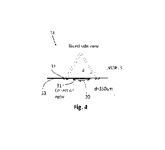
Figures 3 and 4 show a further embodiment combining the inverse SORS device
24 of Figure 2 with a focusing device functioning as a detector unit 30. The
focusing
device or detector unit 30 is surrounded by rings of VCSELs 32. The detector
unit 30
provides a means for focusing the received Raman scattered light, possibly
onto an
upstream component, for analysis of blood glucose levels in the sample of a
subject.
Figure 4 shows a side view of the embodiment of Figure 3. In Figure 4 the
arrangement including the lens 30 is arranged in contact with a user's skin
33. VCSELs
CA 03196380 2023- 4- 20
WO 2022/084539
PCT/EP2021/079414
19
32 are arranged to irradiate a region at a position, in this non-limiting
example,
approximately 350 micrometres under the surface of (or rather, within) the
user's skin. A
filter 31 is provided generally at the input to the detector 30. Such a filter
may optionally
be provided in any or all of the described examples, but is only shown in
Figure 3.
The optical arrangement described herein provides a number of advantages.
The use of VCSELs facilitates the collection of Raman signal from a larger
volume which
means that the system as a whole is less sensitive to skin variation, such as
skin
thickness variation. In the example in which a plurality of VCSELs are
provided,
preferably in rings of some shape, it is possible to vary collection depths
without actually
having to move anything within the probe. Simply activating a different
selection of the
VCSELs will stimulate Raman signal within a user at different locations or
depths.
Finally, the use of VCSELs enables the reduction in the probe of other optical
elements
such as focussing hardware and the like.
The lens 30 is arranged to receive Raman scattered radiation generated by the
incident radiation from the VCSELs 32 and focus it for onward transmission to
a detector
or a spectrometer for further analysis,.
The focusing device 30 is preferably but not limited to being a collection
optic
which refracts the received Raman scattered light for onward transmission. The
focusing device 30 may also include or consist of one or more of a mirror, a
group of
interconnected mirrors, an array of collection optics, or a combination of
mirrors and
collection optics, and filters.
In order to infer the concentration of glucose in the sample of a subject the
openings of any of the previous arrangements 14, 24, or 34 in combination with
the
features of figures 3 and 4, is applied the surface of skin the subject
chooses as a
sample. The device then emits laser light onto the sample which Raman scatters
the
laser light back to the Raman detector 12, or Raman detector unit 20.
The Raman detectors 12 or Raman detector unit 20 then communicates the
received Raman signal onwards for analysis of the spectra received for the
presence of
CA 03196380 2023- 4- 20
WO 2022/084539
PCT/EP2021/079414
glucose in the sample, and provides an indication of, say, the blood glucose
level in the
sample to the subject.
Referring to Figure 5 an overall assembly is shown incorporating an
arrangement
5 like any of those shown in Figures 1 to 4. A probe 40 is provided having
an outer casing
defining a handle 42 shaped for an operator to hold when using the system.
Cabling 44
is provided coupling the probe 40 to a processing unit 46 such as a general
purpose
computer running particular software or a dedicated hardware unit. The cabling
44 may
be optical, electrical or both and serves to communicate signals or data
between the
10 probe 40 and the processing unit 46. In place of (or as well as)
cabling, a wireless
connection may be used between the probe 40 and the processing unit 46.
Preferably the processing unit 46 includes a display 48 which functions as a
GUI
to indicate a reading or result to a user when a test is done using the
system. In one
15 example the processing unit is entirely electrical without optical
functionality. The optical
componentry and processing is all integrated and incorporated within the probe
40. This
is achievable due to the use of VCSELs enabling miniaturisation of the optics.
Thus, the
cabling 44 is electrical, communicating control signals and data between the
probe 40
and the processing unit 46.
In another example, the probe 40 includes VCSELs but the spectrometer or CCD
devices that might be used are housed within the processing unit, such that
the cabling
44 includes one or more optical fibres as well as electrical cabling for power
and/or
signalling.
In the example shown, a temperature sensor (or sensors) 47 is provided as part
of the probe 40. The temperature sensor 47 is coupled to the processing unit
46 via
conductor 49. The temperature sensor is arranged and configured to measure the
temperature of the VCSELs provided within the probe, and preferably arranged
to couple
the measured temperature to the controller 46. If required, the VCSEL
temperature is
mapped/converted to an excitation wavelength and if necessary, the recorded
spectra
are adjusted in accordance with the excitation wavelength.
CA 03196380 2023- 4- 20
WO 2022/084539
PCT/EP2021/079414
21
Furthermore, in an example, temperature stabilization of the VCSELs is enabled
by use of, say, a thermoelectric cooler, so as to avoid excitation wavelength
drift.
In one example, with or without use of temperature stabilization, the
excitation
wavelength is tracked by use of a spectrometer. If drift is detected the
recorded spectra
can be adjusted in dependence on the excitation wavelength. The excitation
wavelength
can be stabilized by control of VCSEL temperature and/or the applied driving
current or
signal.
In an embodiment an algorithm is used to analyse the received Raman spectrum
to determine the concentration of glucose or some other analyte. If the signal
comes
from the skin it is likely that it will indicate the concentration of glucose
within the
interstitial fluid rather than directly in the blood, but this corresponds
closely to the level
of glucose in the blood albeit with a small time shift. The algorithm, known
as dual
wavelength shift-excitation Raman Difference Spectroscopy is used. The
difference
between the two wavelengths is typically less than 5nm and preferably about
lnm. The
method enables use of a VCSEL probe as described herein arranged to provide
background fluorescence elimination. In a general sense this is done with the
use of two
incident wavelengths. VCSELs are provided having two different transmission
wavelengths and due to their small size it is possible to arrange them all
within the
system as described above with reference to any of figures 1 to 5.
As follows from Kasha's rule, the shift-excitation wavelength for fluorescence
background elimination is unaltered for small changes in excitation photon
energy, while
the generated Raman spectrum does shift according to the excitation photon
energy
change. Thus, by subtraction of two spectra from each other, acquired with
slightly
different excitation wavelengths, provides for the elimination of the
background
florescence while a Raman difference spectrum remains.
In other words, the algorithm for fluorescence background elimination,
eliminates
non-Raman background fluorescence by comparing the shifts in spectral peaks of
observed scattered radiation from a sample, irradiated by at least two
different
wavelengths of radiation by the laser sources. This enables isolation of the
shifted
CA 03196380 2023- 4- 20
WO 2022/084539
PCT/EP2021/079414
22
signal, for analysis of the presence of glucose or some other analyte with the
sampled
volume. In an example this is achieved by providing the optical sources, such
as
VCSELs, in a distributed way around the detector. Different groups of the
individual
optical sources are activated such that the target is sequentially irradiated
by radiation of
the two different wavelengths. In an example where the optical sources are
arranged in
one or more rings, any one of the one or more rings may be made up of optical
sources
in which every other optical source has the same transmission wavelength. If
three
different wavelengths are used, every third optical source will have the same
transmission wavelength.
If the optical sources are not arranged in rings, but, say, in a two
dimensional
array of rows, every other row, may be arranged to have the same transmission
wavelength, with intervening rows having some other transmission wavelength.
Alternatively, in one example, an even greater degree of variation is achieved
in that
every other optical source in both X and Y directions is arranged to have the
same
wavelength and every other optical source to have some common but different,
wavelength.
Where, say, plural rings of sources are used, the different rings may be
arranged
each to have their own different transmission wavelength. Alternatively, in
another
example, every other ring is arranged to have the same first transmission
wavelength,
with the intervening rings having some same but different transmission
wavelength from
the first transmission wavelength.
In a further example, a SWEPT Raman probe is provided using VCSELs as the
optical source. An array of VCSELs having a wavelength range of some desired
value is
provided. The exact number of wavelengths can be varied as per application,
but
typically a spectral range of, say, 750 to 960nm, 750 to 860nm or 850 to 960nm
is
provided. A wavelength step is selected and a bandpass filter provided at some
value
from the original excitation wavelength. The Raman spectrum can then be
reconstructed
using known SWEPT Raman methodologies
In general, the use of VCSELs facilitates the creation of a SWEPT Raman probe
for use in determining in vivo concentrations of analyte in a user's skin.
CA 03196380 2023- 4- 20
WO 2022/084539
PCT/EP2021/079414
23
Figures 6A to 6D show schematically alternative configurations for optical
detectors and sources of an optical arrangement for use in a device for non-
invasive in
vivo measurement of analyte present in the skin of a subject.
Figure 6A shows a configuration in which an optical detector 50 is arranged
within a number of linear arrays 52 of VCSEL optical sources. Figure 6B shows
a
configuration in which optical detector 50 is arranged within a generally
hexagonal
continuous array 54 of VCSELs.
Figure 6C shows an example in which optical detector 50 is arranged between
two parallel linear arrays of VCSEL sources 56 and Figure 6D shows an example
in
which a number of detectors 58 are distributed in a plane amongst a similarly
randomly
distributed array of VCSEL sources 60. In each of the examples shown in
Figures 6A to
6D, it will be appreciated that a detector is provided at some separation from
the optical
sources in the form of VCSELs. Similar to the general configuration of, say,
Figure 3,
the detector or collection optic 50 is arranged within and/or surrounded by
the optical
sources. Similarly, the configurations shown could be used in an "inverse"
manner in
which the optical sources are arranged generally in the position of the
detectors in
Figures 6A to 6C and the detector(s) instead arranged to surround the optical
sources.
Thus, in this configuration an inverse SORS optical arrangement would be
provided.
Looking now at Figure 7, an example of an optical arrangement is shown. The
general configuration is similar to the arrangement of, say, Figure 4
described above. In
this example, a detector 62 is provided with VCSEL sources 64 arranged around
it. The
VCSEL sources are arranged to provide generally parallel beams 66 of light
directed at a
point 68 which is selected to be at the common focus of the detector 62. Thus,
detector
62 typically includes a lens having an acceptance cone 70, i.e. a cone that
defines a
region such that any light generated within the region and directed towards
the detector
will have an angle of incidence such that it can be received and detected by
the detector.
Any Raman signal generated within the detector acceptance cone, and that is
directed
towards the detector can be received by the detector.
Figure 8 shows an example in which VCSEL sources 72 are arranged to provide
divergent VCSEL beams 74. Again, any Raman signal generated within the
detector
CA 03196380 2023- 4- 20
WO 2022/084539
PCT/EP2021/079414
24
acceptance cone 76, and that is directed towards the detector, can be received
by the
detector. It will be appreciated, by comparing Figures 7 and 8 that great
flexibility is
enabled by the present system. Indeed, by providing multiple VCSEL sources
arranged
around a detector, control of the individual VCSELs provides great flexibility
in
determination of the region of illumination and thus investigation.
Figure 9 shows a further example of an optical arrangement for use in a device
for non-invasive in vivo measurement of analyte present in the skin of a
subject. In this
example, a detector 78 is provided. A first and second plurality of VCSELs 80
and 82
are provided. The first plurality of VCSELs 80 is arranged in a ring having a
first
diameter r1 and the second plurality of VCSELs 82 is arranged in a second ring
having a
second diameter r2.
A detection cone 84 is shown schematically. Again, as above, Raman signals
generated within the detector acceptance cone and that is directed towards the
detector
can be detected and used to produce the Raman spectrum for the sample.
Each of the VCSELs in the first and second pluralities 80 and 82 are
preferably
arranged and controlled to provide collimated beams or part-collimated beams
and are
arranged to be controlled independently. By turning on and off different
VCSELs within
the first and second pluralities, the Raman signal generated in different
volumes within
the skin or subject can be collected.
Figure 10 shows a further configuration for optical detectors and sources of
an
optical arrangement for use in a device for non-invasive in vivo measurement
of analyte
present in the skin of a subject. In this example, detectors 86 are provided
having
detection cones 88. A VCSEL source 90 is provided which typically will
comprise a
plurality of individual VCSELs. The VCSEL source produces a divergent VCSEL
beam
92 thus illuminating a large volume within the skin of the subject. Again,
Raman signals
generated anywhere within the acceptance cones of the detectors 86, and that
is
directed towards the detectors, can be detected and used in generation of a
Raman
spectrum.
CA 03196380 2023- 4- 20
WO 2022/084539
PCT/EP2021/079414
Figure 11 shows a further example of an optical configuration for use in a
device
for non-invasive in vivo measurement of analyte present in the skin of a
subject.
In this example, a number of detectors D1 are provided each having an
5 acceptance cone. The acceptance cones 94 are arranged to intersect the
illumination
region of a divergent VCSEL source 96. Thus, the use of plural detectors
ensures that
the signal collected from different areas within the illumination cone 98 of
the VCSEL
source 96 can be distinguished. Furthermore, understanding can be gained
regarding
the depth or general location of the optical source due to the use of multiple
detectors
10 97.
Figure 12 shows a further example of an arrangement of optical detectors and
sources for use in a device for non-invasive in vivo measurement of analyte
present in
the skin of a subject.
In this example, plural detectors 1001, 1002 and 1003 are provided. An optical
connection is provided between each of the detectors and a spectrometer
entrance slit
102. The arrangement of the inputs from each of the optical fibres 101 within
the
spectrometer entrance slit is controlled and fixed such that the spectrum
produced by
each of the signals from the respective fibres 1011 to 1013 can be easily
identified.
With the use of a divergent VCSEL source 104 the arrangement can be used to
obtain accurate depth information relating to the location origin of a
particular spectrum.
For example, if the spectrum of D3 is subtracted from the spectrum derived
from
detector D2 then information regarding the sample within the depth region 106
can be
determined. Similarly, other determinations can be made by subtraction of
particular
pairs of combinations of spectra.
Figure 13 shows a further example of an arrangement 108 of optical detectors
and sources for use in a device for non-invasive in vivo measurement of
analyte present
in the skin of a subject. In this example, a VCSEL source 110 is provided at a
separation from a detector 112. The separation is defined by a sample 114
under
investigation being placed between the VCSEL source 110 and the detector 112.
The
sample could for example be the skin of a subject between the fingers or a
pinch of skin
CA 03196380 2023- 4- 20
WO 2022/084539
PCT/EP2021/079414
26
taken at some other place on a user's body. The area of illumination 116 of
the VCSEL
source overlaps with the detector cone 118 of the detector 112.
As explained above, the use of VCSELs or other such similar optical sources
enables the miniaturisation of the probe and the use of such methodologies as
SORS or
inverse SORS. One further particular advantage of the use of optical sources
such as
VCSELs in an optical probe for the in vivo measurement of analyte
concentration is the
integration of the VCSELs into or around a spectrometer entrance slit as will
now be
described in detail.
Figure 14 is a schematic representation of a known probe for use in a non-
invasive system for measuring blood analyte concentration using Raman
spectroscopy.
Such a probe may for example be used in the in vivo measurement of blood
glucose
concentrations or the concentrations of other analytes such as alcohol. Such a
probe
may be of the type generally described in our earlier International
applications WO
2018/10394, W02016034448, and W02011083111, already referred to above.
The present embodiment provides an integrated probe in which a spectrometer
entrance slit is provided and the probe as shown in Figure 14 and identified
as reference
numeral 114 therein, that typically comprises plural optical and control
components, is
provided integrated as part of the assembly. The illumination sources, such as
VCSELs
are, in effect, provided on the slit. The expression "on the slit" means that
the VCSELs
are provided in close proximity to the slit of the spectrometer itself. In one
example the
slit can be provided in a PCB where the VCSELs and optics are provided on the
same
PCB. Thus, the probe is provided for use in a system, such as a non-invasive
system,
for measuring blood analyte concentration using Raman spectroscopy. Such a
probe
may for example be used in the in vivo measurement of blood glucose
concentrations or
the concentrations of other analytes such as alcohol. The probe is preferably
provided
as for use in a non-invasive system although it will be appreciated that it
can be used in
invasive probes or systems as well. It can, for example be used in industrial
applications
where a Raman spectrometer probe is required. Typical applications include,
for
example the fields of biochemistry, medicine, agriculture, pharmaceuticals,
process
control/Quality control, forensic applications and technologies, chemical
production,
material analysis and environmental monitoring.
CA 03196380 2023- 4- 20
WO 2022/084539
PCT/EP2021/079414
27
Preferably, the illumination sources are provided in the form of VCSELs
although
other possible illumination sources could also be provided. The illumination
can consist
of a single source or multiple sources. In a preferred example, the
illumination sources
comprise paired sources in order to generate an excitation source with
specific optical
specifications.
As will be described below, optical elements like lenses, optical flats and
the like
can be placed in front of the illumination sources and/or the spectrometer
entrance slit.
Such an arrangement including the appropriately sized and configured
miniaturised
optical components in combination with the optical sources such as the VCSELs
still
provides for what may be described as an integrated probe. The contrast can be
noted
markedly in, say, a comparison of Figures 17 and 18. In Figure 17 a
conventional probe
arrangement is provided in which the probe system 114 is provided coupled to a
spectrometer system 116. In Figure 18, the probe system 148 is effectively
provided on
a PCB assembly which is arranged generally in contact or fixed to the side of
the L-
shaped body 150 of the spectrometer.
The illumination sources can consist of two or more individual groups, or
single
sources, which can be individually controlled. Each group can have specific
individual
specifications in order to support Raman spectroscopic techniques such as
stimulated
Raman scattering (SRS), coherent anti-stokes Raman scattering (CARS), shift
excitation
Raman difference spectroscopy (SERDS) and swept source Raman (SSR)
spectroscopy. As discussed herein, the expression "integrated probe" will be
used since
the probe shown in, say Figure 14 as a separate physical component including a
laser
120, focussing optics (130 etc) and the like is integrated in the probe with
the
spectrometer.
The illumination sources are preferably arranged in configurations by taking
advantage of the spatial offset between the illumination and collection optic
based on the
SORS principle described above, thereby allowing depth-sensitive probing.
Indeed, the
operation and control of the integrated probe including the VCSELs or other
optical
sources can be as shown in and described above with reference to any of
figures 1 to
13.
CA 03196380 2023- 4- 20
WO 2022/084539
PCT/EP2021/079414
28
Looking at Figure 14, a probe system 114 is provided coupled to a spectrometer
system 116. The spectrometer entrance slit 118 is the first element in the
spectrometer.
The probe system 114 is thus the part of the overall system (shown in its
entirety in
Figure 14) that provides the controlled light radiation to a sample and
receives from the
sample a produced Raman signal for onward transmission to the spectrometer 116
in
which spectral breakdown and analysis can be performed.
A Raman signal is generated in response to activation of a laser 120 which is
directed via optics 122 such as a beam splitter to impinge upon a sample 124.
A contact
surface 126 may be provided in the form of a transmissive window through which
the
laser beam travels. The laser beam impinges on the sample 124 and interacts
with it
generating a Raman spectrum which is transmitted via other optics including a
filter 128
and one or more lenses 130 to the spectrometer entrance slit 118. Within the
spectrometer, optics 132 are provided which may typically include one or more
lenses
and/or mirrors and a grating so as to direct the received spectrum onto a
detector 134
such as a CCD detector.
The system now to be described integrates the functionality of the probe
system
114 into the spectrometer and on or around its slit thereby facilitating
miniaturisation and
simplification of the apparatus.
Referring to Figures 15 and 16, views of a system similar to that shown in
Figure
14 are provided. In Figure 15, the probe system 114 can be seen coupled to the
spectrometer 116. A laser source 120 is provided which through a conduit 136,
such as
an optical fibre, couples the laser into the probe system 114. An opening 138
is
provided which will typically be placed in contact with a user's skin or
another sample
region for testing.
Referring to Figure 16, a cross section through the probe system 114 can be
seen as can be noted, the system 114 includes various optics in the form of
lenses 140
and filter 142. A directing mirror 144 is provided to receive the laser from
the conduit
136 and direct it to the sample window 138. A dichroic mirror is provided to
direct the
generated Raman signal towards the spectrometer slit 118. The precise
configuration of
CA 03196380 2023- 4- 20
WO 2022/084539
PCT/EP2021/079414
29
the probe system shown either in Figures 14, or 15 and 16 is merely
representative to
demonstrate the general scale of the apparatus and the involved complexity.
Figure 17 shows a plan view of the system of Figure 15. An imaging device or
detector 146 is arranged to receive the Raman spectrum once it has passed
through the
redirecting optics as described above with reference to Figure 14.
Figure 18 shows a plan view of an embodiment of a probe system now to be
described. The probe system 114 is replaced by an integrated probe 148 to be
described in greater detail below. The generally L-shaped body 150 is the
spectrometer
as seen in, say, Figure 14 (represented by reference numeral 116 in Figure 14)
but the
probe system is replaced, facilitating significant miniaturisation.
Figures 19a and 19b show a simplified schematic illustration of the components
of an integrated probe as provided in place of the probe system shown in
Figures 14 to
16.
The integrated probe system comprises a slit plate 146 provided with a slit
148
and a plurality of illumination sources 151 provided thereon. Preferably, the
illumination
sources 151 are VCSELs although other integrated illumination sources can be
used.
Typically, the slit size will be dictated by any or all of requirements for
spectral resolution,
throughput and spectrometer complexity. Typically though, slit dimensions
might be
between 10 and 200 micrometres wide and between 800 and 1600 micrometres long.
Thus, the size and scale of the slit in comparison to the probe system 114
shown in
Figure 15 is significantly smaller.
Figure 19b shows a side view of the system of Figure 19a. As can be seen, the
plural VCSELs 151 are arranged, in this example, on the slit plate 146. The
VCSELs are
arranged in two longitudinal arrays extending along the longitudinal edges of
the slit.
Individual or group control of the VCSELs is possible which allows to adjust
the
excitation power. The overall arrangement of the probe is substantially planar
such that
the width of the probe in its entirety (represented schematically by the
dimension "X") is
between 0.5 and 10mm, and preferably between 1 and 5mm or more preferably
between
1 and 3mm.
CA 03196380 2023- 4- 20
WO 2022/084539
PCT/EP2021/079414
Figure 20 is a schematic view of the control system used to control the
integrated
probe of the present application.
The control system includes a controller 152 which is typically a
microprocessor
5 or an ASIC. The controller 152 is coupled to a laser driver/controller
154 which itself is
coupled to the VCSELs 156. A power supply 158 is provided to provide operating
power
to the laser driver/controller. Typically, the components illustrated
schematically in
Figure 20 are all integrated onto a single unitary PCB such as the slit plate
146 shown in
Figure 19a. The specific configuration of optical sources shown in Figure 19a,
i.e. a
10 single row of optical sources 151 provided on each longitudinal side of
the slit, is not a
limitation of this configuration. Indeed, the general schematic control system
shown in
Figure 20 can be provided with different arrangements of optical sources to
that shown
in figure 19A. Indeed any actual orientation or arrangement of VCSELs 150 on
the slit
plate 146 can be provided.
The components of the control system, as shown schematically in Figure 20, can
be provided on the same PCB as that on which the VCSELs or optical sources are
arranged. In an alternative, they are provided within the envelope or housing
of the
spectrometer, e.g. within the spectrometer 150 as shown in Figure 18. In any
event, the
provision of these components does not detract from the generally planar
nature of the
probe 148.
Figure 21 shows an exploded side view of the integrated probe shown more
schematically in Figure 19b. In the example, the slit plate or PCB 146 is
provided with
the illumination sources 151, typically VCSELs, provided thereon. Illumination
source
optics 160 is provided. Typically, this could be in the form of micro lenses
or an optical
window. In addition, slit optics are provided such as a lens which, in one
embodiment,
could be combined with the illumination source optics 160. As mentioned above
with
reference to Figure 19B, the overall arrangement of the probe is substantially
planar
such that the width of the probe in its entirety (represented schematically by
the
dimension "X") is between 0.5 and lOmm, and preferably between 1 and 5mm or 1
and
3mm.
CA 03196380 2023- 4- 20
WO 2022/084539
PCT/EP2021/079414
31
If the slit specifications/tolerances cannot be met by PCB tooling a metal
plate slit
can be attached to the system.
The optional illumination source optics 160 could be e.g. microslenses or an
optical window.
The slit optics could be e.g. lens or an optical window.
The illumination source optics 160 and the slit optics could be combined in
one
optical element.
An optical filter 164 is provided in the form of a Rayleigh filter. One or
more
Rayleigh filters can be placed on both sides of the slit. In this example, the
Rayleigh
filter 164 is provided behind the slit plate 146, but it will be appreciated
that it can be
provided on the other of the slit plate 146 as well. It will be appreciated
that the probe
assembly 148 is effectively planar which means that it can be provided in
position on the
side of the spectrometer, e.g., the spectrometer 150 in Figure 18. The overall
footprint of
the system including the probe 148 and the spectrometer 150 is, in effect,
substantially
the same as that of the spectrometer 150 alone.
Referring again to Figure 19 and 21, optionally, a metal slit plate is
provided. The
metal slit plate is provided to provide a control over the dimensions of the
slit in the PCB
or slit plate 146. For example, in situations where the slit specifications or
tolerances
cannot be met by PCB tooling, a metal plate slit can be attached to the
system. The
metal plate slit can be placed both on the top side or the back side of the
PCB. In the
example shown, it is provided on the back side of the PCB. The metal slit
plate is
denoted by reference numeral 166. As will be appreciated, the integrated probe
combines the functionality of the various components shown in and described
with
reference to Figure 14 in such a way that significant miniaturisation can be
achieved. In
practice, the entire volume of the probe such as that shown in Figure 17 and
indicated
by reference numeral 114, can be replaced by a single integrated PCB.
Figure 22 shows a schematic representation of an exemplary set up for the
optics. In this example, a 34 by 3 array of single-mode VCSEL emitters 151 is
provided
CA 03196380 2023- 4- 20 SUBSTITUTE SHEET (RULE 26)
WO 2022/084539
PCT/EP2021/079414
32
at a pitch of 28 micrometres provided on a wafer 168. The arrangement
represents a
wafer-engineered slit-optic set up. The example minimises the number of
additional
components required by directly engineering a window 174 into the VCSEL wafer
168.
The window 174 is coated with a long-pass filter is directly engineered into
the VCSEL
wafer 168 using a transparent material. This allows small displacement between
the
emitter arrays and the detection optical axis 173. Typically, the displacement
between
the emitter arrays and the detection optical axis 173 will be less than 100
micrometres.
Reformatting optics 170 can be provided. The reformatting optics is in the
form a
lens and is used to maximise the throughput and magnify the scattered
distribution onto
the existing slit 172. Typically, this boosts throughput by at least 10%.
Precision
CA 03196380 2023-4- 20 SUBSTITUTE SHEET (RULE 26)
WO 2022/084539
PCT/EP2021/079414
33
alignment will be required between the assembly and the existing slit of the
spectrometer.
The VCSEL wafer 168 including the transparent window 174 is shown as a
merely exemplary configuration for the integrated probe.
It is possible that the VCSEL wafer 168 is provided in two sections, one on
either
longitudinal side of the transparent window 174 and they are then machined or
connected together with the transparent window 174 in a known manner.
The spectrometer slit is typically dimensioned such as to have a width of 100
micrometres and a length of 1300 micrometres and a numerical aperture of 0.22.
The optical flux of each emitter in this example is typically 1.5 mVV
representing a
total optical flux of 306 mVV. Preferably the wavelength is between 760 and
850
nanometres although VCSELs of any desired wavelength can be chosen for use in
the
system. The indication of wavelength ranges given above applies equally here.
Figure 23 shows a further example of an integrated probe. In this example,
angled illumination optics ("direction optics") 176 are provided on each side
of the PCBs
168 including the VCSELs. Preferably, a micro lens array 178 is provided
between the
PCBs 168 and the direction optics 176. Each emitter array is now separated
from the
central detection axis by at least 1mm. This allows the VCSEL arrays to be
mounted in
separate packages either side of the slit. The micro lens arrays are placed on
top of the
VCSEL arrays to collimate the light output. This is required due to the
increased
distance between the sources and the tissue. The direction optics 176 tilt the
illumination at a large angle. Again, reformatting optics 180 are provided to
boost the
throughput. This configuration relies on precise placement of the VCSEL
packages with
respect to the slit and reformatting optics 180.
Figures 24a and 24b show plan and cross-sectional views of an exemplary
system similar to that described above with reference to any of Figures 19 to
23. In this
example, a PCB 182 is provided having VCSELs 184 arranged thereon.
CA 03196380 2023- 4- 20
WO 2022/084539
PCT/EP2021/079414
34
The VCSEL packages 184 are arranged on either side of the slit. In this
example, the slit is typically 1500 micrometres long and 100 micrometres wide.
As can be seen, the PCB is arranged on a backing plate 186 and aligned with an
opening 190 therein.
Figure 25 shows a cross sectional view of an exemplary probe. The probe is
shown arranged in contact with a sample 192. The sample could be a user's skin
if
being used for in vivo determination of an analyte concentration or it could
be any other
sample under analysis as explained above. The probe includes an optical
element in
the form of a generally transparent structure. The optical element has a
generally
central region 194 with truncated triangular cut-out 196. The cut-out 196 has
sloped
edges 193 with a light blocking coating and a generally flat upper surface 191
parallel to
the upper surface 197 of the optical element.
The generally flat upper surface is coated with a long pass filter and
represents
the spectrometer entrance slit. A VCSEL block or die 198 is provided on each
side of
the slit. Each VCSEL die 198 includes a VCSEL array with possible micro optics
210
arranged on top of them. Transmitted light from the VCSEL array passes through
inlet
surface 200 on an underside of an overhang within the optical element 195.
Angled
surfaces 202 totally internally reflect the light for onward transmission to
the sample 192.
Semiconductor pads 204 and 206 are arranged on the support surface 208. The
VCSELS will be operated by control circuitry (not shown) which will be
connected to the
pads. The arrangement of Figure 25 thus provides a compact and simple probe
arrangement in which the probe is generally planar
Embodiments of the present invention have been described with particular
reference to the examples illustrated. However, it will be appreciated that
variations and
modifications may be made to the examples described within the scope of the
present
invention.
CA 03196380 2023- 4- 20