Note: Descriptions are shown in the official language in which they were submitted.
WO 2022/100833 PCT/EP2020/081887
1
A surgical device, a system, and a method of manufacturing a surgical device
The present invention relates to a surgical device, a system, and a method of
manufacturing a
surgical device according to the preambles of the independent claims.
The present applicant recently developed a surgical felting device allowing a
biomechanically
advantageous implantation of implants. The developed device allows an improved
fixation as
compared to conventional suturing techniques. One example of such a surgical
felting device
is disclosed in PCT/CH2o19/0000l5. The surgical felting device comprises a
needle that
repeatedly moves through a surgical felt and into tissue. By embedding strands
of the felt
inside the tissue, the needle creates a strong and well distributed mechanical
bond between
the felt and the tissue. Compared to conventional suturing, this technique is
faster and
alleviates adverse effects such as "cheesewiring" of suture, where the suture
cuts through
tissue which can happen through local stress peaks.
However, since the surgical felting device may be handheld and the needle
moves at high
speeds, there is high risk of needle damage. The damage may result from
collisions between
the needle and rigid structures such as bones or other surgical tools. Such
collisions could
lead to a partial destruction of the needle (plastic bending, breaking or
splintering) which
adversely influences the functionality. Even further, parts of the needle may
be lost inside the
patient's body, presenting a health risk. Lastly, the high speeds of the
needles in the tissue
and in the surgical felt may generate undesirable heat that results from
friction.
It is the objective technical problem to overcome the above disadvantages of
the prior art. In
particular, it is the objective technical problem to provide a surgical
felting device that is safe
to use for the operator and the patient. A further object of the present
invention may be to
reduce the heat generated by the felting needle.
The objective technical problem is solved by the features of the independent
claims. One
aspect of the invention relates to a surgical device for felting an implant to
soft tissue of a
patient. The patient may be a human or an animal. Felting may be understood as
the process
of moving a felting needle repeatedly into, and in some embodiments through,
an implant
CA 03198576 2023- 5- 11
WO 2022/100833 PCT/EP2020/081887
2
comprising a felt and thereny entangling nbers ot a telt. A telling needle may
be understood
as a needle including barbs that catch the fibers of the felt and move the
fibers through the
felt and which entangles the fibers of the felt further. In particular, the
felting may not only
entangle the fibers of a felt that forms part of an implant with each other
but also entangle
the fibers of the tissue of a patient with the fibers of the felt. Thereby,
some strands of the felt
become embedded in and entangled with the patient's tissue and some strands of
the tissue
fibers may become embedded in and entangled with the felt. This creates a
strong and well
distributed mechanical bond between tissue and felt.
The surgical device comprises at least one felting needle. The at least one
felting needle is
configured to move reciprocally. A reciprocating motion as mentioned herein
may be
understood to be a repetitive back and forth motion. The motion may be
translational, i.e.
linear, or rotational. Further, the surgical device comprises a connection
interface for
connecting the at least one felting needle to an actuator and for transferring
a reciprocal
motion from the interface to the at least one felting needle. The interface
may provide a
releasable or a permanent attachment. For example, the releasable interface
may include
pins, latches, screws, magnets or other known mechanical or other connection
means. A
permanent attachment using rivets, glue or a one-piece design is also
possible. A releasable
attachment of the at least one needle would allow the at least one needle to
be replaced.
In certain embodiments the device includes a water cooling system to cool down
friction heat
which is generated by the oscillating needle.
The actuator may be pneumatic, hydraulic or electromagnetic. In particular,
the actuator may
be an electric motor or may comprise or use electromagnetic coils or pneumatic
or hydraulic
pressure. The device may be configured to convert a rotational motion of the
actuator to a
translational motion. In particular, the device may comprise a scotch yoke
mechanism or a
piston rod drive mechanism to transform a rotational motion into a
translational motion.
Thereby, more efficient motors may be used. A speed and/or frequency of the
reciprocal
translational motion (oscillation) can be adjustable. The device may comprise
a linear motion
rod to transfer the reciprocal motion from the interface or the actuator to
the at least one
needle. To reduce wear and heating, bearings and low friction materials (e.g.
polytetrafluorethylene) may be used to minimize friction of a linear motion
rod.
CA 03198576 2023- 5- 11
WO 2022/100833 PCT/EP2020/081887
3
The at least one needle is preterawy maae or or comprises stainless steel. The
needle may
include a stainless steel coating. Stainless steel is particularly resistant
to the oxidizing
conditions of the surgical operation. Hence, the present device may e.g. be
used in
arthroscopie surgeries, in which a constant saline flush is applied, that may
oxidize the
needle.
The device may comprise a needle protection mechanism. The needle protection
mechanism
prevents the at least one needle from being damaged during the reciprocal
motion due to a
contact with a rigid structure.
The needle protection mechanism may define a maximal penetration depth of the
at least one
needle. The maximal penetration depth may be adjustable. In particular, the
maximum
penetration depth may be less than the amplitude of the reciprocal motion.
Additionally or
alternatively, the needle protection mechanism may limit the reciprocal motion
or may be
configured to detect an obstacle (i.e. hard tissue) or may decouple an
actuator from the at
least one needle in case a collision occurs or any combination thereof. The
motion may be
limited by modifying the amplitude of the reciprocal motion and/or by limiting
the maximal
penetration depth.
The rigid structure may be for example the bone of a patient or another
surgical tool that may
come in contact the needle while the needle is actuated.
The surgical device may be used for the repair or fixation of anatomical
structures such as:
collagenous tissues such as tendons, menisici, spinal disks or fascia,
ligament reconstructions
(collateral ligaments, cruciate ligaments, etc.), subcutaneous sutures,
conventional suturing
of skin closures, skeletal muscle, heart muscle, valves, and hollow organs
(large vessels,
bladder, esophagus, possibly intestine). In addition, an implant may be
attached to these
anatomical structures.
In one embodiment, the at least one felting needle has a width or diameter of
less than 0.8
mm, preferably less than o.6 mm, most preferably less than 0.4 mm. Needles
with a low
diameter have a lower friction and thus less heat is generated. However, the
lower needle
diameters may lead to more fragile needles.
CA 03198576 2023- 5- 11
WO 2022/100833 PCT/EP2020/081887
4
In one embodiment, the neethe protection mecnamsm mciunes a spacer for
contacting the
soft tissue or patch such that the spacer sets a maximal predetermined
penetration depth for
the at least one needle. The spacer pushes against the tissue and thereby
prevents the needle
from penetrating further than desired. This may be especially useful for
anatomical
structures that are arranged close to bones such as tendons or spinal disks.
Further, the
spacer helps an operator to keep the amplitude of the motion of the needle
within a desired
range (e.g. the felt and the soft tissue) and improves handling.
The spacer may be elastic, in particular, the spacer may include or form a
spring. Elastic may
be understood as allowing the user to press the device against the soft tissue
and thereby
changing the spacing of the spacer dependent on the applied force. In one
embodiment the
spacer may have a curved or bent shape. The elasticity allows a constant
contact of the tip of
the device on the feltable patch or tissue. Further, this may help holding the
soft tissue in
place without the need for further tools and/or assistance. If there is no
constant contact to
the tissue or patch, the tissue or patch could vibrate and reduce a
penetration of the needle
through the tissue. In particular, the spacer prevents the needle from lifting
the patch, when
the needle is retracted (stripping effect). When the needle is retracted from
the felt and/or
the tissue, the barbs and/or friction may pull the patch towards the surgical
device. The
spacer prevents this and the at least one needle will pull out of the patch
and/or the tissue
without lifting it.
In a preferred embodiment, the spacer comprises one or more fingers.
Preferably, the spacer
comprises two, three or more fingers. The one or more fingers comprise a
distal end surface
for contacting the soft tissue such that the reciprocal motion of the needle
does not exceed
the predetermined penetration depth. The one or more fingers may be bent at
their distal
side such that a radial outer surface of the fingers forms the distal end
surface(s) of the
spacer. Thereby, the operator may visually observe the at least one felting
needle allowing for
a more precise control of the device. Further, the bent fingers may allow an
elasticity of the
fingers.
In some embodiments, the fingers may be connected to each other at their
distal side, and in
particular be formed by a single wire. The fingers may be made from a shape
memory-alloy,
e.g. nitinol. The fingers may have a compressed and expanded configuration.
For example the
fingers may be expelled from a distal end of a guide tube for the needle.
CA 03198576 2023- 5- 11
WO 2022/100833 PCT/EP2020/081887
In a preferred embodiment, tne spacer torms a sucte sucn mat tne needle can be
slid along a
surface of the soft tissue. In particular, the one or more fingers may be bent
in the same
direction. The device may then be moved over the soft tissue in the direction
in which the
curvature points with ease while at the same time maintaining a constant
penetration depth
and applying a constant pressure keeping the soft tissue in place. The slide
may alternatively
be formed similarly to slides known from sewing machines.
In a preferred embodiment, the at least one felting needle has a maximal
penetration depth.
The maximal penetration depth may be adjustable. Thereby, the device may be
adapted to
the particular use case. Further, an operator may adjust the penetration depth
in case the
operation is close to hard tissue.
In a preferred embodiment, the reciprocal motion has an amplitude. The
amplitude may be
adjustable. Thereby, the device may be adapted to the particular use case.
Further, an
operator may adjust the maximal penetration depth by adjusting the amplitude
in case the
operation is (too) close to hard tissue.
In a preferred embodiment, the device comprises a guide tube. The guide tube
surrounds the
at least one needle and/or a translation rod at least partially. The
translational rod may
transfer the reciprocal motion from the interface to the at least one needle.
The guide tube
may be an outer tubular member. Further the guide tube may be connected to a
spring in
series or may be elastic.
In a preferred embodiment, the device may comprise a scale indicating the
current
penetration depth. In a preferred embodiment, the device may comprise a scale
indicating
the current maximal penetration depth.
In a preferred embodiment, the needle protection mechanism comprises a needle
collision
sensor. In a preferred embodiment, the collision sensor is configured to
measure the distance
to a rigid structure. The needle collision sensor may be a distance sensor,
such as for example
an ultrasonic sensor. The sensor may measure a distance between the device, in
particular a
housing or the guide tube, and a hard tissue. Thereby, hard tissue or other
obstacles, that
may damage the needle can be detected. Additionally, the device may comprise
an indicator
for indicating the sensed distance to a hard tissue or another obstacle to the
operator. The
indicator may be optical (i.e. a display or a diode) or haptical or acoustic.
CA 03198576 2023- 5- 11
WO 2022/100833 PCT/EP2020/081887
6
In a preferred embodiment, the needle collision sensor is configured to
measure a bending of
the at least one needle and/or the linear motion rod. The sensor may be an
elastic strain
sensor and/or an electromagnetic sensor and/or piezoelectric sensor. When the
at least one
needle collides with hard tissue such as bone, the needle and/or linear motion
rod may bend
before breaking. The needle collision sensor detects the bending. This may
cause a controller
to decouple the needle from the actuator or changes to reduce the maximal
penetration
depth.
In a preferred embodiment, the surgical device comprises a controller. The
controller is
configured to receive a signal from the sensor and adjust the maximal
penetration depth of
the at least one needle based on the measurement of the needle collision
sensor. Thereby an
open control loop is provided allowing for a quick, automatic adjustment of
the penetration
depth in response to the detection of a collision or the detection of the
threat of a collision. In
some embodiments, the actuator may measure the current maximal penetration
depth with a
further sensor and the controller may be configured to receive a signal from
the actuator for a
closed loop control of the maximal penetration depth.
The surgical device may comprise an actuator for driving the reciprocal motion
of the at least
one needle. In a preferred embodiment, the surgical device comprises a
coupling adapted to
transfer a reciprocal motion from the actuator to the at least one needle. The
coupling may be
a slipper clutch. A slipper clutch may be understood herein as a clutch that
decouples the
needle from the actuator, when a threshold force or threshold moment is
exceeded. The
slipper clutch may slip at least partially when a predetermined force or
moment acting on the
at least one needle is exceeded. The needle protection mechanism is thereby
adapted to
decouple the needle from the actuator to prevent the needle from being damaged
when a
collision occurs.
In a preferred embodiment, the slipper clutch includes at least one of: a pin
and a chamfered
face, a ball spring and a corresponding dent for receiving the ball and a
spring bearing for the
at least one needle.
In a preferred embodiment, the needle protection mechanism includes a
predetermined
breaking point. The predetermined breaking point breaks, if the force on the
at least one
CA 03198576 2023- 5- 11
WO 2022/100833 PCT/EP2020/081887
7
needle exceeds a predetermined tnresnota. ine preaetermmea tnreshold may be
lower than
the force necessary to break the needle.
In a preferred embodiment, the predetermined breaking point is part of a
mechanical
coupling for transferring forces onto the at least one needle. The mechanical
coupling
preferably includes a rod having the predetermined breaking point. In some
embodiments,
the predetermined breaking point may be provided by a structural weakening
such as a
tapering, or a ridge, an edge or a dent. In other embodiments, the
predetermined breaking
point is part of the at least one needle.
In a further embodiment, the device is configured to detect a broken needle.
In particular the
needle may be part of the electric circuit or may include a needle collision
sensor as described
above. Further the device may include an indicator, e.g. a warning light, that
is configured to
indicate optically, haptically or acoustically that the needle is broken.
A further aspect of the invention relates to a system comprising a surgical
device as described
above and an actuator. The actuator may be in particular an electric motor for
driving the at
least one needle with the reciprocal motion.
A further aspect of the invention relates to a method of manufacturing a
surgical device.
Preferably the surgical device is a surgical device as described above. The
method includes
the following steps:
Providing a surgical device with at least one felting needle, wherein the at
least one
felting needle is configured to move reciprocally and a connection interface
for
connecting the at least one felting needle to an actuator and for transferring
a
reciprocal motion from the interface for the actuator to the at least one
needle,
Providing a needle protection mechanism, wherein the needle protection
mechanism
prevents the at least one needle from being damaged due to a contact with
rigid
structure during the reciprocal motion.
Non-limiting embodiments of the invention are described, by way of example
only, with
respect to the accompanying drawings, in which:
CA 03198576 2023- 5- 11
WO 2022/100833 PCT/EP2020/081887
8
Figure 1: shows a scnematic perspective view ot a rust
embodiment of a surgical
device according to the invention.
Figures 2A and 2B: show further details with regard to an adjustable maximal
penetration
depth of the surgical device according to figure 1.
Figures 3A and 3 B: show alternative embodiments for a distal tip of the
surgical device
according to figure 1.
Figure 4: shows a schematic perspective view of a second
embodiment of a
surgical device according to the invention.
Figure 5: shows a cross-section of the surgical device
according to figure 4.
Figures 6A and 6B: show an interface for the actuator of a surgical device
according to the
invention.
Figure 7A and 7B: show a further interface for the actuator of a
surgical device according
to the invention.
Figure 8: shows a cross-section of a distal portion of a
third embodiment of a
surgical device according to the invention.
Figure 9: shows a cross-section of a distal portion of a
fourth embodiment of a
surgical device according to the invention in a first position.
Figure 10: shows a cross-section of a distal portion of the
surgical device of figure
9 in a second position.
Figure shows a cross-section of a distal portion of a
fifth embodiment of a
surgical device according to the invention.
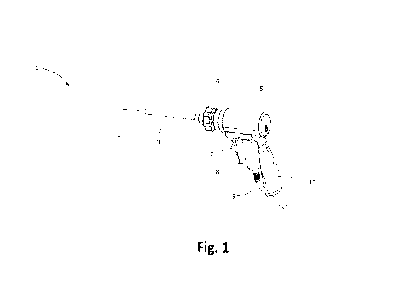
Figure 1 shows a perspective view of a first embodiment of a surgical device 1
according to the
invention. The surgical device 1 includes a housing 5 and a guide tube 3
extending from the
housing 5. A felting needle 2 is arranged within the guide tube 5 and extends
from the end of
CA 03198576 2023- 5- 11
WO 2022/100833 PCT/EP2020/081887
9
the guide tube of 5. The felting neeme 2 can De moved reciprocally forwards
and backwards
along its axis and the axis of the guide tube 3. The surgical device 1
includes a handle 10 with
a grip portion 7. Further, the surgical device 1 is connected to an energy
source with a power
cord 9 that connects to the surgical device.
An actuator -here electrical motor 8 - is arranged within the housing 5 of the
surgical device
1. The motor 8 drives the reciprocal motion of the needle 2. The guide tube 3
may be moved
with respect to the needle 3 using wheel 4. When the wheel 4 is turned, the
guide tube 3 is
retracted in a distal direction such that the tip of the needle 2 is exposed.
When the wheel 4 is
turned in an opposite direction, the guide tube 3 may be pushed in a proximal
direction such
that the tip of the needle 2 can be covered. Consequently, a penetration depth
of the needle of
the reciprocal motion of the needle is dependent on the relative position of
the guide tube. If,
for example, the needle 2 has a movement amplitude of 25 mm but the guide tube
is only
retracted by fo mm from the most distal end position of the needle during the
reciprocal
motion, then the maximal penetration depth of the needle is only fo mm.
Proximal and distal
are understood herein from the perspective of an operator, i.e. distal denotes
a direction away
from the operator and proximal a direction towards the operator.
Setting the guide tube 3 relatively to the needle 2 limits the maximal
penetration depth of the
needle and allows an operator to avoid felting tissues that are deep below the
implantation
site and in particular to avoid collisions with hard tissues such as bone.
Further, the guide
tube 5 protects the needle 2 during transportation and unpacking of the
device. The guide
tube 5 is embedded in a guide plug 6 (see fig. 1) and may be connected in
series to a spring
(see e.g. fig. 5).
Figure 2A shows the relative movement of the guide tube 3 in further detail.
The wheel 4
includes ridges 12 such that an operator can grip the wheel more easily.
Further, the wheel
includes indicators if that show the current position of the guide tube 3. In
the shown
example, the number on the wheel indicates a length of the needle that can
maximally be
exposed, i.e. a maximal penetration depth. Further, the guide tube 3 itself
includes a
penetration depth scale 13. The penetration depth scale 13 is realized as
markings on the
outer surface of the guide tube 3. In the present example, the markings are
circular rings
around the guide tube 3. When the guide tube 3 is retracted, the current
penetration depth
can be read from the last ring that is still visible, i.e. not retracted into
the housing 5. The
depth scale shows the current penetration depth. In case the guide tube 3 is
directly retracted
CA 03198576 2023- 5- 11
WO 2022/100833 PCT/EP2020/081887
as a result of the movement ot tne wneei 4, tne current and maximal
penetration depth are
the same. However, in some embodiments these two may differ as will be
explained with
reference to the second embodiment.
The relative movement of the guide tube 3 is illustrated by the arrows in
figs. 2A and 2B. If
the operator turns the wheel counterclockwise, the guide tube 3 is retracted,
exposing the
indicated maximal penetration depth 16.
Figures 3A and 3B show further embodiments of the guide tube 3 that may be
used
independently or in combination with the retractable guide tube 3. In the
embodiment of
figure 3A, the guide tube 3 comprises at its distal end 24 a protective fork
20 with two fingers
21. The fingers 21 extend from the distal end and are bent around a curve 22.
During use, the
distal end surfaces of the fingers 21 are brought in contact with a soft
tissue of a patient and
define the penetration depth 16 similarly to the end of the guide tube 3 in
the previously
shown embodiment. The curvature of the fingers 21 allows the surgical device
to slide
smoothly over the soft tissue by helping the surgical device 1 to glide along
the felt and/or soft
tissue that are currently felted.
In the embodiment of figure 3B, the protective fork is formed by a wire made
of a shape
memory alloy. During transportation, the wire is held within the guide tube.
Prior to an
operation, the wire is expelled from the distal end 24 of the guide tube and
assumes a bent
position as shown on the right side in figure 3B.
In case an operator pushes in a distal direction, the bends wire or the bent
fingers may act as
a spring allowing to temporarily (depending on the force) increase the
penetration depth if
necessary.
Figures 4 and 5 show a second embodiment of a surgical device 101 according to
the
invention. A perspective view of the surgical device 101 is shown in figures 4
and a cross-
section of the surgical device 101 is shown in figure 5. In general, the
present description uses
similar reference numerals for similar features, when the reference numeral
increases by 100.
For example, the felting needle 2 may be similar to the felting needle 102 of
the embodiment
shown in figure 4.
CA 03198576 2023- 5- 11
WO 2022/100833
PCT/EP2020/081887
11
Similarly to the previously snown nrst embocument, tne second embodiment of a
surgical
device 101 includes a felting needle 102, a guide tube 103, a plug 106, a
wheel 104 for
retracting the guide tube 103, and a housing 105. A power cord 109 is
connected to the
housing 105.
As can be seen from figure 5, the surgical device 101 includes a motor 108.
The motor 108
drives an axis whose rotation is then transferred via gear units 126 to a
scotch yoke 128. The
scotch yoke 128 transforms the rotational motion of the motor 108 into a
translational
motion. One embodiment of a scotch yoke 128 can be seen in detail in figures
6A and 6B. The
scotch yoke 128 transfers the rational movement to a linear movement and the
linear
movement to translational motion rod 119 that is held within the guide tube
103. The
translational motion rod 119 is guided by a bearing 129. The needle 102 is
arranged at the
distal end of the translational rod 119 and travels back and forth, wherein
the amplitude of
the movement is defined by the scotch yoke 128 and the frequency of the
reciprocal
movement is determined by the motor 108.
The surgical device 101 also includes a wheel 104. The wheel 104 includes an
inner thread 130
that engages an outer threading of an insert 132. The outer tube 103 covers
the translational
rod 119 and is covered at its distal end with a distal tip 125. The proximal
side and proximal
end of the guide tube 3 includes a plug 106. The plug 106 is also connected to
a spring 131
along its axial direction. Thereby the spring 131 is arranged between the plug
106 and the
insert 132. During operation of the surgical device 101, the operator may push
the distal tip
with felting needle 102 onto a felt. Thereby, the outer tube 103 is pushed in
a proximal
direction. The spring 131 resists this movement such that an operator has to
push against it.
Thereby, the outer tube 103 covers the sharp needle 102, when the device is
not in use.
Further, the wheel 104 can be used to move the insert 132 back and forth. The
insert delimits
a maximum width 127 for the retraction of the outer tube 103. The maximum
width 127 for
the retraction thus corresponds to a maximal penetration depth of the needle
102. The
delimitation of the maximal penetration depth protects the needle 102 from
colliding with
hard tissue, since the heart tissue (e.g. bony tissue) may be arranged below
the soft tissue.
Setting the maximal penetration depth as described is advantageous, if
different kinds of
tissues with hard tissue beneath are felted (e.g. an 8 mm rotator cuff tendon
or a 12 mm
Achilles tendon).
CA 03198576 2023- 5- 11
WO 2022/100833 PCT/EP2020/081887
12
Besides the protection function, tne spring 131 may also allow a constant
contact of the tip of
the device on the feltable patch, respectively tissue. If there is no constant
contact to the
tissue or felt, the tissue could vibrate and minimize penetration of the
needle through the
tissue.
Figures 6A and 6B show an embodiment of the scotch yoke 128 in detail. The
scotch yoke
may form an interface or a coupling. The scotch yoke 128 includes a wheel 135.
The wheel 135
is driven by the motor 108 through the hexagonal socket 144 and rotates around
direction
134. On a radially outer part of the wheel, a pin 136 is arranged. Further,
the scotch yoke
includes a slider 138. The slider 138 includes a slot 139, in which the pin
136 travels. Due to
the rotation of the wheel forces in a linear direction along the axis of
translational rod 119 are
transferred as indicated by arrows 137 while the forces in the direction
transversal thereto are
not transferred due to the slot 139.
Figure 6B shows an embodiment that shows an example of a mechanical uncoupling
between
the needle 102 and the motor io8. The slider 138 may include a chamfered face
133 at the slot
139. If excessive forces are applied onto the needle 102, these forces are
transferred via the
linear motion rod and the slider to the scotch yoke 128. The chamfered face
133 glides onto
the pin and thereby lifts the slider 137 out of the pin 136 preventing the
application of further
forces onto the needle 102. Hence, the wheel 135 and the slider 138 are
mechanically
decoupled.
In a further example, an electromagnetic motor io8 may be used. In this case,
the amplitude
of the needle 102 may be adjusted, if the motor detects forces above a
threshold on the needle
102. If the forces on needle 102 exceed the electromagnetic forces of the
linear drive, the
needle may be retracted or, the motor may simply be stopped.
A further mechanical uncoupling is shown in figures 7A and 7B. Figures 7A and
7B show a
wheel 235. Wheel 235 is an alternative embodiment of the wheel 135 of figures
6A and 6A.
The wheel 235 includes two parts, an inner wheel 240 and a concentric outer
wheel 241.
Figure 7A shows an exploded view of the wheel 235 and figure 7B shows the
wheel 235 in an
assembled form. Similarly to the wheel 135, the wheel 235 includes a hexagonal
socket 244
and a pin 236. The inner wheel 240 is coupled to the outer wheel with a ball
242. The ball
242 is pushed radially outwardly by a spring (not shown). The outer wheel 241
includes a
dent 243 along its inner circumference for the ball 242. When the inner wheel
240 is set into
CA 03198576 2023- 5- 11
WO 2022/100833 PCT/EP2020/081887
13
the inner circumference ot tne outer wneei 241, tne Dan 242 is pusned radially
inwardly
against the spring force and latches into the dent 243, if correctly aligned.
As long as the ball
242 is in the dent 243, forces are transmitted from the motor io8 to the
needle 102. However,
in case the needle 102 collides with hard tissue, ball 242 is pushed inwards
and no force
beyond a threshold are transmitted. Thereby, the two wheels 240 and 241 are
rotationally
uncoupled.
Alternatively, the ball spring and dent may be replaced by a weak link that
would break in
case of excessive forces or a latch. A latch could be relying on springs or
another compliant
mechanism. A further alternative are magnets that may be finely tuned to
disengage when
needed and re-engage it when the force drops below a threshold. In other
embodiments, the
system can be electromechanical, with force sensors or other sensors (e.g.
strain of the
needle, conductivity) detecting that the forces on the needle exceed a
threshold. A signal of
these sensors may cause a controller to uncouple the needle from the actuator
or stop the
actuator.
A third embodiment of a surgical device 301 according to the invention is
shown in figure 8.
The surgical device 301 includes a guide tube 303, and a needle 302 with a
maximal
penetration depth 316. As can be seen from figure 8, the needle is moved back
and forth
within soft-tissue 46. The needle 302 is driven by a translational rod 319
with a linear motion
337. The translational rod 319 is guided by bearing 329. Additionally, the
surgical device 301
includes an ultrasonic distance sensor 331. The ultrasonic distance sensor 331
is arranged at
the distal end of the guide tube and may measure a distance between a distal
end of the guide
tube 303 and bony tissue 47. The measured distance may be reported to a user
with acoustic,
optical or vibrational cues that allow the user to set the maximal penetration
depth 316. For
example, the maximal penetration depth may be set as described with respect to
figures 1 to
5. Alternatively, the surgical device 301 may additionally include a
controller, that
automatically adjusts the maximal penetration depth 316 to be less than the
measured
distance.
A fourth embodiment of a surgical device 401 is shown in figures 9 and 10. The
surgical
device 401 is similar to the surgical device 301 and includes a guide tube
403, a bearing 429,
and a translational rod 419 that moves along linear motion 437. The
translational rod 419 is
connected to the needle 402. However, the needle 402 is not directly connected
to the
translational rod 419. Instead, the translational forces of the translational
rod 419 and
CA 03198576 2023- 5- 11
WO 2022/100833 PCT/EP2020/081887
14
transferred via a spring 451 and piston cylinder 450 onto tne neeme 402. The
spring 451 and
the cylinder 450 are arranged within a cavity 452 of the translational rod
419. Alternatively,
the spring 451 and the cylinder 450 may be simply arranged at the end of the
translational
rod 419. During normal operation the needle 402 follows the movement of the
translational
rod 419. However, in case the needle 4032 collides with a hard object, the
spring 451 is
compressed and absorbs the excessive forces (see figure to). Additionally, a
damping element
may be included in parallel or series with the spring in order to prevent
unwanted oscillations
of the spring 451. In some embodiments, the spring may be formed by an
elastically
deformable rod (e.g. a nitinol band).
A fifth embodiment of a surgical device 501 is shown in figure it. The
surgical device 501 may
be similar to the surgical device 401 shown in figures 9 and 10. However,
instead of a spring,
the translational rod 519 is connected to the needle 502 with a connection rod
554 and a
predetermined breaking point 553. The connection rod 552 may be an elastically
deformable
rod that is coupled to an element featuring a predetermined breaking point
553. The
predetermined breaking point 553 may be formed by means known in the art e.g.
by using
edges, ridges or different materials. If the needle 502 collides with bony
tissue 47, the
connection rod 554 is deflected. Due to the deflection, translational forces
are converted to
shear forces on the predetermined breaking point 553. The higher the
deflection of the rod,
the higher the shear forces on the predetermined breaking point 553, which is
consequently
more likely to break.
CA 03198576 2023- 5- 11