Note: Descriptions are shown in the official language in which they were submitted.
WO 2022/120344
PCT/US2021/072666
METHOD OF SENSITIZING CANCERS TO IMMUNOTHERAPY USING
IMMUNOMODULATORY AGENTS
CROSS-REFERENCE TO RELATED APPLICATIONS
[0001] This application claims the benefit of United States provisional
application serial no.
63/119,963, filed 1 December 2020. The entire contents of this application is
hereby
incorporated by reference as if fully set forth herein.
GOVERNMENT FUNDING SUPPORT
[0002] This invention was made with government support under grant no.
CA167174
awarded by the National Institutes of Health. The government has certain
rights in the
invention.
BACKGROUND
1. Field of the Invention
[0003] This invention relates to the field of medicine and oncology. In
particular, the
invention provides methods for the treatment of certain cancers using a
combination of
immunomodulatory compounds, including an immune checkpoint inhibitor and an
iRGD
peptide.
2. Background of the Invention
[0004] Cancer is among the leading causes of death worldwide. Despite recent
advances in
science, the impact of cancer immunotherapy on disease progression and overall
survival has
been limited to certain cancers such as melanoma and non-small cell lung
cancer.
Unfortunately, therefore, cancer immunotherapy is unable to elicit responses
in a vast
majority of cancers, including pancreatic cancer. About 92% of patients
diagnosed with
pancreatic cancer will die within five years of diagnosis. Cancers such as
pancreatic ductal
adenocarcinoma (PDAC) are almost completely refractory to all forms of chemo-
and
immuno-therapy. A highly immunosuppressive tumor microenvironment
characterized by
the presence of large numbers of regulatory T cells can drive resistance to
immunotherapy.
Therefore, there is a need for therapeutic agents to transform the tumor
microenvironment to
enhance immunotherapies for various cancers.
[0005] Immune checkpoints are a normal part of the immune system, which works
to
modulate immune responses so that they do not become so strong as to destroy
healthy cells in the body. These immune checkpoints can engage when immune
checkpoint
proteins on the surface of T cells recognize and bind to partrier proteins on
other cells,
1
CA 03199012 2023- 5- 15
WO 2022/120344
PCT/US2021/072666
resulting in an "off' signal for the T cells. When the other cells are tumor
cells, this
inhibition of the immune response can prevent the immune system from
destroying the
tumor,
[0006] Immunt-itherapy drugs called "immune checkpoint inhibitors" work by
blocking
checkpoint proteins from binding with their partner proteins on tumors. This
prevents the
"off' signal from being sent, allowing the T cells to kill cancer cells.
Immune checkpoint
inhibitors can act against a checkpoint protein called CTLA4 or a checkpoint
protein
called PD-I or its partner protein PD--1..1. Some tumors turn down the T cell
response by
producing lots of PD-LI.
[0007] A number of patients treated with immune checkpoint demonstrate tumor
regression
or prolonged stable disease, and some striking responses have been observed.
However,
overall, only a limited proportion of patients respond, and a significant
number of patients
experience adverse effects. Therefore, it would be of considerable benefit to
be able to
improve the number of patients who react positively to immunomodulatory
therapy, and to
provide ways to increase the likelihood that a patient will respond to immune
checkpoint
inhibitor therapy or to avoid or overcome lack of response to such therapy.
[0008] Because of the failure of immunotherapy to effectively treat many forms
of cancer,
and because many patients do not respond to immune checkpoint inhibitors,
there is a great
need in the art for methods of treating cancer with immunotherapies or
immunomodulatory
therapies.
SUMMARY OF THE INVENTION
[0009] Cancer immunotherapy is ineffective in a vast majority of tumors, due
to the
immunosuppressive tumor microenvironment that prevents the infiltration and
effector
function of antitumor adaptive T cells. This application describes the
immunomodulatory
ability of tumor internalizing RGD peptides (iRGD) to sensitize a wide variety
of refractory
cancers to either or both of immunotherapy and chemotherapy. Therefore, this
technology
has the potential to greatly increase the efficacy of existing cancer
immunotherapeutics and
prevent tumor resistance. Previously, earlier work did not recognize that iRGD
itself is
immunomodulatory.
[0010] iRGD peptides can target and deplete immunosuppressive regulatory T
cells in a
tumor-specific manner. Tumor infiltrating regulatory T cells (Tregs) are
enriched in
immunotherapy-refractory tumors such as pancreatic ductal adenocarcinoma
(PDAC),
contributing to their immunosuppressive tumor microenvironment. Treatments
that cause a
2
CA 03199012 2023- 5- 15
WO 2022/120344
PCT/US2021/072666
systemic depletion of Tregs are undesirable due to inflammatory, autoimmune
side effects
following non-specific eradication. Therefore, since iRGD receptors are only
present in the
tumor, using a peptide to specifically target these receptors enables effector
CD8 T-cell
expansion within the tumor, while preventing autoimmune toxicities that arise
from systemic
regulatory T cell depletion.
[0011] iRGD, a 9-amino acid cyclic peptide promotes tumor-specific cell and
tissue
penetration of linked drugs/proteins by binding to av integrins. iRGD therapy
sensitizes
PDAC tumors to both chemotherapy and immune-checkpoint blockade, resulting in
a
significant reduction in tumor burden and prolonged survival in animal models.
Notably, this
technology also can be utilized in other peritoneal tumors due to the
enrichment of iRGD
receptors in tumor-infiltrating regulatory T cells in multiple tumor types. As
such, iRGD
peptides may significantly improve patient outcomes and overall survival in
several
immunotherapy-refractory cancers. In addition, this therapeutic agent
synergizes with
existing cancer therapeutics, leading to reduced tumor burden and improved
survival in
animal models of pancreatic cancer.
[0012] According to one embodiment, provided is a method comprising
administering to the
subject an iRGD peptide, or peptide variant thereof, or iRGD conjugate in
combination with
one or more immune checkpoint inhibitor. In a specific embodiment the iRGD
peptide
comprises the sequence defined in SEQ ID NO:3. In a further specific
embodiment, the
immune checkpoint inhibitor comprises a PD-1 inhibitor, a PD-Li inhibitor, or
a PD-L2
inhibitor or any combination thereof. The one or more immune checkpoint
inhibitor may
comprise 2, 3, or 4 immune checkpoint inhibitors. Examples of immune
checkpoint
inhibitors include, but are not limited to, ipilimumab, tremilimumab,
nivolumab,
pembrolizumab (lambrolizumab), pidilizumab, MPDL3280A, BMS-936559, MPDL3280A,
MEDI4736, MSB0010718C, or any combination thereof. The method may further
comprise
administering to the subject an adjunct cancer therapy selected from the group
consisting of
surgery, radiation therapy, additional immunotherapy, and chemotherapy. The
method
embodiments described herein may treat a wide variety of cancers as is
discussed further
below.
BRIEF SUMMARY OF THE DRAWINGS
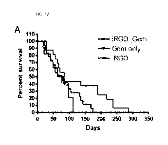
[0013] FIG. IA is a graph showing the percent survival of transgenic Kras-
LSLGD12, p53-
LSL17211, Pdx-]-cre (KPC) mice bearing de novo pancreatic ductal
adenocarcinoma (PDAC)
treated with gemcitabine (GEM).
3
CA 03199012 2023- 5- 15
WO 2022/120344
PCT/US2021/072666
[0014] FIG. 1B is a set of photographs of tumor collected from the mice in
FIG. 1A, stained
for CD8+ T cells. The cells were counted under a microscope using a randomly
selected field
of view, results of which are shown in FIG. 1C. In FIG. 1D, CD8+ T cells in
the PDAC of
the three most long-lived and the four most short-lived KPC mice were
analyzed. Scale bars,
100 lim; *, p <0.05; ***, p <0.001.
[0015] FIG. 2A is a pair of photographs showing KPC organoids with elaborate
folding and a
lumen (arrowhead).
[0016] FIG. 2B is a set of graphs showing data on PD-Li expression in
luciferase-positive
KPC (KPC-luc) organoids analyzed by flow cytometry.
[0017] FIG. 2C is a set of photographs of longitudinal luminescence imaging of
orthotopic
KPC-luc tumors in B6129SF1/J mice.
[0018] FIG. 2D is a set of images of KPC-luc PDAC and liver and lung
metastases. H&E
staining of the primary tumor is shown. Scale bar, 100 i_tm.
[0019] FIG. 2E, FIG. 2F, and FIG. 2G show results of flow cytometry of CD8+ T
cells and
Tregs (FIG. 2E), NPR-1+ Tregs (FIG. 2F), and av133+ and avl35+ Tregs (FIG. 2G)
in PDAC
and spleen (Spl) of normal mice (NMs) and KPC-luc mice (PDAC Ms).
[0020] FIG. 2H is an image showing that intravenously injected FAM-iRGD
(green) targets
CD4+ (magenta) Foxp3+ (red) Tregs in KPC-luc PDAC (white arrowheads). Some
iRGD-
targeted Foxp3+ cells were CD4neg (black arrowheads). Blue = DAPI. Scale bar,
50
[0021] FIG. 21 presents data on av135 and NRP-1 expression in normal mouse
spleen Tregs
cultured alone or with KPC-luc cells.
[0022] FIG. 2J, FIG. 2K, FIG. 2L, and FIG. 2M present data for orthotopic KPC-
luc mice
treated with IV iRGD + GEM with or without anti-PD-Li mAb (clone 10F.9G2) 3x a
week
for 2 weeks. The results show that iRGD + GEM significantly enhanced anti-PD-
Li therapy
(FIG. 2J), NRP-1+ av133 integrin + total Tregs (FIG. 2K) and CD25Ingh Tregs
(FIG. 2L,
insets), and the proportion of CD8+ and CD4+ T cells (FIG. 2M), in the PDAC
and spleen
after iRGD + GEM + anti-PD-Li mAb treatment. Statistics, ANOVA; n.s., not
significant;
<0.001.
[0023] FIG. 3A through FIG. 3B relates to av integrin and NRP-1 expression in
human
PDAC Tregs. FIG. 3A shows expression of avl35 integrin in Tregs isolated from
tumor (blue)
and spleen (red) samples from a PDAC patient. Green is an isotype control.
FIG. 3B is a pair
of images showing av135 integrin (green) in CD3+ (red) Foxp3+ (magenta) T
cells (white
4
CA 03199012 2023- 5- 15
WO 2022/120344
PCT/US2021/072666
arrowheads) and NRP-1 (green) in CD3+ T cells (yellow arrowheads) in human
PDAC.
Foxp3 was not stained in the right panel due to the incompatibility with NRP-1
staining.
DAPI not shown for better visualization of the other colors. Scale bars, 20
pm.
[0024] FIG. 4A through FIG. 4D are a set of graphs showing T cells in
peritoneal tumors
(PTs) in mice generated with ID8 mouse ovarian cancer cells: FIG. 4A, CDR (4%)
and
CD4+ (17%) T cells; FIG. 4B,
5high (32%) and CD2510w (58%) Tregs; FIG. 4C, av133+
NRP-1 Tregs (63%); FIG. 4D, av135 NRP-1 Tregs (26%). The number of T cells
was
low since the PTs were small.
[0025] FIG. 5. Expression of avr35 integrin on Tregs and CTLs isolated from
orthotopic
PDAC (T) and spleen (S) of KPC-derived syngeneic tumor mice (T Ms) and the
spleen of
normal mice (N Ms) analyzed by flow cytometry. p <0.01.
[0026] FIG. 6. Survival of CD4+ T cells in the presence or absence of KPC-
derived PDAC
cells. Splenic T cells from mice were cultured in the presence of KPC-derived
PDAC cells to
expand civ135 integrin + Tregs. Survival was determined by counting the number
of cells using
a heinocytometer. *, p < 0.01.
[0027] FIG. 7. iRGD binding to CD25+ CD4+ T cells (Tregs) and CD25neg CD4+ T
cells
(non-Tregs) isolated from KPC-derived PDAC. The T cells were cultured in the
presence of
fluorescein (FAM)-labeled iRGD at 37 C for 1 hrs. iRGD binding was determined
by flow
cytometry.
[0028] FIG. 8A and FIG. 8B. iRGD binding to CD25 CD4+ T cells (Tregs) and
CD25neg
CD4 T cells (non-Tregs) produced in vitro. The Tregs and non-Tregs were
produced by
culturing mouse splenic T cells in the presence of CD3/CD28 beads and KPC-
derived PDAC
cells. FIG. 8A, FAM-iRGD binding to the Tregs was determined by flow
cytometry. FIG. 8B,
anti-civr35 integrin Abs inhibited FAM-iRGD binding to the Tregs.
[0029] FIG. 9A and FIG. 9B. The effect of iRGD monotherapy on Tregs and the
CTL/Treg
ratio in the PDAC tissue and spleen. Mice bearing orthotopic PDAC were treated
with
systemic iRGD or PBS for 2 weeks. FIG. 9A, Time-dependent changes in the
proportion of
Trcgs and FIG. 9B CTL/Treg ratio in the PDAC tissue.
[0030] FIG. 10A, av(35 integrin + and FIG. 10B NRP-1 Tregs in the PDAC after
iRGD
monotherapy.
[0031] FIG. 11A, Time-dependent changes in the proportion of Tregs and FIG.
11B,
CTL/Treg ratio in the spleen. *, p < 0.05; n.s., not significant.
CA 03199012 2023- 5- 15
WO 2022/120344
PCT/US2021/072666
[0032] FIG. 12A and FIG. 12B. KPC-derived PDAC mice were treated with iRGD
anti-
PD-Li mAb (A; n = 4-6) or iRGD + Gem anti-PD-Li mAb (B; n = 4) 3x a week for
2
weeks. FIG. 12B. Flow cytometry data of CD4+ CD25+ Tregs and CD8+ T cells in
the tumor
and spleen after iRGD + Gem + anti-PD-Li mAb therapy are shown. Tregs halved
and CTLs
doubled in the PDAC but not in the spleen. n.s., not significant; *,p <0.05;
*,p* < 0.01.
DETAILED DESCRIPTION
[0033] 1. Definitions
[0034] Unless defined otherwise, all technical and scientific terms used
herein have the same
meaning as commonly understood by one of ordinary skill in the art. Although
various
methods and materials similar or equivalent to those described herein can be
used in the
practice or testing of the present invention, suitable methods and materials
are described
below. However, the skilled artisan understands that the methods and materials
used and
described are examples and may not be the only ones suitable for use in the
invention.
Moreover, as measurements are subject to inherent variability, any
temperature, weight,
volume, time interval, pH, salinity, molarity or molality, range,
concentration and any other
measurements, quantities or numerical expressions given herein are intended to
be
approximate and not exact or critical figures unless expressly stated to the
contrary.
[0035] As used herein, the term "about," means plus or minus 20 percent of the
recited
value, so that, for example, "about 0.125" means 0.125 0.025, and "about 1.0"
means 1.0
0.2.
[0036] As used herein, the term "iRGD" or -iRGD peptide" refers to a 9-amino
acid cyclic
peptide having sequence (sequence: CRGDKGPDC; SEQ ID NO:2) or a variant
thereof. In
certain specific examples, variants of iRGD include the following
CRGD(R/K/1-1)G(P/V)(D/E/H)C (SEQ ID NO:3), wherein the parentheses set forth
amino
acid options at that position. Other iRGD variants are disclosed in US Pat.
Pub. No.
20090246133, which is incorporated herein in its entirety. Reference to iRGD,
iRGD peptide
or peptide includes peptide variants unless stated otherwise.
[0037] As used herein, the terms "treatment," "treating," and the like, refer
to obtaining a
desired pharmacologic and/or physiologic effect through administering
compound(s) or
composition(s). "Treatment," includes: preventing, partially preventing,
reversing,
alleviating, reducing the likelihood of, or inhibiting the condition or
disease (or symptom
thereof) from occurring in a subject. The subject can include those diagnosed
with a tumor or
cancer, a pre-cancer, or who are predisposed to the condition or disease but
has not yet been
6
CA 03199012 2023- 5- 15
WO 2022/120344
PCT/US2021/072666
diagnosed as having it. ; (b) inhibiting the condition or disease or symptom
thereof, such as,
arresting its development; and (c) relieving, alleviating or ameliorating the
condition or
disease or symptom thereof, such as, for example, causing regression of the
condition or
disease or symptom thereof. Treatment can include administering one or more
agents,
performing a procedure such as surgery or applying radiation and the like, or
both.
[0038] As used herein, the term "administering" and its cognates refer to
introducing an
agent to a subject, and can be performed using any of the various methods or
delivery
systems for administering agents or pharmaceutical compositions, and any route
suitable for
the composition and the subject, as known to those skilled in the art. Modes
of administering
include, but are not limited to oral administration, intravenous,
subcutaneous, intramuscular
or intraperitoneal injections, or local administration directly into or onto a
target tissue (such
as the pancreas, brain, or a tumor). Administration by any route or method
that delivers a
therapeutically effective amount of the drug or composition to the cells or
tissue to which it is
targeted is suitable for use with the invention.
[0039] As used herein, the term "combination,- with respect to administration
of more than
one active agent to a subject, i.e., combination therapy, refers to
administration
simultaneously or at different times. The one or more agents can be delivered
in two or
several pharmaceutical compositions that contain one active agent each, or
using
pharmaceutical compositions that each contain one or more active agent(s). The
different
pharmaceutical compositions can be formulated for the same or different routes
of
administration. The administration of the separate pharmaceutical compositions
can be
accomplished at the same time, in quick succession, or separated in time by
minutes, hours,
days, or weeks. Combination treatment with an immune checkpoint inhibitor and
iRGD may
be presumed to be the case if an immune checkpoint inhibitor and a complement
inhibitor are
prescribed or administered to a subject suffering from cancer by or under
direction of the
same health care professional. A combination pharmaceutical composition
contains more
than one active agent and a pharmaceutically acceptable carrier.
[0040] As used herein, the terms "subject," "individual,- "host," and
"patient," are used
interchangeably to refer to humans or any non-human mammal, and can include
mammalian
farm animals, mammalian sport animals, mammalian companion animals, simians,
non-
human primates, felines, canines, equines, rodents, lagomorphs, bovines,
porcines, ovines,
caprines. A suitable subject for the invention preferably is a human that is
suspected of
having, has been diagnosed as having, or is at risk of developing a
hyperproliferative disease.
7
CA 03199012 2023- 5- 15
WO 2022/120344
PCT/US2021/072666
Conditions amenable to treatment by the invention which define an appropriate
subject or
patient will be discerned easily by the person of skill in the art based on
the disclosures
herein. A "subject in need" is a subject that is at risk of developing cancer,
or who manifests
any characteristics or symptoms of cancer, or who has been diagnosed with
cancer.
[0041] As used herein, the term "cancer", also referred to as a tumor or a
malignant tumor,
refers to any of a group of diseases involving abnormal cell proliferation
(hyperproliferation)
with the potential to invade locally and/or spread to other parts of the body
(metastasize).
The term "cancer" is generally used interchangeably with "tumor" herein
(unless a tumor is
specifically referred to as a "benign" tumor, which is an abnormal mass of
cells that lacks the
ability to invade neighboring tissue or metastasize), and encompasses
malignant solid tumors
(e.g., carcinomas, sarcomas) and malignant growths in which there may be no
detectable
solid tumor mass (e.g., certain hematologic malignancies). In particular,
cancers that are
susceptible to immune checkpoint inhibitors are contemplated for use with the
methods
according to the invention, however immune checkpoint inhibitor-resistant
cancers also can
be treated according to embodiments of the invention. The term "cancer- can
refer to a
primary or metastatic tumor, and includes cancers that are unresectable
cancer, and cancers of
any stage, including stage III cancer and/or stage IV cancer.
[0042] As used herein, the term "antibody" refers to an immunoglobulin and
encompasses
full size antibodies and antibody fragments comprising an antigen binding
site. Antibodies
useful in certain embodiments of the invention may originate from or be
derived from a
mammal, e.g., a human, non-human primate, rodent (e.g., mouse, rat), rabbit,
goat, bovine,
equine, ovine, camelid, or from a bird (e.g., chicken), and may be of any of
the various
antibody isotypes, e.g., the mammalian isotypes: IgG (e.g., of the IgG1 ,
IgG2, IgG3, or IgG4
subclass), IgM, IgA, IgD, and IgE or isotypes that are not found in mammals,
e.g., IgY
(found in birds) or IgW (found in sharks).
[0043] An antibody fragment (Fab) may be, for example, a Fab', F(ab')2, scFv
(single-chain
variable), single domain antibody (e.g., a VHH), or other fragment that
retains or contains an
antigen binding site. See, e.g., Allen, T., Nature Reviews Cancer, Vol.2, 750-
765, 2002, and
references therein for disclosures relating to antibody fragments. The
contents of this
reference are hereby incorporated by reference. Antibodies known in the art as
diabodies,
minibodies, or nanobodies can be used in various embodiments. Bispecific or
multispecific
antibodies may be used in various embodiments. The heavy and light chain of
IgG
immunoglobulins (e.g., rodent or human IgGs) contain four framework regions
(FRI through
8
CA 03199012 2023- 5- 15
WO 2022/120344
PCT/US2021/072666
FR4) separated respectively by three complementarity determining regions (CDR1
through
CDR3). The CDRs, particularly the CDR3 regions and especially the heavy chain
CDR3, are
largely responsible for antibody specificity.
[0044] An antibody may be a chimeric antibody in which, for example, a
variable domain of
non-human origin, e.g., of rodent (e.g., murine) or non-human primate origin)
is fused to a
constant domain of human origin, or a "humanized" antibody in which some or
all of the
complementarity-determining region (CDR) amino acids that constitute an
antigen binding
site (sometimes along with one or more framework amino acids or regions) are
"grafted"
from a rodent antibody (e.g., murine antibody) or phage display antibody to a
human
antibody, thus retaining the specificity of the rodent or phage display
antibody. Thus,
humanized antibodies may be recombinant proteins in which only the antibody
complementarity-determining regions are of non-human origin. Alterations to
antibody
sequence that are involved in the humanization process are generally carried
out through
techniques at the nucleic acid level, e.g., standard recombinant nucleic acid
techniques. In
some embodiments only the specificity determining residues (SDRs), the CDR
residues that
are most crucial in the antibody-ligand interaction, are grafted. The SDRs may
be identified,
e.g., through use of a database of the three-dimensional structures of the
antigen-antibody
complexes of known structures or by mutational analysis of the antibody-
combining site. In
some embodiments an approach is used that involves retention of more CDR
residues,
namely grafting of so-called "abbreviated" CDRs, the stretches of CDR residues
that include
all the SDRs. See, e.g., Kashmiri, S V, Methods. 36(1):25-34 (2005), for
further discussion
of SDR grafting and Almagro J C, Fransson J. Humanization of antibodies. Front
Biosci.
13:1619-33 (2008) for review of various methods of obtaining humanized
antibodies_ These
references are incorporated by reference herein. "Originate from or derived
from refers to
the original source of the genetic information specifying an antibody sequence
or a portion
thereof, which may be different from the species in which an antibody is
initially synthesized.
For example, "human" domains may be generated in rodents (e.g., mice) whose
genome
incorporates human immunoglobulin genes or may be generated using phage
display. See,
e.g., Vaughan, et al, (1998), Nature Biotechnology, 16: 535-539, e.g., for
discussion of
methods that may be used to generate a fully human antibody. This reference is
incorporated
by reference.
[0045] The amino acid sequences of the variable regions of such antibodies are
sequences
that, while derived from and related to the germline sequences encoding
variable domains
9
CA 03199012 2023- 5- 15
WO 2022/120344
PCT/US2021/072666
(VH and/or VL domains) of a particular species (e.g., human), may not
naturally exist within
that species' antibody germline repertoire in vivo. For example, the human
immunoglobulin
genes may have been subjected to in vitro mutagenesis (or, when an animal
transgenic for
human immunoglobulin gene sequences is used, in vivo somatic mutagenesis).
Antibodies
suitable for use with the invention may be polyclonal or monoclonal, though
for purposes of
the present invention monoclonal antibodies are generally preferred as
therapeutic agents.
Antibodies can be glycosylated or non-glycosylated.
[0046] Methods for generating antibodies that specifically bind to virtually
any molecule of
interest are known in the art. For example, monoclonal or polyclonal
antibodies can be
purified from natural sources, e.g., from blood or ascites fluid of an animal
that produces the
antibody (e.g., following immunization with the molecule or an antigenic
fragment thereof)
or can be produced recombinantly, in cell culture and, e.g., purified from
culture medium.
Affinity purification may be used, e.g., protein A/G affinity purification
and/or affinity
purification using the antigen as an affinity reagent.
[0047] Suitable antibodies can be identified using phage display and related
techniques. See,
e.g., Kaser, M. and Howard, G., "Making and Using Antibodies: A Practical
Handbook" and
Sidhu, S., "Phage Display in Biotechnology and Drug Discovery", CRC Press,
Taylor and
Francis Group, 2005, for further information. This reference is incorporated
by reference.
[0048] Methods for generating antibody fragments are well known. For example,
F(ab')2
fragments can be generated, for example, through the use of an Immunopure
F(ab')2
Preparation Kit (PierceTM) in which the antibodies are digested using
immobilized pepsin and
purified over an immobilized Protein A column. The digestion conditions (such
as
temperature and duration) may be optimized by one of ordinary skill in the art
to obtain a
good yield of F(ab')2. The yield of F(ab')2 resulting from the digestion can
be monitored by
standard protein gel electrophoresis. F(ab') can be obtained by papain
digestion of antibodies,
or by reducing the S--S bond in the F(ab')2. A "single-chain Fv" or "scFv"
antibody fragment
comprises the VH and VL domains of an antibody, wherein these domains are
present in a
single polypeptide chain. Typically, an scFv antibody further comprises a
polypeptide linker
between the VH and VL domains, although other linkers could be used to connect
the domains
in certain embodiments.
[0049] As used herein, the term "monoclonal antibody" (MAb) or "monoclonal
antibody
composition" refers to a population of antibody molecules that contain only
one molecular
species of antibody molecule consisting of a unique light chain gene product
and a unique
CA 03199012 2023- 5- 15
WO 2022/120344
PCT/US2021/072666
heavy chain gene product. In particular, the complementarily determining
regions (CDRs) of
the monoclonal antibody are identical in all the molecules of the population.
[0050] As used herein, the term "immune system cell" refers to any of a
variety of cells that
play a role in the immune response. Immune system cells include lymphocytes (T
cells, B
cells, natural killer (NK) cells), dendritic cells, monocytes, macrophages,
eosinophils, mast
cells, basophils, and neutrophils. T cells comprise a number of different
functional classes
that play different roles in the immune response. Different functional classes
may be
distinguished based on cell surface markers and other properties. Most T cells
express an al3
T cell receptor (TCR) through which the cell is able to recognize a specific
antigen in the
context of an appropriate major histocompatibility complex (MHC) molecule,
though a minor
subset expresses the y6 TCR.
[0051] Cytotoxic T cells (CTLs) are typically positive for the cell surface
marker CM, which
serves as a co-receptor for the TCR in recognition of MHC Class 1 molecules on
the surface
of target cells during antigen-specific T cell activation and/or responses.
CTLs and NK cells
play important roles by eliminating infected host cells and tumor cells
through a variety of
mechanisms including the release of cytotoxic substances.
[0052] Helper T cells are typically positive for the cell surface marker CD4,
which serves as
a co-receptor for the TCR in recognition of MHC Class II molecules on the
surface of APCs
during antigen-specific T cell activation. Helper T cells promote the activity
of other
immune system cells (i.e., provide "help") by, among other things, releasing
cytokines that
have a variety of effects such as enhancing survival, proliferation, and/or
differentiation.
[0053] Natural killer cells have the ability to recognize and kill (e.g., by
causing lysis or
apoptosis) cancerous, stressed, or infected cells without requiring antigen-
specific activation
by presentation of antigen in the context of MHC. Instead, their activation is
regulated by a
balance of the activity of activating receptors and inhibitory receptors and
cytokines. NK
cells typically lack cell surface receptors that are highly specific for a
particular antigen and
are able to react rapidly without prior exposure to the antigen.
[0054] As used herein, "effector cells" refers to the activated immune system
cells that defend
the body in an immune response. Effector T cells include cytotoxic T cells and
helper T
cells, which carry out cell-mediated responses. Effector B cells are called
plasma cells and
secrete antibodies. Effector cells also include effector NK cells.
[0055] An antigen-presenting cell (APC) is a cell that can process and display
antigens in
association with major histocompatibility complex (MHC) molecules on its
surface. T cells
11
CA 03199012 2023- 5- 15
WO 2022/120344
PCT/US2021/072666
can recognize these complexes using their T cell receptors (TCRs). APCs also
can display
other molecules (costimulatory proteins) that are required for activating
naive T cells. APCs
that express MHC class II molecules include dendritic cells, macrophages, and
B cells and
may be referred to as professional APCs.
[00561 Dendritic cells (DCs) are white blood cells that occur in most tissues
of the body,
particularly epithelial tissues. DCs serve as a link between peripheral
tissues and lymphoid
organs. Immature DCs sample the surrounding environment and take up antigenic
substances
such as pathogen components or tumor antigens. They undergo maturation and
migrate to
lymph nodes or spleen, where they display fragments of processed antigens at
their cell
surface using MHC Class II (MHCII) complexes. As part of the maturation
process, DCs
upregulate cell-surface molecules that act as co-stimulators in T cell
activation, such as CD80
(B7-1), CD86 (B7-2), and/or CD4O. DCs activate helper T cells by presenting
them with
antigens in the context of MHC11 complexes, together with non-antigen specific
co-
stimulators. DCs and various other APCs have the capacity to activate
cytotoxic T cells and
B cells through presentation of MHC Class I (MHCI)-peptide complexes (cross-
presentation)
and costimulators.
[0057] As used herein, the term "regulatory T cells (Tregs, suppressor T
cells)" refers to a
subpopulation of CD4+ T cells which modulate the immune system, maintain
tolerance to
self-antigens, and abrogate autoimmune disease. These cells generally suppress
or
downregulate induction and proliferation of effector T cells and can be
identified based on a
cell surface marker expression pattern of CD4+CD25+CD1271 . Tregs also are
characterized
by expression of CTLA4 and GITR. Tregs can suppress the activity of other
immune system
cell subsets by a variety of mechanisms such as secretion of immunosuppressive
cytokines
and via cell-cell contact. They can inhibit immune responses at multiple
steps, e.g., at the
induction of activation (e.g., by inhibiting the ability of APCs to stimulate
T cells) and during
effector phases. Tregs are often found in tumors, and increased numbers of
Tregs has been
associated with a worse prognosis in various cancer types. Where it is
intended herein to
refer to a T cell that is a Treg, the T cell will be identified as such. Thus,
unless expressly
indicated a T cell, as used herein, is not a Treg cell.
[0058] As used herein, the term "adjunct cancer therapy" refers to a therapy,
such as surgery,
chemotherapy, radiotherapy, thermotherapy, and laser therapy, that can provide
a beneficial
effect when administered in conjunction with administration of iRGD in
optional
combination with an immune checkpoint inhibitor. The term "anti-cancer agent"
refers to
12
CA 03199012 2023- 5- 15
WO 2022/120344
PCT/US2021/072666
conventional chemotherapy, a molecularly targeted anticancer agent, a cancer
vaccine, a
second immunostimulatory agent, cell-based immunotherapy, or a combination
thereof to the
subject.
[0059] As used herein, an "adjunct cancer therapeutic agent" refers to an
agent, compound,
or composition that possesses selectively cytotoxic or cytostatic effects on
cancer cells
compared to normal cells. Adjunct cancer therapeutic agents can be co-
administered with an
iRGD, and/or an immune checkpoint inhibitor. A non-limiting list of examples
of selected
adjunct cancer therapeutic agents is provided in Table 1, below.
[0060] As used herein, a "peptide" is a sequence of two or more amino acids up
to about 100
amino acids. A "variant" of a particular peptide has one or more alterations
(e.g., additions,
substitutions, and/or deletions, which may be referred to collectively as
"mutations") with
respect to the original peptide sequence. Thus, a variant can be shorter or
longer than the
original peptide of which it is a variant. Conservative substitutions are
preferred when
substitutions are made in a peptide. Conservative substitutions are those
where an amino acid
is replaced with a different amino acid of the same type, such as glutamic
acid for aspartic
acid or alanine for glycine and the like. Persons of skill are aware of such
substitutions.
[0061] The term "variant" also encompasses "fragments." A "fragment" is a
continuous
portion of a polypeptide that is shorter than the original peptide. In certain
embodiments of
the invention a variant peptide has significant sequence identity to the
original polypeptide
over a continuous portion of the variant that comprises at least 70%, at least
80%, at least
90%, at least 95%, or more, of the length of the peptide. Peptides can include
non-traditional
amino acids or D-amino acids as well, or terminal additions or modification
such as C-
terminal amides, and the like. An amino acid "difference" refers to a
substitution, insertion,
or deletion of an amino acid. In certain embodiments, peptide variants also
encompass
peptidomimetics of a peptide or peptide mimics.
[0062] The term "peptidomimetic," as used herein, means a peptide-like
molecule that has
the activity of the peptide upon which it is structurally based. Such
peptidomimetics include
chemically modified peptides, peptide-like molecules containing non-naturally
occurring
amino acids, and peptoids and have an activity such as that from which the
peptidomimetic is
derived (see, for example, Goodman and Ro, Peptidomimetics for Drug Design, in
"Burger's
Medicinal Chemistry and Drug Discovery" Vol. 1 (ed. M. E. Wolff; John Wiley &
Sons
1995), pages 803-861).
[0063] As used herein the term "immune checkpoint protein" refers to a protein
or receptor
13
CA 03199012 2023- 5- 15
WO 2022/120344
PCT/US2021/072666
that functions in an immune checkpoint pathway. Examples of immune checkpoint
proteins
include inhibitory receptors through which an immune checkpoint pathway is
initiated, and
their ligands. Examples of immune checkpoint pathways include the cytotoxic T-
lymphocyte
associated antigen 4 (CTLA4) pathway and the programmed cell death 1 (PD1)
pathway,
both of which are further discussed below. The term "immune checkpoint
molecule"
encompasses immune checkpoint proteins as well as small molecules such as
adenosine that
play a role in immune checkpoint pathways.
[0064] As used herein, the term "immune checkpoint inhibitor" refers to a
class of agents that
activate the immune system to attack tumors by blocking or reducing the
activity of immune
checkpoint molecules such as CTLA4, PD-1, PD-L1, and the like, discussed
below.
[0065] As used herein, the term "effective amount" of an active agent, e.g.,
an immune
checkpoint inhibitor or an iRGD peptide, refers to an amount of the active
agent sufficient to
elicit one or more biological effect(s) of interest in, for example, a subject
to whom the active
agent (or composition) is administered. In some embodiments the biological
effect of an
active agent is enhancement of the efficacy of a second agent.
[0066] As will be appreciated by those of ordinary skill in the art, the
absolute amount of a
particular agent that is effective may vary depending on such factors as the
biological
endpoint, the particular active agent, the target tissue, etc. An effective
amount of an agent or
composition generally is an amount sufficient to achieve one or more of the
following in a
cancer patient: a complete response (remission), a partial response,
achievement of stable
disease as determined by objective criteria, an improvement in symptoms, an
increase in the
length of progression-free survival, or an increase in overall survival. An
effective amount
can be an amount that results in killing of tumor cells, directly or
indirectly or that stops
growth of the tumor cells. Those of ordinary skill in the art will further
understand that an
"effective amount" may be administered in a single dose, or may be achieved by
administration of multiple doses over a period of time. An effective amount of
a
pharmaceutical composition that contains an effective amount of one or more
agents is an
amount of each agent such that the overall composition is effective.
[0067] In some embodiments, an effective amount of an agent or composition can
be an
amount that suppresses (e.g., eliminates) replication of a pathogen in a
subject suffering from
an infection, renders a subject free of the infectious agent, renders the
subject non-infectious,
results in an improvement in symptoms of infection, decreases mortality due to
the infection,
and/or an increases overall survival.
14
CA 03199012 2023- 5- 15
WO 2022/120344
PCT/US2021/072666
2. Overview
[0068] The invention is based on the discovery that iRGD tumor penetrating
peptide
possesses immunomodulatory effects that allow it to be used for treatment of
tumors in
conjunction with immune checkpoint inhibitors. The data presented herein show
a
potentiating, synergistic effect on cancer chemotherapeutic agents when iRGD
is co-
administered with immune checkpoint inhibitors. This technology can also be
applied to
intraperitoneal chemotherapy methods because iRGD (and co-administered drugs)
target various
peritoneal tumors when administered intraperitoneally indicating that it will
likely sensitize
peritoneal metastases of various tumors, such as ovarian cancer (see FIG. 4),
to immunotherapy.
[0069] The concept of tumor-specific immunotherapy is becoming increasingly
important
because non-specific eradication of Tregs can cause inflammatory side effects.
Therefore, the
invention makes certain tumor-specific immunotherapies feasible. iRGD does not
have to be
conjugated to any of the co-administered agents to be effective. The method
allows
immunomodification with enhanced immunotherapy by simple co-administration.
That being
said, iRGD conjugates may be produced and used in combination with immune
checkpoint
inhibitors. Specific examples of iRGD conjugates include iRGD conjugated with
an adjunct
cancer therapeutic agent.
3. Summary of the Results
[0070] iRGD can modulate the immune landscape in pancreatic duct
adenocarcinoma
(PDAC), sensitizing the cancer to immune checkpoint inhibitors (i.e. anti-PD-
L1, anti-PD-1,
and anti-CTLA4 mAbs).
[0071] iRGD specifically depletes Tregs within the tumor.
[0072] iRGD results in expansion of intratumoral CDS+ T cells (effector cells)
in PDAC.
[0073] iRGD enables synergy with chemotherapy and immunotherapy leading to
reduced
tumor burden and prolonged survival in a PDAC mouse model.
[0074] iRGD does not have to be conjugated to anti-cancer drugs or
immunotherapeutics.
[0075] iRGD enhances immunotherapy via co-administration with anti-cancer
drugs or other
immunotherapeutics.
[0076] iRGD can be applied to intraperitoneal chemotherapy.
[0077] iRGD can improve the effectiveness of cancer immunotherapies, since av
integrin
NRP-1+ Tregs are expressed exclusively within tumors in multiple cancers.
4. Embodiments of the invention
A. Immunotherapies
CA 03199012 2023- 5- 15
WO 2022/120344
PCT/US2021/072666
[0078] Immunotherapy is a type of cancer treatment that assists the immune
system in
fighting cancer. The therapy stimulates the immune system to find and attack
cancer cells
rather than directly killing the cancer like traditional cancer chemotherapy
drugs. Most
cancer immunotherapy exploits the fact that tumor cells often have specific
tumor antigens on
their surface that can be specifically recognized and targeted by immune
molecules such as
antibodies or modified antibodies. Immunotherapies according to this invention
B. Immune Checkpoints
[0079] An important function of the immune system is its ability to tell
between normal cells
in the body and those it sees as "foreign." This lets the immune system attack
the foreign
cells while leaving the normal cells alone. To do this, it uses "checkpoints."
Immune
checkpoints are molecules on certain immune cells that need to be activated
(or inactivated)
to start an immune response. In summary, immune checkpoints are immune system
regulators that are crucial for self tolerance, which prevents the immune
system from
attacking normal cells. However, some tumors can protect themselves from
attack by the
immune system by manipulating this system. Drugs that target these checkpoints
hold a lot
of promise as cancer treatments. These drugs are called checkpoint inhibitors.
[0080] Immune checkpoint molecules can be stimulatory (e.g., members of the
tumor
necrosis factor receptor superfamily such as CD27, CD40, 0S40, GITR, and
CD137) or
inhibitory (e.g., A2AR, B7-H3, B7-H4, BTLA, CTLA4, IDO, KIR, LAG3, NOX2, PD-1,
TIM-3, VISTA, and SIGLEC7).
[0081] PD-1 is a checkpoint protein on T cells. It normally acts as a type of
"off switch" that
helps keep the T cells from attacking other cells in the body when it binds
its ligand, which is
present on some normal and cancer cells. When PD-I binds to PD-L. I , it sends
the message
for the T cell not to attack the other cell. Some cancer cells have large
amounts of PD-11,1_,
which helps them hide from an immune attack. The binding of PD-L1 to PD-1, for
example.
keeps T cells from killing tumor cells in the body. Blocking this binding with
an immune
checkpoint inhibitor allows effector T cells to attack and kill tumor cells.
C. Immune Checkpoint Inhibitors
[0082] Immune checkpoint inhibitors are molecules (drugs) that inhibit or
block inhibition of
the immune system, such as by blocking inhibitory checkpoint proteins.
Examples of
checkpoint proteins include CTLA4, and/or PD-1, and/or PD-Li and/or PD-L2.
Pembrolizumab (lambrolizumb; Keytruda), Nivolumab (Opdivo), Atezolizumab
(Tecentriq),
Avelumab (Bevancio), cerniplimab (Libtayo), and Dumalumab (Imfinizi) are FDA-
approved
16
CA 03199012 2023- 5- 15
WO 2022/120344
PCT/US2021/072666
drugs that inhibit PD-1/PD-L1, and are contemplated for use with the
invention. Additional
immune checkpoint inhibitors include MEDI0680, MPDL3280A, AMP-224, BMS-936559,
MPDL3280A, MEDI4736, MSB0010718C, for example. The synergistic effects of with
immune checkpoint inhibitors should apply to any of the foregoing immune
checkpoint
inhibitors, or newly developed immune checkpoint inhibitors targeting the
aforementioned
inhibitory checkpoint proteins or other inhibitory checkpoint proteins to be
elucidated. (See
Freeman et al, JCI 130:1405-1416, 2020)
[0083] Preferably, the immune checkpoint inhibitor comprises an antibody,
aptamer, non-
antibody engineered binding protein, dominant negative protein, or other
specific binding
agent that binds to an inhibitory immune checkpoint molecule, e.g., CTLA4 or
PD1.
[0084] Other PD1 pathway inhibitors also can include RNAi agents or antisense
oligonucleotides that inhibit expression of PD1.
[0085] Preferred immune checkpoint inhibitors are monoclonal antibodies that
target either
PD-1 or PD-L1 to block this binding and boost the immune response against
cancer cells.
Preferred examples of drugs that target and antagonize or block PD-1 include:
pembrolizumab (Keytruda); nivolumab (Opdivo); and eemiplintab (Libtayo).
Preferred
examples of drugs that target and block or antagonize PD-L, I include:
atemlizumab
(Tecentriq); avelumab (Bavencio); and durvalurnab (Imfinzi). These drugs can
be helpful in
treating several types of cancer.
[0086] All combinations of any genus, subgenus, or species of immune
checkpoint inhibitor
and any genus, subgenus, or species of complement inhibitor, compositions
comprising any
such combination, and use of any such combination in any method described
herein, are to be
considered expressly disclosed herein. Any antibody or other specific binder
that can block
or inhibit CTL-4, and/or PD-1 and/or PD-Li and/or PD-L2 can be used with the
inventive
methods, such as nanoparticles, engineered cells, any engineered binding
protein, soluble
receptor, aptamer, peptide or small molecule that binds to an immune
checkpoint protein and
preferably antagonizes or blocks an inhibitory immune checkpoint molecule.
Combinations
or mixtures of any of such immune checkpoint inhibitors are suitable for use
with the
invention.
[0087] In certain embodiments, the immune checkpoint inhibitor comprises
ipilimumab
and/or tremelimumab, which inhibit the CTLA4 pathway. In some embodiments of
the
invention, the immune checkpoint inhibitor inhibits a killer-like
immunoglobulin receptor
(KIR) pathway. For example, in some embodiments the immune checkpoint
inhibitor binds to
17
CA 03199012 2023- 5- 15
WO 2022/120344
PCT/US2021/072666
a KIR or KIR ligand. In some embodiments of the invention, the immune
checkpoint inhibitor inhibits an immune checkpoint pathway involving LAG3,
TIM3, BTLA,
A2AR, or A2BR.
[0088] PD1 has two known ligands, PD1 ligand 1 (PD-Li; also known as B7-H1 and
CD274) and PD-L2 (also known as B7-DC). The PD-1 pathway limits the activity
of T cells
in peripheral tissues at the time of an inflammatory response to infection and
in order to limit
autoimmunity. PD1 is a member of the CD28/CTLA4 family that is expressed on
activated T
cells. Binding of PD1 by its ligands mediates an inhibitory signal that
results in reduced
cytokine production, and reduced T cell survival. PD1 expression is induced
when T cells
become activated. When engaged by one of its ligands, PD1 inhibits kinases
that are
involved in T cell activation.
[0089] Like CTLA4, PD1 is highly expressed on Treg cells, and its activation
can enhance
their proliferation and/or suppressive activity in the presence of a PD1
ligand, which further
suppresses immune function. Since many tumors are highly infiltrated with Treg
cells,
blockade of the PD1 pathway increases antitumor immune responses by decreasing
the
number and/or suppressive activity of Treg cells.
[0090] A PD1 inhibitor is an agent that inhibits the activity of PD1 or its
natural ligand(s)
with the effect that PD l's ability to suppress immune responses is reduced. A
PD1 pathway
inhibitor encompasses any agent that impairs the ability of PD1 to limit T
cell activity or
enhance Treg proliferation and/or suppressor functions. In some embodiments a
PD1
inhibitor specifically binds to PD1 and inhibits its activation or activity.
In some
embodiments a PD1 inhibitor specifically binds to PD1 and blocks interaction
of PD1 with its
ligands.
[0091] In some embodiments a PD1 inhibitor (or a PD-Li inhibitor or PD-L2
inhibitor)
binds with a Kd of about 10-6 M or less, 10-7 M or less, 10-8 M or less, 10-9
M or less, 10-19 M
or less, 10-11 M or less, 10-12 M or less, e.g., between 10-13 M and 10-6 M,
or within any range
having any two of the afore-mentioned values as endpoints. In some embodiments
a PD1
inhibitor (or a PD-Li inhibitor or PD-L2 inhibitor) binds with a Kd of no more
than 10-fold
that of nivolumab, up to 10-fold lower, or up to 100-fold lower than that of
nivolumab when
compared using the same assay.
[0092] In some embodiments, the IC50 values for inhibition by a PD inhibitor
of PD1
binding to its ligands is no more than 10-fold greater, up to 10-fold lower,
or up to 100-fold
lower than that of nivolumab-mediated inhibition of PD1 binding to its
ligands, when
18
CA 03199012 2023- 5- 15
WO 2022/120344
PCT/US2021/072666
compared using the same assay.
[0093] CTLA4 is expressed on T cells, and its principal function is to
regulate the extent of
the early stages of T cell activation. In general, activation of T cells
typically occurs through
engagement of the T cell receptor (TCR) and a costimulatory molecule on the T
cell.
Binding of the T cell receptor to a processed form of its cognate antigen (an
antigen to which
an antigen receptor binds) presented by major histocompatibility complex (MHC)
molecules
on an antigen presenting cell provides a first signal for activation. The
second signal conies
from co-stimulation, in which surface molecules on the antigen presenting cell
bind to co-
stimulatory receptors on T cells and activate intracellular signaling
pathways. CD28 is the
most important co-stimulatory receptor for T cell activation and is expressed
constitutively by
naive T cells (cells that have not encountered cognate antigen). In the
absence of co-
stimulation, T-cell receptor signaling alone can result in anergy.
[0094] CD28 and CTLA4 display a different pattern of expression on T-cells:
while CD28 is
constitutively expressed on the surface of T-cells, CTLA4 is detectable at low
levels in naive
T-cells and more strongly upon T-cell activation. CTLA4 has the same ligands
as does
CD28, but the affinity of CTLA4 is about 10-fold higher than that of CD28.
CTLA4
expression on T cells may counteract the activity of CD28 by competing for
ligand binding,
may actively deliver inhibitory signals to the T cell, or both. Through these
and/or other
mechanisms, CTLA4 inhibits T cell activation, thus reducing immune responses
and anti-
tumor immunity. CTLA4 is also expressed by Tregs and promotes their immune
suppressive
function, further contributing to impairing the immune response to the tumor.
[0095] A CTLA4 inhibitor is an agent that inhibits the activity of CTLA4 with
the effect that
the biological activity of CTLA4 is inhibited or reduced, e.g., that impairs
the ability of
CTLA4 to cause inhibition of T cell activation or impairs the ability of CTLA4
to enhance
Treg proliferation and/or suppressor function. A preferred CTLA4 inhibitor is
an agent that
specifically binds to CTLA4 and inhibits its activation or activity.
[0096] United States Patent Nos. 5,811,097; 5,855,887; 5,977,318; 6,051,227;
6,682,736;
6,207,156; 6,984,720; 7,109,003; 7,132,281; and 7,605,238, United States
Patent Publication
Nos. US2002-0039581, US2002-086014, US2004-0202650, US 2005-0201994, US2006-
0165706, US 2011-0081354, US 2012-0148597, US2013-0011405, US2013-0136749,
US2014-0105914, and US2014-0099325, international Patent Publication Nos. WO
2001/014424, WO 01/14424, WO 00/37504, WO 98/42752, and WO 2004/035607, and
European Patent No. EP1212422B1 describe antibodies that bind to CTLA4 and are
19
CA 03199012 2023- 5- 15
WO 2022/120344
PCT/US2021/072666
incorporated by reference for these disclosures. United States Patent
Publication Nos.
US2003-0054360 and US2006-0246123 disclose anti-CTLA4 aptamers that may be
used in
methods and compositions described herein.
[0097] In some embodiments of the invention a subject is treated with two or
more immune
checkpoint inhibitors in combination (administered together in the same
bifunctional or
multifunctional composition, or in separate compositions to be administered
together or
separately). The two or more immune checkpoint inhibitors can be provided or
administered
as part of a bifunctional or multifunctional agent or compound. For example, a
bispecific,
trispecific, or tetraspecific antibody (or other binding agent) capable of
binding to two, three,
or four distinct immune checkpoint molecules can be used.
[0098] The two or more immune checkpoint inhibitors can inhibit the same or
different
immune checkpoint pathways. For example, in some embodiments a first immune
checkpoint inhibitor inhibits the PD1 pathway and a second or third immune
checkpoint
inhibitor inhibits the CTLA4 pathway. For example, any combination of two or
more of
ipilimumab, nivolumab, pembrolizumab, tremilimumab, pidilizumab, MEDI0680, BMS-
936559, MPDL3280A, MEDI4736, MSB0010718C, or SB0010718C can be used in the
same
treatment method.
[0099] For example, in some embodiments a first immune checkpoint inhibitor
agent inhibits
PD1 or CTLA4 and a second agent comprises a TIM3 inhibitor, BTLA pathway
inhibitor,
KIR inhibitor, LAG3 inhibitor, or adenosine pathway inhibitor. In certain
embodiments the
method involves both a PD1 inhibitor and a CTLA4 inhibitor with a further TIM3
inhibitor,
BTLA pathway inhibitor, KIR inhibitor, LAG3 inhibitor, IDO inhibitor, or
adenosine
pathway inhibitor. It is contemplated that the combination of immune
checkpoint inhibitors
comprises no more than 2, 3, 4, or 5 immune checkpoint inhibitors.
D. RGD
[0100] RGD tripeptide (RGD; SEQ ID NO:1) was originally identified as the
amino acid
sequence within the extracellular matrix protein fibronectin (the binding
motif) that mediates
cell adhesion/attachment. It also acts as an inhibitor of integrin-ligand
interaction and can
reduce apoptosis in the absence of signals and integrin-mediated cell
clustering. The RGD
motif has also been identified in other extracellular matrix proteins,
including vitronectin and
laminin.
E. iRGD Peptides and Peptidomimetics
[0101] (E.1) The iRGD peptide has been previously described in the art as a 9-
amino acid
CA 03199012 2023- 5- 15
WO 2022/120344
PCT/US2021/072666
cyclic peptide (CRGDKGPDC; SEQ ID NO:2) and a molecular mimicry agent that was
originally identified in an in vivo screening of phage display libraries in
tumor-bearing mice.
iRGD is able to home to tumor tissues and has been used for its bifunctional
action: homing
to tumors and specific binding to neuropilin-l_ (NRP-1.) receptor with
subsequent activation of
a trans-tissue pathway for penetration into tumors. The ROD motif mediates
binding to
certain av integrins expressed on tumor neovasculature and cancer cells. Upon
binding, a
protease cleavage event is activated, revealing a c-terminal motif (R/ICXXR/K)
in the peptide.
This c-terminal motif then can bind to neuropilin-1 and activate an
endocytotic/exocytotic
transport pathway (formation of macropinosome-like vesicles that carry the
peptide and
bystander drugs into the deeper layers of tumor cells) that can be used to
enhance transport of
coupled and coadministered anti-cancer drugs into tumors. Thus, iRGD enhances
tumor-
specific cytotoxicity of almost any kind of co-injected cancer
chemotherapeutic drug. See
Sugahara et al., 2009, 2010; Pang et al., 2014; and United States Patent No.
9,115,170 for
further discussion on these topics.
[0102] However, the work presented in this application has shown that iRGD
peptides have
unexpected immunomodulating effects on their own, namely that they are able,
unexpectedly,
to potentiate the effects of immtme checkpoint inhibitors in a manner that
would not have
been expected based on their previously known effects. iRGD peptides
unexpectedly are able
to deplete or suppress Tregs in a tumor-specific manner, which potentiates the
effects of
immune checkpoint inhibitors.
[0103] Disclosed are methods and compositions related to an isolated peptide
comprising an
amino acid segment comprising the amino acid sequence of SEQ ID NO: 2, or a
variant
thereof. In a specific example a variant relates a peptide defined by SEQ ID
NO:3.
[0104] In alternative embodiments, the iRGD peptide or variant can comprise a
chimera of
the amino acid sequence SEQ ID NO: 2 or SEQ ID NO:3. Such a chimera can be
additive,
where sequence of one sequence is added to another sequence, substitutional,
where sequence
of one sequence is substituted for sequence of another sequence, or a
combination. The
disclosed peptides can consist of the amino acid segment.
[0105] The iRGD peptide or variant can be, for example, non-circular, linear,
circular or
cyclic. The amino acid segment can be circularized or cyclized via any
suitable linkage, for
example, a disulfide bond. The peptide can have any suitable length, such as a
length of less
than 100 residues. The peptide can have a length of, for example, less than 50
residues. The
peptide can have a length of, for example, less than 20 residues.
21
CA 03199012 2023- 5- 15
WO 2022/120344
PCT/US2021/072666
[0106] Also disclosed are iRGD peptidomimetics that may be used in accord with
the
methods and compositions embodiments taught herein.
[0107] As this specification discusses various proteins and protein sequences
it is understood
that the nucleic acids that can encode those protein sequences are also
disclosed. This would
include all degenerate sequences related to a specific protein sequence, i.e.
all nucleic acids
having a sequence that encodes one particular protein sequence as well as all
nucleic acids,
including degenerate nucleic acids, encoding the disclosed variants and
derivatives of the
protein sequences. Thus, while each particular nucleic acid sequence may not
be written out
herein, it is understood that each and every sequence is in fact disclosed and
described herein
through the disclosed protein sequence.
[0108] It is understood that there are numerous amino acid and peptide analogs
which can be
incorporated into the disclosed compositions. For example, there are numerous
D amino acids
or amino acids which have a different functional substituent than those
discussed above. The
opposite stereo isomers of naturally occurring peptides are disclosed, as well
as the stereo
isomers of peptide analogs. These amino acids can readily be incorporated into
polypeptide
chains by charging tRNA molecules with the amino acid of choice and
engineering genetic
constructs that utilize, for example, amber codons, to insert the analog amino
acid into a
peptide chain in a site specific way (Thorson et al.. Methods in Molec. Biol.
77:43-73 (1991),
Zoller, Current Opinion in Biotechnology, 3:348-354 (1992); Ibba,
Biotechnology & Genetic
Engineering Reviews 13:197-216 (1995), Cahill et al., TIBS, 14(10):400-403
(1989); Benner,
TIB Tech, 12:158-163 (1994); Ibba and Hennecke, Bio/technology, 12:678-682
(1994) all of
which are herein incorporated by reference at least for material related to
amino acid
analogs).
[0109] Also disclosed are chimeric proteins containing a disclosed peptide
fused to a
heterologous protein. In one embodiment, the heterologous protein can have a
therapeutic
activity such as immune checkpoint inhibition activity, cylokine activity,
cytotoxic activity or
pro-apoptotic activity. In a further embodiment, the heterologous protein can
be an antibody
or antigen-binding fragment thereof. In other embodiments, the chimeric
protein includes a
peptide containing the amino acid sequence SEQ ID NO: SEQ ID NO: 2 or SEQ ID
NO: 3,
or a peptidomimetic thereof, fused to a heterologous protein. The term
"heterologous." as
used herein in reference to a protein fused to the disclosed peptides, means a
protein derived
from a source other than the gene encoding the peptide or from which the
peptidomimetic is
derived. The disclosed chimeric proteins can have a variety of lengths
including, but not
22
CA 03199012 2023- 5- 15
WO 2022/120344
PCT/US2021/072666
limited to, a length of less than 100 residues, less than 200 residues, less
than 300 residues,
less than 400 residues, less than 500 residues, less than 800 residues or less
than 1000
residues.
[0110] As used herein, "chimera" and "chimeric" refer to any combination of
sequences
derived from two or more sources. This includes, for example, from single
moiety of subunit
(e.g., nucleotide, amino acid) up to entire source sequences added, inserted
and/or substituted
into other sequences. Chimeras can be, for example, additive, where one or
more portions of
one sequence are added to one or more portions of one or more other sequences;
substitutional, where one or more portions of one sequence are substituted for
one or more
portions of one or more other sequences; or a combination. -Conservative
substitutional
chimeras" can be used to refer to substitutional chimeras where the source
sequences for the
chimera have some structural and/or functional relationship and where portions
of sequences
having similar or analogous structure and/or function are substituted for each
other. Typical
chimeric and humanized antibodies are examples of conservative substitutional
chimeras.
[0111] Also disclosed are bifunctional peptides which contains a iRGD peptide
fused to a
second peptide having a separate function. Such bifunctional peptides have at
least two
functions conferred by different portions of the full-length molecule and can,
for example,
display cytotoxic activity and immunomodulatory activity.
[0112] In one example, the iRGD peptide, chimera or bifunctional peptide can
be
circularized or cyclized via a disulfide bond. As used herein in reference to
a peptide, the
term "cyclic" means a structure including an intramolecular bond between two
non-adjacent
amino acids or amino acid analogues. The cyclization can be effected through a
covalent or
non-covalent bond. Intramolecular bonds include, hut are not limited to,
backbone to
backbone, side-chain to backbone and side-chain to side-chain bonds. A
preferred method of
cyclization is through formation of a disulfide bond between the side-chains
of non-adjacent
amino acids or amino acid analogs. Residues capable of forming a disulfide
bond include, for
example, cysteine (Cys), penicillamine (Pen), 1343-pentamethylene cysteine
(Pmc), 13,13-
pentamethylene-3-mercaptopropionic acid (Pmp) and functional equivalents
thereof.
[0113] A peptide also can cyclize, for example, via a lactam bond, which can
utilize a side-
chain group of one amino acid or analog thereof to form a covalent attachment
to the N-
terminal amine of the amino-terminal residue. Residues capable of forming a
lactam bond
include aspartic acid (Asp), glutamic acid (Glu), lysine (Lys), ornithine
(orn), a43-diamino-
propionic acid, y-amino-adipic acid (Adp) and M-(aminomethyl)benzoic acid
(Mamb).
23
CA 03199012 2023- 5- 15
WO 2022/120344
PCT/US2021/072666
Cyclization additionally can be effected, for example, through the formation
of a
lysinonorleucine bond between lysine (Lys) and leucine (Leu) residues or a
dityrosine bond
between two tyrosine (Tyr) residues. The skilled person understands that these
and other
bonds can be included in a cyclic peptide.
(E.2) Conjugates
[0114] Disclosed are conjugates comprising a moiety and an iRGD peptide or
peptide variant
as defined herein. The moiety conjugated to an iRGD peptide or peptide variant
can be any
molecule. For example, moieties that affect the target, such as moieties with
therapeutic
effect, or that facilitate detection, visualization or imaging of the target,
such as fluorescent
molecule or radionuclides are conjugated to an iRGD peptide or peptide
variant. In a specific
example, disclosed are conjugates containing a chemotherapeutic agent linked
to a iRGD
peptide. It is believed that though the data presented herein demonstrates
effects without the
need for conjugating iRGD, an iRGD conjugate will provide synergistic effects
with immune
checkpoint inhibitors.
[0115] iRGD peptides can be usefully combined with, for example, moieties that
can, for
example, promote treat cancer, wound healing, anti-inflammatories, or
analgesics. A variety
of therapeutic agents are useful in the conjugates including, without
limitation, a moiety that
is an adjunct chemotherapeutic agent, anti-angiogenic agent, a pro-angiogenic
agent, a
cytotoxic agent, an anti-inflammatory agent, an anti-arthritic agent, a
polypeptide, a nucleic
acid molecule, a small molecule, a fluorophore, fluorescein, rhodamine, a
radionuclide,
indium-111, technetium-99, carbon-11, carbon-13, or a combination.
[0116] A conjugate containing multiple iRGD peptide molecules can include, for
example,
two or more, three or more, five or more, ten or more, twenty or more, thirty
or more, forty or
more, fifty or more, 100 or more, 200 or more, 300 or more, 400 or more, 500
or more, or
1000 or more iRGD peptide molecules. Moieties useful in a conjugate
incorporating multiple
iRGD peptide molecules include, without limitation, phage, retroviruses,
adenoviruses,
adeno-associated viruses and other viruses, cells, liposomes, polymeric
matrices, non-
polymeric matrices, particles (e.g. microparticles or nanoparticles) such as
gold particles,
microdevices, nanodevices, and nano-scale semiconductor materials.
[0117] A conjugate can contain, for example, a liposome or other polymeric
matrix linked to
at least two iRGD peptide molecules. If desired, the liposome or other
polymeric matrix can
be linked to at least ten, at least 100 or at least 1000 iRGD peptide
molecules. Liposomes can
be useful in such conjugates; liposomes consist of phospholipids or other
lipids, are nontoxic,
24
CA 03199012 2023- 5- 15
WO 2022/120344
PCT/US2021/072666
physiologically acceptable and metabolizable carriers that are relatively
simple to make and
administer (Gregoriadis, Liposome Technology, Vol. 1 (CRC Press, Boca Raton,
Fla.
(1984)). The liposome or other polymeric matrix can optionally include another
component
such as, without limitation, a therapeutic agent, adjunct cancer therapeutic
agent, cytotoxic
agent, anti-angiogenic agent, polypeptide or nucleic acid molecule.
[0118] Components of the disclosed conjugates can be combined, linked and/or
coupled in
any suitable manner. For example, moieties and iRGD peptide molecules can be
associated
covalently or non-covalently, directly or indirectly, with or without a linker
moiety.
(E.3). Moieties
[0119] Disclosed are compositions and methods of directing a moiety to a
target. As used
herein, the term "moiety" is used broadly to mean a physical, chemical, or
biological material
that generally imparts a biologically useful function to a linked molecule. A
moiety can be
any natural or nonnatural material including, without limitation, a biological
material, such as
a cell, phage or other virus; an organic chemical such as a small molecule; a
radionuclide; a
nucleic acid molecule or oligonucleotide; a polypeptide; or a peptide. Useful
moieties
include, yet are not limited to an anti-angiogenic agent, a pro-angiogenic
agent, an adjunct
cancer therapeutic agent, an antibody, a cytotoxic agent, an anti-inflammatory
agent, an anti-
arthritic agent, a polypeptide, a nucleic acid molecule, a small molecule, a
fluorophore,
fluorescein, rhodamine, a radionuclide, indium-111, technetium-99, carbon-11,
carbon-13, or
a combination. Useful moieties further include, without limitation, phage and
other viruses,
cells, liposomes, polymeric matrices, non-polymeric matrices or particles such
as gold
particles, microdevices and nanodevices, and nano-scale semiconductor
materials. These and
other moieties known in the art can be components of a conjugate.
F. Combination Immunomodulatory Treatments for Cancer
[0120] Combination treatment with an immune checkpoint inhibitor and an iRGD
peptide
and/or iRGD conjugate is useful for treating any cancer that expresses
inhibitory immune
checkpoint surface molecules as discussed above. The treatment involves
administration of
the two types of agents in a coordinated manner so as to enhance the efficacy
of an immune
checkpoint inhibitor or reduce the likelihood of resistance or
nonresponsiveness to treatment
with an immune checkpoint inhibitor that is also administered to the subject.
Administration
can be simultaneous or sequential.
[0121] These inventive methods result in an approach to sensitize cancers such
as PDAC to
immunotherapy by unexpectedly potentiating the effects of immune checkpoint
inhibitors that
CA 03199012 2023- 5- 15
WO 2022/120344
PCT/US2021/072666
suppress the immune checkpoint molecules that turn off or decrease immune
responses.
Resistance to immunotherapy is a major issue in the treatment of various
cancers. While various
approaches have been tested, none of them have been proven effective to date,
especially for
PDAC. These inventive methods provide a solution to immunotherapy resistance
and increase
response to immunotherapy.
[0122] iRGD has shown no toxicity. It has been discovered that the iRGD tumor-
penetrating
peptide has immune modulating effects that can potentiate immune checkpoint
inhibitors and
thereby allow immune checkpoint inhibitors to be effective in the case of
resistance and to
have improved efficacy. The inventive methods allow iRGD to act as an adjuvant
for a
standard-of-care chemotherapy for various malignancies, such as PDAC,
melanoma, ovarian
cancer, brain, breast, lung, liver, bile duct, GI tract, prostate, uterine
cancers, mesothelioma,
sarcoma, and the like. The tumors can ne primary, metastatatic, or locally
recurrent tumors.
[0123] The methods of the invention take advantage of iRGD's cancer-specific
immunomodulatory effect to enhance sensitization of the cancer to immune
checkpoint therapy
agents by administering an iRGD peptide or variant thereof along with an
immune checkpoint
inhibitors. This technology depletes Tregs selectively in the cancer tissue,
enhancing the efficacy
of immunotherapy only against the cancer, without affecting the immune system
in the entire
body by generalized effects on Tregs. In summary, the combination treatment
(addition of iRGD
to immune checkpoint inhibitor immunotherapy) of the invention can avoid non-
specific
depletion of Tregs, which can lead to a series of inflammatory side effects.
This combination
therapy can sensitize a wide variety of cancers and can be administered at the
same time as or
sequential to traditional cancer treatment such as systemic chemotherapy,
radiation, surgery, or
other immunotherapy, since the inventive combination therapy does not kill
tumor cells directly,
but allows the natural immune defenses to work against the cancer to maximum
benefit.
[0124] In some embodiments of the invention, combination treatment with an
immune
checkpoint inhibitor and an iRGD peptide or variant thereof comprises pre-
treatment with the
iRGD peptide prior to treatment with an immune checkpoint inhibitor, or vice
versa. The
treatments preferably overlap, such that both components of the combination
treatment are
present in the body of the subject at the same time.
[0125] When two or more agents (e.g., compounds or compositions) are used or
administered in combination" with each other, also referred to as "combination
therapy" or
"co-administration" they may be given at the same time, within overlapping
time periods, or
sequentially (e.g., separated by up to 2-4 weeks, 4-6 weeks, 6-8 weeks, or 8-
12 weeks, in
26
CA 03199012 2023- 5- 15
WO 2022/120344
PCT/US2021/072666
time), at least once, in various embodiments. The agents may be administered
in the same
composition or can be administered separately but sufficiently closely in time
so as to provide
the desired therapeutic effect. Preferably, the two components of the
combination are present
in the body of the subject to be treated at the same time or at least at
overlapping times (i.e.
overlapping biological effects of the administered agents). A person of
ordinary skill in the
art would readily determine appropriate timing, sequence, and dosages of
administration for
particular agents and compositions described herein.
[0126] Either or both of the components of the combination can be applied
repeatedly, and
different time intervals may be used over a course of treatment. There may be
one or more
cycles of administration of a first agent, followed by one or more cycles of
administration of
a second agent, and such cycles can be repeated one or more times. Agents
administered in
combination may be administered via the same route or different routes in
various
embodiments. They may be administered in either order in various embodiments.
In some
embodiments an agent is administered at least once between two doses of
another agent. In
some embodiments an agent is administered at least once between every second,
third, or
fourth dose of another agent. In some embodiments, agents are administered
within 4, 8, 12,
24, 48, 72, or 96 hours of each other at least once. In some embodiments,
agents are
administered within 4, 8, 12, 24, 48, 72, or 96 hours of each other multiple
times. In some
embodiments, a first agent is administered prior to or after administration of
the second agent,
e.g., sufficiently close in time that the two agents are present together at
useful levels within
the body at least once. In some embodiments, the agents are administered
sufficiently close
together in time such that no more than 50%, 75%, or 90% of the earlier
administered agent
has been metabolized to inactive metabolites or eliminated, e.g., excreted,
from the body, at
the time the second agent is administered.
[0127] Combination therapy with an immune checkpoint inhibitor and an iRGD
peptide or
variant thereof results in increased immune-mediated destruction of tumors and
improve the
rate of overall tumor response and duration of response compared to
administration of the
immune checkpoint inhibitor alone. This type of effect can contribute to an
improvement in
overall survival for the patient compared to treatment using an immune
checkpoint inhibitor
alone.
[0128] Overall survival can be measured as the median survival following the
initiation of
treatment with the immune checkpoint inhibitor. Overall survival can
additionally or
alternately be measured as the overall survival rate at, e.g., 1 month, 2
months, 3 months, 4
27
CA 03199012 2023- 5- 15
WO 2022/120344
PCT/US2021/072666
months, 6 months, 9 months, 12 months (1 year), 18 months, 2 years, 3 years, 4
years, 5
years, etc., following the initiation of treatment with the immune checkpoint
inhibitor. In
some embodiments, the overall survival rate at one or more of the afore-
mentioned time
points can be increased by at least 10%, 20%, 30%, 40%, 50%, 60%, 70%, 80%,
90%, 100%,
2.5-fold, 3-fold, 4-fold, 5-fold, or more, in subjects treated with an immune
checkpoint
inhibitor and an iRGD peptide as compared with subjects treated with the same
immune
checkpoint inhibitor but not treated with the iRGD peptide. In some
embodiments, the
overall survival rate at one or more of the afore-mentioned time points is
increased by at least
10%, 20%, 30%, 40%, 50%, 60%, 70%, 80%, 90%, 100%, 2.5-fold, 3-fold, 4-fold, 5-
fold, or
more in subjects who are treated with an immune checkpoint inhibitor, an iRGD
peptide, and
one or more additional anti-cancer therapies, compared with subjects treated
with the same
immune checkpoint inhibitor and same additional anti-cancer therapies but not
treated with
the iRGD peptide. In some embodiments, the overall median survival may be
increased by at
least 1 month, 2 months, 3 months, 4 months, 6 months, 9 months, 12 months, 18
months, 2
years, or more, in subjects treated with an immune checkpoint inhibitor and an
iRGD peptide
as compared with subjects treated with the same immune checkpoint inhibitor
but not treated
with the iRGD peptide. In some embodiments, the overall survival rate at one
or more of the
afore-mentioned time points may be increased by at least 1 month, 2 months, 3
months, 4
months, 6 months, 9 months, 12 months, 18 months, 2 years, or more in subjects
who are
treated with an immune checkpoint inhibitor, one or more additional anti-
cancer therapies,
and a an iRGD as compared with subjects treated with the same immune
checkpoint inhibitor
and same additional anti-cancer therapies but not treated with the iRGD
peptide.
G. Suitable Cancers for Treatment
[0129] In terms of the present invention, preferred cancers for these
treatments include
immune checkpoint inhibitor sensitive and immune checkpoint inhibitor
resistant cancers,
including cancers that previously have responded to immune checkpoint
inhibitor treatments
but have become refractory. Methods described herein may, in general, be used
with regard
to any type of cancer.
[0130] A variety of different tumor types can arise in certain organs. These
can differ with
regard to clinical and/or pathological features and molecular markers. Tumors
arising in a
variety of different organs are described in the WHO Classification of Tumours
series, 4th
ed., or 3rd ed. (Pathology and Genetics of Tumours series), by the
International Agency for
Research on Cancer (IARC), WHO Press, Geneva, Switzerland, all volumes of
which are
28
CA 03199012 2023- 5- 15
WO 2022/120344
PCT/US2021/072666
incorporated herein by reference. Extensive information regarding different
types of cancer
and their diagnosis and treatment may be found in DeVita, V T, et al., DeVita.
Hellman, and
Rosenberg's Cancer: Principles and Practice of Oncology (Cancer: Principles &
Practice,
Lippincott, Williams, and Wilkins, 9th ed. (2011).
[0131] In certain embodiments of the invention, the cancer type is one for
treatment of which
an immune checkpoint inhibitor has been tested in at least one Phase I trial
and resulted in
responses in at least some subjects. In certain embodiments the cancer type is
one for
treatment of which an immune checkpoint inhibitor has been tested in at least
one Phase II
trial and resulted in responses in at least some subjects. In certain
embodiments the cancer
type is one for treatment of which an immune checkpoint inhibitor has been
tested in at least
one Phase III trial and resulted in responses in at least some subjects. In
certain embodiments
the cancer type is one for treatment of which an immune checkpoint inhibitor
has been
approved for use by the US Food & Drug Administration (FDA), the European
Medicines
Agency (EMA), or both.
[0132] A subject who is to be treated or is being treated for a cancer may be
one whom a
medical practitioner has diagnosed as having such a condition. In some
embodiments the
subject may be or may have been monitored for the cancer and/or for response
to treatment.
Diagnosis and/or monitoring may be performed by any appropriate means and may
involve,
for example, detecting a mass on physical examination, by imaging (e.g., X-
ray, CT scan,
MRI scan, PET scan, ultrasound), histopathological examination of a biological
sample or
other means of detecting cancer cells or cancer cell products (e.g., tumor
antigens), detecting
symptoms associated with cancer. In some embodiments the subject may have
exhibited
progressive disease or recurrence despite treatment with one or more
conventional anti-
cancer agents, radiotherapy, or combination thereof. In some embodiments the
patient may
have exhibited progressive disease or recurrence despite treatment with one or
more
molecularly targeted anti-cancer agents and/or radiotherapy.
[0133] In certain embodiments of the invention, the cancer is metastatic,
unresectable, or
both. In some embodiments the cancer is a Stage III, Mb, or Stage IV cancer.
Cancer stages
may be assigned based on the TNM system, described in Sobin L H, Gospodarowicz
M K,
Wittekind Ch. Eds. TNM Classification of Malignant Tumors, 7th ed. Wiley-
Blackwell,
Oxford 2009 or in the American Joint Commission on Cancer (AJCC Cancer Staging
Manual, Eds. Edge et al., Springer, 7th edition, 2010. Stage I and Stage II
cancers also may
be treated.
29
CA 03199012 2023- 5- 15
WO 2022/120344
PCT/US2021/072666
[0134] Therefore, the invention is suitable for use with any cancer, including
but not limited
to breast cancer, biliary tract cancer, bladder cancer, brain cancer (e.g.,
glioblastomas,
medulloblastomas), cervical cancer, choriocarcinoma, colon cancer, endometrial
cancer,
esophageal cancer, gastric cancer, head and neck cancer, hematological
neoplasms (including
acute lymphocytic leukemia and acute myelogenous leukemia, T-cell acute
lymphoblastic
leukemia/lymphoma, hairy cell leukemia, chronic lymphocytic leukemia, chronic
myelogenous leukemia, multiple myeloma, adult T-cell leukemia/lymphoma),
intraepithelial
neoplasms (including Bowen's disease and Paget's disease), liver cancer, lung
cancer
(including non-small cell lung cancer and small cell lung cancer), lymphomas
(including
Hodgkin's disease and lymphocytic lymphomas), malignant mesothelioma, melanoma
(including metastatic melanoma), neuroblastoma, oral cancer (including
squamous cell
carcinoma), ovarian cancer (including ovarian cancer arising from epithelial
cells, stromal
cells, germ cells and/or mesenchymal cells), pancreatic cancer, prostate
cancer, rectal cancer,
renal cell cancer, sarcomas (including angiosarcoma, gastrointestinal stromal
tumors,
leiomyosarcoma, rhabdomyosarcoma, liposarcoma, fibrosarcoma, and
osteosarcoma), renal
cancer (including renal cell carcinoma and Wilms tumor), skin cancer
(including basal cell
carcinoma and squamous cell cancer), stomach cancer, testicular cancer
(including germinal
tumors such as seminoma, non-seminoma (teratomas, choriocarcinomas), stromal
tumors,
and germ cell tumors), and thyroid cancer (including thyroid adenocarcinoma
and medullary
carcinoma).
[0135] Immune checkpoint inhibitors are known to treat a variety of cancer
types, including
but not limited to pancreatic duct adenocarcinoma, breast cancer, bladder
cancer, cervical
cancer, colon cancer, head and neck cancer, Hodgkin lymphoma, liver cancer,
lung cancer,
renal cell cancer, skin cancer, stomach cancer, rectal cancer, and any solid
tumor that is not
able to repair errors in its DNA that occur when the DNA is copied. Therefore,
any of these
cancers are contemplated for use with this invention and are preferred.
Additionally,
preferred cancers for treatment with embodiments according to the invention
harbor av
integrin+ NRP-1+ or 2, Tregs such as ovarian cancer, melanoma, or pancreatic
duct
adenocarcinoma. Any subject suffering from any of these cancers, or the
cancers enumerated
in the preceding paragraph is contemplated to benefit from the methods of the
invention
described here. A highly preferred cancer for the inventive treatment methods
is pancreatic
duct adenocarcinoma.
H. Additional Treatment Components
CA 03199012 2023- 5- 15
WO 2022/120344
PCT/US2021/072666
[0136] In some embodiments of the invention, an additional anti-cancer
treatment modality
is administered in combination (simultaneously or sequentially) with both an
immune
checkpoint inhibitor and an iRGD peptide. Any of a wide variety of anti-cancer
agents can
be used as are known in the art. The particular additional agent may be
selected based on,
e.g., the particular tumor to be treated by the person of skill.
[0137] Anti-cancer agents suitable for use in the invention include, but are
not limited to,
surgery, radiotherapy, chemotherapy (drug therapy), or immunotherapy. Anti-
cancer agents
include a variety of different types of agents, including antibodies,
polypeptides, and small
molecules. Non-limiting examples of cancer chemotherapeutic agents that may be
used
include, e.g., alkylating and alkylating-like agents such as nitrogen mustards
(e.g.,
bendamustine, chlorambucil, chlormethine, cyclophosphamide, ifosfamide,
uramustine, and
melphal an), busulfan, dacarbazine, procarbazine, temozolomide, thioTEPA,
treosulfan,
nitrosoureas (e.g., carmustine, fotemustine, lomustine, streptozocin);
platinum agents (e.g.,
alkylating-like agents such as carboplatin, cisplatin, oxaliplatin,
satraplatin, trinuclear
platinum compounds such as BBR3464 and DH6C1); antimetabolites such as folic
acids (e.g.,
aminopterin, methotrexate, pemetrexed, raltitrexed); purines such as
cladribine, clofarabine,
fludarabine, mercaptopurine, pentostatin, thioguanine; pyrimidines such as
capecitabine,
cytarabine, fluorouracil, floxuridine, gemcitabine; spindle poisons/mitotic
inhibitors such as
taxanes (e.g., docetaxel, paclitaxel), vincas (e.g., vinblastine, vincristine,
vindesine, and
vinorelbine), epothilones; cytotoxic/anti-tumor antibiotics such
anthracyclines (e.g.,
daunorubicin, doxorubicin. epirubicin, idarubicin, mitoxantrone, pixantrone,
and valrubicin),
compounds naturally produced by various species of Streptomyces (e.g.,
actinomycin,
bleomycin, mitomycin, plicamycin) and hydroxyurea; topoisomerase inhibitors
such as
camptotheca (e.g., camptothecin, topotecan, irinotecan) and podophyllums
(e.g., etoposide,
teniposide); monoclonal antibodies for cancer therapy such as anti-receptor
tyrosine kinases
(e.g., cetuximab, panitumumab, trastuzumab), anti-CD20 (e.g., rituximab,
ofatumumab, and
tositumomab), anti-CD19 (e.g., blinatumomab) and others for example
alemtuzumab (an anti-
CD52 antibody), gemtuzumab; photosensitizers such as aminolevulinic acid,
methyl
aminolevulinate, porfimer sodium, and verteporfin; tyrosine and/or
serine/threonine kinase
inhibitors, e.g., inhibitors of Abl, Kit, insulin receptor family member(s).
VEGF receptor
family member(s), EGF receptor family member(s), PDGF receptor family
member(s), FGF
receptor family member(s), mTOR, Raf kinase family, phosphatidyl inositol (PI)
kinases such
as PI3 kinase, PI kinase-like kinase family members, MEK, cyclin dependent
kinase (CDK)
31
CA 03199012 2023- 5- 15
WO 2022/120344
PCT/US2021/072666
family members, Aurora kinase family members (e.g., kinase inhibitors that are
on the market
or have shown efficacy in at least one phase III trial in tumors, such as
cediranib, crizotinib,
dasatinib, dabrafenib, erlotinib, gefitinib, imatinib, lapatinib, nilotinib,
sorafenib, sunitinib,
trametinib, vandetanib, vemurafenib), growth factor receptor antagonists;
retinoids (e.g.,
alitretinoin and tretinoin); altretamine; amsacrine; anagrelide; arsenic
trioxide; asparaginase
(e.g., pegasparagase); bexarotene; proteasome inhibitors such as bortezomib or
carfilzomib;
denileukin diftitox; estramustine; ixabepilone; masoprocol; mitotane;
testolactone; Hsp90
inhibitors; angiogenesis inhibitors, e.g., anti-vascular endothelial growth
factor agents such as
bevacizumab (Avastin) or VEGF receptor antagonists or soluble VEGF receptor
domain
(e.g., VEGF-Trap); matrix metalloproteinase inhibitors, etc. See also Table 1,
below, for
selected adjunct cancer therapeutic agents.
Table 1. Selected Adjunct Cancer Therapeutic Agents.
Abiraterone acetate Becenum Cevarix
Abitrexate Beleodaq Cetuximab
A braxane Behnostat Chlorambucil
ABVD Bendamusine hydrochloride Chlorambucil-
prednisone
ABVE BEP CHOP
ABVE-PC Bevacizumab Cisplatin
AC Bexarotene Clafen
AC-T Bcxxar Clofarabinc
Adcetris Bicalutamide Clofarex
ADE BiCNIJ Clolar
Ado-Trastuzumab Emtansinc Blcomycin CMF
Adriamycin Blinatumomab Cometriz
Adrucil Blincy to COPP
Afatinib Bortezomib COPP-ABV
Afinitor Bosulif Cosmegen
Aldara Bosutnib Crizotinib
Aslesleukin Brentuximab Vedotin CVP
Alemtuzumab Busulfan
Cyclophosphamide
Alimta Busulfex Cyfos
Aloxi Cabazitaxel Cyramza
Ambochlorin Cabozantinib-S-malate Cytarabine
Aminolevulinic acid CAF Cytarabine,
liposomal
Anastrozole Campath Cytosar-U
Aprepitant Camptosar Cytoxan
Aredia Capecitabine Dabrafenib
Armudex CAPDX Dacarbazine
Aromasin Carboplatin Dacogen
Arranon Carboplatin-Taxol Dactinomycin
Arsenic trioxide Carfilzimib Dasatinib
Arserra Carmubri s Daunorubicin
hydrochloride
Asparaginasc crwinia Carmustinc Dccitabinc
chrysanthemi Carmustine implant Degarelix
A vastin Casodex Denileudin
difLitox
Axitinib CeeNLT Denosumab
Azacitidine Ceritinib Dexrazoxane
hydrochloride
BEACOPP Ceruvibidine Dinutuximab
32
CA 03199012 2023- 5- 15
WO 2022/120344
PCT/US2021/072666
DepoCyt Gilotrif Lupron depot
Depofoam Gleevec Lupron depot-
Ped
Dexrazoxane hydrochloride Gliadel Lupron depot-3
month
Docetaxel Gliadel wafer Lupron depot-4
month
Doxil Glucarpidase Lynparza
Doxorubicin hydrochloride Goserelin acetate Marqibo
Doxorubicin hydrochloride Hal aven Matulane
liposome Herceptin Mechlorethamine
Dox-SL HPV bivalent vaccine, recomb
hydrochloride
DTIC-Dome HPV nonavalent vaccine, Megace
Efudex recomb Megestrol
acetate
Elitek HPV quadrivalent vaccine, Mekinist
Ellence recomb Mercaptop urine
Eloxatin Hycamtin Mesna
Eltrombopag olamine Hyper-CVAD Mesnx
Emend Ibrance Methazolastone
Enzalutamide Ibritumomab Tiuxetan Methotrexate
Epirubidin hydrochloride Ibrutinib Methotrexate
LPF
EPOCH ICE Mexate
Erubitux Iclusig Mexate-AQ
Eribulin mesylate Idamycin Mitomycin C
Erivedge Idamycin hydrochloride Mitoxantrone
hydrochloride
Erloinib Idelalisib Mitozytrex
Erwinaze Ifex MOPP
Etoposide Ifosfamide Mozobil
Etoposide phosphate Ifosfamidum Mustargen
Evacet Imatinib Mutamycin
Everolimus Imbruvica Mylcran
Evista Imiquimod Mylosar
Exemestane Inlyta Mylotarg
Fareston Intron A Nanopartiele
paclitaxel
Farydak Iodine 131 Tositumomab Navelbine
Faslodex Ipilimumab Nelarabine
_EEC Iressa Neosar
Femara Irinotecan hydrochloride Neupogen
Filgrastim Istodax Nexavar
Fludara Ixabepilone Nilotinib
Fludarabine phosphate Ixempra Nivolumab
Fluoroplex Jakafi Nolvadex
Fluorouracil Jevtana Nplate
Folex Kadcyla Obinutuzumab
Folex PFS Keoxifene OEPA
FOLFIRI Kepivance Ofatumumab
FOLFIRI-BEVACIZUMAB Keytruda OFF
FOLFIRI-CETUXIMAB Kyprolis Olaparib
FOLFIRINOX Lanreotide acetate Omacetaxine
mepesuccinate
FOLFOX Lpaptinib ditosylate Oncaspar
Folotyn Lenalidomide Ontak
FU-LV Lenvatinib mesylate Opdivo
Fulvestrant Lenvima OPPA
Gardasil Letrozole Oxaliplatin
Gardasil 9 Leucovorin calcium Paclitaxel
Gazy va Leukeran Paclitacel
albumin stabilized
Gefitinib Leuprolide nanoparticle
formulation
Gemcitabine hydrochloride Lefulan PAD
GEMCITABINE-CISPLATIN Linfolizin Palbociclib
GEMCITABINE- LipoDox Palifermin
OXALIPLATIN Liposomal Cytarabine Palonosetron
hydrochloride
Gentuzumab Ozogamicin Lomustine Pamidronate
disodium
Gemzar Lupron Panitumumab
33
CA 03199012 2023- 5- 15
WO 2022/120344
PCT/US2021/072666
Panobinostat Ruxolitinib phosphate Tykerb
Paraplat Sclerosol intrapleural aerosol
Unituxin
Paraplatin (talc) VAMP
Pazipanib hydrochloride Siltuximab Vandetanib
Pegaspargase Sipuleucel-T Vectibix
Peginterferon Alfa-2b Somatuline depot VelP
PEG-Intron Sorafenib tosylate Velban
Pembrolizumab Sprycel Velcade
Pemetrexed disodium STANFORD V Velsar
Perjeta Sterile Talc Powder Vemurafenib
Pertuzumab Steritalc VePesid
Platinol Stivarga Viadur
Platinol-AQ Sunitinib malate VidaLa
Plerixafor Sutent Vinblastine
sulfate
Pomalidomide Sylatron Vincasar PFS
Pomalyst Sylvan Vincristine
sulfate
Ponatinib Synovir Vincristine
sulfate liposome
Pralatexate TAC Vinorelbine
tartrate
Prednisone Tafinlar VIP
Procarbazine hydrochloride Talc Vismodegib
Proleukin Tamoxifen citrate Voraxaze
Prolia Tarabinc PFS Vorinostat
Promacta Tarceva Votnent
Provenge Targretin Wellcovorin
Purinethol Tasigna Xalkori
Radiuim 223 dichloride Taxol XELIRI
Raloxifene hydrochloride Taxotere Xel oda
Ramucirumab Temodar XELOX
Rasbuncase Temozolomide Xgeva
R-CHOP Temsirolimus Xofigo
R-CVP Thalidomide Xtandi
R HPV bivalent vaccine Thalomid Yervoy
R HPV nonavalent vaccine Thiotepa Zaltrap
R HPV quadrivalent vaccine Toposar Zelboraf
Recomb Interferon Alfa-2b Topotecan hydrochloride Zevalin
Regorafenib Toremifene Zinecard
R-EPOCH Torisel Ziv-aflibercept
Revlimid Tositomomab Zoladex
Rheumatrex Totect Zoledronic acid
Rituxan TPF Zolinza
Rituximab Trametinib Zometa
Romidepsin Trastusumab Zydelig
Romiplostim Treanda Zykadia
Rubidomycin Trisenox Zytiga
I. Other Treatments
[0138] A variety of infections also are characterized by a state of immune
dysfunction, e.g.,
anergy or exhaustion, mediated at least in part by immune checkpoint pathways.
Immune
checkpoint inhibitors also can be useful in treating such disorders. For
example, signaling
through PD-1 attenuates T cell antigen receptor signals and inhibits the
cytokine production
and cytolytic function of T cells, both in cancer and in chronic infections.
Blockade of PD-1
or PD-Li during chronic viral infection can restore T cell function and
diminish the viral
34
CA 03199012 2023- 5- 15
WO 2022/120344
PCT/US2021/072666
load. iRGD peptides can potentiate this effect as well. Some embodiments of
the invention
therefore include a method of treating a subject in need of an enhanced immune
response due
to cancer, chronic infection, or chronic inflammatory disease comprising
treating the subject
in combination with an immune checkpoint inhibitor and an iRGD peptide.
[0139] In some embodiments, the subject is one in whom an inhibitory immune
checkpoint
pathway is overactive as compared with a normal, healthy subject, for example
a subject
suffering from one or more of chronic infection, cancer, chronic inflammation.
In some
embodiments of the invention, methods include method of reducing or reversing
immune cell
dysfunction due to cancer or chronic infection in a subject in need thereof
comprising treating
the subject with an immune checkpoint inhibitor and an iRGD peptide. In some
embodiments, the subject is a cancer patient who also suffers from a chronic
infection or
chronic inflammation. In some embodiments the subject does not have cancer but
suffers
from a chronic infection or chronic inflammation. A chronic infection is an
infection that
does not respond to conventional treatments with antibiotics or antivirals, or
that keeps
returning despite treatment. Such infections can occur in virtually any
system, organ, or
tissue in the body, and can include bacterial and viral infections.
J. Pharmaceutical Compositions, Dosage Forms, and Routes of Administration
[0140] In preferred method embodiments, the compounds described herein are
formulated
and are administered as one or more pharmaceutical compositions that include a
pharmaceutically acceptable carrier and one or more pharmaceutical agent,
including one or
more of the inventive compounds described herein, and optionally including one
or more of
the inventive compounds described herein with an additional agent. A
pharmaceutically
acceptable carrier refers to any convenient compound or group of compounds
that is not toxic
to the subject and that does not destroy or significantly diminish the
pharmacological activity
of the therapeutic agent(s) with which it is formulated. Such pharmaceutically
acceptable
carriers or vehicles encompass any of the standard pharmaceutically accepted
solid, liquid, or
gaseous carriers known in the art, such as those discussed in the art.
[0141] A suitable carrier depends on the route of administration contemplated
for the
pharmaceutical composition. Routes of administration are determined by the
person of skill
according to convenience, the health and condition of the subject to be
treated, and the
location and stage of the condition to be treated, however the preferred route
of
administration for the methods of this invention is intravenous, either by
injection or infusion.
Other preferred routes of administration include direct injection into a
particular area in need
CA 03199012 2023- 5- 15
WO 2022/120344
PCT/US2021/072666
of treatment, such as an area of infection, a tumor or the area around a
tumor, injection into a
specific blood vessel that supplies or is located at least in part within an
organ, tissue, or
tumor to be treated, mucosal administration with a mucoadhesive carrier
system, intraarterial
injection, intrathecal injection, subcutaneous injection, intramuscular
injection, and the like.
Immune checkpoint inhibitors preferably are administered intravenously by
injection or
infusion, including bolus injection, or intermittent or continuous infusion,
e.g., using an
infusion pump, etc.
[0142] iRGD peptides preferably also are administered intravenously by
injection or
infusion.
For intravenous administration, a liquid or semi-liquid carrier is most often
used, including a
solution or suspension. The forms which the pharmaceutical composition can
take can
include, but are not limited to: liquids, powders or granules for dilution,
solutions,
suspensions, emulsions, dispersions, lipid vesicles, oils, gels, and the like,
for example
aqueous solutions (e.g., physiological saline solutions, phosphate-buffered
saline solutions,
Ringer's, sodium acetate or potassium acetate solution, 5% dextrose, and the
like), oil-in-
water or water-in-oil emulsions. The carrier also can contain one or more of
ethanol,
glycerol, propylene glycol, water, a carbohydrate (e.g., glucose, sucrose,
lactose), dextrans,
amino acids (e.g., glycine), polyols (e.g., mannitol, a diluent, a filler, a
bulking agent, a
solvent, a tonicity modifying agent, a buffer, a pH-modifying agent, a
surfactant (e.g.,
Tween-80Tm, Pluronic-F108/F68TM, deoxycholic acid, phosphatidylcholine), a
preservative,
an antioxidant, an emulsifier, a chelating agent, an antimicrobial (such as an
antibacterial,
antifungal, or bacteriostatic compound), agent(s) to produce delayed
absorption and/or any
other additional compound or material, as desired. Persons of skill in the art
are well aware
of such compounds and can select any such excipients which are convenient and
known in
the art. One of ordinary skill in the art will be aware of numerous
physiologically acceptable
compounds that may be included in a pharmaceutical composition.
[0143] Preferably, the compositions for injection are sterile, acceptably free
of endotoxin,
and are sufficiently fluid for easy use in a syringe. In addition, the
composition should be
stable under the conditions of manufacture and storage. The pharmaceutical
compositions
optionally are contained in a package or kit. Packages can include multiple
dose vials,
ampoules, pre-filled syringes, infusion bags, boxes, bottles, and the like,
and may include
instructions for use.
[0144] In preferred embodiments, pharmaceutical compositions are sterile
solutions prepared
36
CA 03199012 2023- 5- 15
WO 2022/120344
PCT/US2021/072666
by incorporating one or more of the active compounds in the required amount in
an
appropriate solvent, optionally with one or a combination of ingredients such
as buffers (e.g.,
acetates, citrates, lactates or phosphates), agents for the adjustment of
tonicity (e.g., sodium
chloride or dextrose), antibacterial agents (e.g., benzyl alcohol or methyl
parabens),
antioxidants (e.g., ascorbic acid, glutathione, or sodium bisulfate),
chelating agents (e.g.,
EDTA), and other suitable ingredients etc., as desired, followed by filter-
based sterilization.
[0145] Pharmaceutical compositions can be formulated to contain each of the
components of
the combination treatment alone, or can be formulated to contain both an
immune checkpoint
inhibitor and an iRGD peptide, optionally also including one or more
additional treatment
agents. Supplementary active compounds, e.g., compounds independently useful
for treating
a subject suffering from cancer or an infection, can also be incorporated into
any of a
pharmaceutical composition containing an immune checkpoint inhibitor, an iRGD
peptide, or
both. All of these pharmaceutical compositions also preferably other inert
carriers or
excipients as appropriate for the formulation desired, e.g., as discussed
herein. Therefore, in
certain embodiments the pharmaceutical compositions contain only one active
agent each,
and some pharmaceutical compositions are combinations of two or more active
agents in one
composition.
[0146] In some aspects, the invention described herein comprises a
pharmaceutically
acceptable immune checkpoint inhibitor or pharmaceutically acceptable
composition
comprising an immune checkpoint inhibitor, packaged together in a
pharmaceutical pack or
kit with a package insert (label) approved by a government agency responsible
for regulating
pharmaceutical agents, e.g., the FDA or EMA, wherein the label includes use of
the immune
checkpoint inhibitor in combination with an iRGD peptide.
K. Doses and regimens
[0147] Treatment regimens suitable for the inventive methods include a single
administration
or a course of administrations lasting two or more days, including a week, two
weeks, several
weeks, a month, two months, several months, a year, or more, including
administration for
the remainder of the subject's life. The regimen can include multiple doses
per day, one dose
per day or per week, for example, or a long infusion administration lasting
for an hour,
multiple hours, a full day, or longer.
[0148] Dosage amounts per administration include any amount determined by the
practitioner, and will depend on the size of the subject to be treated, the
state of the health of
the subject, the route of administration, the condition to be treated or
prevented, and the like.
37
CA 03199012 2023- 5- 15
WO 2022/120344
PCT/US2021/072666
In general, appropriate doses of immune checkpoint inhibitor, iRGD peptide, or
other active
agent also depend at least in part upon the potency of the agent and route of
administration.
[0149] Dose ranges that are effective and well tolerated can be selected by
one of ordinary
skill in the art. Those of ordinary skill in the art will also understand that
certain agents are
typically used in combination with other therapies, and that an "effective
amount" of such an
agent for treating a disorder may be an amount such that the therapeutic
effect of interest is
produced by the combination of the agent and the other therapies, also used at
their effective
amounts. Optionally, a dose may be tailored to the particular recipient, for
example, through
administration of increasing doses until a preselected desired response is
achieved. If
desired, the specific dose level for any particular subject may be selected
based at least in part
upon a variety of factors including the activity of the specific compound
employed, the
particular condition being treated and/or its severity, the age, body weight,
general health,
route of administration, and/or any concurrent medication.
[0150] In some embodiments of the invention, an effective amount or dose of an
immune
checkpoint inhibitor ranges from about 0.001 to about 500 mg/kg body weight,
e.g., about
0.01 to 100 mg/kg body weight, e.g., about 0.1 to about 50 mg/kg body weight,
about 0.1 to
about 20 mg/kg body weight, e.g., about 1 to about 10 mg/kg. In some
embodiments of the
invention, an effective amount may be between about 1 mg and about 10,000 mg,
e.g.,
between about 1 mg and about 10 mg, between about 10 mg and about 100 mg,
between
about 100 mg and about 1000 mg, between about 1000 mg and about 2000 mg.
[0151] In some embodiments of the invention, the immune checkpoint inhibitor
is
administered about every 2-6 weeks, e.g., about every 2 weeks, about every 3
weeks, about
every 4 weeks, or about every 6 weeks.
[0152] In some embodiments of the invention, an immune checkpoint inhibitor
antibody is
administered using an escalating dosage regimen including administering a
first dosage at
about 3 mg/kg, a second dosage at about 5 mg/kg, and a third dosage al about 9
mg/kg.
Another escalating dosage regimen may include administering a first dosage of
immune
checkpoint inhibitor antibody about 3 mg/kg, a second dosage of about 3 mg/kg,
a third
dosage of about 5 mg/kg, a fourth dosage of about 5 mg/kg, and a fifth dosage
of about 9
mg/kg. Specific exemplary dosages of immune checkpoint inhibitor antibodies
include 3
mg/kg ipilimumab administered every three weeks for four doses; 10 mg/kg
ipilimumab
every three weeks for eight cycles; 10 mg/kg every three weeks for four cycles
then every 12
weeks; 10 mg/kg MK-3475 every two or every three weeks; 2 mg/kg MK-3475 every
three
38
CA 03199012 2023- 5- 15
WO 2022/120344
PCT/US2021/072666
weeks; 15 mg/kg tremilimumab every three months; 0.1, 0.3, 1, 2, 3 or 10 mg/kg
nivolumab
every two weeks for up to 96 weeks (or longer); 0.3, 1, 3, or 10 mg/kg BMS-
936559 every
two weeks for up to 96 weeks (or longer) (Kyi C. & Postow, M A, FEBS Lett.
(2014)
588(2):368-76; Callahan, M K & Wolchok, J D (2013) J Leukoc Biol 94:41-53);
pembrolizumab at doses of 2 mg/kg and 10 mg/kg every 3 weeks (Hamid, 0., N
Engl J Med.
2013; 369(2):134-44); BMS-936559 at doses of 1 mg/kg, 3 mg/kg, or 10 mg/kg
every 2
weeks (Brahmer, J R, et al., N Engl J Med 2012;366:2455-65); pidilizumab at 3
mg/kg
intravenously every 4 weeks (Westin, J R, et al., Lancet Oncol. 2014 January;
15(1):69-77).
[0153] In some embodiments a PD1 pathway inhibitor, e.g., a PD1 inhibitor, PD-
L1
inhibitor, or PD-L2 inhibitor is used in an amount sufficient to decrease one
or more
biological activities of PD1 by at least 20%, 30%, 40%, 50%, 60%, 70%, 80%,
90%, 95%, or
100% relative to a suitable control. In some embodiments a PD1 pathway
inhibitor decreases
the biological activity of PD1 by reducing binding of PD1 to PD-L1, PD-L2, or
both by at
least 20%, 30%, 40%, 50%, 60%, 70%, 80%, 90%, 95%, or 100% relative to a
suitable
control. PD1 pathway inhibition, e.g., PD1 blockade, can be accomplished by a
variety of
mechanisms using any of a variety of agents, including, e.g., with antibodies
or other agents
that bind PD1 or its ligand(s), PD-Li and/or PD-L2.
[0154] In some embodiments of the invention, a CTLA4 inhibitor is used in an
amount
sufficient to inhibit expression and/or decrease biological activity of CTLA4
by at least 20%,
30%, 40%, 50%, 60%, 70%, 80%, 90%, 95%, or 100% relative to a suitable
control, e.g.,
between 50% and 75%, 75% and 90%, or 90% and 100%. In some embodiments a CTLA4
pathway inhibitor is used in an amount sufficient to decrease the biological
activity of
CTLA4 by reducing binding of CTLA4 to CD80, CD86, or both by at least 20%,
30%, 40%,
50%, 60%, 70%, 80%, 90%, 95%, or 100% relative to a suitable control, e.g.,
between 50%
and 75%, 75% and 90%, or 90% and 100% relative to a suitable control. A
suitable control
in the context of assessing or quantifying the effect of an agent of interest
is typically a
comparable biological system (e.g., cells or a subject) that has not been
exposed to or treated
with the agent of interest, e.g., a CTLA4 pathway inhibitor (or has been
exposed to or treated
with a negligible amount). In some embodiments a biological system may serve
as its own
control (e.g., the biological system may be assessed before exposure to or
treatment with the
agent and compared with the state after exposure or treatment has started or
finished. In
some embodiments a historical control may be used.
[0155] In certain embodiments, an effective dose of a composition as described
herein can be
39
CA 03199012 2023- 5- 15
WO 2022/120344
PCT/US2021/072666
administered to a patient once. In certain embodiments, an effective dose of a
composition as
described herein can be administered to a patient repeatedly. For systemic
administration,
subjects can be administered a therapeutic amount of an iRGD containing
composition as
described herein, such as, e.g. 0.01 mg/kg, 0.1 mg/kg, 0.5 mg/kg, 1.0 mg/kg,
2.0 mg/kg, 2.5
mg/kg, 5 mg/kg, 10 mg/kg, 15 mg/kg, 20 mg/kg, 25 mg/kg, 30 mg/kg, 40 mg/kg, 50
mg/kg,
or more of iRGD amount per weight of subject.
[0156] In some embodiments, after an initial treatment regimen, the treatments
can be
administered on a less frequent basis. For example, after treatment biweekly
for three months,
treatment can be repeated once per month, for six months or a year or longer.
Treatment
according to the methods described herein can reduce levels of a marker or
symptom of a
condition, e.g. tumor size and/or growth by at least 10%, at least 15%, at
least 20%, at least
25%, at least 30%, at least 40%, at least 50%, at least 60%, at least 70%, at
least 80% or at
least 90% or more.
[0157] The dosage of iRGD or iRGD composition as described herein can be
determined by
a physician and adjusted, as necessary, to suit observed effects of the
treatment. With respect
to duration and frequency of treatment, it is typical for skilled clinicians
to monitor subjects
in order to determine when the treatment is providing therapeutic benefit, and
to determine
whether to increase or decrease dosage, increase or decrease administration
frequency,
discontinue treatment, resume treatment, or make other alterations to the
treatment regimen.
The dosing schedule can vary from once a week to daily depending on a number
of clinical
factors, such as the subject's sensitivity a composition as described herein.
The desired dose
or amount of activation can be administered at one time or divided into
subdoses, e.g., 2-4
subdoses and administered over a period of time, e.g., at appropriate
intervals through the day
or other appropriate schedule. In some embodiments, administration can be
chronic, e.g., one
or more doses and/or treatments daily over a period of weeks or months.
Examples of dosing
and/or treatment schedules are administration daily, twice daily, three times
daily or four or
more times daily over a period of 1 week, 2 weeks, 3 weeks, 4 weeks, 1 month,
2 months, 3
months, 4 months, 5 months, or 6 months, or more. A composition as described
herein can be
administered over a period of time, such as over a 5 minute, 10 minute, 15
minute, 20 minute,
or 25 minute period.
4. Examples
[0158] This invention is not limited to the particular processes,
compositions, or
methodologies described, as these may vary. The terminology used in the
description is for
CA 03199012 2023- 5- 15
WO 2022/120344
PCT/US2021/072666
the purpose of describing the particular versions or embodiments only, and is
not intended to
limit the scope of the present invention which will be limited only by the
appended claims.
Although any methods and materials similar or equivalent to those described
herein can be
used in the practice or testing of embodiments of the present invention, the
preferred
methods, devices, and materials are now described. All publications mentioned
herein, are
incorporated by reference in their entirety; nothing herein is to be construed
as an admission
that the invention is not entitled to antedate such disclosure by virtue of
prior invention.
Example 1: iRGD Therapy Increases CD8+ T Cells in Transgenic PDAC Mice.
[0159] FIG. 1 presents data showing that iRGD therapy increases CD8+ T cells
in transgenic
PDAC mice. Long-term treatment with iRGD co-injection therapy significantly
prolonged the
survival of transgenic Kras-LSLGD12, p53-LSL17211, Pdx-1-cre (KPC) mice
treated with
GEM (see FIG. 1A). We randomized mice to therapy with intraperitoneal (IP)
gemcitabine
alone (100 mg/kg/injection), intravenous (IV) iRGD alone (100 kg/injection) or
gemcitabine
co-administered with iRGD. Mice were followed with high resolution ultrasound
and manual
palpation until at least one tumor nodule of 4-5 mm became both palpable and
visible on
consecutive ultrasound exams. At this point, the mice were randomized to one
of the three
treatment arms. Treatment was given every 4 days until the animals were
sacrificed for
having signs of distress. An interim analysis of the data revealed that iRGD
treatment alone
was unlikely to offer any therapeutic benefit based on literature and our
previous
observations, and that arm was discontinued. Ultimately, mice treated with the
combination
of iRGD and gemcitabine survived significantly longer than those treated with
gemcitabine
alone (hazard ratio = 0.53, p = 0.0385). Of note, while there was a difference
in median
survival favoring the combination (71 days versus 84 days), there was a more
striking
difference in the tail of the survival curve such that survival at 180 days
was 0% for
gemcitabine alone versus 40% in the iRGD + gemcitabine group.
[0160] Tumors of mice in the three treatment arms collected were stained for
CD8+ T cells.
The cells were counted under a microscope per randomly selected field of view.
Study of
these PDAC tissues showed that iRGD + GEM therapy expanded CD8+ T cells in the
tumors
(see FIG. 1B and FIG. 1C). iRGD alone did not affect survival but expanded
CD8+ T cells in
the tumor, although to a lesser extent than iRGD + GEM. CD8+ T cells in the
PDAC of 3
most long-lived and 4 most short-lived KPC mice were analyzed. Tumors from
long
survivors in the iRGD + GEM arm had particularly high numbers of CD8+ T cells
(see FIG.
41
CA 03199012 2023- 5- 15
WO 2022/120344
PCT/US2021/072666
1D). The data suggested that iRGD has an immunomodulatory effect.
Example 2: Immunomodulation by iRGD.
[0161] To study the effect of iRGD-based therapies on tumor-infiltrating
lymphocytes (TILs)
more efficiently, an orthotopic syngeneic PDAC mouse model using organoids
established
from KPC PDAC tumors was used. FIG. 2A shows that the KPC organoids have
elaborate
folding and a lumen (arrowhead).
[0162] KPC organoids were provided by the Lowy lab at UCSD to the Sugahara lab
at
Columbia University. Luciferase-positive KPC organoids were prepared and a
clone was
selected that expressed programmed cell death ligand 1 (PD-L1), an immune
checkpoint
(KPC-luc) FIG. 2B shows data on PD-Li expression in KPC-luc organoids analyzed
by flow
cytometry.
[0163] KPC-luc orthotopic tumors grew aggressively. See FIG. 2C, which
presents
longitudinal luminescence imaging of orthotopic KPC-luc tumors in B6129SF1LI
mice. The
tumors metastasized to the liver, lung, and peritoneum in about 4 weeks. See
FIG. 2D, which
shows KPC-luc PDAC and liver and lung metastases. H&E staining of the primary
tumor is
shown. Scale bar, 100 i_tm.
[0164] The PDAC had rich stromal networks within irregular ductal structures
and invasive
cancer cells. In the spleen of the KPC-luc mice, 40% of T cells were CD8+ and
15% of
CD4+ T cells were regulatory T cells (Tregs) similar to splenocytes in normal
mice_ See FTG.
2E. In contrast, (10 day-old) KPC-luc tumors had minimal CD8+ T cells but many
Tregs
(60% of CD4+ T cells), in agreement with previous studies in transgenic KPC
mice (Clark et
al, 2007). About 50-60% of the tumor-infiltrating Tregs expressed NRP-1
similar to splenic
Tregs. See FIG. 2F. Surprisingly, > 90% of tumor-infiltrating Tregs expressed
avI33 integrin
and 20% expressed avI35 integrin, while only < 1-2% of splenic Tregs expressed
the
integrins. See FIG. 2G. FIG. 2E, 2F, and 2G show results of flow cytometry of
CD8+ T cells
and Tregs (FIG. 2E), NPR-1+ Tregs (FIG. 2F), and avI33+ and avI35+ Tregs (FIG.
2G) in
PDAC and spleen (Spl) of normal mice (NMs) and KPC-luc mice (PDAC Ms).
[0165] Based on the finding that Tregs in PDAC, but not those in the spleen
expressed av
integrins and NPR-1 (the receptors of iRGD), studies were performed to
determine whether
iRGD selectively targets PDAC-infiltrating Tregs. Intravenously injected
fluorescein (FAM)-
labeled iRGD targeted Tregs in KPC-luc tumors are shown in FIG. 2H.
Intravenously injected
FAM- iRGD (green) targets CD4+ (magenta) Foxp3+ (red) Tregs in KPC-luc PDAC
(See
42
CA 03199012 2023- 5- 15
WO 2022/120344
PCT/US2021/072666
FIG. 2H, white arrowheads). Some iRGD-targeted Foxp3+ cells were CD4neg (black
arrowheads). Blue, DAPI; scale bar, 50 p.m.
[0166] It is likely that the tumor microenvironment had a major contribution
because splenic
Tregs cultured for 3 days in the presence of KPC-luc cells had increased av
integrins and
NRP-1. See FIG. 21. In this figure, avI35 and NRP-1 expression in normal mouse
spleen
Tregs cultured alone or with KPC-luc cells is shown.
[0167] Orthotopic KPC-luc mice were treated with IV iRGD + GEM with or without
anti-
PD-Li mAb (clone 10F.9G2) 3x a week for 2 weeks. iRGD + GEM significantly
enhanced
anti-PD-Li therapy. See FIG. 2J. In agreement with the depletion of Tregs and
expansion of
CD8+ T cells in the PDAC after iRGD + GEM therapy, a 14-day short-term
treatment of
orthotopic KPC-luc tumor mice with iRGD -F GEM significantly potentiated an
immune
checkpoint inhibitor, anti-PD-L1 monoclonal antibody (clone 10F.9G2).
[0168] The combination led to a significant decrease of both avI33 integrin +
NRP-1+ (FIG.
2K) and av135 integrin + NRP-1+ (not shown) tumor-infiltrating Tregs. NRP-1+
splenic Tregs,
> 99% of which were integrin negative, were not depleted (or even slightly
increased). NRP-
1+ avI33 integrin + total Tregs (FIG. 2K) and CD25nign Tregs (FIG. 2L,
insets), and the
proportion of CD8+ and CD4+ T cells (FIG. 2M), in the PDAC and spleen after
iRGD +
GEM + anti-PD-Li mAb treatment. Statistics, ANOVA; n.s., not significant; ***,
p < 0.001.
CD4+CD251411 Tregs, which are known to be highly immunosuppressive (Okita et
al, 2009;
Miyara et al, 2009), were significantly reduced in the treated tumor (see FIG.
2L). About 94%
of the CD4+CD25ingn Tregs were avI33 integrin + and NRP-1+, which decreased to
23% after
the treatment. Tumor-infiltrating CD8+ cells doubled with the treatment (see
FIG. 2M).
Example 3: av lntegrin and NRP-1 Expression in Human PDAC Tregs.
[0169] Human PDAC tissue harbors Tregs that express avI35 integrins (see FIG.
3A and the
left panel of FIG. 3B). NRP-1+ T cells were also noted in human PDAC tissue
(see FIG. 3B,
right panel). The findings suggest the presence of NRP-1+ Tregs in PDAC tissue
is highly
likely in agreement with previous publications that reported the presence of
NRP-1+ Tregs in
human cancer patients. In contrast, Tregs in the spleen of a human PDAC
patient or blood
from a healthy donor (not shown) did not express av integrins or NRP-1. Thus,
the concept of
tumor-specific Treg targeting with iRGD therapy would hold in humans.
[0170] FIG. 3 shows av integrin and NRP-1 expression in human PDAC Tregs. FIG.
3A
43
CA 03199012 2023- 5- 15
WO 2022/120344
PCT/US2021/072666
shows expression of avI35 integrin in Tregs isolated from tumor (blue) and
spleen (red)
samples from a PDAC patient (cell counting data). Green is an isotype control.
Scale bars, 20
p.m. FIG. 3B shows images of avf35 integrin (green) in CD3 (red) Foxp3
(magenta) T
cells (white arrowheads) and NRP- 1 (green) in CD3+ T cells (yellow
arrowheads) in human
PDAC. Foxp3 was not stained in the right panel due to the incompatibility with
NRP-1
staining. DAPI is not shown for better visualization of the other colors.
Scale bars, 20 pin.
Example 4: T Cells in Peritoneal Tumors in Mice Generated with ID8 Mouse
Ovarian Cancer
Cells.
[0171] The iRGD peptide sensitizes the cancer to immune checkpoint inhibitors,
such as anti-
PD-L1, anti-PD-1, and anti-CTLA4 mAbs. iRGD targets Tregs in the tumor and
allows effector
cells such as CD8+ T cells to expand. The effect is further pronounced when
iRGD is combined
with one or more additional chemotherapeutics such as GEM. The effect is tumor-
specific most
likely because Tregs that express iRGD receptors are only present in the tumor
tissue.
[0172] iRGD-based chemotherapy can sensitize various cancer types because
cancers other than
PDAC, such as ovarian cancer, also harbor ay integrin+ NRP-1+ Tregs (see FIG.
4). Therefore,
the findings above can be immediately applied to the clinic in order to solve
a major issue in
the treatment of various cancers, resistance to immunotherapy.
[0173] FIG. 4 relates to T cells in peritoneal tumors in mice generated with
ID8 mouse
ovarian cancer cells. FIG. 4A shows data on CD8+ (4%) and CD4+ (17%) T cells;
FIG. 4B
shows data on CD25high (32%) and CD2510w (58%) Tregs; FIG. 4C shows data on
avf33+
NRP-1 Tregs (63%); FIG. 4D shows data on m/35+ NRP-1 Tregs (26%). The number
of
T cells was low since the PTs were small.
Example 5: Expression of avI35 Integrin in Regulatory T Cells.
[0174] The iRGD peptide itself has an immunomodulatory effect that leads to
increased
efficacy of checkpoint inhibitors. The expression of av135 integrin is
consistently elevated in
regulatory T cells (Tregs) that infiltrate pancreatic ductal adenocarcinoma
(PDAC) in mice.
See FIG. 5, which shows the expression of avI35 integrin on Tregs and CTLs
isolated from
orthotopic PDAC (T) and spleen (S) of KPC-derived syngeneic tumor mice (T Ms)
and the
spleen of normal mice (N Ms) analyzed by flow cytometry. p ** <0.01. These
data confirm
the findings above.
avI35 integrin was not expressed in cytotoxic CD8 T lymphocytes.
44
CA 03199012 2023- 5- 15
WO 2022/120344
PCT/US2021/072666
[0175] Splenic T cells from mice were cultured in the presence of KPC-derived
PDAC cells
to expand avI35 integrin + Tregs. Survival was determined by counting the
number of cells
using a hemocytometer. p < 0.01. See results in FIG. 6, which shows the
survival of CD4+
T cells in the presence or absence of KPC-derived PDAC cells. Interestingly,
the cells had a
significantly prolonged survival again suggesting that the microenvironment of
the PDAC
tissue supports the expansion of avf35 integrin + Tregs. As stated earlier, co-
culturing CD4+ T
cells with PDAC cells derived from transgenic Kras_LsLorn2, p53-LSL17211, Pdx-
1-cre (KPC)
mice led to the expansion of av135 integrin + Tregs.
Example 6:
[0176] T cells were cultured in the presence of fluorescein (FAM)-labeled iRGD
at 37CC for
1 hour. iRGD binding was determined by flow cytometry. FIG. 7 shows that iRGD
binding
to CD25+ CD4+ T cells (Tregs) and CD25neg CD4 T cells (non-Tregs) isolated
from KPC-
derived PDAC. The data support the earlier finding that iRGD homed to PDAC-
infiltrating
Tregs in vivo, iRGD effectively bound to Tregs isolated from the mouse PDAC
but
minimally to CD25"g CD4+ T cells (non-Tregs).
Example 7: iRGD Binding to T Cells.
[0177] Tregs and non-Tregs were produced by culturing mouse splenic T cells in
the
presence of CD3/CD28 beads and KPC-derived PDAC cells. FIG. 8 shows data for
iRGD
binding to CD25+ CD4+ T cells (Tregs) and CD25neg CD4+ T cells (non-Tregs)
produced in
vitro. FIG. 8A demonstrates that FAM-iRGD binding to the Tregs was determined
by flow
cytometry. FIG. 8B shows that anti-13(A35 integrin Abs inhibited FAM-iRGD
binding to the
Tregs. iRGD also bound to cultured Tregs (but minimally to non-Tregs), which
were
expanded in the condition that induces avf35 integrin expression. The iRGD
binding to the
Tregs was inhibited by anti-avI35 integrin antibodies (Abs) confirming
receptor-mediated
binding of iRGD.
Example 8: Effect of iRGD Monotherapy on Tregs and the CTL/Treg Ratio in the
PDAC
Tissue and Spleen.
[0178] Mice bearing orthotopic PDAC were treated with systemic iRGD or PBS for
2 weeks.
See results in FIG. 9, FIG. 10, and FIG. 11, which show the effect of iRGD
monotherapy on
Tregs and the CTL/Treg ratio in the PDAC tissue and spleen. FIG. 9A and FIG.
911 show
time-dependent changes in the proportion of Tregs and CTL/Treg ratio in the
PDAC tissue.
Systemic treatment of the PDAC mice with iRGD monotherapy significantly
decreased
PDAC-infiltrating Tregs and increased the CTL/Treg ratio.
CA 03199012 2023- 5- 15
WO 2022/120344
PCT/US2021/072666
[0179] FIG. 10A and FIG. 10B show results for avI35 integrin+ and NRP-1 Tregs
in the
PDAC after iRGD monotherapy. There was a significant decrease of avI35
integrin+ Tregs in
the PDAC tissue. FIG. 11A and FIG. 11B shows time-dependent changes in the
proportion
of Tregs and CTL/Treg ratio in the spleen. *, p < 0.05; n.s., not significant.
There was no
change in the Tregs or the CTL/Treg ratio in the spleen, supporting our
finding that there are
minimal avf35 integrin+ Tregs in the spleen. These results suggest that iRGD
helps restore
anti-cancer T cell immunity.
Example 9: iRGD Effects on Anti-Tumor Efficacy.
[0180] KPC-derived PDAC mice were treated with iRGD anti-PD-L1 mAb (A; n = 4-
6) or
iRGD + Gem anti-PD-Li mAb (B; n = 4) 3x a week for 2 weeks. Flow cytometry
data of
CD4 CD25 Tregs and CD8 T cells in the tumor and spleen after iRGD + Gem +
anti-PD-
Li mAb therapy are shown in FIG. 12. Tregs halved and CTLs doubled in the PDAC
but not
in the spleen. n.s., not significant; *,p < 0.05; **, p <0.01. iRGD
significantly enhanced the
anti-tumor efficacy of an anti-programmed cell death ligand 1 (PD-L1) Ab in
PDAC mice,
while iRGD alone or anti-PD-Li Ab alone had no effect (FIG. 12A). Adding
gemcitabine
further enhanced the efficacy of the iRGD + anti-PD-L1 therapy in the PDAC
mice (FIG.
12B).
46
CA 03199012 2023- 5- 15
WO 2022/120344
PCT/US2021/072666
REFERENCES
[0181] All references listed below and throughout the specification are hereby
incorporated
by reference in their entirety.
1. United States Patent No. 9,115,170, Jan. 30, 2013.
2. United States Patent No. 10,669,31 1, Apr. 22, 2016.
3. United States Patent Publication No. 2018-0236076, August 2, 2016.
4. United States Patent Publication No. 2018-0319886, November 8, 2018.
5. United States Patent Publication No. 2017-0246298, August 31, 2017.
6. United States Patent Publication No. 2018-0221362, July 28, 2016.
7. United States Patent No. 10,604,526, Nov. 19, 2018.
8. International Patent Publication No. WO/2018/137658, Jan 24, 2018.
9. United States Patent Publication No. 2020-0156935, Jul. 29, 2019.
10. United States Patent No. 10,300,143, Oct. 2, 2017.
11. Sharma P, Hu-Lieskovan S, Wargo JA, Ribas A. Primary, adaptive, and
acquired
resistance to cancer immunotherapy. Cell. 2017 Feb 9; 168(4): pp. 707-723.
12. Clark CE, Hingorani SR, Mick R, Combs C, Tuveson DA, Vonderheide RH.
Dynamics
of the immune reaction to pancreatic cancer from inception to invasion. Cancer
Res. 2007
Oct 1; 67(19): pp. 9518-27.
13. Brahmer JR, Lacchetti C, Schneider BJ, Atkins MB, Brassil KJ, Caterino JM,
Chau I,
Ernstoff MS, Gardner JM, Ginex P, Hallmeyer S, Holler Chakrabarty J, Leighl
NB, Mammen
JS, McDermott DF, Naing A, Nastoupil L.1, Phillips T, Porter LD, Puzanov I,
Reichner CA,
Santomasso BD, Seigel C, Spira A, Suarez-Almazor ME, Wang Y, Weber JS, Wolchok
JD,
Thompson JA; National Comprehensive Cancer Network. Management of immune-
related
adverse events in patients treated with immune checkpoint inhibitor therapy:
American
Society of Clinical Oncology Clinical Practice Guideline. J Clin Oncol. 2018
Jun 10; 36(17):
pp. 1714-1768.
14. Sugahara KN, Teesalu T, Karmali PP, Kotamraju VR, Agemy L, Greenwald DR,
Ruoslahti E. Coadministration of a tumor-penetrating peptide enhances the
efficacy of cancer
dmgs. Science. 2010 May 21; 328(5981): pp. 1031-5.
15. Cancer Cell, 16(6): 510-520, 2009.
16. Cell Rep., 23(11): 3262-3274, 2018.
17. Sci. Trans. Med., 8(352): 2016.
18. Mol. Cancer Ther., 17(11): 2377-2388, 2018.
47
CA 03199012 2023- 5- 15
WO 2022/120344
PCT/US2021/072666
19. Sci. Rep., 8(1): article 2274, 2018.
20. Nat. Commun., 10(1): article 1336, 2019.
21. J. Control Release, 281: 84-96, 2018.
22. Ann. Oncol., 31(suppl. 4), 2020.
23. Balachandran VP, Beatty GL, Dougan SK. Broadening the impact of
immunotherapy to
pancreatic cancer: Challenges and opportunities. Gastroenterology, 156:2056-
72, 2019.
PMCID: PMC6486864.
24. Brahmer JR, Lacchetti C, Schneider BJ, Atkins MB, Brassil KJ, Caterino JM,
Chau I,
Emstoff MS, Gardner JM, Ginex P, Hallmeyer S, Hotter Chakrab arty J, Leighl
NB, Mammen
JS, McDermott DF, Naing A, Nastoupil LJ, Phillips T, Porter LD, Puzanov I,
Reichner CA,
Santomasso BD, Seigel C, Spira A, Suarez-Almazor ME, Wang Y, Weber JS, Wolchok
JD,
Thompson IA; National Comprehensive Cancer Network. Management of Immune-
Related
Adverse Events in Patients Treated With Immune Checkpoint Inhibitor Therapy:
American
Society of Clinical Oncology Clinical Practice Guideline. J Clin Oncol,
36:1714-68, 2018.
PMCID: PMC6481621.
25. Chaudhary B, Khaled YS, Ammori BJ, Elkord E. Neuropilin 1: function and
therapeutic
potential in cancer. Cancer Immunol Immunother, 63:81-99, 2014. PMID:
24263240.
26. Clark CE, Hingorani SR, Mick R, Combs C, Tuveson DA, Vonderheide RH.
Dynamics
of the immune reaction to pancreatic cancer from inception to invasion. Cancer
Res, 67:9518-
27, 2007. PMID: 17909062.
27. Dean A, Gill S, McGregor M, Broadbridge V, Jarvilainen HA, Price T. Phase
I trial of the
first-in-class agent CEND-1 in combination with gemcitabine and nab paclitaxel
in patients
with metastatic pancreatic cancer. Presented at: ESMO Virtual Congress 2020,
Abstract
1528P.
28. Miyara M, Yoshioka Y, Kitoh A, Shima T, Wing K, Niwa A, Parizot C, Taflin
C, Heike
T, Valeyre D, Mathian A, Nakahata T, Yamaguchi T, Nomura T, Ono M, Amoura Z,
Gorochov G, Sakaguchi S. Functional delineation and differentiation dynamics
of human
CD4+ T cells expressing the FoxP3 transcription factor. Immunity, 30:899-911,
2009. PMID:
19464196.
29. Okita R, Yamaguchi Y, Ohara M, Hironaka K, Okawaki M, Nagamine I, Ikeda T,
Emi A,
Hihara J, Okada M. Targeting of CD4+CD25high cells while preserving
CD4+CD25low cells
with low-dose chimeric anti-CD25 Ab in adoptive immunotherapy of cancer. Int J
Oncol,
34:563-72, 2009. PMID: 19148493.
48
CA 03199012 2023- 5- 15
WO 2022/120344
PCT/US2021/072666
30. Pang HB, Braun GB, Friman T, Aza-Blanc P, Ruidiaz M, Sugahara KN, Teesalu
T,
Ruoslahti E. An endocytosis pathway initiated through neuropilin-1 and
regulated by nutrient
availability. Nat Coalman, 5:4904, 2014. PMCID: PMC4185402.
31. Roy S, Bag AK, Singh RK, Talmadge JE, Batra SK, Datta K. Multifaceted Role
of
Neuropilins in the Immune System: Potential Targets for Immunotherapy. Front
Invnunol,
8:1228, 2017. PMCID: PMC5641316.
32. Sugahara KN, Teesalu T, Karmali PP, Kotamraju VR, Agemy L, Girard OM,
Hanahan D,
Mattrey RF, Ruoslahti E. Tissue- penetrating delivery of compounds and
nanoparticles into
tumors. Cancer Cell, 16:510-20, 2009. PMCID: PMC2791543.
33. Sugahara KN, Scodeller P, Braun GB, de Mendoza TH, Yamazaki CM, Kluger MD,
Kitayama J, Alvarez E, Howell SB, Teesalu T, Ruoslahti E, Lowy AM. A tumor-
penetrating
peptide enhances circulation-independent targeting of peritoneal
carcinomatosis. J Control
Release, 212:59-69, 2015a. PMCID: PMC4508207.
34. Von Hoff DD, Ervin T, Arena FP, Chiorean EG, Infante J, Moore M, Seay T,
Tjulandin
SA, Ma WW, Saleh MN, Harris M, Reni M, Dowden S, Laheru D, Bahary N,
Ramanathan
RK, Tabernero J, Hidalgo M, Goldstein D, Van Cutsem E, Wei X, Iglesias J,
Renschler MF.
Increased survival in pancreatic cancer with nab-paclitaxel plus gemcitabine.
N Engl J Med,
369:1691-703, 2013. PMCID: PMC4631139.
35. Ding et al., Nat. Commun. 10:1336, 2019.
49
CA 03199012 2023- 5- 15