Note: Descriptions are shown in the official language in which they were submitted.
WO 2022/115775
PCT/US2021/061180
-1-
CANCER IMMUNOTHERAPIES TO PROMOTE HYPERAC UTE REJECTION
[0001] This application claims the priority benefit of U.S.
Provisional Patent Application
Serial No. 63/119,359, filed November 30, 2020, which is hereby incorporated
by reference in its
entirety.
FIELD
[0002] The present disclosure relates to cancer immunotherapies
to promote hyper-acute
rejection.
BACKGROUND
[0003] Combination therapy is a common, accepted treatment
approach for virtually all
types of cancers and has been the standard therapeutic approach for several
decades. The basis
for the adoption of combination therapy was the early chemotherapy experience
where it was
determined that the high mutational rate of cancers allowed rapid development
of resistant
strains of tumor cells when only a single agent was employed. The goal of
combination
therapies is to increase efficacy and minimize the development of tumor
resistance or escape.
This is generally achieved by employing 2 or more anti-cancer agents each of
which has a
different mechanism of action, making the development of resistant tumor cells
more difficult
and less likely. The additive or synergistic effects of combining two or more
agents can be the
difference between successful and unsuccessful treatment of the patient.
[0001] Many combination treatment regimens are well known in the
oncology field. As
an example, MOPP (an acronym for mechlorethamine, vincristine, procarbazine,
prednisone) is a
curative treatment regimen for Hodgkins' Disease. Several different
combination regimens
(which all include cisplatin, vinblastine, and bleomycin) are accepted in the
treatment of
testicular cancer, which is curable in up to 98% of diagnosed cases. In all,
more than 300
different combination regimens have been used.
[0002] The main drawback to combination therapy is often that it also
results in an
increase in toxicity. For example, most forms of nonsurgical cancer therapy,
such as external
irradiation and chemotherapy, are limited in their efficacy because of toxic
side effects to normal
tissues and cells as well as the limited specificity of these treatment
modalities for cancer cells.
This limitation is also of importance when anti-cancer antibodies are used for
targeting toxic
agents, such as isotopes, drugs, and toxins, to cancer sites, because, as
systemic agents, they also
circulate to sensitive cellular compartments such as the bone marrow. In acute
radiation injury,
there is destruction of lymphoid and hematopoietic compartments as a major
factor in the
CA 03199581 2023- 5- 18
WO 2022/115775
PCT/US2021/061180
-2-
development of septicemia and subsequent death. Thus, methods of reducing the
toxic effects of
cancer therapy while maintaining or even increasing efficacy are in high
demand.
[0003] In an alternative to combination therapy, recent advances
in immunotherapy
clearly establish that the immune system can be engaged to respond to cancer
and that these
responses can be quite effective and durable. The substantial experience with
immune
checkpoint inhibition suggests its greatest benefit lies in its application to
cancers that harbor
relatively high mutational burdens. But even in such cases only a minority of
patients respond.
Some cancers like prostate cancer lack immune cells in the tumor
microenvironment. This
absence of immune cells, sometimes referred to as a 'cold' microenvironment or
an
immunological 'desert' severely limits the ability to activate the immune
system. Chimeric
antigen receptor T (CAR-T) cells and bi-specific T cell engagers (BiTE)
utilize antibody
targeting of a tumor-associated antigen to direct the T-cell lytic machinery
to lyse cancer cells.
But thus far, CAR-T and BiTE anti-tumor activity has been limited to
hematogenous cancers, not
the far more common solid tumors. Clearly, there remains a need for additional
methods to treat
a variety of cancers.
[0004] The present disclosure is directed to overcoming these
and other deficiencies in
the art.
SUMMARY
[0005] One aspect of the present disclosure relates to a bi-
functional therapeutic for
treating cancer that includes a targeting component which targets a tumor-
associated antigen and
an enzyme which, when delivered to a tumor by said targeting component,
enzymatically
converts the tumor phenotype to that of an incompatible allograft or
xenograft. The enzyme is
coupled to the targeting component.
[0006] Another aspect of the present disclosure relates to a
method of treating cancer.
This method involves selecting a subject having cancer; providing a bi-
functional therapeutic
according to the present disclosure; and administering, to the selected
subject, the bi-functional
therapeutic under conditions effective to treat the cancer.
[0007] Another aspect of the present disclosure relates to a bi-
functional therapeutic for
treating cancer that includes a targeting component which targets the prostate-
specific membrane
antigen (PSMA)/Folate hydrolase 1 (FOLH1) receptor and a glycosyltransferase
which, when
delivered to a tumor by said targeting component, enzymatically converts the
tumor phenotype
to that of an incompatible allograft or xenograft, said glycosyltransferase
being coupled to said
targeting component.
CA 03199581 2023- 5- 18
WO 2022/115775
PCT/US2021/061180
-3-
[0008] Another aspect of the present disclosure relates to a bi-
functional therapeutic for
treating cancer that includes a targeting component which targets a human
epidermal growth
factor receptor (HER) family member and a glycosyltransferase which, when
delivered to a
tumor by said targeting component, enzymatically converts the tumor phenotype
to that of an
incompatible allograft or xenograft, said glycosyltransferase being coupled to
said targeting
component.
[0009] Another aspect of the present disclosure relates to a bi-
functional therapeutic for
treating cancer that includes a targeting component which targets CD19 and a
glycosyltransferase which, when delivered to a tumor by said targeting
component,
enzymatically converts the tumor phenotype to that of an incompatible
allograft or xenograft,
said glycosyltransferase being coupled to said targeting component.
[0010] A novel immuno-therapeutic approach is presented in which
a tumor-targeted
glycosyltransferase alters the glyco-phenotype of the tumor and/or it's blood
vessels by adding a
non-self histo-blood group antigen (HBGA) or alpha-gal glycotope. This
effectively converts
tumor to a HBGA-incompatible allograft or a xenograft. An exemplary embodiment
of this
multifunctional agent can target PSMA/FOLH1 to convert tumor neo-vasculature
to a
mismatched HBGA or xenograft thereby initiating hyper-acute rejection. A half-
century of
transplant experience documents that a RBGA-incompatible allograft or alpha-
gal expressing
xenograft stimulates a robust immune rejection process.
[0011] As described herein, to generate xeno- or alloantigen expression by
tumor,
xenogeneic or allogeneic glycosyltransferases, e.g., alpha gal Transferase
(alpha galT) or
allogeneic glycosyltransferase A and/or B enzyme, all normally resident in the
Golgi, is
delivered to the tumor cell surface¨in effect a molecular-scale heterotopic
allo/xenograft.
Alternatively, the alpha galT, A and/or B enzymes can be targeted to antigens
specific to tumor
neo-vascular endothelial targets such as folate hydrolase 1 (FOLH1) (also
known as prostate-
specific membrane antigen (PSMA)), or vascular endothelial growth factor
receptor-2 (VEGFR-
2), or other targets known to those in the art. In addition to the targeting
of the
glycosyltransferase (alpha galT, glycosyltransferase A and/or B enzymes), the
respective sugar-
nucleotide donor (UDP-gal or UDP-NAcGal) is supplied. In the presence of the
glycosyltransferse at the tumor, the sugar (gal or NAcGal) is added to the
existing glycoproteins
and glycolipids, including products secreted by the targeted cells, to
generate the allo- or xeno-
antigens thereby triggering a vigorous immune response. The converted
allo/xeno proteins
secreted into the microenvironment bind abundant natural antibodies triggering
complement
CA 03199581 2023- 5- 18
WO 2022/115775
PCT/US2021/061180
-4-
activation, an immune response, antibody-dependent cytotoxicity (ADCC) and
serve to convert a
"cold" microenvironment to a "hot" one.
[0012] Glycosyltransferase A and B enzymes differ by only 4 of
their 353 amino acid
residues (Hakomori, "Antigen Structure and Genetic Basis of Histo-Blood Groups
A, B and 0:
Their Changes Associated With Human Cancer," Biochimica et Biophysica Acta
1473:247-266
(1999); Seto et al., "Sequential Interchange of Four Amino Acids From Blood
Group B to Blood
Group A Glycosyltransferase Boosts Catalytic Activity and Progressively
Modifies Substrate
Recognition in Human Recombinant Enzymes,"]. Biol. Chem. 272:14133-14138
(1997), which
are hereby incorporated by reference in their entirety) making them unlikely
to be immunogenic.
Studies of patient sera have confirmed that these enzymes are, as predicted,
not immunogenic.
Indeed, while their HBGA carbohydrate products are highly immunogenic, the
transferase A and
B enzymes have never been reported to be immunogenic. Tumor targeted delivery
of a non-
immunogenic transferase A or B enzyme thereby provides a means to alter the
tumor or neo-
vasculature immuno-phenotype into one that expresses a highly immunogenic non-
self HBGA-
thereby assuming the phenotype of an incompatible allograft and prompting a
robust rejection
response by the host.
[0013] As described herein, for proof of concept, the approach
was validated with the
human-derived GTA or GTB. Alternatively, one could utilize the xenogeneic
alpha-gal
transferase (alpha 1,3 Gal actosyltransferase; alpha-gal T) enzyme that is
mutated/non-functional
in humans and responsible for causing the rejection of xenografted organs from
other mammals.
Use of the alpha-galT enzyme might require humanization or de-immunization of
the alpha-galT,
and there are methods known in the art to accomplish this including, but not
limited to, using
sequences of homologous regions of other glycosyltransferases that are not
immunogenic to
humans. Such humanization or de-immunization methods have been widely and
successfully
used to humanize or de-immunize foreign-derived antibodies prior to use as
therapeutics in
humans. However, studies of patient sera have shown that these enzymes are not
immunogenic.
[0014] The present disclosure presents a novel immuno-
therapeutic approach in which a
tumor-targeted glycosyltransferase alters the histo-blood group antigen
expression of the tumor
and/or its blood supply. This effectively converts tumor to a HBGA-
incompatible allograft.
This multifunctional agent can be used to target PSMA/FOLH1 to convert tumor
neo-vasculature
to a mismatched FIBGA thereby initiating hyper-acute rejection.
[0015] As described herein, a complementary, orthogonal
immunotherapeutic approach
was modeled on the robust immune response to a xeno- or allograft and the
understanding of the
CA 03199581 2023- 5- 18
WO 2022/115775
PCT/US2021/061180
-5-
rejection process that has developed over the past half-century. To achieve
this, the most
extreme form of host vs graft response: hyper-acute rejection (HAR), was
chosen as a model.
[0016] HAR occurs as a result of ancestral mutations in either
of 2 highly related genes:
alpha 1,3 Galactosyltransferase (alpha 1,3 GalT) in the case of xenografts
(Collins, et al.,
"Cardiac Xenografts Between Primate Species Provide Evidence for the
Importance of the
Alpha-Galactosyl Determinant in Hyperacute Rejection," J. Immunol. 154:5500-
5510 (1995),
which is hereby incorporated by reference in its entirety) and the well-known
histo-blood group
antigen (HBGA) locus in the case of allografts (Milland et al,, "ABO Blood
Group and Related
Antigens, Natural Antibodies and Transplantation," Tissue Antigens 68:459-466
(2006), which is
hereby incorporated by reference in its entirety). These two highly related
genes are found on
the same chromosome (9q34), bear 45% homology and are believed to have derived
from the
same ancestral gene (Yamamoto et al., "Molecular Genetic Basis of the Histo-
Blood Group ABO
System," Nature 345:229-233 (1990); Yamamoto et al., "Sugar-Nucleotide Donor
Specificity of
Histo-Blood Group A and B Transferases is Based on Amino Acid Substitutions,"
J. Biol. Chem.
265:19257-19262 (1990); Yamamoto et al., "Genomic Organization of Human Histo-
Blood
Group ABO Genes," Glyeobiology 5:51-58 (1995), which are hereby incorporated
by reference
in their entirety). These alleles code for glycosyltransferases that post-
translationally add a
terminal sugar moiety to the carbohydrate (CHO) chain present on nascent
proteins and lipids
destined for cell membrane expression or secretion. Due to mutation, the alpha
GalT enzyme
was inactivated in humans and old world monkeys, but not other mammals, about
28 million
years ago (Macher et al., "The Gal Alphal,3Gal Beta1,4G1cNAc-R (Alpha-Gal)
Epitope: a
Carbohydrate of Unique Evolution and Clinical Relevance," Biochiin. Biophys.
1780.75-88
(2008), which is hereby incorporated by reference in its entirety). As a
result, xenografted
organs and tissues derived from non-primate mammals express the alpha gal
epitope that is
foreign to humans. In the case of the HBGA locus, a small number of mutations
have led to the
alleles known classically as A, B and 0. The B allele encodes
Glycosyltransferase B (GTB) that,
like its alpha 1,3 GalT homolog, adds a terminal Gal to the CHO chain, the
sole difference being
that transferase B adds the Gal only if a 1,2 fucose is present on the
adjacent Gal. Transferase A
differs functionally from Transferase B only in that it adds a terminal Gal
that is N-acetylated
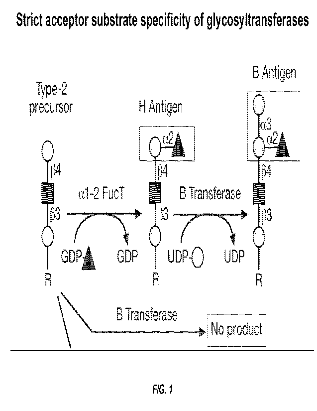
(NAcGal). The 0 gene product is inactive due to a frameshift mutation (FIG.
1).
[0017] The alpha-Gal, HBGA A and HBGA B epitopes generated by
these 3 active
enzymes are expressed widely in nature including bacteria that inhabit the
human gut (Springer
et al., "Blood Group Isoantibody Stimulation in Man by Feeding Blood Group-
Active Bacteria,"
.1 Clin. Invest. 48:1280-1291(1969), which is hereby incorporated by reference
in its entirety).
CA 03199581 2023- 5- 18
WO 2022/115775
PCT/US2021/061180
-6-
As a result, humans lacking the aGalT and the A and/or B alleles are being
continuously
immunized by these bacterially derived epitopes. This leads to very high
levels of natural
antibodies (Abs) to these non-self epitopes that constitute greater than 1% of
plasma
immunoglobulin (Ig) (Galili et al., "One Percent of Human Circulating B
Lymphocytes are
Capable of Producing the Natural Anti-Gal Antibody," Blood 82:2485-2493
(1993); Galili et al.,
"A Unique Natural Human IgG Antibody With Anti-Alpha-Galactosyl Specificity,"
.I. Exp. Med.
160:1519-1531 (1984), which are hereby incorporated by reference in their
entirety). Given the
diversity of the Ab repertoire estimated to be in the billions of different
specificities, this
represents an enormous proportion of endogenous Ig activity. These Abs are
composed of IgMs,
and IgGs that activate the complement cascade which, in turn, can initiate
vascular thrombosis
(Subramaniam et al., "Distinct Contributions of Complement Factors to Platelet
Activation and
Fibrin Formation in Venous Thrombus Development," Blood 129(16):2291-2302
(2017); Foley
et al., "Cross Talk Pathways Between Coagulation and Inflammation," Circ. Res.
118:1392-1408
(2016); and Conway EM, "Reincarnation of Ancient Links Between Coagulation and
Complement," I. Thromb. Haemost. 13(Suppl. 1):S121-532 (2015), which are
hereby
incorporated by reference in their entirety). Other immunoglobulin classes
such as IgA and IgE
can also be directed to these glycol-epitopes. In effect, evolutionary
mutations in these two
genes create an immunological state poised at a tipping point, primed and
ready to respond
rapidly, aggressively and destructively to the appearance of any of these non-
self epitopes. The
immunological effects of these mutations have precluded successful xeno-
transplants in humans
and explain why HBGA matching is the single most important match in solid
organ
transplantation since its critical importance was first recognized by Starzl,
Experience In Renal
Transplantation. (WB Saunders Company, Philadelphia, PA, chapter 6 (1964),
which is hereby
incorporated by reference in its entirety, in the early days of renal
allografts in the 1960's. Since
that time, the disastrous effects of a HBGA mismatch in solid organ
transplants is seen only in
those very rare instances when iatrogenic errors occur (Altman, Doctors
Discuss Transplant
Mistake. New York Times, Feb 22, 2003, which is hereby incorporated by
reference in its
entirety). This background context led to the goal to induce expression of one
of these non-self
epitopes by the host's cancer cells and/or the vascular endothelial cells that
supply the tumor.
BRIEF DESCRIPTION OF THE DRAWINGS
[0018] FIG. 1 shows the strict acceptor substrate specificity of
glycosyltransferases. The
B (or A)-transferase will only add its respective sugar to glycosylation sites
that express the H-
CA 03199581 2023- 5- 18
WO 2022/115775
PCT/US2021/061180
-7-
antigen. Fortuitously, absence of this requisite H-antigen in many normal
tissues prevents off-
target conversion to HBGA A or B Alternatively, in the event that one desires
to intentionally
target a normal or cancerous cell type that naturally lacks the H-antigen,
this can be
accomplished in a manner analogous to that described for adding A or B by also
targeting the
alphal-2 fucosyltransferase and providing GDP-fucose as the fucose donor.
Addition of the
fucose/H-antigen can be done simultaneously with the targeted A or B
transferase or the
additions can be done in a step-wise manner (e.g., first the fucose, then the
A or B addition).
[0019] FIGs. 2A-2B shows that chimeric Ab-GTB protein maintains
immunoreactivity
and enzymatic activity. FIG. 2A is a graph showing that the J591-GTB chimeric
protein
maintains comparable binding immunoreactivity to PSMA relative to the parental
J591 antibody
measured by ELISA. FIG. 2B is a bar graph showing that the chimeric protein
also retains
enzymatic activity demonstrated by its ability to catalyze the transfer of 'AC-
galactose from
upp_14--
galactose, the nucleotide donor, to 2'-fucosyl-lactose (2-FL). This
incorporation
occurs to a high level only when the J591-GTB fusion protein and its acceptor
substrate, 2-FL,
are present. Similar results were obtained with anti-4D5 (her2)-GTB.
[0020] FIG. 3 is a graph showing that GTB activity can be
modulated by C-terminal
extension. J591-GTB activity (% of control) is shown as a function of
increasing length of C-
terminal amino acid extension and measured by incorporation of 1-4C-gal from
UDP-"C-gal to
2'-fucosyl-lactose (2-FL).
[0021] FIG. 4 are images showing that J591-GTB specifically converts
antigen-positive
tumor cells. Tissue sections from a CWR22Rv1 xenograft (heterogeneously PSMA
/HBGA 0),
were incubated with J591-GTB + UDP-gal and immunohistochemically stained for
HBGA B
expression (left panel). Negative control sections including secondary anti-
murine Ig-peroxidase
but lacking mouse anti-HBGA B (middle panel) or.1591-GTB (right panel),
respectively, did not
stain.
[0022] FIG. 5 are images showing the effect of J591-GTB on LNCaP
and PC3 cells.
LNCaP cells (PSMA /HBGA 0; left panel) converted by J591-GTB to HBGA B; PC3
cells
(PSMA/HBGA 0; right panel) do not undergo conversion by J591-GTB.
[0023] FIGs. 6A-6D are images showing that PC3 cells transfected
with PSMA then
treated with J591-GTB. FIG. 6A shows phase contrast images. FIG. 6B shows
cells expressing
PSMA. FIG. 6C shows HBGA B antigen expression_ FIG. 6D is a merge of FIG 6B
and 6C
Only those cells expressing PSMA were converted to HBGA B expression. PSMA-neg
cells,
primarily at left center and top center, remain HBGA B-neg.
CA 03199581 2023- 5- 18
WO 2022/115775
PCT/US2021/061180
-8-
[0024] FIG. 7 are images showing LNCaP cells spiked into a
suspension of Type 0
RBCs and incubated with J591 (Top row); J591-GTB (middle row), or J591-GTB-
54aa
extension (bottom row). The left column shows phase contrast image. The middle
column
shows DAPI nuclear stain. The right column shows murine anti-HBGA B + goat
anti-mouse
IgM-a1exa488. While the PSMA-pos LNCaP cells are converted to HBGA B-pos by
J591-GTB,
with or without the C-terminal extension, bound to their plasma membrane, the
PSMA-neg
RBCs are not converted.
[0025] FIGs. 8A-8D are images showing complement-mediated lysis
in vitro. LNCaP
cells were incubated with either native mAb J591 or mAb J591-GTB fusion
protein. All wells
also got UDP-gal. Subsequently, serum from a type A patient was added as a
source of natural
anti-B Ab and complement. The combination of J591-GTB plus type A serum (FIG.
8A) led to
complete LNCaP lysis. The J591-GTB fusion protein did not induce lysis in the
absence of type
A serum (FIG. 8B). Without the fusion protein, no lysis was detected
regardless of the presence
(FIG. 8C) or absence of type A serum (FIG. 8D).
[0026] FIG. 9 are images showing complement-mediated cytotoxicity of
several cell
lines. Complement-mediated cytotoxicity of several cell lines as observed by
trypan blue
exclusion is shown. The upper panel was treated with J591-GTB + UDP-gal +
human type 0
serum. Cells in the lower panel were treated with the same 0 serum but without
J591-GTB +
UDP-gal. The proportion of dead cells is reported under each photograph as
determined by
FACS (FIG. 10). For the FACS, type 0 serum, without J591-GTB + UDP-gal, served
as a
negative control, whereas 0.1% triton exposure provided a complete lysis
control.
[0027] FIGs. 10A-10H are images showing the in vivo conversion
of prostate and breast
cancers to HBGA B. FIGs. 10A-10D show serial sections through LNCaP xenograft
in SCID
mouse 24 hours after administration of PBS+ UDP-gal (FIG. 10A), b) J591+ UDP-
gal (FIG.
10B), and J591-GTB + UDP-gal (FIG. 10C). FIGs. 10A-10C are
immunohistochemically
stained for HBGA B (all 10x). FIG. 10D shows a serial section from same
specimen stained for
PSMA. See also FIGs. 12A-12E for higher power and additional xenograft lines.
FIGs. 10E-
10H show IVID-1V1B361 breast cancer (HER2+) xenograft after treatment with PBS
+ UDP-gal
(FIG. 10E), 4D5 + UDP-gal (FIG. 10F), 4D5-GTB + UDP-gal (FIGs. 10G and 10H).
Sections
are immunohistochemically stained for TIBGA B expression. Discrete plasma
membrane
staining is apparent In FIG 10C and 10H, adjacent connective tissue does not
get converted,
demonstrating that the specificity of the immuno-phenotypic conversion is
restricted to targeted
tumor.
CA 03199581 2023- 5- 18
WO 2022/115775
PCT/US2021/061180
-9-
[0028] FIGs. 11A-11B are a bar graph (FIG. 11A) and histograms
(FIG. 11B) showing
lysis of B-converted cell lines by type 0 serum as determined by propidium
iodide uptake
measured by FACS and trypan blue exclusion (see FIG. 9). 0 serum in the
absence of B-
conversion does not cause lysis. After treating PSMA-pos cells with J591-GTB +
UDP-gal, the
type 0 serum completely lysed all of the PSMA-pos/B-converted cell lines; PC3,
which is
HBGA 0-pos/PSMA-neg, did not convert to HBGA B and was not lysed.
100291 FIGs. 12A-12E are images showing in vivo conversion of
LNCaP, C4-2 and
CWR22Rv1 xenografts by J591-GTB. FIGs. 12A-12B show LNCaP xenograft treated in
vivo
with: J591 [without GTB] (FIG. 12A) or J591-GTB (FIG. 12B), both with UDP-gal,
immunohistochemically stained with mouse anti-HBGA B; high power. FIG. 12C
shows C4-2
prostate cancer treated in vivo with J591-GTB plus UDP-gal,
immunohistochemically stained
with mouse anti-HBGA B; high power. FIGs. 12D-12E show CWR22Rv1 prostate
cancer,
heterogeneously and weakly PSMA-pos, treated in vivo with J591-GTB plus UDP-
gal. Adjacent
connective tissue is not converted to HBGA B.
100301 FIGs. 13A-13B are a graph (FIG. 13A) and in vivo images of mice
(FIG. 13B)
showing in vivo conversion of HBGA and treatment. Mice were implanted I.P.
with 10 x 106
C4-2-luc cells suspended in Matrigel. Several days later, bioluminescence was
measured and 10
mice with confirmed viable tumor were randomly assigned to one of 2 treatment
arms. All
tumor-bearing mice received a single dose of J591-GTB + UDP-gal + human type 0
serum; in
half of the mice, the serum was heat-inactivated prior to injection. In those
mice treated with
active type 0 serum, the mean photon flux decreased progressively over the
ensuing 13 days
whereas those with inactivated serum experienced mean tumor progression. At
the end of the
experiment on day 13, the difference in bioluminescence between groups was
significant
(p<0.0032). A duplicate experiment yielded consistent results.
[0031] FIGs. 14A-14B are in vivo images and a graph showing the results of
experiment
#2 in which C4-2-luc cells were implanted IP followed later by a single
treatment with J591-
GTB + UDP-gal + human type 0 serum (upper rows) (FIG. 14A). FIG. 14A shows
images of
mice receiving active type 0 serum or type 0 serum which had been previously
heat-inactivated.
Mice receiving heat-inactivated serum demonstrated tumor progression (see plot
of photon flux;
FIG 14B) whereas those getting active serum experienced tumor regression;
experiment 1
results are shown in FIGs. 13A-13B.
[0032] FIG. 15 is a FACS histogram showing CD19, CD20, and CD38
expression in
M1vI1-S cells. Flow cytometry analysis showed the MM1-S multiple myeloma cell
line is CD38
positive, CD19 positive, and CD20 negative.
CA 03199581 2023- 5- 18
WO 2022/115775
PCT/US2021/061180
-10-
[0033] FIGs. 16A-16B are FACS histograms showing ABO expression
of M1\41-S cells.
FIG 16A shows the MM1-S multiple myeloma cells line is A/B negative. FIG. 16B
shows
MM1-S multiple myeloma cells line is 0 positive.
[0034] FIG. 17 is a FACS histogram showing that CD19-V0 MM1-S
myeloma cells can
be converted to B' by GTB + UDP-gal. The GTB can be targeted to myeloma cells
using anti-
CD19, anti-CD38, or anti-BCMA.
[0035] FIG. 18 are images demonstrating that the use of ACUPA, a
small molecule
ligand that binds to PSMA, conjugated to GTB (ACUPA-GTB), to direct conversion
of LNCaP
from HBGA 0 to HBGA B. This demonstrates that, in addition to antibody (or
antibody
derivatives), a small molecule ligand or peptide that binds the target antigen
on the tumor cell or
neo-vascular endothelium can also be used for purposes of targeting the
enzyme. The left panel
shows ACUPA-PEG-1500-GTB treated cells. The right panel shows cells treated
with GTB
only.
[0036] FIG. 19 are images showing the specificity of the
conversion from HBGA 0 to
HBGA B. SK-BR5 breast cancer cells (PSMA-/O+) were co-cultured with LNCaP
prostate
cancer cells (PSMA/0). The two cell types can be distinguished by morphology:
SK-BR5 are
round whereas LNCaP cells are elliptical/spindle. In addition, the LNCaP cells
are marked with
green fluorescent protein (GFP). Incubation with J591-GTB and UDP-gal converts
only the
PSMA + LNCaP cells but not the neighboring cells that lack the PSMA target.
Panels show
DAPI (left panel), GFP (middle panel), and Anti-B (Cy5) (right panel) imaging.
[0037] FIG. 20 are images showing the specificity of the
conversion from HBGA 0 to
HBGA B. As shown, only PSMA cells are converted to B by J591-GTB/UDP-gal.
[0038] FIGs. 21A-21B are FACS histograms showing the specificity
of the conversion
from HBGA 0 to HBGA B. The specificity of conversion was quantified using FACS
by
comparing the concentration of J591 (anti-PSMA)-GTB required to convert LNCaP
(PSMA) to
HBGA B (FIG. 21A) relative to SK-BR5 (PSMA-neg) cells (FIG. 21B). Both cell
lines are 0 .
FACS histograms are shown. No B conversion of SK-BR5 occurs even at
concentrations of
J591-GTB up to 100 kig/mL. By comparison, concentrations as low as 0.012
lig/mL induce the
conversion of the PSMA-positive LNCaP cells.
[0039] FIG. 22 is a table and graph showing the specificity of the
conversion from
EIBGA 0 to HBGA B. A table (left panel) and histogram of MFI from FIGs. 21A-
21B is shown
(right panel). Specificity index exceeds 8,000:1.
CA 03199581 2023- 5- 18
WO 2022/115775
PCT/US2021/061180
-11-
[0040] FIG. 23 is a table and graph showing that both cell
surface and secreted
glycoproteins are glycosylated by the method of the present disclosure. A
graph of cell counts
(top panel) and table (bottom panel) are shown.
[0041] FIGs. 24A-24B are plots showing testing for anti-a1,3GalT
antibodies in serum
samples. FIG. 24B is an expanded view of FIG. 24A showing the lower optical
densities.
[0042] FIG. 25 is an SDS-PAGE gel showing expression and
purification of recombinant
proteins.
[0043] FIGs. 26A-26B are graphs showing binding of scfv-CD19-
aGal to CD19" MM1.S
cells (FIG. 26A) and CD19+ Raji cells) (FIG. 26B).
[0044] FIG. 27 is a graph showing a galactose transfer assay on a mixture
of CD19+ and
CD19" cells.
[0045] FIG. 28 are histograms showing a galactose transfer assay
on CD19+ cells.
[0046] FIG. 29 are scatter plots showing binding and aGal
transfer testing of scfv-aGT to
human B-cells.
[0047] FIG. 30 is a dot plot showing a serum mediated lysis assay on CD19+
cells.
[0048] FIGs. 31A-31B are graphs showing a lysis assay on aGal
transferred B-cells.
FIG. 31A is a graph showing % lysis. FIG. 31B is a graph showing IgG levels
(MEI) and IgM
levels.
[0049] FIGs. 32A-32C show an in vitro checkerboard assay of scfv-
CD19-aGT and
UDP-Gal. FIG. 32A measures binding, FIG. 32B measures alpha gal expression,
and FIG. 32C
measures lysis by human PBMCs.
[0050] FIG. 33 is a bar graph showing the % remaining B-cells at
baseline and at 1 hour,
4 hours, 1 day, 7 days, 14 days, 30 days, and 60 days following the
administration of anti-CD19
scFv-alpha Gal Transferase fusion protein and UDP-gal. B-cell counts were
determined by
examining CD20+/CD3" fluorescence. CD20 was used to avoid confounding the B-
cell count by
presence of anti-CD19 scFv.
DETAILED DESCRIPTION
[0051] The present disclosure teaches a bi-functional
therapeutic for treating cancer that
includes a targeting component which targets a tumor-associated antigen and an
enzyme which,
when delivered to a tumor by said targeting component, enzymatically converts
the tumor
phenotype to that of an incompatible allograft or xenograft. The enzyme is
coupled to the
targeting component.
CA 03199581 2023- 5- 18
WO 2022/115775
PCT/US2021/061180
-12-
[0052] The targeting component can be antibody derived (intact,
monovalent single
chain, Fab'2, Fab, scFv or other) or a peptide. The targeting and enzyme
moieties can be linked
via generation of a fusion gene/protein or via biochemical conjugation.
[0053] The present disclosure also pertains to a method of
treating cancer. The method
involves selecting a subject having cancer and providing a bi-functional
therapeutic according to
the present disclosure. The bi-functional therapeutic is administered, to the
selected subject,
under conditions effective to treat the cancer.
[0054] As used herein, the term "treat" refers to the
application or administration of the
bi-functional therapeutic of the present disclosure to a subject, e.g., a
patient. The treatment can
be to cure, heal, alleviate, relieve, alter, remedy, ameliorate, palliate,
improve or affect the
cancer, the symptoms of the cancer or the predisposition toward the cancer.
[0055] As used herein, the term "subject" is intended to include
human and non-human
animals. Non-human animals include all vertebrates, e.g., mammals and non-
mammals, such as
non-human primates, sheep, dog, cow, chickens, amphibians, reptiles, etc.
[0056] As used herein, the term "cancer" includes all types of cancerous
growths or
oncogenic processes, metastatic tissues or malignantly transformed cells,
tissues, or organs,
irrespective of histopathologic type or stage of invasiveness.
[0057] As used herein, an -incompatible allograff refers to a
tissue or tumor that induces
hyper-acute, acute and/or chronic immune rejection. Hyper-acute rejection
appears in minutes to
a few hours following organ transplantation, or, as described herein, after
conversion of a tumor
or tissue upon delivery of a bifunctional therapeutic. This rapid rejection is
characterized by
vessel thrombosis leading to graft/tumor necrosis. Hyperacute rejection is
caused by the
presence of anti-donor antibodies existing in the recipient before
transplantation/conversion.
100581 As used herein, the -targeting component" is a component
that is able to bind to
or otherwise associate with a tumor-associated antigen. Such tumor associated
antigens include,
but are not limited to the following as well as their peptide fragments:
FOLH1/PSMA, VEGFR,
CD19, CD20, CD25, CD30, CD33, CD38, CD52, B cell Maturation Antigen (BCMA),
CD79,
Somatostatin receptor, 5T4, gp100, Carcinoembryonic antigen (CEA), mammoglobin
A, melan
A/MART-1, MAGE, NY-ESO-1, PSA, tyrosinase, 1-1ER-2/neu, 1-IER-3, EGFR, hTERT,
mesothelin, Nectin-4, TROP-2, Tissue Factor, MUC-1, CA-125, and peptide
fragments thereof,
protein MZ2-E, polymorphic epithelial mucin, folate-binding protein, cancer
testis proteins
MAGE-1 or MAGE-3 or NY-ESO-1, Human chorionic gonadotropin (HCG), Alpha
fetoprotein
(AFP), Pancreatic oncofetal antigen, CA- 15-3,19-9, 549, 195, Squamous cell
carcinoma antigen
(SCCA), Ovarian cancer antigen (OCA), Pancreas cancer associated antigen
(PaA), mutant K-ras
CA 03199581 2023- 5- 18
WO 2022/115775
PCT/US2021/061180
-13-
proteins, mutant p53, nonmutant p53, truncated epidermal growth factor
receptor (EGFR),
chimeric protein p210BCR-ABL, telomerase, survivin, WT1 protein, LMP2 protein,
HPV E6 E7
protein, Idiotype protein, and PAP protein. The preceding list exemplifies
tumor-associated
antigens; additional tumor-associated antigens are known to those in the art.
[0059] The antigen may be an antigen or epitope present, for example, on a
tumor cell
located within the lungs, breast, esophagus, intestine, stomach, rectum, renal-
urinary system,
prostate, bladder, brain, thyroid, liver, pancreas, spleen, skin, connective
tissue, heart, blood
system, or vascular system. The target antigen may be an antigen or epitope
present on a cell
membrane, secreted protein, or on a non-membrane bound protein. Examples of
secreted
proteins include, but are not limited to hormones, enzymes, toxins and
antimicrobial peptides.
[0060] The targeting component may become localized or converge
at a particular
targeted site, for instance, a tumor, a disease site, a tissue, an organ, a
type of cell, an infectious
bacteria or virus, etc.
[0061] For example, contemplated targeting components include a
peptide, polypeptide,
protein, glycoprotein, aptamer, carbohydrate, or lipid. A targeting component
may be a naturally
occurring or synthetic ligand for a cell surface receptor, e.g., a growth
factor, hormone, LDL,
transferrin, etc. A targeting component can be an antibody, which term is
intended to include
antibody fragments and derivatives, characteristic portions of antibodies,
single chain targeting
moieties which can be identified, for example, using procedures such as phage
display.
Targeting components may also be a targeting peptide, targeting
peptidomimetic, or a small
molecule, whether naturally-occurring or artificially created (e.g., via
chemical synthesis).
[0062] In one embodiment, the targeting component is selected
from the group consisting
of an antibody or antigen-binding fragment thereof, a protein, a peptide, and
aptamer, and a
small molecule.
[0063] Antibodies against tumor-associated antigens are known. For example,
antibodies
and antibody fragments which specifically bind markers produced by or
associated with tumors
have been disclosed, inter alba, in U.S. Patent No. 3,927,193 to Hansen, and
U.S. Patent Nos.
4,331,647, 4,348,376, 4,361,544, 4,468,457, 4,444,744, 4,818,709 and 4,624,846
to Goldenberg,
which are hereby incorporated by reference in their entirety. In particular,
antibodies against a
tumor-associated antigen, e.g., a gastrointestinal, lung, breast, prostate,
ovarian, testicular, brain
or lymphatic or hematogenous tumor, a sarcoma or a melanoma, are
advantageously used
Antibodies to tumor-associated antigens are well known to those in the art.
[0064] The antibodies of the present disclosure may exist in a
variety of forms including,
for example, polyclonal antibodies, monoclonal antibodies, intracellular
antibodies
CA 03199581 2023- 5- 18
WO 2022/115775
PCT/US2021/061180
-14-
("intrabodies"), antibody fragments (e.g. Fv, Fab and F(ab)2), half-
antibodies, hybrid derivatives,
as well as single chain antibodies (scFv), chimeric antibodies and de-
immunized or humanized
antibodies (Ed Harlow and David Lane, USING ANTIBODIES: A LABORATORY MANUAL
(Cold
Spring Harbor Laboratory Press, 1999); Houston et al., "Protein Engineering of
Antibody
Binding Sites: Recovery of Specific Activity in an Anti-Digoxin Single-Chain
Fv Analogue
Produced in Escherichia coil," Proc. Natl. Acad. Sci. USA 85:5879-5883 (1988);
Bird et al,
-Single-Chain Antigen-Binding Proteins," Science 242:423-426 (1988), each of
which is hereby
incorporated by reference in its entirety).
[0065] Antibodies of the present disclosure may also be
generated using recombinant
DNA technology, such as, for example, an antibody or fragment thereof
expressed by a
bacteriophage. Alternatively, the synthetic antibody is generated by the
synthesis of a DNA
molecule encoding and expressing the antibody of the present disclosure or the
synthesis of an
amino acid sequence specifying the antibody, where the DNA or amino acid
sequence has been
obtained using synthetic DNA or amino acid sequence technology which is
available and well
known in the art.
[0066] Methods for monoclonal antibody production may be carried
out using the
techniques described herein or are well-known in the art (MONOCLONAL
ANTIBODIES ¨
PRODUCTION, ENGINEERING AND CLINICAL APPLICATIONS (Mary A. Ritter and Heather
M.
Ladyman eds., 1995), which is hereby incorporated by reference in its
entirety). Generally, the
process involves obtaining immune cells (lymphocytes) from the spleen of a
mammal which has
been previously immunized with the antigen of interest either in vivo or in
vitro.
[0067] Alternatively monoclonal antibodies can be made using
recombinant DNA
methods as described in U.S. Patent No. 4,816,567 to Cabilly et al, which is
hereby incorporated
by reference in its entirety. The polynucleotides encoding a monoclonal
antibody are isolated
from mature B-cells or hybridoma cells, for example, by RT-PCR using
oligonucleotide primers
that specifically amplify the genes encoding the heavy and light chains of the
antibody. The
isolated polynucleotides encoding the heavy and light chains are then cloned
into suitable
expression vectors, which when transfected into host cells such as E. coli
cells, simian COS
cells, Chinese hamster ovary (CHO) cells, or myeloma cells that do not
otherwise produce
immunoglobulin protein, monoclonal antibodies are generated by the host cells.
Also,
recombinant monoclonal antibodies or fragments thereof of the desired species
can be isolated
from phage display libraries (McCafferty et al., "Phage Antibodies:
Filamentous Phage
Displaying Antibody Variable Domains," Nature 348:552-554 (1990); Clackson et
al., "Making
Antibody Fragments using Phage Display Libraries," Nature 352:624-628 (1991);
and Marks et
CA 03199581 2023- 5- 18
WO 2022/115775
PCT/US2021/061180
-15-
al., "By-Passing Immunization. Human Antibodies from V-Gene Libraries
Displayed on Phage,"
I. !Viol. Biol. 222:581-597 (1991), which are hereby incorporated by reference
in their entirety).
[0068] The polynucleotide(s) encoding a monoclonal antibody can
further be modified
using recombinant DNA technology to generate alternative antibodies or
derivatives. For
example, the constant domains of the light and heavy chains of a mouse
monoclonal antibody
can be substituted by those regions derived from a human antibody to generate
a chimeric
antibody. Alternatively, the constant domains of the light and/or heavy chains
of a monoclonal
antibody can be substituted by a non-immunoglobulin polypepti de to generate a
fusion antibody.
In other embodiments, the constant regions are truncated or removed to
generate the desired
antibody fragment of a monoclonal antibody. Furthermore, site-directed or high-
density
mutagenesis of the variable region can be used to optimize specificity and
affinity of a
monoclonal antibody.
[0069] The monoclonal antibody of the present disclosure can be
a humanized antibody.
Humanized antibodies are antibodies that contain minimal sequences from non-
human (e.g.,
murine) antibodies within the variable regions. Such antibodies are used
therapeutically to
reduce antigenicity and human anti-mouse antibody responses when administered
to a human
subject. In practice, humanized antibodies are typically human antibodies with
minimal to no
non-human sequences. A human antibody is an antibody produced by a human or an
antibody
having an amino acid sequence corresponding to an antibody produced by a
human.
[0070] In addition to whole antibodies, the present disclosure encompasses
antigen
binding portions of such antibodies. Such binding portions include the
monovalent Fab
fragments, Fv fragments (e.g., single-chain antibody, scFv), and single
variable VH and VL
domains, and F(ab')2 fragments, Bis-scFv, diabodies, triabodies, minibodies,
etc. These
antibody fragments can be made by conventional procedures, such as proteolytic
fragmentation
procedures, as described in James Goding, MONOCLONAL ANTIBODIES:PRINCIPLES AND
PRACTICE 98-118 (Academic Press, 1983) and Ed Harlow and David Lane,
ANTIBODIES: A
LABORATORY MANUAL (Cold Spring Harbor Laboratory, 1988), which are hereby
incorporated
by reference in their entirety, or other methods known in the art.
[0071] It may further be desirable, especially in the case of
antibody fragments, to
modify the antibody in order to increase its serum half-life. This can be
achieved, for example,
by incorporation of a salvage receptor binding epitope into the antibody
fragment by mutation of
the appropriate region in the antibody fragment or by incorporating the
epitope into a peptide tag
that is then fused to the antibody fragment at either end or in the middle
(e.g., by DNA or peptide
synthesis).
CA 03199581 2023- 5- 18
WO 2022/115775
PCT/US2021/061180
-16-
[0072] Antibody mimics are also suitable for use in accordance
with the present
disclosure. A number of antibody mimics are known in the art including,
without limitation,
those known as monobodies, which are derived from the tenth human fibronectin
type III domain
r (Koide et al., "The Fibronectin Type III Domain as a Scaffold
for Novel Binding
Proteins," J. Mol. Biol. 284:1141-1151 ( l998); Koide et al., "Probing Protein
Conformational
Changes in Living Cells by Using Designer Binding Proteins: Application to the
Estrogen
Receptor," Proc. Natl. Acad. Sci. USA 99:1253-1258 (2002), each of which is
hereby
incorporated by reference in its entirety); and those known as affibodies,
which are derived from
the stable alpha-helical bacterial receptor domain Z of staphylococcal protein
A (Nord et al.,
"Binding Proteins Selected from Combinatorial Libraries of an alpha-helical
Bacterial Receptor
Domain," Nature BiotechnoL 15(8):772-777 (1997), which is hereby incorporated
by reference
in its entirety).
[0073] In certain embodiments, the targeting component targets
the prostate-specific
membrane antigen (PSMA) receptor.
[0074] As used herein, "PSMA" or "prostate-specific membrane antigen"
protein refers
to mammalian PSMA, preferably human PSMA protein. PSMA is sometimes referred
to as
folate hydrolase 1 (FOLH1) as PSMA is encoded by the FOLH1 gene. The long
transcript of
PSMA encodes a protein product of about 100-120 kDa molecular weight
characterized as a
type II transmembrane receptor having sequence homology with the transferrin
receptor and
having NAALADase activity (Carter et al,, "Prostate-Specific Membrane Antigen
is a Hydrolase
With Substrate and Pharmacologic Characteristics of a Neuropeptidase," Proc.
Natl. Acad. Sci.
USA 93:749-753 (1996); Israeli et al., "Molecular Cloning of a Complementary
DNA Encoding
a Prostate-Specific Membrane Antigen," Cancer Research 53:227-230 (1993),
which are hereby
incorporated by reference in their entirety).
[0075] Monoclonal anti-PSMA antibodies can be used as the targeting
component in the
bi-functional therapeutic of the present disclosure. Preferably, the
monoclonal antibodies bind to
the extracellular domain of PSMA (i.e., an epitope of PSMA located outside of
a cell such as at
about amino acids 44-750 of human PSMA, of which the amino acid residues
correspond to the
human PSMA sequence disclosed in U.S. Patent No. 5,538,866, which is hereby
incorporated by
reference in its entirety)). Examples of murine monoclonal antibodies to human
PSMA include,
but are not limited to, E99, J415, J533 and J591, which are produced by
hybridoma cell lines
having an ATCC Accession Number HB-12101, HB-12109, HB-12127, and HB-12126,
respectively, all of which are disclosed in U.S. Pat. No. 6,107,090 and U.S.
Pat. No. 6,136,311,
which are hereby incorporated by reference in their entirety. Most preferably,
the murine
CA 03199581 2023- 5- 18
WO 2022/115775
PCT/US2021/061180
-17-
monoclonal antibody is J591, produced by HB-12126, or de-immunized J591
antibody described
in U.S. Patent Nos. 7,045,605 and 7,514,078 to Bander et al., which are hereby
incorporated by
reference in their entirety.
[0076] In some embodiments the targeting component targets an
HER receptor family
member. An exemplary targeting component of an HER receptor family member is
monoclonal
antibody 4D5.
[0077] In certain embodiments, the targeting component is a
peptide that binds to the
tumor-associated antigen. Exemplary peptides include, without limitation,
glutamate-urea-lysine
derivatives such as 2-(3-99S)-5-amino-1-carboxypentyl)ureido) Pentanedioic
acid (ACUPA) that
binds FOLH1/PSMA, somatostatin derivatives that bind SSTR2, and Arg-Gly-Asp
(RGD)
peptide that binds alpha-v/beta-3 integrin.
[0078] The peptides used in conjunction with the present
disclosure can be obtained by
known isolation and purification protocols from natural sources, can be
synthesized by standard
solid or solution phase peptide synthesis methods according to the known
peptide sequence of
the peptide, or can be obtained from commercially available preparations or
peptide libraries.
Included herein are peptides that exhibit the biological binding properties of
the native peptide
and retain the specific binding characteristics of the native peptide.
Derivatives and analogs of
the peptide, as used herein, include modifications in the composition,
identity, and derivitization
of the individual amino acids of the peptide provided that the peptide retains
the specific binding
properties of the native peptide. Examples of such modifications would include
modification of
any of the amino acids to include the D-stereoisomer, substitution in the
aromatic side chain of
an aromatic amino acid, derivitization of the amino or carboxyl groups in the
side chains of an
amino acid containing such a group in a side chain, substitutions in the amino
or carboxy
terminus of the peptide, linkage of the peptide to a second peptide or
biologically active moiety,
and cyclization of the peptide (G. Van Binst and D. Tourwe, "Backbone
Modifications in
Somatostatin Analogues: Relation Between Conformation and Activity," Peptide
Research 5:8-
13 (1992), which is hereby incorporated by reference in its entirety).
[0079] As used herein, "small molecules" are typically organic,
peptide or non-peptide
molecules, having a molecular weight less than 10,000 Da, preferably less than
5,000 Da, more
preferably less than 1,000 Da, and most preferably less than 500 Da. This
class of modulators
includes chemically synthesized molecules, for instance, compounds from
combinatorial
chemical libraries.
[0080] In certain embodiments, the targeting component is an
aptamer. Aptamers are
small single-stranded DNA or RNA oligonucleotides that specifically bind to
their target
CA 03199581 2023- 5- 18
WO 2022/115775
PCT/US2021/061180
-18-
molecules (e.g., a tumor-associated antigen) with high affinity and
specificity. Aptamers are
created using an in vitro selection process termed systematic evolution of
ligands by exponential
enrichment (SELEX), which is described in Ellington et al., "In Vitro
Selection of RNA
Molecules That Bind Specific Ligands," Nature 346:818-822 (1990) and Jayasena,
"Aptamers:
An Emerging Class of Molecules That Rival Antibodies in Diagnostics," (11n.
Chem. 45:1628-
1650 (1999), which are hereby incorporated by reference in their entirety.
Several aptamers
capable of targeting tumor-associated antigens including, without limitation,
MUC1, HER2,
HER3, EpCAM, NF-kB, PSMA, CD44, PD-1, CD137, CD134, PDGF, VEGF, and NCL have
been developed (Jayasena, "Aptamers: An Emerging Class of Molecules That Rival
Antibodies
in Diagnostics," Clin. Chem. 45.1628-1650 (1999), which is hereby incorporated
by reference in
its entirety).
[0081] As used herein, the term "enzyme" encompasses any enzyme,
protein or peptide
which, when delivered to a tumor or tissue by a targeting component, catalyzes
the conversion of
the tumor or tissue to an incompatible allograft.
[0082] In one embodiment, the enzyme is an enzyme involved in post-
translational
modification and is selected from the group consisting of a transferase and a
glycosyltransferase.
[0083] A transferase is any one of a class of enzymes that enact
the transfer of
specific functional groups (e.g. a methyl or glycosyl group) from one molecule
(called the donor)
to another (called the acceptor).
[0084] An exemplary group of transferases includes, without limitation,
glycosyltransferases. Glycosyltransferases catalyze the addition of activated
sugars (donor NDP-
sugars), in a step-wise fashion, to a protein, glycoprotein, lipid or
glycolipid or to the non-
reducing end of a growing oligosaccharide (Lairson et al.,
"Glycosyltransferases: Structures,
Functions, and Mechanisms," Annu. Rev. Biochein. 77:521-55 (2008), which is
hereby
incorporated by reference in its entirety). Glycosyltransferases are well
known in the art.
[0085] Mammals utilize 9 sugar nucleotide donors for
glycosyltransferases: UDP-
glucose, UDP-galactose, UDP-G1cNAc, UDP-GalNAc, UDP-xylose, UDP-glucuronic
acid,
GDP-mannose, GDP-fucose, and CMP-sialic acid.
[0086] For enzymatic saccharide syntheses that involve
glycosyltransferase reactions,
glycosyltransferase can be cloned, or isolated from any source. Many cloned
glycosyltransferases are known, as are their polynucleotide sequences (see,
e.g., "The WWW
Guide To Cloned Glycosyltransferases," Taniguchi et al., 2002, Handbook of
Glycosyltransferases and Related Genes, Springer, Tokyo, which is hereby
incorporated by
reference in its entirety). Glycosyltransferase amino acid sequences and
nucleotide sequences
CA 03199581 2023- 5- 18
WO 2022/115775
PCT/US2021/061180
-19-
encoding glycosyltransferases from which the amino acid sequences can be
deduced are also
well known in the art.
[0087] Glycosyltransferases that can be employed in the methods
of the present
disclosure include, but are not limited to, galactosyltransferases,
fucosyltransferases,
glucosyltransferases, N-acetylgalactosaminyltransferases, N-
acetylglucosaminyltransferases,
glucuronyltransferases, sialyltransferases, mannosyltransferases, glucuronic
acid transferases,
galacturonic acid transferases, and oligoglycosyltransferases. Suitable
glycosyltransferases
include those obtained from eukaryotes, as well as from prokaryotes.
[0088] Glycosyltransferases are critical for the genesis of the
ABO blood group antigen
system. As described supra, the ABO blood system is the primary antigen system
important in
blood transfusion and solid organ transplantation. This histo-blood group
antigen (HBGA)
system is controlled by the activity of GTA and/or GTB glycosyltransferases
that attach sugar
residues (N- acetylgalactosamine or galactose) to a common substrate (the H
antigen). The
enzyme has several phenotypic variants which either alter the carbohydrate
attached (N-
acetylgalactosamine (A) vs galactose (B)) or cause loss of function of the
enzyme so the H
antigen is not modified (0). A variant of A, A2, has a reduced level of N-
acetylgalactosamine
activity and NAc-gal addition. These variants are discriminated currently by
serology and by
lectin binding (defining Al vs A2). Serology can either detect the
modification of the H antigen
or can detect the presence of naturally-occurring antibodies directed to A
and/or B (e.g., a person
with the B pattern of glycosylation will have antibodies directed to A).
[0089] In humans the glycosyltransferase locus, referred to
herein as the ABO locus or
the ABO glycosyltransferase locus, is located on chromosome 9 and contains
seven exons that
span more than 18 kb of genomic DNA. Exon 7 is the largest and contains most
of the coding
sequence. The ABO locus has three main allelic forms: A, B, and 0. The A
"allele" (also
referred to as Al or A2) encodes a glycosyltransferase that enzymatically adds
N-
acetylgalactosamine to the D-galactose end of the H antigen, producing the so-
called A antigen.
The B allele encodes a glycosyltransferase that enzymatically adds D-galactose
to the D-
galactose end of the H antigen, thus creating the so-called B antigen. The 0
allele encodes a
nonfunctional form of glycosyltransferase, resulting in an unmodified H
antigen, creating the so-
called 0 antigen phenotype.
100901 On the genomic level, the ABO glycosyltransferase gene
has many alleles (-300)
These naturally occurring allelic variants are described in Yip, "Sequence
Variation at the
Human ABO Locus," Ann. Hum. Genet. 66:1-27 (2002); Hakomori "Antigen Structure
and
Genetic Basis of Histo-Blood Group A, B, and 0: Their Changes Associated with
Human
CA 03199581 2023- 5- 18
WO 2022/115775
PCT/US2021/061180
-20-
Cancer," Biochimica et Biophysica Acta 1473:247-266 (1999); Seto et al.,
"Sequential
Interchange of Four Amino Acids from Blood Group B to Blood Group A
Glycosyltransferase
Boosts Catalytic Activity and Progressively Modifies Substrate Recognition in
Human
Recombinant Enzymes," J. Biol. Chem. 272:14133-14138 (1997), which are hereby
incorporated
by reference in their entirety, and their use in the bi-functional therapeutic
of the present
disclosure are contemplated. The sequence encoding the catalytic site of the
enzyme lies in exon
7 of the gene; key amino acid residues 176, 235, 266, and 268 control the
specificity of this
active site. Furthermore, a common nucleotide deletion in exon 6 creates a
stop codon that
abolishes synthesis of full-length glycosyltransferase, leading to the 0 or
null phenotype.
[0091] Thus, in some embodiments, the glycosyltransferase is selected from
the group
consisting of glycosyltransferase A (alpha 1-3-N-
acetylgalactosaminlytransferase),
glycosyltransferase B (alpha 1-3-galactosyltransferase), alpha-gal-
transferase, and
glycosyltransferase A (Gly268A1a). Allelic variants, as described supra, are
also contemplated.
[0092] In some embodiments, a glycosyltransferase used in the
method of the present
disclosure is a fucosyltransferase. Fucosyltransferases are known to those of
skill in the art.
Exemplary fucosyltransferases include enzymes which transfer L-fucose from GDP-
fucose to a
hydroxy position of an acceptor sugar. Fucosyltransferases that transfer non-
nucleotide sugars to
an acceptor are also of use in the present disclosure.
[0093] In some embodiments, the glycosyltransferase is a
humanized or de-immunized
glycosyltransferase. Methods of humanizing and/or de-immunizing proteins are
known in the
art.
[0094] Accordingly, one embodiment of the present disclosure
relates to the alteration of
the blood group antigen expression on a tumor and/or the blood supply of the
tumor by a tumor-
targeted glycosyltransferase. As described supra, this effectively converts
the tumor phenotype
to that of an incompatible allograft or xenograft thereby initiating hyper-
acute rejection.
[0095] The bi-functional therapeutic described herein may be
formed such that the
targeting component is a protein or peptide linked to the enzyme via a peptide
bond.
[0096] In certain embodiments, the protein or peptide targeting
component linked to the
enzyme via a peptide bond may be referred to as a chimeric or fusion protein.
As used herein,
the term "chimeric protein" or "fusion protein" encompasses a polypeptide
having a single
continuous polypeptide chain, i.e., a series of contiguous amino acids linked
by peptide bonds or
a series of polypeptide chains covalently or non-covalently linked to one
another (i.e., a
polypeptide complex) that includes at least a portion of a full-length
sequence of first
polypeptide sequence and at least a portion of a full-length sequence of a
second polypeptide
CA 03199581 2023- 5- 18
WO 2022/115775
PCT/US2021/061180
-21-
sequence, where the first and second polypeptides are different polypeptides.
A chimeric
polypeptide also encompasses polypeptides that include two or more non-
contiguous portions
derived from the same polypeptide. A chimeric polypeptide or protein also
encompasses
polypeptides having at least one substitution, wherein the chimeric
polypeptide includes a first
polypeptide sequence in which a portion of the first polypeptide sequence has
been substituted
by a portion of a second polypeptide sequence. The series of polypeptide
chains can be
covalently linked using a suitable biochemical linker or a disulfide bond.
[0097] Coupling of the targeting component and the enzyme can
also be prepared using
chemical linkage (Brennan et al., "Preparation of Bispecific Antibodies by
Chemical
Recombination of Monoclonal Immunoglobulin G1 Fragments," Science 229.81-3
(1985), which
is hereby incorporated by reference in its entirety) or chemical coupling
(Shalaby et al.,
"Development of Humanized Bispecific Antibodies Reactive With Cytotoxic
Lymphocytes and
Tumor Cells Overexpressing the HER2 Protooncogene," J. Exp. Med. 175:217-225
(1992),
which is hereby incorporated by reference in its entirety).
[0098] In other embodiments, the targeting component and the enzyme may be
linked via
non-covalent bonds including, without limitation, hydrogen bonds, ionic bonds,
Van der Waals
forces, and hydrophobic interactions.
100991 Thus, fusion or linkage between a targeting component
(e.g antibody) and an
enzyme may be achieved by conventional covalent or ionic bonds, protein
fusions via genetic
engineering, or heterobifunctional crosslinkers, e.g., carbodiimide,
glutaraldehyde, and the like.
Conventional inert linker sequences (e.g. peptide linkers) which simply
provide for a desired
amount of space between the targeting component and the enzyme may also be
used. The design
of such linkers is well known to those of skill in the art and is described
for example in U.S.
Patent Nos. 8,580,922; 5,525,491; and 6,165,476, which are hereby incorporated
by reference in
their entirety. A variety of coupling or cross-linking agents can be used for
covalent conjugation
of proteins. Examples of cross-linking agents include protein A, carbodiimide,
N-succinimidyl-
S-acetyl- thioacetate (SATA), 5,5'-dithiobis(2-nitrobenzoic acid) (DTNB), o-
phenylenedimaleimide (oPDM), N-succinimidy1-3-(2-pyridyldithio)propionate
(SPDP), and
sulfosuccinimidyl 4-(N-maleimidomethyl) cyclohaxane-1 -carboxylate (sulfo-
SMCC) (see e.g.,
Karpovsky et al., "Production of Target-Specific Effector Cells Using Hetero-
Cross-Linked
Aggregates Containing Anti-Target Cell and Anti-Fc Gamma Receptor Antibodies,"
J. Exp.
Med. 160(6):1686-701 (1984); Liu et al., "Heteroantibody Duplexes Target Cells
for Lysis by
Cytotoxic T Lymphocytes," Proc. Natl. Acad. Sc!. USA 82(24):8648-52 (1985),
which are
hereby incorporated by reference in their entirety). Other methods include
those described in
CA 03199581 2023- 5- 18
WO 2022/115775
PCT/US2021/061180
-22-
Paulus, Behring Ins Mitt No 78, 1 18-132 (1985); Brennan et al., "Preparation
of Bispecific
Antibodies by Chemical Recombination of Monoclonal Immunoglobulin GI
Fragments,"
Science 229:81-83 (1985); Glennie et al., "Preparation and Performance of
Bispecific F(ab'
gamma)2 Antibody Containing Thioether-Linked Fab' Gamma Fragments," J.
Immunol.
139:2367-2375 (1987), which are hereby incorporated by reference in their
entirety).
[00100] A number of other linkers can be used to couple the
targeting component to the
enzyme. For example, a disulfide linkage can be used, as described in Saito et
al., Adv. Drug
Delivery 1?eviews 55:199-215 (2003), which is hereby incorporated by reference
in its entirety.
Linkers that are sensitive to the lower pH found in endosomes or in the tumor
environment can
also be used, including hydrazones, ketals and/or aconitic acids. A hybrid
linker can also be
used, e.g., a linker with two or more potential cleavage sites, e.g., a
disulfide and a hydrazone.
Peptidase-sensitive linkers can also be used, e.g., tumor-specific peptidases,
for example, linkers
sensitive to cleavage by PSA. PEG linkers can also be used (Wiiest et al.,
Oncogene 21:4257-
4265 (2002), which is hereby incorporated by reference in its entirety).
Exemplary linkers
include hydrazone and disulfide hybrid linkers (see Hamann et al.,
Bioconjugate Chem. 13:47-58
(2002); Hamann et al., Bioconjug Chem. 13(1):40-6 (2002), which are hereby
incorporated by
reference in their entirety); SPP (Immunogen); and a variety of linkers
available from Pierce
Biotechnology, Inc. In some embodiments, the linker is SSP (a disulfide
linker, available from
Immunogen), and the ratio of linker to antibody can be varied from, e.g., 7.1
to 4:1. Various
spacer and linker sequences are known in the art and are described in Chen et
al., "Fusion
Protein Linkers: Property, Design and Functionality," Adv. Drug Deily. Rev.
65(10):1357-69
(2013), which is hereby incorporated by reference in its entirety.
[00101] The term 'peptide linker' or spacer refers to a short
peptide fragment that
connects or couples the targeting component and the enzyme moieties of the
polypeptide of the
bi-functional therapeutic. The linker is preferably made up of amino acids
linked together by
peptide bonds. For example, the peptide linker can comprise small amino acid
residues or
hydrophilic amino acid residues (e.g. glycine, serine, threonine, proline,
aspartic acid,
asparagine, etc). For example, the peptide linkers are peptides with an amino
acid sequence with
a length of at least 5 amino acids, or with a length of about 5 to about 100
amino acids, or with a
length of about 10 to 50 amino acids, or alength of about 10 to 15 amino
acids.
[00102] In one example, the linker is made up of a majority of
amino acids that are
sterically unhindered such as glycine and alanine. Thus in a further example,
the linkers are
polyglycines, polyalanines or polyserines.
CA 03199581 2023- 5- 18
WO 2022/115775
PCT/US2021/061180
-23-
[0100] One skilled in the art would appreciate that many
commonly used peptide linkers
may be used in embodiments of the present disclosure. In certain embodiments,
the short
peptide linkers may comprise repeat units to increase the linker length. For
example, a double,
triple or quadruple repeated linker. In one example, the linker comprises a
formula (Gly-Gly-
Gly-Gly-Ser)n (SEQ ID NO: 1) or comprising the formula (Ser-Gly-Gly-Gly-Gly)n
Ser (SEQ ID
NO:2) wherein n is a number from 3 to 6. In some embodiments, the linker is a
(G4S)31inker
(SEQ ID NO: 67).
101011 Non-peptide linkers or spacers are also possible. For
example, alkyl linkers such
as -NH-(CH2)s-C(0)-, wherein s = 2-20 could be used. These alkyl linkers may
be further
substituted by any non-sterically hindering group such as lower alkyl (e.g. C1-
C6), lower acyl,
halogen (e.g. Cl, Br), CN, NH2, phenyl. An exemplary non-peptide linker is a
PEG linker or
spacer having a molecular weight of 100 to 5000 kD, preferably 1000 to 2000
lcD, and more
preferably 15001(D.
[0102] A bifunctional therapeutic according to the present
disclosure may include an N-
terminus coupled to a C-terminus. N-terminus and C-terminus are used herein to
refer to the N-
terminal region or portion and the C-terminal region or portion, respectively,
of the bifunctional
therapeutic protein of the present disclosure. In some embodiments of the
present disclosure, the
C-terminal portion and the N-terminal portion of the bifunctional therapeutic
of the present
disclosure are contiguously joined. In alternative embodiments, the C-terminal
portion and the
N-terminal portion of the bifunctional therapeutic of the present disclosure
are coupled by an
intervening spacer. In one embodiment, the spacer may be a polypeptide
sequence of 1, 2, 3, 4,
5, 6, 7, 8, 9, 10, or more amino acid residues. In some embodiments, the C-
terminal portion
and/or the N-terminal portion of the bifunctional therapeutic of the present
disclosure may
include additional portion(s) coupled to the C-terminal residue and/or the N-
terminal residue of
the chimeric protein of the present disclosure, respectively. In some
embodiments, the additional
portion(s) may be a polypeptide sequence of 1, 2, 3, 4, 5, 6, 7, 8, 9, 10, or
more amino acid
residues. In some embodiments, the N-terminal portion and/or the C-terminal
portion having
such additional portion(s) will maintain the activity of the corresponding
naturally occurring N-
terminal portion of a targeting component and/or C-terminal portion of an
enzyme, respectively.
In some embodiments, the N-terminal portion and/or the C-terminal portion
having such
additional portion(s) will have enhanced and/or prolonged activity compared to
the
corresponding naturally occurring N-terminal portion of a targeting component
and/or C-
terminal portion of an enzyme, respectively. In other embodiments, the C-
terminal portion
and/or the N-terminal portion of the bifunctional therapeutic of the present
disclosure do not
CA 03199581 2023- 5- 18
WO 2022/115775
PCT/US2021/061180
-24-
include any additional portion(s) coupled to the C-terminal residue and/or the
N-terminal residue
of the chimeric protein of the present disclosure, respectively.
[0103] In one embodiment, the N-terminal region comprises the
targeting component. In
certain embodiments, the targeting component is an antibody or antigen-binding
portion thereof
including, without limitation, monomeric single chain antibodies, Fab
fragments, Fab'2, scFv,
and other antibody fragment derivatives such as minibodies, diabodies, and
triabodies. The
antibodies or antigen-binding fragments may maintain or delete the FcRn-
binding domain.
[0104] In one embodiment, the N-terminal region comprises human
J591 heavy chain
and has an amino acid sequence of SEQ ID NO:3 (GenBank Accession No.
CCA78124.1, which
is hereby incorporated by reference in its entirety), or a portion thereof, as
follows:
EVQLQQSGPE LVKPGTSVRI SCKTSGYTFT EYTIHWVKQS HGKSLEWIGN
INPNNGGTTY NQKFEDKATL TVDKSSSTAY MELRSLTSED SAVYYCAAGW
NFDYWGQGTT LTVSS
[0105] In another embodiment, the N-terminal region comprises
human J591 light chain
and has an amino acid sequence of SEQ ID NO:4 (GenBank Accession No.
CCA78125.1, which
is hereby incorporated by reference in its entirety), or a portion thereof, as
follows:
DIVMTQSHKF MSTSVGDRVS IICKASQDVG TAVDWYQQKP GQSPKLLIYW
ASTRHTGVPD RFTGSGSGTD FTLAITNVQS EDLADYFCQQ YNSYPLTFGA
GTKLEIKR
[0106] In another embodiment, the N-terminal region comprises human 4D5
heavy chain
and has an amino acid sequence of SEQ ID NO:5, or a portion thereof, as
follows:
EVQLVESGGG LVQPGGSLRL SCAASGFNIK DTYIHWVRQA PGKGLEWVAR
IYPTNGYTRY ADSVKGRFTI SADTSKNTAY LQMNSLRAED TAVYYCSRWG
GDGFYAMDYW GQGTLVTVSS ASTKGPSVFP LAPSSKSTSG GTAALGCLVK
DYFPEPVTVS WNSGALTSGV HTFPAVLQSS GLYSLSSVVT VPSSSLGTQT
YICNVNHKPS NTKVDKKVEP KSCDKTHTCP PCPAPELLGG PSVFLFPPKP
KDTLMISRTP EVTCVVVDVS HEDPEVKFNW YVDGVEVHNA KTKPREEQYN
STYRVVSVLT VLHQDWLNGK EYKCKVSNKA LPAPIEKTIS KAKGQPREPQ
VYTLPPSREE MTKNQVSLTC LVKGFYPSDI AVEWESNGQP ENNYKTTPPV
LDSDGSFFLY SKLTVDKSRW QQGNVFSCSV MHEALHNHYT QKSLSLSPGK
[0107] In another embodiment, the N-terminal region comprises
human 4D5 light chain
and has an amino acid sequence of SEQ ID NO:6, or a portion thereof, as
follows:
DIQMTQSPSS LSASVGDRVT ITCRASQDVN TAVAWYQQKP GKAPKLLIYS
ASFLYSGVPS RFSGSRSGTD FTLTISSLQP EDFATYYCQQ HYTTPPTFGQ
CA 03199581 2023- 5- 18
WO 2022/115775
PCT/US2021/061180
-25-
GTKVEIKRTV AAPSVFIFPP SDEQLKSGTA SVVCLLNNFY PREAKVQWKV
DNALQSGNSQ ESVTEQDSKD STYSLSSTLT LSKADYEKHK VYACEVTHQG
LSSPVTKSFN RGEC
[0108] In accordance with the above, in some embodiments the C-
terminal region
comprises the enzyme.
[0109] In one embodiment, the C-terminal region comprises the
catalytic domain of
glycosyltransferase B (GTB) and has an amino acid sequence of SEQ ID NO:7
(GenBank
Accession No. AM423112.1, which is hereby incorporated by reference in its
entirety), or a
portion thereof, as follows:
MAEVLRTLAG KPKCHALRPM ILFLIMLVLV LFGYGVLS PR SLMPGSLERG
FCMAVREPDH LQRVSLPRMV YPQPKVLTPC RKDVLVVTPW LAPIVWEGTF
NIDILNEQFR LQNTTIGLTV FAIKKYVAFL KLFLETAEKH FMVGHRVEYY
VFTDQPAAVP RVTLGTGRQL SVLEVGAYKR WQDVSMRRME MISDFCERRF
LSEVDYLVCV DVDMEFRDHV GVEILTPLFG TLEPSFYGSS REAFTYERRP
QSQAYIPKDE GDFYYMGAFF GGSVQEVQRL TRAGHQAMMV DQANGIEAVW
HDESHLNKYL LRHKPIKVLS PEYLWDQQLL GWPAVLRKLR FTAVPKNHQA
VRNP
[0110] In another embodiment, the C-terminal region comprises
the "cis A,B" sequence,
which generates a hybrid sequence of GTB and GTA and has an amino acid
sequence of SEQ ID
NO:8 (GenBank Accession No. ABL75287.1, which is hereby incorporated by
reference in its
entirety), or a portion thereof, as follows:
YVAFLKLFLE TAEKHFMVGH RVHYYVFTDQ PAAVPRVTLG TGRQLSVLEV
GAYKRWQDVS MRRMEMISDF CERRFLSEVD YLVCVDVDME FRDHVGVEIL
TPLFGTLHPS FYGSSREAFT YERRPQSQAY IPKDEGDFYY MGGFFGGSVQ
EVQRLTRACH QAMMVDQANG IEAVWHDESH LNKYLLRHKP TKVLSPEYLW
DQQLLGWPAV LRKLRFTAVP KNHQAVRNP
101111 In another embodiment, the C-terminal region comprises
the catalytic domain of
glycosyltransferase A (GTA) and has an amino acid sequence of SEQ ID NO:9
(GenBank
Accession No. AFB74122.1, which is hereby incorporated by reference in its
entirety), or a
portion thereof, as follows:
MAEVLRTLAG KPECHALRPM ILFLIMLVLV LFGYGVLSPR SLMPGSLERG
FCMAVREPDH LQRVSLPRMV YPQPKVLTPC RKDVLVVTPW LAPIVWEGTF
NIDILNEQFR LQNTTIGLTV FAIKKYVAFL KLFLETAEKH LMVGHRVHYY
VFTDQPAAVP RVTLGTGRQL SVLEVRAYKR WQDVSMRRME MISDFCERRF
CA 03199581 2023- 5- 18
WO 2022/115775
PCT/US2021/061180
-26-
LSEVDYLVCV DVDMEFRDHV GVEILTPLFG TLHPGFYGSS REAFTYERRP
QSQAYIPKDE GDFYYLGGFF GGSVQEVQRL TRACHQAMMV DQANGIEAVW
HDESHLNKYL LREKPIKVLS PEYLWDQQLL GWPAVLRKLR FTAVPKNHQA
VRNP
[0112] In certain embodiments, the tumor having the tumor-associated
antigen expresses
the H-antigen. As used herein, "the H-antigen" refers to an oligosaccharide
chain having a
terminal disaccharide fucose-galactose, where the fucose has an alpha-(1-2)-
linkage. The H-
antigen is produced by a fucosyltransferase and is the building block for the
production of the A
or B antigens within the ABO blood group system.
[0113] Accordingly, the present disclosure also pertains to a method of
treating cancer.
The method involves selecting a subject having cancer and providing a hi-
functional therapeutic
according to the present disclosure The hi-functional therapeutic is
administered to the selected
subject, under conditions effective to treat the cancer.
[0114] Virtually any tumor expressing an H-antigen can be
treated with the bifunctional
therapeutic described herein, including, but not limited to prostate tumors,
adrenocortical
carcinoma tumors, anal tumors, appendix tumors, astrocytoma (childhood
cerebellar or cerebral),
basal-cell carcinoma, bile duct tumors, bladder tumors, bone tumors,
osteosarcoma/malignant
fibrous hi stiocytomas, brain stem gliomas, ependymomas, medulloblastomas,
breast tumors,
bronchial adenomas/carcinoids, Burkitt's lymphomas, carcinoid tumors, cervical
tumors,
childhood tumors, chondrosarcomas, colon tumors, cutaneous T-cell lymphomas,
desmoplastic
small round cell tumors, endometrial tumors, esophageal tumors, Ewing's
sarcomas,
retinoblastomas, gallbladder tumors, gastric (stomach) tumors,
gastrointestinal stromal tumors,
germ cell tumors, gestational trophoblastic tumors, head and neck tumors,
heart tumors,
hepatocellular (liver) tumors, Hodgkin lymphomas, hypopharyngeal tumors, islet
cell
carcinomas (endocrine pancreas), Kaposi sarcomas, kidney tumors, laryngeal
tumors, lip and
oral cavity tumors, non-small cell lung tumors, small cell lung tumors,
lymphomas, melanomas,
Merkel cell tumors, mesotheliomas, multiple endocrine neoplasia, multiple
myelomas,
nasopharyngeal tumors, neuroblastomas, oligodendrogliomas, oral tumors,
oropharyngeal
tumors, ovarian tumors, pancreatic tumors, pleuropulmonary, primary central
nervous system
lymphomas, retinoblastomas, rhabdomyosarcomas, salivary gland tumors, soft
tissue sarcomas,
uterine sarcomas, skin tumors (non-melanoma), small intestine tumors, squamous
cell
carcinomas, stomach tumors, testicular tumors, throat tumors, thymoma and
thymic carcinomas,
thyroid tumors, trophoblastic tumors, and urethral tumors.
CA 03199581 2023- 5- 18
WO 2022/115775
PCT/US2021/061180
-27-
[0115] Some cancers including, but not limited to, hematopoietic
or lymphoid cancers,
mesodermally derived cancers, sarcomas, neuroectodermal cancers, etc may not
express the H
antigen. This can be easily determined by flow cytometry or
immunohistochemistry of a tumor
sample using Ulex lectin binding to reveal the presence or absence of H. When
H is absent,
treatment using the current application can be accomplished in two ways: one
may employ a
targeted fucosyltransferase in order to add the H antigen prior to or
simultaneous with a targeted
glycosyltransferase as previously described. Alternatively, one may target the
alpha gall
enzyme which can add a terminal galactose and does not require the presence of
the 1,2 fucose
(H antigen).
[0116] In one embodiment, the targeting component of the bi-functional
therapeutic targets
the PSMA receptor on tumor vascular endothelium. PSMA expression has been
reported in the
tumor neo-vasculature of a variety of tumors but is absent in normal tissue
vasculature.
Exemplary tissue types that have PSMA-positive vascular endothelium include,
without
limitation, renal, lung, colon, gastric, breast, brain, pancreatic, hepatic,
bladder, esophageal,
adrenal, head and neck, melanoma, and brain tumors. Other embodiments include
targeting
PSMA expressed on the surface of prostate cancer cells, targeting HER2 on
breast and other
HER2-positive cancers, targeting CD19 on B-cell lineage cancers, and targeting
CEA on
colorectal cancers. Other applicable targets are described supra.
[0117] Some aspects of the present disclosure relate to a bi-
functional therapeutic that
includes a targeting component comprising the amino acid sequence of one, two,
three, four,
five, or six CDRs as provided in Tables 1 and 2 herein. In some embodiments,
the targeting
component comprises a modified amino acid sequence, where the modified amino
acid
sequence has at least 80% sequence identity to any one, two, three, four,
five, or six of the CDR
sequences provided in Tables 1 and 2.
Table 1. Heavy Chain CDR Sequences of Suitable Targeting Component Antibodies
HCDR1 HCDR2 HCDR3
mAblFab
SEQ ID SEQ ID
SEQ ID
clone name Sequence Sequence Sequence
NO: NO:
NO:
J591* GYTFTEYTI H 10 NINPNNGGTTYNQKFED 13
GWNFDY 16
4 D 5** GFNIKDTYIH 11 RIYPTNGYTRYADSVKG 14 WGGDGFYAMDYW
17
obexelimab**" SYVMH 12 YINPYNDGTKYNEKFQG 15 GTYYYGTRVFDY
18
(XmAb5871)
CA 03199581 2023- 5- 18
WO 2022/115775
PCT/US2021/061180
-28-
*SeeU.S. Pat. App!. Pub!. No. 2006/0088539, FIGS. 2A-2B, which is hereby
incorporated by
reference in its entirety; **see U.S. Pat. No. 5,821,337, FIGS. 1A-1B, which
is hereby
incorporated by reference in its entirety; ***see EP2059536;
PCT/US2007/075932, which is
hereby incorporated by reference in its entirety.
Table 2. Light Chain CDR Sequences of Suitable Targeting Component Antibodies
LCDR1 LCDR2 LCDR3
mAb/Fab
SEQ ID SEQ ID
SEQ ID
clone name Sequence Sequence Sequence
NO: NO:
NO:
J591* KASQDVGTAVD
19 VVASTRHT 22 QQYNSYPLT 25
4D5- RASQDVNTAVAVV
20 SASFLYS 23 QQHYTTPP 26
Obexelimab*** RSSKSLQNVNGNTYLY 21 RMSNLNS 24 MQHLEYPIT 27
(XmAb5871)
*See U.S. Pat. App!. Pub!. No. 2006/0088539, FIGS. 2A-2B, which is hereby
incorporated by
reference in its entirety; **see U.S. Pat. No. 5,821,337, FIGS. 1A-1B, which
is hereby
incorporated by reference in its entirety; ***see EP2059536;
PCT/US2007/075932, which is
hereby incorporated by reference in its entirety.
[0118] In some embodiments, the heavy chain and/or the light chain variable
regions of the
antibody-based molecule described herein further comprises human or humanized
immunoglobulin heavy chain and/or light chain framework regions, respectively.
[0119] In some embodiments of the present disclosure, the
targeting component comprises
one or two of the sequences provided in Table 3 herein. In some embodiments,
the targeting
component comprises a modified amino acid sequence, where the modified amino
acid sequence
has at least 80% sequence identity to any one or two of the sequences provided
in Table 3.
Table 3. Antibody Variable Heavy (VII) and Variable Light (VI) Antibody
Sequences
mAblFab
SEQ ID
Region Sequence'
clone name
NO:
J591* VH EVQLVQSGPEVKKPGATVKISCKTSGYTFTEYTIHWVKQAPGKG 28
LEVVIGNINPNNGGITYNQKFEDKATLTVDKSTDTAYMELSSLRS
EDTAVYYCAAGWNFDYWGQGTLLTVSS
CA 03199581 2023- 5- 18
WO 2022/115775
PCT/US2021/061180
-29-
mAb/Fab
SEQ ID
Region Sequence/
clone name
NO:
J591
VL DIQMTQSPSSLSTSVGDRVTLTCKASQDVGTAVDVVYQQKPGP 29
SPKLLIYWASTRHTGIPSRFSGSGSGTDFTLTISSLQPEDFADYY
CQQYNSYPLTFGPGTKVDIK
Trastuzumab VH EVQLVESGGGLVQPGGSLRLSCAASGFNIKDTYIHVVVRQAPGK 30
(4D5)** GLEVVVARIYPTNGYTRYADSVKGRFTISADTSKNTAYLQMNSLR
AEDTAVYYCSRWGGDGFYAMDYWGQGTLVTVSS
Trastuzumab VL DIQMTQSPSSLSASVGDRVTITCRASQDVNTAVAVVYQQKPGKA 31
(4D5) PKLLIYSASFLYSGVPSRFSGSRSGTDFTLTISSLQPEDFATYYC
QQHYTTPPTFGQGTKVEIKRT
Obexelimab"*" VH EVQLVESGGGLVKPGGSLKLSCAASGYTFTSYVMHVVVRQAPG 32
(XmAb5871) KGLEWIGYINPYNDGTKYNEKFQGRVTISSDKSISTAYMELSSLR
SEDTAMYYCARGTYYYGTRVFDYWGQGTLVTVSS
Obexelimar* VL DIVMTQSPATLSLSPGERATLSCRSSKSLQNVNGNTYLYWFQQ 33
(XmAb5871) KPGQSPQLLIYRMSNLNSGVPDRFSGSGSGTEFTLTISSLEPED
FAVYYCMQHLEYPITFGAGTKLEIK
TComplementarity-determining regions are shown in bold typeface
See U.S. Pat. App!. Pub!. No. 2006/0088539, FIGS. 2A-2B, which is hereby
incorporated by
reference in its entirety; **see U.S. Pat. No. 5,821,337, FIGS. 1A-1B, which
is hereby
incorporated by reference in its entirety; """see EP2059536;
PCT/U52007/075932, which is
hereby incorporated by reference in its entirety.
101201 Suitable amino acid modifications to the heavy chain CDR
sequences and/or the
light chain CDR sequences of the targeting domain disclosed herein include,
for example,
conservative substitutions or functionally equivalent amino acid residue
substitutions that result
in variant CDR sequences having similar or enhanced binding characteristics to
those of the
CDR sequences disclosed herein as described above. Encompassed by the present
disclosure are
CDRs of Table 1 and 2 containing 1, 2, 3, 4, 5, or more amino acid
substitutions (depending on
the length of the CDR) that maintain or enhance binding of the antibody to its
target (e.g.,
PSMA, CD14, HER2) The resulting modified CDRs are at least 70%, at least 75%,
at least
80%, at least 85%, at least 90%, at least 95% similar in sequence to the CDRs
of Tables 1 and 2.
Suitable amino acid modifications to the heavy chain CDR sequences of Table 1
and/or the light
chain CDR sequences of Tables 1 and 2 include, for example, conservative
substitutions or
CA 03199581 2023- 5- 18
WO 2022/115775
PCT/US2021/061180
-30-
functionally equivalent amino acid residue substitutions that result in
variant CDR sequences
having similar or enhanced binding characteristics to those of the CDR
sequences of Table 1 and
Table 2. Conservative substitutions are those that take place within a family
of amino acids that
are related in their side chains. Genetically encoded amino acids can be
divided into four
families: (1) acidic (aspartate, glutamate); (2) basic (lysine, arginine,
histidine); (3) nonpolar
(alanine, valine, leucine, isoleucine, proline, phenylalanine, methionine,
tryptophan); and (4)
uncharged polar (glycine, asparagine, glutamine, cysteine, serine, threonine,
tyrosine).
Phenylalanine, tryptophan, and tyrosine are sometimes classified jointly as
aromatic amino acids.
Alternatively, the amino acid repertoire can be grouped as (1) acidic
(aspartate, glutamate); (2)
basic (lysine, arginine histidine), (3) aliphatic (glycine, alanine, valine,
leucine, isoleucine,
serine, threonine), with serine and threonine optionally grouped separately as
aliphatic-hydroxyl;
(4) aromatic (phenylalanine, tyrosine, tryptophan); (5) amide (asparagine,
glutamine); and (6)
sulfur-containing (cysteine and methionine) (Stryer (ed.), Biochemistry, 2nd
ed, WH Freeman
and Co., 1981, which is hereby incorporated by reference in its entirety). Non-
conservative
substitutions can also be made to the heavy chain CDR sequences of Table 1 and
the light chain
CDR sequences of Table 2. Non-conservative substitutions involve substituting
one or more
amino acid residues of the CDR with one or more amino acid residues from a
different class of
amino acids to improve or enhance the binding properties of CDR. The amino
acid sequences of
the heavy chain variable region CDRs of Table 1 and/or the light chain
variable region CDRs of
Table 2 may further comprise one or more internal neutral amino acid
insertions or deletions that
maintain or enhance target (e.g., PSMA, CD19, HER2) binding.
[0121] In some embodiments, the VH chain of the targeting domain
comprises any one of
the VH amino acid sequences provided in Table 3 above, or an amino acid
sequence that is at
least 60%, at least 65%, at least 70%, at least 75%, at least 80%, at least
85%, at least 90%, at
least 95% identical to any one of the VH amino acid sequences listed in Table
3. For example,
the targeting domain described herein may comprise: (i) a heavy chain variable
region
comprising an amino acid sequence that is at least 80% identical to SEQ ID NO:
30; (ii) a heavy
chain variable region comprising an amino acid sequence that is at least 80%
identical to SEQ ID
NO: 32; or (iii) a heavy chain variable region comprising an amino acid
sequence that is at least
80% identical to SEQ ID NO: 34.
[0122] In some embodiments, the VL chain of the targeting domain
comprises any one of
the VL amino acid sequences provided in Table 3 above, or an amino acid
sequence that is at
least 60%, at least 65%, at least 70%, at least 75%, at least 80%, at least
85%, at least 90%, at
least 95% identical to any one of the VL amino acid sequences listed in Table
3. For example,
CA 03199581 2023- 5- 18
WO 2022/115775
PCT/US2021/061180
-31 -
the targeting domain described herein may comprise: (i) a light chain variable
region comprising
an amino acid sequence that is at least 80% identical to SEQ ID NO: 29; (ii) a
light chain
variable region comprising an amino acid sequence that is at least 80%
identical to SEQ ID NO:
31; or (iii) a light chain variable region comprising an amino acid sequence
that is at least 80%
identical to SEQ ID NO: 33.
[0123] In some embodiments, the targeting domain disclosed herein
comprises: (i) a heavy
chain variable region comprising an amino acid sequence that is at least 80%
identical to SEQ ID
NO: 30 and a light chain variable region comprising an amino acid sequence
that is at least 80%
identical to SEQ ID NO: 29; (ii) a heavy chain variable region comprising an
amino acid
sequence that is at least 80% identical to SEQ ID NO: 32 and a light chain
variable region
comprising an amino acid sequence that is at least 80% identical to SEQ ID NO:
31; or (iii) a
heavy chain variable region comprising an amino acid sequence that is at least
80% identical to
SEQ ID NO: 33 and a light chain variable region comprising an amino acid
sequence that is at
least 80% identical to SEQ ID NO: 32.
[0124] The targeting domains of the present disclosure may be described or
specified in
terms of their binding affinities. Thus, in some embodiments, the targeting
domains of the
present disclosure include those with a dissociation constant or KD less than
11..IM, 500nM, 250
nM, 200 nM, 100 nM, 50 nM, 40 nM, 30 nM, 25 nM, 20 nM, 15 nM, 14 nM, 13 nM, 12
nM,
11M, 10 nM, 9 nM, 8 nM, 7 nM, 6 nM, 5 nM, 4 nM, 3 nM, 2 nM, or 1 nM.
[0125] Some aspects of the present disclosure relate to a bi-functional
therapeutic for
treating cancer that includes a targeting component which targets the prostate-
specific membrane
antigen (PSMA)/Folate hydrolase 1 (FOLH1) receptor and a glycosyltransferase
which, when
delivered to a tumor by said targeting component, enzymatically converts the
tumor phenotype
to that of an incompatible allograft or xenograft, said glycosyltransferase
being coupled to said
targeting component. This aspect of the present disclosure is useful in
treating a subject with
prostate cancer.
[0126] In some embodiments, the targeting component comprises a
heavy chain variable
region, where said heavy chain variable region includes: a complementarity-
determining region
1 (CDR-H1) comprising an amino acid sequence of SEQ ID NO: 10, or a modified
amino acid
sequence of SEQ ID NO: 10, said modified sequence having at least 80% sequence
identity to
SEQ ID NO: 10; a complementarity-determining region 2 (CDR-H2) comprising an
amino acid
sequence of SEQ ID NO: 13, or a modified amino acid sequence of SEQ ID NO: 13,
said
modified sequence having at least 80% sequence identity to SEQ ID NO: 13; and
a
complementarity-determining region 3 (CDR-H3) comprising an amino acid
sequence of SEQ
CA 03199581 2023- 5- 18
WO 2022/115775
PCT/US2021/061180
-32-
ID NO: 16, or a modified amino acid sequence of SEQ ID NO: 16, said modified
sequence
having at least 80% sequence identity to SEQ ID NO: 16. The sequences of the
heavy chain
CDR sequences are provided in Table 1 above.
[0127] In some embodiments, the targeting component comprises a
heavy chain variable
region including an amino acid sequence that is at least 80% identical to SEQ
ID NO: 28 (Table
3 above).
[0128] The targeting component may further comprise a light chain
variable region, where
said light chain variable region includes: a complementarity-determining
region 1 (CDR-L1)
having an amino acid sequence of SEQ ID NO: 19, or a modified amino acid
sequence of SEQ
ID NO: 19, said modified sequence having at least 80% sequence identity to SEQ
ID NO: 19; a
complementarity-determining region 2 (CDR-L2) having an amino acid sequence of
SEQ ID
NO: 22, or a modified amino acid sequence of SEQ ID NO: 22, said modified
sequence having
at least 80% sequence identity to SEQ ID NO: 22; and a complementarity-
determining region 3
(CDR-L3) having an amino acid sequence of SEQ ID NO: 25, or a modified amino
acid
sequence of SEQ ID NO: 25, said modified sequence having at least 80% sequence
identity to
SEQ ID NO: 25. The sequences of the light chain CDR sequences are provided in
Table 2
above.
[0129] In some embodiments, the light chain variable region
includes an amino acid
sequence that is at least 80% identical to SEQ ID NO: 29 (Table 3 above).
[0130] In some embodiments, the targeting component comprises a heavy chain
variable
region including the CDR-H1 of SEQ ID NO: 10, the CDR-H2 of SEQ ID NO: 13, and
the
CDR-H3 of SEQ ID NO: 16, and a light chain variable region including the CDR-
L1 of SEQ ID
NO: 19, the CDR-L2 of SEQ ID NO: 22, and the CDR-L3 of SEQ ID NO: 25.
101311 In some embodiments, the targeting component comprises a
heavy chain variable
region including an amino acid sequence that is at least 80% identical to SEQ
ID NO: 28 and a
light chain variable region including an amino acid sequence that is at least
80% identical to
SEQ ID NO: 29 (Table 3).
[0132] In some embodiments, the targeting component further
includes a signaling peptide,
optionally where the signaling peptide has the sequence of amino acids 1-19 of
SEQ ID NO: 34.
[0133] In some embodiments, the glycosyltransferase is selected from the
group consisting
of glycosyltransferase A (Alpha 1-3-N-Acetylgalactosaminyltransferase) and
glycosyltransferase
B (alpha 1-3-galactosyltransferase).
[0134] In some embodiments, the glycosyltransferase is
glycosyltransferase A ("GTA")
and has an amino acid sequence of SEQ ID NO: 64, or a portion thereof, as
follows:
CA 03199581 2023- 5- 18
WO 2022/115775
PCT/US2021/061180
-33 -
E PDHLQRVS L PRMVYPQ PKVL T PCRKDVLVVT PWLAP I VWEGT FNI D I LNEQ FRLQNT T I
GL TV-
FAI KKYVAFLKL FLE TAEKHFMVGHRVHYYVFT DQPAAVPRVTLGT GRQLSVLEVRAYKRWQMT
SMRRMEM I SDFCERRFL SEVDYLVCVDVDME FRDHVGVE I L T PL FG T LHPGFYGS SREAFTYER
RPQ SQAY I PKDEGDFYYLGGFFGGSVQEVQRL TRACHQAMMVDQANG I EAVWHDE SHLNKYLLR
HKP TKVLS PE YLWDQQL LGWPAVLRKLRFTAVPKNHQAVRNP
[0135] In some embodiments, the glycosyltransferase is
glycosyltransferase B ("GTB")
and has an amino acid sequence of SEQ ID NO: 65, or a portion thereof, as
follows:
EPDHLQRVSLPRMVYPQPKVLTPCRKDVLVVT PWLAP I VWEGT FNI D I LNEQ FRLQNT T I GL TV
FAI KKYVAFLKL FLE TAEKHFMVGHRVHYYVFT DQPAAVPRVTLGT GRQLSVLEVGAYKRWQDV
SMRRMEM I SDFCERRFL SEVDYLVCVDVDME FRDHVGVE I L T PL FG T LHPS FYGS SREAFTYER
RPQ SQAY I PKDEGDFYYMGAFFGGSVQEVQRL TRACHQAMMVDQANG I EAVWHDE S HLNKYLLR
HKP TKVLS PE YLWDQQL LGWPAVLRKLRFTAVPKNHQAVRN
[0136] Suitable additional glycosylases are described infra. In
some embodiments, the
glycosyltransferase is Marmoset a-1,3 galactosyltransferase (aa90-376) and has
an amino acid
sequence of SEQ ID NO: 66, or a portion thereof, as follows:
ELRLWDWFNPKKRPEVMTVTQWKAPVWEGTYNKAILENYYAKQKI TVGLIVFAI GRY E HYLE
E FVTSANRYFMVGHKVI FYVMVDDVS KAP F I E LGPLRS FKVFEVKPEKRWQD I SMMRMKT I GEH
I LAH I QHEVD FL FCMDVDQVFQDH EGVE T LGQ SVAQLQAWWYKADP DD FTYE RRKE SAAY I
PEG
QGDFYYHAAI FGGTP QVLNI TQEC FKGI LL DKKNDI EAEWHDE S HLNKY FLLNKP SK L S PEY
CWDYHI GLPS D IKTVKL SWQTKEYNLVRKNVGGGS
[0137]
In some embodiments, the bi-functional therapeutic includes: (i) a first
protein
comprising the amino acid sequence of SEQ ID NO: 34 or SEQ ID NO: 35 and a
second protein
comprising the amino acid sequence of SEQ ID NO: 36; (ii) a first protein
comprising the amino
acid sequence of SEQ ID NO: 37 or SEQ ID NO: 38 and a second protein
comprising the amino
acid sequence of SEQ ID NO: 39; (iii) a first protein comprising the amino
acid sequence of
SEQ ID NO: 40 or SEQ ID NO: 41 and a second protein comprising the amino acid
sequence of
SEQ ID NO: 42; (iv) a first protein comprising the amino acid sequence of SEQ
ID NO: 43 or
SEQ ID NO: 44 and a second protein comprising the amino acid sequence of SEQ
ID NO: 45;
(v) the amino acid sequence of SEQ ID NO: 46; (vi) the amino acid sequence of
SEQ ID NO:
47; (vii) the amino acid sequence of SEQ ID NO: 48; or (viii) the amino acid
sequence of SEQ
ID NO: 49 (Table 4).
CA 03199581 2023- 5- 18
WO 2022/115775
PCT/US2021/061180
-34-
Table 4. J591 Bi-Functional Therapeutic Protein Sequences
Protein
SEQ ID
Sequence
Sequence
NO:
huJ591-GTB and huJ591-GTA Protein Sequences
Signal MGWSCIILFLVATATGVHSEVQLVQSGPEVKKPGATVKISCKTSGYTFTEYTIH 34
peptide- WVKQAPGKGLEWIGNINPNNGGITYNQKFEDKATLTVDKSTDTAYMELSSLR
huJ591 H SEDTAVYYCAAGWNFDYVVGQGTLLTVSSASTKGPSVFPLAPSSKSTSGGTAA
chain-GTB LGCLVKDYFPEPVTVSWNSGALTSGVHTFPAVLQSSGLYSLSSVVTVPSSSLG
TQTYICNVNHKPSNTKVDKKVEPKSCDKTHTCPPCPAPELLGGPSVFLFPPKP
KDTLMISRTPEVTCVWDVSHEDPEVKFNVVYVDGVEVHNAKTKPREEQYNST
YRVVSVLTVLHQDWLNGKEYKCKVSNKALPAPIEKTISKAKGQPREPQVYTLP
PSRDELTKNQVSLTCLVKGFYPSDIAVEWESNGQPENNYKTTPPVLDSDGSF
FLYSKLTVDKSRWQQGNVFSCSVMHEALHNHYTQKSLSLSPGKggggsggggsg
gggsEPDHLQRVSLPRMVYPQPKVLTPCRKDVLVVTPWLAPIVWEGTFNIDIL
NEQFRLQNTTIGLTVFAIKKYVAFLKLFLETAEKHF MVGHRVHYYVFTDQPAA
VPRVTLGTGRQLSVLEVGAYKRWQDVSMRRMEMISDFCERRFLSEVDYLVC
VDVDMEFRDHVGVEILTPLFGTLHPSFYGSSREAFTYERRPQSQAYIPKDEG
DFYYMGAFFGGSVQEVQRLTRACHQAMMVDQANGIEAVWHDESHLNKYLL
RHKPTKVLSPEYLWDQQLLGWPAVLRKLRFTAVPKNHQAVRNP
(Signal peptide sequence shown in italic; huJ591 H chain sequence shown in
double underline; linker sequence shown in lowercase; GTB sequence shown in
bold.)
Signal MGWSCIILFLVATATGVHSEVQLVQSGPEVKKPGATVKISCKTSGYTFTEYTIH 35
peptide- WVKQAPGKGLEWIGNINPNNGGITYNQKFEDKATLTVDKSTDTAYMELSSLR
huJ591 H SEDTAVYYCAAGWNFDYWGQGTLLTVSSASTKGPSVFPLAPSSKSTSGGTAA
chain-GTA LGCLVKDYFPEPVTVSWNSGALTSGVHTFPAVLQSSGLYSLSSVVTVPSSSLG
TQTYICNVNHKPSNTKVDKKVEPKSCDKTHTCPPCPAPELLGGPSVFLFPPKP
KDTLMISRTPEVTCVVVDVSHEDPEVKFNWYVDGVEVHNAKTKPREEQYNST
YRVVSVLTVLHQDWLNGKEYKCKVSNKALPAPIEKTISKAKGQPREPQVYTLP
PSRDELTKNQVSLTCLVKGFYPSDIAVEWESNGQPENNYKTTPPVLDSDGSF
FLYSKLTVDKSRWQQGNVFSCSVMHEALHNHYTQKSLSLSPGKggggsggggsg
gggsEPDHLQRVSLPRMVYPQPKVLTPCRKDVLVVTPWLAPIVWEGTFNIDIL
NEQFRLQNTTIGLTVFAIKKYVAFLKLFLETAEKHF MVGHRVHYYVFTDQPAA
CA 03199581 2023- 5- 18
WO 2022/115775
PCT/US2021/061180
-35-
Protein
SEQ ID
Sequence
Sequence
NO:
VPRVTLGTGRQLSVLEVRAYKRWQDVSMRRMEMISDFCERRFLSEVDYLVC
VDVDME FRD HVGVEI LTPLFGTLH PG FYGSSREAFTYERRPQSQAYI PKDEG
DFYYLGGFFGGSVQEVQRLTRACHQAMMVDQANG lEAVWHDESH LNKYLL
RHKPTKVLSPEYLWDQQLLGWPAVLRKLRFTAVPKN HQAVRN P
(Signal peptide sequence shown in italic; huJ591 H chain sequence shown in
double underline; linker sequence shown in lowercase; GTA sequence shown in
bold.)
Signal MGWSCIILFLVATATGVHSDIQMTQSPSSLSTSVGDRVTLTCKASQDVGTAVD 36
peptide- WYQQKPGPSPKWYWASTRHTGIPSRFSGSGSGTDFTLTISSUPEDFADYY
huJ591-LC- CQQYNSYPLTFGPGTKVDI KRTVAAPSVFI FP PSDEQL KSGTASWCLLNN FYP
his tag REAKVQWKVDNALQSGNSQESVTEQDSKDSTYSLSSTLTLSKADYEKH KVYA
CEVTHQGLSSPVTKSFNRGECHHHHHH
(Signal peptide sequence shown in italic; huJ591 L chain sequence shown in
double underline; his tag sequence shown in bold italic.)
huJ591Fab-GTB and huJ591Fab-GTA Protein Sequences
Signal MGWSCIILFLVATA TGVHSEVQLVQSGPEVKKPGATVKISCKTSGYTFTEYTI H
37
peptide- VVVK QAPGKGL EWIGN I NP NNGGTTYNQK F EDKATLTVD
KSTDTAYMELSSLR
huJ591Fab SEDTAVYYCAAGWNFDYVVGQGTLLTVSSASTKGPSVFPLAPSS KSTSGGTAA
-GTB-Myc- LGCLVKDYFPEPVTVSWNSGALTSGVHTFPAVLQSSGLYSLSSVVTVPSSSLG
his tag TQTYICNVNHKPSNTKVDKKVEPKSCDKTHTEPDHLQRVSLPRMVYPQPKVL
TPCRKDVLVVTPWLAPIVWEGTFNI DI LNEQFRLQNTTIGLTVFAIKKYVAFLK
LFLETAEKHFMVG HRVHYYVFTDQPAAVPRVTLGTGRQLSVLEVGAYKRWQ
DVS M RRMEMISDFCE RRFLSEVDYLVCVDVDMEF RDHVGVEI LTPLFGTLHP
SFYGSSREAFTYERRPQSQAYIPKDEG DFYYMGAFFGGSVQEVQRLTRACH
QAMMVDQAN GI EAVWH D ESH LN KYLLRHKPTKVLSPEYLWDQQLLGWPAV
LRKLRFTAVPKNHQAVRNPAAAEQKLISEEDLNGAVEHHHHHH
(Signal peptide sequence shown in italic; huJ591 Fab sequence shown in
double underline; GTB sequence shown in bold; Myc sequence shown in italic
double underline; his tag sequence shown in bold italic.)
CA 03199581 2023- 5- 18
WO 2022/115775
PCT/US2021/061180
-36-
Protein
SEQ ID
Sequence
Sequence
NO:
Signal MGWSCIILFLVATA TGVHSEVQLVQSGPEVK KPGATVK I SCKTSGYTFTEYTI
H 38
peptide- WVKQAPGKGL EWIGN I NP NNGGTTYNQKF EDKATLTVD
KSTDTAYMELSSLR
huJ591Fab SEDTAVYYCAAGWNFDYVVGQGTLLTVSSASTKGPSVFPLAPSS KSTSGGTAA
-GTA-Myc- LGCLVKDYFPEPVTVSWNSGALTSGVHTFPAVLQSSGLYSLSSVVTVPSSSLG
his tag TQTYICNVNHKPSNTKVDKKVEPKSCDKTHTEPDHLQRVSLPRMVYPQPKVL
TPCRKDVLVVTPWLAPIVINEGTFNI DI LNEQFRLQNTTIG LTVFAIKKYVAFLK
LFLETAE KHFMVG HRVHYYVFTDQPAAVPRVTLGTGRQLSVLEVRAYKRWQ
DVSMRRMEMISDFCERRFLSEVDYLVCVDVDMEFRDHVGVEI LTPLFGTLHP
GFYGSSREAFTYERRPQSQAYI PKDEGDFYYLGGFFGGSVQEVQRLTRACH
QAMMVDQANGIEAVWHDESHLN KYLLRHKPTKVLSPEYLWDQQLLGWPAV
LRKLRFTAVPKNHQAVRNPAAAEQKLISEEDLNGAVEHHHHHH
(Signal peptide sequence shown in italic; huJ591 Fab sequence shown in
double underline; GTA sequence shown in bold; Myc sequence shown in italic
double underline; his tag sequence shown in bold italic.)
Signal MGWSCIILFLVATATGVHSDIQMTOSPSSLSTSVGDRVTLICKASQDVGTAVD 39
peptide- WYQQKPGPSPKLLIYWASTRHTGIPSRFSGSGSGTDFTLTISSLQPEDFADYY
huJ591-LC CQQYNSYPLTFGPGTKVDI KRTVAAPSVFI FP PSDEQL KSGTASWCLLNN FYP
REAKVQWKVD NALQSGNSQESVTEQDS KDSTYSLSSTLTLSKADYEK H KVYA
CEVTHQGLSSPVTKSFNRGEC
(Signal peptide sequence shown in italic; huJ591 LC sequence shown in double
underline.)
huJ591-HC67-GTB and huJ591-HC67-GTA Protein Sequences
huJ591- EVQLVQSGPEVKKPGATVKISCKTSGYTFTEYTIHVVVK QAPGKGLEWIGNINP
40
HC67-GTB NNGGTTYNQKFEDKATLTVDKSTDTAYMELSS LRSEDTAVYYCAAGVVNFDY
WGQGTLLTVSSASTKGPSVFPLAPSSKSTSGGTAALGCLVKDYFPEPVTVSW
NSGALTSGVHTFPAVLQSSGLYSLSSVVTVPSSSLGTQTYICNVNHKPSNTKV
DKKVEPKSCDKTHTVPPVPAPELLGGPSVFLFPP KPKDTLMISRTPEVTCVVV
DVS HEDPEVKFNWYVDGVEVHNAKTKPREEQYNSTYRVVSVLTVLHQDWLN
GKEYKCKVSNKALPAPIEKTISKAKGQPREPQVYTKPPSRDELTKNOVSLSCL
VKGFYPSD lAVEWESNGQPENNYKTIVPVLDS DGSFRLASYLTVDKSRVVQQ
GNVFSCSVMHEALHNHYTOKSLSLSPGKggggsggggsggggsEPDHLQRVSLPR
CA 03199581 2023- 5- 18
WO 2022/115775
PCT/US2021/061180
-37-
Protein
SEQ ID
Sequence
Sequence
NO:
MVYPQPKVLTPCRKDVLVVTPWLAPIVWEGTFNIDILNEQFRLQNTTIGLTVF
AIKKYVAFLKLFLETAEKHFMVGHRVHYYVFTDQPAAVPRVTLGTGRQLSVL
EVGAYKRWQDVSMRRMEMISDFCERRFLSEVDYLVCVDVDMEFRDHVGVEI
LTPLFGTLHPSFYGSSREAFTYERRPQSQAYIPKDEGDFYYMGAFFGGSVQE
VQRLTRACHQAMMVDQANGIEAVWHDESHLNKYLLRHKPTKVLSPEYLWD
QQLLGWPAVLRKLRFTAVPKNHQAVRNP
(huJ591-HC67 sequence shown in double underline; linker sequence shown in
lowercase; GTB sequence shown in bold.) HC67 variant amino acids (n=8)
shown in bold double underline.
huJ591- EVQLVQSGP EVK K PGATVKI SCKTSGYTFTEYTIHVVVK QAPGKGLEWI
GNI NP 41
HC67-GTA NNGGTTYNQKFEDKATLTVDKSTDTAYMELSSLRSEDTAVYYCAAGVVNFDY
WGQGTLLTVSSASTKGPSVFPLAPSSKSTSGGTAALGCLVKDYFPEPVTVSW
NSGALTSGVHTFPAVLQSSGLYSLSSVVTVPSSSLGTQTYICNVNHKPSNTKV
DK KVEP KS CD KTHTVPPVPAPE LLGGPSVFLFPP KP KDTLMI S RIP EVTCVVV
DVSHEDPEVKFNWYVDGVEVHNAKTKPREEQYNSTYRVVSVLTVLHQDWLN
GKEYKCKVSNKALPAPIEKTISKAKGQPREPQVYTKPPSRDELTKNOVSLSCL
VKGFYPSD lAVEWESNGQPENNYKTIVPVLDS DGSFRLASYLTVDKSRVVQQ
GNVFSCSVMHEALHNHYTOKSLSLSPGKggggsggggsggggsEPDHLQRVSLPR
MVYPQPKVLTPCRKDVLVVTPWLAPIVWEGTFNIDILNEQFRLQNTTIGLTVF
AIKKYVAFLKLFLETAEKHFMVGHRVHYYVFTDQPAAVPRVTLGTGRQLSVL
EVRAYKRWQDVSMRRMEMISDFCERRFLSEVDYLVCVDVDMEFRDHVGVEI
LTPLFGTLHPGFYGSSREAFTYERRPQSQAYIPKDEGDFYYLGGFFGGSVQE
VQRLTFtACHQAMMVDQANGIEAVWHDESHLNKYLLRHKPTKVLSPEYLWD
QQLLGWPAVLRKLRFTAVPKNHQAVRNP
(huJ591-HC67 sequence shown in double underline; linker sequence shown in
lowercase; GTA sequence shown in bold.) HC67 variant amino acids (n=8)
shown in bold double underline.
huJ591-LC- DIQMTQSPSSLSTSVGDRVTLTCKASQDVGTAVDVVYQQKPGPSPKWYWAS 42
his tag TRHTGI PSRFSGSGSGTDFTLTISSLQPEDFADYYCQQYNSYPLTFGPGTKVDI
KRTVAAPSVFIFPPSDEQL KSGTAS \NCLLNNFYPREAKVQVVKVDNALQSGN
CA 03199581 2023- 5- 18
WO 2022/115775
PCT/US2021/061180
-38-
Protein
SEQ ID
Sequence
Sequence
NO:
SQESVTEQDSKDSTYSLSSTLTLSKADYEKHKVYACEVTHQGLSSPVIKSFNR
GECHHHHHH
(huJ591 LC sequence shown in double underline; his tag sequence shown in
bold italic.)
huJ591-HC67-GTB-54aa and huJ591-HC67-GTA-54aa Protein Sequences
huJ591 MNFGLRLIFLVLTLKGVQCEVQLVQSGPEVKKPGATVKISCKTSGYTFTEYTIH 43
HC67 - VVVKQAPGKGLEWIGNINPNNGGTTYNQKFEDKATLTVDKSTDTAYMELSSLR
GTB-54aa SEDTAVYYCAAGWNFDYVVGQGTLLTVSSASTKGPSVFPLAPSSKSTSGGTAA
LGCLVKDYFPEPVTVSWNSGALTSGVHTFPAVLQSSGLYSLSSVVTVPSSSLG
TQTYICNVNHKPSNTKVDKKVEPKSCDKTHTVPPVPAPELLGGPSVFLFPPKP
KDTLMISRTPEVTCVWDVSHEDPEVKFNVVYVDGVEVHNAKTKPREEQYNST
YRVVSVLTVLHQDWLNGKEYKCKVSNKALPAPIEKTISKAKGQPREPQVYTKP
PSRDELTKNQVSLSCLVKGFYPSDIAVEWESNGQPENNYKTTVPVLDSDGSF
RLASYLTVDKSRWQQGNVFSCSVMHEALHNHYTQKSLSLSPGKggggsggggs
ggggsEPDHLQRVSLPRMVYPQPKVLTPCRKDVLVVTPWLAPIVWEGTFNIDIL
NEQFRLQNTTIGLTVFAIKKYVAFLKLFLETAEKHF MVGHRVHYYVFTDQPAA
VPRVTLGTGRQLSVLEVGAYKRWQDVSMRRMEMISDFCERRFLSEVDYLVC
VDVDMEFRDHVGVEILTPLFGTLHPSFYGSSREAFTYERRPQSQAYIPKDEG
DFYYMGAFFGGSVQEVQRLTRACHQAMMVDQANGIEAVWHDESHLNKYLL
RHKPTKVLSPEYLWDQQLLGWPAVLRKLRFTAVPKN HQAVRN PEFEQKLIS
EEDLNSADIHHTGARSSAHLELTADYKDHDGDYKDHDIDYKDDDDK
(Signal peptide sequence shown in italic; huJ591 H chain sequence shown in
double underline; linker sequence shown in lowercase; GTB sequence shown in
bold; 54aa sequence shown in italic double underline.) HC67 variant amino
acids (n=8) shown in bold double underline.
huJ591- MNFGLRLIFLVLTLKGVQCEVQLVQSGPEVKKPGATVKISCKTSGYTFTEYTIH 44
HC67-GTA- WVKQAPGKGLEWIGNINPNNGGTTYNQKFEDKATLTVDKSTDTAYMELSSLR
54aa SEDTAVYYCAAGWNFDYVVGQGTLLTVSSASTKGPSVFPLAPSSKSTSGGTAA
LGCLVKDYFPEPVTVSWNSGALTSGVHTFPAVLQSSGLYSLSSVVTVPSSSLG
TQTYICNVNHKPSNTKVDKKVEPKSCDKTHTVPPVPAPELLGGPSVFLFPPKP
KDTLMISRTPEVTCVWDVSHEDPEVKFNVVYVDGVEVHNAKTKPREEQYNST
CA 03199581 2023- 5- 18
WO 2022/115775
PCT/US2021/061180
-39-
Protein
SEQ ID
Sequence
Sequence
NO:
YRVVSVLTVLHQDWLNGKEYKCKVSNKALPAPIEKTISKAKGQPREPQVYTKP
PSRDELTKNQVSLSCLVKGFYPSDIAVEVVESNGQPENNYKTIVPVLDSDGSF
RLASYLTVDKSRWQQGNVFSCSVMHEALHNHYTOKSLSLSPGKggggsggggs
ggggsEPDHLQRVSLPRMVYPQPKVLTPCRKDVLVVTPWLAPIVWEGTFNIDIL
NEQFRLQNTTIGLTVFAIKKYVAFLKLFLETAEKHF MVGHRVHYYVFTDQPAA
VPRVTLGTGRQLSVLEVRAYKRWQDVSMRRMEMISDFCERRFLSEVDYLVC
VDVDMEFRDHVGVEILTPLFGTLHPGFYGSSREAFTYERRPQSQAYIPKDEG
DFYYLGGFFGGSVQEVQRLTRACHQAMMVDQANGIEAVWHDESHLNKYLL
RHKPTKVLSPEYLWDQQLLGWPAVLRKLRFTAVPKNHQAVRNPEFEOKLIS
EEDLNSADIHHTGARSSAHLELTADYKDHDGDYKDHDIDYKDDDDK
(Signal peptide sequence shown in italic; huJ591 H chain sequence shown in
double underline; linker sequence shown in lowercase; GTA sequence shown in
bold; 54aa sequence shown in italic double underline.) H067 variant amino
acids (n=8) shown in bold double underline.
huJ591-LC- DIQMTQSPSSLSTSVGDRVTLTCKASQDVGTAVDVVYQQKPGPSP KLLIY WAS
45
his tag TRHTGIPSRFSGSGSGTDFTLTISSLQPEDFADYYCQQYNSYPLTFGPGTKVDI
KRTVAAPSVFIFPPSDEQLKSGTAS\NCLLNNFYPREAKVQWKVDNALQSGN
SQESVTEQDSKDSTYSLSSTLTLSKADYEKHKVYACEVTHQGLSSPVTKSFNR
GECHHHHHH
(huJ591 LC sequence shown in double underline; his tag sequence shown in
bold italic.)
huJ591scFv-Fc67-GTB and huJ591scFv-Fc67-GTA Protein Sequences
huJ591scF DIQMTQSPSSLSTSVGDRVTLTCKASQDVGTAVDVVYQQKPGPSP KLLIYVVAS
46
v-Fc67- TRHTGIPSRFSGSGSGTDFTLTISSLOPEDFADYYCQQYNSYPLTFGPGTKVDI
GTB-his KEVQLVQSGPEVKKPGATVKISCKTSGYTFTEYTIHVVVKQAPGKGLEWIGNIN
tag PNNGGTTYNQKFEDKATLTVDKSTDTAYMELSSLRSEDTAVYYCAAGWNFDY
WGQGTLLTVSSEPKSCDKTHTVPPVPAPELLGGPSVFLFPPKPKDTLMISRTP
EVICVVVDVSHEDPEVKFNVVYVDGVEVHNAKTKPREEQYNSTYRVVSVLTVL
HQDWLNGKEYKCKVSNKALPAPIEKTISKAKGQPREPQVYTKPPSRDELTKN
QVSLSCLVKGFYPSDIAVEWESNGQPENNYKTTVPVLDSDGSFRLASYLTVD
KSRWQQGNVFSCSVMHEALHNHYTQKSLSLSPGKggggsggggsggggsEPDHL
CA 03199581 2023- 5- 18
WO 2022/115775
PCT/US2021/061180
-40-
Protein
SEQ ID
Sequence
Sequence
NO:
QRVSLPRMVYPQPKVLTPCRKDVLVVTPWLAPIVWEGTFNI DILNEQFRLQN
ITIGLTVFAIKKYVAFLKLFLETAEKHFMVGHRVHYYVFTDQPAAVPRVTLGT
GRQLSVLEVGAYKRWQDVSMRRMEMISDFCERRFLSEVDYLVCVDVDMEF
RDHVGVEILTPLFGTLHPSFYGSSREAFTYERRPQSQAYIPKDEGDFYYMGA
FFGGSVQEVQRLTRACHQAMMVDQANGIEAVWHDESHLNKYLLRHKPTKV
LSPEYLWDQQLLGWPAVLRKLRFTAVPKNHQAVRNPHHHHHH
(huJ591scFv-Fc67 sequence shown in double underline; linker sequence
shown in lowercase; GTB sequence shown in bold; his tag sequence shown in
bold italic.) HC67 variant amino acids (n=8) shown in bold double underline.
h591scFv- DIQMTQSPSSLSTSVGDRVTLTCKASQDVGTAVDVVYQQKPGPSP KLLIYVVAS
47
Fc67-GTA- TRHTGIPSRFSGSGSGTDFTLTISSLQPEDFADYYCQQYNSYPLTFGPGTKVD1
his tag KEVQLVQSGPEVKKPGATVKISCKTSGYTFTEYTIHVVVKQAPGKGLEWIGNIN
PNNGGTTYNQKFEDKATLTVDKSTDTAYMELSSLRSEDTAVYYCAAGWNFDY
WGQGTLLTVSSEPKSCDKTHTVPPVPAPELLGGPSVFLFPPKPKDTLMISRTP
EVICVVVDVSHEDPEVKFNWYVDGVEVHNAKTKPREEQYNSTYRVVSVLTVL
HQDWLNGKEYKCKVSNKALPAPIEKTISKAKGQPREPQVYTKPPSRDELTKN
QVSLSCLVKGFYPSDIAVEWESNGQPENNYKTTVPVLDSDGSFRLASYLTVD
KSRWQQGNVFSCSVMHEALHNHYTQKSLSLSPGKggggsggggsggggsEPDHL
QRVSLPRMVYPQPKVLTPCRKDVLVVTPWLAPIVWEGTFNI DILNEQFRLQN
TTIGLIVFAIKKYVAFLKLFLETAEKHFMVGHRVHYYVFTDQPAAVPRVTLGT
GRQLSVLEVRAYKRWQDVSMRRMEMISDFCERRFLSEVDYLVCVDVDMEFR
DHVGVEILTPLFGTLHPGFYGSSREAFTYERRPQSQAYI PKDEGDFYYLGGFF
GGSVQEVQRLTRACHQAMMVDQANGIEAVWHDESHLNKYLLRHKPTKVLS
PEYLWDQQLLGWPAVLRKLRFTAVPKNHQAVRNPHHHHHH
(huJ591scFv-Fc67 sequence shown in double underline; linker sequence
shown in lowercase; GTA sequence shown in bold; his tag sequence shown in
bold italic.) HC67 variant amino acids (n=8) shown in bold double underline.
huJ591scFv-GTB and huJ591scFv-GTA Protein Sequences
huJ591scF METDTLLLWVLLLWVPGSTGEVQLVQSGAEVKKPGASVKI SCKTSGYTFTEYT 48
v-GTB- IHWVKQASGKGLEWIGNINPNNGGTTYNQKFEDRATLTVDKSTSTAYMELSSL
Myc-His tag RSEDTAVYYCAAGWNFDYWGQGTTVTVSSGSTSGGGSGGGSGGGGSSDIV
CA 03199581 2023- 5- 18
WO 2022/115775
PCT/US2021/061180
-41 -
Protein
SEQ ID
Sequence
Sequence
NO:
MTQSPSSLSASVGDRVTITCKASQDVGTAVDWYQQKPGKAPKLLIYWASTRH
TGVPDRFTGSGSGTDFTLTI SSLQP EDFADYFCQQYNSYP LTFGGGTKL El Kgg
ggsggggsggggsEPDHLQRVSLPRMVYPQPKVLTPCRKDVLVVTPWLAPIVWE
GTFNIDILNEQFRLQNTTIGLTVFAIKKYVAFLKLFLETAEKHFMVGHRVHYYV
FTDQPAAVPRVTLGTGRQLSVLEVGAYKRWQDVSMRRMEMISDFCERRFLS
EVDYLVCVDVDMEFRDHVGVEILTPLFGTLHPSFYGSSREAFTYERRPQSQA
YIPKDEGDFYYMGAFFGGSVQEVQRLTRACHQAMMVDQANGIEAVWHDES
HLNKYLLRHKPTKVLSPEYLWDQQLLGWPAVLRKLRFTAVPKNHQAVRNPA
AAEQKLISEEDLNGAVEHHHHHH
(Signal peptide sequence shown in italic; huJ591scFv sequence shown in
double underline; linker sequence shown in lowercase; GTB sequence shown in
bold; Myc sequence shown in italic double underline; his tag sequence shown in
bold italic.)
huJ591scF METDTLLLVVVLLLVVVPGSTGEVQLVQSGAEVKKPGASVKI SCKTSGYTFTEYT 49
v-GTA- I HWVKQASGKGLEWIGNINPNNGGITYNQKFEDRATLTVDKSTSTAYMELSSL
Myc-His tag RSE DTAVYYCAAGWNFDYWGQGTTVTVSSGSTSGGGSGGGSGGGGSSDIV
MTQSPSSLSASVGDRVTITCKASQDVGTAVDWYQQKPGKAPKLLIYWASTRH
TGVPDRFTGSGSGTDFTLTI SSLQP EDFADYFCQQYNSYP LTFGGGTKL El Kgg
ggsggggsgg ggs EPDHLQRVSLPRMVYPQPKVLTPCRKDVLVVTPWLAPIVWE
GTFNIDILNEQFRUATTIGLTVFAIKKYVAFLKLFLETAEKHFMVGHRVHYYV
FTDQPAAVPRVTLGTGRQLSVLEVRAYKRWQDVSMRRMEMISDFCERRFLS
EVDYLVCVDVDMEFRDHVGVEILTPLFGTLHPGFYGSSREAFTYERRPQSQA
YIPKDEGDFYYLGGFFGGSVQEVQRLTRACHQAMMVDQANGIEAVWH DES
HLNKYLLRHKPTKVLSPEYLWDQQLLGWPAVLRKLRFTAVPKNHQAVRNPA
AAEQKLISEEDLNGAVEHHHHHH
(Signal peptide sequence shown in italic; huJ591scFv sequence shown in
double underline; linker sequence shown in lowercase; GTA sequence shown in
bold; Myc sequence shown in italic double underline; his tag sequence shown in
bold italic.)
CA 03199581 2023- 5- 18
WO 2022/115775
PCT/US2021/061180
-42-
101381 In some embodiments, the bi-functional therapeutic includes
a first protein
comprising the amino acid sequence of positions 20-770 of SEQ ID NO: 34 or SEQ
ID NO: 35
and a second protein comprising the amino acid sequence of positions 20-233 of
SEQ ID NO:
36.
[0139] In some embodiments, the bi-functional therapeutic includes a first
protein
comprising the amino acid sequence of positions 20-540 of SEQ ID NO: 37 or SEQ
ID NO: 38
and a second protein comprising the amino acid sequence of positions 20-233 of
SEQ ID NO:
39, optionally where the bi-functional therapeutic comprises a first portion
comprising the amino
acid sequence of positions 20-558 of SEQ ID NO: 37 or SEQ ID NO: 38.
[0140] In some embodiments, the bi-functional therapeutic includes a first
protein
comprising the amino acid sequence of SEQ ID NO: 40 or SEQ ID NO: 41 and a
second protein
comprising the amino acid sequence of positions 1-214 of SEQ ID NO: 42.
[0141] In some embodiments, the bi-functional therapeutic includes
a first protein
comprising the amino acid sequence of positions 20-831 of SEQ ID NO: 43 or SEQ
ID NO: 44
and a second protein comprising the amino acid sequence of positions 1-214 of
SEQ ID NO: 45.
[0142] In some embodiments, the bi-functional therapeutic includes
the amino acid
sequence of positions 1-767 of SEQ ID NO: 46 or SEQ ID NO: 47.
[0143] In some embodiments, the bi-functional therapeutic includes
the amino acid
sequence of positions 20-591 of SEQ ID NO: 48 or SEQ ID NO: 49, optionally
where the bi-
functional therapeutic comprises the sequence of positions 20-597 of SEQ ID
NO: 37 or SEQ ID
NO: 38.
[0144] Another aspect of the present disclosure relates to a bi-
functional therapeutic for
treating cancer that includes a targeting component which targets a human
epidermal growth
factor receptor (HER) family member and a glycosyltransferase which, when
delivered to a
tumor by said targeting component, enzymatically converts the tumor phenotype
to that of an
incompatible allograft or xenograft, said glycosyltransferase being coupled to
said targeting
component. This aspect of the present disclosure is useful in treating a
subject with breast cancer
or any HER2 expressing cancer.
[0145] In some embodiments, the targeting component comprises a
heavy chain variable
region, where said heavy chain variable region includes: a complementarity-
determining region
1 (CDR-H1) comprising an amino acid sequence of SEQ ID NO: 11, or a modified
amino acid
sequence of SEQ ID NO: 11, said modified sequence having at least 80% sequence
identity to
SEQ ID NO: 11; a complementarity-determining region 2 (CDR-H2) comprising an
amino acid
sequence of SEQ ID NO: 14, or a modified amino acid sequence of SEQ ID NO: 14,
said
CA 03199581 2023- 5- 18
WO 2022/115775
PCT/US2021/061180
-43-
modified sequence having at least 80% sequence identity to SEQ ID NO: 14; and
a
complementarity-determining region 3 (CDR-H3) comprising an amino acid
sequence of SEQ
ID NO: 17, or a modified amino acid sequence of SEQ ID NO: 17, said modified
sequence
having at least 80% sequence identity to SEQ ID NO: 17. The sequences of the
heavy chain
CDR sequences are provided in Table 1 above.
[0146] In some embodiments, the targeting component comprises a
heavy chain variable
region including an amino acid sequence that is at least 80% identical to SEQ
ID NO: 30 (Table
3 above).
[0147] The targeting component may further comprise a light chain
variable region, where
said light chain variable region includes: a complementarity-determining
region 1 (CDR-L1)
having an amino acid sequence of SEQ ID NO: 20 or a modified amino acid
sequence of SEQ
ID NO: 20, said modified sequence having at least 80% sequence identity to SEQ
ID NO: 20; a
complementarity-determining region 2 (CDR-L2) having an amino acid sequence of
SEQ ID
NO: 23, or a modified amino acid sequence of SEQ ID NO: 23, said modified
sequence having
at least 80% sequence identity to SEQ ID NO: 23; and a complementarity-
determining region 3
(CDR-L3) having an amino acid sequence of SEQ ID NO: 26, or a modified amino
acid
sequence of SEQ ED NO: 26, said modified sequence having at least 80% sequence
identity to
SEQ ID NO: 26 The sequences of the light chain CDR sequences are provided in
Table 2
above.
[0148] In some embodiments, the light chain variable region includes an
amino acid
sequence that is at least 80% identical to SEQ ID NO: 31 (Table 3 above).
[0149] In some embodiments, the targeting component comprises a
heavy chain variable
region including the CDR-H1 of SEQ ID NO: 11, the CDR-H2 of SEQ ID NO: 14, and
the
CDR-H3 of SEQ ID NO: 17, and a light chain variable region including the CDR-
L1 of SEQ ID
NO: 20, the CDR-L2 of SEQ ID NO: 23, and the CDR-L3 of SEQ ID NO: 26.
[0150] In some embodiments, the targeting component comprises a
heavy chain variable
region including an amino acid sequence that is at least 80% identical to SEQ
ID NO: 30 and a
light chain variable region including an amino acid sequence that is at least
80% identical to
SEQ ID NO: 31 (Table 3).
[0151] In some embodiments, the targeting component further includes a
signaling peptide,
optionally where the signaling peptide has the sequence of amino acids 1-19 of
SEQ ID NO. 50,
[0152] Suitable glycosyltransferases are described in detail
infra.
CA 03199581 2023- 5- 18
WO 2022/115775
PCT/US2021/061180
-44-
[0153] In some embodiments, the glycosyltransferase is selected
from the group consisting
of glycosyltransferase A (Alpha 1-3-N-Acetylgalactosaminyltransferase) and
glycosyltransferase
B (alpha 1-3-galactosyltransferase).
[0154] In some embodiments, the bi-functional therapeutic
includes: (i) a first protein
comprising the amino acid sequence of SEQ ID NO: 50 or SEQ ID NO: Si and a
second protein
comprising the amino acid sequence of SEQ ID NO: 52; (ii) a first protein
comprising the amino
acid sequence of SEQ ID NO: 53 or SEQ ID NO: 54 and a second protein
comprising the amino
acid sequence of SEQ ID NO: 55; (iii) a first protein comprising the amino
acid sequence of
SEQ ID NO: 56 or SEQ ID NO: 57 and a second protein comprising the amino acid
sequence of
SEQ ID NO: 58, (iv) the amino acid sequence of SEQ ID NO: 59; (v) the amino
acid sequence
of SEQ ID NO: 60; (vi) the amino acid sequence of SEQ ID NO: 61; or (vii) the
amino acid
sequence of SEQ ID NO: 62 (Table 5).
Table 5. 4D5 Bi-Functional Therapeutic Protein Sequences
Protein
SEQ ID
Sequence
Sequence
NO:
4D5-GTB and 4D5-GTA Protein Sequences
Signal
MGWSCIILFLVATA TGVHSEVQLVESGGGLVQPGGSLRLSCAASGFNIKDTY 50
peptide-4D5 IHVVVRQAPGKGLEWVARIYFINGYTRYADSVKGRFTISADTSKNTAYLQMN
H chain-GTB SLRAEDTAVYYCSRVVGGDGFYAMDYWGQGTLVTVSSASTKGPSVFPLAPS
SKSTSGGTAALGCLVKDYFPEPVTVSWNSGALTSGVHTFPAVLQSSGLYSL
SSVVTVPSSSLGTQTYICNVNHKPSNTKVDKKVEPKSCDKTHTCPPCPAPEL
LGGPSVFLFPPKPKDTLMISRTPEVICVVVDVSHEDPEVKFNWYVDGVEVH
NAKTKPREEQYNSTYRWSVLTVLHQDWLNGKEYKCKVSNKALPAPIEKTIS
KAKGQPREPQVYTLPPSREEMTKNQVSLTCLVKGFYPSDIAVEVVESNGQPE
NNYKTTPPVLDSDGSFFLYSKLTVDKSRVVOQGNVFSCSVMHEALHNHYTQ
KSLSLSPGggggsggggsggggsEPDHLQRVSLPRMVYPQPKVLTPCRKDVLVV
TPWLAPIVWEGTFNIDILNEQFRLQNTTIGLTVFAIKKYVAFLKLFLETAEKH
FMVGHRVHYYVFTDQPAAVPRVTLGTGRQLSVLEVGAYKRWQDVSMRRM
EMISDFCERRFLSEVDYLVCVDVDMEFRDHVGVEILTPLFGTLHPSFYGSSR
EAFTYERRPQSQAYIPKDEGDFYYMGAFFGGSVQEVQRLTRACHQAMMV
DQANGIEAVWHDESHLNKYLLRHKPTKVLSPEYLWDQQLLGWPAVLRKL
RFTAVPKNHQAVRNP
CA 03199581 2023- 5- 18
WO 2022/115775
PCT/US2021/061180
-45-
Protein
SEQ ID
Sequence
Sequence
NO:
(Signal peptide sequence shown in italic; linker sequence shown in lowercase;
4D5 H chain sequence shown in double underline; GTB sequence shown in
bold.)
Signal MGWSCIILFLVATA TGVHSEVQLVESGGGLVQPGGSLRLSCAASGFNIKDTY 51
peptide-4D5 IHVVVRQAPGKGLEWVARIYPINGYTRYADSVKGRFTISADTSKNTAYLQMN
H chain -GTA SLRAEDTAVYYCSRVVGGDGFYAMDYVVGQGTLVTVSSASTKGPSVFPLAPS
SKSTSGGTAALGCLVKDYFPEPVTVSWNSGALTSGVHTFPAVLQSSGLYSL
SSVVTVPSSSLGTQTYICNVNHKPSNTKVDKKVEPKSCDKTHTCPPCPAPEL
LGGPSVFLFPPKPKDTLMISRTPEVICVVVDVSHEDPEVKFNWYVDGVEVH
NAKTKPREEQYNSTYRWSVLTVLHQDWLNGKEYKCKVSNKALPAPIEKTIS
KAKGQPREPQVYTLPPSREEMTKNQVSLTCLVKGFYPSDIAVEVVESNGQPE
NNYKTTPPVLDSDGSFFLYSKLTVDKSRWQQGNVFSCSVMHEALHNHYTQ
KSLSLSPGggggsggggsggggsEPDHLQRVSLPRMVYPQPKVLTPCRKDVLVV
TPWLAPIVWEGTFNI DI LN EQFRLQNTTIG LTVFAI KKYVAFLKLFLETAE KH
FMVGHRVHYYVFTDOPAAVPRVTLGTGRQLSVLEVRAYKRWQDVSMRRM
EMISDFCERRFLSEVDYLVCVDVDMEFRDHVGVEI LTPLFGTLHPGFYGSS
REAFTYERRPQSQAYIPKDEGDFYYLGGFFGGSVQEVQRLTRACHQAMM
VDQANGI EAVWH DE SHLN KYLLRH KPTKVLSPEYLWDQQLLGWPAVLRK
LRFTAVPKNHQAVRNP
(Signal peptide sequence shown in italic; linker sequence shown in lowercase;
4D5 H chain sequence shown in double underline; GTA sequence shown in
bold.)
Signal MGWSCIILFLVATA TGVHSDIQMTQSPSSLSASVGDRVTITCRASQDVNTAV
52
peptide- 4D5- AWYQQKPGKAPKLLIYSASFLYSGVPSRFSGSRSGTDFTLTISSLQPEDFAT
LC -his tag YYCQQHYTTPPTFGQGTKVEIKRTVAAPSVFIFPPSDEQLKSGTASVVCLLN
NFYPREAKVQWKVDNALQSGNSQESVTEQDSKDSTYSLSSTLTLSKADYEK
HKVYACEVTHQGLSSPVTKSFNRGECHHHHHH
(Signal peptide sequence shown in italic; 405 L chain sequence shown in
double underline; his tag sequence shown in bold italic.)
CA 03199581 2023- 5- 18
WO 2022/115775
PCT/US2021/061180
-46-
Protein
SEQ ID
Sequence
Sequence
NO:
4D5Fab-GTB and 4D5Fab-GTA Protein Sequences
Signal MG WSCIILFLVATA TGVHSEVQLVESGGGLVQPGGSLRLSCAASGFNI KDTY
53
peptide- IHVVVRQAPGKGLEWVARIYFINGYTRYADSVKGRFTISADTSKNTAYLQMN
4D5Fab- SLRAEDTAVYYCSRVVGGDGFYAMDYWGQGTLVTVSSASTKGPSVFPLAPS
GTB-Myc-his SKSTSGGTAALGCLVKDYFPEPVTVSWNSGALTSGVHTFPAVLQSSGLYSL
tag SSVVTVPSSSLGTQTYICNVNHKPSNTKVDKKVEPKSCD KTHTEPDHLQRV
SLPRMVYPQPKVLTPCRKDVLVVTPINLAPIVWE GTFN I DI LN EQFRLQNTTI
GLTVFAIKKYVAFLKLFLETAEKH FMVGHRVHYYVFTDQPAAVPRVTLGTG
RQLSVLEVGAYKRWQDVSMRRME MI SDFCE RRFLSEVDYLVCVDVD M EF
RDHVGVEILTPLFGTLHPSFYGSSREAFTYERRPQSQAYIPKDEGDFYYMG
AFFGGSVQEVQRLTRACHQAMMVDQANGIEAVWHDESHLNKYLLRHKPT
KVLSPEYLWDQQLLGWPAVLRKLRFTAVPKNHQAVRNPAAAEQKLISEED
LNGAVEHHHHHH
(Signal peptide sequence shown in italic; 405 Fab sequence shown in double
underline; GTB sequence shown in bold; Myc sequence shown in italic double
underline; his tag sequence shown in bold italic.)
Signal MG WSCIILFLVATA TGVHSEVQLVESGGGLVQPGGSLRLSCAASGFNI KDTY
54
peptide- IHVVVRQAPGKGLEWVARIYPTNGYTRYADSVKGRFTISADTSKNTAYLQMN
4D5Fab- SLRAEDTAVYYCSRVVGGDGFYAMDYWGQGTLVTVSSASTKGPSVFPLAPS
GTA-Myc-his SKSTSGGTAALGCLVKDYFPEPVTVSWNSGALTSGVHTFPAVLOSSGLYSL
tag SSVVTVPSSSLGTQTYICNVNHKPSNTKVDKKVEPKSCD KTHTEPDHLQRV
SLPRMVYPQPKVLTPCRKDVLVVTPWLAPIVWE GTFN I DI LN EQFRLQNTTI
GLTVFAIKKYVAFLKLFLETAEKH FMVGHRVHYYVFTDQPAAVPRVTLGTG
RQLSVLEVRAYKRWQDVSMRRMEMI SDFCE RRFLSEVDYLVCVDVD MEFR
DHVGVEILTPLFGTLHPGFYGSSREAFTYERRPQSQAYIPKDEG DFYYLGG
FFGGSVQEVQRLTRACHQAMMVDQANGI EAVWH DESH LN KYLLRHKPTK
VLSPEYLWDQQLLGWPAVLRKLRFTAVPKN HQAVRNPAAAEQKLISEEDL
NGAVEHHHHHH
(Signal peptide sequence shown in italic; 405 Fab sequence shown in double
underline; GTA sequence shown in bold; Myc sequence shown in italic double
underline; his tag sequence shown in bold italic.)
CA 03199581 2023- 5- 18
WO 2022/115775
PCT/US2021/061180
-47-
Protein
SEQ ID
Sequence
Sequence
NO:
Signal MGWSCIILFLVATA TGVHSDIQMTQSPSSLSASVGDRVTITCRASQDVNTAV
55
peptide- 405- AWYQQKPGKAPKLLIYSASFLYSGVPSRFSGSRSGTDFTLTISSLQPEDFAT
LC YYCQQHYTTPPTFGQGTKVEIKRTVAAPSVFIFPPSDEQLKSGTASVVCLLN
NFYPREAKVQWKVDNALQSGNSQESVTEQDSKDSTYSLSSTLTLSKADYEK
HKVYACEVTHQGLSSPVTKSFNRGEC
(Signal peptide sequence shown in italic; 405 LC sequence shown in double
underline.)
4D5HC67-GTB and 4D5HC67-GTA Protein Sequences
4D5H067- EVQLVESGGGLVQPGGSLRLSCAASGFNIKDTYIHWVRQAPGKGLEVVVARI 56
GTB YPTNGYTRYADSVKGRFTISADTSKNTAYLQMNSLRAEDTAVYYCSRWGGD
GFYAMDYWGQGTLVTVSSASTKGPSVFPLAPSSKSTSGGTAALGCLVKDYF
PEPVTVSWNSGALTSGVHTFPAVLQSSGLYSLSSVVTVPSSSLGTQTYICNV
NHKPSNTKVDKKVEPKSCDKTHTggggsggggsggggsEPDHLQRVSLPRMVY
PQPKVLTPCRKDVLWTPWLAPIVWEGTFNIDILNEQFRLQNTTIGLTVFAIK
KYVAFLKLFLETAEKHFMVGHRVHYYVFTDQPAAVPRVTLGTGRQLSVLE
VGAYKRWQDVSMRRMEMISDFCERRFLSEVDYLVCVDVDMEFRDHVGVEI
LTPLFGTLHPSFYGSSREAFTYERRPQSQAYIPKDEGDFYYMGAFFGGSVQ
EVQRLTRACHQAMMVDQANGIEAVWHDESHLN KYLLRHKPTKVLSPEYL
WDQQLLGWPAVLRKLRFTAVPKNHQAVRNP
(4D5H67 sequence shown in double underline; linker sequence shown in
lowercase; GTB sequence shown in bold.)
4D5HC67- EVQLVESGGGLVQPGGSLRLSCAASGFNIKDTYIHWVRQAPGKGLEVVVARI 57
GTA YPTNGYTRYADSVKGRFTISADTSK NTAYLQMNSLRAEDTAVYYCSRVVGGD
GFYAMDYVVGQGTLVTVSSASTKGPSVFPLAPSSKSTSGGTAALGCLVKDYF
PEPVTVSWNSGALTSGVHTFPAVLQSSGLYSLSSVVTVPSSSLGTQTYICNV
NHKPSNTKVDKKVEPKSCDKTHTggggsggggsggggsEPDHLQRVSLPRMVY
PQPKVLTPCRKDVLWTPWLAPIVWEGTFNIDILNEQFRLQNTTIGLTVFAIK
KYVAFLKLFLETAEKHFMVGHRVHYYVFTDQPAAVPRVTLGTGRQLSVLE
VRAYKRWQDVSMRRMEMISDFCERRFLSEVDYLVCVDVDMEFRDHVGVEI
LTPLFGTLHPGFYGSSREAFTYERRPQSQAYIPKDEGDFYYLGGFFGGSVQ
CA 03199581 2023- 5- 18
WO 2022/115775
PCT/US2021/061180
-48-
Protein
SEQ ID
Sequence
Sequence
NO:
EVQRLTRACHQAMMVDQANGIEAVWHDESHLN KYLLRHKPTKVLSPEYL
WDQQLLGWPAVLRKLRFTAVPKNHQAVRNP
(4D5H67 sequence shown in double underline; linker sequence shown in
lowercase; GTA sequence shown in bold.)
4D5-LC-his DIQMTQSPSSLSASVGDRVTITCRASQDVNTAVAWYQQKPGKAPKWYSAS 58
tag FLYSGVPSRFSGSRSGTDFTLTISSLQPEDFATYYCQQHYTTPPTFGQGTKV
EIKRTVAAPSVFIFPPSDEQLKSGTASWCLLNNFYPREAKVQVVKVDNALQS
GNSQESVTEQDSK DSTYSLSSTLTLSKADYEKHKVYACEVTHQGLSSPVTK
SFNRGECHHHHHH
(4D5 LC sequence shown in double underline; his tag sequence shown in
bold italic.)
4D5scFv-Fc67-GTB and 4D5scFv-Fc67-GTA Protein Sequences
4D5scFv- EVQLVESGGGLVQPGGSLRLSCAASGFNIKDTYIHWVRQAPGKGLEVVVARI 59
Fc67-GTB- YPTNGYTRYADSVKGRFTISADTSK NTAYLQMNSLRAEDTAVYYCSRVVGGD
his tag GFYAMDYWGQGTLVTVSSGGGGSGGGGSGGGGSDIQMTQSPSSLSASVG
DRVTITCRASQDVNTAVAVVYQQKPGKAPKWYSASFLYSGVPSRFSGSRS
GTDFTLTISSLQPEDFATYYCQQHYTTPPTFGQGTKVEIKggggsggggsggggs
EPDHLQRVSLPRMVYPQPKVLTPCRKDVLVVTPWLAPIVWEGTFNIDILNE
QFRLQNTTIGLTVFAIKKYVAFLKLFLETAEKHFMVG HRVHYYVFTDQPAAV
PRVTLGTGRQLSVLEVGAYKRWQDVSMRRMEMISDFCERRFLSEVDYLVC
VDVDMEFRDHVGVEILTPLFGTLHPSFYGSSREAFTYERRPQSQAYIPKDE
GDFYYMGAFFGGSVQEVQRLTRACHQAMMVDQANGIEAVWHDESHLNKY
LLRHKPTKVLSPEYLWDQQLLGWPAVLRKLRFTAVPKNHQAVRNPHHHH
HH
(4D5scFv-Fc67 sequence shown in double underline; linker sequence shown
in lowercase; GTB sequence shown in bold; his tag sequence shown in bold
italic.)
4D5scFv- EVQLVESGGGLVQPGGSLRLSCAASGFNIKDTYIHWVRQAPGKGLEVVVARI 60
Fc67-GTA- YPTNGYTRYADSVKGRFTISADTSK NTAYLQMNSLRAEDTAVYYCSRVVGGD
his tag GFYAMDYWGQGTLVTVSSGGGGSGGGGSGGGGSDIQMTQSPSSLSASVG
CA 03199581 2023- 5- 18
WO 2022/115775
PCT/US2021/061180
-49-
Protein
SEQ ID
Sequence
Sequence
NO:
DRVTITCRASQDVNTAVAVVYQQK PG KAP KLLIYSASFLYSGVPS RFSGS RS
GTDFTLTISSLQP EDFATYYCQQHYTTP PTFGQGTKVE I Kggggsg gggsggggs
EPDHLQRVSLPRMVYPQPKVLTPCRKDVLVVTPWLAPIVWEGTFN I DI LN E
QFRLQNTTIGLTVFAIKKYVAFLKLFLETAEKHFMVG HRVHYYVFTDQPAAV
PRVTLGTG RQLSVLEVRAYKRWQDVSMRRMEMI SDFCERRF LSEVDYLVC
VDVDMEFRDHVGVEILTPLFGTLHPGFYGSSREAFTYERRPQSQAYIPKDE
GDFYYLGGFFGGSVQEVQRLTRACHQAMMVDQAN GI EAVWHDESHLN KY
LLRHKPTKVLSP EYLWDQQLLGWPAVLRKLRFTAVPKNHQAVRN PHHHH
HH
(4D5scFv-Fc67 sequence shown in double underline; linker sequence shown
in lowercase; GTA sequence shown in bold; his tag sequence shown in bold
italic.)
4D5scFv-GTB and 4D5scFv-GTA Protein Sequences
4D5scFv- METDTLLLVVVLLLVVVPGSTGEVQLVESGGGLVQPGGSLRLSCAASGFNIK D
61
GTB-Myc-His TYI HWVRQAPGKGLEVVVARIYPTNGYTRYADSVKGRFTI SADTS KNTAYLQ
tag MNSLRAEDTAVYYCSRVVGGDGFYAMDYWGQGTLVTVSSGGGGSGGGGS
GGGGSDIQMTQSPSSLSASVGDRVTITCRASQDVNTAVAVVYQQK PGKAP K
LLIYSASFLYSGVPSRFSGSRSGTDFTLTISSLOPEDFATYYCQQHYTTPPTF
GQGTKVEIKggggsggggsggggsEPDHLQRVSLPRMVYPQPKVLTPCRKDVLV
VTPWLAPIVWEGTFN I DI LN EQF RLQNTTI G LTVFAI KKYVAFLKLFLETAEK
HF MVGHRVHYYVFTDQPAAVP RVTLGTG RQLSVLEVGAYKRWQDVSMRR
MEMISDFCERRF LSEVDYLVCVDVDME FRDHVGVEI LTPLFGTLHPSFYGS
SREAFTYERRPQSQAYIPKDEGDFYYMGAFFGGSVQEVQRLTRACHQAM
MVDQANGI EAVWH DE SHLN KYLLRH KPTKVLSPEYLWDQQLLGWPAVLR
KLRFTAVPKNHQAVRNPAAAEQKLISEEDLNGAVEHHHHHH
(Signal peptide sequence shown in italic; 4D5scFv sequence shown in double
underline; linker sequence shown in lowercase; GTB sequence shown in bold;
Myc sequence shown in italic double underline; his tag sequence shown in
bold italic.)
CA 03199581 2023- 5- 18
WO 2022/115775
PCT/US2021/061180
-50-
Protein
SEQ ID
Sequence
Sequence
NO:
4D5scFv- METDTLLLWVLLLVVVPGSTGEVQLVESGGGLVQPGGSLRLSCAASGFNIKD 62
GTA-Myc-His TYIHWVRQAPGKGLEVVVARIYPTNGYTRYADSVKGRFTISADTSKNTAYLQ
tag MNSLRAEDTAVYYCSRVVGGDGFYAMDYWGQGTLVTVSSGGGGSGGGGS
GGGGSDIQMTQSPSSLSASVGDRVTITCRASQDVNTAVAVVYQQKPGKAPK
LLIYSASFLYSGVPSRFSGSRSGTDFTLTISSLQPEDFATYYCQQHYTTPPTF
GQGTKVEI K gg g gsg g ggsggggsEPDHLQRVSLPRMVYPQPKVLTPCRKDVLV
VTPWLAPIVWEGTFN I DI LN EQF RLQNTTI G LTVFAI KKYVAFLKLFLETAEK
H F MVG H RVHYYVFTDQPAAVP RVTLG TG RQLSVL EVRAYK RWQDVSMRR
MEMISDFCERRFLSEVDYLVCVDVDMEFRDHVGVEILTPLFGTLHPGFYGS
SREAFTYERRPQSQAYIPKDEGDFYYLGGFFGGSVQEVQRLTRACHQAM
MVDQANGI EAVWH DE SHLN KYLLRH KPTKVLSPEYLWDQQLLGWPAVLR
KLRFTAVPKNHQAVRNPAAAEQKLISEEDLNGAVEHHHHHH
(Signal peptide sequence shown in italic; 4D5scFv sequence shown in double
underline; linker sequence shown in lowercase; GTA sequence shown in bold;
Myc sequence shown in italic double underline; his tag sequence shown in
bold italic.)
101551 In some embodiments, the bi-functional therapeutic includes
a first protein a first
protein comprising the amino acid sequence of positions 20-774 of SEQ ID NO:
50 or SEQ ID
NO: 51 and a second protein comprising the amino acid sequence of positions 20-
233 of SEQ
1D NO: 52.
[0156] In some embodiments, the bi-functional therapeutic includes
a first protein
comprising the amino acid sequence of positions 20-563 of SEQ ID NO: 53 or SEQ
ID NO. 54
and a second protein comprising the amino acid sequence of positions 20-233 of
SEQ ID NO:
55, optionally where the bi-functional therapeutic comprises a first portion
comprising the amino
acid sequence of positions 20-569 of SEQ ID NO: 53 or SEQ ID NO: 54.
[0157] In some embodiments, the bi-functional therapeutic includes
a first protein
comprising the amino acid sequence of SEQ ID NO: 56 or SEQ ID NO: 57 and a
second protein
comprising the amino acid sequence of positions 1-214 of SEQ ID NO: 58.
[0158] In some embodiments, the bi-functional therapeutic includes
the amino acid
sequence of positions 1-555 of SEQ ID NO: 59 or SEQ ID NO: 60.
CA 03199581 2023- 5- 18
WO 2022/115775
PCT/US2021/061180
-51-
[0159] In some embodiments, the bi-functional therapeutic includes
the amino acid
sequence of positions 20-593 of SEQ ID NO. 61 or SEQ ID NO: 62, optionally
where the bi-
functional therapeutic comprises the sequence of positions 20-599 of SEQ ID
NO: 61 or SEQ ID
NO: 62.
[0160] Another aspect of the present disclosure relates to a bi-functional
therapeutic for
treating cancer that includes a targeting component which targets CD19 and a
glycosyltransferase which, when delivered to a tumor by said targeting
component,
enzymatically converts the tumor phenotype to that of an incompatible
allograft or xenograft,
said glycosyltransferase being coupled to said targeting component. This
aspect of the present
disclosure is useful in treating a subject with a need for elimination of B-
cells or B-cell activity.
In some embodiments, this aspect of the present disclosure is useful to treat
a lymphoma (e.g., a
B cell lymphoma), a B-cell leukemia, and/or autoimmune diseases.
[0161] In some embodiments, the targeting component comprises a
heavy chain variable
region, where said heavy chain variable region includes: a complementarity-
determining region
1 (CDR-H1) comprising an amino acid sequence of SEQ ID NO: 12, or a modified
amino acid
sequence of SEQ ID NO: 12, said modified sequence having at least 80% sequence
identity to
SEQ ID NO: 12; a complementarity-determining region 2 (CDR-H2) comprising an
amino acid
sequence of SEQ ID NO: 15, or a modified amino acid sequence of SEQ ID NO: 15,
said
modified sequence having at least 80% sequence identity to SEQ ID NO: 15; and
a
complementarity-determining region 3 (CDR-H3) comprising an amino acid
sequence of SEQ
ID NO: 18, or a modified amino acid sequence of SEQ ID NO: 18, said modified
sequence
having at least 80% sequence identity to SEQ ID NO: 18. The sequences of the
heavy chain
CDR sequences are provided in Table 1 above.
101621 In some embodiments, the targeting component comprises a
heavy chain variable
region including an amino acid sequence that is at least 80% identical to SEQ
ID NO: 32 (Table
3 above).
[0163] The targeting component may further comprise a light chain
variable region, where
said light chain variable region includes: a complementarity-determining
region 1 (CDR-Li)
having an amino acid sequence of SEQ ID NO: 21, or a modified amino acid
sequence of SEQ
ID NO: 21, said modified sequence having at least 80% sequence identity to SEQ
ID NO: 21; a
complementarity-determining region 2 (CDR-L2) having an amino acid sequence of
SEQ ID
NO: 24, or a modified amino acid sequence of SEQ ID NO: 24, said modified
sequence having
at least 80% sequence identity to SEQ ID NO: 24; and a complementarity-
determining region 3
(CDR-L3) having an amino acid sequence of SEQ ID NO: 27, or a modified amino
acid
CA 03199581 2023- 5- 18
WO 2022/115775
PCT/US2021/061180
-52-
sequence of SEQ ID NO: 27, said modified sequence having at least 80% sequence
identity to
SEQ ID NO: 27. The sequences of the light chain CDR sequences are provided in
Table 2
above.
[0164] In some embodiments, the light chain variable region
includes an amino acid
sequence that is at least 80% identical to SEQ ID NO: 32 (Table 3 above).
[0165] In some embodiments, the targeting component comprises a
heavy chain variable
region including the CDR-H1 of SEQ ID NO: 12, the CDR-H2 of SEQ ID NO: 15, and
the
CDR-H3 of SEQ ID NO: 18, and a light chain variable region including the CDR-
L1 of SEQ ID
NO: 21, the CDR-L2 of SEQ ID NO: 24, and the CDR-L3 of SEQ ID NO: 27.
[0166] In some embodiments, the targeting component comprises a heavy chain
variable
region including an amino acid sequence that is at least 80% identical to SEQ
ID NO: 32 and a
light chain variable region including an amino acid sequence that is at least
80% identical to
SEQ ID NO: 33 (Table 3).
[0167] In some embodiments, the targeting component further
includes a signaling peptide,
optionally where the signaling peptide has the sequence of amino acids 1-19 of
SEQ ID NO: 63.
[0168] Suitable glycosyltransferases are described in detail
infra.
[0169] In some embodiments, the glycosyltransferase is selected
from the group consisting
of glycosyltransferase A (Alpha 1-3-N-Acetylgalactosaminyltransferase) and
glycosyltransferase
B (alpha 1-3-galactosyltransferase).
[0170] In some embodiments, the glycosyltransferase is Marmoset a-1,3
galactosyltransferase (aa90-376) having the sequence of SEQ ID NO: 66.
[0171] In some embodiments, the bi-functional therapeutic includes
the amino acid
sequence of SEQ ID NO: 63 (Table 6).
Table 6. Obexelimab Bi-Functional Therapeutic Protein Sequences
Protein Sequence Sequence
SEQ ID
NO:
human IL2 signal MGWSCIILFLVATATGVHSEVOLVESGGGLVKPGGSLKLSCAASG
63
peptide-Obexelimab- YTFTSYVMHVVVIROAPGKGLEWIGYINPYNDGTKYNEKFOGRVTIS
scFv-Marmoset a-1,3 SDKSISTAYMELSSLRSEDTAMYYCARGTYYYGTRVFDYINGOGT
galactosyltransferase LVTVSSggggsggggsggggsggggsDIVMTQSPATLSLSPGERATLSC
(aa90-376)-his tag RSSKSLONVNGNTYLYWFQQKPGOSPOLLIYRMSNLNSGVPDRF
SGSGSGTEFTLTISSLEPEDFAVYYCMGHLEYPITFGAGTKLEIKg
gggsggggsggggsELRLWDWFNPKKRPEVMTVTQWKAPVVWEGT
YNKAILENYYAKOKITVGLTVFAIGRYIEHYLEEFVTSANRYFMVG
CA 03199581 2023- 5- 18
WO 2022/115775
PCT/US2021/061180
-53-
Protein Sequence Sequence
SEQ ID
NO:
HKVIFYVMVDDVSKAPFIELGPLRSFKVFEVKPEKRWQDISMMRM
KTIGEHILAHIQHEVDFLFCMDVDQVFQDHFGVETLGQSVAQLQA
INWYKADPDDFTYERRKESAAYIPFGQGDFYYHAAIFGGTPIQVL
NITQECFKGILLDKKNDIEAEWHDESHLNKYFLLNKPSKILSPEYC
WDYHIGLPSDIKTVKLSWQTKEYNLVRKNVGGGSHHHHHH
(human IL2 signal peptide sequence shown in italic; obexelinnab
sequence shown in double underline; linker sequence shown in
lowercase; scFv shown in italic bold double underline; linker
sequence shown in lowercase; Marmoset oc-1,3
galactosyltransferase (aa90-376) shown in bold; his tag sequence
shown in bold italic.)
[0172] In some embodiments, the bi-functional therapeutic includes
the amino acid
sequence of positions 20-584 of SEQ ID NO: 63, the amino acid sequence of
positions 20-578
of SEQ ID NO: 63, the amino acid sequence of positions 20-287 of SEQ ID NO:
63, the amino
acid sequence of positions 20-272 of SEQ ID NO: 63, the amino acid sequence of
positions 20-
160 of SEQ ID NO: 63, or the amino acid sequence of positions 20-140 of SEQ ID
NO: 63.
[0173] It will be appreciated that the exact dosage of the bi-
functional therapeutic of the
present disclosure is chosen by the individual physician in view of the
patient to be treated. In
general, dosage and administration are adjusted to provide an effective amount
of the agent to
the patient being treated. As used herein, the "effective amount" of a bi-
functional therapeutic
refers to the amount necessary to elicit the desired biological response. As
will be appreciated
by those of ordinary skill in this art, the effective amount of hi-functional
therapeutic of the
present disclosure may vary depending on such factors as the desired
biological endpoint, the
drug to be delivered, the target tissue, the route of administration, etc. For
example, the effective
amount of bi-functional therapeutic might be the amount that results in a
reduction in tumor size
by a desired amount over a desired period of time. Additional factors which
may be taken into
account include the severity of the disease state, age, weight and gender of
the patient being
treated; diet, time and frequency of administration; drug combinations;
reaction sensitivities; and
tolerance/response to therapy.
[0174] An "effective amount" may also be a "a prophylactically
effective amount,"
which refers to an amount of the bi-functional therapeutic as described
herein, which is effective,
CA 03199581 2023- 5- 18
WO 2022/115775
PCT/US2021/061180
-54-
upon single- or multiple-dose administration to the subject, in preventing or
delaying the
occurrence of the onset or recurrence of a disorder, e.g., a cancer, or
treating a symptom thereof
[0175] In general, doses can range from about 25% to about 100%
of the maximum
tolerated dose (MTD) of the bi-functional therapeutic when given as a single
agent. Based upon
the composition, the dose can be delivered once, continuously, such as by
continuous pump, or at
periodic intervals. Dosage may be adjusted appropriately to achieve desired
drug levels, locally,
or systemically. In the event that the response in a subject is insufficient
at such doses, even
higher doses (or effective higher doses by a different, more localized
delivery route) may be
employed to the extent that patient tolerance permits. Continuous IV dosing
over, for example,
24 hours or multiple doses per day also are contemplated to achieve
appropriate systemic levels
of compounds. By way of example, the dosage schedule can be varied, such that
the bi-
functional therapeutic is administered once, twice, three or more times per
week for any number
of weeks or the bi-functional therapeutic is administered more than once
(e.g., two, three, four,
five, six, seven, eight, nine, ten, eleven, twelve, thirteen, fourteen,
fifteen, sixteen, seventeen,
eighteen, nineteen, twenty, twenty-two or twenty-four times) with
administration occurring once
a week, once every two, three, four, five, six, seven, eight, nine or ten
weeks. For example, a bi-
functional therapeutic can be administered at least two, three or four times
at a dosage level
recited above with administration occurring one every four to eight weeks. If
the subject does
not demonstrate an adverse reaction to the hi-functional therapeutic and/or
one or more symptom
of the cancer improves or remains the same, an additional dose or doses can be
given. In some
embodiments, as the period between dosing increases, the amount of bi-
functional therapeutic
can be increased.
[0176] The biodistribution and pharmacokinetics of the bi-
functional therapeutic may be
different for different targeting components. By way of example, a large bi-
functional
therapeutic comprised of a full length, intact antibody will have a longer
plasma and whole body
half-life and tend to remain in the circulation. Such bi-functional
therapeutics will also be more
likely to be excreted via the liver and less likely to penetrate into normal
tissues. Conversely, a
small bi-functional therapeutic comprised of a targeting peptide or small
molecule ligand, for
example, will tend to have a shorter half-life, be excreted via the
kidney/urinary tract and
penetrate normal tissues and tumors more readily.
[0177] In practicing the methods of the present disclosure, the
administering step is
carried out to treat cancer in a subject. In one embodiment, a subject having
cancer is selected
prior to the administering step. Such administration can be carried out
systemically or via direct
or local administration to the tumor site. By way of example, suitable modes
of systemic
CA 03199581 2023- 5- 18
WO 2022/115775
PCT/US2021/061180
-55-
administration include, without limitation orally, topically, transdermally,
parenterally,
intradermally, intramuscularly, intraperitoneally, intravenously,
subcutaneously, or by intranasal
instillation, by intracavitary or intrayesical instillation, intraocularly,
intra-arterialy, intra-
lesionally, or by application to mucous membranes. Suitable modes of local
administration
include, without limitation, catheterization, implantation, direct injection,
dermal/transdermal
application, or portal vein administration to relevant tissues, or by any
other local administration
technique, method or procedure generally known in the art. The mode of
affecting delivery of
the bi-functional therapeutic will vary depending on the type of the hi-
functional therapeutic
(e.g., having an antibody targeting component or a peptide targeting
component) and the disease
to be treated.
[0178] The bi-functional therapeutic of the present disclosure
may be orally
administered, for example, with an inert diluent, or with an assimilable
edible carrier, or it may
be enclosed in hard or soft shell capsules, or it may be compressed into
tablets, or they may be
incorporated directly with the food of the diet. The bi-functional therapeutic
of the present
disclosure may also be administered in a time release manner incorporated
within such devices
as time-release capsules or nanotubes. Such devices afford flexibility
relative to time and
dosage. For oral therapeutic administration, the agents of the present
disclosure may be
incorporated with excipients and used in the form of tablets, capsules,
elixirs, suspensions,
syrups, and the like Such compositions and preparations should contain at
least 0.1% of the
agent, although lower concentrations may be effective and indeed optimal. The
percentage of
the agent in these compositions may, of course, be varied and may conveniently
be between
about 2% to about 60% of the weight of the unit. The amount of the bi-
functional therapeutic of
the present disclosure in such therapeutically useful compositions is such
that a suitable dosage
will be obtained.
[0179] When the bi-functional therapeutic of the present disclosure is
administered
parenterally, solutions or suspensions of the agent can be prepared in water
suitably mixed with a
surfactant such as hydroxypropylcellulose. Dispersions can also be prepared in
glycerol, liquid
polyethylene glycols, and mixtures thereof in oils. Illustrative oils are
those of petroleum,
animal, vegetable, or synthetic origin, for example, peanut oil, soybean oil,
or mineral oil. In
general, water, saline, aqueous dextrose and related sugar solution, and
glycols, such as
propylene glycol or polyethylene glycol, are preferred liquid carriers,
particularly for injectable
solutions. Under ordinary conditions of storage and use, these preparations
contain a
preservative to prevent the growth of microorganisms.
CA 03199581 2023- 5- 18
WO 2022/115775
PCT/US2021/061180
-56-
[0180] Pharmaceutical formulations suitable for injectable use
include sterile aqueous
solutions or dispersions and sterile powders for the extemporaneous
preparation of sterile
injectable solutions or dispersions. In all cases, the form must be sterile
and must be fluid to the
extent that easy syringability exists. It must be stable under the conditions
of manufacture and
storage and must be preserved against the contaminating action of
microorganisms, such as
bacteria and fungi. The carrier can be a solvent or dispersion medium
containing, for example,
water, ethanol, polyol (e.g., glycerol, propylene glycol, and liquid
polyethylene glycol), suitable
mixtures thereof, and vegetable oils.
[0181] When it is desirable to deliver the bi-functional
therapeutic of the present
disclosure systemically, it may be formulated for parenteral administration by
injection, e.g., by
bolus injection or continuous infusion. Formulations for injection may be
presented in unit
dosage form, e.g., in ampoules or in multi-dose containers, with an added
preservative. The
compositions may take such forms as suspensions, solutions or emulsions in
oily or aqueous
vehicles, and may contain formulatory agents such as suspending, stabilizing
and/or dispersing
agents.
[0182] Intraperitoneal or intrathecal administration of the bi-
functional therapeutic of the
present disclosure can also be achieved using infusion pump devices. Such
devices allow
continuous infusion of desired compounds avoiding multiple injections and
multiple
manipulations.
[0183] In addition to the formulations described previously, the bi-
functional therapeutic
may also be formulated as a depot preparation. Such long-acting formulations
may be
formulated with suitable polymeric or hydrophobic materials (for example as an
emulsion in an
acceptable oil) or ion exchange resins, or as sparingly soluble derivatives,
for example, as a
sparingly soluble salt.
[0184] Another aspect of the present disclosure relates to a pharmaceutical
composition
comprising the bi-functional therapeutic of the present disclosure and a
pharmaceutically
acceptable carrier.
[0185] Bi-functional therapeutics are described above.
[0186] Pharmaceutical compositions containing the bi-functional
therapeutic for use in
the methods of the present disclosure can include a pharmaceutically
acceptable carrier as
described itifta, one or more active agents, and a suitable delivery vehicle
Suitable delivery
vehicles include, but are not limited to, viruses, bacteria, biodegradable
microspheres,
microparticles, nanoparticles, liposomes, collagen minipellets, and
cochleates.
CA 03199581 2023- 5- 18
WO 2022/115775
PCT/US2021/061180
-57-
101871 In one embodiment of the present disclosure, the
pharmaceutical composition or
formulation is encapsulated in a lipid formulation to form a nucleic acid-
lipid particle as
described in Semple et al., "Rational Design of Cationic Lipids for siRNA
Delivery," Nature
Biotech. 28:172-176 (2010), W02011/034798 to Bumcrot et al., W02009/111658 to
Bumcrot et
al., and W02010/105209 to Bumcrot et al., which are hereby incorporated by
reference in their
entirety.
[0188] In another embodiment of the present disclosure, the
delivery vehicle is a
nanoparticle. A variety of nanoparticle delivery vehicles are known in the art
and are suitable for
delivery of the bi-functional therapeutic of the present disclosure (see e.g.,
van Vlerken et al.,
"Multi-functional Polymeric Nanoparticles for Tumour-Targeted Drug Delivery,"
Expert Op/n.
Drug Deily. 3(2):205-216 (2006), which is hereby incorporated by reference in
its entirety).
Suitable nanoparticles include, without limitation, poly(beta-amino esters)
(Sawicki et al.,
"Nanoparticle Delivery of Suicide DNA for Epithelial Ovarian Cancer Cell
Therapy," Adv. Exp.
Med. Biol. 622:209-219 (2008), which is hereby incorporated by reference in
its entirety),
polyethylenimine-alt-poly(ethylene glycol) copolymers (Park et al.,
"Degradable
Polyethylenimine-alt-Poly(ethylene glycol) Copolymers As Novel Gene Carriers,"
.I. Control
Release 105(3):367-80 (2005) and Park et al., "Intratumoral Administration of
Anti-KITENIN
shRNA-Loaded PEI-alt-PEG Nanoparticles Suppressed Colon Carcinoma Established
Subcutaneously in Mice," Alanosci. Nanotechnology 10(5):3280-3 (2010), which
are hereby
incorporated by reference in their entirety), and liposome-entrapped siRNA
nanoparticles
(Kenny et al., "Novel Multifunctional Nanoparticle Mediates siRNA Tumor
Delivery,
Visualization and Therapeutic Tumor Reduction In Vivo," .I. Control Release
149(2): 111-116
(2011), which is hereby incorporated by reference in its entirety). Other
nanoparticle delivery
vehicles suitable for use in the present disclosure include microcapsule
nanotube devices
disclosed in U.S. Patent Publication No. 2010/0215724 to Prakash et al., which
is hereby
incorporated by reference in its entirety.
[0189] In another embodiment of the present disclosure, the
pharmaceutical composition
is contained in a liposome delivery vehicle. The term "liposome" means a
vesicle composed of
amphiphilic lipids arranged in a spherical bilayer or bilayers. Liposomes are
unilamellar or
multilamellar vesicles which have a membrane formed from a lipophilic material
and an aqueous
interior. The aqueous portion contains the composition to be delivered
Cationic liposomes
possess the advantage of being able to fuse to the cell wall. Non-cationic
liposomes, although
not able to fuse as efficiently with the cell wall, are taken up by
macrophages in vivo.
CA 03199581 2023- 5- 18
WO 2022/115775
PCT/US2021/061180
-58-
101901 Several advantages of liposomes include: their
biocompatibility and
biodegradability, incorporation of a wide range of water and lipid soluble
drugs; and they afford
protection to encapsulated drugs from metabolism and degradation. Important
considerations in
the preparation of liposome formulations are the lipid surface charge, vesicle
size, and the
aqueous volume of the liposomes.
[0191] Liposomes are useful for the transfer and delivery of
active ingredients to the site
of action. Because the liposomal membrane is structurally similar to
biological membranes,
when liposomes are applied to a tissue, the liposomes start to merge with the
cellular membranes
and as the merging of the liposome and cell progresses, the liposomal contents
are emptied into
the cell where the active agent may act.
[0192] Methods for preparing liposomes for use in the present
disclosure include those
disclosed in Bangham et al., "Diffusion of Univalent Ions Across the Lamellae
of Swollen
Phospholipids," J. Mol. Biol. 13:238-52 (1965); U.S. Patent No. 5,653,996 to
Hsu; U.S. Patent
No. 5,643,599 to Lee et al.; U.S. Patent No. 5,885,613 to Holland et al.; U.S.
Patent No.
5,631,237 to Dzau & Kaneda, and U.S. Patent No. 5,059,421 to Loughrey et al.,
which are
hereby incorporated by reference in their entirety.
[0193] In another embodiment of the present disclosure, the
delivery vehicle is a viral
vector. Viral vectors are particularly suitable for the delivery of nucleic
acid molecules, but can
also be used to deliver molecules encoding the bi-functional therapeutic.
Suitable gene therapy
vectors include, without limitation, adenoviral vectors, adeno-associated
viral vectors, retroviral
vectors, lentiviral vectors, and herpes viral vectors.
[0194] Adenoviral viral vector delivery vehicles can be readily
prepared and utilized as
described in Berkner, "Development of Adenovirus Vectors for the Expression of
Heterologous
Genes," Biotechniques 6:616-627 (1988), Rosenfeld et al., "Adenovirus-Mediated
Transfer of a
Recombinant Alpha 1-Antitrypsin Gene to the Lung Epithelium In Vivo," Science
252:431-434
(1991), WO 93/07283 to Curiel et al., WO 93/06223 to Perricaudet et al., and
WO 93/07282 to
Curiel et al., which are hereby incorporated by reference in their entirety.
Adeno-associated viral
delivery vehicles can be constructed and used to deliver the bi-functional
therapeutic of the
present disclosure to cells as described in Shi et al., "Therapeutic
Expression of an Anti-Death
Receptor-5 Single-Chain Fixed Variable Region Prevents Tumor Growth in Mice,"
Cancer Res.
66:11946-53 (2006); Fukuchi et al., "Anti-A13 Single-Chain Antibody Delivery
via Adeno-
Associated Virus for Treatment of Alzheimer's Disease," Neurobiol. Dis. 23:502-
511(2006);
Chatter] cc et at., "Dual-Target Inhibition of HIV-1 In Vitro by Means of an
Adeno-Associated
Virus Antisense Vector," Science 258:1485-1488 (1992); Ponnazhagan et al.,
"Suppression of
CA 03199581 2023- 5- 18
WO 2022/115775
PCT/US2021/061180
-59-
Human Alpha-Globin Gene Expression Mediated by the Recombinant Adeno-
Associated Virus
2-Based Antisense Vectors," J. Exp. Med. 179:733-738 (1994); and Zhou et al.,
"Adeno-
associated Virus 2-Mediated Transduction and Erythroid Cell-Specific
Expression of a Human
Beta-Globin Gene," Gene Ther. 3:223-229 (1996), which are hereby incorporated
by reference in
their entirety. In vivo use of these vehicles is described in Flotte et al.,
"Stable in Vivo
Expression of the Cystic Fibrosis Transmembrane Conductance Regulator With an
Adeno-
Associated Virus Vector," Proc. Nat'l. Acad. Sci. 90:10613-10617 (1993) and
Kaplitt et al.,
"Long-Term Gene Expression and Phenotypic Correction Using Adeno-Associated
Virus
Vectors in the Mammalian Brain," Nature Genet. 8:148-153 (1994), which are
hereby
incorporated by reference in their entirety. Additional types of adenovirus
vectors are described
in U.S. Patent No. 6,057,155 to Wickham et al.; U.S. Patent No. 6,033,908 to
Bout et al., U.S.
Patent No. 6,001,557 to Wilson et al.; U.S. Patent No. 5,994,132 to
Chamberlain et al.; U.S.
Patent No. 5,981,225 to Kochanek et al.; U.S. Patent No. 5,885,808 to Spooner
et al.; and U.S.
Patent No. 5,871,727 to Curiel, which are hereby incorporated by reference in
their entirety.
[0195] Retroviral vectors which have been modified to form infective
transformation
systems can also be used to deliver a nucleic acid molecule to a target cell.
One such type of
retroviral vector is disclosed in U.S. Patent No. 5,849,586 to Kriegler et
al., which is hereby
incorporated by reference. Other nucleic acid delivery vehicles suitable for
use in the present
disclosure include those disclosed in U.S. Patent Publication No 20070219118
to Lu et al.,
which is hereby incorporated by reference in its entirety.
[0196] Regardless of the type of infective transformation system
employed, it should be
targeted for delivery of the nucleic acid to the desired cell type. For
example, for delivery into a
cluster of cells (e.g., cancer cells) a high titer of the infective
transformation system can be
injected directly within the site of those cells so as to enhance the
likelihood of cell infection.
The infected cells will then express the nucleic acid molecule targeting the
tumor-associated
antigen. The expression system can further contain a promoter to control or
regulate the strength
and specificity of expression of the nucleic acid molecule in the target
tissue or cell.
[0197] As described supra, effective doses of the compositions
of the present disclosure,
for the treatment of a metastatic disease vary depending upon many different
factors, including
type and stage of cancer, means of administration, target site, physiological
state of the patient,
other medications or therapies administered, and physical state of the patient
relative to other
medical complications. Treatment dosages need to be titrated to optimize
safety and efficacy.
[0198] The pharmaceutical compositions of the present disclosure
may include a
"therapeutically effective amount- or a "prophylactically effective amount" of
a bi-functional
CA 03199581 2023- 5- 18
WO 2022/115775
PCT/US2021/061180
-60-
therapeutic of the present disclosure. A "therapeutically effective amount"
refers to an amount
effective, at dosages and for periods of time necessary, to achieve the
desired therapeutic result.
A therapeutically effective amount of the bi-functional therapeutic may vary
according to factors
such as the disease state, age, sex, and weight of the individual, and the
ability of the bi-
functional therapeutic to elicit a desired response in the individual. A
therapeutically effective
amount is also one in which any toxic or detrimental effects of the bi-
functional therapeutic is
outweighed by the therapeutically beneficial effects. A -therapeutically
effective dosage"
preferably inhibits a measurable parameter, e.g., tumor growth rate by at
least about 20%, more
preferably by at least about 40%, even more preferably by at least about 60%,
and still more
preferably by at least about 80% relative to untreated subjects. The ability
of a compound to
inhibit a measurable parameter, e.g., cancer, can be evaluated in an animal
model system
predictive of efficacy in human tumors. Alternatively, this property of a
composition can be
evaluated by examining the ability of the compound to inhibit, such inhibition
in vitro by assays
known to the skilled practitioner.
[0199] A "prophylactically effective amount" refers to an amount effective,
at dosages
and for periods of time necessary, to achieve the desired prophylactic result.
Typically, since a
prophylactic dose is used in subjects prior to or at an earlier stage of
disease, the prophylactically
effective amount will be less than the therapeutically effective amount.
[0200] In certain embodiments, the administering step further
comprises administering
the nucleotide sugar uridine diphosphate galactose (UDP-gal), uridine
diphosphate-N-
acetylgalactosamine (UDP-NAcGal), and/or guanosine diphosphate-fucose (GDP-
fucose).
[0201] The UDP-gal, UDP-NAcGal, and/or GDP-fucose may be
administered by any
suitable route, including but not limited to intravenous, subcutaneous,
intramuscular,
intraperitoneal, oral, rectal, or any other route known in the art. In
addition, the UDP-gal, UDP-
NAcGal, and/or GDP-fucose may be administered concurrent with or subsequent to
the bi-
functional targeted enzyme. In the latter case, i.e., subsequent
administration, the interval
between the targeted enzyme and the nucleotide sugar may range from 1 minute
to 1 week. In a
preferred embodiment, the interval ranges from 1 minute to 48 hours.
[0202] The bi-functional therapeutic described herein may be
used in combination with
other therapies. Administered "in combination", as used herein, means that two
(or more)
different treatments are delivered to the subject during the course of the
subject's affliction with
the disorder, e.g., the two or more treatments are delivered after the subject
has been diagnosed
with the disorder and before the disorder has been cured or eliminated or
treatment has ceased
for other reasons. In some embodiments, the delivery of one treatment is still
occurring when
CA 03199581 2023- 5- 18
WO 2022/115775
PCT/US2021/061180
-61-
the delivery of the second begins, so that there is overlap in terms of
administration. This is
referred to herein as "simultaneous" or "concurrent delivery." In other
embodiments, the
delivery of one treatment ends before the delivery of the other treatment
begins. In some
embodiments, the treatment is more effective because of combined
administration. For example,
the second treatment is more effective, e.g., an equivalent effect is seen
with less of the second
treatment, or the second treatment reduces symptoms to a greater extent, than
would be seen if
the second treatment were administered in the absence of the first treatment,
or the analogous
situation is seen with the first treatment. In some embodiments, delivery is
such that the
reduction in a symptom, or other parameter related to the disorder is greater
than what would be
observed with one treatment delivered in the absence of the other. The effect
of the two
treatments can be partially additive, wholly additive, or greater than
additive. The delivery can
be such that an effect of the first treatment delivered is still detectable
when the second is
delivered.
[0203] Exemplary therapeutic agents include taxol, cytochalasin
B, gramicidin D,
ethidium bromide, emetine, mitomycin, etoposide, tenoposide, vincristine,
vinblastine, colchicin,
doxorubicin, daunorubicin, dihydroxy anthracin dione, mitoxantrone,
mithramycin, actinomycin
D, 1-dehydrotestosterone, glucocorticoids, procaine, tetracaine, lidocaine,
propranolol,
puromycin, maytansinoids, e.g., maytansinol (see U.S Patent No. 5,208,020,
which is hereby
incorporated by reference in its entirety), CC-1065 (see U .S . Patent Nos.
5,475,092, 5,585,499,
5,846,545, which are hereby incorporated by reference in their entirety) and
analogs or homologs
thereof. Therapeutic agents include, but are not limited to, antimetabolites
(e.g., methotrexate, 6-
mercaptopurine, 6-thioguanine, cytarabine, 5-fluorouracil decarbazine),
alkylating agents (e.g.,
mechloretharnine, thioepa chlorambucil, CC-1065, melphalan, carmustine (BSNU)
and
lomustine (CCNU), cyclophosphamide, busulfan, dibromomannitol, streptozotocin,
mitomycin
C, and cis-dichlorodiamine platinum (II) (DDP) cisplatin), anthracyclines
(e.g., daunorubicin
(formerly daunomycin) and doxorubicin), antibiotics (e.g., dactinomycin
(formerly actinomycin),
bleomycin, mithramycin, and anthramycin (AMC)), and anti-mitotic agents (e.g.,
vincristine,
vinblastine, taxol and maytansinoids).
[0204] In other embodiments, the bi-functional therapeutic is
administered in
combination with other therapeutic treatment modalities, including surgery,
radiation,
cryosurgery, and/or thermotherapy. Such combination therapies may
advantageously utilize
lower dosages of the administered therapeutic agents, thus avoiding possible
toxicities or
complications associated with the various monotherapies.
CA 03199581 2023- 5- 18
WO 2022/115775
PCT/US2021/061180
-62-
[02051 In other embodiments, the bi-functional therapeutic is
administered in
combination with an immunomodulatory agent, e.g., IL-1, IL-24, IL-6, or IL-12,
or interferon
alpha or gamma.
[0206] A further aspect of the present disclosure provides a
nucleic acid (for example a
polynucleotide) molecule encoding the bi-functional therapeutic of the present
disclosure. The
polynucleotide may be, for example, DNA, cDNA, PNA, RNA or combinations
thereof, either
single- and/or double-stranded, or native or stabilized forms of
polynucleotides, such as, for
example, polynucleotides with a phosphorothioate backbone and it may or may
not contain
introns so long as it codes for the bi-functional therapeutic. Of course, only
peptides that contain
naturally occurring amino acid residues joined by naturally occurring peptide
bonds are
encodable by a polynucleotide. A still further aspect of the present
disclosure provides a
recombinant expression vector capable of expressing a bi-functional
therapeutic according to the
present disclosure. A variety of methods have been developed to link
polynucleotides, especially
DNA, to vectors for example via complementary cohesive termini. For instance,
complementary
homopolymer tracts can be added to the DNA segment to be inserted to the
vector DNA. The
vector and DNA segment are then joined by hydrogen bonding between the
complementary
homopolymeric tails to form recombinant DNA molecules.
[0207] Synthetic linkers containing one or more restriction
sites provide an alternative
method of j oining the DNA segment to vectors Synthetic linkers containing a
variety of
restriction endonuclease sites are commercially available from a number of
sources including
International Biotechnologies Inc. New Haven, CN, USA. A desirable method of
modifying the
DNA encoding the bi-functional therapeutic of the present disclosure employs
the polymerase
chain reaction as disclosed by Higuchi et al., "A General Method of In Vitro
Preparation and
Specific Mutagenesis of DNA Fragments: Study of Protein and DNA Interactions,"
Nucleic
Acids Res. 16(15):7351-67 (1988), which is hereby incorporated by reference in
its entirety.
This method may be used for introducing the DNA into a suitable vector, for
example by
engineering in suitable restriction sites, or it may be used to modify the DNA
in other useful
ways as is known in the art.
[0208] The nucleic acids of the present disclosure may be chosen
for having codons,
which are preferred, or non-preferred, for a particular expression system. By
way of example,
the nucleic acid can be one in which at least one codon, preferably at least
10% or 20% of the
codons, has been altered such that the sequence is optimized for expression in
E. coll., yeast,
human, insect, NSO, or CHO cells.
CA 03199581 2023- 5- 18
WO 2022/115775
PCT/US2021/061180
-63-
[0209] Typically, the polynucleotide that encodes the bi-
functional therapeutic is placed
under the control of a promoter that is functional in the desired host cell. A
wide variety of
promoters are well known, and can be used in the expression vectors of the
present disclosure,
depending on the particular disclosure. Ordinarily, the promoter selected
depends upon the cell
in which the promoter is to be active. Other expression control sequences such
as ribosome
binding sites, transcription termination sites and the like are also
optionally included. Constructs
that include one or more of these control sequences are termed -expression
vectors."
Accordingly, the present disclosure provides expression vectors into which the
nucleic acid
molecules that encode bi-functional therapeutics are incorporated for high
level expression in a
desired host cell.
[0210] Expression control sequences that are suitable for use in
a particular host cell are
often obtained by cloning a gene that is expressed in that cell. Commonly used
prokaryotic
control sequences, which are defined herein to include promoters for
transcription initiation,
optionally with an operator, along with ribosome binding site sequences,
include such commonly
used promoters as the beta-lactamase (penicillinase) and lactose (lac)
promoter systems (Change
et al., Nature 198:1056 (1977), which is hereby incorporated by reference in
its entirety), the
tryptophan (trp) promoter system (Goeddel et al., Nucleic Acids Res. 8:4057
(1980), which is
hereby incorporated by reference in its entirety), the tac promoter (DeBoer,
et al., Proc. Natl.
Acad. Sci. U.S.A. 80:21-25 (1983), which is hereby incorporated by reference
in its entirety);
and the lambda-derived PL promoter and N-gene ribosome binding site (Shimatake
et
al., Nature 292:128 (1981), which is hereby incorporated by reference in its
entirety). However,
any available promoter that functions in prokaryotes can be used.
[0211] For expression of the bi-functional therapeutic in
prokaryotic cells other than E.
coil, a promoter that functions in the particular prokaryotic species is
required. Such promoters
can be obtained from genes that have been cloned from the species, or
heterologous promoters
can be used. For example, the hybrid trp-lac promoter functions in Bacillus in
addition to E. coll.
[0212] A ribosome binding site (RBS) is conveniently included in
the expression
cassettes of the present disclosure. An RBS in E. coil, for example, consists
of a nucleotide
sequence 3-9 nucleotides in length located 3-11 nucleotides upstream of the
initiation codon
(Shine and Dalgarno, "Determinant of Cistron Specificity in Bacterial
Ribosomes," Nature 254:34-38 (1975); Steitz, In Biological regulation and
development: Gene
expression (ed. R. F. Goldberger), vol. 1, P. 349, 1979, Plenum Publishing,
New York), which
are hereby incorporated by reference in their entirety).
CA 03199581 2023- 5- 18
WO 2022/115775
PCT/US2021/061180
-64-
[0213] For mammalian cells, the control sequences will include a
promoter and
preferably an enhancer derived from immunoglobulin genes, SV40,
cytomegalovirus, etc., and a
polyadenylation sequence, and may include splice donor and acceptor sequences.
[0214] Either constitutive or regulated promoters can be used in
the present disclosure.
Regulated promoters can be advantageous because the host cells can be grown to
high densities
before expression of the bi-functional therapeutic is induced. High level
expression of
heterologous proteins slows cell growth in some situations. An inducible
promoter is a promoter
that directs expression of a gene where the level of expression is alterable
by environmental or
developmental factors such as, for example, temperature, pH, anaerobic or
aerobic conditions,
light, transcription factors and chemicals. Such promoters are referred to
herein as "inducible"
promoters, which allow one to control the timing of expression of the bi-
functional therapeutic.
For E. colt and other bacterial host cells, inducible promoters are known to
those of skill in the
art. These include, for example, the lac promoter, the bacteriophage lambda PL
promoter, the
hybrid trp-lac promoter (Amann et al. Gene 25:167 (1983); de Boer et al. Proc.
Nat'l. Acad. Sc!.
USA 80:21 (1983), which are hereby incorporated by reference in their
entirety), and the
bacteriophage T7 promoter (Studier et al. J. Mol. Biol (1986).; Tabor et al.
Proc. Nat'l. Acad.
Sc!. USA 82: 1074-8 (1985), which are hereby incorporated by reference in
their entirety).
[0215] Selectable markers are often incorporated into the
expression vectors used to
express the bi-functional therapeutic of the present disclosure. These genes
can encode a gene
product, such as a protein, necessary for the survival or growth of
transformed host cells grown
in a selective culture medium. Host cells not transformed with the vector
containing the
selection gene will not survive in the culture medium. Typical selection genes
encode proteins
that confer resistance to antibiotics or other toxins, such as ampicillin,
neomycin, kanamycin,
chloramphenicol, or tetracycline. Alternatively, selectable markers may encode
proteins that
complement auxotrophic deficiencies or supply critical nutrients not available
from complex
media, e.g., the gene encoding D-alanine racemase for Bacilli. Often, the
vector will have one
selectable marker that is functional in, e.g., E. colt, or other cells in
which the vector is replicated
prior to being introduced into the host cell. A number of selectable markers
are known to those
of skill in the art.
[0216] Construction of suitable nucleic acid constructs containing one or
more of the
above listed components employs standard ligation techniques as described in
the references
cited above. Isolated plasmids or DNA fragments are cleaved, tailored, and re-
ligated in the
form desired to generate the nucleic acid constructs (e.g., plasmids)
required. To confirm correct
sequences in plasmids constructed, the plasmids can be analyzed by standard
techniques such as
CA 03199581 2023- 5- 18
WO 2022/115775
PCT/US2021/061180
-65-
by restriction endonuclease digestion, and/or sequencing according to known
methods.
Molecular cloning techniques to achieve these ends are known in the art. A
wide variety of
cloning and in vitro amplification methods suitable for the construction of
recombinant nucleic
acids are well-known to persons of skill. Examples of these techniques and
instructions
sufficient to direct persons of skill through many cloning exercises are found
in Berger and
Kimmel, Guide to Molecular Cloning Techniques, Methods in Enzymology, Volume
152,
Academic Press, Inc., San Diego, Calif. (Berger); and Current Protocols in
Molecular Biology,
F. M. Ausubel et al., eds., Current Protocols, a joint venture between Greene
Publishing
Associates, Inc. and John Wiley & Sons, Inc., (1998 Supplement) (Ausubel),
which are hereby
incorporated by reference in their entirety.
[0217] A variety of common vectors suitable for use as starting
materials for constructing
the nucleic acid constructs and expression vectors of the present disclosure
are well known in the
art. For cloning in bacteria, common vectors include pBR322 derived vectors
such as
pBLUESCRIPTm, and k-phage derived vectors. In yeast, vectors include Yeast
Integrating
plasmids (e.g., YIp5) and Yeast Replicating plasmids (the YRp series plasmids)
and pGPD-2.
Expression in mammalian cells can be achieved using a variety of commonly
available plasmids,
including pSV2, pBC12BI, and p91023, as well as lytic virus vectors (e.g.
vaccinia virus, adeno
virus, and baculovirus), episomal virus vectors (e.g., bovine papillomavirus),
and retroviral
vectors (e.g., murine retroviruses).
[0218] The nucleic acid may then be expressed in a suitable host to produce
a
polypeptide comprising the bi-functional therapeutic of the present
disclosure. Thus, the nucleic
acid encoding the bi-functional therapeutic of the present disclosure may be
used in accordance
with known techniques, appropriately modified in view of the teachings
contained herein, to
construct an expression vector, which is then used to transform an appropriate
host cell for the
expression and production of the bi-functional therapeutic of the present
disclosure. Such
techniques are described infra and also include those disclosed, for example,
in U.S. Patent Nos.
4,440,859, 4,530,901, 4,582,800, 4,677,063, 4,678,751, 4,704,362, 4,710,463,
4,757,006,
4,766,075, and 4,810,648, which are hereby incorporated by reference in their
entirety.
[0219] The methods for introducing the expression vectors into a
chosen host cell are not
particularly critical, and such methods are known to those of skill in the
art. For example, the
expression vectors can be introduced into prokaryotic cells, including E.
coil, by calcium
chloride transformation, and into eukaryotic cells by calcium phosphate
treatment or
electroporation. Other transformation methods are also suitable.
CA 03199581 2023- 5- 18
WO 2022/115775
PCT/US2021/061180
-66-
[0220] The bi-functional therapeutics of the present disclosure
can also be further linked
to other bacterial proteins. This approach often results in high yields,
because normal
prokaryotic control sequences direct transcription and translation. In E.
coil, lacZ fusions are
often used to express heterologous proteins. Suitable vectors are readily
available, such as the
pUR, pEX, and p1V1R100 series. For certain applications, it may be desirable
to cleave the non-
enzyme amino acids from the fusion protein after purification. This can be
accomplished by any
of several methods known in the art, including cleavage by cyanogen bromide, a
protease, or by
Factor Xa (see, e.g., Itakura et al., Science (1977) 198: 1056; Goeddel et
al., PTOC. Natl. Acad.
Sci. USA (1979) 76: 106; Nagai et al., Nature (1984) 309: 810; Sung et al.,
Proc. Natl. Acad. Sd.
USA (1986) 83: 561, which are hereby incorporated by reference in their
entirety). Cleavage
sites can be engineered into the gene for the fusion protein at the desired
point of cleavage.
[0221] More than one bi-functional therapeutic may be expressed
in a single host cell by
placing multiple transcriptional cassettes in a single expression vector, or
by utilizing different
selectable markers for each of the expression vectors which are employed in
the cloning strategy.
[0222] The bi-functional therapeutics can be purified according to standard
procedures of
the art, including ammonium sulfate precipitation, affinity columns, column
chromatography, gel
electrophoresis and the like (see, generally, R. Scopes, Protein Purification,
Springer-Verlag,
New York (1982), Deutscher, Methods in Enzymology Vol. 182: Guide to Protein
Purification., Academic Press, Inc. New York (1990), which is hereby
incorporated by reference
in its entirety). Substantially pure compositions of at least about 70 to 90%
homogeneity are
preferred, and 98 to 99% or more homogeneity are most preferred. By way of
example, when
the targeting component of the bi-functional therapeutic is an antibody,
antibody binding
chromatography, such as ion exchange chromatography, can be used. The ion
exchange
chromatography can be anion exchange chromatography, cation exchange
chromatography, or
both. Types of anion exchange chromatography include, without limitation, Q
Sepharose Fast
Flow , MacroPrep High Q Support , DEAE Sepharose Fast Flow , and Macro-Prep
DEAE .
Types of cation exchange chromatography include, without limitation, SP
Sepharose Fast
Flow , Source 305 , cm Sepharose Fast Flow , Macro-Prep CM Support , and Macro-
Prep
High S Support .
[0223] To facilitate purification of the hi-functional therapeutics of the
present
disclosure, the nucleic acids that encode the bi-functional therapeutics can
also include a coding
sequence for an epitope or "tag" for which an affinity binding reagent is
available, i.e. a
purification tag. Examples of suitable epitopes include the myc and V-5
reporter genes;
expression vectors useful for recombinant production of fusion proteins having
these epitopes
CA 03199581 2023- 5- 18
WO 2022/115775
PCT/US2021/061180
-67-
are commercially available (e.g., Invitrogen (Carlsbad Calif.) vectors
pcDNA3.1/Myc-His and
pcDNA3.1/V5-His are suitable for expression in mammalian cells). Additional
expression
vectors suitable for attaching a tag to the bi-functional therapeutic of the
present disclosure, and
corresponding detection systems are known to those of skill in the art, and
several are
commercially available (e.g., "FLAG" (Kodak, Rochester N.Y.). Another example
of a suitable
tag is a polyhistidine sequence, which is capable of binding to metal chelate
affinity ligands.
Typically, six adjacent histidines are used, although one can use more or less
than six. Suitable
metal chelate affinity ligands that can serve as the binding moiety for a
polyhistidine tag include
nitrilo-tri-acetic acid (NTA) (Hochuli, E. (1990) "Purification of recombinant
proteins with
metal chelating adsorbents" In Genetic Engineering. Principles and Methods, J.
K. Setlow, Ed.,
Plenum Press, New York, which is hereby incorporated by reference in its
entirety;
commercially available from Qiagen (Santa Clarita, Calif.)).
[0224] Purification tags also include maltose binding domains
and starch binding
domains. Purification of maltose binding domain proteins is known to those of
skill in the art.
Starch binding domains are described in WO 99/15636, which is hereby
incorporated by
reference in its entirety.
EXAMPLES
[0225] The examples below are intended to exemplify the practice
of embodiments of the
disclosure but are by no means intended to limit the scope thereof.
Materials and Methods
[0226] Cell lines. Human prostate cancer cell lines LNCaP and
PC3 were purchased
from American Type Culture Collection (Manassas, VA). CWR22Rvl was a gift from
Thomas
Pretlow, MD, Case Western Reserve University. Breast cancer cell line MDA-MB-
361 was a
gift from Christel Larbouret, (Institute of Cancer Research of Montpellier
(France)). LNCaP,
PC3 and CWR22Rv1 were maintained in RPMI1640 medium supplemented with 2 mM L-
glutamine, 1% penicillin-streptomycin and 10% heat inactivated fetal bovine
serum (FBS) (all
supplements from Gemini Bio-products, West Sacramento, CA). MDA-MB-361 was
maintained
in L-15 medium (ATCC) supplemented with 1% penicillin-streptomycin and 20%
FBS.
[0227] Antibodies. Monoclonal antibody (mAb) J591 anti-FOLH1/PSMA, murine
and
de-immunized, were generated as described in Liu et al., "Monoclonal
Antibodies to the
Extracellular Domain of Prostate Specific Membrane Antigen Also React With
Tumor Vascular
Endothelium," Cancer Res. 57:3629-3634 (1997), U.S. Patent No. 7,045,605 to
Bander et al.,
CA 03199581 2023- 5- 18
WO 2022/115775
PCT/US2021/061180
-68-
and U.S. Patent No. 7,514,078 to Bander et al., which are hereby incorporated
by reference in
their entirety. MAb 3E6 anti-PSMA, horseradish peroxidase-labeled polymer
conjugated goat
anti-mouse Ig and horseradish peroxidase conjugated rabbit anti-human IgG were
purchased
from Dako (Carpinteria, CA). MAb 4D5 was purchased as Herceptin
(Genentech/Roche). MAb
anti-A and anti-B antibodies were purchased from Ortho Diagnostic Systems
(Raritan, NJ). (flex
europaeus lectin that recognizes alpha-linked fucose residues for detection of
the 0/H antigen
was purchased from Sigma-Aldrich (St. Louis, MO). Donkey anti-human IgG,
horseradish
peroxidase-conjugated donkey anti-human IgG, alkaline phosphatase-conjugated
donkey anti-
human IgG, FITC-conjugated donkey anti-mouse Ig and FITC-conjugated donkey
anti-human Ig
were purchased from Jackson ImmunoResearch Laboratories (West Grove, PA). Anti-
Flag M2
was from Sigma-Aldrich. "[Wye 800CW-goat anti-mouse secondary antibody was
purchased
from LI-COR Biosciences (Lincoln, Nebraska).
[0228] DNA plasmids used in this study. A series of plasmids can
be constructed for
any desired targeting Ab or Ab construct or peptide plus any
glycosyltransferase including but
not limited to GTB, GTA and fucosyltransferase (FUT1 or FUT2) as outlined in
the example
below.
[0229] DNA plasmids and their protein products over-expressed in
host cells after
transfection or co-transfection are listed below with brief descriptions. Each
plasmid is
described as: Designation of DATA plasmid (its protein product): brief
description. pMG 145 (H
chain): transfection of this plasmid into host cells generates huJ591 heavy
chain. pMG 135 (L
chain): transfection of this plasmid generates huJ591 light chain (L). pMG 145
and pMG 135
(huJ591 antibody): co-transfection of these two plasmids results in co-
expression of heavy and
light chains and functional huJ591 antibody. pMG 181 (H chain-GTB):
transfection of this
plasmid generates a fusion protein with huJ591 heavy chain (H) at the N-
terminus and GTB at
C-terminus (see below). pit/1G 181 and pMG 135 (huJ591-GTB fusion antibody):
co-transfection
of these two plasmids produces heavy and light chains including GTB.
[0230] Construction of GTB and huJ591 or 4D5 heavy chain-GTB (H-
GTB) fusion
expression plasmids. The region of alpha 1,3 galactosyltransferease (GTB) that
includes the
catalytic domain (amino acids 57-354) was subcloned by PCR using the GTB-
encoding plasmid
pBBBB as template. Flag and His tags may be added at the 3' terminus, if
desired, to follow or
aid in the purification of the fusion proteins For example, to constnict the
antibody-GTB fusion
protein, DNA sequence encoding huJ591 heavy chain (H) was ligated to the GTB
catalytic
domain DNA sequence, resulting in plasmid pMG181. The same procedure is also
followed to
generate 4D5-GTB or any Ab (or Ab derivative or peptide)-GTB (or the catalytic
domain of
CA 03199581 2023- 5- 18
WO 2022/115775
PCT/US2021/061180
-69-
GTA for the A antigen). Or, in the case of desiring to target the synthesis of
the H antigen, the
catalytic domain of FUT1 or FUT2 can be incorporated. The glycosyltransferase
enzymes are
preferentially ligated at the C terminus of either the Heavy or Light chain of
the Ab construct.
[0231] Between the Ab sequence and enzyme sequence, a (G4S)3
spacer sequence was
inserted. Alternatively, various fusion protein linkers or spacers can be used
as described by
Chen et al., "Fusion Protein Linkers: Property, Design and Functionality,"
Adv. Drug Del/v. Rev.
65(10):1357-69 (2013), which is hereby incorporated by reference in its
entirety.
[0232] DNA transfection and fusion protein expression. For
huI591-GTB (and
analogously for 4D5-GTB) fusion antibody production, CHO cells were co-
transfected with
pMG181 (H-chain-GTB) and pMG135 (L-chain) using FreeStyle MAX (ThermoFisher
scientific) following the manufacturer's instructions. Supernatants containing
the fusion
antibody were harvested 5 days after transfection and concentrated by Amicon
Ultra 10K
centrifugal filter (Merck Millipore).
[0233] Purification of over-expressed fusion protein. huJ591 was
purified using
protein G-sepharose (GE healthcare) following the manufacturer's instructions.
J591-GTB was
purified using ANTI-FLAG M2 affinity gel (Sigma-Aldrich) following the
manufacturer's
instructions. In brief, supernatant containing the fusion protein was
incubated with M2 affinity
gel for 2 hours followed by washing, eluting with 3xFLAG peptide (Sigma-
Aldrich), and dialysis
against PBS.
[0234] Western blot analyses. Supernatant containing fusion protein or
purified
fraction was separated by a 4-20% SDS-PAGE gel (Life Technology) under
reducing and non-
reducing conditions, and transferred to a polyvinylidene difluoride membrane
(PVDF)
(Millipore, Billerica, MA). The membrane was blocked with 5% dry milk/PBST for
60 minutes.
Anti-flag M2 was incubated with the membrane for 60 minutes. After washing,
IRDye 800CW-
goat anti-mouse secondary antibody was incubated with the membrane for 60
minutes. After
washing, the membrane was analyzed using Odyssey Infrared Imaging System (LI-
COR
Biosciences).
[0235] Immunostaining. Cells (2x10⁵/well) were grown on cover
slips in 12-well plates
for 24 hours prior to experiments. Cells were fixed with 4% paraformaldehyde
(PFA) in PBS,
followed by 3 PBS washes. For detection of human hi sto-blood group antigens,
murine
monoclonal anti-A or anti-B antibody was added for 60 minutes at RT. After
washing with PBS,
cells were incubated with FITC-conjugated donkey anti-mouse immunoglobulin for
60 minutes
and washed with PBS. Expression of HBGA 0 was detected by incubating cells
with FITC-
conjugated Ulex europaeus agglutinin for 60 minutes at RT followed by
visualization under an
CA 03199581 2023- 5- 18
WO 2022/115775
PCT/US2021/061180
-70-
UV microscope. For detection of PSMA, huJ591 was added for 60 minutes at RT.
After PBS
washes, cells were stained with FITC-conjugated anti-human Ig for 60 minutes
and washed with
PBS. Cover slips were mounted and examined under an UV microscope.
[0236] For immunostaining of tissue sections from xenograft
tumors, 3E6 was used for
detection of PSMA expression in paraffin sections, and huJ591 for frozen
sections. Antibodies
for blood group antigen were the same as above. The paraffin sections were
deparaffinized by
placing slides in Histo-Clear followed by rehydrating through graded alcohols
and washing in
Tris-buffered saline-Tween 20 (TBST). The deparaffinized and rehydrated
sections were placed
in Target Retrieval Solution pH 9.0 (Dako) and heated in a water bath (95-99
C) for 30 minutes.
The sections were washed in TBST. Peroxidase block was added for 5 minutes.
After washing
in TBST, the mAbs were added for 60 minutes at RT. Antibody binding was
detected using
peroxidase-labeled polymer conjugated goat anti-mouse Ig and 3,3'-
diaminobenzidine (DAB)
substrate. The sections were visualized after counterstaining with 10%
hematoxylin. The frozen
sections were used for detection of J591-GTB fusion antibody bound to cell
surface PSMA in
vivo. The frozen sections were fixed with pre-cooled acetone for 10 minutes
then washed in
PBS. Peroxidase block was added for 5 minutes. After washing in PBS, J591-GTB
was
detected with a horseradish peroxidase conjugated rabbit anti-human IgG
followed by DAB and
counterstaining as described above Sections incubated directly with huJ591
were used as a
positive control.
[0237] Competition ELISA. Plates were coated overnight at 4 C, with 7E11
antibody
(an antibody that binds the N-terminus/cytoplasmic domain of PSMA) at 15 pg/m1
in 0.05 M
carbonate buffer (pH 9.5). The wells were blocked with 2% HSA in PBS for 30
minutes at RT
and washed. LNCaP cell lysate (containing PSMA) at 1:8 dilution was added for
60 minutes at
RT. After washing with PBS, serial dilutions of murine J591 antibody (30 .1)
were added for 60
minutes and then co-incubated with supernatants containing J591-GIB or huJ591
(1.6 l.t.g Ig/ml;
1) at 4 C overnight. After washing, donkey anti-human IgG-alkaline phosphatase
(1:1,000)
was added for 60 minutes at RT. After washing, the plates were incubated with
pNPP (Sigma)
and read at 405 nm.
192381 Blood group antigen conversion in vitro. Cells (2x10⁵)
were grown on cover
30 slips in 12-well plates for 24 hours. The cover slips were washed with
PBS and transferred to a
wet chamber_ The cells were then incubated with huJ591-GTB (or 4D5-GTB) fusion
antibody
plus UDP-galactose for 30 minutes at 37 C. After washing with PBS, the cells
were fixed with
PFA. B antigen conversion on cell surface was detected by immunostaining as
described above.
CA 03199581 2023- 5- 18
WO 2022/115775
PCT/US2021/061180
-71-
[0239] Lytic activity of normal human 0 or A serum after cancer
cells conversion to
HBGA B in vitro LNCaP cells were grown on 60 well microtiter plates Cells were
incubated
with either native J591 or J591-GTB fusion protein or neither agent; all wells
also got UDP-gal.
Subsequently, sera from type A or 0 patients were added as a source of natural
anti-B Ab and
complement; control wells got J591 without GTB or no serum. After 3 hours,
wells were
washed, fixed with methanol and incubated with 2% Giemsa stain for 25 minutes
before washing
and reading. A similar method was used to test a larger panel of prostate and
breast cancer cell
lines in suspension. Lytic activity was evaluated both by trypan blue
exclusion and by
propidium iodide uptake measured by FACS.
[0240] Blood group antigen conversion in vivo. Under an Institutional
Animal Care
and Use Committee (IUCUC)-approved protocol, NOD SOD mice (Charles River,
Wilmington,
MA) aged 6-8 weeks were injected subcutaneously with 5x10⁶ cells suspended in
200 ul
Matrigel (Corning Life Sciences, Bedford, MD). Cell lines LNCaP, CWR22Ry1, PC3
and
MDA-MB-361 were used in animal experiments. After 14 to 21 days, established
tumors
reached 8 to 10 mm diameter. HuJ591, huJ591-GTB, 4D5, or 4D5-GTB was injected
either
intravenously (IV) or intratumorally. UDP-gal was injected either IV,
intraperitoneally (IP), or
subcutaneously (SQ). Mice were euthanized on days 1, 2, or 3, and tumors and
other organs
were harvested. Half of each tumor was prepared for frozen sections with OCT
compound; the
other half was placed in phosphate-buffered formalin for preparation of
paraffin sections.
Immunostaining is described above.
[0241] An intra-peritoneal xenograft model in NOD/SCID mice was
also developed
using the castrate-resistant human PC cell line C4-2-luciferase. In this
model, human plasma can
be injected IP to provide the natural Abs and complement without causing fluid
overload when
using the IV route. Several days after IP injection of 10x106 C4-2-luc cells
and after confirming
tumor take by bio-luminescence imaging, 2 groups of 5 animals, each with
comparable
median/range of bio-luminescent photon flux, received a single IP treatment
with J591-GTB,
UDP-gal and human type 0 serum. For the control group, the type 0 serum was
heat-inactivated
prior to injection. The total flux of each animal was measured every 3-4 days
for approximately
2 weeks.
Example 1 ¨ Generating Antibody-Glycosyltransferase Fusion Proteins
[0242] First, a chimeric protein was generated that was composed
of tumor targeted Ab
and glycosyltransferase, a prototypic construct that provides a highly
versatile, modular system
possessing multiple functionalities: (1) the Ab specificity is interchangeable
to allow targeting of
CA 03199581 2023- 5- 18
WO 2022/115775
PCT/US2021/061180
-72-
different tumor-associated antigens. Examples of such tumor antigen targets
include, but are not
limited to: FOLH1/PSMA, VEGFr, CD19, CD20, CD25, CD30, CD33, CD38, CD52, CD79,
B-
Cell Maturation Antigen (BCMA), Somatostatin receptor (e.g., SSTR1-5), 5T4,
gp100, CEA,
mammoglobin A, melan A/MART-1, PSA, tyrosinase, HER-2/neu, EGFr, hTERT, MUC1,
mesothelin, Nectin-4, TROP-2, and many others known in the art. The targeting
portion of the
structure can vary from intact (full length dimeric) to monomeric single chain
Ab structures, Fab,
Fab'2, scFy or other Ab fragment derivatives such as minibodies, diabodies,
triabodies, etc.
They may maintain or delete the FcRn-binding domain. Alternatively, the
targeting moiety can
be a peptide that binds to the targeted antigen; examples include but are not
limited to a
glutamate-urea-lysine derivative such as ACUPA (2-(3-((S)-5-amino-l-
carboxypentyl)ureido)
pentanedioic acid) that binds FOLH1/PSMA, a somatostatin derivative that binds
SSTR2, Arg-
Gly-Asp (RGD) peptide that binds alpha-v/beta-3 integrin that is expressed on
proliferating
endothelial cells and other targeting peptides known in the art. These
varieties of targeting
agents and their differing physical properties allow tailoring of different
pharmacokinetics and
biodistributions. For example, larger molecular constructs such as full
length, intact Ab
including the FcRn (neonatal receptor) binding site will have longer plasma
and whole body
half-lives and tend to remain in the circulation; they will more likely be
excreted via the liver
rather than kidney; less likely penetrate into normal tissues due to
intervening normal cell layers
and tight junctions. Conversely, constructs that are smaller, lack FcRn
binding, made with a
targeting peptide rather than antibody, will tend to have shorter half-lives,
more likely be
excreted via the kidney/urinary tract and penetrate normal tissues and tumors
more readily. In
addition to the specificity of target binding, these differing physical
properties, PK and bio-
distributions will influence the adverse event profile of the constructed
agent. (2) the
glycosyltransferase component can be varied based on the substantial body of
knowledge of
naturally occurring allelic variants and their respective properties that can
be exploited to tailor
its functionality. It may also include the alpha-gal-transferase that
generates the highly
immunogenic alpha-gal epitope that is naturally absent in humans. Use of any
enzyme involved
in post-translational modification is possible. In addition to glycosylation,
other examples are
phosphorylation and lipidation.
[0243] As an additional alternative to the generation of a genetically
engineered fusion
protein, one can accomplish the linkage of targeting agent and post-
translational enzyme by use
of chemical linkage of the 2 individual moieties. Such chemical linkages are
known to those in
the art.
CA 03199581 2023- 5- 18
WO 2022/115775
PCT/US2021/061180
-73-
[0244] For initial proof of concept efforts, 3 well-
characterized, clinically validated Abs
were selected: J591 (anti-FOLH1/PSMA (folate hydrolase-l/prostate-specific
membrane
antigen)), 4D5 (trastuzumab; anti-her2), and obexelimab (anti-CD19); 4 Ab
structures: intact
dimeric, intact monomeric, Fab and scFv, and four glycosyltransferase
variants: a 1,3
galactosyltransferase (GTB; AF134414), alpha 1-3-N-
acetylgalactosaminyltransferase (GTA;
AF134415), a-1,3-galactosyltransferase (a-1,3-GalT or a-GalT; EC 2.4.1.87),
and a completely
novel structure described below. GTB transfers a galactose moiety from the
nucleotide-donor
UDP-gal in an a1,3 linkage to the acceptor H antigen to form Gal a (1-3)1_Fuc
(1-2)IGal 131,4
GlcNAc-R (HBGA B); GTE requires the al-2-linked fucose modification of the H
antigen for
activity because the B transferase does not add to an unmodified type-2
precursor. a-1,3-GalT
transfers a galactose moiety from the nucleotide-donor UDP-gal in an a1,3
linkage to Gal 131,4
GlcNAc-R; this enzyme does not require the al-2-linked fucose modification of
the H antigen
for activity. GTB was selected because HBGA type 0 and A individuals
constitute 85-90% of
the population (Galili et al., "A Unique Natural Human IgG Antibody With Anti-
Alpha-
Galactosyl Specificity," J. Exp. Med. 160:1519-1531 (1984), which is hereby
incorporated by
reference in its entirety) and, as noted previously, these individuals harbor
high levels of anti-
HBGA B antibodies. a-1,3-GalT was chosen because it can add the terminal Gal
to cells that do
not form the H-antigen such as those derived from hematopoietic or mesenchymal
cells. The
choice of GTB benefits further as a result of the high level of polyclonal
anti-gal activity
(responsible for hyper-acute rejection of xenografts) that cross-reacts with
HBGA B (Macher et
al., "The Gal alphal,3Gal beta1,4G1cNAc-R (alpha-Gal) Epitope: a Carbohydrate
of Unique
Evolution and Clinical Relevance," Biochhn. Biophys. 1780:75-88 (2008), which
is hereby
incorporated by reference in its entirety) as a result of their substantially
identical structures.
From the GTB (or GTA, a-GalT, or FUT) sequence, the short cytoplasmic, trans-
membrane and
stem regions that are not necessary for enzymatic activity were excised and
replaced with the
respective antibody (or derivative) or peptide sequence creating a chimeric
protein whose
membrane binding becomes reconstituted via the antibody or peptide domain
binding its cognate
antigen located on the plasma membrane. ELISA assays of the chimeric protein
confirmed the
respective Ab binding specificity and immunoreactivity remained intact (FIGs.
2A-2B)
irrespective of whether intact or antibody fragment was used. Incorporation of
14C-gal from
LTDP-14C-gal into a synthetic substrate (fucosyl-lactose (FL)) was also
measured as described by
Yamamoto et al., "Amino Acid Residue at Codon 268 Determines Both Activity and
Nucleotide-
Sugar Donor Substrate Specificity of Human Histo-Blood Group A and B
Transferases. In Vitro
CA 03199581 2023- 5- 18
WO 2022/115775
PCT/US2021/061180
-74-
Mutagenesis Study," J. BiolChem. 271:10515-10520 (1996), which is hereby
incorporated by
reference in its entirety, (FIGs. 2A-2B) that confirmed maintenance of high
GTB activity.
[0245] In addition to creating such fusion proteins by genetic
engineering, one of
knowledge in the art can chemically link a targeting protein, peptide or other
biologic to an
effector enzyme (e.g., glycosyltransferases) that can post-translationally
modify cellular proteins.
Example 2 ¨ Tailoring the Functionality of Glycosyltransferase Activity
[0246] The deep knowledge regarding the A, B and 0 alleles
provides ample
opportunities to further refine the functionality of this component. For
example, among
alternative allelic variants that could be selected is the so-called "cis A,B"
sequence in which the
2 most critical amino acid residues (aa 266 and 268 of GTA (leu and gly) and
GTB (meth and
ala) are interchanged to generate a hybrid sequence (meth and gly) (Yazer et
al., "The Cis-AB
Blood Group Phenotype: Fundamental Lessons in Glycobiology," Transfus. Med.
Rev. 20:207-
217 (2006), which is hereby incorporated by reference in its entirety). This
cis A,B enzyme
sequence synthesizes both TIBGA A and B specificities.
[0247] Other sequences are known which modulate the activity of the enzyme
allowing
one to titrate its potency. For example, a completely novel version of GTB was
developed based
on two naturally occurring mutant alleles of GTA (designated Ae101 and A201).
Ae101 has a
single base insertion and A201 has a single base deletion; each result in a
frameshift. The
frameshifts produce transferases with 37 and 21 amino acid extensions,
respectively, at their C-
termini. These resulting transferases, with their extensions, have enzymatic
activity that is
reduced by 30-50 fold or more (Yip, "Sequence Variation at the Human ABO
Locus," Annals of
Human Genetics 66:1-27 (2002), which is hereby incorporated by reference in
its entirety).
While these 2 mutant alleles were defined in the context of GTA, no such
mutant alleles have
been described in the case of GTB. Nevertheless, completely novel sequences
were generated
by directly incorporating C-terminal sequence extensions into GTB by inserting
a variety of
amino acid sequences of varied length prior to the termination codon. When 4
versions of GTB
that incorporated extensions of 2, 7, 14 and 54 amino acids were tested, the
GTB activity was
reduced progressively by up to 93% (FIG. 3) providing a mechanism whereby one
can dial in the
desired level of activity as well as an off-on switch as described above by
incorporating a
cleavable sequence that would jettison the extension in the presence of tumor-
or tissue-related
endoproteases or endopeptidases such as PSA, metalloproteinases, etc. The
optional, sequence
selection for the extension is at the option of the practitioner, its' only
requirements being that it
be selected to achieve the desired level of enzymatic activity, which can be
measured as
CA 03199581 2023- 5- 18
WO 2022/115775
PCT/US2021/061180
-75-
described below, and that it be non-immunogenic. Non-immunogenicity can be
achieved by
using sequence information of native, non-immunogenic proteins (e.g., albumin)
or it can be
achieved by methods known in the art to derive or determine immunogenicity for
example by
eliminating T-cell binding motifs.
Example 3 ¨ Induced HBGA B Alloantigcn Expression /n Vitro
[0248] To demonstrate the functionality of the constructs, human
prostate cancer cell
lines LNCaP (PSMA-high), CWR22Ry1 (PSMA-heterogeneous and low), and PC-3 (PSMA-
neg), all of which are naturally HBGA 0, were incubated with chimeric J591
(anti-
FOLH1/PSMA)-GTB or J591 (no GTB), both with UDP-gal, in vitro and on SCID
mouse-
derived xenograft tissue sections. Cell lines and tissue sections incubated
with chimeric J591-
GTB + UDP-gal converted to HBGA B whereas those incubated with J591 (without
GTB) +
UDP-gal did not, demonstrating that GTB was necessary for the conversion (FIG.
4).
[0249] In vitro, while J591-GTB converted LNCaP (PSMA-high) from
HBGA 0 to
HBGA B, PC3 (PSMA-neg) did not convert (FIG. 5). The high degree of
specificity of HBGA
conversion was confirmed by testing PC3 cells that had been transfected with
PSMA (PC3-
PSMA). In these cells which heterogeneously express PSMA, only those cells
which were
PSMA-pos were converted; adjacent PSMA-neg cells did not convert (FIG. 6).
[0250] HBGA 0 LNCaP cells were also co-incubated in type 0 whole
blood plus UDP-
Gal and J591 or J591-GTB or J591-GTB-54 amino acid extension. As shown in FIG.
7, while
J591 did not convert any cells, J591-GTB, with or without the extension,
converted the LNCaP
cells, but not the RBCs, from type 0 to HBGA B.
Example 4 ¨ Lytic Activity of Normal Human 0 or A Serum After Cancer Cells
Conversion to HBGA B
[0251] An in vitro assay was used to test the lytic capacity of
normal human 0 and A
sera to lyse prostate or breast cancer cells after conversion to HBGA B
expression. FIGs. 8A-8D
show LNCaP cells (HBGA 0) are lysed when incubated with J591-GTB + UDP-gal +
human A
(or 0 serum) as a source of anti-B and complement components. Omitting human A
or 0 serum
and/or replacing J591-GTB with J591 without GTB resulted in no lysis.
[0252] A larger panel of prostate cancer cell lines was assayed,
all of HBGA 0, both by
trypan blue exclusion (FIG. 9) and uptake of propidium iodide by FACS analysis
(FIG. 10).
Four of these lines (LNCaP, VCaP, MDA-PCa-2b, and CWR22Rv1) express varying
levels of
PSMA, from high to low, and all were lysed when incubated with J591-GTB -I
human 0 or A
CA 03199581 2023- 5- 18
WO 2022/115775
PCT/US2021/061180
-76-
serum containing natural anti-B Ab plus endogenous complement. A 5th cell
line, PC3, that is
PSMA-neg did not get converted and did not lyse (FIGs. 11A-11B). Similar
results were
obtained with breast cancer cell line MDA-MB-361 after conversion by the
chimeric agent mAb
4D5-GTB.
Example 5 ¨ Conversion of HBGA Expression In Vivo
[0253] As the rapid rejection and destruction of HBGA-mismatched
solid organ
transplants is a well-documented and well-established phenomenon in humans
since the early
days of renal allografts (T. Starzl, Experience In Renal Transplantation. (WB
Saunders
Company, Philadelphia, PA, chapter 6 (1964); L. Altman, Doctors Discuss
Transplant Mistake.
New York Times. (2003), which are hereby incorporated by reference in their
entirety), the
critical in vivo experiment was to demonstrate that the HBGA of an established
human cancer
could be converted to that of a highly immunogenic HBGA by virtue of a
"molecular transplant"
of an allogeneic glycosyltransferase, normally functioning within the
golgi/ER, to the plasma
membrane of the tumor cells using a systemically administered, tumor-targeted
approach. For
proof of concept, two clinically well-established tumor-associated antigens
(FOLH1/PSMA and
HER2) derived from 2 of the most common types of solid tumors, prostate and
breast cancers,
respectively, were selected. Multiple tumor lines expressing a wide range in
target expression
levels were tested. PSMA-pos prostate cancers LNCaP, C4-2 and CWR22Ryl and a
her2-pos
breast cancer, MDA-MB361, were established at subcutaneous sites in NOD SCID
mice. J591-
GTB or 4D5/trastuzumab-GTB were administered IV; UDP-gal was administered
either by IV,
IP or subcutaneous route. J591-GTB and 4D5/trastuzumab-GTB converted PSMA-pos
prostate
cancers and the her2-pos breast cancer, respectively (FIGs. 10A-10H. See also
FIGs. 12A-12E).
[0254] HBGA B conversion was poor after IP administration of UDP-
gal relative to IV
or SQ administration. HBGA expression was clearly present at the plasma
membrane. As
anticipated, replacing Ab-GTB with the respective Ab alone resulted in no HBGA
B expression.
Conversion was not detectable in any other tissues nor did the animals develop
any evidence of
toxicity.
Example 6 ¨ Anti-Tumor Activity In Vivo
[0255] Testing the anti-tumor activity that results from Ab-GTB
directed conversion of
HBGA expression in an animal model posed several hurdles as both mice and rats
express a cis
A,B allele as well as the cE1,3 GalT allele. As a result, these rodent models
are both HBGA A-
and B-positive and alpha 1,3 gal-positive, and therefore, tolerant to all of
these glyco-structures.
CA 03199581 2023- 5- 18
WO 2022/115775
PCT/US2021/061180
-77-
In addition, mice have exceptionally weak to inactive complement systems
(Bergman et al.,
Cancer Irnmunol. Innnunother. (2000) 49:259-266; Drake et al., "Passive
Administration of
Antiserum and Complement in Producing Anti-EL4 Cytotoxic Activity in the Serum
of C57BL/6
Mice," J Natl. Cancer Inst. 50:909-14 (1973); Ong et al., "Mouse Strains With
Typical
Mammalian Levels of Complement Activity," Iminunol. Methods 125:147-158
(1989), which
are hereby incorporated by reference in their entirety), in some cases even
inhibiting the function
of other species', including human, complement activity (Ratelade et al., -
Inhibitor(s) of the
Classical Complement Pathway in Mouse Serum Limit the Utility of Mice as
Experimental
Models of Neuromyelitisoptica," Mot. Immunol. 62:104-113 (2014), which is
hereby
incorporated by reference in its entirety). Indeed, the complement activity of
normal human
plasma was assayed in the presence of C57BL/6 plasma and it was found that the
human
complement lytic activity was reduced by approximately 33%. To overcome the
absence of
natural Abs and weak, or even inhibitory, complement system in these animal
models would
require near total replacement of the animals' plasma with human type 0 or A
plasma to provide
the necessary natural Abs and functional complement proteins. This plasma
replacement is
physically impractical, would result in fluid overload, fail to provide the
appropriate
immunoglobulin bio-distribution equilibrium that reflects the human steady
state and be
compromised by the inhibitory effect of native mouse plasma. These issues were
overcome by
developing an intra-peritoneal xenograft model in NOD/SCID mice using the
castrate-resistant
human PC cell line C4-2-luciferase where human plasma could be injected IP to
provide the
natural Abs and complement without causing fluid overload. Several days after
IP injection of
10x106 C4-2-luc cells and after confirming tumor take by bio-luminescence
imaging, 2 groups of
5 animals, each with comparable median/range of bio-luminescent photon flux,
received a single
IP treatment with J591-GTB, UDP-gal and human type 0 serum. For the control
group, the type
0 serum was heat-inactivated prior to injection. The total flux of each animal
was measured
every 3-4 days for approximately 2 weeks. Whereas the animals that received
heat-inactivated
serum experienced significant tumor progression by day 13, those animals
treated with serum
containing active complement regressed by 80% relative to the flux of the
control group
(p=0.0032; FIGs. 13A-13B). A duplicate experiment provided consistent results
(FIGs. 14A-
14B).
Discussion of Examples 1-6
[0256] The immune response to cancer is strikingly different
from the response to an
incompatible allograft. As described herein, a strategy is presented to
selectively modify tumor
CA 03199581 2023- 5- 18
WO 2022/115775
PCT/US2021/061180
-78-
cells to express a non-self, highly immunogenic phenotype¨incompatible HBGA
expression.
The swift and destructive result of an HBGA-incompatible allograft in humans
was made by
Starzl in the early days of renal allografts (T. Starzl, Experience In Renal
Transplantation. (WB
Saunders Company, Philadelphia, PA, chapter 6 (1964), which is hereby
incorporated by
reference in its entirety) and led to HBGA compatibility testing as an
integral and critical part of
donor-recipient matching. Only in those rare cases where an error occurs and
the HBGA
compatibility requirement is violated is this lesson repeated and reinforced
(L. Altman, Doctors
Discuss Transplant Mistake. New York limes (2003), which is hereby
incorporated by reference
in its entirety).
[0257] To execute the strategy, clinically validated tumor-restricted
antibodies
(exemplified by anti-FOLH1/PSMA and anti-HER2) were fused to GTB to produce a
single, bi-
functional protein. Targeted glycosyltransferases (GTs) have similarly been
constructed using a
variety of antibody fragments, peptide/ligands, and constructs. While the
consequence of
incompatible ABO allografts in humans is well established, the challenge in
this effort was to
molecularly "transplant" the post-translational glycosyltransferase function,
normally found in
the golgi, to the tumor (or neo-vascular endothelial) cell surface and do so
in a systemically
administered, tumor-targeted manner.
[0258] These chimeric proteins successfully altered the HBGA of
a variety of cancer cell
lines both in vitro and in vivo. No off-target HBGA conversion or toxicity was
seen in the
animal experiments. As shown herein, HBGA-incompatible cells trigger
complement-mediated
lysis, a response that would be predicted to develop in the cancer patient
just as it has been
demonstrated many times in the clinical transplant setting (L. Altman, Doctors
Discuss
Transplant Mistake. New York Times (2003); T. Starzl, Experience In Renal
Transplantation.
(WB Saunders Company, Philadelphia, PA, chapter 6 (1964), which are hereby
incorporated by
reference in their entirety).
[0259] The biosynthesis of the neo-HBGA requires the presence of
both the GT and the
(fucosylated) H antigen "acceptor structure" on the target cell glycoproteins
and glycolipids
(Milland et al., "ABO Blood Group and Related Antigens, Natural Antibodies and
Transplantation," Tissue Antigens 68:459-466 (2006), which is hereby
incorporated by reference
in its entirety) for the HBGA to be added. As a wide array of carcinomas
express the H antigen
including lung, gastric, colorectal, breast, prostate, ovarian, bladder,
pancreas, etc., these tumor
types would be candidates for this strategy. Normal, non-target cells do not
undergo HBGA
conversion due to: (1) lack of binding of the targeted GTB (or GTA) enzyme and
(2) absence of
the required H Ag from many normal cell types (e.g., bone marrow, liver,
spleen, kidney,
CA 03199581 2023- 5- 18
WO 2022/115775
PCT/US2021/061180
-79-
myocardium, central and peripheral nervous system, etc.) which precludes GTB
(or GTA)
transferase activity at these sites. FOLH1/PSMA expression has been reported
in the tumor neo-
vasculature of a wide variety of tumors but absent in normal tissue
vasculature. Examples of
tumor types that have FOLH1/PSMA-positive neo-vasculature include renal, lung,
colon, gastric,
breast, brain, pancreatic, hepatic, bladder esophageal, adrenal, head and
neck, melanoma and
brain tumors, etc. Less commonly but occasionally FOLH1-positive are
testicular, lymphoid and
sarcomas. Targeting FOLH1/PSMA expression in the tumor neo-vasculature
provides a mean to
alter the HBGA expression within the vascular bed of a wide variety of tumors.
This would, in
turn, lead to a similar phenomenon of hyper-acute rejection seen in solid
tissue allografts of the
wrong HBGA (L. Altman, Doctors Discuss Transplant Mistake. New York Times
(2003), T.
Starzl, Experience In Renal Transplantation. (WB Saunders Company,
Philadelphia, PA, chapter
6 (1964), which are hereby incorporated by reference in their entirety).
[0260] The enzymatic nature of the reaction provides an
amplification effect as each
targeted enzyme molecule converts numerous acceptor molecules. Furthermore,
not only are the
Ab-targeted tumor-associated antigens themselves enzymatically converted but
so are all the
neighboring molecules that are within range of the enzyme. And as most cell
surface molecules
have multiple glycosylation sites¨FOLH1/PSMA, for example, has 10
glycosylation sites (20 if
one considers that FOLH1/PSMA is normally expressed as a homo-dimer)¨the
quantity of non-
self LIBGA sites that can be generated by this approach is very substantial.
Furthermore,
glycoproteins secreted by the targeted neo-vascular or tumor cells are also
subject to HBGA
conversion leading to complement activation in the tumor microenvironment
thereby enhancing
the peri-tumoral immune milieu.
[0261] These aforementioned factors¨enzyme amplification,
conversion of both directly
targeted as well as neighboring molecules and secreted glycoproteins, and the
multiplier of
abundant glycosylation sites¨should result in unprecedented levels of highly
immunogenic
antigen expression by the tumor cells and within the tumor microenvironment
even in the case of
a weakly expressed tumor-associated antigenic target. As HBGA 0 and A patients
constitute
approximately 85% of the population, GTB was utilized in the proof of concept
efforts. In
addition, as the polyclonal anti-gal activity that precludes xeno-
transplantation cross-reacts with
HBGA B (Macher et al., "The Gal alphal ,3Gal beta1,4G1cNAc-R (alpha-Gal)
Epitope: a
Carbohydrate of Unique Evolution and Clinical Relevance," Biochim.
Biophys'.1780.75-88
(2008), which is hereby incorporated by reference in its entirety), induced
expression of HBGA
B by the tumor and/or its blood supply makes it the target of an unprecedented
level of attack by
complement-fixing antibodies capable of mediating high levels of inflammation
and hyper-acute
CA 03199581 2023- 5- 18
WO 2022/115775
PCT/US2021/061180
-80-
rejection. The strategy could be extended to cover HBGA 0, A and B patients
(zi95% of the
population) by use of a GT with both A and B activity. This is achievable by a
single
nucleotide/amino acid change 803G>C (Gly268A1a) of GTA, a mutation that occurs
naturally in
the so-called cis AB GT and which generates both HBGA A and B. The approach
would be
applicable to all but AB patients (---=5% of the population) who harbor
neither natural anti-A nor ¨
B antibodies.
[0262] The method described herein shares many features with,
and is complementary to,
recent successful immunotherapeutic approaches. Similar to CAR-T and hi-
specific Ab
approaches that utilize the T-cell lytic machinery, the present approach
engages the lytic
machinery of the complement cascade. And beyond the direct lytic effect,
triggering the
complement cascade within the tumor microenvironment serves as a bridge to
enhance the
potency of the cellular immune response as C3 activates APCs (Baudino et al.,
"C3 Opsonization
Regulates Endocytic Handling of Apoptotic Cells Resulting in Enhanced T-cell
Responses to
Cargo-Derived Antigens," Proc. Natl. Acad. Sci. USA 111:1503-1508 (2014);
Surace et al.,
"Complement is a Central Mediator of Radiotherapy-Induced Tumor-Specific
Immunity and
Clinical Response," Immunity 42:767-777 (2015), which are hereby incorporated
by reference in
their entirety) to promote T cell priming (Kopf et al., "Complement Component
C3 Promotes T-
cell Priming and Lung Migration to Control Acute Influenza Virus Infection,"
Nature Med.
8:373-378 (2002), which is hereby incorporated by reference in its entirety).
Furthermore,
liberation of free C3d, a fragment of C3, has recently been shown to deplete
Tregs (via
apoptosis), increase infiltration of CD8+ T-cells producing perforin, TNF-a
and IFN-7 and
decrease PD-1 expression by T cells (Platt et al., "C3d Regulates Immune
Checkpoint Blockade
and Enhances Antitumor Immunity,- JCI Insight. 2:e90201 (2017), which is
hereby incorporated
by reference in its entirety). Additionally, activation of the complement
system generates
chemotactic factors such as C3a and C5a that induce inflammation and recruit
inflammatory
cells. This would convert a 'cold' tumor microenvironment into a 'hot' one
further aiding the
immune response. In sum, the approach described herein offers the potential to
expand the
breadth and strength of the immune attack on cancer by directly engaging the
humoral immune
system and the complement cascade and by its role in enhancing the cellular
immune response.
Example 7 ¨ B Conversion of IVEM1-S with Anti-CD19-GTB
[0263] Figures 15-17 show the ability to convert CD19-
positive/HBGA 0-positive
myeloma cells to express HBGA B. In this case, MM1-S myeloma cells that have
been passaged
in tissue culture were tested by fluorescence-activated cell sorting (FACS)
using murine
CA 03199581 2023- 5- 18
WO 2022/115775
PCT/US2021/061180
-81-
monoclonal antibodies to CD19, CD20, CD38 (FIG. 15), HBGA A, HBGA B (Fisher
Scientific
(Ortho) and Ulex-FITC or Ulex-Dylight (Vector Labs ) to detect HBGA 0 (FIGs.
16A-16B).
MM1-S cells were incubated for 1 hour with each of the antibodies, the cells
were washed and
then incubated with an appropriate secondary antibody such as anti-mouse IgM-
Alexa 488 or
647 (Jackson ImmunoResearch) where the primary antibody was an IgM or a tagged
anti-mouse
IgG when the primary was an IgG. After another wash, cells were analyzed by
FACS. As
shown in FIG. 17, the MM1-S cells are incubated with anti-CD19-GTB fusion
protein plus
UDP-gal, and HBGA B expression is compared by FACS to untreated cells.
[0264] FIG. 15 shows MM1-S myeloma cells are CD20-negative,
CD19+ and CD38+.
FIGs. 16A-16B shows that MM1-S cells are HBGA A- and B-negative (FIG. 16A) but
HBGA
0-positive (FIG. 16B). FIG. 17 shows that the MM1-S cells incubated with anti-
CD19-GTB
fusion protein plus UDP-gal convert to high level HBGA B expression relative
to untreated cells.
[0265] These experiments provide another example of tumor-
targeted conversion to
express a foreign antigen: HBGA B. In this case, the target is CD19, a B-cell
marker also
present on B-cell malignancies. It also represents conversion of a
hematogenous tumor type
whereas the other examples provided ____ prostate (PSMA) and breast (HER2) are
examples of
solid tumors.
Example 8 ¨ Targeting Glycosyltransferase via a Small Molecule Ligand as an
Alternative
to Antibody or an Antibody Derivative
[0266] FIG. 18 demonstrates that the targeting of GTA or GTB or alpha-gal
can be done,
not only by antibody-based constructs but also by a peptide/small molecule
ligand-based
targeting agent. In this case, the GTB enzyme was conjugated to 2-(3-((S)-5-
amino-1-
carboxypentyl)ureido) pentanedioic acid (ACUPA), a galactose-urea-lysine-based
ligand that
binds to PSMA. In order to achieve high level binding, a PEG 1500 spacer was
used between
the ACUPA and the GTB moieties. This provided adequate steric freedom for the
ACUPA to
bind the PSMA enzymatic pocket without steric interference from the much
larger GTA/GTB
enzyme.
[0267] FIG. 18 shows that the ACUPA-PEG1500-GTB can convert
LNCaP cells from
HBGA 0 to HBGA B (left panel). Use of pure GTB, without the ACUPA moiety for
targeting,
resulted in no conversion (right panel).
[0268] The flexibility to use a variety of targeting moieties,
from large antibodies of
150kd to smaller antibody-derived formats such as monomeric (75Kd), Fab'2
(100kd), Fab
(50kd), scFy (25kd), down to a short peptide such as ACUPA (1.0kd) enables the
construction of
CA 03199581 2023- 5- 18
WO 2022/115775
PCT/US2021/061180
-82-
fusion proteins with a variety of pharmacokinetic and biodistribution
properties. The larger
fusion proteins will circulate longer, tend to remain in the blood compartment
longer, and be
excreted through the liver, whereas the smaller constructs will tend to have
shorter serum half-
lives, reach/contact the tumor target quicker, and be excreted by the kidney.
These various
options can be taken advantage of to tailor the therapy depending on the
requirements of
different tumor types (e.g., hematologic vs solid tumors).
Example 9 ¨ Specificity and Precision of Conversion; Absence of Bystander
Effect
[0269] As shown in FIGs. 19 and 20, a breast cancer cell line,
SK-BR5 (PSMA-negative)
and the LNCaP (PSMA-positive) prostate cancer cell line were co-cultured. The
cells can be
distinguished as they have distinctive morphologies: SK-BR5 is round and 2
clusters are seen
near the center and top (red circles) of the field in FIG 19 LNCaP is more
spindly and these
cells also express GFP as an identifying marker.
[0270] When the culture was treated with J591-GTB + UDP-gal, the
HBGA B (stained
with Cy5 (violet)), appears only on the PSMA-positive cells. The neighboring
clusters of
PSMA-negative SKBR5 cells (highlighted within the red circles) are not
converted to HBGA B
despite the close proximity to cells that are converted. Similarly, FIG. 20
shows the same
distinguishable cell types. The left panel shows all the cell nuclei stained
with DAPI. The
middle panel shows the spindle shaped LNCaP cells with their green
fluorescence due to GFP
expression. The right panel, after treatment with J591-GTB + UDP-gal, shows
that HBGA B is
expressed only by the PSMA-positive LNCaP cells whereas the PSMA-negative SK-
BR5 cells
remain HBGA B-negative.
[0271] This demonstrates both the high degree of specificity as
well as the absence of a
bystander effect¨even neighboring cells are not converted unless they are
directly targeted and
bind the fusion protein.
Example 10 ¨ Quantifying the Specificity Index
[0272] FIGs. 21A-21B and 22 quantitate the specificity index on
the same 2 PSMA-
positive and -negative cell lines. Different concentrations of anti-PSMA-GTB,
from 100 pg/mL
down to 0.003 pg/mL were incubated, individually, with each of the cell lines
in the presence of
the nucleotide donor UDP-gal. The specificity of conversion was quantified
using FACS by
comparing the concentration of J591 (anti-PSMA)-GTB required to convert LNCaP
(PSMA+) to
HBGA B relative to SK-BR5 (PSMA-negative) cells. Both cell lines are 0+. FACS
histograms
are shown in FIGs. 21A-21B. Note that concentrations greater than 12.5 ug/mL
overlay the 12.5
ug/mL curve and are left off the FACS histogram to simplify viewing.
CA 03199581 2023- 5- 18
WO 2022/115775
PCT/US2021/061180
-83-
[0273] No B conversion of SK-BR5 occurs even at concentrations
of J591-GTB up to
100 ug/mL. By comparison, anti-PSMA-GTB at a concentration as low as 0.012
ug/mL begins
to induce the conversion of the PSMA-positive cells. This data is displayed in
histogram form in
FIG. 22. This indicates that the fusion protein is at least 8,196-fold more
specific for PSMA-
positive cells than those cells that are PSMA-negative. This results from the
ability of the fusion
protein to bind directly to PSMA-positive cells where it concentrates at the
cell surface and
performs its enzymatic function. The enzymatic reaction is far weaker or non-
existent where the
enzyme does not bind to the targeted cell surface. Concentrations above 100
ug/mL were not
tested for reasons of practicality; it is possible that the calculated
specificity index is actually
much greater than 8,000-fold.
[0274] FIGs. 19-22 demonstrate the exquisite specificity of the
conversion reaction being
limited only to target-positive cells and the lack of a bystander effect
whereby even cells that
neighbor a converting/target-positive cell are not converted if those cells
are target-negative and
do not bind the fusion protein.
Example H ¨ Both Cell Surface and Secreted Glycoproteins are Glycosylated by
this
Method
[0275] Because both cell surface and secreted glycoproteins are
glycosylated by the same
cellular processes in the golgi/endoplasmic reticulum, in addition to
converting the HBGA of
cell surface molecules, glycoproteins secreted by the targeted cell also
become HBGA
converted. In this exemplary case, the secreted glycoproteins are converted to
HBGA B-
positive. LNCaP cells were treated with J591-GTB plus UDP-gal for 5 hours
(lOug/m1 anti-
PSMA-GTB + 100[I,M UDP-gal). As a negative control, another set of LNCaP cells
were
incubated with 10pg/m1 anti-PSMA-GTB but without UDP-gal. As a positive
control,
measurement of the cell surface conversion was performed with unconverted
cells serving as
background controls. After the 5 hour incubation, the spent media containing
secreted
glycoproteins was collected from each set of cells and concentrated 10-fold
using an Amicon
3,000 dalton cutoff. The spent media were adsorbed to wells of a plate and
assayed by Elisa for
the presence of HBGA B using IgM anti-HBGA B followed by anti-mouse IgM-
alkaline
phosphatase.
[0276] FIG. 23 shows that, relative to the negative control (un-converted
spent media),
the converted media was positive for the presence of HBGA B on the secreted
proteins.
[0277] This demonstrates that HBGA B conversion is not limited
to the cell surface but
also includes glycoproteins secreted by the targeted cells. In vivo, this
suggests that these
CA 03199581 2023- 5- 18
WO 2022/115775
PCT/US2021/061180
-84-
converted, secreted glycoproteins would permeate the tumor extracellular
space, be bound by
natural anti-B antibody, trigger complement and generate a pro-inflammatory
microenvironment,
recruit inflammatory and immune cells via chemotaxis and further convert the
tumor micro-
environment to a 'hot' one.
Example 12¨ Analysis of the Utilization of Alpha 1,3 Galactosyltransfcrasc
(aGalT)
[0278] In addition to using human glycosyltransferase A or B
enzymes, another
embodiment utilizes the enzyme alpha 1,3 Galactosyltransferase (aGalT, EC
2.4.1.87) that is
functional in all mammals but is inactive in humans and Old World Monkeys due
to evolutionary
mutations. GTA, GTB, and alpha 1,3 GalT are highly homologous and thought to
have derived
from the same ancestral gene. Like GTB, alpha GalT, adds a terminal alpha 1,3
Galactose to the
carbohydrate chain of cell surface and secreted glycoproteins and glycolipids,
but unlike GTB,
alpha GalT can add its Gal in the absence of the H-antigen fucose acceptor
structure. As all
humans lack a functional alpha GalT and, therefore, lack expression of this
terminal alpha 1,3
Gal epitope, they all carry elevated levels of anti-alpha Gal antibodies of
the IgM, IgG, IgA and
IgE classes estimated to consist of approximately 1% of all circulating
immunoglobulin. It is the
immunogenicity of this alpha 1,3 Gal epitope that prevents xenotransplantation
from other
mammals which do have functional aGalT and express the terminal alpha 1,3 Gal
on their tissues
including their blood vessels.
[0279] Use of the alpha GalT abrogates the need to select GTA or
GTB depending on the
blood type of the subject. It also allows use of this treatment approach in
patients who are blood
type AB who do not carry natural antibodies to either HIBGA A or B but do
carry antibodies to
alpha 1,3 Gal. Dispensing with the requirement for the H-antigen fucose as an
acceptor in the
case of alpha GalT also broadens the tissue types that can be addressed. For
example,
hematopoietic cells and mesenchymal-derived cells (and tumors derived from
these cell types),
as well as other tissues, lack expression of the H antigen acceptor. These
tissues/tumors would
not be addressable with GTA or GTB but could be addressed with alpha 1,3 GalT.
[0280] One concern with use of alpha Gal Transferase is whether
the enzyme itself
would be immunogenic in humans given that humans do not express a functional
version of the
enzyme. If this were the case, repeated administration would require that the
enzyme to be
humanized or de-immunized. This concern was assessed by assaying sera from 50
randomly
selected patients of different blood types to see if any anti-alpha GalT
antibodies were present.
[0281] An ELISA assay was performed by coating ct1,3GalT (500
ng/ml) in a 96-well
Half Area High Bind Microplate overnight at 37 C. Negative control wells were
not coated with
CA 03199581 2023- 5- 18
WO 2022/115775
PCT/US2021/061180
-85-
the enzyme. After washing and blocking with PBS-HSA (5%), sera from 50
different donors
were added to the plate for 2 hours at room temperature (RT). This included
sera from 21 0, 20
A, 3 B, and 6 AB type patients. After washing, anti-a1,3GT antibodies were
detected by adding
anti-human IgG + IgM-Alk Phos antibody solution followed by adding PNPP and
reading the
plate at 405 nm. To ensure that a1,3GT was correctly coated, the protein was
detected by using
an anti His-Tag antibody as the a1,3GT was labelled with a His tag (positive
control). As
another control, to ensure that the AP anti-human IgG + IgM was functional,
BSA was coated at
500 ng/ml and anti-BSA antibodies from human serum (HBGA A) were measured
using the
same method.
[0282] It was found that none of the 50 sera contained antibodies to alpha
1,3 GalT
(FIGs. 24A-24B). It is presumed that this is due to the homology of the wide
variety of
glycosyltransferases including, but not limited to, GTA and GTB. This result
indicates that one
can use the alpha GalT enzyme in a fusion protein, in order to generate the
alpha Gal epitope,
without concern that the fusion protein would be immunogenic. Therefore, it is
unlikely to
require de-immunization or humanization.
Example 13 ¨ Expression and Purification of Recombinant Alpha 1,3
Galactosyltransferase (aGalT)
[0283] An anti-CD19 scFv fused to a portion of the alpha 1,3
GalT sequence (aa90-376)
was constructed, analogous to the approach with GTA and GTB. The scFy
sequences used were
derived from Denintuzumab (Den) from Seattle Genetics and Obexelimab (Obx)
from Xencor
which recognize and bind to both cynomolgus and human CD19. The same (G4S)3
spacer/linker
as previously described in the construction of Ab-GTB was used. For the alpha
GalT, the
marmoset sequence was chosen which has 376 amino acid residues and is
consistent with the
general topology of glycosyltransferases: 6 aa cytoplasmic domain, 16 aa
transmembrane
domain, and 354 aa in the luminal domain containing the enzymatic activity.
[0284] The stem region of marmoset a,1,3(iT is comprised of 67
amino acids and spans
amino acids 23-89 of the luminal portion of the enzyme; it can be removed
without affecting
enzyme activity. A truncated 90-376 a1,3GT is functional and was selected for
the fusion
protein. A His tag was added to the enzyme. The construct was expressed in
Expi293F cells and
purified using a metal affinity column.
[0285] SD S-PAGE electrophoresis probed with anti-his revealed
highly pure
preparations of the desired constructs at their appropriate, predicted
molecular weights (FIG. 25).
CA 03199581 2023- 5- 18
WO 2022/115775
PCT/US2021/061180
-86-
Example 14 ¨ Examination of the Functionality and Specificity of the Anti-CD19
scFv-
alpha GaIT Constructs
[0286] To demonstrate the functionality and specificity of the
anti-CD19 scFv-alpha
GalT constructs, a hematopoietic target was chosen that does not express
either the H-antigen
acceptor structure or HBGA A or B: CD19 on Raji B-lymphoma cells. CD19 is also
a validated
tumor target. In addition to functionality, specificity was assessed by
comparing the alpha Gal
addition to CD19+ Raji cells co-incubated with CD19-neg MIVILS cells.
[0287] CD19-positive cancer cell line Raji-GFP was mixed with
CD19-negative cancer
cells (MM1.S) at different ratios and incubated with the scfv-aGalT constructs
(10 g/ml) and
UDP-Gal (5 mM) for 1 hour at 37 C. The presence of a1,3Gal epitopes was then
assessed by
flow cytometry using an anti-cc1,3Gal antibody; Raji-GFP cells were used to
differentiate them
from MMLS cells.
[0288] The fusion protein binds to CD19-positive Raji cells
saturating at 1-10pg/mL but
does not bind to the CD19-negative MMLS cells (FIGs. 26A-26B). The anti-CD19
scFv-aGalT
constructs added a terminal alpha 1,3 Gal to CD19-pos Raji cells but not to
CD10-neg MMIS
cells (FIG. 27) even when the latter was present at a 30-fold excess to the
former. MMILS, even
at high ratio to Raji cells, never became ocGal positive in presence of scfv-
aGalT fusion proteins
+ UDP-Gal.
[0289] The alpha 1,3 GalT itself, without the scFv binding
domain, does not add the Gal
moiety demonstrating that binding via the antibody (or fragment) moiety of the
fusion protein is
required for adding the alpha 1,3 Gal (FIG. 28). In addition, when UDP-gal was
not added, no
Gal was added to the target cells. In the presence of UDP-gal, the alpha 1,3
Gal moiety was
added, but it does not generate a HBGA B epitope as demonstrated by the lack
of binding by an
antibody to HBGA B (FIG. 28).
Example 15 ¨ Ability of the Anti-CD19 scFv-aGalT to Convert Fresh Human
Lymphocytes
[0290] Similarly, the ability of the anti-CD19 scFv-aGalT to
convert fresh human
lymphocytes was tested. To avoid distorting results due to the presence of an
anti-CD19
construct, anti-CD20 was used to identify the B-cells in this experiment.
Cells were incubated
with UDP-gal only, Obx CD19-alpha GalT only, or both Obx CD19-alpha GalT plus
UDP-gal at
the concentrations shown. Two channel FACS was used to measure both binding of
the CD19-
alphaGalT (X-axis) and expression of the alpha Gal epitope (Y-axis) (FIG. 29).
A no treatment
negative control was also run.
CA 03199581 2023- 5- 18
WO 2022/115775
PCT/US2021/061180
-87-
[0291] CD20-negative cells did not bind the fusion protein nor
were they converted to
express alpha 1,3 Gal (FIG. 29, upper panel). CD20-positive cells (FIG. 29,
lower panel)
demonstrated binding of the fusion protein and were converted to express alpha
1,3 Gal only
when UDP-gal was also added.
Example 16 ¨ Lytic Functionality of Human Sera from Different Blood Croup
Donors on
CD19-positive Raji-GFP Cells
[0292] The lytic functionality of human sera from different
blood group donors on
CD19-positive Raji-GFP cells was tested. CD19+ Raji-GFP cells were incubated
with Obx-
ccGT (10 tig/m1) with or without UDP-Gal (5 mM) for 1 hour at 37 C. Sera from
different
donors were then added to the cells and incubated for 4 hours at 37 C.
Viability of the cells was
assessed by flow cytometry by measuring their GFP expression.
[0293] Unless UDP-gal was provided as a nucleoside donor to
complete the anti-CD19
scFv-aGalT conversion to express the terminal alpha 1,3 Gal, only background
lysis was seen
(FIG. 30). But when UDP-gal was included and the alpha gal conversion took
place, human sera
lysed the converted cells. In this experiment, it was found that type 0 and A
sera caused greater
lysis than either type B or AB sera.
Example 17¨ Conversion and Lysis of Fresh Human B-cells Using Autologous Sera
[0294] The above was extended to investigate conversion and
lysis of fresh human B-
cells using autologous sera (from the same donor) where the individual donor's
level of anti-
alpha 1,3 Gal IgG and IgM levels were also measured Human PBMCs from donors
of' different
blood types were incubated with Obx-ccGT (10 gimp and UDP-Gal (5 mM) for 1
hour at 37 C.
An aliquot of cells was analyzed by FACS for binding of human IgG and human
IgM using anti-
gamma or anti-mu chain-specific antibodies. Sera from the same donors was then
added to
another aliquot of the cells and incubated for 4 hours at 37 C. B-cell
depletion was measured by
flow cytometry using anti-CD20.
[0295] The greatest degree of lysis of converted cells was found
by patients of type A
and 0, and this corresponded with the individual patient's level of anti-alpha
1,3 Gal (FIGs.
31A-31B). This suggests that measuring anti-alpha gal prior to treatment will
allow prediction
of patients more or less likely to respond to this treatment approach. It also
suggests that some
patients, particularly type B or AB, may benefit from priming by exposure to
the alpha 1,3 gal
antigen to stimulate a higher level of anti-alpha gal antibodies. This could
occur by
administering an alpha gal containing polysaccharide or glycoprotein
subcutaneously at least a
CA 03199581 2023- 5- 18
WO 2022/115775
PCT/US2021/061180
-88-
week prior to this therapeutic approach or, alternatively, the initial
treatment cycle's induction of
the alpha gal epitope can serve to stimulate production of anti-alpha gal
antibodies to be present
for subsequent cycles. One knowledgeable in the art can assess a series of
patients for their pre-
treatment anti-alpha gal levels and their relationship to response and
determine a threshold below
which response is less likely without priming.
Example 18 ¨ Determination of Optimal Concentrations of Anti-CD19 scFv-aGalT
and
UDP-gal to Generate Human Donor CD19 Cell Lysis Using Autologous
Serum
[0296] The optimal concentrations of the anti-CD19 scFv-aGalT
and UDP-gal to
generate human donor CD19 cell lysis using autologous serum was determined.
Human PBMCs
from a HBGA type-A donor known to induce serum-mediated lysis on ccGal
converted cells
were incubated with Obx-ccGT and UDP-Gal at different concentrations for 1
hour at 37 C.
Serum from same donor was then added to the cells and incubated for 4 hours at
37 C. Binding,
x1,3 Gal transfer and B-cell depletion were assessed by flow cytometry.
[0297] The anti-CD19 scFv-aGalT saturated the CD19 cells at approximately
lOug/mL
(FIGs. 32A-32C). Expression of alpha 1,3 gal was best at a UDP-gal
concentration of
approximately 10mM. B- cell lysis was maximal with anti-CD19 scFv-aGalT at
lOug/mL and
UDP-gal in the range of 5-20mM. B-cell lysis progressively diminished at
concentrations of anti-
CD19 scFv-aGalT > 10 ug/mL and especially >1= 25ug/mL, above the saturation
point of CD19.
This is likely due to unbound anti-CD19 scFv-aGalT competing for UDP-gal
thereby
diminishing available UDP-gal for the cell bound anti-CD19 scFv-aGalT.
[0298] The concentrations of scFv-aGalT and UDP-gal may vary
depending on the
cancer target antigen, its density on the tumor cell membrane and lysis
efficacy may vary
depending on the level of anti-alpha 1,3 Gal antibodies (IgM and/or IgG and/or
IgA and/or IgE)
All of these parameters can be measured pre-treatment, and one of skill in the
art may determine
the optimal concentrations of the various components for treatment of each
individual patient.
Example 19¨ Engineering of Anti-CD19 scFv-alpha Gal Transferase
[0299] Obexelimab-scFv-a-1,3 Gal (SEQ ID NO: 63) was constructed
by fusing an
Obexelimab single chain variable fragment (scFv) in vH-vL orientation to the N-
terminus of
Marmoset derived cc-1,3 galactosyltransferase (aa90-376) via an (G4S)3 linker.
A 6His tag was
added to the C-terminus of the fusion protein to enable affinity
chromatography purification.
CA 03199581 2023- 5- 18
WO 2022/115775
PCT/US2021/061180
-89-
[03001 The generation of the protein was carried out at WuXi
Biologics. Briefly, the
target DNA sequence encoding Obexelimab-scFv-cx-1,3 Gal (SEQ ID NO: 63) was
codon
optimized, synthesized, and subcloned into WuXi Biologics' proprietary
expression vector. The
fusion protein was expressed by transient transfection in CHO cells scaled up
to 2L.
Obexelimab-scFy-a-1,3 Gal was purified from cell culture supernatant by a
three step column
purification process. Nickel affinity chromatography was used in the initial
captured step,
followed by anion-exchange chromatography and then size exclusion
chromatography to obtain
95% protein purity with endotoxin levels <1 EU/mg. The purified protein was
formulated in
histidine buffer pH 6.0 at 20mg/ml. Protein purity was evaluated by SDS-PAGE
and SEC-
HPLC and endotoxin level were tested.
Example 20¨ In Vivo Treatment of a Non-Human Primate with Anti-CD19 scFv-alpha
Gal Transferase Fusion Protein Plus UDP-Gal
[0301] Next, whether in viva treatment of a non-human primate
with anti CD19 scFv-
alpha Gal transferase fusion protein plus UDP-Gal leads to reduction of CD19
B-cell counts or
toxicity was investigated.
[0302] Two cynomolgus monkeys (each 5 kg body weight) underwent
baseline blood
tests to confirm acceptable laboratory values and to measure baseline B-cell
and T-cell counts.
Next, the cynomolgus monkey received an intravenous injection of anti-CD19
scFv-alpha Gal
transferase (SEQ ID NO: 63) at time 0 followed by an injection of UDP-gal.
[0303] Complete blood counts, serum chemistries, liver function tests, and
total
lymphocyte, B-cell counts, and T-cell counts were measured at 1 hour, 4 hours,
and 24 hours,
and at days 7, 14, 30, and 60 post-treatment (FIG. 33). B-cell counts were
determined by
examining the CD20 /CD3- fluorescence. CD20 was used to avoid confounding the
B-cell count
by presence of anti-CD19 scFv
[0304] Treatment of the first subject monkey with anti-CD19 scFv-alpha-Gal
Transferase
plus UDP-Gal led to a 70% reduction in CD19/CD20+ B-cells at 7 days post-
treatment that lasted
8 weeks before returning to its baseline level. The second monkey, that more
recently received a
higher dose of the anti-CD19 scFv-alpha-Gal Transferase, had an 80% B-cell
decline at the first
measurement done at 4 hours. The subject monkeys had no visible signs of
toxicity observed by
veterinarians. The subject monkeys' weight remained unchanged. They had had no
measurable
signs of toxicity on blood testing. In the first monkey, blood tested lab
values, other than the
CD19/CD20 counts remained stable during the 2 month follow up. The second
monkey has data
only up to 48 hours and she is still being studied.
CA 03199581 2023- 5- 18
WO 2022/115775
PCT/US2021/061180
-90-
[0305] These results demonstrate that a non-human primate can
safely be treated by the
methods disclosed herein and that the treatment leads to a substantial
reduction in the targeted
cells.
Example 21 ¨ Engineering of Tumor-Targeted Bi-Functional Therapeutic Protein
[0306] Tumor-targeted fusion proteins were constructed by genetically
fusing the H
chain of tumor targeting antibody to a glycosyltransferase enzyme. In the
examples described
herein, GTA (SEQ ID NO. 64) and GTB (SEQ ID NO: 65) are derived from their
known human
sequences while aGalT is derived from the marmoset sequence (SEQ ID NO: 66).
The post-
translational enzymes used in the examples presented herein were all naturally
expressed in the
Golgi and/or endoplasmic reticulum vesical membranes. For construction of the
bi-functional
fusion proteins described herein, the portions of the enzymes that are not
necessary for
enzymatic function (e.g., the extra-vesical, transmembrane and stem regions)
have been omitted.
[0307] As described herein supra, the tumor-targeting portion of
the fusion protein can
be full length Ab, Fab'2, Fab, scFv, monomeric Ab or any Ab/immunoglobulin
derivatives
thereof. The enzymatic portion of the fusion proteins can be any post-
translational modifying
enzyme; its sequence will generally be human, humanized, primatized (from non-
human
primate) or otherwise deimmunized. The attachment of targeting moiety to
enzyme may be with
or without a linker/spacer. In the examples provided herein, the (G4S)3(SEQ ID
NO: 67)
linker/spacer is used but any linker/spacer known to those in the art may be
used.
[0308] The preparation of these fusion proteins is modular so any
tumor/tissue targeting
moiety may be fused to any post-translational enzyme following the several
examples provided
herein. Sequences used in the engineering and generation of huJ591 and 4D5 bi-
functional
therapeutics are provided in Table 7.
Table 7. Sequences
SEQ ID
Protein Sequence Sequence
NO:
huJ591 heavy EVQLVQSGPEVK KPGATVKISCKTSGYTFTEYTIHVVVKQAPGKGLEW 68
chain IGNINPNNGGTTYNQKFEDKATLTVDKSTDTAYMELSSLRSEDTAVYY
CAAGVVNFDYWGQGTLLTVSSASTKGPSVFPLAPSSKSTSGGTAALG
CLVKDYFPEPVTVSWNSGALTSGVHTFPAVLQSSGLYSLSSVVTVPS
SSLGTQTYICNVNHKPSNTKVDKKVEPKSCDKTHTCPPCPAPELLGG
PSVFLFPPKPKDTLMISRTPEVTCVVVDVSHEDPEVKFNWYVDGVEV
HNAKTKPREEQYNSTYRVVSVLTVLHQDWLNGKEYKCKVSNKALPA
CA 03199581 2023- 5- 18
WO 2022/115775
PCT/US2021/061180
-91 -
SEQ ID
Protein Sequence Sequence
NO:
PIEKTISKAKGQPREPQVYTLPPSRDELTKNQVSLTCLVKGFYPSDIAV
EWESNGQPENNYKTTPPVLDSDGSFFLYSKLTVDKSRWQQGNVFSC
SVMHEALHNHYTQKSLSLSPGK
N-Terminus of EPDHLQRVSLPRMVYPQPKVLTPCRKDVLVVTPVVLAPIVWEGTFNIDI
69
Human GTB (aa LNEQFRLQNTTIGLIVFAIKKYVAFLKLFLETAEKHFMVGHRVHYYVFT
57-354) DQPAAVPRVTLGTGRQLSVLEVGAYKRWQDVSMRRMEMISDFCER
RFLSEVDYLVCVDVDMEFRDHVGVEILTPLFGTLHPSFYGSSREAFTY
ERRPQSQAYIPKDEGDFYYMGAFFGGSVQEVQRLTRACHQAMMVD
QANGIEAVVVHDESHLNKYLLRHKPTKVLSPEYLWDQQLLGWPAVLR
KLRFTAVPKNHQAVRNP
6 his tag HHHHHH
70
huJ591-LC DIQMTOSPSSLSTSVGDRVTLTCKASQDVGTAVDVVYQQKPGPSPKL 71
LIYWASTRHTGIPSRFSGSGSGTDFTLTISSLQPEDFADYYCQQYNSY
PLTFGPGTKVDIKRTVAAPSVFIFPPSDEQLKSGTASVVCENNFYPR
EAKVQVVKVDNALQSGNSQESVTEQDSKDSTYSLSSTLTLSKADYEK
HKVYACEVTHQGLSSPVTKSFNRGEC
huJ591 heavy EVQLVQSGPEVKKPGATVKISCKTSGYTFTEYTIHVVVKQAPGKGLEW 72
chain (VH-CH1- IGNINPNNGGTTYNQKFEDKATLTVDKSTDTAYMELSSLRSEDTAVYY
partial hinge CAAGVVNFDYWGQGTLLTVSSASTKGPSVFPLAPSSKSTSGGTAALG
sequence) CLVKDYFPEPVTVSWNSGALTSGVHTFPAVLQSSGLYSLSSVVTVPS
SSLGTQTYICNVNHKPSNTKVDKKVEPKSCDKTHT
Myc-his tag AAAEQKLISEEDLNGAVEHHHHHH
73
huJ591 heavy EVQLVQSGPEVKKPGATVKISCKTSGYTFTEYTIHVVVKQAPGKGLEW 74
chain IGNINPNNGGTTYNQKFEDKATLTVDKSTDTAYMELSSLRSEDTAVYY
CAAGWNFDYWGQGTLLTVSSASTKGPSVFPLAPSSKSTSGGTAALG
CLVKDYFPEPVTVSWNSGALTSGVHTFPAVLQSSGLYSLSSVVTVPS
SSLGTQTYICNVNHKPSNTKVDKKVEPKSCDKTHTVPPVPAPELLGG
PSVFLFPPKPKDTLMISRTPEVTCVVVDVSHEDPEVKFNWYVDGVEV
HNAKTKPREEQYNSTYRWSVLTVLHQDVVLNGKEYKCKVSNKALPA
PIEKTISKAKGQPREPQVYTKPPSRDELTKNQVSLSCLVKGFYPSDIA
VEWESNGQPENNYKTTVPVLDSDGSFRLASYLTVDKSRWQQGNVF
SCSVMHEALHNHYTQKSLSLSPGK
CA 03199581 2023- 5- 18
WO 2022/115775
PCT/US2021/061180
-92-
SEQ ID
Protein Sequence Sequence
NO:
(H067 variant amino acids (n=8) shown in bold.)
54aa tail EFEQKLISEEDLNSADIHHTGARSSAHLELTADYKDHDGDYKDHDIDY
75
KDDDDK
huJ591scFv/Fc DIQMTQSPSSLSTSVGDRVTLTCKASQDVGTAVDVVYQQ KPGPSP KL
76
LIYWASTRHTGI PSRFSGSGSGTD FTLTI SSLQP EDFADYYCQQY NSY
PLTFGPGTKVDI K EVQLVQSGPEVK KPGATVKISCKTSGYTFTEYTI H
VVVK QAPGKGLEVVI GNI NPNNGGTTYNQKFEDKATLTVDKSTDTAYM
ELSSLRSEDTAVYYCAAGVVNFDYWGQGTLLTVSSEPKSCDKTHTVP
PVPAPELLGGPSVFLFPP KPKDTLMI SRTPEVTCVWDVSHEDPEVKF
NWYVDGVEVH NAKT KPREE QYNSTYRWSVLTVLHQDWL NG K EY KC
KVSNKALPAP I E KTI S KAKGQ P REPQVYTKP PSRDELT KNQVSLSCLV
KGFYPSD IAVEWESNGQ PENNY KTTVPVLDSDGSFRLASYLTVDKSR
VVQQGNVFSCSVMHEALHNHYTQKSLSLSPGK
(H067 variant amino acids (n=8) shown in bold.)
huJ591scFv EVQLVQSGAEVK KPGASVK I SCKTSGYTFTEYTI HVVVKQASGKGLEW
77
IGNINPNNGGTTYNQ KFEDRATLTVDKSTSTAYMELSSLRSEDTAVYY
CAAGWNFDYWGQGTTVTVSSGSTSGGGSGGGSGGGGSSDIVMTQ
SPSSLSASVGDRVTITCKASQDVGTAVDWYQQ KPGKAP KLLIYWAST
RHTGVPDRFTGS GSGTDFTLTI SS LQP ED FADYFCQQYNSYP LTFGG
GT KLEI K
N-Terminus of EP DHLQRVSLP RMVYPQP KVLTPCRKDVLVVTPVVLAP IVWEGTFNI
DI 78
Human GTA (aa LNEQFRLQNTTIGLIVFAI K KYVAFLK LFLETAEK
HFMVGHRVHYYVFT
57-354) DQPAAVP RVTLGTGRQLSVLEVRAYK RWQDVSMRRMEMISDFCERR
FLSEVDYLVCVDVDMEFRDHVGVEI LIP LFGTLHPGFYGSSREAFTYE
RRPQSQAYIP KDEGDFYYLGGFFGGSVQEVQRLTRACHQAMMVDQ
ANGI EAVVVHDESHLNKYLLRHKPTKVLSPEYLVVDQQLLGWPAVLRKL
RFTAVP KNHQAVRNP
Trastuzumab EVQLVESGGGLVQPGGSLRLSCAASGFNI K DTYIHVVVRQAPGKGLE
79
(405) Heavy Chain VVVARIYPTNGYTRYADSVKGRFTISADTSKNTAYLQMNSLRAEDTAV
YYCSRVVGGDGFYAMDYWGQGTLVTVSSASTKGPSVFPLAPSSKST
SGGTAALGCLVKDYFP EPVTVSWNSGALTSGVHTFPAVLQSSGLYSL
CA 03199581 2023- 5- 18
WO 2022/115775
PCT/US2021/061180
-93-
SEQ ID
Protein Sequence Sequence
NO:
SSVVTVPSSSLGTQTYICNVNHKPSNTKVDKKVEPKSCDKTHTCPPC
PAPELLGGPSVFLFPPKPKDTLMISRTPEVTCVVVDVSHEDPEVKFN
VVYVDGVEVHNAKTKPREEQYNSTYRVVSVLTVLHQDWLNGKEYKCK
VSNKALPAPIEKTISKAKGQPREPQVYTLPPSREEMTKNQVSLTCLVK
GFYPSDIAVEWESNGQPENNYKTTPPVLDSDGSFFLYSKLTVDKSRVV
QQGNVFSCSVMHEALHNHYTQKSLSLSPG
Trastuzumab DIQMTQSPSSLSASVGDRVTITCRASQDVNTAVAVVYQQKPGKAPKLL 80
(405) Light Chain lYSASFLYSGVPSRFSGSRSGTDFTLTISSLQPEDFATYYCQQHYTTP
PTFGQGTKVEIKRTVAAPSVFIFPPSDEQLKSGTASVVCLLNNFYPRE
AKVQWKVDNALQSGNSQESVTEQDSKDSTYSLSSTLTLSKADYEKH
KVYACEVTHQGLSSPVTKSFNRGEC
Trastuzumab EVOLVESGGGLVQPGGSLRLSCAASGFNIKDTYIHVVVRQAPGKGLE 81
(4D5) scFv VVVARIYPTNGYTRYADSVKGRFTISADTSKNTAYLQMNSLRAEDTAV
YYCSRVVGGDGFYAMDYWGQGTLVIVSSGGGGSGGGGSGGGGSDI
QMTQSPSSLSASVGDRVTITCRASQDVNTAVAWYQQKPGKAPKLLIY
SASFLYSGVPSRFSGSRSGTDFTLTISSLQPEDFATYYCQQHYTTPPT
FGQGTKVEIK
huJ591-GTB
[0309] huJ591-GTB (H chain) (SEQ ID NO: 34) was constructed by
ligating huJ591
heavy chain (SEQ ID NO: 68) to the N-terminus of human GTB (aa 57-354) (SEQ ID
NO: 69).
huJ591-LC (L chain) (SEQ ID NO: 36) was constructed by adding 6His-tag (SEQ ID
NO: 70) to
the C-terminus of huJ591-LC (SEQ ID NO: 70) to facilitate affinity
chromatography
purification.
[0310] DNA sequence encoding H and T, chain were subcloned into
a pcDNA 3.1
expression vector. Protein production was done using transient expression
method by co-
transfection of H and L chain into CHO cells. huJ591-GTB fusion protein was
purified from the
cell culture supernatant by Nickel affinity chromatography and evaluated by
SDS-PAGE.
hu.1591Fab-GTB
[0311] hu.1591Fab-GTB (H chain) (SEQ ID NO: 37) was constructed
by ligating a
truncated fragment of huJ591 heavy chain (VH-CH1- partial hinge sequence) (SEQ
ID NO:72)
to the N-terminus of human GTB (aa 57-354) (SEQ ID NO: 69). A Myc/his tag (SEQ
ID NO:
CA 03199581 2023- 5- 18
WO 2022/115775
PCT/US2021/061180
-94-
73) was added to the C-terminus to facilitate monitoring expression and
affinity chromatography
purification. huJ591-LC (L chain) (SEQ ID NO: 39) encodes huJ591 light chain
sequence (SEQ
ID NO: 71).
[0312] DNA sequence encoding H and L chain were subcloned into
pcDNA 3.1
expression vector. Protein production was carried out using the transient
expression method by
co-transfection of H-chain and L-chain into CHO cells. Fab-GTB fusion protein
was purified
from the cell culture supernatant by Nickel affinity chromatography and
evaluated by SDS-
PAGE.
huJ591-HC67-GTB
[0313] huJ591-HC67-GTB (H chain) (SEQ ID NO: 40) was constructed by
ligating
huJ591 heavy chain (SEQ ID NO: 74) to the N-terminus of human GTB (aa 57-354)
(SEQ ID
No: 69) via a (G4S)3(SEQ ID NO: 67) linker. Changed aa are labeled in bold
double underline
in Table 4 and bold text in Table 7. J591-LC (L chain) (SEQ ID NO: 42) was
constructed by
adding 6IIis-tag (SEQ ID NO. 70) to the C-terminus of the J591-LC (SEQ ID NO:
71) to
facilitate affinity chromatography purification.
[0314] Monomeric Fc fusion protein production was carried out as
follows. DNA
sequence encoding J591HC67-GTB and L chain were synthesized, subcloned into an
expression
vector, and co-transfected into CHO cells. Cell culture supernatant was
harvested. J591HC67-
GTB fusion protein was purified using Nickel affinity chromatography and
evaluated by SDS-
PAGE.
huJ591-HC67-GTB54aa
[0315] huJ591-HC67-GTB54aa (H chain) (SEQ ID NO: 43) was
modified from huJ591-
HC67-GTB (H chain) (SEQ ID NO: 74) by adding a 54aa tail (SEQ ID NO: 75) at
the C-
terminus of GTB. huJ591-LC (L chain) (SEQ ID NO: 45) was constructed by adding
6His-tag
(SEQ ID NO: 70) to the C-terminus to facilitate affinity chromatography
purification.
[0316] Protein production was carried out using the transient
expression method by co-
transfection of H chain and L chain into CHO cells. huJ591-GTB fusion protein
was purified
from the cell culture supernatant by Nickel affinity chromatography and
evaluated by SDS-
PAGE.
huJ591scFv-Fc67-GTB
[0317] huJ591scFv-Fc67-GTB (SEQ ID NO: 46) encodes (from N to C
terminus) huJ591
single chain variable fragment (scFv)/J591 Fe fragment (SEQ ID NO: 76), human
GTB (aa 57-
CA 03199581 2023- 5- 18
WO 2022/115775
PCT/US2021/061180
-95-
354) (SEQ ID NO: 69). A (G45)3 (SEQ ID NO: 67) linker was added in between Fc
(SEQ ID
NO: 76) and GTB (SEQ ID NO: 69).
103181 A DNA sequence encoding huJ591scFv-Fc67-GTB (SEQ ID NO:
46) was
synthesized, subcloned into an expression vector, and transfected into CHO
cells. Cell culture
supernatant was harvested. huJ591scFv-Fc67-GTB (SEQ ID NO: 46) fusion protein
was
purified using Nickel affinity chromatography and evaluated by SDS-PAGE.
huJ591scFv-GTB
103191 huJ591scFv-GTB (SEQ ID NO: 48) encodes (from N to C
terminus) the de-
immunized version of huJ591 single chain variable fragment (scFv) (SEQ ID NO:
77), human
GTB (aa 57-354) (SEQ ID NO: 69). A (G4S)3(SEQ ID NO: 67) linker was added in
between
scEv (SEQ ID NO: 77) and GTB (SEQ ID NO: 69). A Myc/His tag (SEQ ID NO: 73)
was added
to the C-terminus to facilitate monitoring expression and affinity
chromatography purification.
103201 A DNA sequence encoding huJ591scFv-GTB (SEQ ID NO: 48)
was subcloned
into a pcDNA 3.1 expression vector. Protein production was carried out using
the transient
expression method by transfection of the plasmid into CHO cells. huJ591scFv-
GTB fusion
protein was purified from the cell culture supernatant by Nickel affinity
chromatography and
evaluated by SDS-PAGE.
GTA Constructs
103211 The human GTA (aa 57-354) sequence (SEQ ID NO: 78) was
also used in place
of the GTB sequence (SEQ ID NO: 69) in the DNA constructs described above in
order to
generate the following recombinant proteins: huJ591-GTA (SEQ ID NO: 35),
huJ591Fab-GTA
(SEQ ID NO: 38), huJ591-HC67-GTA (SEQ ID NO: 41), huJ591scFv-Fc67-GTA (SEQ ID
NO:
47), and huJ591scFv-GTA (SEQ ID NO: 49).
Trastuzumab (4D5) Constructs
103221 To target HER2 on breast and other cancers, the Trastuzumab (4D5)
sequence
was used in place of the huJ591 sequence in the DNA constructs described
herein to generate the
following recombinant proteins: 4D5-GTA (SEQ ID NO: Si), 4D5Fab-GTA (SEQ ID
NO: 54),
4D5HC67-GTA (SEQ ID NO: 57), 4D5scFv-Fc67-GTA (SEQ ID NO: 60), 4D5scFv-GTA
(SEQ
ID NO: 62), 4D5-GTB (SEQ ID NO: 50), 4D5Fab-GTB (SEQ ID NO: 53), 4D5HC67-GTB
(SEQ ID NO: 56), 4D5scFv-Fc67-GTB (SEQ ID NO: 59), and 4D5scFv-GTB (SEQ ID NO:
61).
103231 Although preferred embodiments have been depicted and
described in detail
herein, it will be apparent to those skilled in the relevant art that various
modifications, additions,
substitutions, and the like can be made without departing from the spirit of
the present disclosure
CA 03199581 2023- 5- 18
WO 2022/115775
PCT/US2021/061180
-96-
and these are therefore considered to be within the scope of the present
disclosure as defined in
the claims which follow.
CA 03199581 2023- 5- 18