Note: Descriptions are shown in the official language in which they were submitted.
WO 2022/159442 PCT/US2022/012906
1
INTERFEROMETER OPTIC MATERIAL AND RELATED METHODS
CROSS-REFERENCE TO RELATED APPLICATIONS
[0001] The present application claims priority to U.S. Provisional
Application No. 63/138,824
filed January 19, 2021, the content of which is incorporated herein in its
entirety.
BACKGROUND
[0002] Pathological and chemical contamination is a problem for
all industries. With an
increase in understanding of global pandemics, public awareness of the
presence of pathogens
and harmful chemicals in, on, or around the body of mammals have become grave
concerns.
There also exists a need for high throughput, efficient in vitro diagnostic
systems that can
provide medical professionals and members of the public with information
pertaining to
qualitative and quantitative detection data for a variety of pathogens in a
single test sample.
The ability to do this through cost effective, scalable, and efficient
interferometric methods have
been elusive. This novel approach addresses these issues while ensuring that
the optic
material can be deployed and manufactured.
SUMMARY
[0003] An interferometric chip is provided. The interferometric
chip includes a substrate
having one or more waveguide channels having a sensing layer thereon, the
sensing layer
adapted to bind or otherwise be selectively disturbed by one or more analytes.
According to
one embodiment, the interferometric chip includes at least two waveguide
channels coated with
the sensing layer and at least two waveguide channels not coated with the
sensing layer.
According to one embodiment, the interferometric chip includes a blocking
coating. According
to one embodiment, the interferometric chip includes a marker such as a
colorant, a cut edge,
an etching, an affixed label, and any combination thereof. According to one
embodiment, the
substrate includes or is manufactured from at least one optical material.
According to one
embodiment, the sensing layer includes one or more proteins, enzymes,
aptamers, peptides,
nucleic acids, carbohydrates, lipids, or monomers and polymers, or whole cell
microorganisms
suitable for binding one or more analytes. According to one embodiment, the
one or more
waveguide channels each include a different sensing layer to allow the system
to detect
different analytes on each waveguide flow channel. According to one
embodiment, the one or
CA 03199855 2023- 5- 23
WO 2022/159442
PCT/US2022/012906
2
more waveguide flow channels exhibits a length of from about 1.0 mm to about
20 mm.
According to one embodiment, the one or more waveguide flow channels exhibits
a width of
from about 0.1 mm to about 0.3 mm. According to one embodiment, the one or
more
waveguide flow channels exhibits a depth of from about 0.0001 mm to about
0.0010 mm.
[0004]
According to one aspect, a method of manufacturing an interferometric chip
is
provided. According to one embodiment, the method of manufacturing an
interferometric chip
includes one or more of the steps of:
providing a substrate comprising an optical material;
creating one or more waveguide channels on or within the substrate;
coating the one or more waveguide channels with a sensing layer to form an
interferometric chip; and
introducing a marker to the chip. According to one embodiment, the marker is a
colorant, a cut edge, an etching, an affixed label, and any combination
thereof. According to
one embodiment, the step of coating the chip with a sensing layer is performed
via a technique
such as micro-dripping, wick threading, inkjet printing, additive
manufacturing, gravure printing,
aerosol jet printing, spin-coating, dip-coating, silk screen application, felt
marker application, and
micro paintbrush application. According to one embodiment, the micro-dripping
utilizes one or
more micro-pumps and, optionally, one or more nozzles in liquid communication
with the one or
more micro-pumps. According to one embodiment, the method of manufacturing an
interferometric chip includes the step of applying a waveguide channel coating
to the one or
more waveguide channels. According to one embodiment, the waveguide channel
coating
includes at least one metal oxide or metal dioxide.
BRIEF DESCRIPTION OF THE DRAWINGS
[0005] FIG. 1 illustrates a perspective view of one embodiment of a
handheld interferometric
system as provided herein.
[0006] FIG. 2A illustrates a front view of one embodiment of a
handheld interferometric
system as provided herein.
[0007] FIG. 2B illustrates a rear view of one embodiment of a
handheld interferometric
system as provided herein.
[0008] FIG. 3A illustrates a cross-sectional view of an
interferometric chip that may be
integrated into a cartridge system as provided herein.
CA 03199855 2023- 5- 23
WO 2022/159442 PCT/US2022/012906
3
[0009] FIG. 3B illustrates a bottom view of a flow cell wafer having
a serpentine shaped
detection microchannel.
[0010] FIG. 3C illustrates a top view of a chip illustrating the
movement of an light signal
through the chip.
[0011] FIG. 4 illustrates a side view of one embodiment of an
optical assembly typically
found in the handheld interferometric system of FIG. 1.
[0012] FIG. 5A illustrates a cross-sectional view of the optical
assembly of FIG. 4.
[0013] FIG. 5B illustrates an alignment means according to one
embodiment.
[0014] FIG. 5C illustrates an embodiment of a top view of the
optical assembly and
alignment means.
[0015] FIG. 6 illustrates the cross-sectional view of the optical
assembly of FIG. 5A with one
embodiment of a cartridge system inserted in the optical assembly.
[0016] FIG. 7 illustrates a top view of the optical assembly of FIG.
5A with one embodiment
of a cartridge system inserted in the optical assembly.
[0017] FIG. 8A illustrates a view of the top surface of one
embodiment of a single-use
cartridge system.
[0018] FIG. 8B illustrates a view of the bottom surface of one
embodiment of a single-use
cartridge system.
[0019] FIG. 8C illustrates a view of the back surface of one
embodiment of a single-use
cartridge system.
[0020] FIG. 8D illustrates a view of the front surface of one
embodiment of a single-use
cartridge system.
[0021] FIG. 8E illustrates view of one side surface of one
embodiment of a single-use
cartridge system.
[0022] FIG. 8F illustrates a cross-section view (looking downward)
of a one embodiment of
a single-use cartridge system along the horizontal line of FIG. 8E.
[0023] FIG. 9A illustrates a view of the top surface of one
embodiment of a multi-use
cartridge system.
[0024] FIG. 9B illustrates a view of the bottom surface of one
embodiment of a multi-use
cartridge system.
[0025] FIG. 9C illustrates a view of the back surface of one
embodiment of a multi-use
cartridge system.
CA 03199855 2023- 5- 23
WO 2022/159442 PCT/US2022/012906
4
[0026] FIG. 9D illustrates a view of the front surface of one
embodiment of a multi-use
cartridge system.
[0027] FIG. 9E illustrates a side surface view of one embodiment of
a multi-use cartridge
system.
[0028] FIG. 9F illustrates a cross-section view (looking downward)
of one embodiment of a
multi-use cartridge system along the horizontal line of FIG. 9E.
[0029] FIG. 10 illustrates a perspective view of an alternative
single-use cartridge system.
[0030] FIG. 11 illustrates a method of detecting and quantifying the
level of analyte in a test
sample composition.
[0031] FIG. 12A illustrates a quantification and monitoring system
for analytes within an
aqueous target sample from a rinse sink.
[0032] FIG. 12B illustrates a quantification and monitoring system
for analytes within an
aqueous target sample from a suction line.
DETAILED DESCRIPTION
[0033] One or more aspects and embodiments may be incorporated in a
different
embodiment although not specifically described. That is, all aspects and
embodiments can be
combined in any way or combination. When referring to the compounds disclosed
herein, the
following terms have the following meanings unless indicated otherwise. The
following
definitions are meant to clarify, but not limit, the terms defined. If a
particular term used herein
is not specifically defined, such term should not be considered indefinite.
Rather, terms are
used within their accepted meanings.
Definitions
[0034] As used herein, the term "portable" refers to the capability
of the interferometric
systems described herein to be transported or otherwise carried to a target
sample location for
use according to the methods provided herein.
[0035] As used herein, the term "chemical" refers to a form of
matter, natural or synthetic,
having constant chemical composition.
[0036] As used herein, the term "biological materials" refer to
microorganisms, biomarkers,
RNA, DNA, antigens or any portion thereof, antibodies or any portion thereof,
viruses, viral
proteins, metabolites, other proteins, or prions. Biological materials may be
beneficial or
pathogenic and may be dead or alive.
CA 03199855 2023- 5- 23
WO 2022/159442 PCT/US2022/012906
[0037] As used herein, the term "analyte" refers to a substance that
is detected, identified,
measured or any combination thereof by the systems provided herein. The
analyte includes
any solid, liquid, or gas affecting (positively or negatively) an environment
of interest. The
analyte can be beneficial or deleterious. The analyte includes, but is not
limited to, chemicals
as well as biological materials. The analyte may be biological materials or
chemical. A
chemical analyte may include but is not limited to any pesticides, herbicides
(e.g., fluridone),
insecticides, plant growth regulators, biocides, nutrients, polychlorinated
biphenyls (PCB),
volatile organic compounds (e.g., benzene, toluene, ethylbenzene and xylenes),
tetrachloroethylene (PCE), trichloroethylene (TOE), and vinyl chloride (VC)),
gasoline, oil,
nitrites, or metals.
[0038] As used herein, the terms "sample" and "target sample" all
refer to any substance
that may be subject to the methods and systems provided herein. Particularly,
these terms refer
to any matter (animate or inanimate) where an analyte may be present and
capable of being
detected, quantified, monitored or a combination thereof. Suitable examples of
targets include,
but are not limited to, any animate or inanimate surface, soil, food, ambient
air, or soil. Targets
also include air, surfaces, fluids and mixtures thereof in or from
laboratories, healthcare
facilities, human skin, hair or bodily fluids (e.g., whole blood, blood serum,
saliva, vaginal fluids,
semen, mucus, urine, or similar internal fluid), animal skin, hair or bodily
fluid (e.g., whole blood,
blood serum, saliva, vaginal fluids, semen, mucus, urine, or similar internal
fluid), industrial
processes, lakes, rivers, and streams. The target also encompasses exhaled
breath.
[0039] As used herein, the term "buffer" refers to a fluid that is
intended to carry the target
sample.
[0040] As used herein, the term "test sample composition" refers to
the combination of at
least one buffer and target sample taken from a particular environment.
[0041] As used herein, the term "environment" refers to a location
where usage of an
interferometric system occurs such as locations remote from a centralized
laboratory facility.
[0042] As used herein, the term "communication" refers to the
movement of air, liquid, mist,
fog, buffer, test sample composition, or other suitable source capable of
carrying an analyte
throughout or within the cartridge system. The term "communication" may also
refer to the
movement of electronic signals between components both internal and external
to the cartridge
systems described herein.
CA 03199855 2023- 5- 23
WO 2022/159442 PCT/US2022/012906
6
[0043] As used herein, the term "single-use" refers to the cartridge
system being utilized in
an interferometric system for a single test or assay before disposal (i.e.,
not re-used or used for
a second time).
[0044] As used herein, the term "multiple-use" refers to the
cartridge system being utilized
for more than one test sample composition (e.g., assay) before disposal.
[0045] As used herein, the term "multiplex" refers to the cartridge
system being utilized to
detect multiple analytes from one target sample composition.
[0046] As used herein, the term "pathogen," "pathological,"
"pathological contaminant" and
"pathological organism" refer to any bacterium, virus or other microorganism
(fungi, protozoa,
etc.) that can cause disease for a member of the plant or animal kingdom.
[0047] As used herein, the term "point of care" refers to the
applicability of the systems
provided herein to be utilized by a medical professional or other trained user
in various
environments. The systems provided herein may be used by emergency medical
technicians
while providing care and transport of patients.
[0048] As used herein, the term "optical material" refers to
substances used to form an
interferometric chip provided herein. The optical materials are substantially
transparent and
suited to manipulate the flow of light by reflecting, absorbing, focusing or
splitting an optical
beam (e.g., laser beam) used in a Young's interferometer.
[0049] "Optional' or "optionally" means that the subsequently
described event or
circumstance may or may not occur, and that the description includes instances
where said
event or circumstance occurs and instances where it does not.
[0050] In order to address the need for faster and more reliable
handling of analyte
detection and quantification, portable systems and methods are described
herein. Particularly,
methods and systems are provided herein to address the need to monitor,
identify, quantify, and
even certify samples with results provided in a fast, sensitive, and accurate
manner. The
systems as provided herein may be mobile (hand-held) or portable for ease of
point of care use
in various environments.
Optical Interferometry Principles
[0051] The systems provided include a detector that operates via
ultrasensitive, optical
waveguide interferometry. The waveguiding and the interferometry techniques
are combined to
detect, monitor and even measure small changes that occur in an optical beam
along a
propagation pathway. These changes can result from changes in the length of
the beam's path,
CA 03199855 2023- 5- 23
WO 2022/159442 PCT/US2022/012906
7
a change in the wavelength of the light, a change in the refractive index of
the media the beam
is traveling through, or any combination of these, as shown in Equation 1.
cp=2-rrLn/A
Equation 1
[0052] According to Equation 1, cp is the phase change, which is
directly proportional to the
path length, L, and refractive index, n, and inversely proportional to the
wavelength (A) change.
According to the systems and methods provided herein, the change in refractive
index is used.
Optical waveguides are utilized as efficient sensors for detection of
refractive index change by
probing near the surface region of the sample with an evanescent field.
Particularly, the
systems provided herein can detect small changes in an interference pattern.
[0053] According to one embodiment, the waveguide and interferometer
act independently
or in tandem to focus an interferometric diffraction pattern. According to one
embodiment, the
waveguide, interferometer, and sensor act independently or two parts in
tandem, or collectively
to focus an interferometric pattern with or without mirrors or other
reflective or focal median.
According to one embodiment, the waveguide and interferometer exhibit a
coupling angle such
that focus is at an optimum angle to allow the system to be compact and suited
to be portable
and hand-held.
Interferometric System Overview
[0054] The interferometric systems as provided herein are mobile
(handheld) and portable
for ease of use in various environments. The interferometric systems include a
weight and
overall dimensions such that user may hold the entire interferometric system
comfortably in one
hand. According to one embodiment, the entire interferometric system is under
three pounds.
Thus, the present disclosure provides a lightweight, handheld and easy-to-use
interferometric
system that can rapidly, precisely, and accurately provide detection and
quantification of
analytes in a variety of environments.
[0055] The systems as provided herein provide a high throughput
modular design. The
systems as provided herein may provide both qualitative and quantitative
results from one or
more analytes within a test sample composition. Particularly, the systems as
provided herein
may simultaneously provide detection and quantification of one or more
analytes from a target
sample. According to one embodiment, both qualitative and quantitative results
are provided in
real-time or near real time.
CA 03199855 2023- 5- 23
WO 2022/159442 PCT/US2022/012906
8
[0056] The interferometric systems provided herein generally include
a housing for various
detection, analysis and display components. The interferometric system housing
includes a
rugged, stable, shell or case. The interferometric system housing can
withstand hazards of use
and cleaning or disinfection procedures of the case surface. The
interferometric system
housing may be manufactured from a polymer via various techniques such as
injection molding
or 3D printing. The interferometric system housing may be manufactured to
include a coloration
that provides the interferometric system housing with a particular color or
color scheme.
[0057] According to one embodiment, the interferometric systems
provided herein include
components that are sealed, waterproof or water resistant to the outside
environment to
minimize opportunities for contamination of a target sample. The overall
arrangement of
components within the interferometric systems minimize harboring of
contamination in any hard-
to-reach areas allowing for ease of disinfection.
[0058] The interferometric systems provided herein include a
cartridge system. The
cartridge systems provided herein include one or more independent or
integrated optical
waveguide interferometers. The cartridge systems provide efficient test sample
composition
communication through a microfluidic system mounted on or within the cartridge
housing. The
cartridge is suitable for one or more analytes to be detected in a single
sample in a concurrent,
simultaneous, sequential or parallel manner. The cartridge systems provided
herein may be
utilized to analyze in a multiplex manner. That is, one test sample
composition will be tested to
determine the presence of multiple analytes at the same time by utilizing a
plurality of
waveguide channels that interact with the test sample composition.
[0059] The cartridge systems provided herein are easily removable
and disposable allowing
for overall quick and efficient use without the risk of cross-contamination
from a previous target
sample. The cartridge may be safely disposed of after a single use. Disposal
after a single use
may reduce or eliminate user exposure to biological hazards. According to one
embodiment,
the cartridge system includes materials that are biodegradable, or recycled
materials, to reduce
environmental impact. The cartridge system may be cleaned and re-used or
otherwise recycled
after a single use.
[0060] The cartridge system as provided herein may be suited for
multiple or one-time use.
The single-use cartridge system may be manufactured in a manner such that a
buffer solution is
pre-loaded in the microfluidic system. By providing the buffer solution pre-
loaded in the single-
use cartridge system, gas bubbles are reduced or otherwise eliminated. After a
single use, the
entire cartridge system is safely discarded or recycled for later use after
cleaning. Put another
CA 03199855 2023- 5- 23
WO 2022/159442 PCT/US2022/012906
9
way, after introduction and detection of a test sample composition, the entire
single-use
cartridge system is not used again and, instead, discarded.
[0061] The cartridge systems as provided herein may be suited for
multiple uses.
According to such an embodiment, the cartridge system may be used one or more
times prior to
the cartridge system being safely discarded or recycled. The cartridge system
may also be
cleaned and re-used or otherwise recycled after multiple uses. According to
one embodiment,
the cartridge system facilitates cleaning and re-tooling to allow the
cartridge system to be
replenished and returned to operation.
[0062] According to one embodiment, the interferometric systems as
provided herein have
an analyte detection limit down to about 10 picogram/ml. According to one
embodiment, the
systems as provided herein have an analyte detection limit down to about 1.0
picogram/ml.
According to one embodiment, the systems as provided herein have an analyte
detection limit
down to about 0.1 picogram/ml. According to one embodiment, the systems as
provided herein
have an analyte detection limit down to about 0.01 picogram/ml.
[0063] According to one embodiment, the interferometric systems as
provided herein have
an analyte detection limit down to about 3000 plaque forming units per
milliliter (pfu/ml).
According to one embodiment, the systems as provided herein have an analyte
detection limit
down to about 2000 pfu/ml. According to one embodiment, the systems as
provided herein
have an analyte detection limit down to about 1000 pfu/ml. According to one
embodiment, the
systems as provided herein have an analyte detection limit down to about 500
plaque forming
units per milliliter (pfu/ml). According to one embodiment, the systems as
provided herein have
an analyte detection limit down to about 100 plaque forming units per
milliliter (pfu/ml).
According to one embodiment, the systems as provided herein have an analyte
detection limit
down to about 10 plaque forming units per milliliter (pfu/ml). According to
one embodiment, the
systems as provided herein have an analyte detection limit down to about 1
plaque forming
units per milliliter (pfu/ml). According to one embodiment, the systems as
provided herein have
an analyte detection limit to about 1 plaque forming units per liter (pfu/l).
[0064] According to one embodiment, the interferometric systems
provided herein provide
both qualitative and quantitative results at or under 60 minutes after sample
introduction to the
system. According to one embodiment, both qualitative and quantitative results
are provided at
or under 30 minutes. According to one embodiment, both qualitative and
quantitative results
are provided at or under 10 minutes. According to one embodiment, both
qualitative and
quantitative results are provided at or under 5 minutes. According to one
embodiment, both
CA 03199855 2023- 5- 23
WO 2022/159442
PCT/US2022/012906
qualitative and quantitative results are provided at or under 2 minutes.
According to one
embodiment, both qualitative and quantitative results are provided at or under
1 minute.
[0065] The interferometric systems as provided herein may be powered
via alternating
current or direct current. The direct current may be provided by a battery
such as, for example,
one or more lithium or alkaline batteries. The alternating or direct current
may be provided by
alternative energy sources such as wind or solar.
[0066] According to one embodiment, the interferometric system is
stabilized to address
vibrational distortions. The system may be stabilized by various means
including mechanical,
chemically (fluid float or gel pack), computer-assisted system
(electronically), or digitally (e.g.,
via a camera). In some implementations, the systems provided herein allow for
point of use
assays that are stable in various conditions, including ambient temperature
and humidity as well
as extreme heat, cold and humidity.
[0067] The interferometric systems as provided herein may be
equipped with one or more
software packages loaded within. The software may be electronically connected
to the various
system components as provided herein. The software may also be electronically
integrated with
a display for viewing by a user. The display may be any variety of display
types such as, for
example, a LED-backlit LCD. The system may further include a video display
unit, such as a
liquid crystal display ("LCD"), an organic light emitting diode ("OLED''), a
flat panel display, a
solid state display, a cathode ray tube ("CRT"), or other appropriate display
technology.
[0068] According to one embodiment, the interferometric system as
provided herein may
interface with or otherwise communicate with a transmission component. The
transmission
component may be in electronic signal communication with both the cartridge
system and
interferometric system components. The transmission component sends or
transmits a signal
regarding analyte detection data and quantification data. The transmission of
such data may
include real-time transmission via any of a number of known communication
channels, including
packet data networks and in any of a number of forms, including instant
message, notifications,
emails or texts. Such real-time transmission may be sent to a remote
destination via a wireless
signal. The wireless signal may travel via access to the Internet via a
surrounding Wi-Fi
network. The wireless signal may also communicate with a remote destination
via Bluetooth or
other radio frequency transmission. The remote destination may be a smart
phone, pad,
computer, cloud device, or server. The server may store any data for further
analysis and later
retrieval. The server may analyze any incoming data using artificial
intelligence learning
algorithms or specialized pathological, physical, or quantum mechanical
expertise programed
into the server and transmit a signal.
CA 03199855 2023- 5- 23
WO 2022/159442 PCT/US2022/012906
11
[0069] According to one embodiment, the transmission component may
include a wireless
data link to a phone line. Alternatively, a wireless data link to a building
Local Area Network
may be used. The system may also be linked to Telephone Base Unit (TBU) which
is designed
to physically connect to a phone jack and to provide 900 MHz wireless
communications thereby
allowing the system to communicate at any time the phone line is available.
[0070] According to one embodiment, the interferometric system may
include a location
means. Such a location means includes one or more geolocation device that
records and
transmits information regarding location. The location means may be in
communication with a
server, either from a GPS sensor included in the system or a GPS software
function capable of
generating the location of the system in cooperation with a cellular or other
communication
network in communication with the system. According to a particular
embodiment, the location
means such as a geolocation device (such as GPS) may be utilized from within
its own device
or from a mobile phone or similarly collocated device or network to determine
the physical
location of the cartridge system.
[0071] According to one embodiment, the interferometric system
contains a geo-location
capability that is activated when a sample is analyzed to "geo-stamp" the
sample results for
archival purposes. According to one embodiment, the interferometric system
contains a time
and date capability that is activated when a sample is analyzed to time stamp
the sample results
for archival purposes.
[0072] The interferometric systems provided herein may interface
with software that can
process the signals hitting the detector unit. The cartridge system as
provided herein may
include a storage means for storing data. The storage means is located on or
within the
cartridge housing or within the interferometric system housing. The storage
means
communicates directly with electronic components of the interferometric
system. The storage
means is readable by the interferometric system. Data may be stored as a
visible code or an
index number for later retrieval by a centralized database allowing for
updates to the data to be
delivered after the manufacture of the cartridge system. The storage means may
include
memory configured to store data provided herein.
[0073] The data retained in the storage means may relate to a
variety items useful in the
function of the interferometric system. According to a particular embodiment,
the data may
provide the overall interferometric system or cartridge system status such as
whether the
cartridge system was previously used or is entirely new or un-used. According
to a particular
embodiment, the data may provide a cartridge system or interferometric system
identification.
Such an identification may include any series of letter, numbers, or a
combination thereof. Such
CA 03199855 2023- 5- 23
WO 2022/159442 PCT/US2022/012906
12
identification may be machine readable as with a QR code. The identification
may be
alternatively memorialized on a sticker located on the cartridge housing or
interferometric
system housing. According to one embodiment, the cartridge housing contains a
bar code or
QR code. According to one embodiment, the cartridge system contains a bar code
or OR code
for calibration or alignment. According to one embodiment, the cartridge
system contains a bar
code or OR code for identification of the cartridge or test assay to be
performed. According to
one embodiment, the cartridge system contains a bar code or OR code for
identification of the
owner and location of where any data generated should be transmitted. A user
may scan such
a OR code with the interferometric system's external camera prior to use to
use of the system
such that identification and transmission may occur (e.g., automatically or
upon user direction).
[0074] According to a particular embodiment, the data retained in
the storage means may
provide the number of uses remaining for a multiple-use cartridge system.
According to a
particular embodiment, the data may provide calibration data required by
interferometric system
to process any raw data into interpretable results. According to a particular
embodiment, such
data may relate to information about the analyte and any special processing
instructions that
can be utilized by the cartridge system to customize the procedure for the
specific combination
of receptive surface(s) and analyte(s). The interferometric system as provided
herein may
include electronic memory to store data via a code or an index number for
later retrieval by a
centralized database allowing for updates to the data to be delivered after
the manufacture of
the cartridge system.
[0075] The interferometric system may include a memory component
such that operating
instructions for the interferometric system may be stored. All data may be
stored or archived for
later retrieval or downloading onto a workstation, pad, smartphone or other
device. According
to one embodiment, any data obtained from the system provided herein may be
submitted
wirelessly to a remote server. The interferometric system may include logic
stored in local
memory to interpret the raw data and findings directly, or the system may
communicate over a
network with a remotely located server to transfer the raw data or findings
and request
interpretation by logic located at the server. The interferometric system may
be configured to
translate information into electrical signals or data in a predetermined
format and to transmit the
electrical signals or data over a wireless (e.g., Bluetooth) or wired
connection within the system
or to a separate mobile device. The interferometric system may perform some or
all of any data
adjustment necessary, for example adjustments to the sensed information based
on analyte
type or age, or may simply pass the data on for transmission to a separate
device for display or
further processing.
CA 03199855 2023- 5- 23
WO 2022/159442 PCT/US2022/012906
13
[0076] The interferometric systems provided herein may include a
processor, such as a
central processing unit ("CPU"), a graphics processing unit ("GPU"), or both.
Moreover, the
system can include a main memory and a static memory that can communicate with
each other
via a bus. Additionally, the system may include one or more input devices,
such as a keyboard,
touchpad, tactile button pad, scanner, digital camera or audio input device,
and a cursor control
device such as a mouse. The system can include a signal generation device,
such as a
speaker or remote control, and a network interface device.
[0077] According to one embodiment, the interferometric system may
include color
indication means to provide a visible color change to identify a particular
analyte. According to
one embodiment, the system may include a reference component that provides
secondary
confirmation that the system is working properly. Such secondary confirmation
may include a
visual confirmation or analyte reference that is detected and measured by the
detector.
[0078] The interferometric system as provided herein may also
include a transmitting
component. The transmitting component may be in electronic signal
communication with the
detector component. The transmitting component sends or transmits a signal
regarding analyte
detection and quantification data. The transmission of such data may include
real-time
transmission via any of a number of known communication channels, including
packet data
networks and in any of a number of forms, including text messages, email, and
so forth. Such
real-time transmission may be sent to a remote destination via a wireless
signal. The wireless
signal may travel via access to the Internet via a surrounding Wi-Fi network.
The wireless
signal may also communicate with a remote destination via Bluetooth or other
radio frequency
transmission. The remote destination may be a smart phone, pad, computer,
cloud device, or
server. The server may store any data for further analysis and later
retrieval. The server may
analyze any incoming data using artificial intelligence learning algorithms or
specialized
pathological, physical, or quantum mechanical expertise programed into the
server and transmit
a signal.
[0079] According to one embodiment, the interferometric system
includes a wireless data
link to a phone line. Alternatively, a wireless data link to a building Local
Area Network may be
used. The system may also be linked to Telephone Base Unit (TBU) which is
designed to
physically connect to a phone jack and to provide 900 MHz wireless
communications thereby
allowing the system to communicate at any time the phone line is available.
[0080] According to one embodiment, the system may also include
geolocation information
in its communications with the server, either from a GPS sensor included in
the system or a
GPS software function capable of generating the location of the system in
cooperation with a
CA 03199855 2023- 5- 23
WO 2022/159442 PCT/US2022/012906
14
cellular or other communication network in communication with the system.
According to a
particular embodiment, the system may include a geolocation device (such as
GPS or RFID)
either from within its own device or from a mobile phone or similarly
collocated device or
network to determine the physical location of the system.
[0081] According to one embodiment, the interferometric system
includes an external
camera. The external camera may be at least partially located within the
interferometric system
housing but include a lens exposed to the exterior of the housing such that
the external camera
may take photos and video of a target sample prior to collection (e.g., soil,
plant, etc.). The
external camera may capture video or images that aid in the identification of
an analyte and
confirmation of the resulting data. The external camera may also capture video
images that aid
in selecting a proper remedial measure. The external camera may capture video
or images that
aid in the identification of a target sample or source thereof.
[0082] The external camera may capture video or images in connection
with scanning and
identifying a OR code (such as a OR code on an external surface of a cartridge
housing). When
located on an external surface of the cartridge housing, the OR code may also
aid in identifying
ownership of generated data and transmission of such data to a correct owner.
[0083] According to one embodiment, the cartridge system contains a
geo-location
capability that is activated when a sample is analyzed to "geo-stamp" the
sample results for
archival purposes. According to one embodiment, the cartridge system contains
a time and
date capability that is activated when a sample is analyzed to time stamp the
sample results for
archival purposes. According to one embodiment, the cartridge system includes
materials that
are biodegradable, or recycled materials, to reduce environmental impact. Any
used cartridge
system provided herein may be disposed of in any acceptable manner such as via
a standard
biohazard container. According to one embodiment, the cartridge system
facilitates cleaning
and re-tooling to allow the cartridge system to be replenished and returned to
operation.
[0084] According to one embodiment, the cartridge system is
stabilized to address
vibrational distortions. The system may be stabilized by various stabilization
means including
mechanical (alignment means as provided herein), chemically (fluid float or
gel pack), computer-
assisted system (electronically), or digitally (e.g., via a camera or digital
processing).
Microfluidic System Overview ¨ Single-Use Cartridge System
[0085] The single-use cartridge system provided herein includes a
microfluidic system for
communicating or otherwise providing a means for test sample and buffer to mix
thereby
resulting in a test sample composition. The microfluidic system causes the
test sample
CA 03199855 2023- 5- 23
WO 2022/159442 PCT/US2022/012906
composition to move through the detection region to allow for detection and
analysis of one or
more analytes. The microfluidic system includes an injection port for
introduction of a test
sample. The injection port may optionally include a check valve. The
microfluidic system
further includes a first microchannel section having a first end attached in
communication with
the injection port check valve and a second end in communication with a mixing
bladder.
According to one embodiment, the first microchannel section contains a filter
to remove
materials not capable of detection and quantification. The mixing bladder is
sized, shaped and
otherwise configured to store buffer. The mixing bladder is sized, shaped and
otherwise
configured to aid in mixing buffer and test sample to form the test sample
composition. The
mixing bladder may be bypassed such that the test sample composition may be
automatically
discharged or allowed to proceed through the microfluidic system. The mixing
bladder may
include a temperature control means in the form of a metal coil wrapped around
the mixing
bladder such that the temperature control means is heated upon introduction of
an electric
current.
[0086] The microfluidic system further includes second microchannel
section having a first
end attached in communication with the mixing bladder and a second end
attached in
communication with a flow cell having at least one detection microchannel. By
including
multiple two or more detection microchannels, the cartridge system is
particularly suited for high
throughput and improved testing efficiency by being able to detect and
quantify analyte in more
than one test sample composition.
[0087] The microfluidic system further includes at least one pump.
Suitable pumps include
micropumps such as, but are not limited to, syringe pump, diaphragm,
piezoelectric, peristaltic,
valveless, capillary, chemically-powered, or light-powered micropumps.
According to an
alternative embodiment, the microfluidic system further includes at least one
pump that is a,
positive-displacement pump, impulse pump, velocity pump, gravity pump, steam
pump, or
valve-less pump of any appropriate size. According to a single-use embodiment
of the cartridge
system, the cartridge system contains at least one pump located within the
cartridge housing.
According to one embodiment of a single-use cartridge system, the pump
overlays or otherwise
engages or touches the first microchannel section, second microchannel section
and mixing
bladder.
[0088] The microfluidic system of the single-use cartridge system as
provided herein may
be manufactured and packaged under negative pressure or vacuum sealed. In this
manner, the
negative pressure allows for a test sample to be pulled in and become self-
loading upon
introduction of the test sample. The negative pressure further allows for a
test sample to be
CA 03199855 2023- 5- 23
WO 2022/159442 PCT/US2022/012906
16
pulled in in the microfluidic system to reduce, avoid or eliminate bubble
formation upon
introduction of the test sample. According to an alternative embodiment, the
microfluidic system
is manufactured and packaged under a positive pressure. According to either
embodiment, the
microfluidic system of a single-use cartridge system may be pre-loaded with a
buffer solution at
the time of manufacture. The buffer may be custom designed or designated for a
particular
analyte detection. Buffer solution that is used (i.e., buffer waste) and
resulting test sample
composition waste may be contained permanently in the single-use cartridge
system.
[0089] According to one embodiment, the pump can be powered by a
battery or electricity
transferred from the testing device. Alternatively, the energy to power the
pump can be
mechanically transferred by direct force, electromagnetic induction, magnetic
attraction, audio
waves, or piezo electric transfer. According to one embodiment, the cartridge
system includes
at least one pulse dampening component such as a regulator or accumulator or
bladder.
Microfluidic System Overview ¨ Multiple-Use Cartridge System
[0090] The multiple-use cartridge system provided herein includes a
microfluidic system for
communicating or otherwise providing a means for a test sample composition to
move through
the cartridge system and allow for detection and analysis of one or more
analytes. According to
a particular embodiment, the test sample and test sample composition are air
or liquid. An
ingress port is located on a front surface of the multiple-use cartridge
system. The ingress port
is in communication with a first microchannel section having a first end
attached in
communication with an ingress port check valve and a second end in
communication with
second microchannel section. A filter may be located anywhere within the first
microchannel
section.
[0091] The second microchannel section includes a first end in
communication the first
microchannel section and a second end in communication with a flow cell having
at least one
detection microchannel. The cartridge system includes a detection region that
accommodates
or is otherwise adapted to receive the chip and flow cell wafer.
[0092] The detection microchannel is in communication with a first
end of a third
microchannel section. The third microchannel section includes a flow electrode
to approximate
flow rate and is correlated with measured impedance. The third microchannel
section includes
a second end in communication with the first end of a fourth microchannel. The
fourth
microchannel includes a second end in communication with a check valve which,
in turn, is in
communication with an egress port. The chip utilized in the multiple-use
embodiment may be
removable from the cartridge system.
CA 03199855 2023- 5- 23
WO 2022/159442
PCT/US2022/012906
17
[0093] The microfluidic system further includes at least one pump.
Suitable pumps include
micropumps that include, but are not limited to, diaphragm, piezoelectric,
peristaltic, valveless,
capillary, chemically-powered, or light-powered micropumps. According to an
alternative
embodiment, the microfluidic system further includes at least one pump that is
a positive-
displacement pump, impulse pump, velocity pump, gravity pump, steam pump, or
valve-less
pump of any appropriate size. According to one multiple-use embodiment of the
cartridge
system, the cartridge system contains at least one pump located outside
(external to) the
cartridge housing but in communication with the microfluidic system. The
external pump may
be utilized to move test sample composition through the microfluidic system to
aid in removal of
air or bubble that may be present in a liquid test sample composition prior to
use. According to
one embodiment, the cartridge system contains at least one pump dampening
device.
[0094] All of the cartridge systems provided herein may utilize the
pump to manipulate the
communication of test sample composition throughout the microfluidic system.
According to
one embodiment, the pump causes or otherwise aids movement of test sample
composition
through the microchannels as well as the mixing bladder, when present.
Handheld lnterferometric System ¨ Exemplary Embodiment
[0095] FIG. 1 illustrates a perspective view of one embodiment of a
portable interferometric
system 100 as provided herein. The portable interferometric system 100 may
include a display
unit 102. The portable interferometric system 100 may include a housing 104
adapted to fit
within a user's hand.
[0096] FIG. 2A illustrates a front view of one embodiment of a
portable interferometric
system 100 that utilizes the cartridge systems provided herein. The housing
104 includes an
external front surface 106 defining an opening 108 adapted to receive the
cartridge system
provided herein. The opening 108 aids in the alignment and proper position of
the cartridge
system as provided herein within the handheld interferometric system 100. The
opening 108
may optionally include a flap 110 that shields or covers the opening 108 when
the cartridge is
not inserted. The flap 110 may be hinged on any side so as to aid in the
movement of the flap
110 from a first, closed position to a second, open position upon insertion of
the cartridge
system.
[0097] FIG. 2B illustrates a rear view of one embodiment of a
portable interferometric
system 100 as provided herein. The housing 104 is adapted to include USB Type
C 112, USB
Type A 114, data or phone line inlet 116 such as, for example, a RJ45 Ethernet
jack, power
CA 03199855 2023- 5- 23
WO 2022/159442 PCT/US2022/012906
18
cord inlet 118, power switch 120, and external camera or other light sensitive
device 122 such
as, for example, an ambient light sensor.
Chip Overview
[0098] As previously noted, the cartridge systems provided herein
further includes a
detection region. This detection region accommodates or is otherwise adapted
to receive an
interferometric chip and flow cell wafer. The flow cell wafer includes at
least one detection
microchannel. The flow cell wafer is located directly above the chip. The
detection
microchannel may be etched onto a flow cell wafer having a substantially
transparent or clear
panel or window. The detection microchannel aligns with each waveguide channel
in the chip.
[0099]
According to one embodiment, at least one portion or side of the chip is
coated with
a blocking coating. According to one embodiment, the blocking coating includes
at least one
blocking protein or protein blocking reagent. According to one embodiment, the
blocking
coating improves sensitivity by reducing background interference and improving
the signal-to-
noise ratio. According to one embodiment, all external surfaces of the chip
are coated with a
blocking coating. According to one embodiment, at least one waveguide channel
of the chip is
coated with a blocking coating. According to one embodiment, at least one
waveguide channel
such as a reference waveguide channel of the chip is coated with a blocking
coating. The
blocking coating may be applied to substantially prevent unwanted binding of
analytes to sites
on or within the optical material of the chip substrate. Thus, the blocking
coating may also aid in
limiting unwanted analyte binding to the sensing layer on or within the one or
more waveguide
channels.
[00100]
According to one embodiment, the chip is manufactured from a substrate
that is
composed of an optical material as provided herein. According to one
embodiment, the chip is
manufactured from a substrate that is composed of optical glass. According to
one
embodiment, the chip is manufactured from a substrate that is composed of
optical plastic.
[00101]
According to one embodiment, the chip includes a marker. The marker may
be
viewed using a magnifying camera with or without signal processing to
determine uniformity and
any pertinent quality parameters associated with the application of the
sensing layer. The
marker may be introduced or applied during manufacturing of the chip so as to
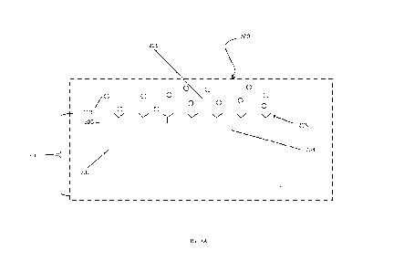
provide visual
means of identifying one side of the chip. The marker may also be utilized to
aid in visual or
mechanical alignment of the chip on or within a cartridge of an
interferometric system as
provided herein.
CA 03199855 2023- 5- 23
WO 2022/159442 PCT/US2022/012906
19
[00102] According to one embodiment, the marking is at least one
colorant, at least one
cut edge, at least one etching, at least one affixed label, or any combination
thereof. According
to one embodiment, the at least one colorant includes at least one dye that
visible to the naked
eye. According to one embodiment, the etching may include a machine-readable
etching, such
as a laser etching. According to one embodiment, the affixed label may be a
identifying material
applied to an external surface of the chip. According to one embodiment, the
cut edge includes
a distinct shape such as a diagonally cut corner (see e.g., FIG. 3C, 311). The
cut corner (311)
may be introduced on any of the chip's four corners. Although not illustrated,
the marking may
include at least one pillar or at least one visual label (such as a dot that
aligns with a laser
beam) to aid in aligning the chip within a cartridge system as described
herein.
[00103] In use, a light signal may be emitted from a light unit
located in the interferometric
system. The light enters flow through entry gradients in the chip and through
one or more
waveguide channels. According to a particular embodiment, there may be two or
more
waveguides channels to determine the presence of a separate analyte that each
of the
individual waveguides channels alone would not have been able to identify
alone. The
evanescent field is created when the light illuminates the waveguide channel.
The light signal is
then directed by exit gradients to a detector unit such as a camera unit. The
detector unit is
configured to receive the light signal and detect an analyte present in a test
sample
composition. The chip may further include a reference waveguide channel.
[00104] According to one embodiment, the one or more waveguide
channels described
herein may include or otherwise be coated with a waveguide channel coating
that includes any
material having a refractive index appropriate for Young's interferometry.
According to one
embodiment, the waveguide channel coating material includes a metal oxide or
metal dioxide.
Suitable waveguide channel coating materials may include, but are not limited
to, tantalum
oxide, tantalum dioxide, tantalum pentoxide, silicon dioxide, titanium oxide,
titanium dioxide, or
any combination thereof.
[00105] A sensing layer may be adhered to a top side of one or
more waveguide
channels. According to a particular embodiment, the sensing layer may include
one or more
proteins, enzymes, aptamers, peptides, nucleic acids, carbohydrates, lipids,
or monomers and
polymers, or whole cell microorganisms suitable for binding one or more
analytes. According to
another embodiment, the sensing layer may include one or more antigens or
antibodies that are
immobilized on the waveguide channel surface to sense the antigen-specific
antibody or
antigen, respectively. According to another embodiment, the sensing layer may
include
CA 03199855 2023- 5- 23
WO 2022/159442 PCT/US2022/012906
envelope, membrane, nucleocapsid N-proteins or different domains of one of the
proteins in a
natural or artificial virus used to delivery interfering RNA (RNAi) as a
treatment.
[00106] According to a particular embodiment, the sensing layer
may include a
molecularly imprinted polymer. The molecularly imprinted polymer leaves
cavities in the
polymer matrix with an affinity for a particular analyte such as an
antibiotic.
[00107] According to a particular embodiment, the sensing layer
may include a DNA
microarray of DNA probes. Each probe may be specific for a pathogen (i.e.,
bacterial species)
and when the probe hybridizes with a sample, the sample/probe complex
fluoresces in UV light
or may be detected via interferometric analysis or internal camera located for
this purpose.
According to one embodiment, the sensing layer may utilize immunoassays on top
of the
waveguide channels for detection of one or more analytes. According to one
embodiment, the
system may include, or function based on, an enzyme-linked immunosorbent assay
(ELISA) or
other ligand binding assays that detect analytes in target samples. According
to one
embodiment, the sensing layer may utilize one or more polypeptides, nucleic
acids, antibodies,
carbohydrates, lipids, receptors, or ligands of receptors, fragments thereof,
and combinations
thereof. According to one such embodiment, the sensing layer is configured to
include one or
more antibodies as well as one or more immunoglobulins to aid in the
indication of the stage of
analyte infection. Suitable immunoglobulins include IgG, IgM, IgA, IgE and
IgD. According to
such an embodiment, the sensing layer may include one or more dyes to aid in
visualization.
The sensing layer may or may not be covalently bonded to each other and the
one or more
waveguide channels. The sensing may be reviewed by using a magnifying camera
to
determine the uniformity and/or other quality parameters of the application of
the sensing layer.
Output of the camera may be analyzed using software to automate the quality
analysis.
Flow Cell Overview
[00108] Each of the cartridge systems described herein include a
flow cell having at least
one detection microchannel adapted to communicate with one or more test sample
compositions flowing through a waveguide channel in a chip beneath the flow
cell. According to
one embodiment, the cartridge systems may include at least two, at least
three, or at least four
detection microchannels with each detection microchannel adapted to
communicate one or
more test sample composition allowing detection of the same or different
analytes.
[00109] Each detection microchannel is located on or within a flow
cell manufactured
from a wafer. The at least one detection microchannel may be etched, molded or
otherwise
engraved into one side of the flow cell wafer. Thus, the at least one
detection microchannel
CA 03199855 2023- 5- 23
WO 2022/159442 PCT/US2022/012906
21
may be shaped as a concave path as a result of the etching or molding within
the flow cell
wafer.
[00110] The flow cell wafer is oriented above the chip during use
such that the detection
microchannel may be orientated or otherwise laid out in variety of flow
patterns above the
waveguide channels. The detection microchannel may be laid out, for example,
in a simple half
loop flow pattern, serial flow pattern, or in a serpentine flow pattern. The
serpentine flow pattern
is particularly suited for embodiments where there are multiple waveguide
channels that are
arranged in a parallel arrangement. By utilizing the serpentine flow pattern,
the test composition
flows consistently over the waveguide channels without varying flow dynamics.
Chip, Flow Cell and Optical Assembly ¨ Exemplary Embodiment
[00111] FIG. 3A illustrates a cross-sectional view of an optical
detection region 200 of a
cartridge system. A chip 201 includes a substrate 202 that includes a
waveguide channel 204
attached to a surface 205 (such as the illustrated top surface) of the chip
202. An evanescent
field 206 is located above the waveguide channel 204. A sensing layer 208 is
adhered to a top
side of the waveguide channel 204. As illustrated, antibodies 210 are shown
that may bind or
otherwise immobilized to the sensing layer 208, however, the sensing layer 208
may be
adapted to bind any variety of analytes. As such, adjusting or otherwise
modifying the sensing
layer 208 allows for the cartridge system to be utilized for multiple
different types of analytes
without having to modify the cartridge system or and surrounding
interferometric system
components. In general use, an light signal (e.g., laser beam) illuminates the
waveguide
channel 204 creating the evanescent field 206 that encompasses the sensing
layer 208.
Binding of an analyte impacts the effective index of refraction of the
waveguide channel 204.
[00112] A bottom view of an exemplary flow cell 300 is illustrated
in FIG. 3B. At least one
detection microchannel 302 is located on or within a flow cell 300
manufactured from a
transparent wafer. The at least one detection microchannel 302 may be etched,
molded or
otherwise engraved into one side of the flow cell wafer 304. Thus, the at
least one detection
microchannel 302 may be shaped as a concave path as a resulted of the etching
or molding
within the flow cell wafer 304. The flow cell wafer 304 may be manufactured a
material such as
opaque plastic, or other suitable material. The flow cell wafer 304 may
optionally be coated with
an anti-reflection composition.
[00113] The movement of an light signal 308 (series of arrows)
through a chip 310 is
illustrated in FIG. 3C. As illustrated, the chip 310 includes a cut corner
311. The light signal
CA 03199855 2023- 5- 23
WO 2022/159442 PCT/US2022/012906
22
308 moves from a light unit 312, such as a laser unit, through a plurality of
entry gradients 314
and through one or more waveguide channels 316. Each channel includes a pair
of
waveguides (321, 323). One of the pair of waveguides 321 is coated with a
sensing layer 208
(as indicated by shading in FIG. 3C). The other one of the pair of waveguides
323 is not coated
with the sensing layer 208 (serving as a reference). The combination of the
light from each in
the pair of waveguides (312, 323) create an interference pattern which is
illuminated on detector
unit 320.
[00114] According to a particular embodiment, the two or more
waveguides channels 316
are utilized that are able to determine the presence of an analyte that each
of the individual
waveguides channels 316 alone would not have been able to identify alone. The
light signal
308 is then directed by exit gradients 318 to a detector unit 320 such as a
camera unit. The
detector unit 320 is configured to receive the light signal 308 and detect any
analyte present in a
target sample composition flowing through the detection microchannel 302 (see
FIG. 3B).
[00115] The chip 310 includes a combination of substrate 202 (see
FIG. 3A), waveguide
channel ( see FIG. 3A part 204 and FIG. 3C part 316) and sensing layer 208
(see FIG. 3A).
The flow cell 300 (see FIG. 3B) is oriented above the top surface 205 of the
chip 310 during use
such that the detection microchannel 302 may be orientated or otherwise laid
out in variety of
flow patterns above the waveguide channels 316. The detection microchannel 302
may be laid
out, for example, in a simple half loop flow pattern, serial flow pattern, or
in a serpentine flow
pattern as illustrated in FIG. 3B. The serpentine flow pattern is particularly
suited for
embodiments where there are multiple waveguide channels 316 that are arranged
in a parallel
arrangement (see FIG. 30). By utilizing the serpentine flow pattern, the test
composition flows
consistently over the waveguide channels 316 without varying flow dynamics.
[00116] The light signal passes through each waveguide channel 316
as illustrated in
FIG. 30, may combine thereby forming diffraction patterns on the detector unit
320. The
interaction of the analyte 210 (see FIG. 3A) and the sensing layer 208 changes
the index of
refraction of light in the waveguide channel per Equation 1. The diffraction
pattern is moved
which is detected by the detector unit 320. The detector unit as provided
herein may be in
electronic communication with video processing software. Any diffraction
pattern movement
may be reported in radians of shift. The processing software may record this
shift as a positive
result. The rate of change in radians that happens as testing is conducted may
be proportional
to the concentration of the analyte.
[00117] FIG. 4 illustrates a side view of an exemplary embodiment
of an optical assembly
unit 400 that can be found in the handheld interferometric systems described
herein (such as in
CA 03199855 2023- 5- 23
WO 2022/159442 PCT/US2022/012906
23
FIGS. 1-2). The optical assembly unit 400 includes an light unit 402 aligned
in an light unit
housing 404. The optical assembly unit 400 includes a detector unit 406, such
as a camera
unit, aligned in a camera unit housing 408.
[00118] FIG. 5A illustrates a cross-sectional view of the optical
assembly unit 400 of FIG.
4. The light unit 402 is situated at an angle relative to the shutter flap
element 420. The shutter
flap element 420 is adapted to slide open and shut under tension from a
shutter spring 422.
The shutter flap element 420 is illustrated in a first, closed position with
no cartridge system
inserted. The shutter flap element 420 includes and upper control arm 423 that
is located within
a rail portion 425.
[00119] A complimentary communication means 424 extends downward
so as to make
electronic contact with electronic communications means located on the
cartridge housing (see
FIGS. 6, 8A and 9A). The complimentary communication means 424 may be metal
contacts
such that, upon insertion, the metal contacts on the exterior surface of the
cartridge housing
touch and establish electronic communication between the cartridge system and
the remaining
components of the interferometric system (e.g., light unit, camera unit,
etc.). The complimentary
communication means 424, as illustrated, include one or more substantially
pointed or "V"
shaped so as to push down into or otherwise contact the cartridge housing
metal contacts. The
number of complimentary communication means 424 may match and align with the
number of
metal contacts on the exterior surface of the cartridge housing.
[00120] At least one downward cantilever bias spring 426 may be
located within the
optical assembly unit 400 such that, upon insertion of the cartridge through
the interferometric
system housing opening, the downward cantilever bias spring 426 pushes against
a top side of
the cartridge housing thereby forcing the cartridge housing against an
opposite side or bottom
portion or surface 428 of the cartridge recess 430 resulting in proper
alignment along a vertical
plane (see FIGS. 5A, 5B, 50 and 6).
[00121] The light unit 402 is optionally adjustable along various
planes for optimal light
signal 432 emission. As illustrated, the signal 432 is shown to be emitted and
focused by at
least one lens 433. A camera unit 406 is situated at an angle relative to the
shutter flap element
420 so as to receive the light signal 432 upon exit from the cartridge (see
FIG. 6).
[00122] A first roll adjustment screw 434 and second roll
adjustment screw 436 are
located on opposing sides of the light unit 402 for adjusting roll of the
light unit 402. A first
upward adjustment screw 438 and second upward adjustment screw 440 are located
in a
parallel manner on each side the light unit 402 for adjusting the light unit
402 towards the
cartridge system (i.e., substantially upward). An angle of incidence screw 442
is located against
CA 03199855 2023- 5- 23
WO 2022/159442 PCT/US2022/012906
24
the light unit 402 to allow for adjustments to the angle of incidence for
proper coupling angle. A
translation screw 444 is located direct communication with the light unit 402
to adjust translation
in the X axis. A spring element 446 maintains the position of the light unit
402 against the light
unite 402 by assisting the adjustment screws (432, 436), incidence screw 442
and translation
screw 444.
[00123] With specific regard to FIGS. 5A, 5B, and 5C, the bottom
portion 428 of the
cartridge recess 430 further includes alignment means that includes at least
one rail portion 425
for engaging both male key portions on the cartridge housing (see 824, 826 of
FIG 8A; see 920,
922 of FIG. 9A). The bottom portion or surface 428 of the cartridge recess 430
includes a first
raised surface 421A and second raised surface 421B. A shutter upper control
arm 423 is
located within the rail portion 425. The rail portion 425 includes a first
rail wing 427 and second
rail wing 429 adapted to receive and engage the male key portions (see 824,
826 of FIG 8A;
see 920, 922 of FIG. 9A). By including such alignment means, the cartridge
systems provided
here may only engage in a certain manner thereby preventing incorrect
insertion and provided
proper optical and microfluidic alignment.
[00124] FIG. 6 illustrates a cross-sectional view of the optical
assembly 400 of FIG. 5A
with one embodiment of a cartridge system 800 inserted in the optical assembly
400. As
illustrated, the shutter flap element 420 is pushed backwards upon insertion
of the cartridge
system 800. While not shown in FIG. 6, the shutter spring 422 as illustrated
in FIG. 5A is
compressed backwards. The shutter flap element 420 moves along a track system
450 having
a stationary male rail 452 on which a female rail portion 454 slides from a
first, closed position
with no cartridge system 800 inserted to a second, open position as
illustrated in FIG. 6 upon
cartridge system 800 insertion.
[00125] FIG. 6 further illustrates positioning of the cartridge
system 800 in the optical
assembly 400. The cartridge system 800 includes an interferometric chip 832
positioned below
the flow cell wafer 888. The cartridge system 800 includes storage means 807
as provided
herein positioned within the cartridge housing 802. While the cartridge system
800 is illustrated
as a single-use system, the alignment and positioning of the single-use
cartridge assembly may
also apply to the multiple-use cartridge systems provided herein (e.g., see
FIGS 9A-9F).
[00126] FIG. 7 illustrates a top view of the optical assembly unit
400 of FIG. 5A with one
embodiment of a cartridge system 800 inserted in the optical assembly unit
400. The cartridge
system 800, as illustrated, is a single-use system, however, a multiple-use
system may be
inserted in the same manner within the interferometric system. The cartridge
system 800
includes a cartridge housing 802 having a top surface 805. The optical
assembly unit 400, as
CA 03199855 2023- 5- 23
WO 2022/159442 PCT/US2022/012906
illustrated, includes a plurality of cantilever bias springs 426. The optical
assembly unit 400
further includes at least one side bias spring 460 (see also FIG. 50) such
that, upon insertion of
the cartridge system 800, the side bias spring 460 pushes against one
horizontal side 860 of the
cartridge housing thereby forcing the cartridge housing 802 into proper
alignment along a
horizontal plane.
Cartridge System Overview
[00127] The cartridge systems provided herein includes a cartridge
housing. The
cartridge housing may be manufactured from any material suitable for single or
multiple-use.
The cartridge may be manufactured according to a variety of additive
processing techniques
such as 3-D printing. The cartridge may be manufactured via traditional
techniques such as
injection molding. The polymer may include a coefficient of expansion such
that the housing
does not expand or contract in a manner that would disrupt alignment of any
microfluidic or
detection components described herein when the cartridge is exposed to heat or
cold
environmental conditions.
[00128] The cartridge housing may include a light prevention means
to aid in reducing,
preventing or eliminating ambient, outside light from interfering the
detection of one or more
analytes. The light prevention means may include colored cartridge housing
(e.g., black
colored) that is color dyed or coated during manufacture. According to one
embodiment, a dye
may be introduced to the polymer to provide a specific color to a region of or
the entire cartridge
housing. Suitable colors include any color that aids in reducing, preventing
or eliminating
ambient, outside light from interfering the detection of one or more analytes.
[00129] The cartridge systems provided herein further includes a
detection region. This
detection region accommodates or is otherwise adapted to receive an
interferometric chip and
flow cell wafer. The flow cell wafer includes at least one detection
microchannel. The flow cell
wafer is located directly above the chip. The detection microchannel may be
etched onto a flow
cell wafer having a substantially transparent or clear panel or window. The
flow cell wafer, the
chip or both the flow cell and chip may be coated with a substance that
reduces or eliminates
fogging or condensation. According to one embodiment, the chip may be heated
to reduce or
elimination fogging or condensation.
[00130] The cartridge systems provided herein are configured or
otherwise adapted or
designed to easily insert and instantly align within an interferometric system
such as, for
example, a hand-held interferometric system. By being configured to allow for
instant
CA 03199855 2023- 5- 23
WO 2022/159442 PCT/US2022/012906
26
alignment, no further adjustment is required by a user to align any
microfluidic components and
any internal detection-related components such as the laser, chip with
waveguides and exposed
channels in a detection region of the cartridge, optical detector and any
other focus-related
components in the interferometric system. According to one embodiment, the
cartridge systems
provided herein may be adjusted to align via manual adjustments.
[00131] The cartridge housing includes dimensions that are
complimentary in size and
shape to the size and shape to an internal surface defining a recess within an
interferometric
system. As provided and illustrated in the non-limiting examples herein, the
cartridge housing
may be generally rectangular in overall shape.
[00132] According to one embodiment, the cartridge system may be
inserted and
removed automatically. According to one embodiment, the cartridge housing
contains a bar
code or OR code. According to one embodiment, the cartridge system contains a
bar code or
OR code for calibration or alignment.
[00133] To aid in alignment, the cartridge housing includes an
alignment means on an
external surface of the cartridge housing. The alignment means may take a
variety of forms that
assure instant alignment of any microfluidic components and any internal
detection-related
components upon insertion of the cartridge within the interferometric system.
The alignment
means also aids in the prevention of incorrect orientation assertion within
the interferometric
system and allows for insertion only after proper alignment is attained. The
alignment means
further allows for the cartridge system to be stabilized to address
vibrational distortions.
[00134] The alignment means may include at least one male key
portion for engaging
and securing within a corresponding female rail located in the interferometric
system. The male
key portion may be disposed on the bottom surface of the cartridge housing,
however, the male
key portion may be located on any exterior surface of the cartridge housing.
Other suitable
alignment means include one or more microswitches or sensing devices that
guide the cartridge
housing to assure proper alignment.
[00135] According to a particular embodiment, the cartridge
housing includes a top
portion and a bottom portion based on the orientation of insertion in an
interferometric system.
The top portion may include a top surface defining at least one through hole
on at least one
external surface of either the top portion or bottom portion. The at least one
through hole is
adapted to receive a removable fastening means for securing the top portion
and bottom portion
together. Suitable fastening means include screws or other suitable fastener
that may be
removed. By allowing the top portion and bottom portion of the cartridge
housing to be
CA 03199855 2023- 5- 23
WO 2022/159442
PCT/US2022/012906
27
separated and re-attached, a user may open the cartridge housing to allow for
cleaning as well
as replacement of the chip.
[00136] The cartridge system as provided herein may include a
temperature control
means to control temperature and humidity. The cartridge system as provided
herein may
include a temperature control means to control test sample composition
temperature. By
controlling temperature and humidity around the cartridge system, the
interferometric system
can provide more repeatable, precise results. According to one embodiment, the
cartridge
system contains heating capability to facilitate consistent measurement and
operation in cold
temperatures. By controlling temperature and humidity around the cartridge
system, fogging or
condensation that causes interference in the detection region of the cartridge
system is reduced
or otherwise eliminated. The temperature control means may be located on or
within the
cartridge housing. According to a single-use cartridge system embodiment, the
temperature
control means is located on or around the mixing bladder of the microfluidic
fluid system
described herein. The temperature control means may be located on an exterior
surface of the
cartridge housing. One suitable temperature control means includes a metal
coil that is heated
upon introduction of an electric current. Another suitable temperature control
means includes
one or more warming bands or Peltier devices that can provide heating or
cooling.
[00137] Each of the cartridge systems described herein include a
flow cell having at least
one detection microchannel adapted to communicate with one or more test sample
compositions flowing through a waveguide channel in a chip beneath the flow
cell. According to
one embodiment, the cartridge systems may include at least two detection
microchannels with
each detection microchannel adapted to communicate one or more test sample
composition
allowing detection of the same or different analytes. According to one
embodiment, cartridge
system includes a flow cell having at least three detection microchannels with
each detection
microchannel adapted to communicate one or more test sample composition
allowing detection
of the same or different analytes. According to one embodiment, cartridge
system includes a
flow cell having at least four detection microchannels with each detection
microchannel adapted
to communicate one or more test sample composition allowing detection of the
same or different
analytes.
Cartridge System ¨ Exemplary Embodiments
[00138] An exemplary embodiment of a single-use cartridge system
800 is illustrated in
FIGS. 8A-F. A top view of a cartridge system 800 is provided in FIG. 8A. The
cartridge system
800 includes a cartridge housing 802 as described herein. The housing 802
includes a top
CA 03199855 2023- 5- 23
WO 2022/159442 PCT/US2022/012906
28
portion 804 (see FIG. 80) having a top surface 805. The top surface 805
includes four heat
stake posts 808 for joining the top portion 804 of the cartridge housing 802
to a bottom portion
810 (See FIG. 80) of the cartridge housing 802. By utilizing heat stake posts
808, the top
portion 804 may be permanently joined to a bottom portion 810 of the cartridge
housing 802.
The top surface 805 includes an injection port 812 for introduction of a test
sample.
[00139] The cartridge housing 802 further includes an electronic
communication means
816 located on a second external surface 818 that is on a different horizontal
plane from the top
surface 805. The electronic communication means 816 as illustrated includes a
plurality of
metal contacts.
[00140] The cartridge system further includes a vent port 820. The
vent port 820 allows
for any air in the microfluidic system 870 (see FIG. 8F), such as in the form
of bubbles, to exit.
The vent port 820 may include a vent cover 821 over the vent port 820. The
vent cover 821
may be fabricated from a material that repels liquid while allowing air or
vapor to pass through
such as, for example, expanded polytetrafluoroethylene (commercially available
as Goretex .
The vent cover 821 allows for air purging from the cartridge system 800 but
will not allow fluid to
pass through such as when a vacuum is applied to prime the microfluidic system
870. In this
way, bubble formation in a liquid test sample composition is removed or
otherwise avoided. The
top surface 805 also includes two port seals 822. The port seals 822 may be
made from rubber
and provides sealing of the microfluidic system 870 within the cartridge
system 800.
[00141] FIG. 8B illustrates a view of the bottom surface 823 of
one embodiment of a
single-use cartridge system 800. The bottom surface 823 includes a first male
key portion 824
and a second male key portion 826. The male keying portions (824, 826) engage
with a
corresponding rail portion (425 - See FIGS. 5A, 5B and 5C) located in the
cartridge recess 430
of the optical assembly 400. The bottom surface 823 further defines a first
detent 828 and a
second detent 830. The detents (828, 830) engage with or otherwise receive a
corresponding
first raised surface and a second raised surface (421A, 421B) inside the
cartridge recess 430 of
the optical assembly 400 (see FIGS. 5A, 5B and 50). When engaged with the
first detent 828
and second detent 830, the first raised surface and second raised surface
(421A, 421B) aid in
securing the cartridge system 800 within the cartridge recess 430.
[00142] The chip 832 is substantially transparent and allows the
light signal to enter,
interact with one or more waveguides channels (See FIG. 30) and allow for
binding of analyte
flowing within the at least one detection microchannel 834 within the flow
cell wafer 888 (see
FIG. 8F).
CA 03199855 2023- 5- 23
WO 2022/159442 PCT/US2022/012906
29
[00143] The bottom surface 823 further defines a light inlet slot
836. The light inlet slot
836 allows for an light signal to enter the cartridge system 800.
Particularly, the light inlet slot
836 allows for an light signal to enter the chip 832 and for the light signal
to move through any
waveguide channels (not shown; see e.g., part 316 of FIG. 3C) in the chip 832
while interacting
with analytes in the at least one detection microchannel 834 before the light
signal is deflected
by one or more gratings (not shown) down to the detector unit 406 (see e.g.,
FIG 5A) and 320
(see FIG. 30).
[00144] FIG. 80 illustrates a view of the back surface 840 of the
cartridge housing 802 of
a single-use cartridge system 800. The cartridge housing 802 includes a top
portion 804 and a
bottom portion 810. The male keying portions (824, 826) are shown extending
from the bottom
portion 810 of the cartridge housing 802.
[00145] FIG. 80 illustrates a view of the front surface 850 of the
cartridge housing 802 of
a single-use cartridge system 800. The male keying portions (824, 826) are
shown extending
from the bottom portion 810 of the cartridge housing 802.
[00146] FIG. 8E illustrates a view of one side surface 860 of the
cartridge housing 802 of
a single-use cartridge system 800, the opposing side being a mirror image.
[00147] FIG. 8F illustrates a cross-section view downward of a
single-use cartridge
system 800 along the horizontal line of FIG. 8E. The cartridge system 800
includes a detection
region 831 that accommodates or is otherwise adapted to receive a chip 832 and
flow cell wafer
888. The single-use cartridge system 800 includes a microfluidic system 870
for communicating
or otherwise providing a means for a test sample composition to move through
the cartridge
system 800 and allow for detection and analysis of one or more analytes. The
microfluidic
system 870 includes an injection port 812 for introduction of a test sample.
The injection port
may 812 optionally include a check valve 872. The microfluidic system 870
further includes a
first microchannel section 874 having a first end 876 attached in
communication with the
injection port check valve 872 and a second end 878 in communication with a
mixing bladder
880. A filter 877 may be located anywhere within the first microchannel
section 874. The
microfluidic system 870 also includes a vent port 820 within the first
microchannel section 874
between the first end 876 and second end 878. The mixing bladder 880 includes
a temperature
control means 881 in the form of a metal coil wrapped around the mixing
bladder 880 such that
the temperature control means 881 is heated upon introduction of an electric
current.
[00148] The microfluidic system 870 further includes second
microchannel section 882
having a first end 884 attached in communication with the mixing bladder 880
and a second end
CA 03199855 2023- 5- 23
WO 2022/159442 PCT/US2022/012906
886 attached in communication with a flow cell wafer 888 having at least one
detection
microchannel 834.
[00149] The microfluidic system 870 further includes third
microchannel section 890
having a first end 892 attached in communication with at least one detection
microchannel 834
and a second end 894 in communication back to the mixing bladder 880 so as to
form a closed
loop.
[00150] The microfluidic system 870 further includes at least one
micropump 898. The
micropump 898, as illustrated, is a piezoelectric pump that overlays or
otherwise engages or
touches one or more of the first microchannel section 874, second microchannel
section 882,
third microchannel section 890 and mixing bladder 880. The micropump 898
manipulates the
communication of test sample composition throughout the microfluidic system
870.
[00151] The single-use cartridge system 800 may further include a
transmission
component 897 as provided herein. The single-use cartridge system 800 may
further include a
location means 899 as provided herein.
[00152] An exemplary embodiment of a multiple-use cartridge system
900 is illustrated in
FIGS. 9A-F.
[00153] A top view of an embodiment of a multi-use cartridge
system 900 is provided in
FIG. 9A. The cartridge system 900 includes a cartridge housing 902 as
described herein. The
housing 902 includes a top portion 904 (see FIG. 90) having a top surface 905.
As illustrated,
the top surface 905 includes four top through holes 908A. The top through
holes 908A are
adapted (e.g., threaded) to receive a removable fastening means (not shown)
for securing the
top portion 904 to a bottom portion 910 (see FIG. 90). The top surface also
includes two
sealing holes 908B that allow for sealing of the chip 936 to the cartridge
housing 902.
[00154] The cartridge housing 902 further includes an electronic
communication means
916 located on a second external surface 918 that is on a different horizontal
plane from the top
surface 905. The electronic communication means 916 as illustrated includes a
plurality of
metal contacts. The top surface 905 also includes two port seals 919 and two
seal plugs (924,
926).
[00155] FIG. 9B illustrates a view of the bottom surface 923 of a
multiple-use cartridge
system 900. The bottom surface 923 includes a first male key portion 920 and a
second male
key portion 922. The male keying portions (920, 922) engage with a
corresponding rail portion
(425 - See FIGS. 5A, 5B and 50) located in the interferometric system. The
bottom surface 923
further defines a first detent 928 and a second detent 930. The detents (928,
930) engage with
CA 03199855 2023- 5- 23
WO 2022/159442 PCT/US2022/012906
31
or otherwise receive a corresponding first raised surface and a second raised
surface (421A,
421B see FIGS. 5B and 5C) inside the cartridge recess 430 (see FIG. 5A) of the
optical
assembly 400 . When engaged with the first detent 928 and second detent 930,
the first raised
surface and second raised surface (421A, 421B) aid in securing the cartridge
system 900 within
the cartridge recess 430.
[00156] The bottom surface further includes bottom through holes
9080 that align and
correspond to the four top through holes 908A. The bottom through holes 908C
may be
adapted (e.g., threaded) to receive a removable fastening means (not shown)
for securing the
top portion 904 to a bottom portion 910 (see FIG. 90).
[00157] The bottom surface 923 further defines a light inlet slot
934. The light inlet slot
934 allows for an light signal to enter the cartridge system 900.
Particularly, the light inlet slot
934 allows for an light signal to enter the chip 936 and for the light signal
to move through any
waveguides in the chip 936 while interacting with analytes in the at least one
detection
microchannel 994 (see FIG. 9F) before the light signal is deflected by one or
more gratings (not
shown) down to the detector unit 406 (see FIG. 5A).
[00158] FIG. 90 illustrates a view of the back surface 940 of one
embodiment of a
multiple-use cartridge system 900. The housing includes a top portion 904 that
is optionally
removable from a bottom portion 910. The male keying portions (920, 922) are
shown
extending from the bottom portion 910 of the cartridge housing 902.
[00159] FIG. 9D illustrates a view of the front surface 950 of one
embodiment of a
multiple-use cartridge system 900. FIG. 9E illustrates view of one side
surface 960 of one
embodiment of a single-use cartridge system 900, the opposite side being a
mirror image.
[00160] FIG. 9F illustrates a cross-section view downward of a
multiple-use cartridge
system 900 along the horizontal line of FIG. 9E. The cartridge system 900 a
storage means
907 as provided herein positioned within the cartridge housing 902. The
multiple-use cartridge
system 900 includes a microfluidic system 970 for communicating or otherwise
providing a
means for a test sample composition to move through the cartridge system 900
and allow for
detection and analysis of one or more analytes. An ingress port 972 is located
on a front
surface 950 (see FIG. 9D) of the multiple-use cartridge system 900. The
ingress port 972 is in
communication with a first microchannel section 974 having a first end 976
attached in
communication with an ingress port check valve 973 and a second end 978 in
communication
with second microchannel section 979. A filter 977 may be located anywhere
within the first
microchannel section 974. A sample electrode 980 and reference electrode 982
are in contact
CA 03199855 2023- 5- 23
WO 2022/159442 PCT/US2022/012906
32
with the second microchannel section 979. Impedance may be measured between
the sample
electrode 980 and reference electrode 982 to confirm the presence of test
sample composition.
[00161] A valve test structure connection 984 is in communication
with any test sample
composition in the microfluidic system 970. The valve test structure
connection 984 may be
fabricated from nitinol shape memory alloy and aids in the movement of test
sample
composition into the cartridge system 900.
[00162] The second microchannel section 979 includes a first end
988 in communication
the first microchannel section 974 and a second end 990 in communication with
a flow cell 992
having at least one detection microchannel 994. The cartridge system 900
includes a detection
region 993 that accommodates or is otherwise adapted to receive the chip 936
and flow cell
992. The chip 936 is substantially transparent and allows the light signal to
enter, interact with
one or more waveguides channels (not shown; see e.g., part 316 of FIG. 3C) and
allow for
binding of analyte flowing within the at least one detection microchannel 994
within the flow cell
992.
[00163] The detection microchannel 994 is in communication with a
first end 996 of a
third microchannel section 998. The third microchannel section 998 includes a
flow electrode
1000 to approximate flow rate and is correlated with measured impedance. The
third
microchannel section 998 includes a second end 1002 in communication with the
first end 1004
of a fourth microchannel 1006. The fourth microchannel 1006 includes a second
end 1008 in
communication with a check valve 1010 which, in turn, is in communication with
an egress port
1012. The sample electrode 980, reference electrode 982, and flow electrode
1000 are each
fabricated from inert nitinol or other conductive material.
[00164] The multiple-use cartridge system 900 may further include
a transmission
component 1014 as provided herein. The multiple-use cartridge system 900 may
further include
a location means 1016 as provided herein.
[00165] An exemplary embodiment of an alternative single-use
cartridge system 1100 is
illustrated in FIG. 10. According to the illustrated embodiment, the cartridge
system 1100
includes a connection mechanism 1102 (or snap-in rod) having opposing ends
(1104, 1106)
extending from the housing 1108. The connection mechanism 1102 aids in
securing and
interfacing the cartridge system 1100 with an interferometric system. Rising
from the housing
1108, are an injection ports 1110 A-D and outlet ports 1120 A-D. The injection
ports 1110 A-D
may be utilized for introducing a test sample, buffer or a test sample
composition. The cartridge
system includes four independent detection microchannel ports that are
independently in
communication with a corresponding detection microchannel (not shown) within a
flow cell (not
CA 03199855 2023- 5- 23
WO 2022/159442 PCT/US2022/012906
33
shown). Buffer may be pre-loaded in the flow cell. Any test sample composition
waste may be
collected from the outlet ports 1120 A-D.
Healthcare Applications
[00166] The interferometric systems provided herein may be
utilized as a point of care
system. The point of care testing may be carried out at or near the site of a
where a healthcare
target sample is obtained. In a healthcare setting, a medical professional may
receive results in
an efficient manner and any care decisions may be implemented immediately. By
being mobile
and utilized at the point of care, the systems provided herein provide a major
technical
advancement in the fight to diagnose and track pathogens that may give rise to
global
pandemics or be the cause of other rising, recurring or endemic diseases.
[00167] The interferometric systems provided herein may be
utilized to analyze and
detect analytes taken from a bodily fluid or gaseous emission of the body.
Such bodily fluids
include, but are not limited to, blood, urine and saliva.
[00168] The interferometric systems provided herein are suited to
detect and quantify
analytes such as one or more of a virus, bacteria, or small molecule such as a
drug or drug
metabolite. The interferometric systems provided herein are particularly
suited to detect and
quantify analytes of particular medical interest such as an illegal/illicit
drug, SARS-CoV-2,
Yersinia pestis (Plague), mycobacterium, influenza virus, hCG, human
immunodeficiency virus,
a particular vitamin, genetic mutation, IgG, IgE, and CD4/T-Cells. The
interferometric systems
provided herein are readily adaptable to new analytes within the healthcare
environment as they
emerge. The interferometric systems put high quality diagnostic results in the
hands of
healthcare professionals in an efficient manner.
[00169] According to one particular embodiment, the
interferometric system provided
here may be utilized in connection with or otherwise equipped to a mobile
vehicle. Suitable
mobile vehicles include, but are not limited to, unmanned aerial vehicles
(UAV), unmanned
ground vehicles (UGV), drones, manned aircraft, and other manned vehicles.
Animal Health Applications
[00170] By being mobile and utilized near the animals being
studied, a user may receive
results in an efficient manner and any care or remedial measure decisions may
be implemented
immediately. The interferometric systems provided herein provide a major
technical
advancement to diagnose pathogens an animal health environment. This rapid
detection will
CA 03199855 2023- 5- 23
WO 2022/159442 PCT/US2022/012906
34
allow for remedial steps to be taken immediately rather than sending the
samples out for lab
testing. This will provide great advantages to the user inasmuch as diseases
could spread
beyond control during the days typically required to send samples out for
testing.
[00171] The systems provided herein provide a means to detect,
quantify, and even track
various analytes within an animal health environment. The systems provided
herein also
provide a means to assess the presence of analytes within animal enclosures,
transportation,
and water supplies. The system described herein also provides a means to
monitor the
microbiomes of animals for pathogens or chemical imbalances that can be used
to provide an
early warning system for detection of unwanted outcomes within an animal
health environment.
[00172] By providing detection and quantification data in an
efficient manner, exposure to
chemicals may be monitored, adjusted and otherwise controlled. According to
such an
embodiment, the system will detect and quantify one or more chemicals at the
parts per million
(ppm), parts per billion (ppb) or parts per trillion (ppt) level.
[00173] According to a particular embodiment, the systems provided
herein may be
utilized to detect and quantify levels of various chemicals in an animal
health environment
including, but not limited to, ammonia, benzene, toluene, xylene,
trichloroethylene,
perchlorethylene, dichloroethylene, vinyl chloride, chloramine, nicotine, n-
methylphenylethylamine methamphetamine, N,N-dimethyl acetamide (DMAC),
dithemylmethylphosphonate (DMMP), methyl salicylate, 2, 4, 6-trinitrotoluene,
acetaldehyde,
methylene chloride, hexane, acetone, methanol, pyrrole, chloroform, chlorine
(or any other
element), hydrochloric acid, ammonia, Freon, or 2-vinylpyridine.
[00174] According to a particular embodiment, the systems provided
herein may be
utilized to detect and quantify levels of bacteria, viruses, or fungi in an
animal health
environment. Such bacteria may originate from exposure to humans, other
animals, or parasites
such as worms, fleas, ticks, lice, and biting flies.
[00175] According to one particular embodiment, the
interferometric system provided
here may be utilized in connection with or otherwise equipped to a mobile
vehicle. Suitable
mobile vehicles include, but are not limited to, unmanned aerial vehicles
(UAV), unmanned
ground vehicles (UGV), drones, manned aircraft, and manned vehicles.
[00176] According to one particular embodiment, the
interferometric system provided
here may be utilized in various types of animal health environments such as a
veterinary office,
animal laboratory testing facility, farm, pasture or home (domesticated
animals).
CA 03199855 2023- 5- 23
WO 2022/159442 PCT/US2022/012906
Agricultural Applications
[00177] By being mobile and utilized at the point of use, a user
may receive results in an
efficient manner and any care or remedial measure decisions may be implemented
immediately.
The interferometric systems provided herein provide a major technical
advancement in the fight
to diagnose and track pathogens that may give rise to crop damage or be the
cause of other
rising, recurring or endemic diseases as well as invading species of pathogen
in an agricultural
environment. The systems provided herein provide a means to indicate and
otherwise aid in the
control of disease surveillance, invasive species of pathogen and pandemic or
widespread
outbreak control. The systems provided herein also provide a means to assess
water quality as
well as serve as a microbiome-based monitoring system to provide an early
warning system for
detection of unwanted pathogens in an agricultural environment.
[00178] According to a particular embodiment, the systems provided
herein may be
utilized to detect and quantify levels of pesticide in an agricultural
environment. By providing
detection and quantification data in an efficient manner within the
agricultural environment,
application and control rate of pesticide may be monitored, adjusted and
otherwise controlled.
According to such an embodiment, the system will detect and quantify pesticide
at the parts per
million (ppm) level. According to another embodiment, the system will detect
and quantify
pesticide at the parts per billion (ppb) level. According to another
embodiment, the system will
detect and quantify pesticide at the parts per trillion (ppt) level.
[00179] According to a particular embodiment, the systems provided
herein may be
utilized to detect and quantify levels of an agricultural herbicide (e.g., 2,4-
D (2,4-
dichlorophenoxyacetic acid) and dicamba (2-methoxy-3,6-dichlorobenzoic acid))
in an
agricultural environment. By providing detection and quantification data in an
efficient manner
within the agricultural environment, application and control rate of herbicide
may be monitored,
adjusted and otherwise controlled. Such control also increases the efficiency
of herbicidal
management. According to such an embodiment, the system will detect and
quantify herbicide
at the parts per million (ppm) level. According to another embodiment, the
system will detect
and quantify herbicide at the parts per billion (ppb) level. According to
another embodiment, the
system will detect and quantify pesticide at the parts per trillion (ppt)
level.
[00180] According to one embodiment, the system may be utilized to
detect and quantify
analytes from any vessel or container that may come internally in contact with
an analyte such
as a chemical contaminant. The system as provided herein may be placed in
fluid
communication with a vessel so as to detect and quantify analytes in real
time. Fluid
CA 03199855 2023- 5- 23
WO 2022/159442 PCT/US2022/012906
36
communication may be established via a tube or other conduit that allows any
fluid containing at
least analyte to come in contact with, or flow through, the system as provided
herein.
[00181] According to a particular embodiment, a liquid or fluid
source containing analyte
may be obtained from an agricultural spray tank. Such a spray tank may be
located on a tractor
(or other agricultural implement), in a field/crop area, at a farmer's
cooperative or other location
where a farmer will utilize spray tank.
[00182] According to the various embodiments described herein, the
systems and
methods provided may reduce the time typically required for spray tank
decontamination,
minimize the need to utilize (and store) large volumes of commercial tank
cleaners, reduce
dependency of the farm equipment operator to execute decontamination processes
without
benefit of knowledge of the point completion, eliminate the application of
improperly
decontaminated spray tank rinsate on labeled crops, and/or reduce legal risk
to the farm
equipment operator by providing documentation of spray tank decontamination.
The
embodiments may also increase the efficiency of a single tank or piece of
application equipment
having multiple specific independent uses.
[00183] According to one particular embodiment, a fluid source of
analytes includes an
industrial/commercial vessel. Such a vessel may be located within or around a
shipping
container that stores and transports a fluid chemical. The shipping container
may be located on
a truck, train, or other means of transportation. The shipping container may
also be located on
or around shipping dock.
[00184] According to one particular embodiment, the
interferometric system provided
here may be utilized in connection with or otherwise equipped to a mobile
vehicle. Suitable
mobile vehicles include, but are not limited to, unmanned aircraft systems
(UAS), unmanned
vehicles (UAV), autonomous vehicles, drones, manned aircrafts, and manned
vehicles.
Aquatic Applications
[00185] By being mobile and utilized near the aquatic environment
in question, a user
may receive results in an efficient manner and any care or remedial measure
decisions may be
implemented immediately. The interferometric systems provided herein provide a
major
technical advancement in the fight to diagnose and track pathogens that are
the cause of rising,
recurring or endemic diseases as well as invading species of pathogen in an
aquatic
environment. The systems provided herein provide a means to indicate and
otherwise aid in the
control of disease surveillance, invasive species of pathogen and pandemic or
widespread
outbreak control. The systems provided herein also provide a means to assess
water quality as
CA 03199855 2023- 5- 23
WO 2022/159442
PCT/US2022/012906
37
well as serve as a microbe - based monitoring system to provide an early
warning system for
detection of unwanted pathogens in an aquatic environment.
[00186] According to a particular embodiment, the systems provided
herein may be
utilized to detect and quantify levels of pesticide in an aquatic environment.
By providing
detection and quantification data in an efficient manner within the aquatic
environment,
application and control rate of pesticide may be monitored, adjusted and
otherwise controlled.
According to such an embodiment, the system will detect and quantify pesticide
at the parts per
million (ppm) level. According to another embodiment, the system will detect
and quantify
pesticide at the parts per billion (ppb) level. According to another
embodiment, the system will
detect and quantify pesticide at the parts per trillion (ppt) level.
[00187] According to a particular embodiment, the systems provided
herein may be
utilized to detect and quantify levels of an aquatic herbicide (e.g., 2,4-D
(2,4-
dichlorophenoxyacetic acid) and flumioxazin (217-fluoro-3,4-dihydro-3-oxo-4-(2-
propyny1)-2H-
1,4-benzoxazin-6-y1]-4,5,6,7-tetrahydro-1H-isoindole-1,3(2H)-dione)) in an
aquatic environment.
By providing detection and quantification data in an efficient manner within
the aquatic
environment, application and control rate of herbicide may be monitored,
adjusted and
otherwise controlled. Such control increases the efficiency of aquatic
vegetation management.
Such vegetation may include water hemp, duck weed or algae.
[00188] According to one embodiment, the system may be utilized to
detect and quantify
analytes from any vessel or container that may come internally in contact with
an analyte such
as a chemical contaminant. The system as provided herein may be placed in
fluid
communication with a vessel so as to detect and quantify analytes in real
time. Fluid
communication may be established via a tube or other conduit that allows any
fluid containing
the fluid containing the aquatic test sample composition to come in contact
with, or flow through,
the system as provided herein.
[00189] According to one particular embodiment, the
interferometric system provided
here may be utilized in connection with or otherwise equipped to a mobile
vehicle. Suitable
mobile vehicles include, but are not limited to, unmanned aerial vehicles
(UAV), unmanned
ground vehicles (UGV), drones, manned aircraft, and manned vehicles.
Food Applications
[00190] By being mobile and utilized near the foodstuff in
question, a user may receive
results in an efficient manner and any care or remedial measure decisions may
be implemented
CA 03199855 2023- 5- 23
WO 2022/159442 PCT/US2022/012906
38
immediately. The interferometric systems provided herein provide a major
technical
advancement to detect, quantify, and even track various chemicals and
pathogens within a food
or food processing environment. The systems provided herein provide a means to
indicate and
otherwise aid in the control of the movement of analytes that impact food
safety and quality.
The systems provided herein also provide a means to assess the presence of
analytes in a food
processing environment as well as serve as a microbiome-based monitoring
system to provide
an early warning system for detection of unwanted pathogens in food.
[00191] According to a particular embodiment, the systems provided
herein may be
utilized to detect and quantify levels of pesticide in a food processing
facility. By providing
detection and quantification data in an efficient manner within the food
processing environment,
exposure to analytes may be monitored, adjusted and otherwise controlled.
According to such
an embodiment, the system will detect and quantify one or more analytes at the
parts per million
(ppm), parts per billion (ppb) or parts per trillion (ppt) level.
[00192] According to a particular embodiment, the systems provided
herein may be
utilized to detect and quantify levels of 2,4-D (2,4-dichlorophenoxyacetic
acid), dicamba (2-
methoxy-3,6-dichlorobenzoic acid), butylated hydroxyaniso le, butylated
hydroxytoluene,
recombinant bovine growth hormone, sodium aluminum sulfate, potassium
aluminum, sulfate,
bisphenol-A (BPA), sodium nitrite/nitrate, polycyclic aromatic hydrocarbons,
heterocyclic
amines, acrylamide, brominated vegetable oil, artificial food coloring/dyes,
and dioxins.
According to one embodiment, the system may be utilized to detect and quantify
analytes from
any vessel or container that may come internally in contact with an analyte
such as a pathogen
or chemical contaminant. The system as provided herein may be placed in fluid
communication
with a vessel or other piece of food processing equipment so as to detect and
quantify analytes
in real time. Fluid communication may be established via a tube or other
conduit that allows any
fluid containing at least analyte to come in contact with, or flow through,
the system as provided
herein.
[00193] According to one particular embodiment, a fluid source of
analytes includes an
industrial or commercial vessel adapted to store, process, or carry food. Such
a vessel may be
located within or around a shipping container that stores and transports food.
The shipping
container may be located on a truck, train, or other means of transportation.
The shipping
container may also be located on or around shipping dock.
[00194] According to one particular embodiment, the
interferometric system provided
here may be utilized in connection with or otherwise equipped to a mobile
vehicle. Suitable
CA 03199855 2023- 5- 23
WO 2022/159442 PCT/US2022/012906
39
mobile vehicles include, but are not limited to unmanned aerial vehicles
(UAV), unmanned
ground vehicles (UGV), drones, manned aircraft, and manned vehicles.
Chemical Applications
[00195] By being mobile and utilized near the point in the
industrial supply chain where
the analyte needs to be measured, a user may receive results in an efficient
manner and any
care or remedial measure decisions may be implemented immediately. The
interferometric
systems provided herein provide a major technical advancement to detect,
quantify and even
track various chemicals within a chemical environment. The systems provided
herein provide a
means to indicate and otherwise aid in the control of the processing, storage,
and movement of
chemicals. The systems provided herein also provide a means to assess the
presence of
chemicals in a water supply as well as serve as a microbe-based monitoring
system to provide
an early warning system for detection of unwanted pathogens.
[00196] According to a particular embodiment, the systems provided
herein may be
utilized to detect and quantify levels of a chemical in an industrial
environment such as in a
chemical processing facility. By providing detection and quantification data
in an efficient
manner within the production environment, exposure to chemicals may be
monitored, adjusted
and otherwise controlled. According to such an embodiment, the system will
detect and
quantify one or more chemicals at the parts per million (ppm), parts per
billion (ppb) or parts per
trillion (ppt) level.
[00197] According to a particular embodiment, the systems provided
herein may be
utilized to detect and quantify levels of a chemical herbicide (e.g., 2,4-D
(2,4-
dichlorophenoxyacetic acid) and dicamba (2-methoxy-3,6-dichlorobenzoic acid))
in a chemical
environment. According to one embodiment, the system may be utilized to detect
and quantify
analytes from a vessel or container that may come internally in contact with
an analyte such as
a chemical contaminant. The system as provided herein may be placed in fluid
communication
with a chemical vessel or other piece of chemical processing equipment so as
to detect and
quantify analytes in real time. Fluid communication may be established via a
tube or other
conduit that allows any fluid containing at least analyte to come in contact
with, or flow through,
the system as provided herein.
[00198] According to a particular embodiment, the systems provided
herein may be
utilized to detect and quantify levels of various chemicals including, but not
limited to, benzene,
toluene, xylene, trichloroethylene, perchlorethylene, dichloroethylene, vinyl
chloride, chloramine,
CA 03199855 2023- 5- 23
WO 2022/159442 PCT/US2022/012906
nicotine, n-methylphenylethylamine methamphetamine, N,N-dimethyl acetamide
(DMAC),
dithemylmethylphosphonate (DMMP), methyl salicylate, 2, 4, 6-trinitrotoluene,
acetaldehyde,
methylene chloride, hexane, acetone, methanol, pyrrole, chloroform, chlorine
(or any other
element), hydrochloric acid, ammonia, Freon, or 2-vinylpyridine.
[00199] According to one particular embodiment, a fluid source of
analytes includes an
industrial or commercial vessel adapted to store, process, or carry one or
more chemicals.
Such a vessel may be located within or around a shipping container that stores
and transports a
fluid chemical. The shipping container may be located on a truck, train, or
other means of
transportation. The shipping container may also be located on or around
shipping dock.
[00200] According to one particular embodiment, the
interferometric system provided
here may be utilized in connection with or otherwise equipped to a mobile
vehicle. Suitable
mobile vehicles include, but are not limited to, unmanned aerial vehicles
(UAV), unmanned
ground vehicles (UGV), drones, manned aircraft, and manned vehicles.
Methods of Detection and Quantification
[00201] FIG. 11 illustrates a method 1200 of detecting and
quantifying the level of analyte
in a test sample composition. The method includes the step of collecting 1202
or otherwise
obtaining a target sample having one or more analytes. In different
embodiments, the target
sample may be taken from the appropriate target depending on the location and
environment.
[00202] According to one embodiment, the method further includes
the optional step of
entering 1204 a user identifier (ID) in the system. Additionally, the method
further includes the
optional step of entering 1205 an identification number associated with the
sample, patient,
analyte of interest, or a combination thereof. The cartridge system utilized
may be equipped
with a label or sticker carrying identifying such information. The label or
sticker may include a
QR code including such information. The label or sticker may be removed prior
to use.
Identifying information may include metadata such as time, GPS data, or other
data generated
by the interferometric system described herein.
[00203] According to one embodiment, the method further includes
the step of
introducing the target sample to the interferometric system 1206. According to
one
embodiment, target sample is introduced to the cartridge by a separate device
such as a
syringe or pump. According to one embodiment, target sample is introduced by
an injection
device. According to one embodiment, the injection device may be permanently
attached to the
cartridge system. According to one embodiment, the injection device is a
pipette. According to
one embodiment, the injection device is a syringe. According to one
embodiment, the injection
CA 03199855 2023- 5- 23
WO 2022/159442 PCT/US2022/012906
41
device is a lance, pipette or capillary tube. When utilizing a multiple-use
cartridge system, the
cartridge system may be fitted to a tube or other transfer mechanism to allow
the sample to be
continuously taken from a large amount of fluid that is being monitored.
[00204] According to one embodiment, the method further includes
the step of mixing
1208 the target sample with a buffer solution to form a test sample
composition. In a multiple-
use cartridge system, such a step may occur prior to the test sample
composition being
introduced to the cartridge system. In a single-use cartridge system, such a
step may occur in
the mixing bladder with the assistance of a pump.
[00205] The method of detecting and quantifying the level of
analyte in a sample includes
initiating waveguide interferometry 1210 on the test sample composition. Such
a step may
include initiating movement of the light signal through the cartridge system
as provided herein
and receiving the light signal within the detector unit. Any changes in an
interference pattern
are representative of analyte in the test sample composition. Particularly,
such changes in an
interference pattern generate data related to one or more analyte in the test
sample
composition. According to one embodiment, the step of initiating 1210
waveguide
interferometry on the test sample composition includes the step of correlating
data from the
phase shift with calibration data to obtain data related to analyte identity,
analyte concentration,
or a combination thereof.
[00206] According to one embodiment, the method further includes
the step of
processing 1212 any data resulting from changes in the interference pattern.
Such changes in
interference pattern may be processed and otherwise translated to indicate the
presence and
amount of an analyte in a test sample composition. Processing may be assisted
by software,
processing units, processor, servers, or other component suitable for
processing. The step of
processing data may further include storing such data in storage means as
provided herein.
[00207] According to one embodiment, the method further includes
the step of
transmitting a data signal 1214. The signal may result in the display of data
on the system. The
step of transmitting data may include displaying the analyte levels via
projecting any real time
data on a screen as described herein. The step of transmitting data may
include transmitting
any obtained data to a mobile phone, smart phone, tablet, computer, laptop,
watch or other
wireless device. The data may also be sent to a device at a remote
destination. The remote
destination device may be a locally operated mobile or portable device, such
as a smart phone,
tablet device, pad, or laptop computer. The destination may also be smart
phone, pad,
computer, cloud device, or server. In other embodiments, the remote
destination may be a
stand-alone or networked computer, cloud device, or server accessible via a
local portable
CA 03199855 2023- 5- 23
WO 2022/159442 PCT/US2022/012906
42
device. A diagnosis of an infection in a healthcare environment may be based
on the analyte
quantity. The diagnosis may be based on the use of one or more immunoglobulins
as detection
materials.
[00208] The method may optionally include the step of disposing of
the test sample
composition 1216 per legal requirements. Such legal requirements assure that
any sample still
containing unacceptable levels of pathological contamination are disposed of
properly so as not
to cause harm to a user or the environment.
[00209] According to one embodiment, the method further includes
the step of initiating
1218 a cleaning or remedial countermeasure against any analyte detected. Such
remedial
measure may include introducing cleaning chemicals or beneficial
microorganisms to the
healthcare environment. The remedial measures may work to kill or otherwise
neutralize any
unwanted analyte present in the healthcare environment where a sample was
taken.
Method of Manufacture ¨ Interferometric Chip
[00210] According to one aspect, a method of manufacturing an
interferometric chip is
provided. According to one embodiment, the method of manufacturing an
interferometric chip
includes the step of providing a substantially transparent substrate. Such a
substrate may be
formed from an optical material. According to one embodiment, the substrate is
manufactured
from a substantially transparent glass. According to one embodiment, the
substrate is
manufactured from a polymer or plastic material (e.g., optical plastic).
[00211] According to one embodiment, the method of manufacturing
an interferometric
chip includes the step of forming one or more waveguide channels in the
substrate material.
According to one embodiment, the one or more waveguide channels are not formed
by
traditional manufacturing techniques such as etching, milling, machining or
carving of the one or
more waveguide channels. According to one embodiment, the one or more
waveguide
channels are formed utilizing semiconductor fabrication techniques that
facilitate the production
of forming one or more waveguides produced simultaneously on the substrate
optical material.
According to one embodiment, the one or more waveguide channels are formed by
an additive
manufacturing technique. The additive manufacturing technique may utilize
computer-aided-
software to direct manufacturing hardware (such as 3D printers) to deposit
optical material via
individual layers so as to form geometric shapes appropriate for the one or
more waveguide
channels.
[00212] According to one embodiment, the one or more channels may
be formed in a
variety of shapes and sizes. The one or more channels may be varied in shape
and size
CA 03199855 2023- 5- 23
WO 2022/159442 PCT/US2022/012906
43
depending on sensitivity of the system. According to one embodiment, the
channel size is
about 1.0 mm to about 20 mm in length. According to one embodiment, the
channel size is
from about 0.1 mm to about 0.3 mm in width. According to one embodiment, the
channel size is
about 0.2 mm in width. According to one embodiment, the channel size is from
about 0.0001 to
about 0.0010 mm in depth. Suitable channel sizes provided herein are merely
for exemplary
purposes and not limiting.
[00213] According to one embodiment, the method of manufacturing
an interferometric
chip includes the step of introducing a coating to the one or more waveguide
channels. The
waveguide channel coating may include any material having a refractive index
appropriate for
Young's interferometry. According to one embodiment, the waveguide channel
coating includes
at least one metal oxide or metal dioxide. Suitable waveguide channel coating
materials may
include, but are not limited to, tantalum oxide, tantalum dioxide, silicon
dioxide, titanium oxide,
titanium dioxide, tantalum pentoxide, or any combination thereof.
[00214] According to one embodiment, the method of manufacturing
an interferometric
chip includes the step of coating a target location on or within the chip with
a sensing layer as
provided herein. According to one embodiment, the target location is the one
or more
waveguide channels. According to one embodiment, the target location is an
entire single side
of the interferometric chip.
[00215] According to a particular embodiment, the step of coating
the target location with
a sensing layer may include micro-dripping or wick threading the sensing layer
to the target
location on the chip. The micro-dripping may utilize one or more micro-pumps.
[00216] According to a particular embodiment, the step of coating
the target location with
a sensing layer may include aerosol-jet printing the sensing layer to the
target location on the
chip. According to a particular embodiment, the step of coating the target
location with a
sensing layer may include spin-coating the sensing layer to the target
location on the chip.
According to a particular embodiment, the step of coating the target location
with a sensing
layer may include dip-coating the sensing layer to the target location on the
chip.
[00217] According to a particular embodiment, the step of coating
the target location may
include inkjet printing the sensing layer to the target location on the chip.
According to a
particular embodiment, inkjet microdispensers may be utilized to create drops
using a
piezoelectric actuator to which a voltage pulse is applied.
[00218] According to one embodiment, three-dimensional or additive
manufacturing is
utilized to effectively deposit the sensing layer to the target location on
the chip. According to
CA 03199855 2023- 5- 23
WO 2022/159442 PCT/US2022/012906
44
one embodiment, gravure printing is utilized to effectively deposit the
sensing layer to the target
location on the chip. According to one embodiment, aerosol printing is
utilized to effectively
deposit the sensing layer to the target location on the chip. According to one
embodiment, silk
screening is utilized to effectively deposit the sensing layer to the target
location on the chip.
According to one embodiment, a felt marker application is utilized to
effectively deposit the
sensing layer to the target location on the chip. According to one embodiment,
a micro
paintbrush is utilized to effectively deposit the sensing layer to the target
location on the chip.
[00219] According to one embodiment, the method of manufacturing
an interferometric
chip includes the step of introducing a marker to the target location to
identify a side of the chip.
The marker can be at least one colorant, at least one cut corner or cut edge,
at least one laser
marking or etching, or any combination thereof.
[00220] Although the present specification describes components
and functions that may
be implemented in particular embodiments with reference to particular
standards and protocols,
the invention is not limited to such standards and protocols. For example,
standards for Internet
and other packet switched network transmission (e.g., TCP/IP, UDP/IP, HTML,
HTTP) represent
examples of the state of the art. Such standards are periodically superseded
by faster or more
efficient equivalents having essentially the same functions. Accordingly,
replacement standards
and protocols having the same or similar functions as those disclosed herein
are considered
equivalents thereof.
[00221] Although specific embodiments of the present invention are
herein illustrated and
described in detail, the invention is not limited thereto. The above detailed
descriptions are
provided as exemplary of the present invention and should not be construed as
constituting any
limitation of the invention. Modifications will be apparent to those skilled
in the art, and all
modifications that do not depart from the spirit of the invention are intended
to be included with
the scope of the appended claims.
PROPHETIC EXAMPLE 1
SARS-CoV-2 Detection and Quantification
[00222] A healthcare setting or mobile testing unit may be set up
to aid in high throughput
detection and quantification of SARS-CoV-2 in a patient. A medical
professional or other
trained user may obtain a sample. A user may obtain a sample in form of a
small blood sample.
In the case of a blood sample, the user may clean a patient's finger with an
alcohol swab. The
user may prick or otherwise lance the finger and obtain an effective amount
(aliquot) of blood for
the system. The aliquot of blood may be obtained via a medical device such as
a disposable
CA 03199855 2023- 5- 23
WO 2022/159442 PCT/US2022/012906
hemoglobin sterile pipette. Since the systems provided herein only need a
small amount of
sample, the system eliminates the need for a large-volume sample and
centrifuging thereby
significantly simplifying use and speeding up the process compared to existing
technologies that
require large sample volume.
[00223] A cartridge is then placed inside the system's detector
component, if not already
present. The cartridge may be fully replaceable and disposable after each use.
The user may
optionally then enter a user identifier (ID) in the system and the system
optionally transmits that
information to the remote server for authentication or stores the information
locally. Any of a
number of identifier labelling techniques, such as radio frequency identifiers
(RFIDs) on or within
a sample may be used. Alternatively, a unique serial number, code or other
identifier
associated with a sample may be manually entered into the system and
optionally transmitted to
a remote server. Additionally, the user may use the system to scan in or
manually enter one or
more substance/contaminant identifiers, such as a Universal Product Code (UPC)
for the one or
more analytes believed to be present in the sample and to inform the remote
server of the one
or more analytes.
[00224] The system may also include geolocation information in its
communications with
the server, either from a GPS sensor included in the system or a GPS software
function capable
of generating the location of the system in cooperation with a cellular or
other communication
network in communication with the system.
[00225] For detection and quantification of SARS-CoV-2, one or
more antibodies specific
to the surface proteins on SARS-CoV-2 will be included on the sensing layer.
The antibodies
may be specific to spike, envelope, membrane and nucleocapsid proteins found
on the surface
of SARS-CoV-2.
[00226] If present, the user may initialize the system by pressing
a start button or other
similar means to initiate any electronic components present in the system. If
present, the user
may optionally press an injection bulb or similar mechanical component to
inject a buffer
solution into the cartridge. Any display on the system may then provide visual
signal that the
system is ready for sample introduction (e.g., signal "READY"). If present,
the user may then
press another external display or button to signal the system that a sample is
ready to be
introduced (e.g., "SAMPLE").
[00227] To introduce a sample, an aliquot of the sample may be
added through a sample
collecting component. Such a step may be accomplished via a disposable pipette
or similar
device that is suited for storing a sample until needed. Next, a user may
press the injection bulb
or similar mechanical component to mix the aliquot of sample and buffer and
introduce the
CA 03199855 2023- 5- 23
WO 2022/159442 PCT/US2022/012906
46
mixture to the cartridge. According to an alternative embodiment, the buffer
may be mixed with
the aliquot sample in a separate step prior to introduction to the cartridge.
A user may then
press the injection bulb or similar mechanical component a one or more times
to ensure the
sample mixture has fully transitioned or otherwise migrated to the cartridge
and begins flowing
across the waveguide channels on the waveguide. Upon arrival at the waveguide,
detection
and quantification processes are undertaken. Depending on the goal of the
point of care
procedure, one or more biological components in the sample will bind to the
sensing layer of the
waveguide channels thereby altering the evanescent field above the waveguide
channels. The
changes in the interference pattern will then create an electronic signal that
can be translated to
produce a reading on a display on an external surface of the system. Any
medical devices used
during use may then be disposed of properly in a biological waste container.
The cartridge
within the system may then be removed and replaced with a new cartridge or
cleaned prior to
next use. The cartridge may be fully disposable and placed in a biological
waste container
along with the medical devices utilized during use.
PROPHETIC EXAMPLE 2
Avian Influenza Detection
[00228] A healthcare setting or mobile testing unit may be set up
to aid in high throughput
detection and quantification of avian influenza in a patient. A medical
professional or other
trained user may carry out the same general steps as set forth in Prophetic
Example 1. When
detecting and quantifying avian influenza in a sample, any avian influenza
analyte particles may
be captured by utilizing avian influenza specific antibodies on the sensing
layer that are
configured to bind to avian influenza surface protein. In some embodiments,
monoclonal and
polyclonal antibodies may be utilized on the sensing layer. In other
embodiments, polyclonal
antibodies may be utilized to attach to one or more antigenic sites on the
viral protein thereby
reducing the likelihood of a false negative indication (avoid looking at only
a single antigenic
epitope).
PROPHETIC EXAMPLE 3
Drug Detection
[00229] The systems provided herein may be utilized to aid in high
throughput detection
and quantification of a small molecule such as a drug or drug metabolite in a
patient. Such
detection methods may be particularly useful for job screening or for legal
reasons. Such
detection methods may also be particularly useful in emergency departments of
healthcare
CA 03199855 2023- 5- 23
WO 2022/159442
PCT/US2022/012906
47
facilities where a drug overdose is suspected. A medical professional or other
trained user may
carry out the same general steps as set forth in Prophetic Example 1. When
detecting and
quantifying drug and drug metabolites in a sample, any target small molecule
may be captured
by utilizing molecularly imprinted polymers on the sensing layer that are
configured to bind to
small molecules. Data regarding a specific class of drug or specific drug
compound may be
provided in an efficient manner to a user. Any corresponding report may be
submitted
wirelessly to a third party such as a prospective employer or law enforcement
agency, if
needed.
PROPHETIC EXAMPLE 4
Pathogenic Detection and Quantification in Dental Offices
[00230] The systems provided herein may be utilized to aid in high
throughput detection
and quantification of SARS-CoV-2 via implementation of systems in dental
offices. Dental
offices deal with several oral diseases but are also subject to common disease
and viral threats
such as HIV, Hepatitis, Flu, and Corona Viruses such as SARS-CoV-2. The United
States
dental industry has over 150,000 dental hygienists which see roughly 8
patients a day or
roughly 1,200,000 per day nationwide (0.38% of United States population daily
or approximately
7.5% of population monthly). More than half of the United States population
visits a dental
hygienist at least once per year. Dental hygienists are trained to deal with
both saliva and blood
and the real potential that the patient could be contagious with various
analytes such as SARS-
CoV-2. Using the systems provided herein to detect for these potential viral
analytes prior to a
dental exam can serve at least two purposes: The systems provided herein can
diagnose a
patient while providing early intervention as well as monitor pathogens to
help prevent outbreak,
epidemic, or pandemic.
[00231] According to one embodiment, the systems provided herein
may be utilized to
screen or otherwise detect a pathogen for each patient prior to or upon
entering a dental office
(HIPAA compliance required). The system may be located in a lobby or separate
area such that
results regarding pathogenic infection may be provided prior to entry into the
office and
subsequent dental treatment. Screening may occur with a saliva or blood sample
from a
patient. Such a screening process may be financially subsidized by a patient's
dental insurance
as well as supported by both the ADA and the AMA. According to one embodiment,
the
systems provided herein may be utilized to provide the dental office with an
additional source of
revenue via patient screening.
CA 03199855 2023- 5- 23
WO 2022/159442 PCT/US2022/012906
48
[00232] FIG. 12A illustrates a quantification and monitoring
system for analytes within an
aqueous target sample from a rinse sink. As illustrated in FIG. 12A, an
interferometric system
1300 may be utilized to monitor the rinse water 1310 flowing out of a dental
"rinse' sink 1320.
In use, the rinse water 1310 may move through a first drain pipe 1330 and
diverted by a valve
1340 between a collection pipe 1350 for the interferometric system 1300 and a
sewer/septic
pipe 1360.
[00233] FIG. 12A illustrates a quantification and monitoring
system for analytes within an
aqueous target sample from suction line. As illustrated in FIG. 12B, an
interferometric system
1400 may be utilized to monitor the rinse water 1410 flowing through a dental
suction line 1420
used during a dental cleaning or other procedure. In use, the rinse water 1410
may move
through a first drain pipe 1430 and diverted by a valve 1440 between a
collection pipe 1450 for
the interferometric system 1400 and a sewer/septic pipe 1460.
[00234] The results of monitoring in a dental facility may be sent
to a third monitoring
service. According to such an embodiment, the interferometric system provides
a reliable
sampling of the general United States population. By enumerating the patients,
geolocating the
suction line or sink, and sending the data to a central location (e.g., cloud-
based server), the
system may function as a digitized monitoring system for mapping results
across the United
States. After sampling rinse water from a particular patient, the system may
initiate a cleaning
and decontamination step of the suction line, pipes and any components within
the system.
According to a particular embodiment, the cartridge system within the
interferometric system
may be removed and replaced with a new cartridge after each use or cleaned
prior to next use.
According to a particular embodiment, the cartridge system within the
interferometric system
may be removed and replaced with a new cartridge periodically.
[00235] The interferometric system can also become part of the
normal maintenance of
those managing the dental office making the detection and monitoring methods
seamless. The
statistical relevance of this type of monitoring allows for non-HIPAA
collection of data while
monitoring the health of a particular region and, in turn, the overall United
States. In the event
HIPAA laws require authorization for this type monitoring, a bypass switch can
be installed and
the system will reduce the sample size for analysis by that number.
PROPHETIC EXAMPLE 5
Bartonella Detection and Quantification
[00236] Individuals at high-risk of acquiring a Bartonella
infection include those who work or
live with animals, or those with high exposure to fleas, ticks, lice and
biting flies. Infections such as
CA 03199855 2023- 5- 23
WO 2022/159442 PCT/US2022/012906
49
bartonellosis are increasingly implicated in complex chronic disease
syndromes, yet are
extremely difficult to diagnose accurately. Thus, an animal health setting may
utilize a system
provided herein for rapid detection and quantification of Bartonella in a pet
such as a cat or dog
that may serve as a vector for transmission.
[00237] A veterinary professional or other trained user may obtain
a sample. A user may
obtain a sample in form of a small blood sample, tissue or other biological
fluid such as pus. In
the case of a blood sample, the user may clean the target area with an alcohol
swab. The user
may the prick or otherwise lance the animal's skin and obtain an effective
amount (aliquot) of
blood for the system. The aliquot of blood may be obtain via a medical device
such as a
disposable haemoglobin sterile pipette. Since the systems provided herein only
need a small
amount of sample, the system eliminates the need for a large-volume sample and
centrifuging
thereby significantly simplifying use and speeding up the process compared to
existing
technologies that require large sample volume.
[00238] A cartridge is then placed inside the system's detector
component, if not already
present. The cartridge may be fully replaceable and disposable after each use.
The user may
optionally then enter a user identifier (ID) in the system and the system
optionally transmits that
information to the remote server for authentication or stores the information
locally. Any of a
number of identifier labelling techniques, such as radio frequency identifiers
(RFIDs) on or within
a sample may be used. Alternatively, a unique serial number, code or other
identifier
associated with a sample may be manually entered into the system and
optionally transmitted to
a remote server. Additionally, the user may use the system to scan in or
manually enter one or
more substance/contaminant identifiers, such as a Universal Product Code (UPC)
for the one or
more analytes believed to be present in the sample and to inform the remote
server of the one
or more analytes. The system may also include geolocation information in its
communications
with the server, either from a GPS sensor included in the system or a GPS
software function
capable of generating the location of the system in cooperation with a
cellular or other
communication network in communication with the system.
[00239] For detection and quantification of Bartonella, one or
more antibodies specific to
the surface proteins on Bartonella will be included on the reactive layer. The
antibodies may be
specific to spike, envelope, membrane and nucleocapsid proteins found on the
surface of
Bartonella.
[00240] If present, the user may initialize the system by pressing
a start button or other
similar means to initiate any electronic components present in the system. If
present, the user
may optionally press an injection bulb or similar mechanical component to
inject a buffer
CA 03199855 2023- 5- 23
WO 2022/159442 PCT/U52022/012906
solution into the cartridge. Any display on the system may then provide visual
signal that the
system is ready for sample introduction (e.g., signal "READY"). If present,
the user may then
press another external display or button to signal the system that a sample is
ready to be
introduced (e.g., "SAMPLE").
[00241] To introduce a sample, an aliquot of the sample may be
added through a sample
collecting component. Such a step may be accomplished via a disposable pipette
or similar
device that is suited for storing a sample until needed. Next, a user may
press the injection bulb
or similar mechanical component to mix the aliquot of sample and buffer and
introduce the
mixture to the cartridge. According to an alternative embodiment, the buffer
may be mixed with
the aliquot sample in a separate step prior to introduction to the cartridge.
A user may then
press the injection bulb or similar mechanical component a one or more times
to ensure the
sample mixture has fully transitioned or otherwise migrated to the cartridge
and begins flowing
across the waveguide channels on the waveguide. Upon arrival at the waveguide,
detection
and quantification processes are undertaken. Depending on the goal of the
point of care
procedure, one or more biological components in the sample will bind to the
reactive surface of
the waveguide channels thereby altering the evanescent field above the
waveguide channels.
The changes in the interference pattern will then create an electronic signal
that can be
translated to produce a reading on a display on an external surface of the
system. Any medical
devices used during use may then be disposed of properly in a biological waste
container. The
cartridge within the system may then be removed and replaced with a new
cartridge or cleaned
prior to next use. The cartridge may be fully disposable and placed in a
biological waste
container along with the medical devices utilized during use.
PROPHETIC EXAMPLE 6
Avian Influenza Detection
[00242] A portable interferometric system as provided herein may
be set up in an animal
health setting to aid in rapid detection and quantification of avian influenza
in a pullet. A
veterinary professional or other user may carry out the same general steps as
set forth in
Prophetic Example 1. When detecting and quantifying influenza in a sample, any
influenza
analyte particles may be captured by utilizing influenza specific antibodies
on the reactive layer
that are configured to bind to influenza surface protein. In some embodiments,
monoclonal and
polyclonal antibodies may be utilized on the reactive layer. In other
embodiments, polyclonal
antibodies may be utilized to attach to one or more antigenic sites on the
viral protein thereby
CA 03199855 2023- 5- 23
WO 2022/159442 PCT/US2022/012906
51
reducing the likelihood of a false negative indication (i.e. avoid looking at
only a single antigenic
epitope).
PROPHETIC EXAMPLE 7
Prion Detection
Chronic Wasting Disease
[00243] A portable interferometric system as provided herein may
be set up in an animal
health setting to aid in rapid detection and quantification of prion
(misfolded proteins) in an
animal suspected of having chronic wasting disease. Such an animal may be a
cow, sheep,
deer or elk. A veterinary professional or other user may carry out the same
general steps as set
forth in Prophetic Example 1. When detecting and quantifying prions or related
markers in a
sample, any prion particles may be captured by utilizing specific antibodies
or aptamers on the
reactive layer that are configured to bind to the target prion.
PROPHETIC EXAMPLE 8
2,4-D or Dicamba Detection and Quantification
[00244] A point of use testing unit may be set up to aid in high
throughput detection and
quantification of 2,4-D or dicamba in an agricultural tank. Dicamba products
are typically diluted
in water in a spray tank and sprayed on crop to selectively kill broadleaf
weeds within fields of
crops genetically modified to be resistant to the herbicidal chemicals (GMO).
Dicamba is known
to cause damage to many broad leaf plants at part per trillion levels.
Similarly, 2,4-D is also a
powerful broad leaf herbicide and can be used in place of dicamba. If either
herbicide residual
is present in the spray tank when it is subsequently used to other crops that
are sensitive to
dicamba or 2,4-D, the non-GMO crop may be killed or significantly damaged
results in loss of
revenue for the grower.
[00245] A sample may be obtained from the tank. A cartridge is
then placed inside the
system's detector component, if not already present. The cartridge may be
fully replaceable
and disposable after each use. The user may optionally then enter a user
identifier (ID) in the
system and the system optionally transmits that information to the remote
server for
authentication or stores the information locally. Any of a number of
identifier labelling
techniques, such as radio frequency identifiers (RFIDs) on or within a sample
may be used.
Alternatively, a unique serial number, code or other identifier associated
with a sample may be
manually entered into the system and optionally transmitted to a remote
server. Additionally,
CA 03199855 2023- 5- 23
WO 2022/159442 PCT/U52022/012906
52
the user may use the system to scan in or manually enter one or more
substance/contaminant
identifiers, such as a Universal Product Code (UPC) for the one or more
analytes believed to be
present in the sample and to inform the remote server of the one or more
analytes. The system
may also include geolocation information in its communications with the
server, either from a
GPS sensor included in the system or a GPS software function capable of
generating the
location of the system in cooperation with a cellular or other communication
network in
communication with the system.
[00246] For detection and quantification of 2,4-D, one or more
antibodies specific to the
2,4-D will be included on the sensing layer of at least one of the waveguides.
For detection and
quantification of dicamba, one or more antibodies specific to 2,4-D may be
included on the
sensing layer of at least one of the waveguides and, in addition, a
molecularly imprinted polymer
(MIP) specifically designed to be sensitive to dicamba will be present on at
least one of the
waveguides.
[00247] If present, the user may initialize the system by pressing
a start button or other
similar means to initiate any electronic components present in the system. If
present, the user
may optionally press an injection bulb or similar mechanical component to
inject a buffer
solution into the cartridge. Any display on the system may then provide visual
signal that the
system is ready for sample introduction (e.g., signal "READY"). If present,
the user may then
press another external display or button to signal the system that a sample is
ready to be
introduced (e.g., "SAMPLE").
[00248] To introduce a sample, an aliquot of the sample may be
added through a sample
collecting component. Such a step may be accomplished via a disposable pipette
or similar
device that is suited for storing a sample until needed. Next, a user may
press the injection bulb
or similar mechanical component to mix the aliquot of sample and buffer and
introduce the
mixture to the cartridge. According to an alternative embodiment, the buffer
may be mixed with
the aliquot sample in a separate step prior to introduction to the cartridge.
A user may then
press the injection bulb or similar mechanical component a one or more times
to ensure the
sample mixture has fully transitioned or otherwise migrated to the cartridge
and begins flowing
across the waveguide channels on the waveguide. Upon arrival at the waveguide,
detection
and quantification processes are undertaken. Depending on the goal of the
point of use
procedure, one or more analytes in the sample will bind to the sensing layer
of the waveguide
channels thereby altering the evanescent field above the waveguide channels.
The changes in
the interference pattern will then create an electronic signal that can be
translated to produce a
reading on a display on an external surface of the system. The cartridge
within the system may
CA 03199855 2023- 5- 23
WO 2022/159442 PCT/U52022/012906
53
then be removed and replaced with a new cartridge or cleaned prior to next
use. The cartridge
may be fully disposable and placed in an appropriate waste container.
[00249] If the tank contains 2,4-0, the waveguides that are
treated with the antibodies will
provide a strong indication of its presence. Due to the similar chemical
nature of dicamba, a
weak signal will also be present from the molecularly imprinted polymer
specific to dicamba.
[00250] If instead of 2,4-D, dicamba is present in the tank,
signals will be generated by
the molecularly imprinted polymer only. The antibody specific to 2,4-D will
not show a positive
result.
[00251] If both dicamba and 2,4-D are present, strong signals
would be generated by
both sensor types.
[00252] The software will use the strength of the signals and the
combination of which
sensors reported values to determine the content and strength of the
contaminants. In this way
the combination of sensors combined data processing can detect and
discriminate dicamba and
2,4-d in a way that is not possible with either of the sensors alone.
PROPHETIC EXAMPLE 9
Surface Water Analyte Detection
[00253] Runoff of crop inputs can present challenges for growers
operating near bodies
of water or near suburban or urban areas. An interferometric system as
provided herein may be
set up to aid in high throughput detection and quantification of one or more
target analytes in a
surface water source (e.g., a stream or drainage channel), particularly where
undesired analytes
may be present. A sample may be obtained by an automatic collection device
that will deliver a
sample aliquot to the device after the system installs a fresh cartridge as
needed. The targeted
analytes may include any chemical contaminant including, but not limited to, a
volatile organic
compound (such as benzene, toluene, ethylbenzene and xylenes),
tetrachloroethylene (PCE),
trichloroethylene (TCE), and vinyl chloride (VC). Other chemical contaminants
include gasoline,
oil, nitrites, metals, insecticides, and pesticides such as fluridone and
algaecides.
PROPHETIC EXAMPLE 10
Microbiome and/or Fungi Analyte Detection in Soil
CA 03199855 2023- 5- 23
WO 2022/159442 PCT/U52022/012906
54
[00254] An interferometric system as provided herein may be set up
to aid in high
throughput detection and quantification of one or more target analytes (e.g.,
pythium,
rhizoctonia, etc or metabolite generated by pythium, rhizoctonia, as well as
biopesticides,
insecticides, etc.) in soil. A sample may be obtained from the soil (e.g,
around the root zone)
and a sample aliquot prepared for delivery to the device. The sample may
contain either
beneficial microbiome and/or fungi or pathogenic microbiome and/or fungi. The
test may
measure either independently or in a multiplex fashion.
PROPHETIC EXAMPLE 11
Pesticide Drift
[00255] An interferometric system as provided herein may be set up
to aid in high
throughput detection of one or more target analytes in a crop field or
surrounding field in the
vicinity of a crop field subject to pesticide application. With proper
placement, the
interferometric system can detect and quantify pesticide drift. Particularly,
pesticide drift can be
detected and quantified field to field and from farm to farm. Detection and
quantification may
also provide an indication of the amount of pesticide that remains in the
target crop field. The
interferometric system may also produce and transmit a certification of
pesticide drift results to a
user or third party.
PROPHETIC EXAMPLE 12
Cholera and Cyanobacteria Detection and Quantification
[00256] A point of use testing unit may be set up to aid in rapid
detection and
quantification of cyanobacteria, cholera (vibrio cholera), or a combination
thereof in a surface
water source. Cholera and cyanobacteria are known to maintain a symbiotic
relationship so
there may be a need to test for both analytes.
[00257] A user may obtain a sample from the water source. A
cartridge is then placed
inside the system's detector component, if not already present. The cartridge
may be fully
replaceable and disposable after each use. The user may optionally then enter
a user identifier
(ID) in the system and the system optionally transmits that information to the
remote server for
authentication or stores the information locally. Any of a number of
identifier labelling
techniques, such as radio frequency identifiers (RFIDs) on or within a sample
may be used.
Alternatively, a unique serial number, code or other identifier associated
with a sample may be
manually entered into the system and optionally transmitted to a remote
server. Additionally,
CA 03199855 2023- 5- 23
WO 2022/159442 PCT/US2022/012906
the user may use the system to scan in or manually enter one or more
substance/contaminant
identifiers, such as a Universal Product Code (UPC) for the one or more
analytes believed to be
present in the sample and to inform the remote server of the one or more
analytes. The system
may also include geolocation information in its communications with the
server, either from a
GPS sensor included in the system or a GPS software function capable of
generating the
location of the system in cooperation with a cellular or other communication
network in
communication with the system.
[00258] For detection and quantification of a cyanobacteria, one
or more antibodies
specific to the cyanobacteria will be included on the receptor layer. The
antibodies may be
specific to microcystins such as microcystin-LR that are found in connection
with cyanobacteria.
[00259] If present, the user may initialize the system by pressing
a start button or other
similar means to initiate any electronic components present in the system. If
present, the user
may optionally press an injection bulb or similar mechanical component to
inject a buffer
solution into the cartridge. Any display on the system may then provide visual
signal that the
system is ready for sample introduction (e.g., signal "READY"). If present,
the user may then
press another external display or button to signal the system that a sample is
ready to be
introduced (e.g., "SAMPLE").
[00260] To introduce a sample, an aliquot of the sample may be
added through a sample
collecting component. Such a step may be accomplished via a disposable pipette
or similar
device that is suited for storing a sample until needed. Next, a user may
press the injection bulb
or similar mechanical component to mix the aliquot of sample and buffer and
introduce the
mixture to the cartridge. According to an alternative embodiment, the buffer
may be mixed with
the aliquot sample in a separate step prior to introduction to the cartridge.
A user may then
press the injection bulb or similar mechanical component a one or more times
to ensure the
sample mixture has fully transitioned or otherwise migrated to the cartridge
and begins flowing
across the waveguide channels on the waveguide. Upon arrival at the waveguide,
detection
and quantification processes are undertaken. Depending on the goal of the
point of use
procedure, one or more analytes in the sample will bind to the receptor
surface of the
waveguide channels thereby altering the evanescent field above the waveguide
channels. The
changes in the interference pattern will then create an electronic signal that
can be translated to
produce a reading on a display on an external surface of the system. Any
collection devices
and used interferometric cartridges used during use may then be disposed of
properly in an
appropriate waste container. The cartridge within the system may then be
removed and
CA 03199855 2023- 5- 23
WO 2022/159442 PCT/U52022/012906
56
replaced with a new cartridge or cleaned prior to next use. The cartridge may
be fully
disposable and placed in an appropriate waste container.
PROPHETIC EXAMPLE 13
Ground Water Analyte Detection
[00261] A point of use testing unit may be set up to aid in rapid
detection and
quantification of one or more target analytes in a subterranean water source
(e.g., ground
water). A user may obtain a sample from the water source and carry out the
steps as set forth
with respect to Example 1. The targeted analytes may include any chemical
contaminant
including, but not limited to, a volatile organic compound such as benzene,
toluene,
ethylbenzene and xylenes), tetrachloroethylene (PCE), trichloroethylene (TOE),
vinyl chloride
(VC), and gasoline. Other chemical contaminants include oil, nitrites, metals,
and pesticides.
PROPHETIC EXAMPLE 14
Chlorpyrifos Detection and Quantification in a Food Processing Plant
[00262] An interferometric system as provided herein may be set up
to aid in rapid
detection and quantification of chlorpyrifos on produce in a food processing
plant. Chlorpyrifos
is an organophosphate insecticide, acaricide and miticide used primarily to
control foliage and
soil-borne insect pests on a variety of food and feed crops. Chlorpyrifos is
not allowed to be
present in foods that are being sold in the United States.
[00263] For detection and quantification of a chlorpyrifos, one or
more antibodies or
aptamers specific to chlorpyrifos may be included on the sensing layer as
described herein. If
the test sample composition is shown to be contaminated with chlorpyrifos,
remedial measures
may be implemented.
PROPHETIC EXAMPLE 15
Grocery Store Produce Testing
[00264] An interferometric system as provided herein may be set up
to aid in detection
and quantification of one or more target analytes on produce as the produce
arrives at a grocery
store. A trained user may obtain a sample from the surface of the produce. The
test sample
may be obtained by an automatic collection device that will deliver a sample
aliquot to the
CA 03199855 2023- 5- 23
WO 2022/159442
PCT/U52022/012906
57
interferometric system. The targeted analytes may include any chemical
contaminant including,
but not limited to, a volatile organic compound such as benzene, toluene,
ethylbenzene and
xylenes), tetrachloroethylene (PCE), trichloroethylene (TCE), vinyl chloride
(VC), and gasoline.
Other chemical contaminants include, oil, nitrites, metals, and pesticides.
PROPHETIC EXAMPLE 16
Microbiome and/or Fungi Detection and Quantification in Produce Storage
[00265] An interferometric system as provided herein may be set up
at a produce storage
facility to aid in high throughput detection and quantification of one or more
target pathogenic
analytes commonly found on produce. Such analytes include, but are not limited
to, E. coli,
salmonella, pythium, asperigillus, rhizoctonia, or metabolites of each of the
same). A sample
may be obtained from the surface of the produce and a sample aliquot prepared
by wiping the
produce with a swab containing buffer. The buffer is expressed from the swab
and could be
transferred to the device with a pipette. The system may measure both
beneficial and
pathogenic analytes either independently or in a multiplex fashion.
PROPHETIC EXAMPLE 17
Agricultural Pesticide Detection
[00266] A portable interferometric system as provided herein may
be set up to aid in
rapid detection and quantification of chlorpyrifos on produce in a chemical
processing plant.
Chlorpyrifos is an organophosphate insecticide, acaricide and miticide used
primarily to control
foliage and soil-borne insect pests on a variety of food and feed crops.
Chlorpyrifos is not
allowed to be present in foods that are being sold in the United States.
[00267] After a vessel is used for processing chlorpyrifos and
before the vessel is used
for another purpose, it is necessary to clean the tank. For detection and
quantification of a
chlorpyrifos, one or more antibodies or aptamers specific to chlorpyrifos may
be included on the
sensing layer as described herein. If the test sample composition is shown to
be contaminated
with chlorpyrifos, remedial cleaning can continue until a suitable level of
cleanliness is achieved.
[00268] The following statements provide a general description of
the disclosure and are
not intended to limit the appended claims.
[0001]
Statement 1. A portable interferometric system for detection and
quantification of
analyte within a healthcare test sample composition, the system comprising:
CA 03199855 2023- 5- 23
WO 2022/159442 PCT/U52022/012906
58
an optical assembly unit, the optical assembly unit comprising a light unit
and a
detector unit each adapted to fit within a portable housing unit; and
a cartridge system adapted to be inserted in the housing and removed after one
or more
uses, the cartridge system comprising an interferometric chip and a flow cell
wafer.
wherein the interferometric chip includes one or more waveguide channels
having a
sensing layer thereon, the sensing layer adapted to bind or otherwise be
selectively disturbed
by one or more analytes within the healthcare test sample composition.
[0002] Statement 2. The portable interferometric system of statement
1, wherein the
portable housing is sized and shaped to fit in a user's hand.
[0003] Statement 3. The portable interferometric system of
statements 1-2, further
comprising at least one display unit.
[0004] Statement 4. The portable interferometric system of
statements 1-3, further
comprising an external camera, the external camera adapted to capture a photo
or video.
[0005] Statement 5. The portable interferometric system of
statements 1-4, comprising an
alignment means for aligning the cartridge system within a cartridge recess in
the interferometric
system.
[0006] Statement 6. The portable interferometric system of
statements 1-5, wherein the
sensing layer comprises one or more antigens, antibodies, DNA, aptamers,
polypeptides,
nucleic acids, carbohydrates, lipids, or molecularly imprinted polymers, or
immunoglobulins
suitable for binding one or more analytes within an healthcare test sample
composition.
[0007] Statement 7. The portable interferometric system of
statements 1-6, configured to
analyze the light signals from two or more waveguide channels to detect the
presence of an
analyte that individual waveguides could not have detected alone.
[0008] Statement 8. The portable interferometric system of
statements 1-7, wherein the one
or more waveguide channels each comprises a different sensing layer to allow
the system to
detect different analytes on each waveguide channel.
[0009] Statement 9. The portable interferometric system of
statements 1-8, wherein the
sensing layer is configured to bind one or more small molecules, antibodies,
virus antigens,
virus proteins, bacteria, fungi, pathogen, RNA, chemical, mRNA or any
combination thereof.
[0010] Statement 10. The portable interferometric system of
statements 1-9, having an
analyte detection limit down to about 1.0 picogram/L.
[0011] Statement 11. The portable interferometric system of
statements 1-10, having an
analyte detection limit down to about 1000 pfu/ml.
CA 03199855 2023- 5- 23
WO 2022/159442 PCT/U52022/012906
59
[0012] Statement 12. The portable interferometric system of
statements 1-11, wherein the
detector has sensitivity to at least 2 pixels per diffraction line pair.
[0013] Statement 13. The portable interferometric system of
statements 1-12, further
comprising a location means adapted to determine the physical location of the
system.
[0014] Statement 14. The portable interferometric system of
statements 1-11, wherein the
analyte is one or more of a fungicide, herbicide, insecticide, fungus,
bacterium, or microbe.
[0015] Statement 15. A method of detecting and quantifying the level
of analyte in an
healthcare test sample composition, the method comprising the steps of:
collecting a healthcare target sample containing one or more analytes;
optionally entering an identification associated with the target sample;
introducing the healthcare target sample to the portable interferometric
system of
statements 1-14;
optionally, mixing the target sample with a buffer solution to form a
healthcare test
sample composition;
initiating waveguide interferometry on the test sample composition;
processing any data resulting from the waveguide interferometry; and
optionally, transmitting any data resulting from the waveguide interferometry.
[0016] Statement 16. The method of statement 15, wherein the step of
transmitting data
includes wirelessly transmitting analyte detection and quantification data to
a mobile device or
server.
[0017] Statement 17. The method of statements 15- 16, further
comprising the step of
displaying data related to the presence of analyte in the test sample
composition on the display
unit.
[0018] Statement 18. The method of statements 15- 17, wherein the
healthcare target
sample is taken from water, soil, air, exhaled breath, skin, hair, or a bodily
fluid or gaseous
emission of the body.
[0019] Statement 19. The method of statements 15-18, wherein the
healthcare target
sample is in the form of, dissolved in, or suspended in a liquid or a gas.
[0020] Statement 20. The method of statements 15-19, wherein the
data resulting from the
waveguide interferometry is provided at or under 30 minutes.
[0021] Statement 21. A portable interferometric system for detection
and quantification of
analyte within an animal health test sample composition, the system
comprising:
an optical assembly unit, the optical assembly unit comprising a light unit
and a detector
unit each adapted to fit within a portable housing unit; and
CA 03199855 2023- 5- 23
WO 2022/159442 PCT/US2022/012906
a cartridge system adapted to be inserted in the housing and removed after one
or more
uses, the cartridge system comprising an interferometric chip and a flow cell
wafer.
wherein the interferometric chip includes one or more waveguide channels
having a
sensing layer thereon, the sensing layer adapted to bind or otherwise be
selectively disturbed
by one or more analytes within the animal health test sample composition.
[0022] Statement 22. The portable interferometric system of
statement 21, wherein the
portable housing is sized and shaped to fit in a user's hand.
[0023] Statement 23. The portable interferometric system of
statements 21-22, further
comprising at least one display unit.
[0024] Statement 24. The portable interferometric system of
statements 21-23, further
comprising an external camera, the external camera adapted to capture a photo
or video.
[0025] Statement 25. The portable interferometric system of
statements 21-24, comprising
an alignment means for aligning the cartridge system within a cartridge recess
in the
interferometric system.
[0026] Statement 26. The portable interferometric system of
statements 21-25, wherein the
sensing layer comprises one or more antigens, antibodies, DNA microarrays,
polypeptides,
nucleic acids, carbohydrates, lipids, or molecularly imprinted polymers, or
immunoglobulins
suitable for binding one or more analytes within an animal health test sample
composition.
[0027] Statement 27. The portable interferometric system of
statements 21-26, configured
to analyze the light signals from two or more waveguide channels to detect the
presence of an
analyte that individual waveguide channels could not have detected alone.
[0028] Statement 28. The portable interferometric system of
statements 21-27, wherein the
one or more waveguide channels each comprises a different sensing layer to
allow the system
to detect different analytes on each waveguide channel.
[0029] Statement 29. The portable interferometric system of
statements 21-28, wherein the
sensing layer is configured to bind one or more chemical, antibody, virus
antigen, virus protein,
bacteria, fungi, pathogen, RNA, mRNA, plant growth regulator, metal or any
combination
thereof.
[0030] Statement 30. The portable interferometric system of
statements 21-29, having an
analyte detection limit down to about 1.0 picogram/L.
[0031] Statement 31. The portable interferometric system of
statements 21-30, having an
analyte detection limit down to about 1000 pfu/ml.
[0032] Statement 32. The portable interferometric system of
statements 21-31, having
sensitivity to at least 2 pixels per diffraction line pair.
CA 03199855 2023- 5- 23
WO 2022/159442
PCT/US2022/012906
61
[0033] Statement 33. The portable interferometric system of
statements 21-32, further
comprising a location means adapted to determine the physical location of the
system.
[0034] Statement 34. The portable interferometric system of
statements 21-33, wherein the
analyte is one or more of a fungicide, herbicide, insecticide, fungus,
bacterium, or
microorganism.
[0035] Statement 35. A method of detecting and quantifying the level
of analyte in an
animal health test sample composition, the method comprising the steps of:
collecting an animal health target sample containing one or more analytes;
optionally entering an identification associated with the target sample;
introducing the animal health target sample to the portable interferometric
system of
statements 21- 34;
optionally, mixing the target sample with a buffer solution to form an animal
health test
sample composition;
initiating waveguide interferometry on the test sample composition;
processing any data resulting from the waveguide interferometry; and
optionally, transmitting any data resulting from the waveguide interferometry.
[0036] Statement 36. The method of statement 35, wherein the step of
transmitting data
includes wirelessly transmitting analyte detection and quantification data to
a mobile device or
server.
[0037] Statement 37. The method of statements 35-36, further
comprising the step of
displaying data related to the presence of analyte in the test sample
composition on the display
unit.
[0038] Statement 38. The method of statements 35-37, wherein the
animal health target
sample is taken from feed, water, soil, air, exhaled breath, skin, hair
tissue, or bodily fluid within
or surrounding an animal health environment.
[0039] Statement 39. The method of statements 35-38, wherein the
animal health target
sample is in the form of, dissolved in, or suspended in a liquid or a gas.
[0040] Statement 40. The method of statements 35-40, wherein the
data resulting from the
waveguide interferometry is provided at or under 30 minutes.
[0041] Statement 41. A portable interferometric system for detection
and quantification of
analyte within an agricultural test sample composition, the system comprising:
an optical assembly unit, the optical assembly unit comprising a light unit
and a detector
unit each adapted to fit within a portable housing unit; and
CA 03199855 2023- 5- 23
WO 2022/159442 PCT/US2022/012906
62
a cartridge system adapted to be inserted in the housing and removed after one
or more
uses, the cartridge system comprising an interferometric chip and a flow cell
wafer.
wherein the interferometric chip includes one or more waveguide channels
having a
sensing layer thereon, the sensing layer adapted to bind or otherwise be
selectively disturbed
by one or more analytes within the agricultural test sample composition.
[0042] Statement 42. The portable interferometric system of
statement 41, wherein the
portable housing is sized and shaped to fit in a user's hand.
[0043] Statement 43. The portable interferometric system of
statements 41-42, further
comprising at least one display unit.
[0044] Statement 44. The portable interferometric system of
statements 41-43, further
comprising an external camera, the external camera adapted to capture a photo
or video.
[0045] Statement 45. The portable interferometric system of
statements 41-44, comprising
an alignment means for aligning the cartridge system within a cartridge recess
in the
interferometric system.
[0046] Statement 46. The portable interferometric system of
statements 41-45, wherein the
sensing layer comprises one or more antigens, antibodies, aptamers, DNA
microarrays,
polypeptides, nucleic acids, carbohydrates, lipids, or molecularly imprinted
polymers, or
immunoglobulins suitable for binding one or more analytes within an
agricultural test sample
composition.
[0047] Statement 47. The portable interferometric system of
statements 41-46, configured
to analyze the light signals from two or more waveguide channels to detect the
presence of an
analyte that individual waveguide channels could not have detected alone.
[0048] Statement 48. The portable interferometric system of
statements 41-47, wherein the
one or more waveguide channels each comprises a different sensing layer to
allow the system
to detect different analytes on each waveguide channel.
[0049] Statement 49. The portable interferometric system of
statements 41-48, wherein the
sensing layer is configured to bind one or more antibodies, virus antigens,
virus proteins,
bacteria, fungi, pathogen, RNA, chemical, mRNA or any combination thereof.
[0050] Statement 50. The portable interferometric system of
statements 41-49, having an
analyte detection limit down to about 1.0 picogram/L.
[0051] Statement 51. The portable interferometric system of
statements 41-50, having an
analyte detection limit down to about 1000 pfu/ml.
[0052] Statement 52. The portable interferometric system of
statements 41-51, wherein the
detector has sensitivity to at least 2 pixels per diffraction line pair.
CA 03199855 2023- 5- 23
WO 2022/159442 PCT/US2022/012906
63
[0053] Statement 53. The portable interferometric system of
statements 41-52, further
comprising a location means adapted to determine the physical location of the
system.
[0054] Statement 54. The portable interferometric system of
statements 41-53, wherein the
analyte is one or more of a fungicide, herbicide, plant growth regulator,
insecticide, fungus,
bacterium, or microbe.
[0055] Statement 55. A method of detecting and quantifying the level
of analyte in an
agricultural test sample composition, the method comprising the steps of:
collecting an agricultural target sample containing one or more analytes;
optionally entering an identification associated with the target sample;
introducing the agricultural target sample to the portable interferometric
system of
statements 41-54;
optionally, mixing the target sample with a buffer solution to form an
agricultural test
sample composition;
initiating waveguide interferometry on the test sample composition;
processing any data resulting from the waveguide interferometry; and
optionally, transmitting any data resulting from the waveguide interferometry.
[0056] Statement 56. The method of statement 55, wherein the step of
transmitting data
includes wirelessly transmitting analyte detection and quantification data to
a mobile device or
server.
[0057] Statement 57. The method of statements 55-56, further
comprising the step of
displaying data related to the presence of analyte in the test sample
composition on the display
unit.
[0058] Statement 58. The method of statements 55-57, wherein the
agricultural target
sample is taken from plant material, agricultural input, building, equipment,
chemical tank,
chemical vessel, agricultural spray tank, soil, water, or air within or
surrounding an agricultural
environment.
[0059] Statement 59. The method of statements 55-58, wherein the
agricultrual target
sample is in the form of, dissolved in, or suspended in a liquid or a gas.
[0060] Statement 60. The method of statements 55-59, wherein the
data resulting from the
waveguide interferometry is provided at or under 30 minutes.
[0061] Statement 61. A portable interferometric system for detection
and quantification of
analyte within a chemical test sample composition, the system comprising:
an optical assembly unit, the optical assembly unit comprising a light unit
and a detector
unit each adapted to fit within a portable housing unit; and
CA 03199855 2023- 5- 23
WO 2022/159442 PCT/US2022/012906
64
a cartridge system adapted to be inserted in the housing and removed after one
or more
uses, the cartridge system comprising an interferometric chip and a flow cell
wafer.
wherein the interferometric chip includes one or more waveguide channels
having a
sensing layer thereon, the sensing layer adapted to bind or otherwise be
selectively disturbed
by one or more analytes within the chemical test sample composition.
[0062] Statement 62. The portable interferometric system of
statement 61, wherein the
portable housing is sized and shaped to fit in a user's hand.
[0063] Statement 63. The portable interferometric system of
statements 61-62, further
comprising at least one display unit.
[0064] Statement 64. The portable interferometric system of
statements 61-63, further
comprising an external camera, the external camera adapted to capture a photo
or video.
[0065] Statement 65. The portable interferometric system of
statements 61-64, comprising
an alignment means for aligning the cartridge system within a cartridge recess
in the
interferometric system.
[0066] Statement 66. The portable interferometric system of
statements 61-65, wherein the
sensing layer comprises one or more antigens, antibodies, DNA microarrays,
polypeptides,
nucleic acids, carbohydrates, lipids, or molecularly imprinted polymers, or
immunoglobulins
suitable for binding one or more analytes within a chemical test sample
composition.
[0067] Statement 67. The portable interferometric system of
statements 61-66, configured
to analyze the light signals from two or more waveguide channels to detect the
presence of an
analyte that individual waveguide channels could not have detected alone.
[0068] Statement 68. The portable interferometric system of
statements 61-67, wherein the
one or more waveguide channels each comprises a different sensing layer to
allow the system
to detect different analytes on each waveguide channel.
[0069] Statement 69. The portable interferometric system of
statements 61-68, wherein the
sensing layer is configured to bind one or more antibodies, virus antigens,
virus proteins,
bacteria, fungi, pathogen, RNA, chemical, mRNA or any combination thereof.
[0070] Statement 70. The portable interferometric system of
statements 61-69, having an
analyte detection limit down to about 1.0 picogram/L.
[0071] Statement 71. The portable interferometric system of
statements 61-70, having an
analyte detection limit down to about 1000 pfu/L.
[0072] Statement 72. The portable interferometric system of
statements 61-71, wherein the
detector has sensitivity to at least 2 pixels per diffraction line pair.
CA 03199855 2023- 5- 23
WO 2022/159442 PCT/US2022/012906
[0073] Statement 73. The portable interferometric system of
statements 61-72, further
comprising a location means adapted to determine the physical location of the
system.
[0074] Statement 74. The portable interferometric system of
statements 61-73, wherein the
analyte is one or more of a fungicide, herbicide, plant growth regulator,
insecticide, fungus,
bacterium, or microbe.
[0075] Statement 75. A method of detecting and quantifying the level
of analyte in a
chemical test sample composition, the method comprising the steps of:
collecting a chemical target sample containing one or more analytes;
optionally entering an identification associated with the target sample;
introducing the chemical target sample to a portable interferometric system of
statements 61-74;
optionally, mixing the target sample with a buffer solution to form a chemical
test sample
composition;
initiating waveguide interferometry on the test sample composition;
processing any data resulting from the waveguide interferometry; and
optionally, transmitting any data resulting from the waveguide interferometry.
[0076] Statement 76. The method of statement 75, wherein the step of
transmitting data
includes wirelessly transmitting analyte detection and quantification data to
a mobile device or
server.
[0077] Statement 77. The method of statements 75-76, further
comprising the step of
displaying data related to the presence of analyte in the test sample
composition on the display
unit.
[0078] Statement 78. The method of statements 75-77, wherein the
chemical target sample
is taken from a chemical tank, chemical vessel, chemical processing equipment,
or soil or air
within or surrounding a chemical processing environment or within the chemical
processing
environment supply chain.
[0079] Statement 79. The method of statements 75-78, wherein the
chemical target sample
is in the form of, dissolved in, or suspended in a liquid or a gas.
[0080] Statement 80. The method of statements 75-79, wherein the
data resulting from the
waveguide interferometry is provided at or under 30 minutes.
[0081] Statement 81. A portable interferometric system for detection
and quantification of
analyte within an aquatic test sample composition, the system comprising:
an optical assembly unit, the optical assembly unit comprising a light unit
and a detector
unit each adapted to fit within a portable housing unit; and
CA 03199855 2023- 5- 23
WO 2022/159442 PCT/US2022/012906
66
a cartridge system adapted to be inserted in the housing and removed after one
or more
uses, the cartridge system comprising an interferometric chip and a flow cell
wafer.
wherein the interferometric chip includes one or more waveguide channels
having a
sensing layer thereon, the sensing layer adapted to bind or otherwise be
selectively disturbed
by one or more analytes within the aquatic test sample composition.
[0082] Statement 82. The portable interferometric system of
statement 81, wherein the
portable housing is sized and shaped to fit in a user's hand.
[0083] Statement 83. The portable interferometric system of
statements 81-82, further
comprising at least one display unit.
[0084] Statement 84. The portable interferometric system of
statements 81-83, further
comprising an external camera, the external camera adapted to capture a photo
or video.
[0085] Statement 85. The portable interferometric system of
statements 81-84, comprising
an alignment means for aligning the cartridge system within a cartridge recess
in the
interferometric system.
[0086] Statement 86. The portable interferometric system of
statements 81-85, wherein the
sensing layer comprises one or more antigens, antibodies, DNA, aptamers,
polypeptides,
nucleic acids, carbohydrates, lipids, or molecularly imprinted polymers, or
immunoglobulins
suitable for binding one or more analytes within an aquatic test sample
composition.
[0087] Statement 87. The portable interferometric system of
statements 81-86, wherein the
system is configured to analyze the light signals from two or more waveguide
channels to detect
the presence of an analyte that individual waveguide channels could not have
detected alone.
[0088] Statement 88. The portable interferometric system of
statements 81-87, wherein the
one or more waveguide channels each comprises a different sensing layer to
allow the system
to detect different analytes on each waveguide channel.
[0089] Statement 89. The portable interferometric system of
statements 81-88, wherein the
sensing layer is configured to bind one or more antibodies, virus antigens,
virus proteins,
bacteria, fungi, pathogen, RNA, chemical, mRNA or any combination thereof.
[0090] Statement 90. The portable interferometric system of
statements 81-89, having an
analyte detection limit down to about 1.0 picogram/L.
[0091] Statement 91. The portable interferometric system of
statements 81-90, having an
analyte detection limit down to about 1000 pfu/ml.
[0092] Statement 92. The portable interferometric system of
statements 81-91, wherein the
detector has sensitivity to at least 2 pixels per diffraction line pair.
CA 03199855 2023- 5- 23
WO 2022/159442
PCT/US2022/012906
67
[0093] Statement 93. The portable interferometric system of
statements 81-92, further
comprising a location means adapted to determine the physical location of the
system.
[0094] Statement 94. The portable interferometric system of
statements 81-93, wherein the
analyte is one or more of a fungicide, herbicide, plant growth regulator,
insecticide, fungus,
bacterium, or microbe.
[0095] Statement 95. A method of detecting and quantifying the level
of analyte in an
aquatic test sample composition is provided, the method comprising the steps
of:
collecting an aquatic target sample containing one or more analytes;
optionally entering an identification associated with the target sample;
introducing the aquatic target sample to a system of statements 81-94;
optionally, mixing the target sample with a buffer solution to form an aquatic
test sample
composition;
initiating waveguide interferometry on the test sample composition;
processing any data resulting from the waveguide interferometry; and
optionally, transmitting any data resulting from the waveguide interferometry.
[0096] Statement 96. The method of statement 95, wherein the step of
transmitting data
includes wirelessly transmitting analyte detection and quantification data to
a mobile device or
server.
[0097] Statement 97. The method of statements 95-96, further
comprising the step of
displaying data related to the presence of analyte in the test sample
composition on the display
unit.
[0098] Statement 98. The method of statements 95-97, wherein the
aquatic target sample
is collected from salt water, fresh water, fish farm, effluent system,
waterway, water reservoir,
potable water source, or sanitary sewer.
[0099] Statement 99. The method of statements 95-98, wherein the
aquatic target sample
is in the form of, dissolved in, or suspended in a liquid or a gas.
[00100] Statement 100. The method of statements 95-99, wherein the data
resulting from
the waveguide interferometry is provided at or under 30 minutes.
[00101] Statement 101. A portable interferometric system for
detection and quantification of
analyte within a food processing test sample composition, the system
comprising:
an optical assembly unit, the optical assembly unit comprising a light unit
and a detector
unit each adapted to fit within a portable housing unit; and
a cartridge system adapted to be inserted in the housing and removed after one
or more
uses, the cartridge system comprising an interferometric chip and a flow cell
wafer,
CA 03199855 2023- 5- 23
WO 2022/159442 PCT/US2022/012906
68
wherein the interferometric chip includes one or more waveguide channels
having a
sensing layer thereon, the sensing layer adapted to bind or otherwise be
selectively disturbed
by one or more analytes within the food processing test sample composition.
[00102] Statement 102. The portable interferometric system of
statement 101, wherein the
portable housing is sized and shaped to fit in a user's hand.
[00103] Statement 103. The portable interferometric system of
statements 101-102, further
comprising at least one display unit.
[00104] Statement 104. The portable interferometric system of
statements 101-103, further
comprising an external camera, the external camera adapted to capture a photo
or video.
[00105] Statement 105. The portable interferometric system of
statements 101-104,
comprising an alignment means for aligning the cartridge system within a
cartridge recess in the
interferometric system.
[00106] Statement 106. The portable interferometric system of
statements 101-105, wherein
the sensing layer comprises one or more antigens, antibodies, DNA microarrays,
polypeptides,
nucleic acids, carbohydrates, lipids, or molecularly imprinted polymers, or
immunoglobulins
suitable for binding one or more analytes within a food processing test sample
composition.
[00107] Statement 107. The portable interferometric system of
statements 101-106,
configured to analyze the light signals from two or more waveguide channels to
detect the
presence of an analyte that individual waveguides channels could not have
detected alone.
[00108] Statement 108. The portable interferometric system of
statements 101-107, wherein
the one or more waveguide flow channels each comprises a different sensing
layer to allow the
system to detect different analytes on each waveguide flow channel.
[00109] Statement 109. The portable interferometric system of
statements 101-108, wherein
the sensing layer is configured to bind one or more antibodies, virus
antigens, virus proteins,
bacteria, fungi, pathogen, RNA, chemical, mRNA or any combination thereof.
[00110] Statement 110. The portable interferometric system of
statements 101-109, having
an analyte detection limit down to about 1.0 picogram/L.
[00111] Statement 111. The portable interferometric system of
statements 101-110, having
an analyte detection limit down to about 1000 pfu/ml.
[00112] Statement 112. The portable interferometric system of
statements 101-101, wherein
the detector has sensitivity to at least 2 pixels per diffraction line pair.
[00113] Statement 113. The portable interferometric system of
statements 101-102, further
comprising a location means adapted to determine the physical location of the
system.
CA 03199855 2023- 5- 23
WO 2022/159442 PCT/US2022/012906
69
[00114] Statement 114. The portable interferometric system of
statements 101-103, wherein
the analyte is one or more of 2,4-0 (2,4-dichlorophenoxyacetic acid), dicamba
(2-methoxy-3,6-
dichlorobenzoic acid), butylated hydroxyanisole, butylated hydroxytoluene,
recombinant bovine
growth hormone, sodium aluminum sulfate, potassium aluminum, sulfate,
bisphenol-A (BPA),
sodium nitrite/nitrate, polycyclic aromatic hydrocarbons, heterocyclic amines,
acrylamide,
brominated vegetable oil, artificial food coloring/dyes, and dioxins
[00115] Statement 115. A method of detecting and quantifying the
level of analyte in a food
processing test sample composition, the method comprising the steps of:
collecting a food processing target sample containing one or more analytes;
optionally entering an identification associated with the target sample;
introducing the chemical target sample to the portable interferometric system
of
statements 101-114;
optionally, mixing the target sample with a buffer solution to form a food
processing test
sample composition;
initiating waveguide interferometry on the test sample composition;
processing any data resulting from the waveguide interferometry; and
optionally, transmitting any data resulting from the waveguide interferometry.
[00116] Statement 116. The method of statement 115, wherein the step
of transmitting data
includes wirelessly transmitting analyte detection and quantification data to
a mobile device or
server.
[00117] Statement 117. The method of statements 115-116, further
comprising the step of
displaying data related to the presence of analyte in the test sample
composition on the display
unit.
[00118] Statement 118. The method of statements 115-117, wherein the
food processing
target sample is taken from a foodstuff, packaging, processing fluid, tank,
vessel, food
processing equipment, food storage equipment, or water, soil or air within or
surrounding a food
processing environment.
[00119] Statement 119. The method of statements 115-118, wherein the
food processing
target sample is in the form of, dissolved in, or suspended in a liquid or a
gas.
[00120] Statement 120. The method of statements 115-119, wherein the
data resulting from
the waveguide interferometry is provided at or under 30 minutes.
[00121] Statement 121. An interferometric chip is provided that
includes a substrate having
one or more waveguide channels having a sensing layer thereon, the sensing
layer adapted to
bind or otherwise be selectively disturbed by one or more analytes.
CA 03199855 2023- 5- 23
WO 2022/159442 PCT/US2022/012906
[00122] Statement 122. The interferometric chip of statement 121,
including at least two
waveguide channels coated with the sensing layer and at least two waveguide
channels not
coated with the sensing layer.
[00123] Statement 123. The interferometric chip of statements 121-
122, further including a
blocking coating.
[00124] Statement 124. The interferometric chip of statements 121-
123, further including a
marker selected from the group consisting of a colorant, a cut edge, an
etching, an affixed label,
and any combination thereof.
[00125] Statement 125. The interferometric chip of statements 121-
124, wherein the
substrate includes at least one optical material.
[00126] Statement 126. The interferometric chip of statements 121-
125, wherein the sensing
layer includes one or more proteins, enzymes, aptamers, peptides, nucleic
acids,
carbohydrates, lipids, or monomers and polymers, or whole cell microorganisms
suitable for
binding one or more analytes.
[00127] Statement 127. The interferometric chip of statements 121-
126, wherein the one or
more waveguide channels each comprises a different sensing layer to allow the
system to
detect different analytes on each waveguide flow channel.
[00128] Statement 128. The interferometric chip of statements 121-
127, wherein the one or
more waveguide flow channels exhibits a length of from about 1.0 mm to about
20 mm.
[00129] Statement 129. The interferometric chip of statements 121-
128, wherein the one or
more waveguide flow channels exhibits a width of from about 0.1 mm to about
0.3 mm.
[00130] Statement 130. The interferometric chip of statements 121-
129, wherein the one or
more waveguide flow channels exhibits a depth of from about 0.0001 mm to about
0.0010 mm.
[00131] Statement 131. A method of manufacturing an interferometric
chip is provided, the
method including the steps of:
providing a substrate comprising an optical material;
creating one or more waveguide channels on or within the substrate;
coating the one or more waveguide channels with a sensing layer to form an
interferometric chip; and
introducing a marker to the chip.
[00132] Statement 132. The interferometric chip of statement 131,
wherein the marker is
selected from the group consisting of a colorant, a cut edge, an etching, an
affixed label, and
any combination thereof.
CA 03199855 2023- 5- 23
WO 2022/159442 PCT/US2022/012906
71
[00133] Statement 133. The interferometric chip of statements 131-
132, wherein the step of
coating the chip with a sensing layer is performed via a technique selected
from the group
consisting of micro-dripping, wick threading, inkjet printing, additive
manufacturing, gravure
printing, aerosol jet printing, spin-coating, dip-coating, silk screen
application, felt marker
application, and micro paintbrush application.
[00134] Statement 134. The interferometric chip of statements 131-
133, wherein the micro-
dripping utilizes one or more micro-pumps and, optionally, one or more nozzles
in liquid
communication with the one or more micro-pumps.
[00135] Statement 135. The interferometric chip of statements 131-
134, further including the
step of applying a waveguide channel coating to the one or more waveguide
channels.
[00136] Statement 136. The interferometric chip of statements 131-
135, wherein the
waveguide channel coating comprises at least one metal oxide or metal dioxide.
CA 03199855 2023- 5- 23