Note: Descriptions are shown in the official language in which they were submitted.
WO 2022/118310
PCT/IL2021/051426
METHODS FOR ENHANCING THERAPEUTIC EFFICACY OF ISOLATED CELLS
FOR CELL THERAPY
CROSS-RFERENCE TO RELATED APPLICATIONS
Benefit is claimed to US Provisional Patent Application No. 63/119,708, filed
December 1, 2020, the contents of which are incorporated by reference herein
in their entirety.
FIELD
This disclosure relates to methods for enhancing the therapeutic efficacy of
isolated
cells for use in cell therapies such as adoptive cell transfer therapies.
BACKGROUND
Adoptive transfer of naturally occurring or genetically redirected tumor-
reactive T-cells
has emerged as one of the most successful immunotherapeutic treatments for
patients with
advanced hematological malignancies and solid cancers, and of cellular therapy
in general.
This therapeutic modality can result in complete and durable responses in a
significant fraction
of patients with metastases refractory to conventional treatments. The
adoptive cell transfer
(ACT) method modifies specific T-cells (either autologous or allogeneic) for
enhanced
targeting of tumor-specific antigens and/or isolates tumor specific T-cells
from a mixed
lymphocyte population. The three ACT types used for cancer immunotherapy
include tumor-
infiltrating lymphocytes (TILs), T-cell receptor (TCR) T-cells, and chimeric
antigen receptor
(CAR)-T-cells (1).
CAR-T-cells are generated from primary T-cells which, following isolation and
expansion, are engineered to express synthetic CARs ¨ receptors that combine
an extracellular,
single chain antibody domain (scFv) that recognizes a specific tumor
associated antigen, with
intracellular signaling domains from the T-cell receptor and costimulatory
receptors (2). With
such modifications, the recognition and clearance of tumor cells by CAR-T-
cells are dependent
on the CAR molecule and not on the binding of traditional T-cell receptor
(TCR) and human
leukocyte antigen (HLA), so that the immune escape caused by the low
expression of HLA in
tumor cells can be overcome (3). Currently, most CAR-cells are CAR-T (CD8+)-
cells that are
suitable for targeting blood cells. However, trials for solid tumors are less
dominated by CAR-
T cells, and employ other platforms such as NK (natural killer) cells (4).
Despite the unchallenged clinical outcomes of CAR-T-cells in the hemato-
oncological
field, their activity has been associated with severe side effects, such as
the cytokine release
1
CA 03200741 2023- 5- 31
WO 2022/118310
PCT/IL2021/051426
syndrome (CRS) and neurotoxicity. Moreover, the translation of these therapies
from liquid to
solid tumors has been hampered by the physical barriers and the
immunosuppressive effects of
the tumor-microenvironment (TME), which significantly decreases CAR-T-cell
activity, at
least in part due to environmental effects on cellular gene expression.
Decreased activity of
CAR-T-cells, T-cell exhaustion and anergy, are also common over time.
Therefore, substantial
challenges regarding safety and efficacy of CAR-T-cells (particularly in solid
tumors), as well
as ACT in general, still need to be overcome (5).
SUMMARY
Described herein is the application of gene editing technologies (GETs) to
modify gene
expression of isolated cells for use in a cell therapy, such as ACT-mediated
therapies.
GETs such as CRISPR (Clustered, Regularly Interspaced, Short Palindromic
Repeats),
TALEN (Transcription Activator-Like Effector Nucleases), or application of ZFN
(zinc-finger
nucleases), provide a very powerful tool in the editing of RNA coding DNA
regions to produce
novel, intrinsic, and highly expressed RNAs and/or shut down malfunctioning
RNAs. The
present disclosure relates to use of these techniques in specific ACT
contexts, such as in the
enhancement of CAR-T cell efficacy by modifying expression of RNAs which
impact T cell
activity upon contact with and activation by a cancer target. In particular
embodiments the
methods described herein relate to modifying the expression patterns of select
protein-coding
and non-coding RNAs, such as miRNAs.
The methods described herein utilize GET as a therapeutic means for the ex
vivo
enhancement of the therapeutic efficacy of hematopoietic stem cells, their
common lymphocyte
progenitors, common myeloid progenitors and their more developed (i.e.,
unipotent) lineage
cell types, for treatment of blood cells-related diseases, autoimmune diseases
and cancers.
Cells that can be modified by the methods described herein are primarily T-
cells or CAR T-
cells, but also include B-cells, natural killer (NK) cells, T-regulatory
cells, macrophages,
mesenchymal stem cells and their lineage cell types. Similar methods described
herein modify
parenchymal cells such as hepatocytes for the treatment of diseases in the
liver. It will be
appreciated that in addition to the noted cell types, any type of pluripotent
cell could be
modified as described herein. Further, in particular embodiments, the cells
for use in a specific
subject are autologous, while in other embodiments, the cells are allogenic.
Similar methods
described herein may be used to modify parenchymal or endocrine cells such as
e.g.,
hepatocytes or pancreatic b-cells for transplantation.
2
CA 03200741 2023- 5- 31
WO 2022/118310
PCT/IL2021/051426
The current methods address one of the major drawbacks of T-cell or CAR-T-cell-
based immunotherapies, such as ACT therapies. It is known that after
activation of T-cells by
their encounter with cancer cells, a change in the gene expression pattern, in
particular of non-
protein-coding RNAs such as miRNAs, occurs as part of the cancer cells'
attempt to inhibit the
T- cell's effect. As a result, "bad" miRNAs (harmful to the therapeutic effect
of the T-cell) are
upregulated and "good" miRNAs (beneficial to the therapeutic effect of the T-
cell) are down-
regulated, which results in dysfunctional T-cell states such as anergy,
tolerance, and
exhaustion. The currently described methods describe a novel approach that
utilizes GET to
block these inhibitory effects on CAR-T cell activity by simultaneous
inhibition of expression
of "bad" genes while increasing the expression of "good" genes ¨ whether
protein coding or
protein non-coding, such as e.g., miRNA, and can be extended similarly for use
in other types
of cells utilized for cell therapies. Moreover, it will be appreciated that in
particular
embodiments, the enhancement of a cell by the described methods is a precursor
to further
steps in the production of a cell for cell therapy.
In particular embodiments, GET is used to edit genetic loci in an ex vivo
cell, such as a
T-cell, in order to simultaneously up-regulate a desired ("good") miRNA and
shut down or
down-regulate an undesired ("bad") miRNA.
One embodiment involves the editing of a single miRNA locus to introduce the
"good"
miRNA into the actively transcribed site of the "bad" miRNA. This editing
event results in up-
regulating the "good" miRNA now expressed under the control of the "bad" miRNA
regulatory
elements while shutting down the "bad" one.
Another embodiment involves editing of a single coding gene locus to introduce
the
"good" miRNA into the actively transcribed site of the "bad" gene. This
editing event results
in up-regulating the "good" miRNA which is now expressed under the control of
the active
"bad" gene regulatory elements, while shutting down the "bad" gene by e.g.,
disrupting its
open reading frame.
In another embodiment, the described methods relate to editing of two loci to
produce a
reciprocal exchange of coding sequences. In parallel to the replacement of the
bad miRNA by
the good one, the bad miRNA is introduced to the endogenous locus of the good
miRNA in
order to preserve basal activity of the bad miRNA. In particular embodiments,
the described
methods encompass a single "bad" gene knocking down by an editing event at a
single genetic
locus involving a single pair of genes ¨ one "bad" and one "good". In other
embodiments,
multiple gene knockdown editing events, including two, three, four, or more,
at multiple
3
CA 03200741 2023- 5- 31
WO 2022/118310
PCT/IL2021/051426
genetic loci of "bad" genes involving knocking-in of a single or several
different "good" genes
are encompassed.
The foregoing and other objects, features, and advantages will become more
apparent
from the following detailed description, which proceeds with reference to the
accompanying
figures.
BRIEF DESCRIPTION OF THE DRAWINGS
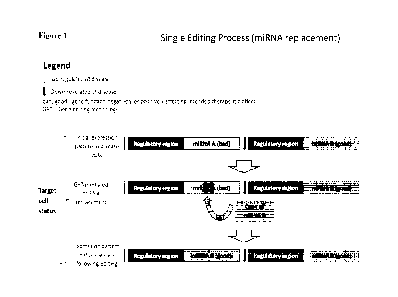
Figure 1 illustrates an embodiment of the described GET-mediated method in
which a
single editing event is used to insert a "good" miRNA which is usually poorly
expressed or
non-expressed and which is desired to be highly expressed into the
transcriptionally active
locus of a "bad" miRNA which expression is to be abolished. The outcome of
this editing
event is the expression of the "good" miRNA in two loci, under two regulatory
regions: the
original locus where its expression is low to none and the highly
transcriptionally active locus
of the "bad- miRNA where its expression is high and follows the pattern
typical of the "bad-
miRNA. By the same editing event, the "bad" miRNA expression is shut down.
Figure 2 illustrates an alternative embodiment of the single editing event
pictured in
Figure 1, in which the "bad" sequence to be disrupted is of a protein-encoding
gene
(exemplified in the figure as an immune checkpoint gene sequence). The outcome
of this
editing event is the expression of the "good" miRNA in two loci, under two
regulatory regions:
the original locus where the directed expression is low and the "bad" protein-
encoding locus
where the directed expression is high. The -bad" protein expression is shut
down.
Figure 3 illustrates the approach in which a double editing event is used to
switch the
locations and transcriptional control of two RNA encoding sequences. The
outcome of the
double editing is the expression of the "good" miRNA in one locus, which is
the "bad" miRNA
locus where the directed expression is high. The "bad" miRNA is expressed in
the "good"
miRNA locus where the directed expression is low.
Figure 4 shows the results of T-cell activation by PMA or ImmunoCultTM. A.
Flow
cytometry measurement (SSC-A versus FSC-A channels) of cell viability
following 72 hours
activation with either PMAJionomycin or ImmunoCultTM; B. Assessment of T-cell
activation
using flow cytometry analysis of CD25 staining by Anti-CD25 Antibody (human),
Phycoerythrin (PE). CD25 is a T-cell activation marker; C. Kinetics of T-cell
activation extent,
following ImmunoCultTM mediated activation was measured in another experiment.
X and Y
axis value ranges for all charts is shown.
4
CA 03200741 2023- 5- 31
WO 2022/118310
PCT/IL2021/051426
Figure 5 shows CD19-CAR-T-cell activation by NALM-6 cells. A. CD19-CAR-
harboring T-cells percentage measured by NGFR staining (NGFR- an extracellular
spacer
derived from the nerve-growth-factor receptor protein and fused to the CAR) vs
FSC-A.
Staining was performed prior to cell activation; B. Assessment of CAR-T and T-
cell activation
using now cytometry analysis of CD25 staining (a T-cell activation marker) by
Anti-CD25
Antibody (human), PE. Staining was performed 24, 48 and 72 hours after
activation of T-cells
by co-culturing at 1:1 ratio with NALM-6 cells [10,000 CD19-CAR with 10,000
NALM-6
(CD19-01, a B- cell precursor leukemia cell line which harbors CD19 surface
protein; C.
Assessment of T-cell function by measurement of NALM-6 cell -killing. 24-, 48-
and 72-hours
following co-culturing of CAR-T or T-cells with the target NALM-6 cells.
Measurement of
NALM-6 cells was performed by staining for CD19 and FACS quantification of
CD19-positive
cells.
Figure 6 shows the fold change of miRNA strands (5p and 3p) expression in
activated
T-cells. The relative amount of each of the indicated miRNA strands, mir-23a
(panel A), mir-
31 (panel B) and mir-28 (panel C) is presented, following 24, 48 and 72 hours
of activation. T-
cells were activated by ImmunolTM. The percent of activated T-cells was
determined by
staining for CD25 and was 61%, 67% and 87% after 24,48 and 72 hours of
activation,
respectively. Data are presented as 2^-AACt values: the fold change in miR-
strand expression
normalized to an endogenous reference gene (RNU6B) and relative to an
untreated (non-
activated) control.
Figure 7 shows the scheme of guide RNA (gRNA) design for the CAS9-CR1SPR-
mediated knockout of hsa-mir-31 and hsa-mir-23a. The locations of the gRNAs on
genomic
DNA relative to hsa-mir-31 and hsa-mir-23a sites, are presented (corresponding
to SEQ ID
NO: 10, nucleotide 93-190; and SEQ ID NO: 14, nucleotide 97-192). PAM -
Protospacer
adjacent motif (A 2-6-base pair DNA sequence immediately following the DNA
sequence
targeted by the Cas9 nuclease in the CRISPR bacterial adaptive immune system);
gRNA ¨
guide RNA (used interchangeably here and throughout with sgRNA-single guide
RNA) - a
single RNA molecule that contains both the custom-designed short crRNA (target
specific)
sequence fused to the scaffold tracrRNA (scaffold region) sequence required
for Cas9 protein
binding.
Figure 8 shows assessment of gRNA pairs for optimized mir-31 knockout (KO). A.
Scheme of guide RNA (gRNA) positions across the sequence of pre-mir-31
(corresponding to
nucleotide 85-190 of SEQ ID NO: 10). The expected length of the deletion
caused by each of
the gRNA pairs is indicated. Arrows define the gRNA location. Pre-mir sequence
is
5
CA 03200741 2023- 5- 31
WO 2022/118310
PCT/IL2021/051426
underlined, and PAM motifs are depicted in fonts of different shading. B.
Results of PCR
amplification with primers flanking the excision sites guided by each of the
gRNA pairs (1+3,
1+4, 2+3, 2+4). CCR5 ¨ negative control showing amplification product derived
from DNA
extracted from cells nucleofected with gRNA pair targeting an unrelated
genomic region for
CCR5. UT (untreated) - amplification product derived from DNA extracted from
non-
nucleofected cells.
Figure 9 shows the results of a T7 endonuclease 1 (T7E1) mismatch detection
assay for
assessment of mir-31 KO efficiency. A. PCR amplification products described in
Figure 5,
panel B, were subjected to T7E1 analysis. Results in the presence of T7
endonuclease 1
(+T7E1) are presented in the left panel and control reactions (-T7E1) - in the
right panel. The
gRNA pair used is indicated above each panel and the observed editing
efficiency (%) is
indicated at the bottom of the left panel. UT (untreated) - T7E1 treatment of
amplification
product derived from DNA extracted non-nucleofected cells. B. Sequence
analysis of the
edited region generated by mir-31 KO using gRNAs 2+3 (SEQ ID NO: 41).
Percentage of
editing success is depicted (100%)
Figure 10 shows the results of a T7 endonuclease 1 (T7E1) mismatch detection
assay
for assessment of mir-23a KO efficiency. Results of T7E1 mismatch detection
assay (+T7E1)
performed on DNA extracted from T-cells edited for the KO of mir-23a using
either of the
indicated gRNA pairs (1+2, 1+3, 4+2, 4+3). Amplification products derived from
DNA
extracted from non-nucleofected cells served as control (UT - untreated). A.
PCR products
generated by PCR amplification with primers flanking the excision sites guided
by each of the
gRNA pairs (1+2, 1+3, 4+2, 4+3), were subjected to T7E1 excision (+T7E1). The
observed
editing efficiency (%) is indicated at the bottom. B. As a control, the same
PCR products as in
panel A were not subjected to T7E1 excision (-T7E1). The observed editing
efficiency (%) is
indicated at the bottom. C. Sequence analysis of the edited region generated
by mir-23a KO
using gRNAs 1+3. Percentage of editing success is depicted (77%) (full
sequence corresponds
to SEQ ID NO: 42). D. Sequence analysis of the edited region generated by mir-
23a KO using
gRNAs 4+3. Percentage of editing success is depicted (91.9%) (full sequence
corresponds to
SEQ ID NO: 43).
Figure 11 shows T-cell activation following mir-31-KO. T-cells were activated
by
ImmunoCultTM (1' activation) immediately after their harvesting. The activated
(expanded) T-
cells were edited for the KO of mir-31 and then were re-activated by
ImmunoCu1tTM (2nd
activation). The assessment of T-cell activation was performed using flow
cytometry analysis
of CD25 staining by Anti-CD25 Antibody (human), PE. Top panels depict Pt
(middle panel)
6
CA 03200741 2023- 5- 31
WO 2022/118310
PCT/IL2021/051426
and 2nd (right panel) activation extent (CD25 staining) of non-edited
(UT=untreated) T-cells.
Right panel is an un-stained control. Bottom panel depict the activation (2"d
activation) extent
of T-cells following 1st activation, mir-31-editing-mediated KO with each of
the indicated
gRNA guide pairs and re-activation. sgRNA-CCR5 ¨ results of re-activation of T-
cells
nucleofected with non-mir-31-targeting gRNAs (targeting CCR5).
Figure 12 shows mir-31 and mir-23a expression following their editing-mediated
KO
(excision). The expression levels of mir-31-5p (panel A) and mir-23a-5p (panel
B) strands was
measured by RT-qPCR in T-cells following the editing-mediated KO of these
mir's and re-
activation (by ImmunoCultTM) of the edited cells. Data are presented as 2^-
AACt values: the
fold change in mir-strand expression normalized to an endogenous reference
gene (RNU6B)
and relative to the level in control T-cells edited with non-relevant gRNAs
(targeting CCR5).
UT (untreated) - mir expression in control, non-edited T-cells; sgRNA-CCR5 ¨
mir-31
expression in control T-cells edited with non-relevant gRNAs (targeting CCR5).
Figure 13 shows validation of mir-28 KI into mir-31 KO site. A. The junction
site
between the mir-31 up-stream region and the mir-28 insert DNA was amplified by
PCR at
various annealing temperatures and the optimal annealing temperature was
determined. The
same junction primers were used for PCR of template DNA extracted from control
T-cells,
which are mir-23a-K0 but were not subjected to mir-28 KI (UT=untreated). B.
ddPCR was
performed in mir-28 KI T-cells (KI) or in non-mir-28-KI T-cells (UT), with
either the junction
primers or the common primers (which amplify the region upstream to mir-31
site, common to
all DNA templates). The graph represents the number of copies (blue dots) per
1AL detected by
the ddPCR when either the common region or the junction area is amplified. To
calculate the
replacement efficiency, the copies/tiL of the Junction area are divided by the
copies/IAL of the
Common region of the respective sample. The percentage obtained (7%) indicates
the
replacement efficiency.
Figure 14 shows mir-23a and mir-28 expression in mir-23-KO/mir-28K1 T-cells.
The
expression of mir-23a and mir-28 strands was measured by RT-qPCR in T-cells
following mir-
23a KO (mir-23 KO) and in T-cells following both mir-23a KO and KT of mir-28
into the mir-
23a KO site (mir-23 KO + mir-28 KI). Both cell populations were reactivated
for 6 hours by
ImmunoCultTM, 5 days post nucleofection (editing). Data are presented as 2A-
AACt values: the
fold change in miR strand expression normalized to an endogenous reference
gene (RNU6B)
and relative to the level in reactivated T-cells edited with unrelated sgRNAs
targeting AAVSI
and co-delivered with a single stranded oligodeoxynucleotide (ssODN) repair
template.
7
CA 03200741 2023- 5- 31
WO 2022/118310
PCT/IL2021/051426
Figure 15 shows expression of genes associated with T-cell exhaustion in mir-
23-
KO/mir-28KI T-cells. The expression of the indicated genes was measured by RT-
qPCR in
edited mir-23a-KO/mir-28-KI T-cells, which were reactivated by either
irradiated PBMCs (A)
or ImmunoCultTm (B) at day 5 post nucleofection (editing) and harvested after
48 hours of
reactivation. Data are presented as 2A-AACt values: the fold change in gene
expression
normalized to an endogenous reference gene and relative to the level in
reactivated T-cells
edited with unrelated sgRNAs targeting AAVSI and co-delivered with a single
stranded
oligodeoxynucleotide (ssODN) repair template. mir-23 KO / mir-28 KI¨ T-cells
in which mir-
23a was replaced with mir-28; UT - Untreated ¨ control T-cells edited with
unrelated sgRNAs.
BRIEF DESCRIPTION OF THE DESCRIBED SEQUENCES
The nucleic and sequences provided herewith are shown using standard letter
abbreviations for nucleotide bases as defined in 37 C.F.R. 1.822. Only one
strand of each
nucleic acid sequence is shown, but the complementary strand is understood as
included by any
reference to the displayed strand. The Sequence Listing is submitted as an
ASCII text file
named 3287 2 2_seqlist ST25, created November 30, 2021, about 10.8 KB, which
is
incorporated by reference herein. In the Sequence Listing:
SEQ ID NO: 1 is the pre-mir sequence nucleotide sequence of hsa-mir-181a-1.
SEQ ID NO: 2 is the genomic region nucleotide sequence of hsa-mir-181a-1.
SEQ ID NO: 3 is the pre-mir sequence nucleotide sequence of hsa-mir-28.
SEQ ID NO: 4 is the genomic region nucleotide sequence of hsa-mir-28.
SEQ ID NO: 5 is the pre-mir sequence nucleotide sequence of hsa- miR-149.
SEQ ID NO: 6 is the genomic region nucleotide sequence of hsa- miR-149.
SEQ ID NO: 7 is the pre-mir sequence nucleotide sequence of hsa- miR-146a.
SEQ ID NO: 8 is the genomic region nucleotide sequence of hsa- miR-146a.
SEQ ID NO: 9 is the pre-mir sequence nucleotide sequence of hsa- miR-31.
SEQ ID NO: 10 is the genomic region nucleotide sequence of hsa- miR-31.
SEQ ID NO: 11 is the pre-mir sequence nucleotide sequence of hsa- miR-21.
SEQ ID NO: 12 is the genomic region nucleotide sequence of hsa- miR-21.
SEQ ID NO: 13 is the pre-mir sequence nucleotide sequence of hsa- miR-23a.
SEQ ID NO: 14 is the genomic region nucleotide sequence of hsa- miR-23a.
SEQ ID NOs: 15-18 are the nucleotide sequences of the sgRNAs targeting mir-31.
SEQ ID NOs: 19-22 are the nucleotide sequences of the sgRNAs targeting mir-23.
8
CA 03200741 2023- 5- 31
WO 2022/118310
PCT/IL2021/051426
SEQ ID NO: 23 is the nucleotide sequence of the single-stranded
oligodeoxynucleotide
(ssODN) used in insertion of miR-28 into the miR-23 locus.
SEQ ID NO: 24 is the nucleotide sequence of the single-stranded
oligodeoxynucleotide
(ssODN) used in insertion of miR-28 into the miR-31 locus.
SEQ ID NOs. 25 and 26 are forward and reverse amplification primers for miR-23
in
T7E1 assay.
SEQ ID NOs. 27 and 28 are forward and reverse amplification primers for miR-31
in
T7E1 assay.
SEQ ID NOs. 29 and 30 are forward and reverse ddPCR amplification primers for
miR-
31 (common region).
SEQ ID NOs. 31 and 32 are forward and reverse ddPCR amplification primers for
miR-
31 (junction region).
SEQ ID NOs. 33 and 34 are forward and reverse RT-qPCR amplification primers
for
LAG-3.
SEQ ID NOs. 35 and 36 are forward and reverse RT-qPCR amplification primers
for
TIM3.
SEQ ID NOs. 37 and 38 are forward and reverse RT-qPCR amplification primers
for
PD1.
SEQ ID NOs. 39 and 40 are forward and reverse RT-qPCR amplification primers
for
BLIMP-1.
SEQ ID NO: 41 is the sequencing analysis from the edited region generated by
mir-31
KO using gRNAs 2+3.
SEQ ID NO: 42 is the sequencing analysis from the edited region generated by
mir-23a
KO using gRNAs 1+3.
SEQ ID NO: 43 is the sequencing analysis from the edited region generated by
mir-23a
KO using gRNAs 4+3.
DETAILED DESCRIPTION
I. Terms
Unless otherwise explained, all technical and scientific temis used herein
have the same
meaning as commonly understood by one of ordinary skill in the art to which
this disclosure
belongs. The singular terms "a," "an," and "the" include plural referents
unless context clearly
indicates otherwise. Similarly, the word "or" is intended to include "and"
unless the context
clearly indicates otherwise. Although methods and materials similar or
equivalent to those
9
CA 03200741 2023- 5- 31
WO 2022/118310
PCT/IL2021/051426
described herein can be used in the practice or testing of this disclosure,
suitable methods and
materials are described below. The term "comprises" means "includes." The
abbreviation,
"e.g.," is derived from the Latin exempli gratia, and is used herein to
indicate a non-limiting
example. Thus, the abbreviation "e.g." is synonymous with the term "for
example."
In case of conflict, the present specification, including explanations of
terms, will
control. In addition, all the materials, methods, and examples are
illustrative and not intended
to be limiting.
Abnormal: Deviation from normal characteristics. Normal characteristics can be
found in a control, a standard for a population, etc. For instance, where the
abnormal condition
is a disease condition, such as a cancer, a few appropriate sources of normal
characteristics
might include an individual who is not suffering from the disease, a non-
cancerous tissue
sample, or a population of immune or immune progenitor cells that have not
been exposed to
the disease microenvironment, such as within a tumor or within or around the
tumor stroma.
Adoptive cell transfer (ACT): a therapeutic method involving transfer of cells
with a
therapeutic activity into a subject after in vitro modification. In a
particular embodiment, the
cells used in ACT originate with the subject to be treated, are removed from
the subject,
modified ex vivo, expanded, and then returned (administered) to the subject.
In a particular
embodiment. ACT methods involve the modification of specific T-cells (either
autologous or
allogeneic) for enhanced targeting of tumor-specific antigen. The three ACT
types used for
cancer immunotherapy include tumor-infiltrating lymphocytes (TILs), T-cell
receptor (TCR)
T-cells, and chimeric antigen receptor (CAR)-T-cells, all of which can be
modified according
to the methods described herein.
Altered expression: Expression of a biological molecule (for example, mRNA,
miRNA, or protein) in a subject or biological sample from a subject that
deviates from
expression of the same biological molecule in a normal or control subject.
Altered expression
of a biological molecule may be associated with a disease, such as the altered
expression of
miR-23 in T-cells in a tumor environment. Expression may be altered in such a
manner as to be
increased or decreased. The directed alteration in expression of an RNA or
protein may be
associated with therapeutic benefits. In a particular embodiment of the
described methods, the
expression of a miRNA that is normally down-regulated in T-cells e.g., after
their activation by
tumor antigens (leading to reduced anti-tumor responses) is increased
following this miRNA
placement into the genetic locus of a miRNA or a protein-coding gene that are
normally up-
regulated in T-cells e.g., after their activation by tumor antigens (also
leading to reduced anti-
tumor responses).
CA 03200741 2023- 5- 31
WO 2022/118310
PCT/IL2021/051426
Amplification: When used in reference to a nucleic acid, any technique that
increases
the number of copies of a nucleic acid molecule in a sample or specimen.
Animal: Living multi-cellular vertebrate organisms, a category that includes
for
example, mammals and birds. The term mammal includes both human and non-human
mammals. Similarly, the term subject includes both human and veterinary
subjects, for
example, humans, non-human primates, dogs, cats, horses, and cows. The
population of cells
for use in the current methods can be a sample taken from or derived from a
sample taken from
any animal.
Biological Sample: Any sample that may be obtained directly or indirectly from
an
organism. Biological samples include a variety of fluids, tissues, and cells,
including whole
blood, plasma, serum, tears, mucus, saliva, urine, pleural fluid, spinal
fluid, gastric fluid, sweat,
semen, vaginal secretion, sputum, fluid from ulcers and/or other surface
eruptions, blisters,
abscesses, tissues, cells (such as, fibroblasts, peripheral blood mononuclear
cells, or muscle
cells), organelles (such as mitochondria), organs, and/or extracts of tissues,
cells (such as,
fibroblasts, peripheral blood mononuclear cells, or muscle cells), organelles
(such as
mitochondria), or organs. The methods described herein can utilize cells of or
derived from any
suitable biological sample, including a tumor sample. In specific embodiments,
the methods
described herein are practiced on cells derived from a blood sample, such as
peripheral blood
mononuclear cells. In other embodiments, the methods described herein are
practiced on T
cells that are derived from solid tumors removed from a subject.
Cancer: The product of neoplasia is a neoplasm (a tumor or cancer), which is
an
abnormal growth of tissue that results from excessive cell division. A tumor
that does not
metastasize is referred to as "benign." A tumor that invades the surrounding
tissue and/or can
metastasize is referred to as "malignant." Neoplasia is one example of a
proliferative disorder.
A "cancer cell" is a cell that is neoplastic, for example a cell or cell line
isolated from a tumor.
The methods described herein can be used to increase the therapeutic (i.e.,
immunological)
efficacy of an immune cell, such as a CAR T cell against a cancer, which in
particular
embodiments is a hematological tumor and in other embodiments is a solid
tumor.
Examples of hematological tumors include leukemias, including acute leukemias
(such
as acute lymphocytic leukemia, acute myelocytic leukemia, acute myelogenous
leukemia and
myeloblastic, promyelocytic, myelomonocy tic, monocytic and erythroleukemia),
chronic
leukemias (such as chronic myelocytic (granulocytic) leukemia, chronic
myelogenous
leukemia, and chronic lymphocytic leukemia), polycythemia vera, lymphoma,
Hodgkin's
disease, non-Hodgkin's lymphoma (indolent and high grade forms), multiple
myeloma,
11
CA 03200741 2023- 5- 31
WO 2022/118310
PCT/IL2021/051426
Waldenstrom's macroglobulinemia, heavy chain disease, myelodysplastic
syndrome, hairy cell
leukemia and myelodysplasia.
Examples of solid tumors, such as sarcomas and carcinomas, include
fibrosarcoma,
myxosarcoma, liposarcoma, chondrosarcoma, osteogenic sarcoma, and other
sarcomas,
synovioma, mesothelioma, Ewing's tumor, leionnyosarcoma, rhabdomyosarcoma,
colon
carcinoma, lymphoid malignancy, pancreatic cancer, breast cancer, lung cancers
(such as small
cell lung carcinoma and non-small cell lung carcinoma), ovarian cancer,
prostate cancer,
hepatocellular carcinoma, squamous cell carcinoma, basal cell carcinoma,
adenocarcinoma,
sweat gland carcinoma, medullary thyroid carcinoma, papillary thyroid
carcinoma,
pheochromocytomas sebaceous gland carcinoma, papillary carcinoma, papillary
adenocarcinomas, medullary carcinoma, bronchogenic carcinoma, renal cell
carcinoma,
hepatoma, bile duct carcinoma, choriocarcinoma, Wilms' tumor, cervical cancer,
testicular
tumor, seminoma, bladder carcinoma, melanoma, and CNS tumors (such as a
glioma,
astrocytoma, medulloblastoma, craniopharyogioma, ependymoma, pinealoma,
hemangioblastoma, acoustic neuroma, oligodendroglioma, menangioma,
neuroblastoma and
retinoblastoma).
Chemotherapeutic agent: An agent with therapeutic usefulness in the treatment
of
diseases characterized by abnormal cell growth or hyperplasia. Such diseases
include cancer,
autoimmune disease as well as diseases characterized by hyperplastic growth
such as psoriasis.
One of skill in the art can readily identify a chemotherapeutic agent (for
instance, see Slapak
and Kufe, Principles of Cancer Therapy, Chapter 86 in Harrison's Principles of
Internal
Medicine, 14th edition; Perry et al., Chemotherapy, Ch. 17 in Abeloff,
Clinical Oncology 2nd
ed., 0 2000 Churchill Livingstone, Inc; Baltzer L, Berkery R (eds): Oncology
Pocket Guide to
Chemotherapy, 2nd ed. St. Louis, Mosby-Year Book, 1995; Fischer DS, Knobf MF,
Durivage
HJ (eds): The Cancer Chemotherapy Handbook, 4th ed. St. Louis, Mosby-Year
Book, 1993).
Examples of chemotherapeutic agents include ICL-inducing agents, such as
melphalan
(Alkeranlm), cyclophosphamide (Cytoxanlm), cisplatin (Platinollm) and busulfan
(Busilvexlm,
MyleranTm). As used herein a chemotherapeutic agent is any agent with
therapeutic usefulness
in the treatment of cancer, including biological agents such as antibodies,
peptides, and nucleic
acids. In particular embodiments of the described methods, the modified cells
for cellular
therapy can be used as part of a therapeutic regimen that includes one or more
chemotherapeutic agents. Such agents can be administered before, currently
with, of following
administration of the modified cells.
12
CA 03200741 2023- 5- 31
WO 2022/118310
PCT/IL2021/051426
Chimeric Antigen Receptor (CAR) T Cells: T cells that have been isolated from
a
subject and modified to express a desired target receptor. CAR-T cells can be
designed to
target specific cells for immunotherapeutic clearance, such as a specific
cancer type. In a
particular embodiment, the methods described herein modify the genetic loci
and associated
expression of miRN As in CAR-'! cells.
Clustered Regularly Interspaced Short Palindromic Repeats (CRISPR): DNA loci,
originally identified in prokaryotes, that contain multiple, short, direct
repetitions of base
sequences. The prokaryotic CRISPR/Cas system has been adapted for use as a
gene editing
technology by transfecting a cell with the required elements including a Cas
nuclease gene and
specifically designed guide RNAs (gRNAs), an organism's genome can be cut and
modified at
any desired location. Methods of preparing compositions for use in genome
editing using the
CRISPR/Cas systems are described in detail in International Patent
Publications WO
2013/176772 and WO 2014/018423.
In some embodiments, one or more vectors driving expression of one or more
elements
of a CRISPR system are introduced into a target cell such that expression of
the elements of the
CRISPR system direct formation of a CRISPR complex at one or more target
sites. For using
CRISPR technology to target a specific DNA sequence, such as a miRNA described
herein, a
user can insert a short DNA fragment containing the target sequence into a
guide RNA
expression plasmid. The sgRNA expression plasmid contains the target sequence
(about 20
nucleotides), a form of the tracrRNA sequence (the scaffold) as well as a
suitable promoter and
necessary elements for proper processing in eukaryotic cells. Such vectors are
commercially
available. Many of the systems rely on custom, complementary oligos that are
annealed to form
a double stranded DNA and then cloned into the sgRNA expression plasmid. Co-
expression of
the sgRNA and the appropriate Cas enzyme from the same or separate plasmids in
transfected
cells results in a single or double strand break (depending of the activity of
the Cas enzyme) at
the desired target site.
Control: Standards appropriate for comparison to a sample, for example a cell
or
population of cells that have not undergone the microRNA editing process
described herein.
Efficacy: Refers to the ability of agent, including a cell, such as an immune
cell, to
elicit or provide a desired therapeutic effect. Efficacy also refers to the
strength or
effectiveness of a therapeutic agent, including the modified cells described
herein. As used
herein, "enhancing efficacy" means to increase the therapeutic action of a
modified cell. For
example, when the agent is a modified cell, "enhancing efficacy" can mean
increasing the
ability of the agent to kill target cells, such as tumor cells. Enhanced
efficacy does not require
13
CA 03200741 2023- 5- 31
WO 2022/118310
PCT/IL2021/051426
actual demonstration of target cytotoxicity. Rather, as described herein, the
efficacy of the
described modified cells is enhanced as a result of changes in gene expression
patterns that can
be predicted to increase cytotoxic effect.
Effective amount of a compound: A quantity of compound sufficient to achieve a
desired effect in a subject being treated. An effective amount of a compound
can be
administered in a single dose, or in several doses, for example daily, during
a course of
treatment. However, the effective amount of the compound will be dependent on
the
compound applied, the subject being treated, the severity and type of the
affliction, and the
manner of administration of the compound.
Encode: A polynucleotide is said to "encode" a polypeptide if, in its native
state or
when manipulated by methods well known to those skilled in the art, it can be
transcribed
and/or translated to produce the mRNA for and/or the polypeptide or a fragment
thereof. The
anti-sense strand is the complement of such a nucleic acid, and the encoding
sequence can be
deduced therefrom. mRNA that is translated to produce protein is "coding- RNA.
Non-coding
RNA, such as the miRNA described herein, are not translated into protein,
however the
expression or inhibition of such miRNA will result in downstream effects on
protein
expression.
Expand: refers to a process by which the number or amount of cells in a cell
culture is
increased due to cell division. Similarly, the terms "expansion" or "expanded"
refers to this
process. The terms "proliferate," "proliferation" or "proliferated" may be
used interchangeably
with the words "expand," "expansion", or "expanded." The cell culture
techniques for use in
the described methods are those common to the art, unless otherwise specified.
Expression Control Sequences: Nucleic acid sequences that regulate the
expression
of a heterologous nucleic acid sequence to which it is operatively linked, for
example the
expression of a microRNA. Expression control sequences are operatively linked
to a nucleic
acid sequence when the expression control sequences control and regulate the
transcription
and, as appropriate, translation of the nucleic acid sequence. Thus,
expression control
sequences can include appropriate promoters, enhancers, transcription
terminators, a start
codon (ATG) in front of a protein-encoding gene, splicing signal for introns,
maintenance of
the correct reading frame of that gene to permit proper translation of mRNA,
and stop codons.
The term "control sequences" is intended to include, at a minimum, components
whose
presence can influence expression, and can also include additional components
whose presence
is advantageous, for example, leader sequences and fusion partner sequences.
Expression
control sequences can include a promoter. A promoter is a minimal sequence
sufficient to
14
CA 03200741 2023- 5- 31
WO 2022/118310
PCT/IL2021/051426
direct transcription. Also included are those promoter elements which are
sufficient to render
promoter-dependent gene expression controllable for cell-type specific, tissue-
specific, or
inducible by external signals or agents; such elements may be located in the
5' or 3' regions of
the gene. In a particular embodiment, the miRNAs of the described methods arc
placed under
the transcriptional control of expression control sequences different from
their normal genetic
locus. In a particular embodiment, the expression of miR-28 is placed under
the control of the
miR-23 expression control sequences.
Gene/Genome/Genomic Editing Technology (GET): Genetic engineering
methodology by which a targeted nucleic acid sequence (i.e., at a specific
location) is deleted,
modified, replaced, or inserted. The methods described herein utilize any GET
to insert a
specified miRNA-coding sequence into a non-native genetic locus so as to be
under the
transcriptional control of that locus. Particular non-limiting examples of GET
include
CRISPR/Cas-associated methods, zinc finger nucleases, TALENs, and use of
triplex forming
molecules such as triplex forming oligonucleotides, peptide nucleic acids, and
tail clamp
peptide nucleic acids, all of which are known in the art.
Heterologous: A type of sequence that is not normally (i.e., in the wild-type
sequence)
found adjacent to a second sequence. In one embodiment, the sequence is from a
different
genetic source, such as a virus or organism, than the second sequence.
Immune response: A response of a cell of the immune system, such as a B cell,
T cell,
or monocyte, to a stimulus. In one embodiment, the response is specific for a
particular
antigen (an -antigen-specific response"), such as an antigen from a leukemia.
In one
embodiment, an immune response is a T cell response, such as a CD4+ response
or a CD8+
(cytotoxic) response. In another embodiment, the response is a B cell
response, and results in
the production of specific antibodies.
Immunotherapy: A method of evoking an immune response against or in response
to
the presence of target antigens, such as are expressed on the surface of a
tumor cell.
Immunotherapy based on cell-mediated immune responses involves generating or
providing a
cell-mediated response to cells that produce particular antigenic
determinants. ACT
immunotherapies, such as CAT T cell-mediated therapy, are also referred to as
immunooncology.
Isolated: An "isolated" biological component (such as a nucleic acid, protein,
cell (or
plurality/population of cells), tissue, or organelle) has been substantially
separated or purified
away from other biological components of the organism in which the component
naturally
occurs for example other tissues, cells, other chromosomal and extra-
chromosomal DNA and
CA 03200741 2023- 5- 31
WO 2022/118310
PCT/IL2021/051426
RNA, proteins and organelles. Nucleic acids and proteins that have been
"isolated- include
nucleic acids and proteins purified by standard purification methods. The term
also embraces
nucleic acids and proteins prepared by recombinant expression in a host cell
as well as
chemically synthesized nucleic acids.
Locus: Genetic location of a gene or particular sequence of DNA on a
chromosomal or
extrachromosomal sequence. A locus can be described with greater or lesser
precision, such
that it can be used in some embodiments to describe the location of a
particular nucleotide
sequence, and in other embodiments to describe a particular coding (or non-
coding) sequence,
as well as its associated expression control sequences. As described herein,
placement of a
miRNA-encoding sequence at a new genetic locus will place its transcription
under the control
of the new locus.
MicroRNA (miRNA): Short, single-stranded RNA molecule of 18-24 nucleotides
long. Endogenously produced in cells from longer precursor molecules of
transcribed non-
coding RNA, miRNAs can inhibit translation, or can direct cleavage of target
mRNAs through
complementary or near-complementary hybridization to a target nucleic acid.
Oligonucleotide: A plurality of joined nucleotides joined by native
phosphodiester
bonds, between about 6 and about 300 nucleotides in length. An oligonucleotide
analog refers
to moieties that function similarly to oligonucleotides but have non-naturally
occurring
portions. For example, oligonucleotide analogs can contain non-naturally
occurring portions,
such as altered sugar moieties or inter-sugar linkages, such as a
phosphorothioatc
oligodeoxynucleotide. Functional analogs of naturally occurring
polynucleotides can bind to
RNA or DNA, and include peptide nucleic acid (PNA) molecules. Particular
oligonucleotides
and oligonucleotide analogs can include linear sequences up to about 200
nucleotides in length,
for example a sequence (such as DNA or RNA) that is at least 6 bases, for
example at least 8,
10, 15, 20, 25, 30, 35, 40, 45, 50, 100 or even 200 bases long, or from about
6 to about 50
bases, for example about 10-25 bases, such as 12, 15 or 20 bases.
Operably linked: A first nucleic acid sequence is operably linked with a
second
nucleic acid sequence when the first nucleic acid sequence is placed in a
functional relationship
with the second nucleic acid sequence. For instance, a promoter is operably
linked to a coding
sequence if the promoter affects the transcription or expression of the coding
sequence.
Generally, operably linked DNA sequences are contiguous and, where necessary
to join two
protein-coding regions, in the same reading frame. In a particular embodiment
of the described
methods the genetic location of a miRNA is changed so that the "moved" miRNA
is operably
linked to expression control sequences different from its original genetic
locus.
16
CA 03200741 2023- 5- 31
WO 2022/118310
PCT/IL2021/051426
Preventing or treating a disease: Preventing a disease refers to inhibiting
the full
development of a disease, for example inhibiting the development of myocardial
infarction in a
person who has coronary artery disease or inhibiting the progression or
metastasis of a tumor in
a subject with a neoplasm. Treatment refers to a therapeutic intervention that
ameliorates a sign
or symptom of a disease or pathological condition after it has begun to
develop.
Transcription activator-like effector nucleases (TALENs): GET methodology
using
a nucleic acid construct or constructs encoding a transcription activator-like
effector nuclease
(TALEN). TALENs have an overall architecture similar to that of ZFNs, with the
main
difference that the DNA-binding domain comes from TAL effector proteins.
Methods of
engineering TAL to bind to specific nucleic acids are described in Cermak, et
al, Nucl. Acids
Res. 1-11 (2011). U.S. Published Application No. 2011/0145940 describes TAL
effectors and
methods of using them to modify DNA, as well as general design principles for
TALE binding
domains.
Target sequence: A target sequence is a portion of ssDNA, dsDNA, or RNA that
can
be hybridized by an oligonucleotide or oligonucleotide analog (e.g., a
morpholino), of
sufficient complementarity to allow for hybridization. The GET methodology for
use in the
described methods utilize oligonucleotides that recognize specific target
sequences to direct the
removal and/or insertion of the described coding RNA or non-coding miRNA
sequences.
Zn finger Nucleases (ZFN): GET technologies take advantage of cellular
machinery
that produce double stranded breaks in DNA. In a particular embodiment, the
GET uses a ZFN
system by which a designed ZFN is expressed from an encoding nucleic acid
plasmid, and
which is able to specifically target a desired sequence Tools for designing
ZFN systems for
gene editing are available online at the Zinc Finger Consortium
(zincfingers.org).
II. Brief overview of several embodiments
Described herein is a method for modifying an isolated cell for cell therapy,
by
providing a plurality of isolated cells in culture; and inserting in the
plurality of cells, at a first
genetic locus comprising a first RNA-encoding sequence, a second RNA-encoding
sequence,
thereby operably-linking the second RNA-encoding sequence to the
transcriptional regulatory
sequence of the first genetic locus and disrupting the first genetic locus. In
the described
method, inserting the second RNA-encoding sequence at the first genetic locus
abolishes the
expression of the first RNA-encoding sequence, either by disrupting or
replacing the sequence
(or subsequent to a prior step in which the first sequence is removed), and
wherein under
conditions sufficient to initiate transcription at the first genetic locus,
expression of the second
17
CA 03200741 2023- 5- 31
WO 2022/118310
PCT/IL2021/051426
RNA-encoding sequence at the first genetic locus is induced whereas the
expression of the first
genetic locus, is eliminated. In the described methods, the described
disruption/insertion is
carried out by a Gene Editing Technology (GET) selected from available GET
methods
including but not limited to application of transcription activator-like
effector nucleases
(TALEN), clustered regularly interspaced short palindromic repeat (CRISPR)¨Cas-
associated
nucleases, and zinc-finger nucleases (ZFN) or any other similar technique for
modifying a
genetic sequence.
In a particular embodiment, the method includes inserting at a second genetic
locus
comprising the second RNA-encoding sequence, the first RNA-encoding sequence,
in addition
to the insertion of the second RNA-encoding sequence into the locus of the
first RNA-encoding
sequence, thereby operably-linking the first RNA-encoding sequence to the
transcriptional
regulatory sequence of the second genetic locus, and wherein under conditions
sufficient to
inhibit transcription at the second genetic locus, expression of the first RNA-
encoding
sequence at the second genetic locus is inhibited.
Both the single editing embodiment and the double editing embodiment involve
the
switching the position of RNA-encoding sequences, and are accordingly also
referred to herein
as the "castling" method.
The first RNA-encoding sequence of the described methods can in some
embodiments
be a non-protein encoding sequence, such as a naiRNA-encoding sequence. In
other
embodiments, the first RNA-encoding sequence can be a protein-encoding
sequence. The
second RNA-encoding sequence of the described methods can be a non-protein
encoding
sequence, such as a miRNA-encoding sequence.
In particular embodiments, the isolated cells are mesenchymal stem cells or
lineage
thereof (including osteoblasts (bone cells), chondrocytes (cartilage cells),
myocytes (muscle
cells), adipocytes (fat cells which give rise to marrow adipose tissue and
hepatocyte-like cells),
or pluripotent hematopoietic stem cells or lineage thereof, such as
erythrocytes, macrophages,
natural killer cells, T lymphocytes. B lymphocytes, or mast cells. In still
further embodiments,
the isolated cells are natural T cells, induced T regulatory cells, cytotoxic
T cells, natural killer
(NK)-T cells, T helper cells, or chimeric antigen receptor (CAR)-T-cells.
In particular embodiments, the isolated cells are parenchymal cells, such as
hepatocytes
or endocrine cells such as pancreatic b-cells.
It will be appreciated that in addition to the noted cell types, any type of
pluripotent cell
could be modified as described herein. Further, in particular embodiments, the
cells for use in a
specific subject are autologous, while in other embodiments, the cells are
allogenic.
18
CA 03200741 2023- 5- 31
WO 2022/118310
PCT/IL2021/051426
Also described herein is a method for enhancing therapeutic efficacy of a
lymphocyte
or a myeloid cell for adoptive cell transfer therapy, by providing a plurality
of isolated
lymphocytes in culture; and inserting, into the isolated lymphocytes, at an
actively transcribed
genetic locus comprising a protein encoding gene such as an inhibitory immune
checkpoint
gene, or encoding a non-protein-coding RNA such as an miRNA associated with
reduced
efficiency of immunotherapy ("bad" genes), a RNA-encoding sequence such as an
miRNA
encoding sequence whose high expression is expected to increase efficiency of
immunotherapy
("good" gene), thereby abolishing expression of the "bad" genes and enhancing
expression of a
"good" gene , wherein the insertion is carried out by a Gene Editing
Technology selected from
available methods including transcription activator-like effector nucleases
(TALEN), clustered
regularly interspaced short palindromic repeat (CRISPR)¨Cas-associated
nucleases, and zinc-
finger nucleases (ZFN).
In particular embodiments, the protein encoding gene is an inhibitory immune
checkpoint gene such as but not limited to CTLA-4 (cytotoxic T lymphocyte
associated protein
4); and/or PD-1 (programmed cell death protein 1); and/or LAG-3 (Lymphocyte
activation
gene 3), TIM3 (T cell immunoglobulin and mucin domain- containing protein 3)
and the like.
III. Gene editing technology (GET)-mediated RNA engineering for enhancing
cellular
therapy
Described herein is the application of GET-mediated genomic engineering to
modify
RNA expression, such as miRNA and/or mRNA expression to optimize and enhance
cell
therapies.
In a general embodiment of the described method, GET-mediated genomic
engineering
is utilized to simultaneously modify expression of two or more target genes in
isolated cells for
use in cell therapies, such as but not limited to ACT or cell transplantation
therapies. Using
GET, a non-coding RNA (such as miRNA) encoding sequence of interest which
under-
expression negatively influences cell therapy performance is inserted into a
transcriptionally
active genetic locus ("first genetic locus") different from that of the
selected sequence ("second
RNA-encoding sequence") and which high expression also negatively influences
performance
of the same type of cell therapy. Such insertion abolishes the expression of
an endogenous
gene (coding or non-coding) at the first genetic locus while operably linking
the expression of
the second RNA-encoding sequence to the transcriptional control sequences of
the first genetic
locus. Accordingly, under conditions sufficient to initiate transcription at
the first genetic locus,
the second RNA-encoding sequence will be expressed.
19
CA 03200741 2023- 5- 31
WO 2022/118310
PCT/IL2021/051426
The single-editing embodiment described above is illustrated in Figure 1, in
which the
actively expressed miRNA-encoding sequence at the first genetic locus is
labeled a "bad"
miRNA (as an illustrative "bad" gene); and the under-expressed miRNA-encoding
sequence at
the second genetic locus is labeled a "good" miRNA (as an illustrative "good"
gene). As
shown in Figure 1, GET-mediated gene editing is used to insert a copy of the
"good' miRNA
at the first genetic locus to disrupt or replace the encoding sequence of the
"bad" miRNA. Such
replacement results in the "good" miRNA's acquisition of the "bad" miRNA's
expression
pattern, which is manifested by its up-regulation under conditions (such as a
disease state) that
up-regulate the "bad" miRNA, and simultaneously abolishes expression of the
"bad" miRNA
(the expression of which limits cell therapy functionality). The "good" miRNA
is also
expressed at its original locus where its expression remains low. Thus, the
final outcome of the
editing approach will be double ¨ abolishment of "bad" miRNA expression while
activating
the "good" miRNA expression, both of which lead to improvement of cell therapy
efficacy.
In a further general embodiment of the described methods, which is illustrated
in Figure
3, two GET-mediated editing processes are carried out, such that the copy of
the second RNA-
encoding sequence ("good miRNA" in Figure 3) is expressed under regulatory
control of the
first genetic locus, and the copy of the first RNA-encoding sequence ("bad
miRNA" in Figure
3) is expressed under the regulatory control of the second genetic locus.
Under particular
environmental conditions, termed a "disease state" in the figure, expression
of the second
RNA-encoding sequence will be induced or enhanced, while expression of the
first RNA-
encoding sequence will be inhibited or repressed to a basal level. Given the
many varied and
interconnected regulatory roles played by miRNAs, such maintenance of a "bad
miRNA" at a
basal level of expression could be beneficial (as opposed to completely
abolishing its
expression).
Similar to Figure 1, Figure 2 illustrates the GET-mediated disruption of an
endogenous
gene at the first genetic locus, labeled a "bad" protein-coding gene, by a
"good" miRNA. Such
a replacement results in increased expression of the "good- miRNA and the
knockdown of
expression of the "bad" protein-coding mRNA, both conferring better cell
therapy efficacy.
The "good" miRNA is also expressed at its original locus where the directed
expression
remains low. In particular embodiments, the -bad" gene that reduces the anti-
tumor efficacy of
e.g., CAR-T cells can be selected from a group of inhibitory immune checkpoint
genes such as
but not limited to PD-1 or CTLA-4. Accordingly, following the editing process
described in
Figure 2. that activity, which can be up-regulated in T-cells in response to
the tumor
environment, will be decreased or even abolished.
CA 03200741 2023- 5- 31
WO 2022/118310
PCT/IL2021/051426
The Gene Editing Technology that can be used in the methods described herein
is
selected from. but not limited to transcription activator-like effector
nucleases (TALEN),
clustered regularly interspaced short palindromic repeat (CRISPR)¨Cas-
associated nucleases,
and zinc-finger nucleases (ZFN) and any other available gene editing method
known to the art.
miRNAs
Micro RNAs (miRNAs) are a group of small non-coding RNAs that negatively
regulate
gene expression via controlling mRNA degradation and/or translation inhibition
through
binding to partially complementary sites primarily located in the 3'-
untranslated regions of
target genes. miRNAs are estimated to regulate the translation of more than
60% of the human
protein-coding genes and thereby are involved in regulation of multiple
biological processes,
including cell cycle control, cell growth and differentiation, apoptosis,
embryo development
and the like. miRNAs are potent cellular modulators due to their ability to
target multiple
molecules within a particular pathway or diverse proteins in converging
pathways or biological
processes. Thus, miRNAs can potently regulate biological networks by
cumulatively or
cooperatively inhibiting their different components. Or alternatively, they
may fine-tune
particular signaling pathways by targeting positive and negative regulatory
components. This
implies that aberrant miRNA expression should proportionately affect those
critical processes,
and as a result, lead to various pathological and occasionally malignant
outcomes. Indeed.
miRNAs have been identified as crucial players in human disease development,
progression,
and treatment response. (6-9).
For example, altered expression of certain miRNAs (some ¨ upregulated, some ¨
downregulated) was reported in several human diseases including schizophrenia,
neurodegenerative diseases like Parkinson's disease and Alzheimer disease,
immune related
disease, fibrotic and cardiac disorders. However, of the many identified miRNA-
disease
associations, the involvement of miRNAs in cancer diseases is the most
prevalent. Differences
in the miRNA's expression between tumors and normal tissues have been
identified in
lymphoma, breast cancer, lung cancer, papillary thyroid carcinoma,
glioblastoma,
hepatocellular carcinoma, pancreatic tumors, pituitary adenomas, cervical
cancer, brain tumors,
prostate cancer, kidney and bladder cancers, and colorectal cancers. These
observations are
supported by the findings that many of the miRNAs are encoded by genomic
regions linked to
cancer and strengthen the notion that miRNAs can act as oncogenes or
conversely, as tumor
suppressors with key functions in tumorigenesis (7, 8, 10-12).
21
CA 03200741 2023- 5- 31
WO 2022/118310
PCT/IL2021/051426
miRNA genes are located in intronic, exonic, or untranslated genomic regions.
Some
miRNAs are clustered in polycistronic transcripts thus allowing coordinated
regulation of their
expression, while others are expressed in a tissue-specific and developmental
stage-specific
manner (6). From their gene loci, miRNAs are initially transcribed by RNA
polymerase II as
long primary transcripts, which are processed into approximately 70-nucleotide
precursors by
the RNAse III enzyme Drosha in the nucleus. The precursor-miRNAs are then
exported into
the cytoplasm by Ran GTPase and Exportin 5 and further processed into an
imperfect 22-mer
miRNA duplex by the Dicer protein complex (13).
Several mechanisms that control microRNA expression may be altered in human
diseases. These include epigenetic changes such as promoter CpG island
hypermethylation,
RNA modification, and histone modifications or genetic alterations such as
mutations,
amplifications or deletions, which can affect the production of the primary
miRNA transcript,
their biogenesis process and/or interactions with mRNA targets (12).
In light of their crucial role in human diseases, miRNAs are attractive
targets for
therapeutic interventions. Molecular approaches that have been pursued to
reverse epigenetic /
genetic silencing of miRNA include direct administration of synthetic miRNA
mimics or
miRNAs encoded in expression vectors or reversion of epigenetic silencing of
miRNA by
demethylating agents such as decitabine or 5-azacytidine. Other molecular
approaches have
been employed to block miRNA functions, such as antisense miRNA-specific
oligonucleotides
(anti-miRs, or antagomirs), tiny anti-miR (targeting specific seed regions of
the whole miRNA
families), miRNA sponges, blockmirs, small molecules targeting miRNAs (SMIRs)
and
blocking extracellular miRNAs in exosomes (14). However, the current miRNA-
based
synthetic oligonucleotide therapeutics still need to overcome problems
associated with
synthetic oligonucleotide drugs, such as degradation by nucleases, renal
clearance, failure to
cross the capillary endothelium, ineffective endocytosis by target cells,
ineffective endosome
release, release of formulated RNA-based drugs from the blood to the target
tissue through the
capillary endothelium and induction of host immune response. When delivered by
expression
vectors, the dangers and drawbacks are those typical for gene therapy:
insertion into silent
genomic regions hampering the transgene expression or disruption/activation of
the host genes
in the vicinity of the integration site leading to potential safety sequels.
Enhancement of cellular therapies
The methods described herein utilize GET methodology to modify cells ex vivo
for use
in cell therapies, including ACT therapies, such as but not limited to
anticancer T cell mediated
22
CA 03200741 2023- 5- 31
WO 2022/118310
PCT/IL2021/051426
immunotherapies. In a particular embodiment, the isolated cells can be
mesenchymal stem
cells. In another embodiment, the isolated cells for use in the described
methods can be
pluripotent hematopoietic stem cells, or a lineage thereof with some
multipotency, or a further
lineage thereof that is unipotent. In particular embodiments such
hematopoietic "lineage cells"
can be erythrocytes, macrophages, natural killer cells, T lymphocytes, B
lymphocytes, or mast
cells. In other particular embodiments, the T lymphocytes can be natural T
cells, induced T
regulatory (Treg) cells, cytotoxic T cells, natural killer-T (NKT)cells, T
helper cells, or
chimeric antigen receptor (CAR)-T-cells.
In certain embodiments, isolated cells for use in the described methods are
parenchymal
cells, such as hepatocytes.
In a particular embodiment, the described methods are employed to modulate
expression of selected miRNAs in T-cell therapies, such as those using CAR-T
cells. Upon
activation, T-cells undergo global gene and miRNA expression remodeling to
support cell
growth, proliferation, and effector functions. However, alterations in the
nature, duration and
setting of antigen stimulations can result in altered miRNA and gene
expression patterns and
subsequently in dysfunctional T-cell states such as anergy, tolerance and/or
exhaustion. As
demonstrated below, using the GET-mediated miRNA engineering described herein,
it is
possible to alter miRNA expression patterns, and by extension alter the
expression patterns of
genes regulated by the miRNAs, to overcome the decreased therapeutic efficacy
of CAR-T
cells.
Additional target T-cells for the use of miRNA engineering in ACT-based
therapy, are
T regulatory lymphocytes (Tregs). Tregs cells are crucial for the maintenance
of immunological tolerance due to their role in shutting down T-cell-mediated
immunity
toward the end of an immune reaction and in the suppression of autoreactive T-
cells. These
cells occur at lower frequency in Systemic lupus erythematosus (SLE), a
chronic inflammatory
autoimmune disorder, which leads to immune dysfunction (15). Using the GET-
mediated
miRNA engineering described herein it will be possible to expand Tregs
isolated from SLE
patients and enhance their autoimmune suppression activity.
The methods described herein apply GET¨mediated miRNA engineering to
simultaneously downregulate genes, such as miRNAs, with negative influence on
T-cell
functions while upregulating those with positive influence.
The described castling method can enable the simultaneous up-regulation of a
desired
"good" miRNA and down-regulation of an undesired "bad" miRNA by replacing the
up-
regulated, harmful miRNA with a copy of the down-regulated one, thus ensuring
a high
23
CA 03200741 2023- 5- 31
WO 2022/118310
PCT/IL2021/051426
expression level of the desired miRNA and shutting down the harmful miRNA (see
Figure 1
for an exemplary embodiment). Similarly, a reciprocal exchange may be
implemented in order
to preserve low levels of the "bad" miRNA. In such methods, in parallel to the
replacement of
the harmful miRNA by the desired one, the desired miRNA is replaced by the
harmful one (see
Figure 3 for an exemplary embodiment).
In yet a further embodiment, a desired "good" miRNA is inserted into the
coding region
of an undesired "bad" gene in T cells ex vivo (e.g., an inhibitory immune
checkpoint gene such
as PD-1 or CTLA-4) by "knock-in" editing, thus simultaneously eliminating the
suppressive
effect of the knocked-down gene and gaining a miRNA-related positive effect.
This
embodiment is illustrated in Figure 2. In the case of miRNA knock-in to the
coding region of a
gene, one should ensure the co-insertion of the appropriate signaling
sequences such as Drosha
processing site and a transcription termination signal (16, 17).
As noted, the described methods can be used in particular embodiments to
enhance the
efficacy of ACT therapy by replacing the expression of one or more miRNA-
encoding
sequences associated with reduced therapeutic efficacy with one or more miRNA
encoding
sequences associated with increased or normal therapeutic efficacy. This
genetic "switching",
also referred to herein as "castling", can be implemented at any ex vivo stage
of the ACT
process. In particular embodiments, the ACT procedure is modified such that an
isolated T-
cell population is genetically edited as described herein [e.g., tumor-
infiltrating lymphocytes
(TILs)] or prior to further modification (e.g., engineering to express
chimeric antigens), or
following other editing-mediated modifications (e.g., engineering to express
chimeric
antigens). In other embodiments, a population of lymphocytes that are "ready"
for
administration to a subject in need thereof are edited according to the
current method,
reexpanded, and then provided to a patient.
Engineering miRNA expression in T cells
In a particular embodiment, the described methods can be employed to alleviate
T-cell
exhaustion and/or anergy, extend their persistence, and/or improve their
efficiency in solid
tumors eradication.
In one embodiment, the described methods can be employed with currently used
strategies and combinations with CAR-T cells, such as the combination of CAR-T-
cells
therapy with checkpoint blockade therapy, which are known to be able to
decrease T-cell
exhaustion in preclinical and clinical studies.
24
CA 03200741 2023- 5- 31
WO 2022/118310
PCT/IL2021/051426
The current checkpoint blockade approaches include using antibodies against
inhibitory
immune checkpoint targets in combination with CAR-T-cells, production and
secretion of
these antibodies by the T-cells themselves, treatment of CAR-T cells ex vivo
with immune
checkpoint gene blocking synthetic oligonucleotides or alternatively use of a
GET-medicated
knockdown of immune checkpoint gene(s) in the CAR-T cells (5).
The described methods of GET-mediated modification of the T-cell genome will
upregulate expression of specific miRNAs while inhibiting expression of other
undesired
miRNAs or other non-coding RNAs or proteins.
The following sections describe exemplary miRNAs, the expression of which can
be
altered using the described methods to increase T cell therapeutic efficacy.
However, this
listing is merely illustrative; and one of skill will appreciate that any
miRNA that is identified
as similarly affecting T cell efficacy can be used. Similarly, although the
illustrative "bad"
genes listed below are miRNA, any nucleic acid encoding a coding or non-coding
RNA that is
detrimental to T cell efficacy can be subject to disruption or replacement
using the described
methods.
"Good" miRNAs with a positive effect on T cell therapeutic efficacy
Expression of these miRNAs is to be increased by editing-mediated insertion
into actively
transcribed "bad" miRNA/coding gene regions
miR-181a
In a particular embodiment, improvement of adoptively- transferred- tumor-
specific T-
cells, modulates TCR signaling thresholds to enhance T-cell activation and
function. Several
miRNAs such as miR-181a have been found to influence TCR signaling by
targeting key
inhibitory phosphatases.
In a particular embodiment, miR-181a is upregulated to simultaneously target
multiple
serine/threonine and tyrosine phosphatases. It can also enhance LCK (LCK proto-
oncogene,
Src family tyrosine kinase) and ERK (MAPK1- mitogen-activated protein kinase
1) activity by
inhibiting DUSP5 (dual specificity phosphatase 5), DUSP6 (dual specificity
phosphatase 5),
PTPN11 (protein tyrosine phosphatase non-receptor type 11), and PTPN22
(protein tyrosine
phosphatase non-receptor type 22). This activity governs the central and
peripheral T-cell
tolerance. Moreover, over-expression of miR-181a in T-cells increased TCR
sensitivity to
cognate antigen and enhanced intracellular calcium flux upon TCR triggering,
resulting in
more pronounced release of 1L-2, which among other activities, promotes the
differentiation of
CA 03200741 2023- 5- 31
WO 2022/118310
PCT/IL2021/051426
T-cells into effector T-cells and into memory T-cells. Thus, T cells
engineered to have
enhanced expression of miR-181a are expected to have increase activation
properties (45, 46).
The hsa-mir-181a-1 sequence is publicly available as follows. All microRNA
sequences noted herein can be found online at mirbase.org.
hsa-mir-181a-1 (miRbase ID: MI0000289) - pre-mir sequence; Human Dec. 2013
(GRCh38/hg38) Assembly; chrl :198,859,044-198,859,153 (109bp)
5'- UGAGUUUUGAGGUUGCUUCAGUGAACAUUCAACGCUGUCGGUGAGUUUGG
AAUUAAAAUCAAAACCAUCGACCGUUGAUUGUACCCUAUGGCUAACCAUCAUC
UACUCCA ¨ 3' (SEQ ID NO: 1)
Bolded sequences represent the 5p (left) and 3p (right) strands of the mature
miRNA.
hsa-mir181a-1 genomic region
Genomic chrl (reverse strand) (300bp) (chrl: 198,859,254 ¨198,858,954)
aatggcataa aaatgcataa aatatatgac taaaggtact gttgtttctg
tctcccatcc ccttcagata cttacagata ctgtaaagtg agtagaattc
TGAGTTTTGA GGTTGCTTCA GTGAACATTC AACGCTGTCG GTGAGTTTGG
AATTAAAATC AAAACCATCG ACCGTTGATT GTACCCTATG GCTAACCATC
ATCTACTCCA tggtgctcag aattcgctga agacaggaaa ccaaaggtgg
acacaccagg actttctctt ccctgtgcag agattatttt ttaaaaggtc
(SEQ ID NO: 2)
Small-case letters represent the pre-miRNA flanking genomic sequence; Capital
letters are pre-
miRNA sequence; bolded are the strands of the mature miRNA.
miR-28
In another embodiment, T cells are engineered by GET to have increased
expression of
miR-28. It has been reported that expression of miR-28 is down-regulated by
approximately
30% in exhausted PD-1+ T-cells extracted from melanomas. miR-28 inhibits the
expression of
the immune checkpoint molecules PD-1, TIM3 and BTLA in T-cells by binding to
their
respective 3' UTRs. Experimentally, the addition of miR-28 mimics can convert
the exhausted
phenotype of PD-1+ T-cells, at least in part, by restoring the secretion of
interleukin-2 (IL-2)
and tumor necrosis factor a (TNF a). In cancer patients, administration of TIM-
3 antibodies
increases proliferation and cytokine production by tumor-antigen-specific T-
cells. Preclinical
studies with TIM-3 show that it is expressed along with PD-1 on tumor-
infiltrating
lymphocytes, and combination therapy targeting these two proteins may augment
T-cell
mediated anti-tumor responses. Multiple anti-PD-1 and anti-PD-Li agents have
been
developed in recent years and can be used along with the described engineered
T cells in
26
CA 03200741 2023- 5- 31
WO 2022/118310
PCT/IL2021/051426
cancer immunotherapies. For instance, pembrolizumab was the first PD-1
inhibitor approved
by the FDA in 2014 for the treatment of melanoma. Also, atezolizumab is a
fully humanized
IgG1 antibody against PD-Li that was FDA approved in 2016 for the treatment of
urothelial
carcinoma and non-small-cell lung cancer. Furthermore, avelumab and durvalumab
are fully
humanized IgG1 antibodies that are FDA approved to treat Merkel cell
carcinoma, urothelial
carcinoma, and non-small-cell lung cancer (18). Collectively, miR-28 may play
an important
role in reversing the terminal status of T-cells into memory cells and
recovering the ability of
T-cells to secrete pro-inflammatory cytokines (19). The above-noted active
agents are all
available for use in described combination therapies.
The hsa-mir-28 sequence is publicly available as follows:
hsa-mir-28 (MirBase ID: MI0000086) - pre-mir sequence; Human Dec. 2013
(GRCh38/hg38) Assembly; chr3:188688781-188688866 (85bp)
5'¨ GGUCCUUGCCCUCAAGGAGCUCACAGUCUAUUGAGUUACCUUUCUGACUUU
CCCACUAGAUUGUGAGCUCCUGGAGGGCAGGCACU¨ 3'
(SEQ ID NO: 3)
Bolded sequences represent the 5p (left) and 3p (right) strands of the mature
miRNA.
hsa-mir-28 gertomic region
Genomic chr3 (Plus strand): 188688680 ¨ 188688966 (286bp)
catctaaata tggcttgtct attcagcaag cacttattaa gtgccttttg 188688730
catggtagac aacatgcttg atgctgaaga tacaagaaaa aatttaaaat 188688780
GGTCCTTGCC CTCAAGGAGC TCACAGTCTA TTGAGTTACC TTTCTGACTT 188668830
TCCCACTAGA TTGTGAGCTC CTGGAGGGCA GGCACTttcg ttcatctgaa 188688880
aaagagctta aatttcagtg ttaatcctag attacaatcc cgcctctatt 188688930
attttaactt tgttcacatc tgttaactgc tctgaa (SEQ ID NO: 4)
Small-case letters represent the pre-miRNA flanking genomic sequence; Capital
letters are pre-
miRNA sequence; bolded are the strands of the mature miRNA.
miR-149-3p
In a further embodiment. T cells are engineered to have enhanced expression of
miR-
149-3p. It has been shown that miR-149-3p reverses CD8+ T-cell exhaustion by
reducing
inhibitory receptors and promoting cytokine secretion in the presence of
breast cancer cells.
Treatment of CD8+ T-cells with an miR-149-3p mimic reduced apoptosis,
attenuated changes
in mRNA markers of T-cell exhaustion and down-regulated mRNAs encoding PD-1,
TIM-3,
BTLA and Foxpl. At the same time, T-cell proliferation, and secretion of
effector cytokines
indicative of increased T-cell activation (IL-2, TNF-a, IFN-1) were up-
regulated after miR-
27
CA 03200741 2023- 5- 31
WO 2022/118310
PCT/IL2021/051426
149-3p mimic treatment. Moreover, the treatment with a miR-149-3p mimic
promoted the
capacity of CD8+ T-cells to kill targeted 4T1 mouse breast tumor cells.
Collectively, these data
show that miR-149-3p can reverse CD8+ T-cell exhaustion and reveal it to be a
potential
antitumor immunotherapeutic agent in breast cancer (20). The hsa- miR-149
sequence is
publicly available as follows:
hsa-mir-149 (MirBase ID: MI0000478) ¨ pre-mir sequence; Human Dec. 2013
(GRCh38/hg38) Assembly; chr2: 240456001- 240456089 (88 bp)
5' - GCCGGCGCCCGAGCUCUGGCUCCGUGUCUUCACUCCCGUGCUUGUCCGAGG
AGGGAGGGAGGGACGGGGGCUGUGCUGGGGCAGCUGGA ¨ 3' (SEQ ID NO: 5)
Bolded sequences represent the 5p (left) and 3p (right) strands of the mature
miRNA.
hsa-mir-149 genomic region
Genomic chr2: (Plus strand): 240455900-240456190 (289 bp)
gtccagcctg cagcgggcct cagggggccg cctcgatcca gcctgcccga 240455950
ggctcccagg ccttcgcccg ccttgcgtcc agcctgccgg gggctcccag 240456000
GCCGGCGCCC GAGCTCTGGC TCCGTGTCTT CACTCCCGTG CTTGTCCGAG 240456050
GAGGGAGGGA GGGACGGGGG CTGTGCTGGG GCAGCTGGAa caacgcaggt 240456100
cgccgggccg gctgggcgag ttggccgggc ggggctgagg ggtcggcggg 240456150
ggaggctgag gcgcgggggc cggtgcgcgg ccgtgaggg (SEQIDDOD:6)
Small-case letters represent the pre-miRNA flanking genomic sequence; Capital
letters are pre-
miRNA sequence; bolded are the strands of the mature miRNA.
"Bad" miRNAs with a negative effect on T cell therapeutic efficacy
Antagonizing actively expressed miRNAs that negatively regulate T-cell immune
responses is an alternative approach to increase T-cell fitness and antitumor
function.
Accordingly, the genomic loci of such miRNA in T-cells are targets for GET-
mediated
knockdown via insertion of 'good" miRNA.
miR-146a
In one embodiment, expression of mir146a can be abolished or inhibited.
miR146a is a
major suppressor of NF-B signaling, and is up-regulated in response to T-cell
activation in
order to dampen effector responses. It has been shown that mir146a knockout
(KO) mice lost
their immunity tolerance. Antagonizing miR146a in T-cells is expected to
augment NF-B
activity in adoptively transferred cells and potentially enhance the potency
of their antitumor
responses (21). Therefore, in some embodiments, GET-mediated deletion, or
suppression of
miR146a in T-cells will enhance efficacy of T-cells.
28
CA 03200741 2023- 5- 31
WO 2022/118310
PCT/IL2021/051426
The hsa-mir-146a sequence is publicly available as follows:
hsa-mir-146a (miRbase ID: MI0000477) ¨ pre-mir sequence, Human Dec. 2013
(GRCh38/hg38) Assembly, chr 5: 160485352 -160485450
5' - CCGAUGUGUAUCCUCAOCU UUGAGAACUGAAUUCCAUGGGUUGUGUCAGU
GUCAGACCUCUGAAAUUCAGUUCUUCAGCUGGGAUAUCUCUGUCAUCGU ¨
3'(SEQ TD NO: 7)
Bolded sequences represent the 5p (left) and 3p (right) strands of the mature
miRNA.
mir146a genomic region: (pre-mir region to be replaced)
Genomic chr5: 160485251- 160485550 (299bp)
agcagctgca ttggatttac caggcttttc actcttgtat tttacagggc 160485301
tgggacaggc ctggactgca aggaggggtc tttgcaccat ctctgaaaag 160485351
CCGATGTGTA TCCTCAGCTT TGAGAACTGA ATTCCATGGG TTGTGTCAGT 160485401
GTCAGACCTC TGAAATTCAG TTCTTCAGCT GGGATATCTC TGTCATCGTg 160485451
ggcttgagga cctggagaga gtagatcctg aagaactttt tcagtctgct 160485501
gaagagcttg gaagactgga gacagaaggc agagtctcag gctctgaag (SEQ ID NO: 8)
Small-case letters represent the pre-miRNA flanking genomic sequence; Capital
letters are pre-
miRNA sequence; bolded are the strands of the mature miRNA.
miR-31
In another embodiment. T cells are engineered to have decreased or shut-down
expression of miR-31. It was demonstrated that naiR-31 production could be a
key event in the
expression of the immune exhaustion phenotype, the causative to the failure of
the T-cell
system to control some cancers and chronic infections. Knocking out miR-31 in
mice
precluded the development of the exhaustion phenotype. In response to chronic
infection with
LCMV, miR-31 deficient CDS+ T-cells express reduced levels of exhaustion
markers and
retain characteristics of effector cells, including production of cytotoxins
and cytokines. Mice
lacking miR-31 expression only in T-cells were protected from the wasting
associated with
chronic infection and harbored lower viral titers. miR-31 over-expressing
cells had increased
expression of Ifna2, Irf3 and Irf7, which are involved in interferon
signaling. Moreover, the
same cells had reduced expression of 68 miR-31 target genes, which included
Ppp6c, a
mediator that down-regulates interferon signaling effects (22-24). Taken
together these
findings indicate that counteracting miR-31 activity is alternative approach
to checkpoint
inhibitory therapy.
The hsa-mir-31 sequence is publicly available as follows:
29
CA 03200741 2023- 5- 31
WO 2022/118310
PCT/IL2021/051426
hsa-mir-31 (miRbase ID: MI0000089) ¨ pre-mir sequence, Human Dec. 2013
(GRCh38/hg38) Assembly, chr9:21512115-21512185
5' - GGAGAGGAGGCAAGAUGCUGGCAUAGCUGUUGAACUGGGAACCUGCUAUG
CCAACAUAUUGCCAUCUUUCC -3' (SEQ ID NO: 9)
Bolded sequences represent the 5p (left) and 3p (right) strands of the mature
miRNA.
mir31 genomic region: (pre-mir region to be replaced)
Genomic chr 9: (reverse strand): 21512286 ¨ 21512015 (271bp)
tttcaattaa tgagtgtgtt ttccctccct caggtgaaag gaaaaatttt 21512236
ggaaaagtaa aacactgaag agtcatagta ttctcctgta acttggaact 21512186
GGAGAGGAGG CAAGATGCTG GCATAGCTGT TGAACTGGGA ACCTGCTATG 21512136
CCAACATATT GCCATCTTTC Ctgtctgaca gcagccatgg ccacctgcat 21512086
gccagtcctt cgtgtattgc tgtgtatgtg cgcccttcct tggatgtgga 21512036
tttccatgac atggcctttc t (SEQIDNO:10)
Small-case letters represent the pre-miRNA flanking genomic sequence; Capital
letters are pre-
miRNA sequence; bolded are the strands of the mature miRNA.
miR-21
In another embodiment, GET is used to engineer T cells having decreased
expression of
miR-21. Carissimi et al showed that memory T-Iymphocytes express higher levels
of miR-21
compared to naïve 'f-lymphocytes. and that miR-21 expression is induced upon
TCR
engagement of naïve T-cells. Activation-induced up-regulation of miR-21 biases
the
transcriptome of differentiating T-cells away from memory T-cells and toward
inflammatory
effector T-cells. Such a transcriptome bias is also characteristic of T-cell
responses in older
individuals who have increased rniR-21 expression, and is reversed by
antagonizing miR-21.
miR-21 targets were identified in Jurkat cells over-expressing miR-21 and were
found
to include genes involved in signal transduction. TCR signaling was dampened
upon miR-21
over-expression in Jurkat cells, resulting in lower ERK phosphorylation, AP-1
activation and
CD69 (plays a role in proliferation) expression. On the other hand, primary
human
lymphocytes in which miR-21 activity was impaired, display IFN-y production
enhancement
and stronger activation in response to TCR engagement as assessed by CD69,
0X40, CD25
and CD127 expression analysis. By intracellular staining of the endogenous
proteins in primary
T-lymphocytes, three key regulators of lymphocyte activation (PLEKHAl, CXCR4,
GNAQ)
were validated as novel miR-21 targets. These results point to miR-21 as a
negative regulator
of signal transduction in T-lymphocytes (25). Altogether, the data suggest
that restraining
miR-21 up-regulation or activity in T-cells may improve their ability to mount
effective
cytotoxic responses (26).
CA 03200741 2023- 5- 31
WO 2022/118310
PCT/IL2021/051426
The hsa-rnir-21 sequence is publicly available as follows:
hsa-mir-21 (miRbase ID: MI0000077) ¨ pre-mir sequence, Human Dec. 2013
(GRCh38/hg38) Assembly, chr17:59841266-59841337 (72 bp)
5' - UGUCGGGUAGCUUAUCAGACUGAUGUUGACUGUUGAAUCUCAUGGCAACA
CCAGUCGAUGGGCUGUCUGACA -3'(SEQ ID NO: 11)
Bolded sequences represent the 5p (left) and 3p (right) strands of the mature
miRNA.
mir-21 genomic region: (pre-mir region to be replaced)
Genomic chr17:59841165-59841437 (172 bp)
gtttttttgg tttgtttttg tttttgtttt tttatcaaat cctgcctgac 59841215
tgtctgcttg ttttgcctac catcgtgaca tctccatggc tgtaccacct 59841265
TGTCGGGTAG CTTATCAGAC TGATGTTGAC TGTTGAATCT CATGGCAACA 59841315
CCAGTCGATG GGCTGTCTGA CAttttggta tctttcatct gaccatccat 59841365
atccaatgtt ctcatttaaa cattacccag catcattgtt tataatcaga 59841415
aactctggtc cttctgtctg gt (SUIDNID:12)
Small-case letters represent the pre-miRNA flanking genomic sequence; capital
letters are pre-
miRNA sequence; bolded are the strands of the mature miRNA.
miR-23a
Effective memory generation in T-cells requires the clearance of the pathogen
or tumor.
Persistent antigen exposure induces CD8+ T-cell "exhaustion", characterized by
up-regulation
of inhibitory receptors including PD-1 (programmed cell death 1), LAG-3, and
CTLA-4,
concomitant with reduced proliferation capacity, effector function and cell
survival. It has
become evident that the reversal of T-cell exhaustion can unleash existing
tumor-specific
cytotoxic T-cells to attack and kill cancerous cells. miR-23a was identified
as a strong
functional repressor of the transcription factor BLIMP-1, which promotes CTL
(CD8+
cytotoxic T lymphocytes) cytotoxicity and effector cell differentiation. In a
cohort of advanced
lung cancer patients, naiR-23a was up-regulated in tumor-infiltrating CTLs,
and its expression
correlated with impaired antitumor potential of patient CTLs. It was
demonstrated that tumor-
derived TGF-13 directly suppresses CTL immune function by elevating miR-23a
and down-
regulating BLIMP-1. Functional blocking of iniR-23a in human CTLs enhanced
granzyme B
expression, and in mice with established tumors, immunotherapy with a small
number of
tumor-specific CTLs in which miR-23a was inhibited, robustly hindered tumor
progression.
Together, these findings indicate that shutting down miR-23a expression is
expected to prevent
the immunosuppression of CTLs that is often observed during adoptive cell
transfer tumor
immunotherapy (22, 27).
31
CA 03200741 2023- 5- 31
WO 2022/118310
PCT/IL2021/051426
The hsa-mir-23a sequence is publicly available as follows:
has-mir-23a (miRbase ID: MI0000079) ¨ pre-mir sequence Human Dec. 2013
(GRCh38/hg38) Assembly, chr19:13,836,587-13,836,659 (73 bp).
5' - GGCCGGCUGGGGUUCCUGGGGAUGGGAUUUGCU UCCUGUCACAAAUCACA
UUGCCAGGGAUUUCCAACCGACC ¨ 3'(SEQ ID NO: 13)
Bolded sequences represent the 5p (left) and 3p (right) strands of the mature
miRNA.
mir23a genomic region: (pre-mir region to be replaced):
Genomic chr19 (reverse strand): 13836760-13836490 (270bp)
gtgtccccaa atctcattac ctcctttgct ctctctctct ttctcccctc 13836710
caggtgccag cctctggccc cgcccggtgc ccccctcacc cctgtgccac 13836660
GGCCGGCTGG GGTTCCTGGG GATGGGATTT GCTTCCTGTC ACAAATCACA 13836610
TTGCCAGGGA TTTCCAACCG ACCctgagct ctgccaccga ggatgctgcc 13836560
cggggacggg gtggcagaga ggccccgaag cctgtgcctg gcctgaggag 13836510
cagggcttag ctgcttgtga (SEQTDW):14)
Small-case letters represent the pre-miRNA flanking genomic sequence; Capital
letters are pre-
miRNA sequence; bolded are the strands of the mature miRNA
"Bad" genes with negative effect on T cells therapeutic efficacy
Inhibitory immune checkpoint genes
T-cells are exposed to persistent antigen and/or inflammatory signals
associated with
infections and cancer. For example, in the case of solid tumors, their
microenvironment is
especially hostile for effective T cell activity presenting barriers to their
penetration, possessing
both intrinsic and extrinsic inhibitory mechanisms that diminish CAR-T-cell
longevity (1) and
decrease their effector function. Together, these conditions result in a state
called T cell
`exhaustion'(28). In order to extend CAR-T cell performance and persistence,
several
approaches have been previously employed, some of which aim at the suppression
of Immune
Checkpoint Targets (ICT), such as PD-1, CTLA-4, LAG-3, or their corresponding
ligands. For
example, there are CAR-T-cells that express secreted antibodies (Fab region)
against PD-Li or
PD-1 (29) or CAR-T cells in which the genes encoding PD-1/ CTLA-4 inhibitory
receptors are
disrupted. Another approach consists of the conversion of PD-1/CTLA-4
inhibitory signals into
activating ones through a chimeric switch-receptor (CSR), harboring a
truncated form of the
PD-1 receptor as the extracellular domain fused with the cytoplasmic signaling
domains of the
CD28 co-stimulatory molecule (5).
In a particular embodiment of the described methods, GET-mediated gene editing
is
used to insert an RNA coding sequence, such as a miRNA coding sequence into a
protein
32
CA 03200741 2023- 5- 31
WO 2022/118310
PCT/IL2021/051426
coding sequence such as the coding sequence of an ICT. In a particular
embodiment, the
described methods involve knock-down of PD-1, CTLA-4, or LAG-3 by the GET-
mediated
knock-in of a miRNA which positively affects T-cell function (e.g., miR-181a,
miR-28 or miR-
149-3p).
miR-146a up-regulation and miR-17 down-regulation in Treg cells for the
treatment of
Systemic Lupus Erythenzatosus (SLE)
Profiling of 156 naiRNA in peripheral blood leukocytes of systemic lupus
erythematosus (SLE) patients revealed the differential expression of multiple
microRNA,
including miR-146a, a negative regulator of innate immunity. Further analysis
showed that
under-expression of miR-146a negatively correlated with clinical disease
activity and with
interferon (IFN) scores in patients with SLE. Of note, overexpression of miR-
146a reduced,
while inhibition of endogenous miR-146a increased, the induction of type I
IFNs in peripheral
blood mononuclear cells (PBMCs). Furthermore, miR-146a directly repressed the
transactivation downstream of type I IFN, and more importantly, introduction
of miR-146a into
the patients' PBMCs alleviated the coordinate activation of the type I IFN
pathway (30). At the
molecular level, miR-146a was shown to suppress the 13-glucan-induced
production of IL-6 and
TNF-ct by inhibiting the dectin-l/tyrosine-protein kinase SYK/NF-KB signaling
pathway (31).
It was also demonstrated that miR-146a directly targets the IRAK1 gene
(interleukin 1 receptor
associated kinasc 1). IRAK1 is partially responsible for IL1-induced
uprcgulation of the
transcription factor NF-kappa B. Thus, it was concluded that miR-146a may
downregulate
IRAK 1 expression and thereby inhibit the activation of inflammatory signals
and secretion of
pro-inflammatory cytokines. Furthermore, it was suggested that the
downregulation of miR-
146a may eliminate its negative effects on the secretion of pro-inflammatory
cytokines,
leading to an increase in IL-6 and TNF-ct levels and thereby may promote the
development of
SLE (32).
In view of the crucial role of miR-146a as a negative regulator of the IFN
pathway in
lupus patients, a further embodiment of the described methods includes GET-
mediated gene
editing for therapeutic intervention in SLE patients. miR-146a expression is
regulated by NF-
KB in a negative feedback mode. Two NF-KB binding sites were identified in the
3' segment of
the miR-146a promoter at nucleotide positions ¨481 to +21 relative to the
start of transcription
(33). Accordingly, in a particular embodiment, the mapped promoter of miR-146a
can be
edited to enhance its activity in hematopoietic stem cells of SLE patients or
alternatively an
additional copy of miR-146a can be introduced under the regulation of a
different promoter.
33
CA 03200741 2023- 5- 31
WO 2022/118310
PCT/IL2021/051426
In a similar embodiment, Treg cells are provided as the target cell for gene
editing. Lu
and colleagues reported that miR-146a is among the miRNAs prevalently
expressed in Treg
cells and showed that it is critical for Treg functions. Indeed, deficiency of
miR-146a resulted
in increased numbers but impaired function of Treg cells and as a consequence,
breakdown of
immunological tolerance with massive lymphocyte activation, and tissue
infiltration in several
organs (34). Contrarily, overexpression of miR-17 in vitro and in vivo leads
to diminished Treg
cell suppressive activity and moreover, ectopic expression of miR-17 imparted
effector T-cell-
like characteristics to Treg cells via the de-repression of effector cytokine
genes. Blocking of
miR-17 resulted in enhanced T-reg suppressive activity. miR-17 expression
increases in Treg
cells in the presence of IL-6 (a pro-inflammatory cytokine highly expressed in
patients with
SLE), and its expression negatively regulates the expression of Eos, which is
a co-regulatory
molecule that works in concert with the Treg cell transcription factor Foxp3
to determine the
transcriptional signature and characteristic suppressive phenotype of Treg
cells. Thus, miR-17
provides a potent layer of Treg cell control through targeting Eos and
possibly additional
Foxp3 coregulators (35).
There are two mechanisms for expanding Tregs that could be used in the present
methods, one involving the use of ex-vivo expansion using anti-CD3 or CD28
antibodies, the
other - involving conversion of conventional T-cells to Tregs through the use
of transforming
growth factor-I3 alone or in combination with all-trans retinoic acid,
rapamycin, or rapamycin
alone (36). Once expanded, Tregs may be genetically manipulated (using GET) to
over-
express miR-146a by insertion of its copy into the locus of mir-17 thus
disrupting its
expression. Then, such genetically manipulated Tregs can be used for the
treatment of SLE as
monotherapy or in combination with other therapies, such as e.g., low-dose IL-
2 therapy. It
was observed that an acquired deficiency of interleukin-2 (IL-2) and related
disturbances in
regulatory T-cell (Treg) homeostasis play an important role in the
pathogenesis of SLE. Low-
dose IL-2 therapy was shown to restore Treg homeostasis in patients with
active SLE and its
clinical efficacy is currently evaluated in clinical trials (37).
In an additional embodiment of using the described methods for treatment of
SLE, B
cells are the target of cells modified by GET mediated gene editing. B cells
have presented an
attractive target for therapies evolving in the oncology field, such as
chimeric antigen receptor
(CAR)-T-cell therapy, which has proven beneficial in targeting B cells. Murine
models point at
CAR-T-cells as a potential treatment for SLE, with results showing extended
survival and
sparing of target organs. Thus, using Tregs expressing the chimeric immune
receptors, such as
CAR and B cell antigen receptors, may result in the direct protection of
normal cells, upon
34
CA 03200741 2023- 5- 31
WO 2022/118310
PCT/IL2021/051426
binding with specific T-cell conjugates. Thus, such CAR-Tregs may also include
an over-
expressed miR-146a/down-regulated mir-17 to enhance their immune-suppressive
function.
GET-mediated miRNA engineering in Hepatocytes
In other embodiments, GET-mediated miRNA-based therapeutics are used for
treating
debilitating chronic diseases, in cases where: (a) there is a capability to
isolate, expand and
reintroduce the target cells back into the relevant organ, to allow ex-vivo
application of GET-
mediated gene editing; and (b) there is an ability to target gene/s encoding
secreted protein/s in
order to have the desired effect in spite of replacing only part of the organ
cells.
In a particular embodiment, the cells that can be used in such treatments are
parenchymal cells, such as e.g., hepatocytes. Hepatocyte transplantation is an
alternative way
to treat patients with liver diseases and more than 20 years of clinical
application and clinical
studies, have demonstrated its efficacy and safety. Moreover, additional cell
sources, such as
stem cell¨derived hepatocytes, are being tested (38, 39).
In one embodiment, targeting of PCSK9 (proprotein convertase subtilisin/kexin
type 9)
is accomplished by GET-mediated editing. PCSK9 is a secreted protein, produced
mainly in
the liver and plays an important role in the regulation of LDL-C (low-density
lipoprotein
cholesterol) homeostasis. PCSK9 binds to the receptor for low-density
lipoprotein particles
(LDL), which typically transport 3,000 to 6,000 fat molecules (including
cholesterol) per
particle, within extracellular fluid. The LDL receptor (LDLR), on liver and
other cell
membranes, binds and initiates ingestion of LDL-particles from extracellular
fluid into cells,
thus reducing LDL particle concentrations. If PCSK9 is blocked, more LDLRs are
recycled
and are present on the surface of cells to remove LDL-particles from the
extracellular
fluid. Therefore, blocking PCSK9 can lower blood LDL-particle concentrations
(40, 41).
In one embodiment, increasing expression of miR-222, miR-191, and/or miR-224
can
directly interact with PCSK9 3'-UTR and down-regulate its expression. Upon
over-expression
of these miRNAs in the HepG2 cell line, PCSK9 mRNA level decreased
significantly,
indicating that miR-191, miR-222, and miR-224 could play important roles in
lipid and
cholesterol metabolism and participate in developing disease conditions such
as
hypercholesterolemia and CVD (cardiovascular disease), by targeting PCSK9
which has a
critical role in LDLR degradation and cellular LDL uptake. miR-191, miR-222,
and/or miR-
224 could thus be used in GET-editing-mediated up-regulation in hepatocytes.
However, miR-
191 seems to be closely associated with the pathogenesis of diverse diseases
and cancer types
and may also be involved in innate immune responses. Moreover, recent studies
demonstrated
CA 03200741 2023- 5- 31
WO 2022/118310
PCT/IL2021/051426
that its inhibition leads to reversal of cancer phenotype (42). miR-224 was
observed to have
high plasma levels in Hepatocellular carcinoma (HCC) patients, and thus may be
suspected as
an effector of tumor progression. On the other hand, miR-222p1asma levels were
significantly
lower among HCC group when compared to control groups (43). Moreover, mir-222
was
identified as a key factor in regulating PMH (primary mouse hepatocytes)
proliferation in vitro
and therefore, mir-222 seems like a plausible candidate for up-regulation in
implanted
hepatocytes (44).
In another embodiment, GET-mediated editing can be used to inhibit mir-
27expression.
mir-27a induces a 3-fold increase in the levels of PCSK9 and directly
decreases the levels of
hepatic LDL receptor by 40%. The inhibition of miR-27a increases the levels of
LDL receptor
by 70%. miR-27a targets the genes LRP6 and LDLRAP1, which key players in the
LDLR
pathway. Therefore, in a particular embodiment, the inhibition of miR-27a is
used to treat
hypercholesterolemia, and can be an alternative to statins. In another
embodiment, it is
achieved by replacement of miR-27a with miR-222, which could lead to an
increase in LDLR
levels as well lowering PCSK9 levels, and thus would be a more efficient
treatment of
hypercholesterolemia.
The following examples are provided to illustrate certain particular features
and/or
embodiments. These examples should not be construed to limit the disclosure to
the particular
features or embodiments described.
EXAMPLES
Example 1: General Methods
T cells activation
PBMCs were activated 4 hours after thawing using ImmunoCu1tTM Human
CD3/CD28/CD2 478 T Cell Activator (5 uL/1x106; STEMCELL Technologies) and IL-2
(100U/uL; Immunotools) and kept at concentration of 2x10⁶ cells/mL.
CD19-CAR T cells activation
To drive CD19-CAR T cells activation, CD19-CAR T cells were co-cultured
together
with NALM-6 (CD19+) cells. Since CD19-CAR T cells were not pre-sorted before
the
experiment but were used as a bulk population (as a mix of CD19-CAR T cells
and untransduced
T cells), the percentage of CD19-CAR+ T cells was assessed indirectly by
staining for LNGFR
(CD271-(LNGFR)-APC clone REA658, Miltenyi) which is present in tandem with the
CD19-
36
CA 03200741 2023- 5- 31
WO 2022/118310
PCT/IL2021/051426
CAR construct. For the experiment, 10,000 CD19-CAR T cells were co-cultured
with 10,000
CD19-CAR T cells.
T cells nucleofection
Three days post-activation, lx106 P13MCs were electroporated with a 4D-N
ucleofector
system (Lonza) using the P3 Primary Cell 4D Nucleofector Kit (Lonza) and the
E0115
program. For the excision experiment, each sgRNA (112.5 pmol, Synthego)
targeting the
chosen gene (miR-31 or miR-23) was incubated separately with the Cas9 protein
(30 pmol,
IDT) for 10 minutes at room temperature to form each individual
ribonucleoprotein (RNP)
complex. At the end of the incubation time, the two separate reactions were
pooled. The
nucleofection solution was added immediately before adding the whole mixture
to the cells
prior nucleofection. For the replacement experiment, the same procedure was
followed, but in
this case, 100 pmol of s sODN (IDT) were added to the RNP mix, right before
the nucleofection
solution. After electroporation, complete RPMI medium supplemented with IL-2
(1000U/mL;
Immunotools) was used to recover the cells before culturing them in a 96-well
U-shaped-
bottom plate (Falcon). After 5 days, cells were split in two wells. One well
was immediately
harvested for genomic DNA extraction using the NucleoSpin Tissue gDNA
extraction kit
(Machery Nagel) following the manufacture's procedure. The resulting DNA was
resuspended
in 40 uL of Nuclease-free water. The cells in the second well were reactivated
using
ImmunoCult and the miRNA were harvested 6-hours or 3 days post-activation to
check the
miRNA-23 or miRNA-31 expression levels. The samples harvested at 6-hours post
activation
were used to evaluate the efficiency of CASTLING while the samples harvested
3-days post
activation were used to estimate the extent of the miRNA knock out. miRNA was
extracted
using the miRVana Kit (Thermo scientific, USA). The cells were harvested and
pelleted at
300 G for 5 minutes. The pellet was washed twice using 1 mL of PBS. After
carefully
removing the PBS, total miRNA extract was obtained following manufacturer's
instructions by
eluting in a final volume of 50 uL RNAse free water. The targeting
subsequences of the
oligonucleotides used for gene editing were as follows:
*sgRNA ID RNA sequence 5' ¨> 3'
mir-31#1 CCUGUAACUUGGAACUGGAG (SEQ ID NO: 15)
rnir-3 #2 CUGGAGAGGAGGCAAGAUGC (SEQ ID NO: 16)
mir-3 1 #3 C UGC UGUCAGACAGGAAAGA (SEQ ID NO: 17)
mir-31#4 UUCCUGUCUGACAGCAGCCA (SEQ ID NO: 18)
mir-23#1 CCAGGAACCCCAGCCGGCCG (SEQ ID NO: 19)
mir-23#2 GACCCUGAGCUCUGCCACCG (SEQ ID NO: 20)
37
CA 03200741 2023- 5- 31
WO 2022/118310
PCT/IL2021/051426
mir-23#3 UCGGUGGCAGAGCUCAGGGU (SEQ ID NO: 21)
mir-23#4 CCAUCCCCAGGAACCCCAGC (SEQ ID NO: 22)
The italicized sequences were the best performing sgRNAs when used in
combination per each
target. These sequences were used for the further CASTLING optimization
steps.
The sgRNA include standard Synthego modifications for stability purposes.
These are: 2"-0-
Methyl at the first three and last three nucleotides; and 3'-phosphorothioate
bonds between the
first three and the last 2 nucleotides.
Knock-in of miR-28 into miR-23 locus
ssODN (single-stranded oligodeoxynucleotide) sequence
TCCCCTCCAGGTGCCAGCCTCTGGCCCCGCCCGGTGCCCCCCTCACCCCTGTGCCACG
GTCCTTGCCCTCAAGGAGCTCACAGTCTATTGAGTTACCTTTCTGACTTTCCCACTA
GATTGTGAGCTCCTGGAGGGCAGGCACTCTGAGCTCTGCCACCGAGGATGCTGCCC
GGGGACGGGGTGGCAGAGAGGCCCCGAAG (SEQ ID NO: 23)
Knock-in of miR-28 into miR-31 locus
ssODN (single-stranded oligodeoxynucleotide) sequence
AAA/1TTGGAAAAGTAAAACACTGAAGAGTCATAGTATTCTCCTGTAACTTGGAACTGGTC
CTTGCCCTCAAGGAGCTCACAGTCTATTGAGTTACCTTTCTGACTTTCCCACTAGAT
TGTGAGCTCCTGGAGGGCAGGCACTTGTCTGACAGCAGCCATGGCCACCTGCATGCC
AGTCCTTCGTGTATTGCTGTGTATGT (SEQ ID NO: 24)
In above ssODN sequences:
Italics: Homology arms, left and right
Non-italics: miR-28 sequence
Reverse Transcription (RT) and qPCR of miRNA
miRNA targets were retrotranscribed in cDNA using the Applied Biosystems0
TaqMan MicroRNA Reverse Transcription Kit and the RT-qPCR was performed by
following the Applied Biosystems TaqMan MicroRNA Assays (Catalog number:
4427975)
procedure.
Total messenger RNA extraction, RT and RT-qPCR
To measure the expression levels of PDCD TIM3, LAG3 and BLIMP-1 genes, total
mRNA from cells harvested 48-hours after the second activation (either using
Immunocult or
through the co-culturing with irradiated PBMCs) was extracted using the
RNAeasy Micro Kit
38
CA 03200741 2023- 5- 31
WO 2022/118310
PCT/IL2021/051426
(QIAGEN) following manufacture's extraction. The total mRNA was
retrotranscribed to
cDNA using the Quantitech RT-kit (QIAGEN). The total cDNA was used as input
for the RT-
qPCR, using dedicated primers (see Table 1) and the Luna Universal qPCR
Master Mix
(NEB) following manufacturer's procedure.
Gene Editing Assays (T7E1, DECODR, ddPCR)
To assess the cleavage efficiency of the nucleases used at the target site,
the T7
Endonuclease 1 (T7E1, NEB) assay was used according to the manufacturer's
recommendations. After genomic DNA isolation (see above), the locus of
interest was
amplified via PCR using the indicated primers (see Table 1) and the Hi-Fi Hot-
Start Q5
Polymerase (NEB). 2.5 uL of the PCR reaction was analyzed by agarose gel
electrophoresis to
confirm the correct amplification size and the remainder of the PCR reaction
was purified
using the PCR purification kit (QIAGEN). The resulting amplicon was eluted in
27 uL of
nuclease-free water. Then, 3 uL of NEB2 buffer (10x) was mixed with the
purified reaction
and the whole mixture was heated up to 95 C for 10 minutes and slowly cooled
down to room
temperature to reanneal the strands. The concentration was determined with the
Nanodrop
2000 device (Thermo Fisher Scientific) and 100 ng of DNA were digested with 1
ul of the
T7E1 in a total volume of 12 pl in a final concentration of lx NEBuffer 2
using nuclease-free
water. The reaction was then incubated for 30 minutes at 37 C in a water
bath. The reaction
was stopped by adding 1.2 pi gel loading dye (NEB) and analyzed on a 2%
agarose gel to
assess the cleavage efficiency. For the quantification, the intensity of the
cleavage bands was
calculated using the ImageJ software. The percentage of indel mutations,
indicative of nuclease
cleavage, is calculated using the ratio between the intensity of the cleavage
bands and the sum
of the intensities of both the uncut and the cleavage bands.
To confirm precise excision, the same PCR primers used for the T7E1 assay
(ID#6219
and ID#6220 for mir23 and ID#6215 and ID#6216 for mir31) were used to amplify
the
corresponding target regions. The resulting amplicons were sequenced using the
Sanger
method. The sequencing files obtained (.abl) were uploaded to the online tool
"DECODR"
(available online at decodr.org) that is capable to identify insertion and
deletion mutations of
up to 500bp within a PCR amplicon.
To investigate the replacement (i.e., "castling") efficiency, a droplet
digital PCR
(ddPCR)-based assay was designed. In the assay, a pair of primer binds outside
of the editing
region (referred to as common region) and a second pair binds only if the
replacement occurs.
The common region of the miRNA-31 was amplified using the primers indicated in
Table 1
39
CA 03200741 2023- 5- 31
WO 2022/118310
PCT/IL2021/051426
(ID#6217 and ID#6412). The ddPCR was performed using the QX200TM ddPCRTM
EvaGreen
Supermix #1864034 (Biorad) following the manufacturer's recommendation and the
Tm was
set at 58.7 C.
Table 1: Amplification Primers
Assay Target Sequence (5'-3-) Tm ITG
ID SEQ ID
(C')
NO
miR-23 TCTAGGTATCTCTGCCTC
61 6219 25
CTTAGCCACTGTGAACAC 6220
26
T7E1 miR-31 GGAACTACCCACAAACCTCCTG 66 6215 27
ACAGGCCAATGTGGCTAG 6216
28
ddPCR Common GTCACAATTTCATCCCTGTG 58.7 6217
29
(miR-31) region GATGTAGTTAGGCACAGGAG 6412
30
Junction GCGGACACTCTAAGGAAGAC 58.7 6490
31
region CTCCTTGAGGGCAAGGACC
6494 32
LAG3 GCCTCCGACTGGGTCATTTT
5770 33
CTTTCCGCTAAGTGGTGATGG 5771
34
RT-qPCR TIM3 CTGCTGCTACTACTTACAAGGTC 4913
35
for GCAGGGCAGATAGGCATTCT 4914
36
exhaustion PD1 CCAGGATGGTTCTTAGACTCCC 4911
37
profiling TTTAGCACGAAGCTCTCCGAT 4912
38
BLIMP- GTATTGTCGGGACTTTGCAG 5903
39
1 CTCAGTGCTCGGTTGCTTTAG 5904
40
Example 2: Establishment and characterization of CAR-T Cells for miRNA
Replacement
This example describes the establishment of the CAR-T cells for demonstrating
the
miRNA "castling."
Activating peripheral blood mononuclear cells (PBMCs) using different stimuli
and assessment
of T-cells expansion/activation
Frozen PBMCs were thawed for 4 hours and then were activated for 72 hours,
using
either phorbol myristate acetate (PMA)/ionomycin [PMA (10 ng/ml) and ionomycin
(250 ng/m1)] or ImmunoCultTM (STEMCELL Technologies Inc.; ImmunoCultTM Human
CD3/CD28 T Cell Activator). Following activation, cells were analyzed, using
flow cytometry,
for T-cell CD25 activation marker. As shown in Figure 4, activation with
PMA/ionomycin
resulted in a higher extent of activation (93% of viable cells were CD25 ),
while
ImmunoCultTM induced the activation of 79% of the cells (Figure 4, panel 13).
However, the
PMA/ionomycin treatment caused a substantial cell death (30% viable cells)
while after
treatment with ImmunoCultTM 63% of the cells were viable (Figure 4, panel A).
In light of
these results, lmmunoCultTM treatment was selected as the T-cell activation
method in
subsequent experiments.
CA 03200741 2023- 5- 31
WO 2022/118310
PCT/IL2021/051426
The kinetics of ImmunoCu1tTM mediated T-cell activation was evaluated by
staining for
the CD25 activation marker at 24-. 48-, and 72-hours following activation, and
was shown to
increase from 61% activation extent after 24 hours to an 87% peak after 72
hours (Figure 4,
panel C).
Activation of Chinieric Antigen Receptor (CAR)-T cells
CD19-CAR-T cells were generated in the Lab of Dr. Claudio Muss lino (Freigurg
Univ.). CD19-CAR was integrated via Lentivirus transduction with expression
driven by PGK
promoter. Percentage of CD19-CAR-T cells in the cell population, was measured
by NGFR
staining (an extracellular spacer fused to the CAR and derived from the nerve-
growth-factor
receptor protein) and determined as45% (Figure 5, panel A). CAR-T cells were
then activated
by co-culturing at 1:1 ratio [10.000 CD19-CAR with 10,000 NALM-6 (CD19 )] with
target
NALM-6 cells, a B cell precursor leukemia cell line which harbors CD19 surface
protein. The
extent of NALM-6 cells-induced activation in CAR-T cells was compared to the
activation of
non-CAR T-cells and was measured by staining for CD25. As shown in Figure 5,
panel B,
CD19-CAR-T cells are activated to a higher extent by NALM-6 cells (73, 62 and
51%
activated cells after 24, 48 and 72 hours of co-culturing, respectively)
compared to the non-
CAR T-cell population (33, 33 and 20% activated cells after 24, 48 and 72
hours of co-
culturing, respectively). The peak of CAR-T-cells activation was at 24 hours
following co-
culturing with the NALM-6 target cells and a decrease in activation level is
observed at the
later time points.
Cytotoxicity function of the activated CD19-CAR-T cells against the co-
cultured
NALM-6 cells, was measured by staining for CD19 antigen which is the surface
marker of the
target NALM-6 cells. The amount of survived NALM-6 cells was 27%, 21% and 30%
of the
initial count, 24, 48 and 72 hours after co-culturing, respectively. Co-
culturing of NALM-6
cells with naive, non-CD19-CAR, T-cells, resulted in moderate decrease of cell
counts, 51%
and 54% after 24 and 48 hours, respectively, whereas after 72 hours no
decrease was observed
(Figure 5, panel C). These results demonstrate the targeting-specificity of
CD19-CAR-T cells
and their potency in controlling NALM-6 cell expansion.
Kinetics of selected miRNA expression levels during T cells activation
RNA was purified from the activated T-cells (by ImmunoCultTm), using the
mirVanaTM
miRNA Isolation Kit (InvitrogenTM, Thermo Fisher Scientific corporation) which
is designed
to isolate small RNAs. The relative amount of each of the listed above miRNA
strands, was
41
CA 03200741 2023- 5- 31
WO 2022/118310
PCT/IL2021/051426
quantified by reverse-transcription-qPCR (RT-qPCR), using strand-specific
TaqManTm
MicroRNA kits (Applied BiosystemsTM, Thermo Fisher Scientific corporation).
The expression levels of the miRNA strands were calculated using the AACt
method:
the measured expression level of each miRNA strand was normalized to the
expression level of
the endogenous reference gene RN U613. The ratio (fold change) between
normalized
expression values in activated cells relative to the normalized expression
values in non-
activated cells (untreated control), were calculated and represent the fold
change in miRNA
expression (2^-AACt values).
In all three miRNAs (miR-31, miR-23a and miR-28), the fold change of the 3p
strands
is lower compared to the fold changes in the levels of the 5p strands,
probably due to their
rapid degradation following the loading of the 5p strands into the RISC
complex. The levels of
mir-23a-5p and mir-31-5p strands in activated T-cells are elevated by
approximately 8 and 17
fold, respectively, compared to their levels in non-activated T-cells, at all
measured time points
(Figure 6, panel A,B upper panels), whereas mir-28-5p is slightly elevated
(x4) at 24 hours of
T-cell activation but decreases to baseline level at 72 hours, which is the
peak of T-cell
activation (Figure 3-C, upper panel). These results strengthen the notion that
both mir-23a and
mir-31 are up-regulated upon T-cell activation, while the levels of both mir-
28 strands are at
baseline levels at the peak of T-cell activation. These patterns of expression
render these miRs
suitable for gene-editing-mediated Castling.
Example 3: CRISPR-mediated miRNA knockout
This example shows the establishment of a gene editing system for knocking out
pre-
mir31 and pre-mir23a, the expression of both of which was shown to be
associated with
decreased T cell anticancer efficacy.
Design and selection of guide-RNAs (gRNAs) for the editing-mediated knockout
of pre-mir31
and pre-mir23a
Four gRNAs were designed for optimizing the editing-mediated knockout (KO) of
miRNAs mir-31 and mir-23a (Figure 7). The KO of each of the miRNAs in T-cells,
was tested
using each of four pairs of sgRNAs (see Table 2 below, sequences are described
in Example 1),
as follows: PBMCS were activated with ImmunoCultTM for 72 hours and aliquoted
to lx106
cells for each KO experiment. Each cell aliquot was subjected to nucleofection
(electroporation-based transfection method which enables transfer of nucleic
acids such as
DNA and RNA into cells by applying a specific voltage and reagents) with one
pair of sgRNAs
42
CA 03200741 2023- 5- 31
WO 2022/118310
PCT/IL2021/051426
(0.75 pmol each) and 3ug of Cas9 protein. 5 days post nucleofection half of
the cells were
harvested for genomic DNA extraction and sequence analysis and the remaining
half was kept
in culture for further reactivation 7 days later.
Table 2- mir-23a and mir-31 KO experiment design
*Sample sgRNA amount Cas9 Protein GFP
(IDT) mRNA
sgRNA 1+3 0.75 pmol (each) 3 ug
sgRNA 1+4 0.75 pmol (each) 3 ug
sgRNA 2+3 0.75 pmol (each) 3 ug
sgRNA 2+4 0.75 pmol (each) 3 ug
sgRNA G399 (CCR5) 0.75 pmol (each) 3 ug
GFP mRNA 500 ng
UT
Each KO experiment contained one pair of gRNAs (0.75pm01 each) and 3ug CAS9
protein. As a
control, GFP mRNA was transfected into the cells. Another control comprised of
a nonrelevant gRNA
pair targeting CCR5. sgRNA - single guide RNA- a single RNA molecule that
contains the custom-
designed short crRNA (target specific) sequence fused to the scaffold tracrRNA
(scaffold region)
sequence.
The DNA extracted from the edited T-cells was subjected to PCR amplification
using
primers flanking the excision sites directed by each of the gRNA pairs. As
shown in Figure 8,
the expected deletion sizes were achieved with each of the gRNA pairs.
Further analysis of the DNA extracted from the edited cells employed the T7
endonuclease 1 (T7E1) mismatch detection assay, which is a widely used method
for
evaluating the activity of site-specific nucleases, such as the clustered
regularly interspaced
short palindromic repeats (CRISPR)-Cas9 system. The principle of this assay
comprises the
PCR amplification of the target region, using primers flanking the deletion
site and then
denaturing and re-annealing of the PCR products. This process results in the
formation of
duplexes which comprise a mixture of non-deleted and deleted fragments and of
duplexes in
which one strand is deleted and the other is not. The latter duplexes contain
a region of
unpaired nucleotides, termed bulge. When endonuclease T7E1 is added it cleaves
the budges,
thus detecting deleted molecules.
Results of the T7 endonuclease 1 (T7E1) mismatch detection assay (Figure 6-A)
demonstrates a high mir-31 editing efficiency with all four gRNA pairs and
especially with the
2+3 pair. The PCR product obtained from cells nucleofected with gRNAs 2+3, was
subjected
to sequence analysis and the expected deletion of 52 nucleotides, was
confirmed (Figure 9,
panel B).
43
CA 03200741 2023- 5- 31
WO 2022/118310
PCT/IL2021/051426
In a similar manner, four gRNA pairs were assessed for the editing-mediated KO
of
mir-23a. All the sgRNA pairs tested lead to generation of the expected
deletion size and
demonstrated high editing efficiency of miRNA-23 KO (Figure 10, panels A and
B). Sequence
analysis verification was performed on the PCR products obtained from cells
nucleofected with
gRNAs 1+3 and 4+3, and the expected deletion sizes of 71 and 65 nucleotides,
respectively,
was confirmed (Figure 10, panels C and D).
Example 4: Characterization of edited KO-T-cells
This example shows the characterization of T-cells in which miRNA-23 and miRNA-
31 have been knocked out, as shown in Example 3.
Assessment of the re-activation capability of edited T-cells
The capability of re-activation of the T-cells, following mir-31-K0 by
nucleofection
with each of the gRNA pairs, was assessed. Edited cells were activated with
ImmunoCultTM as
described above and the extent of activation was determined 72 hours later by
flow cytometry
following staining with T-cell CD25 activation marker. As shown in Figure 11,
edited cells can
be reactivated up to 80%.
Assessment of miRNA expression following editing-mediated KO
The expression of mir-31-5p and mir-23a-5p strands was measured by RT-qPCR in
T-
cells as described above after the editing-mediated KO of mir-31 and mir-23a,
using CAS9 and
gRNAs 2+3 and 2+4, respectively. Cells were re-activated with lmmunoCultTM, 5
days after
nucleofection and 72 hours following re-activation RNA was extracted from the
cells and
subjected to RT-qPCR quantification of mir-strands. As shown in Figure 12, the
expression of
both mir-31-5p and mir 23a-5p strands is undetected in KO T-cells, whereas in
the negative
controls of non-edited T-cells (untreated=UT) and of T-cells edited with non-
related gRNAs
targeting CCR5, the expression of both 5p mir strands is evident.
Example 5: Castling ¨ Knock-in of microRNA into site of microRNA KO
This example demonstrates proof of the castling concept, by which an
undesirable
mircroRNA coding sequence is replaced at a genetic locus with the coding
sequence of a
desirable microRNA.
44
CA 03200741 2023- 5- 31
WO 2022/118310
PCT/IL2021/051426
Knock-in (KI) of mir-28 DNA segment into rnir-31 KO site
A single-strand DNA oligonucleotide (86 nucleotides long) harboring pre-mir-28
sequence, was used as a donor for the KI of mir-28 into the site of mir-31 in
mir-3 1-KU T-
cells. The KI of mir-28 sequence into mir-31 KO-site was validated using PCR
amplification
of the junction site between mir-31 up-stream region and the mir-28 insert
(Figure 13, panel
A). In order to determine mir-28 KI efficiency. a Droplet Digital PCR (ddPCR)
analysis was
performed. ddPCR is a method for performing digital PCR that is based on water-
oil emulsion
droplet technology. A sample is fractionated into 20,000 droplets, and PCR
amplification of the
template molecules occurs in each individual droplet. The positive droplets
are then counted to
obtain a precise, absolute target quantification. ddPCR was performed using
the same junction
primers described above (representing KI positive events). As a control, the
region upstream to
mir-31 site, which is a common region of both KI and KO templates, was
amplified to provide
a measure to all the DNA samples (Figure 13, panel B). The calculated
efficiency of mir-28 KI
into mir-31 KO site was 7%.
Knock-in (KI) of rnir-28 DNA segment into rnir-23a KO site
Editing-mediated KI of mir-28 into mir-23a KO site was performed and the
Nucleofected T cells were re-activated with Immunocult at day 5 post
nucleofection. RNA was
extracted from the cells 6 hours post-activation and the expression levels of
both mir strands
were measured by RT-qPCR to verify the editing-mediated miR replacement. As
shown in
Figure 14, the expression of both mir-23a strands is nearly undetected in both
cell populations
indicating a high efficiency of mir-23a KO. The expression of mir-28 strands
was undetected
in activated mir-23a KO cells whereas in activated mir23a-KO/mir-28-KI T-cells
their
expression is elevated confirming the successful editing-mediated replacement
of mir-23a by
mir-28 (Figure 14).
To assess the functionality of editing-mediated miR replacement (castling) in
T-cells,
the expression of genes associated with T-cell exhaustion and regulated by the
edited miRs
(mir-23-a and mir-28), was measured by RT-qPCR 48 hours after the reactivation
(at day 5
post nucleofection) of the edited cells, by either ImmunoCu1tTM or irradiated
PBMCs
(Irradiated PBMC are ideal for use as antigen-presenting cells in combination
with anti-CD3
antibodies to stimulate T cell activation and proliferation). As demonstrated
in Figure 15, the
levels of the immune checkpoint genes PD1, TIM-3, and LAG-3 which are
regulated by mir-
28, are -50% lower in activated mir-23a-KO/mir28-KI T-cells compared to their
levels in non-
edited activated T-cells. On the other hand, the level of BLIMP-1 which is
regulated by mir-
CA 03200741 2023- 5- 31
WO 2022/118310
PCT/IL2021/051426
23a, is upregulated (x 1.5-2.5) in activated mir-23a-KO/m1r28-KI T-cells
compared to their
levels in non-edited activated T-cells. The transcriptional repressor BLIMP-1
is known to
promote the terminal differentiation of T-cells into short-lived cytotoxic T
lymphocytes (CTL)
rather than long-lived central memory (CM) T cells. The upregulation of BLIMP-
1 therefore
indicates that a greater likelihood that the KO/K1 '1' cells will have
increased immunoactivity in
contrast to normal T cells.
Taken together, the results presented herein demonstrate that it is possible
to affect the
expression of immune check point genes in T-cells (as an illustrative protein
coding sequence)
by replacing a miR with a detrimental effect on T-cell function with a miRNA
with a beneficial
effect.
References
1. Thanindratarn et al., Cancer Treatment Reviews 82 (2020) 101934
2. Wu et al., Science. 2015 October 16; 350(6258)
3. Wei et al., Journal of Hematology & Oncology (2019) 12:62
4. JX, et al., Nature Reviews Drug Discovery (2019) 18 (11): 821-822
5. Abreu et al., Journal of Controlled Release 319 (2020) 246-261
6. Bartel Cell, (2009) 136(2): p. 215-233
7. Esteller, NATURE REVIEWS genetics (2011) Vol 12 :861
8. Li and Kowdley (2012) MicroRNAs in Common Human Diseases, Cenomics,
Proteomics &
Bioinformatics, pp: 246-253
9. Small & Olson (2011), Nature 469: 336-342
10. Zhang (2006), PNAS vol. 103 ( 24): 9136-9141
11. Lu et al.(2005), Nature, 435,834-838
12. Giza (2014) DISCOVERIES 2(4): e34
13. O'Brien, 2018, Front. Endocrinol., 9: 402
14. Ling et al. (2013), Nat. Rev. Drug Discovery 12 : 847-865.
15. The Regulatory T Cell in Active Systemic Lupus Erythematosus Patients: A
Systemic Review and
Meta-Analysis
16. Lee et al., The EMBO Journal (2004) 23,4051-4060
17. Diederichs, Nature Structural & Molecular Biology volume 22, pages279-
281(2015)
18. Venkatesh Sivanandam et al, Molecular Therapy: Oncolytics Vol. 13 June
2019, pp 93
19. Li Q et al (2016) Oncotarget 7: 53735-53750
20. Zhang et al. (2019) miR-149-3p reverses CD8+ T-cell exhaustion by reducing
inhibitory receptors
and promoting cytokine secretion in breast cancer cells\ Published:09 October
2019https://doi.org/10.1098/rsob.190061
21. Shahriar et al(2020) Biomedicine & Pharmacotherapy 126 110099
22. Gagnon &Ansel (2019) Noncoding RNA Investig. 3:
doi:10.21037/ncri.2019.07.02.
23. Bhela &Rouse (2017) Cellular & Molecular Immunology volume 14, pages954-
956
24. Moffettet al (2017) Nat Immunol 18: 791-799
25. Carissimi et al (2014), Biochimie 107: 319-326
26. Kim et al (2018), Cell Rep. 2018 November 20; 25(8): 2148-2162
27. Linet al. (2014) J Clin Invest 124:5352-67. [PubMed: 25347474])
28. Wherry & Kurachi, Nat. Rev. Immunol. 15,486-499 (2015)
29. Renier et al.; Nature Biotechnol 2018;36:847-56
30. Tang etal (2009) ARTHRITIS & RHEUMATISM Vol. 60, No. 4, pp 1065-1075
31. Du et al (2017) Oncotarget 8: 37355-37366
32. Zhou et al (2019), Exp Ther Med. 18(4): 3078-3084
46
CA 03200741 2023- 5- 31
WO 2022/118310
PCT/IL2021/051426
33. Li, et al 2015)Upregulation of MicroRNA-146a by Hepatitis B Virus X
Protein Contributes to
Hepatitis Development by Downregulating Complement Factor H, mBio. 6(2):
e02459-14
34. Lu, et al., Cell, vol. 142, no. 6, pp. 914-929, 2010
35. Yang et al, Immunity. 2016 July 19; 45(1): 83-93
36. Patel et al, EMJ Hematol. 2020;8111]:105-112
37. Rose et al., Cells 2019, 8(10)
38. B.E.Uygun, et. al, (2011), Comprehensive Biomaterials, Volume 5,2011,
Pages 575-585
39. Stem Cells and Hepatocyte Transplantation, Stuart Forbes, Stephen Strom,
in Zakim and Boyer's
Hepatology (Seventh Edition), Zakim and Boyer's Hepatology (Seventh Edition),
A Textbook of Liver
Disease, 2018, Pages 84-97.e3
40. Weinreich& W.H. Frishman (2014), Cardiology in Review. 22 (3): 140-6
41. Gearing (2015), Science in the News. Harvard University
42. Nagpal & R. Kulshreshtha (2014), Front. Genet.,
https://doi.org/10.3389/fgene.2014.00099
43. Amr et al, (2017), Genes Dis. 4(4):215-221
44. Higashi, et. at. (2019), Biochemical and Biophysical Research
Communications, Vol. 511, pp: 644-
649
45. Ye et al., Nature Communications volume 9, Article number: 3060 (2018) ;
46. Li et al., Cell 129, 147-161, 2007
In view of the many possible embodiments to which the principles of the
disclosed
invention may be applied, it should be recognized that the illustrated
embodiments are only
preferred examples of the invention and should not be taken as limiting the
scope of the
invention. Rather, the scope of the invention is defined by the following
claims. We therefore
claim as our invention all that comes within the scope and spirit of these
claims.
47
CA 03200741 2023- 5- 31