Note: Descriptions are shown in the official language in which they were submitted.
WO 2022/130213
PCT/1B2021/061722
AUTOMATIC INFUSION VALVE
PRIORITY CLAIM
[0um]
This application claims the benefit of priority of U.S. Provisional Patent
Application Serial No. 63/126,675 titled "AUTOMATIC INFUSION VALVE," filed
on December 17, 2020, whose inventors are Sean Christopher Madden and
Parthasarathy Parishram, which is hereby incorporated by reference in its
entirety as though fully and completely set forth herein.
BACKGROUND
Field
[0002]
Embodiments of the present disclosure generally relate to devices for
ophthalmic procedures, and more particularly, to devices for intraocular fluid
delivery.
Description of the Related Art
[0003]
Microsurgical procedures frequently require precision cutting, removal,
and manipulation of various body tissues. For example, certain ophthalmic
surgical procedures, such as pars plana vitrectomies, require cutting and
removal
of portions of the vitreous humor, a transparent gel-like material that fills
the
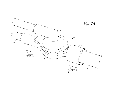
posterior segment of the eye. Simultaneously while removing the vitreous
humor, a liquid solution (e.g., balanced salt solution (BSS)) is typically
infused
into the eye to maintain intraocular pressure and prevent collapse of the eye
wall.
In cases of retinal breaks or retinal detachment, the liquid solution may then
be
exchanged for air, through a process known as fluid-air exchange, to help push
out subretinal fluid from the intraocular space while maintaining intraocular
pressure and temporarily holding the retina in place. During such procedures,
the liquid and air are provided by separate supply lines that are conjoined
with a
singular downstream infusion line via a stopcock.
[0004]
In some cases, the air pressure in the gas supply line may build up and
cause air to escape into the infusion line, forming air bubbles in the
infusion liquid
1
CA 03201538 2023- 6-7
WO 2022/130213
PCT/IB2021/061722
which may travel to the eye and negatively affect intraocular pressure during
surgery. Conventional designs for check valves of infusion stopcocks, however,
do not allow venting through the gas supply line without reverse leakage of
liquid
and thus, there is currently no effective way to remove the air bubbles or
prevent
their escape into infusion liquids. Additionally, during some procedures,
infusion
fluids must be back-flowed through the infusion line in order for other
surgical
fluids, such as a retinal tamponade, to be injected into the intraocular
space. In
such cases, the amount of infusion fluid that may be back-flowed is limited by
the
inability to purge infusion gases through the gas supply line without reverse
leakage of liquids therein, which may damage the air pump and/or cause
additional complications during fluid infusion.
[0005]
Therefore, what is needed in the art are improved fluid control valves
for ophthalmic fluid infusion that enable aspiration and purging of gases.
SUMMARY
[0006]
The present disclosure generally relates to devices for surgical
procedures, and more particularly, surgical devices for ophthalmic fluid
infusion
and aspiration.
[0007]
In certain embodiments, a valve assembly for fluid infusion is provided.
The valve assembly includes a first portion with a first conduit having a
first port
and a first cavity having a proximal end and a distal end opposite of the
proximal
end. The proximal end is fluidly coupled to the first port via the first
conduit, and
the distal end has a cross-sectional area greater than a cross-sectional area
of
the first conduit. The valve assembly further includes a second portion with a
second conduit having a second port, a third conduit having a third port, and
a
second cavity disposed between the second conduit and the third conduit and
2
CA 03201538 2023- 6-7
WO 2022/130213
PCT/IB2021/061722
fluidly coupling the second port to the third port, where the second cavity is
adjacent to the first cavity. A filter having a hydrophobic membrane is
disposed
between and partitions the first cavity from the second cavity and the
hydrophobic membrane partially defines the second cavity.
[0008]
In certain embodiments, a fluid infusion system for opthalmic
procedures is provided. The fluid infusion system includes a surgical console
having a first fluid line coupled to a gas fluid source and a second fluid
line
coupled to a liquid fluid source. The fluid infusion system further includes a
valve
assembly fluidly coupled to the first fluid line and the second fluid line.
The valve
assembly includes a first conduit having a first port in fluid communication
with
the first fluid line, a second conduit having a second port in fluid
communication
with the second fluid line, and a third conduit having a third port in fluid
communication with a third fluid line. Flow rates of fluids through the first,
second, and third lines is controlled by the surgical console The first,
second,
and third conduits are further coupled to an intermediary cavity and in fluid
communication with each other. A filter is disposed within the intermediary
cavity
and partitions the first conduit from the second and third conduits. The
filter
includes a hydrophobic membrane disposed on a side thereof opposite the first
conduit and configured to prevent flow of liquids from the second and third
conduits into the first conduit while allowing bi-directional flow of gases
therebetween.
BRIEF DESCRIPTION OF THE DRAWINGS
[0009]
So that the manner in which the above-recited features of the present
disclosure can be understood in detail, a more particular description of the
disclosure, briefly summarized above, may be had by reference to embodiments,
some of which are illustrated in the appended drawings. It is to be noted,
3
CA 03201538 2023- 6-7
WO 2022/130213
PCT/IB2021/061722
however, that the appended drawings illustrate only exemplary embodiments and
are therefore not to be considered limiting of its scope, and may admit to
other
equally effective embodiments.
[0010]
Figure 1 illustrates a perspective view of an exemplary surgical
console, according to certain embodiments of the present disclosure.
[0011]
Figure 2A illustrates a perspective view of an exemplary valve
assembly, according to certain embodiments of the present disclosure.
[0012]
Figure 2B illustrates a perspective exploded view of the valve
assembly of Figure 2A, according to certain embodiments of the present
disclosure.
[0013]
Figure 20 illustrates a schematic cross-sectional view of the valve
assembly of Figure 2A, according to certain embodiments of the present
disclosure.
[0014]
Figure 3A illustrates a perspective view of an exemplary valve
assembly, according to certain embodiments of the present disclosure.
[0015]
Figure 3B illustrates a perspective exploded view of the valve
assembly of Figure 3A, according to certain embodiments of the present
disclosure.
[0016]
Figure 3C illustrates a schematic cross-sectional view of the valve
assembly of Figure 3A, according to certain embodiments of the present
disclosure.
4
CA 03201536 2023- 6-7
WO 2022/130213
PCT/IB2021/061722
[0017]
Figure 4A illustrates a schematic plan view of an exemplary
operational mode of the valve assemblies of Figures 2A-2C and 3A-30,
according to certain embodiments of the present disclosure.
[0018]
Figure 4B illustrates a schematic plan view of an exemplary
operational mode of the valve assemblies of Figures 2A-2C and 3A-3C,
according to certain embodiments of the present disclosure.
[0019]
Figure 40 illustrates a schematic plan view of an operational mode of
the valve assemblies of Figures 2A-20 and 3A-30, according to certain
embodiments of the present disclosure.
[0020]
To facilitate understanding, identical reference numerals have been
used, where possible, to designate identical elements that are common to the
figures. It is contemplated that elements and features of one embodiment may
be
beneficially incorporated in other embodiments without further recitation.
DETAILED DESCRIPTION
[0021]
The present disclosure generally relates to fluid control valves for
delivering and/or aspirating fluid during ophthalmic surgeries and procedures.
For example, the fluid control valves described herein may be used during
vitrectomies, such as pars plana vitrectomies for the treatment of posterior
segment diseases. Vitrectomies typically require cutting and removal of
portions
of the vitreous humor. In order to maintain intraocular pressure and prevent
collapse of the eye during such surgical procedures, liquid is infused into
the
intraocular space and thereafter aspirated. In certain procedures, the liquid
is
then exchanged with air or other gases during a process known as fluid-air
exchange. During such processes, it is typically beneficial to purge or vent
any
CA 03201538 2023- 6-7
WO 2022/130213
PCT/IB2021/061722
undesired gases in the infusion line and/or the intraocular space to maintain
intraocular pressure. The fluid control valves and methods described herein
provide improved structures and mechanisms for infusion fluid flow regulation
that enable upstream purging and/or venting of gases from infusion lines while
also preventing liquids from the infusion lines to leak into the gas supply
lines.
[0022]
In certain embodiments, a valve assembly includes a first portion
configured to fluidly couple with a gas supply line and a second portion
configured to fluidly couple with a liquid supply line and an infusion line.
The first
portion and the second portion are partitioned or separated from each other by
a
filter having a hydrophobic membrane configured to prevent the flow of liquids
therethrough while allowing the free flow of gas. Accordingly, an infusion
liquid
may be flowed through the second portion while gases may be simultaneously
aspirated into the first portion, without any liquids travelling into the gas
supply
line. The gas supply line may thus be utilized to vent or purge gases from the
infusion line before or during performance of surgical procedures.
[0023]
Figure 1 illustrates a perspective view of an exemplary surgical
console 100 that may be utilized in combination with the fluid control valves
described herein. The surgical console 100 is operably coupled, physically or
wirelessly, to any number of user interfaces, including a foot controller 102
and a
surgical tool 104 such as a vitrector. The surgical console 100 provides one
or
more port connectors 106 for physically coupling the user interfaces to
various
components of the surgical console 100. For example, the surgical tool 104 may
be fluidly coupled with a vacuum source via a vacuum supply line 108 disposed
through a port connector 106 to enable aspiration of cut vitreous from the
patient's eye. Similarly, one or more port connectors 106 may be utilized to
couple a fluid infusion system 110 with one or more infusion fluid sources,
(e.g.,
an air/gas source, a liquid perfluorocarbon source, a silicone oil infusion
(S01)
6
CA 03201538 2023- 6-7
WO 2022/130213
PCT/IB2021/061722
source, a BSS source, etc.) to enable infusion of fluids into the eye during
vitreous removal. As shown in Figure 1, the fluid infusion system 110 includes
an infusion line 112 fluidly coupled with a gas supply line 114 and a separate
liquid supply line 116 at a three-way automatic valve assembly 118, which may
enable selective flow of different infusion fluids through the infusion line
112.
[0024]
In operation, the user may control an aspect or mechanism of the
surgical tool 104 and/or the fluid infusion system 110 via actuation of the
foot
controller 102, which may include a foot pedal. For example, the user may
press
down on (e.g., depress) the foot controller 102 to initiate and increase a
flow rate
of an infusion fluid from a fluid source through the fluid infusion system 110
and
into the eye of the patient. Alternatively, reducing depression of the foot
controller 102 (e.g., lifting the user's foot) may decrease and ultimately
stop the
flow of fluid through the fluid infusion system 110. Accordingly, in certain
embodiments, the flow rate of infusion fluids through the fluid infusion
system
110 corresponds to the amount of depression of the foot controller 102. In
certain embodiments, the surgical console 100 further includes a display 120
for
displaying information to the user (the display may also incorporate a
touchscreen for receiving user input). Thus, the display 120 may display
information about infusion fluid parameters, such as infusion fluid flow rates
and
intraocular pressure, to the user during operation thereof.
[0025]
Figures 2A-2C illustrate a valve assembly 200 for flow control of
infusion fluids during surgical procedures. Valve assembly 200 is an example
of
the automatic three-way valve assembly 118, which may be utilized in
combination with the fluid infusion system 110 and the surgical console 100
described above. As shown in more detail in Figures 2A-2C, the valve assembly
200 generally includes a hydrophobic filter (shown as hydrophobic filter 222
in
Figures 2B-20) disposed between valve assembly 200's first portion (e.g., an
7
CA 03201538 2023- 6-7
WO 2022/130213
PCT/IB2021/061722
upper body), configured to fluidly couple with a gas supply line, and valve
assembly 200's second portion (e.g., a lower body), configured to fluidly
couple
with a fluid supply line. The partitioning of the first portion and the second
portion
by the hydrophobic filter enables active bi-directional flow of gases, such as
air,
between the gas supply line and an infusion line while passively preventing
liquids from travelling into the gas supply line. Thus, gases may be vented or
purged from the fluid infusion system 110 during fluid infusion to enable
improved
control of intraocular pressure during surgical procedures.
[0026]
Figure 2A illustrates a perspective view of the valve assembly 200,
while Figure 2B illustrates a perspective exploded view thereof and Figure 2C
illustrates a cross-sectional view thereof. Accordingly, Figures 2A-2C are
herein
described together for clarity.
[0027]
As noted above, the valve assembly 200 generally includes an upper
body 232 configured to interface (e.g., couple) with a lower body 202. In
certain
embodiments, the upper body 232 and lower body 202 are formed of any
suitable plastic materials, such as acrylonitrile butadiene styrene (ABS),
polycarbonate (PC), nylon, and acrylic, which may be transparent or opaque in
color. The upper body 232 has a base 238 from which an arm 234 extends in a
proximal direction (e.g., toward a surgical console or gas source) for
coupling
with gas supply line 114. Note that, as described herein, a distal end or
portion
of a component refers to the end or portion that is closer in line to a
patient's
body during use thereof. On the other hand, a proximal end or portion of the
component refers to the end or the portion that is distanced further away in
line
from the patient's body (e.g., closer to the surgical console).
[0028]
The gas supply line 114 couples with a port 237 at a proximal end of
the arm 234, which provides fluid connection with a conduit 236 extending
8
CA 03201538 2023- 6-7
WO 2022/130213
PCT/IB2021/061722
through a length of the arm 234. In certain embodiments, a diameter of the
port
237 is substantially the same or slightly larger than an outer diameter of the
gas
supply line 114 to allow a distal end of the gas supply line 114 to be
securely fit
within the port 237. In certain embodiments, the outer diameter of the
proximal
end of the arm 234 is substantially the same or slightly smaller than an inner
diameter of the gas supply line 114 to allow the distal end of the gas supply
line
114 to secure fit over the proximal end of the arm 234.
[0029]
The conduit 236 extends from the proximal end of the arm 234 to a
distal end of the arm 234 and opens into a cavity 240 within the base 238 of
the
upper body 232. In certain embodiments, the arm 234 and thus the conduit 236
have one or more curved portions to create an angled flow path for gases
between the gas supply line 114 and the cavity 240. For example, the arm 234
and the conduit 236 may have a bend disposed at about a 90-degree angle
between the proximal and distal ends thereof, thus creating an elbow-like gas
flow path. The bending of the gas flow path enables a three-way connection of
the valve assembly 200 between the gas supply line 114, the liquid supply line
116, and an infusion line 108.
[0030]
The cavity 240 is disposed at the distal end of conduit 236 and
generally has one or more dimensions greater than a width or diameter of the
conduit 236. In certain embodiments, the cavity 240 has a cross-sectional area
that gradually increases from an end of the cavity 240 nearest the arm 234 to
an
end of the cavity 240 furthest from the arm 234 (e.g., nearest the lower body
202). For example, the end of the cavity 240 nearest the arm 234 may have
substantially the same cross-sectional area as the distal end of the conduit
236
while the end of the cavity 240 furthest from the arm 234 may have a cross-
sectional area greater than a cross-sectional area of the distal end of the
conduit
236. In certain embodiments, the cavity 240 has a frustoconical-like shape.
The
9
CA 03201538 2023- 6-7
WO 2022/130213
PCT/IB2021/061722
increased cross-sectional area of the cavity 240 at the distal end thereof
enables
utilization of a larger-area filter 222 between the upper body 232 and the
lower
body 202 and provides more surface area through which gases may be vented or
purged from liquids flowing between the liquid supply line 116 and the
infusion
line 108.
[0031]
Generally, a lower surface of the upper body 232 and an upper surface
of the lower body 202 are configured to interface with or engage each other
and
secure the filter 222 therebetween. In certain embodiments, the lower body 202
couples to the upper body 232 at a lower surface of the base 238 such that the
cavity 240 faces a chamber 204 (e.g., a second cavity or reservoir) located at
a
central position of the lower body 202. The chamber 204 may have a cross-
sectional area substantially the same or greater than the cross-sectional area
of
the end of the cavity 240 nearest the chamber 204 so as not to constrict air
flow
between the upper body 232 and the lower body 202 and vice versa. The lower
body 202 further includes extensions 206 and 210 on opposing sides of the
chamber 204, where each extension 206 and 210 has a conduit 208 or 212
formed therethrough, respectively. The conduits 208 and 212 extend from the
chamber 204 in opposing directions toward ports 209 and 211 located at the
proximal and distal ends of the extensions 206 and 210, respectively. In
certain
embodiments, the port 209 is configured to fluidly couple with a liquid supply
line
116, while the port 211 is configured to couple to infusion line 108.
[0032]
Similar to the port 237, the port 209 may have a diameter substantially
the same or slightly larger than an outer diameter of the liquid supply line
116 to
allow a distal end of the liquid supply line 116 to be securely fit within the
port
209. Alternatively, an outer diameter of the proximal end of the extension 206
may be substantially the same or slightly smaller than an inner diameter of
the
liquid supply line 116 to allow the distal end of the liquid supply line 116
to
CA 03201538 2023- 6-7
WO 2022/130213
PCT/IB2021/061722
securely fit over the proximal end of the extension 206. The extension 212,
however, is generally sized to have an outer diameter substantially the same
or
slightly smaller than an inner diameter of the proximal end of the infusion
line
108. Accordingly, the infusion line 108 is configured to securely fit around
the
extension 212. In certain embodiments, the extension 212 may include a locking
mechanism, such as a Luer lock 250, which is configured to couple with the
infusion line 108 and provide additional mechanical holding force for a leak-
free
seal between the valve assembly 200 and the infusion line 108. For example,
the Luer lock 250 may comprise a threaded interior surface 252 through which
the proximal end of the infusion line 108 may be secured within.
[0033]
The filter 222 is disposed between the cavity 240 of the upper body
232 and the chamber 204 of the lower body 202, thus partially defining both
the
cavity 240 and the chamber 204. The filter 222 includes any suitable type of
membrane filter having a hydrophobic membrane 224 permeable to gas. The
hydrophobic membrane 224 may also be capable of capturing individual viruses
and bacteria, thus acting as a sterile barrier to prevent viruses and bacteria
from
entering the eye from the low pressure gas source.
[0034]
In some examples, the filter 222 includes a membrane 224 formed of
polytetrafluoroethylene (PTFE), expanded PTFE (ePTFE), polycarbonate track
etch (PCTE), polyesters (e.g., polyethylene terephthalate (PET)), nylon,
cellulose
(e.g., surfactant free cellulose acetate (SCFA), cellulose nitrate (ON),
cellulose
acetate (CA), polyethersulfone (PES), glass fibers, or acrylic copolymers. The
membrane 224 may further be unsupported or supported by a backing 226
formed of materials including but not limited to polyester, polyethylene,
polypropylene, or nylon. For example, in certain embodiments, the filter 222
includes an ePTFE membrane 224 having a polyester backing 226. Generally,
the hydrophobic membrane 224 of the filter 222 is oriented to face the chamber
11
CA 03201536 2023- 6-7
WO 2022/130213
PCT/IB2021/061722
204 so as to prevent liquids from flowing from the chamber 204 through the
filter
222 and into the cavity 240. The membrane 224 has a pore size ranging
between about 0.1 pm to about 10.0 pm, such as between about 0.2 pm to about
pm, such as between about 0.5 pm to about 3.0 pm, such as a between about
0.8 pm to about 1.2 pm. Furthermore, the membrane 224 may have a thickness
ranging between about 150 pm to about 300 pm, such as between about 200 pm
to about 250 pm.
[0035]
During operation, infusion liquid from the liquid source, such as silicone
oil or balanced salt solution (BSS), may flow through the liquid supply line
116,
into the lower body 202 of the valve assembly 200, and through the infusion
line
108 toward the patient's eye and vice-versa. Alternatively, infusion gases
from
the gas source, such as air, may flow through the gas supply line 114, into
the
upper body 232, past the filter 222 into the lower body 202, and then into the
infusion line 108 toward the patient's eye and vice-versa. The placement of
the
hydrophobic filter 222 between the upper body 232 and the lower body 202
passively prevents liquid from flowing up into the upper body 232 and the gas
supply line 116, while allowing gases to pass therethrough. Accordingly, the
valve assembly 200 enables the venting, purging, and/or back-flow of gases
during fluid infusion procedures while preventing the escape of liquid into
the gas
supply line 116, which is described in further detail below.
[0036]
Please note that although a single filter 222 is depicted in Figures 2A-
2C, in certain embodiments, it is further contemplated that the valve assembly
200 may include two or more filters arranged in a linear or stacked
configuration.
The two or more filters may be formed of different materials and/or have
different
pore sizes relative to each other. For example, in certain embodiments, the
valve assembly 200 may include a second filter disposed between the upper and
lower bodies 232, 202 and upstream of the filter 222 (e.g., closer in line to
the
12
CA 03201538 2023- 6-7
WO 2022/130213
PCT/IB2021/061722
gas supply line 114). In such embodiments, the second filter may have a pore
size smaller than the filter 222 and provide filtration of gases flowed
through the
gas supply line 114, while the filter 222 provides a hydrophobic barrier to
prevent
leakage of liquid therein.
[0037]
Figures 3A-3C illustrate another valve assembly 300, which functions
in substantially the same manner as the valve assembly 200 depicted in Figures
2A-20, but with a different structure. Accordingly, Figures 3A-30 are
described
together for clarity, and parts of the valve assembly 300 corresponding to the
above-described parts of the valve assembly 200 are marked with the same
reference numerals.
[0038]
As shown, the upper body 232 of the valve assembly 300 includes a
base 338, which generally has a plate-like shape and further includes one or
more ridges (e.g., ribs or grooves) 342 extending from a lower surface thereof
that form one or more channels within a cavity 340 (shown in Figure 30). In
certain embodiments, the ridges 342 are annular or semi-annular ridges that
circumscribe the distal end of the conduit 236. The ridges 342 provide added
support for the filter 222 when the valve assembly 300 is in an assembled
state.
[0039]
Similar to the cavity 240 of the valve assembly 200, the cavity 340
fluidly couples with the conduit 236 and has one or more cross-sectional
dimensions greater than a width or diameter of the conduit 236. However,
unlike
the cavity 240, the cavity 340 has a cross-sectional area that steeply or
abruptly
increases from an end of the cavity 340 nearest the arm 234 to an end of the
cavity 340 furthest from the arm 234. As described above, the increased cross-
sectional area of the cavity 340 enables utilization of a larger-area filter
222,
which provides more surface area through which gases may be vented or purged
from liquids flowing between the liquid supply line 116 and the infusion line
108.
13
CA 03201538 2023- 6-7
WO 2022/130213
PCT/IB2021/061722
[0040]
The lower body 202 of the valve assembly 300 includes a basin 350
coupled to a flow-through member 358. The basin 350 is configured to interface
and engage with the base 338 of the upper body 232 and secure the filter 222
therebetween. In certain embodiments, the basin 350 couples to the upper body
232 at a lower surface of the base 338 such that the cavity 340 of the base
338
faces a cavity 354 of the basin 350. At least a portion of the cavity 354 may
have
a cross-sectional area substantially the same or greater than the cross-
sectional
area of the end of the cavity 340 nearest the basin 350 so as not to constrict
air
flow between the upper body 232 and the lower body 202 and vice versa. For
example, an end of the cavity 354 opposite the flow-through member 358 may
have a cross-sectional area substantially the same or greater than the cross-
sectional area of the end of the cavity 340 nearest the basin 350.
[0041]
Similar to the base 338, the basin 304 includes one or more ridges 352
extending from an upper surface thereof into a cavity 354. The ridges 352 are
configured to provide support to the filter 222 when the valve assembly 300 is
in
an assembled state and may be annular or semi-annular in shape, defining one
or more channels therein. In certain embodiments, the ridges 352 circumscribe
a
proximal end of a channel 356 that fluidly couples the cavity 354 with an
intermediate conduit 360 of the flow-through member 358. The intermediate
conduit 360, in turn, extends and fluidly connects the extensions 206 and 210
of
the lower body 202, which are configured to couple with the liquid supply line
116
and the infusion line 108 at ports 209 and 211, respectively.
[0042]
During operation of the valve assembly, infusion liquid from the liquid
source may flow through the liquid supply line 116, into the flow-through
member
358 of the lower body 202, and through the infusion line 108 toward the
patient's
eye and vice versa. Alternatively, infusion gases from the gas source may flow
through the gas supply line 114, into the upper body 232, past the filter 222
into
14
CA 03201536 2023- 6-7
WO 2022/130213
PCT/IB2021/061722
the lower body 202, and through the infusion line 108 toward the patient's eye
and vice-versa. Similar to the valve assembly 200, the disposition of the
hydrophobic filter 222 between the upper body 232 and the lower body 202
passively prevents the flow of liquids into the upper body 232 and the gas
supply
line 116, while allowing gases to pass therethrough. Thus, like the valve
assembly 200, the valve assembly 300 facilitates venting, purging, and/or back-
flow of gases during fluid infusion procedures while preventing the escape of
liquid into the gas supply line 116.
[0043]
Please note that, as discussed above with reference to the valve
assembly 200, although a single filter 222 is depicted in Figures 3A-3C, it is
further contemplated that the valve assembly 300 may include two or more
filters
arranged in a linear or stacked configuration. The two or more filters may be
formed of different materials and/or have different pore sizes relative to
each
other. For example, in certain embodiments, a second filter having a finer
pore
size may be disposed upstream of the filter 222 to provide additional
filtration of
gases flowed through the gas supply line 114, while the filter 222 provides a
hydrophobic barrier and prevent liquids from flowing therein.
[0044]
Figures 4A-40 schematically illustrate operational modes of the valve
assemblies 200, 300 during fluid infusion procedures. In particular, Figures
4A-
4C illustrate the flow of liquid solutions (e.g., BSS), represented by lines
410, and
the flow of gases (e.g., air), represented by lines 420, through the fluid
infusion
system 110 having the valve assembly 200, as described above. Please note
that although the valve assembly 200 is depicted in Figures 4A-40, the valve
assembly 300 may be utilized in substantially the same manner. Further, please
note that unbroken lines (e.g., continuous lines) represent open or active
flow,
while broken lines (e.g., dashed lines) represent closed or no flow.
CA 03201538 2023- 6-7
WO 2022/130213
PCT/IB2021/061722
[0045]
Figure 4A depicts the fluid infusion system 110 during a first operation
of liquid infusion, which may be selected and/or controlled by a user (e.g., a
surgeon) via a surgical console, such as surgical console 100. As shown,
infusion liquid 410 is controllably flowed between the liquid source 470 and
eye
402 via liquid supply line 116, valve assembly 200, and infusion line 108,
while
air or gas flow through gas supply line 114 is stopped or shut off. To control
a
pressure within the fluid infusion system 110 and thus, the eye 402, the user
may
adjust the direction and flow rate of the liquid 410 to or from the liquid
source 470
via the surgical console 100. The flow control valve 200 enables liquid 410 to
flow between the liquid supply line 116 and the infusion line 108, while also
preventing the liquid 410 from flowing into the gas supply line 114 and
towards
the gas source 480 due to the presence of the hydrophobic filter 222.
Accordingly, the valve assembly 200 provides a passive means of preventing
leakage of liquid 410 into gas supply line 114, which contrasts with
conventional
flow control valves that may allow the escape of at least some liquid 410 into
the
gas supply line 114 during use thereof.
[0046]
Figure 4B depicts the fluid infusion system 110 during a second
operation of liquid infusion in which the pressure of air 420 within the gas
supply
line 114 is actively modulated while infusion liquid 410 is flowed between the
liquid source 470 and eye 402. As described above, the pressure within the
fluid
infusion system 110 and the eye 402 is controlled by adjusting the direction
and
flow rate of the liquid 410 to or from the liquid source 470 via the surgical
console
100. When left unchecked, pressure within the gas supply line 114 may
inadvertently build up during infusion and cause air 420 to leak into the
liquid 410
being injected into the eye 402, thereby negatively affecting the intraocular
pressure thereof. Therefore, in certain embodiments, it may be desired to
apply
a vacuum pressure (e.g., negative pressure) to the gas supply line 114 to vent
16
CA 03201536 2023- 6-7
WO 2022/130213
PCT/IB2021/061722
the gas supply line 114 and prevent the undesired escape of air 420 into the
liquid 420 as bubbles. In certain embodiments, active venting of the gas
supply
line 114 may also be desired to purge the infusion liquid 410 of gases already
trapped therein as the liquid 410 passes into the infusion line 108. Similar
to the
pressure of liquid
[0047]
Since conventional flow control valves cannot prevent the leakage of
liquid 410 into the gas supply line 114, venting of the gas supply line 114
with a
conventional valve is extraordinarily difficult. In comparison, as a result of
the
hydrophobic filter 222, the valve assembly 200 facilitates active venting of
the
gas supply line 114 during infusion of liquid 410 into the eye 402, thus
reducing
or eliminating the possibility of unwanted gases being flowed into eye 402 and
disrupting the intraocular pressure therein.
[0048]
Figure 4B is further representative of the fluid infusion system 110
during an infusion fluid back-flow operation. Back-flow of infusion fluids may
be
necessitated when the eye 402 is injected, via a separate cannula or injection
device, with a retinal tamponade (or other fluid treatment) such as
intraocular
air/gas, silicone oil, or perfluoron. As a result, infusion fluids previously
flowed
through the infusion line 108 may need to be back-flowed. Because conventional
flow control valves cannot backflow or purge gases into the gas supply line
114
without leakage of infusion liquid, only a limited volume of infusion fluids
can be
back-flowed without risking the chance of liquid leakage into the gas supply
line
114 or gas leakage into the liquid supply line 116. In contrast, the
hydrophobic
filter 222 of the valve assembly 200 in Figure 4B enables backflow of gases
into
the gas supply line 114 without leakage of infusion liquids, thus allowing a
greater volume of the infusion fluids to be back-flowed into their respective
supply lines and further enabling a greater volume of treatment fluids to be
injected into the eye 402.
17
CA 03201538 2023- 6-7
WO 2022/130213
PCT/IB2021/061722
[0049]
Figure 40 depicts the fluid infusion system 110 during a third operation
of liquid infusion. The operational mode depicted in Figure 40 may be
performed, for example, during a fluid-air exchange to help push out
subretinal
fluid from the intraocular space of the eye 402. As shown, air 420 is flowed
from
the gas source 480 to the eye 402, while liquid flow through the liquid supply
line
116 is shut off to prevent escape of liquid 410 into the infused air 420.
Accordingly, the pressure within the fluid infusion system 110 and the eye 402
is
controlled by adjusting the direction and flow rate of the air 420 to or from
the gas
source 480 via the surgical console 100.
[0050]
In summary, embodiments of the present disclosure include structures
and mechanisms for improved intraocular pressure maintenance during
ophthalmic procedures, and in particular, improved fluid control valves for
intraocular fluid infusion.
The valve assemblies described above include
embodiments wherein a hydrophobic filter is disposed between a gas supply line
and a liquid supply line and/or infusion line. The utilization of the
hydrophobic
filter enables bi-directional flow of gases between a gas supply line and the
patient's eye, while also passively preventing the leakage of liquids into the
gas
supply line. Accordingly, the aforementioned valve assemblies are particularly
beneficial during fluid infusion of the intraocular space, as gas may be
vented
from infusion liquids during infusion or black-flowed from the eye during
injection
of other treatments, thus allowing better control of the intraocular pressure
within
the eye.
[0051]
While the foregoing is directed to embodiments of the present
disclosure, other and further embodiments of the disclosure may be devised
without departing from the basic scope thereof, and the scope thereof is
determined by the claims that follow.
18
CA 03201538 2023- 6-7