Note: Descriptions are shown in the official language in which they were submitted.
CA 03201626 2023-05-11
WO 2022/119805
PCT/US2021/061120
BIOLOGICAL VESICLES DISPLAYING CELL SURFACE PROTEINS AND METHODS RELATED TO
SAME
CROSS-REFERENCE TO RELATED APPLICATIONS
This application claims priority to U.S. Patent Application No. 63/120,167,
filed on December 1,
2020; U.S. Patent Application No. 63/212,021, filed on June 17, 2021; and U.S.
Patent Application No.
63/227,039, filed on July 29, 2021, the entire contents of each of which are
incorporated herein by
reference in their entirety.
SEQUENCE LISTING
The instant application contains a Sequence Listing which has been submitted
electronically in
ASCII format and is hereby incorporated by reference in its entirety. Said
ASCII copy, created on
November 29, 2021, is named 50474-231W04 Sequence Listing 11 29 21 ST25 and is
4,713 bytes in
size.
FIELD OF THE INVENTION
Provided herein are biological vesicles displaying cell surface proteins, as
well as methods of
using such vesicles to identify and characterize protein-protein interactions.
BACKGROUND
Plasma membrane-expressed proteins and their interactors play a prominent role
in the initiation
of signal transduction to the cytosol of the cell, and thus are key regulators
of most biological pathways.
Increasing evidence demonstrates that receptors have a complex landscape of
interacting partners in the
extracellular milieu that directly influence their biological functions. As a
result, dysregulation of receptor-
ligand crosstalk often underlies pathology and disease progression. However,
receptor interaction
networks remain understudied, due to of the biochemical challenges associated
with maintaining
membrane proteins in their native conformation proteins and typically weak
interactions among receptors.
Thus, there is an unmet need for methods and compositions for the
identification of interactions
between cell surface proteins, as well as novel modulators of such
interactions and methods of using the
.. same.
SUMMARY OF THE INVENTION
In one aspect, the disclosure features a method for identifying a protein-
protein interaction, the
method comprising (a) providing a collection of target polypeptides that are
immobilized on one or more
solid surfaces; (b) contacting the collection of step (a) with a biological
vesicle (BV) comprising a
heterologous membrane-associated protein and a membrane-budding agent under
conditions permitting
the binding of the heterologous membrane-associated protein and at least one
of the target polypeptides,
wherein the heterologous membrane-associated protein is expressed at or above
a threshold level on the
surface of the By; and (c) detecting an interaction between the heterologous
membrane-associated protein
and the at least one target polypeptide, thereby identifying a protein-protein
interaction.
In some aspects, one or more of the target polypeptides is immobilized to a
distinct location on the
one or more solid surfaces.
1
CA 03201626 2023-05-11
WO 2022/119805
PCT/US2021/061120
In some aspects, detecting an interaction comprises detecting a signal at a
location on the solid
surface that is above a threshold level.
In some aspects, the membrane-budding agent is selected from the group
consisting of a HIV gag
protein, Acyl.Hrs, ARRDC1, and ARF6. In some aspects, the membrane-budding
agent is a HIV gag
protein.
In some aspects, the membrane-budding agent further comprises a detectable
marker, and
detecting an interaction comprises detecting a level of the detectable marker
at a location on the solid
surface that is above a threshold level. In some aspects, the detectable
marker is an enzyme that
produces a fluorescent signal in the presence of a substrate. In some aspects,
the enzyme is Renilla
luciferase (Rluc) and the substrate is Rluc substrate.
In some aspects, the BV comprises a membrane marker, and detecting an
interaction comprises
detecting a level of the membrane marker at a location on the solid surface
that is above a threshold level.
In some aspects, the membrane marker is a cholesterol marker. In some aspects,
the cholesterol marker
is AMPLEXTm Red.
In some aspects, the interaction is a transient interaction.
In some aspects, the interaction is a low-affinity interaction.
In some aspects, the heterologous membrane-associated protein is a full-length
protein.
In some aspects, the heterologous membrane-associated protein comprises a
protein fragment, a
tag, and an anchor.
In some aspects, the anchor tethers the protein fragment to the surface of a
membrane of a By.
In some aspects, the anchor is a glycosylphosphatidyl-inositol (GPI)
polypeptide.
In some aspects, the tag can be directly or indirectly visualized. In some
aspects, the tag
comprises a moiety that can be detected using an antibody or an antibody
fragment. In some aspects, the
tag is a glycoprotein D (gD) polypeptide.
In some aspects, the expression level of the heterologous membrane-associated
protein is
determined using a biolayer interferometry (BLI) assay.
In some aspects, the tag is a gD polypeptide, expression of the heterologous
membrane-
associated protein is detected using an anti-gD antibody, and the threshold
level is a shift of 1.5 nm, as
measured using the BLI assay at 30 C.
In some aspects, the tag comprises a fluorescent protein.
In some aspects, the heterologous membrane-associated protein is a
transmembrane receptor or
a fragment thereof. In some aspects, the receptor is a single-pass
transmembrane (STM) receptor.
In some aspects, the protein fragment is an extracellular domain.
In some aspects, each member of the collection of target polypeptides is an Fc-
tagged
extracellular domain, and wherein the solid surface is coated with protein A.
In some aspects, the collection of target polypeptides comprises the
extracellular domains of at
least 25% of the proteins of Table 4. In some aspects, the collection of
target polypeptides comprises the
extracellular domains of at least 50% of the proteins of Table 4. In some
aspects, the collection of target
polypeptides comprises the extracellular domains of at least 75% of the
proteins of Table 4. In some
aspects, the collection of target polypeptides comprises the extracellular
domains of at least 90% of the
2
CA 03201626 2023-05-11
WO 2022/119805
PCT/US2021/061120
proteins of Table 4. In some aspects, the collection of target polypeptides
comprises the extracellular
domains of all of the proteins of Table 4.
In another aspect, the disclosure features a BV comprising (a) a heterologous
membrane-
associated protein comprising a protein fragment, a tag, and an anchor,
wherein the heterologous
membrane-associated protein is present on the outer face of the BV and (b) a
membrane-budding agent.
In another aspect, the disclosure features a BV comprising (a) a heterologous
membrane-
associated protein comprising a protein fragment, a tag, and an anchor,
wherein the heterologous
membrane-associated protein is present on the outer face of the BV and (b) a
membrane-budding agent,
the BV being produced by a process comprising (i) providing a parent cell that
has been modified to
express the heterologous membrane-associated protein and the membrane-budding
agent and (ii)
isolating the BV from the parent cell.
In some aspects, the membrane-budding agent is selected from the group
consisting of a HIV gag
protein, Acyl.Hrs, ARRDC1, and ARF6. In some aspects, the membrane-budding
agent is a HIV gag
protein.
In some aspects, the anchor tethers the protein fragment to the surface of a
lipid membrane of a
By. In some aspects, the anchor is a GPI polypeptide.
In some aspects, the tag can be directly or indirectly visualized. In some
aspects, the tag
comprises a moiety that can be detected using an antibody or an antibody
fragment. In some aspects, the
tag is a gD polypeptide.
In some aspects, the tag comprises a fluorescent protein.
In some aspects, the protein fragment is an extracellular domain of a
transmembrane receptor. In
some aspects, the transmembrane receptor is a STM receptor.
In some aspects, the BV produces a shift that is at or above a threshold level
when contacted with
an antibody against the tag, as measured using a BLI assay.
In some aspects, the tag is a gD polypeptide, the antibody is an anti-gD
antibody, and the
threshold level is a shift of 1.5 nm, as measured using the BLI assay at 30 C.
In some aspects, the membrane-budding agent comprises a detectable marker. In
some aspects,
the detectable marker is an enzyme that produces a fluorescent signal in the
presence of a substrate. In
some aspects, the enzyme is Rluc and the substrate is Rluc substrate.
In some aspects, the BV comprises a membrane marker. In some aspects, the
membrane marker
is a cholesterol marker. In some aspects, the cholesterol marker is AMPLEXTm
Red.
In some aspects, the BV is produced by a mammalian parent cell. In some
aspects, the BV is an
extracellular vesicle (EV). In some aspects, the BV is an exosome or a
microvesicle. In some aspects,the
BV is a virus-like particle (VLP).
In some aspects, the parent cell has been transfected with a plasmid encoding
the heterologous
membrane-associated protein and a plasmid encoding the membrane-budding agent.
In another aspect, the disclosure features a method of identifying a modulator
of the interaction
between a protein of Table 1 and a protein of Table 2, the method comprising
(a) providing a candidate
modulator; (b) contacting a protein of Table 1 with a protein of Table 2 in
the presence or absence of the
candidate modulator under conditions permitting the binding of the protein of
Table 1 to the protein of
3
CA 03201626 2023-05-11
WO 2022/119805
PCT/US2021/061120
Table 2, wherein the protein of Table 1 and the protein of Table 2 are
reported to interact in Table 3; and
(c) measuring the binding of the protein of Table 1 to the protein of Table 2,
wherein an increase or
decrease in binding in the presence of the candidate modulator relative to
binding in the absence of the
candidate modulator identifies the candidate modulator as a modulator of the
interaction between the
protein of Table 1 and the protein of Table 2.
In another aspect, the disclosure features a method of identifying a modulator
of a downstream
activity of a protein of Table 1, the method comprising (a) providing a
candidate modulator; (b) contacting
the protein of Table 1 with a protein of Table 2 in the presence or absence of
the candidate modulator
under conditions permitting the binding of the protein of Table 1 to the
protein of Table 2, wherein the
protein of Table 1 and the protein of Table 2 are reported to interact in
Table 3; and (c) measuring a
downstream activity of the protein of Table 1, wherein a change in the
downstream activity in the
presence of the candidate modulator relative to the downstream activity in the
absence of the candidate
modulator identifies the candidate modulator as a modulator of the downstream
activity of the protein of
Table 1.
In another aspect, the disclosure features a method of identifying a modulator
of a downstream
activity of a protein of Table 2, the method comprising (a) providing a
candidate modulator; (b) contacting
the protein of Table 2 with a protein of Table 1 in the presence or absence of
the candidate modulator
under conditions permitting the binding of the protein of Table 2 to the
protein of Table 1, wherein the
protein of Table 1 and the protein of Table 2 are reported to interact in
Table 3; and (c) measuring a
downstream activity of the protein of Table 2, wherein a change in the
downstream activity in the
presence of the candidate modulator relative to the downstream activity in the
absence of the candidate
modulator identifies the candidate modulator as a modulator of the downstream
activity of the protein of
Table 2.
In some aspects, the increase or decrease in binding is at least 70%, as
measured by a surface
plasmon resonance (SPR) assay, a BLI assay, or an enzyme-linked immunosorbent
assay (ELISA).
In some aspects, the modulator is an inhibitor of the downstream activity of
the protein of Table 1
or Table 2. In some aspects, the modulator is an activator of the downstream
activity of the protein of
Table 1 or Table 2.
In some aspects, the change in the downstream activity is a decrease in the
amount, strength, or
duration of the downstream activity. In some aspects, the change in the
downstream activity is an
increase in the amount, strength, or duration of the downstream activity.
In some aspects, the modulator is a small molecule, an antibody or antigen-
binding fragment
thereof, a peptide, a mimic, an antisense oligonucleotide, or a small
interfering RNA (siRNA).
In some aspects, the antigen-binding fragment is a bis-Fab, an Fv, a Fab, a
Fab'-SH, a F(ab')2, a
diabody, a linear antibody, an scFv, an ScFab, a VH domain, or a VHH domain.
In some aspects, the antibody or antigen-binding fragment thereof binds the
protein of Table 1. In
some aspects, the antibody or antigen-binding fragment thereof binds the
protein of Table 2.
In some aspects, the protein of Table 1 is LRRC15. In some aspects, the
protein of Table 2 is
TEM1. In some aspects, the downstream activity is tumor growth. In some
aspects, tumor growth is
4
CA 03201626 2023-05-11
WO 2022/119805
PCT/US2021/061120
decreased in the presence of the modulator. In some aspects, tumor growth is
decreased by at least
20%, as measured in a tumor growth assay.
In some aspects, the modulator is an antibody or antigen-binding fragment
thereof targeting
LRRC15.
In some aspects, the modulator is an antibody or antigen-binding fragment
thereof targeting
TEM1.
In another aspect, the disclosure features a method of identifying a modulator
of the interaction
between LRRC15 and TEM1, the method comprising (a) providing a candidate
modulator; (b) contacting
LRRC15 with TEM1 in the presence or absence of the candidate modulator under
conditions permitting
the binding of LRRC15 to TEM1; and (c) measuring the binding of LRRC15 to
TEM1, wherein an increase
or decrease in binding in the presence of the candidate modulator relative to
binding in the absence of the
candidate modulator identifies the candidate modulator as a modulator of the
interaction between
LRRC15 and TEM1.
In another aspect, the disclosure features a method of identifying a modulator
of a downstream
activity of LRRC15, the method comprising (a) providing a candidate modulator;
(b) contacting LRRC15
with TEM1 in the presence or absence of the candidate modulator under
conditions permitting the binding
of LRRC15 to TEM1; and (c) measuring a downstream activity of LRRC15, wherein
a change in the
downstream activity in the presence of the candidate modulator relative to the
downstream activity in the
absence of the candidate modulator identifies the candidate modulator as a
modulator of the downstream
activity of LRRC15.
In another aspect, the disclosure features a method of identifying a modulator
of a downstream
activity of TEM1, the method comprising (a) providing a candidate modulator;
(b) contacting TEM1 with
LRRC15 in the presence or absence of the candidate modulator under conditions
permitting the binding
of TEM1 to LRRC15; and (c) measuring a downstream activity of TEM1, wherein a
change in the
downstream activity in the presence of the candidate modulator relative to the
downstream activity in the
absence of the candidate modulator identifies the candidate modulator as a
modulator of the downstream
activity of TEM1.
In some aspects, the increase or decrease in binding is at least 70%, as
measured by an SPR
assay, a BLI assay, or ELISA.
In some aspects, the downstream activity is tumor growth.
In some aspects, tumor growth is decreased in the presence of the modulator.
In some aspects,
tumor growth is decreased by at least 20%, as measured in a tumor growth
assay.
In another aspect, the disclosure features a method for identifying a
biological vesicle (BV) having
an altered binding profile, the method comprising (a) providing a collection
of target polypeptides that are
.. immobilized on one or more solid surfaces; (b) contacting the collection of
step (a) with a BV of interest; (c)
detecting an interaction between the BV of interest and the at least one
target polypeptide, thereby
identifying an interaction profile; and (d) comparing the interaction profile
of the BV of interest to the
interaction profile of a control By, wherein a difference between the
interaction profile of the BV of interest
and the interaction profile of the control BV identifies the BV of interest as
one having an altered binding
profile.
5
CA 03201626 2023-05-11
WO 2022/119805
PCT/US2021/061120
In some aspects, the collection of target polypeptides comprises the
extracellular domains of at
least 25% of the proteins of Table 4. In some aspects, the collection of
target polypeptides comprises the
extracellular domains of at least 50% of the proteins of Table 4. In some
aspects, the collection of target
polypeptides comprises the extracellular domains of at least 75% of the
proteins of Table 4. In some
aspects, the collection of target polypeptides comprises the extracellular
domains of at least 90% of the
proteins of Table 4. In some aspects, the collection of target polypeptides
comprises the extracellular
domains of all of the proteins of Table 4.
In some aspects, the BV of interest is an engineered By.
In some aspects, the BV of interest is derived from a sample from a subject.
In some aspects, the
BV of interest and the control BV are derived from different tissues or
different cell types. In some aspects,
the BV of interest is derived from a diseased tissue and the control BV is
derived from healthy tissue.
In another aspect, the disclosure features a protein complex comprising (a) a
BV comprising a
heterologous membrane-associated protein and a membrane-budding agent and (b)
a target polypeptide,
wherein the heterologous membrane-associated protein and the target
polypeptide are bound to one
another.
In another aspect, the disclosure features a method of identifying a modulator
of the interaction
between a protein of Table 5 and a protein of Table 6, the method comprising
(a) providing a candidate
modulator; (b) contacting a protein of Table 5 with a protein of Table 6 in
the presence or absence of the
candidate modulator under conditions permitting the binding of the protein of
Table 5 to the protein of
Table 6, wherein the protein of Table 5 and the protein of Table 6 are
reported to interact in Table 7; and
(c) measuring the binding of the protein of Table 5 to the protein of Table 6,
wherein an increase or
decrease in binding in the presence of the candidate modulator relative to
binding in the absence of the
candidate modulator identifies the candidate modulator as a modulator of the
interaction between the
protein of Table 5 and the protein of Table 6.
In another aspect, the disclosure features a method of identifying a modulator
of a downstream
activity of a protein of Table 5, the method comprising (a) providing a
candidate modulator; (b) contacting
the protein of Table 5 with a protein of Table 6 in the presence or absence of
the candidate modulator
under conditions permitting the binding of the protein of Table 5 to the
protein of Table 6, wherein the
protein of Table 5 and the protein of Table 6 are reported to interact in
Table 7; and (c) measuring a
downstream activity of the protein of Table 5, wherein a change in the
downstream activity in the
presence of the candidate modulator relative to the downstream activity in the
absence of the candidate
modulator identifies the candidate modulator as a modulator of the downstream
activity of the protein of
Table 5.
In another aspect, the disclosure features a method of identifying a modulator
of a downstream
activity of a protein of Table 6, the method comprising (a) providing a
candidate modulator; (b) contacting
the protein of Table 6 with a protein of Table 5 in the presence or absence of
the candidate modulator
under conditions permitting the binding of the protein of Table 6 to the
protein of Table 5, wherein the
protein of Table 5 and the protein of Table 6 are reported to interact in
Table 7; and (c) measuring a
downstream activity of the protein of Table 6, wherein a change in the
downstream activity in the
presence of the candidate modulator relative to the downstream activity in the
absence of the candidate
6
CA 03201626 2023-05-11
WO 2022/119805
PCT/US2021/061120
modulator identifies the candidate modulator as a modulator of the downstream
activity of the protein of
Table 6.
In some aspects, the increase or decrease in binding is at least 70%, as
measured by a surface
plasmon resonance (SPR) assay, a BLI assay, or an enzyme-linked immunosorbent
assay (ELISA).
In some aspects, the modulator is an inhibitor of the downstream activity of
the protein of Table 5
or Table 6.
In some aspects, the modulator is an activator of the downstream activity of
the protein of Table 5
or Table 6.
In some aspects, the change in the downstream activity is a decrease in the
amount, strength, or
duration of the downstream activity. In some aspects, the change in the
downstream activity is an
increase in the amount, strength, or duration of the downstream activity.
In some aspects, the modulator is a small molecule, an antibody or antigen-
binding fragment
thereof, a peptide, a mimic, an antisense oligonucleotide, or a small
interfering RNA (siRNA).
In some aspects, the antigen-binding fragment is a bis-Fab, an Fv, a Fab, a
Fab'-SH, a F(ab')2, a
diabody, a linear antibody, an scFv, an ScFab, a VH domain, or a VHH domain.
In some aspects, the antibody or antigen-binding fragment thereof binds the
protein of Table 5.
In some aspects, the antibody or antigen-binding fragment thereof binds the
protein of Table 6.
In some aspects, the protein of Table 5 is ADGRB1.
In some aspects, the protein of Table 6 is PD-L1.
In some aspects, the downstream activity is tumor growth. In some aspects,
tumor growth is
decreased in the presence of the modulator. In some aspects, tumor growth is
decreased by at least
20%, as measured in a tumor growth assay.
In some aspects, the modulator is an antibody or antigen-binding fragment
thereof targeting PD-
L1.
In some aspects, the protein of Table 6 is ICOSLG.
In some aspects, the downstream activity is T cell activation. In some
aspects, T cell activation is
increased in the presence of the modulator. In some aspects, T cell activation
is increased by at least
20%.
In some aspects, the modulator is an antibody or antigen-binding fragment
thereof targeting
ICOSLG.
In some aspects, the modulator is an antibody or antigen-binding fragment
thereof targeting
ADGRB1.
In another aspect, the disclosure features a method of identifying a modulator
of the interaction
between PD-L1 and ADGRB1, the method comprising (a) providing a candidate
modulator; (b) contacting
PD-L1 with ADGRB1 in the presence or absence of the candidate modulator under
conditions permitting
the binding of PD-L1 to ADGRB1; and (c) measuring the binding of PD-L1 to
ADGRB1, wherein an
increase or decrease in binding in the presence of the candidate modulator
relative to binding in the
absence of the candidate modulator identifies the candidate modulator as a
modulator of the interaction
between PD-L1 and ADGRB1.
7
CA 03201626 2023-05-11
WO 2022/119805
PCT/US2021/061120
In another aspect, the disclosure features a method of identifying a modulator
of a downstream
activity of PD-L1, the method comprising (a) providing a candidate modulator;
(b) contacting PD-L1 with
ADGRB1 in the presence or absence of the candidate modulator under conditions
permitting the binding
of PD-L1 to ADGRB1; and (c) measuring a downstream activity of PD-L1, wherein
a change in the
downstream activity in the presence of the candidate modulator relative to the
downstream activity in the
absence of the candidate modulator identifies the candidate modulator as a
modulator of the downstream
activity of PD-L1.
In another aspect, the disclosure features a method of identifying a modulator
of a downstream
activity of ADGRB1, the method comprising (a) providing a candidate modulator;
(b) contacting ADGRB1
with PD-L1 in the presence or absence of the candidate modulator under
conditions permitting the
binding of ADGRB1 to PD-L1; and (c) measuring a downstream activity of ADGRB1,
wherein a change in
the downstream activity in the presence of the candidate modulator relative to
the downstream activity in
the absence of the candidate modulator identifies the candidate modulator as a
modulator of the
downstream activity of ADGRB1.
In some aspects, the increase or decrease in binding is at least 70%, as
measured by an SPR
assay, a BLI assay, or ELISA.
In some aspects, the downstream activity is tumor growth. In some aspects,
tumor growth is
decreased in the presence of the modulator. In some aspects, tumor growth is
decreased by at least
20%, as measured in a tumor growth assay.
In another aspect, the disclosure features a method of identifying a modulator
of the interaction
between ICOSLG and ADGRB1, the method comprising (a) providing a candidate
modulator; (b)
contacting ICOSLG with ADGRB1 in the presence or absence of the candidate
modulator under
conditions permitting the binding of ICOSLG to ADGRB1; and (c) measuring the
binding of ICOSLG to
ADGRB1, wherein an increase or decrease in binding in the presence of the
candidate modulator relative
to binding in the absence of the candidate modulator identifies the candidate
modulator as a modulator of
the interaction between ICOSLG and ADGRB1.
In another aspect, the disclosure features a method of identifying a modulator
of a downstream
activity of ICOSLG, the method comprising (a) providing a candidate modulator;
(b) contacting ICOSLG
with ADGRB1 in the presence or absence of the candidate modulator under
conditions permitting the
binding of ICOSLG to ADGRB1; and (c) measuring a downstream activity of
ICOSLG, wherein a change
in the downstream activity in the presence of the candidate modulator relative
to the downstream activity
in the absence of the candidate modulator identifies the candidate modulator
as a modulator of the
downstream activity of ICOSLG.
In another aspect, the disclosure features a method of identifying a modulator
of a downstream
activity of ADGRB1, the method comprising (a) providing a candidate modulator;
(b) contacting ADGRB1
with ICOSLG in the presence or absence of the candidate modulator under
conditions permitting the
binding of ADGRB1 to ICOSLG; and (c) measuring a downstream activity of
ADGRB1, wherein a change
in the downstream activity in the presence of the candidate modulator relative
to the downstream activity
in the absence of the candidate modulator identifies the candidate modulator
as a modulator of the
downstream activity of ADGRB1.
8
CA 03201626 2023-05-11
WO 2022/119805
PCT/US2021/061120
In some aspects, the increase or decrease in binding is at least 70%, as
measured by an SPR
assay, a BLI assay, or ELISA.
In some aspects, the downstream activity is T cell activation. In some
aspects, T cell activation is
decreased in the presence of the modulator. In some aspects, T cell activation
is increased by at least
20%.
In another aspect, the disclosure features a method for characterizing an
interaction profile of a
cell line, the method comprising (a) modifying the cell line to comprise a
membrane-budding agent; and (b)
characterizing an interaction profile of a biological vesicle (BV) produced by
the cell line.
In another aspect, the disclosure features a method for characterizing an
interaction profile of a
cell line that has been modified to comprise a membrane-budding agent, the
method comprising
characterizing an interaction profile of a BV produced by the cell line.
In another aspect, the disclosure features a method for identifying a change
in the interaction
profile of a cell line, the method comprising (a) modifying the cell line to
comprise a membrane-budding
agent; (b) characterizing an interaction profile of a BV produced by the cell
line at a first time point; (c)
characterizing an interaction profile of a BV produced by the cell line at a
second time point; and (d)
comparing the interaction profile of the BV produced at the first time point
to that of the BV produced at the
second time point, wherein a difference between the interaction profile of the
BV produced at the first time
point and that of the BV produced at the second time point identifies a change
in the interaction profile of
the cell line.
In another aspect, the disclosure features a method for identifying a change
in the interaction
profile of a cell line that has been modified to comprise a membrane-budding
agent, the method
comprising (a) characterizing an interaction profile of a BV produced by the
cell line at a first time point; (b)
characterizing an interaction profile of a BV produced by the cell line at a
second time point; and (c)
comparing the interaction profile of the BV produced at the first time point
to that of the BV produced at the
second time point, wherein a difference between the interaction profile of the
BV produced at the first time
point and that of the BV produced at the second time point identifies a change
in the interaction profile of
the cell line.
In some aspects, the cell line is a mammalian cell line. In some aspects, the
mammalian cell line
is an immune cell line, a neuronal cell line, or a fibroblast cell line. In
some aspects, the immune cell line
comprises one or more of T-cells, B-cells, or monocytes.
In some aspects, the method comprises exposing the cell line to a stimulus
following the first time
point and before the second time point.
In some aspects, the stimulus is a condition or agent that induces signaling.
In some aspects, the
stimulus is a condition or agent that induces a disease-related state. In some
aspects, the cell line is an
immune cell line and the disease-related state is immune exhaustion.
In some aspects, the stimulus is a condition or agent that induces
differentiation.
In some aspects, the method further comprises characterizing an interaction
profile of a BV
produced by the cell line at one or more additional time points.
In another aspect, the disclosure features a method for identifying a
difference in the interaction
profiles of two cell lines, the method comprising (a) modifying each of the
cell lines to comprise a
9
CA 03201626 2023-05-11
WO 2022/119805
PCT/US2021/061120
membrane-budding agent; (b) characterizing an interaction profile of a BV
produced by the first cell line; (c)
characterizing an interaction profile of a BV produced by the second cell
line; and (d) comparing the
interaction profile of the BV produced at the first cell line to that of the
BV produced by the second cell line,
wherein a difference between the interaction profile of the BV produced by the
first cell line and that of the
BV produced by the second cell line identifies a difference in the surface
protein profiles of two cell lines.
In another aspect, the disclosure features a method for identifying a
difference in the interaction
profiles of two cell lines that have been modified to comprise a membrane-
budding agent, the method
comprising (a) characterizing an interaction profile of a BV produced by the
first cell line; (b) characterizing
an interaction profile of a BV produced by the second cell line; and (c)
comparing the interaction profile of
the BV produced at the first cell line to that of the BV produced by the
second cell line, wherein a difference
between the interaction profile of the BV produced by the first cell line and
that of the BV produced by the
second cell line identifies a difference in the surface protein profiles of
two cell lines.
In some aspects, expression of the membrane-budding agent is inducible.
In some aspects, characterizing the interaction profile of the BV comprises
determining a level of
one or more membrane-associated proteins of interest on the By.
In some aspects, characterizing the interaction profile of the BV comprises
determining a level of
one or more receptors of interest on the By.
In some aspects, characterizing the interaction profile of the BV is performed
using a method
comprising (a) providing a collection of target polypeptides that are
immobilized on one or more solid
surfaces; (b) contacting the collection of target polypeptides in step (a)
with the By; and (c) detecting an
interaction between the BV and the at least one target polypeptide of the
collection of target polypeptides,
thereby identifying an interaction profile.
In some aspects, the collection of target polypeptides comprises the
extracellular domains of at
least 25% of the proteins of Table 4. In some aspects, the collection of
target polypeptides comprises the
extracellular domains of at least 50% of the proteins of Table 4. In some
aspects, the collection of target
polypeptides comprises the extracellular domains of at least 75% of the
proteins of Table 4. In some
aspects, the collection of target polypeptides comprises the extracellular
domains of at least 90% of the
proteins of Table 4. In some aspects, the collection of target polypeptides
comprises the extracellular
domains of all of the proteins of Table 4.
In some aspects, the method further comprises characterizing a cytoplasmic
protein profile of the
By.
In some aspects, the membrane-budding agent is selected from the group
consisting of a HIV gag
protein, Acyl.Hrs, ARRDC1, and ARF6.
In another aspect, the disclosure features a BV comprising a heterologous
membrane-budding
agent, wherein the BV is produced by a process comprising (i) providing a
parent cell line that has been
modified to express the membrane-budding agent under inducible control; (ii)
inducing expression of the
membrane-budding agent, and (iii) isolating the BV from the parent cell line.
In some aspects, the membrane-budding agent is selected from the group
consisting of a HIV gag
protein, Acyl.Hrs, ARRDC1, and ARF6.
In some aspects, the parent cell line is a mammalian cell line.
CA 03201626 2023-05-11
WO 2022/119805
PCT/US2021/061120
In some aspects, the BV is an extracellular vesicle (EV).
In another aspect, the disclosure features a method for assessing an enzymatic
activity of a
membrane-associated protein, the method comprising conducting an assay for
enzymatic activity on a BV
comprising the protein.
In some aspects, the membrane-associated protein is a peptidase and the assay
for enzymatic
activity is an assay for peptidase activity.
In some aspects, the membrane-associated protein is a protease and the assay
for enzymatic
activity is an assay for protease activity.
In some aspects, the membrane-associated protein is a kinase and the assay for
enzymatic
activity is an assay for kinase activity.
In some aspects, the membrane-associated protein is a phosphatase and the
assay for enzymatic
activity is an assay for phosphatase activity.
In some aspects, the membrane-associated protein is endogenous to a parent
cell from which the
BV is derived.
In some aspects, the membrane-associated protein is heterologous to a parent
cell from which the
BV is derived. In some aspects, the heterologous membrane-associated protein
is a full-length protein. In
some aspects, the heterologous membrane-associated protein comprises a protein
fragment, a tag, and
an anchor. In some aspects, the anchor tethers the protein fragment to the
surface of a membrane of the
By. In some aspects, the anchor is a glycosylphosphatidyl-inositol (GPI)
polypeptide.
In another aspect, the disclosure features a method of purifying a BV from a
culture medium or a
sample from a subject, the method comprising contacting a BV with a solid
surface comprising one or
more of the proteins of Table 8 or Table 9, wherein the one or more proteins
of Table 8 or Table 9 have
been modified to comprise an Fc region.
In some aspects, the sample from the subject is a urine sample, a blood
sample, or a digested
.. tissue sample
In some aspects, the solid surface is a column comprising Protein A-
functionalized beads and the
method comprises flowing the conditioned media comprising the one or more of
the proteins of Table 8 or
Table 9 over the column.
In some aspects, the method further comprises flowing the culture medium
comprising the BV
over the column.
In some aspects, the method further comprises eluting the By.
BRIEF DESCRIPTION OF THE DRAWINGS
FIG. 1A is a schematic diagram showing the isolation of receptor-expressing
extracellular
vesicles (EVs) from cell culture. EXPI293FTM cells were transiently
transfected with a plasmid encoding a
receptor of interest and a plasmid encoding an HIV gag protein fused to
Renilla luciferase (Rluc). Cells
and debris were separated from the EV-containing supernatant by centrifugation
and filtration. A 50%
sucrose cushion was used to remove small protein aggregates, and small
vesicles were isolated.
11
CA 03201626 2023-05-11
WO 2022/119805
PCT/US2021/061120
FIG. 1B is a set of negative stain electron micrographs showing EVs prepared
with (right panel)
and without (left panel) the sucrose cushion step. EV preparations were
diluted to the same protein
concentration prior to electron microscopy. Arrows point to representative EVs
in the sample.
FIG. 1C is a pair of graphs showing the size distribution (in nm) of EVs
carrying a full-length (FL)
PVR protein (left panel) or a protein comprising the PVR ectodomain, a
glycoprotein D (gD) tag, and a
glycosylphosphatidylinositol (GPI) linker (gD-GPI) (right panel), as measured
using nanoparticle tracking
analysis. Five replicates are shown in each graph. Black line represents the
mean; gray line represents
the standard error of the mean. EVs are consistently about 120 nm in size.
FIG. 2A is a diagram showing transmembrane proteins embedded in the plasma
membrane of
the cell and in the EV membrane.
FIG. 2B is a diagram showing two experimental setups for EV-expressed
receptors. Left: HIV
gag proteins and full-length transmembrane receptors embedded in a membrane of
the cell and in EV
membranes. Right: HIV gag proteins and lipid-anchored ectodomains comprising
gD-GPI tags
embedded in a membrane of the cell and in EV membranes.
FIG. 2C is a graph showing the particle count of EVs in the 20-500 nm size
range produced by
parent cells that have been transformed with an HIV gag protein (With Gag) and
control untransformed
cells (No Gag), as measured using nanoparticle tracking analysis.
FIG. 20 is a graph showing the luminescence signal of Rluc in a 3-fold
dilution series of an EV
preparation produced from parent cells that were transformed with a plasmid
encoding an HIV gag protein
fused to Rluc.
FIG. 2E is a schematic diagram showing EVs expressing full-length PVR bound to
the surface of
a mammalian cell expressing a PVR ligand and a set of micrographs showing EVs
bound to the surface
of cells expressing the indicated full-length PVR ligands. EVs comprised gag-
NeonGreen, and green
represents direct fluorescence from the EVs. DNA of the mammalian cells is
shown in blue.
FIG. 2F is a schematic diagram showing EVs expressing the PVR ectodomain with
a g D-GPI tag
bound to the surface of a mammalian cell expressing a PVR ligand and a set of
micrographs showing
EVs bound to the surface of cells expressing the indicated full-length PVR
ligands. EVs comprised gag-
NeonGreen, and green represents direct fluorescence from the EVs. DNA of the
mammalian cells is
shown in blue. Scale bar is 20 m.
FIG. 2G is a schematic diagram and a graph showing the design and results of a
biolayer
interferometry (BLI) experiment. 0D226-Fc or a control human IgG were attached
to a sensor. The
sensor was dipped into a solution comprising EVs expressing full-length (FL)
PVR or gD-GPI PVR
ectodomains or monomeric PVR protein (PVR monomer), and the BLI signal (in nm)
was measured.
Right panel is a zoom of the signal above 0 nm.
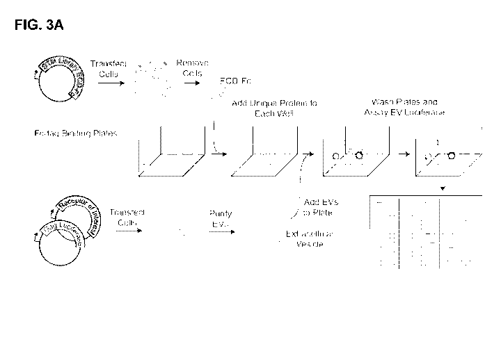
FIG. 3A is a schematic diagram showing the workflow of the RDIMIS (Receptor-
Display In
Membranes Interaction Screen) protocol. EVs are isolated from the conditioned
media of cells
expressing the receptor of interest alongside gag-luc. A library of single-
pass transmembrane (STM)
proteins, expressed as Fc-tagged ectodomains (ECD-Fc), are immobilized on
plates. Receptor-EVs are
screened against the collection of plate-bound STM proteins using a semi-
automated workflow. EV
binding to interacting ectodomains in the library is detected using
luminescence.
12
CA 03201626 2023-05-11
WO 2022/119805
PCT/US2021/061120
FIG. 3B is a scatter plot showing the results of two independent RDIMIS
screens (Repeat 1 and
Repeat 2) testing for interaction between PVR gD-GPI EVs and the STM protein
library.
FIG. 3C is a scatter plot showing the results of an RDIMIS screen testing for
interaction between
PVR gD-GPI EVs and the STM protein library (Repeat 2 of Fig. 3B) and an RDIMIS
screen testing for
interaction between full-length (FL) PVR EVs and the STM protein library.
FIG. 4A is a set of photomicrographs showing Western blots of whole cell
lysates (Cells) or EVs
expressing the full-length untagged receptors PD1, PD-L1, EPHA3, CD248,
LRRC15, PVR, or PVRL1
and stained with an antibody specific for the receptor. Anti-tubulin (a-Tub)
and anti-actin (a-Actin)
staining are provided as controls.
FIG. 4B is a set of photomicrographs showing Western blots of whole cell
lysates or EVs
expressing the indicated g D-GPI tagged receptor ectodomains and stained with
an antibody specific for
the gD tag (a-gD). Anti-tubulin (a-Tub) and anti-actin (a-Actin) staining are
provided as controls.
FIG. 4C is a set of negative stain electromicrographs showing selective anti-
gD immunogold
labeling of gD-GPI expressing vesicles.
FIG. 40 is a graph showing the design and results of a biolayer interferometry
experiment. An
anti-gD antibody was attached to a sensor. The sensor was incubated with EVs
expressing the indicated
gD-GPI ectodomains, and the BLI signal (in nm) was measured.
FIG. 5A is a scatter plot showing the results of an RDIMIS screen testing for
interaction between
PVR gD-GPI EVs and the STM protein library and an RDIMIS screen testing for
interaction between PD-
L1 gD-GPI EVs and the STM protein library. Screens are plotted against one
another to differentiate
receptor-specific hits (near either axis) from the generic vesicle binders
common between the screens.
Hits whose signal is above the 98% quantile for each individual screen and for
which there is at least a 4x
enrichment for a specific screen are labeled. Other hits are identified as
generic vesicle binders common
between the screens.
FIG. 5B is a scatter plot showing the results of an RDIMIS screen testing for
interaction between
CD80 gD-GPI EVs and the STM protein library and an RDIMIS screen testing for
interaction between
CD276 gD-GPI EVs and the STM protein library. Receptor-specific hits are
located near the axes. Hits
whose signal is above the 98% quantile for each individual screen and for
which there is at least a 4x
enrichment for a specific screen are labeled. Other hits are identified as
generic vesicle binders common
between the screens.
FIG. 5C is a set of diagrams showing the overlap between the binding partners
identified for PVR,
PD-L1, CD80, and CD276 in the present study and interactions listed in the
STRING, Bioplex and Biogrid
databases. For PD-Li/CD274, no interactions with members of the STM library
were present in the
Bioplex database. No experimentally verified interactions were listed in
STRING for CD276/B7-H3.
FIG. 6A is a scatter plot showing the results of an AVEXIS screen testing for
interaction between
LRRC15 ectodomain pentamers and the STM protein library. LRRC15 pentamer
binding was not
observed above background in the well containing the CD248 ectodomain
(highlighted). Gray dots
indicate positive control wells on each plate, in which stock pentamer was
added but not washed away.
FIG. 6B is a set of diagrams showing a comparison between the hits identified
herein using
LRRC15 full-length (FL) or gD-GPI ectodomain in EVs and those represented in
the Bioplex and Biogrid
13
CA 03201626 2023-05-11
WO 2022/119805
PCT/US2021/061120
databases. No interaction with experimental evidence between LRRC15 and a STM
protein in the library
was represented in the STRING database.
FIG. 7A is a scatter plot showing the results of an RDIMIS screen testing for
interaction between
LRRC15 gD-GPI EVs and the STM protein library. Results are compared with PVR
screen results shown
in Fig 30. Hits whose signal is above the 98% quantile for each individual
screen and for which there is
at least a 4x enrichment for a specific screen are labeled. Other hits are
identified as generic vesicle
binders common between the screens.
FIG. 7B is a scatter plot showing the results of an RDIMIS screen testing for
interaction between
LRRC15 full-length EVs and the STM protein library. Results are compared with
PVR screen results
.. shown in Fig 30. Hits whose signal is above the 98% quantile for each
individual screen and for which
there is at least a 4x enrichment for a specific screen are labeled. Other
hits are identified as generic
vesicle binders common between the screens.
FIG. 8A is a scatter plot showing bulk RNA-seq expression levels (transcripts
per million (TPM))
of LRRC15 (x-axis) and 0D248 (y-axis) for head and neck squamous cell
carcinoma. Each point
represents a single patient sample. Spearman's rank correlation coefficient
and significance values are
given on the top right.
FIG. 8B is a scatter plot showing bulk RNA-seq expression levels (transcripts
per million (TPM))
of LRRC15 (x-axis) and 0D248 (y-axis) for breast invasive carcinoma. Each
point represents a single
patient sample. Spearman's rank correlation coefficient and significance
values are given on the top
right.
FIG. 8C is a pair of Uniform Manifold Approximation and Projection (UMAP)
dimensionality
reduction plots showing non-tumor cells from single-cell RNA-seq data of head
and neck cancer patients.
Cells are shaded by expression level of the indicated marker genes (left;
LRRC15; right: 0D248).
FIG. 9A is a scatter plot showing bulk RNA-seq expression levels (transcripts
per million (TPM))
of LRRC15 (x-axis) and 0D248 (y-axis) for pancreatic ductal adenocarcinoma
(The Cancer Genome
Atlas (TOGA) data).
FIG. 9B is a scatter plot showing bulk RNA-seq expression levels (transcripts
per million (TPM))
of LRRC15 (x-axis) and 0D248 (y-axis) for urothelial bladder carcinoma (The
Cancer Genome Atlas
(TOGA) data).
FIG. 9C is a pair of UMAP dimensionality reduction plots showing non-tumor
cells from single-cell
RNA-seq data of head and neck cancer patients. Cells are shaded by expression
level of the indicated
marker genes (left; DCN cancer-associated fibroblasts; right: RGS5 cancer-
associated pericyte markers).
FIG. 10A is a scatter plot showing the results of an AVEXIS screen testing for
interaction between
0D248 ectodomain pentamers and the STM protein library. 0D248 pentamer binding
was not observed
above background in the well containing the LRRC15 ectodomain (highlighted).
Gray dots indicate
positive control wells on each plate, in which stock pentamer was added but
not washed away.
FIG. 10B is a graph showing the results of a surface plasmon resonance (SPR)
assay in which
LRRC15-Fc was captured by Protein A on the SPR chip and the indicated analytes
were added.
LRRC15 was loaded at either 5 g/mL (red and green lines) or 50 g/mL
concentration. Analytes were
.. loaded at 400 nM concentration.
14
CA 03201626 2023-05-11
WO 2022/119805
PCT/US2021/061120
FIG. 11A is a schematic diagram and a graph showing the design and results of
a BLI
experiment. 0D248 was expressed as a recombinant protein and immobilized on a
sensor. The sensor
was contacted with a solution comprising LRRC15-Fc (500 nM) or EVs comprising
LRRC15 full-length or
gD-GPI LRRC15 (0.25 mg/ml), and the BLI signal (in nm) was measured.
FIG. 11B is a schematic diagram and a set of micrographs showing binding of
EVs comprising
gag-NeonGreen and full-length (FL) LRRC15, gD-GPI LRRC15 (LRRC15-gD), or an
empty-vector control
to cells transiently expressing full-length 0D248 or gD-GPI 0D248.
FIG. 11C is a schematic diagram and a set of micrographs showing binding of
0D248 (as a
tetramerized recombinant ectodomain) or an empty-vector control to cells
transiently expressing full-
length LRRC15 or gD-GPI LRRC15. Scale bar represents 20 m.
FIG. 110 is a bar graph showing a quantification of EV binding in Fig. 11B
based on NeonGreen
signal levels (mean standard error for three independent replicates).
FIG. 12A is a scatter plot showing a comparison of Renilla luciferase
fluorescence and
fluorescence of cholesterol detected using the AMPLEXTm Red Cholesterol Assay
Kit (Thermo Fisher)
(Cholesterol) as a readout for a dilution series of PD-L1 and PVR gD-GPI EVs
harvested from
EXPI293FTM cells transfected with gag-Rluc or untransfected cells.
FIG. 12B is a bar graph showing the relative signal levels from Renilla
luciferase and the
AMPLEXTm Red Cholesterol Assay Kit (Thermo Fisher) observed in a small-scale
RDIMIS screen with the
listed genes immobilized in wells and probed using PD-L1 gD-GPI EVs. Results
for both readouts are
normalized to the respective PDCD1 signals.
Fig. 12C is a scatter plot showing the results of an RDIMIS screen performed
using cholesterol
as a readout on EVs expressing gag-Rluc (x-axis) or vesicles harvested from
untransfected cells (y-axis).
Fig. 13A is a set of photomicrographs showing Western blots of EVs expressing
full-length (FL)
or gD-GPI tagged ectodomains of the receptors PVR, PD-L1, CD276, CD80, and
LRRC15 stained with
an antibody specific for the gD tag (a-gD). a-Actin, a-PVR, and a-LRRC15
staining are also shown.
Fig. 13B is a schematic diagram and a graph showing the design and results of
a BLI
experiment. Biotinylated PD-L1 was incubated with a streptavidin BLI
biosensor. This was then
incubated with EVs expressing full-length or gD-GPI LRTM1 or a vector control,
and the BLI signal (in
nm) was measured.
Fig. 13C is a set of micrographs showing EVs bound to the surface of cells
transiently expressing
full-length or gD-GPI tagged PD-L1. EVs contained full-length or gD-GPI LRTM1
or a vector control. EVs
comprised gag-NeonGreen, and green represents direct fluorescence from the
EVs. DNA of the
mammalian cells is shown in blue.
Fig. 130 is a schematic diagram and a graph showing the design and results of
a BLI
experiment. Biotinylated PD-L1 was incubated with a streptavidin BLI
biosensor. This was then
incubated with EVs expressing full-length or gD-GPI LRTM1 or a vector control
in the presence or
absence of different concentrations of Fc-tagged PD1 ectodomain or a human IgG
control, and the BLI
signal (in nm) was measured.
CA 03201626 2023-05-11
WO 2022/119805
PCT/US2021/061120
Fig. 14A is a scatter plot showing the results of an RDIMIS screen performed
using EVs
comprising PVR gD-GPI (Y axis) compared to "empty" EVs derived from cells
transfected with a vector
control (X axis).
Fig. 14B is a scatter plot showing the results of an RDIMIS screen performed
using EVs
comprising PD-L1 gD-GPI (Y axis) compared to "empty" EVs derived from cells
transfected with a vector
control (X axis).
Fig. 14C is a scatter plot showing the results of an RDIMIS screen performed
using EVs
comprising 0D80 gD-GPI (Y axis) compared to "empty" EVs derived from cells
transfected with a vector
control (X axis).
Fig. 140 is a scatter plot showing the results of an RDIMIS screen performed
using EVs
comprising 0D276 gD-GPI (Y axis) compared to "empty" EVs derived from cells
transfected with a vector
control (X axis).
Fig. 14E is a scatter plot showing the results of an RDIMIS screen performed
using EVs
comprising LRRC15 gD-GPI (Y axis) compared to "empty" EVs derived from cells
transfected with a
vector control (X axis).
Fig. 14F is a scatter plot showing the results of an RDIMIS screen performed
using EVs
comprising full-length LRRC15 (Y axis) compared to "empty" EVs derived from
cells transfected with a
vector control (X axis).
Fig. 15 is a network diagram showing the generic vesicle binders identified
herein (green boxes)
integrated with the IgSF Interactome's list of high confidence interactions
(1) (blue edges) and the
experimental and database list of interactions from STRING (2) (red edges) to
identify potential
interaction partners (blue boxes). The height of the boxes represent
normalized expression values in
HEK293 cells from The Cell Atlas (3) to estimate the expression of the
potential binding partners in the
EV parent cells and, therefore, the EVs themselves.
Fig. 16A is a set of scatter plots showing correlations and correlation
coefficients for each of the
RDIMIS screens performed. Screens were done in several batches: 1) PVR gD-GPI
repeat 1, 2) PVR
gD-GPI repeat 2 and PD-L1 gD-GPI, 3) CD80 gD-GPI and CD26 gD-GPI, 4) LRRC15 gD-
GPI, LRRC15
FL and PVR FL, 5) Vesicle control which are EVs with no overexpressed receptor-
of-interest.
Fig. 16B is a set of scatter plots showing the correlation between CD80 g D-
GPI and PVR-FL
screens. Two populations of generic vesicle binders are shown. Lower panel:
zoomed-in view of x axis.
Right panel: correlation with generic vesicle binders removed.
Fig. 17A is a graph showing the results of a BLI experiment in which membranes
were disrupted
with the cholesterol binder Filipin III. CD248 monomer were loaded onto a
NiNTA biosensor and
incubated with LRRC15 gD-GPI EVs that had been pre-treated for 30 minutes at
room temperature with
filipin III. Empty vesicles or filipin III are shown as negative controls.
Fig. 17B is a graph showing the results of a BLI experiment in which membranes
were disrupted
with the cholesterol binder Filipin III. CD248 monomer were loaded onto a
NiNTA biosensor and
incubated with full-length LRRC15 EVs that had been pre-treated for 30 minutes
at room temperature
with filipin III. Empty vesicles or filipin III are shown as negative
controls.
Fig. 17C is a graph showing binding of the EVs of Fig. 17A to an anti-gD
antibody.
16
CA 03201626 2023-05-11
WO 2022/119805
PCT/US2021/061120
Fig. 170 is a graph showing the results of a BLI experiment in which membranes
were disrupted
with Methyl-beta cyclodextrin (MPCD). 0D248 monomer were loaded onto a NiNTA
biosensor and
incubated with LRRC15 gD-GPI EVs that had been pre-treated for 30 minutes at
room temperature with
filipin III. Empty vesicles or filipin III are shown as negative controls.
Fig. 17E is a graph showing the results of a BLI experiment in which membranes
were disrupted
with M[3CD. CD248 monomer were loaded onto a NiNTA biosensor and incubated
with full-length
LRRC15 EVs that had been pre-treated for 30 minutes at room temperature with
filipin III. Empty vesicles
or filipin III are shown as negative controls.
Fig. 17F is a graph showing binding of the EVs of Fig. 17D to an anti-gD
antibody.
Fig. 18A is a scatter plot showing level of antibody surface staining (a.u.)
for >500 multi-
transmembrane receptors expressed on cells and a pair of photomicrographs
showing representative cell
surface staining for a low-expressing receptor (DRD2) and a high-expressing
receptor (S1PR1).
Background staining is denoted by the line.
Fig. 18B is a scatter plot showing level of surface staining (a.u.) using an
anti-FLAG antibody and
fluorescence of a Venus tag (X-axis; total receptor (a.u.)) for >400 G protein-
coupled receptors (GPCRs)
engineered with an N-terminal FLAG tag and a C-terminal Venus. The inset
images are
photomicrographs showing representative cell surface staining (magenta) and
Venus fluorescence
(green) for a low-expressing receptor (DRD2), a high-expressing receptor
(S1PR1), and a very highly
expressed single-transmembrane receptor (EGFR). Background staining was
determined by average of
signal on untransfected cells and is denoted by the line.
Fig. 18C is a circle chart showing characteristics of the 1791 members of the
multi-
transmembrane (MTMR) receptor library. Only >500 members have an extracellular
HIS tag, and only
about half of those receptors show staining above background.
Fig. 180 is a circle chart showing the proportions of the GPCRs of Fig. 18B
having low, medium,
and high FLAG staining expression levels. "Medium" receptor expression was
defined as ten times the
background signal.
Fig. 19A is a scatter plot showing results of a screen for binding (a.u.) of
EGF-647 to members of
the multi-transmembrane receptor library. EGF-647 bound only to EGFR, which
was printed on each
plate as a transfection control. The inset panel is a photomicrograph showing
the fluorescent ligand.
DAPI staining is shown.
Fig. 19B is a scatter plot showing results of a screen for binding (a.u.) of
RSPO3 to members of
the multi-transmembrane receptor library. RSPO3 bound to LGR4 and LGR5.
Imaging artifacts are
denoted by an X. The inset panel is a photomicrograph showing the fluorescent
ligand. DAPI staining is
shown.
Fig. 19C is a scatter plot showing results of a screen for binding (a.u.) of
PVR to members of the
multi-transmembrane receptor library. PVR bound to CD226, a single-pass
transmembrane receptor that
was added as a positive control. The inset panel is a photomicrograph showing
the fluorescent ligand.
DAPI staining is shown.
Fig. 190 is a scatter plot showing results of a screen for binding (a.u.) of
PD-L1 to members of
the multi-transmembrane receptor library. PD-L1 bound to PVR the adhesion G
protein-coupled receptor
17
CA 03201626 2023-05-11
WO 2022/119805
PCT/US2021/061120
B1 (ADGRB1), as well as to the single-pass transmembrane receptors PD1, PDL2,
0D80, and EPHA3,
which were added as positive controls. The inset panel is a photomicrograph
showing the fluorescent
ligand. DAPI staining is shown.
Fig. 20A is a schematic diagram showing an extracellular vesicle (EV)
comprising a tagged multi-
pass transmembrane receptor. The extracellular regions of the receptor are on
the outside of the EV, and
the intracellular regions are in the lumen of the EV. Locations of the FLAG
tag and fluorescent tag (Luc)
are shown.
Fig. 20B is a negative stain electron microscopy image showing EVs.
Fig. 20C is a graph showing the size distribution (in nm) and concentration
(106 particles per mL)
of EVs from cells transfected with PVR and GAG; transfected with PVR only; or
not transfected (control),
as measured using NanoSight particle tracking.
Fig. 200 is a bar graph showing the mean size (nm) of the EVs of Fig. 200.
Fig. 20E is a graph showing the results of a BLI experiment assessing binding
of an anti-gD
antibody to EVs derived from cells transfected with PVR and GAG; PVR only; or
EVs from untransfected
cells.
Fig. 20F is a graph showing the results of a BLI experiment assessing binding
of the PVR ligand
TIGIT (TIGIT Fc) to EVs derived from cells transfected with PVR and GAG; PVR
only; or EVs from
untransfected cells.
Fig. 20G is a graph showing the results of a BLI experiment assessing binding
of an anti-FLAG
antibody to EVs comprising the G protein-coupled receptors (GPCRs) ADGRB1,
LGR4, and LGR5.
GPCRs comprised an N-terminal FLAG tag.
Fig. 21A is a scatter plot showing results of a screen for binding (a.u.) of
EVs comprising PVR to
members of the multi-transmembrane receptor library and to positive controls.
PVR bound to the positive
controls. Imaging artifacts are denoted by an X. The inset panel is a
photomicrograph showing vesicle
fluorescence from GAG-neonGreen fusion. DAPI staining is shown.
Fig. 21B is a scatter plot showing results of a screen for binding (a.u.) of
EVs comprising PD-L1
to members of the multi-transmembrane receptor library and to positive
controls. PVR bound to the
positive controls and to ADGRB1. Imaging artifacts are denoted by an X. The
inset panel is a
photomicrograph showing vesicle fluorescence from GAG-neonGreen fusion. DAPI
staining is shown.
Fig. 21C is a scatter plot showing results of a screen for binding (a.u.) of
ADGRB1 to members of
a library comprising the extracellular domains of Fc-fused single
transmembrane receptors (STMRs).
Interactions with RTN4R and PD-L1 were confirmed and new interactions were
revealed.
Fig. 22A is a bar graph showing quantification of binding of the recombinant
proteins PD-L1-Fc,
ICOSLG-Fc, and RTN4R-Fc (each conjugated to a protein A plate) to EVs
comprising ADGRB1 or LGR4.
Fig. 22B is a set of photomicrographs showing the results of assays for
binding of the
recombinant proteins PD-L1-Fc, ICOSLG-Fc, and RTN4R-Fc to HEK cells expressing
ADGRB1 fused to
Venus. In the merged image, DAPI is shown in blue; the Venus signal from the
ADGRB1 fusion protein is
shown in green, and the signal for staining of the Fc tag is shown in magenta.
Co-localized green and
magenta signals are shown in white.
18
CA 03201626 2023-05-11
WO 2022/119805
PCT/US2021/061120
Fig. 23 is a schematic diagram showing the design of the GPCR screening
platform.
Comprehensive libraries are collected in 384-well plate format. A
comprehensive collection of
overexpression plasmids are printed onto 384-well imaging plates. Cells are
reverse transfected, then
treated with fluorescent ligand and analyzed in a high throughput, high
content imager
Fig. 24A is a set of photomicrographs showing the results of assays for
binding of the
recombinant protein PD-L1-Fc to HEK cells expressing ADGRB1 fused to Venus. In
the merged image,
DAPI is shown in blue; the Venus signal from the ADGRB1 fusion protein is
shown in green, and the
signal for staining of the Fc tag is shown in magenta. Co-localized green and
magenta signals are shown
in white. All contrast and brightness settings are matched to Fig. 22B.
Fig. 24B is a set of photomicrographs showing the results of assays for
binding of the
recombinant protein ICOSLG-Fc to HEK cells expressing ADGRB1 fused to Venus.
In the merged
image, DAPI is shown in blue; the Venus signal from the ADGRB1 fusion protein
is shown in green, and
the signal for staining of the Fc tag is shown in magenta. Co-localized green
and magenta signals are
shown in white. All contrast and brightness settings are matched to Fig. 22B.
Fig. 24C is a set of photomicrographs showing the results of assays for
binding of the
recombinant protein RTN4R-Fc to HEK cells expressing ADGRB1 fused to Venus. In
the merged image,
DAPI is shown in blue; the Venus signal from the ADGRB1 fusion protein is
shown in green, and the
signal for staining of the Fc tag is shown in magenta. Co-localized green and
magenta signals are shown
in white. All contrast and brightness settings are matched to Fig. 22B.
Fig. 25A is a graph showing the results of bioluminescent energy transfer
(BRET) assays for [3-
arrestin and SH2 recruitment in HEK cells. No activation of ADGRB1 or EphA3
was observed in
response to PD-L1.
Fig. 25B is a graph showing calcium sensing (GCaMP6s fluorescence) following
treatment of
HEK cells. No response was observed downstream of ADGRB1 or EphA3 in response
to PD-L1.
Fig. 25C is a graph showing cAMP stimulation (assessed by GLOSENSORTM)
following
treatment of HEK cells. No response was observed.
Fig. 250 is a graph showing cAMP inhibition following treatment of HEK cells.
No response was
observed.
Fig. 26A is a scatter plot showing results of a screen for binding of EVs
comprising ADGRB1 to
members of a secreted protein Fc library. A positive control (anti-FLAG
antibody) is labeled.
Fig. 26B is a scatter plot showing results of a screen for binding of EVs
comprising ADGRB1 to
members of a library comprising the extracellular domains of single
transmembrane receptors fused to Fc
(STM library). Binding to RTN4R and PD-L1 was confirmed, and new interactions
were identified. Novel
hits are labeled.
Fig. 26C is a scatter plot showing results of a screen for binding of EVs
comprising LGR4 to
members of a secreted protein Fc library. A positive control (anti-FLAG
antibody) is labeled.
Fig. 260 is a scatter plot showing results of a screen for binding of EVs
comprising LGR4 to
members of the STM library. A positive control (RSP03) is labeled. Novel hits
are shown in green.
19
CA 03201626 2023-05-11
WO 2022/119805
PCT/US2021/061120
Fig. 26E is a scatter plot showing results of a screen for binding of EVs
comprising LGR5 to
members of a secreted protein Fc library. A positive control (anti-FLAG
antibody) is labeled. Novel hits
are shown in green.
Fig. 26F is a scatter plot showing results of a screen for binding of EVs
comprising LGR5 to
members of the STM library. A positive control (anti-FLAG antibody) is
labeled. Novel hits are shown in
green. Shared LGR4 and LGR5 hits are shown in blue.
Fig. 27 is a bar graph showing the results of a carboxypeptidase M (CPM)
activity assay. CPM-
FL: vesicles comprised full-length CPM. CPMgD: vesicles comprised gD-GPI (gD)
CPM. pRK EV: vector
control; PBS only: buffer control.
Fig. 28 is a pair of photomicrographs showing a pair of Western blots showing
levels of total
EPHA3 and phosphorylated EPHA3 species (pEPHA2/3/4 and pEPHA3/4/5) detected in
EVs comprising
full-length EPHA3 (EPHA3-FL) and PDL1-Fc, EPHA3-Fc, the EPHA3 ligands EFNA1-Fc
and EFNA5-Fc,
and full-length PD Li. pRK EV: vector control.
DETAILED DESCRIPTION OF THE INVENTION
I. DEFINITIONS
As used herein, the term "biological vesicle" or "By" refers to a lipid
bilayer-delimited particle that
is naturally secreted from a parent cell, e.g., a mammalian cell. BVs may be,
e.g., extracellular vesicles
(EVs; nanometer-sized particles, e.g., recombinant extracellular vesicles
(rEVs)), exosomes,
microvesicles, or virus-like particles (VLPs). VLPs are described, e.g., in
Titeca et al., Nature Protocols,
12(5): 881-898, 2017). BV compositions or preparations may include only one of
EVs, exosomes,
microvesicles, or VLPs, or may include a mixture of two, three, or all four of
EVs, exosomes, microvesicles,
and VLPs. BVs contain proteins folded and inserted into their native membranes
using the parent cell's
endogenous machinery. In some aspects, BVs include proteins that are not
native to the parent cell, e.g.,
proteins that the parent cell has been modified to express (e.g., heterologous
membrane-associated
proteins, e.g., heterologous membrane-associated proteins comprising a protein
fragment, a tag, and an
anchor). Production of BVs by a parent cell may be increased by contacting the
parent cell with a
membrane-budding agent (e.g., transforming the cell with a membrane-budding
agent, e.g., a HIV gag
protein) and/or exposing the cell to conditions that promote the formation of
BVs. In some aspects, the
BV is purified from the parent cell (e.g., purified from a culture medium
comprising the parent cell).
As used herein, the term "membrane-budding agent" refers to an agent that
increases the
production of BVs (e.g., extracellular vesicles (EVs), exosomes,
microvesicles, and/or virus-like particles
(VLPs)) by the parent cell. In some aspects, the membrane-budding agent is an
HIV gag protein. In some
aspects, the HIV gag protein has the amino acid sequence of SEQ ID NO: 1. In
some aspects, the HIV
gag protein has at least 90% identity to the amino acid sequence of SEQ ID NO:
1, e.g., 91%, 92%, 93%,
94%, 95%, 96%, 97%, 98%, or 99% identity to SEQ ID NO: 1. In some aspects, the
parent cells are
transformed with the membrane-budding agent. Cells may additionally or
alternatively be exposed to a
condition (e.g., a culture condition) that increases the production of BVs
(e.g., extracellular vesicles (EVs),
exosomes, microvesicles, and/or virus-like particles (VLPs)) by the parent
cell. Further examples of
membrane-budding agents include self-assembling VLPs (e.g., MLGag, AARDC1
(e.g., hAARDC1), and
CA 03201626 2023-05-11
WO 2022/119805
PCT/US2021/061120
Acyl.Hrs); agents that enhance endogenous vesicle formation pathways such as
exosome or tumor
pathways (e.g., RhoA.F3OL, ARF6.067L, VPS4a, HAS3, CD9, 0D63, and CD81); and
factors associated
with apoptotic bodies (e.g., constitutively active ROCK1).
The term "about" as used herein refers to the usual error range for the
respective value readily
known to the skilled person in this technical field. Reference to "about" a
value or parameter herein
includes (and describes) aspects that are directed to that value or parameter
per se.
The term "single transmembrane receptor," "single-pass transmembrane
receptor," or "STM
receptor," as used herein, refers to a protein having a single transmembrane
domain. In some aspects,
the STM receptor is expressed on the cell surface. Exemplary STM receptors are
provided in Table 4, as
well as in PCT/US2020/025471, Martinez-Martin et al., Cell, 174(5): 1158-1171,
2018, and Clark et al.,
Genome Res, 13: 2265-2270, 2003, each of which is incorporated by reference
herein in its entirety. In
some aspects, the STM protein has the UniProt annotation "leucine-rich,"
"cysteine-rich," "ITIM/ITAM"
(immunoreceptor tyrosine-based inhibition motif/immunoreceptor tyrosine-based
activation motif), "TN FR"
(tumor necrosis factor receptor), "TLR/ILR" (Toll-like receptor/interleukin
receptor), "semaphorin," "Kinase-
like," "Ig-like" (immunoglobulin-like), "fibronectin," "ephrin," "EGF,"
"cytokineR," or "cadherin." STM
receptors may be identified based on, e.g., the presence of a signal peptide
or a predicted
transmembrane region in the amino acid sequence. In some aspects, the STM
receptor is expressed as
an extracellular domain.
As used herein, the term "extracellular domain" or "ECD" refers to a protein
domain that is
predicted to be localized outside of the outer plasma membrane of the cell. In
some instances, the ECD
is an ECD of a receptor, e.g., a STM receptor. In some aspects, the ECD is an
ECD of an IgSF protein.
In some aspects, the ECD is the ECD of PDPN. In some aspects, the boundaries
of the extracellular
domain may be identified by prediction of domains that indicate that the
protein crosses the plasma
membrane, e.g., a transmembrane domain (e.g., a transmembrane helix). In some
aspects, the presence
of an extracellular domain may be predicted by the presence of a domain,
sequence, or motif that
indicates that the protein is trafficked to the plasma membrane, e.g., a
signal sequence or a
glycosylphosphatidylinositol (GPI) linkage site. In some aspects, the
boundaries of the ECD are
determined according to UniProt annotations. In some aspects, the ECD is
soluble. In some aspects, the
extracellular domain is expressed in the context of a full-length protein. In
other aspects, the extracellular
domain is expressed as an isolated extracellular domain, e.g., a sequence of
amino acid residues
comprising only the amino acid residues of a protein that are predicted to be
extracellular.
In some aspects, the isolated ECD is included in a fusion protein. In some
aspects, inclusion in a
fusion protein increases solubility, ease of expression, ease of capture
(e.g., on a protein A-coated plate),
multimerization, or some other desirable property of the ECD. In some aspects,
the ECD or ECD fusion
protein is a monomer. In other aspects, the ECD or ECD fusion protein is a
multimer, e.g., a tetramer or
a pentamer. In some aspects, the ECD is fused to a human IgG. In some aspects,
the ECD is fused to a
human Fc tag. In some aspects, the ECD is fused to an Avidity AVITAGTm (Avi
tag). In some aspects,
the ECD is fused to a polyhistidine (His) tag. In some aspects, the ECD is
fused to a glycoprotein D (gD)
tag and a glycosylphosphatidylinositol (GPI) linker, e.g., a gD-GPI tag. In
other aspects, the ECD is fused
to the pentamerization domain of rat cartilaginous oligomeric matrix protein
(COMP) and the P-lactamase
21
CA 03201626 2023-05-11
WO 2022/119805
PCT/US2021/061120
protein, e.g., as described in Bushell et al., Genome Res, 18: 622-630, 2008.
In some aspects, the ECD
fusion protein further includes a cleavage sequence, e.g., a TEV cleavage
sequence, to allow removal of
one or more domains. In some instances, an ECD fusion protein having an Avi
tag and an Fc tag
cleavable at a cleavage sequence is further processed to remove the Fc tag, to
biotinylate the Avi tag,
and to fuse the biotinylated ECD fusion protein to a fluorescent streptavidin
(SA), e.g., to form a
tetramerized ECD fusion protein. In some instances, the isolated ECD or ECD
fusion protein is purified.
As used herein, a "modulator" is an agent that modulates (e.g., increases,
decreases, activates,
or inhibits) a given biological activity, e.g., an interaction or a downstream
activity resulting from an
interaction. A modulator or candidate modulator may be, e.g., a small
molecule, an antibody, an antigen-
binding fragment (e.g., a bis-Fab, an Fv, a Fab, a Fab'-SH, a F(ab')2, a
diabody, a linear antibody, an
scFv, an ScFab, a VH domain, or a VHH domain), a peptide, a mimic, an
antisense oligonucleotide
(ASO), or a small interfering RNA (siRNA).
By "increase" or "activate" is meant the ability to cause an overall increase,
for example, of 20%
or greater, of 50% or greater, or of 75%, 85%, 90%, or 95% or greater. In
certain aspects, increase or
activate can refer to a downstream activity of a protein-protein interaction.
By "reduce" or "inhibit" is meant the ability to cause an overall decrease,
for example, of 20% or
greater, of 50% or greater, or of 75%, 85%, 90%, or 95% or greater. In certain
aspects, reduce or inhibit
can refer to a downstream activity of a protein-protein interaction.
"Affinity" refers to the strength of the sum total of noncovalent interactions
between a single
binding site of a molecule (e.g., a receptor) and its binding partner (e.g., a
ligand). Unless indicated
otherwise, as used herein, "binding affinity" refers to intrinsic binding
affinity, which reflects a 1:1
interaction between members of a binding pair (e.g., receptor and ligand). The
affinity of a molecule X for
its partner Y can generally be represented by the dissociation constant (KD).
Affinity can be measured by
common methods known in the art, including those described herein.
"Complex" or "complexed" as used herein refers to the association of two or
more molecules that
interact with each other through bonds and/or forces (e.g., Van der Waals,
hydrophobic, hydrophilic
forces) that are not peptide bonds. In one aspect, a complex is
heteromultimeric. It should be
understood that the term "protein complex" or "polypeptide complex" as used
herein includes complexes
that have a non-protein entity conjugated to a protein in the protein complex
(e.g., including, but not
limited to, chemical molecules such as a toxin or a detection agent).
The term "parent cell" refers to cells from which BVs are produced. Parent
cells include cells into
which an exogenous nucleic acid has been introduced, including the progeny of
such cells. Parent cells
include "transfected cells," "transformed cells," and "transformants," which
include the primary
transformed cell and progeny derived therefrom without regard to the number of
passages. Progeny may
not be completely identical in nucleic acid content to a parent cell, but may
contain mutations. Mutant
progeny that have the same function or biological activity as screened or
selected for in the originally
transformed cell are included herein. In some aspects, the parent cell is
stably transformed with the
exogenous nucleic acid. In other aspects, the parent cell is transiently
transformed with the exogenous
nucleic acid.
22
CA 03201626 2023-05-11
WO 2022/119805
PCT/US2021/061120
The term "leucine-rich repeat-containing protein 15" or "LRRC15," as used
herein, broadly refers
to any native LRRC15 from any mammalian source, including primates (e.g.,
humans) and rodents (e.g.,
mice and rats), unless otherwise indicated. The term encompasses full-length
LRRC15 and isolated
regions or domains of LRRC15, e.g., the LRRC15 ECD. The term also encompasses
naturally occurring
variants of LRRC15, e.g., splice variants or allelic variants. The amino acid
sequence of an exemplary
human LRRC15 is shown under UniProt Accession No. 08TF66. Minor sequence
variations, especially
conservative amino acid substitutions of LRRC15 that do not affect LRRC15
function and/or activity, are
also contemplated by the invention.
The term "programmed cell death 1 ligand 1" or "PD-L1," as used herein,
broadly refers to any
native PD-L1 from any mammalian source, including primates (e.g., humans) and
rodents (e.g., mice and
rats), unless otherwise indicated. PD-L1 is also called 0D274. The term
encompasses full-length PD-L1
and isolated regions or domains of PD-L1, e.g., the PD-L1 ECD. The term also
encompasses naturally
occurring variants of PD-L1, e.g., splice variants or allelic variants. The
amino acid sequence of an
exemplary human PD-L1 is shown under UniProt Accession No. 09NZ07. Minor
sequence variations,
especially conservative amino acid substitutions of PD-L1 that do not affect
PD-L1 function and/or
activity, are also contemplated by the invention.
The term "poliovirus receptor" or "PVR," as used herein, broadly refers to any
native PVR from
any mammalian source, including primates (e.g., humans) and rodents (e.g.,
mice and rats), unless
otherwise indicated. The term encompasses full-length PVR and isolated regions
or domains of PVR,
e.g., the PVR ECD. The term also encompasses naturally occurring variants of
PVR, e.g., splice variants
or allelic variants. The amino acid sequence of an exemplary human PVR is
shown under UniProt
Accession No. A0A0C4DG49. Minor sequence variations, especially conservative
amino acid
substitutions of PVR that do not affect PVR function and/or activity, are also
contemplated by the
invention.
The term "CD80," as used herein, broadly refers to any native CD80 from any
mammalian
source, including primates (e.g., humans) and rodents (e.g., mice and rats),
unless otherwise indicated.
CD80 is also called B7-1. The term encompasses full-length CD80 and isolated
regions or domains of
CD80, e.g., the CD80 ECD. The term also encompasses naturally occurring
variants of CD80, e.g.,
splice variants or allelic variants. The amino acid sequence of an exemplary
human CD80 is shown
under UniProt Accession No. P33681. Minor sequence variations, especially
conservative amino acid
substitutions of CD80 that do not affect CD80 function and/or activity, are
also contemplated by the
invention.
The term "0D276," as used herein, broadly refers to any native 0D276 from any
mammalian
source, including primates (e.g., humans) and rodents (e.g., mice and rats),
unless otherwise indicated.
0D276 is also called B7-H3. The term encompasses full-length 0D276 and
isolated regions or domains
of 0D276, e.g., the 0D276 ECD. The term also encompasses naturally occurring
variants of 0D276,
e.g., splice variants or allelic variants. The amino acid sequence of an
exemplary human 0D276 is
shown under UniProt Accession No. Q5ZPR3. Minor sequence variations,
especially conservative amino
acid substitutions of 0D276 that do not affect 0D276 function and/or activity,
are also contemplated by
the invention.
23
CA 03201626 2023-05-11
WO 2022/119805
PCT/US2021/061120
The terms "TEM1," "0D248," and "endosialin," as used herein, broadly refer to
any native TEM1
from any mammalian source, including primates (e.g., humans) and rodents
(e.g., mice and rats), unless
otherwise indicated. The term encompasses full-length TEM1 and isolated
regions or domains of TEM1,
e.g., the TEM1 ECD. The term also encompasses naturally occurring variants of
TEM1, e.g., splice
variants or allelic variants. The amino acid sequence of an exemplary human
TEM1 is shown under
UniProt Accession No. Q9HCUO. Minor sequence variations, especially
conservative amino acid
substitutions of TEM1 that do not affect TEM1 function and/or activity, are
also contemplated by the
invention.
The terms "ADGRB1," "adhesion GPCR B1," and "adhesion G protein-coupled
receptor B1," as
used herein, broadly refer to any native ADGRB1 from any mammalian source,
including primates (e.g.,
humans) and rodents (e.g., mice and rats), unless otherwise indicated. The
term encompasses full-
length ADGRB1 and isolated regions or domains of ADGRB1, e.g., the ADGRB1
ECDs. The term also
encompasses naturally occurring variants of ADGRB1, e.g., splice variants or
allelic variants. The amino
acid sequence of an exemplary human ADGRB1 is shown under UniProt Accession
No. 014514. Minor
sequence variations, especially conservative amino acid substitutions of TEM1
that do not affect TEM1
function and/or activity, are also contemplated by the invention.
The terms "ICOSLG," "inducible T cell costimulatory ligand," and "ICOS
ligand," as used herein,
broadly refer to any native ICOSLG from any mammalian source, including
primates (e.g., humans) and
rodents (e.g., mice and rats), unless otherwise indicated. The term
encompasses full-length ICOSLG and
isolated regions or domains of ICOSLG, e.g., the ICOSLG ECD. The term also
encompasses naturally
occurring variants of ICOSLG, e.g., splice variants or allelic variants. The
amino acid sequence of an
exemplary human ICOSLG is shown under UniProt Accession No. 075144. Minor
sequence variations,
especially conservative amino acid substitutions of ICOSLG that do not affect
ICOSLG function and/or
activity, are also contemplated by the invention.
The term "protein," as used herein, refers to any native protein from any
vertebrate source,
including mammals such as primates (e.g., humans) and rodents (e.g., mice and
rats), unless otherwise
indicated. The term encompasses "full-length," unprocessed protein any form of
the protein that results
from processing in the cell. The term also encompasses naturally occurring
variants of the protein, e.g.,
splice variants or allelic variants, e.g., amino acid substitution mutations
or amino acid deletion mutations.
The term also includes isolated regions or domains of the protein, e.g., the
extracellular domain (ECD).
An "isolated" protein or peptide is one which has been separated from a
component of its natural
environment. In some aspects, a protein or peptide is purified to greater than
95% or 99% purity as
determined by, for example, electrophoresis (e.g., SDS-PAGE, isoelectric
focusing (IEF), capillary
electrophoresis) or chromatography (e.g., ion exchange or reverse phase HPLC).
An "isolated" nucleic acid refers to a nucleic acid molecule that has been
separated from a
component of its natural environment. An isolated nucleic acid includes a
nucleic acid molecule
contained in cells that ordinarily contain the nucleic acid molecule, but the
nucleic acid molecule is
present extrachromosomally or at a chromosomal location that is different from
its natural chromosomal
location.
24
CA 03201626 2023-05-11
WO 2022/119805
PCT/US2021/061120
The term "vector," as used herein, refers to a nucleic acid molecule capable
of propagating
another nucleic acid to which it is linked. The term includes the vector as a
self-replicating nucleic acid
structure as well as the vector incorporated into the genome of a host cell
into which it has been
introduced. Certain vectors are capable of directing the expression of nucleic
acids to which they are
operatively linked. Such vectors are referred to herein as "expression
vectors."
The term "antibody" herein is used in the broadest sense and encompasses
various antibody
structures, including but not limited to monoclonal antibodies, polyclonal
antibodies, multispecific
antibodies (e.g., bispecific antibodies), and antibody fragments (e.g., bis-
Fabs) so long as they exhibit the
desired antigen-binding activity.
An "antigen-binding fragment" or "antibody fragment" refers to a molecule
other than an intact
antibody that comprises a portion of an intact antibody that binds the antigen
to which the intact antibody
binds. Examples of antigen-binding fragments include but are not limited to
bis-Fabs; Fv; Fab; Fab, Fab'-
SH; F(ab')2; diabodies; linear antibodies; single-chain antibody molecules
(e.g., scFv, ScFab); and
multispecific antibodies formed from antibody fragments.
A "single-domain antibody" refers to an antibody fragment comprising all or a
portion of the heavy
chain variable domain or all or a portion of the light chain variable domain
of an antibody. In certain
aspects, a single-domain antibody is a human single-domain antibody (see,
e.g., U.S. Patent No.
6,248,516 B1). Examples of single-domain antibodies include but are not
limited to a VHH.
A "Fab" fragment is an antigen-binding fragment generated by papain digestion
of antibodies and
consists of an entire L chain along with the variable region domain of the H
chain (VH), and the first
constant domain of one heavy chain (CH1). Papain digestion of antibodies
produces two identical Fab
fragments. Pepsin treatment of an antibody yields a single large F(ab')2
fragment which roughly
corresponds to two disulfide linked Fab fragments having divalent antigen-
binding activity and is still
capable of cross-linking antigen. Fab' fragments differ from Fab fragments by
having an additional few
residues at the carboxy terminus of the CH1 domain including one or more
cysteines from the antibody
hinge region. Fab'-SH is the designation herein for Fab' in which the cysteine
residue(s) of the constant
domains bear a free thiol group. F(ab')2 antibody fragments originally were
produced as pairs of Fab'
fragments which have hinge cysteines between them. Other chemical couplings of
antibody fragments
are also known.
The term "Fc region" herein is used to define a C-terminal region of an
immunoglobulin heavy
chain, including native sequence Fc regions and variant Fc regions. Although
the boundaries of the Fc
region of an immunoglobulin heavy chain might vary, the human IgG heavy chain
Fc region is usually
defined to stretch from an amino acid residue at position Cys226, or from
Pro230, to the carboxyl-
terminus thereof. The C-terminal lysine (residue 447 according to the EU
numbering system) of the Fc
region may be removed, for example, during production or purification of the
antibody, or by
recombinantly engineering the nucleic acid encoding a heavy chain of the
antibody. Accordingly, a
composition of intact antibodies may comprise antibody populations with all
Lys447 residues removed,
antibody populations with no Lys447 residues removed, and antibody populations
having a mixture of
antibodies with and without the Lys447 residue.
CA 03201626 2023-05-11
WO 2022/119805
PCT/US2021/061120
"Fv" consists of a dimer of one heavy- and one light-chain variable region
domain in tight, non-
covalent association. From the folding of these two domains emanate six
hypervariable loops (3 loops
each from the H and L chain) that contribute the amino acid residues for
antigen binding and confer
antigen binding specificity to the antibody. However, even a single variable
domain (or half of an Fv
comprising only three CDRs specific for an antigen) has the ability to
recognize and bind antigen,
although often at a lower affinity than the entire binding site.
The terms "full-length antibody," "intact antibody," and "whole antibody" are
used herein
interchangeably to refer to an antibody having a structure substantially
similar to a native antibody
structure or having heavy chains that contain an Fc region as defined herein.
"Single-chain Fv" also abbreviated as "sFv" or "scFv" are antibody fragments
that comprise the VH
and VL antibody domains connected into a single polypeptide chain. Preferably,
the scFv polypeptide
further comprises a polypeptide linker between the VH and VL domains, which
enables the scFv to form
the desired structure for antigen binding. For a review of scFv, see
Pluckthun, The Pharmacology of
Monoclonal Antibodies, vol. 113, Rosenburg and Moore eds., Springer-Verlag,
New York, pp. 269-315
(1994); Malmborg etal., J. Immunol. Methods 183:7-13, 1995.
The term "small molecule" refers to any molecule with a molecular weight of
about 2000 daltons
or less, e.g., about 1000 daltons or less. In some aspects, the small molecule
is a small organic
molecule.
The term "mimic," "peptide mimic," "polypeptide mimic," or "molecular mimic,"
as used herein,
refers to a polypeptide having sufficient similarity in conformation and/or
binding ability (e.g., secondary
structure, tertiary structure) to a given polypeptide or to a portion of said
polypeptide to bind to a binding
partner of said polypeptide. The mimic may bind the binding partner with
equal, less, or greater affinity
than the polypeptide it mimics. A molecular mimic may or may not have obvious
amino acid sequence
similarity to the polypeptide it mimics. A mimic may be naturally occurring or
may be engineered. In
some aspects, the mimic is a mimic of the protein of Table 1. In other
aspects, the mimic is a mimic of
the protein of Table 2. In yet other aspects, the mimic is a mimic of another
protein that binds to the
protein of Table 1 or the protein of Table 2. In some aspects, the mimic is a
mimic of the protein of Table
5. In other aspects, the mimic is a mimic of the protein of Table 6. In yet
other aspects, the mimic is a
mimic of another protein that binds to the protein of Table 5 or the protein
of Table 6. In some aspects,
the mimic may perform all functions of the mimicked polypeptide. In other
aspects, the mimic does not
perform all functions of the mimicked polypeptide.
As used herein, the term "conditions permitting the binding" of two or more
proteins to each other
(e.g., a protein of Table 1 and a protein of Table 2 or a protein of Table 5
and a protein of Table 6) refers
to conditions (e.g., protein concentration, temperature, pH, salt
concentration) under which the two or
more proteins would interact in the absence of a modulator or a candidate
modulator. Conditions
permitting binding may differ for individual proteins and may differ between
protein-protein interaction
assays (e.g., surface plasmon resonance assays, biolayer interferometry
assays, enzyme-linked
immunosorbent assays (ELISA), extracellular interaction assays, and cell
surface interaction assays.
"Percent ( /0) amino acid sequence identity" with respect to a reference
polypeptide sequence is
defined as the percentage of amino acid residues in a candidate sequence that
are identical with the
26
CA 03201626 2023-05-11
WO 2022/119805
PCT/US2021/061120
amino acid residues in the reference polypeptide sequence, after aligning the
sequences and introducing
gaps, if necessary, to achieve the maximum percent sequence identity, and not
considering any
conservative substitutions as part of the sequence identity. Alignment for
purposes of determining
percent amino acid sequence identity can be achieved in various ways that are
within the skill in the art,
for instance, using publicly available computer software such as BLAST, BLAST-
2, ALIGN or Megalign
(DNASTAR) software. Those skilled in the art can determine appropriate
parameters for aligning
sequences, including any algorithms needed to achieve maximal alignment over
the full-length of the
sequences being compared. For purposes herein, however, % amino acid sequence
identity values are
generated using the sequence comparison computer program ALIGN-2. The ALIGN-2
sequence
comparison computer program was authored by Genentech, Inc., and the source
code has been filed with
user documentation in the U.S. Copyright Office, Washington D.C., 20559, where
it is registered under
U.S. Copyright Registration No. TXU510087. The ALIGN-2 program is publicly
available from
Genentech, Inc., South San Francisco, California, or may be compiled from the
source code. The
ALIGN-2 program should be compiled for use on a UNIX operating system,
including digital UNIX V4.0D.
All sequence comparison parameters are set by the ALIGN-2 program and do not
vary.
In situations where ALIGN-2 is employed for amino acid sequence comparisons,
the % amino
acid sequence identity of a given amino acid sequence A to, with, or against a
given amino acid
sequence B (which can alternatively be phrased as a given amino acid sequence
A that has or comprises
a certain % amino acid sequence identity to, with, or against a given amino
acid sequence B) is
calculated as follows:
100 times the fraction X/Y
where X is the number of amino acid residues scored as identical matches by
the sequence alignment
program ALIGN-2 in that program's alignment of A and B, and where Y is the
total number of amino acid
residues in B. It will be appreciated that where the length of amino acid
sequence A is not equal to the
length of amino acid sequence B, the % amino acid sequence identity of A to B
will not equal the %
amino acid sequence identity of B to A. Unless specifically stated otherwise,
all % amino acid sequence
identity values used herein are obtained as described in the immediately
preceding paragraph using the
ALIGN-2 computer program.
The term "sample," as used herein, refers to a composition that is obtained or
derived from a
subject and/or individual of interest that contains a cellular and/or other
molecular entity that is to be
characterized and/or identified, for example, based on physical, biochemical,
chemical, and/or
physiological characteristics. For example, the phrase "disease sample" and
variations thereof refers to
any sample obtained from a subject of interest that would be expected or is
known to contain the cellular
and/or molecular entity that is to be characterized. Samples include, but are
not limited to, tissue
samples, primary or cultured cells or cell lines, cell supernatants, cell
lysates, platelets, serum, plasma,
vitreous fluid, lymph fluid, synovial fluid, follicular fluid, seminal fluid,
amniotic fluid, milk, whole blood,
plasma, serum, blood-derived cells, urine, cerebro-spinal fluid, saliva,
buccal swab, sputum, tears,
perspiration, mucus, tumor lysates, and tissue culture medium, tissue extracts
such as homogenized
tissue, tumor tissue, cellular extracts, and combinations thereof. The sample
may be an archival sample,
27
CA 03201626 2023-05-11
WO 2022/119805
PCT/US2021/061120
a fresh sample, or a frozen sample. In some aspects, the sample is a formalin-
fixed and paraffin-
embedded (FFPE) tumor tissue sample.
BIOLOGICAL VESICLES DISPLAYING PROTEINS
The study of interactions involving membrane-bound proteins (e.g., receptor-
ligand interactions)
is instrumental to understanding cellular communication occurring in the
extracellular milieu. However,
progress on identifying and understanding these interactions has lagged behind
that of cytoplasmic
proteins, in part because receptor-ligand interactions take place in
membranes. Physiological
membranes contain a complex mix of lipids, sterols, proteins and glycans, all
of which can participate in
interactions. In addition, membranes help cluster, orient and fold receptors,
strengthening weak protein-
protein interactions. Standard methods for assessing protein-protein
interactions typically require the
absence of, or extraction from, cellular membranes. As a result, these
commonly utilized methodologies
underrepresent receptor-ligand interactions.
The disclosure features proteins (e.g., transmembrane receptors) displayed on
the surface of
biological vesicles (BVs), e.g., extracellular vesicles (EVs). In some
aspects, the disclosure features a BV
comprising (a) a heterologous membrane-associated protein comprising a protein
fragment, a tag, and an
anchor, wherein the heterologous membrane-associated protein is present on the
outer face of the BV and
(b) a membrane-budding agent. In some aspects, the membrane-budding agent is
an HIV gag protein.
In some aspects, the disclosure features a BV comprising (a) a heterologous
membrane-
associated protein comprising a protein fragment, a tag, and an anchor,
wherein the heterologous
membrane-associated protein is present on the outer face of the BV and (b) a
membrane-budding agent,
wherein the membrane budding agent is an HIV gag protein, the BV being
produced by a process
comprising (i) providing a parent cell that has been modified to express the
heterologous membrane-
associated protein and the membrane-budding agent and (ii) isolating the BV
from the parent cell.
A. Protein fragments
In some aspects, a protein fragment is an extracellular domain of a
transmembrane receptor, e.g.,
a single-pass transmembrane (STM) receptor or a multi-pass transmembrane
receptor (multi-
transmembrane receptor; MTMR), e.g., a G protein-coupled receptor (GPCR).
Exemplary STM receptors
are described in Section III herein and are provided in Table 2 and Table 4.
B. Anchors
In some aspects, an anchor tethers the protein fragment to the surface of a
lipid membrane of a
By. In some aspects, the anchor is a glycosylphosphatidyl-inositol (GPI)
polypeptide. In some aspects,
the anchor is a moiety used in protein lipidation, e.g., a moiety used in
cysteine palmitoylation, glycine
myristoylation, lysine fatty-acylation, cholesterol esterification, cysteine
prenylation, or serine fatty-
acylation.
28
CA 03201626 2023-05-11
WO 2022/119805
PCT/US2021/061120
C. Tags
In some aspects, a tag can be directly or indirectly visualized, or otherwise
detected. For example,
the tag may comprise a moiety that can be detected using an antibody or an
antibody fragment, e.g., may
be a glycoprotein D (gD) polypeptide. In some aspects, the tag comprises a
fluorescent protein. In some
aspects, the protein fragment is conjugated (e.g., fused) to a gD-GPI
construct comprising a gD tag and a
GPI anchor.
D. Membrane-budding agents
BVs may further comprise a membrane-budding agent that, when present in the
parent cell,
increases the production of BVs (e.g., extracellular vesicles (EVs), exosomes,
microvesicles, and/or virus-
like particles (VLPs)) by the parent cell. The parent cell may be transfected
with the membrane-budding
agent, and the membrane-budding agent may be inherited by the By, e.g., during
the membrane budding
process.
In some aspects, the membrane-budding agent is an HIV gag protein. In some
aspects, the HIV
gag protein has the amino acid sequence of SEQ ID NO: 1. In some aspects, the
HIV gag protein has at
least 90% identity to the amino acid sequence of SEQ ID NO: 1, e.g., 91%, 92%,
93%, 94%, 95%, 96%,
97%, 98%, or 99% identity to SEQ ID NO: 1. In some aspects, the membrane-
budding agent (e.g., HIV
gag protein) comprises a marker that can be directly or indirectly visualized,
or otherwise detected. In
some aspects, the detectable marker is a fluorescent protein. In some aspects,
the detectable marker is
an enzyme that produces a fluorescent signal in the presence of a substrate,
e.g., Renilla luciferase (Rluc).
Additional membrane-budding agents are described in Section 111(0), below.
Cells may additionally or alternatively be exposed to a condition (e.g., a
culture condition) that
increases the production of BVs (e.g., extracellular vesicles (EVs), exosomes,
microvesicles, and/or virus-
like particles (VLPs)) by the parent cell. EVs comprising a membrane-budding
agent may be referred to as
recombinant EVs (rEVs).
The membrane-budding agent and/or agent or condition that increases BV
production may
increase BV production by the parent cell by, e.g., 1.5-fold, 2-fold, 2.5-
fold, 3-fold, 3.5-fold, 4-fold, or more
than 4-fold (e.g., 1.5 to 2.5-fold, 2.5 to 3.5-fold, or 3.5 to 4.5-fold). In
some aspects, the membrane-
budding agent (e.g., HIV gag protein) increases BV production by the parent
cell by about 4-fold.
E. Parent cells and methods of isolation
Biological vesicles (BVs) include any suitable lipid vesicle structure that
has been derived from
(e.g., produced by and separated from) a parent cell. In some aspects, the BV
is produced by a
mammalian cell. The cell may be, e.g., an EXPI293FTM cell. In some aspects,
the BV is an extracellular
vesicle (EV), an exosome, a microvesicle, or a virus-like particle (VLP). In
some aspects, BV preparations
or compositions include mixtures of EVs, exosomes, microvesicles, and/or VLPs.
BVs may be separated
from parent cells and/or large EVs and protein aggregates (e.g., separated
from the culture media of
parent cells), e.g., using centrifugation (e.g., ultracentrifugation).
In some aspects, the parent cell has been transfected with a plasmid encoding
the heterologous
membrane-associated protein and a plasmid encoding the membrane-budding agent.
The heterologous
29
CA 03201626 2023-05-11
WO 2022/119805
PCT/US2021/061120
membrane-associated protein and the membrane-budding agent may be encoded by a
single plasmid, or
may be encoded by separate plasmids.
The level of expression of the heterologous membrane-associated protein and/or
the membrane-
budding agent on the surface of the parent cell and/or on the surface of the
BV may be assessed. In some
aspects, the level of expression of the heterologous membrane-associated
protein is assessed using
biolayer interferometry (BLI), wherein the BV produces at least a shift that
is at or above a threshold level
when contacted with an antibody against the tag associated with the
heterologous membrane-associated
protein. In some aspects, the tag is a gD polypeptide, the antibody is an anti-
gD antibody, and the shift is
at least 1.5 nm when the BLI assay is performed at 30 C. In other aspects, the
BV is contacted with an
antibody specific for the heterologous membrane-associated protein.
In some aspects, the BV comprises a marker that allows direct or indirect
visualization of the By,
e.g., a membrane marker (e.g., a fluorescent membrane marker). In some
aspects, the membrane marker
is a cholesterol marker, e.g., AMPLEXTm Red.
METHODS OF IDENTIFYING PROTEIN-PROTEIN INTERACTIONS
Biological vesicles (BVs) provide a protein-purification free method for
obtaining binding-
competent receptors. The BVs bearing receptors may then be tested for
interaction with ligands of the
receptor (e.g., libraries of ligands), thus providing a method for identifying
and assessing protein-protein
interactions.
In some aspects, the disclosure features a method for identifying a protein-
protein interaction, the
method comprising (a) providing a collection of target polypeptides,
optionally wherein the collection of
target polypeptides is immobilized on one or more solid surfaces; (b)
contacting the collection of step (a)
with a biological vesicle (BV) comprising a heterologous membrane-associated
protein and a membrane-
budding agent under conditions permitting the binding of the heterologous
membrane-associated protein
and at least one of the target polypeptides, wherein the membrane budding
agent is an HIV gag protein
and wherein the heterologous membrane-associated protein is expressed at or
above a threshold level on
the surface of the By; and (c) detecting an interaction between the
heterologous membrane-associated
protein and the at least one target polypeptide, thereby identifying a protein-
protein interaction.
A. Heterologous membrane-associated proteins
The heterologous membrane-associated protein may be any protein or polypeptide
or fragment
thereof that can be incorporated into an EV.
In some aspects, the heterologous membrane-associated protein is a full-length
protein. In other
aspects, the heterologous membrane-associated protein comprises a protein
fragment, a tag, and an
anchor. The protein fragment may be, e.g., an extracellular domain (e.g., an
extracellular domain of a
protein of interest, e.g., a transmembrane receptor. An ECD is a domain of a
protein that is predicted to
be localized outside of the plasma membrane of the cell. This domain of the
protein is thus available to
interact with the extracellular environment, e.g., interact with soluble
proteins and ECDs of other proteins
on the cell or on an adjacent cell. The ECD or ECDs of a protein may be
identified by bioinformatics
analysis, e.g., by analysis of UniProt annotations. For example, the
boundaries of the ECD may be
CA 03201626 2023-05-11
WO 2022/119805
PCT/US2021/061120
identified relative to the boundary of an adjacent predicted transmembrane
region, e.g., a transmembrane
helix. In some aspects, the presence of an extracellular domain may be
predicted by the presence of a
domain, sequence, or motif that indicates that the protein is trafficked to
the plasma membrane, e.g., a
signal sequence or a glycosylphosphatidylinositol (GPI) linkage site. In some
aspects, the extracellular
domain is expressed in the context of a full-length protein. In other aspects,
the extracellular domain is
expressed as an isolated extracellular domain, e.g., a sequence of amino acid
residues comprising only
the amino acid residues of a protein that are predicted to be extracellular.
In some aspects, the isolated
extracellular domain is expressed in a fusion protein.
In some aspects in which the heterologous membrane-associated protein
comprises a protein
fragment, a tag, and an anchor, the anchor tethers the protein fragment to the
surface of a lipid membrane
of a By. In some aspects, the anchor is a glycosylphosphatidyl-inositol (GPI)
polypeptide. In some
aspects, the anchor is a moiety used in protein lipidation, e.g., a moiety
used in cysteine palmitoylation,
glycine myristoylation, lysine fatty-acylation, cholesterol esterification,
cysteine prenylation, or serine fatty-
acylation.
In some aspects in which the heterologous membrane-associated protein
comprises a protein
fragment, a tag, and an anchor, the tag can be directly or indirectly
visualized, or otherwise detected. For
example, the tag may comprise a moiety that can be detected using an antibody
or an antibody fragment,
e.g., may be a glycoprotein D (gD) polypeptide. In some aspects, the tag
comprises a fluorescent protein.
In some aspects, the protein fragment is conjugated (e.g., fused) to a gD-GPI
construct
comprising a gD tag and a GPI anchor. In some aspects, the heterologous
membrane-associated protein
is a protein provided in Table 1 (e.g., an ECD of a protein provided in Table
1 conjugated to a gD-GPI
construct) or a protein provided in Table 5, below.
Table 1. By-expressed proteins
LRRC15
PD-Li/CD274
PVR
CD80/B7-1
0D276/B7-H3
B. BVs and parent cells
Biological vesicles (BVs) include any suitable lipid vesicle structure that
has been derived from
(e.g., produced by and separated from) a parent cell, as described in Section
II(E) herein.
In some aspects, the parent cell has been transfected with a plasmid encoding
the heterologous
membrane-associated protein and a plasmid encoding the membrane-budding agent.
The heterologous
membrane-associated protein and the membrane-budding agent may be encoded by a
single plasmid, or
may be encoded by separate plasmids.
The level of expression of the heterologous membrane-associated protein and/or
the membrane-
budding agent on the surface of the parent cell and/or on the surface of the
BV may be assessed. In some
aspects, the level of expression of the heterologous membrane-associated
protein is assessed using
biolayer interferometry (BLI), wherein the BV produces at least a shift that
is at or above a threshold level
31
CA 03201626 2023-05-11
WO 2022/119805
PCT/US2021/061120
when contacted with an antibody against the tag associated with the
heterologous membrane-associated
protein. In some aspects, the tag is a gD polypeptide, the antibody is an anti-
gD antibody, and the
threshold level is a shift of at least 1.5 nm when the BLI assay is performed
at 30 C. In other aspects, the
BV is contacted with an antibody specific for the heterologous membrane-
associated protein.
In some aspects, the BV comprises a marker that allows direct or indirect
visualization of the By,
e.g., a membrane marker (e.g., a fluorescent membrane marker). In some
aspects, the membrane marker
is a cholesterol marker, e.g., AMPLEXTm Red.
C. Membrane-budding agents
BVs may further comprise a membrane-budding agent that increases the
production of BVs (e.g.,
extracellular vesicles (EVs), exosomes, microvesicles, and/or virus-like
particles (VLPs)) by the parent cell,
as described in Section II(D) herein.
In some aspects, the membrane-budding agent is an HIV gag protein. In some
aspects, the HIV
gag protein has the amino acid sequence of SEQ ID NO: 1. In some aspects, the
HIV gag protein has at
least 90% identity to the amino acid sequence of SEQ ID NO: 1, e.g., 91%, 92%,
93%, 94%, 95%, 96%,
97%, 98%, or 99% identity to SEQ ID NO: 1.
Further exemplary membrane budding agents include self-assembling VLPs (e.g.,
MLGag,
AARDC1 (e.g., hAARDC1), and Acyl.Hrs); agents that enhance endogenous vesicle
formation pathways
such as exosome or tumor pathways (e.g., RhoA.F3OL, ARF6.Q67L, VPS4a, HAS3,
CD9, 0D63, and
CD81); and factors associated with apoptotic bodies (e.g., constitutively
active ROCK1).
In some embodiments, the membrane budding agent is MLGag, Acyl.Hrs, ARRDC1
(e.g.,
hAARDC1), ARF6 (e.g., ARF6Q67L), RhoA (e.g., RhoA.F3OL), or a combination
thereof.
In some aspects, the membrane budding agent is a Gag protein, e.g., a chimeric
Gag protein (e.g.,
a chimeric Gag protein as discussed in Hammarstedt et al., J ViroL 78(11):
5686-97, 2004 or Chen et al.,
Proc Natl Acad Sci USA, 98(26): 15239-44, 2001). In some aspects, the chimeric
Gag protein comprises
a portion of HIV Gag and a portion of Gag from a different retrovirus. For
example, but not by way of
limitation, the chimeric Gag comprises an HIV Gag, wherein a region of the HIV
Gag known to direct its
localization is replaced with functionally homologous regions from Moloney
murine leukemia virus (MLV),
a murine retrovirus. In certain embodiments, the replaced region of the HIV
Gag is a matrix domain
(MA), thus generating a chimeric Gag referred to herein as MLGag. In certain
embodiments, chimeric and
full-length Gag proteins can be generated from endogenous retrovirus (ERV)
sequences derived from any
species, e.g., as described in Stocking et al., Cell MoL Life Sci.,
65(21):3383-3398, 2008. In certain
embodiments, the vesicle factor is MLGag.
In certain embodiments, the vesicle factor is an arrestin domain-containing
protein 1 (ARRDC1).
In certain embodiments, the vesicle factor is a murine ARRDC1 (mARRDC1). In
certain embodiments,
the vesicle factor is a human ARRDC1 (hARRDC1). ARRDC1 is a tetrapeptide PSAP
motif of an
accessory protein and is a host protein that induces EV formation. It has been
shown that
overexpression of ARRDC 1 results in enhanced microvesicle (MV) formation.
Such effect is mediated by
the recruitment of Tsg 101 via PSAP/PTAP peptide. Overexpression of ATPase VP
54a results in further
enhancement in MV formation (Nabhan et al., Proc Nat! Acad Sci U SA, 109(11):
4146-51, 2012).
32
CA 03201626 2023-05-11
WO 2022/119805
PCT/US2021/061120
In certain embodiments, the vesicle factor is ADP ribosylation factor-6
(ARF6). It has been
shown that ARF6 is a Rho GTPase that drives microvesicle formation in tumor
cells in an ERK-dependent
manner (Muralidharan-Chari et al., Curr Biol., 19(22): 1875-85, 2009. In
certain embodiments, the vesicle
factor is a constitutively active form of ARF6. For example, but not by way of
limitation, the constitutively
active form of ARF6 is ARF6.067L (see, e.g., Peters et al., J. Cell Biol,
128(6):1003-1017, 1995).
In certain embodiments, the vesicle factor is a mutant RhoA/ROCK1 that can
also drive
microvesicle formation in tumor cells (Li et al., Oncogene, 31(45): 4740-9,
2012). In certain embodiments,
the vesicle factor is a constitutively active form of RhoA. For example, but
not by way of limitation, the
constitutively active form of RhoA is RhoA.F3OL (see, e.g., Lin et al., JBC,
274(33): 23633-23641, 1999).
In certain embodiments, the vesicle factor comprises a plasma membrane (PM)
binding domain, a
self-assembly domain, and an endosomal sorting complex required for transport
(ESCRT) recruiting
domain. The design principle for EV formation is to enable rapid generation of
new EV factors/cargo. It
has been shown that PM targeting and high order oligomerization drives EV
incorporation (Fang et al.,
PLoS Biol., 5(6): e158, 2007. In certain embodiments, the vesicle factor is
Acyl.Hrs that comprises a PM
binding domain of acylation tag and the C-terminal domain of hepatocyte growth
factor-regulated tyrosine
kinase substrate (Hrs) that consists of a self-assembly domain of coiled
coils, and an ESCRT recruiting
domain. In certain embodiments, the vesicle factor is MLGag that comprises a
PM binding domain of
Matrix, a self-assembly domain of capsid, and an ESCRT recruiting domain of
p6. In certain embodiments,
the vesicle factor comprises a self-assembly domain and an ESCRT recruiting
domain. In certain
embodiments, the vesicle factor is ARRDC1 that comprises a self-assembly
domain of arrestin domain,
and an ESCRT recruiting domain.
Additional vesicle factors can be identified by any method known in the art.
For example, but not
by way of limitation, a screen of a cDNA library of all proteins, e.g., human
proteins, can be performed to
identify a single gene or a combination of genes that increases production of
EVs. Alternatively or
additionally, a CRISPR or RNAi screen can be performed to identify a single
gene or a combination of
genes that inhibits production of EVs.
In some aspects, the membrane-budding agent (e.g., HIV gag protein) comprises
a marker that
can be directly or indirectly visualized, or otherwise detected. In some
aspects, the detectable marker is
an enzyme that produces a fluorescent signal in the presence of a substrate,
e.g., the enzyme is Renilla
luciferase (Rluc) and the substrate is Rluc substrate.
Cells may additionally or alternatively be exposed to a condition (e.g., a
culture condition) that
increases the production of BVs (e.g., extracellular vesicles (EVs), exosomes,
microvesicles, and/or virus-
like particles (VLPs)) by the parent cell.
The membrane-budding agent and/or agent or condition that increases BV
production may
increase BV production by the parent cell by, e.g., 1.5-fold, 2-fold, 2.5-
fold, 3-fold, 3.5-fold, 4-fold, or more
than 4-fold (e.g., 1.5 to 2.5-fold, 2.5 to 3.5-fold, or 3.5 to 4.5-fold). In
some aspects, the membrane-
budding agent (e.g., HIV gag protein) increases BV production by the parent
cell by about 4-fold.
33
CA 03201626 2023-05-11
WO 2022/119805
PCT/US2021/061120
D. Collections of target polypeptides
In some aspects, the collection of target polypeptides is a collection of
transmembrane receptors
or fragments thereof. In some aspects, the receptors are single-pass
transmembrane (STM) receptors.
STM receptor proteins are a large category of membrane-bound receptors having
a single domain
passing through the plasma membrane. Many STM receptors are expressed on the
cell surface, and
thus may participate in the extracellular interactome. Exemplary STM receptors
are provided in Tables 2
and 4 and in Martinez-Martin et al., Cell, 174(5): 1158-1171, 2018 and Clark
et al., Genome Res, 13:
2265-2270, 2003.
In some aspects, the protein fragment is an extracellular domain (ECD), e.g.,
an ECD identified as
described above. In some aspects, each member of the collection of target
polypeptides is an Fc-tagged
extracellular domain, and the solid surface is coated with protein A. In some
aspects, one or more of the
target polypeptides is immobilized to a distinct location on the one or more
solid surfaces. In other
aspects, the one or more target polypeptides are not immobilized to a surface.
Table 2. STM library proteins
TEM1/CD248/endosialin
PLXDC2
PTP RD
SARAF
ASGR1
BMP10
CPM
LDLR
PILRA
PRRG2
C6orf72
LRTM1
CDHR2
IGF2 R
NC R3
SUSD3
CLEC17A
PVRL4
BTNL3
CDHR2
GLT8D2
KIAA1467
RNF152
LRFN1
MXRA5
PVRL1
LRIT2
PLA2R1
SLITRK4
Table 3. Interactions between By-expressed proteins and STM library proteins
Partner A Partner B
LRRC15 TEM1/CD248/endosialin
PLXDC2
PTP RD
SARAF
ASGR1
BMP10
CPM
34
CA 03201626 2023-05-11
WO 2022/119805 PCT/US2021/061120
LDLR
PILRA
PRRG2
PD-L1/0D274 C6orf72
LRTM1
CDH R2
IGF2R
NC R3
SUSD3
PVR CLEC17A
PVRL4
CD80/B7-1 BTNL3
CDH R2
GLT8D2
KIAA1467
RNF152
0D276/B7-H3 LRFN1
MXRA5
PVRL1
LRIT2
PLA2R1
SLITRK4
Table 4. Tagged STM library
Prey Entrez Prey short Prey Entrez Prey short Prey Entrez
Prey short name
ID name ID name ID
1110032F04R1K 68725 FCRLA 84824 NOTCH2 4853
A1BG 1 FCRLB 127943 NOTCH3 4854
ACE 1636 FGFR1 2260 NOTCH4 4855
ACE2 59272 FGFR2 2263 NPDC1 56654
ACPP 55 FGFR3 2261 NPHS1 4868
ACPT 93650 FGFR4 2264 NPR1 4881
ACVR1 90 FGFRL1 53834 NPR2 4882
ACVR1B 91 FLRT1 23769 NPR3 4883
ACVR1C 130399 FLRT2 23768 NPTN 27020
ACVR2A 92 FLRT3 23767 NRCAM 4897
ACVR2B 93 FLT1 2321 NRG1 3084
ACVRL1 94 FLT3 2322 N RG2 9542
ADAM10 102 FLT3LG 2323 N RG4 145957
ADAM11 4185 FLT4 2324 NRN1 51299
ADAM12 8038 FNDC3A 22862 NRN1L 123904
ADAM15 8751 FNDC4 64838 NRP1 8829
ADAM17 6868 FNDC9 408263 NRP2 8828
ADAM18 8749 FOLR1 2348 NRXN1 9378
ADAM19 8728 FOLR2 2350 NRXN2 9379
ADAM2 2515 FRRS1L 23732 NRXN3 9369
ADAM20 8748 FSTL4 23105 NT5E 4907
ADAM21 8747 FSTL5 56884 NTM 50863
ADAM22 53616 FURIN 5045 NTNG1 22854
ADAM23 8745 FXYD5 53827 NTRK1 4914
CA 03201626 2023-05-11
WO 2022/119805 PCT/US2021/061120
ADAM28 10863 GAS1 2619 NTRK2 4915
ADAM29 11086 GFRA1 2674 NTRK3 4916
ADAM30 11085 GFRA2 2675 OMG 4974
ADAM32 203102 GFRA3 2676 OPCML 4978
ADAM33 80332 GFRA4 64096 OSCAR 126014
ADAM7 8756 GFRAL 389400 OSMR 9180
ADAM8 101 GHR 2690 OSTM1 28962
ADAM9 8754 GLG1 2734 OTOA 146183
AGER 177 GLIPR1 11010 P2RX2 22953
AJAP1 55966 GLIPR1L2 144321 PAPLN 89932
ALCAM 214 GML 2765 PARM1 25849
ALK 238 GP1BA 2811 PCDHGB6 56100
ALPI 248 GP1BB 2812 PCDHGC3 5098
ALPL 249 GP2 2813 PCDHGC5 93708
ALPP 250 GP5 2814 PCSK4 54760
AMH R2 269 GP6 51206 PCSK7 9159
AMICA1 120425 GP9 2815 PDCD1 5133
AMIG01 57463 GPA33 10223 PDCD1LG2 80380
AMIG02 347902 GPC1 2817 PDGFRA 5156
AMIG03 386724 GPC2 221914 PDGFRB 5159
AMN 81693 GPC3 2719 PDGFRL 5157
ANTXR1 84168 GPC4 2239 PDPN 10630
ANTXR2 118429 GPC6 10082 PEAR1 375033
APCDD1 147495 GPIHBP1 338328 PECAM1 5175
APCDD1L 164284 GPNMB 10457 Phospholipase
Inhibitor
APLP1 333 GPR116 221395 P116 221476
APLP2 334 GPR124 25960 PIGR 5284
APOO 79135 GPR125 166647 PIK3IP1 113791
APP 351 GUCY2C 2984 PILRA 29992
AREG 374 GUCY2D 3000 PILRB 29990
ART1 417 GUCY2F 2986 PLA2R1 22925
ART3 419 GYPA 2993 PLB1 151056
ART4 420 GYPB 2994 PLXDC1 57125
ATRAID 51374 GYPC 2995 PLXDC2 84898
ATRN 8455 GYPE 2996 PLXNA1 5361
ATRNL1 26033 HAPLN2 60484 PLXNA2 5362
AXL 558 HAPLN3 145864 PLXNA3 55558
AZGP1 563 HAPLN4 404037 PLXNA4 91584
BACE1 23621 HAVCR1 26762 PLXNB1 5364
BACE2 25825 HAVCR2 84868 PLXNB2 23654
BAMBI 25805 HBEGF 1839 PLXNB3 5365
BCAM 4059 HOST 10870 PLXNC1 10154
36
CA 03201626 2023-05-11
WO 2022/119805 PCT/US2021/061120
BCAN 63827 HEG1 57493 PLXND1 23129
BMPR1A 657 HEPACAM 220296 PMEL 6490
BMPR1B 658 HEPACAM2 253012 PMEPA1 56937
BMPR2 659 HEPH 9843 PODXL 5420
BOO 91653 HEPHL1 341208 PODXL2 50512
BSG 682 HFE 3077 PRIMA1 145270
BST1 683 HFE2 148738 PRLR 5618
BST2 684 HHLA2 11148 PRND 23627
BTC 685 HYAL2 8692 PRNP 5621
BTLA 151888 ICAM1 3383 PROCR 10544
BTN1A1 696 ICAM2 3384 PRRG2 5639
BTN2A1 11120 ICAM3 3385 PRRG3 79057
BTN2A2 10385 ICAM4 3386 PRRG4 79056
BTN2A3P 54718 ICAM5 7087 PRSS21 10942
BTN3A1 11119 ICOS 29851 PRSS41 360226
BTN3A2 11118 ICOSLG 23308 PRSS55 203074
BTN3A3 10384 IFNAR1 3454 PRSS8 5652
BTNL2 56244 IFNAR2 3455 PRTG 283659
BTNL3 10917 IFNGR1 3459 PSG1 5669
BTNL8 79908 IFNGR2 3460 PSG2 5670
BTNL9 153579 IFNLR1 163702 PSG3 5671
BUTR1 100129094 IGDCC3 9543 PSG4 5672
010orf26 54838 IGDCC4 57722 PSG5 5673
010orf35 219738 IGF1R 3480 PSG6 5675
010orf54 64115 IGF2R 3482 PSG7 5676
C11orf24 53838 IGFBP7 3490 PSG8 440533
C11orf87 399947 IGFBPL1 347252 PSG9 5678
C11orf92 399948 IGFLR1 79713 PTCRA 171558
012orf53 196500 IGLON5 402665 PTGFRN 5738
012orf59 120939 IGSF1 3547 PTK7 5754
014orf132 100132684 IGSF11 152404 PTPRA 5786
014orf180 400258 IGSF21 84966 PTPRB 5787
014orf37 145407 IGSF3 3321 PTPRC 5788
015orf24 56851 IGSF5 150084 PTPRCAP 5790
016orf54 728070 IGSF6 10261 PTPRD 5789
016orf91 283951 IGSF8 93185 PTPRE 5791
016orf92 146378 IGSF9 57549 PTPRF 5792
017orf80 55028 IGSF9B 22997 PTPRG 5793
C18orf1 753 IL1 ORA 3587 PTPRH 5794
019orf18 147685 IL10RB 3588 PTPRJ 5795
019orf24 55009 IL11RA 3590 PTPRK 5796
019orf38 255809 IL12B 3593 PTPRM 5797
019orf63 284361 IL12RB1 3594 PTPRN 5798
37
CA 03201626 2023-05-11
WO 2022/119805 PCT/US2021/061120
C19orf75 284369 IL12RB2 3595 PTPRN2 5799
C1orf101 257044 IL13RA1 3597 PTPRO 5800
C1orf130 400746 IL13RA2 3598 PTPRR 5801
C1orf159 54991 IL15RA 3601 PTPRS 5802
01orf85 112770 IL17RA 23765 PTPRT 11122
C2orf82 389084 IL17RB 55540 PTPRU 10076
C2orf89 129293 IL17RC 84818 PTPRZ1 5803
C3orf18 51161 IL17RD 54756 PVR 5817
C3orf35 339883 IL17RE 132014 PVRL1 5818
C3orf45 132228 IL18BP 10068 PVRL2 5819
C4orf32 132720 IL18R1 8809 NECTIN3 25945
C4orf34 201895 IL18RAP 8807 PVRL4 81607
C5orf15 56951 IL1R1 3554 PXDN 7837
C6orf25 80739 IL1R2 7850 PXDNL 137902
C6orf72 116254 IL1RAP 3556 QS0X1 5768
C9orf11 54586 IL1RAPL1 11141 QS0X2 169714
CA12 771 ILI RAPL2 26280 RAET1E 135250
CA14 23632 IL1RL1 9173 RAET1G 353091
CA4 762 IL1RL2 8808 RAET1L 154064
CA9 768 IL20RA 53832 RAMP1 10267
CACHD1 57685 IL20RB 53833 RAMP2 10266
CACNA2D3 55799 IL21R 50615 RAMP3 10268
CACNA2D4 93589 IL22RA1 58985 RECK 8434
CADM1 23705 IL23R 149233 RELL1 768211
CADM2 253559 IL27RA 9466 RELT 84957
CADM3 57863 IL2RA 3559 RET 5979
CADM4 199731 IL2RB 3560 RGMA 56963
CATSPERD 257062 IL2RG 3561 RGMB 285704
CATSPERG 57828 IL31RA 133396 ROB01 6091
CAV3 859 IL3RA 3563 ROB02 6092
CCDC107 203260 IL4R 3566 ROB03 64221
CCDC47 57003 IL5RA 3568 ROB04 54538
CD101 9398 IL6R 3570 ROR1 4919
CD109 135228 IL6ST 3572 ROR2 4920
CD14 929 IL7R 3575 ROS1 6098
CD160 11126 IL9R 3581 RPN1 6184
CD163 9332 ILDR1 286676 RPRML 388394
CD164 8763 ILDR2 387597 RTN4R 65078
CD164L2 388611 IMPG2 50939 RTN4RL1 146760
CD177 57126 INSR 3643 RYK 6259
CD180 4064 INSRR 3645 SARAF 51669
CD19 930 ISLR 3671 SCARF1 8578
CD1A 909 ISLR2 57611 SCARF2 91179
38
CA 03201626 2023-05-11
WO 2022/119805 PCT/US2021/061120
CD1B 910 ITFG1 81533 SCN1B 6324
CD1C 911 ITGA1 3672 SCN2B 6327
CD1D 912 ITGA10 8515 SCN3B 55800
CD1E 913 ITGA11 22801 SCN4B 6330
CD2 914 ITGA2 3673 SDC1 6382
CD200 4345 ITGA2B 3674 SDC2 6383
CD200R1 131450 ITGA3 3675 SDC3 9672
CD200R1L 344807 ITGA4 3676 SDC4 6385
0D22 933 ITGA5 3678 SDK1 221935
0D226 10666 ITGA6 3655 SDK2 237979
0D24 100133941 ITGA7 3679 SECTM1 6398
0D244 51744 ITGA8 8516 SEL1L3 23231
0D247 919 ITGA9 3680 SELE 6401
0D248 57124 ITGAD 3681 SELL 6402
0D27 939 ITGAE 3682 SELP 6403
0D274 29126 ITGAL 3683 SELPLG 6404
0D276 80381 ITGAM 3684 SEMA3A 10371
0D28 940 ITGAV 3685 SEMA3B 7869
CD300A 11314 ITGAX 3687 SEMA3C 10512
CD3000 10871 ITLN1 55600 SEMA3D 223117
CD300E 342510 IZUM01 284359 SEMA3E 9723
CD300LB 124599 IZUM02 126123 SEMA3F 6405
CD300LD 100131439 JAG1 182 SEMA3G 56920
CD300LF 146722 JAG2 3714 SEMA4A 64218
CD300LG 146894 JAM2 58494 SEMA4B 10509
0D302 9936 JAM3 83700 SEMA4C 54910
0D320 51293 JTB 10899 SEMA4D 10507
0D33 945 KAZALD1 81621 SEMA4F 10505
0D34 947 KCNE4 23704 SEMA4G 57715
CD3D 915 KDR 3791 SEMA5A 9037
CD3E 916 KIAA0090 23065 SEMA5B 54437
CD3G 917 KIAA0319 9856 SEMA6A 57556
CD4 920 KIAA0319L 79932 SEMA6B 10501
CD40 958 KIAA1024 23251 SEMA6C 10500
0D44 960 K1AA1324 57535 SEMA6D 80031
0D46 4179 KIAA1324L 222223 SEMA7A 8482
0D47 961 K1AA1644 85352 SERTM1 400120
0D48 962 KIR2DL1 3802 SEZ6 124925
CD5 921 KIR2DL2 3803 SEZ6L 23544
0D52 1043 KIR2DL3 3804 SEZ6L2 26470
0D55 1604 KIR2DL4 3805 SGCA 6442
0D58 965 KIR2DL5A 57292 SGCE 8910
CD6 923 KIR2DL5B 553128 SH ISA2 387914
39
CA 03201626 2023-05-11
WO 2022/119805 PCT/US2021/061120
0D68 968 KIR2DS1 3806 SHISA3 152573
CD7 924 KIR2DS2 100132285 SH ISA4 149345
CD79A 973 KIR2DS3 3808 SHISA5 51246
CD79B 974 KIR2DS4 3809 SIGIRR 59307
CD80 941 KIR2DS5 3810 SIGLEC1 6614
0D83 9308 KIR3DL1 3811 SIGLEC10 89790
0D84 8832 KIR3DL2 3812 SIGLEC11 114132
0D86 942 KIR3DL3 115653 SIGLEC12 89858
CD8A 925 KIR3DP1 548594 SIGLEC14 100049587
CD8B 926 KIR3DS1 3813 SIGLEC15 284266
0D93 22918 KIR3DX1 90011 SIGLEC16 400709
0D96 10225 KIRREL 55243 SIGLEC5 8778
0D99 4267 KIRREL2 84063 SIGLEC6 946
0D99L2 83692 KIRREL3 84623 SIGLEC7 27036
CDCP1 64866 KIT 3815 SIGLEC8 27181
CDH1 999 KITLG 4254 SIGLEC9 27180
CDH10 1008 KL 9365 SIRPA 140885
CDH11 1009 KLB 152831 SIRPB1 10326
CDH12 1010 KLRAP1 10748 SIRPB2 284759
CDH13 1012 KREMEN1 83999 SIRPD 128646
CDH15 1013 KREMEN2 79412 SIRPG 55423
CDH16 1014 L1CAM 3897 SIT1 27240
CDH17 1015 LAG3 3902 SKINTL 391037
CDH18 1016 LAIR1 3903 SLAMF1 6504
CDH19 28513 LAI R2 3904 SLAMF6 114836
CDH2 1000 LAMP1 3916 SLAMF7 57823
CDH20 28316 LAMP2 3920 SLAMF8 56833
CDH22 64405 LAMP3 27074 SLAMF9 89886
CDH24 64403 LAMPS 24141 SLITRK1 114798
CDH26 60437 LAX1 54900 SLITRK2 84631
CDH3 1001 LAYN 143903 SLITRK3 22865
CDH4 1002 LCTL 197021 SLITRK4 139065
CDH5 1003 LDLR 3949 SLITRK5 26050
CDH6 1004 LDLRAD2 401944 SLITRK6 84189
CDH7 1005 LDLRAD3 143458 SMAGP 57228
CDH8 1006 LEPR 3953 SOGA3 387104
CDH9 1007 LIFR 3977 SORCS1 114815
CDHR3 222256 LILRA1 11024 SORCS2 57537
CDHR5 53841 LILRA2 11027 SORCS3 22986
CDON 50937 LILRA3 11026 SORL1 6653
CEACAM1 634 LILRA4 23547 SORT1 6272
CEACAM16 388551 LILRA5 353514 SPACA1 81833
CEACAM18 729767 LILRA6 79168 SPACA4 171169
CA 03201626 2023-05-11
WO 2022/119805 PCT/US2021/061120
CEACAM19 56971 LILRB1 10859 SPATA9 83890
CEACAM20 125931 LILRB2 10288 SPINT1 6692
CEACAM21 90273 LILRB3 11025 SPINT2 10653
CEACAM3 1084 LILRB4 11006 SPN 6693
CEACAM4 1089 LILRB5 10990 SPRN 503542
CEACAM5 1048 LING01 84894 STAB1 23166
CEACAM6 4680 LING02 158038 STAB2 55576
CEACAM7 1087 LING03 645191 STIM1 6786
CEACAM8 1088 LING04 339398 SUSD1 64420
CHL1 10752 LMLN 89782 SUSD2 56241
CHODL 140578 L001 60348 160348 SUSD3 203328
CILP 8483 L00256223 256223 SUSD4 55061
CILP2 148113 L00374383 374383 SUSD5 26032
CLCA2 9635 L00376666 376666 TACSTD2 4070
CLCA4 22802 L00652900 652900 TAPBP 6892
CLEC14A 161198 LRCH4 4034 TAPBPL 55080
CLMP 79827 LRFN1 57622 TARM1 441864
CLSTN1 22883 LRFN2 57497 TCP11 6954
CLSTN2 64084 LRFN3 79414 TCTN2 79867
CLSTN3 9746 LRFN4 78999 TCTN3 26123
CNTFR 1271 LRFN5 145581 TDGF1 6997
CNTN1 1272 LRIG1 26018 TECTA 7007
CNTN2 6900 LRIG2 9860 TECTB 6975
CNTN3 5067 LRIG3 121227 TEK 7010
CNTN4 152330 LRIT1 26103 TEX101 83639
CNTN5 53942 LRIT2 340745 TEX29 121793
CNTN6 27255 LRIT3 345193 TGFA 7039
CNTNAP1 8506 LRP10 26020 TGFBR1 7046
CNTNAP2 26047 LRP11 84918 TGFBR2 7048
CNTNAP3 79937 LRP12 29967 TGFBR3 7049
CNTNAP4 85445 LRP3 4037 TGOLN2 10618
CNTNAP5 129684 LRP4 4038 THBD 7056
CPM 1368 LRP5 4041 THSD1 55901
CR1 1378 LRP6 4040 THSD7A 221981
CR2 1380 LRP8 7804 THSD7B 80731
CRB1 23418 LRPAP1 4043 THY1 7070
CRB2 286204 LRRC15 131578 TIE1 7075
CRB3 92359 LRRC19 64922 TIGIT 201633
CRIM1 51232 LRRC24 441381 TIMD4 91937
CRLF1 9244 LRRC25 126364 TLR1 7096
CRLF2 64109 LRRC26 389816 TLR10 81793
CRTAM 56253 LRRC3 81543 TLR2 7097
CSF1 1435 LRRC32 2615 TLR3 7098
41
CA 03201626 2023-05-11
WO 2022/119805 PCT/US2021/061120
CSF1R 1436 LRRC33 375387 TLR4 7099
CSF2RA 1438 LRRC37A 9884 TLR5 7100
CSF2RB 1439 LRRC37A2 474170 TLR6 10333
CSF3R 1441 LRRC37A3 374819 TLR7 51284
CSPG4 1464 LRRC37B 114659 TLR8 51311
CSPG5 10675 LRRC38 126755 TLR9 54106
CTLA4 1493 LRRC3B 116135 TMEFF1 8577
CUZD1 50624 LRRC4 64101 TMEFF2 23671
CX3CL1 6376 LRRC4B 94030 TMEM108 66000
CXADR 1525 LRRC4C 57689 TMEM119 338773
CXCL16 58191 LRRC52 440699 TMEM123 114908
CXorf68 100132963 LRRC55 219527 TMEM130 222865
CYYR1 116159 LRRC66 339977 TMEM132A 54972
DAG1 1605 LRRN1 57633 TMEM132B 114795
DCBLD1 285761 LRRN2 10446 TMEM132D 121256
DCBLD2 131566 LRRN3 54674 TMEM132E 124842
DCC 1630 LRRN4 164312 TMEM154 201799
DDOST 1650 LRRN4CL 221091 TMEM156 80008
DDR1 780 LRRTM1 347730 TMEM158 25907
DDR2 4921 LRRTM2 26045 TMEM167A 153339
DGCR2 9993 LRRTM3 347731 TMEM167B 56900
DLK1 8788 LRRTM4 80059 TMEM183A 92703
DLK2 65989 LRTM1 57408 TMEM183B 653659
DLL1 28514 LRTM2 654429 TMEM190 147744
DLL3 10683 LSAMP 4045 TMEM207 131920
DLL4 54567 LSR 51599 TMEM213 155006
DNER 92737 LTBR 4055 TMEM240 339453
DPCR1 135656 LTK 4058 TMEM25 84866
DPEP1 1800 LY6D 8581 TMEM27 57393
DPEP2 64174 LY6E 4061 TMEM52 339456
DPEP3 64180 LY6G6C 80740 TMEM59 9528
DSC1 1823 LY6G6D 58530 TMEM59L 25789
DSC2 1824 LY6G6F 259215 TMEM81 388730
DSC3 1825 LY6H 4062 TMEM92 162461
DSCAM 1826 LY6K 54742 TMEM95 339168
DSCAML1 57453 LY75 4065 TMEM9B 56674
DSG1 1828 LY9 4063 TMIE 259236
DSG2 1829 LYPD1 116372 TMIGD1 388364
DSG3 1830 LYPD3 27076 TMIGD2 126259
DSG4 147409 LYPD5 284348 TMPRSS12 283471
DTPQ5903 147645 LYPD6 130574 TMX4 56255
ECSM2 641700 LYPD6B 130576 TNFRSF10A 8797
EDA2R 60401 LYSMD3 116068 TNFRSF1OB 8795
42
CA 03201626 2023-05-11
WO 2022/119805 PCT/US2021/061120
EDAR 10913 LYSMD4 145748 TNFRSF100 8794
EFNA3 1944 LYVE1 10894 TNFRSF1OD 8793
EFNA5 1946 M6PR 4074 TNFRSF11A 8792
EFNB1 1947 MADCAM1 8174 TNFRSF11B 4982
EFNB2 1948 MAG 4099 TNFRSF12A 51330
EFNB3 1949 MAMDC4 158056 TNFRSF13B 23495
EGF 1950 MANSC1 54682 TNFRSF13C 115650
EGFR 1956 MCAM 4162 TNFRSF14 8764
ELFN1 392617 MDGA1 266727 TNFRSF17 608
ELFN2 114794 MDGA2 161357 TNFRSF18 8784
EMB 133418 MEGF10 84466 TNFRSF19 55504
EMCN 51705 MEGF11 84465 TNFRSF1A 7132
ENG 2022 MEGF8 1954 TNFRSF1B 7133
ENPP5 59084 MEGF9 1955 TNFRSF21 27242
EPCAM 4072 MEP1A 4224 TNFRSF25 8718
EPGN 255324 MEP1B 4225 TNFRSF4 7293
EPHA1 2041 MERTK 10461 TNFRSF6B 8771
EPHA10 284656 MET 4233 TNFRSF8 943
EPHA2 1969 MFAP3 4238 TNFRSF9 3604
EPHA3 2042 MFAP3L 9848 TNFSF15 9966
EPHA4 2043 MFI2 4241 TP53113 90313
EPHA5 2044 MICA 4276 TPBG 7162
EPHA6 285220 MICB 4277 TPO 7173
EPHA7 2045 MILR1 284021 TPSG1 25823
EPHA8 2046 MMGT1 93380 TREH 11181
EPHB1 2047 MMP14 4323 TREM1 54210
EPHB2 2048 MMP15 4324 TREM2 54209
EPHB3 2049 MMP16 4325 TREML1 340205
EPHB4 2050 MMP24 10893 TREML2 79865
EPHB6 2051 MOG 4340 TREML4 285852
EPOR 2057 MPEG1 219972 TRIL 9865
ERBB2 2064 MPL 4352 TXNDC15 79770
ERBB3 2065 MPZ 4359 TY RO3 7301
ERBB4 2066 MPZL1 9019 TYROBP 7305
EREG 2069 MPZL2 10205 ULBP1 80329
ERMAP 114625 MPZL3 196264 ULBP2 80328
ERN2 10595 MR1 3140 ULBP3 79465
ESAM 90952 MRC1 4360 UMOD 7369
EVA1C 59271 MRC2 9902 UMODL1 89766
EVC2 132884 MSLN 10232 UNC5A 90249
EVI2A 2123 MST1R 4486 UNC5B 219699
EVI2B 2124 MUC1 4582 UNC5C 8633
F11R 50848 MUC13 56667 UNC5D 137970
43
CA 03201626 2023-05-11
WO 2022/119805
PCT/US2021/061120
F3 2152 MUC15 143662 UPK3BL 100134938
FAI M3 9214 MUC21 394263 VASN 114990
FAM171A1 221061 MUSK 4593 VCAM1 7412
FAM171B 165215 MXRA5 25878 VLDLR 7436
FAM174A 345757 MXRA7 439921 VNN1 8876
FAM174B 400451 MXRA8 54587 VNN2 8875
FAM187A 66784 MYEOV 26579 VNN3 55350
FAM187B 148109 NAGPA 51172 VPREB1 7441
FAM189A2 9413 NCAM1 4684 VPREB3 29802
FAM200A 221786 NCAM2 4685 VSIG1 340547
FAM209A 200232 NCAN 1463 VSIG10 54621
FAM209B 388799 NCLN 56926 V51G2 23584
FAS 355 NCR1 9437 V5IG4 11326
FCAMR 83953 NCR2 9436 VSTM1 284415
FCAR 2204 NCR3 259197 VSTM2A 222008
FCER1A 2205 NCSTN 23385 VSTM2B 342865
FCGR1A 2209 NEGRI 257194 VSTM2L 128434
FCGR1B 2210 NE01 4756 VSTM4 196740
FCGR1C 100132417 NET01 81832 VSTM5 387804
FCGR2A 2212 NET02 81831 VTCN1 79679
FCGR2B 2213 NFAM1 150372 WFIKKN1 117166
FCGR2C 9103 NFASC 23114 WFIKKN2 124857
FCGR3A 2214 NGFR 4804 XG 7499
FCGR3B 2215 NLGN1 22871 XPNPEP2 7512
FCGRT 2217 NLGN2 57555 ZAN 7455
FCRL1 115350 NLGN3 54413 ZP1 22917
FCRL2 79368 NLGN4X 57502 ZP2 7783
FCRL3 115352 NLGN4Y 22829 ZP3 7784
FCRL4 83417 NOM01 23420 ZP4 57829
FCRL5 83416 NOM03 408050 ZPBP 11055
FCRL6 343413 NOTCH1 4851 ZPBP2 124626
ZPLD1 131368
In some aspects, the collection of target polypeptides comprises the
extracellular domains of at
least 5%, at least 6%, at least 7%, at least 8%, at least 9%, at least 10%, at
least 11%, at least 12%, at
least 13%, at least 14%, at least 15%, at least 16%, at least 17%, at least
18%, at least 19%, at least 20%,
at least 21%, at least 22%, at least 23%, at least 24%, at least 25%, at least
26%, at least 27%, at least
28%, at least 29%, at least 30%, at least 31%, at least 32%, at least 33%, at
least 34%, at least 35%, at
least 36%, at least 37%, at least 38%, at least 39%, at least 40%, at least
41%, at least 42%, at least 43%,
at least 44%, at least 45%, at least 46%, at least 47%, at least 48%, at least
49%, at least 50%, at least
51%, at least 52%, at least 53%, at least 54%, at least 55%, at least 56%, at
least 57%, at least 58%, at
least 59%, at least 60%, at least 61%, at least 62%, at least 63%, at least
64%, at least 65%, at least 66%,
44
CA 03201626 2023-05-11
WO 2022/119805
PCT/US2021/061120
at least 67%, at least 68%, at least 69%, at least 70%, at least 71%, at least
72%, at least 73%, at least
74%, at least 75%, at least 76%, at least 77%, at least 78%, at least 79%, at
least 80%, at least 81%, at
least 82%, at least 83%, at least 84%, at least 85%, at least 86%, at least
87%, at least 88%, at least 89%,
at least 90%, at least 91%, at least 92%, at least 93%, at least 94%, at least
95%, at least 96%, at least
97%, at least 98%, at least 99%, or 100% of the proteins of Table 4, e.g., 5%-
15%, 15%-25%, 25%-35%,
35%-455, 45%-55%, 55%-65%, 65%-75%, 75%-85%, 85%-95%, or 95%-100% of the
proteins of Table 4.
In some aspects, the collection of target polypeptides comprises the
extracellular domains of at
least 100, at least 150, at least 200, at least 250, at least 300, at least
350, at least 400, at least 450, at
least 500, at least 550, at least 600, at least 650, at least 700, at least
750, at least 800, at least 850, at
least 900, at least 950, at least 1000, at least 1050, at least 1100, at least
1150, or all 1195 of the proteins
of Table 4, e.g., comprises the extracellular domains of 100-150, 150-200, 200-
250, 250-300, 300-350,
350-400, 400-450, 450-500, 500-550, 550-600, 600-650, 650-700, 750-800, 800-
850, 850-900, 900-950,
950-1000, 1000-1050, 1050-1100, 1100-1150, or all 1195 of the polypeptides of
Table 4.
In some aspects, the collection of target polypeptides comprises the
extracellular domains of at
least 25% of the proteins of Table 4. In some aspects, the collection of
target polypeptides comprises the
extracellular domains of at least 50% of the proteins of Table 4. In some
aspects, the collection of target
polypeptides comprises the extracellular domains of at least 75% of the
proteins of Table 4. In some
aspects, the collection of target polypeptides comprises the extracellular
domains of at least 90% of the
proteins of Table 4. In some aspects, the collection of target polypeptides
comprises the extracellular
domains of all of the proteins of Table 4.
In other aspects, the receptors are multi-transmembrane receptors (MTMRs),
e.g., members of the
GPCR superfamily.
E. Assays for interaction
To perform the protein-protein interaction assay, the collection of target
polypeptides (e.g., target
polypeptides immobilized to a surface, e.g., target polypeptides immobilized
in wells of a plate) is
contacted with the BV comprising the heterologous membrane-associated protein
(e.g., contacted with a
solution comprising purified BVs). The assay may then be incubated and washed
one or more times to
remove non-bound BVs.
In some aspects, an interaction between the heterologous membrane-associated
protein and the
at least one target polypeptide is identified by detecting a signal at a
location on the solid surface that is
above a threshold level. The signal detected may be from one or more
visualizable components of the By,
as follows.
In some aspects, the membrane-budding agent (e.g., HIV gag protein) further
comprises (e.g., is
conjugated to) a detectable marker, and detecting an interaction comprises
detecting a level of the
detectable marker at a location on the solid surface that is above a threshold
level. In some aspects, the
detectable marker is an enzyme that produces a fluorescent signal in the
presence of a substrate. In some
aspects, the enzyme is Renilla luciferase (Rluc), and the assay further
comprises adding Rluc substrate,
thus generating a fluorescent signal at a location on the solid surface at
which an interaction has taken
place.
CA 03201626 2023-05-11
WO 2022/119805
PCT/US2021/061120
In some aspects, the BV comprises a membrane marker, and detecting an
interaction comprises
detecting a level of the membrane marker at a location on the solid surface
that is above a threshold level.
In some aspects, the membrane marker is a cholesterol marker. In some aspects,
the cholesterol marker
is AMPLEXTm Red.
In some aspects, the interaction is a transient interaction.
In some aspects, the interaction is a low-affinity interaction.
In some aspects, the proteins provided in Table 1 and the STM proteins
provided in Table 4 are
tested for interaction in a protein-protein interaction assay as described
above.
In some aspects, the assay described herein may identify the interactions
provided in Table 3.
In some aspects, the assay described herein may identify interactions between
LRRC15 and
TEM1/0D248/endosialin, PLXDC2, PTPRD, SARAF, ASGR1, BMP10, CPM, LDLR, PILRA,
and/or
PRRG2.
In some aspects, the assay described herein may identify interactions between
PD-Li /0D274
and C6orf72, LRTM1, CDHR2, IGF2R, NCR3, and/or SUSD3.
In some aspects, the assay described herein may identify interactions between
PVR and
CLEC17A and/or PVRL4.
In some aspects, the assay described herein may identify interactions between
CD80/B7-1 and
BTNL3, CDHR2, GLT8D2, KIAA1467, and/or RNF152.
In some aspects, the assay described herein may identify interactions between
0D276/B7-H3
and LRFN1, MXRA5, PVRL1, LRIT2, PLA2R1, and/or SLITRK4.In some aspects, one or
more multi-
transmembrane receptors (MTMRs), e.g., members of the GPCR superfamily, are
tested for interaction in
a protein-protein interaction assay as described above.
In some aspects, the proteins provided in Table 5 and the proteins provided in
Table 6 are tested
for interaction in a protein-protein interaction assay as described above.
In some aspects, the assay described herein may identify the interactions
provided in Table 7.
Table 5. Query proteins
ADGRB1
LGR4
LGR5
Table 6. Library proteins
PD-L1
ICOSLG
DNER
CNTN6
CLPS
EDIL3
IZUM04
IZUM01
BTNL3
CD93
CEACAM16
IL-6
46
CA 03201626 2023-05-11
WO 2022/119805
PCT/US2021/061120
LRRC4C
SCARF1
TRIL
CLPS
EDIL3
IZUM04
CD93
GPR125
IL6R
SCARF1
TRIL
Table 7. Interactions between query proteins and library proteins
Partner A Partner B
PD-L1
I
ADGRB1 COSLG
DNER
CNTN6
CLPS
EDIL3
IZUM04
IZUM01
BTNL3
LGR4 CD93
CEACAM16
IL-6
LRRC4C
SCARF1
TRIL
CLPS
EDIL3
IZUM04
C
LGR5 D93
GPR125
IL6R
SCARF1
TRIL
In some aspects, the assay described herein may identify interactions between
ADGRB1 and PD-
L1, ICOSLG, DNER, and/or CNTN6.
In some aspects, the assay described herein may identify interactions between
LGR4 and CLPS,
EDIL3, IZUM04, IZUM01, BTNL3, 0D93, CEACAM16, IL-6, LRRC4C, SCARF1, and/or
TRIL.
In some aspects, the assay described herein may identify interactions between
LGR5 and CLPS,
EDIL3, IZUM04, 0D93, GPR125, 1L6-R, and/or TRIL.
F. BV-protei n complexes
In another aspect, the disclosure features a protein complex comprising (a) a
BV comprising a
heterologous membrane-associated protein and a membrane-budding agent and (b)
a target polypeptide,
wherein the heterologous membrane-associated protein and the target
polypeptide are bound to one
another. Exemplary heterologous membrane-associated proteins and target
polypeptides are described in
47
CA 03201626 2023-05-11
WO 2022/119805
PCT/US2021/061120
Sections IIIA and IIID, respectively. In some aspects, the target polypeptide
is immobilized on a surface
and the complex is localized on the surface.
IV. METHODS OF IDENTIFYING A MODULATOR OF A PROTEIN-PROTEIN INTERACTION
A. Assays for modulation of interaction
i. Proteins of Table 1 and Table 2
In some aspects, the disclosure features identifying a modulator of the
interaction between a
protein of Table 1 and a protein of Table 2, the method comprising: (a)
providing a candidate modulator
(e.g., a candidate modulator described in Section IV herein); (b) contacting a
protein of Table 1 with a
protein of Table 2 in the presence or absence of the candidate modulator under
conditions permitting the
binding of the protein of Table 1 to the protein of Table 2, wherein the
protein of Table 1 and the protein of
Table 2 are reported to interact in Table 3; and (c) measuring the binding of
the protein of Table 1 to the
protein of Table 2, wherein an increase or decrease in binding in the presence
of the candidate modulator
relative to binding in the absence of the candidate modulator identifies the
candidate modulator as a
modulator of the interaction between the protein of Table 1 and the protein of
Table 2.
TEM1 and LRRC15
In some aspects, the disclosure features a method of identifying a modulator
of the interaction
between LRRC15 and TEM1, the method comprising (a) providing a candidate
modulator; (b) contacting
LRRC15 with TEM1 in the presence or absence of the candidate modulator under
conditions permitting
the binding of LRRC15 to TEM1; and (c) measuring the binding of LRRC15 to
TEM1, wherein an increase
or decrease in binding in the presence of the candidate modulator relative to
binding in the absence of the
candidate modulator identifies the candidate modulator as a modulator of the
interaction between
LRRC15 and TEM1.
iii. Proteins of Table 5 and Table 6
In some aspects, the disclosure features identifying a modulator of the
interaction between a
protein of Table 5 and a protein of Table 6, the method comprising: (a)
providing a candidate modulator
(e.g., a candidate modulator described in Section IV herein); (b) contacting a
protein of Table 5 with a
protein of Table 6 in the presence or absence of the candidate modulator under
conditions permitting the
binding of the protein of Table 5 to the protein of Table 6, wherein the
protein of Table 5 and the protein of
Table 6 are reported to interact in Table 7; and (c) measuring the binding of
the protein of Table 5 to the
protein of Table 6, wherein an increase or decrease in binding in the presence
of the candidate modulator
relative to binding in the absence of the candidate modulator identifies the
candidate modulator as a
modulator of the interaction between the protein of Table 5 and the protein of
Table 6.
iv. PD-L1 and ADGRB1
In some aspects, the disclosure features a method of identifying a modulator
of the interaction
between PD-L1 and ADGRB1, the method comprising (a) providing a candidate
modulator; (b) contacting
PD-L1 with ADGRB1 in the presence or absence of the candidate modulator under
conditions permitting
48
CA 03201626 2023-05-11
WO 2022/119805
PCT/US2021/061120
the binding of PD-L1 to ADGRB1; and (c) measuring the binding of PD-L1 to
ADGRB1, wherein an
increase or decrease in binding in the presence of the candidate modulator
relative to binding in the
absence of the candidate modulator identifies the candidate modulator as a
modulator of the interaction
between PD-L1 and ADGRB1.
v. ICOSLG and ADGRB1
In some aspects, the disclosure features a method of identifying a modulator
of the interaction
between ICOSLG and ADGRB1, the method comprising (a) providing a candidate
modulator; (b)
contacting ICOSLG with ADGRB1 in the presence or absence of the candidate
modulator under
conditions permitting the binding of ICOSLG to ADGRB1; and (c) measuring the
binding of ICOSLG to
ADGRB1, wherein an increase or decrease in binding in the presence of the
candidate modulator relative
to binding in the absence of the candidate modulator identifies the candidate
modulator as a modulator of
the interaction between ICOSLG and ADGRB1.
vi. Assays for modulation of interaction
In some aspects, the candidate modulator is provided to a cell (e.g., a
mammalian cell); to cell
culture media; to conditioned media; to a purified form of a protein of Table
1 (e.g., a form of Protein 1
expressed on a BV) and/or a protein of Table 2; and/or to a purified form of a
protein of Table 5 (e.g., a
form of Protein 5 expressed on a BV) and/or a protein of Table 6. In some
aspects, the candidate
modulator is provided at a concentration of at least 0.1 nM, 0.5 nM, 1 nM, 10
nM, 50 nM, 100 nM, 250
nM, 500 nM, 750 nM, 1 M, 2 M, 3 M, 5 M, or 10 M. In some aspects, the
candidate modulator is
provided at a concentration of between 0.1 nM and 10 M. In some aspects, the
candidate modulator is
provided in a solution, e.g., in a soluble form.
In some aspects, the candidate modulator is identified as a modulator if the
increase in binding is
at least 70% (e.g., as measured by surface plasmon resonance, biolayer
interferometry, or an enzyme-
linked immunosorbent assay (ELISA). In some aspects, the increase in binding
is at least 5%, at least
10%, at least 20%, at least 30%, at least 40%, at least 50%, at least 60%, at
least 70%, at least 80%, at
least 90%, at least 100%, or more than 100% (e.g., 5%-10%, 10%-20%, 20%-30%,
30%-40%, 40%-50%,
50%-60%, 60%-70%, 70%-80%, 80%-90%, 90%-100%, or more than 100%). In some
aspects, the
increase in binding is at least 70%.
In some aspects, the candidate modulator is identified as a modulator if the
decrease in binding is
at least 70% (e.g., as measured by surface plasmon resonance, biolayer
interferometry, or an enzyme-
linked immunosorbent assay (ELISA)). In some aspects, the decrease in binding
is at least 5%, at least
10%, at least 20%, at least 30%, at least 40%, at least 50%, at least 60%, at
least 70%, at least 80%, at
least 90%, or 100% (e.g., 5%-10%, 10%-20%, 20%-30%, 30%-40%, 40%-50%, 50%-60%,
60%-70%,
70%-80%, 80%-90%, or 90%-100%). In some aspects, the decrease in binding is at
least 70%.
Exemplary methods for identifying modulators of protein-protein interactions,
as well as agents that may
modulate such interactions, are described below and in PCT/US2020/025471,
which is hereby
incorporated by reference in its entirety.
49
CA 03201626 2023-05-11
WO 2022/119805
PCT/US2021/061120
Modulation of the interaction between the protein of Table 1 and the protein
of Table 2 or
between the protein of Table 5 and the protein of Table 6 may be identified as
an increase in protein-
protein interaction in the presence of the modulator compared to protein-
protein interaction in the
absence of the modulator, e.g., an increase of 5%, 10%, 15%, 20%, 25%, 30%,
35%, 40%, 45%, 50%,
55%, 60%, 65%, 70%, 80%, 90%, 95%, 100%, or more than 100% (e.g., 5%-10%, 10%-
20%, 20%-30%,
30%-40%, 40%-50%, 50%-60%, 60%-70%, 70%-80%, 80%-90%, or 90%-100%) in protein-
protein
interaction. Alternatively, modulation may be identified as a decrease in
protein-protein interaction in the
presence of the modulator compared to protein-protein interaction in the
absence of the modulator, e.g.,
an decrease of 5%, 10%, 15%, 20%, 25%, 30%, 35%, 40%, 45%, 50%, 55%, 60%, 65%,
70%, 80%,
90%, 95%, or 100% (e.g., 10%-20%, 20%-30%, 30%-40%, 40%-50%, 50%-60%, 60%-70%,
70%-80%,
80%-90%, or 90%-100%) in protein-protein interaction. The assay for protein-
protein interaction may be,
e.g., a surface plasmon resonance (SPR) assay, a biolayer interferometry (BLI)
assay, an enzyme-linked
immunosorbent assay (ELISA), an extracellular interaction assay, or a cell
surface interaction assay.
SPR assays for modulation of protein-protein interaction
In some aspects, the assay for protein-protein interaction is a surface
plasmon resonance (SPR)
assay. In some aspects, modulation of the binding of the protein of Table 1 to
the protein of Table 2 or of
the protein of Table 5 to the protein of Table 6 is measured as a difference
in SPR signal response units
(RU) in the presence compared to the absence of the modulator.
BLI assays for modulation of protein-protein interaction
In some aspects, the assay for protein-protein interaction is a biolayer
interferometry (BLI) assay.
In some aspects, the BLI assay is performed using isolated extracellular
domains (ECDs). In some
aspects, modulation of the binding of the protein of Table 1 to the protein of
Table 2 or of the protein of
Table 5 to the protein of Table 6 is measured as a difference in wavelength
shift (AA) measured at a
biosensor tip in the presence compared to the absence of the modulator.
ELISA for modulation of protein-protein interaction
In some aspects, the assay for protein-protein interaction is an enzyme-linked
immunosorbent
.. assay (ELISA). In some aspects, a first protein is bound to a plate (e.g.,
directly bound to a plate or
bound to a plate via an affinity tag recognized by an antibody bound to a
plate) and a second protein is
provided in a soluble form, e.g., as an isolated ECD. An interaction between
the first protein and the
second protein may be detected by providing an antibody that binds to the
second protein or to an affinity
tag thereof, wherein the antibody can be detected, e.g., visualized, in an
assay for presence of the
antibody.
Other assays for modulation of protein-protein interaction
In some aspects, the assay is an extracellular interaction assay, e.g., as
described in
PCT/US2020/025471, which is incorporated herein by reference in its entirety.
In some aspects, the
assay is a cell surface interaction assay, e.g., as described in
PCT/US2020/025471. In some aspects,
CA 03201626 2023-05-11
WO 2022/119805
PCT/US2021/061120
the assay is an isothermal titration calorimetry (ITC) assay, an assay
comprising immunoprecipitation, or
an assay comprising an ALPHASCREENTM technology.
B. Assays for changes in downstream activity
i. Proteins of Table 1 and Table 2
In some aspects, the disclosure features a method of identifying a modulator
of a downstream
activity of a protein of Table 1, the method comprising: (a) providing a
candidate modulator (e.g., a
candidate modulator described in Section IV herein); (b) contacting the
protein of Table 1 with a protein of
Table 2 in the presence or absence of the candidate modulator under conditions
permitting the binding of
the protein of Table 1 to the protein of Table 2, wherein the protein of Table
1 and the protein of Table 2
are reported to interact in Table 3; and (c) measuring a downstream activity
of the protein of Table 1,
wherein a change in the downstream activity in the presence of the candidate
modulator relative to the
downstream activity in the absence of the candidate modulator identifies the
candidate modulator as a
modulator of the downstream activity of the protein of Table 1.
In some aspects, the disclosure features a method of identifying a modulator
of a downstream
activity of a protein of Table 2, the method comprising: (a) providing a
candidate modulator (e.g., a
candidate modulator described in Section IV herein); (b) contacting the
protein of Table 2 with a protein of
Table 1 in the presence or absence of the candidate modulator under conditions
permitting the binding of
the protein of Table 2 to the protein of Table 1, wherein the protein of Table
1 and the protein of Table 2
are reported to interact in Table 3; and (c) measuring a downstream activity
of the protein of Table 2,
wherein a change in the downstream activity in the presence of the candidate
modulator relative to the
downstream activity in the absence of the candidate modulator identifies the
candidate modulator as a
modulator of the downstream activity of the protein of Table 2.
ii. TEM1 and LRRC15
In some aspects, the disclosure features a method of identifying a modulator
of a downstream
activity of LRRC15, the method comprising (a) providing a candidate modulator;
(b) contacting LRRC15
with TEM1 in the presence or absence of the candidate modulator under
conditions permitting the binding
of LRRC15 to TEM1; and (c) measuring a downstream activity of LRRC15, wherein
a change in the
downstream activity in the presence of the candidate modulator relative to the
downstream activity in the
absence of the candidate modulator identifies the candidate modulator as a
modulator of the downstream
activity of LRRC15.
In some aspects, the disclosure features a method of identifying a modulator
of a downstream
activity of TEM1, the method comprising (a) providing a candidate modulator;
(b) contacting TEM1 with
LRRC15 in the presence or absence of the candidate modulator under conditions
permitting the binding
of TEM1 to LRRC15; and (c) measuring a downstream activity of TEM1, wherein a
change in the
downstream activity in the presence of the candidate modulator relative to the
downstream activity in the
absence of the candidate modulator identifies the candidate modulator as a
modulator of the downstream
activity of TEM1. In some aspects, the increase or decrease in binding is at
least 70%, as measured by
surface plasmon resonance, biolayer interferometry, or ELISA.
51
CA 03201626 2023-05-11
WO 2022/119805
PCT/US2021/061120
iii. Proteins of Table 5 and Table 6
In some aspects, the disclosure features a method of identifying a modulator
of a downstream
activity of a protein of Table 5, the method comprising: (a) providing a
candidate modulator (e.g., a
candidate modulator described in Section IV herein); (b) contacting the
protein of Table 5 with a protein of
.. Table 6 in the presence or absence of the candidate modulator under
conditions permitting the binding of
the protein of Table 5 to the protein of Table 6, wherein the protein of Table
5 and the protein of Table 6
are reported to interact in Table 7; and (c) measuring a downstream activity
of the protein of Table 5,
wherein a change in the downstream activity in the presence of the candidate
modulator relative to the
downstream activity in the absence of the candidate modulator identifies the
candidate modulator as a
modulator of the downstream activity of the protein of Table 5.
In some aspects, the disclosure features a method of identifying a modulator
of a downstream
activity of a protein of Table 6, the method comprising: (a) providing a
candidate modulator (e.g., a
candidate modulator described in Section IV herein); (b) contacting the
protein of Table 6 with a protein of
Table 5 in the presence or absence of the candidate modulator under conditions
permitting the binding of
.. the protein of Table 6 to the protein of Table 5, wherein the protein of
Table 5 and the protein of Table 6
are reported to interact in Table 7; and (c) measuring a downstream activity
of the protein of Table 6,
wherein a change in the downstream activity in the presence of the candidate
modulator relative to the
downstream activity in the absence of the candidate modulator identifies the
candidate modulator as a
modulator of the downstream activity of the protein of Table 6.
iv. PD-L1 and ADGRB1
In some aspects, the disclosure features a method of identifying a modulator
of a downstream
activity of PD-L1, the method comprising (a) providing a candidate modulator;
(b) contacting PD-L1 with
ADGRB1 in the presence or absence of the candidate modulator under conditions
permitting the binding
of PD-L1 to ADGRB1; and (c) measuring a downstream activity of PD-L1, wherein
a change in the
downstream activity in the presence of the candidate modulator relative to the
downstream activity in the
absence of the candidate modulator identifies the candidate modulator as a
modulator of the downstream
activity of PD-L1.
In some aspects, the disclosure features a method of identifying a modulator
of a downstream
activity of ADGRB1, the method comprising (a) providing a candidate modulator;
(b) contacting ADGRB1
with PD-L1 in the presence or absence of the candidate modulator under
conditions permitting the
binding of ADGRB1 to PD-L1; and (c) measuring a downstream activity of ADGRB1,
wherein a change in
the downstream activity in the presence of the candidate modulator relative to
the downstream activity in
the absence of the candidate modulator identifies the candidate modulator as a
modulator of the
downstream activity of ADGRB1. In some aspects, the increase or decrease in
binding is at least 70%,
as measured by surface plasmon resonance, biolayer interferometry, or ELISA.
v. ICOSLG and ADGRB1
In some aspects, the disclosure features a method of identifying a modulator
of a downstream
.. activity of ICOSLG, the method comprising (a) providing a candidate
modulator; (b) contacting ICOSLG
52
CA 03201626 2023-05-11
WO 2022/119805
PCT/US2021/061120
with ADGRB1 in the presence or absence of the candidate modulator under
conditions permitting the
binding of ICOSLG to ADGRB1; and (c) measuring a downstream activity of
ICOSLG, wherein a change
in the downstream activity in the presence of the candidate modulator relative
to the downstream activity
in the absence of the candidate modulator identifies the candidate modulator
as a modulator of the
downstream activity of ICOSLG.
In some aspects, the disclosure features a method of identifying a modulator
of a downstream
activity of ADGRB1, the method comprising (a) providing a candidate modulator;
(b) contacting ADGRB1
with ICOSLG in the presence or absence of the candidate modulator under
conditions permitting the
binding of ADGRB1 to ICOSLG; and (c) measuring a downstream activity of
ADGRB1, wherein a change
in the downstream activity in the presence of the candidate modulator relative
to the downstream activity
in the absence of the candidate modulator identifies the candidate modulator
as a modulator of the
downstream activity of ADGRB1. In some aspects, the increase or decrease in
binding is at least 70%,
as measured by surface plasmon resonance, biolayer interferometry, or ELISA.
vi. Assays for changes in downstream activity
In some aspects, the candidate modulator is provided at a concentration of at
least 0.1 nM, 0.5
nM, 1 nM, 10 nM, 50 nM, 100 nM, 250 nM, 500 nM, 750 nM, 1 M, 2 M, 3 M, 5
M, or 10 M. In some
aspects, the candidate modulator is provided at a concentration of between 0.1
nM and 10 M. In some
aspects, the candidate modulator is provided at a range of concentrations,
e.g., as in Fig. 4F. In some
aspects, the candidate modulator is provided is provided in a solution, e.g.,
in a soluble form. In some
aspects, the candidate modulator is provided to an organism comprising the
protein of Table 1 and the
protein of Table 2, to a tissue comprising the protein of Table 1 and the
protein of Table 2, to a cell (e.g.,
a mammalian cell), to cell culture media, to conditioned media, and/or to a
purified form of a protein of
Table 1 and/or a protein of Table 2. In some aspects, the candidate modulator
is provided to an organism
comprising the protein of Table 5 and the protein of Table 6, to a tissue
comprising the protein of Table 5
and the protein of Table 6, to a cell (e.g., a mammalian cell), to cell
culture media, to conditioned media,
and/or to a purified form of a protein of Table 5 and/or a protein of Table 6.
In some aspects, the modulator is an activator of the downstream activity of
the protein of Table 1
or Table 2. In some aspects, the candidate modulator is identified as a
modulator if the increase in the
downstream activity of the protein of Table 1 or the protein of Table 2 is at
least 30%. In some aspects,
the modulator is an activator of the downstream activity of the protein of
Table 5 or Table 6. In some
aspects, the candidate modulator is identified as a modulator if the increase
in the downstream activity of
the protein of Table 5 or the protein of Table 6 is at least 30%. In some
aspects, the increase in the
downstream activity is at least 5%, at least 10%, at least 20%, at least 30%,
at least 40%, at least 50%, at
least 60%, at least 70%, at least 80%, at least 90%, at least 100%, or more
than 100% (e.g., at least 5%-
10%, 10%-20%, 20%-30%, 30%-40%, 40%-50%, 50%-60%, 60%-70%, 70%-80%, 80%-90%,
90%-
100%, or more than 100%). In some aspects, the increase in the downstream
activity is at least 30%. In
some aspects, the change in the downstream activity is an increase in the
amount, strength, or duration
of the downstream activity.
53
CA 03201626 2023-05-11
WO 2022/119805
PCT/US2021/061120
In some aspects, the modulator is an inhibitor of the downstream activity of
the protein of Table 1
or Table 2. In some aspects, the candidate modulator is identified as a
modulator if the decrease in the
downstream activity of the protein of Table 1 or the protein of Table 2 is at
least 30%. In some aspects,
the modulator is an inhibitor of the downstream activity of the protein of
Table 5 or Table 6. In some
aspects, the candidate modulator is identified as a modulator if the decrease
in the downstream activity of
the protein of Table 5 or the protein of Table 6 is at least 30%. In some
aspects, the decrease in the
downstream activity is at least 5%, at least 10%, at least 20%, at least 30%,
at least 40%, at least 50%, at
least 60%, at least 70%, at least 80%, at least 90%, or 100% (e.g., at least
5%-10%, 10%-20%, 20%-
30%, 30%-40%, 40%-50%, 50%-60%, 60%-70%, 70%-80%, 80%-90%, or 90%-100%). In
some aspects,
the decrease in downstream activity is at least 30%. In some aspects, the
change in the downstream
activity is a decrease in the amount, strength, or duration of the downstream
activity.
In some aspects, the downstream activity of the protein of Table 1 or the
protein of Table 2 is
assessed in one or more assays. In some aspects, the downstream activity of
the protein of Table 5 or
the protein of Table 6 is assessed in one or more assays.
In some aspects, the downstream activity is an activity relating to the
development or progression
of a disease, e.g., a cancer.
In some aspects, the downstream activity is tumor growth. In some aspects, the
protein of Table
1 is LRRC15, the protein of Table 2 is TEM1, and the downstream activity is
tumor growth. In some
aspects, tumor growth is decreased by at least 10%, at least 20%, at least
30%, at least 40%, at least
.. 50%, at least 60%, at least 70%, at least 80%, at least 90%, or 100% (e.g.,
at least 10 /0-20%, 20%-30%,
30%-40%, 40%-50%, 50%-60%, 60%-70%, 70%-80%, 80%-90%, or 90%-100%) in the
presence of the
modulator, as measured in a tumor growth assay. In some aspects, tumor growth
is decreased by at
least 20% in the presence of the modulator, as measured in a tumor growth
assay.
In some aspects, the protein of Table 5 is ADGRB, the protein of Table 2 is PD-
L1, and the
downstream activity is tumor growth. In some aspects, tumor growth is
decreased by at least 10%, at
least 20%, at least 30%, at least 40%, at least 50%, at least 60%, at least
70%, at least 80%, at least
90%, or 100% (e.g., at least 10 /0-20%, 20%-30%, 30%-40%, 40%-50%, 50%-60%,
60%-70%, 70%-80%,
80%-90%, or 90%-100%) in the presence of the modulator, as measured in a tumor
growth assay. In
some aspects, tumor growth is decreased by at least 20% in the presence of the
modulator, as measured
in a tumor growth assay.
In some aspects, the protein of Table 5 is ADGRB, the protein of Table 2 is PD-
L1, and the
downstream activity is engulfment of bacterial cells or apoptotic cells. In
some aspects, the rate of
engulfment is increased by at least 10%, at least 20%, at least 30%, at least
40%, at least 50%, at least
60%, at least 70%, at least 80%, at least 90%, or 100% (e.g., at least 10%-
20%, 20%-30%, 30%-40%,
40%-50%, 50%-60%, 60%-70%, 70%-80%, 80%-90%, or 90%-100%) in the presence of
the modulator.
In some aspects, the protein of Table 5 is ADGRB, the protein of Table 2 is
ICOSLG, and the
downstream activity is T cell activation. In some aspects, T cell activation
is increased by at least 10%, at
least 20%, at least 30%, at least 40%, at least 50%, at least 60%, at least
70%, at least 80%, at least
90%, or 100% (e.g., at least 10 /0-20%, 20%-30%, 30%-40%, 40%-50%, 50%-60%,
60%-70%, 70%-80%,
54
CA 03201626 2023-05-11
WO 2022/119805
PCT/US2021/061120
80%-90%, or 90%-100%). In some aspects, T cell activation is increased by at
least 20% in the presence
of the modulator.
C. Small molecules
In some aspects, the modulator or candidate modulator is a small molecule.
Small molecules are
molecules other than binding polypeptides or antibodies as defined herein that
may bind, preferably
specifically, to a protein of Table 1 and/or a protein of Table 2 or to a
protein of Table 5 and/or a protein of
Table 6. Binding small molecules may be identified and chemically synthesized
using known
methodology (see, e.g., PCT Publication Nos. W000/00823 and W000/39585).
Binding small molecules
are usually less than about 2000 daltons in size (e.g., less than about 2000,
1500, 750, 500, 250 or 200
daltons in size), wherein such organic small molecules that are capable of
binding, preferably specifically,
to a polypeptide as described herein may be identified without undue
experimentation using well known
techniques. In this regard, it is noted that techniques for screening small
molecule libraries for molecules
that are capable of binding to a polypeptide target are well known in the art
(see, e.g., PCT Publication
Nos. W000/00823 and W000/39585). Binding small molecules may be, for example,
aldehydes,
ketones, oximes, hydrazones, semicarbazones, carbazides, primary amines,
secondary amines, tertiary
amines, N-substituted hydrazines, hydrazides, alcohols, ethers, thiols,
thioethers, disulfides, carboxylic
acids, esters, amides, ureas, carbamates, carbonates, ketals, thioketals,
acetals, thioacetals, aryl halides,
aryl sulfonates, alkyl halides, alkyl sulfonates, aromatic compounds,
heterocyclic compounds, anilines,
alkenes, alkynes, diols, amino alcohols, oxazolidines, oxazolines,
thiazolidines, thiazolines, enamines,
sulfonamides, epoxides, aziridines, isocyanates, sulfonyl chlorides, diazo
compounds, acid chlorides, or
the like.
In some aspects, the binding of a protein of Table 1 and a protein of Table 2
is decreased (e.g.,
decreased by 5%, 10 /0, 20%, 30%, 40%, 50%, 60%, 70%, 80%, 90%, or 100% (e.g.,
5%-10%, 10 /0-
20%, 20%-30%, 30%-40%, 40%-50%, 50%-60%, 60%-70%, 70%-80%, 80%-90%, or 90%-
100%)) in the
presence of the small molecule. In some aspects, the binding of a protein of
Table 1 and a protein of
Table 2 is increased (e.g., increased by 5%, 10%, 20%, 30%, 40%, 50%, 60%,
70%, 80%, 90%, 100%, or
more than 100% (e.g., 5%-10 /0, 10 /0-20%, 20%-30%, 30%-40%, 40%-50%, 50%-60%,
60%-70%, 70%-
80%, 80%-90%, 90%-100%, or more than 100%)) in the presence of the small
molecule. In some
aspects, a downstream activity of the protein of Table 1 and/or the protein of
Table 2 is decreased (e.g.,
decreased by 5%, 10 /0, 20%, 30%, 40%, 50%, 60%, 70%, 80%, 90%, or 100% (e.g.,
5%-10%, 10 /0-
20%, 20%-30%, 30%-40%, 40%-50%, 50%-60%, 60%-70%, 70%-80%, 80%-90%, or 90%-
100%)) in the
presence of the small molecule. In some aspects, a downstream activity of the
protein of Table 1 and/or
the protein of Table 2 is increased (e.g., increased by 5%, 10%, 20%, 30%,
40%, 50%, 60%, 70%, 80%,
90%, 100%, or more than 100% (e.g., 5%-10%, 10%-20%, 20%-30%, 30%-40%, 40%-
50%, 50%-60%,
60%-70%, 70%-80%, 80%-90%, 90%-100%, or more than 100%)) in the presence of
the small molecule.
In some aspects, the binding of a protein of Table 5 and a protein of Table 6
is decreased (e.g.,
decreased by 5%, 10 /0, 20%, 30%, 40%, 50%, 60%, 70%, 80%, 90%, or 100% (e.g.,
5%-10%, 10 /0-
20%, 20%-30%, 30%-40%, 40%-50%, 50%-60%, 60%-70%, 70%-80%, 80%-90%, or 90%-
100%)) in the
presence of the small molecule. In some aspects, the binding of a protein of
Table 5 and a protein of
CA 03201626 2023-05-11
WO 2022/119805
PCT/US2021/061120
Table 6 is increased (e.g., increased by 5%, 10%, 20%, 30%, 40%, 50%, 60%,
70%, 80%, 90%, 100%, or
more than 100% (e.g., 5%-10%, 10%-20%, 20%-30%, 30%-40%, 40%-50%, 50%-60%, 60%-
70%, 70%-
80%, 80%-90%, 90%-100%, or more than 100%)) in the presence of the small
molecule. In some
aspects, a downstream activity of the protein of Table 5 and/or the protein of
Table 6 is decreased (e.g.,
decreased by 5%, 10%, 20%, 30%, 40%, 50%, 60%, 70%, 80%, 90%, or 100% (e.g.,
5%-10%, 10%-
20%, 20%-30%, 30%-40%, 40%-50%, 50%-60%, 60%-70%, 70%-80%, 80%-90%, or 90%-
100%)) in the
presence of the small molecule. In some aspects, a downstream activity of the
protein of Table 5 and/or
the protein of Table 6 is increased (e.g., increased by 5%, 10%, 20%, 30%,
40%, 50%, 60%, 70%, 80%,
90%, 100%, or more than 100% (e.g., 5%-10%, 10%-20%, 20%-30%, 30%-40%, 40%-
50%, 50%-60%,
60%-70%, 70%-80%, 80%-90%, 90%-100%, or more than 100%)) in the presence of
the small molecule.
D. Antibodies and antigen-binding fragments
In some aspects, the modulator or candidate modulator is an antibody or an
antigen-binding
fragment thereof binding a protein of Table 1 and/or a protein of Table 2. In
some aspects, the antigen-
binding fragment is a bis-Fab, an Fv, a Fab, a Fab'-SH, a F(ab')2, a diabody,
a linear antibody, an scFv,
an ScFab, a VH domain, or a VHH domain. In some aspects, the antibody or
antigen-binding fragment
thereof binds the protein of Table 1. In other aspects, the antibody or
antigen-binding fragment thereof
binds the protein of Table 2.
In some aspects, the binding of a protein of Table 1 and a protein of Table 2
is decreased (e.g.,
decreased by 5%, 10%, 20%, 30%, 40%, 50%, 60%, 70%, 80%, 90%, or 100% (e.g.,
5%-10%, 10%-
20%, 20%-30%, 30%-40%, 40%-50%, 50%-60%, 60%-70%, 70%-80%, 80%-90%, or 90%-
100%)) in the
presence of the antibody or antigen-binding fragment. In some aspects, the
binding of a protein of Table
1 and a protein of Table 2 is increased (e.g., increased by 5%, 10%, 20%, 30%,
40%, 50%, 60%, 70%,
80%, 90%, 100%, or more than 100% (e.g., 5%-10%, 10%-20%, 20%-30%, 30%-40%,
40%-50%, 50%-
60%, 60%-70%, 70%-80%, 80%-90%, 90%-100%, or more than 100%)) in the presence
of the antibody
or antigen-binding fragment. In some aspects, a downstream activity (e.g., a
downstream activity
described in Section 1118 herein, e.g., CAF contractility, immune checkpoint
inhibition, suppression of cell
proliferation, modulation of target phosphorylation, inhibition of cell
migration, suppression of tumor
formation, suppression of cell invasion, macrophage polarization, regulation
of phagocytosis, osteoclast
differentiation, activation of a signaling pathway, or formation of filopodia)
of the protein of Table 1 and/or
the protein of Table 2 is decreased (e.g., decreased by 5%, 10%, 20%, 30%,
40%, 50%, 60%, 70%, 80%,
90%, or 100% (e.g., 5%-10%, 10 /0-20%, 20%-30%, 30%-40%, 40%-50%, 50%-60%, 60%-
70%, 70%-
80%, 80%-90%, or 90%-100%)) in the presence of the antibody or antigen-binding
fragment. In some
aspects, a downstream activity (e.g., a downstream activity described in
Section 1118 herein) of the protein
of Table 1 and/or the protein of Table 2 is increased (e.g., increased by 5%,
10%, 20%, 30%, 40%, 50%,
60%, 70%, 80%, 90%, 100%, or more than 100% (e.g., 5%-10%, 10 /0-20%, 20%-30%,
30%-40%, 40%-
50%, 50%-60%, 60%-70%, 70%-80%, 80%-90%, 90%-100%, or more than 100%)) in the
presence of the
antibody or antigen-binding fragment.
In some aspects, the modulator or candidate modulator is an antibody or an
antigen-binding
fragment thereof binding a protein of Table 5 and/or a protein of Table 6. In
some aspects, the antigen-
56
CA 03201626 2023-05-11
WO 2022/119805
PCT/US2021/061120
binding fragment is a bis-Fab, an Fv, a Fab, a Fab'-SH, a F(ab')2, a diabody,
a linear antibody, an scFv,
an ScFab, a VH domain, or a VHH domain. In some aspects, the antibody or
antigen-binding fragment
thereof binds the protein of Table 5. In other aspects, the antibody or
antigen-binding fragment thereof
binds the protein of Table 6.
In some aspects, the binding of a protein of Table 5 and a protein of Table 6
is decreased (e.g.,
decreased by 5%, 10%, 20%, 30%, 40%, 50%, 60%, 70%, 80%, 90%, or 100% (e.g.,
5%-10%, 10%-
20%, 20%-30%, 30%-40%, 40%-50%, 50%-60%, 60%-70%, 70%-80%, 80%-90%, or 90%-
100%)) in the
presence of the antibody or antigen-binding fragment. In some aspects, the
binding of a protein of Table
Sand a protein of Table 6 is increased (e.g., increased by 5%, 10%, 20%, 30%,
40%, 50%, 60%, 70%,
80%, 90%, 100%, or more than 100% (e.g., 5%-10%, 10%-20%, 20%-30%, 30%-40%,
40%-50%, 50%-
60%, 60%-70%, 70%-80%, 80%-90%, 90%-100%, or more than 100%)) in the presence
of the antibody
or antigen-binding fragment. In some aspects, a downstream activity (e.g., a
downstream activity
described in Section III B herein, e.g., CAF contractility, immune checkpoint
inhibition, suppression of cell
proliferation, modulation of target phosphorylation, inhibition of cell
migration, suppression of tumor
formation, suppression of cell invasion, macrophage polarization, regulation
of phagocytosis, osteoclast
differentiation, activation of a signaling pathway, or formation of filopodia)
of the protein of Table 5 and/or
the protein of Table 6 is decreased (e.g., decreased by 5%, 10%, 20%, 30%,
40%, 50%, 60%, 70%, 80%,
90%, or 100% (e.g., 5%-10%, 10 /0-20%, 20%-30%, 30%-40%, 40%-50%, 50%-60%, 60%-
70%, 70%-
80%, 80%-90%, or 90%-100%)) in the presence of the antibody or antigen-binding
fragment. In some
aspects, a downstream activity (e.g., a downstream activity described in
Section III B herein) of the protein
of Table 5 and/or the protein of Table 6 is increased (e.g., increased by 5%,
10%, 20%, 30%, 40%, 50%,
60%, 70%, 80%, 90%, 100%, or more than 100% (e.g., 5%-10%, 10 /0-20%, 20%-30%,
30%-40%, 40%-
50%, 50%-60%, 60%-70%, 70%-80%, 80%-90%, 90%-100%, or more than 100%)) in the
presence of the
antibody or antigen-binding fragment.
E. Peptides
In some aspects, the modulator or candidate modulator is a peptide that binds
to a protein of
Table 1 and/or a protein of Table 2. The peptide may be the peptide may be
naturally occurring or may
be engineered. In some aspects, the peptide is a fragment of the protein of
Table 1, the protein of Table
2, or another protein that binds to the protein of Table 1 or the protein of
Table 2. The peptide may bind
the binding partner with equal, less, or greater affinity than the full-length
protein. In some aspects, the
peptide performs all functions of the full-length protein. In other aspects,
the peptide does not perform all
functions of the full-length protein.
In some aspects, the binding of a protein of Table 1 and a protein of Table 2
is decreased (e.g.,
decreased by 5%, 10%, 20%, 30%, 40%, 50%, 60%, 70%, 80%, 90%, or 100% (e.g.,
5%-10%, 10%-
20%, 20%-30%, 30%-40%, 40%-50%, 50%-60%, 60%-70%, 70%-80%, 80%-90%, or 90%-
100%)) in the
presence of the peptide. In some aspects, the binding of a protein of Table 1
and a protein of Table 2 is
increased (e.g., increased by 5%, 10%, 20%, 30%, 40%, 50%, 60%, 70%, 80%, 90%,
100%, or more
than 100% (e.g., 5%-10%, 10 /0-20%, 20%-30%, 30%-40%, 40%-50%, 50%-60%, 60%-
70%, 70%-80%,
80%-90%, 90%-100%, or more than 100%)) in the presence of the peptide. In some
aspects, a
57
CA 03201626 2023-05-11
WO 2022/119805
PCT/US2021/061120
downstream activity of the protein of Table 1 and/or the protein of Table 2 is
decreased (e.g., decreased
by 5%, 10%, 20%, 30%, 40%, 50%, 60%, 70%, 80%, 90%, or 100% (e.g., 5%-10%, 10%-
20%, 20%-30%,
30%-40%, 40%-50%, 50%-60%, 60%-70%, 70%-80%, 80%-90%, or 90%-100%)) in the
presence of the
peptide. In some aspects, a downstream activity of the protein of Table 1
and/or the protein of Table 2 is
increased (e.g., increased by 5%, 10%, 20%, 30%, 40%, 50%, 60%, 70%, 80%, 90%,
100%, or more
than 100% (e.g., 5%-10%, 10 /0-20%, 20%-30%, 30%-40%, 40%-50%, 50%-60%, 60%-
70%, 70%-80%,
80%-90%, 90%-100%, or more than 100%)) in the presence of the peptide.
In some aspects, the modulator or candidate modulator is a peptide that binds
to a protein of
Table 5 and/or a protein of Table 6. The peptide may be the peptide may be
naturally occurring or may
be engineered. In some aspects, the peptide is a fragment of the protein of
Table 5, the protein of Table
6, or another protein that binds to the protein of Table 5 or the protein of
Table 6. The peptide may bind
the binding partner with equal, less, or greater affinity than the full-length
protein. In some aspects, the
peptide performs all functions of the full-length protein. In other aspects,
the peptide does not perform all
functions of the full-length protein.
In some aspects, the binding of a protein of Table 5 and a protein of Table 6
is decreased (e.g.,
decreased by 5%, 10%, 20%, 30%, 40%, 50%, 60%, 70%, 80%, 90%, or 100% (e.g.,
5%-10%, 10%-
20%, 20%-30%, 30%-40%, 40%-50%, 50%-60%, 60%-70%, 70%-80%, 80%-90%, or 90%-
100%)) in the
presence of the peptide. In some aspects, the binding of a protein of Table 5
and a protein of Table 6 is
increased (e.g., increased by 5%, 10%, 20%, 30%, 40%, 50%, 60%, 70%, 80%, 90%,
100%, or more
than 100% (e.g., 5%-10%, 10 /0-20%, 20%-30%, 30%-40%, 40%-50%, 50%-60%, 60%-
70%, 70%-80%,
80%-90%, 90%-100%, or more than 100%)) in the presence of the peptide. In some
aspects, a
downstream activity of the protein of Table 5 and/or the protein of Table 6 is
decreased (e.g., decreased
by 5%, 10%, 20%, 30%, 40%, 50%, 60%, 70%, 80%, 90%, or 100% (e.g., 5%-10%, 10%-
20%, 20%-30%,
30%-40%, 40%-50%, 50%-60%, 60%-70%, 70%-80%, 80%-90%, or 90%-100%)) in the
presence of the
peptide. In some aspects, a downstream activity of the protein of Table 5
and/or the protein of Table 6 is
increased (e.g., increased by 5%, 10%, 20%, 30%, 40%, 50%, 60%, 70%, 80%, 90%,
100%, or more
than 100% (e.g., 5%-10%, 10 /0-20%, 20%-30%, 30%-40%, 40%-50%, 50%-60%, 60%-
70%, 70%-80%,
80%-90%, 90%-100%, or more than 100%)) in the presence of the peptide.
F. Mimics
In some aspects, the modulator or candidate modulator is a mimic, e.g., a
molecular mimic, that
binds to a protein of Table 1 and/or a protein of Table 2. The mimic may be a
molecular mimic of the
protein of Table 1, the protein of Table 2, or another protein that binds to
the protein of Table 1 or the
protein of Table 2. In some aspects, the mimic may perform all functions of
the mimicked polypeptide. In
other aspects, the mimic does not perform all functions of the mimicked
polypeptide.
In some aspects, the binding of a protein of Table 1 and a protein of Table 2
is decreased (e.g.,
decreased by 5%, 10%, 20%, 30%, 40%, 50%, 60%, 70%, 80%, 90%, or 100% (e.g.,
5%-10%, 10%-
20%, 20%-30%, 30%-40%, 40%-50%, 50%-60%, 60%-70%, 70%-80%, 80%-90%, or 90%-
100%)) in the
presence of the mimic. In some aspects, the binding of a protein of Table 1
and a protein of Table 2 is
increased (e.g., increased by 5%, 10%, 20%, 30%, 40%, 50%, 60%, 70%, 80%, 90%,
100%, or more
58
CA 03201626 2023-05-11
WO 2022/119805
PCT/US2021/061120
than 100% (e.g., 5%-10%, 10%-20%, 20%-30%, 30%-40%, 40%-50%, 50%-60%, 60%-70%,
70%-80%,
80%-90%, 90%-100%, or more than 100%)) in the presence of the mimic. In some
aspects, a
downstream activity of the protein of Table 1 and/or the protein of Table 2 is
decreased (e.g., decreased
by 5%, 10%, 20%, 30%, 40%, 50%, 60%, 70%, 80%, 90%, or 100% (e.g., 5%-10%, 10%-
20%, 20%-30%,
30%-40%, 40%-50%, 50%-60%, 60%-70%, 70%-80%, 80%-90%, or 90%-100%)) in the
presence of the
mimic. In some aspects, a downstream activity of the protein of Table 1 and/or
the protein of Table 2 is
increased (e.g., increased by 5%, 10%, 20%, 30%, 40%, 50%, 60%, 70%, 80%, 90%,
100%, or more
than 100% (e.g., 5%-10%, 10 /0-20%, 20%-30%, 30%-40%, 40%-50%, 50%-60%, 60%-
70%, 70%-80%,
80%-90%, 90%-100%, or more than 100%)) in the presence of the mimic.
In some aspects, the modulator or candidate modulator is a mimic, e.g., a
molecular mimic, that
binds to a protein of Table 5 and/or a protein of Table 6. The mimic may be a
molecular mimic of the
protein of Table 5, the protein of Table 6, or another protein that binds to
the protein of Table 5 or the
protein of Table 6. In some aspects, the mimic may perform all functions of
the mimicked polypeptide. In
other aspects, the mimic does not perform all functions of the mimicked
polypeptide.
In some aspects, the binding of a protein of Table 5 and a protein of Table 6
is decreased (e.g.,
decreased by 5%, 10%, 20%, 30%, 40%, 50%, 60%, 70%, 80%, 90%, or 100% (e.g.,
5%-10%, 10%-
20%, 20%-30%, 30%-40%, 40%-50%, 50%-60%, 60%-70%, 70%-80%, 80%-90%, or 90%-
100%)) in the
presence of the mimic. In some aspects, the binding of a protein of Table 5
and a protein of Table 6 is
increased (e.g., increased by 5%, 10%, 20%, 30%, 40%, 50%, 60%, 70%, 80%, 90%,
100%, or more
than 100% (e.g., 5%-10%, 10 /0-20%, 20%-30%, 30%-40%, 40%-50%, 50%-60%, 60%-
70%, 70%-80%,
80%-90%, 90%-100%, or more than 100%)) in the presence of the mimic. In some
aspects, a
downstream activity of the protein of Table 5 and/or the protein of Table 6 is
decreased (e.g., decreased
by 5%, 10%, 20%, 30%, 40%, 50%, 60%, 70%, 80%, 90%, or 100% (e.g., 5%-10%, 10%-
20%, 20%-30%,
30%-40%, 40%-50%, 50%-60%, 60%-70%, 70%-80%, 80%-90%, or 90%-100%)) in the
presence of the
mimic. In some aspects, a downstream activity of the protein of Table 5 and/or
the protein of Table 6 is
increased (e.g., increased by 5%, 10%, 20%, 30%, 40%, 50%, 60%, 70%, 80%, 90%,
100%, or more
than 100% (e.g., 5%-10%, 10 /0-20%, 20%-30%, 30%-40%, 40%-50%, 50%-60%, 60%-
70%, 70%-80%,
80%-90%, 90%-100%, or more than 100%)) in the presence of the mimic.
V. METHODS OF TREATMENT COMPRISING MODULATORS OF IDENTIFIED PROTEIN-
PROTEIN INTERACTIONS
In some aspects, a modulator of a protein-protein interaction described herein
is used to treat or
delay progression of a pathological state, disease, disorder, or condition,
e.g., a cancer.
In some aspects, the modulator increases or decreases the amount, strength, or
duration of a
downstream activity of the protein-protein interaction, e.g., tumor formation
or tumor growth, in an
individual to whom the modulator has been administered.
A. Cancers
In some aspects, a modulator of a protein-protein interaction described herein
(e.g., a modulator
of the interaction between LRRC15 and TEM1; a modulator of an interaction
between PD-L1 and
59
CA 03201626 2023-05-11
WO 2022/119805
PCT/US2021/061120
ADGRB1; or a modulator of an interaction between ADGRB1 and ICOSLG), e.g., a
small molecule, an
antibody, an antigen-binding fragment, a peptide, a mimic, an antisense
oligonucleotide, or an siRNA, is
used to treat or delay progression of a cancer in a subject in need thereof.
In some aspects, the subject
is a human. The cancer may be a solid tumor cancer or a non-solid tumor
cancer. Solid cancer tumors
.. include, but are not limited to a bladder cancer, a melanoma, a breast
cancer, a colorectal cancer, a lung
cancer, a head and neck cancer, a kidney cancer, an ovarian cancer, a
pancreatic cancer, or a prostate
cancer, or metastatic forms thereof. In some aspects, the cancer is a bladder
cancer. Further aspects of
bladder cancer include urothelial carcinoma, muscle invasive bladder cancer
(MIBC), or non-muscle
invasive bladder cancer (NMIBC). In some aspects, the bladder cancer is a
metastatic urothelial
carcinoma (mUC). In some aspects, the cancer is a breast cancer. Further
aspects of breast cancer
include a hormone receptor-positive (HR+) breast cancer, e.g., an estrogen
receptor-positive (ER+) breast
cancer, a progesterone receptor-positive (PR+) breast cancer, or an ER+/PR+
breast cancer. Other
aspects of breast cancer include a HER2-positive (HER2+) breast cancer. Yet
other aspects of breast
cancer include a triple-negative breast cancer (TNBC). In some aspects, the
breast cancer is an early
breast cancer. In some aspects, the cancer is a lung cancer. Further aspects
of lung cancer include an
epidermal growth factor receptor-positive (EGFR+) lung cancer. Other aspects
of lung cancer include an
epidermal growth factor receptor-negative (EGFR-) lung cancer. Yet other
aspects of lung cancer include
a non-small cell lung cancer, e.g., a squamous lung cancer or a non-squamous
lung cancer. Other
aspects of lung cancer include a small cell lung cancer. In some aspects, the
cancer is a head and neck
cancer. Further aspects of head and neck cancer include a squamous cell
carcinoma of the head & neck
(SCCHN). In some aspects, the cancer is a kidney cancer. Further aspects of
kidney cancer include a
renal cell carcinoma (RCC). In some aspects, the cancer is a liver cancer.
Further aspects of liver cancer
include a hepatocellular carcinoma. In some aspects, the cancer is a prostate
cancer. Further aspects of
prostate cancer include a castration-resistant prostate cancer (CRPC). In some
aspects, the cancer is a
metastatic form of a solid tumor. In some aspects, the metastatic form of a
solid tumor is a metastatic form
of a melanoma, a breast cancer, a colorectal cancer, a lung cancer, a head and
neck cancer, a bladder
cancer, a kidney cancer, an ovarian cancer, a pancreatic cancer, or a prostate
cancer. In some aspects,
the cancer is a metastatic urothelial carcinoma (mUC). In some aspects, the
cancer is a non-solid tumor
cancer. Non-solid tumor cancers include, but are not limited to, a B-cell
lymphoma. Further aspects of B-
cell lymphoma include, e.g., a chronic lymphocytic leukemia (CLL), a diffuse
large B-cell lymphoma
(DLBCL), a follicular lymphoma, myelodysplastic syndrome (MDS), a non-Hodgkin
lymphoma (NHL), an
acute lymphoblastic leukemia (ALL), a multiple myeloma, an acute myeloid
leukemia (AML), or a mycosis
fungoides (MF).
B. Combination therapies
In some aspects of the above-described methods of treatment and prophylaxis,
the method
comprises administering to the individual at least one additional therapy
(e.g., one, two, three, four, or
more than four additional therapies). The modulator may be administered to the
individual prior to,
concurrently with, or after the at least one additional therapy.
60
CA 03201626 2023-05-11
WO 2022/119805
PCT/US2021/061120
C. Methods of delivery
The compositions utilized in the methods described herein (e.g., a modulator
of a protein-protein
interaction described herein, e.g., a small molecule, an antibody, an antigen-
binding fragment, a peptide,
a mimic, an antisense oligonucleotide, or an siRNA) can be administered by any
suitable method,
including, for example, intravenously, intramuscularly, subcutaneously,
intradermally, percutaneously,
intraarterially, intraperitoneally, intralesionally, intracranially,
intraarticularly, intraprostatically,
intrapleurally, intratracheally, intrathecally, intranasally, intravaginally,
intrarectally, topically,
intratumorally, peritoneally, subconjunctivally, intravesicularly, mucosally,
intrapericardially,
intraumbilically, intraocularly, intraorbitally, orally, transdermally,
intravitreally (e.g., by intravitreal
injection), by eye drop, by inhalation, by injection, by implantation, by
infusion, by continuous infusion, by
localized perfusion bathing target cells directly, by catheter, by lavage, in
cremes, or in lipid compositions.
The compositions utilized in the methods described herein can also be
administered systemically or
locally. The method of administration can vary depending on various factors
(e.g., the compound or
composition being administered and the severity of the condition, disease, or
disorder being treated). In
some aspects, a modulator of a protein-protein interaction is administered
intravenously, intramuscularly,
subcutaneously, topically, orally, transdermally, intraperitoneally,
intraorbitally, by implantation, by
inhalation, intrathecally, intraventricularly, or intranasally. Dosing can be
by any suitable route, e.g., by
injections, such as intravenous or subcutaneous injections, depending in part
on whether the
administration is brief or chronic. Various dosing schedules including but not
limited to single or multiple
administrations over various time-points, bolus administration, and pulse
infusion are contemplated
herein.
A modulator of a protein-protein interaction described herein (and any
additional therapeutic
agent) may be formulated, dosed, and administered in a fashion consistent with
good medical practice.
Factors for consideration in this context include the particular disorder
being treated, the particular
mammal being treated, the clinical condition of the individual patient, the
cause of the disorder, the site of
delivery of the agent, the method of administration, the scheduling of
administration, and other factors
known to medical practitioners. The modulator need not be, but is optionally
formulated with and/or
administered concurrently with one or more agents currently used to prevent or
treat the disorder in
question. The effective amount of such other agents depends on the amount of
the modulator present in
the formulation, the type of disorder or treatment, and other factors
discussed above. These are
generally used in the same dosages and with administration routes as described
herein, or about from 1
to 99% of the dosages described herein, or in any dosage and by any route that
is empirically/clinically
determined to be appropriate.
VI. METHODS OF IDENTIFYING BIOLOGICAL VESICLES HAVING ALTERED BINDING
PROFILES
In some aspects, the disclosure features a method for identifying a biological
vesicle (BV) having
an altered binding profile, the method comprising (a) providing a collection
of target polypeptides that are
immobilized on one or more solid surfaces; (b) contacting the collection of
step (a) with a BV of interest; (c)
detecting an interaction between the BV of interest and the at least one
target polypeptide, thereby
61
CA 03201626 2023-05-11
WO 2022/119805
PCT/US2021/061120
identifying an interaction profile; and (d) comparing the interaction profile
of the BV of interest to the
interaction profile of a control By, wherein a difference between the
interaction profile of the BV of interest
and the interaction profile of the control BV identifies the BV of interest as
one having an altered binding
profile.
In some aspects, the collection of target polypeptides comprises the
extracellular domains of at
least 5%, at least 6%, at least 7%, at least 8%, at least 9%, at least 10%, at
least 11%, at least 12%, at
least 13%, at least 14%, at least 15%, at least 16%, at least 17%, at least
18%, at least 19%, at least 20%,
at least 21%, at least 22%, at least 23%, at least 24%, at least 25%, at least
26%, at least 27%, at least
28%, at least 29%, at least 30%, at least 31%, at least 32%, at least 33%, at
least 34%, at least 35%, at
least 36%, at least 37%, at least 38%, at least 39%, at least 40%, at least
41%, at least 42%, at least 43%,
at least 44%, at least 45%, at least 46%, at least 47%, at least 48%, at least
49%, at least 50%, at least
51%, at least 52%, at least 53%, at least 54%, at least 55%, at least 56%, at
least 57%, at least 58%, at
least 59%, at least 60%, at least 61%, at least 62%, at least 63%, at least
64%, at least 65%, at least 66%,
at least 67%, at least 68%, at least 69%, at least 70%, at least 71%, at least
72%, at least 73%, at least
74%, at least 75%, at least 76%, at least 77%, at least 78%, at least 79%, at
least 80%, at least 81%, at
least 82%, at least 83%, at least 84%, at least 85%, at least 86%, at least
87%, at least 88%, at least 89%,
at least 90%, at least 91%, at least 92%, at least 93%, at least 94%, at least
95%, at least 96%, at least
97%, at least 98%, at least 99%, or 100% of the proteins of Table 4, e.g., 5%-
15%, 15%-25%, 25%-35%,
35%-455, 45%-55%, 55%-65%, 65%-75%, 75%-85%, 85%-95%, or 95%-100% of the
proteins of Table 4.
In some aspects, the collection of target polypeptides comprises the
extracellular domains of at
least 100, at least 150, at least 200, at least 250, at least 300, at least
350, at least 400, at least 450, at
least 500, at least 550, at least 600, at least 650, at least 700, at least
750, at least 800, at least 850, at
least 900, at least 950, at least 1000, at least 1050, at least 1100, at least
1150, or all 1195 of the proteins
of Table 4, e.g., comprise the extracellular domains of 100-150, 150-200, 200-
250, 250-300, 300-350, 350-
400, 400-450, 450-500, 500-550, 550-600, 600-650, 650-700, 750-800, 800-850,
850-900, 900-950, 950-
1000, 1000-1050, 1050-1100, 1100-1150, or all 1195 of the polypeptides of
Table 4.
In some aspects, the collection of target polypeptides comprises the
extracellular domains of at
least 25% of the proteins of Table 4. In some aspects, the collection of
target polypeptides comprises the
extracellular domains of at least 50% of the proteins of Table 4. In some
aspects, the collection of target
polypeptides comprises the extracellular domains of at least 75% of the
proteins of Table 4. In some
aspects, the collection of target polypeptides comprises the extracellular
domains of at least 90% of the
proteins of Table 4. In some aspects, the collection of target polypeptides
comprises the extracellular
domains of all of the proteins of Table 4.
In some aspects, the extracellular domain of the prey protein (e.g., STM
protein) has a native
conformation, e.g., a conformation observed in the wild-type protein. In some
aspects, the extracellular
domain of the prey protein (e.g., STM protein) comprises a native post-
translational modification.
In some aspects, the BV of interest is an engineered By, e.g., a BV derived
from a parent cell that
has been modified (e.g., modified to express a heterologous protein, e.g., a
receptor). The control BV may
be, e.g., a BV produced by a control process or a BV derived from an
unmodified parent cell.
62
CA 03201626 2023-05-11
WO 2022/119805
PCT/US2021/061120
In some aspects, the BV is a BV for use as a drug delivery vehicle. The
control BV may be, e.g., a
BV having a desired characteristic of a drug delivery vehicle. In some
aspects, the method is used for
quality control, e.g., to detect unexpected changes in the binding profile of
EVs (e.g., changes arising from
the overexpression of target receptors, addition of therapeutics, or
modifications of the cells generating
the EVs).
In some aspects, the BV of interest is derived from a sample from a subject.
In some aspects, the
method is used for unbiased profiling of the receptors for EVs from samples
from a subject (e.g., used for
comparison of EVs produced by different tissues, cell types, tumor cell lines,
or immune cells. In some
aspects, the BV of interest and the control BV are derived from different
tissues or different cell types. In
some aspects, the BV of interest is derived from a diseased tissue (e.g.,
tumor tissue), and the control BV
is derived from healthy tissue.
VII. METHODS OF CHARACTERIZING INTERACTION PROFILES OF CELL LINES
A. Methods of characterizing interaction profiles of cell lines
In some aspects, the disclosure features a method for characterizing an
interaction profile of a cell
line, the method comprising (a) modifying the cell line to comprise a membrane-
budding agent; and (b)
characterizing an interaction profile of a biological vesicle (BV) produced by
the cell line.
In some aspects, the disclosure features a method for characterizing an
interaction profile of a cell
line that has been modified to comprise a membrane-budding agent, the method
comprising characterizing
an interaction profile of a BV produced by the cell line.
In some aspects, the cell line may be a mammalian cell line. In some aspects,
the mammalian cell
line is a neuronal cell line, a fibroblast cell line, or an immune cell line.
In some aspects, the immune cell
line comprises one or more of T-cells, B-cells, or monocytes (e.g., consists
of T-cell, or consists of B-cells,
or consists of monocytes), e.g., is a T cell line, a B cell line, or a
monocyte cell line.
In some aspects, the cell line is a mammalian cell line representing (e.g.,
derived from) a tissue
type of interest, (e.g., a diseased or healthy tissue type) or a cell type of
interest or a cell line representing
(e.g., derived from) a tumor of interest (e.g., a tumor cell line). In some
aspects, the cell line (e.g.,
mammalian cell line) is derived from a sample from a subject, e.g., a subject
having a disease.
Exemplary membrane-budding agents are provided in Section 111(0) herein. In
some aspects, the
membrane-budding agent is selected from the group consisting of a HIV gag
protein, Acyl.Hrs, ARRDC1,
and ARF6. In some aspects, the membrane-budding agent is a HIV gag protein. In
some aspects,
expression of the membrane-budding agent in the cell line is inducible.
Exemplary constructs for
inducible expression of the membrane-budding agent that may be introduced into
the cell line include (a)
an inducible promoter that relieves or suppresses the expression of the
membrane-budding agent after
the addition of a small molecule (e.g., a cell-permeable small molecule),
e.g., the T-REXTm System; (b) a
small molecule-induced degradation system in which the membrane-budding agent
is rapidly degraded
upon induction (e.g., the TIR1 auxin inducible degron (AID) system); and (c) a
small molecule-induced
stabilization system in which the membrane-budding agent comprises a
degradation domain and the
protein is protected from degradation upon induction (e.g., the Shield-1 ¨
FKBP system). Integration of
constructs can be performed, e.g., by using CRISPR-0as9 or genome engineering
techniques to insert
63
CA 03201626 2023-05-11
WO 2022/119805
PCT/US2021/061120
the construct into a safe-harbor locus. Alternatively, methods of random
integration such as the PiggyBac
Transposon System (SBI) may be used. Expression of the membrane-budding agent
in the cell line may
be induced at any desired time point; thus, production of BVs be the cell line
may be induced at any time
point.
In some aspects, characterizing the interaction profile of the BV comprises
determining a level of
one or more membrane-associated proteins of interest (e.g., one or more
receptors of interest) on the By.
In some aspects, characterizing the interaction profile of the BV is performed
using a method
comprising (a) providing a collection of target polypeptides that are
immobilized on one or more solid
surfaces; (b) contacting the collection of target polypeptides in step (a)
with the By; and (c) detecting an
interaction between the BV and the at least one target polypeptide of the
collection of target polypeptides,
thereby identifying an interaction profile.
In some aspects, the collection of target polypeptides comprises the
extracellular domains of at
least 5%, at least 6%, at least 7%, at least 8%, at least 9%, at least 10%, at
least 11%, at least 12%, at
least 13%, at least 14%, at least 15%, at least 16%, at least 17%, at least
18%, at least 19%, at least 20%,
at least 21%, at least 22%, at least 23%, at least 24%, at least 25%, at least
26%, at least 27%, at least
28%, at least 29%, at least 30%, at least 31%, at least 32%, at least 33%, at
least 34%, at least 35%, at
least 36%, at least 37%, at least 38%, at least 39%, at least 40%, at least
41%, at least 42%, at least 43%,
at least 44%, at least 45%, at least 46%, at least 47%, at least 48%, at least
49%, at least 50%, at least
51%, at least 52%, at least 53%, at least 54%, at least 55%, at least 56%, at
least 57%, at least 58%, at
least 59%, at least 60%, at least 61%, at least 62%, at least 63%, at least
64%, at least 65%, at least 66%,
at least 67%, at least 68%, at least 69%, at least 70%, at least 71%, at least
72%, at least 73%, at least
74%, at least 75%, at least 76%, at least 77%, at least 78%, at least 79%, at
least 80%, at least 81%, at
least 82%, at least 83%, at least 84%, at least 85%, at least 86%, at least
87%, at least 88%, at least 89%,
at least 90%, at least 91%, at least 92%, at least 93%, at least 94%, at least
95%, at least 96%, at least
97%, at least 98%, at least 99%, or 100% of the proteins of Table 4, e.g., 5%-
15%, 15%-25%, 25%-35%,
35%-45%, 45%-55%, 55%-65%, 65%-75%, 75%-85%, 85%-95%, or 95%-100% of the
proteins of Table 4.
In some aspects, the collection of target polypeptides comprises the
extracellular domains of at
least 100, at least 150, at least 200, at least 250, at least 300, at least
350, at least 400, at least 450, at
least 500, at least 550, at least 600, at least 650, at least 700, at least
750, at least 800, at least 850, at
least 900, at least 950, at least 1000, at least 1050, at least 1100, at least
1150, or all 1195 of the proteins
of Table 4, e.g., comprise the extracellular domains of 100-150, 150-200, 200-
250, 250-300, 300-350, 350-
400, 400-450, 450-500, 500-550, 550-600, 600-650, 650-700, 750-800, 800-850,
850-900, 900-950, 950-
1000, 1000-1050, 1050-1100, 1100-1150, or all 1195 of the polypeptides of
Table 4.
In some aspects, the collection of target polypeptides comprises the
extracellular domains of at
least 25% of the proteins of Table 4. In some aspects, the collection of
target polypeptides comprises the
extracellular domains of at least 50% of the proteins of Table 4. In some
aspects, the collection of target
polypeptides comprises the extracellular domains of at least 75% of the
proteins of Table 4. In some
aspects, the collection of target polypeptides comprises the extracellular
domains of at least 90% of the
proteins of Table 4. In some aspects, the collection of target polypeptides
comprises the extracellular
domains of all of the proteins of Table 4.
64
CA 03201626 2023-05-11
WO 2022/119805
PCT/US2021/061120
In some aspects, the extracellular domain of the prey protein (e.g., STM
protein) has a native
conformation, e.g., a conformation observed in the wild-type protein. In some
aspects, the extracellular
domain of the prey protein (e.g., STM protein) comprises a native post-
translational modification.
In some aspects, the method further comprises characterizing a cytoplasmic
protein profile of the
BV (e.g., comprises characterizing proteins present in the lumen of the BV).
B. Methods of identifying changes in interaction profiles of cell lines
In some aspects, the disclosure features a method for identifying a change in
the interaction
profile of a cell line, the method comprising (a) modifying the cell line to
comprise a membrane-budding
agent; (b) characterizing an interaction profile of a BV produced by the cell
line at a first time point; (c)
characterizing an interaction profile of a BV produced by the cell line at a
second time point; and (d)
comparing the interaction profile of the BV produced at the first time point
to that of the BV produced at the
second time point, wherein a difference between the interaction profile of the
BV produced at the first time
point and that of the BV produced at the second time point identifies a change
in the interaction profile of
the cell line.
In some aspects, the disclosure features a method for identifying a change in
the interaction
profile of a cell line that has been modified to comprise a membrane-budding
agent, the method
comprising (a) characterizing an interaction profile of a BV produced by the
cell line at a first time point; (b)
characterizing an interaction profile of a BV produced by the cell line at a
second time point; and (c)
comparing the interaction profile of the BV produced at the first time point
to that of the BV produced at the
second time point, wherein a difference between the interaction profile of the
BV produced at the first time
point and that of the BV produced at the second time point identifies a change
in the interaction profile of
the cell line.
In some aspects, the method comprises exposing the cell line to a stimulus
following the first time
point and before the second time point; thus, the method can be used to
identify changes in the interaction
profile of the cell line that occur as a result of exposure to the stimulus.
The stimulus may be, e.g., a
condition or agent that induces signaling, a condition or agent that induces a
disease-related state, and/or
a condition or agent that induces differentiation. In some aspects, the cell
line is an immune cell line and
the disease-related state is immune exhaustion.
In other aspects, the method does not comprise exposing the cell line to a
stimulus. For example,
in some aspects, the first time point and the second time point are selected
to be at different stages during
differentiation of a cell line, or BVs produced at a first time point and a
second time point are assessed to
determine whether the cell line has differentiated between the time points.
In some aspects, the method further comprises characterizing an interaction
profile of a BV
produced by the cell line at one or more additional time points, e.g., at 1,
2, 3, 4, 5, 6, 7, 8, 9, 10, or more
than 10 additional time points. In some aspects, the method comprises exposing
the cell line to a stimulus
following the second time point, e.g., exposing the cell line to a stimulus
before the one or more additional
time points.
The cell line may be a mammalian cell line. In some aspects, the mammalian
cell line is a
neuronal cell line, a fibroblast cell line, or an immune cell line. In some
aspects, the immune cell line
CA 03201626 2023-05-11
WO 2022/119805
PCT/US2021/061120
comprises one or more of T-cells, B-cells, or monocytes (e.g., consists of T-
cell, or consists of B-cells, or
consists of monocytes), e.g., is a T cell line, a B cell line, or a monocyte
cell line.
In some aspects, the cell line is a mammalian cell line representing (e.g.,
derived from) a tissue
type of interest, (e.g., a diseased or healthy tissue type) or a cell type of
interest or a cell line representing
(e.g., derived from) a tumor of interest (e.g., a tumor cell line). In some
aspects, the cell line (e.g.,
mammalian cell line) is derived from a sample from a subject, e.g., a subject
having a disease.
Exemplary membrane-budding agents are provided in Section 111(0) herein. In
some aspects, the
membrane-budding agent is selected from the group consisting of a HIV gag
protein, Acyl.Hrs, ARRDC1,
and ARF6. In some aspects, the membrane-budding agent is a HIV gag protein. In
some aspects,
expression of the membrane-budding agent in the cell line is inducible.
Exemplary membrane-budding
agents are provided in Section 111(0) herein, and exemplary inducible
constructs are described in Section
VII(A), above. Expression of the membrane-budding agent in the parent cell
line, and thus production of
BVs by the parent cell line, may be induced at any desired time point. For
example, in some aspects,
expression of the membrane-budding agent is induced at the first time point,
the second time point, and
optionally at one or more additional time points. In some aspects, expression
of the membrane-budding
agent is not induced in the interval between the first time point and the
second time point, e.g., the
membrane-budding agent is not expressed in the interval between the first time
point and the second time
point.
In some aspects, characterizing the interaction profile of the BV produced by
the cell line at a first
time point and the BV produced by the cell line at a second time point
comprises determining a level of one
or more membrane-associated proteins of interest (e.g., one or more receptors
of interest) on each of the
BVs, as described in Section VII(A), above.
In some aspects, the method further comprises characterizing a cytoplasmic
protein profile of the
BV produced by the cell line at a first time point and the BV produced by the
cell line at the second time
point (e.g., comprises characterizing proteins present in the lumen of the BV
produced by the cell line at
the first time point and the BV produced by the cell line at the second time
point).
C. Methods of comparing interaction profiles of cell lines
In some aspects, the disclosure features a method for identifying a difference
in the interaction
profiles of two cell lines, the method comprising (a) modifying each of the
cell lines to comprise a
membrane-budding agent; (b) characterizing an interaction profile of a BV
produced by the first cell line; (c)
characterizing an interaction profile of a BV produced by the second cell
line; and (d) comparing the
interaction profile of the BV produced at the first cell line to that of the
BV produced by the second cell line,
wherein a difference between the interaction profile of the BV produced by the
first cell line and that of the
BV produced by the second cell line identifies a difference in the surface
protein profiles of two cell lines.
In some aspects, the disclosure features a method for identifying a difference
in the interaction
profiles of two cell lines that have been modified to comprise a membrane-
budding agent, the method
comprising (a) characterizing an interaction profile of a BV produced by the
first cell line; (b) characterizing
an interaction profile of a BV produced by the second cell line; and (c)
comparing the interaction profile of
the BV produced at the first cell line to that of the BV produced by the
second cell line, wherein a difference
66
CA 03201626 2023-05-11
WO 2022/119805
PCT/US2021/061120
between the interaction profile of the BV produced by the first cell line and
that of the BV produced by the
second cell line identifies a difference in the surface protein profiles of
two cell lines.
In some aspects, the first cell line and the second cell line are mammalian
cell lines. In some
aspects, the BV produced by the first cell line and the BV produced by the
second cell line are derived
from different tissues or different cell types. In some aspects, the BV
produced by the first cell line is
derived from a diseased tissue (e.g., tumor tissue), and the BV produced by
the second cell line is derived
from healthy tissue.
Exemplary membrane-budding agents are provided in Section 111(0) herein. In
some aspects, the
membrane-budding agent is selected from the group consisting of a HIV gag
protein, Acyl.Hrs, ARRDC1,
and ARF6. In some aspects, the membrane-budding agent is a HIV gag protein. In
some aspects,
expression of the membrane-budding agent in the cell line is inducible.
Exemplary membrane-budding
agents are provided in Section 111(0) herein, and exemplary inducible
constructs are described in Section
VII(A), above.
In some aspects, characterizing the interaction profile of the BV produced by
the cell line at a first
time point and the BV produced by the cell line at a second time point
comprises determining a level of one
or more membrane-associated proteins of interest (e.g., one or more receptors
of interest) on each of the
BVs, as described in Section VII(A), above.
In some aspects, the method further comprises characterizing a cytoplasmic
protein profile of the
BV produced by the cell line at a first time point and the BV produced by the
cell line at the second time
point.
In another aspect, the disclosure features a BV comprising a heterologous
membrane-budding
agent, wherein the BV is produced by a process comprising (i) providing a
parent cell line that has been
modified to express the membrane-budding agent under inducible control; (ii)
inducing expression of the
membrane-budding agent, and (iii) isolating the BV from the parent cell line.
In some aspects, the
membrane-budding agent is selected from the group consisting of a HIV gag
protein, Acyl.Hrs, ARRDC1,
and ARF6. In some aspects, the membrane-budding agent is a HIV gag protein. In
some aspects, the
parent cell line is a mammalian cell line. In some aspects, the BV is an
extracellular vesicle (EV).
VIII. METHODS OF ASSESSING MEMBRANE PROTEIN ACTIVITY USING BIOLOGICAL
VESICLES
In some aspects, the disclosure features a method for assessing an enzymatic
activity of a
membrane-associated protein, the method comprising conducting an assay for
enzymatic activity on a
biological vesicle (BV) comprising the protein.
Exemplary BVs are described in Section III(B) herein. In some aspects, the
membrane-
associated protein is endogenous to the BV and/or the parent cell thereof,
i.e., the enzymatic activity of an
endogenous membrane-associated protein is assessed using the method.
In other aspects, the BV membrane-associated protein is a heterologous
membrane-associated
protein present on the outer face of the By, i.e., the enzymatic activity of
the heterologous membrane-
67
CA 03201626 2023-05-11
WO 2022/119805
PCT/US2021/061120
associated protein is assessed using the method. BVs comprising heterologous
membrane-associated
proteins are described in Section II herein.
In some aspects, the heterologous membrane-associated protein is a full-length
protein. In other
aspects, the heterologous membrane-associated protein is a protein fragment.
In some aspects, the
heterologous membrane-associated protein comprises a protein fragment, a tag,
and an anchor (e.g., an
anchor that tethers the protein fragment to the surface of a membrane of the
By, e.g., a
glycosylphosphatidyl-inositol (GPI) polypeptide).
In some aspects, the membrane-associated protein is a peptidase and the assay
for enzymatic
activity is an assay for peptidase activity, e.g., an assay for degradation of
one or more known or putative
substrates of the peptidase.
In some aspects, the membrane-associated protein is a protease and the assay
for enzymatic
activity is an assay for protease activity, e.g., an assay for degradation of
one or more known or putative
substrates of the protease.
In some aspects, the membrane-associated protein is a kinase and the assay for
enzymatic
activity is an assay for kinase activity, e.g., an assay for phosphorylation
of one or more known or putative
substrates of the kinase.
In some aspects, the membrane-associated protein is a phosphatase and the
assay for enzymatic
activity is an assay for phosphatase activity, e.g., an assay for
dephosphorylation of one or more known or
putative substrates of the phosphatase.
IX. METHODS OF PURIFYING BIOLOGICAL VESICLES
In some aspects, the disclosure features a method of purifying a biological
vesicle (BV) from a
culture medium (e.g., a liquid culture medium) or a sample from a subject
(e.g., a liquid sample, e.g., a
urine sample, a blood sample, or a digested tissue sample), the method
comprising contacting a BV (e.g.,
.. a BV in a culture medium) with a solid surface comprising one or more of
the generic vesicle binder
proteins of Table 8 or Table 9.
Table 8. Generic vesicle binder genes
AGER CLEC4A GFRA2 MAG ROB02
ALK CLEC4G GLG1 MRC1 RTN4R
AMICA1 CLSTN2 HAVCR1 MSR1 SARAF
APLP2 CLSTN3 HAVCR2 NFASC SELL
APP CR2 HBEGF NLGN3 SELP
ASGR1 CSF1R ICAM5 NRG2 SIGLEC10
BTC DDOST IFNLR1 NRP1 SIGLEC14
C14orf180 DPCR1 IGFBPL1 NRP2 SIGLEC15
C19orf59 EDA IGSF3 OLR1 SIGLEC5
CD177 EPHA6 IL15RA PLXDC2 SIGLEC6
0D22 EPHA7 IL1RL1 PRND SIGLEC7
CD300A EPHB1 KLRK1 PRNP SIGLEC8
CD300E EPHB2 KREMEN1 PSG4 SIGLEC9
CD300LB EPHB3 KREMEN2 PSG5 SIRPA
CD300LF FCAMR LAG3 PTPRB SIRPG
CD300LG FCRL2 LDLR PTPRD SPRN
68
CA 03201626 2023-05-11
WO 2022/119805
PCT/US2021/061120
0D33 FGFR4 LILRA1 PTPRF TIE1
CD6 FGFRL1 LILRA3 PTPRS TIMD4
0D72 FIBCD1 LILRA6 PXDN TMEM132A
CILP FLT1 LILRB2 RARRES1 TNFRSF11B
CLEC10A FSTL5 LILRB5 RNF13 TREML2
CLEC14A GFRA1 LRPAP1 ROB01 WBP1
Table 9. List of Generic Vesicle Binder Genes and Scores
Gene Name Average Score Gene Name Average Score
HAVCR1 1.463141 NRP2 0.283246
MAG 1.120588 LDLR 0.280714
TIMD4 0.984632 PILRA 0.266112
SIGLEC7 0.972398 IFNLR1 0.265823
CD300LF 0.805923 SIGLEC5 0.264558
SIRPA 0.76706 NRG2 0.256761
SIGLEC9 0.737391 LILRA3 0.251963
MRC1 0.688295 EPHB1 0.248776
SIGLEC8 0.674243 OLR1 0.246329
CD300LG 0.669373 0D33 0.240488
MSR1 0.572835 0D177 0.240066
SIGLEC10 0.558422 0D72 0.23771
0D22 0.544031 LILRB2 0.233592
IGSF3 0.533152 PRNP 0.232195
SIRPG 0.495548 RARRES1 0.231731
FCRL2 0.486656 EPHA6 0.228359
ROB01 0.463355 FIBCD1 0.225934
SELP 0.458027 AGER 0.225417
CLEC10A 0.426572 PTPRS 0.221677
C19orf59 0.419371 ICAM5 0.22125
SIGLEC15 0.414953 C14orf180 0.220482
TMEM132A 0.413924 NLGN3 0.21094
FGFRL1 0.401978 BTC 0.202919
ASGR1 0.401957 PLXDC2 0.199299
GFRA1 0.394782 EPHB3 0.197941
CLEC14A 0.383711 IL15RA 0.193258
TREML2 0.368332 LAG3 0.193037
FLT1 0.364596 LILRA1 0.190473
PTPRD 0.360015 MEGF10 0.189179
PTPRB 0.353434 ROB02 0.181367
PRND 0.344341 IL1RL1 0.177439
GFRA2 0.339367 PSG4 0.169163
NRP1 0.331184 LILRB5 0.162532
RNF13 0.32993 SIGLEC6 0.156384
APLP2 0.320281 LILRA6 0.153831
SARAF 0.318255 ALK 0.15204
HBEGF 0.309675 PSG5 0.147583
EPHB2 0.299617 CD2 0.137838
FSTL5 0.296042 PTPRF 0.118992
CD300A 0.294351 SORT1 0.110567
69
CA 03201626 2023-05-11
WO 2022/119805
PCT/US2021/061120
Exemplary BVs are described in Section III(B) herein. The BV may be an
unmodified BV or may
comprise an exogenous protein (e.g., a heterologous membrane-associated
protein, e.g., a full-length
heterologous membrane-associated protein or a heterologous membrane-associated
protein comprising a
protein fragment, a tag, and an anchor) as described in Section II herein. In
some aspects, the BV
comprises a membrane-budding agent and is produced by a process comprising (i)
providing a parent cell
that has been modified to express the membrane-budding agent and (ii)
isolating the BV from the parent
cell.
The solid surface may be any stationary surface suitable for affinity
purification, e.g., a column
(e.g., a column comprising Protein A-functionalized beads), a bead, a plane,
or a plate.
The solid surface may be modified to comprise the one or more of the proteins
of Table 8 or
Table 9 using any appropriate method. In some aspects, the solid surface
comprises a moiety with
affinity for the one or more proteins of Table 8 or Table 9, and the solid
surface is contacted with (e.g.,
washed with) the one or more proteins of Table 8 or Table 9, thus immobilizing
the one or more proteins
of Table 8 or Table 9 on the solid surface. The one or more proteins of Table
8 or Table 9 may be
modified to comprise a moiety (e.g., a tag), and the affinity of the moiety
comprised by the solid surface
may be to the moiety or tag. For example, in one aspect, the solid surface
comprises Protein A and the
one or more proteins of Table 8 or Table 9 have been modified to comprise an
Fc region.
In some aspects, the solid surface comprises a single protein of Table 8 or
Table 9. In some
aspects, the solid surface comprises HAVCR1, MAG, TIMD4, SIGLEC7, CD300LF,
SIRPA, SIGLEC9,
MRC1, SIGLEC8, or CD300LG. In some aspects, the solid surface comprises
HAVCR1. In some
aspects, the solid surface comprises MAG. In some aspects, the solid surface
comprises SIGLEC7. In
some aspects, the solid surface comprises CD300LF. In some aspects, the solid
surface comprises 2, 3,
4, 5, 6, 7, 8, 9, or all 10 of HAVCR1, MAG, TIMD4, SIGLEC7, CD300LF, SIRPA,
SIGLEC9, MRC1,
SIGLEC8, and CD300LG.
In some aspects, the solid surface comprises 2, 3, 4, 5, 6, 7, 8, 9, 10, 11,
12, 13, 14, 15, 16, 17,
18, 19, 20, 21, 22, 23, 24, 25, or more than 25 proteins of Table 8 or Table
9, e.g., comprises 2-5, 5-10,
10-15, 15-20, or 20-25 of the proteins of Table 8 or Table 9. In some aspects,
the solid surface
comprises two of the proteins of Table 8 or Table 9. In some aspects, the
solid surface comprises three
of the proteins of Table 8 or Table 9. In some aspects, the solid surface
comprises four of the proteins of
Table 8 or Table 9. In some aspects, the solid surface comprises five of the
proteins of Table 8 or Table
9.
In some aspects, the one or more of the proteins of Table 8 or Table 9 are
human proteins. In
some aspects, the BVs are derived from human cells.
In some aspects, contacting the BV with the solid surface comprises flowing
culture medium or a
sample from a subject (e.g., a liquid sample, e.g., a urine sample, a blood
sample, or a digested tissue
sample) comprising the BV over the solid surface. In some aspects, the culture
medium is conditioned
medium. In some aspects, the culture medium or sample comprises one or more
parent cells from which
the BV was produced. In other aspects, the culture medium or sample does not
comprise parent cells,
e.g., has been processed to remove parent cells.
CA 03201626 2023-05-11
WO 2022/119805
PCT/US2021/061120
In some aspects, the method further comprises detaching (e.g., eluting) the BV
from the solid
surface. BVs may be eluted using any method suitable to detach the BV from the
solid surface. For
example, washes (e.g., harsh washes, e.g., high-salt washes) and/or
appropriate ligands may be used to
detach EVs. For example, in some aspects, the one or more proteins of Table 8
or Table 9 comprise a
member of the SIGLEC family of proteins and one or more sialoglycans is used
in elution.
In some aspects, the solid surface is a column comprising Protein A-
functionalized beads and the
method comprises flowing the conditioned media comprising the one or more of
the proteins of Table 8 or
Table 9 over the column, wherein the one or more proteins of Table 8 or Table
9 have been modified to
comprise an Fc region (e.g., a human Fc region), thus immobilizing the one or
more proteins of Table 8 or
Table 9; flowing the culture medium or sample from the subject comprising the
BVs over the column; and
eluting the BVs from the column.
In some aspects, the method comprises an ultracentrifugation-based cleanup
step. In other
aspects, the method does not comprise ultracentrifugation.
In some aspects, the method is used for large-scale purification of BVs. In
some aspects, the
method is performed using a sample volume of at least 10 mL, i.e., at least 10
mL of a culture medium or
a sample from a subject is processed according to the method. For example, in
some aspects, the
method is performed using a sample volume of at least 15 mL, 20 mL, 25 mL, 30
mL, 35 mL, 40 mL, 45
mL, 50 mL, 55 mL, 60 mL, 65 mL, 70 mL, 75 mL, 80 mL, 85 mL, 90 mL, 95 mL, or
100 mL (e.g., is
performed using a sample volume of 10-20 mL, 20-30 mL, 30-40 mL, 40-50 mL, 50-
60 mL, 60-70 mL, 70-
80 mL, 80-90 mL, or 90-100 mL). In some aspects, the method is performed using
a sample volume of at
least 50 mL. In some aspects, the method is performed using a sample volume of
at least 100 mL. For
example, in some aspects, the method is performed using a sample volume of at
least 150 mL, 200 mL,
250 mL, 300 mL, 350 mL, 400 mL, 450 mL, 500 mL, 550 mL, 600 mL, 650 mL, 700
mL, 750 mL, 800 mL,
850 mL, 900 mL, 950 mL, or 1 L (e.g., is performed using a sample volume of
100-200 mL, 200-300 mL,
300-400 mL, 400-500 mL, 500-600 mL, 600-700 mL, 700-800 mL, 800-900 mL, or 900
mL-1 L).
BVs purified according to these methods may be used in any of the methods
described herein.
All patent, patent publication and literature references cited in the present
specification are
hereby incorporated by reference in their entirety.
X. EXAMPLES
Example 1. An extracellular vesicle-based screen for detection of
extracellular protein-protein
interactions in membranes
A. Background
Membrane proteins play an essential role in translating extracellular cues
into intracellular
.. responses. Their cell-surface exposure, resulting in increased
accessibility to therapeutic molecules, and
their ability to orchestrate cellular behavior makes them attractive drug
targets. Therefore, it is
unsurprising that, while they make up only -30% of human genes, they account
for over 60% of all drug
targets (Santos et al., Nat. Rev. Drug Discov., 16: 9-34, 2016.
The identification of receptor-ligand interactions is thus instrumental to
understanding cellular
communication occurring in the extracellular milieu. However, progress on the
characterization of
71
CA 03201626 2023-05-11
WO 2022/119805
PCT/US2021/061120
membrane proteins and their interaction partners has lagged far behind that of
cytoplasmic proteins. This
is in part due to difficulties in expressing membrane proteins in their native
conformations and the lack of
techniques that have sufficient sensitivity to detect the weak interactions
common for membrane proteins
(Martinez-Martin, J. ImmunoL Res, 2017: 2197615, 2017; Wright, MoL Biosyst. 5:
1405-1412, 2009).
Methods designed for general protein-protein interaction discovery require
either well-folded purified
protein (e.g., microarray- or plate-based screening methods (Martinez-Martin,
J. ImmunoL Res, 2017:
2197615, 2017; Wright et al., Biochem. Soc. Trans,, 38: 919-922, 2010)), or
strong interactions that can
survive extraction of proteins from membranes and washes (e.g. affinity
purification-mass spectrometry
(AP/MS) (Huttlin et al., bioRxiv, doi:10.1101/2020.01.19.905109, 2020)). While
some newer approaches
have been able to capture many membrane protein interactions by using
multimerization to strengthen
the weak interactions (Bushell et al., Genome Res., 18: 622-630, 2008; Husain
et al., MoL CelL
Proteomics, 18: 2310-2323, 2019), they may still miss key interactions because
the proteins are removed
from their native membrane contexts.
Physiological membranes contain a complex mix of lipids, sterols, proteins and
glycans that can
participate in interactions (Gorii, Biochim. Biophys. Acta - Biomembr., 1838:
1467-1476, 2014). In
addition, membranes can strengthen individually weak protein-protein
interactions by clustering and
orienting (Banjade et al., Elife, 3: 1-24, 2014; Taylor et al., Cell, 169: 108-
119e.20, 2017; Hu et al., Proc.
Natl. Acad. Sci. U.S.A., 110: 15283-15288, 2013). These membrane-dependent
aspects of the receptor-
ligand interactions remain a major bottleneck in the development of screening
methods. Recent
advances in proximity-based techniques allow detection of interactions in
membranes and have been
instrumental for the study of transient binders (Geri et al., Science, 367:
1091-1097, 2020; Li et al., Cell,
180: 373-386.e15, 2020, Gingras et al., Curr. Opin. Chem. BioL, 48: 55-54,
2019). However, these
techniques often struggle to distinguish direct interaction partners from
nearby bystanders, typically focus
on binding partners within the same cell (in-cis interactions), and are often
incompatible with high-
throughput studies. Alternative approaches such as nanodiscs and liposome
particles enable membrane
protein reconstitution and have been successfully employed to study
challenging receptors (Rouck et al.,
FEBS Lett., 591: 2057-2088, 2017; De Franceschi et al., J. Cell Sci., 132:
2019). However, these
methods require protein purification, which can disrupt native folds and
rarely account for potential protein
or non-protein cofactors. As a result, the extracellular protein cross-talk
remains remarkably
.. underrepresented in existing datasets (Wright et al., Biochem. Soc. Trans.,
38: 919-922, 2010; Bausch-
Fluck et al., Proc. Natl. Acad. Sci., 2018). These limitations underscore the
need for additional
techniques specifically designed for the study of membrane proteins, with
sufficient throughput and
sensitivity for the characterization of receptor interactomes.
The need for an in-membrane receptor display method has spurred several
solutions that take
advantage of machinery from enveloped viruses to incorporate receptors into
mammalian membranes.
For example, microarrays consisting of herpes simplex virions (VirD)
displaying different membrane
proteins have been used successfully as a GPCR library for ligand discovery
(Hu et al., Proc. Natl. Acad.
Sci. U.S.A., 110: 15283-15288, 2013; Da Syu et al., Nat. Commun., 10: 1-12,
2019. Additionally,
extracellular vesicles containing the HIV gag protein, termed recombinant
extracellular vesicles (rEVs),
.. have been used to display multi-transmembrane proteins for immunization and
antibody generation
72
CA 03201626 2023-05-11
WO 2022/119805
PCT/US2021/061120
(Tucker et al., Proc. Natl. Acad. Sci. U.S.A., 115: E4990-E4999, 2018) as well
as antibody selection and
ligand-binding characterization (Willis et al., Biochemistry, 47: 6988-6990,
2008). These rEVs have lipid
and protein compositions similar to the naturally occurring EVs that play a
role in cellular communication
and pathogenesis of multiple disease (Lavado-Garcia et al., J. Proteome Res.,
19: 4516-4532, 2020;
Geeurickx et al., Nat. Commun., 10: 1-12, 2019). Therefore, defining
interactomes for rEVs can provide
insights into both the binding profile of a displayed receptor-of-interest and
basic EV biology.
To address the limitations of these previous approaches, an assay combining
the robustness of a
direct protein-protein interaction screen with the presentation of a target-of-
interest in the context of a
membrane was designed using EVs, e.g., rEVs as described above. EVs are
nanometer-sized, lipid
bilayer-delimited particles that are naturally secreted from cells (Colombo et
al., Annu. Rev. Cell Dev.
BioL, 30: 255-289, 2014). EVs contain proteins folded and inserted into their
native membranes using the
cell's endogenous machinery. Herein, EVs are shown to provide a protein-
purification free method for
obtaining binding-competent receptors. To take advantage of these naturally
secreted particles, RDIMIS
(Receptor-Display In Membranes Interaction Screen) (also called EVEXIS
(extracellular vesicle (EV)-
based extracellular interaction screen)), a new platform for receptor-ligand
discovery, was developed.
RDIMIS allows for rapid, unbiased identification of single-pass transmembrane
(STM) protein
interactomes by screening any protein-of-interest expressed on EVs against a
comprehensive
conditioned media library of STM ectodomains (Czajkowsky et al., EMBO MoL
Med., 4: 1015-1028, 2012;
Martinez-Martin et al., Cell, 174: 1158-1171.e19, 2018).
RDIMIS is a time-efficient method for the elucidation of receptor-ligand
interactions that is
receptor-agnostic and thus applicable to most targets of interest that can be
incorporated in EVs.
Profiling the interactome of a membrane protein requires only a plasmid for
its expression. The EV
context obviates time-consuming and uncertain protein purifications while
retaining the advantages of
studying proteins in a simplified, plate-based system. EVs use the endogenous
cellular machinery to
translate, fold and insert the protein into the membrane; therefore, the
likelihood of expressing functional
protein is optimal. Furthermore, capturing the STM library proteins directly
from conditioned media
eliminates all protein purification steps, enabling higher throughput studies,
and again minimizing
resource-consuming purification steps without compromising protein activity
(Husain et al., MoL CelL
Proteomics, 18: 2310-2323, 2019). This allows characterization of the
interactome of a membrane
protein-of-interest to be performed in about a week, limited primarily by the
time it takes cells to produce
proteins and EVs.
While the rEV-based approach enables the use of full-length untagged receptor
molecules, use of
gD-GPI tagged ectodomains can achieve similar results and skirts the need for
elusive high-affinity
antibodies to measure receptor incorporation. The gD-GPI tag works across
receptor families and allows
direct quantitative comparison of expression by BLI. This tagging strategy
also enables the direct
comparison of the interaction profiles of the ectodomain and full-length
protein, quickly identifying
interactions in which the transmembrane or cytoplasmic domains may play a
role. Lastly, the gD-GPI tag
can anchor non-membrane proteins, providing a way to study extracellular
matrix or secreted factors in
proximity to membranes.
73
CA 03201626 2023-05-11
WO 2022/119805
PCT/US2021/061120
rEVs also contain a cytoplasmic luminal space with cytoskeletal elements that
can play roles in
higher-order complex formation (Banjade et al., Elife, 3: 1-24, 2014;
Keerthikumar et al., J. MoL BioL, 428:
688-692, 2016). In addition to capturing the complexity of cellular membranes,
the small size and stability
of rEVs (Colombo and Raposo, Annu. Rev. Cell Dev. BioL, 30: 255-289, 2014)
make them a very
attractive vehicle for high-throughput screening in a physiologically relevant
context. As such, the
approach implemented in this study can be coupled to any library of choice,
from high-coverage
collections to more focused libraries such as receptor families or protein
disease variants, enabling
sensitive and relatively low resource-intensive identification of interactomes
while maximizing query
protein quality.
Beyond identification of protein interactomes and deorphanization of complex
or hard-to-purify
targets, the RDIMIS approach has wide applications in the study of receptor,
membrane as well as
human EV biology. RDIMIS identified over a hundred vesicle-specific binders
for a population of HEK
cell-derived, human EVs and showed that the interactome of recombinant and
endogenously generated
EVs were remarkably similar. This is consistent with previous works showing
strong similarities between
the proteomic and lipid compositions of rEVs and endogenous EVs ( Lavado-
Garcia et al., J. Proteome
Res., 19: 4516-4532, 2020; Geeurickx et al., Nat. Commun., 10: 1-12, 2019).
Therefore, these binders
may shed light on endogenous EV tropism and signaling. Results indicate that
EVs interact with major
signaling proteins such as immunomodulatory proteins (i.e. SIGLEC family, LILR
family and CD300
family), growth regulators proteins (i.e. growth factor receptors like FGFR4,
FLT1 and NRP proteins and
several receptor-tyrosine kinases) or neuronal proteins (i.e. APP and CLSTN
proteins), supporting the
idea that EVs mediate intercellular communication. This work sets the stage
for future studies focused on
disease-, tissue- or cell-specific EVs and their roles in cellular
communication or immune responses.
Given the throughput and reproducibility, this method provides a way to assess
the quality of EVs on the
global scale. As EVs are being explored as avenues for drug delivery (Vader et
al., Adv. Drug Deliv. Rev,
106: 148-156, 2016), RDIMIS can provide a way to address concerns about
variability, immunogenicity
and off-target effects associated with EV complexity. Since this platform
provides a unique tool to
elucidate the players that influence EV functions at the molecular level, it
can detect unexpected changes
in the binding profile of EVs arising from the overexpression of target
receptors, addition of therapeutics
or modifications of the cells generating the EVs.
B. Extracellular vesicles for membrane display
A method for receptor-ligand discovery in native membranes requires several
parts. First, it must
capture stable pieces of cellular membranes. This can be achieved by purifying
EVs, lipid bilayer-
contained particles naturally generated by most cells (Figs. 1A-1C) (Colombo
et al., Annu. Rev. Cell Dev.
BioL, 30: 255-289, 2014). EVs incorporate a host of membrane-associated
macromolecules
(Keerthikumar et al., J. MoL BioL, 428: 688-692, 2016), making them a
microcosm of the cellular
membrane environment (Fig. 2A). This environment often participates in
membrane protein interactions,
making EVs a very attractive basis for a high-throughput platform.
Second, large quantities of EVs are likely necessary for high-throughput,
sensitive screening. In
order to maximize EV production, EXPI293FTM cells, which provide an efficient
system for protein
74
CA 03201626 2023-05-11
WO 2022/119805
PCT/US2021/061120
expression, were chosen (Heath et al., Sci. Rep., 8: 1-12, 2008; Arena et al.,
MAbs, 11: 977-986, 2019).
EXPI293FTM cells were cultured in EXPI293TM Expression Media, shaking, at
150rpm. All cells were
cultured at 37 C, 5% 002.
To further boost EV production (Geeurickx et al., Nat. Commun., 10: 1-12,
2019; Cervera et al., J.
Biotechnol., 166: 152-165, 2013), cells were transfected with an HIV gag
construct (Fig. 2B). Gag
expression increased the yield of 20-500 nm vesicles by almost 4-fold (Fig.
20).
Third, robust and quantitative methods are needed for detecting EVs. Cells
release their EVs into
a surrounding medium that also contains a complex and variable background of
other molecules. These
make it challenging to get reproducible quantitative data from downstream
assays. To address this issue,
a one-step density-based EV purification was optimized. This gave a pure and
consistent population of
rEVs free of aggregates (Figs. lA and 1B). Lastly, high-throughput screening
necessarily shrinks the
scale of each reaction. This creates a challenge for robust and quantitative
detection of bound EVs.
Renilla luciferase (Rluc) was therefore fused to the HIV gag construct. This
gave robust luminescence
signals linearly proportional to vesicle concentration over nearly three
orders of magnitude (Fig. 2D).
Next, to determine whether EVs are broadly applicable as platforms for
receptor display, a variety
of full-length, untagged, receptors were overexpressed in cells along with gag-
Rluc. In all cases tested,
the receptors were readily detectable in EVs using antibodies specific for the
receptors (Fig. 4A). To test
a larger set of receptors and measure their incorporation in EVs, detection
methods not dependent on
receptor-specific antibodies were needed. To address this, a collection of
unrelated receptor
ectodomains was fused to a glycoprotein D (gD)-glycosylphosphatidylinositol
(GPI) tag. The GPI
provides a lipid anchor that maintains ectodomains in membranes, while the gD
epitope tag allows
ectodomain detection using an anti-gD antibody. When these receptors were
tested for incorporation into
vesicles, most of the receptors assayed were readily detectable in EVs using
an anti-gD antibody (Figs.
4B, 40, and 13A). Thus, the gD-GPI tagging strategy allows for easy detection
of a variety of proteins in
EVs.
C. Generation of extracellular vesicles expressing membrane proteins
HEK293T or EXPI293TM cells were transiently transfected with a receptor of
interest or an empty
vector control (as indicated) and a plasmid expressing HIV gag fused to either
Rluc (for screening) or
mNeonGreen (for visualization). All plasmids transfected were cloned into the
expression vector pRK5
(Genentech). For vesicle harvesting, cells were removed by either spinning at
300 x g for 10 minutes
followed by clearing at 2000 x g for 20 minutes (for expressions < 100 mL) or
filtered out (for expressions
100 mL). For full library screens, rEV expression was done at a 1 L scale and
grown for 7 days.
cOmpleteTm EDTA-free Protease Inhibitor Cocktail Tablets (Roche) were added as
per specifications.
After fully dissolving the tablets, the conditioned media was spun at 12,000 x
g for 40 minutes to remove
any remaining dense particulates and microvesicles. The supernatants were
transferred to 70 mL
polycarbonate ultracentrifuge tubes (Beckman Coulter) at a volume of about 60
mL per tube. 10 mL of
50% sucrose were layered from the bottom of the tube using a syringe and long
needle, forming a
sucrose cushion. Samples were spun at 100,000 x g for 90 minutes in a Ti-45
rotor. Vesicles float on top
of the sucrose. Media above the vesicle layer was aspirated. Two tubes worth
of cushion and vesicles
CA 03201626 2023-05-11
WO 2022/119805
PCT/US2021/061120
were combined and diluted to 70 mL in new ultracentrifuge tubes using PBS.
Samples were spun again
at 100,000 x g and the resulting pellet was dissolved in PBS. HALTTm Protease
Inhibitor Cocktail
(Thermo Fisher Scientific) was added to lx. All spins were done at 4 C.
HEK293T cells were used to confirm that overexpressing receptors and gag
protein fusions were
detectable in vesicles. HEK293T were cultured in DMEM + GlutaMax supplemented
with 10% FBS and
penicillin/streptomycin (Thermo Fisher Scientific).
Example 2. Identification of binding partners for PVR
A. Identification of binding partners for PVR using EVs
To determine whether receptors expressed on EVs were accessible and binding
competent, a
proof-of-concept study was performed using the receptor PVR.
The identification of binding partners for a receptor-of-interest requires
that the rEVs display
accessible and binding-competent receptor on their surfaces. The poliovirus
receptor (PVR) was used for
proof-of-concept studies. PVR is a useful benchmark because it is known to
bind to a variety of well-
characterized cell surface-expressed receptors with different binding
affinities (Husain et al., Mol. Cell.
Proteomics, 18: 2310-2323, 2019). To determine whether PVR on rEVs was active,
rEVs expressing full-
length PVR were isolated and tested for binding to a number of PVR binding
partners, expressed on the
cell surface. PVR-rEVs selectively bound to cells expressing PVR ligands, with
negligible background in
untransfected cells under the conditions tested (Fig. 2E).
Next, to determine whether the gD-GPI strategy resulted in binding-competent
receptor display,
the PVR ectodomain was fused to a gD-GPI tag. gD-GPI vesicles were measured at
a total protein
concentration of 0.1mg/mL in PBS against 10 g/mL mouse anti-gD antibody
(Abcam) using Anti-Mouse
IgG Fc Capture Biosensors (ForteBio) by BLI. Similar to EVs expressing full-
length PVR, PVR
ectodomain-gD-GPI EVs selectively bound to cells expressing the PVR ligands,
indicating that the tagged
ectodomain was active for binding (Fig. 1F). The gD tag also enabled detection
of PVR receptor
expression on the EVs by electron microscopy (Fig. 4C). For general vesicle
staining, the suspension of
vesicles was adsorbed for 15 minutes to the surface of formvar- and carbon-
coated TEM grids. After a
short rinse with distilled water, samples were stained with 2% phosphotungstic
acid (PTA) for 60 seconds
and then air dried. For gD epitope detection, vesicles were adsorbed for 30
minutes and blocked with
Aurion Blocking Solution for goat conjugates for 30 minutes. Samples were then
stained with mouse anti-
gD (abcam) for 1 hour in the blocking solution and detected using goat-anti
mouse 12nm gold conjugate.
Samples were then washed with PBS for 15 minutes, washed with water for 1
minute, and stained with
1% uranyl acetate for 1 minute before being blotted and air dried. Imaging was
done with a JEOL JEM-
1400 transmission electron microscope (TEM) and a GATAN ULTRASCANO 1000 CCD
camera at
magnifications from 5,000x to 50,000x.
B. Comparison of EV-based assays to assays using recombinant protein
Next, we determined whether rEVs can bind tightly enough to survive the
washing steps and
handling delays of our high-throughput assays. To this end, we determined the
kinetics of binding for
rEVs displaying either full-length PVR or the gD-GPI tagged ectodomain of PVR
and for recombinant
76
CA 03201626 2023-05-11
WO 2022/119805
PCT/US2021/061120
monomeric PVR to the known ligand 0D226. Using a non-destructive technique,
biolayer interferometry
(BLI), a strong negative BLI signal was observed for both PVR rEV types, as
expected given the large
size of the rEVs (Fig. 2G, left panel). This was not observed when PVR-rEVs
were incubated with a
human IgG as a control, suggesting that this is a specific interaction (Fig.
2G, right panel). The rEVs
showed little dissociation over ten minutes, making them suitable for high-
throughput screening (Fig. 2G,
right panel). Further, the EVs achieved faster association and slower
dissociation relative to the PVR
monomer, indicative of a higher affinity interaction (Fig. 2G, right panel).
Notably, and consistent with the
cell-based assays (Figs. 2E and 2F), the gD-GPI tagged PVR EVs showed a
similar binding profile to the
full-length PVR EVs, further showing that the engineered receptor is amenable
for detection of in-trans
binding partners.
C. BLI methods
BLI measurements were performed using an 8-channel OCTET RED system
(ForteBio). For
PVR rEV binding, CD226-Fc (R&D SYSTEMS()) or native human IgG (abcam) was
loaded at 25 nM onto
.. Anti-Human IgG Fc Capture Biosensors (ForteBio). All recombinant proteins
were loaded for 300
seconds. All measurements were performed at 30 C. Analysis was performed on
the Octet System
Data Analysis software (ForteBio). A PBS buffer control was subtracted to
account for drift in the
instrument (unless PBS curve is shown). Alignment was to baseline and Savitzky-
Golay filtering was
performed. Values greater than 2 nm of association over 600 seconds are
recommended.
D. EV sample characterization methods
Total protein concentrations of the EV samples were measured by mixing 1.5 I_
of the sample
with 148.5 I_ of Quick Start Bradford Protein Assay Reagent (Bio-Rad). The
concentration was
calculated against a titrated BSA curve. This reagent contains methanol, which
permeabilizes
membranes, so no detergents were added. EV particle numbers and concentrations
were calculated
from nanoparticle tracking analysis using NanoSight NTA (Malvern Panalytical).
Vesicles at 0.1mg/mL
total protein were diluted 1000x in PBS and run for 5 repeats of 1-minute-long
recordings using a 488 nm
laser. Traces were analyzed using NTA 3.4 software, which provided a particle
concentration that was
used to calculate a molarity.
E. Discussion
This example demonstrates that both full-length, native proteins and gD-GPI
tagged ectodomains
of the receptors under study are incorporated in EVs and are amenable to EV-
based binding studies.
Anchoring ectodomains to EVs using gD-GPI tagging increases the capabilities
of RDIMIS. First, it
enables comparison of the interaction profiles of ectodomains and full-length
proteins. Second, when
studying unrelated receptors, the gD-GPI tag provides a common way to compare
expression and
localization across receptors, avoiding the need for often unavailable
receptor-specific antibodies. Lastly,
the gD-GPI tag can anchor non-membrane proteins, providing a way to study
extracellular matrix or
secreted factors in proximity to membranes.
77
CA 03201626 2023-05-11
WO 2022/119805
PCT/US2021/061120
Levels of the target of interest in EVs appear to depend primarily on the
level of expression in
parent cells. Therefore, for low-expressing proteins, optimization of
experimental conditions may be
required to achieve sufficient expression for efficient detection of binding
partners.
Example 3. RDIMIS enables identification of STM protein interactomes in high
throughput
A. Design of EVs and ectodomain library for high-throughput screen
The RDIMIS system was next used for high-throughput discovery of receptor-
ligand interactions
(Fig. 3A).
To achieve unbiased identification, EVs expressing a protein of interest were
assessed for
binding with a previously developed conditioned media library consisting of
most human single-pass
transmembrane (STM) proteins expressed as ectodomain-Fc tag fusions (Martinez-
Martin et al., Cell,
174(5): 1158-1171, 2018). To generate the library, cells were transfected with
plasmids encoding the
ectodomain-Fc tag fusions to induce them to express and secrete Fc-tagged
ectodomains into the growth
media. The media was then transferred to protein A-coated plates, with each
well of the plate receiving a
different Fc-tagged ectodomain. This resulted in a collection of immobilized
ectodomains suitable for high
throughput screening (Martinez-Martin et al., Cell, 174(5): 1158-1171, 2018).
In parallel, cell cultures for EV production were transiently transfected with
plasmids encoding the
receptor of interest and gag-Renilla luciferase (Rluc) (Fig. 3A). The receptor-
containing EVs were
isolated using an optimized purification protocol that enabled rapid large-
scale isolation of EVs (Fig. 1A).
After EVs were isolated, they were incubated with the plates containing the
STM protein library. Plates
were then thoroughly washed to remove unbound EVs. To detect interactions
between the receptor-
containing EVs and the STM proteins immobilized in the wells, Rluc substrate
was added, thus
generating a fluorescent signal in the wells where an interaction had taken
place (Fig. 3A).
B. STM interactome of PVR EVs
The RDIMIS platform was used to study the STM interactome of PVR-EVs (isolated
EVs carrying
a gD-GPI-tagged PVR ectodomain). Display of the receptor on the vesicles was
confirmed using BLI and
by Western blot, which showed robust binding of the rEVs to the gD antibody
(Figs. 4D and 13A).
Notably, RDIMIS identified all expected PVR binding partners: 0D96, 0D226,
KIR2DL5A, PVRL3,
PVRL4, and TIGIT (Fig. 3B, blue) reproducibly across two independent rEV and
STM library preparations
(correlation coefficient of 0.90; Fig. 16A). These screens were performed
using two independent PVR-EV
preparations and two independent STM library preparations to control for
potential variability during
expression or preparation of the samples. Moreover, and importantly, the hits
were virtually identical
when RDIMIS was utilized to study rEVs carrying full-length, untagged, PVR
(Fig. 30), with an overall
correlation coefficient of 0.88 (Fig. 16A). Together, these results further
demonstrate that the expression
of gD-GPI tagged receptors on rEVs allows detection of relevant ligands in-
trans, which, in combination
with the automated workflow developed, enables robust identification of
membrane protein interactomes
in an unbiased fashion and with enhanced sensitivity for detection of high and
low affinity interactions.
78
CA 03201626 2023-05-11
WO 2022/119805
PCT/US2021/061120
C. STM interactome of 87 family proteins
To further benchmark the sensitivity of this technique, the platform was
applied to study three
members of the B7 family of immunoregulatory proteins, which contains
prominent immune receptors
including the checkpoint inhibitor PD-L1. All three proteins were expressed as
gD-GPI ectodomain
fusions, allowing for their expression in vesicles to be monitored and
directly compared with PVR gD-GPI
containing rEVs. In all rEVs, the tagged ectodomains bound to an anti-gD
antibody at levels comparable
to PVR when assayed by BLI (Fig. 4D) or by Western blot (Fig. 13A).
a. STM interactome of PD-L1 EVs
RDIMIS was applied to, PD-L1/0D274. PD-L1 was expressed as a gD-GPI-tagged
ectodomain
to allow for characterization of the PD-L1 EVs prior to screening, as
described above. The PD-L1 EVs
showed readily detectable binding to the gD antibody by BLI (Fig. 4D). When
screened for interaction
with the STM library, PD-L1 EVs identified the known PD-L1 ligands PDCD1
(PD1), EPHA3, 0D80
(B7.1), and PDCD1LG2 (PDL2) with high confidence. Since the estimated
dissociation constant for the
PD-L1/PD-L2 interaction is approximately 10 M (Lee et al., Nat. Commun., 7: 1-
9, 2016), this result
further demonstrates that RDIMIS can identify biochemically challenging weak
interactions. In addition, a
number of other high-scoring hits were identified (Fig. 5A). While IGF2R has
been found to be broadly
sticky in unrelated experiments (Husain et al., MoL Cell. Proteomics, 18: 2310-
2323, 2019) and was thus
labeled as non-specific interactor, the other hits represent new putative
binders for PD-L1.
b. STM interactome of CD80 and CD276 EVs
Finally, to further ensure the wide applicability of RDIMIS, the newly
platform was applied to two
additional membrane-expressed receptors, 0D80 (B7-1) and 0D276 (B7-H3) (Fig.
5B). Again, all
relevant partners were detected for both proteins, confirming the broad
utility of this methodology to cell
surface-expressed targets. The well-described binders 0D28, CTLA4, and PD-L1
and the more recently
described binder NGFR were identified as the highest-scoring hits for the
immune receptor CD80. In the
case of B7-H3/0D276, deorphanized only recently through advances in screening
technology (Husain et
al., MoL Cell. Proteomics, 18: 2310-2323, 2019), RDIMIS captured the recently
described interactor
IL20RA, as well as MXRA5, which was found to be a non-specific interactor in
the same study (Fig. 5B).
Notably, in both cases, the known interaction partners were among the highest-
scoring hits, while several
additional putative receptor-specific binding partners were identified that
were previously not described in
the literature, demonstrating the sensitivity of this method.
D. Comparison with published databases
Next, to get an overall sense of the number of the landscape of interactions
identified for these
four prominent immune receptors, the overlap between the receptor-specific
hits identified using the
RDIMIS method and interactions listed in the STRING (Szklarczyk et al.,
Nucleic Acids Res., 47: D607-
D613, 2019), Bioplex (Huttlin et al., bioRxiv, doi:10.1101/2020.01.19.905109,
2020) and Biogrid
(Oughtred et al., Nucleic Acids Res., 47: D529-D541, 2019) databases (Fig.
5C), some of the most
comprehensive repositories for protein interactions, was assessed.
79
CA 03201626 2023-05-11
WO 2022/119805
PCT/US2021/061120
For an even comparison, only interactions between proteins that were present
in the STM library
queried in this study were considered. For the STRING database, only
interactions designated as having
experimental evidence were used. To generate a stringent list of hits for all
screens, a cutoff was drawn
at the 98% quantile for each screen because the distribution of the data
deviated from a normal
distribution and had a long upper tail. In all screens, a receptor-specific
hit was called if signal in a
particular screen was at least 4x that of the other screen.
As expected, most of the well-characterized interactions were represented in
at least one of the
databases, with eleven out of twelve interactions in STRING listed as having
experimental evidence
overlapping with our dataset (Fig. 50). In addition, for all immune receptors
under study, RDIMIS
identified putative interactions that were not represented in publicly
available databases (Fig. 50). This
suggests that identification of new interactions may be facilitated when the
receptors of interest are
studied in the context of the plasma membrane.
For CD80, the STRING database suggested an additional binding partner, 0D86.
Additional
review of the literature, including the paper cited in support of this
interaction in the STRING database,
suggested these proteins do not appear to interact. While Bioplex and Biogrid
do identify additional
putative partners, these have largely not been validated.
E. RDIMIS screen methods
Vesicles were diluted into a final concentration of 0.03-0.05 mg/mL (as
measured by Bradford) in
1 x PBS + 0.49 mM MgCl2 + 0.9 mM CaCl2 (PCM) + 1% BSA Fraction V (Sigma).
Preparation of the
human receptor library was performed using an integrated robotic system
consisting of automated liquid
handling devices (plate dispensers and washer) to allow for high throughput
analysis of protein-protein
interactions. Conditioned media containing Fc-tagged receptor ECDs were
dispensed into white 384 well
Protein A-coated plates (Thermo Fisher Scientific) and stored at 4 C until
needed. Concentration of the
ECD-Fcs, varied but averaged 159 g/mL. Plates were washed three times with
PCM to remove
unbound components of the conditioned media. Vesicles were added to the plates
and allowed to sit
overnight at 4 C. Plates were washed three times with PCM to remove unbound
vesicles. To prevent
drying, 25 I_ of PCM was added to the plates. For a positive control used for
normalization, 25 I_ of the
same vesicle stocks used in the screens were added into the first column of
each plate after all washing
steps. Since these are not washed, these positive control wells represent an
input value, though due to
automation limitations, they are diluted when compared to a well that is 100%
bound. 25 I_ of 1 M
coelanterazine h (Promega) in PCM is dispensed into the wells, incubated for 5
minutes, and then read
on a TECAN using 0.1s of luminescence read time. Since the majority of wells
in the screens did not bind
to any of the vesicles, additional negative control wells were not used in the
analysis; rather, signals were
analyzed with respect to the distribution of intensities across the whole
screen to call hits. Empty wells
tended to have higher signal than wells that had received ECD-Fc conditioned
media that fell into the
bottom half of the distribution and did not represent a non-binding Fc signal.
CA 03201626 2023-05-11
WO 2022/119805
PCT/US2021/061120
F. Quantification of EVs using a membrane stain
Cholesterol is a main component of the lipidic bilayer of the EVs. EVs were
labeled with the
AMPLEXTm Red Cholesterol Assay Kit (Thermo Fisher), thus enabling detection of
EVs comprising non-
tagged targets and/or endogenous EVs. A full RDIMIS screen was performed using
either rEVs that were
transfected with a vector control but contained gag-Rluc or EVs harvested from
untransfected cells (Fig.
12C). Both EV species behaved similarly (correlation coefficient of 0.92),
showing that rEVs and naturally
generated EVs from the same cell line have a similar binding profile.
EVs were generated and harvested as described above. PD-L1 or PVR gD-GPI EVs
containing
gag-Rluc were serially diluted into a white 384 well plate, and EV
concentration was measured using
.. either Rluc by the addition of coelenterazine h (as in RDIMIS) or using the
AMPLEXTm Red Cholesterol
Assay Kit (Thermo Fisher) following the recommended protocol. To measure PD-L1
gD-GPI RDIMIS
using the AMPLEXTm Red Cholesterol Assay Kit, EVs were allowed to bind as with
normal RDIMIS.
Plates were then aspirated dry, and 25 I_ of the AMPLEXTm Red reagent was
dispensed into each well.
Stock EV solution was added to the first column of each plate to serve as a
positive control. Plates were
incubated for 30 minutes at 37 C and read on a Tecan M1000 InfinitePro using
excitation at 560 +/1
lOnm and fluorescence detection at 590 +/- lOnm (Figs. 12A-12C).
Cholesterol esterase was included at the recommended amounts to ensure all
cholesterol esters
were also detected. Vesicles were incubated with Fc-ECD proteins as in the
EVEXIS screen and washed
as with EVEXIS. Rather than adding coelantrazine-h, plates were manually
flicked dry. For the titration
against luciferase signal, four 3x serial dilutions were made. 20 I_ of 0.5
M Coelantrazine h (Promega)
was added, incubated for 5 minutes, and read on a Tecan M1000 InfinitePro. 20
I_ of the Amplex Red
Cholesterol Assay mix, as specified in the manual, was added to the wells,
incubated for 1 hour, and
read. Luminescence was read out on a Tecan M1000 InfinitePro using 0.1s of
luminescence read time.
Fluorescence was read out in a TECAN using an excitation of 560 nm and an
emission wavelength of
590 nm. A blank well with just PBS and Coelantrazine h and Amplex Red
Cholesterol Assay mix was also
measured and the values subtracted from the signal. For PD-L1 gD-GPI EV
binding, wells were flicked
dry between the Coelantrazine h and Amplex Red Cholesterol Assay. Rather than
subtracting a blank
well, a well where no Fc-tagged species was transfected but conditioned media
was still added, was
used.
G. Interactions of LRTM1
To further assess the sensitivity of the assay, one of the new hits that had a
low signal, LRTM1,
was investigated. Since commercially available protein could not be found for
LRTM1, LRTM1 expressed
on rEVs was used to study the interaction. This study showed that PD-L1
ectodomains selectively bound
to LRTM1 on rEVs presented as either a gD-GPI tagged ecotodomain or as the
full-length protein (Fig.
13B). LRTM1 rEVs also selectively bound to gD-GPI tagged or full-length PD-L1
expressed on cells over
cells transfected with a vector control (Fig. 13C). Since PD1 (PDCD1) is a
known interaction partner of
PD-L1 and the target of checkpoint blockade immunotherapy in cancer,
experiments were performed to
determine whether these interactions competed. Increasing concentrations of
recombinant PD1-Fc
81
CA 03201626 2023-05-11
WO 2022/119805
PCT/US2021/061120
protein were able to outcompete LRTM1 vesicle binding in a concentration-
dependent manner (Fig. 13D),
suggesting that the proteins bind to similar regions on PD-L1.
Example 4. RDIMIS enables identification of generic extracellular vesicle
binders
Interestingly, all unrelated screens revealed additional hits scattered
among the expected binding
partners (Table 8). As rEVs are complex mixtures, it was not clear whether
these new binders were
specific to the receptors expressed on the rEVs, or to the vesicles
themselves. To address this, screens
for rEVs displaying different receptors were directly compared to identify
common and differential binders.
Batch-matched screens were plotted against each other (Figs. 5A and 5B) or
against a screen with cells
transfected with a vector control instead of a receptor-of-interest done
independently (Figs. 14A-14F).
This revealed three distinct groups: PVR-specific binders (blue), PD-L1-
specific binders (red), and a
population of hits that were not enriched in a particular screen and
therefore, were not receptor-specific
(Figs. 5A and 5B, gray shading; Tables 8 and 9).
Notably, several protein families were enriched in the generic vesicle binder
list, including sugar
binders like CLECs and SIGLECs, but also signaling receptors like LILRs and
ephrin receptors. To
determine whether any biological pathways or functions were overrepresented,
gene ontology (GO)
enrichment analysis was performed for the set of generic vesicle binders
identified in the four RDIMIS
screens shown in Figs. 5A and 5B. Table 10 shows results of a GO enrichment
analysis for the generic
vesicle binders of Table 8, as performed using PANTHER15.0 overrepresentation
test (2020-03-23
release). Table 11 shows results of a GO enrichment analysis for the
generic vesicle binders of Table 9,
as performed using the PANTHER16.0 release overrepresentation test (Mi et al.,
Nucleic Acids Res, 49:
D394-D403, 2021). The molecular functions significantly enriched included
carbohydrate, sulfur and
anion binding, all consistent with general binding to vesicles and cellular
membranes. The list was also
enriched in proteins associated with tertiary granule and tertiary granule
membranes by a GO cellular
component analysis (Table 12), further suggesting that these are general EV
binders. GO biological
process analysis was also performed but no significant results were found.
Thus, this platform enables
the identification of vesicle-specific binders that are conserved across
interactomes for unrelated targets
of interest, suggesting previously unknown receptors for the EVs.
Table 10. GO enrichment analysis for generic vesicle binders
GO molecular function raw P-value FOR
carbohydrate derivative binding 9.40E-09 1.02E-05
anion binding 1.82E-07 9.88E-05
sialic acid binding 3.65E-07 1.32E-04
heparin binding 2.59E-06 7.02E-04
sulfur compound binding 6.37E-06 1.38E-03
carbohydrate binding 6.48E-06 1.17E-03
glycosaminoglycan binding 6.14E-05 9.50E-03
carboxylic acid binding 8.90E-05 1.20E-02
organic acid binding 8.90E-05 1.07E-02
axon guidance receptor activity 1.47E-04 1.59E-02
small molecule binding 2.09E-04 2.06E-02
FDR: false discovery rate.
82
CA 03201626 2023-05-11
WO 2022/119805
PCT/US2021/061120
Table 11. GO enrichment analysis for generic vesicle binders
GO molecular function raw P-value FOR
carbohydrate derivative binding 1.20E-07 1.29E-04
sialic acid binding 2.10E-07 1.13E-04
heparin binding 6.57E-06 2.36E-03
sulfur compound binding 1.45E-05 2.60E-03
carbohydrate binding 8.41E-06 .26E-03
glycosaminoglycan binding 7.87E-09 1.06E-02
carboxylic acid binding 8.63E-06 1.86E-03
axon guidance receptor activity 3.01E-04 2.94E-02
transmembrane signaling receptor 6.67E-05 1.03E-02
activity
signaling receptor activity 7.89E-05 9.44E-03
molecular transducer activity 7.89E-05 8.50E-03
FDR: false discovery rate.
Table 12. GO cellular component analysis for generic vesicle binders
GO cellular component raw P-value FOR
tertiary granule membrane 1.80E-05 1.28E-02
tertiary granule 7.25E-05 2.58E-02
cytoplasmic vesicle 2.38E-04 5.65E-02
intracellular vesicle 2.46E-04 4.38E-02
While the GO analysis suggested that many of the generic vesicle binders
recognize common
cell-surface modifications, many of the binders may also have protein
interaction partners in the rEVs. To
generate a list of potential binders, the generic vesicle binder list was
cross-references with the published
immunoglobulin superfamily receptome (Verschueren et al., Cell, 182: 329-
344.e19, 2020) and the
STRING database of interactions (Szklarczyk et al., Nucleic Acids Res., 47:
D607-D613, 2019). Using
these data, a network of potential interactions was generated (Fig. 15). Each
node is a protein found in
the network color-coded by whether it is a generic vesicle binder (green) or
one identified from the
published works and databases (blue). The source of the data is color-coded in
the edges. To get a
sense of whether these proteins are likely to be in the vesicles, expression
data were cross-referenced for
HEK293 cells, the parent cell line for the EXPI293TM cells used to generate
the rEVs, using data from the
Cell Atlas (Thul et al., Science, 356(6340), 2017. Based on the finding that
the incorporation of a protein
into the rEVs was typically correlated with its expression in the cells, it
was reasoned that a highly
expressed protein was more likely to be responsible for the binding that was
detected. Expression is
shown as the height of the boxes surrounding each protein name in Fig, 15,
with taller boxes representing
greater expression. This network suggests several highly expressed proteins
that have known
interactions with proteins in the generic vesicle binders list.
While high reproducibility was found for screens performed at the same time
(PVR gD-GPI repeat
2 and PD-L1 gD-GPI; CD276 and CD80 gD-GPI; LRRC15 and PVR FL) or between cells
expressing the
same receptor (PVR g D-GPI repeats or PVR-FL), some variability was observed
when all of the screens
were plotted against each other (Fig. 16A). In particular, while some of
the lower correlation was driven
by receptor-specific hits (i.e. PVR gD-GPI vs. PD-L1 gD-GPI), the screens with
the worst correlation were
the CD80 gD-GPI and PVR FL screens. Interestingly, that this was driven by two
separable populations
83
CA 03201626 2023-05-11
WO 2022/119805
PCT/US2021/061120
of generic vesicle binders, one that seemed to be consistent between the
screens (Fig. 16B, sky blue
dots) and one that was enriched in the CD80 g D-GPI screen (Fig. 16B, gold
dots). However, both
populations could still be distinguished from CD80 gD-GPI specific hits, as
those hits were significantly
more enriched in the CD80 gD-GPI screen. This could be seen by either zooming
in near the y-axis (Fig.
16B) or by removing the list of generic vesicle binders common between all of
the screens (Fig. 16B). To
determine whether was some difference between these two populations, GO
molecular functions analysis
was performed on the two populations identified in this screen (Tables 13 and
14). Interestingly, heparin
and glycosaminoglycan binding was enriched in the first population of generic
vesicle binders (Table 13),
while sialic acid binding and transmembrane signaling activity was enriched in
the second population
(Table 14).
Table 13. CD80 gD-GPI enriched generic binders
GO Molecular Function Raw p-value FOR
Heparin binding 4.52E-08 4.87E-05
Sulfur compound binding 1.42E-07 7.62E-05
Glycosaminoglycan binding 3.78E-06 1.36E-03
Carbohydrate derivative binding 1.75E-04 4.71E-04
Table 14. PVR FL enriched generic binders
GO Molecular Function Raw p-value FDR
Sialic acid binding 4.58E-07 4.93E-04
Anion binding 3.98E-06 2.14E-03
Transmembrane-ephrin receptor 5.21E-06 1.87E-03
activity
Carbohydrate derivative binding 5.90E-06 1.59E-03
Ephrin receptor activity 9.69E-06 2.09E-03
Carboxylic acid binding 1.10E-05 1.98E-03
Carbohydrate binding 5.29E-05 8.14E-03
Transmembrane receptor 3.64E-04 4.90E-02
protein tyrosine kinase activity
To get the list for each individual pair of screens, a cutoff was drawn at the
90% quantile because
the distribution of the data deviated from a normal distribution and had a
long upper tail. This was done
for each screen individually and the final list was the list of genes that was
common between all screens
which removed screen-specific hits. To rank the genes, the intensity was
averaged across all screens
and sorted by that average. Gene Ontology analysis was done using the
PANTHER15.0 release
overrepresentation test (2020-03-23 release) or the PANTHER16.0 release
overrepresentation test (Mi et
al., Nucleic Acids Res, 49: D394-D403, 2021) on the list of generic vesicle
binders. The reference list
was a list of all genes in the STM library. GO molecular function complete,
biological process complete
and cellular component complete annotation data sets were analyzed using the
Fisher's exact test.
84
CA 03201626 2023-05-11
WO 2022/119805
PCT/US2021/061120
The RDIMIS approach thus allows profiling of vesicle-specific binders, e.g.,
for the study of
human EV biology. More than 100 binders for HEK cell-derived, human EVs were
identified, suggesting
putative receptors that may mediate EV interactions with cells, an aspect of
vesicle biology that has
remained elusive due to the lack of optimal tools (Gonda et al., MoL Cancer
Res., 17: 337-347, 2019).
Our results indicate that EVs interact with major signaling proteins such as
immunomodulatory proteins
(e.g. SIGLEC family, LILR family and CD300 family), growth regulator proteins
(e.g. growth factor
receptors like FGFR4, FLT1 and NRP proteins and several receptor-tyrosine
kinases) and neuronal
proteins (e.g. APP and CLSTN proteins), supporting the idea that EVs mediate
intercellular
communication. Given the throughput and reproducibility, this method provides
a way to assess the
quality of EVs on the global scale. Further, this platform provides a unique
tool to elucidate the players
that influence EV functions at the molecular level; therefore, it can be used
to detect unexpected changes
in the binding profile of EVs arising from the overexpression of target
receptors, addition of therapeutics,
or modifications of the cells generating the EVs.
Example 5. RDIMIS identifies CO248 as a novel interaction partner for the
orphan cancer-relevant
receptor LRRC15
The above-described results suggested that RDIMIS may enable deorphanization
of challenging
targets refractory to other biochemical screening approaches. As an example,
RDIMIS was used to study
the cancer-associated fibroblast (CAF) receptor LRRC15. LRRC15 recently
emerged as a specific
marker for CAFs associated with large tumors (Dominguez et al., Cancer
Discov., 10(2): 232-253, 2020).
Despite this biological importance, no interaction partners have been
identified, and thus basic aspects of
LRRC15 biology remain undefined. LRRC15 interaction partners were first
searched for using a
previously implemented technology, miniaturized AVEXIS (Martinez-Martin et
al., Cell, 174(5): 1158-
1171, 2018; Bushell et al., Genome Res., 18: 622-630, 2008). This technology
screens pentamerized
LRRC15 ectodomain against the STM protein library described above. Despite the
high sensitivity of the
AVEXIS screen for detection of transient interactions previously shown with
this technology, no binding
partners were identified for LRRC15 (Fig. 6A), suggesting that LRRC15 may
require a more
physiologically relevant setting for optimal activity. To test this
hypothesis, LRRC15 was screened as gD-
GPI (Fig. 7A) and full-length (Fig. 7B) receptor-rEVs using RDIMIS. Notably,
both of these efforts
identified similar sets of putative interactors for LRRC15, which were not
described in available databases
(Fig. 6B).
0D248 was biotinylated using EZLINKTM Sulfo NHS-LC-LC-Biotin (Thermo Fisher)
and cleaned
up on a Zeba desalting column with a 7K MWCO (Thermo Fisher). 0D248 was loaded
onto Streptavidin
(SA) Biosensors at 25 nM. LRRC15-Fc protein (Genentech) was provided at 500
nM. LRRC15 rEVs
were provided at 0.25 mg/mL total protein (2.5-3.5 nM).
Since 0D248 was a top scoring hits in both screens, its expression is
upregulated in tumor
stroma (Rouleau et al., Clin. Cancer Res., 14: 7223-7236, 2008; Rouleau et
al., mt. J. OncoL, 39: 73-89,
2011; Teicher et al., 10: 993-100, 2019), and it has been suggested to promote
tumor growth (Maia et al.,
BMC Cancer, 2: 1-12, 2011), the LRRC15-0D248 interaction was selected for
further characterization.
While LRRC15 and 0D248 have been independently reported to be upregulated in
solid tumors (Rouleau
CA 03201626 2023-05-11
WO 2022/119805
PCT/US2021/061120
et al., Clin. Cancer Res., 14: 7223-7236, 2008; Purcell et al., Cancer Res.,
78: 4059-4072, 2018), it was
unclear whether they were expressed in the same tumor samples. Significant
correlations between the
expression of 0D248 and LRRC15 (Figs. 8A, 8B, 9A, and 9B) were identified in
bulk RNA-seq data from
The Cancer Genome Atlas (TCGA) for four different tumor indications. These
correlations suggest that
CD248 and LRRC15 are either found on the same cell type or are co-regulated.
To distinguish between
these two possibilities, single-cell RNA-seq data from head and neck cancer
patients were re-analyzed to
highlight LRRC15 and CD248 expression (Puram et al., Cell, 171: 1611-1624e.24,
2017). This analysis
revealed that LRRC15 and CD248 are co-expressed on a subset of CAFs (co-
occurrence score (Odds
ratio) = 9.44) (Fig. 8C), with CD248 showing a broader expression that
encompasses all CAF and cancer-
associated pericyte (CAP) cells identified using markers such as DCN and RGS5
(Fig. 9C).
Together, these results position RDIMIS as a robust method to identify new
interactors for
receptors not amenable to other technologies that rely on recombinant protein
expression. Further, while
it is unclear whether the interaction between LRRC15 and CD248 is occurring on
the same cell or
between cells, the above analysis suggests that these proteins have ample
opportunity to interact in
patient tumors, providing a potential biological context where this
interaction might be relevant.
A. The LRRC15-CD248 interaction requires LRRC15 expression on a membrane
The LRRC15-CD248 interaction was further characterized using biophysical and
biochemical
methods. First, a miniature AVEXIS assay was performed using CD248
pentamerized ectodomains.
Similar to LRRC15 (Fig. 6A), no high-confidence hits were identified when
CD248 pentamerized
ectodomains were screened against the STM protein library (Fig. 10A).
Consistent with this result, no
binding was observed between LRRC15 and CD248 recombinant proteins when the
interaction was
analyzed by either BLI or surface plasmon resonance, even when experimental
conditions to maximize
sensitivity of detection were employed (Figs. 10B and 11A). BLI analysis
confirmed lack of detectable
binding when both LRRC15 and CD248 were tested as recombinant ectodomains. In
contrast, the
interaction was readily detectable when LRRC15 was displayed on EVs (Fig.
11A). Similarly, this
interaction could also be observed on the plasma membrane of a cell. First,
rEVs displaying LRRC15
bound to CD248 overexpressed on the cell surface (Fig. 11B) over ten times
more than cells transfected
with an empty vector control (Fig. 11D). Second, LRRC15 was expressed on cells
and incubated with
CD248 recombinant protein that was biotinylated and tetramerized using
fluorescently labeled
streptavidin. Binding was readily detectable for LRRC15-expressing cells, but
not for cells expressing a
control protein (Figs. 11B-11D). These assays reinforce the notion that this
interaction requires a
membrane, but not specifically rEVs.
To better understand what might be underlying the membrane dependence of this
interaction, the
rEV membranes were altered either by disrupting them using Filipin III, which
forms cholesterol
ultrastructures (Figs. 17A-17C) or by depleting membrane cholesterol with
methyl-p-cyclodextrin (M13CD)
(Figs. 17D-17F) (Petro et al., Toxicon, 48: 1035-1045, 2006). That treating
cells with 100 M Filipin III or
15 mM M6CD all but eliminated the binding of LRRC15 gD-GPI expressing vesicles
to CD248 monomer
as shown by BLI. These agents also affected the detection of the gD epitope
tag by an anti-gD antibody.
86
CA 03201626 2023-05-11
WO 2022/119805
PCT/US2021/061120
Interestingly, while 100 M Filipin III also dramatically reduced the binding
of LRRC15 FL rEVs to 0D248,
a minor effect was observed with MPCD.
Though it was shown that LRRC15 and 0D248 can interact in a membrane-dependent
manner, it
was not known whether they were present in the same physiological environment.
Whereas LRRC15
and 0D248 have been independently reported to be upregulated in solid tumors
(Rouleau et al., Clin.
Cancer Res. 14: 7223-7236, 2008; Purcell et al., Cancer Res., 78: 4059-4072,
2018), it was unclear
whether they were expressed in the same tumor samples. Bulk RNA-seq data from
The Cancer Genome
Atlas (TCGA) for four different tumor indications showed significant
correlations between the expression
of CD248 and LRRC15 (Figs. 8A, 8B, and 9A-9C). This correlation suggested that
CD248 and LRRC15
are either found on the same cell type or that CD248 and LRRC15 are co-
regulated. To help answer that
question, single-cell RNA-seq data from head and neck cancer patients was re-
analyzed to highlight
LRRC15 and CD248 expression (Puram et al., Cell, 171: 1611-1624.e24, 2017).
This revealed that
LRRC15 and CD248 are co-expressed on a subset of CAFs (co-occurrence score
(Odds ratio) = 9.44)
(Fig. 8C), with CD248 showing a broader expression that encompasses all CAF
and cancer-associated
pericytes (CAPs) identified using markers such as DCN and RGS5 (Fig. 9C).
There are several models
that may explain the membrane dependence of the interaction. The simplest
model is that the LRRC15
ectodomain requires a membrane environment for proper folding. Interestingly,
since both the full-length
and gD-GPI-tagged ectodomain captured this interaction, the determinants
responsible for this
dependence may not be within the transmembrane domain or a precise spacing
between the membrane
and the ectodomain. Another plausible explanation is that the presence of the
membrane promotes the
formation of highly clustered arrays of receptors, increasing protein avidity
beyond the tested
pentamerization, to stabilize the interaction. This is in part supported by
the evidence that Filipin III and
M[3CD can disrupt this interaction. In particular, Filipin III, which is
thought to bind to but not remove
hydroxysterols like cholesterol in the membrane (Bolard, BBA- Rev. Biomembr.,
864: 257-304, 1986),
could be reducing the ability of receptors to cluster. While the Filipin III
could be causing a general
disruption to the membrane and therefore affecting both the full-length and gD-
GPI tagged species, the
effect of M6CD was primarily on the gD-GPI tagged LRRC15. This is consistent
with works suggesting
that GPI-anchored receptors like the folate receptor depend on membrane
cholesterol for clustering
(Rothberg et al., J. Cell Biol., 111: 2931-2938, 1990). Alternatively, the
LRRC15-CD248 interaction may
depend on the recruitment of a yet unknown factor that promotes or stabilizes
the complex.
For immunofluorescence assays, HEK293T cells were split into 96-well
SENSOPLATESTm
(Greiner Bio-One) coated with 0.1 mg/mL Poly-D-Lysine (Gibco) for 30 minutes
at 37 C. Cells were
transfected using LTX Reagent (Thermo Fisher Scientific) according to the
manufacturer's specifications.
For fluorescent rEV experiments, rEVs were harvested from EXPI293F1m cells
transiently co-transfected
with gag-mNeonGreen and the receptor of interest. They were purified by
ultracentrifugation at 100,000 x
g for 90 minutes after a 10 minute spin at 300 x g and a 1 hr spin at 3,000 x
g to remove cells and debris.
rEVs were resuspended in PBS and were incubated with cells for 30 minutes at 4
C. Cells were washed
with PBS and fixed using 4% PFA for 10 minutes. CD248 protein (R&D Systems)
was biotinylated using
EZLINKTM Sulfo NHS-LC-LC-Biotin (Thermo Fisher Scientific), cleaned up on a
Zeba 7K MWCO
87
CA 03201626 2023-05-11
WO 2022/119805
PCT/US2021/061120
desalting column, and tetramerized using streptavidin-APC (Agilent). DNA was
stained with 10 g/mL
Hoechst 33342 (Tocris Bioscience).
Altogether, these results confirm 0D248 as a new interaction partner for
LRRC15, and indicate
that the LRRC15-0D248 interaction is facilitated in the context of a membrane.
These findings highlight
the advantages of RDIMIS, which provides increased performance for detection
of interactions that
involve membrane proteins, and that are refractory to other technologies that
necessitate the use of
recombinant proteins.
Beyond identification of protein interactomes, the rEV-based receptor-display
can provide
quantitative explorations of receptor behavior in membranes. For example, BLI
can be used to
characterize the binding kinetics of a receptor with its ligand in a membrane
(Fig. 11A).
Example 6. G protein-coupled receptor interaction screening enables the
discovery of a novel
receptor for PD-L1 (programmed cell death ligand 1) called adhesion GPCR B1
(ADGRB1)
A. Background
The G protein-coupled receptor (GPCR) superfamily encompasses nearly 20% of
the
extracellular proteins in the human genome and is the target of over one third
of all FDA approved drugs.
However, only two biological drugs, which normally disrupt extracellular
protein interactions, target
GPCRs. GPCRs have lagged behind in biological drug development partly because
extracellular
interaction mapping of GPCRs has lagged behind the rest of the human genome.
Therefore, a GPCR-
focused extracellular interaction screen was implemented to map the
interactions of receptors targeted for
immunotherapies in cancer. A novel receptor for PD-L1 (programmed cell death
ligand 1) called
adhesion GPCR B1 (ADGRB1) was observed. Secondly, the interactions of ADGRB1
were mapped
against the majority of the single-transmembrane receptors in the human
genome, and a novel interaction
with ICOSLG (inducible T cell costimulatory ligand) was observed. These data
demonstrate the potential
for GPCR interaction screening in mapping new biology and developing new
avenues for therapeutic
interventions in cancer.
The field of interaction mapping, often referred to as "interactomics," was
developed in order to
understand cellular protein/protein interactions. Beginning with the
development of the yeast-two-hybrid
screen in 1989 (Young, Biology of Reproduction, 58(2): 302-311, 1998),
interaction mapping has yielded
many insights into how protein interaction networks function under
physiological conditions, and in
disease. Recently, the field of interactomics has exploded with thorough
mapping of context-specific
protein networks (Go et al., Nature, 595: 120-124, 2021; Huttlin et al., Cell,
184(11): 3022-3040, 2021).
One of the major challenges in mapping protein/protein interactions is to
include proteins embedded in
cellular membranes. Defining interaction networks in the extracellular space
is even more challenging,
due to the typically transient interactions that often involve
posttranslational modifications, such as
cysteine reduction and glycosylation (Martinez-Martin, J. lmmunol. Res, 2017:
2197615, 2017).
Therefore, extracellular protein interaction mapping has lagged behind in the
interactomics field, as new
technologies have been required to drive the field forward.
One of the key insights into developing extracellular interaction technologies
was the recognition
that avidity is a critical component to deciphering transient, but
physiologically relevant, extracellular
88
CA 03201626 2023-05-11
WO 2022/119805
PCT/US2021/061120
protein interactions (Gonzalez, Methods, 57(4): 448-458, 2012). Screens have
been developed to utilize
avidity to map interactions across most of the extracellular proteins in the
human genome. For example,
the discovery that the poliovirus receptor (PVR) interacts with TIGIT (T cell
immunoglobulin and ITIM
domain) was made by screening potential proteins as Fc-tagged dimers (Yu et
al., Nature Immunology,
10(1): 48-57, 2009). Microbeads have been used to multimerize ligands and
screen against protein
microarrays to map the extracellular interactome of the human adenovirus
(Martinez-Martin et al., Nature
Communications, 7: 11473, 2016). Similarly, baculovirus was used as a way to
present receptors as a
multimerized probe for screening against these protein microarrays (Tom et
al., Analytical Biochemistry,
479: 1-5, 2015). Large-scale libraries of Fc-tagged extracellular proteins
have been used to screen for
interactions using microbeads (Husain et al., MoL Cell. Proteomics, 18: 2310-
2323, 2019) and also
ligands that were multimerized by genetic fusion for avidity-based
extracellular interaction screening
(Martinez-Martin et al., Cell, 174(5): 1158-1171, 2018; Verschueren et al.,
Cell, 182: 329-344.e19, 2020).
However, despite these successes, one major family of extracellular proteins
has remained intractable in
interactomic screens: multi-transmembrane receptors (MTMRs).
The human genome encodes more than 5,000 receptors and secreted proteins that
interact with
the extracellular space. Single-transmembrane receptors encompass over 2,000
of these extracellular
proteins, while secreted proteins represent over 600 (Uhlen et al., Science
Signaling, 12(609), 2019).
The remaining less than 2,000 extracellular proteins are MTMRs and over 800 of
those belong to the G
protein-coupled receptor (GPCR) superfamily. The GPCR superfamily has a rich
history of successful
-- clinical therapeutics developed against it, with one third of all FDA
approved drugs targeting just over 100
of these 800 GPCRs (Congreve et al., Cell, 181(1): 81-91, 2020). The remaining
members of the GPCR
superfamily include over 400 olfactory receptors, although these receptors
have mostly restricted
expression in the olfactory epithelium. However, olfactory receptors should
not be excluded from
consideration for drug development as they have been shown to be important
drivers of physiology and
dysregulated in disease models in tissues where the olfactory receptors are
expressed outside of the
olfactory epithelium (Pronin and Slepak, The Journal of Biological Chemistry,
296: 100475, 2021). The
GPCR superfamily is vital in almost every physiological system and is such a
successful drug target
because the receptors are expressed at the plasma membrane but their
expression is relatively low and
restricted to specific cell types.
While the plasma membrane localization and low and restricted expression of
the GPCR
superfamily make these proteins ideal targets for drug development, these same
properties present
unique challenges for studying GPCR biology. Indeed, there are still over 100
orphan GPCRs with
unknown ligands that control critical physiological functions (Laschet et al.,
Biochemical Pharmacology,
153: 62-74, 2018). Additionally, interaction mapping of GPCRs has lagged
behind other extracellular
proteins (Dunn et al., Pharmacological Reviews, 71(4): 503-519, 2019). GPCRs
are also under-
represented in biological drug development. While GPCRs encompass over 30% of
FDA-approved
drugs, the GPCR superfamily is only the target of 2% of the FDA approved
biological drugs (Hutchings,
Expert Opinion on Biological Therapy, 20(8); 925-935, 2020. Biological drug
development allows for
robust protein interaction disruption or enhancement, tissue targeting, and
conjugated drug delivery (Lu et
al., Journal of Biomedical Science, 27(1): 1,2020). Secondly, it is possible
to develop complex
89
CA 03201626 2023-05-11
WO 2022/119805
PCT/US2021/061120
pharmacophores to virtually any allosteric site that is available as an
antigen; this potential could speed
the development of next generation GPCR drug classes, such as biased agonists
or allosteric
modulators. Therefore, a set of GPCR-focused tools that would allow for
interaction mapping and
biological drug characterization were built.
The first GPCR-focused platform described herein is a cellular overexpression
system that
utilizes ligand multimerization to detect protein/protein interactions on the
cell surface. Secondly, GPCRs
were packaged into recombinant extracellular vesicles (Geeurickx et al., Nat.
Commun., 10: 1-12, 2019)
and screened for interactions against the recently published library of Fc-
tagged single-transmembrane
receptor extracellular domains (Verschueren et al., Cell, 182: 329-344.e19,
2020). Using these
complementary approaches, orphan ligands can be screened across all of the
GPCRs in the human
genome and orphan GPCRs can be screened against most of the proteins in the
extracellular space.
B. Employment of a comprehensive cell-based overexpression interaction screen
reveals
adhesion receptor 81 (ADGRB1) is a novel receptor for PD-L1 (programmed cell
death ligand 1)
A comprehensive library of multi-transmembrane receptors in mammalian
overexpression vectors
was developed in collaboration with the DNASU plasmid repository (Seiler et
al., Nucleic Acids Research,
42 (Database issue), D1253-1260) to clone their collection of multi-
transmembrane receptors in gateway
donor vectors into the pT-Rex-DEST31 plasmid (Invitrogen). This created an N-
terminal HIS tag that was
detected on the surface of cells (Fig. 18A). In addition, a G protein-coupled
receptor-focused DNA library
was created, with an N-terminal FLAG tag and C-terminal Venus. The GPCR
collection allowed for the
detection of low-expressing receptors (Fig. 18B). In all, the HIS-tagged MTMR
collection allows for
mostly unlabeled receptors to be overexpressed at the cell surface (Fig. 18C),
while the GPCR-Venus
collection allows for detection of low-expressing receptors above background
staining (Fig. 18D).
Using this comprehensive library, four fluorescently labelled peptide ligands
were screened using
-- high-throughput transfections and high-content imaging (Fig. 23). EGF and
RSPO3 were chosen
because they have well-characterized receptors. PD-L1 and PVR bind to complex
networks of the
immunoglobulin superfamily of receptors and are the target of many biological
therapeutics (Andrews et
al., Nat Immunol, 20: 1425-1434, 2019). EGF-647 bound only to the control EGFR
added to each plate
as a transfection control (Fig. 19A). R-spondin 3 (RSPO3) was fused to an
Avidity AVITAGTm (Avi tag) to
allow for biotinylation and tetramerization with an APC labeled streptavidin.
This tetramerized RSPO3
bound to its known G protein-coupled receptors, leucine rich repeat GPCR (LGR)
4 and 5 (Fig. 19B). The
extracellular domain of the polio virus receptor (PVR) was tetramerized and
found to only bind to the
control single-transmembrane receptor CD226 that was added to the screen as a
control (Fig. 19C).
When the extracellular domain of programmed cell death ligand 1 (PD-L1) was
screened as a
fluorescent tetramer, it bound to controls and also to adhesion GPCR B1
(ADGRB1) (Fig. 19D). Based
on this unexpected finding, a method for mapping the complete interactions of
one GPCR against families
of potential interacting proteins was developed.
C. A vesicle-based interaction map for ADGRB1 and leucine rich GPCRs
Recombinant extracellular vesicles (rEVs) (Geeurickx et al., Nat. Commun., 10:
1-12, 2019) were
used in order to map the interactions of G protein-coupled receptors (GPCRs).
rEVs are generated by
CA 03201626 2023-05-11
WO 2022/119805
PCT/US2021/061120
co-transfecting a receptor of interest along with the GAG protein from HIV, as
described in Example 1.
GAG stimulates the production of microvesicles and exosomes (Geeurickx et al.,
Nature Protocols, 16:
603-633, 2021), which have a uniform packaging orientation (Fig. 20A) and size
(Figs. 20B-20D). GAG
co-transfection enhanced the packaging of receptors into vesicles (Figs. 20E
and 20F), and GPCRs
trafficked efficiently into rEVs (Fig. 20G), as shown using biolayer
interferometry (BLI). BLI allows for the
detection of vesicles where the association curve is inverted (Cameron et al.,
Octet Potency Assay:
Development, Qualification and Validation Strategies. Satorius Application
Note, 2021), allowing
confident determination that the association is with an rEV-sized particle in
our BLI readouts.
The poliovirus receptor (PVR) and programmed cell death ligand 1 (PD-L1) were
packaged into
vesicles, along with a fluorescently labelled GAG, and these rEVs were
screened against the
comprehensive multi-transmembrane library. As with the recombinant proteins
shown in Figs. 18A-18D,
PVR packaged into rEVs bound to only the control single-transmembrane
receptors (Fig. 21A) and PD-L1
bound to the control receptors as well as ADGRB1 (Fig. 21B).
Next, the recently published (Martinez-Martin et al., Nature Communications,
7: 11473, 2016;
.. Verschueren et al., Cell, 182: 329-344.e19, 2020) avidity-based
extracellular interaction library of proteins
was adapted to map the interactions of GPCRs packaged into rEVs. Briefly, the
library comprises a large
collection of Fc-tagged proteins in mammalian expression vectors with a signal
sequence for secretion
into the media. After transfection, the conditioned media is incubated on
Protein A coated white plates,
thus capturing the proteins from the media, as described above. For this
screen, each GPCR was fused
.. to Rluc8 (Loening et al., Protein Engineering, Design & Selection, 19(9):
391-400, 2006) in order to allow
for rapid and sensitive detection of receptor-ligand interactions. GPCRs
packaged into rEVs binding to a
member of the protein library were detected using luciferase-produced light.
ADGRB1 was screened against the collection of sing le-transmembrane receptor
extracellular
domains (STM library) fused to Fc (Fig. 21C). The interactions between ADGRB1
and RTN4R family
members (Chong et al., Genome Biology, 19(1): 205, 2018), as well as PD-L1,
were confirmed. A
number of novel interactions were also uncovered, including ICOSLG, a protein
related to PD-L1 (Table
15; Greenwald et al., Annual Review of Immunology, 23: 515-548, 2005). Figs.
26A-26F show the results
of screens for binding of EVs comprising ADGRB1, LGR4, or LGR5 to the STM
library or to a library of
secreted proteins fused to Fc. Novel interactions identified in these screens
are shown in Table 15.
Table 15. Interactions identified in screen
PD-L1
ICOSLG
ADGRB1
DNER
CNTN6
CLPS
EDIL3
IZUM04
IZUM01
LG R4 BTNL3
CD93
CEACAM16
IL-6
LRRC4C
91
CA 03201626 2023-05-11
WO 2022/119805
PCT/US2021/061120
SCARF1
TRIL
CLPS
EDIL3
IZUM04
C
LGR5 D93
GPR125
IL6R
SCARF1
TRIL
D. Recombinant proteins confirm ADGRB1 binders found
Recombinant proteins were used to confirm some the interactions (Fig. 22A).
Cells
overexpressing ADGRB1 fused to Venus (Fig. 22B) were treated with recombinant
PD-L1, ICOSLG, or
RTN4R fused to an Fc tag and robust binding was observed by staining for the
Fc tag. This binding was
not observed for ADGRB2 or ADGRB3 (Figs. 24A-24C).
E. Discussion
G protein-coupled receptor (GPCR) interaction mapping is a powerful, yet
underdeveloped area
of research. In this Example, two interaction-mapping platforms have been
adapted to accommodate
GPCRs (Fig. 23). The first is a cell-based platform that can be used to
uncover novel receptors for
orphan ligands. Here, this cell-based platform was used to discover a
previously unappreciated receptor
for PD-L1 called adhesion GPCR B1 (ADGRB1). Secondly, an avidity-based
interaction screen was
implemented using GPCRs in recombinant extracellular vesicles (rEVs), as
described above. In this way,
a GPCR of interest can be screened against libraries of potential interacting
partners. Using this vesicle-
based platform, ADGRB1 was shown to bind to ICOSLG.
ADGRB1 belongs to a family of 33 adhesion GPCRs that share a remarkable long N-
terminus.
Adhesion GPCRs are thought to autoproteolyze this long extracellular domain in
the Golgi apparatus but
remain complexed together at the plasma membrane. Upon binding of ligands, the
large extracellular
domain dissociates from the seven transmembrane domain, and the remaining
short stalk activates the
seven transmembrane domain, in a similar way to the protease activated
receptor family (Nijmeijer et al.,
Biochemical Pharmacology, 114: 88-102, 2016). Adhesion GPCRs have been shown
to activate G
proteins and also recruit arrestins (Kishore et al., The Journal of Biological
Chemistry, 29i(7): 3385-
3394). The large extracellular domain of ADGRB1 binds to lipopolysaccharides
and phosphatidylserine,
and receptor activation is believed to drive engulfment of bacterial cells and
apoptotic cells (Park et al.,
Nature, 450(7168): 430-434, 2007; Das et al., The FASEB Journal, 28(5): 2214-
2224). ADGRB1
expression on macrophages was previously demonstrated (Park et al., Nature,
450(7168): 430-434,
2007); however, that observation has recently been challenged (Hsiao et al.,
Frontiers in Immunology, 10:
962, 2019). ADGRB1 is also known to be a tumor suppressor gene that is down-
regulated in cancer cells
(Zhu et al., Cancer Cell, 33(6): 1004-1016e15, 2018).
It is interesting to note that ADGRB1 binding to two of the classical ligands
for T cell activation
(PD-L1 and ICOSLG) was observed. However, these ligands are known to have
opposing effects on T
cell activation, where PD-L1 inhibits T cells and ICOSLG activates them
(Greenwald et al., Annual
92
CA 03201626 2023-05-11
WO 2022/119805
PCT/US2021/061120
Review of Immunology, 23: 515-548, 2005). One of the ways that tumors evade T
cells is by
overexpressing PD-L1, which drives the silencing of the T cells, thus allowing
tumors to evade the
immune system. Indeed, PD-1/PD-L1 has been targeted by biological drugs to
disinhibit T cell silencing
and is a proven clinically effective immunotherapy for cancer (Lee et al.,
Scientific Reports, 7(1): 5532,
2017). The interaction of ADGRB1 with PD-L1 and ICOSLG may offer a new avenue
for drug
development, as PD-1/PD-L1 blockade can be ineffective (Lee et al., Frontiers
in Pharmacology, 12:
681320, 2021).
One unanswered question is the impact of the interaction of ADGRB1 with PD-L1
and ICOSLG
on signaling. It has been difficult to develop an ADGRB1-based signaling model
in heterologous cell lines
(Figs. 25A-25D). Recently, another adhesion GPCR family member was shown to
activate Ga, in
response to N-termini binding and also by treatment with cholesterols, which
bind directly to the seven
transmembrane domain (Ping et al., Nature, 589(7843), 620-626, 2021). Previous
work has shown that
ADGRB1 is not cleaved in HEK cells (Arac et al., The EMBO Journal, 31(6): 1364-
1378, 2012), and also
that ADGRB1 signals through a non-canonical effector called ELMO (Park et al.,
Nature, 450(7168): 430-
434, 2007). Activation of ADGRB1 may be only possible in relevant cell lines
or in vivo.
GPCRs are a critical target for many drug development programs. Many small
molecule agents
against GPCRs have successfully been brought to the clinic. However, novel
drug modalities, such as
biologicals, have lagged behind small molecules for GPCR drug development.
Novel modalities,
especially antibody-based biologicals, offer a robust way to target
extracellular protein interactions and
also to modulate receptor activation. Here, methods for mapping GPCR
extracellular interactions were
developed and new interactions that have implications for cancer
immunotherapies were found.
F. Materials and methods
Cell culture
HEK 293T and COS7 cells were maintained in DMEM + 10% FBS, 10 mM HEPES pH 7.4,
and
Penicilin-Streptomycin (100 U/mL). EXPI293FTM cells were cultured in EXPI293TM
expression media
shaking at 150 RPM.
Multi-transmembrane receptor library generation
Receptors were cloned into either the pT-Rex-DEST31 plasmid (Invitrogen) or
the pRK plasmid
(Genentech) and sequence verified. 100 ng of receptor DNA (at 10 ng/ I_ for 10
L/well) was plated into
each well of a 384 well black Aurora Microplate (cat #ABC2-312-1B-PDL). DNA-
printed Aurora plates
were sealed and stored at -20 C until the day of the experiment.
On the day of the transfection, DNA-printed Aurora plates were thawed at room
temperature and
spun down. 20 I_ of Opti-MEMTm (Thermo Fisher, cat #11058021) with
LIPOFECTAMINETm LTX diluted
1:0.0072 and PLUSTM reagent diluted 1:0.0024 (Thermo Fisher, cat #15338100)
was added to each well
of the 384 well plate and incubated for 20 minutes at 37 C with 5% 002.
Afterwards, 20 I_ of COS7
cells, diluted at 150,000 cells/mL in DMEM + 10% FBS, HEPES and P/S, were
added to each well and
incubated at 37 C with 5% CO2 for 48 hours.
93
CA 03201626 2023-05-11
WO 2022/119805
PCT/US2021/061120
Receptor expression analysis
After 48 hours, expression was verified by receptor fluorescence. Cells were
starved in Opti-
MEMTm + 5% BSA for 45 minutes at 37 C with 5% CO2. Next, rabbit anti-HIS
antibody (Cell Signaling,
cat #2365) or mouse anti-FLAG antibody (Sigma, cat #F3165) were diluted 1:1000
in Opti-MEMTm + 5%
.. BSA and incubated on cells for 45 minutes at 4 C. Cells were then washed in
PBS + Ca/Mg and fixed in
4% PFA for 20 minutes at room temperature, washed again in PBS + Ca/Mg,
stained with a rabbit or
mouse Alexa-647 secondary antibody diluted 1:1000 in Opti-MEMTm + 5% BSA at
room temperature.
The cells were then washed, then DAPI stained (Thermo Fisher, cat# 62248) at 1
g/mL for 20 minutes at
room temperature, washed again, and stored in PBS + Ca/Mg at 4 C until the day
of imaging.
Receptor expression was evaluated by capturing two imaging fields using a 10X
objective on an
IN Cell Analyzer 6000 (GE Healthcare) with preset filters for DAPI, GFP and
Cy5. The IN Cell Analyzer
6000 Development software was used to draw regions of interest for the DAPI,
GFP, and Cy5 channels
and count the total number of objects as well as the pixel density for the
entire field. Total expression is
expressed as the GFP channel divided by the total number of cells (DAPI
counts). Surface expression is
expressed as the antibody channel (Cy5) divided by total number of cells.
Cell-based interaction screening
Similar to the receptor expression analysis, cells were blocked 48 hours after
transfection in Opti-
MEMTm + 5% BSA. Instead of primary antibodies, cells were treated with 100 nM
tetramerized ligand
diluted in Opti-MEMTm + 5% BSA for 45 minutes at 4 C. Tetramers were prepared
as previously
described (Verschueren et al., Cell, 182(2): 329-344.e19, 2020) with minor
modifications. Briefly, the total
mass of streptavidin-APC (Agilent, PJ275) and the biotinylated protein of
interest (RSPO3 and PVR were
produced in-house, PD-L1 was purchased from Bio-Techne, cat# AVI156) was
calculated. The total
volume of protein needed was aliquoted and the volume of streptavidin was
added in four steps with 10-
minute incubations at room temperature in between. After the final addition
and incubation, the tetramer
was diluted to a final concentration of 100 nM in Opti-MEMTm + 5% BSA. For EGF-
647, the fluorescent
protein was purchased from Thermo Fisher (cat # E35351) and diluted to 100 nM
in Opti-MEMTm + 5%
BSA. Cells were then washed with PBS + Ca/Mg and fixed in 4% PFA for 20
minutes at room
temperature, washed again, and DAPI stained (Thermo Fisher, cat# 62248) and
stored at 4 C in PBS +
.. Ca/Mg until the day of imaging.
For the vesicle-based interaction screening, vesicles were added instead of
tetramerized ligand,
and GAG was fused to Neon Green.
Imaging was done as described above for the receptor expression analysis.
Recombinant extracellular vesicle preparations
100 mL of EXPI293FTM cells were transfected with 100 g of DNA, with a split
of 50 g of GAG
DNA and 50 g of receptor. Seven days after transfection, the cells were spun
down and media was
filtered through a 0.2 micron filter. Protease inhibitor (Roche) was added to
the filtered media and it was
spun at a slow speed of 2,000 x g for 30 minutes. Next, the media was spun at
100,000 x g for 90
minutes. Vesicle pellets were reconstituted in PBS + Ca/Mg and stored until
the day of experiments.
94
CA 03201626 2023-05-11
WO 2022/119805
PCT/US2021/061120
Electron Microscopy
Electron microscopy was performed as described herein. The suspension of
vesicles was
adsorbed for 15 minutes to the surface of formvar and carbon coated TEM grids.
After a short rinse with
distilled water, the sample was stained with 2% phosphotungstic acid (PTA) for
EV prep cleanup for 60
seconds and then air dried. Samples were then washed with PBS for 15 minutes,
with water for 1 minute,
and stained with 1% uranyl acetate for 1 minute before being blotted and air
dried. Imaging was done with
a JEOL JEM-1400 transmission electron microscope (TEM) and a GATAN ULTRASCANO
1000 CCD
camera at magnifications from 5000x to 50000x. Scale bars are indicated in the
images.
NanoSight
Vesicles were diluted in clean PBS and NanoSight (Malvern Panalytical) data
was collected and
averaged for three runs with the clean PBS used as a baseline.
Biolayer interferometry
Vesicles were diluted into PBS and the gD antibody (abcam, cat# ab6507) and
FLAG antibody
(Sigma, cat# F3165) were diluted 1:10 in PBS; TIGIT-Fc (Bio-techne cat# 7898-
TGB) was diluted to 100
g/mL. AMC tips (Sartorius, cat# 18-5088) or AHC tips (cat# 18-5060) were used
to capture the
antibodies or Fc tag, respectively.
Vesicle-based interaction screening
Vesicle-based interaction screens were performed as described above. For the
control proteins
and follow-up experiments, recombinant proteins and antibodies were added to
empty wells of the white
384 well protein A coated plates (Thermo Fisher, cat# N0I15133) and allowed to
incubate for at least 24
hours at 4 C.
Ligand binding
For ligand binding, the same protocol was used as for the interaction
screening, except that
receptors of interest were transiently overexpressed in HEK cells using
calcium phosphate transfections,
then plated onto Aurora imaging plates. Instead of tetramerized ligand, Fc-
fused proteins (B10-
TECHNEO, PD-L1 cat #AVI156, ICOSLG cat# AVI165, and RTN4R cat# 1208-NG) were
incubated on
cells at a concentration of 10 g/mL (-70 nM for each dimer). Anti-human Fc
fused to Alexa-647 was
then used as a secondary antibody, and images were captured on an IN Cell
Analyzer 6000.
Example 7. Cell-state profiling using rEV-based RDIMIS in different cell lines
The above Examples demonstrate that rEVs can be used to identify and
characterize interactions
with receptors of interest. In addition, a number of binding partners that
interact with rEVs in general
were identified (see, e.g., Example 4). These interactions may capture a
snapshot of the state of the cells
of origin (parent cells) for the rEVs, as receptors of interest and many other
membrane and cytoplasmic
proteins appear to be incorporated into rEVs at levels proportional to their
expression levels in the parent
cells.
It is therefore hypothesized that rEVs can capture cell surface proteins
(e.g., can capture a
representative sample of the cell surface proteins at a given time point) and
RDIMIS can thus be used to
CA 03201626 2023-05-11
WO 2022/119805
PCT/US2021/061120
profile interactions that are relevant for cells with different cell states.
Specifically, rEVs may be used as a
proxy for cellular interactions with the rEVs' parent cells and may capture
important interactions that arise
due to, e.g., disease-specific stimulation, differentiation, and cell-type
differences. One advantage of this
approach is that it captures not only the aggregate binding behavior of cell-
surface proteins, including
complexes and complicated interaction dynamics, but also interactions that
depend on non-protein
components like glycosylation marks and lipids.
Methods
Stable cell lines that express a vesicle budding factor and a readout for
RDIMIS (e.g., a vesicle
budding factor attached to a readout for RDIMIS, e.g., gag-Renilla luciferase
(gag-RLuc)) are generated
for cell lines including but not limited to: immune cell lines representing T-
cells (JURKATs), B-cells
(BJABs) and monocytes (THP1s); neuronal cell lines; and fibroblast cell lines,
rEVs are generated from
these cell lines as described in Example 1.
The cell lines and rEVs generated therefrom are implemented for:
1. Characterization of differences in the interaction profiles of rEVs from
different cell lines
representative of different cell types; and
2. Characterization of the interaction profiles of rEVs from the same cell
line over time (e.g., at
time points before and after the addition of stimuli, before and after
inducing signaling, and/or before and
after induction of a disease-related state (e.g., immune exhaustion) or at two
or more time points in a
differentiation pathway or profile). In one approach, rEVs are collected at
select time points, e.g., after
stimuli are applied. In another approach, the cell line is modified such that
the budding factor is present
(e.g., expressed) only during selected periods. Expression of the budding
factor may be controlled using:
(a) an inducible promoter that relieves or suppresses the expression of the
budding factor after
the addition of a small molecule (e.g., a cell-permeable small molecule),
e.g., the T-REXTm System;
(b) a small molecule-induced degradation system in which a protein (e.g., the
budding factor) is
rapidly degraded upon induction (e.g., the TIR1 auxin inducible degron (AID)
system); or
(c) a small molecule-induced stabilization system in which a protein (e.g.,
the budding factor)
comprises a degradation domain and the protein is protected from degradation
upon induction (e.g., the
Shield-1 ¨ FKBP system).
Integration of constructs can be performed, e.g., by using CRISPR-Cas9 or
genome engineering
techniques to insert the construct into a safe-harbor locus. Alternatively,
methods of random integration
such as the PiggyBac Transposon System (SBI) may be used.
Example 8. Use of EVs to measure membrane protein-associated enzymatic
activities
The above Examples demonstrate that EVs can be used to display membrane
proteins and
membrane-associated proteins in the context of a membrane. This includes
proteins with enzymatic
activity, such as peptidases, proteases, and phosphatases. The activities of
such proteins can be
detected and assayed on EVs, thus allowing biochemical characterization of the
enzymatic effects of
membrane proteins in or on membranes using assays usually designed for
recombinant proteins.
96
CA 03201626 2023-05-11
WO 2022/119805
PCT/US2021/061120
In one approach, rEVs comprising proteins with enzymatic activity are used in
enzymatic assays
for membrane proteins. In another approach, rEVs comprising proteins with
enzymatic activity are used
in drug discovery for, e.g., molecular glues, wherein peptidases, proteases,
phosphatases, and kinases
can be recruited to another membrane protein on the same EV or to inhibit
specific membrane-bound
enzymatic activities.
Peptidase activity assay
As a proof of concept, it was tested whether the peptidase activity of
Carboxypeptidase M (CPM)
could be detected and assayed on vesicles. Either full-length (FL) or gD-GPI
(gD) CPM was expressed in
vesicles.
An assay for peptidase activity was performed as follows. This assay was
modified from the
manufacturer's provided protocol for use of Recombinant Human Carboxypeptidase
M Protein (R&D
SYSTEMS ).
1. EVs were diluted. 200 pL Assay Buffer was added, and further reagents
were added as listed
below.
2. The substrate Bz-Ala-Arg-OH (50 mM stock solution) was diluted to 1 mM in
Assay Buffer (126
pL in 6300 pL Assay Buffer)
3. 150 pL of EVs and 150 pL 1 mM substrate were mixed. Controls containing 150
pL of 1 mM
substrate only were prepared.
4. Reactions were incubated for 10 minutes at room temperature.
5. Reactions were stopped by adding 300 pL of a solution containing 15 mM o-PA
in 0.2 M NaOH
containing 0.1% (v/v) 2-Mercaptoethanol and mixing well. (1260 pL 2M NaOH +
12.6 pL Bme +
507 pL o-PA + 10.8 mL water)
6. 150 pL of EVs was added to controls after stopping the reaction.
7. All samples were incubated for 10 minutes at room temperature.
8. 195 pL of the incubated samples were loaded in triplicate into a plate.
9. Remaining EV solution was also added to plates too to confirm no substrate
background.
10. Samples were read at excitation and emission wavelengths of 330 nm and 450
nm (top read),
respectively, in endpoint mode.
Results of the assay are shown in Fig. 27. Robust peptidase activity on CPM-
expressing EVs
was detected (Fig. 27). This activity was significantly greater than that
observed in EVs not
overexpressing CPM (pRK EV; vector control showing background levels of
peptidase activity). This
activity was more readily detectable compared to peptidase activity observed
using recombinant protein.
Kinase activity assay
It was also demonstrated that signs of kinase activity can be detected on
vesicles: as shown in
Fig. 28, co-expression of EPHA3 EVs with the EPHA3 ligands EFNA1-Fc and EFNA5-
Fc enhanced the
amount of EPHA3 phosphorylated species detected in vesicles. EVs were lysed
and put into sample
buffer and run on an acrylamide gradient gel to separate the protein species.
They were then blotted with
97
CA 03201626 2023-05-11
WO 2022/119805
PCT/US2021/061120
EPHA3, phospho-specifics, tubulin antibodies, and primary antibodies and
detected with either
fluorescently tagged secondary antibodies (680 and 800nm LI-CORO dyes) or anti-
human antibodies to
detect the Fc region on the various proteins being expressed. The blot was
then imaged on a LI-CORO
instrument. The same samples were loaded separately onto 2 different gels
corresponding to the top and
bottom images.
Example 9. Use of generic EV binders for vesicle purification
The gold standard for EV purification to date is ultracentrifugation, which is
a long and
cumbersome process. While there are some affinity-based methods to purify
specific EVs, they are either
too dirty, receptor-specific, or are designed for too small a scale to be
useful for RDIMIS screening and
other applications that require large quantities of EVs.
As described in Example 4, generic vesicle binders have been identified. These
generic vesicle
binders can be used to affinity purify EVs directly from conditioned medium.
The generic vesicle binders
have been ranked by ability to bind to the pure recombinant EVs described
herein (Table 9), and they can
be used to purify human EVs in general. In one approach, the binding
conditions described in the above
examples are reproduced on a column. In one approach, the column comprises
Protein A-functionalized
beads (e.g., is a PROTEIN A SEPHAROSEO column). Conditioned media containing
one or more of the
top generic EV binders (Table 9) modified to comprise an Fc region (e.g., a
human Fc region) is flowed
over the column, the column is washed, and then conditioned media containing
EVs is flowed over the
column. The EVs are then eluted using appropriate methods, e.g., harsh washes
like high salt and/or
specific ligands like sialoglycans for the SIGLEC family of proteins. A quick
ultracentrifugation-based
cleanup may be performed if necessary.
98