Note: Descriptions are shown in the official language in which they were submitted.
WO 2022/136624
PCT/EP2021/087447
1
African Swine Fever DIVA immunoassay
FIELD OF THE INVENTION
The invention relates to a diagnostic use of an African swine fever virus
(ASFV) CD2v antigen, a method,
a device, and a kit for the detection of the presence of ASFV antibodies in a
test sample
BACKGROUND OF THE INVENTION
The background of the disease African swine fever (ASF), its causative agent:
the ASF virus, and
attempts to control the virus have been the subject of many recent reviews
(Arias et al., Vaccines 5, 35,
2017; Galindo et al., Viruses 9, 103, 2017; Revilla et al., Advances in Vir.
Res. 100, 2018; Sanchez et al.,
Vir. Res. 265, 150-155, 2019; Biome et al., Vir. Res. 287, 98099, 2020; Bosch-
Camds et al., Porcine
Health Management, 2020 6: 17; Dixon et al., Annu. Rev. Anim. Biosci., 2020,
8:221-246.
In the early 1900s, ASF was reported in East Africa as an acute haemorrhagic
fever causing the death of
almost all infected domestic pigs. The source of infection was identified as a
virus that spread from an
ancient sylvatic cycle. Since then, ASFV has spread to most sub-Saharan
African countries and Europe.
Eradication of the disease was achieved in Europe by the mid-19905. The 2007
introduction to Georgia in
the Caucasus heralded a new transmission era, as ASFV subsequently spread to
many, mostly East-
European, countries. In 2018, the situation worsened considerably when ASFV
was detected in China,
which is believed to contain half the world's swine population. The high socio-
economic impact of ASF
results from animal suffering, loss of business in the pig production chain,
costs of disease control, and
loss of trade. Large epidemics can result in a dramatic size reduction of
national pig herds and inflation of
prices of pig and pork products. ASF is listed as a notifiable disease by the
World Organisation for Animal
Health (01E).
The host range of ASFV is restricted to suids and to soft ticks of the
Ornithodoros genus. In its wild suid
hosts in Africa, ASFV infection causes mild clinical signs and can result in
longer-term persistent
infections. In contrast, most ASFV isolates cause an acute haemorrhagic fever,
with a case fatality rate
approaching 100 %, in domestic pigs and wild boar. Disease observed in
domestic pigs and wild boar
include acute and peracute forms, which are caused by highly virulent isolates
and result in death within 4
to 15 days post infection. Moderately virulent isolates cause lower case
fatality (30 - 70%). Low-virulence
isolates result in low or no case fatalities and absence of vascular lesions.
However, signs of chronic
disease, such as joint inflammations, can be observed. The clinical signs of
acute ASF include high fever,
loss of appetite, and increasing lethargy and morbidity. Bloody diarrhoea,
vomiting, and abortion may also
be observed.
ASFV is one of the largest- and most complex, cytoplasmic, double-stranded DNA
viruses The virus
replicates in cells of the mononuclear phagocyte system, mainly monocytes and
macrophages, although
other cell types can be infected. ASFV virions are icosahedral structures of
approximately 200 nm, which
are formed by concentric layers, comprising an internal core, a core shell, an
inner membrane, a capsid,
CA 03202683 2023- 6- 16
WO 2022/136624
PCT/EP2021/087447
2
and, in the extracellular virions, an external envelope. This virus is the
only member of the family
Asfarviridae, and is classified within the genus Asfivirus.
The ASFV genome varies in length from 170 to 190 kbp among different ASFV
strains. This is due to the
size variability of several open reading frames (ORFs), especially in the
multigene family (MGF) regions
of the genome, and to the variation of short tandem repeats within genes and
intergenic regions.
Depending on the strain, the genome contains 150 to 167 ORFs which are
involved in viral replication
and morphogenesis as well as in modulation of host cell functions and immune
evasion. On the basis of
molecular genotyping, 23 distinct genotypes of ASFV have been described to
date.
The roles of various ASFV structural- and non-structural proteins in viral
infection, immunogenicity and
virulence have been investigated in the past and are reviewed in, inter alia,
Jia et al., J.Vet.Res. 61, 135-
143, 2017; Biome et al., Virus Research 287, 98099, 2020; and Bosch-CamOs et
al., Porcine Health
Management, 2020 6:17. More than 50 proteins are packaged into virus
particles, while more than 100
proteins are involved in infection. ASFV proteins under current investigation
are i.a. pp220, pp62, p54,
p30,p72, p14.5, p17, CD2v, A238Lp, A179Lp, A238Lp, A224Lp, DP71Lp, and
proteins encoded by
MG Fs.
Despite the fact that several research groups during the past few years have
developed novel vaccine
technologies, ranging from inactivated-, recombinant protein/peptide-, DNA-,
and live-attenuated virus
(LAV) vaccine candidates, to date, a commercial, efficacious and safe ASFV
vaccine does not exist.
Hence, presently, only prevention-, control- and eradication measures can be
taken to combat ASF
disease. These are mainly based on early detection by laboratory diagnosis and
on the implementation of
strict sanitary measures, movement- and trade restrictions as well as on
culling of infected herds.
These problems can theoretically be solved through the use of so-called marker
vaccines. Such vaccines
lack one or more of the immunogenic viral proteins, as a result of which
animals immunized with marker
vaccines will not produce antibodies against all immunogenic viral proteins.
The differences in the ASFV
antibody palette between vaccinated and infected animals can be detected in
diagnostic tests designed
for this purpose. Such tests thus allow for "Differentiating Infected from
Vaccinated Animals" (DIVA).
The availability of an effective and safe ASF (marker) vaccine would improve
ASF disease control- and
eradication programs, thus improving animal welfare and reducing economic
losses. However, the
complexity of the ASF virus itself and the lack of understanding of the
intricacies of protective immunity to
ASFV has hampered so far the commercial availability of a safe and effective
vaccine.
Although safe, inactivated ASFV vaccines do not confer protection even in the
presence of strong
adjuvants.
Several attempts to develop ASFV subunit vaccines have been reported (Bosch-
CamOs et al., 2020,
supra). Currently, more than forty ASFV proteins have been investigated. These
include proteins such as,
CA 03202683 2023- 6- 16
WO 2022/136624
PCT/EP2021/087447
3
p30, p12, p72, p54, p22, CD2v, and D117L. However, vaccines based on
immunogenic subunit proteins
provided no or only low-, homologous protection against virulent ASFV
challenge.
Live attenuated virus (LAV) vaccines are considered to be the most promising
type of vaccine to combat
ASF. Recently, attempts are made to develop recombinant LAVs based on live,
replicating ASFV strains
from which genes related to virulence and/or blockage of the host immune
response have been
inactivated. Examples of ASFV genes targeted for deletion in order to improve
the safety of ASFV strains
include: DP71L, several MGF 360- and MGF 505 genes, 9GL, DP96R, CD2v, A283L,
A224L, EP153R,
A276R, DP148R, B119L, and DP96R, among others.
WO 2018/005358 (University of Connecticut) discloses a novel mutant ASFV-G
A9GL/ AUK virus,
resulting from the deletion of a large portion of both the 9GL (B119L) gene
and the UK (DP96R) gene of
the parental Georgia 2007 strain.
WO 2020/049194 (University of Madrid) discloses and characterizes a field
isolate of ASFV named
Lv17/VVB/Rie1. This ASFV strain was isolated from an infected wild boar, in
Latvia. The new ASFV strain
was used as a live attenuated vaccine in wild boar by oral administration and
proved to be both safe and
efficacious.
US 2020/0129609 (Pirbright Institute) discloses the deletion of five MGF 360
genes 10L, 11L, 12L, 13L,
14L and three MGF505 genes 1R, 2R, 3R as well as the interruption of
additional genes (MGF360
9L,MGF 505 4R andDP148R). These mutations resulted in the attenuation of a
virulent virus and
vaccination with the new mutant strain inducted 100 % protection against
challenge with the parental
ASFV strain.
It is generally accepted that in order to successfully combat the present
world-wide ASFV epidemic, an
additional requirement for a truly efficient vaccination strategy has to be
fulfilled besides the availability of
a safe and efficacious vaccine, namely: the availability of a diagnostic assay
that allows for reliable DIVA
approach. In general, a DIVA diagnostic assay is a diagnostic assay designed
and adapted such that it
can be used in conjunction with a safe and efficacious DIVA vaccine. Together,
such an assay and
accompanying vaccine make it possible to eradicate a disease based on
immunologic prophylaxis and
infection surveillance. Basically, the active component in a DIVA vaccine
displays a phenotypic/genotypic
characteristic that differs from that of the pathogen circulating in the field
(negative marker).
According to the European Union Reference Laboratory for ASF (eurl-asf),
currently, PCR is considered
the 'gold standard' test for early detection of the disease due to its
superior sensitivity, specificity,
robustness, and high throughput application to detect the ASFV genome in any
kind of clinical samples
from domestic pigs, pigs, wild boar, and ticks. Over the last twenty years, a
variety of PCR tests, including
both conventional and real-time PCR assays, have been developed and validated
to detect a wide range
of ASF isolates belonging to different known virus genotypes. All of these PCR
assays have been
CA 03202683 2023- 6- 16
WO 2022/136624
PCT/EP2021/087447
4
designed using the VP72-coding region, a highly conserved gene coding the
major viral protein, assuring
the (potential) detection of any ASFV isolate.
Detection of specific antibodies against ASFV by ELISA is the 01E-prescribed
test for international trade
so far. Currently, a number of ASF ELISA variants is available as well as
several OIE "in house" versions
of the test based on the use of live virus as antigen. Three commercial ELISA
kits (INGENASA, IDVET
and SVANOVIR) are validated and available for the detection of anti-ASFV
antibodies. These ELISA
assays are based on the most antigenic proteins described so far, such as:
p72, p32, pp62, and p54 (see
https://asf-referencelab.info/asf/en/procedures-diagnosis/diagnostic-
procedures).
Kollnberger et al., (J. Gen. Virol. 83, 1331-1342, 2002) identified the
principal serological immune-
determinants of ASFV by ELISA screening of expressed ASFV proteins with
convalescent antiserum and
identified 14 viral proteins that stimulated an antibody response that was
recognized in the ELISA. These
included six proteins encoded by previously unassigned ORFs (B602L, C44L,
0P312R, E184L, K145R,
and K205R), as well as some of the more well-studied structural- (A104R, p10,
p32, p54, and p73) and
non-structural proteins (RNA reductase F334Lp, F778Rp, DNA ligase (NP419Lp),
and thymidine kinase
(K169Rp)).
In WO 2020/102370 ASFV diagnostic antigens were validated using ASFV
convalescent serum. A
chimeric antigen designated KPI712 was recognized more strongly than p32, p54,
p72, and pp62, which
have previously been evaluated as diagnostic antigens.
However, none of the above documents identified an ASFV protein that can be
used as an antigen in a
diagnostic assay on the one hand, and that can be used as an accompanying
marker immunogen in a
marker vaccine on the other hand that allows DIVA.
It is therefore an object of the present invention to provide an in vitro
diagnostic assay capable of
serologically distinguishing between a sample from an animal that was
vaccinated with an ASFV marker
vaccine and a sample from an animal infected with an ASFV circulating in the
field.
CA 03202683 2023- 6- 16
WO 2022/136624 PCT/EP2021/087447
LEGENDS TO THE FIGURES
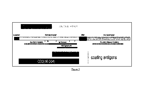
Figure 1
Schematic representation of the full length ASFV CD2v protein, its domains and
fragments used in the
5 Examples. The numbering is based on GenBank acc. no. CAD2068420.
Figure 2
ASFV CD2v protein amino acid sequence alignments of various ASFV strains.
Visualization of alignment with MView: https://www.ebi.ac.uk/Tools/msa/mview/.
Nrs. 1-8 are genotype II strains, serogroup 8 CD2v.
Nrs. 9-15 are genotype I strains, serogroup 4 CD2v.
The concordance to the SEQ ID numbers is given below:
Fig. 2 nr. Name GenBank acc.nr. aa nrs. start-end SEQ ID NO:
1 Rhodesia AJB28392.1 1-375 8
2 LV17/WB/Riel 1-140 9
3 VN/Pig/HN/19 QEH60630.1 1-360 10
4 Po119_53050_C195 Q0W03114.1 1-360 11
5 wbBS01 QDL88089.1 1-360 12
6 Georgia2007 YP 009927182.1 1-360 13
7 Volgograd_2012 AJB28407.1 1-360 14
8 VNUA/HY QC527843.1 1-360 15
9 NHV YP_009702625.1 1-304 16
10 OURT YP_009703666.1 1-304 17
11 Liv13/33 Q1D21219.1 1-370 18
12 Lisbon60 AAM90854.1 1-373 19
13 47/Ss/2008 YP 009703302.1 1-394 20
14 BA71 NP_042752.1 1-402 21
P-60 AJB28388.1 1-402 22
Figure 3
Relative optical densities measured at 450 nm in ELISA (CD2 "16-204" antigen)
for various serum
samples.
Figure 4
Relative optical densities measured at 450 nm in ELISA (CD2 "132-204" antigen)
for various serum
samples.
Figure 5
Relative optical densities measured at 450 nm in ELISA (CD2 "132-204" antigen)
for various serum
samples.
CA 03202683 2023- 6- 16
WO 2022/136624
PCT/EP2021/087447
6
Figure 6
ELISA optical densities measured at 450 nm (CD2 "132-204" antigen) at several
serum sample dilutions
in various sample diluents.
Figure 7
The effect of the size of the CD2v fragment on ELISA performance. ELISA
optical densities were
measured at 450 nm using CD2v fragments of different length, and different
serum samples which were
diluted at 1:300.
NB: The CD2v fragment 132-204 was not tested with sera S13, S15, S19 or S21,
for lack of peptide
material.
Figure 8
The effect of detergent concentration in the sample diluent on the P/N ratio
of the ELISA. The CD2v
peptide fragment used was CD2 "132-204", and a series of dilutions of the
various serum samples.
Figure 9
The effect of salt concentration in the sample diluent on the P/N ratio of the
ELISA. The CD2 "132-204"
peptide was used, and several dilutions of serum samples.
NB: The datapoints for the C-67 serum are fully overlapped by those of the S3
serum.
Figure 10
The effect of PBS buffer in the sample diluent on the P/N ratio of the ELISA.
The CD2 "132-204" peptide
was used, and several dilutions of serum samples.
CA 03202683 2023- 6- 16
WO 2022/136624
PCT/EP2021/087447
7
DESCRIPTION OF THE INVENTION
Surprisingly, it was found that this object can be met by an in vitro
diagnostic immunoassay for the
detection of anti-ASFV antibodies wherein the assay is based on an isolated
ASFV CD2v antigen.
The observation that an isolated ASFV CD2v antigen can be used to effectively
distinguish between
ASFV infected animals and animals vaccinated with an ASFV (CD2-) marker
vaccine, for the first time
now, allows the implementation of a DIVA strategy to combat the epidemic.
An important step towards this advantageous observation was the recognition by
the inventors that,
despite reports in the prior art that the ASFV CD2v protein is a weak
immunogen (Ruiz-Gonzalvo et al.,
Virology 196, 769-777, 1993; Argilaguet et al., PLoS ONE 7(9): e40942.
doi:10.1371/journal.pone.0040942; Gomez-Puertas et al., J. of Virol. Aug.
1996, p. 5689-5694;
Lokhandwala et al., Vet. Micr. 235, 10-20, 2019 and PLoS ONE 12(5): e0177007.
https://doi.org/
10.1371/journal.pone.0177007, 2017), an isolated ASFV CD2v antigen can
advantageously be used in an
immunoassay for the purpose of the present invention.
A test sample obtained from an animal vaccinated with an accompanying LAV CD2v-
marker vaccine can
be serologically distinguished from a test sample obtained from an animal
infected with a wild-type ASFV
strain, with the required specificity and sensitivity (Examples 1-3). This
observation allows for the first time
to combat the ASF epidemic with a DIVA strategy the veterinary field has long
been waiting for.
The Examples also show that in a CD2v-antigen based antibody ELISA,
convalescent ASFV swine
antiserum could not be distinguished from an ASFV negative control swine serum
sample with
confidence, as a result of the occurrence of non-specific binding of
components in anti-ASFV antiserum
with an ASFV CD2v antigen in an immunoassay. Treatment of the convalescent
swine serum sample with
a sample diluent revealed that (i) the CD2v protein of the ASFV can be used in
an immunoassay as an
antigen to detect the presence or absence of anti-CD2v antibodies in a swine
test sample, with sufficient
specificity and sensitivity, (ii) the ASFV gene encoding the CD2v protein
(EP402R) is an appropriate
target for genetic modification resulting in a LAV ASFV strain that can be
used as a DIVA vaccine, (iii) the
CD2v protein in wild-type ASFV is of sufficient immunogenicity to induce a
detectable anti-CD2v antibody
response in swine and (iv) modified LAV ASFV can accompany the immunoassay in
advantageous
diagnostic protocols allowing DIVA.
Therefore, in a first aspect the invention provides a use of an isolated
African swine fever virus (ASFV)
CD2v protein or an antigenic fragment thereof, bound to a solid support, as an
antigen in an
immunoassay, characterized in that the CD2v protein or antigenic fragment
thereof is used to detect the
presence (that includes the absence) of ASFV antibodies in a test sample
obtained from a swine
vaccinated with an accompanying ASFV live attenuated virus CD2v-marker vaccine
(LAV CD2v-marker
vaccine).
CA 03202683 2023- 6- 16
WO 2022/136624
PCT/EP2021/087447
8
ASFV CD2v protein is a known- and well-established ASFV protein (Borca et al.,
Virology 199, 463-468,
1994; Rodriguez et al., J. Gen. Virol. 67, 5312-5320, 1993). It is a
glycoprotein with a relative molecular
weight of about 105 kDa that is responsible for the haemadsorption phenotype
of ASFV infected cells in
vitro and is encoded by the EP402R gene on the ASFV genome. This ASFV protein
is the viral homolog
(CD2v) of cellular T-lymphocyte surface adhesion receptor CD2 proteins. Based
on sequence data and
hydropathy profiles, ASFV CD2v protein resembles typical (CD2) class III
transmembrane proteins.
Generally, the full-length ASFV CD2v protein contains four different sections:
(i) a hydrophobic leader at
the N-terminal side of the protein, (ii) a hydrophilic, extracellular domain
comprising a multitude of
potential N-linked glycosylation sites, (iii) a hydrophobic stretch of amino
acids that act as a
transmembrane domain, and (iv) a C-terminal hydrophilic, cytoplasmic domain
which contains a large
number of typical, imperfect repeats of the hexa peptide (PPPKPC) (Figure 1).
Detailed information
regarding ASFV CD2v protein and the EP402 gene of a large number of ASFV
strains, including the
genomic location of the ASFV genes, (alignment of) nucleotide/amino acid
sequence information,
identification of the four CD2v domains and other annotations, can be found in
Figure 2 and the various
public nucleic acid- and protein sequence data bases, such as the NCB! genome
database, UniProt,
EMBL/GenBank and the European Union reference laboratory for African Swine
Fever (EURL-ASF)
at Centro de investigacion en sanidad animal (CISA-INIA) (https://asf-
referencelab.info/asf/en/sequence-
data-base). In Zhu and Meng (Database,1-9, 2020) the authors report the
establishment of an ASFV
database wherein the collective public genomic- and proteomic ASFV information
is collected and made
available. ASFVdb is freely accessible at http://asfvdb.popgenetics.net and
viruSITE genome browser;
http://virusite.org/index.php, Stano, M., Beke, G., Klucar, L. (2016):
viruSITE - integrated database for
viral genomics. Database (Oxford). bawl 62.doi:10.1093/database/baw162.
The sequences of CD2v ASFV proteins and their polypeptide fragments used
herein can vary from the
specific sequences disclosed herein. This is due to the existing natural
sequence variation among ASFV
strains, as is apparent from the sequences available from the above-mentioned
public sequence
databases and Figure 2. The specific CD2v amino sequence and specific sequence
numbering described
herein relate to the ASFV reference strain Georgia 2007/1 and is also
disclosed in GenBank under acc.
No. CAD2068420 (SEQ ID NO: 1) The complete genomic nucleotide sequence and
amino acid
sequences of the polypeptides encoded by the Georgia 2007/1 genome are also
shown in GenBank,
under accession no. FR682468.
In particular, the ASFV CD2v protein used herein is defined as a protein
comprising an extracellular
domain comprising an amino acid sequence with at least 95 % amino acid
sequence identity to SEQ ID
NO: 2 (CD2 "16-204"), preferably at least 99% amino acid sequence identity to
SEQ ID NO: 2 or 100 %
sequence identity, in the regions of overlap (alignment with MUSCLE algorithm
www.ebi.ac.uk/Tools/msa/muscle/).
In the context of the present invention an antigenic fragment of an ASFV CD2v
protein as described
above can also be used as the antigen. Such an antigenic fragment represents a
truncated from of the
CA 03202683 2023- 6- 16
WO 2022/136624
PCT/EP2021/087447
9
CD2v protein and is a polypeptide comprising one or more epitopes that can be
recognized by anti-ASFV
CD2v antibodies in a test sample obtained from a swine infected with a wild-
type ASFV.
Preferably, the antigenic fragment is a polypeptide comprising an
extracellular domain of the CD2v
protein or an antigenic fragment of the extracellular domain.
An extracellular domain of an ASFV CD2v protein is located at the N-terminal
side of a transmembrane
domain.
An extracellular domain or transmembrane domain of an ASFV CD2v protein can be
identified on the
basis of its typical amino acid sequence by methods know in the art, such as
described by Kyte and
Doolittle (J. Mol. Biol. 157, 105-132) and Rodriguez et al., (J. Virol. 67,
5312-5320, 1993). Alternatively,
such domains are disclosed in the public sequence databases for known ASFV
strains or can be
identified by amino acid sequence alignment with one or more of the amino acid
sequences of ASFV
extracellular domains available from the public sequence databases. For
example, the four domains of
the Georgia 2007/1 CD2v protein span approximately the following amino acid
regions: leader: aa 1-15;
extracellular domain: aa 16-204; transmembrane region: aa 205-229; and
extracellular domain: aa 230-
360, whereby the amino acid numbers are indicated in relation to the numbering
of the reference amino
acid sequence SEQ ID NO: 1.
In a particularly preferred embodiment an extracellular domain of an ASFV CD2v
protein comprises an
amino acid sequence with at least 95 A amino acid sequence identity to SEQ ID
NO: 2, preferably at
least 99 % amino acid sequence identity to SEQ ID NO: 2 or 100 % amino acid
sequence identity, in the
regions of overlap.
In another preferred embodiment, an antigenic fragment of the extracellular
domain for use in the present
invention is a polypeptide comprising an amino acid sequence with at least 95
% amino acid sequence
identity to SEQ ID NO: 3 (CD2 "132-204"), preferably at least 99% amino acid
sequence identity to SEQ
ID NO: 3 or 100 % amino acid sequence identity, in the regions of overlap.
In a more preferred embodiment, an antigenic fragment of the extracellular
domain for use in the present
invention is a polypeptide comprising an amino acid sequence with at least 95
% amino acid sequence
identity to a sequence selected from SEQ ID NO: 23 and 24; even more
preferably at least 99 % amino
acid sequence identity to a sequence selected from SEQ ID NO: 23 and 24; still
more preferably 100 %
amino acid sequence identity to a sequence selected from SEQ ID NO: 23 and 24,
in the regions of
overlap.
In a most preferred embodiment, an antigenic fragment of the extracellular
domain for use in the present
invention is a polypeptide comprising an amino acid sequence with at least 95
% amino acid sequence
identity to SEQ ID NO: 25; even more preferably at least 99 % amino acid
sequence identity to SEQ ID
CA 03202683 2023- 6- 16
WO 2022/136624
PCT/EP2021/087447
NO: 25; still more preferably 100% amino acid sequence identity to SEQ ID NO:
25, in the regions of
overlap.
For the invention, SEQ ID NO: 3 is CD2 "132-204"; SEQ ID NO: 23 is CD2 "132-
194"; SEQ ID NO: 241s
5 CD2 "142-204"; and SEQ ID NO: 25 is CD2 "142-194".
In the Examples, it is shown that in case the CD2 "132-204" fragment of the
extracellular domain (of a
genotype ll strain) is used as an antigen in an ELISA, also genotype I
positive test samples react with this
antigen, whereas it is also shown that the complete extracellular polypeptide
CD2 "16-204" is not
10 recognized by antibodies in genotype I positive samples. Thus, the CD2
"132-204" fragment can
advantageously be used according to the invention in a DIVA immunoassay for
serologically
distinguishing between samples from swine vaccinated by either genotype I or
genotype ll accompanying
LAV strains, and samples from swine infected with wild-type ASFV containing an
intact CD2v gene.
Therefore, in an even more preferred embodiment an antigenic fragment of the
extracellular domain used
herein is a polypeptide comprising an ASFV amino acid sequence consisting of
an amino acid sequence
with at least 95 % amino acid sequence identity to SEQ ID NO: 3 (CD2 "132-
204"), preferably at least 99
% amino acid sequence identity to SEQ ID NO: 3 or 100 % amino acid sequence
identity, in the regions
of overlap.
In a yet even more preferred embodiment, an antigenic fragment of the
extracellular domain used herein
is a polypeptide comprising an ASFV amino acid sequence consisting of an amino
acid sequence with at
least 95 % amino acid sequence identity to a sequence selected from SEQ ID NO:
23 and 24; more
preferably at least 99 % amino acid sequence identity to a sequence selected
from SEQ ID NO: 23 and
24; still more preferably 100 % amino acid sequence identity to a sequence
selected from SEQ ID NO: 23
and 24, in the regions of overlap.
In a most preferred embodiment, an antigenic fragment of the extracellular
domain used herein is a
polypeptide comprising an ASFV amino acid sequence consisting of an amino acid
sequence with at least
95 % amino acid sequence identity to SEQ ID NO: 25; more preferably at least
99 % amino acid
sequence identity to SEQ ID NO: 25; still more preferably 100 % amino acid
sequence identity to SEQ ID
NO: 25, in the regions of overlap.
Alternatively, an antigenic fragment of the extracellular domain used herein
is a polypeptide comprising
an ASFV amino acid sequence consisting of an amino acid sequence with at least
95 %, at least 99 % or
100% amino acid sequence identity, in the regions of overlap, to any of the
fragments 132-194, 132-214,
122-194, 122-204 or 142-214, as shown in SEQ ID NO: 1; as well as to any of
the fragments 132-194,
142-204, or 142-194, as shown in SEQ ID NO: 1.
An ASFV CD2v antigen as described above can be of any serogroup known for ASF
viruses, in particular
of serogroup 4 or 8, preferably of serogroup 8.
CA 03202683 2023- 6- 16
WO 2022/136624
PCT/EP2021/087447
11
ASFV serogroup clustering is based on examining the inhibition of the ASFV
haemadsorption phenotype
by serum belonging to the same group. Presently, the existence of serogroups 1-
8 is established
(Malogolovkin et al., J. Gen. Virol. 96, 866-873, 2015).
Furthermore, an ASFV CD2v antigen as described above may comprise a tag to
allow the detection of
protein expression or purification of the antigen. Suitable tags include a
6xHis tag, a c-Myc domain:
EQKLISEEDL (SEQ ID NO: 4), a hemagglutinin tag: YPYDVPDYA (SEQ ID NO: 5), a
maltose-binding
protein, a glutathione-S-transferase, a maltose- binding protein, a FLAG tag
peptide, a biotin acceptor
peptide, a streptavidin-binding peptide, or a calmodulin-binding peptide, as
disclosed in Chatterjee (Opin.
Biotech 17, 353-358, 2006). A FLAG tag or His tag is a preferred tag.
For the manufacture of a CD2v antigen used herein, common- and commercially
available conventional
peptide synthesis methods and -recombinant DNA expression systems and methods
can be used, that
include bacterial-, yeast-, fungal-, insect- and vertebrate cell expression
systems. Ample guidance with
regard to prokaryotic- and eukaryotic expression systems is given i.a. in
reviews and text books on
recombinant DNA expression methods such as: Trepe, K., Applied Microbiology
and Biotechnology, 72,
Number 2 (2006), 211-222; Production of Recombinant Proteins: Novel Microbial
and Eukaryotic
Expression Systems, edited by Gellissen, G. Publisher: Wiley-VCR, ISBN:
3527310363 edition 2005,
Expression systems, edited by Michael Dyson and Yves Durocher, Scion
Publishing Ltd, ISBN
9781 904842439 edition 2007.
Advantageously, a CD2v antigen can be prepared by using a Baculovirus-insect
cell expression system.
Examples of scientific articles, text-books, and reviews illustrating this
system are: Luckow et al., 1988,
Bio-technology, vol. 6, p. 47; Baculovirus Expression Vectors: A Laboratory
Manual by David R. O'Reilly,
Oxford University press, 1993, ISBN: 0716770172; The Baculovirus Expression
System: A laboratory
guide, ed. King & Possee, 1992, ISBN: 9401050473; and a review is: van Oers et
al., 2015, J. of Gen.
Virology, 96, 6-23. Expression and purification of ASFV polypeptides in E.
coli- and insect cell systems
are, for example, described in Lokhandwala et al., PLOS ONE, May 2017, and
Kollnberger et al. (supra).
Tools and kits are commercially available for the efficient generation of
baculoviruses for use in the
present invention, such as: BactoBacTM (Thermo Fisher Sci., Waltham, MA.,
USA); ProEasyTM (AB
Vector, San Diego, CA., USA); and flashBACTM (Oxford Expression Technologies,
Oxford, UK).
A "marker vaccine" is a well-known concept in the veterinary vaccinology
field. A marker vaccine
comprises- and/or expresses an altered polypeptide immunogen that differs
immunogenically from the
corresponding wild-type polypeptide immunogen by lacking at least one epitope,
or having a different
version of an epitope, as compared to the wild-type version. Typically, the
(gene encoding the)
polypeptide immunogen in- or expressed by the marker vaccine has been altered
by biochemical- or
recombinant DNA techniques, and the result is that the lack of an antibody
response against a wild-type
moiety in the altered immunogen in the marker vaccine can be used to
serologically detect infected
CA 03202683 2023- 6- 16
WO 2022/136624
PCT/EP2021/087447
12
animals independent of vaccinations. This will allow a serologic DIVA.
Typically, the altered immunogen is
an immunogen that is absent, or is a fragment of the wildtype polypeptide
immunogen.
The term immunogen as used herein refers to a molecule's (such as a protein or
polypeptide) capability
of eliciting a specific antibody response by an organism's immune system,
whereas the term antigen
refers to a molecule's capability of specific binding to antibodies produced
by an organism's immune
system.
An epitope as used herein is a stretch of, typically 5-15, amino acids within
a protein or polypeptide that is
capable of eliciting an antibody response specific for this moiety and/or of
binding with the specific
antibodies produced by such a response.
A LAV CD2v-marker vaccine as used herein is a vaccine that comprises a live,
attenuated, replicating
ASFV marker vaccine strain that is capable of expressing an altered CD2v
polypeptide immunogen that is
serological distinguishable from a CD2v polypeptide immunogen of a wild-type
ASFV strain.
With an "accompanying" LAV CD2v-marker vaccine is meant a vaccine comprising a
CD2v marker
vaccine strain as defined above and wherein an altered CD2v polypeptide
immunogen is aligned with-
and designed to be different from a CD2v polypeptide antigen in an immunoassay
such that the CD2v
polypeptide antigen is serologically capable of detecting antibodies in a test
sample specific for a wild-
type moiety of a CD2v polypeptide immunogen and is not capable of recognizing
antibodies specific for
an altered moiety of a CD2v polypeptide immunogen.
Thus, an accompanying LAV CD2v-marker vaccine comprises a CD2v-marker vaccine
strain, as defined
above, that triggers an effective immune response in swine resulting in an
antibody repertoire in a serum
sample of a vaccinated swine lacking antibodies that are present in an
antibody repertoire in a serum
sample of a swine infected with a wild-type ASFV. Differentiating between
infected- and vaccinated- or
negative animals is thus based on an immunoassay detecting antibodies specific
for one or more ASFV
CD2v epitopes that are missing in the marker vaccine.
In particular, the accompanying LAV CD2v-marker vaccine comprises an ASFV CD2v-
marker vaccine
strain that comprises and/or is capable of expressing a truncated CD2v protein
or no CD2v protein.
Preferably, the truncated CD2v protein is a polypeptide fragment of the CD2v
protein that lacks an
extracellular domain or a fragment thereof.
More preferably, the truncated CD2v protein is a polypeptide fragment of the
CD2v protein that lacks a
fragment of the extracellular domain.
CA 03202683 2023- 6- 16
WO 2022/136624
PCT/EP2021/087447
13
In an even more preferred embodiment, the truncated CD2v protein is a
polypeptide fragment of the
CD2v protein that lacks a fragment of the extracellular domain of the CD2v
protein, comprising an amino
acid sequence with at least 95 % amino acid sequence identity to SEQ ID NO: 3
(CD2 "132-204"),
preferably at least 99 % amino acid sequence identity to SEQ ID NO: 3 or 100 %
amino acid sequence
identity, in the regions of overlap.
In a yet even more preferred embodiment, the truncated CD2v protein is a
polypeptide fragment of the
CD2v protein that lacks a fragment of the extracellular domain of the CD2v
protein, comprising an amino
acid sequence with at least 95 % amino acid sequence identity to a sequence
selected from SEQ ID NO:
23 and 24; even more preferably at least 99 % amino acid sequence identity to
a sequence selected from
SEQ ID NO: 23 and 24; still more preferably 100 % amino acid sequence identity
to a sequence selected
from SEQ ID NO: 23 and 24, in the regions of overlap.
In a most preferred embodiment, the truncated CD2v protein is a polypeptide
fragment of the CD2v
protein that lacks a fragment of the extracellular domain of the CD2v protein,
comprising an amino acid
sequence with at least 95 % amino acid sequence identity to SEQ ID NO: 25;
even more preferably at
least 99 % amino acid sequence identity to SEQ ID NO: 25; still more
preferably 100 % amino acid
sequence identity to SEQ ID NO: 25, in the regions of overlap.
In a still more preferred embodiment, the truncated CD2v protein is a
polypeptide fragment of the CD2v
protein that lacks a fragment of the extracellular domain of the CD2v protein,
that comprises an ASFV
amino acid sequence consisting of an amino acid sequence with at least 95 %
amino acid sequence
identity to SEQ ID NO: 3 (CD2 "132-204"), preferably at least 99% amino acid
sequence identity to SEQ
ID NO: 3 or 100 % amino acid sequence identity, in the regions of overlap.
In an even still more preferred embodiment, the truncated CD2v protein is a
polypeptide fragment of the
CD2v protein that lacks a fragment of the extracellular domain of the CD2v
protein, that comprises an
ASFV amino acid sequence consisting of an amino acid sequence with at least 95
% amino acid
sequence identity to a sequence selected from SEQ ID NO: 23 and 24; more
preferably at least 99 %
amino acid sequence identity to a sequence selected from SEQ ID NO: 23 and 24;
still more preferably
100 % amino acid sequence identity to a sequence selected from SEQ ID NO: 23
and 24, in the regions
of overlap.
In a most preferred embodiment, the truncated CD2v protein is a polypeptide
fragment of the CD2v
protein that lacks a fragment of the extracellular domain of the CD2v protein,
that comprises an ASFV
amino acid sequence consisting of an amino acid sequence with at least 95 %
amino acid sequence
identity to SEQ ID NO: 25; more preferably at least 99 % amino acid sequence
identity to SEQ ID NO: 25;
still more preferably 100 % amino acid sequence identity to SEQ ID NO: 25, in
the regions of overlap.
In a specific embodiment of the use of a ASFV CD2v antigen in an immunoassay,
as described above,
detecting the presence (including the absence) of ASFV antibodies in a test
sample obtained from a
CA 03202683 2023- 6- 16
WO 2022/136624
PCT/EP2021/087447
14
swine vaccinated with the accompanying LAV CD2v-marker vaccine, the ASFV CD2v
antigen has no
epitope in common, and, in particular, no overlapping amino acid sequence,
with the altered CD2v
polypeptide immunogen of- or expressed by the CD2v-marker vaccine strain.
VVith no overlapping amino
acid sequence is meant that the ASFV CD2v antigen in the immunoassay and the
altered CD2v
polypeptide immunogen of - or expressed by the LAV CD2v-marker vaccine strain
are from different
regions of the CD2v protein and show no overlap at their termini.
More preferably, an ASFV CD2v antigen in the immunoassay and an altered CD2v
polypeptide
immunogen in the accompanying marker vaccine, as described above, represent
two different, non-
overlapping, fragments of an extracellular domain of an ASFV CD2v protein.
Suitable live-attenuated ASFV CD2v-marker vaccine strains are known in the art
or can be prepared by
recombinant DNA techniques using standard methods, such as CRISPR-Cas or
homologous
recombination, or can be isolated from the field.
Recently, results of various research activities have been published that
disclose the (rational) design of
ASFV LAV strains by means of genetically modifying ASFV strains (see ASFV
review articles, supra, and
references cited therein). These prior art documents disclose a variety of
ASFV genes that can be
mutated to arrive at attenuated- and efficacious ASFV vaccine strains.
The prior art also discloses the generation of various ASFV mutant strains
that comprise- or express
altered CD2v proteins: Gallardo et al. (Transbound. Emerg. Dis. 66, 1399-1404,
2019) and Barasona et
al. (Front. Vet. Sci. 6;137, 2019). ASFV strain Lv17/VVB/Riel (WO 2020/049194)
has been tested for its
safety and efficacy profile after immunization of domestic pigs and wild boar.
Lv17NVB/Rie1 is a naturally
attenuated strain that has a truncated CD2v protein (encoded by a mutant
EP402R gene) and has a non-
haemadsorbing phenotype in vitro. Another naturally occurring, non-pathogenic
ASFV isolate, OURT88/3,
comprises frameshift mutations in the sequence encoding the cytoplasmic domain
of CD2v that result in
the final 215 amino acids not being translated. Borca et al. (J. Virol. 72,
2881-2889, 1998 and Sci Rep.
2020, 10:494) and Monteagudo et al. (J. Virol. 91, 2017, 91(21):e01058-17)
disclose the generation of a
CD2v deletion mutant by means of recombinant DNA techniques, based on ASFV
strains Malawi,
Georgia 2007/1 and BA71, respectively. Chen et al. (Sci China Life Sci, 63,
2020) discloses the
generation of a seven-gene deleted ASFV strain (HLJ/18) that is effective and
safe as a live-attenuated
virus vaccine in swine. Among other deletions, also the gene encoding the CD2v
protein is deleted in
HLJ/18.
An ASFV CD2v antigen and ASFV CD2v-marker vaccine strain to be used in the
present invention may
be derived from any ASFV genotype or any ASFV strain, such as one of the
following strains: Georgia
2007/1, Benin 97/1, Kenyan and Malawi. Preferred ASFV genotypes are I or II.
ASFV genotyping is
based on genetically characterizing an ASFV genome through partial sequencing
of the C-terminal end of
the p72 protein (encoded by the B646L gene) which represents the ASFV major
capsid protein. This
CA 03202683 2023- 6- 16
WO 2022/136624
PCT/EP2021/087447
method has defined 24 different genotypes to date (Bastos et al., Arch. Virol.
2003 Apr;148:693-706.
2003; Quembo et al., Transbound. Emerg. Dis.; 65, 420-431, 2018).
In a preferred embodiment, the accompanying LAV CD2v-marker vaccine is based
on ASFV strain
5 Lv17/VVB/Rie1, disclosed in WO 2020/049194, and the ASFV CD2v antigen is
a polypeptide comprising
an ASFV amino acid sequence consisting of an amino acid sequence with at least
95 % amino acid
sequence identity to SEQ ID NO: 3 (CD2 "132-204"), preferably at least 99 %
amino acid sequence
identity to SEQ ID NO: 3 or 100 % amino acid sequence identity, in the regions
of overlap.
10 In a more preferred embodiment, the accompanying LAV CD2v-marker vaccine
is based on ASFV strain
Lv17/WB/Rie1, disclosed in WO 2020/049194, and the ASFV CD2v antigen is a
polypeptide comprising
an ASFV amino acid sequence consisting of an amino acid sequence with at least
95 % amino acid
sequence identity to a sequence selected from SEQ ID NO: 23 and 24; even more
preferably at least 99
% amino acid sequence identity to a sequence selected from SEQ ID NO: 23 and
24; still more preferably
15 100 % amino acid sequence identity to a sequence selected from SEQ ID
NO: 23 and 24, in the regions
of overlap.
In a most preferred embodiment, the accompanying LAV CD2v-marker vaccine is
based on ASFV strain
Lv17/VVB/Rie1, disclosed in WO 2020/049194, and the ASFV CD2v antigen is a
polypeptide comprising
an ASFV amino acid sequence consisting of an amino acid sequence with at least
95 % amino acid
sequence identity to SEQ ID NO: 25; even more preferably at least 99 % amino
acid sequence identity to
SEQ ID NO: 25; still more preferably 100 A amino acid sequence identity to
SEQ ID NO: 25, in the
regions of overlap.
An accompanying LAV CD2v-marker vaccine to be used in the present invention
can be prepared by
conventional methods such as those commonly used for commercially available
live-attenuated virus
vaccines. Briefly, a susceptible substrate is inoculated with a live-
attenuated CD2v-marker vaccine strain
as described above and propagated until the virus has replicated to a desired
titre after which ASFV
containing material is harvested. Subsequently, the harvested material,
purified and/or concentrated, if
needed, together with a pharmaceutically acceptable carrier or diluent are
formulated into a
pharmaceutical preparation with immunizing properties. Carriers include
stabilizers, preservatives and
buffers. Suitable stabilizers are, for example SPGA (sucrose, phosphate,
glutamate, and albumin),
carbohydrates (such as sorbitol, mannitol, starch, sucrose, dextran, glutamate
or glucose), proteins (such
as dried milk serum, albumin or casein) or degradation products thereof.
Suitable buffers are for example
PBS-, Tris- or HEPES buffers. Suitable preservatives are thimerosal,
merthiolate and gentamicin.
The vaccine may be administered by intramuscular-, subcutaneous-, intradermal-
, oral- or intranasal
inoculation or injection, in an amount which is effective to protect a swine
against ASF disease. This
amount may vary according to the animal being inoculated, taking into
consideration the age and weight
of the animal
CA 03202683 2023- 6- 16
WO 2022/136624
PCT/EP2021/087447
16
In the Examples it is demonstrated that for the first time a successful ASFV
DIVA approach has been
established by the combined use of a DIVA diagnostic assay and an accompanying
DIVA LAV CD2v-
marker vaccine, both as defined above. The inventors determined, on the one
hand, that ASFV CD2v
represents an appropriate immunogen in wild-type ASFV and, on the other hand,
that ASFV CD2v also
represents an appropriate antigen that can be used in an immunoassay with the
required specificity and
sensitivity to allow DIVA. A DIVA method as described above allows for
vaccination while still retaining
the possibility of serological surveillance for the presence of infection,
thereby providing for the first time a
powerful- and practical tool to combat ASF in animals that can easily be
scaled-up, inter alia because the
method does not involve the use of live infectious ASFV that would require
performing such a method in
high containment facilities.
Therefore, in a particular embodiment, an ASFV CD2v antigen as described above
is used in an
immunoassay, characterized in that the immunoassay is a DIVA immunoassay.
Generally, in order to make a final differentiation between infected and
vaccinated animals, test scores
need to be interpreted as being positive or negative. In practice that means:
being above or below a
certain threshold value. This can conveniently be done by incorporating into
the method a number of
reference samples to be tested alongside the test samples, as for example
described in the Examples.
Positive and negative reference samples can be prepared in swine, or can be
obtained from several
institutions, and (national)reference laboratories world-wide, for example the
European Union Reference
Laboratory for ASFV, Centro de investigacion en sanidad animal (CISA-INIA),
Madrid, Spain.
The solid support to be used in an immunoassay as described above can in
principle be any solid
support, provided it allows the performance of the use according to the
invention, in particular: the binding
of an ASFV CD2v antigen as described above to the solid support. It can be of
different size, shape or
form. Binding can occur via conventional means, such as by covalent- or by non-
covalent interaction (i.a.
adsorption or coating). Alternatively, binding can be achieved through
biotinylated CD2v antigen linked to
an avidin-coated solid support.
In particular, the solid support is a microtiter plate, vial, bead paper
strip, membrane, gel or lateral flow
strip. Preferably the solid support is a microtitre plate.
In a further aspect the present invention provides a method for distinguishing
between ASFV infected
animals (positive test result) and vaccinated animals (negative test result)
wherein the method is an
immunoassay, characterized in that an isolated ASFV CD2v protein or an
antigenic fragment thereof, as
described above, that is bound to a solid support is used as an antigen, the
marker vaccine is an
accompanying LAV CD2v-marker vaccine and the method comprises a step of
examining a test sample
obtained from the animal for the presence of ASFV CD2v antibodies that bind to
the antigen.
CA 03202683 2023- 6- 16
WO 2022/136624
PCT/EP2021/087447
17
In this additional aspect of the invention and embodiments hereof, the
definition of the specific terms
referred to herein and the various embodiments of this aspect are the same as
those described for the
first aspect above.
In an embodiment of this aspect the invention provides a method as outlined
above wherein the antigenic
fragment is a polypeptide comprising an extracellular domain of the CD2v
protein or an antigenic
fragment of the extracellular domain, more in particular, the antigenic
fragment of the extracellular domain
is a polypeptide comprising an amino acid sequence with at least 95 % amino
acid sequence identity to
SEQ ID NO: 3 (CD2 "132-204"), even more in particular, the antigenic fragment
of the extracellular
domain comprises an ASFV amino acid sequence consisting of an amino acid
sequence with at least 95
% amino acid sequence identity to SEQ ID NO: 3 (CD2 "132-204"), in the regions
of overlap.
In a preferred embodiment of this aspect the antigenic fragment of the
extracellular domain is a
polypeptide comprising an amino acid sequence with at least 95 % amino acid
sequence identity to a
sequence selected from SEQ ID NO: 23 and 24; even more preferably at least 99
% amino acid
sequence identity to a sequence selected from SEQ ID NO: 23 and 24; still more
preferably 100 % amino
acid sequence identity to a sequence selected from SEQ ID NO: 23 and 24, in
the regions of overlap.
In a most preferred embodiment of this aspect the antigenic fragment of the
extracellular domain is a
polypeptide comprising an amino acid sequence with at least 95 % amino acid
sequence identity to SEQ
ID NO: 25; even more preferably at least 99 % amino acid sequence identity to
SEQ ID NO: 25; still more
preferably 100 % amino acid sequence identity to SEQ ID NO: 25, in the regions
of overlap.
In a further embodiment of this aspect the invention provides a method as
outlined above wherein the
accompanying LAV CD2v-marker vaccine comprises an ASFV CD2v-marker vaccine
strain that
comprises and/or expresses an altered CD2v polypeptide immunogen, more in
particular the altered
CD2v polypeptide immunogen lacks an extracellular domain of the CD2v protein
or a fragment thereof, or
the ASFV CD2v antigen and the altered CD2v polypeptide immunogen have no
overlapping amino acid
sequence, all as defined above.
The design of an immunoassay to be used in the various aspects of this
invention, as described above, is
similar to commonly used immunoassays that are based on solid support-bound
antigen. In principle, the
immunoassay is based on the formation of an antibody-antigen complex followed
by the subsequent
examination of the presence (including the absence) of such a complex.
Handbooks, such as those
mentioned below, describe a variety of diagnostics assays and their specific
features that can be used
herein (Handbook of Immunoassay Technologies, by Vashist, Sandeep K. and
Luong, John H.T., 2018;
and: Immunoassays: Development, Applications and Future Trends, by R.
O'Kennedy, C. Murphy 2017).
Detailed information regarding the set-up, protocols, standard operating
procedures, reagents, and the
like, for ASFV immunoassays to be used in the present invention are also
disclosed, for example, by the
European Union Reference Laboratory for ASFV (supra)., the FAO (Beltran-
Alcrudo et al., 2017, African
CA 03202683 2023- 6- 16
WO 2022/136624
PCT/EP2021/087447
18
swine fever: detection and diagnosis - A manual for veterinarians. FAO Animal
Production and Health
Manual No. 19, Rome) and in: Gallardo et al., Virus Research 271, 197676,
2019.
In a more specific embodiment of the method according to the invention the
method comprises the steps
of:
1. incubating the test sample with the antigen in an assay mixture,
2. allowing formation of an ASFV CD2v antibody-antigen complex in the assay
mixture, and
3. detecting the presence of the antibody-antigen complex in the assay
mixture.
In this embodiment, detecting the presence of an antibody-antigen complex, may
involve the use of a
detecting antibody conjugated to a label.
In particular, it may involve contacting the complex with the antibody-label
conjugate.
The nature of the label is not critical and can be any label customarily used
in immunoassays. The label is
an entity that provides for-, or is capable of triggering a detectable signal.
In particular, the label is an enzyme, fluorophore, chromophore, radioisotope,
enzymatic substrate,
chemiluminescent molecule, or colloidal gold.
Preferably, the label is an enzyme that can be directly- or indirectly
conjugated to the detecting antibody,
in particular by biotin/avidin conjugation_
Typically, the enzyme used herein is horseradish peroxidase (HRP) and the
enzyme substrate is TMB
(3,3,5.5' tetramethylbenzidine).
In a particularly preferred embodiment of the invention as described above,
the immunoassay is an
ELISA (enzyme linked immunosorbent assay). Advantages of an ELISA include its
practicality, reliability,
swiftness and easiness to scale-up.
ELISA's are well known in the art, and a variety of types in format and
protocols can be applied herein.
An immunoassay as described above may be based upon direct- or indirect
antigen-antibody reactions. A
direct assay comprises a one-step binding of a sample antibody to the antigen.
An indirect assay
comprises a two-step binding process involving the use of a primary (sample)
antibody and a labelled
secondary (detection) antibody capable of binding to the primary antibody. The
immunoassay can also be
a competitive immunoassay in which antibodies in a sample compete for a
limited number of antigen
binding sites with labelled secondary antibody capable of binding to the
antigen.
In a preferred method according to the present invention as described above,
an indirect ELISA is used
comprising the steps of:
1. incubating a test sample with solid support-bound antigen in an assay
mixture,
2. adding a labelled antibody capable of recognizing anti-ASFV CD2v antibody
to the assay mixture,
CA 03202683 2023- 6- 16
WO 2022/136624
PCT/EP2021/087447
19
3. adding an enzyme substrate to the assay mixture to produce a detectable
signal, and
4. measuring the signal.
When a chromogenic substrate is added to the assay mixture to develop colour,
samples with a high
antibody concentration generate a higher signal than those containing a lower
antibody concentration.
In a further preferred method according to the present invention as described
above, a competition ELISA
is used comprising the steps of,
1. incubating a test sample and an antibody capable of binding to the antigen
with solid
support-bound antigen in an assay mixture,
2. adding an enzyme substrate to the assay mixture to produce a detectable
signal, and
3. measuring the signal.
When chromogenic substrate is added to the assay mixture to develop colour,
samples with a high
antibody concentration generate a lower signal than those containing low
antibody concentration, yielding
the inverse correlation between antibody concentration in the sample and
colour development in the
assay.
ELISA results are usually expressed in arbitrary units of absorbance,
typically between 0.1 and 2.5 optical
density (OD) units, depending on the properties and settings of the technical
equipment used for the
readout. Routinely, appropriate positive- and negative control samples are
included, and most-times
samples are tested in multifold. Standardisation is obtained by including (a
dilution range of) a defined
reference sample, which also allows matching a certain score to pre-set
threshold values for determining
positives or negatives, and allows correlation to a biological meaning, for
example: the discrimination
between an animal being infected by a wild-type virus or being vaccinated with
a marker-vaccine.
A particularly preferred ELISA is shown in the Examples.
In an alternative method according to the present invention the immunoassay is
a lateral flow
(immunochromatographic) assay. Lateral flow immunoassays are commonly used in
the art. In principle,
a lateral flow immunoassay operates on the same principle as an ELISA as
described above.
In a lateral flow immunoassay to be used in the present invention, the
antigen, as described above, can
be bound as a test line to a solid support having the capacity to transport
fluid as a result of capillary
activity, such as porous paper or (nitrocellulose)membrane, microstructured
polymer, or sintered polymer.
In essence, the solid support runs sample liquid of the test sample containing
the antibody to be detected
from an absorption zone along the surface of the support. An antibody-antigen
complex can then be
formed at the test line and detected in a detection zone of the solid support
where the antigen is bound to
the solid support.
Therefore, in a particular embodiment of the method of the invention, the
immunoassay used herein is a
lateral flow immunoassay.
CA 03202683 2023- 6- 16
WO 2022/136624
PCT/EP2021/087447
More in particular, the lateral flow immunoassay comprises the steps of:
1. incubating the test sample with the solid support in an absorption zone,
2. allowing the formation of an antibody-antibody/label complex,
5 3. allowing the movement of the complex laterally through the solid
support,
4. capturing the complex by an antigen bound to the solid support at a test
line thereby allowing the
formation of an antibody-antigen-antibody/label complex, and
5. detecting the presence of the complex in an assay mixture in a detecting
zone.
10 The label used in the lateral flow immunoassay can be any label
customary used in LF immunoassays,
and can, in particular, be a coloured particle, such as a latex-, nanonnetre
sized- or gold particle, a
fluorescent-, magnetic labelled- or radio frequency identification (RFID)
particle.
An LF immunoassay used herein can operate as either a competitive- or a
sandwich assay.
The inventors initially observed that when an ASFV CD2v antibody positive
serum test sample was
incubated with a CD2v antigen in an ELISA the signal-to-noise ratio was
suboptimal, as a result of which
the specificity of the ELISA was negatively affected and no reliable DIVA
immunoassay could result from
this. It was subsequently found that this limitation was due to short term
intermolecular interactions
unrelated to the specific antigen-antibody interaction. The Examples
demonstrate that this negative effect
could be overcome by incorporating a dilution (of the swine antiserum) step in
the immunoassay that
limits these non-specific intermolecular interactions. Sample diluents that
can be used in this step display
an increased stringency.
The term stringency of a sample diluent is defined herein as a number that
represents a ratio between an
absorption value (OD unit) of a diluted positive serum control sample/an
absorption value (OD unit) of a
diluted negative serum control sample (P/N ratio) as measured in an ELISA, in
particular as described in
the Examples.
Therefore, in an advantageous method of the invention the swine test sample is
diluted with a sample
diluent of an optimal stringency sufficient to limit undesired non-specific
interactions without affecting
specific antigen-antibody interactions to an undesired level.
A sample diluent to be used in the present invention may have a stringency P/N
ratio of 5 or more,
preferably of 10 or more as measured in an ELISA.
The Examples demonstrate and provide further guidance that, and how, both
incorporating a sample
dilution step and increasing the stringency of the sample diluents, allows a
CD2v antigen-based
immunoassay as defined above to become a reliable DIVA immunoassay. The sample
dilution step
decreases the non-specific interactions between anti-swine ASFV antiserum and
the CD2v antigen in the
Elisa and, thus the increase of the P/N ratio and can be designed by the
skilled person by using
CA 03202683 2023- 6- 16
WO 2022/136624
PCT/EP2021/087447
21
appropriate sample diluents of increased stringency, such that at the same
time the Elisa (OD) signal for
the positive control sample is maintained at an appropriate level.
Sample diluents that can advantageously be used in a method of the present
invention may comprise
customary buffers, such as PBS- or TRIS buffers to which surfactants, such as
Tween TM 20 or Tween 80,
Triton, Na-deoxycholate, sodium dodecyl sulphate, an aminoxide or CHAP
detergent, are added.
In a preferred embodiment of the method for distinguishing according to the
invention, or alternatively in a
preferred embodiment of the method for determining according to the invention,
the sample diluent
comprises one or more of the surfactants selected from Tween TM 20, Tween 80,
and an aminoxide.
Preferably the aminoxide is Aminoxide WS 35, also known as:
cocamidopropylamine oxide. More
preferably the Aminoxide WS 35 is a compound with CAS nr. 53988-60-6.
In a preferred embodiment the surfactant is comprised in the sample diluent at
between 1 and 5 % w/v;
more preferably at 2 - 4 % w/v, or even at 3 %w/v.
Therefore, in a preferred embodiment of the invention the method according to
the invention comprises a
step wherein the test sample is diluted with a sample diluent that has a
stringency resulting in a P/N ratio
of? 5, preferably ? 10.
Advantageous P/N ratios can also be obtained by diluting the swine test sample
with the sample diluent in
ratio of 1:100 to 1:2700, preferably in a ratio of 1:100 to 1:900, more
preferably in a ratio of 1:100 to
1:300, more in particular, in a ratio of 1:300.
In further preferred method according to the invention the swine test sample
is diluted with a sample
diluent that has a stringency resulting in a P/N ratio of? 5, preferably? 10
and at a dilution in ratio of
1:100 to 1:2700, preferably in a ratio of 1:100 to 1:900, more preferably in a
ratio of 1:100 to 1:300, more
in particular, in a ratio of 1:300.
In a preferred embodiment of the method for distinguishing according to the
invention, or alternatively in a
preferred embodiment of the method for determining according to the invention,
the sample diluent
comprises a salt at between 0.01 and 1 M. More preferably at between 0.05 and
0.5 M, even more
preferably at 0.1 M.
In a preferred embodiment the salt is magnesium chloride.
In a most preferred embodiment the sample diluent comprises Tween, aminoxide,
and magnesium
chloride.
The various aspects of the present invention as outlined above can
advantageously be applied by testing
a sample derived from a swine that is susceptible to infection with ASFV.
Specifically, a swine is a porcine
CA 03202683 2023- 6- 16
WO 2022/136624
PCT/EP2021/087447
22
animal of the family of Suidae, and preferably a porcine animal of the genus
Sus, for example, a pig or
wild boar. Preferably the swine is a domestic pig.
Therefore, in a preferred embodiment of the various aspects of the invention,
the test sample is derived
from a domestic pig.
The test sample for use in the various aspects of the present invention can,
in principle, be any type of
sample from a swine possibly containing anti-ASFV CD2v antibodies, for example
a plasma- or a serum
sample. Preferably the sample is a serum sample.
Another aspect of the invention is a device for use in a method for detecting
the presence of ASFV CD2v
antibodies in a test sample obtained from a swine vaccinated with an
accompanying LAV CD2v-marker
vaccine as described above, the device comprising an isolated ASFV CD2v
antigen bound to a solid
support, as described above.
A further aspect of the present invention is a diagnostic kit comprising a
device as described above.
A diagnostic kit according to the invention can comprise a single packaging
unit that comprises additional
components to be applied in a method according to the present invention.
In particular, the diagnostic kit additionally comprises one or more
containers comprising:
- a sample diluent,
- an antibody-label conjugate,
- a positive control sample, and/or
- a negative control sample.
In a more particular embodiment, the diagnostic kit described above also
comprises instructions for use of
the kit with a test sample obtained from a swine that is vaccinated with an
accompanying LAV CD2v-
marker vaccine as described above.
In particular, the instructions for use describe that the diagnostic kit can
be used for DIVA and that a test
sample from an ASFV infected swine will be positive in that test, in contrast
to a test sample from a
vaccinated non-infected swine that will be negative.
CA 03202683 2023- 6- 16
WO 2022/136624
PCT/EP2021/087447
23
EXAMPLES
Example 1 - ASFV CD2v-based ELISA
To verify if anti-CD2v antibodies in pig serum can be detected by ELISA, an
ELISA was performed using
a fragment of the ASFV CD2v protein, referred to as CD2 "16-204". CD2 "16-204"
spans the extracellular
domain and lacks the leader sequence, the transmembrane domain and the proline-
rich intracellular part
of the full-length CD2v protein (Figure 1). It contains a GP64 signal peptide:
MVSAIVLYVLLAAAAHSAFA
(SEQ ID NO: 6) at its N-terminus and a exHis-tag at its C-terminus. It was
produced by Baculovirus
expression and subsequent purification by GenScript.
Serum samples were obtained from the European Union Reference Laboratory for
African Swine Fever
(CISA-INIA, Spain).
For the ELISA, a 96-well microtiter plate was coated overnight at 2-8 C with a
solution containing the
CD2v fragment at a concentration of 1 pg/ml. Plates were washed four times
with wash buffer (0.04 M
PBS + 0.15 % Tween20) before they were blocked with casein for 1 hour at 37 C.
After washing the
plates 4 times, 3-fold serial dilutions of serum samples in EIA buffer (0.2 M
PBS + 0.1 % BSA) were
prepared in well A to well G of each column (well H only contained EIA buffer
and functioned as a
control). Serum samples were pre-diluted 1:100 in EIA buffer. Plates with
serum dilutions were incubated
for 1 hour at 37 C and subsequently washed 4 times with wash buffer. To each
well a solution with a
peroxidase-labelled goat anti-swine IgG (H-FL) antibody was added and plates
were incubated for 1 hour
at 37 C after which they were washed 4 times with wash buffer. Then a
3,3',5,5'-tetramethylbenzidine
(TMB) substrate solution was added to each well and incubated for at least 10
min. The colouring
reaction was stopped by adding 4N H2SO4. Optical densities were measured at
450 nm with a microtiter
plate reader and data analysed.
The results are presented in Figure 3. Sera from swine infected with genotype
II ASFV, serotype 8 strains
(Si, S2, and S3-ASFV strain Lv17/VVB/Rie1; WO 2020/049194) displayed at a
serum dilution of 1:900 a
clear positive signal above the negative serum 0-67. Serum sample C+113,
obtained from a swine that
was infected twice with a genotype I ASFV strain and subsequently also with a
genotype II strain, gave a
clear positive signal that was set at 100 %. Sera from swine infected with
genotype I, serotype 4 ASFV
strains (S13, S15, S19, S21) could not be distinguished from the negative
serum control. Thus, the
results demonstrate that CD2v is immunogenic and that an ELISA based on the
CD2 "16-204" fragment
can be used to measure anti-CD2v antibodies induced by genotype II ASFV
strains.
CA 03202683 2023- 6- 16
WO 2022/136624
PCT/EP2021/087447
24
Example 2 - CD2v-based ELISA for the detection of genotype I and II ASFV
strains
A truncated version of the CD2 "16-204" extracellular CD2v fragment was
designed. This fragment, CD2
"132-204" lacks the N-terminal 131 amino acids of the CD2v protein (Figure 1).
It contains at its C-
terminus a 5x GlyGlyGlySer (SEQ ID NO: 7) linker followed by a Flag-tag, and
was produced by
Baculovirus expression and subsequent purification by GenScript.
The ELISA was performed as described in Example 1.
The results are presented in Figure 4. Sera from swine infected with genotype
ll ASFV strains (S1 and
S2) or genotype I ASFV strains (S13, 815, S19, S21) all showed at a serum
dilution of 1:300 a clear
positive signal above the negative serum C-67. Serum sample C+113 gave also a
clear positive signal
that was set at 1008)/0. Thus, an ELISA based on the CD2 "132-204" fragment
can be used to measure
anti-CD2v antibodies induced by either genotype Userotype 401 genotype
II/serotype 8 ASFV strains.
20 Example 3 - CD2v-based ELISA as a DIVA immunoassay
The CD2v fragment that was used in Example 2, CD2 "132-204", was also used in
this experiment. The
ELISA was performed as described in Example 1, but the sera were diluted
(1:300) in 0.04 M PBS + 0.05
% v/v Tween20 instead of in EIA buffer. CD2v-positive serum samples C+113, Si
and S2, and the
negative serum sample C-67 were included in the ELISA. Serum sample S3 was
also included, which is
derived from a swine that was immunized with the Ly17/VVB/Rie1 vaccine strain
(that only express the
first 131 amino acids of CD2v). The results are presented in Figure 5. The
0D450 value obtained with the
0+113 serum sample at a serum dilution of 1:300 was set at 100%. Sera from
swine infected with non-
vaccine genotype II strains containing intact EP402R genes (0+113, Si and S2)
showed a clear positive
signal well above the negative serum C-67. However, the serum sample derived
from the Lv17/VVB/Rie1-
immunized swine (S3) generated a signal similar to the negative control serum.
This is in contrast to the
observation made in Example 1 that anti-CD2v antibodies are present in sample
S3. It can be explained
by the fact that anti-CD2v antibodies in sample S3 are directed against the
part in CD2 "16-204" that does
not overlap with CD2 "132-204". Therefore, the data confirms that vaccine
strain Ly17/VVB/Rie1 cannot
induce antibodies against CD2 "132-204". Thus, an ELISA based on the CD2 "132-
204" fragment can be
used to differentiate ASFV-infected animals from ASFV-vaccinated animals if
the ASF vaccine does not
induce antibodies that react with the CD2 "132-204" fragment.
CA 03202683 2023- 6- 16
WO 2022/136624
PCT/EP2021/087447
Example 4 - Effect of sample diluent
The effect of sample diluent on the signal-to-noise ratio was investigated in
this example.
5 A
In this experiment CD2v fragment CD2 "132-204" was used. The ELISA was
performed as described in
Example 1 but the sera were diluted either in EIA buffer 0.04 M PBS + 0,2 M
NaCI + 0.1 % w/v BSA),
EIA/T (EIA + 0,05% v/v Tween 20), PBS/T (0,04 M PBS + 0,15 M NaCI + 0,05% v/v
Tween 20) or a low
metal-salt, high detergent (LSHD) buffer that contains: 3 % v/v Tween 20, 3 %
v/v Aminoxide WS 35, and
10 0.1 M magnesium-chloride, and does not contain a phosphate buffer.
The CD2v-positive serum sample
C+113 and the negative serum sample C-67 were included in the ELISA as well as
serum sample S3
(derived from an animal infected with the Lv17/WB/Rie1 vaccine strain). For a
reliable DIVA
immunoassay, the 0D450 values for the S3 sample should be similar to that of
the negative serum
sample C-67.
Figure 6A shows that EIA, a buffer without detergent and with a relatively
high salt concentration,
provides poor separation of the sample dilution curves. And the 0D450 signal
of S3 is clearly above that
of C-67, meaning that EIA is not an appropriate buffer for a CD2-based DIVA
ELISA. By using a low salt
buffer containing a low concentration of detergent as the sample diluent,
PBS/T, the assay can be
improved (Figure 6B): The dilution curve of sample S3 overlaps with that of
the negative serum sample,
however, the 0D450 values of C+113 are a bit lower than with EIA. The P/N
ration is 5.7. To further
separate the positive signal from the negative signals, a LSHD buffer was
evaluated (Figure 6C). LSHD
as sample diluent provides the most optimal signal-to-noise ratio among the
three buffers (P/N ratio 10,3),
allowing the clear separation of samples that should be negative in the assay
from samples that should
give a positive signal. Thus, an ELISA based on the CD2 "132-204" fragment can
be used in combination
with a low-salt, high detergent buffer to differentiate ASFV-infected animals
from ASFV-vaccinated
animals.
The addition of urea to the EIA/T sample diluent decreased the P/N ratios
mainly due to reduced signals
for the positive control.
In this experiment, CD2v fragment CD2 "16-204" was used. The ELISA was
performed as described in
Example 1. The stringency of sample diluents varied by increasing the content
of surfactant and metal
salts. The stringencies of the three sample diluents used were 2.9, 5.7 and
9.9 respectively. Table 1
demonstrates that only when a sample diluent having a stringency of 5.7 or
more resulted in a clear
distinction between the OD value of the negative control sample compared to
the OD of the vaccine test
sample.
CA 03202683 2023- 6- 16
WO 2022/136624
PCT/EP2021/087447
26
Table 1
OD serum test samples ratio
Relative
stringency of C+113 S1 S2 S3 NC (C-67)
0+113/NC S3/NC
sample diluent
low 2.679 2.493 2.396 2.538 0.934 2.9
2.7
medium 2.285 1.814 2.277 1.773 0.403 5.7
4.4
high 1.787 1.092 1.143 0.886 0.180 9.9
4.9
C
In this experiment, CD2v fragment CD2 "132-204" was used. The ELISA was
performed as described in
Example 1. The same sample diluents of experiment B and EIA sample diluent
were used. Serum
samples were diluted as shown in Table 2. Testing 1/100 dilution of the sera
the sample diluent of high
stringency results in a P/N ratio of 5.8 and the serum from the vaccinated
animal (S3) is as negative as
the negative control whereas the noise (0.4) is relatively high.
Testing 1/300 dilution of the sera with this sample diluent results in a P/N
ratio of 10.0 and the serum
from the vaccinated animal (S3) is as negative as the negative control and the
noise (<0.2) is low. The
P/N results obtained with the other sample diluents were less satisfactory (<
5) for all dilutions.
Table 2
OD serum test samples
ratio
Sample
C+113 Si S2 83 NC (C-67)
0+113/NC
dilutions
1:100 2.167 1.691 2.046 0.403 0.376
5.8
1:300 1.752 1.111 2.151 0.194 0.176
10.0
1:900 1.210 0.620 1.615 0.113 0.111
10.9
1:2700 0.618 0.301 0.943 0.073 0.077
8.0
1:8100 0.261 0.151 0.468 0.062 0.063
4.1
1:24300 0.133 0.091 0.210 0.061 0.060
2.2
1:72900 0.094 0.068 0.112 0.055 0.060
1.6
1:218700 0.089 0.054 0.057 0.061 0.064
1.4
CA 03202683 2023- 6- 16
WO 2022/136624
PCT/EP2021/087447
27
Example 5 - Further characterisation of CD2v fragment size for use in the
ELISA
Several shorter and longer versions of the CD2 "132-204" extracellular CD2v
fragment were designed.
These fragments were contained amino acids: "122-204", "142-204", "132-194",
"132-214", or "122-194"
from CD2v. In addition these fragments had at their C-termini the linker of
SEQ ID NO: 7, followed by a
Flag-tag. The peptide fragments were produced by baculovirus expression and
subsequent purification by
GenScript, as described in Example 2.
The ELISA was performed as described in Example 1.
The results are presented in Figure 7: for all peptides tested, sera from
swine infected with genotype ll
ASFV strains (Si and S2), or genotype I ASFV strains (S13, S15, S19, S21), at
a serum dilution of 1:300
showed a clear positive signal above the signal of the negative serum C-67.
Serum sample C+113 gave
also a clear positive signal that was set at 100 %. As before, serum from an
animal vaccinated with a
genotype ll strain (S3) did not react in the ELISA.
From this analysis it is clear that both the CD2v peptide fragments 132- 194
(SEQ ID NO: 23) and 142 -
204 (SEQ ID NO: 24) provided a positive signal in the ELISA. Consequently, it
is safe to conclude that the
relevant epitope on that peptide is located between amino acids 142 and 194 of
the CD2v protein.
Therefore the peptide 142-194 (SEQ ID NO: 25) can effectively be used in the
ASFV DIVA of the
invention.
CA 03202683 2023- 6- 16
WO 2022/136624
PCT/EP2021/087447
28
Example 6- Variations of the sample diluent
This Example tested the effect of variations in the composition of the sample
diluent, on the signal-to-
noise ratio of the ELISA for the invention.
A: Effect of deteroent concentration in the sample diluent
CD2v fragment CD2 "132-204" was used, and the ELISA was performed as described
in Example 1 with
the exception that the test sera were diluted in different versions of a
sample diluent: 0.01 M PBS +
0.33% v/v Tween 20; 0.01 M PBS + 3% v/v Tween 20; 0.01 M PBS + 0.33% v/v Tween
80; 0.01 M PBS +
3% v/v Tween 80: 0.01 M PBS; or into the LSHD buffer.
The CD2v-positive serum sample C+113 and the negative serum sample C-67 were
included in the
ELISA as well as serum sample S3 (derived from an animal infected with the
Lv17/WB/Riel vaccine
strain). For a reliable DIVA immunoassay, the 0D450 values for the S3 sample
should be similar to that of
the negative control serum.
The ELISA results for the different sample diluents are presented in Figure 8,
and the corresponding P/N
scores are presented in Table 3.
Figure 8 panels A-E show that buffers containing Tween (panels 8 A - D)
provided a better separation of
the positive and negative samples than a diluent without Tween and only
containing PBS (panel 8 E).
This indicated that the presence of Tween in the sample diluent is important.
The corresponding P/N ratios as presented in Table 3 show that the LSHD buffer
(panel 8 F) gave the
best results, followed by the diluent with: 0.01 M PBS + 0.33% v/v Tween 20.
This also illustrates which
sample diluents comply with having a stringency of 5 or more, or of 10 or
more.
In general, buffers with Tween 20 provided better P/N ratios than buffers with
Tween 80 instead.
Table 3: P/N ratios related to the data presented in Figure 8
Positive/Negative ratios (for the dilutions)
Sample diluent 1:100 1:300 1:900 1:2700
1:8100 1:24300
0.01 M PBS + 0.33% v/v Tween 20 3.5 6.2 9.8 12.4 19.9
30.7
0.01 M PBS + 3% v/v Tween 20 2.5 2.7 4.3 12.6 14.9
17.9
0.01 M PBS + 0.33% v/v Tween 80 2.2 3.9 6.3 8.9 11.4
14.5
0.01 M PBS + 3% Tween 80 2.1 2.6 3.2 7.1 12.3
17.1
0.01 M PBS 0.9 2.3 3.6 4.3 7.9
5.7
LSHD 5.4 16.5 25.4 27.7 43.6
40.3
CA 03202683 2023- 6- 16
WO 2022/136624
PCT/EP2021/087447
29
B: Effect of salt concentration in the sample diluent
Again CD2v fragment CD2 "132-204" was used. The ELISA was performed as
described in Example 1,
but the sera were taken up in variations of the sample diluent: 0.01 M PBS +
3% v/v Tween 20 + 0.1 M
MgCl2; 0.01 M PBS + 3% v/v Tween 20 + 0.33 M MgCl2; 0.01 M PBS + 3% v/v Tween
20 + 1 M MgCl2;
or into the LSHD buffer.
The CD2v-positive serum sample C+113 and the negative serum sample C-67 were
included in the
ELISA, as well as serum sample S3.
The ELISA results are presented in Figure 9, and the corresponding P/N ratios
in Table 4.
Figure 9 panels A-C show that the lowest concentration of salt (i.e. 0.1 M
MgCl2) in the sample diluent
provided the best separation of the sample curves. In other words: by
increasing the salt concentration,
the capacity to distinguish between positive and negative samples is decreased
(panels 9 B and C).
Apparently, a lower salt concentration has a beneficial effect on the strength
of the signal of the positive
sample, which in turn has a positive effect on the P/N ration (Table 4).
LSHD as sample diluent provided the most optimal signal-to-noise ratio among
the three diluents tested
(Figure 9 D; Table 4).
NB: In panel 9 D, the datapoint for the C+113 serum at dilution 1:24300 is
clearly an outlier, and most
likely an experimental fault.
Table 4: P/N ratios related to the data presented in Figure 9
Positive/Negative ratios (for the dilutions)
Sample diluent 1:100 1:300 1:900 1:2700
1:8100 1:24300
0.01 M PBS + 3% v/v Tween 20 +
8.3 15.8 20.2 22.8 20.2 18.6
0.1 M MgCl2
0.01 M PBS + 3% v/v Tween 20 +
6.7 12.3 16.8 20.9 27.7 27.8
0.33 M MgCl2
0.01 M PBS + 3% v/v Tween 20 +
4.4 4.2 3.1 2.9 2.7 1.4
1.0 M MgCl2
LSHD 8.5 16.6 33.8 38.8
50.9 74.0
CA 03202683 2023- 6- 16
WO 2022/136624
PCT/EP2021/087447
C: Effect of a buffer in the sample diluent
The CD2v fragment CD2 "132-204" was used. The ELISA was performed as described
in Example 1,
whereby the sera were taken up into sample diluent with: 0.01 M PBS + 3% v/v
Tween 20 + 0.1 M MgCl2;
3% v/v Tween 20 + 0.1 M MgCl2; 0.01 M PBS + 3% v/v Tween 20; or into the LSHD
diluent.
5 The CD2v-positive serum sample C+113 and the negative serum sample C-
67 were included in the
ELISA, as well as serum sample S3 which was derived from an animal inoculated
with the Lv17/WB/Rie1
vaccine strain.
Results of the ELISA are presented in Figure 10, and the corresponding P/N
ratios are listed in Table 5.
Figure 10 shows that all the buffers tested provided clear separation of the
sample dilution curves. The
sample diluent without PBS (having 3% v/v Tween 20 + 0.1 M MgCl2) performed
almost as good as
LSHD; compare panels 10 C and D, and the P/N ratios in Table 5. Therefore, it
is preferable not to use
PBS, or at least not to use a phosphate buffer, in the sample diluent for the
invention.
Table 5: P/N ratios related to the data presented in Figure 10
Positive/Negative ratios (for the dilutions)
Sample diluent 1:100 1:300 1:900 1:2700
1:8100 1:24300
PBS + 3% v/v Tween 20 1.6 2.5 4.2 6.8 8.4
7.6
PBS + 3% v/v Tween 20 + 0.1 M
1.8 3.3 5.7 9.2 12.8 12.1
MgCl2
3% v/v Tween 20 + 0.1 M MgCl2 2.2 3.6 6.1 10.3 16.0
16.7
LSHD 2.8 5.1 8.5 13.9 17.0
14.8
CA 03202683 2023- 6- 16