Note: Descriptions are shown in the official language in which they were submitted.
CA 03206197 2023-06-21
WO 2022/155499
PCT/US2022/012569
ACTIVE DELIVERY OF RADIOTRACERS ACROSS THE BLOOD BRAIN BARRIER
CROSS-REFERENCE TO RELATED APPLICATIONS
This application claims priority to U.S. Provisional Patent Application Serial
No.:
63/137,543 entitled "METHODS OF DELIVERING LARGE MOLECULES TO THE BRAIN"
filed on January 14, 2021, and to U.S. Provisional Application 63/298,393
entitled
"ACTIVE DELIVERY OF RADIOTRACERS ACROSS THE BLOOD BRAIN BARRIER" filed
January 11,2022, the entireties of which are hereby incorporated by reference.
STATEMENT OF GOVERNMENT SUPPORT
This invention was made with government support under grant numbers
5R01N504572711 awarded by National Institutes of Neurological Disorders and
Stroke.
The government has certain rights in this invention.
FIELD
[0001] Provided herein are efficient methods for delivering one or more
imaging or
diagnostic substance(s) to the brain.
BACKGROUND
[0002] The concept of the blood brain barrier ("BBB") was discovered by
Paul Ehrlich
in 1885. Erhlich was a Nobel prize-winning German physician and scientist
studying
staining. Ehrlich injected aniline into animals and noticed that the dyes
stained all of the
organs of some kinds of animals except for their brains. In 1913, one of
Ehrlich's students,
Edwin Goldman, injected the dye into the cerebrospinal fluids of animals
brains directly.
Goldman found that although the brains became dyed, the rest of the body did
not. The
experiments demonstrated the existence of some sort of compartmentalization
between the
brain and the rest of the body. The BBB was observed and shown to exist with
the
introduction of the scanning electron microscope in the 1960s.
[0003] The BBB is a highly selective semipermeable membrane border of
endothelial
cells that prevents solutes in circulating blood from non-selectively crossing
into the fluid of
the central nervous system. At the interface between the blood and the brain,
endothelial
cells are joined with tight junctions. The tight junctions are composed of
smaller subunits of
transmembrane proteins, such as occludin, claudins, and junctional adhesion
molecules.
The selectivity of the BBB is the result of the tight junctions formed between
endothelial cells
of brain capillaries that restrict the passage of solutes.
[0004] The transmembrane proteins are anchored into the endothelial cells
by
another protein complex that includes tight junction protein 1 and associated
proteins.
Astrocyte cell projections, called astrocytic feet, surround the endothelial
cells and provide
biochemical support to the endothelial cells. Pericytes are embedded in the
capillary
- 1 -
CA 03206197 2023-06-21
WO 2022/155499
PCT/US2022/012569
basement membrane.
[0005] The BBB allows the passage of some molecules by passive diffusion as
well
as the selective active transport of various nutrients, ions, organic anions,
and
macromolecules such as glucose, water, and amino acids that are crucial to
neural function.
The BBB restricts the passage of pathogens, the diffusion of solutes in blood,
and large or
hydrophilic molecules, such as antibodies, into cerebrospinal fluid. The BBB
restricts the
passage of substances more selectively than endothelial cells of capillaries
elsewhere in the
body.
[0006] Delivery of diagnostic molecules to the brain is especially
challenging
because it must take into account the special anatomy of the brain as well as
the restrictions
imposed by the special junctions of the BBB. Invasive methods have been
developed to
directly deliver molecules to and from the brain such as, for example, brain
microdialysis,
intracerebral implantation, and intraventricular delivery. But, invasive
methods can be
problematic for patients as the methods may cause damage in surrounding
tissues. Non-
invasive methods have also been developed such as, for example, prodrug
technologies,
efflux pump inhibitors, receptor-mediated transport, osmotic agents, and BBB
modulators.
The success of current non-invasive methods has also been limited.
[0007] What is needed is an efficient method or composition for delivering
one or
more substance(s) to the brain.
SUMMARY
[0008] A first aspect provides a method of delivering one or more imaging
or
diagnostic substance(s) to the brain in a subject in need thereof, comprising
administering
an antigen binding molecule to the subject, wherein the antigen binding
molecule binds an
antigen in peripheral immune cells. In some embodiments, the peripheral immune
cells are
myeloid cells, NK cells, macrophages, monocytes, granulocytes, dendritic
cells, and/or
inflammatory cells that pass through the brain. In some embodiments, the
granulocytes are
one or more of neutrophil(s), eosinophil(s), and basophil(s).
[0009] In some embodiments, the subject is in need of diagnosis. In some
embodiments, the delivery of the one or more substances to the brain
identifies the location
of inflammation associated with a disease or condition. In some embodiments,
the disease
or condition comprises one or more of multiple sclerosis, Alzheimer's disease,
Huntington's
disease, Parkinson's disease, epilepsy, brain tumor, stroke, amyotrophic
lateral sclerosis,
spinal cord and/or brain trauma, a disease or condition which would benefit
from enzyme
replacement therapy ("ERT"), a neurological disease, chronic inflammatory
conditions, acute
inflammatory conditions, or bacterial infection. In some embodiments, the
disease or
condition comprises autoimmune diseases and infection. In some embodiments,
the
- 2 -
CA 03206197 2023-06-21
WO 2022/155499
PCT/US2022/012569
disease or condition comprises frontotemporal dementia ("FTD"), viral
infections, and
parasitic infections. In some embodiments, the viral infections comprise one
or more of HIV
or West Nile Virus. In some embodiments, parasitic infections comprise one or
more of
schistosomiasis and/or trypanosomiasis.
[0010] In some embodiments, the one or more substance(s) are different
from the
antigen binding molecule and the one or more substance(s) is linked to the
antigen binding
molecule. In some embodiments, the antigen binding molecule and/or one or more
substance(s) is one or more of antibody(s), antibody fragment(s), biologic(s),
peptides, small
molecule(s), engineered protein scaffold(s), a nucleic acid, or a CRISPR-Cas9
molecule. In
some embodiments, the nucleic acid is RNA and/or DNA. In some embodiments, the
nucleic acid is an antisense oligonucleotide (See, for example, Brenner et al.
Gene Specific
Therapies-the next therapeutic milestone in neurology. Neurological Research
and Practice
2: 25 (2020); and Quemener et al. The powerful world of antisense
oligonucleotides: From
bench to bedside. WIREs RNA 11:e 1594 (2020), each of which is incorporated in
their
entirety herein).
[0011] In some embodiments, the one or more substance(s) comprises one or
more
diagnostic or substance(s). In some embodiments, the one or more diagnostic
substance(s)
comprises one or more PET and/or MRI probe(s), radiolabel(s), or isotope(s).
[0012] In some embodiments, the antigen is TREM1, TREM2, GPR84,
(inducible)
toll-like receptor, or a nucleotide-binding oligomerization domain-like
receptor. In some
embodiments, the subject is human. In some embodiments, the administration is
through IV,
intramuscular, subcutaneous, intraperitoneal, intravitreal, or intrathecal.
[0013] A second aspect of the invention provides a composition for
delivering one or
more substance(s) to the brain in a subject in need thereof, the composition
comprising an
antigen binding molecule, wherein the antigen binding molecule binds an
antigen expressed
on peripheral immune cells. In some embodiments, the peripheral immune cells
are myeloid
cells, NK cells, macrophages, monocytes, granulocytes, dendritic cells, and/or
inflammatory
cells that pass through the brain. In some embodiments, the granulocytes are
one or more
of neutrophil(s), eosinophil(s), and basophil(s).
[0014] In some embodiments, the subject is in need of diagnosis. In some
embodiments, the diagnosis comprises diagnosis of a disease or condition. In
some
embodiments, the delivery of the one or more substances to the brain
identifies the location
of inflammation associated with a disease or condition. In some embodiments,
the disease
or condition comprises one or more of multiple sclerosis, Alzheimer's disease,
Huntington's
disease, Parkinson's disease, epilepsy, brain tumor, stroke, amyotrophic
lateral sclerosis,
spinal cord and/or brain trauma, a disease or condition which would benefit
from enzyme
- 3 -
CA 03206197 2023-06-21
WO 2022/155499
PCT/US2022/012569
replacement therapy ("ERT"), a neurological disease, chronic inflammatory
conditions, acute
inflammatory conditions, or bacterial infection. In some embodiments, the
disease or
condition comprises autoimmune diseases and infection. In some embodiments,
the
disease or condition comprises autoimmune diseases and infection. In some
embodiments,
the disease or condition comprises frontotemporal dementia ("FTD"), viral
infections, and
parasitic infections. In some embodiments, the viral infections comprise one
or more of HIV
or West Nile Virus. In some embodiments, parasitic infections comprise one or
more of
schistosomiasis and/or trypanosomiasis.
[0015] In some embodiments, the one or more substance(s) are different
from the
antigen binding molecule and the one or more substance(s) is linked to the
antigen binding
molecule. In some embodiments, the antigen binding molecule and/or one or more
substance(s) is one or more of antibody(s), antibody fragment(s), biologic(s),
peptide(s),
small molecule(s), engineered protein scaffold(s), a nucleic acid, or a CRISPR-
Cas9
molecule. In some embodiments, the nucleic acid is RNA and/or DNA. In some
embodiments, the nucleic acid is an antisense oligonucleotide.
[0016] In some embodiments, the one or more substance(s) comprises or
consists of
one or more diagnostic substance(s). In some embodiments, the one or more
diagnostic
substance(s) comprises or consists of PET and/or MRI probe(s), radiolabel(s)
or isotope(s).
[0017] In some embodiments, the antigen is TREM1, TREM2, GPR84,
(inducible)
toll-like receptor, or a nucleotide-binding oligomerization domain-like
receptor. In some
embodiments, the subject is human. In some embodiments, the administration is
through IV,
intramuscular, subcutaneous, intraperitoneal, intravitreal, or intrathecal .
[0018] A third aspect provides a nucleic acid encoding any of the
compositions or the
antigen binding molecules set forth herein. In some embodiments, the nucleic
acid
comprises a vector. Some embodiments provide a host transformed with the
vector. Some
embodiments provide a method for the production of one or more composition(s)
or the
antigen binding molecules comprising the steps of expressing any of the
nucleic acid
provided herein in a prokaryotic or eukaryotic host cell and recovering the
one or
more composition(s) or the antigen binding molecules from the cell or the cell
culture
supernatant.
BRIEF DESCRIPTION OF THE DRAWINGS
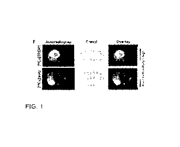
[0019] FIG. 1A shows 3D sagittal maximum intensity projection PET images
of
representative sham and MCAo stroke mice 1.5-2 days post-surgery injected with
either
[64.cui
j TREM1-mAb or [64Cu] Isotype-control-mAb. FIG. 1B shows quantitation of PET
signal
in spleen and FIG. 1C shows quantitation of PET signal in intestines, both
showing a higher
uptake of [64Cu]fREM1-mAb in MCAo mice (n=12) compared to sham mice (n=10).
FIG.
- 4 -
CA 03206197 2023-06-21
WO 2022/155499
PCT/US2022/012569
1D shows in vivo brain PET quantitation reveals significantly higher
[64Cu]fREM1-mAb
signal within the infarct of MCAo mice, compared to uptake in a corresponding
contralateral
brain region and also uptake in an equivalent brain region from Sham mice (n=9
per group).
FIG. lE shows quantification of ex vivo brain autoradiography (AR) of mice
imaged with
r64.c u
L FREM1-mAb or [64Cu]isotype-control-mAb (n = 9-10 biologically independent
samples
per group, mean s.e.m.; two tailed Student's unpaired t-test, *P < 0.05).
FIG. 1F shows a
representative autoradiography images of coronal brain sections, cresyl violet
staining and
overlay of autoradiography and cresyl staining from mice imaged with
[64Cu]fREM1-mAb or
[64--
uu]isotype-control-mAb 36 h after MCAo.
[0020] FIG. 2 shows the results from tracking of peripheral infiltrating
activated
myeloid cells with TREM1-PET in a mouse model of chronic/progressive multiple
sclerosis
(experimental autoinimune encephalomyelitis. EAE model). Representative images
show
that TREMI-PET can visualize markedly elevated tracer uptake in the spleen,
bone marrow,
spinal cord, and brain of EAE versus naive mice and TREM1 knockout (KO) EAE
mice.
[0021] FIG. 3 shows that quantitation of the TREM1-PET signal is able to
detect pro-
inflammatory peripheral CNS-infiltrating myeloid cells in EAE mice. TREM1-PET
signal in the
spinal cord and brain regions is significantly higher in EAE versus naïve and
TREM1 KO
EAE mice.
[0022] FIG. 4 shows that TREM1-PET is more sensitive than TSPO-PET at
detecting
toxic inflammation in EAE. TSPO-PET is unable to delineate activated myeloid
cells in the
lumbar and thoracic spinal cord as clearly as TREM1-PET.
[0023] FIG. 5 shows the study design for assessing TREM1-PET as a tool to
monitor
disease in RR-EAE.
[0024] FIG. 6 shows TREM1-PET provides sensitive monitoring of relapses
and
remissions in EAE. The left side of the figure shows [64Cu] TREM1-mAb images
in RR-EAE
mice, with the top showing in-vivo PET/CT images and the bottom showing spinal
cord
autoradiography. The right side of the figure shows TREM1-PET quantification
with the top
showing quantification of TREM1-PET signal in the lumbar/spinal cord and the
bottom
showing TREM1-PET signal in the cervical/thoracic spinal cord.
[0025] FIG. 7 shows TREM1-PET provides sensitive monitoring of relapses
and
remissions in EAE. Inset i) shows TREM1-PET quantification of whole brain;
inset ii) shows
TREM1-PET quantification of the medulla; inset iii) shows TREM1-PET
quantification of
whole pons; and inset iv) shows TREM1-PET quantification of the cerebellum.
[0026] FIG. 8 shows TREM1-PET imaging enables highly specific in vivo
detection of
innate immune activation in a mouse model of LPS-induced sepsis. [64Cu]fREM1-
mAb
PET/CT image of a vehicle control mouse (left) and an LPS injected mouse
(center) and
- 5 -
CA 03206197 2023-06-21
WO 2022/155499
PCT/US2022/012569
r64"-
uujisotype-control PET/CT image of a LPS injected mouse (right).
[0027] FIG. 9 further demonstrates that TREM-1 is a specific tool for
detecting
activated myeloid cells after LPS challenge using quantification of PET images
(top panel,
left), ex vivo biodistribution data (top panel, right) and autoradiography
(bottom panel) from
the different groups.
[0028] FIG. 10 shows TREM1-PET imaging can detect subtle neuroinflammation
(i.e., peripheral CNS-infiltrating pro-inflammatory myeloid cells) in the
brain of a mouse
model of sepsis.
[0029] FIG 11 shows elevated TREM1-PET signal in brain of APPSwe vs. age-
matched (10 month old) wild-type mice. Images demonstrate increased binding of
the
TREM1-PET tracer in the choroid plexus, ventricles, hippocampus and the
cortical regions of
the APPSwe transgenic mice compared to the age/sex-matched wild type mice.
[0030] FIG. 12 shows increased [64Cu] TREM1-mAb signal in hippocampus of
5XFAD compared to age-matched (6 month old) wild-type mice. TREM1-PET and
autoradiography of 5XFAD and wild-type (WT) mice.
FIG. 13. (A) Whole body representative 3D maximum intensity projection PET/CT
images
ofVeh-WT, LPS-WT, and LPS-K/0 mice 20 h after injection of 64Cu-TREM1-mAb in
addition
to LPS-WT mice 20 h after injection of 64Cu-lsotype-control-mAb (LPS-ISO- WT).
(B)
Quantification of PET images (C) and ex vivo gamma-counting of liver, lung,
and spleen.
Statistical analysis performed using one-way ANOVAs.
FIG. 14. Autoradiography of spleen sections from Veh-WT, LPS-WT, LPS-ISO, and
LPS/K/O
mice. H&E staining overlaid with autoradiography reveals tracer binding is
restricted to the
marginal zone and red pulp which contain macrophages .
FIG. 15. Proportions of myeloid and lymphoid cell subpopulations within the
spleen and
lungs of LPS-WT, Veh-WT wild-type C57BL/6, mice and LPS-K/O TREm1 knockout
mice.
Percentage of TREM1 = cells as a proportion of all live single cells, as well
as within each
parent population (i.e., myeloid and lymphoid), in the spleen and lungs. *vs
VehWT, +vs
LPS-K/O. ** p <0.01, *** p <0.001, **** p <0.0001. Data are representative of
at least 2
independent experiments.
FIG. 16. (A) Quantification of PET signal in brain regions of interest was
performed using a
segmented 3D mouse brain atlas and corona! brain PET/CT images from Veh WT,
LPS-WT,
LPS-ISO-WT, and LPS-K/O mice 20 h post-injection of tracer. Quantification of
/01D/g in
whole brain using (B) PET images and (C) gamma counting (one-way ANOVAs). (D)
Quantification of PET signal in segmented brain regions of interest (2-way AN
OVA). *vs
Veh-WT, #vs LPS-ISO, +vs LPS-K/O (* p < 0.05, ** p < 0.01, *** p < 0.001 ,
**** p < 0.0001).
Data expressed as mean SD.
- 6 -
CA 03206197 2023-06-21
WO 2022/155499
PCT/US2022/012569
FIG. 17. Representative autoradiography (autorad) images and nissl staining of
coronal
brain sections from Veh-WT, LPS-WT, LPS-ISO-WT, and LPS-K/O mice 20 h post-
injection
of 64Cu-TREM1-mAb or 64Cu-lsotype control with nissl staining for anatomical
correlation.
FIG. 18. Proportions of myeloid and lymphoid cell subpopulations within the
brain of LPS-
\ATT, Veh-WT, and LPS-K/O mice. Percentage of TREM1+ cells as a proportion of
all live
single cells, as well as within each parent population (i.e., microglia,
myeloid, and lymphoid)
in the brain. *vs Veh-WT, +vs LPS-K/O. (* p <0.05, *** p <0.001 , **** p
<0.001 ). Data are
representative of at least 2 independent experiments.
FIG. 19. (A) Log2 fold change heat map representation of cytokines (rows) for
LPStreated
and saline-treated wild-type (WT) mice (columns) normalized to vehicle
average.
Row/column order are the resullts of unsupervised hierarchical clustering.
Resulting three
primary cytokine clusters 1-3. Correlogram of PET signal ( /01D/g of 64Cu-TREM
1-mAb
uptake) in brain (medulla), lung, and spleen versus plasma cytokine levels.
(B) Circle size
and color denote Pearson's correlation coefficient. Venn diagram of shared
biological
ontology annotations (ENSEMBL database) between clusters. Fraction of genes in
cluster
with shared annotation. Significant hits identified via over-representation
analysis (onesided
Fishers Exact Test) and annotations shared by greater than 40% of cytokines in
cluster 1
are depicted (*p<0.05). (C) Kaplan-Meier survival curves for TREM1 K/O or \ATT
mice treated
with 15 mg/kg LPS. Log-rank hazard ratio and Log-rank (Mantel-Cox) test
*p<0.05.
Appearance and activity levels of TREM1 K/O and \ATT mice post-LPS challenge
based on an
adapted murine sepsis scoring 5y5tem55 (unpaired t-tests per time-point). (* p
< 0.05, ** p <
0.01 ).
Fig. 20. Translational imaging approach for the detection of activated
peripheral CNS
infiltrating myeloid cells. Peripheral myeloid cells (Le., rnonocytes,
macrophages, neutrophils
and dendritic cells) are recruited to the central nervous system (CNS) during
multiple
sclerosis (MS). Peripheral myeloid cells, in addition to brain resident
rnicroglia, are
associated with CNS MS lesions and are fundamental to disease progression and
remission.
We have identified triggering receptor on myeloid cells 1 (TREMI) positron
emission
tomography (PET) as a novel approach to detect pathogenic peripheral CNS-
infiltrating
myeloid cells in the experimental autoimmune encephalomyelitis (EAE) mouse
using
[64CulTREMI-mAb whole-body PET/CT.
Fig. 21 TREMI peripheral myeloid cell expansion and CNS-infiltration is
observed in EAE.
from a pre-symptomatic disease state. EAE was induced in C571b16 and Tremi
knockout
(KO) mice, Brain, spinal cord and spleen tissues were harvested for single
cell flow
cytometry at different disease states (A). Representative expansion of TREMi c
o45hiCDII
loa= myeloid cells in the spinal cord of naive, pre EAE, low EAE, high EAE,
and Trem I KO
- 7 -
CA 03206197 2023-06-21
WO 2022/155499
PCT/US2022/012569
EAE mice (B). Representative histogram of c o45in-c a 1113+ microglia,
CD45hiCD 11 b+
myeloid, and CD45+c o 11 b- lymphoid cell populations in the spinal cord of a
low EAE
mouse (C). Populations of peripheral myeloid, microglia, and lymphoid cells
within the
spleen (D), spinal cord (E), and whole brain (F) of naive and EAE (pre, low,
high and KO)
mice. Percentage of TREM celis (frequency of all live single cells) in the
spleen (G),
spinal cord (H) and brain (I). Statistical analysis was performed using a 2-
way ANOVA and
Tukey's multiple comparison test. +vs. naive mice, #vs. KO EAE mice, and "
denotes direct
comparison of groups (+ P :8 0.05, ++ P 0.0 1, ++++ P :8 0,0001 1#1 P :S 0,01,
',='$44p
:S 0.001, #ffi##p .80.0001, ***" P 0.0001 ).
Fig, 22, In vivo TREMI PET enables early detection of disease in EAE. PET/CT
was
performed 20 h following r4CujTREM1-mAb injection and tissues were harvested
for ex vivo
analysis (A). TREMi-PET images highlighting elevated signal in the spinal cord
(arrow),
spleen (outline), and bone of EAE mice versus Treml knockout (KO) EAE and
naive mice
(B). Quantification of rCuiTREMI-mAb PET images in lumbar (C) and
thoracic/cervical
regions of the spinal cord (D), whole brain (E), medulla (F), pons (G), spleen
(H), femur (I)
and heart (J) of naive and EAE mice, High resolution ex vivo digital
autoradiography of the
spinal cord (K) and brain (K) supporting PET findings. Statistical analysis
was performed
using a 1-way ANOVA and Tukey's multiple comparison test. +vs. naive mice,
#vs. KO EAE
mice and *denotes direct comparison of groups (+p :5 0.05, ++p V 0.01 +++p :5
0.001,
++++p :8 0.0001 , #p :8 0.05, #,gp :5 0,01, .###p :8 0.001, <figref></figref>p :S 0.0001,
*P :8 0.05, **P
0.01). Data expressed as mean SD.
Fig. 23. TREMI-PET is a more sensitive tool for detecting neuroinflammation in
EAE
compared to the gold-standard TSPO-PET. TSPO-PET/CT imaging was performed 50-
60
min following injection of rFiGE-I 80 and tissues were harvested for ex vivo
analysis (A).
Representative l'89GE-180 images (B). TSPO-PET quantification in
cervical/thoracic spinal
cord (C) lumbar spinal cord (D) and whole brain (E) of EAE (pre, low and high)
and naive
mice. Representative spinal cord and brain [18F]GE-180 autoradiography images
(Niss I
overlay) (F). EAE-to-naive ratios of r34CujTREMI-mAb and [18F]GE-180 signal
using ex vivo
autoradiography (G-1) and gamma counting (J-L). Statistical analysis was
performed using a
1-way ANOVA followed by Tukey's post-hoc test for PET quantification and t-
tests for EAE-
to-naive ratios. +vs.
Fig. 24. TREMi+ cells are present in human MS white matter brain lesions.
Demyelinated
and myelinated regions of a MS white matter lesion were revealed with
LLIXO1Fast Blue
histochemical preparation (A) and Myelin Basic Protein immunohistochemical
staining (B) in
adjacent brain biopsy sections ( drug- and steroid-naive tumefactive
demyelinating MS).
Severe axonal damage observed by staining of neurofilament (C), which revealed
- 8 -
CA 03206197 2023-06-21
WO 2022/155499
PCT/US2022/012569
transected degenerating axons and the formation of axonal bulbs (blue arrows),
Similar
pattern of axonal degeneration observed by Bielschowskys Silver stain (D). H&E
staining
revealed infiltration of immune cells in perivascular regions (E), including a
higher proportion
of T lymphocytes (F) and B lymphocytes (G) in perivascular regions and
surrounding neural
parenchyma. TREMi cells (red arrows) were observed in perivascular regions (H,
counterstained with hematoxylin). Compared to white matter of non-MS control
tissue (I),
tumefactive MS white matter biopsy showed a high number of TREfvli-positive
cells (J).
Scale bar A-C, E-G, 1-J = 50 pm; D, H = 10 pm.
Fig. 25. Flow cytornetry gating strategy. Live single cells were
differentiated into microglia
(CD45intCDi Ib+), peripheral myeloid (CD45hiCDi lb-) and lymphoid (CD4seco1 I
b-) cells.
Separate gates were set for brain (A), spinal cord (B) and spleen (C). Myeloid
cells were
further differentiated into neutrophils (CD45hieD 11 IdLy6G-$) and
monocytesimacrophagesidendritic cells (DCs) (CD4511 iCDI lb+146G1 Levels of
TREMI
expression was investigated on all cell subtypes.
Fig. 26. TREMI expression on neutrophil and monocyteimacrophageldendritic cell
populations. Representative infiltrating TREM CD4511iCD 11 b.Ly6G,s
neutrophils and
CD45h.lCD 11 beLy6G= monocytes (Mo )!macrophages (M<I>)!dendritic cells (DCs)
in the
spinal cord of a WT EAE mouse (A). Percentage of TREMI+ cells (i/ofrequency of
parent)
in the spleen, spinal cord and brain of WI and Tram! knockout (KO) EAE mice
:',13).
Statistical analysis was performed using a l-way ANOVA and Tukey's multiple
comparisOn
test. *vs. KO EAE, (P :3 0.01 , ***P :S 0.001 , ****P :3 0.0001), Data
expressed as
mean SD.
Fig. 27. TREMI is not expressed on endothelial cells, astrocytes or neurons in
EAE. Gating
strategy to assess TREMI levels on endothelial cells (0D31 S'), astrocytes
(CD4S-, ASCA2e),
and neurons (CD4S-, CD90e) in spinal cord tissue form EAE mice (A). Histogram
of TREMI
levels versus positive control beads (B). TREMI cells as a proportion of all
live single cells
(C) and of parent cells (D) in spinal cord. Data expressed as mean SD.
DETAILED DESCRIPTION
[0031] Provided
herein are methods and compositions for efficiently delivering one or
more substance(s) to the brain of a subject. An antigen binding molecule is
administered to
the subject, wherein the antigen binding molecule binds an antigen in
peripheral immune cells.
Peripheral immune cells may be myeloid cells, NK cells, macrophages, and/or
inflammatory
cells that pass through the brain. The subject may require diagnosis of a
disease or condition,
such as multiple sclerosis, Alzheimer's disease, Huntington's disease,
Parkinson's disease,
epilepsy, brain tumor, stroke, amyotrophic lateral sclerosis, spinal cord
and/or brain trauma, a
disease or condition which would benefit from enzyme replacement therapy
("ERT"), a
- 9 -
CA 03206197 2023-06-21
WO 2022/155499
PCT/US2022/012569
neurological disease, chronic inflammatory conditions, acute inflammatory
conditions, or
bacterial infection. The one or more antigen binding molecule(s) and/or one or
more
substance(s) may be one or more of antibody(s), biologic(s), peptide(s), small
molecule(s),
engineered protein scaffold(s), a nucleic acid, or a CRISPR-Cas9 molecule.
I. Definitions
[0032] Unless otherwise defined, all terms of art, notations and other
scientific
terminology used herein are intended to have the meanings commonly understood
by those
of skill in the art to which this invention pertains. In some cases, terms
with commonly
understood meanings are defined herein for clarity and/or for ready reference,
and the
inclusion of such definitions herein should not necessarily be construed to
represent a
difference over what is generally understood in the art. The techniques and
procedures
described or referenced herein are generally well understood and commonly
employed using
conventional methodologies by those skilled in the art, such as, for example,
the widely
utilized molecular cloning methodologies described in Sambrook et al.,
Molecular Cloning: A
Laboratory Manual 2nd ed. (1989) Cold Spring Harbor Laboratory Press, Cold
Spring
Harbor, NY. As appropriate, procedures involving the use of commercially
available kits and
reagents are generally carried out in accordance with manufacturer defined
protocols and/or
parameters unless otherwise noted.
[0033] As used herein, the singular forms "a," "an," and "the" include the
plural
referents unless the context clearly indicates otherwise.
[0034] The term "about" indicates and encompasses an indicated value and a
range
above and below that value. In certain embodiments, the term "about" indicates
the
designated value 10%, 5%, or 1%. In certain embodiments, the term
"about" indicates
the designated value one standard deviation of that value.
[0035] The term "antigen" or "Ag" as used herein shall refer to a molecule
or
molecular structure that can be bound by a molecule, such as an antibody or a
molecule on
a B cell antigen receptor. Antigens are targeted by antibodies and can bind
any suitable
molecule. Antigens are usually proteins, peptides, and polysaccharides. Lipids
and nucleic
acids may also become antigens when combined with proteins and
polysaccharides.
Saccharides and lipids also qualify as antigens.
[0036] The term "antigen binding molecule" as used herein, refers to any
molecule
capable of binding to an antibody. The term antigen binding molecule may
include, for
example, without limitation, a protein, polypeptide, or molecular complex. An
antigen binding
molecule may comprise or consist of one or more of antibody(s), antibody
fragment(s),
biologic(s), peptide(s), small molecule(s), engineered protein scaffold(s), a
nucleic acid, or a
CRISPR-Cas9 molecule. An antigen binding molecule may include one or more
- 10 -
CA 03206197 2023-06-21
WO 2022/155499
PCT/US2022/012569
complementary determining region ("CDR") that alone or in combination with
other
molecules bind to a particular antigen.
[0037] As used herein, "TREM1," "TREM-1," "Triggering Receptor Expressed on
Myeloid Cells 1," "Triggering Receptor Expressed on Monocytes 1," "CD354," or
"CD354
antigen" shall refer to a protein that in humans is encoded by the TREM1 gene.
[0038] As used herein, "TREM2," "TREM-2," "Triggering Receptor Expressed On
Myeloid Cells 2," "Triggering Receptor Expressed On Myeloid Cells 2a,"
"Triggering
Receptor Expressed On Monocytes 2," "Trem2a," "Trem2b," "Trem2c," and "PLOSL2"
shall
refer to a protein that in humans is encoded by the TREM2 gene.
[0039] As used herein, "GPR84," "G Protein-Coupled Receptor 84," "EX33," "G-
Protein Coupled Receptor 84," "Inflammation-Related G Protein-Coupled Receptor
EX33,"
Inflammation-Related G-Protein Coupled Receptor EX33," and "GPCR4" shall refer
to a
protein that in humans is encoded by the GPR84 gene.
[0040] As used herein, a "toll-like receptor" or "TLR" shall refer to any
one of a class
of proteins that in humans plays a key role in the innate immune system. Toll-
like receptors
are single-pass membrane-spanning receptors usually expressed on sentinel
cells such as
macrophages and dendritic cells.
[0041] As used herein, "TSPO" or "translocator protein" shall refer to an
18 kDa
protein mainly found in the outer mitochondria! membrane. In humans, TSPO is
encoded by
the TSPO gene. TSPO is considered the gold standard as a transport protein.
[0042] As used herein, a "nucleotide-binding oligomerization domain-like
receptors,"
"NOD-like receptors," "NLRs," and "nucleotide-binding leucine-rich repeat
receptors" shall
refer to intracellular sensors of pathogen-associated molecular patterns
(PAMPs) that enter
the cell via phagocytosis or pores and damage-associated molecular patterns
(DAMPs) that
are associated with cell stress. Nucleotide-binding oligomerization domain-
like receptors are
types of pattern recognition receptors (PRRs) and play key roles in the
regulation of the
innate immune response.
[0043] As used herein, "biologic," "biopharmaceutical," and "biological
medical
product" shall refer to a pharmaceutical drug product manufactured in,
extracted from, or
semisythesized from biological sources. A biologic can be, for example,
without limitation,
vaccines, blood, blood components, allergenics, somatic cells, gene therapies,
tissues,
recombinant therapeutic proteins, and living medicines used in cell therapy.
Biologics can
be composed of sugars, proteins, or nucleic acids or complex combinations of
any of these
substances. Biologics (or their precursors or components) are isolated from
living sources¨
human, animal, plant, fungal, or microbial.
[0044] As used herein, "peptides" shall refer to short chains of amino
acids between
- 11 -
CA 03206197 2023-06-21
WO 2022/155499
PCT/US2022/012569
two and fifty amino acids, where the amino acids are linked by peptide bonds.
Chains of
fewer than 10 or fifteen amino acids are sometimes referred to as
oligopeptides, such as
dipeptides, tripeptides, and tetrapeptides. A polypeptide is a longer,
continuous,
unbranched peptide chain of up to approximately fifty amino acids.
[0045] A polypeptide that contains more than approximately fifty amino
acids is
known as a protein. Proteins consist of one or more polypeptide(s) arranged in
a biologically
functional way, often bound to ligands such as coenzymes and cofactors or to
another
protein or other macromolecule, such as DNA or RNA. All peptides except cyclic
peptides
have an N-terminal (amine) and a C-terminal (carboxyl group) residue at the
end of the
peptide. Peptides frequently have post-translational modifications such as
phosphorylation,
hydroxylation, sulfonation, palmitoylation, glycosylation, and disulfide
formation.
[0046] As used herein, "small molecule" shall refer to a low molecular
weight (< 1000
daltons) organic compound that may regulate a biological process, with a size
on the order
of about 1 nm.
[0047] As used herein, "engineered protein scaffold" or "protein scaffold"
refers to a
protein, or part thereof, that has a defined three-dimensional structure when
assembled and
a capacity to support molecules or polypeptide domains in or on the structure.
[0048] As used herein, "CRISPR-Cas9 molecule" or "clustered regularly
interspaced
short palindromic repeats" refers to a family of DNA sequences found in the
genomes of
prokaryotic organisms such as bacteria and archaea. The CRISPR-Cas system is a
prokaryotic immune system that confers resistance to foreign genetic elements
such as
those present within plasmids and phages and provides a form of acquired
immunity. RNA
harboring the spacer sequences helps Cas (CRISPR-associated) proteins
recognize and cut
foreign pathogenic DNA. CRISPR are found in approximately 50% of sequenced
bacterial
genomes and nearly 90% of sequenced archaea. CRISPR gene editing systems
commonly
utilize the ca59 gene.
[0049] As used herein, "nucleic acid" shall refer to the overall name for
DNA and
RNA and derivatives therefrom.
[0050] As used herein, "DNA" or "deoxyribonucleic acid" shall each refer to
a nucleic
acid containing the genetic instructions for functioning of organisms. DNA
segments
carrying information are called genes.
[0051] As used herein, "RNA" or "ribonucleic acid" shall refer to a
polymeric molecule
essential in various biological roles in coding, decoding, regulation, and
expression of genes.
Like DNA, RNA is assembled as a chain of nucleotides, but unlike DNA, RNA is
found in
nature as a single strand folded onto itself, rather than a paired double
strand. Cellular
organisms use messenger RNA ("mRNA") to convey genetic information that
directs
- 12 -
CA 03206197 2023-06-21
WO 2022/155499
PCT/US2022/012569
synthesis of specific proteins.
[0052] The term "immunoglobulin" refers to a class of structurally related
proteins
generally comprising two pairs of polypeptide chains: one pair of light (L)
chains and one pair
of heavy (H) chains. In an "intact immunoglobulin," all four of these chains
are
interconnected by disulfide bonds. The structure of immunoglobulins has been
well
characterized. See, e.g., Paul, Fundamental Immunology 7th ed., Ch. 5(2013)
Lippincott
Williams & Wilkins, Philadelphia, PA. Briefly, each heavy chain typically
comprises a heavy
chain variable region (VH) and a heavy chain constant region (CH). The heavy
chain constant
region typically comprises three domains, CH1, CH2, and CH3. Each light chain
typically
comprises a light chain variable region (VL) and a light chain constant
region. The light chain
constant region typically comprises one domain, abbreviated CL.
[0053] The term "antibody" describes a type of immunoglobulin molecule and
is used
herein in its broadest sense. An antibody specifically includes intact
antibodies (e.g., intact
immunoglobulins) and antibody fragments.
[0054] The VH and VL regions may be further subdivided into regions of
hypervariability ("hypervariable regions (HVRs);" also called "complementarity
determining
regions" (CDRs)) interspersed with regions that are more conserved. The more
conserved
regions are called framework regions (FRs). Each VH and VL generally comprises
three
CDRs and four FRs, arranged in the following order (from N-terminus to C-
terminus): FR1 -
CDR1 - FR2 - CDR2 - FR3 - CDR3 - FR4. The CDRs are involved in antigen
binding, and
confer antigen specificity and binding affinity to the antibody. See Kabat et
al., Sequences of
Proteins of Immunological Interest 5th ed. (1991) Public Health Service,
National Institutes of
Health, Bethesda, MD, incorporated by reference in its entirety.
[0055] The light chain from any vertebrate species can be assigned to one
of two
types, called kappa and lambda, based on the sequence of the constant domain.
[0056] The heavy chain from any vertebrate species can be assigned to one
of five
different classes (or isotypes): IgA, IgD, IgE, IgG, and IgM. These classes
are also
designated a, 6, E, y, and p, respectively. The IgG and IgA classes are
further divided into
subclasses on the basis of differences in sequence and function. Humans
express the
following subclasses: IgG1, IgG2, IgG3, IgG4, IgA1, and IgA2.
[0057] The amino acid sequence boundaries of a CDR can be determined by one
of
skill in the art using any of a number of known numbering schemes, including
those
described by Kabat et al., supra ("Kabat" numbering scheme); Al-Lazikani et
al., 1997, J.
Mol. Biol., 273:927-948 ("Chothia" numbering scheme); MacCallum et al., 1996,
J. Mol.
262:732-745 ("Contact" numbering scheme); Lefranc et al., Dev. Comp. Immunol.,
2003,
27:55-77 ("IMGT" numbering scheme); and Honegge and PlOckthun, J. Mol. Biol.,
2001,
- 13 -
CA 03206197 2023-06-21
WO 2022/155499
PCT/US2022/012569
309:657-70 ("AHo" numbering scheme), each of which is incorporated by
reference in its
entirety.
[0058] Table 1 provides the positions of CDR-L1, CDR-L2, CDR-L3, CDR-H1,
CDR-
H2, and CDR-H3 as identified by the Kabat and Chothia schemes. For CDR-H1,
residue
numbering is provided using both the Kabat and Chothia numbering schemes.
[0059] Unless otherwise specified, the numbering scheme used for
identification of a
particular CDR herein is the Kabat numbering scheme. Variant and equivalent
antibodies
with a Chothia numbering scheme are intended to be within the scope of the
invention.
Table 1. Residues in CDRs according to Kabat and Chothia numbering schemes.
CDR Kabat Chothia
Ll L24-L34 L24-L34
L2 L50-L56 L50-L56
L3 L89-L97 L89-L97
H31-H35B
H1 (Kabat Numbering) H26-H32 or H34*
H1 (Chothia Numbering) H31-H35 H26-H32
H2 H50-H65 H52-H56
H3 H95-H102 H95-H102
*The C-terminus of CDR-H1, when numbered using the Kabat numbering convention,
varies
between H32 and H34, depending on the length of the CDR.
[0060] The "EU numbering scheme" is generally used when referring to a
residue in
an antibody heavy chain constant region (e.g., as reported in Kabat et al.,
supra). Unless
stated otherwise, the EU numbering scheme is used to refer to residues in
antibody heavy
chain constant regions described herein.
[0061] An "antibody fragment" comprises a portion of an intact antibody,
such as the
antigen binding or variable region of an intact antibody. Antibody fragments
include, for
example, Fv fragments, Fab fragments, F(ab)2fragments, Fab' fragments, scFv
(sFv)
fragments, and scFv-Fc fragments.
[0062] "Fv" fragments comprise a non-covalently linked dimer of one heavy
chain
variable domain and one light chain variable domain.
[0063] "Fab" fragments comprise, in addition to the heavy and light chain
variable
domains, the constant domain of the light chain and the first constant domain
(CH1) of the
heavy chain. Fab fragments may be generated, for example, by papain digestion
of a full-
length antibody.
[0064] "F(a1:02" fragments contain two Fab' fragments joined, near the
hinge region,
by disulfide bonds. F(a1:02 fragments may be generated, for example, by pepsin
digestion of
- 14 -
CA 03206197 2023-06-21
WO 2022/155499
PCT/US2022/012569
an intact antibody. The F(ab') fragments can be dissociated, for example, by
treatment with
fl-mercaptoethanol.
[0065] "Single-chain Fv" or "sFv" or "scFv" antibody fragments comprise a
VH domain
and a VL domain in a single polypeptide chain. The VH and VL are generally
linked by a
peptide linker. See PlOckthun A. (1994). Antibodies from Escherichia coll. In
Rosenberg M. &
Moore G.P. (Eds.), The Pharmacology of Monoclonal Antibodies vol. 113 (pp. 269-
315).
Springer-Verlag, New York, incorporated by reference in its entirety. "scFv-
Fc" fragments
comprise an scFv attached to an Fc domain. For example, an Fc domain may be
attached
to the C-terminal of the scFv. The Fc domain may follow the VH or VL depending
on the
orientation of the variable domains in the scFv (i.e., VH-VL or VL-VH). Any
suitable Fc domain
known in the art or described herein may be used.
[0066] The term "minibody" refers to an antibody fragment (such as one
that
contains a VL-VH-CH3) with bivalent binding to an antigen.
[0067] The term "monoclonal antibody" (mAb) refers to an antibody from a
population of substantially homogeneous antibodies. A population of
substantially
homogeneous antibodies comprises antibodies that are substantially similar and
that bind
the same epitope(s), except for variants that may normally arise during
production of the
monoclonal antibody. Such variants are generally present in only minor
amounts. A
monoclonal antibody is typically obtained by a process that includes the
selection of a single
antibody from a plurality of antibodies. For example, the selection process
can be the
selection of a unique clone from a plurality of clones, such as a pool of
hybridoma clones,
phage clones, yeast clones, bacterial clones, or other recombinant DNA clones.
The
selected antibody can be further altered, for example, to improve affinity for
the target
("affinity maturation"), to humanize the antibody, to improve its production
in cell culture,
and/or to reduce its immunogenicity in a subject.
[0068] The term "chimeric antibody" refers to an antibody in which a
portion of the
heavy and/or light chain is derived from a particular source or species, while
the remainder
of the heavy and/or light chain is derived from a different source or species.
[0069] "Humanized" forms of non-human antibodies are chimeric antibodies
that
contain minimal sequence derived from the non-human antibody. A humanized
antibody is
generally a human immunoglobulin (recipient antibody) in which residues from
one or more
CDR(s) are replaced by residues from one or more CDR(s) of a non-human
antibody (donor
antibody). The donor antibody can be any suitable non-human antibody, such as
a mouse,
rat, rabbit, chicken, llama, or non-human primate antibody having a desired
specificity,
affinity, or biological effect. In some instances, selected framework region
residues of the
recipient antibody are replaced by the corresponding framework region residues
from the
- 15 -
CA 03206197 2023-06-21
WO 2022/155499
PCT/US2022/012569
donor antibody. Humanized antibodies may also comprise residues that are not
found in
either the recipient antibody or the donor antibody. Such modifications may be
made to
further refine antibody function. For further details, see Jones et al.,
Nature, 1986, 321:522-
525; Riechmann et al., Nature, 1988, 332:323-329; and Presta, Curr. Op.
Struct. Biol., 1992,
2:593-596, each of which is incorporated by reference in its entirety.
[0070] A "human antibody" is one which possesses an amino acid sequence
corresponding to that of an antibody produced by a human or a human cell, or
derived from
a non-human source that utilizes a human antibody repertoire or human antibody-
encoding
sequences (e.g., obtained from human sources or designed de novo). Human
antibodies
specifically exclude humanized antibodies.
[0071] An "isolated antibody" is one that has been separated and/or
recovered from
a component of its natural environment. Components of the natural environment
may
include enzymes, hormones, and other proteinaceous or non proteinaceous
materials. In
some embodiments, an isolated antibody is purified to a degree sufficient to
obtain at least
15 residues of N-terminal or internal amino acid sequence, for example by use
of a spinning
cup sequenator. In some embodiments, an isolated antibody is purified to
homogeneity by
gel electrophoresis (e.g., SDS-PAGE) under reducing or non-reducing
conditions, with
detection by Coomassie blue or silver stain. An isolated antibody includes an
antibody in
situ within recombinant cells, since at least one component of the antibody's
natural
environment is not present. In some embodiments, an isolated antibody is
prepared by at
least one purification step.
[0072] With regard to the binding of an antibody to a target molecule, the
terms
"binding" or "binds to" a particular antigen (e.g., a polypeptide target) or
an epitope on a
particular antigen mean binding that is measurably different from a non-
selective interaction.
Binding can be measured, for example, by determining binding of a molecule
compared to
binding of a control molecule. Binding can also be determined by competition
with a control
molecule that is similar to the target, such as an excess of non-labeled
target. In that case,
binding is indicated if the binding of the labeled target to a probe is
competitively inhibited by
the excess non-labeled target.
[0073] Percent "identity" between a polypeptide sequence and a reference
sequence
is defined as the percentage of amino acid residues in the polypeptide
sequence that are
identical to the amino acid residues in the reference sequence, after aligning
the sequences
and introducing gaps, if necessary, to achieve the maximum percent sequence
identity.
Alignment for purposes of determining percent amino acid sequence identity can
be
achieved in various ways that are within the skill in the art, for instance,
using publicly
available computer software such as BLAST, BLAST-2, ALIGN, MEGALIGN (DNASTAR),
- 16 -
CA 03206197 2023-06-21
WO 2022/155499
PCT/US2022/012569
CLUSTALW, or CLUSTAL OMEGA software. Those skilled in the art can determine
appropriate parameters for aligning sequences, including any algorithms needed
to achieve
maximal alignment over the full length of the sequences being compared.
[0074] A "conservative substitution" or a "conservative amino acid
substitution,"
refers to the substitution of one or more amino acid(s) with one or more
chemically or
functionally similar amino acid(s). Conservative substitution tables providing
similar amino
acids are well known in the art. Polypeptide sequences having such
substitutions are known
as "conservatively modified variants." Such conservatively modified variants
are in addition
to and do not exclude polymorphic variants, interspecies homologs, and
alleles. By way of
example, the following groups of amino acids are considered conservative
substitutions for
one another.
Acidic Residues D and E
Basic Residues K, R, and H
Hydrophilic Uncharged Residues S, T, N, and Q
Aliphatic Uncharged Residues G, A, V, L, and I
Non-polar Uncharged Residues C, M, and P
Aromatic Residues F, Y, and W
Alcohol Group-Containing Residues S and T
Aliphatic Residues I, L, V, and M
Cycloalkenyl-associated Residues F, H, W, and Y
Hydrophobic Residues A, C, F, G, H, I, L, M, T, V, W, and Y
Negatively Charged Residues D and E
Polar Residues C, D, E, H, K, N, Q, R, S, and T
Positively Charged Residues H, K, and R
Small Residues A, C, D, G, N, P, S, T, and V
Very Small Residues A, G, and S
Residues Involved in Turn Formation A, C, D, E, G, H, K, N, Q, R, S, P, and
T
Flexible Residues Q, T, K, S, G, P, D, E, and R
Group 1 A, S, and T
Group 2 D and E
Group 3 N and Q
Group 4 Rand K
Group 5 I, L, and M
- 17 -
CA 03206197 2023-06-21
WO 2022/155499
PCT/US2022/012569
Group 6 F, Y, and W
Group A A and G
Group B D and E
Group C N and Q
Group D R, K, and H
Group E I, L, M, V
Group F F, Y, and W
Group G S and T
Group H C and M
Additional conservative substitutions may be found, for example, in Creighton,
Proteins:
Structures and Molecular Properties 2nd ed. (1993) W. H. Freeman & Co., New
York, NY.
[0075] The term "amino acid" refers to the twenty common naturally
occurring amino
acids. Naturally occurring amino acids include alanine (Ala; A), arginine
(Arg; R), asparagine
(Asn; N), aspartic acid (Asp; D), cysteine (Cys; C); glutamic acid (Glu; E),
glutamine (Gin;
Q), Glycine (Gly; G); histidine (His; H), isoleucine (Ile; l), leucine (Leu;
L), lysine (Lys; K),
methionine (Met; M), phenylalanine (Phe; F), proline (Pro; P), serine (Ser;
S), threonine (Thr;
T), tryptophan (Trp; \A/), tyrosine (Tyr; Y), and valine (Val; V).
[0076] As used herein, the term "peripheral immune cells" shall refer to
immune cells
that reside outside of the brain. During a neuroimmune response, sometimes
peripheral
immune cells are able to cross various blood or fluid brain barriers in order
to respond to
pathogens that have entered the brain. As the central nervous system is
considered an
immune-privileged organ due to the blood-brain barrier, there is a relatively
low number of
surveilling peripheral immune cells found within the brain parenchyma.
[0077] As used herein, "myeloid cells" or "myelogenous cells" shall refer
to
monocytes, macrophages, neutrophils, basophils, eosinophils, erythrocytes, and
megakaryocytes and platelets. In some embodiments, myeloid cells shall refer
to any cell of
myeloid lineage.
[0078] As used herein, "NK cells," "natural killer cells," "large granular
lymphocytes,"
or "LGL" shall refer to a type of cytotoxic lymphocyte critical to the innate
immune system.
The role of NK cells is analogous to that of cytotoxic T cells in the
vertebrate adaptive
immune response.
[0079] As used herein, "macrophages" shall refer to a type of white blood
cell of the
immune system that engulfs and digests cellular debris, foreign substances,
microbes,
cancer cells, and anything else that does not have the type of proteins
specific to healthy
- 18-
CA 03206197 2023-06-21
WO 2022/155499
PCT/US2022/012569
body cells on its surface in a process called phagocytosis. Macrophages are
large
phagocytes and are found in essentially all tissues, where they patrol for
potential
pathogens. They take various forms (with various names) throughout the body
(e.g.,
histiocytes, Kupffer cells, alveolar macrophages, microglia, and others), but
all are part of the
mononuclear phagocyte system.
[0080] As used herein, "monocytes" shall refer to a type of leukocyte, or
white blood
cell. They are the largest type of leukocyte and can differentiate into
macrophages and
myeloid lineage dendritic cells. As a part of the vertebrate innate immune
system
monocytes also influence the process of adaptive immunity.
[0081] As used herein, "granulocytes," "polymorphonuclear leukocytes,"
"PMN,"
"PML," and "PMNL" shall refer to a category of white blood cells in the innate
immune
system characterized by the presence of granules in their cytoplasm. They have
varying
shapes of the nucleus and are usually divided into three segments. There are
four types of
granulocytes: basophils, eosinophils, neutrophils, and mast cells.
[0082] As used herein, "neutrophils" shall refer to the most abundant type
of
phagocyte found in the bloodstream, constituting 60% to 65% of the total
circulating white
blood cells and consisting of two subpopulations. Neutrophils stain a neutral
pink on
hematoxylin and eosin (H&E) histological or cytological preparations. They are
formed of
stem cells in bone marrow and divide into subpopulations of neutrophil-killers
and neutrophil-
cagers. They are short-lived and highly motile, or mobile, as they can enter
parts of tissue
where other cells/molecules cannot.
[0083] As used herein, "eosinophils," "eosinophiles," or "acidophils" shall
refer to a
variety of white blood cells which appear brick red after staining with eosin,
a red dye, using
the Romanowsky method. Eosinophils are largely responsible for combatting
multicellular
parasites and certain infections in vertebrates. They develop during
hematopoiesis in the
bone marrow before migrating to the blood, after which they are terminally
differentiated and
do not multiply.
[0084] Eosinophils are acid loving due to their large acidophilic
cytoplasmic granules.
Their small granules contain chemical mediators, such as eosinophil
peroxidase,
ribonuclease (RNase), deoxyribonuclease (DNase), lipase, plasminogen, and
major basic
protein. When these mediators are released during degranulation, they are
highly toxic to
both parasite and host tissues.
[0085] As used herein, "basophils" shall refer to a type of white blood
cell that is
susceptible to staining by basic dyes. Basophils are the least common type of
granulocyte,
representing about 0.5% to 1% of circulating white blood cells. Basophils are
the largest
type of granulocyte, however.
- 19 -
CA 03206197 2023-06-21
WO 2022/155499
PCT/US2022/012569
[0086] As used herein, "dendritic cells" shall refer to antigen-presenting
cells (also
known as accessory cells) of the mammalian immune system. Their main function
is to
process antigen material and present it on the cell surface to the T cells of
the immune
system. They act as messengers between the innate and adaptive immune systems.
[0087] As used herein, "B cells" or "B lymphocytes" shall refer to a type
of white
blood cell that functions in the humoral immunity component of the adaptive
immune system
by secreting antibodies. Additionally, B cells present antigen as professional
antigen-
presenting cells and secrete cytokines.
[0088] As used herein, "diagnosis" shall to identifying the nature or stage
of an
illness or other problem.
[0089] As used herein, "IV," "intravenous," or "intravenous route of
administration"
shall refer to fluid delivery directly into a vein. Intravenous can be used
both for injections,
using a syringe at higher pressures; as well as for infusions, typically using
only the pressure
supplied by gravity. Intravenous infusions are commonly referred to as drips.
[0090] As used herein, "intramuscular," "intramuscular injection," "IM
injection," or
"IM" shall refer to injection of a substance directly into muscle.
[0091] As used herein, "subcutaneous" "subcutaneous injection," "Sc," "SQ,"
"sub-
cu," "sub-Q," "SubQ," or "subcut" shall refer to administration of a bolus
into the subcutis, the
layer of skin directly below the dermis and epidermis, collectively referred
to as the cutis.
[0092] As used herein, "intraperitoneal" "intraperitoneal injection," "IP
injection," or
"IP" shall refer to injection of a substance into the peritoneum (body
cavity).
[0093] As used herein, "intrathecal" "intrathecal administration," or
"intrathecal" shall
refer to a route of administration for drugs via injection into the spinal
canal or into the
subarachnoid space so that it reaches the cerebrospinal fluid (CSF).
[0094] As used herein, "intravitreal" or "intravitreal administration" is a
route of
administration of a drug or other substance in which the substance is
delivered to the
vitreous humor of the eye.
[0095] As used herein, the term "subject" means a mammal or a human. In
some
embodiments subjects include, but are not limited to, monkeys, dogs, cats,
mice, rats, cows,
horses, camels, avians, goats, and sheep.
[0096] As used herein, "MRI," "magnetic resonance imaging," "nuclear
magnetic
resonance imaging," or "NMR" shall refer to a medical imaging technique used
in radiology
to form pictures of the anatomy and the physiological processes of the body.
Antiden Bindind Molecules and/or One or More Substance(s)
[0097] The current invention is drawn to methods and compositions for
delivering
one or more substance(s) to the brain in a subject in need thereof, wherein
the one or more
- 20 -
CA 03206197 2023-06-21
WO 2022/155499
PCT/US2022/012569
antigen binding molecule(s) that binds one or more antigen(s) in peripheral
immune cells is
administered to the subject. In some embodiments, the one or more antigen
binding
molecule(s) comprises or consists of one or more of antibody(s), antibody
fragment(s),
biologics, peptide(s), small molecule(s), engineered protein scaffold(s), a
nucleic acid, or one
or more CRISP-Cas9 molecule(s).
[0098] In some embodiments, the one or more antigen binding molecule(s)
comprises or consists of antibodies and/or antibody fragments. In some
embodiments, the
antibody and/or antibody fragment comprises or consists of a monoclonal
antibody or
chimeric antibody. In some embodiments, the antibody or antibody fragment
comprises or
consists of a mouse antibody or antibody fragment. In some embodiments, the
antibody or
antibody fragment comprises or consists of a humanized antibody or antibody
fragment. In
some embodiment, the antibody or antibody fragment comprises or consists of a
human
antibody or antibody fragment. In some embodiments, the antibody or antibody
fragment
comprises or consists of an isolated antibody or antibody fragment.
[0099] In some embodiments, the antibody or antibody fragment comprises a
binding
domain that binds to one or more antigen(s). In some embodiments, the one or
more
antigen(s) comprises or consists of TREM1, TREM2, GPR84, a toll-like receptor,
or a
nucleotide-binding oligomerization domain-like receptor.
[00100] In some embodiments, the binding domain comprises a light chain. In
some
embodiments, the light chain is a kappa light chain. In some embodiments, the
light chain is
a lambda light chain.
[00101] In some embodiments, the binding domain comprises a heavy chain. In
some embodiments, the heavy chain is an IgA. In some embodiments, the heavy
chain is
an IgD. In some embodiments, the heavy chain is an IgE. In some embodiments,
the heavy
chain is an IgG. In some embodiments, the heavy chain is an IgM. In some
embodiments,
the heavy chain is an IgG1. In some embodiments, the heavy chain is an IgG2.
In some
embodiments, the heavy chain is an IgG3. In some embodiments, the heavy chain
is an
IgG4. In some embodiments, the heavy chain is an IgA1. In some embodiments,
the heavy
chain is an IgA2.
[00102] In some embodiments, the binding domain is an antibody fragment. In
some
embodiments, the antibody fragment is an Fv fragment. In some embodiments, the
antibody
fragment is a Fab fragment. In some embodiments, the antibody fragment is a
F(a13')2
fragment. In some embodiments, the antibody fragment is a Fab' fragment. In
some
embodiments, the antibody fragment is an scFv (sFv) fragment. In some
embodiments, the
antibody fragment is an scFv-Fc fragment. In some embodiments, the antibody
fragment is
a minibody. In some embodiments, the antibody fragment is a single domain
antibody.
- 21 -
CA 03206197 2023-06-21
WO 2022/155499
PCT/US2022/012569
[00103] In some embodiments, the binding domain is a chimeric antibody. In
some
embodiments, the binding domain is a humanized antibody. In some embodiments,
the
binding domain is a human antibody.
[00104] In some embodiments, the binding domain binds TREM1, TREM2, GPR84,
a
toll-like receptor, and/or a nucleotide-binding oligomerization domain-like
receptor. In some
embodiments, the binding domain binds TREM2. In some embodiments, the binding
domain
binds GPR84. In some embodiments, the binding domain binds a toll-like
receptor. In some
embodiments, the binding domain binds a nucleotide-binding oligomerization
domain-like
receptor.
[00105] In some embodiments, the binding domain binds TREM1 (See, for
example,
hftos://www.rnds sterns.com/ roductsimouse-trern-l-antibody- 174031 mab1187,
which is
incorporated by reference herein). In some embodiments, the binding domain has
a certain
percent identity to one or more sequence(s) of binding domains that bind
TREM1. In some
embodiments, the binding domain has a percent identity that is at least about
70%, at least
about 75%, at least about 80%, at least about 85%, at least about 90%, or at
least about
95% to one or more sequence(s) of binding domains that bind TREM1. In some
embodiments, the binding domain has one or more conservative substitution(s)
as compared
to one or more sequence(s) of binding domains that bind TREM1.
[00106] In some embodiments, the one or more antigen binding molecule(s)
comprises or consists of one or more peptide(s). In some embodiments, the one
or more
peptide(s) comprises a chain of amino acids between about two and about fifty
amino acids.
In some embodiments, the one or more peptide(s) comprises a chain of amino
acids fewer
than about 10 or fewer than about fifteen amino acids. In some embodiments,
the one or
more peptide(s) comprises or consists of one or more of a dipeptide(s),
tripeptide(s), and/or
tetrapeptide(s).
[00107] In some embodiments, the one or more antigen binding molecule(s)
comprises or consists of one or more polypeptide(s). A polypeptide that
contains more than
approximately fifty amino acids is known as a protein. Proteins consist of one
or more
polypeptide(s) arranged in a biologically functional way, often bound to
ligands such as
coenzymes and cofactors or to another protein or other macromolecule, such as
DNA or
RNA. All peptides except cyclic peptides have an N-terminal (amine) and a C-
terminal
(carboxyl group) residue at the end of the peptide. Peptides frequently have
post-
translational modifications such as phosphorylation, hydroxylation,
sulfonation,
palmitoylation, glycosylation, and disulfide formation.
[00108] In some embodiments, the one or more antigen binding molecule(s)
comprises or consists of one or more small molecule(s). Small molecules are
organic
- 22 -
CA 03206197 2023-06-21
WO 2022/155499
PCT/US2022/012569
compounds with a molecular weight that is usually less than about 1000
daltons. Small
molecules may regulate a biological process. Larger structures such as nucleic
acids and
proteins and many polysaccharides are not small molecules, although their
constituent
monomers (ribo- or deoxyribonucleotides, amino acids, and monosaccharides,
respectively)
are often considered small molecules. Small molecules can have a variety of
biological
functions or applications, such as serving as cell signaling molecules or
drugs or in many
other roles. Small molecules may be natural or artificial.
[00109] In some embodiments, the one or more antigen binding molecules
comprises
of consist of one or more engineered protein scaffold(s). An engineered
protein scaffold has
a three-dimensional structure that when assembled has a capacity to support
molecules
and/or polypeptide domains in or on the structure.
[00110] In some embodiments, the one or more antigen binding molecule(s)
comprises or consist of one or more nucleic acid(s). Nucleic acids are
composed of
nucleotides, monomers made of three components: a 5-carbon sugar, a phosphate
group,
and a nitrogenous base. If the sugar is the compound ribose, then the nucleic
acid is RNA; if
the sugar is deoxyribose or derived from deoxyribose, then the nucleic acid is
DNA.
[00111] Nucleic acids are the most important of all biomolecules. They are
found in
all living things. They function to encode and store information of every
living life form. They
transmit and express that information from the interior of the cell. They also
transmit
information to the next generation of organism.
[00112] Information is ultimately encoded and conveyed via a sequence of
nucleotides, the nucleic acid sequence. Strings of nucleotides are bounded to
form helical
backbones and assembled into chains of base-bases selected from canonical (and
sometimes non-canonical) nucleobases. The nucleobases are adenine, cytosine,
guanine,
thymine, and uracil. Using amino acid and the process of protein synthesis,
nucleic acid
sequences store and transmit information, such as coded information in genes
that allows
one to express proteins.
[00113] In some embodiments, the one or more nucleic acids comprise one or
more
DNA molecules. DNA often consists of two long polymers of simple units called
nucleotides,
with backbones made of sugars and phosphate groups joined by ester bonds.
These two
strands run in opposite directions to each other and are, therefore, anti-
parallel. The
sequence of these four nucleobases along the backbone encodes the information.
[00114] Information is read using the genetic code, which specifies the
sequence of
the amino acids within proteins. The code is read by copying stretches of DNA
into the
related nucleic acid RNA in a process called transcription. Within cells, DNA
is organized
into long structures called chromosomes.
- 23 -
CA 03206197 2023-06-21
WO 2022/155499
PCT/US2022/012569
[00115] Eukaryotic organisms (animals, plants, fungi, and protists) store
most of their
DNA inside the cell nucleus and some of their DNA in organelles, such as
mitochondria or
chloroplasts. In contrast, prokaryotes (bacteria and archaea) store their DNA
only in the
cytoplasm. Within the chromosomes, chromatin proteins, such as histones,
compact and
organize DNA. These compact structures guide the interactions between DNA and
other
proteins, helping control which parts of the DNA are transcribed.
[00116] In some embodiments, the one or more nucleic acids comprise one or
more
RNA. Some RNA molecules play an active role within cells by catalyzing
biological
reactions, controlling gene expression, or sensing and communicating responses
to cellular
signals. One of the active processes is protein synthesis, a universal
function in which RNA
molecules direct the synthesis of proteins on ribosomes. This process uses
transfer RNA
("mRNA") molecules to deliver amino acids to the ribosome, where ribosomal
("rRNA") then
links amino acids together to form coded proteins.
[00117] In some embodiments, the one or more antigen binding molecule(s)
comprises or consists of one or more CRISPR-Cas9 molecule(s). CRISPR-Cas9
sequences
are derived from DNA fragments of bacteriophages that have previously infected
the
prokaryote. The sequences are used to detect and destroy DNA from similar
bacteriophages during subsequent infections. Hence these sequences play a key
role in the
antiviral (i.e. anti-phage) defense system of prokaryotes.
[00118] The CRISPR-Cas system is a prokaryotic immune system that confers
resistance to foreign genetic elements such as those present within plasmids
and phages
and provides a form of acquired immunity. RNA harboring the spacer sequences
helps Cas
(CRISPR-associated) proteins recognize and cut foreign pathogenic DNA. Other
RNA-
guided Cas proteins cut foreign RNA. CRISPR are found in approximately 50% of
sequenced bacterial genomes and nearly 90% of sequenced archaea.
[00119] CRISPR gene editing systems commonly utilize the ca59 gene. This
editing
process has a wide variety of applications including basic biological
research, development
of products, and treatment of diseases (See, for example, Zhang F, Wen Y, Guo
X (2014).
CRISPR/Cas9 for genome editing: progress, implications and challenges. Human
Molecular
Genetics. 23); CRISPR-CAS9, TALENS and ZFNS - the battle in gene editing
https://www.ptglab.com/news/blog/crispr-cas9-talens-and-zfns-the-battle-in-
gene-editing;
and Hsu PD, Lander ES, Zhang F (June 2014). Development and applications of
CRISPR-
Cas9 for genome engineering. Cell. 157 (6): 1262-1278, each of which is
incorporated by
reference herein in their entirety).
Antiqens
[00120] The invention is drawn to one or more substance(s) to the brain in
a subject in
- 24 -
CA 03206197 2023-06-21
WO 2022/155499
PCT/US2022/012569
need thereof, comprising administering an antigen binding molecule to the
subject, wherein
the antigen binding molecule binds an antigen in peripheral immune cells. Any
antigen
present in peripheral immune cells would be suitable. For example, TREM1,
TREM2,
GPR84, a (inducible) toll-like receptor, or a nucleotide-binding
oligomerization domain-like
receptor would all be suitable antigens.
[00121] In some embodiments, the antigen comprises or consists of TREM1
(See for
example the uniprot sequence listing for TREM1 at
https://www.uniprot.orWuniprotiQ9NP99,
which is incorporated by reference herein). In some embodiments, the antigen
is a fragment
or has a certain percent identity to TREM1. In some embodiments, the antigen
has a
percent identity that is at least about 70%, at least about 75%, at least
about 80%, at least
about 85%, at least about 90%, or at least about 95% to TREM1.
[00122] TREM 1 is a receptor belonging to the Ig superfamily and is
expressed on
myeloid cells. TREM1 amplifies neutrophil and monocyte-mediated inflammatory
responses,
such as those triggered by bacterial and fungal releases.
[00123] TREM1 is a highly specific biomarker of pro-inflammatory myeloid-
driven
immune responses in the well-established mouse model of systemic inflammation
and LPS-
induced sepsis. TREM1 expression is markedly elevated in the spleen, lungs,
and brain
after LPS challenge and is predominantly restricted to peripheral myeloid
cells (i.e., dendritic
cells, macrophages, monocytes, and neutrophils). Moreover, increased TREM1
binding was
identified in these regions using [64Cu]fREM1-mAb PET imaging, demonstrating
the
specificity and sensitivity of TREM1-PET to non-invasively visualize and track
pro-
inflammatory myeloid-driven immune responses in vivo.
[00124] TREM1-PET signal correlated with pro-inflammatory cytokine
signatures and
TREM1 K/O resulted in prolonged survival following LPS administration. (See,
Bouchon, a,
Facchetti, F., Weigand, M. a & Colonna, M. TREM-1 amplifies inflammation and
is a crucial
mediator of septic shock. Nature 410,1103-1107 (2001); Schenk, M., Bouchon,
A., Seibold,
F. & Mueller, C. TREM-1¨expressing intestinal macrophages crucially amplify
chronic
inflammation in experimental colitis and inflammatory bowel diseases. J. Clin.
Invest. 117,
3097-3106 (2007); Poukoulidou, T. etal. TREM-1 expression on neutrophils and
monocytes
of septic patients: relation to the underlying infection and the implicated
pathogen. BMC
Infect. Dis. 11,309 (2011); Cohen, J. TREM-1 in sepsis. Lancet (London,
England) 358,
776-8 (2001); and Nathan, C. & Ding, A. TREM-1: A new regulator of innate
immunity in
sepsis syndrome. Nat. Med. 7,530-532 (2001), each of which is hereby
incorporated by
reference in its entirety).
[00125] TREM1 may be useful in sepsis (See, for example, Gibot, S. etal.
Plasma
level of a triggering receptor expressed on myeloid cells-1: Its diagnostic
accuracy in
- 25 -
CA 03206197 2023-06-21
WO 2022/155499
PCT/US2022/012569
patients with suspected sepsis. Ann. Intern. Med. 141,9-15+1(2004); which is
incorporated
by reference herein in its entirety) and also in a broad range of chronic
inflammatory
diseases including inflammatory bowel disease (IBD), arthritis, and cancer.
TREM1 is also a
major contributor to cerebral injury following stroke (See., Liu, Q. etal.
Peripheral TREM1
responses to brain and intestinal immunogens amplify stroke severity. Nat.
Immunol. (2019),
which is incorporated by reference in its entirety herein), indicating a role
for TREM1 in the
pathophysiology of neurological disease.
[00126] Whole-body TREM1-PET imaging has not only demonstrated increased
peripheral myeloid cell infiltration into ischemic brain tissue but also
revealed increased
peripheral innate immune responses in the spleen and intestines of stroked
mice. Further
investigation of TREM1 expression on intestinal macrophages showed that it
plays a role in
gut permeability and bacterial translocation, highlighting the advantages of
TREM1-PET as a
tool to characterize innate immune responses in the whole-body, which may not
be revealed
when using traditional ex vivo approaches.
[00127] In some embodiments, the antigen comprises or consists of TREM2.
The
TREM2 protein functions in immune response and may be involved in chronic
inflammation
by triggering the production of constitutive inflammatory cytokines. TREM2
binds
phospholipids (preferably anionic lipids) such as phosphatidylserine,
phosphatidylethanolamine, phosphatidylglycerol, and sphingomyelin.
[00128] TREM2 regulates microglial proliferation by acting as an upstream
regulator
of the Wnt/beta-catenin signaling cascade. TREM2 is required for microglial
phagocytosis of
apoptotic neurons and also required for microglial activation and phagocytosis
of myelin
debris after neuronal injury and of neuronal synapses during synapse
elimination in the
developing brain. TREM2 regulates microglial chemotaxis and process outgrowth
and also
the microglial response to oxidative stress and lipopolysaccharide. TREM2
suppresses
PI3K and NF-kappa-B signaling in response to lipopolysaccharide, thus
promoting
phagocytosis, suppressing pro-inflammatory cytokine and nitric oxide
production, inhibiting
apoptosis, and increasing expression of IL10 and TGFB. During oxidative
stress, TREM2
promotes anti-apoptotic NF-kappa-B signaling and ERK signaling.
[00129] TREM2 plays a role in microglial MTOR activation. TREM2 regulates
age-
related changes in microglial numbers. TREM2 triggers activation of immune
responses in
macrophages and dendritic cells. TREM2 mediates cytokine-induced formation of
multinucleated giant cells which are formed by the fusion of macrophages. In
dendritic cells,
TREM2 mediates up-regulation of chemokine receptor CCR7 and dendritic cell
maturation
and survival. TREM2 is involved in the positive regulation of osteoclast
differentiation.
[00130] In some embodiments, the antigen comprises or consists of GPR84.
GPR84
- 26 -
CA 03206197 2023-06-21
WO 2022/155499
PCT/US2022/012569
is a member of the metabolic G protein-coupled receptor family and its
expression has been
described predominantly in immune cells. GPR84 activation is involved in the
inflammatory
response, but the mechanisms by which it modulates inflammation have been
incompletely
described.
[00131] In some embodiments, the antigen comprises or consists of one or
more toll-
like receptor(s). Toll-like receptors are single-pass membrane-spanning
receptors usually
expressed on sentinel cells such as macrophages and dendritic cells. Toll-like
receptors
usually recognize structurally conserved molecules derived from microbes. Once
microbes
have breached physical barriers such as the skin or intestinal tract mucosa,
they are
recognized by TLRs, which activate immune cell responses. Toll-like receptors
include
TLR1, TLR2, TLR3, TLR4, TLR5, TLR6, TLR7, TLR8, TLR9, TLR10, TLR11, TLR12, and
TLR13, though the last three are not found in humans.
[00132] In some embodiments, the antigen comprises or consists of one or
more
nucleotide-binding oligomerization domain-like receptor(s). Nucleotide-binding
oligomerization domain-like receptors are types of pattern recognition
receptors (PRRs) and
play key roles in the regulation of the innate immune response. Nucleotide-
binding
oligomerization domain-like receptors can cooperate with toll-like receptors
and regulate
inflammatory and apoptotic response.
[00133] Nucleotide-binding oligomerization domain-like receptors are found
in
lymphocytes, macrophages, and dendritic cells. They are also found in non-
immune cells,
for example, those in the epithelium. Nucleotide-binding oligomerization
domain-like
receptors are highly conserved through evolution and their homologs have been
discovered
in many different animal species, as well as the plant kingdom.
[00134] NLRs contain a NACHT (NOD or NBD ¨ nucleotide-binding domain)
domain,
which is common to all NLRs. Most NLRs also have a C-terminal leucine-rich
repeat (LRR)
and a variable N-terminal interaction domain. NACHT domain mediates ATP-
dependent
self-oligomerization and LRR senses the presence of ligand. N-terminal domain
is
responsible for homotypic protein-protein interaction and can consist of
caspase recruitment
domain (CARD), pyrin domain (PYD), acidic transactivating domain, or
baculovirus inhibitor
repeats (BIRs).
[00135] Well-described receptors include NOD1 and NOD2. The recognition of
their
ligands recruits oligomerization of NACHT domain and CARD-CARD interaction
with CARD-
containing serine-threonine kinase RIP2 which leads to activation of RIP2.
RIP2 mediates
the recruitment of kinase TAK1 which phosphorylates and activates IkB kinase.
The
activation of IkB kinase results in the phosphorylation of inhibitor IkB which
releases NF-KB
and its nuclear translocation. NF-KB then activates expression of inflammatory
cytokines.
- 27 -
CA 03206197 2023-06-21
WO 2022/155499
PCT/US2022/012569
[00136] NOD1 and NOD2 recognize peptidoglycan motifs from bacterial cell
which
consists of N-acetylglucosomine and N-acetylmuramic acid. These sugar chains
are cross-
linked by peptide chains that can be sensed by NODs. NOD1 recognizes a
molecule called
meso-diaminopimelic acid (meso-DAP) mostly found in Gram-negative bacteria
(for example
Helicobacter pylori, Pseudomanas aeruginosa). NOD2 proteins can sense
intracellular
muramyl dipeptide (MDP), typical for bacteria such as Steptococcus pneumoniae
or
Mycobacterium tuberculosis.
[00137] NLRPs and IPAF subfamilies of NLRs and are involved in the
formation of the
inflammasome. The best characterized inflammasome is NLRP3, the activation of
which
through PAMPs or DAMPs leads to the oligomerization. The pyrin domain of NLRs
binds to
an adaptor protein ASC (PYCARD) via PYD-PYD interaction. ASC contains PYD and
CARD
domain and links the NLRs to an inactive form of caspase 1 through the CARD
domain. All
these protein-protein interactions form a complex called the inflammasome.
[00138] One skilled in the art would recognize other antigens in peripheral
immune
cells (See, Taylor P.R. X et al. Macrophage Receptors and Immune Recognition.
Annu.
Rev. Immunol. 2005. 23: 901-44, which is incorporated by reference in its
entirety herein).
For example, without limitation, in some embodiments, the antigen comprises or
consists of
one or more of SR-A, CD36, CD14, CD11 b, TLT1 (TREM-like inhibitor), CD200
receptor,
SIRPa (aka CD172a), M-CSF receptor, Siglec-1, Siglec-3, PIRB, CD69, Mannose
Receptor,
CR3, CR4, Axl, Mer, EMR1, EMR2, EMR3, EMR4, CD14, Ly49Q, MICL (myeloid
inhibitor C-
type lectin Receptor), CLEC-1/2, KLRF1 (Killer cell lectin-like receptor 1),
and/or MDL-1
(myeloid DAP12-associating Lectin-1).
Peripheral Immune Cells
[00139] The methods and compositions of delivering one or more substances
to a
brain of a subject in need comprising administering an antigen binding
molecule to the brain
of the subject in need, wherein the antigen binding molecule binds an antigen
in peripheral
immune cells can be applied to any peripheral immune cells that pass through
the brain.
Antigen binding molecules are pulled into the brain since peripheral immune
cells are very
good at crossing the BBB in certain contexts. In some embodiments, the
peripheral immune
cells comprise or consists of one or more of myeloid cell(s), NK cell(s),
macrophage(s),
monocyte(s), granulocyte(s), dendritic cell(s), and/or inflammatory cells that
pass through the
brain. In some embodiments, the granulocytes comprise or consist of one or
more of
neutrophil(s), eosinophil(s) and basophil(s).
[00140] In some embodiments, the peripheral immune cells comprise or
consist of
one or more myeloid cell(s). Myeloid is the general term that refers to
monocytes,
macrophages, neutrophils, basophils, eosinophils, erythrocytes, and
megakaryocytes and
- 28 -
CA 03206197 2023-06-21
WO 2022/155499
PCT/US2022/012569
platelets. In some embodiments, the one or more myeloid cell(s) comprises or
consists of
one or more erythrocyte(s). In some embodiments, the one or more myeloid
cell(s)
comprises or consists of one or more megakaryocyte(s). In some embodiments,
the one or
more myeloid cells comprises or consists of one or more platelet(s).
[00141] In some embodiments, the peripheral immune cells comprise or
consist of
one or more NK cell(s). NK cells provide rapid responses to virus-infected
cells acting at
around 3 days after infection. NK cells also respond to tumor formation.
[00142] Typically, immune cells detect the major histocompatibility complex
(MHC)
presented on infected cell surfaces, triggering cytokine release, causing the
death of the
infected cell by lysis or apoptosis. NK cells are unique, however, as they
have the ability to
recognize and kill stressed cells in the absence of antibodies and MHC,
allowing for a much
faster immune reaction.
[00143] NK cells were named "natural killers" because of the notion that
they do not
require activation to kill cells that are missing "self markers. This role is
especially important
because harmful cells that are missing MHC I markers cannot be detected and
destroyed by
other immune cells, such as T lymphocyte cells.
[00144] NK cells can be identified by the presence of CD56 and the absence
of CD3
(CD56+,CD3-). NK cells differentiate and mature in the bone marrow, lymph
nodes, spleen,
tonsils, and thymus. NK cells differ from natural killer T cells (NKTs)
phenotypically, by
origin and by respective effector functions.
[00145] Often, NKT cell activity promotes NK cell activity by secreting
interferon
gamma. In contrast to natural killer T cells, NK cells do not express T cell
receptors or pan T
marker CD3 or surface immunoglobulin (Ig) B cell receptors.
[00146] In addition to natural killer cells being effectors of innate
immunity, both
activating and inhibitory NK cell receptors play important functional roles,
including self-
tolerance and the sustaining of NK cell activity. NK cells also play a role in
the adaptive
immune response.
[00147] NK cells are cytotoxic. Small granules in their cytoplasm contain
proteins
such as perforin and proteases. Upon release in close proximity to a cell
slated for killing,
perforin forms pores in the cell membrane of the target cell, creating an
aqueous channel
through which the granzymes and associated molecules can enter, inducing
either apoptosis
or osmotic cell lysis.
[00148] Cytokines play a crucial role in NK cell activation. Cytokines
serve to signal
to the NK cell the presence of viral pathogens in the affected area. Cytokines
involved in NK
activation include IL-23, IL-15, IL-18, IL-2, and CCL5.
[00149] NK cells are activated in response to interferons or macrophage-
derived
- 29 -
CA 03206197 2023-06-21
WO 2022/155499
PCT/US2022/012569
cytokines. They serve to contain viral infections while the adaptive immune
response
generates antigen-specific cytotoxic T cells that can clear the infection. NK
cells work to
control viral infections by secreting IFNy and TNFa. IFNy activates
macrophages for
phagocytosis and lysis and TNFa acts to promote direct NK tumor cell killing.
[00150] Natural killer cells often lack antigen-specific cell surface
receptors, so are
part of innate immunity, i.e. able to react immediately with no prior exposure
to the pathogen.
NKs can be seen to play a role in tumor immunosurveillance by directly
inducing the death of
tumor cells, even in the absence of surface adhesion molecules and antigenic
peptides. The
role of NK cells is critical to immune success particularly because T cells
are unable to
recognize pathogens in the absence of surface antigens. Tumor cell detection
results in
activation of NK cells and consequent cytokine production and release.
[00151] In some embodiments, the peripheral immune cells comprise or
consist of
one or more macrophage(s). Macrophages are large phagocytes and are found in
essentially all tissues, where they patrol for potential pathogens. They take
various forms
(with various names) throughout the body (e.g., histiocytes, Kupffer cells,
alveolar
macrophages, microglia, and others), but all are part of the mononuclear
phagocyte system.
[00152] Besides phagocytosis, they play a critical role in innate immunity
and also
help initiate specific defense mechanisms of adaptive immunity by recruiting
other immune
cells such as lymphocytes. They are important as antigen presenters to T
cells.
[00153] Beyond increasing inflammation and stimulating the immune system,
macrophages also play an important anti-inflammatory role and can decrease
immune
reactions through the release of cytokines. Macrophages that encourage
inflammation are
called M1 macrophages, whereas those that decrease inflammation and encourage
tissue
repair are called M2 macrophages. This difference is reflected in their
metabolism; M1
macrophages have the unique ability to metabolize arginine to the "killer"
molecule nitric
oxide, whereas M2 macrophages have the unique ability to metabolize arginine
to the
"repair" molecule ornithine.
[00154] Human macrophages are about 21 micrometers (0.00083 in) in diameter
and
are produced by the differentiation of monocytes in tissues. They can be
identified using flow
cytometry and immunohistochemical staining through expression of proteins,
such as CD14,
CD40, CD11 b, CD64, F4/80, (mice)/EMR1 (human), lysozyme M, MAC-1/MAC-3, and
CD68.
[00155] Macrophages are phagocytes and are highly specialized in removal of
dying
or dead cells and cellular debris. This role is important in chronic
inflammation, as the early
stages of inflammation are dominated by neutrophils, which are ingested by
macrophages if
they come of age. The neutrophils are at first attracted to a site, where they
perform their
- 30 -
CA 03206197 2023-06-21
WO 2022/155499
PCT/US2022/012569
function and die, before they are phagocytized by the macrophages. When at the
site, the
first wave of neutrophils, after the process of aging and after the first 48
hours, stimulate the
appearance of the macrophages whereby these macrophages will then ingest the
aged
neutrophils.
[00156] The removal of dying cells is, to a greater extent, handled by
fixed
macrophages, which will stay at strategic locations such as the lungs, liver,
neural tissue,
bone, spleen, and connective tissue, ingesting foreign materials such as
pathogens and
recruiting additional macrophages if needed.
[00157] Macrophages are responsible for protecting tissues from foreign
substances,
but are also suspected to be important in the formation of important organs
like the heart and
brain. They are cells that possess a large smooth nucleus, a large area of
cytoplasm, and
many internal vesicles for processing foreign material.
[00158] In some embodiments, the peripheral immune cells comprise or
consist of
one or more monocyte(s). Monocytes are amoeboid in appearance and have
nongranulated
cytoplasm. Containing unilobar nuclei, monocytes are one of the types of
mononuclear
leukocytes which shelter azurophil granules. The archetypal geometry of the
monocyte
nucleus is ellipsoidal; metaphorically bean-shaped or kidney-shaped, although
the most
significant distinction is that the nuclear envelope is not hyperbolically
furcated into lobes.
[00159] Monocytes compose 2% to 10% of all leukocytes in the human body and
serve multiple roles in immune function. Such roles include replenishing
resident
macrophages under normal conditions; migration within approximately 8-12 hours
in
response to inflammation signals from sites of infection in the tissues; and
differentiation into
macrophages or dendritic cells to effect an immune response. In an adult
human, half of the
monocytes are stored in the spleen. These change into macrophages after
entering into
appropriate tissue spaces and can transform into foam cells in endothelium.
[00160] Monocytes are produced by the bone marrow from precursors called
monoblasts, bipotent cells that differentiate from hematopoietic stem cells.
Monocytes
circulate in the bloodstream for about one to three days and then typically
move into tissues
throughout the body where they differentiate into macrophages and dendritic
cells. They
constitute between three and eight percent of the leukocytes in the blood.
[00161] Monocytes and their macrophage and dendritic cell progeny serve
three main
functions in the immune system. These are phagocytosis, antigen presentation,
and cytokine
production. Monocytes can perform phagocytosis using intermediary (opsonizing)
proteins
such as antibodies or complement that coat the pathogen, as well as by binding
to the
microbe directly via pattern-recognition receptors that recognize pathogens.
Monocytes are
also capable of killing infected host cells via antibody-dependent cell-
mediated cytoxicity.
-31 -
CA 03206197 2023-06-21
WO 2022/155499
PCT/US2022/012569
Vacuolization may be present in a cell that has recently phagocytized foreign
matter.
[00162] Many factors produced by other cells can regulate the chemotaxis
and other
functions of monocytes. These factors include most particularly chemokines,
such as
monocyte chemotactic protein-1 (CCL2) and monocyte chemotactic protein-3
(CCL7);
certain arachidonic acid metabolites such as Leukotriene B4 and members of the
5-
Hydroxycosaetraenoic acid and 5-oxo-eicosatetraenoic family of OXE1 receptor
agonists
(e.g., 5-HETE and 5-oxo-ETE); and N-Formylmethionone leucyl-phenylalanine and
other N-
formylated oligopeptides which are made by bacteria and activate the formyl
peptide
receptor 1.
[00163] Microbial fragments that remain after such digestion can serve as
antigens.
The fragments can be incorporated into MHC molecules and then trafficked to
the cell
surface of monocytes (and macrophages and dendritic cells). This process is
called antigen
presentation and it leads to activation of T lymphocytes, which then mount a
specific immune
response against the antigen.
[00164] Other microbial products can directly activate monocytes and this
leads to
production of pro-inflammatory and, with some delay, of anti-inflammatory
cytokines. Typical
cytokines produced by monocytes are TNF, IL-1, and IL-2.
[00165] In some embodiments, the peripheral immune cells comprise or
consist of
one or more granulocyte(s). Granulocytes are white blood cells in the innate
immune
system characterized by the presence of granules in their cytoplasm. They have
varying
shapes of the nucleus and their nucleus is usually divided into three
segments. There are
four types of granulocytes: basophils, eosinophils, neutrophils, and mast
cells.
[00166] In some embodiments, the granulocytes comprise or consist of one or
more
neutrophil(s). Neutrophils are phagocytic and are normally found in the
bloodstream. During
the beginning phase of inflammation, particularly as a result of bacterial
infection,
environmental exposure, and some cancers, neutrophils are one of the first
responders of
inflammatory cells to migrate toward the site of inflammation. Neutrophils
migrate through
the blood vessels and then through interstitial tissue, following chemical
signals such as IL-8,
C5a, fMLP, Leukotriene B4, and H202 during chemotaxis.
[00167] Neutrophils have three strategies for directly attacking micro-
organisms:
phagocytosis, release of soluble anti-microbial (including granule proteins),
and generation
of neutrophil extracellular traps (NETs). Neutrophils can also secrete
products that
stimulate monocytes and macrophages; these secretions increase phagocytosis
and the
formation of reactive oxygen compounds involved in intracellular killing.
[00168] In some embodiments, the granulocytes comprises or consist of one
or more
eosinophil(s). Eosinophils are largely responsible for combatting
multicellular parasites and
- 32 -
CA 03206197 2023-06-21
WO 2022/155499
PCT/US2022/012569
certain infections in vertebrates. They develop during hematopoiesis in the
bone marrow
before migrating to the blood, after which they are terminally differentiated
and do not
multiply.
[00169] Eosinophils are acid loving due to their large acidophilic
cytoplasmic granules.
Their small granules contain chemical mediators, such as eosinophil
peroxidase,
ribonuclease (RNase), deoxyribonuclease (DNase), lipase, plasminogen, and
major basic
protein. When these mediators are release during degranulation, they are
highly toxic to
both parasite and host tissues.
[00170] In normal individuals, eosinophils make up about 1-3% of white
blood cells,
and are about 12-17 micrometers in size with bibbed nuclei. While they are
released into
the bloodstream as neutrophils are, eosinophils reside in tissue. They are
found in the
medulla and the junction between the cortex and medulla of the thymus and in
the lower
gastrointestinal tract, ovaries, uterus, spleen, and lymph nodes. They are not
found in the
lungs, skin, esophagus, or other internal organs.
[00171] Eosinophils persist in the circulation for 8-12 hours and can
survive in tissue
for an additional 8-12 days in the absence of stimulation. Eosinophils are
unique
granulocytes as they have the capacity to survive for extended periods of time
after their
maturation.
[00172] Following activation, eosinophils effector functions include
production of a)
cationic granule proteins and their release by degranulation; b) reactive
oxygen species such
as hypobromite, superoxide, and peroxide; c) lipid mediators, like eicosanoids
from the
leukotriene and prostaglandin families, d) enzymes such as elastase; e) growth
factors such
as TGF beta, VEGF, and PDGF; and f) cytokines such as IL-1, IL-2, IL-4, IL-5,
IL6, IL-8, IL-
13, and TNF-alpha. Major basic protein, eosinophil peroxidase, and eosinophil
cationic
protein are toxic to many tissues.
[00173] Eosinophil cationic protein and eosinophil-derived neurotoxin are
ribonucleases with antiviral activity. Eosinophil cationic protein creates
toxic pores in the
membranes of target cells, allowing potential entry of other cytotoxic
molecules to the cell,
can inhibit proliferation of T cells, suppress antibody production by B cells,
induce
degranulation by mast cells, and stimulate fibroblast cells to secrete mucus
and
glycosaminoglycan. Eosinophil peroxidase forms reactive oxygen species and
reactive
nitrogen intermediates that promote oxidative stress in the target, causing
cell death by
apoptosis and necrosis.
[00174] There are also eosinophils that play a role in fighting viral
infections, which is
evident from the abundance of RNases they contain within their granules, and
in fibrin
removal during inflammation. Eosinophils are responsible for tissue damage and
- 33 -
CA 03206197 2023-06-21
WO 2022/155499
PCT/US2022/012569
inflammation in many diseases, including asthma. High levels of IL-5 has been
observed to
up regulate the expression of adhesion molecules, which then facilitate the
adhesion of
eosinophils to endothelial cells, thereby causing inflammation and tissue
damage. An
accumulation of eosinophils in the nasal mucosa is considered a major
diagnostic criterion
for nasal allergies.
[00175] In some embodiments, the granulocytes comprises or consist of one
or more
basophil(s). Basophils are white blood cells that are susceptible to staining
by basic dyes.
Basophils are the least common type of granulocyte, representing about 0.5% to
1% of
circulating white blood cells. Basophils are the largest type of granulocyte,
however.
[00176] Basophils are responsible for inflammatory reactions during immune
responses, as well as in the formation of acute and chronic allergic diseases,
including
anaphylaxis, asthma, atopic dermatitis, and hay fever. They also produce
compounds that
co-ordinate immune responses, including histamine and serotonin that induce
inflammation
and heparin that prevents blood clotting.
[00177] Basophils arise and mature in bone marrow. When activated,
basophils
degranulate to release histamine, proteogycans such as heparin and
chondroitin, and
proteolytic enzymes such as elastase and lysophospholiopase. Basophils also
secrete lipid
mediators like leukotrienes (LTD-4) and several cytokines. Histamine and
proteoglycans are
pre-stored in the cell's granules while the other secreted substances are
newly generated.
Each of these substances contributes to inflammation.
[00178] In some embodiments, the peripheral immune cells comprise or
consist of
one or more dendritic cell(s). Dendritic cells main function is to process
antigen material and
present it on the cell surface to the T cells of the immune system. They act
as messengers
between the innate and adaptive immune systems.
[00179] Dendritic cells are present in those tissues that are in contact
with the
external environment, such as the skin (where there is a specialized dendritic
cell type called
the Langerhands cell) and the inner lining of the nose, lungs, stomach, and
intestines. They
can also be found in an immature state in the blood.
[00180] Once activated, dendritic cells migrate to the lymph nodes where
they interact
with T cells and B cells to initiate and shape the adaptive immune response.
Every helper T-
cell is specific to one particular antigen. Only professional antigen-
presenting cells
(macrophages, B lymphocytes, and dendritic cells) are able to activate a
resting helper T-cell
when the matching antigen is presented. However, in non-lymphoid organs,
macrophages
and B cells can only activate memory T cells whereas dendritic cells can
activate both
memory and naïve T cells and are the most potent of all the antigen-presenting
cells.
[00181] In the lymph node and secondary lymphoid organs, all three cell
types can
- 34 -
CA 03206197 2023-06-21
WO 2022/155499
PCT/US2022/012569
activate naive T cells. Whereas mature dendritic cells are able to activate
antigen-specific
naive CD8+ T cells, the formation of CD8+ memory T cells requires the
interaction of
dendritic cells with CD4+ helper T cells. This help from CD4+ T cells
additionally activates the
matured dendritic cells and licenses them to efficiently induce CD8+ memory T
cells, which
are also able to be expanded a second time. For this activation of dendritic
cells, concurrent
interaction of all three cell types, namely CD4+ T helper cells, CD8+ T cells,
and dendritic
cells, seems to be required.
Administration and Composition(s)
[00182] The methods and compositions of the current invention may be
administered
by any suitable method. Such methods include, for example, without limitation,
IV,
intramuscular, subcutaneous, intraperitoneal, intravitreal, or intrathecal
administration .
[00183] In some embodiments, administration comprises or consists of IV
administration . Intravenous administration can be used both for injections,
using a syringe
at higher pressures; as well as for infusions, typically using only the
pressure supplied by
gravity. Intravenous infusions are commonly referred to as drips. Intravenous
administration
is the fastest way to deliver medications and fluid replacement throughout the
body, because
they are introduced directly into the circulation.
[00184] A continuous infusion may be used to correct fluid and electrolyte
imbalances,
or when it is desirable to have a constant blood concentration of a medication
over time.
Continuous infusions are used where the variation in concentration that arises
from gaps in
administration would be undesirable. They may also be used instead of
intermittent bolus
injections for the same reason.
[00185] Infusions can also be intermittent, in which case the medication is
administered over a period of time, then stopped, and this is later repeated.
Intermittent
infusion may be used when there are concerns about the stability of medicine
in solution for
long periods of time (as is common with continuous infusions), or to enable
the
administration of medicines which would be incompatible if administered at the
same time in
the same IV line.
[00186] Any additional medication to be administered IV at the same time as
an
infusion may be connected to the primary tubing; this is termed a secondary
IV, or IV
piggyback. This prevents the need to use multiple IV access lines on the same
person.
When administering a secondary IV medication, the primary bag is held lower
than the
secondary bag so that the secondary medication can flow into the primary
tubing, rather than
fluid from the primary bag flowing into the secondary tubing. The fluid from
the primary bag
is needed to help flush any remaining medication from the secondary IV from
the tubing into
the patient.
- 35 -
CA 03206197 2023-06-21
WO 2022/155499
PCT/US2022/012569
[00187] Some medications are administered as a bolus dose, called IV push.
A
syringe containing the medication is connected to an access port in the
primary tubing and
the medication is administered through the port. A bolus dose may be
administered rapidly
or may also be administered over the course of a few minutes, depending on the
medication.
In some cases, a bolus of non-medicated solution is administered after the
medication to
push the medicine into the bloodstream, termed a flush.
[00188] A standard IV infusion set consists of a pre-filled, sterile
container (glass
bottle, plastic bottle, or plastic bag) of fluids with an attachment that
allows the fluid to flow
one drop at a time, making it easy to see the flow rate and reducing the risk
of air bubbles.
These kits may also contain a sterile tube, a clamp to regulate flow, a
connector to attach the
access device, and devices to enable piggybacking. Many systems of
administration employ
a drip chamber, which prevents air from entering the bloodstream (air
embolism), and allows
an estimation of flow rate.
[00189] An IV administration may consist only of a bag of fluid hanging on
a pole
above the height of the person to whom medication is being administered. In
this way,
gravity will cause the fluid to flow into the IV line and the person's vein.
In such "gravity" !Vs,
it is not possible to precisely control the rate of administration.
[00190] Alternatively, an infusion pump may be used to allow precise
control over the
flow rate and total amount delivered. A pump will be programmed based on the
number and
size of infusions being administered to ensure all medicine is fully
administered without
allowing the access line to run dry. Pumps are primarily utilized when a
constant flow rate is
important or where changes in rate of administration would have consequences.
[00191] The simplest form of intravenous access is by passing a hollow
needle
through the skin directly into the vein. This needle can be connected directly
to a syringe
(used either to withdraw blood or deliver its contents into the bloodstream)
or may be
connected to a length of tubing and thence whichever collection or infusion
system is
desired.
[00192] A peripheral cannula is the most common intravenous access method
utilized
in hospitals. The most convenient site is often the arm, especially the veins
on the back of
the hand, or the median cubital vein at the elbow, but any identifiable vein
can be used.
Often it is necessary to use a tourniquet which restricts the venous drainage
of the limb and
makes the vein bulge.
[00193] Once the needle is in place, it is common to draw back slightly on
the syringe
to aspirate blood, thus verifying that the needle is really in a vein. The
tourniquet should be
removed before injecting to prevent extravasation of the medication. The part
of the catheter
that remains outside the skin is called the connecting hub; it can be
connected to a syringe
- 36 -
CA 03206197 2023-06-21
WO 2022/155499
PCT/US2022/012569
or an intravenous infusion line, or capped with a heplock or saline lock, a
needleless
connection filled with a small amount of heparin or saline solution to prevent
clotting,
between uses of the catheter. Ported cannula have an injection port on the top
that is often
used to administer medicine.
[00194] In some embodiments, administration comprises or consists of
intramuscular
administration. Muscles have larger and more numerous blood vessels than
subcutaneous
tissue. Intramuscular injections usually have faster rates of absorption than
subcutaneous or
intradermal injections. The volume of injection is often limited to 2-5
milliliters, depending on
injection site.
[00195] Possible sites for IM injection include: deltoid, dorsoguteal,
rectus femoris,
vastus laterlalis, and ventrogluteal muscles. Sites that are bruised, tender,
red, swollen,
inflamed or scarred are avoided.
[00196] To perform an IM injection, the selected site is cleansed with an
antimicrobial
and is allowed to dry. It is injected with the dominant hand using a quick,
darting motion
perpendicular to the patient's body at an angle between 72 and 90 degrees, as
a faster
injection is less painful. The needle is then stabilized with the non-dominant
hand while the
dominant hand slides to the plunger to slowly instill the medication, as a
rapid injection
causes more discomfort. The needle is withdrawn at the same angle inserted.
This is to
ensure that the medication does not leak back along the needle track. Gentle
pressure is
applied with a gauze but the site is not massaged to prevent forcing the
medication into
subcutaneous tissue.
[00197] In some embodiments, administration comprises or consists of
subcutaneous
administration. Subcutaneous tissue has few blood vessels and so drugs
injected
subcutaneously are for slow, sustained rates of absorption. Sites of injection
may include
the outer area of the upper arm, the abdomen, from the rib margin to the iliac
crest and
avoiding a 2-inch circle around the navel, the front of the thigh, midway to
the outer side,
4 inches below the top of the thigh to 4 inches above the knee, the upper
back, and the
upper area of the buttock just behind the hip bone. Subcutaneous injections
are inserted at
45 to 90 degree angles, depending on amount of subcutaneous tissue present and
length of
needle- a shorter, 3/8" needle is usually inserted 90 degrees and a 5/8"
needle is usually
inserted at 45 degrees.
[00198] In some embodiments, administration comprises or consists of
intraperitoneal
administration . Intraperitoneal administration may be preferred when large
amounts of
blood replacement fluids are needed or when low blood pressure or other
problems prevent
the use of a suitable blood vessel for intravenous injection.
[00199] In some embodiments, administration comprises or consists of
- 37 -
CA 03206197 2023-06-21
WO 2022/155499
PCT/US2022/012569
intrathecaladministration . Intrathecal administration is often used as a way
to avoid the BBB.
Drugs given by the intrathecal route often have to be compounded specially by
a pharmacist
or technician because they cannot contain any preservative or other
potentially harmful
inactive ingredients that are sometimes found in standard injectable drug
preparations.
[00200] In some embodiments, administration comprises or consists of
intravitreal
administration. Intravitreal administration is a route of administration where
a drug or other
substance is delivered to the vitreous humor of the eye.
[00201] The antigen binding compositions suitable for the invention may
comprise or
consist of additional substances. For example, the composition may comprise
one or more
excipient(s). Any suitable excipient may be used, and one of ordinary skill in
the art is
capable of selecting suitable excipients. Additional excipients include, for
example, those
described in the Handbook of Pharmaceutical Excipients, Rowe et al. (Eds.) 6th
Ed. (2009),
incorporated by reference herein in its entirety.
[00202] Further encompassed herein are anhydrous compositions and dosage
forms
comprising an antibody. Anhydrous pharmaceutical compositions and dosage forms
provided herein can be prepared using anhydrous or low moisture containing
ingredients
and low moisture or low humidity conditions. Pharmaceutical compositions and
dosage
forms that comprise lactose and at least one active ingredient that comprises
a primary or
secondary amine can be anhydrous if substantial contact with moisture and/or
humidity
during manufacturing, packaging, and/or storage is expected.
Nucleic Acids, Vectors, Hosts, and Cell Lines
[00203] A third aspect provides a nucleic acid encoding any of the
compositions or the
antigen binding molecules set forth herein. In some embodiments, the nucleic
acid
comprises a vector. Some embodiments provide a host transformed with the
vector. Some
embodiments provide a method for the production of one or more composition(s)
or the
antigen binding molecules comprising the steps of expressing any of the
nucleic acid
provided herein in a prokaryotic or eukaryotic host cell and recovering the
antigen binding
molecules from the cell or the cell culture supernatant.
[00204] For recombinant production of the compositions or the antigen
binding
molecules, the nucleic acid encoding it may be isolated and inserted into a
replicable vector
for further cloning (i.e., amplification of the DNA) or expression. In some
aspects, the nucleic
acid may be produced by homologous recombination, for example as described in
U.S.
Patent No. 5,204,244.
[00205] Many different vectors are known in the art. The vector components
generally
include, but are not limited to, one or more of the following: a signal
sequence, an origin of
replication, one or more marker gene(s), an enhancer element, a promoter, and
a
- 38 -
CA 03206197 2023-06-21
WO 2022/155499
PCT/US2022/012569
transcription termination sequence, for example as described in U.S. Patent
No. 5,534,615,
which is incorporated in its entirety herein.
[00206] Suitable host cells include any prokaryotic (e.g., bacterial),
lower eukaryotic
(e.g., yeast), or higher eukaryotic (e.g., mammalian) cells. Suitable
prokaryotes include
eubacteria, such as Gram-negative or Gram-positive organisms, for example,
Enterobacteriaceae such as Escherichia (E. col!), Enterobacter, Erwinia,
Klebsiella, Proteus,
Salmonella (S. typhimurium), Serratia (S. marcescans), Shigella, Bacilli (B.
subtilis and B.
licheniformis), Pseudomonas (P. aeruginosa), and Streptomyces. One useful E.
coli cloning
host is E. coli 294, although other strains such as E. coli B, E. coli X1776,
and E. coli
W3110 are suitable.
[00207] In addition to prokaryotes, eukaryotic microbes such as filamentous
fungi
or yeast are also suitable cloning or expression hosts for antibody-encoding
vectors.
Saccharomyces cerevisiae, or common baker's yeast, is a commonly used lower
eukaryotic host microorganism. However, a number of other genera, species, and
strains are available and useful, such as Schizosaccharomyces pombe,
Kluyveromyces
(K. lactis, K fragilis, K bulgaricus K wickeramii, K waltii, K drosophilarum,
K
thermotolerans, and K. marxianus), Yarrowia, Pichia pastoris, Candida (C.
albicans),
Trichoderma reesia, Neurospora crassa, Schwanniomyces (S. occidentalis), and
filamentous fungi such as, for example Penicillium, Tolypocladium, and
Aspergillus (A.
nidulans and A. niger).
[00208] Useful mammalian host cells include COS-7 cells, HEK293 cells; baby
hamster kidney (BHK) cells; Chinese hamster ovary (CHO); mouse sertoli cells;
African
green monkey kidney cells (VERO-76), and the like.
[00209] The host cells used to produce the compositions or antigen binding
molecules of this invention may be cultured in a variety of media.
Commercially
available media such as, for example, Ham's F10, Minimal Essential Medium
(MEM),
RPMI-1640, and Dulbecco's Modified Eagle's Medium (DMEM) are suitable for
culturing
the host cells. In addition, any of the media described in Ham et al., Meth.
Enz., 1979,
58:44; Barnes et al., Anal. Biochem., 1980, 102:255; and U.S. Patent Nos.
4,767,704,
4,657,866, 4,927,762, 4,560,655, and 5,122,469, or WO 90/03430 and WO 87/00195
may be used; each of the above-noted references are incorporated by reference
herein
in their entirety.
[00210] Any of these media may be supplemented as necessary with hormones
and/or other growth factors (such as insulin, transferrin, or epidermal growth
factor),
salts (such as sodium chloride, calcium, magnesium, and phosphate), buffers
(such as
HEPES), nucleotides (such as adenosine and thymidine), antibiotics, trace
elements
- 39 -
CA 03206197 2023-06-21
WO 2022/155499
PCT/US2022/012569
(defined as inorganic compounds usually present at final concentrations in the
micromolar range), and glucose or an equivalent energy source. Any other
necessary
supplements may also be included at appropriate concentrations that would be
known to
those skilled in the art.
[00211] The culture conditions, such as temperature, pH, and the like, are
those
previously used with the host cell selected for expression, and will be
apparent to the
ordinarily skilled artisan.
[00212] When using recombinant techniques, the compositions or antigen
binding
molecules can be produced intracellularly, in the periplasmic space, or
directly secreted
into the medium. If the antigen binding molecule is produced intracellularly,
as a first
step, the particulate debris, either host cells or lysed fragments, is
removed, for
example, by centrifugation or ultrafiltration. For example, Carter et al.
(BioTechnology,
1992, 10:163-167, which is incorporated by reference in its entirety herein)
describes a
procedure for isolating antibodies which are secreted to the periplasmic space
of E. coll.
Briefly, cell paste is thawed in the presence of sodium acetate (pH 3.5),
EDTA, and
phenylmethylsulfonylfluoride (PMSF) over about 30 minutes. Cell debris can be
removed by centrifugation.
[00213] In some embodiments, the compositions or the antigen binding
molecule is
produced in a cell-free system. In some embodiments, the cell-free system is
an in vitro
transcription and translation system as described in Yin et al., mAbs, 2012,
4:217-225,
incorporated by reference in its entirety. In some embodiments, the cell-free
system utilizes
a cell-free extract from a eukaryotic cell or from a prokaryotic cell. In some
embodiments, the
prokaryotic cell is E. coll. Cell-free expression of the compositions or
antigen binding
molecules may be useful, for example, where compositions or antigen binding
molecules
accumulates in a cell as an insoluble aggregate, or where yields from
periplasmic expression
are low.
[00214] Where the compositions or the antigen binding molecule is secreted
into the
medium, supernatants from such expression systems are generally first
concentrated
using a commercially available protein concentration filter, for example, an
Amicon or
Millipore Pelicon ultrafiltration unit. A protease inhibitor such as PMSF
may be
included in any of the foregoing steps to inhibit proteolysis and antibiotics
may be
included to prevent the growth of adventitious contaminants.
[00215] The compositions or the antigen binding molecule prepared from the
cells
can be purified using, for example, hydroxylapatite chromatography, gel
electrophoresis, dialysis, and affinity chromatography, with affinity
chromatography
being a particularly useful purification technique. The suitability of protein
A as an
- 40 -
CA 03206197 2023-06-21
WO 2022/155499
PCT/US2022/012569
affinity ligand depends on the species and isotype of any immunoglobulin Fc
domain
that is present in the antibody. Protein A can be used to purify antibodies
that are
based on human 71, y2, or y4 heavy chains (Lindmark et al., J. lmmunol. Meth.,
1983,
62:1-13). Protein G is useful for all mouse isotypes and for human y3 (Cuss et
al.,
EMBO J., 1986, 5:1567-1575).
[00216] The matrix to which the affinity ligand is attached is most often
agarose, but
other matrices are available. Mechanically stable matrices such as controlled
pore glass or
poly(styrenedivinyl)benzene allow for faster flow rates and shorter processing
times than can
be achieved with agarose. Where the compositions or antigen binding molecules
comprises
a CH3 domain, the BakerBond ABX resin is useful for purification.
[00217] Other techniques for protein purification, such as fractionation on
an ion-
exchange column, ethanol precipitation, Reverse Phase HPLC, chromatography on
silica,
chromatography on heparin Sepharose , chromatofocusing, SDS-PAGE, and ammonium
sulfate precipitation are also available, and can be applied by one of skill
in the art.
[00218] Following any preliminary purification step(s), the mixture
comprising
compositions or antigen binding molecules of interest and contaminants may be
subjected to
low pH hydrophobic interaction chromatography using an elution buffer at a pH
between
about 2.5 to about 4.5, generally performed at low salt concentrations (e.g.,
from about 0 to
about 0.25 M salt).
Preparation of the compositions or antiqen bindinq molecules
[00219] Antigen binding molecules may be obtained by any method known to
one
skilled in the art. Antibodies may be obtained, for example, using the
hybridoma method first
described by Kohler et al., Nature, 1975, 256:495-497, and/or by recombinant
DNA methods
{see e.g., U.S. Patent No. 4,816,567). Monoclonal antibodies may also be
obtained, for
example, using phage or yeast-based libraries. See e.g., U.S. Patent Nos.
8,258,082 and
8,691,730.
[00220] In the hybridoma method, a mouse or other appropriate host animal
is
immunized to elicit lymphocytes that produce or are capable of producing
antibodies that will
specifically bind to the protein used for immunization. Alternatively,
lymphocytes may be
immunized in vitro. Lymphocytes are then fused with myeloma cells using a
suitable fusing
agent, such as polyethylene glycol, to form a hybridoma cell. See Coding J.W.,
Monoclonal
Antibodies: Principles and Practice 3rd ed. (1986) Academic Press, San Diego,
CA.
[00221] The hybridoma cells are seeded and grown in a suitable culture
medium that
contains one or more substance(s) that inhibit the growth or survival of the
unfused, parental
myeloma cells. For example, if the parental myeloma cells lack the enzyme
hypoxanthine
guanine phosphoribosyl transferase (HGP T or HPRT), the culture medium for the
- 41 -
CA 03206197 2023-06-21
WO 2022/155499
PCT/US2022/012569
hybridomas typically will include hypoxanthine, aminopterin, and thymidine
(HAT medium),
which substances prevent the growth of HGPRT-deficient cells.
[00222] Useful myeloma cells are those that fuse efficiently, support
stable high-level
production of antibody by the selected antibody-producing cells, and are
sensitive media
conditions, such as the presence or absence of HAT medium. Among these,
preferred
myeloma cell lines are murine myeloma lines, such as those derived from MOP-21
and MC-
11 mouse tumors (available from the Salk Institute Cell Distribution Center,
San Diego, CA),
and SP-2 or X63-Ag8-653 cells (available from the American Type Culture
Collection,
Rockville, MD). Human myeloma and mouse-human heteromyeloma cell lines also
have
been described for the production of human monoclonal antibodies. See e.g.,
Kozbor, J.
Immunol, 1984, 133:3001.
[00223] After the identification of hybridoma cells that produce antibodies
of the
desired specificity, affinity, and/or biological activity, selected clones may
be subcloned by
limiting dilution procedures and grown by standard methods. See Coding, supra.
Suitable
culture media for this purpose include, for example, D-MEM or RPMI-1640
medium. In
addition, the hybridoma cells may be grown in vivo as ascites tumors in an
animal.
[00224] DNA encoding the monoclonal antibodies may be readily isolated and
sequenced using conventional procedures (e.g., by using oligonucleotide probes
that are
capable of binding specifically to genes encoding the heavy and light chains
of the
monoclonal antibodies). Thus, the hybridoma cells can serve as a useful source
of DNA
encoding antibodies with the desired properties. Once isolated, the DNA may be
placed into
expression vectors, which are then transfected into host cells such as
bacteria (e.g., E. coli),
yeast (e.g., Saccharomyces or Pichia sp.), COS cells, Chinese hamster ovary
(CHO) cells,
or myeloma cells that do not otherwise produce antibody, to produce the
monoclonal
antibodies.
[00225] Humanized antibodies may be generated by replacing most, or all, of
the
structural portions of a monoclonal antibody with corresponding human antibody
sequences.
Consequently, a hybrid molecule is generated in which only the antigen-
specific variable, or
CDR, is composed of non-human sequence. Methods to obtain humanized antibodies
include those described in, for example, Winter and Milstein, Nature, 1991,
349:293-299;
Rader et al., Proc. Nat. Acad. Sci. U.S.A., 1998, 95:8910-8915; Steinberger et
al., J. Biol.
Chem., 2000, 275:36073-36078; Queen et al., Proc. Natl. Acad. Sci. U.S.A.,
1989, 86:
10029-10033; and U.S. Patent Nos. 5,585,089, 5,693,761, 5,693,762, and 6,
180,370.
[00226] Human antibodies can be generated by a variety of techniques known
in the
art, for example by using transgenic animals (e.g., humanized mice). See,
e.g., Jakobovits et
al., Proc. Natl. Acad. Sci. U.S.A., 1993, 90:2551; Jakobovits et al., Nature,
1993, 362:255-
- 42 -
CA 03206197 2023-06-21
WO 2022/155499
PCT/US2022/012569
258; Bruggermann et al., Year in Immuno., 1993, 7:33; and U.S. Patent Nos.
5,591,669,
5,589,369 and 5,545,807. Human antibodies can also be derived from phage-
display
libraries {see e.g., Hoogenboom et al., J. Mol. Biol, 1991, 227:381-388; Marks
et al., J. Mol.
Biol, 1991, 222:581-597; and U.S. Pat. Nos. 5,565,332 and 5,573,905). Human
antibodies
may also be generated by in vitro activated B cells {see e.g., U.S. Patent.
Nos. 5,567,610
and 5,229,275). Human antibodies may also be derived from yeast-based
libraries {see e.g.,
U.S. Patent No. 8,691,730).
Disease(s)
[00227] In some embodiments, the disease or condition comprises or consists
of one
or more of multiple sclerosis, Alzheimer's disease, Huntington's disease,
Parkinson's
disease, epilepsy, brain tumor, stroke, amyotrophic lateral sclerosis, spinal
cord and/or brain
trauma, a disease or condition which would benefit from enzyme replacement
therapy
("ERT"), a neurological disease, chronic inflammatory conditions, acute
inflammatory
conditions, or bacterial infection. In some embodiments, the disease or
condition comprises
or consists of autoimmune diseases and infection. In some embodiments, the
disease or
condition comprises or consists of frontotemporal dementia ("FTD"), viral
infections, and/or
parasitic infections. In some embodiments, the viral infections comprises or
consists of one
or more of HIV or West Nile Virus. In some embodiments, parasitic infections
comprises or
consists of one or more of schistosomiasis and/or trypanosomiasis.
[00228] In some embodiments, the disease or condition comprises or consists
of
multiple sclerosis. Multiple sclerosis (MS) is a prototypical inflammatory
demyelinating
disease of the central nervous system (CNS). Its clinical manifestations begin
typically in the
third and fourth decade of life. MS represents a prime cause of neurological
disability in
young adults and has wide health, psychological, economic and social
consequences. MS
affects more women than men.
[00229] Clinically, MS manifests itself as neurological deficits that
frequently exhibit a
relapsing and remitting pattern and can resolve completely or can leave
residual deficits.
The deficits can involve any part of the CNS alone or in combination.
Pyramidal-motor,
and/or visual manifestations, the latter due either to inflammatory
demyelination in the
afferent visual pathways (optic neuritis) or in the efferent visual pathways
(ocular motility
disorders such as internuclear ophthalmoplegia) are among the most common
manifestations. Eventually, many people with relapse-onset MS have fewer
clinically
recognizable relapses and develop a gradual neurological progression.
[00230] There are several MS subtypes: relapsing-remitting MS (RRMS), with
relapses (flare-ups) of disease separated by periods without clinical
progression; secondary
progressive, SPMS, which represents the phase of the disease where a gradual
neurological
- 43 -
CA 03206197 2023-06-21
WO 2022/155499
PCT/US2022/012569
deterioration (progression) follows a period of RR disease; and primary
progressive, PPMS,
where the neurological deterioration is present from the onset, most
frequently without
superimposed relapses. A rare variant where a few acute exacerbations are
superimposed
on the gradual PPMS-like course is called progressive-relapsing MS (PRMS).
Individuals
who have experienced a single typical episode of inflammatory demyelination
suggestive of
being the first attack of MS but have not had a second event are said to have
clinically
isolated syndrome (CIS).
[00231] There are four key pathological features of MS: (a) inflammation,
which is
generally believed to be the main trigger of the events leading to CNS tissue
damage in the
majority of cases; (b) demyelination, the hallmark of MS, where the myelin
sheath or the
oligodendrocyte cell body is destroyed by the inflammatory process; (c) axonal
loss or
damage; and (d) gliosis (astrocytic reaction to CNS damage).
[00232] The pathological correlate of relapses is inflammation and
disruption of the
blood¨brain barrier, clinical relapses being thought to correspond to fresh
waves of
inflammatory cell infiltration in the CNS. The pathological correlate of long-
term disability
and progression is irreversible axonal loss. The acute MS lesion is
characterized by
inflammatory pass throughs with various immune cells and active demyelination
(macrophages with myelin debris in their cytoplasm); when this lesion becomes
chronic,
there is significant loss of myelin with few if any inflammatory pass throughs
and gliosis,
which gives lesions their plaque appearance.
[00233] In some embodiments, the disease or condition comprises or consists
of
Alzheimer's disease. Alzheimer's disease is a type of dementia that affects
memory,
thinking, and behavior. Alzheimer's disease is the most common form of
dementia, a
general term for memory loss and other cognitive abilities serious enough to
interfere with
daily life. Alzheimer's disease generally worsens overtime. Alzheimer's
disease is a
multifactorial disorder leading to progressive memory loss and eventually
death.
[00234] One of the pathological features of the disease is the abnormal
accumulation
of toxic Af3 peptides in the brain parenchyma. These peptides are cleavage
products
derived from the amyloid precursor protein (APP) through endoproteolytic
cleavage operated
by specific secretases, BACE-1 and y-secretase. APP mutations alter the
processing of the
protein by shifting the nonamyloidogenic processing towards amyloidogenic
processing,
which eventually leads to generation of highly fibrillogenic, toxic A/31-42
peptides.
[00235] Presenilin-1 (PS-1) and presenilin-2 (PS-2) function as a catalytic
site for y-
secretase and mutations in PS-1 or PS-2 further increase the production of
amyloidogenic
Af3. Human AD neurons also contain intraneuronal inclusions of
hyperphosphorylated tau
protein, called neurofibrillary tangles. These abnormal protein inclusions
alter neuronal
- 44 -
CA 03206197 2023-06-21
WO 2022/155499
PCT/US2022/012569
function and result in neuron death. Mutations in APP and PS1 have been linked
to familial,
inherited forms of AD, which account less than 10 % of the clinical AD cases.
Indeed, the
majority of the diagnosed AD patients have a sporadic form of the disease in
which the
underlying cause remains unknown.
[00236] In some embodiments, the disease or condition comprises or consists
of
Huntington's disease. Huntington's disease ("HD") is a fatal genetic disorder
that causes the
progressive breakdown of nerve cells in the brain. It deteriorates a person's
physical and
mental abilities, usually during their prime working years. Huntington's
disease has no cure.
HD is known as the quintessential family disease because every child of a
parent with HD
has a 50/50 chance of inheriting the faulty gene. Today, there are
approximately 41,000
symptomatic Americans and more than 200,000 at-risk of inheriting the disease.
[00237] The symptoms of HD are described as having ALS, Parkinson's, and
Alzheimer's simultaneously. Symptoms usually appear between the ages of 30 to
50 and
worsen over a 10 to 25-year period. Ultimately, the weakened individual
succumbs to
pneumonia, heart failure, or other complications. Symptoms include personality
changes
and mood swings, forgetfulness and impaired judgment, unsteady gate and
involuntary
movements, slurred speech and difficulty swallowing and significant weight
loss.
[00238] Everyone has the gene that causes HD but only those that inherit
the
expansion of the gene will develop HD and perhaps pass it on to each of their
children.
Huntington's disease is caused by an expansion of a repeating CAG triplet
series in the
huntingtin gene on chromosome 4, which results in a protein with an abnormally
long
polyglutamine sequence. HD is one of a larger family of polyglutamine repeat
disorders, all
of which are neurodegenerative diseases. In some embodiments, the disease or
condition
comprises or consists of a polyglutamine repeat disorders.
[00239] The normal function of huntingtin is not known, but the expanded
polyglutamine sequence in the huntingtin protein is in some way toxic to brain
cells. Just as
in other polyglutamine expansion disorders, certain neurons appear to be more
vulnerable to
damage in HD. Atrophy is most marked in the corpus striatum of the basal
ganglia, including
the caudate and putamen. In later phases of the disease, other regions of the
brain are also
affected.
[00240] Every person who inherits the expanded HD gene will eventually
develop the
disease. It is inherited in an autosomal dominant fashion, so that each child
of an affected
parent has a 50% chance of developing the disease. There is currently no cure
or treatment
which can halt, slow, or reverse the progression of the disease.
[00241] In some embodiments, the disease or condition comprises or consists
of
Parkinson's disease or a Lewy body dementia disease. Parkinson's disease is a
brain
- 45 -
CA 03206197 2023-06-21
WO 2022/155499
PCT/US2022/012569
disorder that leads to shaking, stiffness, slowness of movement, and
difficulty with walking,
balance, and coordination. Other symptoms may include depression, difficulty
swallowing,
chewing, and speaking, urinary problems or constipation, skin problems, and
sleep
disruption. Parkinson's symptoms usually begin gradually and get worse
overtime. As the
disease progresses, people may have difficulty walking and talking. Both men
and women
can have Parkinson's disease. However, the disease affects about 50 percent
more men
than women.
[00242] Parkinson's disease occurs when nerve cells, or neurons, in an area
of the
brain that controls movement become impaired and/or die. Normally, these
neurons
produce dopamine. When the neurons die or become impaired, they produce less
dopamine, which causes the movement problems of Parkinson's. It is not clear
what causes
neurons that produce dopamine to die.
[00243] People with Parkinson's also lose the nerve endings that produce
norepinephrine, the main chemical messenger of the sympathetic nervous system,
which
controls many automatic functions of the body, such as heart rate and blood
pressure. The
loss of norepinephrine might help explain some of the non-movement features of
Parkinson's, such as fatigue, irregular blood pressure, decreased movement of
food through
the digestive tract, and sudden drop in blood pressure when a person stands up
from a
sitting or lying-down position.
[00244] Many brain cells of people with Parkinson's contain Lewy bodies,
unusual
clumps of the protein alpha-synuclein. There is an effort underway to better
understand the
normal and abnormal functions of alpha-synuclein and its relationship to
genetic mutations
that impact Parkinson's disease and Lewy body dementia.
[00245] Although some cases of Parkinson's appear to be hereditary, and a
few can
be traced to specific genetic mutations, in most cases the disease occurs
randomly and
does not seem to run in families. Many researchers now believe that
Parkinson's disease
results from a combination of genetic factors and environmental factors such
as exposure to
toxins.
[00246] One clear risk factor for Parkinson's is age. Although most people
with
Parkinson's first develop the disease at about age 60, about 5 to 10 percent
of people with
Parkinson's have "early-onset" disease, which begins before the age of 50.
Early-onset
forms of Parkinson's are often, but not always, inherited, and some forms have
been linked
to specific gene mutations.
[00247] There is no cure for Parkinson's disease. Treatments include drugs
that
increase the level of dopamine in the brain and drugs that affect other
chemicals in the body.
And drugs that help control non-motor functions. The main therapy for Parkin's
disease is
- 46 -
CA 03206197 2023-06-21
WO 2022/155499
PCT/US2022/012569
levodopa, also called L-dopa. Usually, people take levodopa along with another
medication
called carbidopa.
[00248] In some embodiments, the disease or condition comprises or consists
of
epilepsy. Epilepsy is a group of neurological diseases characterized by
recurrent epileptic
seizures. Epileptic seizures are episodes that can vary from brief and nearly
undetectable
periods to long periods of vigorous shaking. These episodes can result in
physical injuries,
including broken bones.
[00249] In epilepsy, seizures have a tendency to recur and, as a rule, have
no
immediate underlying cause. Isolated seizures that are provoked by a specific
cause such
as poisoning are not deemed to represent epilepsy. People with epilepsy may be
treated
differently in various areas of the world and experience varying degrees of
social stigma due
to their condition.
[00250] The underlying mechanism of epileptic seizures is excessive and
abnormal
neuronal activity in the cortex of the brain. Most of the time the reason is
unknown. Some
cases occur as the result of brain injury, stroke, brain tumors, infections of
the brain, or birth
defects. Known genetic mutations are directly linked to only a small
proportion of cases.
[00251] Diagnosis often involves ruling out other conditions that might
cause similar
symptoms, such as fainting, and determining if another cause of seizures is
present, such as
alcohol withdrawal or electrolyte problems. This may be partly done by imaging
and blood
tests. Epilepsy can often be confirmed with an electroencephalogram (EEG), but
a normal
test does not rule out the condition.
[00252] Epilepsy that occurs as a result of other issues may be
preventable. Seizures
are controllable with medication in about 70% of cases; inexpensive anti-
seizure medications
are often available. In those whose seizures do not respond to medication,
surgery,
neurostimulation, or dietary changes may be considered. Not all cases of
epilepsy are
lifelong and many people improve to the point that treatment is no longer
needed.
[00253] In some embodiments, the disease or condition comprises or consists
of brain
tumor. A brain tumor is a mass or growth of abnormal cells in the brain. A
brain tumor can
either be cancerous (malignant) or benign. Cancerous tumors can be divided
into primary
tumors, which start within the brain, and secondary tumors, which are
metastatic tumors.
[00254] All types of brain tumors may produce symptoms that vary depending
on the
part of the brain involved. Symptoms may include headaches, seizures, problems
with
vision, and vomiting and mental changes. Other symptoms may include difficulty
walking,
speaking, or with sensations.
[00255] The cause of most brain tumors is unknown. Uncommon risk factors
include
exposure to vinyl chloride, Epstein-Barr virus, ionizing radiation, and
inherited syndromes.
- 47 -
CA 03206197 2023-06-21
WO 2022/155499
PCT/US2022/012569
Diagnosis is usually by medical examination with a CT or MRI. Results are
often confirmed
by a biopsy.
[00256] Many different types of brain tumors exist including, without
limitation,
gliomas, meningiomas, acoustic neuromas, pituitary adenomas, medulloblastomas,
germ
cell tumors, atrocytomas, and craniopharyngioma. In some embodiments, the
brain tumor
comprises or consists of a glioma. A glioma begins in the brain or spinal cord
and include
astrocytomas, ependymomas, glioblastomas, oligoastrocytomas and
oligodendrogliomas.
[00257] In some embodiments, the brain tumor comprises or consists of a
meningioma. A meningioma is a tumor that arises from the membranes that
surround your
brain and spinal cord (meninges). Most meningiomas are noncancerous.
[00258] In some embodiments, the brain tumor comprises or consists of an
acoustic
neuroma. An acoustic neuroma is normally a benign tumor that develops on the
nerves that
control balance and hearing leading from your inner ear to your brain.
[00259] In some embodiments, the brain tumor comprises or consists of a
pituitary
adenoma. Pituitary adenomas are mostly benign tumors that develop in the
pituitary gland
at the base of the brain. Pituitary adenomas can affect the pituitary hormones
with effects
throughout the body.
[00260] In some embodiments, the brain tumor comprises or consists of a
medulloblastoma. Meduloblastomas are the most common cancerous brain tumors in
children. A medulloblastoma starts in the lower back part of the brain and
tends to spread
through the spinal fluid. A medulloblastoma tumor is less common in adults,
but they do
occur.
[00261] In some embodiments, the brain tumor comprises or consists of a
germ cell
tumor. Germ cell tumors may develop during childhood where the testicles or
ovaries will
form. But sometimes germ cell tumors affect other parts of the body, such as
the brain.
[00262] In some embodiments, the brain tumor comprises or consists of an
astrocytoma. An astrocytoma begins in cells called astrocytes that support
nerve cells.
Astrocytoma can be a slow-growing tumor, or it can be an aggressive cancer
that grows
quickly.
[00263] In some embodiments, the brain tumor comprises or consists of a
craniopharyngioma. A craniopharyngioma is a rare, noncancerous tumor that
starts near the
brain's pituitary gland, which secretes hormones that control many body
functions. As the
craniopharyngioma slowly grows, it can affect the pituitary gland and other
structures near
the brain.
[00264] In some embodiments, the brain tumor comprises or consists of a
secondary,
or metastatic, brain tumor. A secondary or metastatic brain tumor results from
cancer that
- 48 -
CA 03206197 2023-06-21
WO 2022/155499
PCT/US2022/012569
starts elsewhere in the body and then spreads (metastasizes) to the brain.
Secondary, or
metastatic, brain tumors are about four times as common as primary brain
tumors with about
half of metastases coming from lung cancer. Primary brain tumors occur in
around 250,000
people a year globally, making up less than 2% of cancers. In children younger
than 15,
brain tumors are second only to acute lymphoblastic leukemia as the most
common form of
cancer.
[00265] In some embodiments, the disease or condition comprises or consists
of
stroke. Stroke is a multiphasic process in which initial cerebral ischemia is
followed by
secondary injury from immune responses to ischemic brain components. Stroke is
the fourth
leading cause of death and the leading cause of disability.
[00266] Thrombolytic therapy and endovascular removal of thrombi are the
only
approved therapies for acute ischemic stroke and must be instituted early
after the ischemic
event. Because of this limited window of therapeutic time, only a small
percentage of stroke
patients have so far benefited from these interventions.
[00267] On the other hand, innate and adaptive immune responses initiated
after
cerebral ischemia unfold over days to weeks after stroke. Cerebral ischemia
causes release
of highly immunogenic cellular components, or danger/damage-associated
molecular
patterns (DAMPs), from the brain into the systemic circulation. These DAMPs
activate and
recruit peripheral innate and adaptive immune cells to ischemic brain regions.
Experimental
manipulations suggest that both toxic and protective inflammatory processes
are activated
after stroke, with toxic effects including generation of proinflammatory
cytokines, proteases
and reactive oxygen species by inflammatory cells, and protective effects
consisting of
clearance of injured tissue by myeloid cells and the establishment of a
regenerative
environment.
[00268] Early post-stroke innate immune responses are be amplified by
TREM1. In a
rodent model of transient focal cerebral ischemia, TREM1 is selectively
induced in peripheral
myeloid cells that traffic to the ischemic brain.
[00269] Inhibition of TREM1 reduces stroke injury. Peripheral TREM1
induction
occurs not only in spleen, but also in intestinal inflammatory macrophage
subsets following
sympathetic-mediated increases in gut permeability. TREM1 amplifies gut
permeability, the
peripheral innate immune response, and bacterial translocation to the
periphery. The
peripheral myeloid cells worsen cerebral injury via TREM1 amplification of
immune
responses to both sterile brain components and gut microbial pathogens.
[00270] In some embodiments, the disease or condition comprises or consists
of
amyotrophic lateral sclerosis. Amyotrophic lateral sclerosis, also known as
motor neuron
disease ("MND"), ALS, or Lou Gerhig's disease, is a disease that causes the
death of
- 49 -
CA 03206197 2023-06-21
WO 2022/155499
PCT/US2022/012569
neurons controlling involuntary muscles. Amyotrophic lateral sclerosis is a
progressive
nervous system disease that affects nerve cells in the brain and spinal cord,
causing loss of
muscle control.
[00271] Amyotrophic lateral sclerosis is characterized by stiff muscles,
muscle
twitching, and gradually worsening weakness due to muscles decreasing in size.
It can
begin with a weakness in the arms or with difficulty swallowing. Some people
develop mild
difficulties with thinking and behavior. Most people have pain. Most lose the
ability to walk,
use their hands, speak, swallow, and breathe.
[00272] Most of the time the cause of ALS is not known. ALS causes the
motor
neurons to gradually deteriorate and then die. Motor neurons extend from the
brain to the
spinal cord to muscles throughout the body. When motor neurons are damaged,
they stop
sending messages to the muscles, so the muscles can't function.
[00273] In some embodiments, the disease or condition comprises spinal cord
and/or
brain trauma. Brain or spinal injuries differ in complexity and severity and
range in effect
from mild to severe. In the United States, there are an estimated 2.87 million
Traumatic
Brain Injury (TBI)-related emergency department visits, hospitalizations, and
deaths each
year, while there are nearly 18,000 new spinal cord injuries each year.
[00274] In some embodiments, the disease or condition comprises or consists
of TBI
or chronic traumatic encephalopathy. Symptoms of TBI can range from brief loss
of
consciousness, headache, and confusion to those symptoms plus persistent
headache,
repeated vomiting or nausea, and convulsions or seizures. Chronic traumatic
encephalopathy, a progressive disease caused by repeated brain trauma, can
lead to
memory loss, confusion, personality and behavior changes, and difficulty with
attention,
organizing thoughts, and balance and motor skills. Spinal cord injuries often
impair body
function, ranging from limited or weak movement to no function below the level
of the injury.
There currently are no cures.
[00275] In some embodiments, the disease or condition comprises or consists
of a
disease or condition which would benefit from enzyme replacement therapy
("ERT").
Enzyme replacement therapy ("ERT") is a medical treatment which replaces an
enzyme that
is deficient or absent in the body. Usually, this is done by giving the
patient an intravenous
(IV) infusion of a solution containing the enzyme.
[00276] ERT is currently available for some lysosomal storage diseases such
as
Gaucher disease, Fabry disease, MPSI, MPS!! (Hunter syndrome), MPS VI, and
Pompe
disease. ERT does not correct the underlying genetic defect, but it increases
the
concentration of the enzyme that the patient is lacking. ERT has also been
used to treat
patients with severe combined immunodeficiency (SCID) resulting from an
adenosine
- 50 -
CA 03206197 2023-06-21
WO 2022/155499
PCT/US2022/012569
deaminase deficiency (ADA-SCID).
[00277] In some embodiments, the disease or condition comprises or consists
of
frontotemporal dementia ("FTD"). Frontotemporal dementia is an umbrella term
for a group
of uncommon brain disorders that primarily affect the frontal and temporal
lobes of the brain.
These areas of the brain are generally associated with personality, behavior
and language.
[00278] In frontotemporal dementia, portions of these lobes shrink
(atrophy). Signs
and symptoms vary, depending on which part of the brain is affected. Some
people with
frontotemporal dementia have dramatic changes in their personality and become
socially
inappropriate, impulsive, or emotionally indifferent, while others lose the
ability to use
language properly.
[00279] Frontotemporal dementia is often misdiagnosed as a psychiatric
problem or
as Alzheimer's disease. But frontotemporal dementia tends to occur at a
younger age than
does Alzheimer's disease. Frontotemporal dementia often begins between the
ages of 40
and 65.
[00280] Signs and symptoms of frontotemporal dementia can be different from
one
individual to the next. Signs and symptoms get progressively worse overtime,
usually over
years. Clusters of symptom types tend to occur together, and people may have
more than
one cluster of symptom types.
[00281] The most common signs of frontotemporal dementia involve extreme
changes
in behavior and personality, such as increasing inappropriate social behavior,
loss of
empathy and other interpersonal skills, such as having sensitivity to
another's feelings, lack
of judgment, loss of inhibition, lack of interest (apathy), which can be
mistaken for
depression, repetitive compulsive behavior, such as tapping, clapping or
smacking lips, a
decline in personal hygiene, changes in eating habits, usually overeating or
developing a
preference for sweets and carbohydrates, eating inedible objects, and
impulsively wanting to
put things in the mouth.
[00282] Some subtypes of frontotemporal dementia lead to language problems
or
impairment or loss of speech. Primary progressive aphasia, semantic dementia,
and
progressive agrammatic (confluent) aphasia are all considered to be
frontotemporal
dementia. Problems in this category include increasing difficulty in using and
understanding
written and spoken language, such as having trouble finding the right word to
use in speech
or naming objects, trouble naming things, possibly replacing a specific word
with a more
general word such as it for pen, no longer knowing word meanings, having
hesitant speech
that may sound telegraphic, and making mistakes in sentence construction.
[00283] It is not always clear what causes frontal temporal dementia. In
frontotemporal dementia, the frontal and temporal lobes of the brain shrink.
In addition,
-51 -
CA 03206197 2023-06-21
WO 2022/155499
PCT/US2022/012569
certain substances accumulate in the brain. There are genetic mutations that
have been
linked to frontotemporal dementia. But more than half of the people who
develop
frontotemporal dementia have no family history of dementia. Recently,
researchers have
confirmed shared genetics and molecular pathways between frontotemporal
dementia and
amyotrophic lateral sclerosis (ALS).
[00284] In some embodiments, the disease or condition comprises or consists
of viral
infections. In some embodiments, the viral infections comprise or consist of
HIV or West
Nile Virus. The human immunodeficiency viruses (HIV) are two species of
Lentivirus (a
retrovirus) that infect humans. Overtime, HIV causes acquired immunodeficiency
syndrome
or AIDS. AIDS I is a condition in which progressive failure of the immune
system allows life-
threatening opportunistic infections and cancer to thrive. Without treatment,
average
survival time after infection with HIV is estimated to be 9 to 11 years.
[00285] In most cases, HIV is a sexually transmitted infection and occurs
by contact
with or transfer of a blood, pre-ejaculate, semen, or vaginal fluids. Non-
sexual transmission
can occur from an infected mother to her infant during pregnancy, during
childbirth by
exposure to her blood or vaginal fluid, and through breast milk. Within these
bodily fluids,
HIV is present as both free virus particles and virus within infected immune
cells.
[00286] HIV infects vital cells in the human immune system, such as helper
T cells
(specifically CD4+ T cells), macrophages, and dendritic cells. HIV infection
leads to low
levels of CD4+ T cells through a number of mechanisms, including abortively
infected T cells,
apoptosis of uninfected bystander cells, direct viral killing of infected
cells, and killing of
infected CD4+ T cells by CD8+ cytotoxic T lymphocytes that recognize infected
cells. When
CD4+ T cell numbers decline below a critical level, cell-mediated immunity is
lost, and the
body becomes progressively more susceptible to opportunistic infections,
leading to the
development of AIDS.
[00287] West Nile virus (WNV) is the leading cause of mosquito-borne
disease in the
continental United States. It is most commonly spread to people by the bite of
an infected
mosquito. Cases of West Nile virus occur during mosquito season, which starts
in the
summer and continues through fall. There are no vaccines to prevent or
medications to treat
WNV in people.
[00288] Fortunately, most people infected with WNV do not feel sick. About
1 in 5
people who are infected develop a fever and other symptoms. About 1 out of 150
infected
people develop a serious, sometimes fatal, illness. One can reduce your risk
of WNV by
using insect repellent and wearing long-sleeved shirts and long pants to
prevent mosquito
bites.
[00289] About 1 in 150 people who are infected develop a severe illness
affecting the
- 52 -
CA 03206197 2023-06-21
WO 2022/155499
PCT/US2022/012569
central nervous system such as encephalitis (inflammation of the brain) or
meningitis
(inflammation of the membranes that surround the brain and spinal cord).
Symptoms of
severe illness include high fever, headache, neck stiffness, stupor,
disorientation, coma,
tremors, convulsions, muscle weakness, vision loss, numbness and paralysis.
Severe
illness can occur in people of any age; however, people over 60 years of age
are at greater
risk.
[00290] People with certain medical conditions, such as cancer, diabetes,
hypertension, kidney disease, and people who have received organ transplants,
are also at
greater risk. Recovery from severe illness might take several weeks or months.
Some
effects to the central nervous system might be permanent. About 1 out of 10
people who
develop severe illness affecting the central nervous system die.
[00291] No vaccine or specific antiviral treatments for West Nile virus
infection are
available. Over-the-counter pain relievers can be used to reduce fever and
relieve some
symptoms. In severe cases, patients often need to be hospitalized to receive
supportive
treatment, such as intravenous fluids, pain medication, and nursing care.
[00292] In some embodiments, the disease or condition comprises or consists
of a
parasitic infection. In some embodiments, the parasitic infection comprises of
consists of
schistosomiasis. In some embodiments, the parasitic infection comprises or
consists of
trypanosomiasis.
[00293] Schistosomiasis, also known as bilharzia, is a disease caused by
infection
with freshwater parasitic worms in certain tropical and subtropical countries.
The parasite
can be found in sub-Saharan Africa, the Middle East, Southeast Asia, and the
Caribbean.
Freshwater becomes contaminated from infected animal or human urine or feces.
The
parasites penetrate human skin to enter the bloodstream and migrate to the
liver, intestines,
and other organs. A rash, itchy skin, fever, chills, cough, headache, belly
pain, joint pain,
and muscle aches are symptoms.
[00294] Although the worms that cause schistosomiasis are not found in the
United
States, people are infected worldwide. In terms of impact this disease is
second only to
malaria as the most devastating parasitic disease. Schistosomiasis is
considered one of the
neglected tropical diseases. The parasites that cause schistosomiasis live in
certain types of
freshwater snails. The infectious form of the parasite, known as cercariae,
emerge from the
snail into the water. One can become infected when skin comes in contact with
contaminated freshwater.
[00295] Schistosomiasis is caused by some species of blood trematodes
(flukes) in
the genus Schistosoma. The three main species infecting humans are Schistosoma
haematobium, S. japonicum, and S. mansoni. Three other species, more localized
- 53 -
CA 03206197 2023-06-21
WO 2022/155499
PCT/US2022/012569
geographically, are S. mekongi, S. intercalatum, and S. guineensis (previously
considered
synonymous with S. intercalatum). There have also been a few reports of hybrid
schistosomes of cattle origin (S. haematobium, x S. bovis, x S. curassoni, x
S. mattheei)
infecting humans. Unlike other trematodes, which are hermaphroditic,
Schistosoma spp. are
dioecous (individuals of separate sexes). In addition, other species of
schistosomes, which
parasitize birds and mammals, can cause cercarial dermatitis in humans but
this is clinically
distinct from schistosomiasis.
[00296] A rash or itchy skin may develop days after being infected. Fever,
chills,
cough, and muscle aches can begin within 1-2 months of infection. Most people
have no
symptoms at this early phase of infection.
[00297] When adult worms are present, the eggs that are produced usually
travel to
the intestine, liver, or bladder, causing inflammation or scarring. Children
who are
repeatedly infected can develop anemia, malnutrition, and learning
difficulties. After years of
infection, the parasite can also damage the liver, intestine, lungs, and
bladder. Eggs are
even sometimes found in the brain or spinal cord and can cause seizures,
paralysis, or
spinal cord inflammation. Symptoms of schistosomiasis are caused by the body's
reaction
to the eggs produced by worms, not by the worms themselves.
[00298] Safe and effective medication is available for treatment of both
urinary and
intestinal schistosomiasis. Praziquantel, a prescription medication, is taken
for 1-2 days to
treat infections caused by all schistosome species.
[00299] Trypanosomiasis is the name of several diseases in caused parasitic
protozoan trypanosomes of the genus Trypanosoma. In humans this includes
African
trypanosomiasis and Chaga's disease.
[00300] In some embodiments, the disease or conditions comprises or
consists of
African trypanosomiasis. African trypanosomiasis, or African sleeping
sickness, is caused
by either Tryponosoma brucei gamiense or Trypanosoma brucei rhodesiense. It
threatens
some 65 million people in sub-Saharan Africa, especially in rural areas and
populations
disrupted by war or poverty.
[00301] The disease is characterized by two stages. Initially, the first
stage of the
disease is characterized by fevers, headaches, itchiness, and joint pains,
beginning one to
three weeks after the bite. Weeks to months later, the second stage begins
with confusion,
poor coordination, numbness, and trouble sleeping. Diagnosis is by finding the
parasite in a
blood smear or in the fluid of a lymph node. A lumbar puncture is often needed
to tell the
difference between first and second stage disease.
[00302] The second phase of the disease, the neurological phase (also
called the
meningoencephalic stage), begins when the parasite invades the central nervous
system by
- 54 -
CA 03206197 2023-06-21
WO 2022/155499
PCT/US2022/012569
passing through the blood brain barrier. Progression to the neurological phase
occurs after
an estimated 21-60 days in case of T. b. rhodesiense infection and 300-500
days in case of
T. b. gambiense infection. Sleep-wake disturbances are a leading feature of
neurological
stage and give the disease its common name African sleeping sickness. Infected
individuals
experience a disorganized and fragmented sleep-wake cycle. Those affected
experience
sleep inversion resulting in daytime sleep and somnolence, and nighttime
periods of
wakefulness and insomnia. Additionally, those affected also experience
episodes of sudden
sleepiness.
[00303] Neurological symptoms include tremor, general muscle weakness,
hemiparesis, paralysis of a limb, abnormal muscle tone, gait disturbance,
ataxia, speech
disturbances, paraesthesia, hyperaesthesia, anaesthesia, visual disturbance,
abnormal
reflexes, seizures, and coma. Parkinson-like movements might arise due to non-
specific
movement disorders and speech disorders. Individuals may exhibit psychiatric
symptoms
which may sometimes dominate the clinical diagnosis and may include
aggressiveness,
apathy, irritability, psychotic reactions, hallucinations, anxiety, emotional
lability, confusion,
mania, attention deficit, and delirium.
[00304] Without treatment, the disease is invariably fatal, with
progressive mental
deterioration leading to coma, systemic organ failure, and death. An untreated
infection with
T.b. rhodesience will cause death within months whereas an untreated infection
with T.b.
gambiense will cause death after several years. Damage caused in the
neurological phase
is irreversible
[00305] Treatment of the second stage may involve eflornithine or a
combination of
nifurtimox and eflornithine for TbG. Fexinidazole is a more recent treatment
that can be
taken by mouth, for either stages of TbG. While melarsoprol works for both
types, it is
typically only used for TbR, due to serious side effects. Without treatment
sleeping sickness
typically results in death.
Diagnosis
[00306] The methods and compositions of the subject invention for
delivering one or
more substance(s) to the brain in a subject in need thereof comprises or
consists of an
antigen binding molecule that binds an antigen on peripheral immune cells as
set forth
herein. In some embodiments, the subject is in need of diagnosis. Therapeutic
selection
and monitoring are hampered by the lack of sensitive central nervous system
immune
biomarkers. As such, existing imaging strategies cannot distinguish between
toxic and
beneficial immune responses. For example, there is a critical need for non-
invasive
molecular imaging to accurately track and quantify the movement of cells, such
as myeloid
cells, to the brain. In MS patients, for example, there is a need to
discriminate between
- 55 -
CA 03206197 2023-06-21
WO 2022/155499
PCT/US2022/012569
active disease and remission.
[00307] In some embodiments, the subject is in need of diagnosis. In some
embodiments, the subject is in need for diagnosis is for one or more of
disease(s) or
condition(s), such as those set forth in this application. In some
embodiments, the subject is
in need of diagnosis for one or more disease(s) or condition(s) comprising or
consisting of
one or more of multiple sclerosis, Alzheimer's disease, Huntington's disease,
Parkinson's
disease, epilepsy, brain tumor, stroke, amyotrophic lateral sclerosis, spinal
cord and/or brain
trauma, a disease or condition which would benefit from enzyme replacement
therapy
("ERT"), a neurological disease, chronic inflammatory conditions, acute
inflammatory
conditions, or bacterial infection. In some embodiments, the disease or
condition comprises
or consists of autoimmune diseases and infection. In some embodiments, the
disease or
condition comprises or consists of frontotemporal dementia ("FTD"), viral
infections, and/or
parasitic infections. In some embodiments, the viral infections comprises or
consists of one
or more of HIV or West Nile Virus. In some embodiments, parasitic infections
comprises or
consists of one or more of schistosomiasis and/or trypanosomiasis.
[00308] In some embodiments, the disease of condition comprises or consists
of one
or more of the stage(s) of multiple sclerosis. In some embodiments, the one or
more
stage(s) of multiple sclerosis comprise or consist of one or more of active
disease and
remission. In some embodiments, diagnosis comprises or consists of
distinguishing
between one or more stage(s) of disease.
[00309] In some embodiments, the one or more substance(s) comprises or
consist of
one or more diagnostic or theranostic substance(s). In some embodiments, the
one or more
diagnostic or theranostic substance(s) comprises or consists of one or more
MRI and/or PET
probe(s), radiolabel(s), or isotope(s).
[00310] Positron emission tomography (PET) is a highly sensitive and
noninvasive
nuclear imaging technology widely used for preclinical and clinical imaging of
diseases. A
PET imaging agent, in its most basic form, is thus comprised of a targeting
entity (to
visualize the biomarker of interest) and a positron-emitting radionuclide.
[00311] To this end, a number of new tracers, often based on antibody
platforms,
have emerged for application in immunotherapy settings. By exploiting the high
specificity
of antibodies for their targets, as well as the high sensitivity of PET, these
PET agents have
demonstrated immense potential for the visualization of immune targets and in
some cases
may be used to stratify potential responders.
[00312] Radiometals as radiolabels are becoming increasingly accessible and
are
utilized frequently in the design of radiotracers for imaging. Nuclear
properties ranging from
the emission of y-ray and [3+-particles (imaging) to Auger electron, 13- and a-
particles
- 56 -
CA 03206197 2023-06-21
WO 2022/155499
PCT/US2022/012569
(therapy) in combination with long half-lives are ideally matched with the
relatively long
biological half-life of monoclonal antibodies in vivo.
[00313] Radiometal labeling of antibodies requires the conjugation of a
metal chelate
to the antibody (See, for example, Boros et al., Chemical aspects of metal ion
chelation in
the synthesis and application antibody-based radiotracers, J. Labelled Comp
Radiopharm
2018 Jul; 61(9): 652-671, which is incorporated by reference herein in its
entirety). This
chelate must coordinate the metal under mild conditions required for the
handling of
antibodies, as well as provide high kinetic, thermodynamic, and metabolic
stability once the
metal ion is coordinated in order to prevent in vivo release of the radiolabel
before the target
site is reached.
[00314] Common radiolabels are set forth in Table 1.
Table 1: Common radionuclides used in PET imaging and their relevant
properties
Radionuclid Half- Decay Production route(s) GS-GS Partici Applicatio
life, t112 mode (% Q- e end- n
branching value point
ratio) (key) energy
/ keV
64Cu 12.701 E+8+ 64Ni(p,n)64cu 1675.0 p+ Immuno-
(61.5%) 3 (64Ni) 653.03 PET and
P+ 579.4 13- RIT
(17.60%) (64Zn) 579.4
13- (38.5%)
67Cu 61.83 h 13- (100%) 68Zn(p,2p)67Cu 561.7 13- RIT
76Zn(p,a)67Cu 561.7 (Immuno-
67Zn(n,p)67Cu SPECT)
68Zn(y,p)67Cu
67Ga 3.2617 E (100%) natZn(p,x)67Ga 1000.8 Auger Immuno-
68Zn(p,2n)67Ga and CE SPECT and
RIT
86y 14.74 h E+8+ 86Sr(p,n)86Y 5240 p+ Immuno-
(100%) 3141 PET
p+ (31.9%)
86Zr 78.41 h E+8+ 86Y(p,n)86Zr 2833 p+ 902 Immuno-
(100%) PET
P+
(22.74%)
90y 64.00 h 13- (100%) 66Sr/60Y 2280.1 13- RIT
2280.1
66mTc 6.01 h 13- 66Mo/66mTc Excited 13- Immuno-
(0.0037%) state 435.9 SPECT
IT (parent)
(99.9963% level:
142.68
- 57 -
CA 03206197 2023-06-21
WO 2022/155499
PCT/US2022/012569
Radionuclid Half- Decay Production route(s) GS-GS Partici Applicatio
life, t112 mode (% e end- n
branching value point
ratio) (key) energy
/ keV
2.8047 E (100%) iiicd(p,n)m,gin 862 Auger Immuno-
and CE SPECT and
RIT
1231 13.223 E (100%) 124Xe-(p-,2n)123Cs/123Xe/123 1228.6 Auger RIT
4 h I and CE (Immuno-
124xe (3,pn)1231 SPECT)
123Te(p,n)1231
1241 4.1760 E+P+ 124-re(p,n)1241 3159.6 p+ Immuno-
(100%) 2137.6 PET
p+ (22.7%)
1311 8.0252 p- (100%) 130-re(n, y)131-re/131 970.8 p- RIT
806.9
Auger
and CE
177Lu 6.647 d p- (100%) 176Lu(n, y)177Lu 498.3 p- RIT
imyb(n, y)177ybl1 Lu 498.3 (Immuno-
Auger SPECT)
and CE
213Bi 45.59 a(2.20%) 225M/213Bi P- P- RIT
p- 1423 1423
(97.80%) a 5988 a 5875
Auger
and CE
225M 10.0 d a(100%) 229T h/225 Ra/225M 5935.1 a 5830 RIT
Auger
and CE
[00315] Conjugating of chelator and subsequent radiolabeling of mAbs
requires
chemical reactions on the protein. Functionalization usually involves post-
translational
modification of amino-acid side chains (particularly peptide bond formation
using the primary
amine group of lysine residues), derivatization of cysteine sulfhydryl groups,
or site-specific
labelling of glycans using chemical and/or enzymatic methods. More recent
protein
engineering routes have also exploited site-specific enzymatic ligation with
prominent
methods including transglutaminase derivatization, sortase coupling and
formylglycine
reactions to produce a range of ADCs. While the chemical nature of the linker
group plays
an important role in determining the metabolic stability and pharmacokinetics
of a
radiolabeled mAb, an equally important decision revolves around the choice of
the
radiometal ion and the chelation chemistry used to produce thermodynamically
and
kinetically stable radiometal ion complexes.
[00316] Table 2 provides common chemical structures of chelates.
- 58 -
CA 03206197 2023-06-21
WO 2022/155499
PCT/US2022/012569
Table 2
.,.
k. .'
ii=
Whd .A=Vi .,..40a#4
g:/ ...N. ''',%=f.. i,Visi WA' se0.0
H
4kif:43:4matt:wkwAs WM ts:Ms/VADTA DMA
KAMM K % Mo
::::: =:*
.,,,. ...õ. :i..::::::::, =:=:i::,.A.,...
...,4,,Vt
4,04
c
sie
,. whd 1.sool
ASOWOM. Cti.X=A'-iBTA .p.M.N.UNAkalsAt
,...,.,õ:.
'N:'...:..tOP m.;3=C'''''N''''..\1:::'....s:W41 Wx0:'''''' estO '
.4. 14...:"':\coa
, s
'
0.
'0;41
W.Mt MU MIA
0.;',k3li
, . .. .. ,..
W)..k.C. '',N. k. '-(x,k,it. W=.$0. 'Pi .. .V:*1
,
,.. -k.
a3A \ ,::2::::::., kV...00i =i'' ''' .i i:0*
p4a.W.NOTA .00tik NOMA
[00317] As antibodies have long biological half-lives in vivo, they are
well suited for
labelling with the long-lived radiometals. After a chelator is attached to the
antibody
platform of choice, the compound may be incubated with a radiometal, purified,
and
administered to the patient in need. Any radiometal may be used as set forth
herein and as
known to one skilled in the art including, for example, without limitation,
84Cu or 89Zr.
[00318] PET scans can then be performed. In preclinical studies, these
imaging
sessions are often spread out over a long period of time, depending on the
radionuclide and
targeting platform in use. By imaging several times after injection of the
tracer, the optimal
imaging timepoint may be determined, at which the signal-to-background ratio
is ideal. In
clinical scenarios, this is often not feasible and a single imaging session is
often performed.
[00319] In immunotherapy settings, PET studies have unique considerations
relative
to other imaging studies which may target, for example, overexpressed
molecules on
cancer cell surfaces. However, the real potential of PET in these settings is
to
noninvasively monitor the infiltration, distribution and activation of
peripheral immune cells
- 59 -
CA 03206197 2023-06-21
WO 2022/155499
PCT/US2022/012569
in the brain over time and in response to various therapeutic interventions.
In this way,
these molecular imaging techniques are unrivalled and hold the capability to
revolutionize
the treatment paradigm.
[00320] MRI can also be used in conjunction with PET (i.e., using a PET/MR
scanner). MRI scanners use strong magnetic fields, magnetic field gradients,
and radio
waves to generate images of the organs in the body. MRI does not involve X-
rays or the use
of ionizing radiation, which distinguishes it from CT and PET scans.
[00321] Certain atomic nuclei are able to absorb radio frequency energy
when placed
in an external magnetic field. The resultant evolving spin polarization can
induce an RF
signal in a radio frequency coil and thereby be detected. In clinical and
research MRI,
hydrogen atoms are most often used to generate a macroscopic polarization that
is detected
by antennas close to the subject being examined. Hydrogen atoms are naturally
abundant in
humans and other biological organisms, particularly in water and fat. For this
reason, most
MRI scans essentially map the location of water and fat in the body, and
provide an
anatomical reference frame for the PET scan.
[00322] Pulses of radio waves excite the nuclear spin energy transition,
and magnetic
field gradients localize the polarization in space. By varying the parameters
of the pulse
sequence, different contrasts may be generated between tissues based on the
relaxation
properties of the hydrogen atoms therein.
Dosage
[00323] Forms of the compositions or antigen binding molecules are provided
in some
embodiments. Dosage forms can be administered to subjects by various routes
including,
but not limited to, IV, intramuscular, subcutaneous, intraperitoneal,
intravitreal, or intrathecal
administration.
[00324] Suitable vehicles that can be used to provide parenteral dosage
forms are
well known to those skilled in the art. Examples include, but are not limited
to, water for
injection; aqueous vehicles such as for example including, but not limited to,
sodium chloride
injection, ringer's injection, dextrose injection, dextrose and sodium
chloride injection,
lactated ringer's injection; water miscible vehicles such as for example
including, but not
limited to, ethyl alcohol, polyethylene glycol, and polypropylene glycol; and
non-aqueous
vehicles, such as, for example including, but not limited to, corn oil,
cottonseed oil, peanut
oil, sesame oil, ethyl oleate, isopropyl myristate, and benzyl benzoate.
[00325] A doctor will determine the dosage which she considers most
appropriate
according to the age, weight, condition, and other factors specific to the
subject to be
treated.
[00326] In certain embodiments, exemplary doses of compositions or antigen
binding
- 60 -
CA 03206197 2023-06-21
WO 2022/155499
PCT/US2022/012569
molecules include milligram or microgram amounts of the antibody per kilogram
of subject or
sample weight (e.g., about 10 micrograms per kilogram to about 50 milligrams
per kilogram,
about 100 micrograms per kilogram to about 25 milligrams per kilogram, or
about 100
microgram per kilogram to about 10 milligrams per kilogram). In certain
embodiment, the
dosage of the compositions or antigen binding molecules provided herein, based
on weight,
administered to prevent, treat, manage, or ameliorate a disorder, or one or
more symptom(s)
thereof in a subject is 0.1 mg/kg, 1 mg/kg, 2 mg/kg, 3 mg/kg, 4 mg/kg, 5
mg/kg, 6 mg/kg, 10
mg/kg, or 15 mg/kg or more of a subject's body weight. In another embodiment,
the dosage
of the compositions or antigen binding molecules provided herein administered
to prevent,
treat, manage, or ameliorate a disorder, or one or more symptom(s) thereof in
a subject is
0.1 mg to 200 mg, 0.1 mg to 100 mg, 0.1 mg to 50 mg, 0.1 mg to 25 mg, 0.1 mg
to 20 mg,
0.1 mg to 15 mg, 0.1 mg to 10 mg, 0.1 mg to 7.5 mg, 0.1 mg to 5 mg, 0.1 to 2.5
mg, 0.25 mg
to 20 mg, 0.25 to 15 mg, 0.25 to 12 mg, 0.25 to 10 mg, 0.25 mg to 7.5 mg, 0.25
mg to 5 mg,
0.25 mg to 2.5 mg, 0.5 mg to 20 mg, 0.5 to 15 mg, 0.5 to 12 mg, 0.5 to 10 mg,
0.5 mg to 7.5
mg, 0.5 mg to 5 mg, 0.5 mg to 2.5 mg, 1 mg to 20 mg, 1 mg to 15 mg, 1 mg to 12
mg, 1 mg
to 10 mg, 1 mg to 7.5 mg, 1 mg to 5 mg, or 1 mg to 2.5 mg.
[00327] It may be necessary to use dosages of the compositions or antigen
binding
molecules outside the ranges disclosed herein in some cases, as will be
apparent to those of
ordinary skill in the art. .
EXAMPLES
Example 1: TREM1-PET imaging of a mouse stroke model (MCAO) shows that
[64cu,
JTREM1-mAb can detect innate immune activation in the brain in addition to the
peripheral tissues (spleen and gut)
[00328] All middle cerebral artery occlusion-reperfusion (abbreviated MCAo)
experiments were performed by an experimenter blinded to genotype or
pharmacological
treatment as described previously (See, Liang X et al. Neuronal and vascular
protection by
the prostaglandin E2 EP4 receptor in a mouse model of cerebral ischemia. J.
Clin. Invest
121, 4362-4371 (2011); and Longa EZ, Weinstein PR, Carlson S & Cummins R
Reversible
middle cerebral artery occlusion without craniectomy in rats. Stroke 1, 84-91
(1989), which
is incorporated by reference herein). The 8-12-week-old male C57BL/6J mice
were
randomized and subjected to either sham surgery or 45 min of MCA occlusion
followed by
reperfusion, with survival up to 14 days. Neuroscores were assessed as
follows: 0, no
deficit; 1, forelimb weakness and torso turning to the ipsilateral side when
held by the tail; 2,
circling to affected side; 3, unable to bear weight on affected side; 4, no
spontaneous
locomotor activity or barrel rolling (See, Gelderblom M et al. Temporal and
spatial dynamics
of cerebral immune cell accumulation in stroke. Stroke 40, 1849-1857 (2009),
which is
- 61 -
CA 03206197 2023-06-21
WO 2022/155499
PCT/US2022/012569
incorporated by reference in its entirety herein, including any drawings).
[00329] Conjugation of anti-mouse TREM1-mAb and isotype-contol-mAb (R&D)
with
DOTA was performed according to standard procedures using metal-free buffers.
A solution
of DOTA-NHS ester (Macrocyclics Inc.) in dimethyl sulfoxide (25 mmol 1-1; 9-12
pl) was
added to 1 ml of HEPES buffer (0.1 mol 1-1, pH 8.8) containing 500 pg of TREM1-
mAb or
isotype-control-mAb, and the reaction mixture was incubated at 4 C overnight.
The reaction
was quenched with Tris pH 7.4 (Sigma), excess DOTA-NHS was removed by Zeba
Spin
Desalting Columns (0.5 ml, 70K molecular weight cut-off, ThermoFisher
Scientific), and the
resulting solution was buffer-exchanged into ammonium acetate buffer (0.1 M,
pH 5.5) for
64Cu labeling. DOTA-conjugate solutions were concentrated by ultrafiltration
(Vivaspin 2 ml,
Sartorius) to 1-3 mg m1-1, snap-frozen in liquid nitrogen and stored at -80 C
before
radiolabeling. The number of DOTA chelators coupled per antibody molecule was
estimated
to be between 2 and 4 for both TREM1 and isotype-control, measured via matrix-
assisted
laser desorption/ionization-time of flight MS, by comparison with unconjugated
mAb versus
DOTA-conjugated mAb.
[00330] Both DOTA-TREM1-mAb and DOTA-isotype-control-mAb were radiolabeled
with 64Cu (tv2 = 12.7 h) using standard methods and metal-free buffers, with
some
modifications. DOTA-TREM1-mAb/DOTA-isotype-control-mAb (100 pg) in 30-50 pl of
0.25
[64mol 1-1 ammonium acetate buffer (0.1 M, pH 5.5) was mixed with pH-balanced
64CuCl2
solution (44-74 MBq, pH 4.5-5.0, University of Wisconsin) at 37 C with gentle
shaking at
400 r.p.m. After a 30-60 min incubation period, 0.1 M EDTA (0.5 M, pH 8.0) was
added to a
final concentration of 0.01 M and incubated at 22 C for 15 min to scavenge
unchelated
.cui
jCuCl2 in the reaction mixture.
[00331] Purification of each radiolabeled antibody was achieved by G25
Sephadex
size- exclusion purification (NAP-5 column). Radiochemical purity was
determined by
instant thin-layer chromatography with TEC-Control Chromatography strips
(Biodex Medical
Systems), developed in saline, and size-exclusion liquid chromatography with a
Phenomenex SEC 3000 column (Torrance) with sodium phosphate buffer (0.1 mol 1-
1, pH
6.8) at a flow rate of 1.0 ml min-1. 64Cu -labeled anti-TREM1-mAb (that is,
[64Cu]fREM1-
mAb) and 64Cu-labeled isotype-control mAb (that is, [64Cu]lS0-mAb) were
obtained with high
specific radioactivity (>0.400 MBq pg-1), radiochemical purity (>99%) and
labeling efficiency
(70-95%) and formulated in phosphate-buffered saline (0.1 mol 1-1 NaCI, 0.05
mol 1-1
sodium phosphate (pH 7.4)).
[00332] T2-weighted structural MRI images were acquired 1.0-1.5 d post-MCAo
surgery to confirm successful stroke and provide anatomical reference for PET
image
- 62 -
CA 03206197 2023-06-21
WO 2022/155499
PCT/US2022/012569
analysis. Images were acquired using a 7 T MRI Varian Magnex Scientific MR
scanner
system and a millipede quadrature radiofrequency coil.
[00333] MCAo and sham mice were injected with [64Cu]fREM1-mAb (1.31-4.38
MBq)
or [64Cu]lS0-mAb (1.59-3.63 MBq) intravenously. PET tracer was injected 12 h
after MCAo,
and mice were imaged 3 h and 24 h later. PET images acquired at 3 hours were
not as
useful as those acquired at 24 hours since antibody-PET tracers have a long
blood
residence and high levels of unbound tracer in blood can obscure visualization
of bound
tracer in tissues. An antibody-PET tracer must sufficiently clear from blood
before imaging
to achieve high signal-to-background images.
[00334] Mice were then imaged at 19-20 hours post intravenous injection.
Mice were
anesthetized using isoflurane gas (2.0-3.0% for induction and 1.5-2.5% for
maintenance).
A CT image was acquired immediately before each PET scan. CT raw images were
acquired at 80 kVp at 500 pA, two-bed position, half-scan 220 of rotation and
120
projections per bed position with a cone beam micro-X-ray source (50 pm focal
spot size)
and a 2,048 pixel x 3,072 pixel X-ray detector. On the basis of attenuation
correction from
the CT measurements, each 10 minute static PET scan was acquired with default
settings of
coincidence, a timing window of 3.4 ns and an energy window of 350-650 keV.
PET and CT
image files were co-registered and analyzed using Inveon Research Workspace
software
(IRW, v.4.0; Siemens). PET images were reconstructed with the three-
dimensional ordered
subsets expectation maximization (OSEM3D) algorithm.
[00335] The PET system can deliver - 1.5-2.0-mm spatial resolution, and a
maximum
field of view of 10 cm x 30 cm. OSEM3D/maximum a posteriori (MAP)
reconstruction yields
uniform spatial resolution in all directions, with an average full width at
half maximum of
1.656 0.06 mm. All PET images were reconstructed using two iterations of
OSEM3D
algorithm (12 subsets) and 18 iterations of the accelerated version of 3D-MAP
(that is,
FASTMAP)¨ matrix size of 128 x 128 x 159.
[00336] PET, CT, and brain MR image files were co-registered and analyzed
with
VivoQuant (VQ, v. 2.0, inviCRO) and IRW software (v.4.0). Regions of interest
(ROls) were
drawn around the infarct using the MR image as a guide and then copied to the
contralateral
hemisphere using VQ software, while peripheral organ ROls were drawn using
IRW. The
mean concentration of radioactivity contained within each ROI (Bq cm-3) was
used to
calculate percentage injected dose (ID) per g (%ID g-1) values, using the
decay-corrected
dose for each mouse at the time of the PET scan.
[00337] Following the final PET scan, mice were deeply anesthetized with 2-
2.5%
isoflurane. Blood samples (100-200 pl) were collected via cardiac puncture
immediately
before transcardial perfusion using 20-30 ml of PBS. All mice that underwent
PET imaging
- 63 -
CA 03206197 2023-06-21
WO 2022/155499
PCT/US2022/012569
were euthanized after perfusion with saline to remove possible unbound
intravascular
[64Cu]TREM1-mAb. Blood, heart, liver, lungs, spleen, left brain, and right
brain hemispheres
were extracted/ dissected from each mouse, placed in a tube for gamma counting
and
weighed. Satisfactory perfusions were verified by visual inspection of brain
tissue.
Dissected tissues were then counted via an automated gamma counter (Cobra II
Auto-
Gamma counter; Packard Biosciences Co.) and tissue-associated radioactivity
was then
normalized to tissue weight and to the amount of radioactivity administered to
each mouse,
and decay-corrected to time of tracer injection using diluted aliquots of the
initial
administered dose as standards.
[00338] At 20 hours post-injection of radiotracer, n = 3 mice injected with
[64Cu]fREM1-mAb (3.34- 7.88 MBq) and n = 3 mice injected with [64Cu] isotype-
control-mAb
(2.90-7.97 MBq) were deeply anesthetized using isoflurane gas (2.0-3.0%) and
perfused
with 30-50 ml of PBS. Brain tissue was quickly embedded in optimal cutting
temperature
compound (Tissue-Tek) and corona! sections (20 pm) were obtained for ex vivo
autoradiography. Autoradiography was conducted and the anatomy of brain
sections was
confirmed by Nissl staining (cresyl violet acetate; Sigma Aldrich) using
standard techniques.
20-pmthick sections were mounted on microscope slides (Fisherbrand Superfrost
Plus
Microscope Slides), air-dried for 10 minutes, and then exposed to a high-
resolution digital
storage phosphor screen (GE Lifesciences) for 72 hours at -20 C. Ex vivo
autoradiography
images of brain sections were quantified by drawing ROls around the infarct
using Nissl
staining to verify. The digital storage phosphor screen was scanned using a
Typhoon 9410
Variable Mode Imager (Amersham Biosciences) and images were analyzed using
ImageJ
(image processing and analysis software in Java, v.1.45s).
[00339] Results can be seen in FIGS. 1A-1F. The images on the top left in
FIG. 1A
show 3D sagittal maximum intensity projection PET/CT images of representative
sham and
MCAo mice, 1.5-2 days post-surgery, injected with either [64Cu]fREM1-mAb or
[64Cu]lsotype-control-mAb. The images directly below in FIG. 1B and FIG. 1C
that show
quantitation of in vivo PET signal in spleen and intestines, respectively, and
demonstrates
higher uptake of [64Cu]fREM1-mAb in MCAo mice (n=12) compared to sham mice
(n=10)
and that the signal specifically reflects TREM1 due to significantly less
signal in the same
tissues of MCAo (n=8) and sham mice (n=3) injected with [64Cu]lsotype control-
mAb. The
images on the top right in FIG. 1D shows quantitation of brain signal from in
vivo PET
images and ex vivo autoradiography, respectively. As can be seen, in vivo
quantitation
reveals significantly higher [64Cu]fREM1-mAb signal within the infarct of MCAo
mice,
compared to uptake in a corresponding contralateral brain region and also
uptake
in an equivalent brain region from Sham mice (n=9 per group). Also, as can be
seen, FIG.
- 64 -
CA 03206197 2023-06-21
WO 2022/155499
PCT/US2022/012569
1E shows ex vivo biodistribution of brain hemispheres, following removal of
unbound
intravascular tracer (i.e., perfusion), corroborates brain PET quantitation
(n=9 MCAo, n=10
Sham). ****=p<0.001, ***=p<0.005, **=p<0.01, *=p<0.05. FIG. 1F Representative
autoradiography images of coronal brain sections, cresyl violet staining and
overlay of
autoradiography and cresyl staining from mice imaged with [84Cu]TREM1-mAb or
[84Cu]isotype-control-mAb 36 h after MCAo. FIG. 1 shows that TREM1 is a
sensitive and
specific PET imaging biomarker that crosses the blood brain barrier and
enables detection of
myeloid cell-driven immune responses in the CNS and periphery after ischemic
stroke.
Example 2: Tracking peripheral infiltrating activated myeloid cells with TREM1-
PET in
a mouse model of chronic multiple sclerosis
[00340] Wild-type (WT) and TREM1 KO mice were induced with experimental
autoimmune encephalomyelitis ("EAE") using M0G35_55emulsified in immune
adjuvant and
subsequently grouped by disease severity. EAE is the most commonly used
experimental/preclinical model for multiple sclerosis. EAE is a complex
condition in which
the interaction between a variety of immunopathological and neuropathological
mechanisms
leads to an approximation of the key pathological features of MS:
inflammation,
demyelination, axonal loss, and gliosis.
[00341] Mice were categorized as pre-symptomatic (pre; score 0, >lg weight
loss in
48 h), low (score 0.5-2), or high EAE (score 2.5-4.5). Anti-TREM1 monoclonal
antibody
(mAb) was DOTA-conjugated and radiolabeled with 84Cu. PET/CT imaging was
performed
20 hours post-injection of [84Cu]TREM1-mAb (95-120 pCi, >99% RCP) or 50-60 min
after
TSPO-targeted radiotracer [18F]GE-180 (231-269 pCi, >99% RCP). Following PET,
mice
were perfused to remove unbound intravascular tracer and radioactivity in
central nervous
system (CNS) tissues was measured using a gamma counter.
[00342] Spinal cords were further analyzed via high-resolution
autoradiography. Flow
cytometry was performed on CNS tissues using TREM1, CD45, CD11 b, CD11 c, CD3,
and
Ly-6G to delineate immune cell populations. Quantitative PCR was performed to
assess
changes in mRNA expression. LP17, a decoy receptor peptide known to attenuate
TREM1
signaling, was administered daily to pre EAE mice (10, 15 mg/kg, or saline
i.p) for 10 days.
[00343] The results can be seen in FIG. 2, FIG. 3, and FIG. 4. In FIG. 2,
the left
image shows naïve mice; the next image over labeled Pre-EAE shows a
presymptomatic
mouse; the middle image shows a low EAE mouse with a limp tail; the image
further right
shows a mouse with high EAE and hind limb paralysis; and the image furthest to
the right
shows a TREM1 EAE knockout mouse, also with a limp tail. PET/CT images are 10
minute
static scans acquired 20 hours after injection of the tracer.
[00344] FIG. 3 displays results quantification of TREM-1 PET signal for
spinal cord
- 65 -
CA 03206197 2023-06-21
WO 2022/155499
PCT/US2022/012569
and brain regions. The left side of FIG. 3 shows spinal cord (SC), with the
left graph
showing lumbar SC and the right graph showing thoracic SC. The right side of
FIG. 3 shows
the brain regions, labelled appropriately along the bottom as cerebellum,
pons, medulla, and
whole brain. As can be seen from the comparison of naIve, pre-symptomatic, low
EAE, high
EAE, and TREM1 KO EAE, TREM1-PET detects pro-inflammatory peripheral CNS-
infiltrating myeloid cells in EAE.
[00345] FIG. 4 displays comparative results for detecting toxic
inflammation in EAE
using either TREM1-PET or TSPO-PET. The left side shows control and low EAE.
The left
side of the top middle shows control, pre, low EAE, and high EAE for
cervical/thoracic spinal
cord and the right side of the top middle shows control, pre, low EAE, and
high EAE for
lumbar spinal cord. The bottom shows control, pre, low EAE, and high EAE for
brain
regions: cerebellum, medulla, pons, and whole brain. The chart on the right
side shows the
ratio of tracer binding in CNS tissues (EAE/control) for brain,
cervical/thoracic spinal cord,
and lumbar spinal cord. TREM1-PET is more sensitive than TSPO-PET at detecting
toxic
inflammation in EAE.
Example 3: TREM1-PET of a relapsing remitting MS mouse model (RR-EAE)
[00346] SJL mice were induced with relapsing-remitting experimental
autoimmune
encephalomyelitis (RR-EAE) using PLP139_151emulsified in immune adjuvant. Mice
during
active EAE disease (exhibiting paresis and/or paralysis) and remission
(exhibiting complete
recovery following initial EAE symptoms, similar to what occurs in RR-MS
patients) were
used. Anti-TREM1 monoclonal antibody (mAb) was DOTA-conjugated and
radiolabeled with
64Cu. PET/CT imaging was performed 20 h post-injection of [64Cu]fREM1-mAb (95-
120 pCi,
>99% RCP). Following PET imaging, mice were perfused with saline to remove any
unbound intravascular tracer and spinal cords were analyzed via high-
resolution
auto radiography.
[00347] FIG. 5 shows the study design for assessing TREM1-PET as a tool to
monitor
disease in RR-EAE. Disease is induced and mice are monitored and scored daily
for
disease severity. A first EAE wave occurs between day 8 and day 13. Remission
occurs
between day 15 and day 18. A second EAE wave occurs between day 22 and day 26.
[00348] Results can be seen in FIG. 6, and in FIG. 7. The left side of FIG.
6 shows
r64.cu
L FREM1-mAb PET in RR-EAE mice, with the top showing in vivo representative
PET/CT images for mice in the naïve group, the first EAE wave, EAE remission,
and then
EAE relapse and the bottom showing lumbar and thoracic spinal cord
autoradiography for a
naïve, the first EAE wave, EAE remission, and then an EAE relapse mouse. The
top right
side of FIG. 6 shows TREM1 PET quantification for naïve, the first EAE wave,
EAE
remission, and EAE relapse mice for lumbar spinal cord. The bottom right side
of FIG. 6
- 66 -
CA 03206197 2023-06-21
WO 2022/155499
PCT/US2022/012569
shows TREM1 PET quantification for naïve, the first EAE wave, EAE remission,
and then
EAE relapse mice for cervical/thoracic spinal cord.
[00349] FIG. 7 shows TREM1-PET brain quantification. The top left side of
FIG. 7
shows TREM1 PET quantification for naïve, the first EAE wave, EAE remission,
and EAE
relapse mice for the whole brain. The top right side of FIG. 7 shows TREM1 PET
quantification for naïve mice, the first EAE wave, EAE remission, and then an
EAE relapse
for the medulla. The bottom left side of FIG. 7 shows TREM1 PET quantification
for naïve
mice, the first EAE wave, EAE remission, and then an EAE relapse for the pons.
The
bottom right side of FIG. 7 shows TREM1 PET quantification for naïve mice, the
first EAE
wave, EAE remission, and then an EAE relapse for the cerebellum. FIG. 6 and
FIG. 7 show
that TREM1-PET imaging agent [64Cu]fREM1-mAb crosses the blood brain barrier
and
provides sensitive monitoring of relapses and remissions in EAE.
Example 4: TREM1-PET imaging of a mouse model of LPS-induced septic shock
demonstrated the ability of our imaging agent to cross the BBB and detect
subtle
inflammation in the brain
[00350] Anti-TREM1 monoclonal antibody (mAb) was DOTA-conjugated and
subsequently radiolabeled with 64Cu. Static PET/CT images were acquired 20
hours post IV
administration of [64Cu]fREM1-mAb (90-100 pCi) to 1) wild-type (wt) mice
following IP
injection of 5 mg/kg LPS (LPS-wt), 2) saline-treated wt mice (vehicle-wt), and
3) LPS-treated
TREM1 knockout mice (LPS-KO). [64Cu]-labeled isotype-control-mAb was
administered to
wt mice following IP injection of 5 mg/kg LPS (LPS-ISO-wt) to evaluate
specificity of
[64Cu]TREM1-mAb. After perfusion of mice, organs were dissected and
radioactivity was
measured with a gamma counter to corroborate PET findings. Quantitative
analysis of PET
images was performed by drawing 3D volume region-of-interest (ROI) on tissues
of interest
such as spleen, lung, liver, and brain regions. Flow cytometry of spleens and
brains was
performed to correlate PET signal with levels of TREM1 positive myeloid cells.
[00351] FIG. 8 shows TREM1-PET is a specific tool for detecting activated
myeloid
cells after LPS challenge. Representative 3D maximum intensity projection
images are
depicted. The left image shows a C57/BL6 wild-type mice treated with vehicle
(Vehicle-wt);
the middle image shows a wild-type mice treated with LPS (LPS-wt); and the
right image
shows wt mice treated with LPS and imaged with a radiolabeled isotype control
antibody
(LPS-ISO-wt). The pattern of [64Cu]fREM1-mAb uptake in LPS-wt mice corresponds
with
the increase in splenic myeloid cells, relative to vehicle treated mice, known
to occur after
LPS challenge and also with inflammation in the lungs and liver, which are
among the first
organs to be inflamed in LPS-induced sepsis. Low activity in the spleen,
liver, and lung of
LPS-wt mice injected with [64Cu]isotype control-mAb confirms the specificity
of the TREM1
- 67 -
CA 03206197 2023-06-21
WO 2022/155499
PCT/US2022/012569
tracer.
[00352] FIG. 9 further demonstrates that TREM1-PET is a specific tool for
detecting
activated myeloid cells after LPS challenge. The top left shows quantitative
analysis of in-
vivo PET imaging and the top right shows ex-vivo bio distribution (20h post-
injection of
tracer), each of which corroborates PET imaging findings. The bottom shows a
spleen
autoradiography (20 hours post injection) which shows significantly increased
uptake in LPS-
wt spleen compared to vehicle (Vehicle-wt), isotype (LPS-ISO-wt), and knockout
mice (LPS-
KO). (*=<0.05, **=<0.01, +=<0.005, vs. LPS-wt) (n=5-9/group).
[00353] FIG. 10 shows TREM1-PET imaging reveals subtle neuroinflammation in
a
mouse model of sepsis and uptake of the TREM-1 antibody in the brain. Top (a)
representative corona! PET/CT images show increased brain uptake for the
antibody for the
LPS-wt group compared to Veh-wt, LPS-K/O, and LPS-wt mice imaged with
[64Cu]isotype
control-mAb (LPS-ISO-wt). Bottom (b) shows quantification of brain uptake
observed in PET
images and through ex vivo gamma counting of whole brain tissue (*=<0.05,
**=<0.01,
+=<0.005, vs. LPS-wt) (n=5-9/group), with the left side showing in vivo PET
imaging and the
right side showing ex vivo biodistribution. FIG. 10 demonstrates the delivery
of one or more
substance(s) to the brain using the TREM-1 antibody.
Example 5: TREM1-PET imaging of two Alzheimer's disease mouse models reveals
elevated signal in the brain parenchyma, choroid plexus, and ventricles
compared
with age/sex-matched wild-type controls.
[00354] TREM1-specific mAb was radiolabeled with 64Cu (See, Liu Q. et al.,
Peripheral TREM1 responses to brain and intestinal immunogens amplify stroke
severity,
Nature Immunology, August; 20(8): 1023-1034, (2019), which is incorporated by
reference
herein in its entirety). The radiolabeled antibody ([64Cu]-TREM1-mAb) was
administered to
two familial AD (FAD) models, the 5XFAD mouse model and the APPSwe (TG2576)
mouse
model, in addition to age/sex-matched wild-type littermates.
[00355] 5XFAD transgenic mice develop AD pathogenesis rapidly in their
brains, with
amyloid plaques appearing in the hippocampus beginning at 3 and 4 months of
age (See,
Oakley, H. et al. Intraneuronal beta-amyloid aggregates, neurodegeneration,
and neuron
loss in transgenic mice with five familial Alzheimer's disease mutations:
potential factors in
amyloid plaque formation. J. Neurosci. 26, 10129-10140 (2006), which is
incorporated by
reference in its entirety herein). The 5xFAD model rapidly develops severe
amyloid
pathology. These mice accumulate high levels of intraneuronal A842, beginning
around 1.5
months of age. Extracellular amyloid deposition begins around 2 months, first
in the
subiculum and layer V of the cortex, and increase rapidly with age. Plaques
are found
throughout the hippocampus and cortex by six months; in older mice, plaques
are present in
- 68 -
CA 03206197 2023-06-21
WO 2022/155499
PCT/US2022/012569
the thalamus, brainstem, and olfactory bulb, but are absent from the
cerebellum.
[00356] Mice were imaged 20 hours after injection of [64Cu]fREM1-mAb using
PET/CT. FIG. 11 shows results in the 5xFAD model. [64Cu]fREM1-mAb PET signal
was
significantly elevated in the hippocampus of 6 month 5X FAD mice compared to
wild-types
as shown by in vivo PET imaging on the top left side and on the right side.
[00357] Immediately following PET, all mice were perfused to remove any
unbound
intravascular tracer. Brains were dissected and sectioned (40 pm-thick coronal
hemi-brain
slices) for ex vivo autoradiography to confirm PET findings.
[00358] The bottom left of FIG. 12 (autoradiography overlaid with Nissl
staining)
shows an increased [64Cu]fREM1-mAb PET signal by ex vivo autoradiography in
5XFAD
mice as compared to wild type mice. The results show that [64Cu]fREM1-mAb PET
signal
was significantly elevated in the hippocampus of 6-month 5X FAD mice compared
to wild-
types.
[00359] PET signal was also significantly elevated in the APPSwe (TG2576)
model as
compared to age matched littermate control mice. APPswe/PS1dE9 mice
overexpress the
Swedish mutation of APP, together with PS1 deleted in exon 9 (See Jankowsky
JL, Fadale
DJ, Anderson J, et al. Mutant presenilins specifically elevate the levels of
the 42 residue /3-
amyloid peptide in vivo: evidence for augmentation of a 42-specific y
secretase. Human
Molecular Genetics. 2004;13(2):159-170, which is incorporated by reference
herein in its
entirety). These mice develop the first Af3 plaques at 4 months of age. Even
though these
mice do not exhibit frank neuronal loss, the APPswe/PS1dE9 mice display a
variety of other
clinically relevant AD-like symptoms.
[00360] FIG. 11 shows elevated TREM1-PET signal in the brain of APPSwe mice
(right side) as compared to age-matched wild-type mice (left side). In both
the 5xFAD model
and the APPSwe model, [64Cu]fREM1-mAb PET signal was evident in the
hippocampus,
cerebral cortex, and choroid plexus.
[00361] These findings indicate that the radiolabeled TREM1 antibody is
able to cross
the blood brain barrier (BBB). Taken together, these PET and confirmatory
autoradiography
findings demonstrate presence of radiolabeled TREM1 antibody in brain
parenchyma in FAD
mice but not wild type control littermates. These findings provide proof that
[64Cu]fREM1-
mAb signal can be detected in preclinical FAD models to assess spatial
distribution of
disease modifying myeloid inflammation.
Example 6: Whole body imaging of maladaptive myeloid cell activation using
TREM-1
PET.
[00362] Anti-TREM1 monoclonal antibody (mAb) was DOTA-conjugated and
radiolabeled with copper-64 (64Cu). Static PET/CT images were acquired 3-40
hours after
- 69 -
CA 03206197 2023-06-21
WO 2022/155499
PCT/US2022/012569
intravenous administration of 64Cu-TREM1-mAb to wild-type (WT) mice treated
with 5 mg/kg
LPS (LPS-WT) or vehicle alone (Veh-WT). Gamma counting and autoradiography
were
conducted to confirm in vivo findings. RT-qPCR and flow cytometry were
performed to
assess alterations in TREM1 expression and cellular specificity in different
tissues from LPS-
WT versus Veh-WT mice. Luminex was used to investigate the relationship
between
TREM1-PET signal and inflammatory plasma cytokine signatures. Finally, the
effect of
genetically knocking out TREM1 on sickness behavior in LPS-injected mice was
tested via
survival studies and murine sepsis scoring.
[00363] Quantification of TREM1-PET images revealed significantly higher
signal in
organs known to be affected by LPS challenge (brain, liver, lung, and spleen:
p<0.01 vs
Veh-WT), which was confirmed by ex vivo gamma counting and autoradiography.
The
specificity of 64Cu-TREM1-mAb was verified by its significantly lower binding
in the brain,
lungs, and spleen of LPS-treated-TREM/ knockout mice (LPS-K/O) versus LPS-WT
mice
(p<0.01), in addition to the relatively lower binding of 64Cu-lsotype-control
in LPS-treated WT
mice (LPS-ISO-WT). Flow cytometry demonstrated significant increases in TREM1+
myeloid
cells in the brain, lungs, and spleen of LPS-WT versus Veh-WT mice
(p<0.01¨p<0.0001),
which was corroborated by RT-qPCR. Furthermore, TREM1-PET signal correlated
with pro-
inflammatory cytokine signatures and decreased survival of LPS-injected mice,
all as
follows.
[00364] To identify the optimal time-point for imaging this model (i.e.,
the time post-
tracer injection that affords the highest signal-to-background images), serial
10-minute static
TREM1-PET imaging was performed at 3, 20, and 40 hours post-injection (hpi) of
tracer, in a
small cohort of wild-type (WT) mice that received an intraperitoneal (i.p.)
injection of 5 mg/kg
LPS (LPS-WT) or saline (vehicle [Vell-WT). Tracer was injected 4-5 hours after
mice
received LPS or saline. Both systemic- and neuro-inflammation have been
reported in this
mouse model as early as 1 hour following i.p. LPS and maintained for at least
72 hours, with
some reports that brain TNF-a levels remain elevated for months after a single
i.p. injection.
Following final PET/CT imaging with 64Cu-TREM1-mAb or a 64Cu-labeled isotype-
control
mAb (64Cu-lsotype-control-mAb, used to assess specificity of the TREM1-PET
tracer), ex
vivo gamma counting of tissues and high-resolution autoradiography were
conducted to
confirm in vivo findings at the optimal imaging time-point. RT-qPCR was
performed on mice
from a separate cohort to assess levels of TREM1 in tissues known to be
inflamed following
LPS challenge (i.e., brain, liver, lungs, spleen) between 3-72 hours. Flow
cytometry was
carried out using LPS-WT, Veh-WT, and TREM/-knockout (K/O) mice administered
LPS
(LPS-K/O) mice to validate TREM1 as a specific marker of innate immune
activation in the
brain, lung, and spleen. The relationship between TREM1-PET signal and
peripheral
- 70 -
CA 03206197 2023-06-21
WO 2022/155499
PCT/US2022/012569
inflammatory plasma cytokine signatures was investigated using a bead-based
immunoassay (i.e., Luminex). Finally, the effect of attenuating TREM1
signaling via genetic
K/O was studied by comparing the murine sepsis score and survival of WT
compared to
TREM1-KI0 mice administered with LPS.
[00365] Female C57BL/6 \ATT and TREM1-KI0 mice (8-12 weeks, original
breeders
provided by Dr. Christoph Mueller, University of Bern) were housed under a 12-
hour
light/dark schedule with ad libitum food and water access. LPS (Escherichia
coli lyophilized
powder; Sigma) was dissolved in sterile saline immediately prior to i.p.
injection (5 mg/kg).
Veh-WT mice received equivalent volumes (by weight) of sterile saline.
[00366] Conjugation of anti-mouse anti-TREM1-mAb and isotype-control-mAb
(R&D,
IgG2A) with 1,4,7,10-tetraazacyclododecane-1,4,7,10-tetraacetic acid (DOTA)
and
subsequent radiolabeling with 64Cu (half-life: 12.7 hours) was performed using
standard
procedures and metal-free buffers. 64Cu-TREM1-mAb and 64Cu-lsotype-control
were
obtained with high molar activity (>0.400 MBq/pg), labeling efficiency (70-
99%), and >99%
radiochemical purity.
[00367] LPS-WT, Veh-WT, and LPS-K/O mice were injected with 3.3-4.4 MBq of
64Cu-TREM1-mAb or 64Cu-lsotype-control-mAb (both formulated in phosphate-
buffered
saline) intravenously (i.v.) 4 hours following LPS injection. PET/CT images
were acquired
19-20 hpi using a dual PET/CT scanner (Inveon; Siemens). Static PET images (10-
minutes)
were reconstructed using a 3-dimensional ordered subsets expectation
maximization
algorithm.
[00368] PET and CT images were co-registered using Inveon Research
Workplace
image analysis software (v4.2; Siemens) and CT images were used to manually
determine
liver, lung, and spleen regions of interest (ROls). Brain PET quantification
was performed
using a semi-automated brain atlas approach in VivoQuant. PET data is
expressed as
percent injected dose per gram (%ID/g).
[00369] Following PET, a blood sample was collected from each mouse via
cardiac
puncture immediately prior to transcardial perfusion. After perfusion, the
heart, lungs, liver,
spleen, kidney, and brain were dissected from each mouse, and gamma counting
was
performed using a Cobra II Auto-Gamma counter (Packard Biosciences Co.) to
quantify
/01D/g. Ex vivo high-resolution autoradiography was performed using 40pm-thick
brain and
spleen sections. These sections were subsequently stained with cresyl violet
(Sigma
Aldrich) and hematoxylin and eosin (H&E, Fisher Scientific) to visualize
regional tracer
binding in brain and spleen respectively.
[00370] Single cell suspensions were obtained from brain, lung, and spleen
via
mechanical homogenization following transcardial PBS perfusion. Live myeloid,
lymphoid,
- 71 -
CA 03206197 2023-06-21
WO 2022/155499
PCT/US2022/012569
and astrocyte populations were stained prior to 2% paraformaldehyde (ChemCruz)
fixation
and analyzed using FlowJo software (Tree Star Inc.).
[00371] Blood collected in EDTA-coated tubes (BD) was centrifuged (400-500
RCF,
minutes). Resulting plasma samples were analyzed by the Stanford Human Immune
Monitoring Core using a 38-plex murine-specific Luminex array
(eBiosciences/Affymetrix).
[00372] A concentration of 15 mg/kg LPS was selected as the dose for all
survival
studies after obtaining pilot study results assessing morbidity rate following
single injection of
5, 10, 15, or 20 mg/kg LPS. A single dose of 5 or 10 mg/kg did not lead to any
morbidity
within 24-48 hours while 20 mg/kg had very potent effects of >50% morbidity
within 24
hours; 15 mg/kg was closest to, without exceeding, the dose that leads to
death of 50% of
mice (i.e., LD50). Female TREM1-K/0 mice and WT littermates (20-23 weeks) were
injected i.p. with LPS (15 mg/kg of LPS dissolved in saline). Mice were
monitored daily for a
week, and appearance (coat smoothness and piloerection) and activity level
(natural or
when provoked) assessed using a numerical murine sepsis severity scoring
system.
[00373] GraphPad Prism (v9.01) was used to perform statistical analyses of
flow
cytometry, in vivo PET and ex vivo gamma counting data; R (v3.3.3) was used
for cytokine
analysis. All data was assessed for normalization, and parametric and non-
parametric tests
were applied as appropriate. A p-value ).05 was considered significant.
[00374] Pilot TREM1-PET imaging of LPS-WT versus Veh-WT mice revealed no
significant difference in signal in the liver or spleen at 3 hpi. Conversely,
quantification of
images at 20 and 40 hpi demonstrated significantly higher signal in both liver
and spleen of
LPS-WT mice, without any substantial difference in signal-to-noise between
time points. RT-
qPCR data revealed higher levels of TREM1 in the liver, lungs, and spleen of
LPS-WT
compared to Veh-WT mice at 3-24 hours, with no significant difference at 72
hours. Hence,
hpi of 64Cu-TREM1-mAb (-24hours pi LPS) was chosen as the optimal timepoint to
perform imaging for all subsequent studies (See FIG. 13A).
[00375] Quantification of PET signal in peripheral tissues from a larger
follow-up study
revealed significantly elevated TREM1-PET signal in liver (p<0.0001), lungs
(p=0.0005), and
spleen (p=0.0003) of LPS-WT compared to Veh-WT mice (See FIG. 13B).
Importantly, LPS-
WT mice imaged with 64Cu-lsotype-control-mAb exhibited significantly reduced
signal
compared to LPS-WT mice imaged with 64Cu-TREM1-mAb (liver: p<0.0001, lungs:
p<0.0001, spleen: p=0.0011), confirming the specificity of 64Cu-TREM1-mAb.
Specificity
was further illustrated by significantly lower PET signal in the lungs of LPS-
K/O mice (vs
LPS-WT: p=0.0016) and the fact there was no significant difference in splenic
signal
between Veh-WT and LPS-K/O mice. In vivo findings were validated by ex vivo
gamma
counting of liver, lung, and splenic tissues (FIG. 13C) and high-resolution
autoradiography of
- 72 -
CA 03206197 2023-06-21
WO 2022/155499
PCT/US2022/012569
the spleen overlaid with H&E staining (FIG. 14). Autoradiography revealed a
distinct pattern
of 64Cu-TREM1-mAb binding restricted to the marginal zone and red pulp (FIG.
14), which
contain macrophages; uptake was not observed in the T- and B-cell-rich white
pulp.
[00376] TREM1-PET imaging was further corroborated by flow cytometry data,
which
showed significant increases in the frequency of the CD45h1CD11b+ myeloid
cells in the
spleens of LPS-WT mice (versus Veh-WT: p=0.0033, and LPS-K/O mice: p=0.0018)
with an
upward trend observed in the lungs (Veh-WT:p=0.08, LPS-K/O: p=0.01, FIG. 15).
Notably,
significant increases in TREM1+ cell frequency were observed in the lungs and
spleen of
LPS-WTs compared to Veh-WT (lungs: p=0.0016; spleen: p=0.0075) and LPS-K/O
(lungs;
p=0.0001; spleen: p<0.0001) mice, with expression highly restricted to myeloid
populations
(FIG. 15). Subsequent characterization of myeloid cells revealed constitutive
TREM1
expression on splenic CD45h'CD11b+Ly6G+ neutrophils in Veh-WT and LPS-WT
compared
to LPS-K/O mice (Veh-WT: p<0.0001; LPS-WT: p<0.0001), with more significant
expression
detected in LPS-WT versus Veh-WT mice (p=0.0002). Similar results were
observed in the
lungs (vs LPS-K/O: LPS-WT: p=0.0114; Veh-WT: p=0.059). Substantial TREM1
upregulation was also demonstrated on
CD45h'CD11b+Ly6G- monocyte/macrophages/dendritic cells (DCs) in LPS-WT mice
(vs Veh-
WT: lungs: p=0.0022, spleen: p<0.0001; vs LPS-K/O: lungs: p=0.0002, spleen:
p<0.0001).
[00377] Quantification of brain PET/CT images revealed significantly higher
binding of
64Cu-TREM1-mAb in the whole brain of LPS-WT compared to both Veh-WT (p=0.0012)
and
LPS-ISO-WT (p=0.0005) mice (FIG. 16A-B). This is supported by significantly
reduced
signal in whole brain tissues of LPS-K/O vs LPS-WT animals, as assessed by
gamma
counting following perfusion to remove unbound intravascular tracer (p=0.0037)
(FIG. 16C).
Regional quantification demonstrated significantly increased 64Cu-TREM1-mAb
signal in the
cortex (p=0.0313), hippocampus (p=0.0035), medulla (p=0.0017), midbrain
(p=0.0169), and
pons (p=0.0337) of LPS-WT versus Veh-WT mice (FIG. 16D). Signal also increased
compared to LPS-ISO-WT in the hippocampus (p=0.0428), medulla (p=0.0011), and
pons
(p<0.0001). Reduced signal in LPS-K/O compared to LPS-WT mice was only
observed in
the medulla (p=0.0037) and not in the whole brain or other regions. High
resolution ex vivo
autoradiography of coronal brain sections demonstrated specific binding of
64Cu-TREM1-
mAb in the cerebellum, cortex, hippocampus, and medulla of LPS-WT mice (FIG.
17).
Moreover, flow cytometry and RT-qPCR further support these findings. Flow
cytometry
demonstrated a significant increase in CD45h'CD11b+ myeloid cell infiltration
into the brains
of LPS-WT versus Veh-WT animals (p=0.0112). Conversely, resident CD45midCD11
b+
microglia and CD45-CD11b-GFAP+ astrocyte populations did not demonstrate
significant
changes in frequency post-LPS treatment (p=0.8098 and p=0.0654 respectively)
(FIG. 18).
- 73 -
CA 03206197 2023-06-21
WO 2022/155499
PCT/US2022/012569
As in the periphery, increased TREM1 + cells were observed in the brains of
LPS-WT animals
(vs Veh-WT: p<0.0001; vs LPS-K/O: p<0.0001), with TREM1 predominantly
expressed on
myeloid cells (FIG. 18). Brain TREM1 upregulation in LPS-WT mice was observed
on both
CD45hiCD11b+Ly6G+ neutrophil (vs Veh-WT: p=0.0004; vs LPS-K/O: p<0.0001) and
CD45hiCD11b+Ly6G monocyte/macrophage/DC (vs Veh-WT: p<0.0001; vs LPS-K/O:
p<0.0001) populations. Resident brain CD45midCD11b+ microglia showed a
significant, yet
comparatively low, increase in TREM1 (vs Veh-WT: p=0.0005; vs LPS-K/O:
p<0.0005) (FIG.
18), as previously demonstrated after ischemia. Low levels of TREM1 were also
detected
on CD45hiCD11 b- lymphoid cells in \ATT mice compared to K/O mice (LPS-WT:
p=0.0162;
Veh-WT: p=0.0306). TREM1 was not significantly expressed on astrocytes.
Increased
TREM1 mRNA expression in the brains of LPS-WT compared to Veh-WT mice, at 3-24
hours after LPS challenge, reinforced these findings.
[00378] To further assess the relationship between TREM1-PET and peripheral
inflammation, unsupervised hierarchical clustering of plasma cytokine
signatures was
performed (FIG. 19A). Three primary cytokine clusters were revealed in LPS-WT
compared
to Veh-WT mice: cluster 1 was upregulated, cluster 2 downregulated, and
cluster 3 exhibited
no changes. Notably, TREM1-PET signal in the spleen exhibited strong positive
correlations
with cluster 1 cytokines (FIG. 19A). Similar, but weaker, trends were observed
for TREM1-
PET signal in lungs, while TREM1-PET signal in the brain exhibited a
significant correlation
with only VEGF and MIPla cytokines in cluster 1. Automatic functional gene
annotations
indicated distinct biological roles for these clusters (FIG. 19B). The
majority fraction of all
cytokines in the three clusters were annotated with the terms inflammatory
response and
immune response (FIG. 19B). Over-representation analysis identified nine
unique
annotations as significantly over-represented from cluster 1, notably
"response to LPS,"
"monocyte chemotaxis," and "chemokine-mediated signaling," suggesting
association with
pro-inflammatory responses to LPS. Cell response to TNF was significantly
enriched in
cluster 1. Although cell response to IL1 was annotated in a similarly high
fraction of cluster 1
genes, it did not reach significance.
[00379] Given the link between TREM1 expression levels, TREM1-PET signal,
and
pro-inflammatory cytokine signatures in response to LPS-induced sepsis,
whether excessive
inflammation associated with TREM1 expression was linked to sickness behavior
was
evaluated. Genetic knockout of TREM1 led to statistically-improved motor
activity
(p=0.0281-0.0031) and appearance (p=0.0424-0.0043) of LPS-K/O compared to LPS-
WT
mice (FIG. 19C). Additionally, data from a small survival study (n=10)
indicate improved
survival in LPS-K/O mice by day 7 (55% vs 10%, p=0.1151). Investigation in
larger cohorts
is needed to further assess these promising results (FIG 19C).
- 74 -
CA 03206197 2023-06-21
WO 2022/155499
PCT/US2022/012569
Example 7: TREMI is a highly specific biomarker of peripheral myeloid cells in
EAE
To evaluate TREMi as a biornarker of maladaptive myeloid-driven immune
responses in the
EAE mouse mod& of MS, single cell flow cytometry was performed on spleen,
spinal cord,
and brain tissues from wildtype (WT) naive, WT EAE, and Tremi KO EAE mice. EAE
disease
severity was assessed using a standardized scoring method (Hooke Laboratories;
https://hookelabs.comiseRficesicroleaelMouseEAEscoring,html). Tissues were
harvested
from WT EAE mice at pre-symptomatic (pre score 0, no paresis/paralysis, ¨ 1 g
weight loss
in 24-43 b), low EAE (score 0.5-2, bind limb paresis/paralysis), and high EAE
(score 2.5-4.5,
hindlimb paralysis and forelimb paresis) disease states to profile the
temporal dynamics of
myeloid cells across disease progression. First, we assessed the proportions
of microgila
(CD45inteD 1 I b.i.), peripheral myeloid (CD45hiCD 11 lo.i.), and lymphoid
(CD45+c o 11 b-)
cells across all groups to confirm CNS infiltration of peripheral immune cells
during EAE
using flow cytometry. Significant myeloid cell expansion was confirmed in the
spleens of all
EAE groups (pre, low, high, and KO EAE) compared to naive mice (P :6 0.0001).
CNS-
infiitration of peripheral myeloid cells was dramatically increased in the
spinal cord of low
and high scoring EAE mice compared to both naive and KO EAE mice (P :S
0.0001), with a
trend towards significance observed in pre EAE mice (P 0.0683 vs naive).
Increased
myeloid cell frequency was also seen in KO EAE compared to naive mice (P =
0.0113). A
similar pattern of myeloid cell infiltration was observed in the brains of EAE-
induced mice
(P :6 0.0001-0.0022). A significant reduction in microglia frequency was found
in the brains
of all WT EAE (pre, low, high) mice versus naive (P.: 0.0085-0.0296), This was
not
observed in KO EAE mice versus naive, suggesting that this effect is likely
due to the
magnitude of peripheral myeloid cell infiltration in WT EAE mice, Next, we
investigated
TREMI expression on immune cell populations. TREMi was selectively expressed
on
peripheral myeloid cells in WI EAE mice with no expression evident on
microglial or
lymphoid cells). Elevated TREMi.:. myeloid cells were observed in the spleen,
spinal cord,
and brains of virr EAE compared to naive and KO EAE animals (P :S 0.0016-
0.0001).
Notably, CNS-infiltration of TREMI myeloid cells was evident in the spinal
cord and brains
of WT pre EAE mice prior to clinical onset. Spinal cord TREMI.:. myeloid cell
infiltration
increased further upon symptom development during low and high disease (P :3
0.0001 ). in
contrast, brain TREIVid.i. myeloid cell infiltration reduced with disease
progression, displaying
lower TREMI myeloid cell levels in high scoring disease compared to pre EAE
states (P :5
0.0001). To better characterize the subtypes of cells expressing TREMi,
myeloid cells
populations from EAE mice were further segregated into neutrophils (CD45hiCD
11
b-.-Ly6G-) and rnonocyte (Mo)/macrophage (M0)/dendritic cells (DC) (CD45hiCDi
populations. The percentage of TREM 1 neutrophils and TREM 1 Mo/MOIDCs was
- 75 -
CA 03206197 2023-06-21
WO 2022/155499
PCT/US2022/012569
markedly increased in the spleen, spinal cord, and brain of WT EAE compared to
KO EAE
mice (P :S 0.01-0.0001). Notably, a high proportion of neutrophiis (spinal
cord: 92-94.7%,
brain: 93.1-96.7%) and Mo/MO/DCs (spinal cord: 64.8-72.8%, brain: 56.8-64.3%)
that
infiltrated the CNS in WT EAE mice exhibited TREMlexpression. To further
confirm the cell
specificity of TREM 1, we performed additional flow cytornetry studies of
spinal cord tissue
to isolate endothelial cells (CD31 neurons (CD4S-0D90+), and astrocytes
(CD45-
ACSA2.3-). Flow cytometric analysis determined that TREMi was not expressed on
any of
these cell types in EAE. Taken together, these data reveal TREM1 as highly
specific marker
of peripheral myeloid cells, as well as an early and sustained biomarker of
disease in EAE.
In vivo TREMI-PET enables early detection of disease in EAE mice prior to
symptom
onset. We subsequently assessed the ability of TREM1-PET imaging to detect and
track
peripheral myeloid cells in vivo. F4CufTREMI-mAb PET/CT (10 min static) was
performed in
WT EAE, KO EAE, and naive mice 20 h post-injection. TREMi-PET images revealed
markedly elevated signal in the spinal cord (white arrow), spleen (white
outline), and bone of
\NT EAE mice compared to naive and KO EAE mice. [64CujTREMI-rnAb PET signal in
naive
and KO EA.E. mice was primarily observed in the heart and descending aorta,
typical of
antibody-based PET tracers that are residing in the blood pool and not binding
to a specific
target. Therefore, the increased signal observed in \NT EAE mice indicates
specific binding
of j 64Cu ]TREM 1-rhAb to TREM I cells. PET image quantification confirmed a
significant
increase in rCulTREMl-rnAb binding in the lumbar (15.82- 24,23 %Dig [injected
dose per
gram]) and cervicalithoracic (8.31- 13,35 %D/g) spinal cord of WT. EAE versus
naive and
KO EAE mice (P ::5 0.01-0.0001). Notably. increased [64Cu]TREM1-mAb binding
was
evident in pre EAE mice and increased further upon symptom manifestation in
low and high
EAE mice. This signal pattern reflects the elevated TREM 1 + myeloid cell
infiltration
characterized using flow cytornetryõ and further supports the specificity and
selectivity of our
TREM1 probe. Brain PET signal was expressed as a ratio over the heart signal
for individual
mice to account for the contribution of unbound tracer in the blood pool.
Increased PET
signal ratios were detected in VV'T EAE compared to naive and KO EAE mice in
the whole
brain as well as the white matter rich regions, pons and medulla (PS 0.05-
0.0001). CT-
guided quantification of the spleen and femur signal demonstrated increased
p4Cu1IREMi-
mikb binding in WT EAE (spleen: 13.84- 16.98 %D/g, femur: 13.76-20.01
,..t10/g) compared
to naive and KO EAE mice (P S 0.001-0.0001), whereas reduced tracer
accumulation in the
heart was observed for all WT EAE mice (P ;;;;.: 0.01 vs. naive and KO EAE).
This is in line
with increased [64C.',t(TREM1-mAb remaining in the blood pool of naive and KO
EAE mice
due to lower or lack of TREM l cells respectively in these mice. To
investigate whether the
increased TREM1-PET signal in the CNS was a result of M0G35.5s-induced
inflammation or
- 76 -
CA 03206197 2023-06-21
WO 2022/155499
PCT/US2022/012569
due to the action of the immune adjuvant ( complete Freund's adjuvant CFA)
used to
induce EAFE, control mice were induced with a CFA emulsion without MOG35-55.
and PET
imaging was performed. Peripheral immune responses in the bone marrow and
spleen in
control mice were observed with TREMI-PET. Significantly higher signal was
detected when
comparing ali WT EAE groups to control mice in both the cervicalithoracic and
lumbar spinal
cord (PS 0.001). In contrast, no differences in binding were observed in the
brain;
suggesting increased TREM 1-PET brain signal is CF A-driven, Similarly;
peripheral binding
in the spleen and femur did not differ significantly between control and EAE
mice, likely due
to the peripheral immune responses initiated by CF A in these regions. In
further support of
this, principai component analysis (PCA) of TREMl-PET data identified the
lumbar and
cervical/thoracic spinal cord as the regions that most specifically
distinguish EAE mice from
naive. Furthermore, TREMI-PET signal in spinal cord regions was found to be a
highly
sensitive and specific approach to identify EAE disease as demonstrated by ROC
(receiver
operating characteristic) analysis. Following imaging, mice were perfused to
remove the
contribution of tracer in the blood pool, and high-resolution ex vivo
autoradiography and
gamma counting were performed. Autoradiography confirmed increased tracer
retention in
the brains and spinal cord of \NT EAE mice with increased signal reaching up
to 2- and 8-
fold of that seen in naive mice respectively. Gamma counting of CNS and
peripheral
tissues further supported PET and autoradiography findings. Additionally,
since free Cu (if
present due to tracer instability) will accumulate in the liver, liver signal
was assessed and
found to be similar across groups. This verified the in vivo stability of our
E"''CulTREM1-mAb
imaging approach in this model and highlights the strength of our
observations.
TREM1-PET is a more sensitive tool for detecting neuroinflammation in EAE
compared
to TSPO-PET Next, we examined the sensitivity of TREMI-PET compared to the
gold
standard approach for assessing neuroinflammation in vivo for both preclinicai
and clinical
research TSPO-PET. Naive and EAE mice were injected with [18F1GE-180, a highly
sensitive and selective tracer for TSPO in rodents (26), and PET images were
acquired 50-
60 min following tracer administration. In contrast to TREMI-PET, TSPO-PET did
not detect
the known increase in CNS myeloid cell infiltration in EAE mice. In fact,
significantly reduced
uptake was seen in cervical/thoracic and lumbar spinal cords of EAE compared
to naive
mice (P :5 0.01-0.001). This reduction in spinal cord signal is likely due to
a combination of
differences in tracer excretion between EAE and naive mice in addition to
responses caused
by CF A alone, and the lack of cell specificity of TSPO compared to TREMI.
Specifically,
peritoneum adhesions encompassing the spleen, mesentery, and stomach, as well
as
abdominal granulomatous inflammation, have been reported in mice injected with
CFA (27),
These Cf A-induced effects in the abdomen may explain the increased [18F]GE-
'180
- 77 -
CA 03206197 2023-06-21
WO 2022/155499
PCT/US2022/012569
abdominal signal observed in EAE mice, therefore reducing the availability of
tracer to bind
to spinal cord regions. This hypothesis is further supported by the decreased
CNS (18EIGE-1
80 binding and increased abdominal signal in control mice who received CFA
emulsion
without MOG3s.ss (P :S 0.01-0.001) compared to naive untouched mice. of note,
no
significant differences were observed in the brain of EAE versus naive mice.
Ex vivo
autoradiography supported these in vivo PET findings 0. To directly assess the
sensitivity of
[64Cu]TREMI-mAb and [F]GE-1$0 for detecting myeloid cell inflammatory
responses in the
CNS, we compared EAE-to-naive ratios using ex vivo autoradiography and gamma
counting.
Significantly elevated [54Cii ]TREMI-mAb binding ratios were observed in the
spinal cord
and brains of EAE mice, reaching 14-17-fold than seen with [F]GE-18O (PS 0.05--
0.0001). Additionally, quantitative PCR results confirmed TREK" transcription
was
upregulated in WI EAE spinal cord tissue compared to naive animals at a
significantly
higher fold than TSPO (cervical/thoracic: 25.4 vs 585.4-fold, lumbar: 18.3 vs
1027.5-fold, P
:5 0.0017-0.0001). These results corroborated the high sensitivity of TREM1-
PET for
detecting pro-inflammatory myeloid cells and demonstrate the ability of TREMl-
PET to
successfully monitor active EAE with increased sensitivity and selectivity
compared to
TSPO-PET.
TREMI-PET signal correlates with a pro-inflammatory cytokineichemokine profile
To
better understand the immune signature associated with a positive TREMi-PET
signal, we
performed multi-plex analysis of plasma cytokine profiles in WT EAE and naive
mice.
Analysis of individual cytokines revealed increased levels of
chemokinesicytokines
associated with pro-inflammatory responses and disease
developmentlproliferation in EAE
groups (28, 29). Significant increases were observed in IP-10/CXCLIO,
GCSF/CSF3, IViCP3,
1FNy, MIP IP, and INFa in high (P= 0.024-0.048) and low EAE (P 0.036) compared
to
naive mice. A trend towards significance was also seen for these
cytokinesSchemokines in
pre EAE animals (P 0.05-0.11), Moreover, TREMi-PET signal in the spleen and
CNS
positively correlated with many of these cytokinesichemokines including TNFa,
RANTES,
MCP3, 1P- 10SCXCLi 0, iL-18, and lFNy.. However, correlation was identified
with other
cytokinesichemokines associated with EAE/MS including GMCSF, 1L-6 and 1L-23 to
name a
few. These results indicate that blood tests alone cannot successfully
identify EAE disease,
Nonetheless, adjunct assessment of blood cytokine levels with TREMI-PET
imaging, which
we have shown is a more sensitive indicator of EAE disease, revealed a
positive correlation
between TREK signal and pro-inflammatory chemokinesicytokines.
MERU gene expression is associated with a pre-inflammatory neuroimmune
response To examine the immune responses associated with -T.:REM,' expression,
neuroinflammatory gene signatures from spinal cord tissues of naive and EAE
mice were
- 78 -
CA 03206197 2023-06-21
WO 2022/155499
PCT/US2022/012569
assessed via the Nanostring preset neuroinflammation panel. Significantly
distinct immune
signatures in EAE and healthy naive mice were revealed with PCA. Heat map
hierarchical
clustering reinforced these findings. By deploying the Nanostring gene set
enrichment
designations, we determined that neutrophil degranulation and regulation of
the
complement cascade were among the most upregulated during EAE pathology,
whereas
neuronal system and mitophagy gene sets were downregulated. To further
characterize
neurairnmune changes represented by TREMI expression, Tremi gene expression
was
correlated with canonical homeostatic or inflammatory markers, Tram! snowed
strong
positive correlation with pro-inflammatory innate immune targets (i.e.
Ciec7a,Ji-I a,11-1 /3,
Tnf, Anxei, Len 2, Cc/2, Cc/5, Cxr/10, Ccr2, Cc./7 (28- 33))(P:=, 0.0046-8.6e=
6). Trem I was
also negatively correlated with homeostatic, neuronal, or myelination markers
(i.e., Arc,
Opaiin, Entpd2, Gjal , Rbfox3 (30, 34. 37)) (9e-4 < P < 0.024).
Blocking TREMi signaling genetically and pharmacologically attenuates EAE
severity.To explore the biological relevance of TREMI in EAE development and
progression, we assessed disease trajectory in KO EAE compared to \.AfT EAE
animals.
While 83% of WT mice developed EAE, only 40% of Trerni KO mice developed
symptoms
during the same tirnefrarne. Tram! KO also reached significantly lower levels
of disease
severity (P ¨ 0.01-0.001). Next, to determine if blocking TREM I signaling
during disease
development could have therapeutic effects, we treated WT EAE animals with LP
17 (10 or
15 mg/kg) - a 17mer peptide decoy receptor, previously shown to attenuate
TREMi signaling
(25, 38)- and compared disease progression to vehicle (saline) treated mice.
Treatment was
initiated at the pre EAE stage as this is the earliest time-point disease
could be detected,
and because our data showed TREMI to be highly expressed at this stage.
Significant
attenuation of disease severity was seen in WI EAE animals treated with
15rng/kg LPI 7,
but not 10mg/kg LPI 7, compared to saline treated mice (P ::8 0.05-0.01).
These results
suggest that TREMI signaling contributes to disease progression and is a
potential
therapeutic target to reduce symptom severity.
TREMI is a clinically translatable biomarker for MS To establish the clinical
relevance of
TREMI in MS, we probed the presence of TREMI., cells in human MS brain
lesions. We
obtained a temporal lobe white matter brain biopsy from a treatment-naive
adolescent
female subsequently diagnosed with tumefactive MS - a rare form of MS
characterized by
demyelinating lesions greater than 2 cm. Histopathological processing revealed
a significant
loss of myelin as reflected by both Luxol Fast Blue histochemical preparation
and myelin
basic protein staining. Immunostaining for neurofilarnents and Bielschowsky's
silver stain
further demonstrated severe axonal degeneration and formation of axonal bulbs.
Additional
staining with Hematoxylin-Easin (H & E) revealed significant perivascular
infiltration of
- 79 -
CA 03206197 2023-06-21
WO 2022/155499
PCT/US2022/012569
mononuclear inflammatory cells, as well as infiltration of CD3-t- T cells and
CD20-t- B cells,
although the latter to a lesser extent . Adjacently stained biopsy sections
also demonstrated
high numbers of TREM 1 cells, compared to control non-MS white matter, which
was
nearly devoid of TREM le cells.
Discussion
Activated myeloid cells are an early and persistent feature of MS and play a
central role in
disease progression and remission. However, due to a lack of non-invasive
myeloid-specific
imaging techniques, our current understanding of the in vivo temporal
dynamics, spatial
distribution, and beneficial versus toxic nature of innate immune responses in
clinical MS is
severely limited.. Thus, clinical imaging strategies that enable visualization
of innate immune
status in the CNS are essential to improve disease staging and therapeutic
monitoring in MS
patients. Motivated by the clinical need for a functionally relevant myeloid-
specific I rnagrng
biomarker, we sought to validate TREM I as a biomarker of proinflammatory
myeloid cells in
the EAE mouse model of MS. We first confirmed the infiltration and expansion
of myeloid
cells with EAE induction and subsequently examined which immune cell subtypes
expressed
TREMi in CNS and spleen tissues. Flow cytometry revealed highly selective
expression of
TRElVil on peripheral myeloid cells (i.e.: neutrophils, monocytes,
macrophages, and dendritic
Importantly, no expression was observed on lymphoid cells, endothelial cells,
microglia, astrocytes, or neurons within spinal cord tissue. In the CNS, TREMi
e myeloid cell
infiltration was found to be dramatically increased in WT EAE mice (vs naive
and Trerni KO
EAE) as early as the pre-symptomatic disease state. Together with the
extensive data on
rodent and human models that support TREMi as a potent amplifier of
proinflammatory
innate immune responses (.19- 24, 39), these data reveal TREMi as an early and
specific
biomarker of toxic peripheral myeloid cell responses in EAE. Owing to the high
specificity of
TREM I expression on peripheral myeloid lineage cells, we investigated the
ability of
TREMi-PET to track whole-body maladaptive innate immune responses in vivo
using our
novel [64,Cu]TRElVII-mAb tracer. We previously demonstrated the high
specificity of
[64CurfREM1-m,Ab in vitro, with 24-fold higher tracer binding seen in TREMI-
transfe.-,cted
versus untransfected HEK293 cells (25)L Here, in vivo TRElVII-PET accurately
detected
disease in VVT EAE mice at a very early stage of disease (prior to any muscle
weakness/paralysis) in addition to mice with increasing disease severity.
Markedly elevated [
64Cu FREIVI I-mAb signal was identified in the spinal cord, brain, spleen, and
bone of VVT
EAE compared to both negligible levels in naive and Trem I KO EAE mice.
Notably, the
absence of in vivo signal in KO EAE mice provides further evidence of the
specificity of this
tracer, Ex vivo biodistribution and high-resolution autora.diogra.phy of
tissues following
perfusion - to remove unbound intra vascular tracer confirmed in vivo findings
and further
- 80 -
CA 03206197 2023-06-21
WO 2022/155499
PCT/US2022/012569
reinforced the specificity of [Cu ]TREM 1-mAb. TSPO-PET is currently
considered the
current gold standard approach to detect neuroinflamrnation in vivo. In
clinical research
studies, TSPO-PET has shown variable capacity to detect rnicroglial activation
in MS
patients. Some studies reveal increased signal at acute stage disease, while
others snow no
differences (40-42). The inconsistent findings may be attributed to the lack
of cellular
specificity or functional information available for TSPO as a neuroirnmune
imaging
biomarker. In this study, we demonstrate that TREMI-PET has superior
sensitivity for
detecting mala.daptive innate immune responses in EAE mice than TSPO-PET. In
contrast to
TREMI-PET, in vivo TSPO-PET did not identify increased CNS inflammation known
to occur
in this model. Ex vivo EAR-to-naive ratios (using autora.diogra.phy and gamma
counting) of
CNS tissues following perfusion confirmed the markedly enhanced discrimination
of EAR
disease with [ 4Cti]TREMI-mAb compared to [189(3E-180. A major advantage of
TREMI as
a biomarker is its functional relevance. T,REMi has been shown to specifically
amplify pro-
inflammatory innate immune responses overcoming the limitations of other
imaging
biornarkers of neuroinflammation such as TSPO. Here, we further characterize
the
proinfiamrnatory cytokine milieu in EAE animals, which correlates with disease
severity and
notably TREM I -PET signal. Furthermore, we demonstrate that Trem I gene
expression in
EAE mice positively correlates with genes well characterized in
proinflammatory signaling
like Tnf e, and Cxcif 0 and negatively correlates with homeostatic markers
of
neuronal integrity (Rbfox3) and myelination (Opaiin), Ablation of Fremi and
attenuation of
TREMI-signaling in models of peripheral inflammation have previously been
shown to
significantly reduce immune-driven injury and improve outcome measures (21,
25), Here, we
confirm the strong link of TREM Ito pathologic immune responses in the RAE
mouse
model. Genetic knockout of Tremi resulted in significantly delayed EAE onset,
Additionally,
treatment with a pharmacologic agent known to attenuate TREM 1 signaling (LP
17)
significantly reduces EAR disease severity. These results demonstrate the
critical role
TRErvii plays in driving EAR and supports TREM 1 as a potential therapeutic
target to
modulate maladaptive innate immune responses. Current standard of care imaging
techniques used to diagnose and monitor clinical MS (i.e,, [vIRI) cannot
provide sufficiently
early or granular molecular information regarding patients immune signatures
in the CNS.
The lack of non-invasive methods to assess CNS immune status in MS patients
limits the
ability to select the most appropriate therapy and obtain real time predictors
of treatment
response for a given patient. This is particularly pertinent considering the
heterogenous
treatment responses among patients with MS. Our finding that TREMde immune
cells are
present in the brain lesions of a treatment naive MS patient supports TREMI as
a clinically
translatable biomarker and further investigation in clinical MS is warranted.
Therefore,
-81 -
CA 03206197 2023-06-21
WO 2022/155499
PCT/US2022/012569
considering our evidence supporting TREMI as a specific, early, and
functionally relevant
biomarker of maladaptive innate immune responses, TREM I -PET imaging has high
potential for significant clinical impact on early-stage diagnosis and therapy
selection/monitoring. Specifically, the early upregulation of TREM i at a pre-
symptomatic
state indicates that PET imaging of TREM i in MS patients could potentially
predict relapse
early, prior to symptom worsening, and offer a novel therapeutic, and
diagnosfic target for
disease management. This study is not without limitations. Notably, the
monoclonal antibody
used to generate [34CulTREM1-mAb cannot be directly translated for use in
human
subjects.. However, we are in the process of radialabeling an and-human
antibody, and and-
human antibody fragments are currently being generated for clinical use. To
ensure effective
translation of a humanized tracer, thorough preclinical evaluation must be
performed,
including in vitro uptake studies in human myeloid cell lines, human plasma
stability assays
and in vivo imaging in human Trerni knock-in mice. It is also worth noting
that preclinical
investigation in animal models is an essential component to enable translation
of diagnostic
and therapeutic approaches, as well as to enhance our understanding of disease
mechanisms. As the first myeloid-specific imaging approach, our rodent
rCulTREMI-mAb
tracer has the potential to transform our fundamental understanding of in vivo
innate
immune responses in a broad range of inflammatory conditions (including but
not limited to
atherosclerosis, arthritis, inflammatory bowel disease, numerous
neurodegenerative
diseases) and represents a tool for in vivo preclinical screening of novel
immunomodulatory
therapeutics. Here, we showed that specifically attenuating TREM 1 signaling
via LP 17
treatment significantly reduced disease severity in EAE mice. However, TREMI-
PET imaging
in LPI 7- treated mice could not be performed since LP 17 acts as a decoy
receptor for
TREM I- binding available endogenous TREMI ligands and preventing the binding
of our
tracer. Finally it is worth noting that TREM 1 expression has previously been
reported in
pulmonary and aortic endothelial cells (43), thus endothelial TREMI expression
should be
validated in future organs/diseases of interest. In conclusion, TREM I is an
early and highly
specific biornarker of malaciaptive innate immune responses. This is the first
report of a
highly specific PET imaging strategy for detecting pathogenic peripheral CNS-
infiltrating
myeloid cells in EAE. TREMi-PET accurately detects active EAE disease, even
prior to
symptom manifestation, and with increased sensitivity compared to the gold
standard of
TSPO-PET. TREMI signaling is associated with a pro-inflammatory immune
signature, and
dampening its function (e.g.; via Trani/ KO or LPI 7 treatment) can alter
disease progression.
Moreover, TREM I., immune cells are evident not just in mice but in human
early stage MS
brain lesions- highlighting its promise as a clinically relevant imaging
biomarker. Taken
- 82 -
CA 03206197 2023-06-21
WO 2022/155499
PCT/US2022/012569
together, TREMI-PET imaging has high potential for significant clinical impact
on early-stage
diagnosis, therapy selection, and relapse monitoring for MS.
Materials and Methods
Experimental Design
Here we sought to investigate TRErvii as a functionally relevant and
translatable imaging
biornarker of maladaptive innate immune responses using the EAE mouse model of
MS.
Flow cytornetry of spinal cord, brain, and spleen tissue was performed to
characterize
TREM 1 expression on immune subsets of WT EAE mice across pre, low, and high
EAE
disease states (n 6--91group). A separate cohort was used to assess the
sensitivity and
selectivity of TREMi-PET to detect peripheral infiltrating myeloid cells in
different disease
groups (WT naive, WT EAE, KO EAE: and WT control, n 9-12/group). Results were
confirmed using ex vivo autoradiography and gamma counting of organs (n 4-
13/group).
Cardiac blood samples were taken from these mice prior to perfusion to assess
cytokine
profiles. To allow for accurate comparison of tracers, in vivo TSPO-PET using
rFIGE-I80
(n = 8-13/group) and subsequent ex vivo autoradlography (n = 6-11/group) and
gamma
counting (n = 3-5/group) was performed. RNA was extracted from
cervical/thoracic and
lumbar spinal cord tissue for gPCR and Nanostring gene expression analyses (n
= 5-9/group
). LP 17 treatment studies were performed in an additional cohort of mice
(n'=4/group).
Human TREMi immuriohistochemistry was performed on temporal lobe Human white
matter
brain biopsy tissue acquired from Dr, Vogel (Stanford Department of
Pathoiogy).
EAE induction
EAE was induced in female C57BLI6 WT mice (9-13 weeks, Jackson Laboratories
#00884)
and Them! KO mice (12-22 weeks, bred in-house) as previously described using
MOG3s....
55 emulsified in CF A (Hooke Laboratories, Lawrence, MA). Control WT mice
received
injections of CF A without i\Ii0G3s-ss (Hooke Laboratories, Lawrence, MA).
Both EAE and
control mice received two intraperitoneal pertussis toxin (PTX) injections (80-
150 ng) 2 and
24 hours post MOG induction. Untouched: naive littermates were used as
additional
controls, EAE mice were weighed and scored from day 6 onwards and grouped by
disease
severity for each study (i.e.., pre, low and high EAE). EAE severity score
exceeding 4 was
chosen as a final endpoint. All mice were housed under a 12 h light/dark
schedule with ad
libitum access to food and water and acclimatized for I week prior to
experiments. All animal
procedures were approved by the Stanford Administrative Panel on Laboratory
Animal Care
(APLAC), accredited by the Association for the Assessment and Accreditation of
Laboratory
Animal Care (AAALAC International). All federal and state regulations
governing the
humane care and use of laboratory animals were upheld. For the subsequent
experimental
- 83 -
CA 03206197 2023-06-21
WO 2022/155499
PCT/US2022/012569
procedures, mice were anesthetized using isoflurane gas (2.0-3.0 '==.) for
induction and
2.5 % for maintenance, unless otherwise noted).
Flow cytometry
Following perfusion, brain, spinal cord, and splenic tissues were harvested
before
mechanical homogenization in ice cold CNS buffer (2,5% HEPES pH 7.5
[Invitrogenj, in
Hanks Balance Salt Solution (HBSS) without Ca/Mg [Gibco]) and F ACS buffer (2%
Fetal
Bovine Serum in PBS), respectively. Samples were filtered through 40 pm cell
strainers and
centrifuged at 340g (7 min, 4(C). For myelin removal, CNS samples were
resuspended in
standard isotonic percoll solution (Cytiva) and spun at 800g (20 minõ 4C)
followed by a
single wash with CNS buffer. Supernatant was aspirated and samples were
resuspended in
F ACS buffer at a concentration of 1- 2 million cells/sample. Cells were
washed with PBS
prior to staining with live/dead aqua (ThermoFisher Scientific, 20 min, RT).
Samples were
resuspended and incubated with the following fluorescently labeled antibodies
(45 min, RT):
APC TREM1 (R&D Systems), PACBlue CDllc (Biolegend), PE-Cy7 Ly-6G (Biolegend),
APC-
Cy7 CDilb (Biolegend), PerCPCy5,5 CD45 (Bio legend). Samples were washed and
PF A
fixed (2% PFA, 20 min, RT). Cells were washed and resuspended in F ACS buffer
for final
analysis. Tissues from EAE mice were harvested over 3 days to ensure an
adequate n
number of mice that fell within the pre, low or high EAE disease categories.
All samples
were run on the same day to reduce variance. APPENDIX 1
DOTA conjugation and ("CulTREM1-mAb radiosynthesis
Anti-mouse TREM1-mAb (R&D Systems) was conjugated with 1,4,7,10-
tetraazacyclododecane- I ,4,7, 10-tetraacetic acid (DOTA) according to
standard procedures
using metal-free buffer as previously described (44). Briefly, DOTA-NHS ester
(rvlacrocyclics
Inc.) dissolved in dimethyl sulfoxide was incubated with rnTREMI-mAb in HEPES
buffer (0.1
mol/L, pH 8õ8, 4C) and quenched after 14-16 h with biological grade TRJS
buffer (pH 8.0, I
M, Sigma). Excess DOTA-NHS was removed by Zeba Spin Desalting Columns (70K
MWCO, ThermoFisher Scientific) and buffer-exchanged into ammonium acetate
buffer (0.1
M, pH 5.5). DOTA-TREM1-mAb Al as concentrated (1.5-3.5 ing/rnL) by
ultrafiltrafion
(Vivaspin, Sartorius), snap frozen, and stored at -80"'C prior to
radiolabeling. Liquid
chromatography-mass spectrometry was used to determine an average of 2-3 DOT A
molecules per antibody. Radiolabeling of DOTA-TREMI-mAb with ':4Cu (ty. = 12.7
h) was
carried out using standard methods with some modifications (45). In brief,
DOTA-TREIVII-
mAb dissolved in ammonium acetate buffer (0.1 M. pH 5.5) was incubated
'34:CuCh solution
(University of Wisconsin, Madison) with gentle shaking (300 rpm, pH 5.5, 30-60
min, 37'C),
EDT A (0.5 M, pH 8.0, invitrogen) was added to scavenge unchelated 64CuCh.
Radiochemical purity was determined by instant thin-layer chromatography with
TEC-Control
- 84 -
CA 03206197 2023-06-21
WO 2022/155499
PCT/US2022/012569
Chromatography strips (Biodex Medical Systems). [54CujTREMI-niAb was obtained
with high
molar activity (>2000 GBglumol), radiochemical purity (99%), and labeling
efficiency (98--
99%), and formulated in phosphate buffered saline (0,1 M NaCI, 0.05 M sodium
phosphate,
pH 7,4).
[15FJGE-180 Radiosynthesis
[1890E-180 was synthesized as previously described (46). Chemical and
radiochemical
purity were determined by reverse-phase analytical HPLC (Phenornenex Luna
column, 150
x 4.6 mm, particle size 3 pm, pore size 100 A) using a 11 min 50-95% gradient
(0.1 mg/mL
ascorbic acid in H20:MeCN, monitored by gamma detection and UV at 254 and 280
nm). In
vivo PET imaging studies Mice were anesthetized with isofiurane gas and
intravenously
injected with rCu ITREM 1- mAb (90- 120 uCi) or [13F]GE-180 (160-200 uCi). Ten
min static
PET images were acquired 18- 21 and 50-60 min post-injection of [54Cu]TREMI-
mAb and
riT]GE-1 80 respectively using a dual microPETICT scanner (inveon, Siemens). A
3-
dimensional ordered subsets expectation maximum (2 iterations) and MAP-OP (18
iterations) algorithm was utilized to reconstruct PET images (128 x 128 x 159
matrix size,
0.776 x 0.776 x 0.96mm voxel size). CT images were acquired prior to each PET
scan to
provide an anatomical reference frame in addition to scatter and attenuation
correction for
PET data. Only EAE mice that fell within the pre, low or high EAE disease
categories on
experimental days were included for imaging studies. All mice successfully
tail vein
cannuiated and injected with radiotracer were included in data analysis.
PET image analysis
PET images were analyzed using Vivoquant software (version 4.0, inviCRO) and
visualized
using Inveon Research Workspace (IRW, version 4.0; Siemens). Brain uptake was
quantified as previously described (47). lo brief, a 3-dimensional mouse brain
atlas was
utilized to obtain tracer uptake values in a priori regions of interest (whole
brain, pans,
medulla, and cerebellum). Cervicallthoracic and lumbar spinal cord
quantification was
obtained via segmentation and exclusion of the vertebral column. Spleen, femur
and heart
RO Is were drawn manually using CT images as reference.
Ex vivo gamma counting and autoradiography
Immediately following PET, tissues were collected for ex vivo biodistribution
and
autoradiography. Mice were deeply anesthetized (2.5-3.0 % isoflurane) and
blood samples
were collected via cardiac puncture prior to PBS perfusion. Tissues of
interest (heart, lung,
liver, kidney, spleen, brain, spinal cord, and tail) were individually
harvested, weighed, and
gamma counted (Cobra II Auto-Gamma counter, Packard Biosciences Co, Hidex
automatic
gamma counter, Hidex). CNS tissues were further analyzed via digital
autoradiography.
Brain tissue was fresh frozen in optimum cutting temperature compound (0.C.T.,
Sakura
- 85 -
CA 03206197 2023-06-21
WO 2022/155499
PCT/US2022/012569
Finetek Inc,) and sectioned at 20 pm using a cryostat (Micron.)). Slide-
mounted brain
sections and spinal cord tissues were exposed to a storage phosphor film
(FujifiIrn, GE
Healthcare) for 10 half-lives at -20"C and scanned using a Typhoon phosphor
imager
(Amersharn Biosciences). Brain section anatomy was confirmed by Nissl (cresyl
violet
acetate: Sigma Aldrich) staining as previously described (48) and analyzed
with imagesi
(image processing software, version 2Ø0).
Blood cytokine analysis
Cardiac blood samples were placed in EDTA coated blood collection tubes (BD)
and
centrifuged (400-500 RCF) for 10 min. Plasma was transferred to a sterile
Eppendorf, spun
at 13,000 RCF and stored at -80'C. Plasma samples were run by the Stanford
Human
Immune Monitoring Core facility using a 39-plex murine-specific Luminex array
(Thermo
Fisher Scientific),
RNA extraction, cDNA synthesis, and ciRT-PCR
Spinal cords were flash frozen in Trizol (invitrogen). Tissues were
homogenized using a
motorized handheld rnicrotube homogenizer and centrifuged (10,000 RPM, 4"C).
RNA
isolation followed the Trizol RNA extraction protocol. Nucleic acids isolation
required
chloroform (Sigma Aldrich). mRNA product was suspended in nuclease free water
and
assessed for concentration and quality using a BioSpectrometer (Eppendorf).
This mRNA
served as starting material for gPCR and Nanostring experiments, cDNA was
synthesized
using the RT2 First Strand kit (Qiagen)õ The synthesis reaction used 1250 ng
of startinc.3
RNA material and incubation steps were completed in the Thermal Cycler Mini
Amp (Applied
Biosystem) following the kit protocol. All PCR reactions included 5 pi_ of
SYBR green
polymerase (Qiagen); 0.5 pt... of specified RT-PCR primer; 1.5 pt... of
nuclease free water,
and 3 pie of cDNA product. TSPO (Qiagen) and TREMI (Qiagen) primers were used.
GAFDH was used as a housekeeping gene for all tissues, Reactions were
completed in the
applied biosystems QuantShldio 6 Real-Time PCR machine. Each sample was nm
with
three technical replicates, and fold change for each gene was calculated by
deriving 2ddCT....
Transcripts with undetectable values were assigned a cycle threshold of 38 for
analysis as
previously described (49). Samples with high variation between technical
replicates
(SD>0.70) were excluded from analysis. APPENDIX 1
Nanostring nCounter Technology
Modulation of neuroimmune gene signatures was assessed in gross lumbar spinal
cord
tissue (naive; n=5; WI EAE; nal 0) using a customized nCounter
neuroinflammation panel
(Nanostring Technologies) which measures gene expression changes for over 700
neuroinflammation-reiated genes). Ten additional genes were added to the
Neuroinflammation panel: (TSPO, GSK3b, PTGS1, Ccil lb, 0D25, CD30, 0D138,
SIGMARI;
- 86 -
CA 03206197 2023-06-21
WO 2022/155499
PCT/US2022/012569
TRPV1 RAGE). RNA samples (100 ng) were prepped foliowing the of ficial
hybridization
protocol (https twww.nanostring.com/wp-content/uploads/2021 /03/MAN-I 0056-05-
Gene-
ExpressionHybridization- Protocol.pdf) and reactions were incubated ( 18 h, 65
C) to ensure
optimal hybridization. Hybridized RNA was diluted in 30 pL of nuclease free
molecular
grade water and loaded onto the SPTRING cartridge. Assay was completed on the
SPRiNT
profiler as per manufacturer instruction. Through systematic review of the
most differentially
expressed genes, we identified 16 significantly up- or down-regulated genes,
previously
studied in MS/EAE pathology, and directly compared their expression change to
that of
TREM 1.
L131 7 treatment studies
LP17 peptide (sequence: LQVIDSGL YRCVIYHPP) was synthesized by the Protein and
Nucleic Acid Core Facility at Stanford University as previously described (,./
91. WT EAE mice
were induced with EAE and monitored daily as previously outlined. Once mice
were
considered pre-EAE (>. I g weight loss in 48 h), mice were randomly split into
LP1 7 10
mpg: LPI 7 15 mg/kg: and vehicle (saline) treatment groups. LP 17 or saline
was
administered daily (i.p.) for 10 days and disease level was recorded. APPENDIX
1
Human TREMi immunohistochemistry
Human control and MS brain Ff PE tissue sections were deparaffinized by
heating (1 hr,
56 C) and immediately passed through xylenes and a graded Et0H series from
100% I,
100% II, 95%, 70% into PBS (5 min per incubation). Antigen retrieval was
performed by
incubating tissue in a 10 rnM citrate buffer solution with 0.05% Tween 20 (pH
6.0) under low
boil (20 min). Sections were cooled to RT then washed with PBS. Endogenous
peroxidase
activity was quenched by incubating in 2% H202 solution while shaking (20 min,
RT). To
further permeabilize tissue, sections were incubated in PBS with 0.3% Triton-X
while
shaking (2 x 10 min, RT). Tissue was blocked with I 0% normal donkey serum
(Jackson
ImmunoResearch Laboratories. Inc.,) in PBS on a shaker (1 h, RT) and incubated
(overnight, 4 C) in rabbit polycional anti-TREM1 (Abeam) at 1 :200 dilution.
Tissue was then
PBS washed (3 x 10 min) and incubated in secondary antibody solution (2 h, RT)
while
shaking. Biotinyiated goat anti-rabbit (1: 1000, Vector Laboratories) was used
as secondary
antibody. Sections were PBS washed (3 x 10 mm) and incubated (1 h, RT) with
Avidin-Biotin
complex (VECTASTAIN Elite, Vector Laboratories). Sections were developed with
the
Vector 3,3'-diaminobenzicline (DAB) substrate kit (SK-4100). Sections were
washed and
nuclei were counterstained with hernatoxylin (Abeam). Sections were dehydrated
through a
graded alcohol series, cleared in xylenes, and coverslipped with DPX mounting
media
(Sigma-Aldrich). Photomicrographs were acquired using a Keyence BZ-X710
microscope
running BZ-X Viewer Software (Keyence).
- 87 -
CA 03206197 2023-06-21
WO 2022/155499
PCT/US2022/012569
Statistical analysis
Statistical analyses of flow cytometry, in vivo PET, ex vivo biodistribution
data, and oPCR
was performed using GraphPad Prism (version 9.01). Ail data was assessed for
normalization and parametric and non-parametric tests were applied as
appropriate.
Statistical analyses were performed using i-tests, one-way, and two-way
analysis of
variance (ANOVA) with multiple comparisons as indicated. Statistical analyses
of cytokine
and Nahostring data were performed in the Nanostring nSolver Advanced Analysis
package
and R version 4Ø2 with R studio version 1. 1.453. Heatmaps were generated
using
Pheatrnap (package version 1.3. 1056). PCA was performed using the FactoMineR
(package version 2.4 and visualized using the factoextra (package version
1,0.7). ROC
curves were calculated and plotted using the plotROC (package version 2.2,1 ).
Bivariate
heatrnap was generated using ggp10t2 (package version 3.3.3) and correlation
analysis with
TREMI-PET was performed using GraphPad Prism (version 9.01 ).
Example 8
Methods: Anti-TREM1 monoclonal antibody (mAb) was DOTA-conjugated and
radiolabeled
with copper-64 (64Cu). Static PET/CT images were acquired 3-40 hours after
intravenous
administration of 64Cu-TREM1-mAb to wild-type (WT) mice treated with 5 mg/kg
LPS (LPS-
WT) or vehicle alone (Veh-WT). Gamma counting and autoradiography were
conducted to
confirm in vivo findings. RT-qPCR and flow cytometry were performed to assess
alterations
in TREM1 expression and cellular specificity in different tissues from LPS-WT
versus Veh-
WT mice. Luminex was used to investigate the relationship between TREM1-PET
signal and
inflammatory plasma cytokine signatures. Finally, the effect of genetically
knocking out
TREM1 on sickness behavior in LPS-injected mice was tested via survival
studies and
murine sepsis scoring.
Results: Quantification of TREM1-PET images revealed significantly higher
signal in organs
known to be affected by LPS challenge (brain, liver, lung, and spleen: p<0.01
vs Veh-WT),
which was confirmed by ex vivo gamma counting and autoradiography. The
specificity of
64Cu-TREM1-mAb was verified by its significantly lower binding in the brain,
lungs, and
spleen of LPS-treated-TREM1 knockout mice (LPS-K/O) versus LPS-WT mice (p<0.01
), in
addition to the relatively lower binding of 64Cu-lsotype-control in LPS-
treated WT mice (LPS-
1SO-WT). Flow cytometry demonstrated significant increases in TREM 1 + myeloid
cells in
the brain, lungs, and spleen of LPS-WT versus Veh-WT mice (p<0.01- p<0.0001 ),
which
was corroborated by RT-qPCR. Furthermore, TREM1-PET signal correlated with pro-
inflammatory cytokine signatures and decreased survival of LPS injected mice.
- 88 -
CA 03206197 2023-06-21
WO 2022/155499
PCT/US2022/012569
Conclusion: 64Cu-TREM1-mAb is a highly promising myeloid cell-specific PET
tracer with
potential to shed light on the spatiotemporal dynamics of innate immune
dysfunction in the
whole body and brain across a broad range of diseases.
MATERIALS AND METHODS
Study Design
Antibody-based PET tracers are highly effective for imaging immune responses,
owing to
their high target specificity and long biological half-lives (27). Here, we
investigate the utility
of 64Cu-TREM1-mAb for detecting toxic innate immune responses in a mouse model
of
LPS-induced sepsis. To identify the optimal time-point for imaging this model
(i.e., the time
post-tracer injection that affords the highest signal-to-background images),
serial 10-minute
static TREM1-PET imaging was performed at 3, 20, and 40 hours post-injection
(hpi) of
tracer, in a small cohort of wild-type (VVT) mice that received an
intraperitoneal (i.p.) injection
of 5 mg/kg LPS (LPS-WT) or saline (vehicle [Veh]-WT). Tracer was injected 4-5
hours after
mice received LPS or saline. Both systemic- and neuro-inflammation have been
reported in
this mouse model as early as 1 hour following i.p. LPS and maintained for at
least 72 hours,
with some reports that brain TNF-a levels remain elevated for months after a
single i.p.
injection (28,29). Following final PET/CT imaging with 64Cu-TREM1-mAb or a
64Cu-labeled
isotype-control mAb (64Cu-lsotypecontrol- mAb, used to assess specificity of
the TREM1-
PET tracer), ex vivo gamma counting of tissues, and high-resolution
autoradiography were
conducted to confirm in vivo findings at the optimal imaging time-point. RT-
qPCR was
performed on mice from a separate cohort to assess levels of TREM1 in tissues
known to be
inflamed following LPS challenge (i.e., brain, liver, lungs, spleen) between 3-
72 h. Flow
cytometry was carried out using LPS-WT, Veh-WT, and TREM1-knockout (K/O) mice
administered LPS (LPSK/ 0) mice to validate TREM1 as a specific marker of
innate immune
activation in the brain, lung, and spleen (30,31). The relationship between
TREM1-PET
signal and peripheral inflammatory plasma cytokine signatures was investigated
using a
bead-based immunoassay (i.e., Luminex). Finally, the effect of attenuating
TREM1 signaling
via genetic Kb O was studied by comparing the murine sepsis score and survival
of WT
compared to TREM1-K/0 mice administered with LPS (see Supplemental Table 1 for
number of mice per experiment).
Murine model of LPS-induced sepsis
Female C57BU6 WT and TREM1-K/0 mice (8-12 weeks, original breeders provided by
Dr.
Christoph Mueller, University of Bern) were housed under a 12-hour light/dark
schedule with
ad libitum food and water access. LPS (Escherichia coli lyophilized powder;
Sigma) was
dissolved in sterile saline immediately prior to i.p. injection (5 mg/kg). Veh-
WT mice received
equivalent volumes (by weight) of sterile saline. The Stanford Administrative
Panel on
- 89 -
CA 03206197 2023-06-21
WO 2022/155499
PCT/US2022/012569
Laboratory Animal Care (APLAC), which is accredited by the Association for the
Assessment
and Accreditation of Laboratory Animal Care (AAALAC International) and upholds
all federal
and state regulations governing the humane care and use of laboratory animals,
approved
all animal experiments.
64Cu-TREM1-mAb radiosynthesis
Conjugation of anti-mouse anti-TREM1-mAb and isotype-control-mAb (R&D, IgG2A)
with
1,4,7, 10-tetraazacyclododecane-1,4,7,10-tetraacetic acid (DOTA) and
subsequent
radiolabeling with 64Cu (half-life: 12.7 hours) was performed using standard
procedures and
metal-free buffers, as previously described (16,32,33). 64Cu-TREM1- mAb and
64Cu-
lsotype-control were obtained with high molar activity (>0.400 MBq/pg),
labeling efficiency
(70-99%), and >99% radiochemical purity.
In vivo PET/CT imaging
LPS-WT, Veh-WT, and LPS-K/O mice were injected with 3.3-4.4 MBq of 64CuTREM1-
mAb
or 64Cu-lsotype-control-mAb (both formulated in phosphate-buffered saline)
intravenously
(i.v.) 4 hours following LPS injection. PET/CT images were acquired 19-20 hpi
using a dual
PET/CT scanner (Inveon; Siemens). Static PET images (10-min) were
reconstructed using a
3-dimensional ordered subsets expectation maximization algorithm (16).
Image analysis
PET and CT images were co-registered using Inveon Research Workplace image
analysis
software (v4.2; Siemens) and CT images used to manually determine liver, lung,
and spleen
regions of interest (ROls). Brain PET quantification was performed using a
semi-automated
brain atlas approach in VivoQuant (version 3.0, inviCRO), as previously
described (34). PET
data is expressed as percent injected dose per gram (%ID/g).
Ex vivo gamma counting and autoradiography
Following PET, a blood sample was collected from each mouse via cardiac
puncture
immediately prior to transcardial perfusion. After perfusion, the heart,
lungs, liver, spleen,
kidney, and brain were dissected from each mouse, and gamma counting was
performed
using a Cobra II Auto-Gamma counter (Packard Biosciences Co.) to quantify
/01D/g. Ex vivo
high-resolution autoradiography was performed using 40pm-thick brain and
spleen sections
(16). These sections were subsequently stained with cresyl violet (Sigma
Aldrich) and
hematoxylin and eosin (H&E, Fisher Scientific) to visualize regional tracer
binding in brain
and spleen respectively.
Flow cytometry
Single cell suspensions were obtained from brain, lung, and spleen via
mechanical
homogenization following transcardial PBS perfusion. Live myeloid, lymphoid,
and astrocyte
- 90 -
CA 03206197 2023-06-21
WO 2022/155499
PCT/US2022/012569
populations were stained prior to 2% paraformaldehyde (ChemCruz) fixation and
analyzed
using FlowJo software (Tree Star Inc.).
Plasma cytokine analysis
Blood collected in EDTA-coated tubes (BO) was centrifuged (400-500 RCF, 10
minutes).
Resulting plasma samples were analyzed by the Stanford Human Immune Monitoring
Core
using a 38-plex murine-specific Luminex array (eBiosciences/Affymetrix)
Survival and behavioral studies
A concentration of 15 mg/kg LPS was selected as the dose for all survival
studies after
obtaining pilot study results assessing morbidity rate following single
injection of 5, 10, 15, or
20 mg/kg LPS. A single dose of 5 or 10 mg/kg did not lead to any morbidity
within 24-48 h
while 20 mg/kg had very potent effects of >50% morbidity within 24 h; 15 mg/kg
was closest
to, without exceeding, the dose that leads to death of 50% of mice (i.e.,
LD50). Female
TREM1-K/0 mice and WT littermates (20-23 weeks) were injected i.p. with LPS
(15 mg/kg of
LPS dissolved in saline). Mice were monitored daily for a week, and appearance
(coat
smoothness and piloerection) and activity level (natural or when provoked)
assessed using a
numerical murine sepsis severity scoring system ( 35).
Statistical analysis
Graph Pad Prism (v9.01) was used to perform statistical analyses of flow
cytometry, in vivo
PET and ex vivo gamma counting data; R (v3.3.3) was used for cytokine analysis
(Supplemental Methods). All data was assessed for normalization, and
parametric and non-
parametric tests were applied as appropriate. A p-value :50.05 was considered
significant.
RESULTS
TREM1-PET imaging enables specific non-invasive visualization of innate immune
activation in the liver, lung, and spleen during LPS-induced sepsis. Pilot
TREM 1-PET
imaging of LPS-WT versus Veh-WT mice revealed no significant difference in
signal in the
liver or spleen at 3 hpi. Conversely, quantification of images at 20 and 40
hpi demonstrated
significantly higher signal in both liver and spleen of LPS\ATT mice (liver:
p<0.0001, spleen:
p<0.0001), without any substantial difference in signal-to-noise between time
points. RT-
qPCR data revealed higher levels of TREM1 in the liver, lungs, and spleen of
LPS-WT
compared to Veh-WT mice at 3-24h, with no significant difference at 72h.
Hence, 20 hpi of
64CuTREM 1-mAb (-24h pi LPS) was chosen as the optimal timepoint to perform
imaging
for all subsequent studies.
Quantification of PET signal in peripheral tissues from a larger follow-up
study revealed
significantly elevated TREM1-PET signal in liver {p<0.0001), lungs (p=0.0005),
and spleen
(p=0.0003) of LPS-WT compared to Veh-WT mice. Importantly, LPS\ATT mice imaged
with
64Cu-lsotype-control-mAb exhibited significantly reduced signal compared to
LPS-WT mice
- 91 -
CA 03206197 2023-06-21
WO 2022/155499
PCT/US2022/012569
imaged with 64Cu-TREM1-mAb (liver: p<0.0001 , lungs: p<0.0001 , spleen:
p=0.0011 ),
confirming the specificity of 64Cu-TREM1-mAb. Specificity was further
illustrated by
significantly lower PET signal in the lungs of LPS-K/O mice (vs LPS-WT:
p=0.0016), and the
fact there was no significant difference in splenic signal between Veh-WT and
LPS-K/O
mice. In vivo findings were validated by ex vivo gamma counting of liver,
lung, and splenic
tissues and high-resolution autoradiography of the spleen overlaid with H&E
staining.
Autoradiography revealed a distinct pattern of 64Cu-TREM1-mAb binding
restricted to the
marginal zone and red pulp, which contain macrophages (36); uptake was not
observed in
the T- and B-cell-rich white pulp.
TREM 1-PET imaging was further corroborated by flow cytometry data, which
showed
significant increases in the frequency of the CD45hiCD11 b+ myeloid cells in
the spleens of
LPS-WT mice (versus Veh-WT: p=0.0033, and LPS-K/O mice: p=0.0018) with an
upward
trend observed in the lungs (Veh-WT:p=0.08, LPS-K/O: p=0.01). Notably,
significant
increases in TREM1 + cell frequency was observed in the lungs and spleen of
LPS-WTs
compared to Veh-WT (lungs: p=0.0016; spleen: p=0.0075) and LPS K/O (lungs;
p=0.0001 ;
spleen: p<0.0001) mice, with expression highly restricted to myeloid
populations.
Subsequent characterization of myeloid cells revealed constitutive TREM1
expression on
splenic CD45hiCD11b+Ly6G+ neutrophils in Veh-WT and LPS-WT compared to LPS-K/O
mice (Veh-WT: p<0.0001 ; LPS-WT: p<0.0001 ), with more significant expression
detected in
LPS-WT versus Veh-WT mice (p=0.0002). Similar results were observed in the
lungs (vs
LPS-K/O: LPSWT: p=0.0114; Veh-WT: p=0.059). Substantial TREM1 upregulation was
also
demonstrated on CD45hiCD11 b+Ly6G- monocyte/macrophages/dendritic cells (DCs)
in
LPS-WT mice (vs Veh-WT: lungs: p=0.0022, spleen: p<0.0001 ; vs LPS-K/O: lungs:
p=0.0002, spleen: p<0.0001 ).
TREM1-PET imaging enables detection of subtle neuroinflammation during LPS
induced sepsis. Quantification of brain PET/CT images revealed significantly
higher binding
of 64Cu-TREM1-mAb in the whole brain of LPS-WT compared to both Veh-WT
(p=0.0012)
and LPS-ISO-WT (p=0.0005) mice. This is supported by significantly reduced
signal in whole
brain tissues of LPS-K/O vs LPS-WT animals, as assessed by gamma counting
following
perfusion to remove unbound intravascular tracer (p=0.0037). Regional
quantification
demonstrated significantly increased 64Cu-TREM1-mAb signal in the cortex
(p=0.0313),
hippocampus (p=0.0035), medulla (p=0.0017), midbrain (p=0.0169), and pons
(p=0.0337) of
LPS-WT versus Veh-WT mice. Signal also increased compared to LPS-ISO-WT in the
hippocampus (p=0.0428), medulla (p=0.0011 ), and pons (p<0.0001 ). Reduced
signal in
LPS-K/O compared to LPS-WT mice was only observed in the medulla (p=0.0037)
and not
in the whole brain or other regions. High resolution ex vivo autoradiography
of corona! brain
- 92 -
CA 03206197 2023-06-21
WO 2022/155499
PCT/US2022/012569
sections demonstrated specific binding of 64Cu-TREM 1-mAb in the cerebellum,
cortex,
hippocampus, and medulla of LPS-WT mice. Moreover, flow cytometry and RT-qPCR
further
support these findings. Flow cytometry demonstrated a significant increase in
CD45hiCD11
b+ myeloid cell infiltration into the brains of LPS-WT versus Veh-WT animals
(p=0.0112).
Conversely, resident co45midCD11 b+ microglia and CD45=CD11b=GFAP+ astrocyte
populations did not demonstrate significant changes in frequency post-LPS
treatment
(p=0.8098 and p=0.0654 respectively). As in the periphery, increased TREM1+
cells were
observed in the brains of LPS-WT animals (vs Veh-WT: p<0.0001; vs LPS-K/O:
p<0.0001 ),
with TREM1 predominantly expressed on myeloid cells. Brain TREM 1 upregulation
in LPS-
WT mice was observed on both CD4ShiCD11 b+Ly6G+ neutrophil (vs Veh-WT:
p=0.0004;
vs LPS-K/O: p<0.0001) and CD45hiCD11 b+Ly6G= monocyte/macrophage/DC (vs Veh-
WT:
p<0.0001 ; vs LPS-K/O: p<0.0001) populations. Resident brain co45midCD1 1 b+
microglia
showed a significant, yet comparatively low, increase in TREM1 (vs Veh-WT:
p=0.0005; vs
LPS-K/O: p<0.0005), as previously demonstrated after ischemia ( 16). Low
levels of TREM 1
were also detected on co45hiCD11 b= lymphoid cells in WT mice compared to K/O
mice
(LPS-WT: p=0.0162; Veh-WT: p=0.0306). TREM1 was not significantly expressed on
astrocytes. Increased TREM1 mRNA expression in the brains of LPS-WT compared
to Veh-
WT mice, at 3-24h after LPS challenge, reinforced these findings.
TREM1 is a biologically relevant imaging biomarker that reflects pro-
inflammatory
cytokine signatures and decreased survival. To further assess the relationship
between
TREM1-PET and peripheral inflammation, we performed unsupervised hierarchical
clustering of plasma cytokine signatures. Three primary cytokine clusters were
revealed in
LPS-WT compared to Veh-WT mice: cluster 1 was upregulated, cluster 2
downregulated,
and cluster 3 exhibited no changes. Notably, TREM1-PET signal in the spleen
exhibited
strong positive correlations with cluster 1 cytokines. Similar, but weaker,
trends were
observed for TREM1-PET signal in lungs, while TREM1-PET signal in the brain
exhibited a
significant correlation with only VEGF and MIP1 a cytokines in cluster 1.
Automatic
functional gene annotations indicated distinct biological roles for these
clusters. The majority
fraction of all cytokines in the three clusters were annotated with the terms
"inflammatory
response" and "immune response". Over-representation analysis identified nine
unique
annotations as significantly over-represented from cluster 1, notably
"response to LPS,"
"monocyte chemotaxis," and "chemokine-mediated signaling," suggesting
association with
pro-inflammatory responses to LPS. "Cell response to TNF" was significantly
enriched in
cluster 1. Although "cell response to IL 1" was annotated in a similarly high
fraction of cluster
1 genes, it did not reach significance. Given the link between TREM1
expression levels,
TREM1-PET signal, and proinflammatory cytokine signatures in response to LPS-
induced
- 93 -
CA 03206197 2023-06-21
WO 2022/155499
PCT/US2022/012569
sepsis, we evaluated if excessive inflammation associated with TREM1
expression was
linked to sickness behavior. Genetic knockout of TREM1 led to statistically-
improved motor
activity (p=0.0281-0.0031) and appearance (p=0.0424-0.0043) of LPS-K/O
compared to
LPSWT mice. Additionally, data from a small survival study (n=10) indicate
improved
survival in LPS-K/O mice by day 7 (55% vs 10%, p=0.1151 ). Investigation in
larger cohorts
is needed to further assess these promising results.
Discussion
Novel non-invasive methods permitting accurate detection of innate immune
dysfunction are
critical to enhance the understanding, diagnosis, and treatment of
inflammatory disorders.
Identifying specific biomarkers of innate immune cells and their functional
phenotypes,
paired with subsequent PET tracer development, is thus an area of great
interest with
important implications for a broad range of diseases. The most widely studied
PET imaging
agents for visualizing inflammation bind to TSPO - a protein localized on the
outer
mitochondrial membrane of myeloid and nonmyeloid cells ( 37,38). Although TSPO
PET
tracers have provided indispensable information regarding the link between
inflammation
and disease in vivo, both in rodent models and patients (23,39,40), the target
itself has
important limitations (41); these include its lack of cell specificity (i.e.,
it is expressed by
endothelial cells, astrocytes, and myeloid cells to different extents
depending on the
context), as well as its high basal expression in peripheral organs, making it
challenging to
discern inflammation in systemic disorders. Importantly, it is unclear what a
positive TS PO-
PET signal represents in terms of beneficial and/or maladaptive immune
responses (23,42).
Other targets being explored for PET imaging of inflammation, including
cyclooxygenase-2,
cannabinoid receptor type 2, P2X purinoceptor 7, colony stimulating factor 1
receptor, and
monoamine oxidase-B, also lack cell specificity, have high homeostatic basal
expression,
and/or fail to provide functionally relevant information regarding immune cell
status (43). To
overcome these limitations, we developed a PET tracer targeting TREM1 , a
highly specific
biomarker of pro-inflammatory myeloid-driven immune responses (16). Here, we
assessed
the utility of this tracer in mice with LPS-induced sepsis, a well established
model of
systemic inflammation. PET imaging using 64Cu-TREM1-mAb enabled sensitive, non-
invasive detection of activated myeloid lineage cells in the brain, lungs, and
spleen of LPS-
WT mice - organs known to be inflamed during sepsis (28,44- 46). Tracer
specificity was
validated by low binding of 64Cu-TREM1-mAb and 64Culsotype- control-mAb in LPS-
K/O
and LPS-WT mice, respectively. Importantly, RT-qPCR data revealed there was
indeed
elevated TREM1 mRNA 3-24h after LPS challenge. Moreover, flow cytometry
results
indicated that TREM 1 protein expression was predominantly restricted to
peripheral myeloid
cells (i.e., DCs, macrophages, monocytes, and neutrophils) in LPS-induced
septic mice.
- 94 -
CA 03206197 2023-06-21
WO 2022/155499
PCT/US2022/012569
Finally, we showed that TREM1 is a biologically relevant imaging biomarker
reflecting pro-
inflammatory immune responses and decreased survival, as the TREM1- PET signal
correlated with pro-inflammatory cytokine signatures and TREM1 ablation
prolonged survival
following LPS administration. These results provide evidence for a strong
biological
connection between induction of myeloid TREM 1 expression and generation of
toxic
immune responses, further strengthening the rationale for pursuing TREM1 as a
specific
imaging biomarker of maladaptive inflammation (20,47-49). Importantly, TREM1
has
previously been demonstrated to be critical in amplifying and regulating pro-
inflammatory
responses to microbial infection ( 50-53): upregulation on neutrophils and
monocytes/macrophages has been reported in both patients experiencing
microbial sepsis
and mouse models of septic shock (50,52-54). Moreover, TREM1 modulators
(including
triptolide, LR12, and LP17) reduce pro-inflammatory mediators and increase
survival in
animal models of sepsis (55,56). Our finding of improved survival and reduced
sickness
behavior in LPS-K/O mice (compared to LPS\ATTs) further supports the
functional relevance
ofTREM1 in the pathology of LPS-induced sepsis.
TREM 1 is emerging as a diagnostic and therapeutic target not only for sepsis
(50,52-54),
but also IBD (20), arthritis (21), and cancer (57). Most recently, we
identified TREM1 as a
major contributor to cerebral injury following stroke (16), indicating a role
for this receptor in
the pathophysiology of neurological disease. Whole-body TREM1-PET imaging
revealed
marked infiltration of peripheral myeloid cells into ischemic brain tissue, as
well as increased
myeloid cell activation in the spleen and, unexpectedly, intestines of stroked
mice. TREM1-
PET imaging can therefore be used as a powerful non-invasive tool to
investigate the
relationship between peripheral and central myeloid cell-driven immune
responses in the
context of different diseases.
Since innate immune dysfunction, and specifically myeloid cell activation, is
at the heart of
many devastating diseases, TREM 1-PET has enormous potential to be broadly
applicable
and reveal novel insights into the spatial and temporal dynamics of pathogenic
myeloid cell
responses in real time. Such discoveries will not only impact our basic
understanding of
disease but can also be utilized to improve the development and translation of
immunomodulatory therapeutic approaches.
TREM1 is a highly specific imaging biomarker of pro-inflammatory myeloid cells
and a
potential therapeutic target in murine septic shock. We provide strong
evidence that TREM
1-PET is a sensitive, accurate, and functionally relevant non-invasive imaging
strategy for
visualizing and quantifying whole body maladaptive myeloid cell activation in
living subjects.
- 95 -