Note: Descriptions are shown in the official language in which they were submitted.
WO 2022/174349
PCT/CA2022/050235
ASSAY SAMPLE VOLUME NO
CROSS-REFERENCE TO RELATED APPLICATIONS
[0001] This application claims priority to United States
provisional patent application
US63/151,409 filed 19 February 2021, which is hereby incorporated by reference
herein in its
entirety.
FIELD OF THE INVENTION
[0002] The present invention pertains to the field of analytical
chemistry and particularly to
sample volume detection in an in vitro diagnostic device. The present
invention provides a
method for sample volume calibration for in vitro diagnostic devices, lateral
flow assays (LFA)
devices, and for sample calibration during diagnostics automation.
BACKGROUND
[0003] Lateral flow assays, also known as immunochromatographic
assays or strip tests, are
immunoassays that are used to detect the presence or absence of a target
analyte in a sample.
Lateral flow assays are suitable for point-of-care testing, provide a result
extremely quickly, and
offer simple, user-friendly operation. Additionally, automation of lateral
flow assay testing has
proven to be a reliable method of testing for identification of target
analytes in multiple
samples in a short amount of time. Lateral flow assay strips based on the
principles of
immunochromatography exist for a wide array of target analytes, for example
for measuring
human chorionic gonadotropin, monitoring ovulation, detecting infectious
disease organisms,
analyzing drugs of abuse, and measuring other analytes important to human
physiology.
Lateral flow assay products have also been introduced for veterinary testing,
agricultural
applications, environmental testing, and product quality evaluation. While the
first lateral flow
assay tests provided qualitative results based on the presence or absence of a
signal line
indicative of the presence or absence of an analyte in a sample, test design
has progressed
toward semi-quantitative and quantitative assays with the integration of hand-
held readers and
automated high throughput analysers.
1
CA 03207259 2023- 8-2
WO 2022/174349
PCT/CA2022/050235
[0004] Most lateral flow test strips are modeled after existing
immunoassay formats and
are typically sandwich assays in which an antigen or compound of interest is
immobilized
between two layers of antibodies: a capture antibody and a detection antibody.
In serum
assays, antibodies are detected as indicators of various disease states and
immunological
status and detect the formation of a complex between a reporter particle that
is free in the
sample stream and a capture reagent that is bound to the membrane at a test
line. Other
microfluidic paper-based analytical devices can perform more complex tests, as
well as parallel
multiplexing tests, in multiple flow directions. The ability to work with
smaller volumes is
important when testing samples that are difficult to acquire in large volume,
such as point-of-
care tests for human health. In addition, adaptation of lateral flow assay to
automated sample
handling and detection with small sample volumes increases the number of
samples that can
be run on a single sample collected and offers the ability to do confirmatory
assays for
experiment confirmation and calibration.
[0005] Lateral flow assays traditionally rely on the use of
antibodies that are conjugated to
colored detection moieties, also referred to as reporters. Reporters can
include, for example,
visible and fluorescent dyes, latex beads, enzyme detection conjugates, gold
nanoparticles,
silver nanoparticles, titanium nanoparticles, europium fluorophores, and
quantum dots.
Imnnunochromatography colorimetric assays have been developed for rapid
testing based on
visual inspection or absorption measurement, however these can have low
sensitivity and
accuracy that is insufficient for quantitation of the amount or concentration
of analyte in a
sample.
[0006] In one example of a semi-quantitative lateral flow assay,
United States patent
10,613,082 to Ehrenkranz describes a diagnostic test system having a lateral-
flow
chromatographic assay cassette that includes a capture ligand to capture at
least one analyte
of interest and at least one reporter for visualizing the interaction of the
analyte of interest and
the capture ligand. Further disclosed is a light source capable of
transmitting at least one
wavelength of light configured to yield a detectable signal from the at least
one reporter to be
2
CA 03207259 2023- 8-2
WO 2022/174349
PCT/CA2022/050235
captured by an optical detector and means for providing an at least two-point
calibration curve
for quantification of the at least one analyte of interest.
[0007] In both automated and non-automated systems calibrating the
amount of sample
applied to the membrane can be used in higher sensitivity quantitation of the
concentration of
analyte-of-interest in the sample. In one example of sample volume control
United States
patent 6,008,056 to Thieme describes an apparatus for assaying a preselected
volume of
sample on a chromatographic strip having a sample receiving reservoir for
receiving a
preselected volume of sample or a preselected volume of sample and reagent.
The reservoir
has an overflow outlet and a moat in communication with the overflow outlet
for receiving the
sample or the reagent when the reservoir is full such that dense sample first
occupies the
sample receiving reservoir with less dense excess sample being rejected.
[0008] In high throughput systems and in systems where quantitation
of the concentration
of analyte-of-interest in the original sample is important information for
diagnostic analysis, an
accurate measurement of applied sample volume is an important control factor
in assay
quantitation. These small volumes are ideal for sensitive or multiplexed
tests, however
introduce the added complexity of automated analysis systems having to
accommodate for
small volumes of air in the sample or other dispense inaccuracies. In
particular, systems
designed to handle very low volumes of fluid are often very sensitive to
trapped air in the
fluidic system which can cause significant differences in delivered fluid and
thereby variation in
the amount of applied sample, thus affecting the assay results. There remains
a need for a
method and apparatus for sample volume normalization in a lateral flow assay
to calibrating the
volume of dispensed sample for accurate quantitative analysis.
[0009] This background information is provided for the purpose of
making known
information believed by the applicant to be of possible relevance to the
present invention. No
admission is necessarily intended, nor should be construed, that any of the
preceding
information constitutes prior art against the present invention.
3
CA 03207259 2023- 8-2
WO 2022/174349
PCT/CA2022/050235
SUMMARY OF THE INVENTION
[0010] An object of the present invention is to provide a method,
apparatus, and device
for sample volume normalization and fluid sample calibration in a diagnostic
assay device using
fluorescence imaging to determine the sample volume added in the deposit area.
Another
object of the present invention is to provide a method for sample volume
normalization in a
diagnostic flow assay to provide more accurate and reproducible results of
measurement of
analyte-of-interest in the fluid sample in an automated or semi-automated
diagnostic system.
[0011] In an aspect there is provided a method of fluid sample
calibration comprising:
applying a volume of a fluid sample to a sample addition area of a membrane of
an assay
device, the deposit area comprising a fluorescent reporter, the fluid sample
comprising at least
one fluorescence disruptor component that disrupts the fluorescence of the
fluorescent
reporter; and imaging the sample addition area to detect the disruption of
fluorescence in the
fluorescent reporter in the deposit area prior to running an assay, wherein
the disruption of
fluorescence of the fluorescent reporter in the sample addition area is
indicative of the volume
of the fluid sample deposited on the sample addition area.
[0012] In another aspect there is provided a method of fluid sample
calibration
comprising: applying a volume of a fluid sample to a sample addition area of a
membrane of
an assay device, the deposit area comprising a reporter, the fluid sample
comprising at least
one component that changes an optical characteristic of the reporter; and
imaging the sample
addition area to detect the optical characteristic of the reporter in the
deposit area prior to
running an assay; wherein the change in optical characteristic of the reporter
in the sample
addition area is indicative of application of the volume of the fluid sample
to the sample
addition area.
[0013] In an embodiment, the sample addition area is partially
covered or fully covered by
the reporter.
[0014] In another embodiment, the assay device is a lateral flow
assay device further
comprising, downstream the sample addition area, a detection area comprising
at least one
test line and at least one control line, and a wicking area
4
CA 03207259 2023- 8-2
WO 2022/174349
PCT/CA2022/050235
[0015] In another embodiment, the fluorescent reporter is one or
more of a fluorescent
dye, fluorescent latex bead, fluorescent enzyme detection conjugate, gold
nanoparticle, silver
nanoparticle, titanium nanoparticle, europium fluorophore, and quantum dot.
[0016] In another embodiment, the method further comprises
determining whether the
volume of fluid applied to the sample addition area is above an acceptable
threshold or within
an acceptable range.
[0017] In another embodiment, the method further comprises
determining whether the
volume of fluid sample applied to the sample addition area is within an
acceptable range.
[0018] In another embodiment, the method further comprises
determining the volume of
the fluid sample applied to the deposit area. In an embodiment, determining
the volume of the
fluid sample applied to the deposit area is done by comparing the fluorescent
signal of the
reporter in the deposit area before the sample fluid is added to the
fluorescent signal of the
reporter after the fluid sample is added.
[0019] In another embodiment, the method further comprises adding a
developing
solution to run the assay.
[0020] In another embodiment, the method further comprises imaging
an assay result at a
test line; quantifying an amount of analyte of interest captured at the test
line; and calculating
a concentration of an analyte of interest in the sample fluid using the amount
of analyte of
interest captured at the test line and correcting for the calibrated volume of
fluid sample
applied to the sample addition area.
[0021] In another embodiment, the method further comprises
calculating the volume of
fluid sample added to the sample addition area by comparing the disruption of
fluorescence of
the fluorescent reporter to a standard curve.
[0022] In another embodiment, a quality control metric is applied
based on the volume of
fluid sample added to the sample addition area, and wherein the quality
control metric
determines the suppression of any subsequent analyte measurement made.
[0023] In another embodiment, the volume of the fluid sample is
between about 0.2p1 and
10pL.
CA 03207259 2023- 8-2
WO 2022/174349
PCT/CA2022/050235
[0024] In another embodiment, the fluid sample comprises blood.
[0025] In another embodiment, the fluid sample is diluted prior to
application on the
membrane.
[0026] In another embodiment, the fluid sample is a biological
fluid sample.
[0027] In another embodiment, the fluid sample is applied by an
automated device or
syringe.
[0028] In another embodiment, the method further comprises applying
developing
solution to the flow assay membrane to run the assay and detect an analyte of
interest.
[0029] In another aspect there is provided diagnostic analyser
comprising: a fluid dispense
area comprising a sample conduit for dispensing a sample volume in a sample
spot onto a
lateral flow assay membrane at a sample addition area, and a developing
solution conduit; an
imaging area comprising a light source for illuminating the assay membrane and
an optical
detection device for imaging the assay membrane; a shuttle comprising a
movement
mechanism to move the lateral flow assay membrane between the fluid dispense
area and the
imaging area, the lateral flow assay membrane comprising a sample addition
area with a
reporter, a detection area comprising a binding molecule, and a capture ligand
capable of
capturing and localizing at least one analyte of interest from the sample
volume in the
detection area of the assay membrane; and a processor assembly for
quantification of the
dispensed sample volume to the sample addition area based on an image
collected by the
optical detection device after sample addition and prior to an assay run,
wherein the processor
employs an interpretive algorithm stored in a computer readable format to (i)
calculate a
fluorescence intensity of the sample spot, and (ii) convert the sample spot
fluorescence
intensity to a quantification of the sample volume dispensed at the sample
addition area.
[0030] In an embodiment, the analyser further comprises a control
system for controlling
movement of the shuttle.
[0031] In another embodiment, the algorithm compares the sample
spot intensity to a
calibration curve.
CA 03207259 2023- 8-2
WO 2022/174349
PCT/CA2022/050235
[0032] In another embodiment, the light source that emits at a
fluorescent wavelength and
the detector is a fluorescent detector.
[0033] In another aspect there is provided a method of sample
volume normalization
comprising: applying a volume of a fluid sample to a deposit area on a flow
assay membrane,
the deposit area comprising a fluorescent reporter, the fluid sample
comprising at least one
fluorescence disruptor component that disrupts the fluorescence of the
fluorescent reporter;
exposing the fluorescent reporter at the deposit area to light of a wavelength
to excite the
fluorescent reporter; imaging the membrane at the deposit area by detecting a
fluorescence
intensity of the fluorescent reporter in the deposit area; and determining the
volume of the
fluid sample applied to the deposit area by comparing the fluorescence
intensity at the deposit
area to a standard curve, wherein the fluorescence intensity in the deposit
area is correlated
with the volume of fluid sample applied in the deposit area.
BRIEF DESCRIPTION OF THE FIGURES
[0034] For a better understanding of the present invention, as well
as other aspects and
further features thereof, reference is made to the following description which
is to be used in
conjunction with the accompanying drawings, where:
[0035] Figure 1 is an isometric view of a lateral flow assay
device;
[0036] Figure 2 is a top view of a cartridge housing for a lateral
flow assay device;
[0037] Figure 3 is block diagram of an automated analyser system;
[0038] Figure 4 is a block diagram of a fluid addition area and an
imaging system for an
automated analyser;
[0039] Figure 5 is a front cross-sectional view of an optical
imaging system for an
automated analyser;
[0040] Figure 6 is a flowchart for a method of sample volume
normalization with a
reporter;
[0041] Figure 7 is a flowchart for a method of sample volume
normalization with a
fluorescent reporter with fluorescence quenching;
7
CA 03207259 2023- 8-2
WO 2022/174349
PCT/CA2022/050235
[0042] Figure 8 is a set of photographic images of a deposit area
with varying amounts of
added sample;
[0043] Figure 9 is a graph of spot intensity vs. dispensed sample
volume;
[0044] Figure 10 is a graph of sample spot width vs. dispensed
sample volume; and
[0045] Figure 11 is a photograph of the results port of low,
medium, and high signal
intensity results from three different assay runs.
DETAILED DESCRIPTION OF THE INVENTION
[0046] Unless defined otherwise, all technical and scientific terms
used herein have the
same meaning as commonly understood by one of ordinary skill in the art to
which this
invention belongs.
[0047] As used in the specification and claims, the singular forms
"a", "an" and "the"
include plural references unless the context clearly dictates otherwise.
[0048] As used herein, the terms "comprising," "having,"
"including" and ''containing,"
and grammatical variations thereof, are inclusive or open-ended and do not
exclude additional,
unrecited elements, features, and/or method steps. These terms, when used
herein in
connection with a composition, device, article, system, use, or method, denote
that additional
elements, features, and/or method steps may be present. A composition, device,
article,
system, use, or method described herein as comprising certain elements and/or
steps may
also, in certain embodiments consist essentially of those elements and/or
steps, and in other
embodiments consist of those elements and/or steps, whether or not these
embodiments are
specifically referred to.
[0049] As used herein, the term "about" refers to an approximately
+/-10% variation from
a given value. It is to be understood that such a variation is always included
in any given value
provided herein, whether or not it is specifically referred to. The recitation
of ranges herein is
intended to convey both the ranges and individual values falling within the
ranges, to the same
place value as the numerals used to denote the range, unless otherwise
indicated herein.
[0050] The use of any examples or exemplary language, e.g. such as,
"exemplary
embodiment", "illustrative embodiment" and for example" is intended to
illustrate or denote
8
CA 03207259 2023- 8-2
WO 2022/174349
PCT/CA2022/050235
aspects, embodiments, variations, elements or features relating to the
invention and not
intended to limit the scope of the invention.
[0051] As used herein, the terms "connect" and "connected" refer to
any direct or indirect
physical association between elements or features of the present disclosure.
Accordingly, these
terms may be understood to denote elements or features that are partly or
completely
contained within one another, attached, coupled to, disposed on, joined
together, in
communication with, operatively associated with, or fluidically coupled to,
etc., even if there
are other elements or features intervening between the elements or features
described as
being connected.
[0052] The term "sample" as used herein, refers to a volume of a
liquid, fluid, solution, or
suspension, intended to be subjected to qualitative or quantitative
determination of any of its
properties or components, such as the presence or absence of a component, the
concentration
of a component, etc. Typical samples in the context of the present invention
as described
herein are derived from human or animal bodily fluids such as but not limited
to blood, plasma,
serum, lymph, urine, saliva, semen, amniotic fluid, gastric fluid, phlegm,
sputum, mucus, tears,
stool, etc. Other types of samples are derived from human or animal tissue
samples where the
tissue sample has been processed into a liquid, solution, or suspension to
reveal particular
tissue components for examination. Other non-limiting examples of samples that
can be used
are environmental samples, food industry samples, and agricultural samples.
[0053] The terms "analyte" and "analyte of interest" in this
disclosure refer to any and all
clinically, diagnostically, or relevant analytes present in a sample. Analytes
of interest can
include but are not limited to antibodies, hormones, proteins, antigens, and
other biologically
relevant molecules. Some non-limiting examples of antibodies include
antibodies that bind
food antigens, and antibodies that bind infectious agents such as viruses and
bacteria, for
example anti-CCP, anti-streptolysin-O, anti-HIV, anti-hepatitis (anti-HBc,
anti-HBs etc), specific
antibodies against microbial proteins, and antibodies against known
environmental molecules
or allergens. The analyte of interest can also be a by-product of metabolism
or a molecule
secreted by cells as a response to any other genetic, drug or environmental
factor.
9
CA 03207259 2023- 8-2
WO 2022/174349
PCT/CA2022/050235
[0054] The term "analyser" as used herein, refers to any apparatus
enabling the
processing of one or more analytical test or flow assay devices, and in which
a plurality of test
devices can be processed. The analyser can comprise a plurality of components
configured for
loading, incubating, testing, transporting, imaging, storing, and evaluating a
plurality of
analytical test elements in a manual, automated, or semi-automated fashion,
and in which
sample and/or other fluids may be automatically dispensed and processed
substantially
without user intervention. Analysers include but are not limited to clinical
diagnostic apparatus
and point-of-care type devices.
[0055] The term "reaction" as used herein, refers to any
interaction which takes place
between one or more components of a sample and at least one reagent or
reagents on or in,
or added to, the substrate of the test device, or between two or more
components present in
the sample. The term "reaction" is used to define the chemical or binding
interaction taking
place between an analyte and a reagent on the test device as part of the
qualitative or
quantitative determination of the analyte.
[0056] The term "sample volume normalization" as used herein,
refers to the method of
calibrating the sample volume added to the deposit area on a lateral flow
assay device strip or
flow assay membrane or other assay device. The calibration allows for
quantification and
precision in addition of the sample volume to the deposit area. In automated
analyser and
point-of-care devices, sample volume normalization can provide confirmation of
sample
addition to an assay membrane, resulting in fewer false negative tests. In
addition, sample
volume normalization can enable more accurate quantitation of the test results
as well as
extrapolation of concentration of the analyte of interest in the sample fluid
as well as any
original sample where the fluid originated.
[0057] Herein is described is a method, apparatus, and device for
sample fluid volume
normalization, confirmation, and calibration in an in vitro diagnostic
analyser device using
fluorescence imaging of an assay membrane having a fluorescent reporter after
application of a
fluid sample and prior to the assay run. By applying a sample fluid comprising
a fluorescence
CA 03207259 2023- 8-2
WO 2022/174349
PCT/CA2022/050235
disruptor to a deposit area having a fluorescent reporter, a fluorescence
optical characteristic
of the fluorescent reporter on the membrane changes upon interaction with the
fluid sample.
The volume of applied fluid sample can then be measured by proxy by measuring
the change
in the optical characteristic of the fluorescent reporter or disruption or
change to the reporter
fluorescence at the deposit area. In one embodiment, by imaging the deposit
area comprising
the fluorescent reporter after sample has been applied, the change in
fluorescence of the
reporter in the deposit area can be detected, and the volume or volume range
of fluid sample
applied to the deposit area can be determined by comparing the change in
fluorescence of the
reporter in the deposit area to a standard curve. The positive confirmation of
presence of an
appropriate amount of added fluid sample in the deposit area prior to assay
run can further
give an indication that a volume of sample in the acceptable volume range for
the assay has
been applied, reducing the number of false negative results. In automated
systems where the
applied sample volume can be variable, and especially in assays for which very
small amounts
of sample are used, such as less than 10 pL, ensuring that an appropriate
amount of sample
volume was added to the test membrane confirms that sample was indeed applied
and that a
negative assay result is a true result.
[0058] Increasingly it is preferred, especially while scaling up
diagnostic assays including
lateral flow assays for high throughput, that the dispensing of the samples to
be analysed is
conducted in automated or semi-automated fashion, and in which sample elements
are
automatically dispensed and processed substantially without user intervention.
In principle
automation of high throughput assays should standardize results as well as
increase processing
speed, and automation of diagnostic assays reduces cost, batch size and assay
reproducibility.
However fluid handling in automated devices can be highly sensitive to
disruption, such as by
entrapped air, and the tiny amounts of sample applied to each assay membrane
can vary
widely. Distinguishing a negative test from one where the device has failed to
deliver sample
can be difficult to discern. Using the determined sample volume applied to the
deposit area
and the detected results in the results area on the assay membrane of a
diagnostic device,
11
CA 03207259 2023- 8-2
WO 2022/174349
PCT/CA2022/050235
such as a lateral flow assay device, the concentration of analyte of interest
in the sample fluid
can also be calculated.
[0059] The presently described method, and associated system and
device, uses
fluorescence signal disruption or fluorescence signal quenching to determine
the sample
volume added in the deposit area to provide a more accurate measurement of the
concentration of analyte-of-interest in the fluid sample. The present method,
device, and
system can thus be used to visualize the interaction of the sample added to
the membrane to
determine whether an appropriate volume of sample has been added to the
membrane to
provide improved accuracy during high throughput testing. In another
embodiment,
calculating the exact volume of sample added can be used to correct an assay
result to the
dispensed volume to enable a more accurate concentration of the analyte of
interest in the
sample to be determined. In one preferable embodiment, the detector on the
deposit area of
the assay membrane can be a fluorescent detector and the method can be used
with a sample
comprising a component that quenches, hides, or diffuses the fluorescence of
the fluorescent
detector. Fluorescence imaging with illumination at an appropriate wavelength
can be used to
correlate the fluorescence intensity in the deposit area before and after
sample addition to the
deposit area, and the volume of fluid sample or volume range of fluid sample
applied in a
deposit area can be determined by comparing the difference in fluorescence
intensity before
and after sample addition and quenching. Fluorescent measurement has been
found to
improve sensitivity by one to two orders of magnitudes in comparison with
color-based
measurement.
[0060] Figure 1 is an isometric view of a lateral flow assay device
10 which can be used
with the present method and apparatus. The diagnostic test device described
herein comprises
a flow assay membrane comprising, in series along a flow path: a sample
addition area 16
comprising a reporter 22; a detection area 18 comprising a binding molecule;
and a wicking
area 20. The assay device 10 is preferably in a cartridge housing. In use, one
or more reporter
22 is used for sample volume normalization of a sample fluid, and the reporter
22 is deposited
on the sample addition area 16. The binding molecule binds an analyte of
interest, or a
12
CA 03207259 2023- 8-2
WO 2022/174349
PCT/CA2022/050235
molecule that binds the analyte of interest, which is detected by a detection
conjugate that can
be the same or different than the reporter. The chromatographic strip or
membrane in assay
device 10 can also comprise and be fluidly connected to an application pad
(not shown) having
a detection area with a reporter, a conjugate release pad, and an absorbent
pad. The extracted
sample and other compounds are transferred across the lateral flow membrane by
a
chromatographic mechanism, such as by capillary action. A sample addition area
16 at one
side of the lateral flow assay device 10 extends to the results or detection
area 18 and wicking
area 20, with the arrow showing the direction of flow of developing solution,
also referred to as
the flow path. The sample addition area 16 has a reporter 22 deposited thereon
such that upon
application of fluid sample through a sample port in the cartridge and onto
the sample
addition area 16, an optical characteristic of the reporter 22 will be
modified in the location
where the sample fluid has been applied which can be detected by optical
detection means.
For the purposes of this description, a reporter is an agent which is
detectable with respect to
its physical distribution and/or the intensity of the signal it delivers.
[0061] The defined fluid flow path 36, shown with an arrow, extends
from the sample
addition area 16 to the wicking area 20, and the sample addition area 16
disposed at one end
of the lateral flow assay device 10 forms a portion of a fluid flow path
extending through
detection area 18. Once developing solution is added to the sample addition
area 16, partially
overlapping with addition area or upstream of the sample addition area at the
optional
conjugate pad 14, the sample and developing solution flows along the defined
fluid flow path
36 due to capillary action between the sample addition area 16 and the wicking
area 20. The
sample addition area 16 on the diagnostic assay device 10 is the area on the
lateral flow
membrane strip where a sample to be analysed is dispensed. In a typical
lateral flow assay, a
stationary or bound binding molecule at results line 24 indicates the presence
(or absence) of
an analyte of interest, with relative line intensity being correlated with the
amount of analyte of
interest in the sample applied to the assay strip. The control line 26 also
comprises a stationary
or bound (immobilized) reporter binding molecule which binds with a reporter
molecule once
the reporter passes the control line 26 to indicate a valid test. The lateral
flow assay device or
13
CA 03207259 2023- 8-2
WO 2022/174349
PCT/CA2022/050235
assay test strip is referred to in terms of the exemplary embodiment shown,
however it will be
readily apparent that other flow assay test strip device designs and possible
variants of these
designs could also be similarly configured for interrelationships with the
presently described
method and device for sample volume normalization in a lateral flow assay,
particularly in an
automated analyser system, as herein described. In particular, the assay
device can have
multiple test lines, multiple control lines, and other modifications.
[0062] To run the assay, sufficient developing solution, also
referred to as mobile fluid, is
applied either directly to the sample addition area after imaging of the
sample addition spot,
or to a developing solution reservoir in a cartridge, in which case an
optional conjugate pad 14
can carry the developing solution from the reservoir down the flow path of the
lateral flow
assay strip. In the embodiment shown, conjugate pad 14 at the first or
upstream end of the
fluid flow path 36 draws sample fluid in the desired direction along the
lateral flow test strip
from a reservoir in the cartridge and acts as a wick to provides a capillary
force to draw up and
move developing solution into the membrane of the test strip and through the
sample addition
area 16 of the assay device. The conjugate pad 14 can include a porous
material such as, for
example, nitrocellulose. Conjugate pad 14 is optionally bendable, shown
extending off from an
optional solid support 28, to accommodate a lowered buffer well in the assay
cartridge base
and further positioned by an optional wick guide in the assay cartridge base
and/or lid.
[0063] Downstream from the detection area 18 along the fluid flow
path 36 is the wicking
area 20 in fluid communication with the detection area. Wicking area 20 at the
opposite end of
the fluid flow path 36 draws sample fluid in the desired direction along the
flow assay strip 10.
The wicking area 20 is an area of the assay strip 10 with the capacity of
receiving liquid sample
and any other material in the flow path, such as for example unbound reagents,
wash fluids,
etc. The wicking area 20 provides a capillary force to move the liquid sample
through and out
the detection area of the assay strip. The wicking area can include a porous
material such as,
for example, nitrocellulose. The wicking area can further include non-
capillary fluid driving
means, such as using evaporative heating. Optionally a hydrophilic foil or
layer can be
positioned directly onto at least a portion of the wicking area 20 or other
part of the assay
14
CA 03207259 2023- 8-2
WO 2022/174349
PCT/CA2022/050235
device to enhance the overall flow rate or process time of a sample applied to
the flow assay
device. The lateral flow assay strip can also comprise an optional filter
material (not shown)
which can be placed within the sample addition area 16 to filter particulates
from the sample
or, in the case where the sample comprises blood, to filter blood cells from
blood so that
plasma can travel through the device.
[0064] Obvious asymmetry in the design of the flow assay strip also
provides ease of
assembly of the flow assay strip within an assay cartridge and provides a
directionality of the
flow path so that the flow assay strip is properly aligned inside the
cartridge. The lateral flow
assay test strip can also optionally comprise one or more flow channels,
optionally cut or
pressed into the surface of the membrane substrate. The fluid flow path may
also include
additional separate areas containing one or more reagents, antibodies, or
detection conjugate,
as well other areas or sites along the fluid path that can be used for washing
of the sample and
any bound or unbound components thereof. The assay membrane can also be
optionally
treated to adjust the sample properties, such as, for example, by pH level or
viscosity.
[0065] Components of the flow assay devices such as the physical
structure of the device
described herein can be prepared from, for example, copolymers, blends,
laminates,
metallized foils, metallized films or metals, waxes, adhesives, or other
suitable materials known
to the skilled person, and combinations thereof. Alternatively, device
components can be
prepared from copolymers, blends, laminates, metallized foils, metallized
films or metals
deposited on any one or a combination of the following materials or other
similar materials
known to the skilled person, examples of which include but are not limited to
paraffins,
polyolefins, polyesters, styrene containing polymers, polycarbonate, acrylic
polymers, chlorine
containing polymers, acetal homopolymers and copolymers, cellulosics and their
esters,
nitrocellulose, fluorine containing polymers, polyamides, polyimides,
polymethylmethacrylates,
sulfur containing polymers, polyurethanes, silicon containing polymers, other
polymers, glass,
and ceramic materials. Alternatively, components of the assay device can be
made with a
plastic, polymer, elastomer, latex, silicon chip, or metal. In one example,
the elastomer can
comprise polyethylene, polypropylene, polystyrene, polyacrylates, silicon
elastomers, or latex.
CA 03207259 2023- 8-2
WO 2022/174349
PCT/CA2022/050235
Alternatively, components of the device can be prepared from latex,
polystyrene latex or
hydrophobic polymers. In one example, a hydrophobic polymer can be used for
the cartridge
or membrane support comprising, for example, polypropylene, polyethylene, or
polyester.
Alternatively, components of the device can comprise TEFLON , polystyrene,
polyacrylate, or
polycarbonate. Alternatively, device components can be made from plastics
which are capable
of being embossed, milled or injection molded, or from surfaces of copper,
silver and gold
films upon which may be adsorbed various long chain alkanethiols. The
structures of plastic
which are capable of being milled or injection molded can optionally comprise
one or more of,
for example, polystyrene, polycarbonate, polyacrylate, and cyclo-olefin
polymer. The assay
device or lateral flow assay strip can also comprise an optional filter
material which can be
placed within and/or downstream the sample addition area to filter
particulates from the
sample, for example to filter or trap blood cells or particulate matter from
blood so that added
plasma can travel through the device.
[0066] Various configurations of diagnostic assay devices and
lateral flow assay devices are
known, including but not limited to variation in device dimensions, materials,
porosity of the
substrate, presence or absence of topographical features on the substrate,
channel shape and
configuration, and method of manufacturing the channel. The particular lateral
flow assay strip
is referred to throughout this description in terms of an exemplary
embodiment, however it
will be readily apparent that other device designs and possible variants of
these designs could
also be similarly configured.
[0067] The described lateral flow assay device 10 is particularly
useful for immunoassay
formats which are typically sandwich assays wherein the membrane is coated
with a capture
antibody or protein, sample is added, and any antigen or antibody present in
the sample binds
to the capture molecule. In standard immunoassays, a detecting antibody binds
to antigen in
the sample, an enzyme-linked secondary antibody binds to the detecting
antibody or to the
antigen, and a substrate in the fluid is converted by the enzyme into a
detectable form. In an
automated system, detection can be done automatically using a visualization
system such as a
camera or other detection system. The visualization system can also comprise
one or more
16
CA 03207259 2023- 8-2
WO 2022/174349
PCT/CA2022/050235
light sources emitting the same or different wavelengths of light, one or more
lenses for
focusing and enlargement of the test area, and one or more optical filters for
eliminating or
selecting specific wavelengths of light.
[0068] Figure 2 illustrates a top perspective view of a cartridge
housing 12 for a lateral
flow assay device having openings for developing solution port 30, sample port
32, and results
port 34. In a preferred embodiment the lateral flow assay device is almost
entirely
encapsulated by a cartridge or housing, enabling sample addition at a sample
addition area
and detection of reaction at the detection area through apertures in the
cartridge lid. The
cartridge 30 can comprise a cartridge bottom, cartridge side walls, and
cartridge end walls to
provide additional solidity and durability to the lateral flow assay strip. A
cover or lid can be
optionally included. A results port 34 in the cartridge is positioned around
the detection area
to enable one or more detector to detect reaction in the detection area. The
term "detector"
as used here refers to devices that are configured to detect and/or measure
signals gathered
by the detector and/or other devices/components in the detection area of the
lateral flow
assay strip.
[0069] In use, sample addition area on the lateral flow assay
device receives sample,
optionally via a dispenser in an automated analyser, through the sample port
32 in the
cartridge lid. The cartridge protects and holds the lateral flow assay strip
and can be adapted
for automated transfer in an automated analyser for high throughput lateral
flow analysis.
Sample applied to the sample addition area interacts with the reporter and
changes an optical
characteristic of the reporter species deposited on the sample addition area
such that the
location where sample was added to the sample addition area can be visualized
by an optical
detector, optionally assisted with one or more sources of illumination. In one
example, the
reporter in the sample addition area is a fluorescence reporter and the sample
fluid comprises
a component for quenching the fluorescence of the reporter. In another
example, the sample
fluid can comprise a dye that binds with or changes an optical characteristic
of the reporter,
such as, for example, the colour of the reporter in the area where sample has
been applied. In
another example, the sample fluid could contain a pigment that decreases the
detectable
17
CA 03207259 2023- 8-2
WO 2022/174349
PCT/CA2022/050235
component of the signal of the reporter, such as, for example, the addition of
whole blood. In
another example, the reporter could exclude the reporter from an area by
molecular
interactions such as hydrophobicity.
[00701 Fluorescence is the ability of certain chemicals to give off
visible light after
absorbing radiation at a wavelength that the chemical can absorb. To detect a
fluorescence
signal, the fluorescent reporter on the membrane is exposed to light from an
excitation light
source at a wavelength that the reporter can absorb, and an imaging system
images light at
the emission wavelength of the reporter. Fluorescence disruption mechanisms
work in the
present method and system to change or disrupt a signal imaged from a
fluorescent reporter
deposited on a membrane. The disruption can work by, for example, blocking the
light
reaching the reporter from the excitation light source, blocking the light
emitted from the
fluorescent reporter back to the imaging system, changing the location of the
reporter on the
membrane, or quenching of the fluorescent reporter. Fluorescence disruption is
primarily
caused by fluorescence quenching, fluorescence displacement, fluorescence
dilution,
fluorescence obfuscation, or a combination thereof. Fluorescence dilution
occurs when a
sample volume is added and results in solubilization of the fluorescent
reporter, resulting in a
colour change in the area wetted by the sample, changing the fluorescence
background in the
sample addition area compared to before sample was added. Fluorescence
quenching occurs
when the fluorescence disruptor in the sample fluid decreases the fluorescence
intensity of a
given substance by interfering with energy transfer in the fluorescent
reporter. A variety of
processes can result in fluorescence quenching, such as excited state
reactions, energy
transfer, and complex-formation. Fluorescence displacement occurs when the
fluorescent
reporter is moved out of a region causing a reduction in fluorescence in that
region, or a rise in
fluorescence outside of the wetted region. Fluorescence obfuscation occurs
when the
fluorescence disruptor either physically blocks light from the excitation
light source reaching
the reporter, or blocks fluorescent reemission light from reaching the imaging
system. In one
example of fluorescence obfuscation, red bloods cells are inherently dark, so
when the sample
18
CA 03207259 2023- 8-2
WO 2022/174349
PCT/CA2022/050235
fluid comprises red blood cells, these can block fluorescent light from either
reaching the
reporter or block outgoing reemitted light, or both.
[0071] Preferably, the reporter on the sample addition area also
serves as the reporter or
detection molecule for the immunoassay reaction and reacts with the analyte of
interest either
directly or through a cascade of one or more reactions to generate a
detectable signal such as
a colored or fluorescent signal. In one embodiment, the reporter also includes
conjugate
material. The term "conjugate" means any moiety bearing both a detection
element and a
binding partner. Preferably, the reporter in the sample addition area
comprises formulations of
but not limited to europium, a radio-labelled molecule, a fluorochrome, or
colloidal gold
particles. The sample can be dispensed onto the sample addition area in a
manual,
automated, or semi-automated manner. Once sample has been added to the assay
membrane
and the sample addition area has been imaged for sample volume normalization,
developing
solution is applied through developing solution port 30 so that the developing
solution can be
drawn by capillary action down the flow path of the diagnostic assay device to
run the assay.
The developing solution travels along the flow path to the reaction area or
detection area on
the assay membrane substrate, which is visible through results port 34 in the
cartridge lid.
[0072] One or more additional reagent or detection agent or
molecule other than the
reporter on the sample addition area of the assay strip can also be added to
the sample or pre-
loaded onto the membrane before or during the running of the assay in a
location on the
membrane between the sample addition area and upstream the detection area,
which in some
immunoassay devices is referred to as a conjugate release area. The sample and
a reagent
plume will be contained in the fluid flow and travel along the fluid path. The
reagent plume can
contain any of the reagent materials that have been dissolved or deposited
along the flow path
of the lateral flow assay strip, or those added in the sample, developing
solution, or a
combination thereof. The reagent plume can include the conjugate having both
the detection
element and binding partner, in which case it is often referred to as a
conjugate plume. For
example, if the analyte is a specific protein, the conjugate may be an
antibody that will
specifically bind that protein to a detection element, such as a fluorescence
probe. The capture
19
CA 03207259 2023- 8-2
WO 2022/174349
PCT/CA2022/050235
element could then be another antibody that also specifically binds to that
protein. In another
example, if the marker or analyte is DNA, the capture molecule can be, but is
not limited to,
synthetic oligonucleotides, analogues, thereof, or specific antibodies. Other
suitable capture
elements include antibodies, antibody fragments, aptamers, and nucleic acid
sequences,
specific for the analyte to be detected. A non-limiting example of a suitable
capture element is
a molecule that bears avidin functionality that can bind to a conjugate
containing a biotin
functionality. The detection area can also include multiple detection areas
and include one or
more markers. The detection or results area includes one or more bound
reagents reactive for
detecting a target component within the sample area.
[0073] In addition, an interrupting reagent can be used to wash the
sample and other
unbound components present in the fluid flow path into the wicking area. These
additional
reagents and/or reporters can either be added on the reagent area prior to use
and potentially
dried on the reagent area, added to the reagent area just prior to use using a
reagent metering
device on the analyser, added into the developing solution or the developing
solution port, or
a combination thereof. The reagent can also be added via an optional reagent
metering device
in the analyser. Reagents that can be added include but are not limited to
binding partners
such as antibodies or antigens for immunoassays, detection agents, conjugated
antibodies,
tagging molecules, fluorophores, biomarker specific antibodies, DNA and RNA
aptamers with
or without resonance energy transfer (RET) pairs and respective target
analytes, substrates for
enzyme assays, probes for molecular diagnostic assays, and auxiliary materials
such as materials
that stabilize the integrated reagents, materials that suppress interfering
reactions, and the like.
[0074] Figure 3 is a block diagram of an automated analyser 50
capable of handling a
plurality of lateral flow assay cartridges at a time. Cartridge shuttle 54
shuttles a lateral flow
assay cartridge 40 comprising an assay device between first hopper 52a and
second hopper
52b, where cartridges are stored vertically during loading, before assay runs,
during assay
development, and after the assay run. Cartridge shuttle 54 engages with a
lateral flow assay
cartridge 40, which has a lateral flow assay device inside the cartridge
housing, and comprises
one or more movement mechanism, such as a translational and/or elevational
conveyance
CA 03207259 2023- 8-2
WO 2022/174349
PCT/CA2022/050235
motor, that can move the assay cartridge 40 between cartridge hoppers 52a,
52b, as well as
into and out of the fluid dispense system 56 and imaging system 58 of the
automated analyser
50. The cartridge shuttle 54 in an automated analyser system can be positioned
below imaging
system 58. In this position the assay cartridge 40 housing a lateral flow
assay device is
positioned for illumination and imaging in the automated analyser. A movement
mechanism
can accurately move the cartridge shuttle 54 to below the fluid dispense
system 56 where the
lateral flow assay device is positioned for dispense of sample or developing
solution. To ensure
that the assay cartridge 40 is in the desired location for receiving fluid,
imaging, as well as in
alignment with the cartridge hoppers 52a, 52b, the cartridge shuttle 54 can be
operatively
connected with one or more mechanical mechanism capable of fine control
changes in
translation and elevation. Fine positioning and location control of the
cartridge shuttle 54 with
a control system enables adjustments to the alignment of the cartridge during
operation to
optimize imaging, fluid control, and storage, which can further improve assay
results. It is
understood that other combinations and configurations of the analyser can be
used with the
presently described assay device and method.
[0075] Figure 4 is a block diagram of an example fluid addition
system 56 of an automatic
analyser. During sample dispense, a lateral flow assay cartridge 40 is brought
into alignment
with the fluid dispense system 56 by a cartridge shuttle 54 such that sample
fluid can be
transferred from a sample loading device, shown in this embodiment as sample
syringe 60 into
a sample port of the cartridge 40. When the sample volume is dispensed onto
the lateral flow
assay membrane it appears as a spot, referred to herein as the sample spot.
The lateral flow
assay membrane having a sample volume dispensed thereon is then shuttled to
the imaging
area for imaging the spot size and spot intensity of the sample spot. Once the
sample spot has
been imaged for processing to determine the dispensed sample volume the
control system
delivers developing solution to the lateral flow assay cartridge through
developing solution
conduit 62 to run the assay. After the assay is run the lateral flow assay
cartridge is shuttled
back to the imaging area for imaging the results area on the lateral flow
assay membrane.
21
CA 03207259 2023- 8-2
WO 2022/174349
PCT/CA2022/050235
[0076] Figure 5 is a front cross-sectional view of an optical
imaging system 58 for an
automated analyser. The imaging system 58 has one or more illumination device
or light
source 66a, 66b for illuminating an area on the lateral flow assay cartridge
or assay membrane
in the imaging area of the analyser. The illumination device or light source
can be one or more
light emitting diode (LED) light sources, array of LED lights, or any other
light source that fits
into the required footprint of the analyser. An optical imaging device 64 is
positioned to take
an optical image of the illuminated assay device. The imaging device can be,
for example, a
camera, charge-coupled device (CCD) sensor, or complementary metal oxide
semiconductor
(CMOS) sensor. The light captured by the imaging device can be visible,
infrared, and of single
or of multiple wavelengths. The system can also include one or more lenses 70
for focusing or
sizing the imaging area. The system can also include one or more optical
filter 68 for
eliminating or selecting specific wavelengths of light for accurate
measurement of the reporter
molecule.
[0077] Figure 6 is a flowchart for an example method of sample
volume normalization 100
with a reporter. As previously described, fluid sample is applied to the
sample addition or
sample deposit area on a lateral flow membrane having a fluorescent reporter
deposited
thereon 102. The deposit area is then imaged to detect changes in optical
characteristic of
reporter 104 compared to what the sample deposit area would look like with no
sample
added. The change in optical characteristic of the reporter is then compared
to a standard
curve for intensity, spot size, or both spot intensity and spot size to
determine the applied
sample volume 106. This step is called the sample volume normalization, and
the calculated
sample volume based on the sample spot size and spot intensity can be used
together with the
assay results to determine the amount of analyte of interest in the applied
sample. In particular,
an additional measurement of line intensity at the test line can be used to
calculate the
concentration of the analyte of interest in the sample solution based on the
known volume of
sample fluid added to the ample addition area. Alternatively, a sample
normalization
calculation can be done that measures the change in optical characteristic to
determine if the
volume of applied sample is within an acceptable minimum and maximum volume
range to run
22
CA 03207259 2023- 8-2
WO 2022/174349
PCT/CA2022/050235
the assay accurately. This puts a bound on the volume range required to run
the test and
ensures that a sufficient volume within the acceptable volume range is added
to the assay
device. The calculated concentration of analyte of interest in the sample
volume can also be
used to calculate the concentration of analyte of interest in the original
sample based, for
example, on a known dilution of the applied sample compared to an undiluted
sample.
Developing solution is then applied to the lateral flow assay membrane to run
the assay 108.
After the assay is complete, an image is taken of the results area to detect
presence of an
analyte of interest at one or more test lines and preferably also at a control
line 110. The
calculated applied sample volume and/or intensity at the test line can then
optionally be used
to determine concentration of analyte of interest in the fluid sample 112. In
particular, by
imaging the applied volume by spot size or signal intensity or both, comparing
the applied
volume to a calibration value, calibrating volume to the change in test line
signal, and
correcting the test line signal based on volume an accurate measurement of
concentration of
analyte of interest in the sample can be discerned.
[0078] To calculate volume at the sample addition area, disruption
of the fluorescent
reporter deposited at the sample addition area must be measurable. The colour,
contrast,
appearance, tone, shade or any other disruption of the reporter that changes
the signal and
helps algorithmically bound the region on which the fluid sample has been
dispensed can be
used. A characterization of known fluid sample volumes vs. area or intensity
of the disrupted
reporter at the sample spot is performed in order to create a standard curve,
estimate its
imprecision, and setup the method for interpolation to determine the fluid
sample volume of
an unknown dispense.
[0079] Figure 7 is a flowchart for a method of sample volume
normalization with a
fluorescent reporter 150 and a sample fluid that causes a change in
fluorescence intensity or
quenching of fluorescence of the fluorescent reporter at a sample addition
area. In a preferred
embodiment, a fluorescent reporter is deposited on the assay membrane and the
fluorescent
signal disruption or signal quenching caused by the interaction of a
fluorescence interrupting
component of the sample to disrupt the fluorescence of the fluorescent
reporter provides a
23
CA 03207259 2023- 8-2
WO 2022/174349
PCT/CA2022/050235
dark area on the sample addition area where the sample with the fluorescence
disruption
component has been added. Quenching of fluorescence is shown here as one
example of
fluorescence disruption, however it is understood that other fluorescence
disruption
mechanisms can also work in the same way, for example fluorescence
displacement,
fluorescence dilution, and fluorescence obfuscation. In one example, when the
sample
comprises blood or a blood dilution, the presence of components in the blood
cause a loss of
fluorescence where the sample has been dispensed. In particular, the presence
of metal-
centered porphyrins such as haem in blood causes fluorescence quenching of
fluorescent
reporters. The imaging and detection of a sample spot in the sample addition
area where the
sample has been dispensed blocks the fluorescence transmission of the reporter
deposited on
the sample addition area of the assay membrane to confirm a correct dispense.
In the first step
a volume of a fluid sample is applied to a deposit area on a flow assay
membrane having a
fluorescence reporter deposited thereon 152. The fluorescent reporter is a
species that is
detectable in an imaging or wavelength detection range different than the
fluid sample when
excited by an illumination light source and can re-emit light of a different
wavelength after light
excitation from the illumination source at a first wavelength. When used with
a fluorescent
reporter, the fluid sample has at least one component that quenches the
fluorescence of the
fluorescent reporter at the deposit area. Fluorescent labels, such as
fluorescent dyes and
fluorescent proteins, offer several advantages compared to colorimetric labels
in lateral flow
assays, such as greater assay sensitivity, quantitative readout, and the
possibility of
multiplexing for simultaneous on-site measurement of different substances from
a single
sample. Commonly used fluorescent labels used in lateral flow assays include
gold
nanoparticles (GNP), quantum dots, fluorophores, and fluorescent microspheres.
The sample
addition area in the second step is then exposed to a light of a wavelength to
excite the
fluorescent reporter 154. The excitation light used in the exposure can be,
for example, one or
more light emitting diodes (LEDs), lasers, incandescent light sources, or
fluorescent light
sources. The next step is to image the membrane at the deposit area image
deposit area to
detect quenched fluorescence intensity of the fluorescent reporter on the
membrane 156.
24
CA 03207259 2023- 8-2
WO 2022/174349
PCT/CA2022/050235
Upon addition of the fluid sample to the deposit area fluorescence quenching
of the
fluorescent reporter by the sample fluid occurs when the sample fluid contains
a component
that disrupts fluorescence of the fluorescent reporter. Imaging of the loss in
fluorescence
intensity is such that quenched fluorescence intensity can be compared to a
standard curve to
determine the applied sample volume 158. The amount of fluid sample added to
the deposit
area is inversely proportional to the fluorescence intensity of the
fluorescent reporter and the
spot size increases with increasing applied sample volume. The imaging can be
done by a
device or component of a device that includes optical parts and is configured
to capture
images of the sample spot. The standard curve defines a correlation between
the dispensed
volume of fluid sample and the fluorescence intensity of the fluorescent
reporter in the deposit
area. The assay is then run with developing solution such that any analyte of
interest in the
sample flows to a test line 160, and the assay results are imaged at the
results area to detect
fluorescence intensity of the reporter at test line 162 whether it be the same
reporter as used
to assess the sample volume added or a separate reporter for measuring the
analyte in
question. Finally, using the applied sample volume and fluorescence intensity
at the test line,
the concentration of analyte of interest in the fluid sample can be determined
164 as an
optional final step. After the assay has been run, the signal received at the
test line signals on
different assay membranes is not only proportional to a difference in sample
volume added to
the assay membrane, but also the concentration of analyte of interest in the
sample fluid. The
test line signal can be used to detect the concentration of analyte measured
at the test line
compared to a standard curve of test line fluorescence, optionally in
combination with a
volume normalization as discerned from the calculated volume of sample added.
In one
method, as long as the detected volume applied is within an acceptable range
the signal at the
test line can be used with the test line standard curve to approximate the
concentration of
analyte of interest in the sample fluid.
[0080] Lateral flow membrane analysis can be used to detect small
amounts of small
molecules and antibodies in very small volume samples. Because the amount of
fluid sample
added to a lateral flow membrane is very small (1-3 pL) it becomes difficult
to calibrate the
CA 03207259 2023- 8-2
WO 2022/174349
PCT/CA2022/050235
sample volume added, and lack of calibration of the sample volume added can
lead to
inaccuracy in investigating an analyte of interest in the sample.
Additionally, without knowing
the accurate amount of sample added to the assay device, a miscalculation can
result in
determining the amount of reagent that was added to the assay device which can
affect any
downstream quantitative calculations. In an example, if the chromatographic
strip being
utilized receives too little sample, the reagent will not be properly
apportioned with respect to
the sample and thus an incorrect indication of the analyte presence is
possible or a false
negative will result. On the other hand, if too much sample is delivered the
reagent supply may
be insufficient to saturate the sample and can result in an error in detecting
the analyte
present, and qua ntitation thereof.
[0081] Example 1
[0082] A set of lateral flow assay membranes were loaded with
samples of varying
volumes. The strip area or sample addition area at the sample port of each of
the lateral flow
assay membranes had previously been coated with Europium (0.05% Eu-Ab) which
serves as
the reporter. Opl, 1pl, 2p1, and 3p1 of whole blood was added to the sample
addition area of
four individual lateral flow assay membrane strips, each encased in a
cartridge. A two second
dwell time was added between the time that the sample was dispensed onto the
sample
addition area and the time that the sample was imaged in the imaging position.
After the dwell
time the cartridge was moved to the imaging position and an image was captured
with an
imaging time of three seconds. Each of the lateral flow assay cartridges was
then imaged.
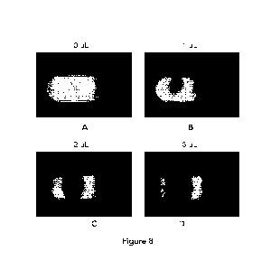
Examples of imaging for sample volume normalization are shown in Figure 8,
which is a set of
photographic images of the deposit area with varying amounts of added sample.
Two 365 nm
UV-A LEDs were used to illuminate each lateral flow membrane strip, causing
the Eu to
fluoresce at 625 nm. A filter blocks the excitation wavelength and passes the
emission
wavelength. The resulting image is captured by a RGB camera, and the red band
image was
analyzed. As shown in Figure 8, the volume of sample added was (A) OpL; (B)
1pL; (C) 2pL; and
(D) 3pL. The spot size where the sample volume is evident in the images, and
the fluorescence
darkening of the membrane comprising the fluorescent Eu reporter is also
evident in the loss of
26
CA 03207259 2023- 8-2
WO 2022/174349
PCT/CA2022/050235
signal intensity as the volume of applied sample increases. The oval outline
is the outline of the
sample port in the cartridge. As shown in panel B, upon addition of 1 pL of
fluid sample to the
sample deposit area the fluorescence intensity of the fluorescent reporter
decreases. As the
location of the sample spot is evident in the sample addition area, the non-
fluorescing area
outside the spot can also be normalized as not related to sample-induced
fluorescence
disruption or quenching. As the amount of fluid sample added to the deposit
area increases
the fluorescence intensity of the fluorescent reporter decreases. When the
amount of fluid
sample added to the deposit area is 0 pL the unbroken fluorescence is emitted
by the
fluorescent reporter and the fluorescent intensity is 25.26 a.u., with a
decrease in intensity with
increased sample volume. The volume of sample applied to the membrane and
reporter
intensity result is shown in Table 1 below.
Table 1:
Dispensed Spot Width
Volume (uL) (Pixels) Intensity (a.u.)
0 0 25.26
1 593 16.53
2 877 13.19
3 1053 9.87
[0083] The image is normalized against calibration data that scales
the image intensity to
produce a uniform image across all systems. Previous camera calibration steps
can be used to
determine the pixel scaling factor used to convert pixels to mm dimensions.
For image
analysis, a region of interest (ROI) in the image was selected corresponding
to the area of the
sample deposit window, and all image data outside this region was ignored.
Image analysis
techniques were used to segment the region coated with the blood sample (dark
area) from
the background (bright red area, shown as bright white in Figure 8 panel A).
The segmentation
threshold is determined in a preceding calibration step, in which the sample
port area average
pixel intensity is calculated. This may also be calculated for each assay
device prior to sample
27
CA 03207259 2023- 8-2
WO 2022/174349
PCT/CA2022/050235
dispense, or may be previously calibrated. The segmented sample area can be
analyzed to
determine the pixel area of the sample, as well as the relative intensity of
the sample area. The
calibrated pixel scaling values are used to transform the sample area pixels
to real world units,
for example, into mm2. This area can then be related to sample volume. In
another
embodiment, upper and lower bounds on acceptable intensity and/or surface area
of the
applied sample can provide an indication that a suitable sample volume was
added to achieve
the required result precision.
[0084] A quality control metric can also be applied based on the
volume of fluid sample
added to the sample addition area by determining whether the sample volume
added falls
within an appropriate range. For example, a particular assay requires a
minimum amount of
analyte of interest in the sample to produce a detectable change or detectable
test line in a
results area, and if the minimum concentration is not supplied in the sample
fluid the test line
will read as a negative result. If less than an expected amount of sample
volume is added then
a negative result will occur if the sample does not have a high concentration
of analyte of
interest. Conversely, if too much sample fluid is added the test line can be
overloaded and the
positive result provided would be much higher than expected, even potentially
nullifying the
result of a sensitive assay with a false positive. In an automated system, a
quality control metric
can determine whether the surface area, change in optical intensity, or both,
are indicative of a
sample volume in a desired range. If the desired range is met then the test
cartridge can be
found to have been adequately run. If the quality control metric determines
that the sample
volume added was either less or more than the volume range requirements, the
assay test can
be suppressed of any subsequent analyte measurement made.
[0085] Figure 9 is a graph of spot intensity vs. dispensed sample
volume. A standard curve
is prepared by plotting a graph of different amount of fluid sample dispensed
at the deposit
area against fluorescence intensity exhibited by the fluorescent reporter at
the deposit area
upon dispensing respective fluid sample. The fluorescence intensity captured
by the imaging
the membrane at the deposit area from the standard curve is then correlated
with the volume
of fluid sample applied to determine the volume of the fluid sample added at
the deposit area.
28
CA 03207259 2023- 8-2
WO 2022/174349
PCT/CA2022/050235
[0086] Referring to Figure 10, a standard curve for the spot width
vs. dispensed volume of
the fluid sample at the deposit area. The width of the sample spot can be used
to determine
the volume of fluid sample dispensed in the deposit area. The shown standard
curve can be
used to determine the amount of sample volume added to the LFA membrane by
comparing a
spot of detected with to a known volume. After imaging of the sample addition
port, a
developing solution was added (1% Tween 20, 1 % Triton X-100, 0.03% ProClin
300 in lx PBS
(75 HQ) to run the assays.
[0087] Figure 11 is a photograph of the results area of low,
medium, and high signal
intensity results from three different assay runs with identification of the
binding locations of
test and control lines on the assay membrane at the detection area. These
images represent
potential variability that can add to result imprecision due to inaccurate
addition of sample
volume. With sample volume normalization the sample volume added to the assay
can be
incorporated into the assay result to provide a more accurate indication of
the test results.
Further, more accurate quantification of the concentration of an analyte in a
sample of interest
can be done based on the calculated volume of analyte added to the assay
membrane in
combination with the detected imaged result of the analyte with fluorescent
marker at the test
line.
[0088] An automated analyser can also be used to detect a volume of
sample added to
the sample area before an assay is run in the case where the sample added is
detectable on
the membrane by the imaging device even without a detector species.
Preferably, illumination
during imaging provides improved detection, and the wavelength and intensity
of the
illumination can be modified to optimally illuminate the sample spot. In one
example, a sample
of blood or diluted blood can be added to the sample area and then visualized
to detect
where sample has changed the visual or optical characteristics detected by the
imaging device,
evidenced by a change in color. The change can be analysed to extrapolate the
volume of
sample added in a similar manner as is done when a detector species is
present, however in
this case the volume analysis can be done in the absence of a detector species
as the color of
the sample is sufficient to detect the spot size and volume of sample added. A
method of data
29
CA 03207259 2023- 8-2
WO 2022/174349
PCT/CA2022/050235
analysis similar to that used in the case of a detector can be used, and the
quantification of
sample volume can be determined using a change in intensity detected at the
imaging device,
change in spot shape, change spot size, or a combination thereof. In a similar
manner, a
colored species that does not interfere with the assay can be added to the
sample such that
the sample spot can be visualized in the absence of a detector species
previously applied to
the membrane at the sample area. The color difference between the area of the
membrane
that received the spot and the area around the sample spot can be detected by
the imaging
device, optionally with additional illumination. Other sample types that may
have pre-existing
color detectable by the illumination device include, for example, biological
samples and
environmental samples.
[0089] The device, apparatus, and method of the present invention
can be used for
various types of assays, including but limited to immunoassays,
immunochemistry assays,
immunohistochemistry assays, immunocytochemistry assays, immunoblotting
assays,
immunoprecipitation assays, nucleic acid assays, nucleic acid hybridization
assays, northern
blotting assays, southern blotting assays, DNA footprinting assays,
microarrays, nucleic acid
sequencing, polymerase chain reaction (PCR) assays, ligation assays, cloning
assays,
nephelometry assays, and cell aggregation assays, and any variations or
combinations thereof.
In some embodiments, the assay is a sandwich assay, in which capture agent and
detection
agent are configured to bind to analyte at different locations thereof,
forming capture agent-
analyte-detection agent sandwich. In some embodiments, the assay is a
competitive assay, in
which analyte and detection agent compete with each other to bind to the
capture agent. In
some embodiments, the assay is a nephelometry assay that is used to determine
the levels of
several blood plasma proteins, such as but not limited to immunoglobulin M,
immunoglobulin
G, and/or innmunoglobulin A. In some embodiments, the assay is an immunoassay,
in which
protein analyte is detected by antibody-antigen interaction. In some
embodiments, the assay is
a nucleic acid assay, in which nucleic acids (e.g. DNA or RNA) are detected by
hybridization
with complementary oligonucleotide probes.
CA 03207259 2023- 8-2
WO 2022/174349
PCT/CA2022/050235
[0090] All publications, patents and patent applications mentioned
in this specification are
indicative of the level of skill of those skilled in the art to which this
invention pertains and are
herein incorporated by reference. The reference to any prior art in this
specification is not, and
should not be taken as, an acknowledgement or any form of suggestion that such
prior art
forms part of the common general knowledge.
[0091] The invention being thus described, it will be obvious that
the same may be varied
in many ways. Such variations are not to be regarded as a departure from the
scope of the
invention, and all such modifications as would be obvious to one skilled in
the art are intended
to be included within the scope of the following claims.
31
CA 03207259 2023- 8-2