Note: Descriptions are shown in the official language in which they were submitted.
CA 03209737 2023-07-26
WO 2022/164568 PCT/US2021/065722
ANALYSIS OF EMBEDDED TISSUE SAMPLES
USING FLUORESCENCE-BASED DETECTION
CROSS-REFERENCE TO RELATED APPLICATIONS
[0001] This application claims priority to and benefit of U.S. Patent
Application No.
63/144,372, filed on February 1, 2021, the contents of which are incorporated
herein by
reference in its entirety
FIELD OF THE INVENTION
[0002] The present disclosure relates to analysis of embedded tissue
samples such as a
fomialin-fixed paraffin-embedded sample, using fluorescence-based detection,
automated tissue
section preparation, and artificial intelligence.
BACKGROUND
[0003] The formation of a formalin-fixed paraffin-embedded (FFPE) tissue
block serves to
preserve the morphology and cellular content of a tissue sample. Tissue
processing generally
involves placing an isolated tissue in fomialin for a time period such as a
few days, and then
embedding the tissue in a paraffin wax. FFPE samples can be conveniently
stored at room
temperature for extended periods of time and are especially useful for
immunohistochemical
staining and morphology analyses. FFPE samples may also be used for profiling
gene
expression and studying diseases.
[0004] At the time of biological testing, the FFPE tissue block is
generally trimmed by
cutting the tissue block on a microtome. The tissue block may be analyzed to
determine the
boundaries of the tissue in the FFPE by a technician or using an automated
method. In the
former case, a technician generally examines the FFPE block to observe the
diffuse image of the
tissue embedded in the paraffin. The technician may ascertain what the cross-
sectional area of a
section comprising the tissue should look like and compare that to the tissue
sections as they
emerge from the microtome blade. Preferably, the tissue block is trimmed to
expose a
representative amount of tissue to the surface of the block and to ensure that
the block face is in
line with the knife's edge.
1
CA 03209737 2023-07-26
WO 2022/164568 PCT/US2021/065722
100051 During automated analysis, a camera is commonly utilized to image
the tissue. A
light source illuminates the surface of the tissue block at an angle to
distinguish the difference
between the paraffin and tissue surfaces. Since paraffin is comparably
smoother than tissue,
automated analysis utilizes the different natural textures of paraffin and
tissue to differentiate
between the two materials.
100061 Many existing methods provide inaccurate and inconsistent data when
used to
analyze different tissue and paraffin types, since such methods are sensitive
to variability of
optical and surface characteristics of tissue and paraffin. In some cases, it
is quite difficult to
distinguish tissue from paraffin in an FFPE sample using existing methods.
100071 Following microtomy from the tissue block, the tissue section is
mounted to a slide
by smoothing the tissue section in a water bath and baking it. Hematoxylin and
Eosin (H&E)
staining is performed and the tissue section is reviewed by a pathologist.
During the pathologist
review of an H&E stained tissue section suspected to contain cancer cells,
they will identify
which cells are cancerous amongst the surrounding benign cells which can
consist of stroma,
fibroblasts, blood vessels, and extracellular matrix. Tumor cells can have
larger nuclei compared
to normal cells of the same origin or compared to the many other cell types
present including
those in the extracellular matrix. Extracellular matrix and normal cell types
tend to have much
higher levels of collagen and elastin compared to tumor cells.
100081 The identification of mutations in tumors allows for pathologists
and oncologists to
direct patient prognosis and therapy. Oncogenes are genes that in a normal
cell promote protein
production to stimulate normal growth of cells. Presence of mutations in
oncogenes promotes
unsuppressed growth and proliferation of tumor cells. Tumor suppressor genes
are genes that
normally function to inhibit over-proliferation of proteins in normal cells.
Mutations in tumor
suppressor genes promote increased growth and proliferation of tumor cells.
Drugs have been
developed for tumors having certain mutations in oncogenes or tumor
suppressors. The immune
response role in cancer also indicates that mutations in other regulatory
pathways can be targeted
by drug therapy.
100091 For the identification of mutations in tumor cells, there is
preferably a tumor content
of 20% or greater for the success of quantitative polymerase chain reaction
(qPCR) and other
2
CA 03209737 2023-07-26
WO 2022/164568 PCT/US2021/065722
next generation sequencing (NGS) tests, methods used to identify mutations.
Macro-dissection is
a tumor enrichment method whereby the pathologist carefully selects regions of
interest (ROI)
that preferably contain greater than 20% tumor content. The pathologist draws
a circle around
the region with a marker on the top of the II&E stained section. A technician
then uses multiple
unstained FFPE tissue sections mounted onto slides to select the ROI
identified by the
pathologist. The technician typically scrapes the tissue in the ROI off of the
slide and places it
into a centrifuge tube. DNA from the scraped tissue is isolated for NGS
testing. Macro-
dissection requires significant time from the pathologists, as it is very time
consuming to sit at a
bright field microscope to evaluate multiple slides to find the best example
with high tumor
content and then manually circle ROIs.
[0010] Nuclear segmentation in digital microscopic tissue images can enable
extraction of
high-quality features for nuclear morphometric and other analyses in
computational pathology.
However, conventional image processing techniques such as Otsu and watershed
segmentation
do not work effectively on challenging cases such as chromatin-sparse and
crowded nuclei. In
contrast, machine learning-based segmentation techniques are effective over a
more general set
of nuclear appearances. However, training machine learning algorithms requires
large image
datasets in which a vast number of nuclei must be been annotated. Typically, a
large publicly
accessible dataset of li&E stained tissue images with painstakingly annotated
nuclear boundaries
are used for nuclear segmentation algorithm development. An informative
dataset should include
a diversity of nuclear appearances from several patients, disease states, and
organs, and
techniques trained on it are likely to generalize well and work right out-of-
the-box on novel
H&E stained images.
[0011] Accordingly, there is a need for additional methods and apparatus
for determining a
region of interest in a tissue sample in an embedding medium, and to
facilitate the efficient
preparation of useful tissue sections from the embedded sample.
SUMMARY OF THE INVENTION
[0012] As an aspect of the present invention, a method is provided for
determining an
amount of a tissue or a cell preparation exposed at a surface of a sample
embedded in an
3
CA 03209737 2023-07-26
WO 2022/164568 PCT/US2021/065722
embedding medium such as paraffin. The method comprises irradiating the
embedded tissue or
cell preparation sample at a wavelength which causes endogenous components of
the tissue to
autofluoresce; obtaining an image of the autofluorescence emitted by the
embedded tissue or cell
preparation sample; and determining a percentage of the image at the surface
of the embedding
medium which is occupied by tissue or cell preparation.
[00131 As another aspect of the present invention, a method is provided for
identifying
different cell types in an embedded tissue sample. The method comprises
irradiating the
embedded tissue sample at a wavelength which causes endogenous components of a
tissue to
autofluoresce; obtaining an image of the autofluorescence emitted by the
tissue sample; and
identifying different cell types in the image of the autofluorescence emitted
by the tissue sample
based upon autofluorescence characteristics.
100141 As another aspect of the present invention, a method is provided for
training an
artificial intelligence (Al) system to identify a region of interest (ROD in
an embedded tissue
sample comprising a tissue and an embedding medium. The method comprises
irradiating the
embedded tissue sample at a wavelength which causes endogenous components of
the tissue to
autofluoresce; obtaining an image of the autofluorescence emitted by the
embedded tissue
sample; annotating the image to indicate the ROIs in the image; and inputting
the annotated
image into the AT system, wherein the AI system learns to identify ROls in
unannotated images.
100151 As another aspect of the present invention, a method is provided for
preparing a tissue
specimen comprising a region of interest (ROD from an embedded sample. The
method
comprises obtaining a trained Al system adapted for identifying the ROI from
an unstained
embedded sample; irradiating the embedded sample comprising a tissue and an
embedding
medium with electromagnetic radiation having an excitation wavelength;
generating a
fluorescence image of the embedded sample; using the trained Al system to
identify the ROI in
the embedded sample based on the fluorescence image and without staining the
embedded
sample; and collecting a portion of the embedded sample identified as having
the ROI as the
tissue specimen.
100161 As another aspect of the present invention, a method is provided for
imaging a sample
of a biological tissue. The method comprises irradiating a biological tissue
sample at an
4
CA 03209737 2023-07-26
WO 2022/164568 PCT/US2021/065722
excitation wavelength which causes endogenous components of the biological
tissue to
autofluoresce; obtaining an image of the autofluorescence emitted by the
biological tissue sample
to identify regions of the biological tissue comprising extracellular matrix;
and staining nuclei in
the biological tissue sample with a nuclear stain to identify regions of the
biological tissue
comprising cellular nuclei.
100171 The present invention also comprises apparatus configured to perform
the various
steps of the methods described herein.
[00181 These and other features and advantages of the present methods and
apparatus will be
apparent from the following detailed description, in conjunction with the
appended claims.
BRIEF DESCRIPTION OF THE DRAWINGS
100191 The present teachings are best understood from the following
detailed description
when read with the accompanying drawing figures. The features are not
necessarily drawn to
scale.
[00201 FIGs. IA and 1B show graphs of the excitation spectra and the
emission spectra of
various fluorophores endogenous to human tissue. FIG. lA shows the excitation
spectra of a
number of biological molecules, and FIG1B. shows the emission spectra of the
same biological
molecules. Proteins can serve as endogenous fluorophores and can be detected
or tracked by
monitoring the protein's fluorescence emission. FIGs. 1C and 1D are
collections of fluorescence
emission images of various slices of a FFPE tissue block sample. FIG. IC shows
an image of
fluorescence of tissue in the presence of paraffin using a 365nm excitation
source with an
emission filter centered at 560 nm (55nm wide bandpass). FIG. 1D shows
paraffin fluorescence
using a 280nm excitation source and an emission filter centered at 405 nm
(20nm wide
bandpass). As shown in FIG. 1D, paraffin can undergo fluorescence without
appreciable
fluorescence of tissue in an embedded sample. The results indicate that
contrast between tissue
and paraffin may be further enhanced by examining fluorescence in the region
of the paraffin.
100211 FIGs. 2A-I shows brightfield and fluorescence images of different
tissue types. FIGs
2A, 2D, and 2G are images captured using a white light source with a long pass
emission filter
with a cut-on wavelength at 405nm; FIGs. 2B, 2E, and 2H are images captured
using a 470nm
CA 03209737 2023-07-26
WO 2022/164568 PCT/US2021/065722
excitation source with an emission filter centered at 545nm (30nm bandpass).
FIGs. 2C, 2F, and
21 are images captured using a 300nm excitation source with an emission filter
centered at
545nm (30nm bandpass). FIGs. 2A to 2C are images of the adipose tissue. FIGs.
2D to 2F are
images of the uterus tissue. FIGs. 2G to 21 are images of an embedded tissue
of unknown type.
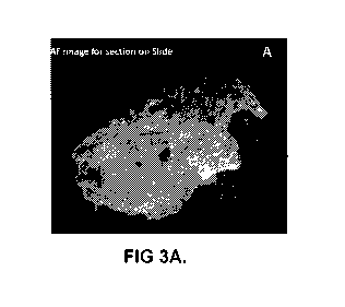
j00221 FIGs. 3A to 3D show autofluorescence images and corresponding H&E
brightfield
images of a 5-micrometer tissue section containing breast carcinoma The
autofluorescence image
was captured using a 300 nm excitation coupled with a 545-30 band-pass
emission filter and a
camera. FIGs. 3A and 3B show the fluorescence and H&E images of the tissue
section,
respectively. A pathologist annotation (blue circle) indicates a large region
of interest containing
a majority of carcinoma cells as compared to the surrounding tissue which
contains other cell
types in the autofluorescence image in FIG. 3C and the H&E in FIG. 3D.
[0023] FIGs. 4A to 4D show autofluorescence images and corresponding H&E
images of a
tissue block containing breast carcinoma. An FFPE block on a microtome was
imaged in FIG.
4A prior to microtomy of a 5-micrometer tissue section. The autofluorescence
image was
captured using a 300 nm excitation coupled with a 545-30 band-pass emission
filter and a
camera. This tissue section was H&E stained and a brightfield WSI was captured
in FIG. 4B
(also seen in FIGs. 3B and 3D). A pathologist annotation (blue circle)
indicates a large region of
interest containing a majority of carcinoma cells as compared to the
surrounding tissue which
contains other cell types in the autofluorescence image in FIG. 4C and the H&E
in FIG. 4D.
[0024] FIGs. 5A to 5D illustrate a fluorescence-based image processing
system used to
detect the exposed tissue. Autofluorescence imaging distinguished tissue,
which is sharply
focused, and subsurface tissue which appears defocused. The area of exposed
tissue is estimated
by measuring the local focus. A cartoon drawing of a cassette containing FFPE
tissues
embedded in paraffin from the top FIG. 5A and side FIG. 5B orientation shows
that embedded
tissue is visible at different focal planes. Fluorescence images can be used
to create a depth map
for the tissue block for determination of sharply versus defocused tissue. FIG
5C and 5D show in
gray-scale two fluorescence images captured using a 300nm excitation source
with an emission
filter centered at 545nm (30nm wide bandpass) and the corresponding depth map
of each tissue
embedded in the paraffin generated based on their fluorescence images.
6
CA 03209737 2023-07-26
WO 2022/164568 PCT/US2021/065722
100251 FIG. 6A is a cartoon illustration of an artificial block comprising
an artificially
shaped tissue for analysis by digital microscopy. FIG. 6B is a photograph of
the artificially
shaped tissue embedded in paraffin to make an artificial FFPE block. FIG. 6C
shows the
artificial block illuminated at 300nm, which exposed tissue detected by the
54th cut. In order to
determine the ground truth data an artificial block was created by cutting
tissue into a regular
shape and measuring it by digital microscopy (cartoon shown in A). The tissue
was then re-
embedded in paraffin to make an FFPE block (B) that upon microtomy had known
parameters
for when the tissue is exposed thereby providing ground truth data for
algorithm development.
In (C), the tissue is illuminated at 300nm for algorithm development which is
indicated by the
exposed tissue detected by the 54th cut. The btightfield raw image
demonstrates the
improvement in fluorescence imaging which shows sharply focused tissue and
distinguished
tissue from paraffin in a superior way.
[0026] FIGs. 7A to 7C demonstrate performance of the present techniques
during automated
microtomy of an FFPE block containing lung tissue. Algorithm performance is
demonstrated
during automated microtomy of an FFPE block containing lung tissue.
Fluorescence imaging is
shown for the tissue block at wavelengths (300nm). The center panel indicates
the algorithmic-
based detection of exposed tissue for the 25th five micron slice (1), the
120th slice (B) and the
367th slice (C). The 24th slice had a very little tissue as compared to the
deeper cuts into the
FFPE block.
[0027] FIGs. 8A and 8B are images of a FFPE tissue block under bright-field
and a UV
source at 300 nm. FIG. 8C is an associated depth map showing the topology of
the tissue surface
with yellow peak indicates the point closest to the block surface and FIG. 8D
shows a predicted
plane (the mesh plane on the top of the map), generated using the 3D depth map
in FIG. 8C,
optimal for sectioning the tissue.
[0028] FIGs. 9A-F shows sub-surface topology detection of FFPE tissue using
fluorescence-
based imaging. High resolution images indicate that autofluorescence imaging
distinguishes
different cell types in paraffin embedded tissues adding detailed sub-surface
topology
information to the captured images. Breast tissue was imaged using a filter at
470nm excitation
and 525nm emission (B and E). Breast tissue was imaged using a filter at 365nm
7
CA 03209737 2023-07-26
WO 2022/164568 PCT/US2021/065722
excitation/445nm emission (C and F). (A and D) shows images of H&E stained
tissue sections
of breast tissue with regions of squamous epithelium that are also visible
with fluorescence
imaging (indicated by arrows in A, B and C) and adipose cells (indicated by
arrows in D, E and
F). 200X magnification.
100291 FIGs. I0A-F shows scanned whole-slide-images (WSIs) demonstrating a
novel
workflow provided by the present disclosure. WSI of autofluorescence imaged
unstained
deparaffinized FFPE tissue is shown in FIG. 10A and FIG. 10D at 20X
magnification. The
extracellular matrix is clearly seen in the FITC (green) channel. WSI of
fluorescence of DAPI
stained tissue in FIG. 10B and FIG. 10E show that the nuclei is clearly
discernable from
extracellular matrix with the addition of DAPI containing fluorescence
mounting media (step 5
of the workflow in Table 3). WSI of the H&E shows the corresponding regions in
FIG. IOC and
FIG. IOF and show tumor cells, adipose and extracellular matrix (step 7 of the
workflow in Table
3).
DETAILED DESCRIPTION
100301 The present methods generally utilize autofluorescence of endogenous
fluorophores
in tissues (and cell preparations) to distinguish tissue from an embedding
medium such as
paraffin or an epoxy resin. The present disclosure will generally describe the
present methods as
applied to tissues, but it should be understood that such descriptions apply
to cell preparations as
well. In some embodiments, the tissue or cell preparation is selected from the
group consisting
of tissue, cell pellets, and cell spheroids (which may comprise two or more
cell types).
Contrasting between tissue and an embedding medium can be achieved by
irradiating an
embedded sample such as a formalin-fixed paraffin-embedded (FFPE) tissue block
at an
appropriate wavelength and detecting the resulting endogenous autofluorescence
emission from
the tissue. The autofluorescence emission can be used to identify components
of the tissue and
locations thereof. For example, the present methods can be used to determine a
percentage of
tissue located at a surface of a fonnalin-fixed paraffin-embedded (FFPE)
tissue block. The
fluorescence methods of the present disclosure can be performed prior to
biological analysis or
staining of a tissue section. In some embodiments, the present methods reduce
or avoid staining
of tissue sections. The present methods are effective for a wide variety of
tissue types and can be
8
CA 03209737 2023-07-26
WO 2022/164568 PCT/US2021/065722
used to identify tissue components in cases where such components are
difficult to distinguish
under normal lighting conditions.
[0031] Fluorescence, which is the emission of light by a substance that has
absorbed
electromagnetic radiation, is commonly used to elucidate the presence or
amount of an analyte.
Fluorescent compounds are capable of absorbing and emitting light under
certain conditions,
where the emitted light is generally of lower energy. Autofluorescence is
natural emission of
light by biological molecules, generally at a wavelength peak or pattern, when
the molecules are
irradiated at certain wavelengths. Each fluorescent biological molecule has
its own excitation
and emission spectrum. In human and animal tissue, proteins such as collagen
and elastin are
capable of autofluorescence. FIG. 1A shows the excitation spectra of a number
of biological
molecules, and FIG. 1B shows the emission spectra of the same biological
molecules. Proteins
can serve as endogenous fluorophores and can be detected or tracked by
monitoring the protein's
autofluorescence emission.
[0032] FIG. 1C shows an image of autofluorescence of tissue in the presence
of paraffin
using a 365nm excitation source with an emission filter centered at 560 nm
(55nm wide
bandpass). FIG. 1D shows paraffin fluorescence using a 280nm excitation source
and an
emission filter centered at 405 nm (20nm wide bandpass). As shown in FIG. 1D,
paraffin can
undergo fluorescence without appreciable autofluorescence of tissue in an
embedded sample.
The results indicate that contrast between tissue and paraffin may be further
enhanced by
examining fluorescence in the region of the paraffin.
[0033] Table 1 shows excitation and emission maxima of endogenous
fluorophores which
can be used for identifying tissue components. Table 1 is adapted from
Ramanujam, N.
Fluorescence Spectroscopy of Neoplastic and Non-Neoplastic Tissues. Neoplasia.
2000 2, 89-
117.
Table 1
Endogenous Fluorophores Excitation Maxima (nm) Emission Maxima (nm)
Amino Acids
Tryptophan 280 350
Tyrosine 275 300
Phenyl alanine 260 280
Structural proteins
9
CA 03209737 2023-07-26
WO 2022/164568
PCT/US2021/065722
Collagen I 325, 360 I 400, 405
Elastin 290, 325 340, 400
Enzymes and coenzymes
FAD, Flavins I 450 535
NADH 290,351 440,460
NADPH 336 464
Vitamins
Vitamin A 327 510
Vitamin K 335 480
Vitamin D 390 480
Vitamin B6 pyridoxine 332, 340 400
Vitamin B6 pyridoxamine 335 480
Vitamin B6 pyridoxal 330 385
Vitamin B6 pyridoxic acid 315 425
Vitamin B6 pyridoxal 5'- 330 400
phosphate
Vitamin B12 275 305
Lipids
Phospholipids 436 540, 560
Lipofuscin 340-395 540, 430-460
Ceroid 340-395 430-460, 540
Porphyrins 400-450 630, 690
100341
Table 2 shows common peak excitation wavelengths identified across many tissue
types. Spectral contributions from common molecular components/endogenous
fluorophores are
shared between tissue types. Table 2 is adapted from Favreau, P. F. et al.
Label-free spectroscopic
tissue characterization using fluorescence excitation-scanning spectral
imaging. J. Biophotonics.
2019;e201900183.
Table 2
Peak excitation wavelength (nm) Tissues
360 Heart, liver, lung, pancreas, skeletal
muscle,
trachea
375 Colon, esophagus, kidney, spleen
395 Colon, esophagus, kidney, pancreas,
trachea
480 Heart, kidney, liver, lung, skeletal
muscle,
spleen, trachea
CA 03209737 2023-07-26
WO 2022/164568 PCT/US2021/065722
100351 FIG. 2 shows brightfield and autofluorescence images of different
tissue types. The
peak excitation and emission wavelengths for autofluorescence vary between
tissue types due to
variations in concentrations of the common molecular components or endogenous
fluorophores;
structural proteins, enzymes, lipids and porphyrins. Brightfield images show
less subsurface
topology in adipose (panel A), uterus (panel D) and colon (panel G).
Autofluorescence of
adipose was optimal using the 470nm excitation filter - 545 emission, 30nm
bandpass width
(panel B) as compared to the 300 nm emission filter 545 emission, 30nm
bandpass width
(panel C). For uterus and colon, autofluorescence peak excitation/emission was
optimal using
the 300nm excitation filter with a 545nm emission, with 30nm bandpass width
(panels F and I)
as compared to the 470nm excitation filter (panels E and H).
[0036] For determination of the optimal filter set to use for imaging
autofluorescence, Tables
1 and 2 can be used as a general guide, and multiple filter sets can be
tested, including filter sets
that are commercially available. Adipose tissue contains a high concentration
of phospholipids
which have a peak excitation around 436nm and 540nm emission. For uterus and
colon,
structural proteins are predominant which have a peak excitation at 325nm and
290 nm
respectively and emission at 400nm emission.
100371 Fluorescence-based imaging allows for a greatly improved 2-
dimensional (21))
determination of the sub-surface topology of the tissue section compared to
white light imaging.
This is due to the illumination of tissue autofluorescence of cellular
components such as collagen
and elastin using different excitation and emission filters. This adds a great
deal of context to the
sub-surface topology relative to white light illumination alone and leads to
improved algorithm
development for use in the automation of microtomy for trimming.
[0038] Prior methods for trimming using white light imaging is largely
qualitative and
inaccurate when used to analyze different tissue and paraffin types due to
lack of sensitivity to
variability of optical and surface characteristics of tissue and paraffin. The
present method
provides a quantitative method for determining the optimal plane of sectioning
and also the
estimation of how far to trim before an optimal quantity of tissue is exposed.
The benefit of this
method is based on the determination of the tissue sub-surface topology by
autofluorescence-
11
CA 03209737 2023-07-26
WO 2022/164568 PCT/US2021/065722
based imaging that is not visible with white light which will allow for highly
improved
algorithms for trimming in an automated microtomy setup.
[0039]
Determining An Amount Of Tissue Or Cell Preparation Exposed At A Surface
[0040] The present disclosure provides a method of determining an amount of
tissue or cell
preparation exposed at a surface of a sample embedded in an embedding medium.
[0041] In manual sectioning, a technician's qualitative assessment of
exposed tissue can be
wasteful in the use of the tissue sample, as they might trim too far. In some
embodiments, the
present methods use an algorithm based on autofluorescence of the tissue
sample and a local
focus measurements to estimate the topology of tissue surface buried in the
paraffin and to
identify plane of sectioning to increase or maximize tissue use. By
quantitively assessing the area
of tissue exposed, the usage of the tissue can be increased to achieve its
diagnostic potential. The
present method can also speed up the trimming process by estimating how far to
trim before the
appropriate percentage of tissue is exposed, and it can do so in a highly
accurate way that is
quantitative as opposed to the qualitative method employed using a white light
illuminated image
and the human eye or algorithms associated with white light illumination.
100421 The present method comprises irradiating the embedded tissue or cell
preparation
sample at a wavelength which causes endogenous components of the tissue to
autofluoresce;
obtaining an image of the autofluorescence emitted by the embedded tissue or
cell preparation
sample; and determining a percentage of the image at the surface of the
embedding medium
which is occupied by tissue or cell preparation.
[0043] In some embodiments, the percentage of the image at the surface of
the embedding
medium which is occupied by tissue is determined by: slicing a tissue section
from the embedded
tissue sample, wherein the embedded tissue sample comprises the tissue and the
embedding
medium; irradiating the embedded tissue sample with electromagnetic radiation
having an
excitation wavelength; generating a fluorescence image from an
autofluorescence emission of
the embedded sample; determining a local focus measure for pixels of the
fluorescence image;
constructing a depth map of the tissue based on evaluating image blur of the
autofluorescence;
and determining a sectioning plane for the embedded sample, based on the depth
map.
12
CA 03209737 2023-07-26
WO 2022/164568 PCT/US2021/065722
100441 The present disclosure deploys focus-measure-based detection methods
to quantitate
the amount of exposed tissue in the FFPE block during trimming. The
autofluorescence imaging
provides additional information that potentially provides more consistent
signals across various
tissue types. The autofluorescence imaging will be used to quantitate the
amount of tissue that is
exposed for optimal trimming and sectioning.
[0045] In some embodiments, the local focus measure is determined by
applying an operator
to the fluorescence image. For example, the operator can be a modified
Laplacian operator. In
some embodiments, the local focus measure is measured in an n-by-n
neighborhood surrounding
a plurality of pixels in an input image.
100461 In some embodiments, the method further comprises performing one or
more
processing operations to obtain the fluorescence image. The processing
operations can be
selected from the group consisting of image registration, contrast
enhancement, and image
smoothing.
[0047] In some embodiments, the desired amount of tissue within the
sectioning plane and a
cut tissue section is from 10 to 100%. For example, in some embodiments, the
desired amount of
tissue within the sectioning plane and a cut tissue section is about 10%,
about 20%, about 30%,
about 40%, about 50%, about 60%, about 70%, about 80%, about 90%, or about
100% or within
a range with endpoints between any two of the foregoing values. In some
embodiments, the
exposed tissue is identified by normalizing a focus metric on each slice image
on a first slice
image. In some embodiments, the tissue section from the embedded tissue sample
will be cut at
the sectioning plane when the desired amount of tissue is present. In some
embodiments, after
the desired amount of tissue is determined, the tissue section from the
embedded sample of the
sectioning plane is cut.
Identifying Different Cell Types In An Embedded Tissue Sample
[0048] The present disclosure also provides a method of identifying
different cell types in an
embedded tissue sample. The method comprises irradiating the embedded tissue
sample at a
wavelength which causes endogenous components of a tissue to autofluoresce;
obtaining an
image of the autofluorescence emitted by the tissue sample; and identifying
different cell types in
13
CA 03209737 2023-07-26
WO 2022/164568 PCT/US2021/065722
the image of the autofluorescence emitted by the tissue sample based upon
autofluorescence
characteristics.
[0049] In some embodiments, the different cell types in the embedded tissue
sample are
determined by: exposing a tissue section from the embedded tissue sample,
wherein the
embedded tissue sample comprises a tissue and an embedding medium; irradiating
the embedded
tissue sample with electromagnetic radiation having an excitation wavelength;
generating a
fluorescence image from autofluorescence emission of the embedded sample;
determining a
local focus measure for pixels of the fluorescence image; and constructing a
depth map of the
tissue based on evaluating image blur of the fluorescence. In some
embodiments, the depth map
of the tissue is a subsurface topology of the tissue within the embedding
medium.
[0050] In some embodiments, the fluorescence images are captured by an
imaging device.
[0051] In some embodiments, the local focus measure is determined by
applying an operator
to the fluorescence image, such as a modified Laplacian operator. In some
embodiments, the
local focus measure is measured in an n-by-n neighborhood surrounding a
plurality of pixels in
an input image. In some embodiments, the method further comprises performing
one or more
processing operations to obtain the fluorescence image, such as processing
operations selected
from the group consisting of image registration, contrast enhancement, and
image smoothing.
100521 In some embodiments, a predicted optimal plane of sectioning for the
tissue sample is
based on evaluating the subsurface topology of the tissue within the embedding
medium.
Training An Artificial Intelligence (Al) System To Identify A Region Of
Interest (ROI) In
An Embedded Tissue Sample
[0053] The present disclosure also provides a method of training an
artificial intelligence
(Al) system to identify a region of interest (ROI) in an embedded tissue
sample comprising a
tissue and an embedding medium. The method comprises irradiating the embedded
tissue
sample at a wavelength which causes endogenous components of the tissue to
autofluoresce;
obtaining an image of the autofluorescence emitted by the embedded tissue
sample; annotating
the image to indicate the ROls in the image; and inputting the annotated image
into the Al
system, wherein the Al system learns to identify ROIs in unannotated images.
14
CA 03209737 2023-07-26
WO 2022/164568 PCT/US2021/065722
100541 In some embodiments, the embedded tissue sample has been stained
with a
hematoxylin and eosin (H&E) stain, an immunohistochemisby (II-IC) stain, or an
immunofluorescence (IF) stain for protein markers. In some embodiments, the
image of the
embedded tissue sample is annotated on a whole slide image of the embedded
tissue sample.
[0055] In some embodiments, the embedded sample is irradiated as part of a
tissue block,
and the method further comprises slicing the embedded sample from the tissue
block as a tissue
section.
[0056] In some embodiments, the trained Al system is adapted for
identifying a ROI
containing tumor cells in accordance with tumor enrichment methods for
molecular assays.
100571 In some embodiments, the Al system comprises at least one of a
machine learning
system, a deep learning system, a neural network, a convolutional neural
network, a fully
convolutional neural network, a segmentation convolutional neural network, a
recurrent neural
network, a statistical model-based system, or a deterministic algorithm-based
analysis system.
[0058] In some embodiments, the AI system learns to identify ROls in
unannotated images
by: obtaining an untrained or pretrained Al system; staining the embedded
tissue sample with a
stain detectable by bright field or autofluorescence-based imaging; generating
one or more
stained images of a stained embedded tissue sample by bright field or
autofluorescence imaging;
annotating the ROI of the stained embedded tissue sample on the one or more
stained images;
mapping the annotated ROI of the one or more stained images to the unstained
autofluorescence
image; and training the untrained Al system by using a mapped fluorescence
image. In some
embodiments, Al system training data is obtained from the mapped
autofluorescence image in
order to train the untrained or pretrained Al system.
[0059] In some embodiments, the AI system is adapted for identifying the
ROI on an
unstained embedded tissue sample. In some embodiments, the ROI identifies
tumor containing
regions. In some embodiments, the ROI is identified by detecting an
autofluorescence level
corresponding to concentration of endogenous fluorophores from extracellular
matrix and
cytoplasm surrounding tumor nuclei.
100601 In some embodiments, the autofluorescence level of the extracellular
matrix and
cytoplasm surrounding the tumor nucleus is compared to a autofluorescence
level of a control
CA 03209737 2023-07-26
WO 2022/164568 PCT/US2021/065722
sample. The control sample can comprise a control nucleus, control cytoplasm,
control stroma,
or control extracellular matrix components. In some embodiments, the
autofluorescence level of
the extracellular matrix or cytoplasm surrounding the tumor nucleus is lower
than the
autofluorescence level of the control sample. The control nucleus may be
smaller than tumor
nuclei with fine chromatin and a single nucleolus.
100611 In some embodiments, the method further comprises: mounting the
embedded sample
as a tissue section onto a slide and imaging the tissues using a first filter
and a second filter;
treating the tissue section to remove the embedding medium and to permeabilize
nuclei to
facilitate a stain for nuclei; applying a stain such as DAPI which is specific
for nuclei to the
tissue section to form a nuclei-stained tissue section; imaging the nuclei-
stained tissue section to
produce a nuclei-stained tissue section image; removing the nuclei stain
mounting medium;
applying a hematoxylin and eosin (H&E) stain, an immunohistochemistry (IHC)
stain, or an
immunofluorescence (IF) stain to produce a cell-stained tissue section;
coverslipping the cell-
stained tissue section; and imaging the cell-stained tissue section using
bright field imaging to
produce the stained image.
[0062] The present disclosure provides a workflow where the unstained
deparaffinized tissue
is first imaged for autofluorescence in the FITC and DAPI filters to identify
cytosol and
extracellular matrix. The deparaffinized tissue is then treated by either
pepsin or e-field
treatment to allow DAPI staining to penetrate the nuclei. The DAPI stained
tissue is then imaged
again for fluorescence in the FITC and DAPI channels. The first image shows
FITC
autofluorescence only. The second image shows FRC and DAPI stained
fluorescence which
very clearly distinguishes the cytosol and extracellular matrix from the
nuclei. An added benefit
of this technique is that the nuclear boundaries are very clearly identified
even between crowded
nuclei. Hence, for Al training, the second DAPI stained image could be used as
a base and
mapped back to H&E images to accomplish nuclear segmentation of tumor nuclei
versus
surrounding normal cells and extracellular matrix. This workflow would save
the pathologist
from the painstaking process of annotating H&E stained section for the nuclei
segmentation
model training.
16
CA 03209737 2023-07-26
WO 2022/164568 PCT/US2021/065722
100631 The workflow will assist in the training of machine learning
algorithms developed for
macrodissection as well as for nuclear segmentation in a more clear and
concise manner than
using data derived by pathologists and computational biologists analysis of
H&E stained tissue
sections.
[0064] The present disclosure describes a workflow that may be used for (1)
algorithm-based
detection methods to perform nuclear segmentation for nuclear morphometric
analysis, and (2)
algorithm-based detection methods to perform macrodissection of tumor regions
of interest for
subsequent genomic testing methods. The algorithms developed using this
workflow may be
deployed using an unstained FFPE block during microtomy or using a tissue
section on a slide.
100651 Prior techniques for nuclear segmentation algorithm development
requires a large
database of painstakingly annotated H&E images. The present methods do not
require
pathologist annotation as the DAPI stained nuclei are very clearly
distinguished from the
autofluorescence seen in the cytosolic compartments and extracellular matrix.
[0066] Prior techniques for algorithm development for macrodissection also
required
pathologist annotation of H&E stained tissue sections that are then mapped
back to the
autofluorescence image of the block face or a tissue section mounted onto a
slide. Algorithms
developed for macrodissection may use differences in the size of tumor and
normal nuclei, and
they can quantitate the number of tumor cells in a region of interest for
selection.
Preparing A Tissue Specimen Comprising A Region Of Interest (ROI) From An
Embedded
Sample
[0067] The present disclosure also provides a method of preparing a tissue
specimen
comprising a region of interest (ROI) from an embedded sample, comprising:
obtaining a trained
Al system adapted for identifying the ROI from an unstained embedded sample;
irradiating the
embedded sample comprising a tissue and an embedding medium with
electromagnetic radiation
having an excitation wavelength; generating a fluorescence image of the
embedded sample;
using the trained Al system to identify the ROI in the embedded sample based
on the
fluorescence image and without staining the embedded sample; and collecting a
portion of the
embedded sample identified as having the ROI as the tissue specimen. The
fluorescence image
can be generated from a tissue section comprising the embedded sample and/or
from a tissue
17
CA 03209737 2023-07-26
WO 2022/164568 PCT/US2021/065722
block comprising the embedded sample. In some embodiments, the fluorescence
image is
generated from a tissue section, and a collected portion of the embedded
sample is collected from
a tissue block.
100681 In some embodiments, the embedding medium is removed from the
collected portion
of the embedded sample to provide the tissue specimen. In some embodiments,
cellular
components are removed from the tissue specimen to provide a nucleic acid
specimen and
determining a nucleic acid sequence from the nucleic acid specimen.
[0069] In some embodiments, the trained Al system is obtained by:
irradiating the embedded
tissue sample at a wavelength which causes endogenous components of the tissue
to
autofluoresce; obtaining an image of the autofluorescence emitted by the
embedded tissue
sample; annotating the image to indicate the ROls in the image; and inputting
the annotated
image into the AI system, wherein the Al system learns to identify ROIs in
unannotated images.
[0070] In some embodiments, the Al system comprises at least one of a
machine learning
system, a deep learning system, a neural network, a convolutional neural
network, a fully
convolutional neural network, a segmentation convolutional neural network, a
recurrent neural
network, a statistical model-based system, or a deterministic algorithm-based
analysis system.
[0071] In some embodiments, the trained Al system is obtained by: imaging
the embedded
sample as part of the tissue block using autofluorescence and slicing the
embedded sample from
the tissue block as the tissue section; mounting the embedded sample as the
tissue section onto a
slide and imaging the tissues using a first filter and a second filter;
treating the tissue section to
remove the embedding medium and to permeabilize nuclei to a stain for nuclei;
applying a nuclei
stain in a nuclei stain mounting medium to the tissue section to form a nuclei-
stained tissue
section; imaging the nuclei-stained tissue section to produce a nuclei-stained
tissue section
image; removing the nuclei stain mounting medium; applying a hematoxylin and
eosin (H&E)
stain, an immunohistochemistry (IIIC) stain, or an immunofluorescence (IF)
stain to produce a
cell-stained tissue section; coverslipping the cell-stained tissue section;
and imaging the cell-
stained tissue section using bright field imaging to produce the stained
image.
Imaging A Sample Of A Biological Tissue
18
CA 03209737 2023-07-26
WO 2022/164568 PCT/US2021/065722
100721 The present disclosure also provides a method of imagine a sample of
a biological
tissue comprising: irradiating a biological tissue sample at an excitation
wavelength which
causes endogenous components of the biological tissue to autofluoresce;
obtaining an image of
the autofluorescence emitted by the biological tissue sample to identify
regions of the biological
tissue comprising extracellular matrix; and staining nuclei in the biological
tissue sample with a
nuclear stain to identify regions of the biological tissue comprising cellular
nuclei.
[00731 in some embodiments, the biological tissue sample is deparaffinized
and pretreated
with a cleaning solution. For example, the cleaning solution can comprise
xylene and pepsin
digestion or e-field treatment with rehydration. In some embodiments, the
nuclear stain contains
fluorescent mounting media. In some embodiments, the biological tissue sample
is scanned by
fluorescence imaging. In some embodiments, the DAPI is removed by soaking the
tissue section
in water.
[0074] In some embodiments, a trained Al system is developed.
[0075] In some embodiments, the step of identifying regions of the
biological tissue
comprising cellular nuclei allows for identification of tumor cells and
nuclear stained tumor
cells.
[0076] In some embodiments, the biological tissue sample is used to develop
each
autofluorescence image. In some embodiments, the autofluorescence image is a
first image. In
some embodiments, a second image is obtained by staining the nuclei in the
biological tissue
sample with a nuclear stain. In some embodiments, the first image and the
second image identify
tumor boundaries in the biological tissues. In some embodiments, the second
image is mapped to
an H&E or IFIC image in order to show nuclear segmentation of tumor cells.
[0077] In some embodiments, the regions of the biological tissue comprising
cellular nuclei
are tumor nuclei. In some embodiments, the regions of the biological tissue
comprising cellular
nuclei are identified by detecting the autofluorescence level of the
extracellular matrix
surrounding the tumor nuclei. The autofluorescence level of the extracellular
matrix surrounding
the tumor nuclei can be compared to a autofluorescence level of a control
sample. The control
sample can comprise control nuclei, control cytoplasm, control stroma, or
control extracellular
matrix components. The autofluorescence level of the extracellular matrix
surrounding the tumor
19
CA 03209737 2023-07-26
WO 2022/164568 PCT/US2021/065722
nuclei may be lower than the autofluorescence level of the control sample. In
some
embodiments, the control nuclei are smaller than the tumor nuclei are mono-
nucleate, contains
one nucleoli and fine chromatin.
Additional Information Regarding The Present Methods
[0078] Various embodiments of the foregoing methods may be implemented in
any desirable
manner. In some embodiments, the embedding medium is paraffin. In some
embodiments, the
embedding medium is an epoxy resin.
[0079] In some embodiments, the autofluorescence emission is detected using
an imaging
device. In some embodiments, the imaging device comprises a camera such as a
digital camera.
In such cases, an embedded sample comprising tissue and an embedding medium
such as
paraffin is irradiated with light and the resulting autofluorescence emission
is captured using a
digital camera. The presence of autofluorescence in the digital image provides
an indication that
tissue is present in the sample under study. In some embodiments, the present
method is
performed using an optical system comprising a digital camera and a microtome.
In some
embodiments, the present method is performed using a fluorescence microscope.
[0080] In some embodiments, the embedding medium exhibits no substantial
autofluorescence when irradiated at a chosen wavelength.
[0081] The present methods may be used to analyze a tissue of any type. In
some
embodiments, the tissue is a human tissue. In some embodiments, the tissue is
an animal tissue.
In some embodiments, the tissue is a mouse, rat, dog, or primate tissue. The
present method may
be used to analyze a tissue section from any organ or anatomical part. in some
embodiments, the
tissue is isolated from the breast, prostate, lung, colon, rectum, urinary
bladder, uterine corpus,
thyroid, kidney, oral cavity (e.g., tonsil), pancreas, liver, cervix, stomach,
small intestine, brain,
spinal cord, heart, bone, joints, esophagus, gallbladder, adipose, skin,
spleen, placenta, penis,
urethra, fallopian tube, ovary, vulva, adrenal glands, appendix, or eye. In
some embodiments,
the tissue is pelleted cells from a human or an animal source. In some
embodiments, the present
method is used to test a diseased or healthy tissue. In some embodiments, the
present method is
used to identify cancer, infectious disease, metabolic disease, degenerative
disease, inflammatory
disease, or a combination thereof.
CA 03209737 2023-07-26
WO 2022/164568 PCT/US2021/065722
100821 In some embodiments, the embedded sample is a formalin-fixed
paraffin-embedded
sample. The fonnalin-fixed paraffin-embedded sample may be formed from any
type of
paraffin. In some embodiments, the paraffin is a blend of fully refined
paraffin wax and a
synthetic resin or polymer. In some embodiments, the paraffin comprises
dimethyl sulfoxide
(DMS0). In some embodiments, the formalin-fixed paraffin-embedded sample is
formed from
granulated paraffin wax, fully refined paraffin wax, semi-refined paraffin
wax, or a combination
thereof. Thus, in some embodiments, a tissue may be distinguished from
granulated paraffin
wax, fully refined paraffin wax, or semi-refined paraffin wax in a fonnalin-
fixed paraffin-
embedded sample. In some embodiments, the formalin-fixed paraffin-embedded
sample is
formed from Spectrum paraffin, Millipore paraffin, Fisheifinest Histopath
paraffin wax, EMS
Paramat, Paraplast, Polyfin, Sakura Finetek Tissue Tek VIP, Leica Surgipath
Paraplast, or a
combination thereof.
[0083] In some embodiments, the embedding medium is an epoxy resin. In some
embodiments, the epoxy resin is a glycidyl epoxy resin. In some embodiments,
the epoxy resin
is a non-glycidyl epoxy resin. In some embodiments, the epoxy resin is a non-
glycidyl resin
selected from an aliphatic and cyclo-aliphatic resin. In some embodiments, the
epoxy resin is a
glycidyl epoxy selected from glycidyl amine, glycidyl ester, glycidyl ether,
and a combination
thereof. In some embodiments, the epoxy resin is ethylene glycol diglycidyl
ether. In some
embodiments, the epoxy resin is Araldite, Quetol, Epon 812, Embed 812, Poly-
Bed 812, or a
combination thereof. In some embodiments, the epoxy resin is a glycerol-based
aliphatic epoxy
resin. In some embodiments, embedding a tissue in an epoxy resin provides
tissue sections
having improved morphology.
[0084] In some embodiments, the embedded sample is cut or sliced to provide
a slice and a
trimmed block. In some embodiments, the embedded sample is sliced or trimmed
on a
microtome. In some embodiments, the autofluorescence of an embedded sample is
detected
while the embedded sample is being sliced or trimmed by a microtome. The
trimmed block is
irradiated with light, and analyzed to determine the presence of
autofluorescence. Imaging may
be used to determine the presence of autofluorescence. The trimming and/or
irradiation process
21
CA 03209737 2023-07-26
WO 2022/164568 PCT/US2021/065722
is repeated as needed. For example, the trimming/irradiation process may be
repeated until the
surface of the tissue is found.
(00851 In some embodiments, autofluorescence of one or more endogenous
species is
measured quantitatively to determine the location of tissue in an embedded
sample.
[0086] In some embodiments, pixel intensity of a fluorescence digital image
is used to
determine the components and/or location of a tissue in an embedded sample. A
trimmed block
is irradiated with light, and a digital image is acquired using a fluorescence
microscope. The
fluorescence microscope system comprises software that converts photons
detected during
fluorescence analysis to pixel intensity values, allowing the user to
determine the pixel intensity
for a region of interest. The trimmed block may be further sliced or trimmed
and analyzed by
the fluorescence microscope system to provide a second digital image. A
comparison of the
pixel intensity of two or more digital images can be used to determine the
location of the tissue
in the embedded sample. For example, an increase in pixel intensity values
between two digital
images can indicate that the tissue in the trimmed block is exposed and is
ready to be cut and
used for biological testing.
[0087] In some embodiments, the present method is used to determine the
components
and/or location of a surface of a tissue sample in an embedded sample. In some
embodiments,
the present method is used to determine the location of a tissue-to-embedding
medium transition
or embedding medium-to-tissue transition in an embedded sample. In some
embodiments, the
present method is used to locate a tissue in its entirety.
[0088] In some embodiments, the method comprises slicing a section from the
embedded
sample and accepting or rejecting the section based on the determined location
of the tissue
surface. in some embodiments, the irradiating is performed multiple times and
the embedded
sample is cut prior to each irradiation.
[0089] The autofluorescence emission of an endogenous species in tissue may
be used to
determine the components and/or location of a tissue in an embedded sample.
Any endogenous
fluorophore in tissue may be used. In some embodiments, the endogenous
fluorophore is
collagen, elastin, tryptophan, a porphyrin, a flavin, NADH, pyridoxin, a lipo-
pigment, or a
combination thereof. In some embodiments, the autofluorescence emission of
collagen is used to
22
CA 03209737 2023-07-26
WO 2022/164568 PCT/US2021/065722
determine the location of a tissue in an embedded sample. In some embodiments,
the
autofluorescence emission of elastin is used to determine the location of a
tissue in an embedded
sample. In some embodiments, the autofluorescence emission of tryptophan is
used to determine
the components and/or location of a tissue in an embedded sample. In some
embodiments, one
or more of collagen, elastin, and tryptophan are used to determine the
location of a tissue in an
embedded sample.
[0090] In some embodiments, an excitation light having a wavelength of from
about 320 nm
to about 380 nm is used to detect collagen autofluorescence. In some
embodiments, collagen
maximum autofluorescence emission is detected at a wavelength of from about
375 nm to about
425 nm.
[00911 In some embodiments, an excitation light having a wavelength of from
about 320 nm
to about 380 nm is used to detect elastin autofluorescence. In some
embodiments, elastin
maximum autofluorescence emission is detected at a wavelength of from about
400 nm to about
450 nm.
[0092] In some embodiments, an excitation light having a wavelength of from
about 180 nm
to about 230 nm is used to detect tryptophan autofluorescence. In some
embodiments,
tryptophan maximum autofluorescence emission is detected at a wavelength of
from about 300
nm to about 350 nm.
[0093] The embedded sample may be irradiated with light having any suitable
wavelength.
In some embodiments, an embedded sample is irradiated with light having a
wavelength of from
about 200 nm to about 600 nm. Thus, in some embodiments, an embedded sample is
irradiated
with light having a wavelength of from about 200 nm to about 600 nm, from
about 200 nm to
about 550 nm, from about 200 nm to about 500 nm, from about 200 nm to about
450 nm, from
about 200 nm to about 400 nm, from about 200 nm to about 350 nm, from about
250 nm to about
600 nm, from about 250 nm to about 550 nm, from about 250 nm to about 500 nm,
from about
250 nm to about 450 nm, from about 250 nm to about 400 nm, from about 300 nm
to about 500
nm, from about 300 nm to about 550 nm, from about 300 nm to about 600 nm, from
about 350
nm to about 600 nm, from about 400 nm to about 600 nm, from about 450 nm to
about 600 nm,
23
CA 03209737 2023-07-26
WO 2022/164568 PCT/US2021/065722
from about 350 nm to about 550 nm, from about 350 nm to about 500 nm, from
about 400 nm to
about 600 nm, from about 400 nm to about 550 nm, or from about 450 nm to about
600 nm.
[0094] The autofluorescence emission of the embedded sample can be detected
at any
suitable wavelength, usually the maximum emission wavelengths. In some
embodiments, the
embedded sample has a maximum autofluorescence emission at a wavelength of
from about 300
nm to about 600 nm. Thus, in some embodiments, the embedded sample has a
maximum
autofluorescence emission at a wavelength of from about 300 nm to about 600
nm, from about
300 nm to about 550 nm, from about 300 nm to about 500 nm, from about 300 nm
to about 450
nm, from about 300 nm to about 400 nm, from about 350 nm to about 600 nm, from
about 350
nm to about 550 nm, from about 350 nm to about 500 nm, from about 350 nm to
about 450 nm,
from about 400 nm to about 600 nm, from about 450 nm to about 550 nm, or from
about 500 nm
to about 600 nm.
[0095] Fluorescence methods are generally performed using a light source
and a detector
configured to detect fluorescence as known in the art. In some embodiments,
fluorescence
techniques are carried out using a light source capable of shining light at a
particular wavelength
or range thereof. In some embodiments, an embedded sample is irradiated using
one or more
light sources. In some embodiments, the light source is a light-emitting diode
(LED) light
source. In some embodiments, the light source is a mercury arc lamp. In some
embodiments,
the light source is a xenon arc lamp. In some embodiments, the light source is
a LASER. In
some embodiments, the present method is performed using a fluorescence system
having one or
more excitation filters. In some embodiments, the fluorescence system
comprises an aperture
and one or more emission filters. In some embodiments, the fluorescence system
comprises an
imaging lens and an imaging camera.
[0096] An embedded sample may be formed using any suitable method. In some
embodiments, a tissue is obtained from a subject and sectioned. The tissue is
contacted with a
formalin solution and fixed for at least 48 hours at room temperature. The
tissue is commonly
dehydrated using a series of ethanol baths and then embedded into a wax block.
The wax
generally comprises a mixture of straight chain alkanes having a chain length
of from about 20 to
about 40 carbons. In some embodiments, glutaraldehyde is used as a fixative to
embed a tissue
24
CA 03209737 2023-07-26
WO 2022/164568 PCT/US2021/065722
in an epoxy resin. The embedded sample may be sliced or sectioned for any
subsequent analysis
(e.g., microscopic slide analysis).
[0097] In some embodiments, the embedded sample may be further trimmed or
sectioned to
form a tissue section or slice. The embedded sample may be trimmed or
sectioned using any
suitable method (e.g., using a microtome blade). In some embodiments, a
clearing agent such as
a xylene can be used to remove the embedding medium from the section. In some
embodiments,
the tissue section is stained using at least one stain such as a Haematoxylin
and/or Eosin,
Acid/Basic Fuchsin, or Gram stain. In some embodiments, the tissue section may
be mounted
onto a slide for analysis. The stained tissue section may undergo further
analysis using any
suitable method (e.g., pathological analysis using a microscope).
[0098] In some embodiments, the present methods are performed to locate an
embedded
tissue for use in a fluorescence in situ hybridization (FISH) testing method.
In some
embodiments, the present methods are performed to locate an embedded tissue
for use in a
chromogenic in situ hybridization (CISH) testing method.
[0099] In another embodiment, the present disclosure provides a method of
determining the
location of a tissue in an embedded sample by irradiating an embedded sample
comprising a
tissue and an embedding medium with at least one light source to produce a
first fluorescence
emission and a second fluorescence emission; detecting the first fluorescence
emission and the
second fluorescence emission; and determining the location of at least a
portion of the tissue in
the embedded sample based on the first fluorescence emission and the second
fluorescence
emission.
r001001 In some embodiments, an embedded sample is irradiated with light
having a
wavelength of from about 250 nm to about 325 nm. In some embodiments, an
embedded sample
is irradiated with light having a wavelength of from about 300 nm to about 400
nm. In some
embodiments, an embedded sample is irradiated concurrently at both
wavelengths. In some
embodiments, bright field microscopy is used in combination with the present
method to
determine the location of the tissue in the embedded sample.
[00101] In some embodiments, a first fluorescence emission is generated by
fluorescence of
an embedding medium in the embedded sample (e.g., paraffin). In some
embodiments, a second
CA 03209737 2023-07-26
WO 2022/164568 PCT/US2021/065722
fluorescence emission is generated by autofluorescence of a tissue component
present in the
embedded sample. In some embodiments, the first fluorescence emission has
maximum
fluorescence at a wavelength of from about 375 nm to about 425 nm and the
second fluorescence
emission has maximum fluorescence at a wavelength of from about 500 nm to
about 600 nm.
1001021 In some embodiments, the embedded sample is irradiated using two or
more light
sources (e.g., two, three, four, five, or six). In some embodiments, the two
or more light sources
are the same. In some embodiments, the two or more light sources are
different. In some
embodiments, the sample is irradiated simultaneously or separately by the two
or more light
sources.
1001031 In some embodiments, the method is performed in the absence of a
dichroic filter.
1001041 In some embodiments, the method comprises front illuminating an
embedded sample
traversely, such as at an oblique angle of from about 10 degrees to about 20
degrees from a plane
of a face of the embedded sample. In some embodiments, a fluorescence emission
is collected
by a lens having a high numerical aperture. In some embodiments, illumination
from two or
more traverse directions (e.g., left or right or top or bottom) produces a
uniform excitation and
emission pattern.
1001051 In some embodiments, a high numerical aperture objective lens is used
for excitation
and collection of emitted light, as well as a filter cube with a dichroic beam
splitter with
excitation and emission filters. In some embodiments, an additional lens is
used after the
dichroic filter to focus the emitted light onto an imaging sensor.
1001061 In some embodiments, the embedding medium is weakly autofluorescent.
Thus, in
some embodiments, a fluorescent dye can be added to the embedding medium. The
fluorescent
dye emits light at a different wavelength than the emission wavelength of an
endogenous
fluorophore in the tissue sample, thus a fluorescence emission from the
fluorescent dye can be
used to determine the location of tissue in an embedded sample. The
fluorescent dye may be
incorporated into the embedding medium prior to formation of the embedded
sample.
1001071 In another embodiment, the disclosure provides an apparatus for
slicing a tissue
section from an embedded sample. The apparatus comprises a microtome
comprising a sample
holder adapted for linear motion, a knife holder and a knife held by the knife
holder opposite the
26
CA 03209737 2023-07-26
WO 2022/164568 PCT/US2021/065722
sample holder, such that when the sample holder is moved linearly, a sample
held by the sample
holder is sliced by the knife to form a tissue section; at least one light
source directed at the
sample holder; and an optical system positioned to capture emitted light from
a sample held by
the sample holder.
[001081 In some embodiments, the apparatus comprises at least two light
sources. In some
embodiments, the apparatus comprises at least three light sources. In some
embodiments, the
apparatus comprises at least four light sources.
1001091 In some embodiments, the apparatus comprises a filter cube with a
dichroic beam
splitter with excitation and emission filters. In some embodiments, the
apparatus comprises a
dichroic filter. In some embodiments, the apparatus comprises an additional
lens after the
dichroic filter to focus the emitted light onto an imaging sensor. In some
embodiments, the
apparatus comprises an emission filter in a filter cube assembly, where
switching of at least one
excitation source switches at least one filter.
1001101 In some embodiments, the apparatus includes one or more excitation
filters. In some
embodiments, the apparatus comprises an aperture. In some embodiments, the
apparatus
comprises a lens having a high numerical aperture. In some embodiments, the
apparatus
comprises one or more emission filters. In some embodiments, the apparatus
comprises an
imaging lens. In some embodiments, the optical system comprises a camera. In
some
embodiments, the optical system comprises a digital camera. In some
embodiments, the optical
system is capable of detecting at least one fluorescence emission.
1001111 In some embodiments, the apparatus comprises a microtome blade, at
least one light
source, at least one excitation filter, at least one aperture, at least one
emission filter, a lens
assembly, and at least one camera.
1001121 In some embodiments, the apparatus comprises a microtome blade, at
least two light
sources, at least two excitation filters, at least one aperture, at least two
emission filters, a lens
assembly, at least one camera, and at least one mechanism for switching
between emission
filters.
1001131 In some embodiments, the apparatus comprises a microtome blade, at
least two light
sources, at least two excitation filters, at least one aperture, a dual-band
bandpass emission filter,
27
CA 03209737 2023-07-26
WO 2022/164568 PCT/US2021/065722
a lens assembly, and at least one multi-color camera. In some embodiments, the
multicolor
camera has microfilters in front of each pixel.
1001141 In some embodiments, the apparatus comprises a microtome blade, at
least one light
source, at least one excitation filter, at least one aperture, an objective
lens assembly, a tube lens
or relay lens, a dichroic beamsplitter, at least one emission filter in filter
cube assembly, at least
one camera. In some embodiments, switching between excitation sources is
accompanied by
switching filters.
1001151 The present methods and apparatus can be used with a variety of
fluorescence-based
imaging systems. In some embodiments, the present apparatus comprises a
microtome blade, one
or more light sources, one or more excitation filters, a 2-dimensional
aperture, one or more
emission filters, an imaging lens, and/or an imaging camera. A single-color
fluorescence
imaging system can be used to image an embedded sample, which typically
comprises a an LED
light source, an excitation filter, an aperture, an emission filter with a
mechanism for switching
emission filters, a lens assembly, and a camera. A multi-color fluorescence
imaging system can
be used to image an embedded sample, which typically comprises multiple LED
light sources,
excitation filters, apertures, an emission filter with a motorized wheel
assembly, a lens assembly,
and a camera. Other components of the fluorescence imaging systems can include
two-color
band-pass emission filters, a color camera having a multi-color image sensor,
an objective lens
assembly, a dichroic beam splitter, a tube lens or relay lens. In some
embodiments, switching of
the excitation source is accompanied by switching the filters in accordance
with an embodiment
of the disclosure.
1001161 In some embodiments, the optical system comprises a processor in
communication
with the optical system and configured to provide a signal based on a
fluorescence emission from
the sample.
28
CA 03209737 2023-07-26
WO 2022/164568 PCT/US2021/065722
EXAMPLE I
1001171 This Example illustrates an embodiment of a method for macrodissection
of tumor
tissue using fluorescence-based imaging of a tissue section FIGs. 3A to 3D
show
autofluorescence images of a 5 micrometer tissue section containing breast
carcinoma and the
corresponding H&E images which have been marked to indicate where a region of
interest of
tumor is located in relation to surrounding normal cells. An FFPE tissue
section mounted onto a
slide was imaged using autofluorescence in FIG. 3A and 3C. The 5 micrometer
section was then
mounted onto a slide and stained by H&E, and the stained tissue sections are
shown in Figs. 3B
and 3D. A pathologist annotation (blue circle) indicates a large region of
interest containing a
majority of carcinoma cells as compared to the surrounding tissue which
contains other cell types
in the autofluorescence image (Fig. 3C) and in the H&E stained section (Fig.
3D).
1001181 In tumor containing tissue, the nuclei of tumor cells are typically
enlarged and nuclear
material is condensed. Thus, nuclei of tumor cells do not fluoresce to the
same degree as other
cellular components; stroma and extracellular matrix components.
1001191 For algorithm development, a segmentation convolutional neural network
(CNN)
model is developed to detect tumor enriched regions of interest (ROI)
generated from labeled
autofluorescence images of the tissue section on the slide. To collect the
labeled training data,
the low resolution autofluorescence images of the sections are taken of the
slide by using
autofluorescence at 300 nm excitation coupled with a 545-30 band-pass emission
filter. The
sections are then H&E stained, and imaged at 40x magnification. A pathologist
then annotates
the ROI of tumor cells in the H&E stained whole slide image (WSI) and identify
ROI contain
20% or greater tumor content as ground truth. By mapping this annotated ROI to
the
corresponding autofluorescence image, the same region is masked in the
autofluorescence image
as labeled training data. The segmentation CNN model trained based on this
training set can be
applied to autofluorescence images of unknown samples to identify tumor
enriched ROI and thus
enriched for genomics tests. The ROI is marked on the back of the slide for
the mounted FFPE
tissue section and ready for the next step in the workflow.
29
CA 03209737 2023-07-26
WO 2022/164568 PCT/US2021/065722
EXAMPLE 2
1001201 This Example illustrates an embodiment of a method for macrodissection
of tumor
tissue using fluorescence-based imaging of a tissue block FIGs. 4A to 4D show
the
autofluorescence images of a block containing breast carcinoma and the
corresponding H&E
images where a region of interest of tumor is located in relation to
surrounding normal cells. An
FFPE block containing breast carcinoma was imaged using autofluorescence in
FIGs. 4A and
4C. The 5 micrometer section cut from the FFPE block was then mounted onto a
slide and
stained by H&E (FIG. 4B and FIG. 4D). A pathologist annotation (blue circle)
indicates a large
region of interest containing a majority of carcinoma cells as compared to the
surrounding tissue
which contains other cell types in the autofluorescence image (FIG. 4C) and in
the H&E stained
section (FIG. 4D).
1001211 As noted above, in tumor containing tissue, the nuclei of tumor cells
are typically
enlarged and nuclear material is condensed. Thus, nuclei of tumor cells do not
fluoresce to the
same degree as other cellular components; stroma and extracellular matrix
components.
1001221 For algorithm development, a segmentation convolutional neural network
(CNN)
model is developed to detect tumor enriched regions of interest (ROI)
generated from labeled
autofluorescence images. To collect the labeled training data, the low
resolution
autofluorescence images of the sections are taken on the trimming stage by
using
autofluorescence at 300 nm excitation coupled with a 545-30 band-pass emission
filter. The
sections then will be mounted on the slide, H&E stained, and imaged at 40x
magnification. The
pathologist will then annotate the ROI of tumor cells in the H&E stained whole
slide image
(WSI) and identify ROI contain 20% or greater tumor content as ground truth.
By mapping this
annotated ROI to the corresponding autofluorescence image, the same region is
masked in the
autofluorescence image as labeled training data. The segmentation CNN model
trained based on
this training set can be applied to autofluorescence images of unknown samples
to identify tumor
enriched ROI and thus enriched for genomics tests. The ROI is marked on the
back of the slide
for the mounted FFPE tissue section and ready for the next step in the
workflow.
CA 03209737 2023-07-26
WO 2022/164568 PCT/US2021/065722
EXAMPLE 3
1001231 This Example illustrates focus-measure-based detection methods to
quantitate the
amount of exposed tissue in the FFPE block during trimming. The
autofluorescence imaging
provides additional information that potentially provides more consistent
signals across various
tissue types. Algorithms will be used to quantitate the amount of tissue that
is exposed for
optimal trimming and sectioning. A quantitative approach to evaluate the
percentage of exposed
tissue in the block would enhance the expediency of the automated trimming
station and
conserve tissue.
1001241 In manual sectioning, the histotechnician's qualitative assessment of
exposed tissue
can be both wasteful use of the FFPE tissue sections as they might trim too
far. By quantitively
assessing the area tissue exposed, the usage of the tissue can be optimized to
achieve their
diagnostic potential. It will also speed up the trimming process by estimating
how far to trim
before the appropriate percentage of tissue is exposed in a highly accurate
way that is
quantitative as opposed to the qualitative method employed using a white light
illuminated image
and the human eye or algorithms associated with white light illumination.
1001251 FIGs. 5A to 5D illustrate a fluorescence based image processing system
used to detect
the exposed tissue. Autofluorescence imaging distinguishes tissue, which is
sharply focused and
subsurface tissue which appears defocused. The area of exposed tissue was
estimated by
measuring the local focus. A cartoon drawing of a cassette containing FFPE
tissue embedded in
paraffin from the top (FIG. 5A) and side (FIG. 5B) orientation shows that
embedded tissue is
visible at different focal planes. Fluorescence images can be used to create a
depth map for the
tissue block for determination of sharply versus defocused tissue.
1001261 The images in FIG. 5C and FIG. 5D were collected automatically by a 6
megapixel
CMOS sensor parallel to the cut surface of the tissue block after trimming
each slice. To improve
the image quality, several processing operations, including image
registration, CLAHE contrast
enhancement, and Gaussian blur, are performed. A modified Laplacian operator
is applied on the
whole image to determine the local focus measure for each pixel. A depth map
of the tissue is
constructed based on evaluating image blur. The exposed tissue surface is
identified by
31
CA 03209737 2023-07-26
WO 2022/164568 PCT/US2021/065722
normalizing the focus metric of each slice image on the first slice image.
Tissues were sculptured
into regular geometrical shapes and embedded in paraffin to serve as ground
truth for evaluating
the algorithm.
EXAMPLE 4
[001271 In order to collect reliable data, an artificial block was created
by cutting tissue into
an artificial shape and measuring its fluorescence by digital microscopy. FIG.
6A is a cartoon
illustration of an artificial block comprising the artificially shaped tissue.
FIG. 6B is a
photograph of the artificially shaped tissue re-embedded in paraffin to make
an artificial FFPE
block. Upon microtomy of the artificial block, it will have known parameters
for when the tissue
is exposed, thereby providing reliable data for algorithm development. In FIG.
6C, the tissue is
illuminated at 300nm for algorithm development which is indicated by the
exposed tissue
detected by the 54th cut. The brightfield raw image demonstrates the
improvement in
fluorescence imaging which shows sharply focused tissue and distinguishes
tissue from paraffin
in a superior way.
EXAMPLE 5
1001281 FIGs. 7A to 7C demonstrated performance of the algorithm developed in
Example 3
during automated microtomy of an FFPE block containing lung tissue.
Fluorescence imaging is
shown for the tissue block at wavelengths (300nm). The center panel indicates
the algorithmic-
based detection of exposed tissue for the 24th five micron slice (FIG. 7A),
the 120th slice (FIG.
7B) and the 367th slice (FIG. 7C). The 24th slice had very little tissue as
compared to the deeper
cuts into the FFPE block.
EXAMPLE 6
1001291 To explore which excitation/emission optical bands detects more
accurate sub-surface
topology of tissue, a fluorescence imaging system was constructed with several
LED sources
(including 300 nm, 470 nm & white light) and up to six distinct emission
filters, arranged in a
carousel. The LED sources illuminate the tissue block at different off-axis
angles between 45 and
60 degrees from normal incidence. The images in FIGs. 8A and 8B were collected
automatically
32
CA 03209737 2023-07-26
WO 2022/164568 PCT/US2021/065722
by a 6 megapixel CMOS sensor parallel to the surface of the tissue block
before trimming. To
improve the image quality, several processing operations, including image
registration, CLAIM
contrast enhancement, and Gaussian blur, are performed. A depth map of the
tissue shown in
FIG. 8C is constructed based on the local focus measurement for each pixel. A
predicted optimal
plane of sectioning to maximize tissue usage shown in FIG. 8D could be
calculated based on the
tissue topology.
1001301 Images of the FFPE tissue block under (FIG. 8A) the bright-field and
(FIG. 8B) a UV
source at 300 nm. FIG. 8C is an associated depth map showing the topology of
tissue surface
with yellow as the highest value, indicating that sub-surface topography is
evaluable in a low-
resolution setup built onto a microtome. FIG. 8D is a predicted optimal plane
of sectioning to
maximize tissue usage.
EXAMPLE 7
1001311 FIG. 9 shows sub-surface topology detection of formalin-fixed paraffin
embedded
tissue using fluorescence-based imaging using the system of Example 6. High
resolution images
indicate that autofluorescence imaging distinguishes different cell types in
paraffin embedded
tissues adding detailed sub-surface topology information to the captured
images. Breast tissue
was imaged using a filter at 470nm excitation and 525nm emission (panels B and
E). Breast
tissue was imaged using a filter at 365nm excitation/445nm emission (panels C
and F). (panels A
and D) shows images of H&E stained tissue sections of breast tissue with
regions of squamous
epithelium that are also visible with fluorescence imaging (indicated by
arrows in panels A, B
and C) and adipose cells (indicated by arrows in panels D, E and F.). The
adipose and squamous
epithelium are distinct as compared to the surrounding stroma that appears
very bright with
fluorescence imaging. 200X magnification.
EXAMPLE 8
1001321 This example describes a novel workflow that can be used for (1)
algorithm-based
detection methods to perform nuclear segmentation for nuclear morphometric
analysis, and/or
(2) algorithm-based detection methods to perform macrodissection of tumor
regions of interest
33
CA 03209737 2023-07-26
WO 2022/164568 PCT/US2021/065722
for subsequent genomic testing methods. The algorithms developed using this
workflow can be
deployed using an unstained FFPE block during microtomy or using a tissue
section on a slide.
An embodiment of the novel workflow is described in Table 3:
Table 3
Steps Method Purpose
1 An HIT tissue block is imaged To determine the autofluorescence
signature of
during microtomy
using the tissue embedded in paraffin in the FFPE
autofluorescence block
Tissue section is mounted onto a To determine the location of tumor nuclei,
other
slide and scanned using DAPI and cell type nuclei and extracellular matrix
FITC filters on a commercial
scanner
3 Tissue section is deparaffinized and To remove residual paraffin and
permeabilize the
pretreated using traditional xylene nuclei to DAPI dye
and pepsin digestion or e-field
treatment with rehydration
4 Apply DAPI
containing To dye nuclei blue to add contextual information
fluorescence mounting media and regarding the FITC autofluorescence of
coverslip section extracellular matrix, collagen and
elastins
Scan the tissue section To provide spatial resolution of nuclei and
surrounding extracellular matrix to map to other
scans for algorithm development
6 Remove DAPI mounting media by To prepare the section for H&E staining
soaking section in water
7 H&E stain and mount coverslips, To provide ground truth data to be
annotated by
scan using brightfield imaging a pathologist for algorithm development
8 Algorithm development Image registration and mapping of
annotations to
all scans for nuclear segmentation algorithms and
other applications of interest
[00133] FIG. 10 shows scanned WSIs demonstrating the workflow. WSI of
autofluorescence
imaged unstained deparaffinized FFPE tissue are shown in panel A and panel D
at 20X
magnification. The extracellular matrix is clearly seen in the FITC (green)
channel. WSI of
fluorescence of DAPI stained tissue panel B and panel E show that the nuclei
are clearly
discernable from extracellular matrix with the addition of DAPI containing
fluorescence
mounting media (step 5 of the workflow in Table 1). WSI of the H&E stained
tissue of the
34
CA 03209737 2023-07-26
WO 2022/164568 PCT/US2021/065722
corresponding regions panel C and panel F show tumor cells, adipose and
extracellular matrix
(step 7 of the workflow in Table 2).
References
1001341 Walter et al. US 2010/0118133 Al
1001351 Wu et al. US 2019/0188446 Al
1001361 Schleifer et al. US 2019/0368982 Al
1001371 TissueMark from Philips Computational Pathology
1001381 Ramanujam, N., Flourescence Spectroscopy of Neoplastic and Non-
Neoplastic
Tissues. Neoplasia. 2000 2, 89-117.
1001391 Favreau, P.F. et al. Label-free spectroscopic tissue characterization
using
fluorescence excitation-scanning spectral imaging. J. Biophotonics.
2019;e201900183
EXEMPLARY EMBODIMENTS
1001401 Exemplary embodiments provided in accordance with the presently
disclosed subject
matter include, but are not limited to, the claims and the following
embodiments:
1001411 Embodiment 1. A method of determining an amount of tissue or cell
preparation
exposed at a surface of a sample embedded in an embedding medium comprising:
irradiating the
embedded tissue or cell preparation sample at a wavelength which causes
endogenous
components of the tissue to autofluoresce; obtaining an image of the
autofluorescence emitted by
the embedded tissue or cell preparation sample; and determining a percentage
of the image at the
surface of the embedding medium which is occupied by tissue or cell
preparation.
1001421 Embodiment 2. The method of embodiment 1, wherein the percentage of
the image at
the surface of the embedding medium which is occupied by tissue is determined
by: slicing a
tissue section from the embedded tissue sample, wherein the embedded tissue
sample comprises
the tissue and the embedding medium; irradiating the embedded tissue sample
with
electromagnetic radiation having an excitation wavelength; generating a
fluorescence image
from an autofluorescence emission of the embedded sample; determining a local
focus measure
for pixels of the fluorescence image; constructing a depth map of the tissue
based on evaluating
CA 03209737 2023-07-26
WO 2022/164568 PCT/US2021/065722
image blur of the fluorescence; and determining a sectioning plane for the
embedded sample,
based on the depth map.
1001431 Embodiment 3. The method of embodiment 2, wherein the local focus
measure is
determined by applying an operator to the fluorescence image.
[00144] Embodiment 4. The method of embodiment 3, wherein the operator is a
modified
Laplacian operator.
1001451 Embodiment 5. The method of embodiment 2 or 3, further comprising
performing
one or more processing operations to obtain the fluorescence image, wherein at
least one of the
processing operations is selected from the group consisting of image
registration, contrast
enhancement, and image smoothing.
[00146] Embodiment 6. The method of any of embodiments 2 to 5, wherein the
desired
amount of tissue within the sectioning plane and a cut tissue section is from
10 to 100% of the
maximum cross-sectional area of the tissue within the tissue block
[00147] Embodiment 7. The method of any of embodiments 2 to 6, wherein the
local focus
measure is measured in an n-by-n neighborhood surrounding a plurality of
pixels in an input
image.
1001481 Embodiment 8. The method of any of embodiments 2 to 7, wherein the
exposed tissue
is identified by normalizing a focus metric on each slice image on a first
slice image.
[00149] Embodiment 9. The method of embodiment 8, wherein the tissue section
from the
embedded tissue sample will be cut at the sectioning plane when the desired
amount of tissue is
present.
[00150] Embodiment 10. The method of embodiment 9, wherein after the desired
amount of
tissue is determined, the tissue section from the embedded sample of the
sectioning plane is cut.
[00151] Embodiment 11. A method of identifying different cell types in an
embedded tissue
sample comprising: irradiating the embedded tissue sample at a wavelength
which causes
endogenous components of a tissue to autofluoresce; obtaining an image of the
autofluorescence
emitted by the tissue sample; and identifying different cell types in the
image of the
autofluorescence emitted by the tissue sample based upon autofluorescence
characteristics.
36
CA 03209737 2023-07-26
WO 2022/164568 PCT/US2021/065722
1001521 Embodiment 12. The method of embodiment 11, wherein the different cell
types in
the embedded tissue sample are determined by: exposing a tissue section from
the embedded
tissue sample, wherein the embedded tissue sample comprises a tissue and an
embedding
medium; irradiating the embedded tissue sample with electromagnetic radiation
having an
excitation wavelength; generating a fluorescence image from autofluorescence
emission of the
embedded sample; determining a local focus measure for pixels of the
fluorescence image; and
constructing a depth map of the tissue based on evaluating image blur of the
fluorescence.
[00153] Embodiment 13. The method of embodiment 12, wherein the fluorescence
images are
captured by an imaging device.
[00154] Embodiment 14. The method of embodiment 12 or 13, wherein the local
focus
measure is determined by applying an operator to the fluorescence image
1001551 Embodiment 15. The method of embodiment 14, wherein the operator is a
modified
Laplacian operator.
[00156] Embodiment 16. The method of embodiments 12 to 14, further comprising
performing one or more processing operations to obtain the fluorescence image,
wherein at least
one of the processing operations is selected from the group consisting of
image registration,
contrast enhancement, and image smoothing.
[00157] Embodiment 17. The method of any of embodiments 12 to 16, wherein the
local
focus measure is measured in an n-by-n neighborhood surrounding a plurality of
pixels in an
input image.
1001581 Embodiment 18. The method of any of embodiments 12 to 17, wherein the
depth map
of the tissue is a subsurface topology of the tissue within the embedding
medium.
[00159] Embodiment 19. The method of any of embodiments 12 to 18, wherein a
predicted
optimal plane of sectioning for the tissue sample is based on evaluating the
subsurface topology
of the tissue within the embedding medium.
1001601 Embodiment 20. The method of embodiment 1, wherein the tissue or cell
preparation
is selected from the group consisting of tissue, cell pellets, and cell
spheroids.
[00161] Embodiment 21. The method of embodiment 20, wherein the cell spheroids
comprise
two or more cell types.
37
CA 03209737 2023-07-26
WO 2022/164568 PCT/US2021/065722
1001621 Embodiment 22. A method of training an artificial intelligence (Al)
system to identify
a region of interest (ROI) in an embedded tissue sample comprising a tissue
and an embedding
medium, wherein the method comprises: irradiating the embedded tissue sample
at a wavelength
which causes endogenous components of the tissue to autofluoresce; obtaining
an image of the
autofluorescence emitted by the embedded tissue sample; annotating the image
to indicate the
ROIs in the image; and inputting the annotated image into the Al system,
wherein the Al system
learns to identify ROls in unannotated images.
1001631 Embodiment 23. The method of embodiment 22, wherein the Al system
comprises at
least one of a machine learning system, a deep learning system, a neural
network, a
convolutional neural network, a fully convolutional neural network, a
statistical model-based
system, or a deterministic algorithm-based analysis system.
1001641 Embodiment 24. The method of embodiment 22, wherein the Al system is a
convolutional neural network.
1001651 Embodiment 25. The method of embodiment 22, wherein the AI system
comprises a
segmentation convolutional neural network.
1001661 Embodiment 26. The method of embodiment 22, wherein the Al system is a
recurrent
neural network.
1001671 Embodiment 27. The method of embodiment 22 or 26, wherein the embedded
tissue
sample has been stained with a hematoxylin and eosin (H&E) stain, an
immunohistochemistry
(IHC) stain, or an immunofluorescence (IF) stain for protein markers.
1001681 Embodiment 28. The method of any of embodiments 22 to 27, wherein the
image of
the embedded tissue sample is annotated on a whole slide image of the embedded
tissue sample.
1001691 Embodiment 29. The method of any of embodiments 22 to 28, wherein the
trained AI
system is adapted for identifying a ROI containing tumor cells in accordance
with tumor
enrichment methods for molecular assays.
1001701 Embodiment 30. The method of any of embodiments 22 to 29, wherein the
embedded
sample is irradiated as part of a tissue block, and the method further
comprises slicing the
embedded sample from the tissue block as a tissue section.
38
CA 03209737 2023-07-26
WO 2022/164568 PCT/US2021/065722
[00171] Embodiment 31. The method of any of embodiments 22 to 30, wherein the
AI system
learns to identify ROls in unannotated images by: obtaining an untrained or
pretrained Al
system; staining the embedded tissue sample with a stain detectable by bright
field or
fluorescence-based imaging; generating one or more stained images of a stained
embedded tissue
sample by bright field or fluorescence imaging; annotating the ROI of the
stained embedded
tissue sample on the one or more stained images; mapping the annotated ROI of
the one or more
stained images to the unstained autofluorescence image; and training the
untrained Al system by
using a mapped fluorescence image.
[00172] Embodiment 32. The method of embodiment 31, wherein Al system training
data is
obtained from the mapped autofluorescence image in order to train the
untrained or pretrained Al
system.
1001731 Embodiment 33. The method of any of embodiments 22 to 32, wherein the
Al system
is adapted for identifying the ROI on an unstained embedded tissue sample.
[00174] Embodiment 34. The method of any of embodiments 22 to 33, wherein the
ROI
identifies tumor containing regions.
1001751 Embodiment 35. The method of embodiment 34, wherein the ROI is
identified by
detecting a fluorescence level corresponding to concentration of endogenous
fluorophores from
extracellular matrix and cytoplasm surrounding tumor nuclei.
[00176] Embodiment 36. The method of embodiment 35, wherein the
autofluorescence level
of the extracellular matrix and cytoplasm surrounding the tumor nucleus is
compared to a
autofluorescence level of a control sample.
[00177] Embodiment 37. The method of embodiment 36, wherein the control sample
comprises a control nucleus, control cytoplasm, control stroma, or control
extracellular matrix
components.
[00178] Embodiment 38. The method of embodiment 36 or 37, wherein the
autofluorescence
level of the extracellular matrix or cytoplasm surrounding the tumor nucleus
is lower than the
autofluorescence level of the control sample.
[00179] Embodiment 39. The method of embodiment 14, wherein the control
nucleus may be
smaller than tumor nuclei with fine chromatin and a single nucleolus.
39
CA 03209737 2023-07-26
WO 2022/164568 PCT/US2021/065722
[00180] Embodiment 40. The method of embodiment 22, further comprising:
mounting the
embedded sample as a tissue section onto a slide and imaging the tissues using
a first filter and a
second filter; treating the tissue section to remove the embedding medium and
to perrneabilize
nuclei to facilitate a stain for nuclei; applying a stain such as DAPI which
is specific for nuclei to
the tissue section to form a nuclei-stained tissue section; imaging the nuclei-
stained tissue section
to produce a nuclei-stained tissue section image; removing the nuclei stain
mounting medium;
applying a hematoxylin and eosin (ME) stain, an immunohistochemistry (LW)
stain, or an
immunofluorescence (IF) stain to produce a cell-stained tissue section;
coverslipping the cell-
stained tissue section; and imaging the cell-stained tissue section using
bright field imaging to
produce the stained image.
[00181] Embodiment 41. A method of preparing a tissue specimen comprising a
region of
interest (ROD from an embedded sample, comprising: obtaining a trained Al
system adapted for
identifying the ROI from an unstained embedded sample; irradiating the
embedded sample
comprising a tissue and an embedding medium with electromagnetic radiation
having an
excitation wavelength; generating a fluorescence image of the embedded sample;
using the
trained AI system to identify the ROE in the embedded sample based on the
fluorescence image
and without staining the embedded sample; and collecting a portion of the
embedded sample
identified as having the ROI as the tissue specimen.
[00182] Embodiment 42. The method of embodiment 41, wherein the fluorescence
image is
generated from a tissue section comprising the embedded sample.
[00183] Embodiment 43. The method of embodiment 41, wherein the fluorescence
image is
generated from a tissue block comprising the embedded sample.
[00184] Embodiment 44. The method of embodiment 41, wherein the fluorescence
image is
generated from a tissue section, and a collected portion of the embedded
sample is collected from
a tissue block.
1001851 Embodiment 45. The method of any of embodiments 41 to 44, wherein the
embedding medium is removed from the collected portion of the embedded sample
to provide
the tissue specimen.
CA 03209737 2023-07-26
WO 2022/164568 PCT/US2021/065722
1001861 Embodiment 46. The method of embodiment 45, further comprising
removing
cellular components from the tissue specimen to provide a nucleic acid
specimen and
determining a nucleic acid sequence from the nucleic acid specimen.
1001871 Embodiment 47. The method of any of embodiments 41 to 46, wherein the
trained AI
system is obtained by: irradiating the embedded tissue sample at a wavelength
which causes
endogenous components of the tissue to autofluoresce; obtaining an image of
the
autofluorescence emitted by the embedded tissue sample; annotating the image
to indicate the
ROIs in the image; and inputting the annotated image into the Al system,
wherein the Al system
learns to identify ROls in unannotated images.
1001881 Embodiment 48. The method of embodiment 47, wherein the Al system is a
convolutional neural network.
1001891 Embodiment 49. The method of embodiment 47, wherein the Al system
comprises a
segmentation convolutional neural network.
1001901 Embodiment 50. The method of embodiment 47, wherein the AI system is a
recurrent
neural network.
1001911 Embodiment 51. The method of any of embodiments 41 to 50, wherein the
trained Al
system is obtained by: imaging the embedded sample as part of the tissue block
using
autofluorescence and slicing the embedded sample from the tissue block as the
tissue section;
mounting the embedded sample as the tissue section onto a slide and imaging
the tissues using a
first filter and a second filter; treating the tissue section to remove the
embedding medium and to
permeabilize nuclei to a stain for nuclei; applying a nuclei stain in a nuclei
stain mounting
medium to the tissue section to form a nuclei-stained tissue section; imaging
the nuclei-stained
tissue section to produce a nuclei-stained tissue section image; removing the
nuclei stain
mounting medium; applying a hematoxylin and eosin (H&E) stain, an
immunohistochemistry
(II-IC) stain, or an immunofluorescence (IF) stain to produce a cell-stained
tissue section;
coverslipping the cell-stained tissue section; and imaging the cell-stained
tissue section using
bright field imaging to produce the stained image.
1001921 Embodiment 52. A method of imaging a sample of a biological tissue
comprising:
irradiating a biological tissue sample at an excitation wavelength which
causes endogenous
41
CA 03209737 2023-07-26
WO 2022/164568 PCT/US2021/065722
components of the biological tissue to autofluoresce; obtaining an image of
the autofluorescence
emitted by the biological tissue sample to identify regions of the biological
tissue comprising
extracellular matrix; and staining nuclei in the biological tissue sample with
a nuclear stain to
identify regions of the biological tissue comprising cellular nuclei.
[00193] Embodiment 53. The method of embodiment 52, wherein the biological
tissue sample
is deparaffinized and pretreated with a cleaning solution.
[001941 Embodiment 54. The method of embodiment 53, wherein the cleaning
solution
comprises xylene and pepsin digestion or e-field treatment with rehydration.
[00195] Embodiment 55. The method of any of embodiments 52 to 54, wherein the
nuclear
stain contains fluorescent mounting media.
[00196] Embodiment 56. The method of any of embodiments 52 to 55, wherein the
biological
tissue sample is scanned by fluorescence imaging.
[00197] Embodiment 57. The method of any of embodiments 52 to 56, wherein the
DAPI is
removed by soaking the tissue section in water.
[00198] Embodiment 58. The method of any of embodiments 52 to 57, wherein a
trained Al
system is developed.
1001991 Embodiment 59. The method of any of embodiments 52 to 58, wherein
identifying
regions of the biological tissue comprising cellular nuclei allows for
identification of tumor cells
and nuclear stained tumor cells.
[00200] Embodiment 60. The method of any of embodiments 52 to 59, wherein the
biological
tissue sample is used to develop each autofluorescence image.
[00201] Embodiment 61. The method of embodiment 60, wherein the fluorescence
image is a
first image.
[00202] Embodiment 62. The method of embodiment 61, wherein a second image is
obtained
by staining the nuclei in the biological tissue sample with a nuclear stain.
1002031 Embodiment 63. The method of embodiment 62, wherein the first image
and the
second image identify tumor boundaries in the biological tissues.
[00204] Embodiment 64. The method of embodiment 62 or 63, wherein the second
image is
mapped to an H&E or IHC image in order to show nuclear segmentation of tumor
cells.
42
CA 03209737 2023-07-26
WO 2022/164568 PCT/US2021/065722
1002051 Embodiment 65. The method of any of embodiments 52 to 64, wherein the
regions of
the biological tissue comprising cellular nuclei are tumor nuclei.
1002061 Embodiment 66. The method of embodiment 65, wherein the regions of the
biological
tissue comprising cellular nuclei are identified by detecting the
autofluorescence level of the
extracellular matrix surrounding the tumor nuclei.
1002071 Embodiment 67. The method of embodiment 66, wherein the
autofluorescence level
of the extracellular matrix surrounding the tumor nuclei is compared to an
autofluorescence level
of a control sample.
1002081 Embodiment 68. The method of embodiment 67, wherein the control sample
comprises control nuclei, control cytoplasm, control stroma, or control
extracellular matrix
components.
(002091 Embodiment 69. The method of embodiment 68, wherein the fluorescence
level of
the extracellular matrix surrounding the tumor nuclei is lower than the
fluorescence level of the
control sample.
1002101 Embodiment 70. The method of embodiment 69, wherein the control nuclei
are
smaller than the tumor nuclei are mono-nucleate, contains one nucleoli and
fine chromatin
1002111 In view of this disclosure it is noted that the methods and apparatus
can be
implemented in keeping with the present teachings. Further, the various
components, materials,
structures and parameters are included by way of illustration and example only
and not in any
limiting sense. In view of this disclosure, the present teachings can be
implemented in other
applications and components, materials, structures and equipment to implement
these
applications can be determined, while remaining within the scope of the
appended claims.
1002121 As disclosed herein, a number of ranges of values are provided. It is
understood that
each intervening value, to the tenth of the unit of the lower limit, unless
the context clearly
dictates otherwise, between the upper and lower limits of that range is also
specifically disclosed.
Each smaller range between any stated value or intervening value in a stated
range and any other
stated or intervening value in that stated range is encompassed within the
invention. The upper
and lower limits of these smaller ranges may independently be included or
excluded in the range,
43
CA 03209737 2023-07-26
WO 2022/164568 PCT/US2021/065722
and each range where either, neither, or both limits are included in the
smaller ranges is also
encompassed within the invention, subject to any specifically excluded limit
in the stated range.
Where the stated range includes one or both of the limits, ranges excluding
either or both of
those included limits are also included in the invention.
1002131 Unless defined otherwise, all technical and scientific terms used
herein have the same
meaning as commonly understood by one of ordinary skill in the art to which
this disclosure
belongs. Although any methods and materials similar or equivalent to those
described herein can
also be used in the practice or testing of the present teachings, some
exemplary methods and
materials are now described.
1002141 It is to be understood that the terminology used herein is for
purposes of describing
particular embodiments only, and is not intended to be limiting. The defined
terms are in
addition to the technical and scientific meanings of the defined terms as
commonly understood
and accepted in the technical field of the present teachings.
1002151 The term "autofluorescence" refers to the natural emission of light by
a biological
molecule such as a protein.
1002161 The term "fluorophore" refers to a fluorescent compound that can re-
emit light upon
excitation with light. The term "endogenous fluorophore" refers to a naturally-
occurring
biological substance capable of autofluorescence.
1002171 A "fixed" tissue is one that has been contacted with a fixing agent
for a suitable
period of time.
1002181 An "embedded tissue" or "embedded sample" is a tissue sample that is
partially or
completely surrounded by an embedding medium such as a paraffin or an epoxy
resin. The
embedded tissue or embedded sample of the present disclosure should not be
confused with a
tissue section that results from slicing or trimming of an embedded tissue.
1002191 The term "fonnalin-fixed paraffin-embedded block" or "formalin-fixed
paraffin-
embedded sample" or "FFPE sample" refers to a formalin-treated tissue embedded
in paraffin.
1002201 The terms "pixel intensity" or "pixel intensity values" are used
interchangeably and
refer to the detected fluorescent signal averaged over a region of interest in
a digital image.
During acquisition of a digital image, the photons that are detected at each
pixel are converted to
44
CA 03209737 2023-07-26
WO 2022/164568 PCT/US2021/065722
an intensity value that is proportional to the number of detected photons. The
pixel intensity can
be used to determine the local concentration of fluorophores in a specimen.
1002211 As used in the specification and appended claims, and in addition to
their ordinary
meanings, the terms "substantial" or "substantially" mean to within acceptable
limits or degree to
one having ordinary skill in the art. For example, "substantially cancelled"
means that one
skilled in the art considers the cancellation to be acceptable.
1002221 As used in the specification and the appended claims and in addition
to its ordinary
meaning, the terms "approximately" and "about" mean to within an acceptable
limit or amount to
one having ordinary skill in the art. The term "about" generally refers to
plus or minus 15% of
the indicated number. For example, "about 10" may indicate a range of 8.5 to
11.5. For
example, "approximately the same" means that one of ordinary skill in the art
considers the items
being compared to be the same.
1002231 In the present disclosure, numeric ranges are inclusive of the numbers
defining the
range. In the present disclosure, wherever the word "comprising" is found, it
is contemplated
that the words "consisting essentially of' or "consisting of' may be used in
its place.
1002241 Unless defined otherwise, all technical and scientific terms used
herein have the same
meaning as commonly understood by those working in the fields to which this
disclosure pertain.
1002251 All patents and publications referred to herein are expressly
incorporated by
reference. The citation of any publication is for its disclosure prior to the
filing date and should
not be construed as an admission that the present claims are not entitled to
antedate such
publication. Further, the dates of publication provided can be different from
the actual
publication dates which can be independently confirmed.
1002261 As used in the specification and appended claims, the terms "a," "an,"
and "the"
include both singular and plural referents, unless the context clearly
dictates otherwise.
1002271 As will be apparent to those of skill in the art upon reading this
disclosure, each of the
individual embodiments described and illustrated herein has discrete
components and features
which can be readily separated from or combined with the features of any of
the other several
embodiments without departing from the scope or spirit of the present
teachings. Any recited
CA 03209737 2023-07-26
WO 2022/164568
PCT/US2021/065722
method can be carried out in the order of events recited or in any other order
which is logically
possible.
46