Note: Descriptions are shown in the official language in which they were submitted.
WO 2022/192619
PCT/US2022/019872
COMPOSITIONS AND METHODS FOR REMOVING BIO-SYNTHETIC NANO-
PARTICLES FROM BODILY FLUIDS
BACKGROUND OF THE INVENTION
FIELD OF THE INVENTION
[0001] The present invention relates to methods and compositions for removing
and/or separating
out bio-synthetic nano-particless from body fluids and other solutions.
DESCRIPTION OF THE BACKGROUND
[0002] There is need for an artificial oxygen (02) carrier for use when banked
blood is unavailable
or undesirable. To address this need, the inventors developed ErythroMer (EM),
a first-in-class,
bio-synthetic, nano-cyte blood substitute. EM is a self-assembled, deformable,
hybrid lipid-
oligomer based nanoparticle that incorporates high per particle payloads of
hemoglobin (Hb) (Fig
1). The 'artificial cell' design has yielded a prototype that emulates key
elements of red blood cell
(RBC) physiology and represents an innovative addition to transfusion
medicine. To date, efforts
to develop Hb-based oxygen carriers (HBOCs) have failed, because of design
flaws which do not
preserve physiologic interactions of RBCs, in particular: HBOCs capture 07 in
lungs, but do not
release 07 effectively to tissue, and HBOCs trap endothelial nitric oxide
(NO), causing
vasoconstriction. The EM design (Fig 1) surmounts these weaknesses by: 1)
encapsulating Hb in
a nanoparticle with novel geometry, with an optimized surface area to volume
ratio, 2) controlling
02 capture/release with a novel shuttle for a small-molecule designed to lower
Hb 02 affinity
(RSR13, efaproxiral), 3) attenuating NO uptake through shell properties, and
4) retarding
methemoglobin (metHb) formation by co-packaging a reduction system. Moreover,
5) EM is
designed for sterile lyophilizati on and is amenable to facile reconstitution
after extended, ambient
1
CA 03210619 2023- 8- 31
WO 2022/192619
PCT/US2022/019872
dry storage. EM offers a pragmatic approach to a complex need and is designed
for cost-effective
production at scale.
[0003] The EM prototype has passed rigorous initial ex vivo and in vivo "proof
of concept" testing.
07 delivery has been demonstrated in a novel murine model that reports post-
transfusion cellular
02 delivery using a transgenic hypoxia inducible factor (HIF-1a)
bioluminescent construct. EM is
also being validated in rabbit models of hemorrhagic shock and polytrauma.
SUMMARY OF THE INVENTION
[0004] Because EM introduces an acellular form of hemoglobin into the plasma
portion of the
circulation, it introduces unique challenges to the clinical laboratory
because of serum and
plasma color interference or because of the nanoparticle shell itself. Blood
specimens obtained
during the period that ErythroMer is present in vivo appear hemolyzed.
Although, hemolysis is a
well-understood problem in the clinical laboratory, patients receiving
ErythroMer may achieve
free hemoglobin concentrations that exceed those seen with typical hemolysis
In addition,
ErythroMer is a nanoparticle that may impact light-scattering analysis methods
which cannot be
practically avoided during analysis, save for developing a blank prior to
analysis or by removing
the particle entirely.
[0005] EM is designed to be used in chiefly at the point of injury to bridge
to a clinical setting
where further lifesaving care is performed. To manage trauma in clinical
settings, complete
blood count (CBC) and comprehensive metabolic panels (CMP) are performed to
assess the
amounts and concentrations of various cells, proteins, and substances in blood
which impact the
blood's ability to acutely sustain life by delivering oxygen and energy to the
rest of the body.
Assessments of hemoglobin abundance (oxygen carrying capacity), platelet
abundance (clotting
ability), and hematocrit (hemoglobin as a percent of whole blood) are impacted
by EM. These
2
CA 03210619 2023- 8- 31
WO 2022/192619
PCT/US2022/019872
assessments are taken every 15 to 30 minutes in patients experiencing blood
loss and considered
alongside changes in blood pressure to determine need for further transfusion
in the clinical
setting. Preliminary data shows that EM strongly interferes with the optical
methods used to
determine CBC (Fig 2B) and CMP (Fig 2A, C). Therefore, there is an urgent need
for a strategy
for selective removal of EM from blood specimens.
[0006] The present invention is directed toward a robust 'bait and capture'
strategy to efficiently
remove EM from whole blood specimens based on supramolecular assembly based on
host-guest
interaction of adamantane ("ADM") and 13-CD. This targeted capture strategy
comprises
adamantane tagged EM as the 'bait' and 13-CD functionalized PS beads as the
capture model.
(Figure 4) According to the invention, EM presenting with ADM functionalities
forms a strong
inclusion complex with surface abundant 3-CD functionalities on a resin and
enables selective
removal of the EM from the blood. The choice of ADM and I3-CD as 'bait and
capture' pair is
deliberate because of their strong binding affinity (K = 5.2>< 104 M-1) and
excellent biological
compatibility. The carbonaceous ADM is known to induce minimal biological
response, whereas
I3-CD forms stronger inclusion complex with the molecule in comparison to
other ubiquitous
biological substances, e.g. human serum albumin, globulin etc.
[0007] According to further embodiments of the invention, the adamantane-
tagged EM is
lyophilized for packaging, transport and/or storage. The lyophilized
adamantane-baited EM is a
powder comprising EM amphiphilic precursor, cholesterol and PEG-PE hemoglobin
and
allosteric effector, the adamantane tag, and optionally also including
cryoprotectants.
Reconstitution at the original EM production concentration (or concentrated)
is achieved with
PBS/water by simple mixing.
3
CA 03210619 2023- 8- 31
WO 2022/192619
PCT/US2022/019872
[0008] Thus, the invention presents a uniquely designed targeted capture
strategy to address the
unmet need of removing EM from whole blood specimens for the clinical
monitoring of blood
samples. Notwithstanding the foregoing, and the examples provided herein, it
is understood that
the targeted capture strategy and compositions described herein may be used to
remove any type
of bio-synthetic nanoparticles from whole blood, other body fluids, and any
other solution. The
lyophilized bait tagged EM composition of the present invention may be
packaged, stored,
transported, for example, in the form of pre-filled tubes or other containers
for reconstitution at
the site of use. Similarly, the capture composition may be packaged in the
form of
hemoperfusion cartridges for removing tagged EM from the bloodstream in living
patients via
extracorporeal circuits, e.g., dialysis systems, CPB systems, and the like.
Additionally, the
capture composition may be used to remove tagged EM from perfusate in ex vivo
organ/organoid preparations.
BRIEF DESCRIPTION OF THE DRAWINGS
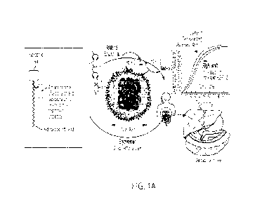
[0009] Figures 1A and 1B are representations of the ErythroMer (EM) V2 design,
features and
benefits relative to HBOCs, in which the amphiphilic precursor comprises (a)
pH responsive
groups that control availability of the allosteric effector (RSR13), enabling
context-responsive
control of 02 binding and (b) a negatively charged 'head' facilitating
biocompatibility of the
exofascial surface. The construct mimics endogenous biomolecules and is
subject to enzymatic
digestion and complete degradation in vivo to end-products identical to that
of 'natural' peptides
and lipids.
[0010] Figure 2 shows EM interference results: (A) metabolic panel and (B) CBC
panel of human
plasma with and without EM; (C) overlaid metabolic panel spectral scan (300-
900nm).
4
CA 03210619 2023- 8- 31
WO 2022/192619
PCT/US2022/019872
[0011] Figure 3 is a representation of the Bait and Capture design strategy
according to an
embodiment of the invention. The amphiphilic precursor KC01003 will be co-self-
assembled with
adamantane-PE (4) to prepare EM-1003 presented with 'bait' functionalities on
the surface. The
surface coverage is optimized to have minimum numbers of 'bait' molecules to
avoid detrimental
biological effects. The column is packed with polystyrene (PS) beads
conjugated with 13-
cyclodextrin functionalities. Highly selective interaction between adamantane
and (3-cyclodextrin
through host-guest chemistry enables efficient separation of EM from blood.
[0012] Figure 4A shows a scheme for synthesis of 'bait' component adamantane
functionalized
DPPE (ADM-DPPE)
[0013] Figure 4B shows a scheme for synthesis of capture agent I3-cyclodextrin
functionalized PS
beads (CD-PS).
[0014] Figure 5 shows confirmation of 13-DC functionalization on PS beads
(incubated with
pyrene; bead size 09S nm).
[0015] Figure 6A shows confirmation of EM capture by particle count using
nanoparticle tracking
analysis (ZetaVi ew).
[0016] Figure 6B shows confirmation of EM capture by diameter after capture
using nanoparticle
tracking analysis (Z et aVi ew).
[0017] Figure 7 is a representation of lyophilization and final lyophilized AM-
tagged EM product
according to an embodiment of the invention.
[0018] Figure 8 shows various properties of an AM-tagged EM particle according
to an
embodiment of the invention before lyophilization, four days after
reconstitution, and 14 days after
reconstitution.
DETAILED DESCRIPTION
CA 03210619 2023- 8- 31
WO 2022/192619
PCT/US2022/019872
[0019] According to embodiments of the invention, there is presented a bio-
engineered and
scalable approach to remove EM from whole blood specimens. (Fig 3) The
proposed
supramolecular approach offers several advantages, e.g. (i) facile formation
of the inclusion
complex under ambient condition makes the pilot-scale study feasible without
introducing any
special genetic or biochemical techniques; (ii) the relatively small molecular
adamantane
("ADM") tag is expected to have little to none effect on the EM structures and
functions and (iii)
the optimal ratio of 13-eyelodextrin (13-CD) funetionalization on polystyrene
(PS) beads
introduces insignificant changes to the pore size of the column beads and
hence safely clear the
blood stream. The present invention will pave the pathway for the translation
of artificial RBCs.
According to further embodiments of the invention, the bait and capture design
is optimized for
usability at point of care settings. This involves mechanisms to effectively
remove ErythroMer
from the patient blood samples, with minimal disruption to already existing
blood collection and
sample processing techniques, including. customizing already existing blood
collection tubes to
be able to capture and retain ErythroMer in the tube itself, leaving whole
blood components free
for further analysis. This entails a comprehensive development of immobilizing
the capture
system in the blood collection tube, titrated to a concentration to
effectively remove EM
specifically and fully from the blood, keeping in mind the goal that the blood
collection tubes
with the special capture functionality are scalable and can be manufactured
commercially with
minimal changes to their existing method of manufacturing. Concurrently, the
bait is compatible
with the lyophilization process for ErythroMer's final and most stable form.
[0020] Design, synthesis and physico-chemical characterization of EM
presenting with
'bait' functionalities.
6
CA 03210619 2023- 8- 31
WO 2022/192619
PCT/US2022/019872
[0021] Example 1. Design and synthesis of 'bait' tagged EM. EM tagged with ADM
is
prepared and the small molecules and the nanoparticles are physico-chemically
characterized for
supramolecular host-guest chemistry with I3-CD containing resins. Amino (-NH2)
groups 1,2-
dipalmitoyl-sn-glycero-3-phosphoethanolamine (DPPE) are functionalized with
ADM tags using
1-adamantane-carboxylic acid N-hydroxysuccinimide ester (ADM-NHS). (Figure 4A)
This
compound, ADM-DPPE, is characterized analytically for their purity and
introduced in the nano-
assembly of EM at a varied molar ratio. The optimum concentration of ADM-DPPE
tag on EM
particles is then determined based on two parameters: (i) highest binding
capability and (ii)
optimum Hb encapsulation efficiency. Accordingly, the formulations having
different ADM- tag
ratios is first interacted with a fixed concentration of13-CD and the binding
capabilities of the
tagged EM particles are calculated from the respective isothermal calorimetry
curves. The
particle having the highest binding efficiency is chosen and the concentration
of ADM- tag is
used as the optimal one. Secondly, this optimized EM particle is then loaded
with hemoglobin
and the encapsulation efficiency is calculated.
[0022] Example 2. Physico-chemical characterization. We evaluate hydrodynamic
particle
size, polydispersity, and electrophoretic potential, TEM, AFM, and stability.
To determine the
long-term storage stability of EM-1003-bait, a 12-month study is performed at -
20 C, 5 C, 25 C,
and under the accelerated condition of 40 C (lyophilized EM is spread on a
glass dish to have a
thin homogeneous layer). Thermal (dry heat) accelerated stress stability
analysis is performed up
to 12 months at 40 C, 50 C, and 60 C and for one month at 80 C. EM is also
subjected to
thermal stress at 25 C and 40 C under acidic (0.1 M HC1: pH 1; 0.01 M HC1: pH
2; HC1: pH 4.5)
and alkaline (0.1 M NaOH: pH 13; 0.01 M NaOH: pH 12) conditions up to 7 days.
The pH of all
7
CA 03210619 2023- 8- 31
WO 2022/192619
PCT/US2022/019872
the solutions is measured using a pH meter at RT. The particle size and
colloidal stability over
different time intervals are measured using the Nanosizer ZS and ZetaView.
[0023] Development and characterization of a 'capture' resin and demonstration
of
separation capability.
[0024] Example 3. Preparation and characterization of the capture resin: 13-
Cyclodextrin is
often used as the host carrier in a host-guest supramolecular assembly because
of its water
solubility, low cytotoxicity and superior bi compatibility. In this context,
we prefer the ADM/f3-
CD host¨guest pair as a well-established supramolecular system. Mono-6-0-(p-
toluenesulfony1)-
13-cyclodextrin is synthesized from 1-(p-Toluenesulfonyl) imidazole and p-
cyclodextrin. This
compound is then used for the functionalization of amino polystyrene (NH2-PS)
beads to
produce 13-cyclodextrinylated PS (CD-PS) beads under mild alkaline condition.
(Figure 4B) To
confirm the successful functionalization of the PS beads with 8-cyclodextrin,
the CD-PS beads
are mixed with a ethanolic solution of pyrene, centrifuged at 10000 rcf for 2
mins, washed with
ethanol twice to remove any free pyrene and then imaged under a fluorescence
microscope to
visualize the fluorescence of the functionalized beads. The successfully
functionalized PS beads
show high green emission, while the unfunctionalized beads do not demonstrate
any background
fluorescence. (Fig 5) This confirms the successful preparation of the capture
resin, ready to be
tested with the ADM-DPPE tagged EM particles.
[0025] Example 4. Demonstration of EM capture from a mixture of EM and blood:
To
demonstrate EM capture mediated by the CD-PS beads, the optimized ADM-tagged
EM
particles with fluorescent NBD-PE doping is prepared. In a similar manner, CD-
PS beads are
treated with these fluorescently tagged EM particles, suspended in buffer,
centrifuged, washed
and imaged under fluorescent microscope. After successfully validating the
capture of EM
8
CA 03210619 2023- 8- 31
WO 2022/192619
PCT/US2022/019872
particles by CD-PS beads, the ADM-tagged EM particles are loaded with
hemoglobin. The Hb
loaded ADM-tagged EM particles are then suspended with blood and the capture
of EM particles
is monitored from the whole blood specimens.
[0026] Example 5. Demonstration and optimization of the EM surface: The %
functionalization of 13-CD to the PS beads is calculated from pH titration.
The change in
neutralization pH is compared between the unfunctionalized amino PS beads and
CD-PS beads
and the % functionalization of I3-CD per gm of PS beads is calculated. The
ratio of I3-CD per gm
of PS beads is then varied by controlling the concentration of mono-6-0-(p-
toluenesulfony1)-13-
cyclodextrin reacted with amino PS beads. The optimum concentration of fl-CD
is then selected
based on their maximum efficiency of EM removal from the whole blood specimen.
[0027] Example 6. Preliminary biocompatibility studies. Preliminary complement
activation
analysis and blood smear preparation studies is conducted. The CH50 enzyme
immunoassay
(ETA) (Sigma) is used to evaluate the magnitude of complement activation as a
result of the
addition of EM-1003-Bait to human serum. The kit is used according to the
manufacturer's
protocol. A blood smear preparation is performed to observe morphological
changes in
lymphocytes and blood clumping using clinical microscopy technique
(conventional light
microscopy) under high power field. Particular attention is paid to observe
significant clumping
or morphological changes in blood cells treated with EM-1003 and EM-1003-Bait
(blood: NP=
9:1)
[0028] Anticipated Results and Risk Factors. EM particle with ADM functional
groups on the
surface is synthesized. Mono-6-0-(p-toluenesulfony1)413-cyclodextrin is
synthesized from 1-(p-
Toluenesulfonyl) imidazole and I3-CD and used for the functionalization of
amino polystyrene
(NH2-PS) beads to produce CD-PS beads. This invention validates the capture of
EM particles
9
CA 03210619 2023- 8- 31
WO 2022/192619
PCT/US2022/019872
by CD-PS beads with and without the encapsulation of Hb. The Hb loaded Ad-
tagged EM
particles are suspended with blood and the capture of EM particles is
monitored. Further CBC
and metabolic panel analyses are conducted to confirm the successful
disappearance of EM
interference. The supramolecular approach of the invention brings several
advantages, e.g (i)
facile formation of the inclusion complex under ambient condition makes the
pilot-scale study
feasible without introducing any special genetic or biochemical techniques;
(ii) the relatively
small ADM-tag is expected to have little to none effect on the EM structures
and functions and
(iii) the optimal ratio of 13-CD functionalization on PS beads introduces
insignificant changes to
the pore size of the column beads and hence safely clear the blood stream.
[0029] From our preliminary capture studies, EM was found to be successfully
captured by the
capture resin. (Fig 6) It was observed that average number of EM particles
decreased only when
treated with 13-CD tagged capture resin. Increased concentration of CD-PS
decreased EM
particles significantly (Fig 6A). Further it has been observed that the
average hydrodynamic
diameter of the EM particles increased after successful capture mediated by
the 13-CD tagged
resin (Fig 6B). If EM stability becomes an issue, the 'bait' functional groups
may be optimized
to reach the maximum stability.
[0030] The adamantane-tagged EM is lyophilized for packaging, transport and/or
storage. The
lyophilized adamantane-baited EM is a powder comprising EM amphiphilic
precursor,
cholesterol and PEG-PE hemoglobin and allosteric effector, the adamantane tag,
and optionally
also including cryoprotectants. Reconstitution at the original EM production
concentration (or
concentrated) is achieved with PBS/water by simple mixing and gentle
vortexing/agitation.
CA 03210619 2023- 8- 31
WO 2022/192619
PCT/US2022/019872
[0031] It is noted that the bait and capture methods and compositions
described herein may be
used to remove any type of biosynthetic nanoparticle from any bodily fluid or
constituent part
thereof.
[0032] While several embodiments and methodologies of the present disclosure
have been
described and shown in the drawings, it is not intended that the present
disclosure be limited
thereto, as it is intended that the present disclosure be as broad in scope as
the art will allow and
that the specification be read likewise. Therefore, the above description
should not be construed
as limiting, but merely as exemplifications of particular embodiments and
methodologies. Those
skilled in the art will envision other modifications within the scope of the
claims appended
hereto.
11
CA 03210619 2023- 8- 31