Note: Descriptions are shown in the official language in which they were submitted.
CA 03210656 2023-08-02
WO 2022/183047 PCT/US2022/017990
REGULATORY T CELL (TREG) EXTRACELLULAR VESICLE COMPOSITIONS AND METHODS
CROSS-REFERENCE TO RELATED APPLICATIONS
[0001] This application claims the benefit of U.S. Provisional Application
No. 63/208,395,
filed June 08, 2021, and U.S. Provisional Application No. 63/154,449, filed
February 26, 2021,
each of which is incorporated by reference herein in its entirety.
1. FIELD
[0002] The present disclosure provides anti-inflammatory and restorative
extracellular
vesicles (EVs) that are derived from ex vivo-expanded human suppressive immune
cells, e.g.,
regulatory T cells (Tregs) and that are useful in the treatment of diseases
such as amyotrophic
lateral sclerosis (ALS), Alzheimer's disease, and other neurological diseases,
as well as
inflammatory, metabolic, and autoimmune diseases or dysfunctions.
2. BACKGROUND
[0003] Inflammatory and neuroinflammatory mechanisms contribute to a wide
variety of
devastating diseases, including such neurodegenerative diseases as amyotrophic
lateral sclerosis
(ALS), Parkinson's disease and multiple sclerosis. Neurodegenerative diseases
such as this
direct a tremendous health and economic burden that will only exacerbate
further over time.
[0004] Currently, no disease-modifying treatments for such diseases are
available. Anti-
inflammatory treatments have been utilized for decades in attempting to
ameliorate a multitude
of neurodegenerative diseases. Little progress, however, has been made with
single drug/target
approaches.
[0005] Increasingly, studies point to immune system involvement in the
etiology of diseases
such as this, and point to dysfunction of immune cells as a chief mediator of
disease
pathogenesis. The complex signaling mechanisms and built-in redundancies of
the immune
system and its constituents may help explain the ineffectiveness of such
single drug/single target
anti-inflammatory approaches.
[0006] Recently great promise has been demonstrated with regulatory T cell
(Treg) cell
1
CA 03210656 2023-08-02
WO 2022/183047 PCT/US2022/017990
therapy, which may represent a more global approach to suppressing immune
system
dysfunction contributing to disease. For example, clinical trials involving
administration of
expanded autologous Tregs to ALS patients report that the Treg therapy slowed
progression rates
during early and later stages of the disease, and that Treg suppressive
function correlated with
the slowing of disease progression (Thonhoff, J.R. et al., 2018, Neurology-
Neuroimmunology
Neuroinflammation 5(4)).
[0007] Nonetheless, there still exists a need for development of additional
treatments that can
suppress inflammatory and/or promote anti-inflammatory immune system
components, and can
do so in the pro-inflammatory, toxic microenvironment of the disease state.
3. SUMMARY
[0008] Presented herein are extracellular vesicles (EVs) that exhibit
impressive anti-
inflammatory activity, both in vitro and in vivo. The EVs presented herein are
derived from ex
vivo-expanded human suppressive immune cells, for example regulatory T cells
(Tregs). As
demonstrated herein, the EVs of the present disclosure retain the immune
suppressive activities
of the cells from which they are derived. Moreover, as EVs are not themselves
cells, they avoid
potential cell-based issues such as immune rejection and the possibility of
polarization to a pro-
inflammatory cell type. As such, the anti-inflammatory EVs presented herein
are particularly
useful for treatment of a variety of diseases such as, for example,
neurodegenerative disorders
such as amyotrophic lateral sclerosis (ALS).
[0009] Results presented herein demonstrate that the EVs of the present
disclosure are able
to potently suppress T responder cell proliferation and pro-inflammatory
myeloid, e.g.,
macrophage, activity in vitro, and also exert potent anti-inflammatory effects
in vivo, via either
intravenous or intranasal administration. For example, in vivo results
presented herein using
anti-inflammatory Treg EV compositions of the disclosure demonstrate an anti-
inflammatory
effect in a model of inflammation and a motor neuron degenerative disease
modeling ALS. For
example, results presented herein demonstrate that the EVs are able to
suppress brain and
peripheral inflammation in an in vivo model of neuroinflammation, and are also
able to suppress
inflammation and extend survival in an in vivo model of amyotrophic lateral
sclerosis (ALS).
The results presented herein also demonstrate that the Treg EVs have a greater
suppressive effect
on pro-inflammatory immune cells than EVs derived from mesenchymal stem cells
(MSCs).
2
CA 03210656 2023-08-02
WO 2022/183047 PCT/US2022/017990
[0010] Moreover, the anti-inflammatory EVs presented herein exhibit
remarkable batch-to-
batch consistency in size, stability and activity and exhibit a unique
structural signature as, for
example, characterized by Treg EV surface marker and RNA profiles. Still
further, as
demonstrated herein, the methods presented herein yield potent anti-
inflammatory EVs
exhibiting similar structural and suppressive activity characteristics whether
the original Treg
starting material is obtained from healthy subjects or ALS patients.
[0011] In one aspect, presented herein is an isolated, cell-free population
of anti-
inflammatory extracellular vesicles (EVs), wherein the anti-inflammatory EVs
are derived from
ex vivo-expanded human suppressive immune cells, wherein: i) the population
exhibits a size
diameter distribution of about 50 nm to about 150 nm; ii) the population
comprises EV surface
CD2, CD25 and HLA-DRDPDQ; iii) the population comprises hsa-miR-1290, hsa-miR-
146a-5p,
and hsa-miR-155-5p micro-RNAs (miRNAs); and iv) the population exhibits an
ability to
suppress myeloid cells, for example, macrophages, as measured by an ability to
reduce pro-
inflammatory cytokine production by the myeloid cells (e.g., exhibit an
ability to decrease the
expression of IL-6, IL-8, IL1f3 or Interferon-y in the myeloid cells) and an
ability to increase the
expression of one or more anti-inflammatory markers in the myeloid cells
(e.g., an ability to
increase the expression of IL-10, Argl and/or CD206 in the myeloid cells), or
as measured by an
ability to suppress proliferation of responder T cells; and wherein the human
suppressive
immune cells are regulatory T cells (Tregs). In certain embodiments, the human
Tregs are from
a healthy human subject. In certain embodiments, the human Tregs are from a
human subject
diagnosed with or suspected of having Amyotrophic Lateral Sclerosis (ALS).
[0012] In certain embodiments, the population of anti-inflammatory EVs
further comprises
EV surface CD44, CD29, CD4 and CD45. In certain embodiments, the population of
anti-
inflammatory EVs further comprises EV surface CD44, CD29, CD4 and CD45. In
certain
embodiments, the population of anti-inflammatory EVs further comprises EV
surface CD9,
CD63 and CD81. In certain embodiments, the population of anti-inflammatory EVs
substantially lacks EV surface CD3, CD19, CD8, CD56, CD105, CD1c, CD49e, ROR1,
CD209,
SSEA-4, CD40, CD62P, CD11c, CD40, MSCP, CD146, CD86, CD326, CD133, CD142, CD31
and CD14. In certain embodiments, the population of anti-inflammatory EVs
further comprises
EV surface CD44, CD29, CD4 and CD45. In certain embodiments, the population of
anti-
inflammatory EVs further comprises EV surface CD44, CD29, CD4 and CD45, CD9,
CD63 and
3
CA 03210656 2023-08-02
WO 2022/183047 PCT/US2022/017990
CD81, and ii) substantially lacks EV surface CD3, CD19, CD8, CD56, CD105,
CD1c, CD49e,
ROR1, CD209, SSEA-4, CD40, CD62P, CD11c, CD40, MSCP, CD146, CD86, CD326,
CD133,
CD142, CD31 and CD14.
[0013] In certain embodiments, the ratio of hsa-miR-146a-5p to hsa-miR-155-
5p present in
the population of anti-inflammatory EVs is about 2 to about 3. In certain
embodiments, the
abundance of hsa-miR-1290 in the population of anti-inflammatory EVs is at
least 2-fold that of
hsa-mir-155-5p. In specific embodiments, the ratio of hsa-miR-146a-5p to hsa-
miR-155-5p
present in the population of anti-inflammatory EVs is about 2 to about 3 and
the abundance of
hsa-miR-1290 in the population of anti-inflammatory EVs is at least 2-fold
that of hsa-mir-155-
5p.
[0014] In particular embodiments, at least about 90% of the EVs of the
population of anti-
inflammatory exhibit a size diameter of about 50 nm to about 150 nm. In
certain embodiments,
the population of anti-inflammatory EVs exhibits a mean size diameter of about
80 nm to about
110 nm. In certain embodiments, the population of anti-inflammatory EVs
exhibits a median
size diameter of about 70 nm to about 110 nm. In certain embodiments, the
population of anti-
inflammatory EVs exhibits a mode size diameter of about 65nm to about 95 nm.
In specific
embodiments, at least about 90% of the EVs in the population of anti-
inflammatory EVs exhibit
a size diameter of about 50 to about 150 nm, and the population exhibits a
mean size diameter of
about 80 nm to about 110 nm, a median size diameter of about 70 nm to about
110 nm, and a
mode size diameter of about 65 nm to about 95 nm.
[0015] Presented herein are isolated, cell-free populations of anti-
inflammatory EVs,
wherein the anti-inflammatory EVs are derived from ex vivo-expanded human
suppressive
immune cells, for example regulatory T cells (Tregs). Also presented herein
are pharmaceutical
compositions and cryopreserved compositions comprising an isolated, cell-free
population of
anti-inflammatory EVs described herein, methods of producing the EV
populations and methods
of using the EVs for treatment of diseases, such as neurodegenerative
diseases, e.g., ALS.
[0016] In one aspect, provided herein is an isolated, cell-free population
of anti-
inflammatory extracellular vesicles (EVs), wherein the anti-inflammatory EVs
are derived from
ex vivo-expanded human suppressive immune cells. In some embodiments, the
human
suppressive immune cells are regulatory T cells (Tregs). In some embodiments,
the Tregs are
from a healthy human subject.
4
CA 03210656 2023-08-02
WO 2022/183047 PCT/US2022/017990
[0017] In some embodiments, the Tregs are from a human subject diagnosed
with or
suspected of having a neurodegenerative disorder. In some embodiments, the
neurodegenerative
disorder is Alzheimer's disease. In some embodiments, the neurodegenerative
disorder is
Amyotrophic Lateral Sclerosis (ALS). In some embodiments, the
neurodegenerative disease is
multiple sclerosis (MS). In some embodiments, the neurodegenerative disease is
Parkinson's
Disease.
[0018] In some embodiments, the Tregs are from a human subject who is
diagnosed as
having, or suspected of having had, a stroke.
[0019] In some embodiments, the Tregs are from a geriatric human subject.
[0020] In some embodiments, the Tregs are from multiple human subjects. In
some
embodiments, the Tregs are from multiple unrelated human subjects.
[0021] In some embodiments, the anti-inflammatory EVs exhibit an ability to
increase the
expression of one or more anti-inflammatory markers in inflammatory cells. In
some
embodiments, the inflammatory cells are myeloid cells. In some embodiments,
the anti-
inflammatory EVs exhibit an ability to increase the expression of IL-10, Argl
and/or CD206 in
inflammatory cells.
[0022] In some embodiments, the anti-inflammatory EVs exhibits an ability
to suppress
inflammatory cells, as measured by pro-inflammatory cytokine production by the
inflammatory
cells. In some embodiments, the inflammatory cells are myeloid cells. In some
embodiments,
the myeloid cells are monocytes, macrophages, or microglia. In some
embodiments, the
macrophages are M1 macrophages. In some embodiments, the M1 macrophages are
induced
pluripotent stem cell (iPSC)-derived M1 macrophages.
[0023] In some embodiments, the ability to suppress inflammatory cells is
measured by IL-6,
IL-8, TNFa, IL1f3 and/or Interferon-y production by the inflammatory cells.
[0024] In some embodiments, the anti-inflammatory EVs exhibit an ability to
increase the
expression of IL-1-, Argl and/or CD206 and an ability to suppress IL-6, IL-8,
TNFa, IL1f3
and/or Interferon-y production in inflammatory cells, e.g., myeloid cells, for
example,
macrophages.
[0025] In some embodiments, the anti-inflammatory EVs exhibit a suppressive
function, as
determined by suppression of proliferation of responder T cells. In some
embodiments, the
proliferation of responder T cells is determined by flow cytometry or
thymidine incorporation.
CA 03210656 2023-08-02
WO 2022/183047 PCT/US2022/017990
[0026] In some embodiments, the population is a saline-containing
population of anti-
inflammatory EVs. In some embodiments, the population is a physiological
saline-containing
population of anti-inflammatory EVs. In some embodiments, the population is a
phosphate-
buffered saline-containing population of anti-inflammatory EVs.
[0027] In some embodiments, the population of anti-inflammatory EVs
comprises exosomes
and microvesicles. In some embodiments, the majority of the EVs are exosomes.
In some
embodiments, at least about 80%, about 90%, or about 95% of the EVs are
exosomes. In some
embodiments, the majority of the EVs are microvesicles. In some embodiments,
at least about
80%, about 90%, or about 95% of the EVs are microvesicles.
[0028] In some embodiments, the population of anti-inflammatory EVs
comprises at least
about 50% exosomes. In some embodiments, at least about 60% of the EVs are
exosomes. In
some embodiments, at least about 70% of the EVs are exosomes.
[0029] In some embodiments, the population of anti-inflammatory EVs
comprises at least
about 50% microvesicles. In some embodiments, at least about 60% of the EVs
are
microvesicles. In some embodiments, at least about 70% of the EVs are
microvesicles.
[0030] In some embodiments, the majority of the EVs in a population of anti-
inflammatory
EVs provided herein have diameters from about 30 nm to about 1000 nm. In some
embodiments, the majority of the EVs have diameters from about 30 nm to about
100 nm, about
30 nm to about 150 nm, about 30 to about 200 nm, about 40 to about 100 nm,
about 80 to about
100 nm, about 80 to about 110 nm, about 80 to about 125 nm, or about 100 to
about 120 nm. In
some embodiments, the majority of the EVs have diameters from about 60 nm to
about 1000 nm,
about 70 nm to about 1000 nm, about 80 nm to about 1000 nm, 100 to about 1000
nm, about 200
to about 1000 nm, or about 300 to about 1000 nm. In some embodiments, the
majority of the
EVs have diameters from about 20 nm to about 300 nm, about 20 nm to about 275
nm, about 20
to about 250 nm, about 20 to about 200 nm, or about 20 nm to about 175 nm.
[0031] In another aspect, provided herein is a pharmaceutical composition
comprising an
isolated, cell-free population of anti-inflammatory EVs provided herein. In
certain
embodiments, the pharmaceutical composition comprising an isolated, cell-free
population of
anti-inflammatory EVs provided herein in saline. In some embodiments, the
population of anti-
inflammatory EVs comprises about 1x10⁶ to about lx1014 EVs, about 1x10⁸ to
about lx1014
EVs, about 1x10⁸ to about lx1012 EVs, about 1x10⁸ to about lx101 EVs, about
lx101 to about
6
CA 03210656 2023-08-02
WO 2022/183047 PCT/US2022/017990
1x10¹⁴ EVs, or about lx101 to about lx1012EVs. In some embodiments, the
population of anti-
inflammatory EVs comprises about 1x10⁹ EVs, about 5x10⁹EVs, about lx101 EVs,
about
5x101 EVs, about lx1011EVs, about 5x10" EVs, or about lx1012EVs. In some
embodiments,
the population of anti-inflammatory EVs comprises about lx106 to about
lx1014EVs/ml, about
1x10⁸ to about lx1014EVs/ml, about 1x10⁸ to about lx1012EVs/ml, about 1x10⁸ to
about lx101
EVs/ml, about lx101 to about lx1014EVs/ml, or about lx101 to about
lx1012EVs/ml. In some
embodiments, the population of anti-inflammatory EVs comprises about 5x10⁸
EVs/ml, about
1x10⁹EVs/ml, about 2.5x10⁹ EVs/ml, about 5x10⁹ EVs/ml, about lx101 EVs/ml,
about 2.5x101
EVs/ml, about 5x101 EVs/ml, about lx1011EVs/ml, about 2.5x10" EVs/ml, about
5x10¹¹
EVs/ml, or about lx1012EVs/ml.
[0032] In some embodiments, the population of anti-inflammatory EVs
comprises in a
pharmaceutical composition provided herein comprises about 1 pg to about 200
mg EVs. In
some embodiments, the population of anti-inflammatory EVs comprises about 1 pg
to about 15
mg EVs. In some embodiments, the population of anti-inflammatory EVs comprises
about 1 pg
to about 15 mg EV/ml.
[0033] In some embodiments, the pharmaceutical composition is a
cryopreserved
pharmaceutical composition. In some embodiments, the pharmaceutical
composition had
previously been cryopreserved.
[0034] In another aspect, provided herein is a cryopreserved composition
comprising an
isolated, cell-free population of anti-inflammatory EVs provided herein.
[0035] In another aspect, provided herein is a method of producing an
isolated, cell-free
population of anti-inflammatory extracellular vesicles (EVs), said method
comprising the steps
of: (a) ex-vivo expanding a human suppressive immune cell population in
culture media to
produce a culture comprising the cells, the culture media and anti-
inflammatory EVs; and
(b) isolating the anti-inflammatory EVs from the culture. In some embodiments,
the human
suppressive immune cell population is a population of regulatory T cells
(Tregs).
[0036] In some embodiments, step (b) comprises removing cells from the
culture, followed
by polyethylene glycol precipitation of the culture. In some embodiments, step
(b) comprises:
(i) removing the cells from the culture to produce a cell-free, anti-
inflammatory EV-containing
solution; and (ii) isolating the anti-inflammatory EVs from the cell-free,
anti-inflammatory EV-
containing solution of (i).
7
CA 03210656 2023-08-02
WO 2022/183047 PCT/US2022/017990
[0037] In some embodiments, step (i) comprises passing the culture through
a filter such that
the cells are retained by the filter, and thereby removed from the culture. In
some embodiments,
step (i) comprises microfiltration.
[0038] In some embodiments, step (ii) comprises step (ii-a): passing the
cell-free, anti-
inflammatory EV-containing solution through a filter such that the anti-
inflammatory EVs are
retained by the filter. In some embodiments, the filter has a molecular weight
cut-off (MWCO)
of about 200 kilodaltons (kDa) to about 600 kDa. In some embodiments, the
filter has an
MWCO of about 500 kDa.
[0039] In some embodiments, step (ii) comprises ultrafiltration. In some
embodiments, step
(ii) further comprises step (ii-b): performing buffer exchange such that the
isolated, cell-free
population of anti-inflammatory EVs produced is a buffer-containing isolated,
cell-free
population of anti-inflammatory EVs. In some embodiments, the buffer is a
saline-containing
buffer. In some embodiments, the saline-containing buffer is physiological
saline. In some
embodiments, the saline-containing buffer is PBS.
[0040] In some embodiments, step (ii-b) comprises diafiltration.
[0041] In some embodiments, steps (ii-a) and (ii-b) are performed
simultaneously.
[0042] In some embodiments, step (b) comprises tangential flow filtration.
[0043] In some embodiments, the culture media in step (a) is serum-free. In
some
embodiments, the culture media in step (a) comprises serum. In some
embodiments, the serum is
human AB serum. In some embodiments, the serum is depleted for serum-derived
EVs.
[0044] In some embodiments, a method of producing an isolated, cell-free
population of anti-
inflammatory EVs further comprises, prior to step (a), the step of enriching
Tregs from a cell
sample suspected of containing Tregs, to produce a baseline Treg cell
population that is the
population of Tregs that is then expanded in step (a). In some embodiments,
the cell sample is a
leukapheresis cell sample. In some embodiments, the method further comprises
obtaining the
cell sample from a donor by leukapheresis. In some embodiments, the cell
sample is not stored
overnight or frozen before carrying out the enriching step. In some
embodiments, the cell
sample is obtained within 30 minutes before initiation of enriching step. In
some embodiments,
the enriching step comprises depleting CD8+/CD19+ cells then enriching for
CD25+ cells. In
some embodiments, step (a) is carried out within 30 minutes of the enriching
step.
[0045] In some embodiments, step (a) of a method of producing an isolated,
cell-free
8
CA 03210656 2023-08-02
WO 2022/183047 PCT/US2022/017990
population of anti-inflammatory EVs comprises culturing the Tregs in a culture
media that
comprises beads coated with anti-CD3 antibodies and anti-CD28 antibodies. In
some
embodiments, the beads are first added to the culture media within about 24
hours of the
initiation of the culturing. In some embodiments, beads coated with anti-CD3
antibodies and
anti-CD28 antibodies are added to the culture media about 14 days after beads
coated with anti-
CD3 antibodies and anti-CD28 antibodies were first added to the culture
medium.
[0046] In some embodiments, step (a) further comprises adding IL-2 to the
culture medium
within about 6 days of the initiation of culturing. In some embodiments, step
(a) further
comprises replenishing the culture medium with IL-2 about every 2-3 days after
IL-2 is first
added to the culture medium.
[0047] In some embodiments, step (a) further comprises adding rapamycin to
the culture
medium within about 24 hours of the initiation of the culturing. In some
embodiments, step a)
further comprises replenishing the culture medium with rapamycin every 2-3
days after the
rapamycin is first added to the culture medium.
[0048] In some embodiments, step (a) is automated. In some embodiments,
step a) takes
place in a bioreactor.
[0049] In some embodiments, step (b) of a method of producing an isolated,
cell-free
population of anti-inflammatory EVs may commence at any point during step a).
[0050] In some embodiments, the Tregs enriched in step (a) are from a
healthy human
subject. In some embodiments, the Tregs are from a human subject diagnosed
with or suspected
of having a neurodegenerative disorder. In some embodiments, the
neurodegenerative disorder
is Alzheimer's disease, Amyotrophic Lateral Sclerosis (ALS), multiple
sclerosis (MS), or
Parkinson's Disease. In some embodiments, the Tregs are from a human subject
who is
diagnosed as having, or suspected of having had, a stroke. In some
embodiments, the Tregs are
from a geriatric human subject. In some embodiments, the Tregs are from
multiple human
subjects.
[0051] In some embodiments, the human suppressive immune cell population
expanded in
step (a) is a genetically engineered human suppressive immune cell population.
[0052] In some embodiments, the population of Tregs expanded in step (a) is
a genetically
engineered population of Tregs.
[0053] In another aspect, provided herein is a pharmaceutical composition
comprising an
9
CA 03210656 2023-08-02
WO 2022/183047 PCT/US2022/017990
isolated, cell-free population of anti-inflammatory EVs, wherein the
population is made by any
one of the methods described herein.
[0054] In some embodiments, a method of producing an isolated, cell-free
population of anti-
inflammatory EVs further comprises (c) cryopreserving the isolated, cell-free
population of anti-
inflammatory EVs, thereby producing a cryopreserved, isolated, cell-free
population of anti-
inflammatory EVs. Also presented herein are cryopreserved compositions
comprising an
isolated, cell-free population of anti-inflammatory EVs, wherein the
cryopreserved compositions
are made using such methods.
[0055] In some embodiments, the method further comprises thawing the
cryopreserved,
isolated cell-free population of anti-inflammatory EVs after cryopreservation
for about 1 week, 1
month, about 3 months, about 6 months, about 9 months, about 12 months, about
18 months or
about 24 months. Also provided herein are compositions, for example,
pharmaceutical
compositions, comprising an isolated, cell-free population of anti-
inflammatory EVs, wherein
the compositions, for example, pharmaceutical compositions, are made using
such methods.
[0056] In another aspect, provided herein is an isolated, cell-free
population of anti-
inflammatory EVs, wherein the anti-inflammatory EVs are derived from an ex
vivo-expanded
Treg cell population that exhibits an ability to suppress inflammatory cells,
as measured by pro-
inflammatory cytokine production by the inflammatory cells, wherein the
inflammatory cells are
macrophages or monocytes from human donors or generated from induced
pluripotent stem
cells, wherein the ex vivo- expanded Treg cell population has been expanded
from baseline
Tregs, and wherein, in the ex vivo- expanded Treg cell population: (a)
expression of one or more
dysfunctional baseline signature gene products listed in Table 3 and/or Table
4 is decreased
relative to the expression of the one or more gene products in baseline Tregs;
(b) expression of
one or more dysfunctional baseline signature gene products listed in Table 5
is decreased relative
to the expression of the one or more gene products in baseline Tregs; (c)
expression of one or
more Treg-associated signature gene products listed in Table 6 is increased
relative to the
expression of the one or more gene products in baseline Tregs; (d) expression
of one or more
mitochondria signature gene products listed in Table 7 is increased relative
to the expression of
the one or more gene products in baseline Tregs; (e) expression of one or more
cell proliferation
signature gene products listed in Table 8 is increased relative to the
expression of the one or
more gene products in baseline Tregs; or (f) expression of one or more highest
protein
CA 03210656 2023-08-02
WO 2022/183047 PCT/US2022/017990
expression signature gene products listed in Table 9 is increased relative to
the expression of the
one or more gene products in baseline Tregs. In some embodiments, provided
herein is a
pharmaceutical composition comprising the isolated, cell-free population of
anti-inflammatory
EVs.
[0057] In another aspect, provided herein is a method of treating a
disorder associated with
Treg dysfunction, the method comprising administering to a subject in need of
said treatment a
pharmaceutical composition provided herein.
[0058] In another aspect, provided herein is a method of treating a
disorder associated with
Treg deficiency, the method comprising administering to a subject in need of
said treatment a
pharmaceutical composition provided herein.
[0059] In another aspect, provided herein is a method of treating a
disorder associated with
overactivation of the immune system, the method comprising administering to a
subject in need
of said treatment a pharmaceutical composition provided herein.
[0060] In another aspect, provided herein is a method of treating an
inflammatory condition
driven by a T cell response, the method comprising administering to a subject
in need of said
treatment a pharmaceutical composition provided herein.
[0061] In another aspect, provided herein is a method of treating an
inflammatory condition
driven by a myeloid cell response, the method comprising administering to a
subject in need of
said treatment a pharmaceutical composition provided herein. In some
embodiments, the
myeloid cell is a monocyte, macrophage or microglia.
[0062] In another aspect, provided herein is a method of treating a
neurodegenerative
disorder in a subject in need thereof, the method comprising administering to
a subject in need of
said treatment a pharmaceutical composition provided herein. In some
embodiments, the
neurodegenerative disease is ALS, Alzheimer's disease, Parkinson's disease,
frontotemporal
dementia or Huntington's disease.
[0063] In some embodiments, the neurodegenerative disease is ALS,
Alzheimer's disease,
Parkinson's disease, frontotemporal dementia, multiple sclerosis or
Huntington's disease.
[0064] In another aspect, provided herein is a method of treating an
autoimmune disorder in
a subject in need thereof, the method comprising administering to a subject in
need of said
treatment a pharmaceutical composition provided herein. In some embodiments,
the
autoimmune disorder is polymyositis, ulcerative colitis, inflammatory bowel
disease, Crohn's
11
CA 03210656 2023-08-02
WO 2022/183047 PCT/US2022/017990
disease, celiac disease, systemic sclerosis (scleroderma), multiple sclerosis
(MS), rheumatoid
arthritis (RA), Type I diabetes, psoriasis, dermatomyosititis, lupus, e.g.,
systemic lupus
erythematosus, or cutaneous lupus, myasthenia gravis, autoimmune nephropathy,
autoimmune
hemolytic anemia, autoimmune cytopenia, autoimmune encephalitis, autoimmune
hepatitis,
autoimmune uveitis, alopecia, thyroiditis or pemphigus.
[0065] In another aspect, provided herein is a method of treating graft-
versus-host disease in
a subject in need thereof, the method comprising administering to a subject in
need of said
treatment a pharmaceutical composition provided herein. In some embodiments,
the subject has
received a bone marrow transplant, kidney transplant or liver transplant.
[0066] In another aspect, provided herein is a method of improving islet
graft survival in a
subject in need thereof, the method comprising administering to a subject in
need of said
treatment a pharmaceutical composition provided herein.
[0067] In another aspect, provided herein is a method of treating cardio-
inflammation in a
subject in need thereof, the method comprising administering to a subject in
need of said
treatment a pharmaceutical composition provided herein. In some embodiments,
the cardio-
inflammation is associated with atherosclerosis, myocardial infarction,
ischemic cardiomyopathy
or heart failure.
[0068] In another aspect, provided herein is a method of treating
neuroinflammation in a
subject in need thereof, the method comprising administering to a subject in
need of said
treatment a pharmaceutical composition provided herein. In some embodiments,
the
neuroinflammation is associated with stroke, acute disseminated
encephalomyelitis, acute optic
neuritis, acute inflammatory demyelinating polyradiculoneuropathy, chronic
inflammatory
demyelinating polyradiculoneuropathy, Guillain-Barre syndrome, transverse
myelitis,
neuromyelitis optica, epilepsy, traumatic brain injury, spinal cord injury,
encephalitis, central
nervous system vasculitis, neurosarcoidosis, autoimmune or post-infectious
encephalitis or
chronic meningitis.
[0069] In another aspect, provided herein is a method of treating a
Tregopathy in a subject in
need thereof, comprising administering to a subject in need of said treatment
a pharmaceutical
composition provided herein. In some embodiments, the Tregopathy is caused by
a FOXP3,
CD25, cytotoxic T lymphocyte-associated antigen 4 (CTLA4), LPS-responsive and
beige-like
anchor protein (LRBA), or BTB domain and CNC homolog 2 (BACH2) gene loss-of-
function
12
CA 03210656 2023-08-02
WO 2022/183047 PCT/US2022/017990
mutation, or a signal transducer and activator of transcription 3 (STAT3) gain-
of-function
mutation.
[0070] In some embodiments, the anti-inflammatory EVs administered to a
subject are
derived from Tregs that are autologous to the subject. In some embodiments,
the anti-
inflammatory EVs are derived from Tregs that are allogeneic to the subject.
[0071] In some embodiments, the pharmaceutical composition is administered
via intranasal
administration. In some embodiments, the intranasal administration is via
aerosol inhalation or
nasal drip. In some embodiments, the pharmaceutical composition is
administered
intravenously. In some embodiments, the pharmaceutical composition is
administered by local
injection.
[0072] In some embodiments, a method of treatment provided herein further
comprises
administering to the subject a pharmaceutical composition comprising a
therapeutic population
of Tregs, wherein the Tregs had been ex vivo expanded and cryopreserved, and
wherein the
Tregs are not further expanded prior to the administering. In some
embodiments, the therapeutic
population of Tregs is autologous to the subject. In some embodiments, the
therapeutic
population of Tregs is allogeneic to the subject. In some embodiments, the
pharmaceutical
composition comprising the therapeutic population of Tregs is administered
intravenously. In
some embodiments, the pharmaceutical composition comprising the anti-
inflammatory EVs and
the pharmaceutical composition comprising the therapeutic population of Tregs
are administered
to the patient on the same day.
[0073] In certain embodiments, the methods of treatment presented herein
comprise
administering to a subject in need of treatment a pharmaceutical composition
comprising an
isolated, cell-free population of anti-inflammatory EVs, wherein the EVs had
been cryopreserved
and thawed prior to being administered to the subject. In certain embodiments,
the methods of
treatment presented herein comprise administering to a subject in need of
treatment a
pharmaceutical composition comprising an isolated, cell-free population of
anti-inflammatory
EVs, wherein the EVs are stored at 4 C, for example, are stored overnight at
4 C, prior to being
administered to the subject. In particular embodiments, the methods of
treatment presented
herein comprise administering to a subject in need of treatment a
pharmaceutical composition
comprising an isolated, cell-free population of anti-inflammatory EVs wherein
the EVs had been
13
CA 03210656 2023-08-02
WO 2022/183047 PCT/US2022/017990
cryopreserved, thawed and stored at 4 C, for example, stored overnight at 4
C, prior to being
administered to the subject.
[0074] In certain embodiments, the methods of treatment presented herein
comprise
administering to a subject in need of treatment a pharmaceutical composition
comprising an
isolated, cell-free population of anti-inflammatory EVs wherein the EVs had
undergone at least
two freeze/thaw cycles prior to being administered to the subject, e.g., had
undergone about 2 to
about 20 freeze/thaw cycles prior to being administered to the subject.
[0075] Further illustrative embodiments are as follows:
1. An isolated, cell-free population of anti-inflammatory extracellular
vesicles (EVs),
wherein the anti-inflammatory EVs are derived from ex vivo-expanded human
suppressive
immune cells,
wherein:
i) the population exhibits a size diameter distribution of about 50 nm to
about 150 nm;
ii) the population comprises EV surface CD2, CD25 and HLA-DRDPDQ;
iii) the population comprises hsa-miR-1290, hsa-miR-146a-5p, and hsa-miR-155-
5p
micro-RNAs (miRNAs);
iv) the population exhibits an ability to suppress myeloid cells, as measured
by an ability
to reduce pro-inflammatory cytokine production by the myeloid cells and an
ability to
increase the expression of one or more anti-inflammatory markers in the
myeloid cells, or
as measured by an ability to suppress proliferation of responder T cells; and
wherein the human suppressive immune cells are regulatory T cells (Tregs).
2. The population of anti-inflammatory EVs of embodiment 1, wherein at
least about 90% of
the EVs in the population exhibit a size diameter of about 50 nm to about 150
nm.
3. The population of anti-inflammatory EVs of embodiment lor 2, wherein the
population
exhibits a mean size diameter of about 80 nm to about 110 nm.
4. The population of anti-inflammatory EVs of any one of embodiments 1-3,
wherein the
population exhibits a median size diameter of about 70 nm to about 110 nm.
5. The population of anti-inflammatory EVs of any one of embodiments 1-4,
wherein the
population exhibits a mode size diameter of about 65nm to about 95 nm.
14
CA 03210656 2023-08-02
WO 2022/183047 PCT/US2022/017990
6. The population of anti-inflammatory EVs of embodiment 1, wherein at
least about 90% of
the EVs in the population exhibit a size diameter of about 50 to about 150 nm,
and the
population exhibits a mean size diameter of about 80 nm to about 110 nm, a
median size
diameter of about 70 nm to about 110 nm, and a mode size diameter of about 65
nm to
about 95 nm.
7. The population of anti-inflammatory EVs of any one of embodiments 1-6,
wherein the
population further comprises EV surface CD44, CD29, CD4 and CD45.
8. The population of anti-inflammatory EVs of any one of embodiments 1-7,
wherein the
population further comprises EV surface CD9, CD63 and CD81.
9. The population of anti-inflammatory EVs of any one of embodiments 1-8,
wherein the
population substantially lacks EV surface CD3, CD19, CD8, CD56, CD105, CD1c,
CD49e, ROR1, CD209, SSEA-4, CD40, CD62P, CD11c, CD40, MSCP, CD146, CD86,
CD326, CD133, CD142, CD31 and CD14.
10. The population of anti-inflammatory EVs of embodiment 1 or 6, wherein
the population
further comprises EV surface CD44, CD29, CD4, CD45, CD9, CD63 and CD81, and
wherein the population substantially lacks EV surface CD3, CD19, CD8, CD56,
CD105,
CD1c, CD49e, ROR1, CD209, SSEA-4, CD40, CD62P, CD11c, CD40, MSCP, CD146,
CD86, CD326, CD133, CD142, CD31 and CD14.
11. The population of anti-inflammatory EVs of any one of embodiments 1-10,
wherein the
ratio of hsa-miR-146a-5p to hsa-miR-155-5p in the population is about 2 to
about 3.
12. The population of anti-inflammatory EVs of any one of embodiments 1-11,
the abundance
of hsa-miR-1290 is at least 2-fold that of hsa-mir-155-5p.
13. The population of anti-inflammatory EVs of any one of embodiments 1-12,
wherein the
Tregs are from a healthy human subject.
14. The population of anti-inflammatory EVs of any one of embodiments 1-12,
wherein the
Tregs are from a human subject diagnosed with or suspected of having a
neurodegenerative disorder.
CA 03210656 2023-08-02
WO 2022/183047 PCT/US2022/017990
15. The population of anti-inflammatory EVs of any one of embodiments 1-14,
wherein the
anti-inflammatory EVs exhibit an ability to increase the expression of IL-10,
Argl and/or
CD206 in the myeloid cells.
16. The population of anti-inflammatory EVs of any one of embodiments 1-15,
wherein the
anti-inflammatory EVs exhibit an ability to decrease the expression of IL-6,
IL-8, IL10 or
Interferon-y in the myeloid cells.
17. The population of anti-inflammatory EVs of embodiment 1, wherein the
proliferation of
responder T cells is determined by flow cytometry or thymidine incorporation.
18. The population of anti-inflammatory EVs of any one of embodiments 1-17,
wherein the
population is a saline-containing population of anti-inflammatory EVs.
19. An isolated, cell-free population of anti-inflammatory extracellular
vesicles (EVs),
wherein the anti-inflammatory EVs are derived from ex vivo-expanded human
suppressive immune cells.
20. The population of anti-inflammatory EVs of embodiment 19, wherein the
human
suppressive immune cells are regulatory T cells (Tregs).
21. The population of anti-inflammatory EVs of embodiment 20, wherein the
Tregs are from a
healthy human subject.
22. The population of anti-inflammatory EVs of embodiment 21, wherein the
Tregs are from a
human subject diagnosed with or suspected of having a neurodegenerative
disorder.
23. The population of anti-inflammatory EVs of embodiment 22, wherein the
neurodegenerative disorder is Alzheimer's disease.
24. The population of anti-inflammatory EVs of embodiment 22, wherein the
neurodegenerative disorder is Amyotrophic Lateral Sclerosis (ALS).
25. The population of anti-inflammatory EVs of embodiment 22, wherein the
neurodegenerative disease is multiple sclerosis (MS).
16
CA 03210656 2023-08-02
WO 2022/183047 PCT/US2022/017990
26. The population of anti-inflammatory EVs of embodiment 22, wherein the
neurodegenerative disease is Parkinson's Disease.
27. The population of anti-inflammatory EVs of embodiment 20, wherein the
Tregs are from a
human subject who is diagnosed as having, or suspected of having had, a
stroke.
28. The population of anti-inflammatory EVs of embodiment 20 wherein the
Tregs are from a
geriatric human subject.
29. The population of anti-inflammatory EVs of any one of embodiments 20-
28, wherein the
Tregs are from multiple human subjects.
30. The population of anti-inflammatory EVs of embodiment 29, wherein the
Tregs are from
multiple unrelated human subjects.
31. The population of anti-inflammatory EVs of any one of embodiments 19-
30, wherein the
anti-inflammatory EVs exhibit an ability to increase the expression of one or
more anti-
inflammatory markers in inflammatory cells.
32. The population of anti-inflammatory EVs of embodiment 31, wherein the
inflammatory
cells are myeloid cells.
33. The population of anti-inflammatory EVs of embodiment 31 or 32, wherein
the anti-
inflammatory EVs exhibit an ability to increase the expression of IL-10, Argl
and/or
CD206 in inflammatory cells.
34. The population of anti-inflammatory EVs of any one of embodiments 19-
33, wherein the
anti-inflammatory EVs exhibits an ability to suppress inflammatory cells, as
measured by
pro-inflammatory cytokine production by the inflammatory cells.
35. The method of embodiment 34, wherein the inflammatory cells are myeloid
cells.
36. The population of anti-inflammatory EVs of embodiment 35, wherein the
myeloid cells
are monocytes, macrophages, or microglia.
37. The population of anti-inflammatory EVs of embodiment 36, wherein the
macrophages
17
CA 03210656 2023-08-02
WO 2022/183047 PCT/US2022/017990
are M1 macrophages.
38. The population of anti-inflammatory EVs of embodiment 37, wherein the
M1
macrophages are induced pluripotent stem cell (iPSC)-derived M1 macrophages.
39. The population of anti-inflammatory EVs of any one of embodiments 31-
38, wherein the
ability to suppress inflammatory cells is measured by IL-6, IL-8, TNFa, IL1f3
and/or
Interferon-y production by the inflammatory cells.
40. The population of anti-inflammatory EVs of any one of embodiments 19-39
wherein the
anti-inflammatory EVs exhibit a suppressive function, as determined by
suppression of
proliferation of responder T cells.
41. The population of anti-inflammatory EVs of embodiment 40, wherein the
proliferation of
responder T cells is determined by flow cytometry or thymidine incorporation.
42. The population of anti-inflammatory EVs of any one of embodiments 19-
41, wherein the
population is a saline-containing population of anti-inflammatory EVs.
43. The population of anti-inflammatory EVs of any one of embodiments 19-
41, wherein the
population is a physiological saline-containing population of anti-
inflammatory EVs.
44. The population of anti-inflammatory EVs of any one of embodiments 19-
41, wherein the
population is a phosphate-buffered saline-containing population of anti-
inflammatory
EVs.
45. The population of anti-inflammatory EVs of any one of any one of
embodiments 19-44,
wherein the population of anti-inflammatory EVs comprises exosomes and
microvesicles.
46. The population of anti-inflammatory EVs of embodiment 45, wherein the
majority of the
EVs are exosomes.
47. The population of anti-inflammatory EVs of embodiment 46, wherein at
least about 80%,
about 90%, or about 95% of the EVs are exosomes.
48. The population of anti-inflammatory EVs of embodiment 47 wherein the
majority of the
18
CA 03210656 2023-08-02
WO 2022/183047 PCT/US2022/017990
EVs are microvesicles.
49. The population of anti-inflammatory EVs of embodiment 48, wherein at
least about 80%,
about 90%, or about 95% of the EVs are microvesicles.
50. The population of anti-inflammatory EVs of embodiment 45, wherein the
majority of the
EVs have diameters from about 30 nm to about 1000 nm.
51. The population of anti-inflammatory EVs of embodiment 45, wherein the
majority of the
EVs have diameters from about 30 nm to about 100 nm, about 30 nm to about 150
nm,
about 30 to about 200 nm, about 40 to about 100 nm, about 80 to about 100 nm,
about 80
to about 110 nm, about 80 to about 125 nm, or about 100 to about 120 nm.
52. The population of anti-inflammatory EVs of embodiment 25 wherein the
majority of the
EVs have diameters from about 60 nm to about 1000 nm, about 70 nm to about
1000 nm,
about 80 nm to about 1000 nm, 100 to about 1000 nm, about 200 to about 1000
nm, or
about 300 to about 1000 nm.
53. A pharmaceutical composition comprising an isolated, cell-free
population of anti-
inflammatory EVs of any one of embodiments 1-52.
54. The pharmaceutical composition of embodiment 53, wherein the population
of anti-
inflammatory EVs comprises about lx106 to about lx1014 EVs, about lx108 to
about
lx1014EVs, about 1x10⁸ to about lx1012EVs, about 1x10⁸ to about lx101 EVs,
about
lx101 to about lx1014EVs, or about lx101 to about lx1012EVs.
55. The pharmaceutical composition of embodiment 53, wherein the population
of anti-
inflammatory EVs comprises about lx106 to about lx1014 EVs/ml, about lx108 to
about
lx1014EVs/ml, about 1x10⁸ to about lx1012EVs/ml, about 1x10⁸ to about 1x101
EVs/ml,
about 1x101 to about lx1014EVs/ml, or about 1x101 to about lx1012EVs/ml.
56. The pharmaceutical composition of embodiment 53, wherein the population
of anti-
inflammatory EVs comprises about 11.ig to about 200 mg EVs.
57. The pharmaceutical composition of embodiment 53, wherein the population
of anti-
19
CA 03210656 2023-08-02
WO 2022/183047 PCT/US2022/017990
inflammatory EVs comprises about 1 1.ig to about 15 mg EVs.
58. The pharmaceutical composition of embodiment 53, wherein the population
of anti-
inflammatory EVs comprises about 1 1.ig to about 15 mg EV/ml.
59. The pharmaceutical composition of any one of embodiments 53-58, wherein
the
pharmaceutical composition is a cryopreserved pharmaceutical composition.
60. The pharmaceutical composition of any one of embodiments 53-58, wherein
the
pharmaceutical composition had previously been cryopreserved.
61. A cryopreserved composition comprising an isolated, cell-free
population of anti-
inflammatory EVs of any one of embodiments 1-53.
62. A method of producing an isolated, cell-free population of anti-
inflammatory extracellular
vesicles (EVs), said method comprising the steps of:
a. ex-vivo expanding a human suppressive immune cell population in culture
media to
produce a culture comprising the cells, the culture media and anti-
inflammatory EVs; and
b. isolating the anti-inflammatory EVs from the culture.
63. The method of embodiment 62, wherein the human suppressive immune cell
population is
a population of regulatory T cells (Tregs).
64. The method of embodiment 62 or 63 wherein step b) comprises removing
cells from the
culture, followed by polyethylene glycol precipitation of the culture.
65. The method of embodiment 62 or 63, wherein step b) comprises:
i) removing the cells from the culture to produce a cell-free, anti-
inflammatory EV-
containing solution; and
ii) isolating the anti-inflammatory EVs from the cell-free, anti-inflammatory
EV-
containing solution of i).
66. The method of embodiment 65, wherein step i) comprises passing the
culture through a
filter such that the cells are retained by the filter, and thereby removed
from the culture.
CA 03210656 2023-08-02
WO 2022/183047 PCT/US2022/017990
67. The method of embodiment 65 or 66, wherein step i) comprises
microfiltration.
68. The method of any one of embodiments 65-67, wherein step ii) comprises
step ii-a):
passing the cell-free, anti-inflammatory EV-containing solution through a
filter such that
the anti-inflammatory EVs are retained by the filter.
69. The method of embodiment 68, wherein the filter has a molecular weight
cut-off
(MWCO) of about 200 kilodaltons (kDa) to about 600 kDa.
70. The method of embodiment 69, wherein the filter has an MWCO of about
500 kDa.
71. The method of any one of embodiments 65-70, wherein step ii) comprises
ultrafiltration.
72. The method of any one of embodiments 68-71, wherein step ii) further
comprises step ii-
b): performing buffer exchange such that the isolated, cell-free population of
anti-
inflammatory EVs produced is a buffer-containing isolated, cell-free
population of anti-
inflammatory EVs.
73. The method of embodiment 72, wherein the buffer is a saline-containing
buffer.
74. The method of embodiment 73, wherein the saline-containing buffer is
physiological
saline.
75. The method of embodiment 74, wherein the saline-containing buffer is
PBS.
76. The method of any one of embodiments 73-75, wherein step ii-b)
comprises diafiltration.
77. The method of any one of embodiment 73-76 wherein steps ii-a) and ii-b)
are performed
simultaneously.
78. The method of any one of embodiments 62-77, wherein step b) comprises
tangential flow
filtration.
79. The method of any one of embodiments 62-78, wherein the culture media
in step a) is
serum-free.
80. The method of any one of embodiments 62-79, wherein the culture media
in step a)
21
CA 03210656 2023-08-02
WO 2022/183047 PCT/US2022/017990
comprises serum.
81. The method of embodiment 80, wherein the serum is human AB serum.
82. The method of embodiment 80 or 81, wherein the serum is depleted for
serum-derived
EVs.
83. The method of any one of embodiments 62-82 further comprising, prior to
step a), the step
of enriching Tregs from a cell sample suspected of containing Tregs, to
produce a baseline
Treg cell population that is the population of Tregs that is then expanded in
a).
84. The method of embodiment 83, wherein the cell sample is a leukapheresis
cell sample.
85. The method of embodiment 83 or 84, wherein the method further comprises
obtaining the
cell sample from a donor by leukapheresis.
86. The method of any one of embodiments 83-85, wherein the cell sample is
not stored
overnight or frozen before carrying out the enriching step.
87. The method of any one of embodiments 83-86, wherein the cell sample is
obtained within
30 minutes before initiation of enriching step.
88. The method of any one of embodiments 82-87, wherein the enriching step
comprises
depleting CD8+/CD19+ cells then enriching for CD25+ cells.
89. The method of any one of embodiments 62-88, wherein step a) is carried
out within 30
minutes of the enriching step.
90. The method of any one of embodiments 62-89, wherein step a) comprises
culturing the
Tregs in a culture media that comprises beads coated with anti-CD3 antibodies
and anti-
CD28 antibodies.
91. The method of embodiment 90, wherein the beads are first added to the
culture media
within about 24 hours of the initiation of the culturing.
92. The method of embodiment 90 or 91, wherein beads coated with anti-CD3
antibodies and
22
CA 03210656 2023-08-02
WO 2022/183047 PCT/US2022/017990
anti-CD28 antibodies are added to the culture media about 14 days after beads
coated with
anti-CD3 antibodies and anti-CD28 antibodies were first added to the culture
medium.
93. The method of any one of embodiments 90-92, wherein step a) further
comprises adding
IL-2 to the culture medium within about 6 days of the initiation of culturing.
94. The method of embodiment 93, wherein step a) further comprises
replenishing the culture
medium with IL-2 about every 2-3 days after IL-2 is first added to the culture
medium.
95. The method of any one of embodiments 90-94, wherein step a) further
comprises adding
rapamycin to the culture medium within about 24 hours of the initiation of the
culturing.
96. The method of embodiment 95, wherein step a) further comprises
replenishing the culture
medium with rapamycin every 2-3 days after the rapamycin is first added to the
culture
medium.
97. The method of any one of embodiments 62-96, wherein step a) is
automated.
98. The method of any one of embodiments 62-97, wherein step a) takes place
in a bioreactor.
99. The method of any one of embodiments 62-98, wherein step b) may
commence at any
point during step a).
100. The method of any one of embodiments 63-99, wherein the Tregs are from a
healthy
human subject.
101. The method of any one of embodiments 63-99, wherein the Tregs are from a
human
subject diagnosed with or suspected of having a neurodegenerative disorder.
102. The method of embodiment 101, wherein the neurodegenerative disorder is
Alzheimer's
disease, Amyotrophic Lateral Sclerosis (ALS), multiple sclerosis (MS), or
Parkinson's
Disease.
103. The method of any one of embodiments 63-102, wherein the Tregs are from a
human
subject who is diagnosed as having, or suspected of having had, a stroke.
23
CA 03210656 2023-08-02
WO 2022/183047 PCT/US2022/017990
104. The method of any one of embodiments 63-102, wherein the Tregs are from a
geriatric
human subject.
105. The method of any one of embodiments 63-104, wherein the Tregs are from
multiple
human subjects.
106. The method of embodiment 62, wherein the human suppressive immune cell
population is
a genetically engineered human suppressive immune cell population.
107. The method of any one of embodiments 63-106, wherein the population of
Tregs is a
genetically engineered population of Tregs.
108. A pharmaceutical composition comprising an isolated, cell-free population
of anti-
inflammatory EVs, wherein the population is made by any one of the methods of
embodiment 62-107.
109. The method of any one of embodiments 62-107, further comprising: c)
cryopreserving the
isolated, cell-free population of anti-inflammatory EVs, thereby producing a
cryopreserved, isolated, cell-free population of anti-inflammatory EVs.
110. The method of embodiment 109, further comprises thawing the
cryopreserved, isolated
cell-free population of anti-inflammatory EVs after cryopreservation for about
1 week, 1
month, about 3 months, about 6 months, about 9 months, about 12 months, about
18
months or about 24 months.
111. A pharmaceutical composition comprising the isolated, cell-free
population of anti-
inflammatory EVs of embodiment 110.
112. An isolated, cell-free population of anti-inflammatory EVs, wherein the
anti-inflammatory
EVs are derived from an ex vivo-expanded Treg cell population that exhibits an
ability to
suppress inflammatory cells, as measured by pro-inflammatory cytokine
production by the
inflammatory cells, wherein the inflammatory cells are macrophages or
monocytes from
human donors or generated from induced pluripotent stem cells, wherein the ex
vivo-
expanded Treg cell population has been expanded from baseline Tregs, and
wherein, in
the ex vivo- expanded Treg cell population:
24
CA 03210656 2023-08-02
WO 2022/183047 PCT/US2022/017990
a) expression of one or more dysfunctional baseline signature gene products
listed in
Table 3 and/or Table 4 is decreased relative to the expression of the one or
more gene
products in baseline Tregs;
b) expression of one or more dysfunctional baseline signature gene products
listed in
Table 5 is decreased relative to the expression of the one or more gene
products in
baseline Tregs;
c) expression of one or more Treg-associated signature gene products listed in
Table 6 is
increased relative to the expression of the one or more gene products in
baseline Tregs;
d) expression of one or more mitochondria signature gene products listed in
Table 7 is
increased relative to the expression of the one or more gene products in
baseline Tregs;
e) expression of one or more cell proliferation signature gene products listed
in Table 8 is
increased relative to the expression of the one or more gene products in
baseline Tregs; or
f) expression of one or more highest protein expression signature gene
products listed in
Table 9 is increased relative to the expression of the one or more gene
products in baseline
Tregs.
113. A pharmaceutical composition comprising the isolated, cell-free
population of anti-
inflammatory EVs of embodiment 112.
114. A method of treating a disorder associated with Treg dysfunction, the
method comprising
administering to a subject in need of said treatment the composition of any
one of
embodiments 53-60, 108, 111, or 113.
115. A method of treating a disorder associated with Treg deficiency, the
method comprising
administering to a subject in need of said treatment the pharmaceutical
composition of any
one of embodiments 53-60, 108, 111, or 113.
116. A method of treating a disorder associated with overactivation of the
immune system, the
method comprising administering to a subject in need of said treatment the
pharmaceutical
composition of any one of embodiments 53-60, 108, 111, or 113.
117. A method of treating an inflammatory condition driven by a T cell
response, the method
comprising administering to a subject in need of said treatment the
pharmaceutical
CA 03210656 2023-08-02
WO 2022/183047 PCT/US2022/017990
composition of any one of embodiments 53-60, 108, 111, or 113.
118. A method of treating an inflammatory condition driven by a myeloid cell
response, the
method comprising administering to a subject in need of said treatment the
pharmaceutical
composition of any one of embodiments 53-60, 108, 111, or 113.
119. The method of embodiment 118, wherein the myeloid cell is a monocyte,
macrophage or
microglia.
120. A method of treating a neurodegenerative disorder in a subject in need
thereof, the method
comprising administering to a subject in need of said treatment the
pharmaceutical
composition of any one of embodiments 53-60, 108, 111, or 113.
121. The method of embodiment 120, wherein the neurodegenerative disease is
ALS,
Alzheimer's disease, Parkinson's disease, frontotemporal dementia or
Huntington's
disease.
122. A method of treating an autoimmune disorder in a subject in need thereof,
the method
comprising administering to a subject in need of said treatment the
pharmaceutical
composition of any one of embodiments 53-60, 108, 111, or 113.
123. The method of embodiment 122, wherein the autoimmune disorder is
polymyositis,
ulcerative colitis, inflammatory bowel disease, Crohn's disease, celiac
disease, systemic
sclerosis (scleroderma), multiple sclerosis (MS), rheumatoid arthritis (RA),
Type I
diabetes, psoriasis, dermatomyosititis, systemic lupus erythematosus,
cutaneous lupus,
myasthenia gravis, autoimmune nephropathy, autoimmune hemolytic anemia,
autoimmune cytopenia, autoimmune encephalitis, autoimmune hepatitis,
autoimmune
uveitis, alopecia, thyroiditis or pemphigus.
124. A method of treating graft-versus-host disease in a subject in need
thereof, the method
comprising administering to a subject in need of said treatment the
pharmaceutical
composition of any one of embodiments 53-60, 108, 111, or 113.The method of
embodiment 106, wherein the subject has received a bone marrow transplant,
kidney
transplant or liver transplant.
26
CA 03210656 2023-08-02
WO 2022/183047 PCT/US2022/017990
125. A method of improving islet graft survival in a subject in need thereof,
the method
comprising administering to a subject in need of said treatment the
pharmaceutical
composition of any one of embodiments 53-60, 108, 111, or 113.
126. A method of treating cardio-inflammation in a subject in need thereof,
the method
comprising administering to a subject in need of said treatment the
pharmaceutical
composition of any one of embodiments 53-60, 108, 111, or 113.
127. The method of embodiment 126, wherein the cardio-inflammation is
associated with
atherosclerosis, myocardial infarction, ischemic cardiomyopathy or heart
failure.
128. A method of treating neuroinflammation in a subject in need thereof, the
method
comprising administering to a subject in need of said treatment the
pharmaceutical
composition of any one of embodiments 53-60, 108, 111, or 113.
129. The method of embodiment 128, wherein the neuroinflammation is associated
with stroke,
acute disseminated encephalomyelitis, acute optic neuritis, acute inflammatory
demyelinating polyradiculoneuropathy, chronic inflammatory demyelinating
polyradiculoneuropathy, Guillain-Barre syndrome, transverse myelitis,
neuromyelitis
optica, epilepsy, traumatic brain injury, spinal cord injury, encephalitis,
central nervous
system vasculitis, neurosarcoidosis, autoimmune or post-infectious
encephalitis or chronic
meningitis.
130. A method of treating a Tregopathy in a subject in need thereof,
comprising administering
to a subject in need of said treatment the pharmaceutical composition of any
one of
embodiments 53-60, 108, 111, or 113.
131. The method of embodiment 130, wherein the Tregopathy is caused by a
FOXP3, CD25,
cytotoxic T lymphocyte-associated antigen 4 (CTLA4), LPS-responsive and beige-
like
anchor protein (LRBA), or BTB domain and CNC homolog 2 (BACH2) gene loss-of-
function mutation, or a signal transducer and activator of transcription 3
(STAT3) gain-of-
function mutation.
132. The method of any one of embodiments 114-131, wherein the anti-
inflammatory EVs are
27
CA 03210656 2023-08-02
WO 2022/183047 PCT/US2022/017990
derived from Tregs that are autologous to the subject.
133. The method of any one of embodiments 114-131 wherein the anti-
inflammatory EVs are
derived from Tregs that are allogeneic to the subject.
134. The method of any one of embodiment 114-133, wherein the pharmaceutical
composition
is administered via intranasal administration.
135. The method of embodiment 134 wherein the intranasal administration is via
aerosol
inhalation or nasal drip.
136. The method of any one of embodiment 114-135, wherein the pharmaceutical
composition
is administered intravenously.
137. The method of any one of embodiment 114-135, wherein the pharmaceutical
composition
is administered by local injection.
138. The method of any one of embodiments 114-137, wherein the method further
comprises
administering to the subject a pharmaceutical composition comprising a
therapeutic
population of Tregs, wherein the Tregs had been ex vivo expanded and
cryopreserved, and
wherein the Tregs are not further expanded prior to the administering.
139. The method of embodiment 138, wherein the therapeutic population of Tregs
is
autologous to the subject.
140. The method of embodiment 138, wherein the therapeutic population of Tregs
is allogeneic
to the subject.
141. The method of any one of embodiments 138-140, wherein the pharmaceutical
composition
comprising the therapeutic population of Tregs is administered intravenously.
142. The method of any one of embodiments 138-141, wherein the pharmaceutical
composition
comprising the anti-inflammatory EVs and the pharmaceutical composition
comprising
the therapeutic population of Tregs are administered to the patient on the
same day.
143. The method of any one of embodiments 114-140, wherein the isolated, cell-
free
28
CA 03210656 2023-08-02
WO 2022/183047 PCT/US2022/017990
population of anti-inflammatory EVs had been cryopreserved and thawed prior to
being
administered to the subject.
144. The method of any one of embodiments 114-140, wherein the isolated, cell-
free
population of anti-inflammatory EVs are stored overnight at 4 C prior to
being
administered to the subject.
145. The method of embodiment 144, wherein the isolated, cell-free population
of anti-
inflammatory EVs had been cryopreserved then thawed and stored at 4 C
overnight prior
to being administered to the subject.
146. The method of any one of embodiments 114-140, wherein the isolated, cell-
free
population of anti-inflammatory EVs had undergone at least two freeze/thaw
cycles prior
to being administered to the subject.
147. The method of embodiment 146, wherein the isolated, cell-free population
of anti-
inflammatory EVs had undergone about 2 to about 20 freeze/thaw cycles prior to
being
administered to the subject.
4. BRIEF DESCRIPTION OF THE FIGURES
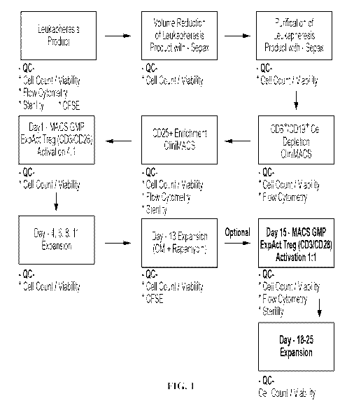
[0076] FIG. 1. Process flow diagram for an exemplary process of Treg
isolation, enrichment
and ex-vivo expansion.
[0077] FIG. 2A-2K. FIG. 2A: graphic depicting the two EV populations (mixed
Treg-
derived EVs and enriched or pure Treg EVs) which were generated, and
references to which
populations are utilized in experiments depicted in FIGS. 2B-2K. The mixed EV
population
obtained from Treg cultures were produced using the improved Treg ex-vivo
expansion protocol
described in Example 1. The Tregs were obtained from ALS patients and the
culture medium
utilized during this expansion process contains 5% Human AB serum. Thus, the
anti-
inflammatory EVs isolated from this culture are present together with EVs the
media serum. It is
estimated that the Treg-derived anti-inflammatory EV population is
approximately 20-30% of
the total EV population. The second Treg population was collected from healthy
patient samples
and expanded using the improved Treg ex-vivo expansion protocol described in
Example 1, but
29
CA 03210656 2023-08-02
WO 2022/183047 PCT/US2022/017990
using culture medium containing exosome-depleted fetal bovine serum (FBS)
instead of human
AB serum. Thus, the anti-inflammatory EV population produced from this culture
constitutes a
pure batch of EVs derived from ex-vivo expanded human Tregs. FIGS. 2B-2F and
2I-2K: The
experiments utilized EV populations isolated using PEG. FIGS. 2G-2H: The
experiments
utilized EV populations isolated using tangential flow filtration (TFF). FIG.
2B: Treg mixed
EVs reduce iPSC-derived M1 IL-6 protein by ¨70% following co-culture of lx108
Treg EVs per
50,000 M1 cells stimulated with LPS/IFNy overnight. FIG. 2C: The mixed Treg
EVs are able to
suppress Tresp proliferation at escalated dosing. FIG. 2D: The pure Treg EV
batches
demonstrate the ability to suppress IL-6 transcript. FIG. 2E: Pure Treg EVs
suppress M1 11-6
protein following overnight stimulation. FIG. 2F: Pure Treg EVs suppress Tresp
proliferation at
escalated dosing. FIG. 2G: Mixed Treg EVs were shown to be able to suppress M1
IL-6 protein
production regardless of whether isolation was performed via PEG precipitation
or TFF (n=3; the
PEG and TFF isolation protocols were performed on expanded Tregs from the same
three
patients from the clinical trial) FIG. 2H: Mixed Treg EVs were shown to be
able to suppress
Tresp proliferation regardless of whether isolation was performed via PEG
precipitation or TFF
(n=3; the PEG and TFF isolation protocols were performed on expanded Tregs
from the same
three patients). FIG. 21: Exemplary size profile of Treg EV produced and as
described in this
example, which demonstrates a single peak distribution within a 20-200nm. FIG.
2J: Graph
depicting Miltenyi MACSPlex Exosome Kit (Miltenyi Biotec) analysis of Treg
(mixed) EVs and
media EVs. FIG. 2K: Graph depicting Miltenyi MACSPlex Exosome KIT (Miltenyi
Biotec)
analysis of Treg (mixed) EVs and media EVs. ALS Treg EVs n=7; media EVs n=3.
Numbers
shown as averages +/- SEM with analysis via one-way ANOVA with Tukey's post
hoc testing.
** indicates a p-value of less than 0.01; *** indicates a p-value of less than
0.001.
[0078] FIG. 3A-3D. The anti-inflammatory effects of Treg EVs were evaluated
in an LPS-
induced neuroinflammation model. Briefly, 2mg/kg LPS were injected
intraperitoneally. Two
hours after the injection, pure Treg EVs were administered intranasally. Brain
regions and
spleen CD11b+ myeloid cells were isolated following 12 hours post-intranasal
administration.
Pro-inflammatory transcripts were analyzed to assess anti-inflammatory
effects. FIG. 3A:
Graphic describing the LPS-induced neuroinflammation model and Treg EV
treatment paradigm.
FIG. 3B: Intranasal Treg EVs reduce IL-6 and IL-10 transcripts in the
hippocampus. FIG. 3C:
Reduced IL-6 transcripts following intranasal Treg EV treatment in the cortex
of mice. FIG. 3D:
CA 03210656 2023-08-02
WO 2022/183047 PCT/US2022/017990
Reduction in peripheral myeloid cell activation following intranasal treatment
of Treg EVs;
demonstrates reduced IL-6 and TNF transcripts in spleen-derived, CD11b+
myeloid cells. P-
values are *p<0.05 and **p<0.01.
[0079] FIG. 4A-4F. Treg EVs were given every two weeks in SOD1 mice
intranasally
starting at day 90 (approximately 20 days after symptoms start to manifest in
this model) to
assess the mouse clinical benefit of multiple rounds of intranasal Treg EVs.
After the sacrifice of
the mouse, inflammatory markers in the inflamed lumbar section of the spinal
cord were
assessed through RNA analysis. FIG. 4A: Graphic depicting the intranasal Treg
EV treatment
paradigm for the SOD1 mouse model of ALS. FIG. 4B: Intranasal Treg EV
treatment increases
the probability of survival compared to intranasal PBS treatments. FIG. 4C:
The Treg EV
treatments slowed the progression of disease as defined by a modified scoring
system used to
assess mouse ALS progression; the effects were more prominent when the mice
were going
through the rapid phase of their disease progression. FIG. 4D: The treatment
significantly
prolonged disease duration. FIG. 4E: The average lifespan is increased in
animals who received
the Treg EV treatment. FIG. 4F: Lumbar spinal cords were dissected after
animals reached their
ethical endpoints. RNA analysis was done using the lumbar spinal cord tissue
to examine
inflammatory markers. Decreased levels of inflammatory markers were observed
in the spinal
cord, while increased signals of Tregs (FOXP3) and anti-inflammatory M2
macrophages
(CD206) were observed in the treated animals. Numbers shown as averages +/-
SEM and with
one-way ANOVA analysis wit hTukey's post hoc test (PBS n=3, LPS+PBS n=4,
LPS+Treg EV
n=4, Trey EV only n=3. * indicates a p-value of less than 0.05; *** indicates
a p-value of less
than 0.001.
[0080] FIG. 5A-5C. FIG. 5A: Treg EVs were able to suppress M1 pro-
inflammatory IL-6
protein by 46% at a dose of 1x10⁸ EVs and 30.6% at a dose of 1x10⁷ EV compared
to MSC EVs
that suppressed 13.7% and 3.3%, respectively. FIG. 5B: Treg EVs suppressed Ml-
derived pro-
inflammatory IL-8 protein by 60% at a dose of lx108 and 50% at a dose of ix i0
dose compared
to MSC EV that showed a 20% suppression at the dose of lx108 dose. "Control
exo" in FIG. 5A
and 5B: EVs derived from non-exosome depleted media without cell culture. FIG.
5C: Treg
EVs suppressed T cell proliferation more than MSC EVs in a comparison study.
[0081] FIG. 6A-6C. Treg EV stability and function were evaluated after 1 to
20 freeze/thaw
cycles and after storage at -20 C for 3 months, 6 months, or 12 months. FIG.
6A: No loss in
31
CA 03210656 2023-08-02
WO 2022/183047 PCT/US2022/017990
Treg EV particle number was observed following multiple (up to 20) freeze/thaw
cycles. FIG.
6B: No significant deviation in Treg EV particle size was observed during the
same freeze/thaw
cycles. FIG. 6C: Treg EV suppression of T cell proliferation did not decrease
over time in
frozen -20 C storage.
[0082] FIG. 7A-7B. FIG. 7A: Treg EV concentrations (EV particles/ml media)
after EV
isolation from expansion media. EV concentration from media alone ("Media") is
also shown.
FIG. 7B: Fold-increase in EVs from ex vivo-expanded Treg cell populations
compared to EVs
from media alone.
[0083] FIG. 8.Exemplary size profile of Treg EVs isolated using a TFF
protocol. The
profile shows an EV mean of 92.1 nm 4.2 nm and a mode of 73.3 nm 6.1 nm.
[0084] FIG. 9A-9B. FIG. 9A: Treg EVs derived from ex vivo-expanded ALS
patient Tregs
induce M1 cells to increase Argl mRNA expression in an EV concentration-
dependent manner.
FIG. 9B: Treg EVs derived from ex vivo-expanded ALS patient Tregs induce M1
cells to
increase CD206 mRNA expression in an EV concentration-dependent manner.
[0085] FIG. 10A-10C. FIG. 10A: Treg EVs were shown to be significantly more
potent
than MSC EVs in suppressing M1 pro-inflammatory IL-6 protein production (n=3
for each
group; *** indicates a p-value of less than 0.001, compared to the
corresponding MSC EVs).
FIG. 10B: Treg EVs were shown to be significantly more potent than MSC EVs in
suppressing T
cell proliferation (n=3 for each group; *** indicates a p-value of less than
0.001, compared to the
corresponding MSC EVs). FIG. 10C: Treg EVs were shown to be significantly more
potent than
MSC EVs in suppressing M1 pro-inflammatory IL-8 protein production (n=3 for
each group;
*** indicates a p-value of less than 0.001, compared to the corresponding MSC
EVs). The Treg
EVs utilized for these experiments were pure Treg EVs and the MSC EVs utilized
for these
experiments were pure MSC EVs.
[0086] FIG. 11A-11B. FIG. 11A: The mean of particle size of EVs isolated
via TFF.
FIG. 11B: The mode of particle size of EVs isolated via TFF.
[0087] FIG. 12. Recovery of EVs isolated via TFF (n=6, results reported as
mean SD).
[0088] FIG. 13. A flow chart of a process of producing a population of
Tregs in a bioreactor.
[0089] FIG. 14A-14F. FIG. 14A: Graphic describing the LPS-induced model of
acute
inflammation where WT mice are administered LPS via intraperitoneal injection
and
subsequently treated with single tail vein (IV) injections of different doses
of Treg EVs.
32
CA 03210656 2023-08-02
WO 2022/183047 PCT/US2022/017990
Following overnight treatment, mice were sacrificed and peripheral immune
cells were isolated
from the mice spleens for subsequence inflammatory transcript analysis. FIG.
14B: Graphic
describing the LPS-induced model of acute inflammation where WT mice are
administered LPS
via intraperitoneal injection and subsequently treated with single tail vein
(IV) injections of
different doses of Treg EVs. Following overnight treatment, mice were
sacrificed, brain tissue
(hippocampus and cortex) was isolated and neuroinflammatory marker transcript
analysis was
performed. FIG. 14C: Graphs depicting pro-inflammatory transcript fold changes
for IL6 and
iNOS in CD11b+ myeloid cells from the spleen following IV treatment of Treg
EVs. FIG. 14D:
Graphs depicting pro-inflammatory transcript fold changes for IL lb and IFNy
in CD11b+
myeloid cells from the spleen following IV treatment of Treg EVs. FIG. 14E:
Graphs depicting
fold changes of anti-inflammatory associated transcripts of CD206 (MRC1) and
CD163 in
CD11b+ myeloid cells following treatment with Treg EVs. FIG. 14F: Graphs
depict fold
changes in FOXP3 and IL2RA (CD25) in fresh spleen isolated CD4+CD25+ Tregs
following
Treg EV treatment. Data shown as averages SEM and statistical analysis done
with one-way
ANOVA analysis with Tukey's post hoc test (PBS n=5, LPS n=5, LPS+Treg EV lx109
n=5
(peripheral tissue), 4-5 (brain tissue), LPS+Treg EV lx101 n=5 (peripheral
tissue), 4-5 (brain
tissue), LPS+Treg EV lx1011 n=5 (peripheral tissue), 4-5 (brain tissue)). p-
values are *p <0.05,
**p <0.01 and ***p<0.001.
[0090] FIG. 15A-15B. FIG. 15A: Graphs depict fold changes in IL-6, ILlb,
and TNF RNA
in the hippocampus following IV Treg EV treatment. FIG. 15B: Graphs depict
fold changes in
IL-6, ILlb, and TNF in the cortex following Treg EV treatment. Data shown as
averages SEM
and with one-way ANOVA analysis with Tukey's post hoc testing (PBS n=5, LPS
n=5,
LPS+Treg EV 1x10⁹ n=4-5, LPS+Treg EV lx101 n=4-5, LPS+Treg EV lx1011 n=5).
[0091] FIG. 16A-16B. FIG. 16A: Size distribution of TFF isolated Treg EVs
generated via
nanoparticle tracking analysis. FIG 16B: Particle size data from TFF isolated
Treg EVs
generated via nanoparticle tracking analysis.
[0092] FIG. 17. Treg EVs suppression of T cell proliferation in vitro
(n=6).
[0093] FIG. 18. Quantification of Treg functional proteins in Treg EVs
using ELISA.
[0094] FIG. 19A-19B. Quantification of residual IL2 and albumin
concentrations as a
percent of original total amount, in the concentrated batch following TFF, and
in final exemplary
dose formulation.
33
CA 03210656 2023-08-02
WO 2022/183047 PCT/US2022/017990
[0095] FIG. 20A-20E. Fig. 20A: Stability of Treg EVs at room temperature
and 4 C.
Presented are the aggregate of results using Treg EVs from bioreactor runs
BioR4-6 through 8
hour timepoints and BioR5 and BioR6 for all timepoints. Fig. 20B: Stability of
Treg EVs at
room temperature and 4 C, showing the breakout of the individual sample
results that were
aggregated into the result shown in FIG. 20A. Fig. 20C: Stability of particle
size distribution of
Treg EVs at room temperature and 4 C. As with FIG. 20A, presented are the
aggregate of results
using Treg EVs from bioreactor runs BioR4-6 through 8 hour timepoints and
BioR5 and BioR6
for all timepoints. FIG. 20D: Stability of Treg EVs after prolonged storage at
-20 C and -80 C.
For each of the timepoints, n=3 (Treg EVs from BioR4-6). FIG. 20E: Stability
of particle size
distribution of Treg EVs after prolonged storage at -20 C and -80 C. For each
of the timepoints,
n=3 (Treg EVs from BioR4-6).
[0096] FIG. 21. Ability of Treg EVs to suppress activated, pro-inflammatory
M1 cells. **
indicates a p-value of less than 0.01; *** indicates a p-value of less than
0.001.
[0097] FIG. 22. Signature of EV surface proteins. Top panel: Bioreactor TFF
Treg EVs.
Bottom panel: Media EVs.
[0098] FIG. 23. Signature of ALS patient Treg EV surface proteins.
[0099] FIG. 24A-24B. Graphs depicting levels of exosome markers associated
with ALS
expanded Treg EVs and Control expanded Treg EVs (n=3 for ALS expanded Treg EVs
and n=6
for Control Expanded Treg EVs).
5. DETAILED DESCRIPTION
[00100] Described herein are anti-inflammatory EV populations derived from ex
vivo-
expanded human suppressive immune cells, for example regulatory T cells
(Tregs). The EVs
presented herein exhibit impressive anti-inflammatory activity, both in vitro
and in vivo. For
example, results presented herein demonstrate that the EVs of the present
disclosure are able to
potently suppress T responder cell proliferation and pro-inflammatory myeloid,
e.g.,
macrophage, activity in vitro, and also exert potent anti-inflammatory effects
in vivo. Briefly,
results presented herein demonstrate that the EVs are able to suppress brain
and peripheral
inflammation in an in vivo model of neuroinflammation, and are also able to
suppress
inflammation, extend survival and slow later stage disease progression in vivo
model of
amyotrophic lateral sclerosis (ALS). The anti-inflammatory EVs exhibit
suppressive effects on
34
CA 03210656 2023-08-02
WO 2022/183047 PCT/US2022/017990
pro-inflammatory responses via either intravenous or intranasal
administration. The results
presented herein demonstrate that the Treg EVs have a greater suppressive
effect on pro-
inflammatory immune cells than EVs derived from mesenchymal stem cells (MSCs).
[00101] Moreover, the anti-inflammatory EVs presented herein exhibit
remarkable batch-to-
batch consistency in size, stability and activity and exhibit a unique
structural signature as, for
example, characterized by Treg EV surface marker and RNA profiles. Still
further, as
demonstrated herein, the methods presented herein yield potent anti-
inflammatory EVs
exhibiting similar structural and suppressive activity characteristics whether
the original Treg
starting material is obtained from healthy subjects or ALS patients.
[00102] Without wishing to be bound by theory or mechanism, it appears that
EVs of the
present disclosure retain the immune suppressive activities of the cells from
which they are
derived. Moreover, as EVs are not themselves cells, they avoid potential cell-
based issues such
as immune rejection and the possibility of polarization to a pro-inflammatory
cell type. As such,
the anti-inflammatory EVs presented herein are particularly useful for
treatment of a variety of
diseases such as, for example, neurodegenerative disorders such as amyotrophic
lateral sclerosis
(ALS).
[00103] Presented herein are isolated, cell-free populations of anti-
inflammatory EVs,
wherein the anti-inflammatory EVs are derived from ex vivo-expanded human
suppressive
immune cells, for example regulatory T cells (Tregs). Also presented herein
are pharmaceutical
compositions and cryopreserved compositions comprising an isolated, cell-free
population of
anti-inflammatory EVs described herein, methods of producing the EV
populations and methods
of using the EVs for treatment of diseases, such as neurodegenerative
diseases, e.g., ALS.
[00104] Recitation of ranges of values herein are merely intended to serve as
a shorthand
method of referring individually to each separate value falling within the
range, unless otherwise
indicated herein, and each separate value is incorporated into the
specification as if it were
individually recited herein.
[00105] Unless specifically stated or apparent from context, as used herein,
the terms "a",
"an", and "the" are understood to be singular or plural, and denote "one or
more."
[00106] The terms "include," "such as," and the like are intended to convey
inclusion without
limitation, unless otherwise specifically indicated.
[00107] The terms "or" and "and" can be used interchangeably and can be
understood to mean
CA 03210656 2023-08-02
WO 2022/183047 PCT/US2022/017990
"and/or."
[00108] The description herein of any aspect or embodiment of the invention
using terms such
as "comprising", "having", "including" or "containing" with reference to an
element or elements
is intended to provide support for a similar aspect or embodiment of the
invention that "consists
of', "consists essentially of', or "substantially comprises" that particular
element or elements,
unless otherwise stated or clearly contradicted by context (e.g., a
composition described herein as
comprising a particular element should be understood as also describing a
composition
consisting of that element, unless otherwise stated or clearly contradicted by
context).
[00109] The terms "about" and "approximately" as used herein, are
interchangeable, and
should generally be understood to refer to a range of numbers around a given
number, as well as
to all numbers in a recited range of numbers (e.g., "about 5 to 15" means
"about 5 to about 15"
unless otherwise stated). Moreover, all numerical ranges herein should be
understood to include
each whole integer within the range. In particular, unless otherwise noted the
terms mean within
plus or minus 10% of a given value or range. In instances where an integer is
required, the terms
mean within plus or minus 10% of a given value or range, rounded either up or
down to the
nearest integer.
5.1 Anti-Inflammatory Extracellular Vesicles (EVs)
[00110] Presented herein are isolated, cell-free population of anti-
inflammatory extracellular
vesicles (EVs), wherein the anti-inflammatory EVs are derived from ex vivo-
expanded human
suppressive immune cells. In certain embodiments, the anti-inflammatory EVs
are derived from
human regulatory T cells (Tregs).
[00111] In certain embodiments, an isolated, cell-free population of anti-
inflammatory EVs is
produced by a method described herein, for example, as described in Section
5.2, below.
[00112] For ease of reference, unless otherwise noted, the terms
"extracellular vesicles,"
"EVs", "extracellular vesicle particles," and "EV particles" are used
interchangeable herein.
Moreover, as should be self-evident, it is to be understood that, as used
herein, reference to "anti-
inflammatory extracellular vesicles," "anti-inflammatory EVs," "anti-
inflammatory exosomes,"
and the like, includes the Treg EVs and Treg exosomes described in detail
herein. While, for
ease of description, not every embodiment presented herein is reproduced to
recite, e.g., both
"anti-inflammatory EVs" and "Treg EVs," it is to be understood that each
embodiment reciting
such an "anti-inflammatory" embodiment includes and may be substituted for a
corresponding
36
CA 03210656 2023-08-02
WO 2022/183047 PCT/US2022/017990
"Treg EV" and "Treg exosome" as these are described herein.
[00113] EVs are membrane-bound particles released by cells. Generally, EVs
comprise one
or more constituents from the cells from which they are released, e.g., one or
more DNA, RNA
(e.g., coding and/or non-coding RNA, for example, mRNA microRNA, and/or long
non-coding
RNA), protein (e.g., signaling proteins, receptors, other surface proteins,
glycoproteins and/or
enzymes) or non-protein, e.g., lipid, constituents. EVs generally range in
size from about 30 nm
to about 1000 nm in diameter. Larger EVs are sometimes referred to as
"microvesicles."
Roughly speaking, microvesicles have size diameters larger than about 200 nm.
Smaller EVs are
sometimes referred to as "exosomes." Roughly speaking, exosomes have size
diameters that
range from about 30-40 nm to about 150-200 nm. Methods for determining EV
particle size and
concentration are well known to those of skill in the art.
[00114] Methods for determining EV particle size, concentration and purity are
well known,
including determinations that use dynamic light scattering or single particle
tracking analysis, or
utilize techniques such as flow cytometry, ELISA, or electron microscopy. See,
e.g., Balaj et al.
(2015) Sci Rep 5, 10266, Nakai et al. (2016) Sci Rep 6, 33935 and Camino et
al. (2019)
Respiratory Research 20:240. In a particular embodiment, routine determination
may be
performed using nanoparticle analyzers, e.g., NanoSight (Malvern Panalytical)
nanoparticle
analyzers.
[00115] In some embodiments, EVs may be analyzed for the presence of exosome
markers
and/or Treg markers (e.g., CD25) by protein analysis using Western blot,
ELISA, and other
protein-associated assays, or commercially available arrays such as the
ExoCheckTM Exosome
Antibody Array (System Biosciences) and/or MACSPlex Exosome Kit (Miltenyi
Biotec). In
certain embodiments, a population of EVs may be analyzed for the presence of
proteins
associated with serum. In particular embodiments, an EV population described
herein is
substantially free of proteins associated with serum.
[00116] In certain embodiments, the anti-inflammatory EVs described herein
exhibit an
ability to increase the expression of one or more anti-inflammatory markers in
inflammatory
cells. For example, in particular embodiments, the anti-inflammatory EVs
described herein
exhibit an ability to increase the transcription of and/or level of mRNA
expression of one or
more genes encoding anti-inflammatory protein in inflammatory cells. In
another example, in
particular embodiments, the anti-inflammatory EVs described herein exhibit an
ability to
37
CA 03210656 2023-08-02
WO 2022/183047 PCT/US2022/017990
increase translation, processing, secretion and/or activation of one or more
anti-inflammatory
protein produced by inflammatory cells.
[00117] In specific embodiments, the anti-inflammatory marker is IL-10, Argl,
and/or
CD206. In specific examples, the inflammatory cells are myeloid cells, for
example, monocytes,
macrophages or microglia, e.g., human inflammatory cells, for example, human
monocytes,
macrophages or microglia.
[00118] In certain embodiments, the anti-inflammatory EVs described herein
exhibit an
ability to suppress inflammatory cells. For example, in certain embodiments,
the anti-
inflammatory EVs described herein exhibit an ability to suppress inflammatory
cells as measured
by pro-inflammatory cytokine production by the inflammatory cells.
[00119] In some embodiments, the ability to suppress inflammatory cells is
measured by IL-6,
TNFa, IL1(3, IL8, and/or Interferon-y production by the inflammatory cells. In
some
embodiments, the ability to suppress inflammatory cells is measured by IL-6
production by the
inflammatory cells.
[00120] In particular embodiments, the inflammatory cells are myeloid cells,
for example,
monocytes, macrophages or microglia e.g., human inflammatory cells, for
example, human
myeloid cells, such as human monocytes, macrophages or microglia. In specific
examples, the
myeloid cells, e.g., monocytes, macrophages or microglia, are from human
donors or generated
from induced pluripotent stem cells. In certain embodiments, the macrophages
are M1
macrophages, such as induced pluripotent stem cell (iPSC)-derived M1
macrophages.
[00121] In certain embodiments, the anti-inflammatory EVs described herein
exhibit an
ability to suppress pro-inflammatory M1 cells. In some embodiments, the
ability to suppress
pro-inflammatory M1 cells is measured by IL-6 production by the pro-
inflammatory M1 cells.
[00122] In certain embodiments, the anti-inflammatory EVs described herein
exhibit an
ability to suppress pro-inflammatory M1 cell IL-6 protein production by about
20% to about
70%, about 20% to about 50%, about 25% to about 50%, about 30% to about 50%,
or about 25%
to about 45%.
[00123] In certain embodiments, the anti-inflammatory EVs described herein
exhibit an
ability to suppress inflammatory cells as determined by suppression of
proliferation of responder
T cells. In particular embodiments, the proliferation of responder T cells is
determined by flow
cytometry or thymidine incorporation, e.g., tritiated thymidine incorporation.
38
CA 03210656 2023-08-02
WO 2022/183047 PCT/US2022/017990
[00124] In certain embodiments, the anti-inflammatory EVs described herein
exhibit an
ability to suppress inflammatory cells (e.g., as measured by pro-inflammatory
cytokine
production and/or responder T cell proliferation) and an ability to increase
expression of one or
more inflammatory markers in inflammatory cells.
[00125] In certain aspects, a population of anti-inflammatory EVs as described
herein
comprises exosomes. In other aspects, a population of anti-inflammatory EVs
described herein
comprises microvesicles. In yet other aspects, a population of anti-
inflammatory EVs as
described herein comprises exosomes and microvesicles.
[00126] In certain embodiments, the majority of EVs of a population of anti-
inflammatory
EVs as described herein are exosomes. For example, in certain embodiments at
least about 50%,
at least about 55%, at least about 60%, at least about 65%, at least about
70%, at least about
75%, at least about 80%, at least about 85%, at least about 90%, at least
about 95%, at least
about 98%, at least about 99% or more of the EVs of a population of anti-
inflammatory
exosomes described herein are exosomes.
[00127] In certain embodiments, the majority of EVs of a population of anti-
inflammatory
EVs as described herein are microvesicles. For example, in certain embodiments
at least about
50%, at least about 55%, at least about 60%, at least about 65%, at least
about 70%, at least
about 75%, at least about 80%, at least about 85%, at least about 90%, at
least about 95%, at
least about 98%, at least about 99% or more of the EVs of a population of anti-
inflammatory
exosomes described herein are microvesicles.
[00128] In certain embodiments, the EVs of a population of anti-inflammatory
EVs as
described herein have size diameters of about 5 nm to about 1000 nm. In
certain embodiments,
the EVs of a population of anti-inflammatory EVs as described herein have size
diameters of
about 10 nm to about 1000 nm. In certain embodiments, the EVs of a population
of anti-
inflammatory EVs as described herein have size diameters of about 15 nm to
about 1000 nm. In
certain embodiments, the EVs of a population of anti-inflammatory EVs as
described herein have
size diameters of about 20 nm to about 1000 nm. In certain embodiments, the
EVs of a
population of anti-inflammatory EVs as described herein have size diameters of
about 30 nm to
about 1000 nm. In certain embodiments, the EVs of a population of anti-
inflammatory EVs as
described herein have size diameters of about 20 nm to about 300 nm. In
certain embodiments,
the EVs of a population of anti-inflammatory EVs as described herein have size
diameters of
39
CA 03210656 2023-08-02
WO 2022/183047 PCT/US2022/017990
about 20 nm to about 275 nm. In certain embodiments, the EVs of a population
of anti-
inflammatory EVs as described herein have size diameters of about 20 nm to
about 250 nm. In
certain embodiments, the EVs of a population of anti-inflammatory EVs as
described herein have
size diameters of about 20 nm to about 200 nm. In certain embodiments, the EVs
of a population
of anti-inflammatory EVs as described herein have size diameters of about 20
nm to about 175
nm. In certain embodiments, the EVs of a population of anti-inflammatory EVs
as described
herein have size diameters of about 50 nm to about 200 nm. In certain
embodiments, the EVs of
a population of anti-inflammatory EVs as described herein have size diameters
of about 50 nm to
about 175 nm. In certain embodiments, the EVs of a population of anti-
inflammatory EVs as
described herein have size diameters of about 50 nm to about 150 nm.
[00129] In certain embodiments, the majority (e.g., at least about 50%, at
least about 55%, at
least about 60%, at least about 65%, at least about 70%, at least about 75%,
at least about 80%,
at least about 85%, at least about 90%, at least about 95%, at least about
98%, at least about 99%
or more) of the EVs of a population of anti-inflammatory EVs as described
herein have size
diameters of about 5 nm to about 1000 nm, about 10 nm to about 1000 nm, about
15 nm to about
1000 nm, about 20 nm to about 1000 nm, or about 30 nm to about 1000 nm.
[00130] In certain embodiments, the majority (e.g., at least about 50%, at
least about 55%, at
least about 60%, at least about 65%, at least about 70%, at least about 75%,
at least about 80%,
at least about 85%, at least about 90%, at least about 95%, at least about
98%, at least about 99%
or more) of the EVs of a population of anti-inflammatory EVs as described
herein have size
diameters of about 20 nm to about 300 nm, about 20 nm to about 275 nm, about
20 nm to about
250 nm, about 20 nm to about 200 nm, or about 20 nm to about 175 nm.
[00131] In certain embodiments, the majority (e.g., at least about 50%, at
least about 55%, at
least about 60%, at least about 65%, at least about 70%, at least about 75%,
at least about 80%,
at least about 85%, at least about 90%, at least about 95%, at least about
98%, at least about 99%
or more) of the EVs of a population of anti-inflammatory EVs as described
herein have size
diameters of about 20 nm to about 200 nm.
[00132] In certain embodiments, the majority (e.g., at least about 50%, at
least about 55%, at
least about 60%, at least about 65%, at least about 70%, at least about 75%,
at least about 80%,
at least about 85%, at least about 90%, at least about 95%, at least about
98%, at least about 99%
or more) of the EVs of a population of anti-inflammatory EVs as described
herein have size
CA 03210656 2023-08-02
WO 2022/183047 PCT/US2022/017990
diameters of about 50 nm to about 200 nm, about 50 nm to about 175 nm, or
about 50 nm to
about 150 nm.
[00133] In certain embodiments, the majority (e.g., at least about 50%, at
least about 55%, at
least about 60%, at least about 65%, at least about 70%, at least about 75%,
at least about 80%,
at least about 85%, at least about 90%, at least about 95%, at least about
98%, at least about 99%
or more) of the EVs of a population of anti-inflammatory EVs as described
herein have size
diameters of about 50 nm to about 150 nm.
[00134] In certain embodiments, the majority of EVs of a population of anti-
inflammatory
EVs as described herein have size diameters less than about 300 nm, less than
about 200 nm, less
than about 150 nm or less than about 100 nm. For example, in certain
embodiments at least
about 50%, at least about 55%, at least about 60%, at least about 65%, at
least about 70%, at
least about 75%, at least about 80%, at least about 85%, at least about 90%,
at least about 95%,
at least about 98%, at least about 99% or more of the EVs of a population of
anti-inflammatory
exosomes described herein have size diameters less than about 300 nm, less
than about 200 nm,
less than about 150 nm, or less than about 100 nm.
[00135] In certain embodiments, the EVs of a population of anti-inflammatory
EVs as
described herein have size diameters of about 30 nm to about 300 nm, about 30
nm to about 250
nm, about 30 nm to about 200 nm, about 30 nm to about 160 nm, about 30 nm to
about 150 nm,
about 30 nm to about 100 nm, about 40 nm to about 300 nm, about 40 nm to about
200 nm,
about 40 nm to about 160 nm, about 40 nm to about 150 nm, about 40 nm to about
100 nm,
about 60 nm to about 300 nm, about 60 nm to about 200 nm, about 60 nm to about
160 nm,
about 60 nm to about 150 nm, about 60 nm to about 125 nm, about 60 nm to about
110 nm,
about 60 nm to about 100 nm, about 60 nm to about 80 nm, about 70 nm to about
300 nm, about
70 nm to about 200 nm, about 70 nm to about 160 nm, about 70 nm to about 150
nm, about 70
nm to about 125 nm, about 70 nm to about 110 nm, about 70 nm to about 100 nm,
about 80 nm
to about 300 nm, about 80 nm to about 200 nm, about 80 nm to about 160 nm,
about 80 nm to
about 150 nm, about 80 nm to about 125 nm, about 80 nm to about 110 nm, about
80 nm to
about 100 nm, or about 110 nm to about 120 nm.
[00136] In certain embodiments, the majority (e.g., at least about 50%, at
least about 55%, at
least about 60%, at least about 65%, at least about 70%, at least about 75%,
at least about 80%,
at least about 85%, at least about 90%, at least about 95%, at least about
98%, at least about 99%
41
CA 03210656 2023-08-02
WO 2022/183047 PCT/US2022/017990
or more) of the EVs of a population of anti-inflammatory EVs as described
herein have size
diameters of about 30 nm to about 300 nm, about 30 nm to about 250 nm, about
30 nm to about
200 nm, about 30 nm to about 160 nm, about 30 nm to about 150 nm, about 30 nm
to about 100
nm, about 40 nm to about 300 nm, about 40 nm to about 200 nm, about 40 nm to
about 160 nm,
about 40 nm to about 150 nm, about 40 nm to about 100 nm, about 60 nm to about
300 nm,
about 60 nm to about 200 nm, about 60 nm to about 160 nm, about 60 nm to about
150 nm,
about 60 nm to about 125 nm, about 60 nm to about 110 nm, about 60 nm to about
100 nm,
about 60 nm to about 80 nm, about 70 nm to about 300 nm, about 70 nm to about
200 nm, about
70 nm to about 160 nm, about 70 nm to about 150 nm, about 70 nm to about 125
nm, about 70
nm to about 110 nm, about 70 nm to about 100 nm, about 80 nm to about 300 nm,
about 80 nm
to about 200 nm, about 80 nm to about 160 nm, about 80 nm to about 150 nm,
about 80 nm to
about 125 nm, about 80 nm to about 110 nm, about 80 nm to about 100 nm, or
about 110 nm to
about 120 nm.
[00137] In certain embodiments, the majority (e.g., at least about 50%, at
least about 55%, at
least about 60%, at least about 65%, at least about 70%, at least about 75%,
at least about 80%,
at least about 85%, at least about 90%, at least about 95%, at least about
98%, at least about 99%
or more) of the EVs of a population of anti-inflammatory EVs as described
herein have a size
diameter of about 30 nm, about 40 nm, about 50 nm, about 60 nm, about 65 nm,
about 70 nm,
about 75 nm, 80 nm, about 85 nm, about 90 nm, about 95 nm, about 100 nm, about
110 nm to
about 120 nm, about 150 nm, about 175 nm, about 200 nm, about 250 nm or about
300 nm.
[00138] In certain embodiments, the majority (e.g., at least about 50%, at
least about 55%, at
least about 60%, at least about 65%, at least about 70%, at least about 75%,
at least about 80%,
at least about 85%, at least about 90%, at least about 95%, at least about
98%, at least about 99%
or more) of the EVs of a population of anti-inflammatory EVs as described
herein have size
diameters greater than about 300 nm, greater than about 400 nm, greater than
about 500 nm,
greater than about 500 nm, greater than about 700 nm, or greater than about
800 nm.
[00139] In certain embodiments, the EVs of a population of anti-inflammatory
EVs as
described herein have size diameters of about 200 nm to about 1000 nm, about
300 nm to about
1000 nm, about 400 nm to about 1000 nm, about 500 nm to about 1000 nm, about
600 nm to
about 1000 nm, about 700 nm to about 1000 nm, about 800 nm to about 1000 nm,
about 200 nm
to about 800 nm, about 300 nm to about 800 nm, about 400 nm to about 800 nm,
about 500 nm
42
CA 03210656 2023-08-02
WO 2022/183047 PCT/US2022/017990
to about 800 nm, about 600 nm to about 800 nm, about 200 nm to about 600 nm,
about 300 nm
to about 600 nm, about 400 nm to about 600 nm, about 200 nm to about 500 nm,
or about 300
nm to about 500 nm.
[00140] In certain embodiments, the majority (e.g., at least about 50%, at
least about 55%, at
least about 60%, at least about 65%, at least about 70%, at least about 75%,
at least about 80%,
at least about 85%, at least about 90%, at least about 95%, at least about
98%, at least about 99%
or more) of EVs of a population of anti-inflammatory EVs as described herein
have size
diameters of about 200 nm to about 1000 nm, about 300 nm to about 1000 nm,
about 400 nm to
about 1000 nm, about 500 nm to about 1000 nm, about 600 nm to about 1000 nm,
about 700 nm
to about 1000 nm, about 800 nm to about 1000 nm, about 200 nm to about 800 nm,
about 300
nm to about 800 nm, about 400 nm to about 800 nm, about 500 nm to about 800
nm, about 600
nm to about 800 nm, about 200 nm to about 600 nm, about 300 nm to about 600
nm, about 400
nm to about 600 nm, about 200 nm to about 500 nm, or about 300 nm to about 500
nm.
[00141] In certain embodiments, the majority (e.g., at least about 50%, at
least about 55%, at
least about 60%, at least about 65%, at least about 70%, at least about 75%,
at least about 80%,
at least about 85%, at least about 90%, at least about 95%, at least about
98%, at least about 99%
or more) of the EVs of a population of anti-inflammatory EVs as described
herein have a size
diameter of about 400 nm, about 450 nm, about 500 nm, about 600 nm, about 650
nm, about 700
nm, about 750 nm, 800 nm, about 850 nm, about 900 nm, about 950 nm, or about
1000 nm.
[00142] In certain embodiments, the EVs of a population of anti-inflammatory
EVs as
described herein have a mean size diameter of about 30 nm to about 1000 nm. In
certain
embodiments, the EVs of a population of anti-inflammatory EVs as described
herein have a
mean size diameter of less than about 300 nm, less than about 200 nm, less
than about 150 nm or
less than about 100 nm. In certain embodiments, the EVs of a population of
anti-inflammatory
EVs as described herein have a mean size diameter of about 30 nm to about 300
nm, about 30
nm to about 250 nm, about 30 nm to about 200 nm, about 30 nm to about 160 nm,
about 30 nm
to about 150 nm, about 30 nm to about 100 nm, about 40 nm to about 300 nm,
about 40 nm to
about 200 nm, about 40 nm to about 160 nm, about 40 nm to about 150 nm, about
40 nm to
about 100 nm, about 60 nm to about 300 nm, about 60 nm to about 200 nm, about
60 nm to
about 160 nm, about 60 nm to about 150 nm, about 60 nm to about 125 nm, about
60 nm to
about 110 nm, about 60 nm to about 100 nm, about 60 nm to about 80 nm, about
70 nm to about
43
CA 03210656 2023-08-02
WO 2022/183047 PCT/US2022/017990
300 nm, about 70 nm to about 200 nm, about 70 nm to about 160 nm, about 70 nm
to about 150
nm, about 70 nm to about 125 nm, about 70 nm to about 110 nm, about 70 nm to
about 100 nm,
about 80 nm to about 300 nm, about 80 nm to about 200 nm, about 80 nm to about
160 nm,
about 80 nm to about 150 nm, about 80 nm to about 125 nm, about 80 nm to about
110 nm,
about 80 nm to about 100 nm, or about 110 nm to about 120 nm.
[00143] In certain embodiments, the EVs of a population of anti-inflammatory
EVs as
described herein have a mean size diameter of about 30 nm, about 40 nm, about
50 nm, about 60
nm, about 65 nm, about 70 nm, about 75 nm, 80 nm, about 85 nm, about 90 nm,
about 95 nm,
about 100 nm, about 110 nm to about 120 nm, about 150 nm, about 175 nm, about
200 nm, about
250 nm or about 300 nm.
[00144] In certain embodiments, the EVs of a population of anti-inflammatory
EVs as
described herein have a mean size diameter of about 80 nm to about 110 nm,
about 80 nm to
about 100 nm, about 80 to about 95 nm, about 80-90 nm, about 85 nm to about
110 nm, about 85
nm to about 100 nm, about 85 to about 95 nm, about 90 nm to about 110 nm,
about 90 nm to
about 100 nm, or about 90 to about 95 nm.
[00145] In certain embodiments, the EVs of a population of anti-inflammatory
EVs as
described herein have a mean size diameter greater than about 300 nm, greater
than about 400
nm, greater than about 500 nm, greater than about 500 nm, greater than about
700 nm, or greater
than about 800 nm. In certain embodiments, the EVs of a population of anti-
inflammatory EVs
as described herein have a mean size diameter of about 200 nm to about 1000
nm, about 300 nm
to about 1000 nm, about 400 nm to about 1000 nm, about 500 nm to about 1000
nm, about 600
nm to about 1000 nm, about 700 nm to about 1000 nm, about 800 nm to about 1000
nm, about
200 nm to about 800 nm, about 300 nm to about 800 nm, about 400 nm to about
800 nm, about
500 nm to about 800 nm, about 600 nm to about 800 nm, about 200 nm to about
600 nm, about
300 nm to about 600 nm, about 400 nm to about 600 nm, about 200 nm to about
500 nm, or
about 300 nm to about 500 nm.
[00146] In certain embodiments, the EVs of a population of anti-inflammatory
EVs as
described herein have a mean size diameter of about 400 nm, about 450 nm,
about 500 nm, about
600 nm, about 650 nm, about 700 nm, about 750 nm, 800 nm, about 850 nm, about
900 nm,
about 950 nm, or about 1000 nm. In certain embodiments, the EVs of a
population of anti-
inflammatory EVs as described herein have a mean size diameter of about 80nm
to about 110
44
CA 03210656 2023-08-02
WO 2022/183047 PCT/US2022/017990
nm. In certain embodiments, the EVs of a population of anti-inflammatory EVs
as described
herein have a mean size diameter of about 85nm to about 100 nm. In certain
embodiments, the
EVs of a population of anti-inflammatory EVs as described herein have a mean
size diameter of
about 80nm to about 100 nm. In certain embodiments, the EVs of a population of
anti-
inflammatory EVs as described herein have a mean size diameter of about 85nm
to about 95 nm.
[00147] In certain embodiments, the EVs of a population of anti-inflammatory
EVs as
described herein have a median size diameter of about 30 nm to about 1000 nm.
In certain
embodiments, the EVs of a population of anti-inflammatory EVs as described
herein have a
median size diameter of less than about 300 nm, less than about 200 nm, less
than about 150 nm
or less than about 100 nm. In certain embodiments, the EVs of a population of
anti-
inflammatory EVs as described herein have a median size diameter of about 30
nm to about 300
nm, about 30 nm to about 250 nm, about 30 nm to about 200 nm, about 30 nm to
about 160 nm,
about 30 nm to about 150 nm, about 30 nm to about 100 nm, about 40 nm to about
300 nm,
about 40 nm to about 200 nm, about 40 nm to about 160 nm, about 40 nm to about
150 nm,
about 40 nm to about 100 nm, about 60 nm to about 300 nm, about 60 nm to about
200 nm,
about 60 nm to about 160 nm, about 60 nm to about 150 nm, about 60 nm to about
125 nm,
about 60 nm to about 110 nm, about 60 nm to about 100 nm, about 60 nm to about
80 nm, about
70 nm to about 300 nm, about 70 nm to about 200 nm, about 70 nm to about 160
nm, about 70
nm to about 150 nm, about 70 nm to about 125 nm, about 70 nm to about 110 nm,
about 70 nm
to about 100 nm, about 75 nm to about 100 nm, about 75 nm to about 195 nm,
about 75 nm to
about 90 nm, about 75 nm to about 85 nm, about 80 nm to about 300 nm, about 80
nm to about
200 nm, about 80 nm to about 160 nm, about 80 nm to about 150 nm, about 80 nm
to about 125
nm, about 80 nm to about 110 nm, about 80 nm to about 100 nm, about 80 nm to
about 95 nm,
about 85 nm to about 95 nm, or about 110 nm to about 120 nm. In certain
embodiments, the
EVs of a population of anti-inflammatory EVs as described herein have a median
size diameter
of about 30 nm to about 1000 nm.
[00148] In certain embodiments, the EVs of a population of anti-inflammatory
EVs as
described herein have a median size diameter of about 70 nm to about 100 nm.
In certain
embodiments, the EVs of a population of anti-inflammatory EVs as described
herein have a
median size diameter of about 75 nm to about 100 nm. In certain embodiments,
the EVs of a
population of anti-inflammatory EVs as described herein have a median size
diameter of about
CA 03210656 2023-08-02
WO 2022/183047 PCT/US2022/017990
75 nm to about 95 nm.
[00149] In certain embodiments, the EVs of a population of anti-inflammatory
EVs as
described herein have a median size diameter of about 30 nm, about 40 nm,
about 50 nm, about
60 nm, about 65 nm, about 70 nm, about 75 nm, 80 nm, about 85 nm, about 90 nm,
about 95 nm,
about 100 nm, about 110 nm to about 120 nm, about 150 nm, about 175 nm, about
200 nm, about
250 nm or about 300 nm.
[00150] In certain embodiments, the EVs of a population of anti-inflammatory
EVs as
described herein have a median size diameter greater than about 300 nm,
greater than about 400
nm, greater than about 500 nm, greater than about 500 nm, greater than about
700 nm, or greater
than about 800 nm. In certain embodiments, the EVs of a population of anti-
inflammatory EVs
as described herein have a median size diameter of about 200 nm to about 1000
nm, about 300
nm to about 1000 nm, about 400 nm to about 1000 nm, about 500 nm to about 1000
nm, about
600 nm to about 1000 nm, about 700 nm to about 1000 nm, about 800 nm to about
1000 nm,
about 200 nm to about 800 nm, about 300 nm to about 800 nm, about 400 nm to
about 800 nm,
about 500 nm to about 800 nm, about 600 nm to about 800 nm, about 200 nm to
about 600 nm,
about 300 nm to about 600 nm, about 400 nm to about 600 nm, about 200 nm to
about 500 nm,
or about 300 nm to about 500 nm.
[00151] In certain embodiments, the EVs of a population of anti-inflammatory
EVs as
described herein have a median size diameter of about 400 nm, about 450 nm,
about 500 nm,
about 600 nm, about 650 nm, about 700 nm, about 750 nm, 800 nm, about 850 nm,
about 900
nm, about 950 nm, or about 1000 nm.
[00152] In certain embodiments, the EVs of a population of anti-inflammatory
EVs as
described herein have a mode size diameter of about 30 nm to about 1000 nm. In
certain
embodiments, the EVs of a population of anti-inflammatory EVs as described
herein have a
mode size diameter of less than about 300 nm, less than about 200 nm, less
than about 150 nm or
less than about 100 nm. In certain embodiments, the EVs of a population of
anti-inflammatory
EVs as described herein have a mode size diameter of about 30 nm to about 300
nm, about 30
nm to about 250 nm, about 30 nm to about 200 nm, about 30 nm to about 160 nm,
about 30 nm
to about 150 nm, about 30 nm to about 100 nm, about 40 nm to about 300 nm,
about 40 nm to
about 200 nm, about 40 nm to about 160 nm, about 40 nm to about 150 nm, about
40 nm to
about 100 nm, about 60 nm to about 300 nm, about 60 nm to about 200 nm, about
60 nm to
46
CA 03210656 2023-08-02
WO 2022/183047 PCT/US2022/017990
about 160 nm, about 60 nm to about 150 nm, about 60 nm to about 125 nm, about
60 nm to
about 110 nm, about 60 nm to about 100 nm, about 60 nm to about 80 nm, about
70 nm to about
300 nm, about 70 nm to about 200 nm, about 70 nm to about 160 nm, about 70 nm
to about 150
nm, about 70 nm to about 125 nm, about 70 nm to about 110 nm, about 70 nm to
about 100 nm,
about 80 nm to about 300 nm, about 80 nm to about 200 nm, about 80 nm to about
160 nm,
about 80 nm to about 150 nm, about 80 nm to about 125 nm, about 80 nm to about
110 nm,
about 80 nm to about 100 nm, or about 110 nm to about 120 nm. In certain
embodiments, the
EVs of a population of anti-inflammatory EVs as described herein have a mode
size diameter of
about 65 nm to about 85 nm, about 65 nm to about 80 nm, about 65 nm to about
75 nm, about 70
nm to about 85 nm, about 70 nm to about 80 nm, or about 70 nm to about 75 nm.
[00153] In certain embodiments, the EVs of a population of anti-inflammatory
EVs as
described herein have a mode size diameter of about 65 nm to about 95 nm. In
certain
embodiments, the EVs of a population of anti-inflammatory EVs as described
herein have a
mode size diameter of about 75 nm to about 85 nm. In certain embodiments, the
EVs of a
population of anti-inflammatory EVs as described herein have a mode size
diameter of about 75
nm to about 85 nm.
[00154] In certain embodiments, the EVs of a population of anti-inflammatory
EVs as
described herein have a mode size diameter of about 30 nm, about 40 nm, about
50 nm, about 60
nm, about 65 nm, about 70 nm, about 75 nm, 80 nm, about 85 nm, about 90 nm,
about 95 nm,
about 100 nm, about 110 nm to about 120 nm, about 150 nm, about 175 nm, about
200 nm, about
250 nm or about 300 nm.
[00155] In certain embodiments, the EVs of a population of anti-inflammatory
EVs as
described herein have a mode size diameter greater than about 300 nm, greater
than about 400
nm, greater than about 500 nm, greater than about 500 nm, greater than about
700 nm, or greater
than about 800 nm. In certain embodiments, the EVs of a population of anti-
inflammatory EVs
as described herein have a mode size diameter of about 200 nm to about 1000
nm, about 300 nm
to about 1000 nm, about 400 nm to about 1000 nm, about 500 nm to about 1000
nm, about 600
nm to about 1000 nm, about 700 nm to about 1000 nm, about 800 nm to about 1000
nm, about
200 nm to about 800 nm, about 300 nm to about 800 nm, about 400 nm to about
800 nm, about
500 nm to about 800 nm, about 600 nm to about 800 nm, about 200 nm to about
600 nm, about
300 nm to about 600 nm, about 400 nm to about 600 nm, about 200 nm to about
500 nm, or
47
CA 03210656 2023-08-02
WO 2022/183047 PCT/US2022/017990
about 300 nm to about 500 nm.
[00156] In certain embodiments, the EVs of a population of anti-inflammatory
EVs as
described herein have a mode size diameter of about 400 nm, about 450 nm,
about 500 nm, about
600 nm, about 650 nm, about 700 nm, about 750 nm, 800 nm, about 850 nm, about
900 nm,
about 950 nm, or about 1000 nm.
[00157] In certain aspects a population of anti-inflammatory EVs as described
herein is a
buffer-containing population of anti-inflammatory EVs. The anti-inflammatory
EVs described
herein are derived from ex vivo-expanded human suppressive immune cells, e.g.,
Tregs. As
explained in detailed below, a population of such anti-inflammatory EVs may be
isolated from a
culture comprising ex vivo expanding human suppressive immune cells, e.g.,
Tregs and culture
media. In certain instances, as part of the process of isolating EVs from the
culture, the culture
media may be replaced with a buffer, for example a sterile buffer, e.g., a
buffer suitable for
administration to a human, such as suitable for administration to a human for
therapeutic use. In
such instances, the resulting isolated, cell-free population of anti-
inflammatory EVs may be
referred to as a buffer-containing population of anti-inflammatory EVs.
[00158] Similarly, in certain embodiments, a population of anti-inflammatory
EVs as
described herein is a saline-containing population of anti-inflammatory EVs.
In particular
embodiments, a population of anti-inflammatory EVs as described herein is a
normal saline-
containing population of anti-inflammatory EVs. In particular embodiments, a
population of
anti-inflammatory EVs as described herein is a 0.9% saline-containing
population of anti-
inflammatory EVs. In particular embodiments, a population of anti-inflammatory
EVs as
described herein is a phosphate-buffer saline-containing population of anti-
inflammatory EVs.
[00159] The isolated, cell-free populations of anti-inflammatory EVs described
herein are
substantially free of cellular material, microparticles or other contaminants
(e.g., organelles,
lipids, cholesterol) from the cell or tissue source from which the EVs are
derived, e.g., from the
human suppressive immune cells, for example Tregs, from which the EVs are
derived. For
example, the isolated, cell-free populations of anti-inflammatory EVs
described herein generally
contain less than about 5 weight percent, less than about 1 weight percent,
less than about 0.5
weight percent, less than about 0.1 weight percent, or less than about 0.01
weight percent of free
of cellular material, microparticles or other contaminants (e.g., organelles,
lipids, cholesterol)
from the cell or tissue source from which the EVs are derived, e.g., from the
human suppressive
48
CA 03210656 2023-08-02
WO 2022/183047 PCT/US2022/017990
immune cells, for example Tregs, from which the EVs are derived.
[00160] In certain embodiments, the isolated, cell-free populations of anti-
inflammatory EVs
described herein are present in a composition that is substantially free of
other EVs. For
example, in certain embodiments, the isolated, cell-free populations of anti-
inflammatory EVs
described herein are present in a composition that contains less than about
20%, less than about
10%, less than about 5%, or less than about 1% other EVs.
[00161] In certain embodiments, an isolated, cell-free population of anti-
inflammatory EVs
described herein is present in a composition that comprises other EVs, wherein
the isolated, cell-
free population of anti-inflammatory EVs makes up about 10%, about 20%, about
25%, about
30%, about 35%, about 40%, about 50%, about 60%, about 70%, about 80%, about
90%, about
95%, or greater than about 95% of the EVs in the composition. In specific
embodiments, the
other EVs are serum EVs, for example, bovine serum EVs or human serum EVs.
[00162] In certain embodiments, the cell-free population of anti-inflammatory
EVs are made
up of anti-inflammatory EVs that comprise one or more cargos, e.g., drugs,
detectable labels,
proteins, nucleic acids, e.g., RNAs such as mRNAs and or miRNAs heterologous
to the EVs.
The cargo or cargos of the resulting anti-inflammatory EVs may be present
within the Evs and/or
present on the EV surface.
[00163] In certain embodiments, the cell-free population of anti-inflammatory
EVs have been
produced from Treg cells that have been genetically engineered, e.g.,
genetically engineered to
express a cargo that is loaded into the EVs as they are produced.
Alternatively, one or more
cargos may, for example, be introduced into the culture media during EV
production process and
loaded into the EVs as the EVs are produced. In yet other alternatives one or
more cargos may
be introduced into the EVs via such techniques as electroporation or
sonication. The cargo or
cargos of the resulting anti-inflammatory EVs may be present within the Evs
and/or present on
the EV surface.
5.1.1. Gene Product Expression Profile
[00164] The populations of anti-inflammatory EVs provided herein may be
characterized by
their gene product expression profiles. For example, if the population of anti-
inflammatory EVs
is derived from a population of Tregs cultured in a serum-containing medium
that contains serum
EVs (also referred to as "media EVs"), the gene product expression profile of
the population of
49
CA 03210656 2023-08-02
WO 2022/183047 PCT/US2022/017990
anti-inflammatory EVs may be compared to the media EVs. In this context of EV
gene product
expression as discussed herein, two values can be considered to be
substantially the same when
the difference between them is not statistically significant (e.g., p > 0.1, >
0.05, > 0.01, or >
0.001) and/or if the fold-difference between the two values is less than about
1.5-fold or less than
about 2-fold (increase or decrease).
[00165] Changes in gene product expression may be expressed as "-fold
increase" or "-fold
decrease" or as the binary logarithm (or "1og2") of the ¨fold change. In some
embodiments,
gene product expression is increased at least about 4-fold. In some
embodiments, gene product
expression is increased about 5-10-fold, about 10-15-fold, about 15-20-fold,
about 20-25-fold,
about 25-30-fold, about 30-35-fold, about 35-40-fold, about 40-45-fold, about
45-50-fold, about
50-60-fold, about 60-70 fold, about 70-80-fold, about 80-90-fold, about 90-100-
fold, or at least
about 100-fold. In some embodiments, gene product expression is decreased at
least about 4-
fold. In some embodiments, gene product expression is increased about 5-10-
fold, about 10-15-
fold, about 15-20-fold, about 20-25-fold, about 25-30-fold, about 30-35-fold,
about 35-40-fold,
about 40-45-fold, about 45-50-fold, about 50-60-fold, about 60-70 fold, about
70-80-fold, about
80-90-fold, about 90-100-fold, or at least about 100-fold.
[00166] In some embodiments, a population of anti-inflammatory EVs provided
herein is
characterized by its gene product expression profile as depicted in Table 10.
In some
embodiments, a population of anti-inflammatory EVs provided herein is
characterized by the
detectable expression of at least 5, at least 10, at least 20, at least 30, at
least 40, at least 50, at
least 60, at least 70, at least 80, at least 90, at least 100, at least 110,
at least 120, at least 130, at
least 140, at least 150, at least 160, at least 170, at least 180, at least
190, or all of the 191 gene
products listed in Table 10. In a particular embodiment, such gene products
include at least one
of MIF, LGALS3 and S100A4. In other particular embodiments, such gene products
include at
least MIF, LGALS3 and S100A4.
[00167] In some embodiments, a population of anti-inflammatory EVs provided
herein is
characterized by an increased expression (e.g., an at least 1.5-fold, at least
2-fold, at least 3-fold,
at least 4-fold, at least 5-fold, at least 10-fold, at least 20-fold, at least
50-fold, at least 100-fold,
at least 200-fold, at least 500-fold, or at least 1000-fold expression, an
expression at a 1og2 fold
of at least 0.5, at least 1, at least 2, at least 3, at least 5, at least 7,
at least 8, at least 9, or at least
10, and/or a statistically significant increased expression (e.g., p <0.1,
<0.05, <0.01, or <
CA 03210656 2023-08-02
WO 2022/183047 PCT/US2022/017990
0.001)) of at least 5, at least 10, at least 20, at least 30, at least 40, at
least 50, at least 60, at least
70, at least 80, at least 90, at least 100, at least 110, at least 120, at
least 130, at least 140, at least
150, at least 160, at least 170, at least 180, at least 190, or all of the 191
gene products listed in
Table 10, compared to a reference EV, for example, compared to the
corresponding media EVs
when the anti-inflammatory EVs are cultured in a serum-containing media. In a
particular
embodiment, such gene products include at least one of MIF, LGALS3 and S100A4.
In other
particular embodiments, such gene products include at least MIF, LGALS3 and
S100A4.
[00168] Gene product expression may be determined by any method known in the
art, for
example, quantitative real-time PCR, FluidigmTm Chip assays, RNA sequencing,
or proteomics
analysis (e.g., single-shot proteomics). In a specific embodiment, gene
product expression is
determined by proteomics analysis (e.g., single-shot proteomics). In a
specific embodiment, the
proteomics analysis is performed by mass spectrometry.
[00169] Similarly, the populations of Tregs from which the anti-inflammatory
EVs provided
herein are derived herein may be characterized by their gene product
expression profiles. For
example, the gene product expression profile of a population of Tregs provided
herein may be
compared to the Tregs at baseline. In this context, the term "baseline," or
"baseline Treg cell
population denotes a population of Tregs that has been enriched from a patient
sample but has
not yet been expanded. In this context of Treg gene product expression as
discussed herein, two
values are considered to be substantially the same when the difference between
them is not
statistically significant (i.e., p> 0.05) and/or if the binary log of the fold-
difference between the
two values is less than about 2-fold (increase or decrease).
[00170] Changes in gene product expression may be expressed as "-fold
increase" or "-fold
decrease" or as the binary logarithm (or "1og2") of the ¨fold change. In some
embodiments,
gene product expression is increased at least about 4-fold. In some
embodiments, gene product
expression is increased about 5-10-fold, about 10-15-fold, about 15-20-fold,
about 20-25-fold,
about 25-30-fold, about 30-35-fold, about 35-40-fold, about 40-45-fold, about
45-50-fold, about
50-60-fold, about 60-70 fold, about 70-80-fold, about 80-90-fold, about 90-100-
fold, or at least
about 100-fold. In some embodiments, gene product expression is decreased at
least about 4-
fold. In some embodiments, gene product expression is increased about 5-10-
fold, about 10-15-
fold, about 15-20-fold, about 20-25-fold, about 25-30-fold, about 30-35-fold,
about 35-40-fold,
about 40-45-fold, about 45-50-fold, about 50-60-fold, about 60-70 fold, about
70-80-fold, about
51
CA 03210656 2023-08-02
WO 2022/183047 PCT/US2022/017990
80-90-fold, about 90-100-fold, or at least about 100-fold.
[00171] The level of gene product expression may be above or below the limit
of detection of
the method being utilized to measure gene product expression. In some
embodiments, the
expression level of a gene product listed in any of Table3-Table 7 may be
undetectable (i.e.,
below the level of detection for the method utilized such as single-shot
proteomics) in a
population of Tregs from which the anti-inflammatory EVs provided herein are
derived. In some
embodiments, the expression level of a gene product listed in any of Table 3-
Table 7 may be
detectable (i.e., above the level of detection for the method utilized such as
single-shot
proteomics) in a population of Tregs from which the anti-inflammatory EVs
provided herein are
derived. In some embodiments, the expression level of a gene product listed in
any of Table3-
Table 7 may become detectable or undetectable in a population of Tregs from
which the anti-
inflammatory EVs provided herein are derived upon enrichment or expansion of
the population
of Tregs. Gene product expression may be determined by any method known in the
art, for
example, quantitative real-time PCR, FluidigmTm Chip assays, RNA sequencing,
or proteomics
analysis (e.g., single-shot proteomics). In a specific embodiment, gene
product expression is
determined by proteomics analysis (e.g., single-shot proteomics). In a
specific embodiment, the
proteomics analysis is performed by mass spectrometry.
[00172] In some embodiments, expression of one or more of the gene products
listed in Table
3 and/or Table4 is decreased in the Tregs from which the anti-inflammatory EVs
provided herein
are derived post-expansion compared to the Tregs at baseline. In some
embodiments, expression
of one or more of the gene products listed in Table 3 is decreased in the
Tregs from which the
anti-inflammatory EVs provided herein are derived post-expansion compared to
the Tregs at
baseline. In some embodiments, expression of one or more of the gene products
listed in Table 4
is decreased in the Tregs from which the anti-inflammatory EVs provided herein
are derived
post-expansion compared to the Tregs at baseline.
[00173] In some embodiments, expression of one or more of the gene products
listed in Table
5-Table 9 is increased in the Tregs from which the anti-inflammatory EVs
provided herein are
derived post-expansion compared to the Tregs at baseline. In some embodiments,
the expression
of one or more gene products associated with a dysfunctional Treg phenotype
(e.g., one or more
of the gene products listed in Table 3) is decreased in the Tregs from which
the anti-
inflammatory EVs provided herein are derived post-expansion compared to the
Tregs at baseline.
52
CA 03210656 2023-08-02
WO 2022/183047 PCT/US2022/017990
A dysfunctional Treg phenotype includes, for example, dysregulated calcium
dynamics, loss of
MECP2 binding ability to 5-Hydroxymethylcytosine (5hmC)-DNA, dysregulation of
MECP2
expression or activity, and loss of MECP2 regulation, phosphorylation or
binding abilities.
5.1.2. Treg EV Surface Marker Profile
[00174] The populations of anti-inflammatory EVs provided herein may be
characterized by
their Treg EV surface marker profiles. If the population of anti-inflammatory
EVs is derived
from a population of Tregs cultured in a serum-containing medium that contains
serum EVs (also
referred to as "media EVs"), a Treg EV surface marker profile of the
population of anti-
inflammatory EVs may be compared to the surface marker profile of the media
EVs. For
example such a comparison may be used to identify and, optionally remove or
subtract from the
profile aspects contributed by the media EVs.
[00175] In particular embodiments, an EV surface marker profile may be
produced by using
antibody-based methods of detecting epitopes on the surface of the EVs. For
example, well
known techniques utilizing utilizing cocktails of various fluorescently
labeled bead populations,
each coated with a specific antibody binding to a surface epitope of interest
in conjunction with
flow cytometry methods may be used to produce an EV surface marker profile.
For example, the
MACSPlex Exosome Kit (Miltenyi Biotec) utilizes such a technique. In a
particular
embodiment, a population of anti-inflammatory EVs provided herein is
characterized by a Treg
surface marker profile as generated using a MACSPlex Exosome Kit (Miltenyi
Biotec).
[00176] An EV surface marker profile may, for example, be presented using
relative intensity
units, for example, median intensity units, e.g., median fluorescence
intensity (1VIF I) units, such
that the EV surface marker profile conveys the detectable presence or absence
of markers being
assayed and additionally conveys the relative amounts of such markers.
[00177] In a specific example, a population of anti-inflammatory EVs as
described herein
exhibits a Treg EV surface marker profile as shown in Example 21 ("Treg EV
surface marker
signature"), below. In a specific example, a population of anti-inflammatory
EVs as described
herein exhibits a Treg EV surface marker profile as shown in Example 21 ("Treg
EV surface
marker signature"), below, generated using the technques described in Example
21, e.g.,
generated using a MACSPlex Exosome Kit (Miltenyi Biotec).
53
CA 03210656 2023-08-02
WO 2022/183047 PCT/US2022/017990
[00178] Each of the surface markers (e.g., CD2, CD25 etc.) are well-known, as
are antibodies
specific for epitopes present on such markers. It is noted that "HLA-DRDPDQ"
refers to HLA-
Class II molecules HLA-DR, HLA-DP and HLA-DQ. As the Treg EVs described herein
are
generally obtained from human Treg cells that have been ex vivo expanded, in
particular
embodiments, such surface markers are human (e.g., human CD2, human HLA-
DRDPDQ,
CD25 etc.).
[00179] In certain embodiments, a population of anti-inflammatory EVs provided
herein is
characterized by a Treg EV surface marker profile comprising at least one, two
or all three of
CD2, HLA-DRDPDQ, and/or CD25. In particular embodiments, a population of anti-
inflammatory EVs provided herein is characterized by a Treg EV surface marker
profile
comprising CD2, HLA-DRDPDQ, and CD25. In particular embodiments, a population
of anti-
inflammatory EVs provided herein is characterized by a Treg EV surface marker
profile
comprising CD2, HLA-DRDPDQ, and CD25, wherein CD2 is present at an amount at
least 1-
fold, 2-fold or 5-fold higher than HLA-DRDPDQ and/or CD25, as measured using
the assay
used to measure or identify the Treg EV surface marker profile, e.g, a
MACSPlex Exosome Kit
assay.
[00180] As used herein, when a population of anti-inflammatory EVs provided
herein is
described as being characterized by a Treg surface marker profile comprising
one or marker
recited markers, this refers to the population of anti-inflammatory EVs
comprising a detectable
amount of the marker using the assay used to measure or identify the Treg EV
surface marker
profile. Such assays are well known in the art and, for example, an antibody-
based detection
assay, e.g., an antibody-based detection technique comprising detection of
directly or indirectly
labeled, e.g., fluorescently labeled, antibodies specific for a surface marker
epitope. Such an
assay may, for example comprise utilizing differentially labeled beads coated
with antibodies
specific for a surface marker epitope in conjunction with flow cytometry. In
specific
embodiments, such an assay may utilize MACSPlex Exosome Kit assay such as, for
example,
described in Example 21 ("Treg EV surface marker signature"), below.
[00181] In certain embodiments, a population of anti-inflammatory EVs provided
herein is
characterized by a Treg EV surface marker profile comprising at least one, two
or all three of
CD2, HLA-DRDPDQ, and/or CD25, and substantially lacking at least one, two,
three, four, five,
six, seven, eight, nine, ten, eleven, twelve, thirteen, fourteen, fifteen,
sixteen, seventeen,
54
CA 03210656 2023-08-02
WO 2022/183047 PCT/US2022/017990
eighteen, nineteen, twenty, twenty-one or all of CD3, CD19, CD8, CD56, CD105,
CD1c,
CD49e, ROR1, CD209, SSEA-4, CD40, CD62P, CD11c, CD40, MSCP (mesenchymal stem
cell-like protein), CD146, CD86, CD326, CD133, CD142, CD31, and/or CD14.
[00182] In certain embodiments, a population of anti-inflammatory EVs provided
herein is
characterized by a Treg EV surface marker profile comprising CD2, HLA-DRDPDQ,
and CD25,
and substantially lacking at least one, two, three, four, five, six, seven,
eight, nine, ten, eleven,
twelve, thirteen, fourteen, fifteen, sixteen, seventeen, eighteen, nineteen,
twenty, twenty-one or
all of CD3, CD19, CD8, CD56, CD105, CD1c, CD49e, ROR1, CD209, SSEA-4, CD40,
CD62P,
CD11 c, CD40, MSCP (mesenchymal stem cell-like protein), CD146, CD86, CD326,
CD133,
CD142, CD31, and/or CD14.
[00183] In certain embodiments, a population of anti-inflammatory EVs provided
herein is
characterized by a Treg EV surface marker profile comprising CD2, HLA-DRDPDQ,
and CD25,
and substantially lacking CD3, CD19, CD8, CD56, CD105, CD1c, CD49e, ROR1,
CD209,
SSEA-4, CD40, CD62P, CD11c, CD40, MSCP (mesenchymal stem cell-like protein),
CD146,
CD86, CD326, CD133, CD142, CD31, and CD14.
[00184] In particular embodiments, "substantially lacking" as used herein may
refer to a level
that is below the level of detection using the assay used to measure or
identify the Treg EV
surface marker profile, e.g., a MACSPlex Exosome Kit assay. In particular
embodiments,
"substantially lacking," as used herein, may refer to a level that is at least
5-fold, 10-fold, 15-
fold, or 20-fold less than the level of CD2, HLA-DRDPDQ and/or CD25, if such
markers are
part of the Treg EV surface marker profile, as measured using the assay used
to measure or
identify the Treg EV surface marker profile, e.g, a MACSPlex Exosome Kit
assay. In particular
embodiments CD2 is part of the Treg EV surface marker profile and
"substantially lacking," as
used herein, refers to a level that is at least 5-fold, 10-fold, 15-fold, or
20-fold less than the level
of CD2, as measured using the assay used to measure or identify the Treg EV
surface marker
profile, e.g, a MACSPlex Exosome Kit assay.
[00185] In certain embodiments, a population of anti-inflammatory EVs provided
herein is
characterized by a Treg EV surface marker profile comprising at least one, two
or all three of
CD2, HLA-DRDPDQ, and/or CD25 and further comprising at least one of CD44,
CD29, CD4
and/or CD45. In certain embodiments, a population of anti-inflammatory EVs
provided herein is
characterized by a Treg EV surface marker profile comprising CD2, HLA-DRDPDQ,
and CD25
CA 03210656 2023-08-02
WO 2022/183047 PCT/US2022/017990
and further comprising at least one of CD44, CD29, CD4 and/or CD45. In certain
embodiments,
a population of anti-inflammatory EVs provided herein is characterized by a
Treg EV surface
marker profile comprising CD2, HLA-DRDPDQ, and CD25 and further comprising
CD44,
CD29, CD4 and CD45. In certain embodiments, a population of anti-inflammatory
EVs
provided herein is characterized by a Treg EV surface marker profile
comprising CD2, HLA-
DRDPDQ, and CD25 and further comprising CD44, CD29, CD4 and CD45, wherein CD2
is
present at an amount at least 1-fold, 2-fold or 5-fold higher than HLA-DRDPDQ
and/or CD25,
and CD44, CD29, CD4 and CD45, as measured using the assay used to measure or
identify the
Treg EV surface marker profile, e.g, a MACSPlex Exosome Kit assay.
[00186] In particular embodiments of any such population of anti-inflammatory
EVs
described directly hereinabove, such a population is further characterized by
a Treg EV surface
marker profile substantially lacking at least one, two, three, four, five,
six, seven, eight, nine, ten,
eleven, twelve, thirteen, fourteen, fifteen, sixteen, seventeen, eighteen,
nineteen, twenty, twenty-
one or all of CD3, CD19, CD8, CD56, CD105, CD1c, CD49e, ROR1, CD209, SSEA-4,
CD40,
CD62P, CD11 c, CD40, MSCP (mesenchymal stem cell-like protein), CD146, CD86,
CD326,
CD133, CD142, CD31, and/or CD14. In particular embodiments of any such
population of anti-
inflammatory EVs described directly hereinabove, such a population is further
characterized by a
Treg EV surface marker profile substantially lacking CD3, CD19, CD8, CD56,
CD105, CD1c,
CD49e, ROR1, CD209, SSEA-4, CD40, CD62P, CD11c, CD40, MSCP (mesenchymal stem
cell-like protein), CD146, CD86, CD326, CD133, CD142, CD31, and CD14.
[00187] In certain embodiments, a population of anti-inflammatory EVs provided
herein is
characterized by a Treg EV surface marker profile comprising: i) at least one,
two or all three of
CD2, HLA-DRDPDQ, and/or CD25, ii) further comprising at least one of CD44,
CD29, CD4
and/or CD45, and iii) further comprising at least one of HLA-ABC, CD24, CD69,
CD41b,
and/or CD42a. In certain embodiments, a population of anti-inflammatory EVs
provided herein
is characterized by a Treg EV surface marker profile comprising: i) CD2, HLA-
DRDPDQ, and
CD25, ii) further comprising at least one of CD44, CD29, CD4 and/or CD45, and
iii) further
comprising at least one of HLA-ABC, CD24, CD69, CD41b, and/or CD42a, for
example, at least
one of HLA-ABC, CD24 and/or CD69. In certain embodiments, a population of anti-
inflammatory EVs provided herein is characterized by a Treg EV surface marker
profile
comprising: i) CD2, HLA-DRDPDQ, and CD25, ii) further comprising CD44, CD29,
CD4 and
56
CA 03210656 2023-08-02
WO 2022/183047 PCT/US2022/017990
CD45, and iii) further comprising at least one of HLA-ABC, CD24, CD69, CD41b,
and/or
CD42a, for example, at least one of HLA-ABC, CD24 and/or CD69. In certain
embodiments, a
population of anti-inflammatory EVs provided herein is characterized by a Treg
EV surface
marker profile comprising: i) CD2, HLA-DRDPDQ, and CD25, ii) further
comprising CD44,
CD29, CD4 and CD45, and iii) further comprising HLA-ABC, CD24, and CD69, and,
optionally, CD41b and CD42a. In certain embodiments, a population of anti-
inflammatory EVs
provided herein is characterized by a Treg EV surface marker profile
comprising: i) CD2, HLA-
DRDPDQ, and CD25, ii) further comprising CD44, CD29, CD4 and CD45, and iii)
further
comprising HLA-ABC, CD24, and CD69, and, optionally, CD41b and CD42a. wherein
CD2 is
present at an amount at least 1-fold, 2-fold or 5-fold higher than HLA-DRDPDQ
and/or CD25,
and CD44, CD29, CD4, CD45, HLA-ABC, CD24, CD69, and (if present) CD41b and
CD42a, as
measured using the assay used to measure or identify the Treg EV surface
marker profile, e.g, a
MACSPlex Exosome Kit assay.
[00188] In particular embodiments of any such population of anti-inflammatory
EVs
described directly hereinabove, such a population is further characterized by
a Treg EV surface
marker profile substantially lacking at least one, two, three, four, five,
six, seven, eight, nine, ten,
eleven, twelve, thirteen, fourteen, fifteen, sixteen, seventeen, eighteen,
nineteen, twenty, twenty-
one or all of CD3, CD19, CD8, CD56, CD105, CD1c, CD49e, ROR1, CD209, SSEA-4,
CD40,
CD62P, CD11 c, CD40, MSCP (mesenchymal stem cell-like protein), CD146, CD86,
CD326,
CD133, CD142, CD31, and/or CD14. In particular embodiments of any such
population of anti-
inflammatory EVs described directly hereinabove, such a population is further
characterized by a
Treg EV surface marker profile substantially lacking CD3, CD19, CD8, CD56,
CD105, CD1c,
CD49e, ROR1, CD209, SSEA-4, CD40, CD62P, CD11c, CD40, MSCP (mesenchymal stem
cell-like protein), CD146, CD86, CD326, CD133, CD142, CD31, and CD14.
[00189] In certain embodiments, a population of anti-inflammatory EVs provided
herein is
characterized by a Treg EV surface marker profile comprising: i) at least one,
two or all three of
CD2, HLA-DRDPDQ, and/or CD25, ii) further comprising at least one of CD44,
CD29, CD4
and/or CD45, iii) further comprising at least one of HLA-ABC, CD24, CD69,
CD41b, and/or
CD42a, for example, at least one of HLA-ABC, CD24 and/or CD69; and iv) further
comprising
at least one exosome or EV marker, for example at least one, two or all three
of CD81, CD63
and/or CD9. In certain embodiments, a population of anti-inflammatory EVs
provided herein is
57
CA 03210656 2023-08-02
WO 2022/183047 PCT/US2022/017990
characterized by a Treg EV surface marker profile comprising: i) CD2, HLA-
DRDPDQ, and
CD25, ii) further comprising at least one of CD44, CD29, CD4 and/or CD45, iii)
further
comprising at least one of HLA-ABC, CD24, CD69, CD41b, and/or CD42a, for
example, at least
one of HLA-ABC, CD24 and/or CD69; and iv) further comprising at least one
exosome or EV
marker, for example at least one, two or all three of CD81, CD63 and/or CD9.
In certain
embodiments, a population of anti-inflammatory EVs provided herein is
characterized by a Treg
EV surface marker profile comprising: i) CD2, HLA-DRDPDQ, and CD25, ii)
further
comprising CD44, CD29, CD4 and CD45, iii) further comprising at least one of
HLA-ABC,
CD24, CD69, CD41b, and/or CD42a, for example, at least one of HLA-ABC, CD24
and/or
CD69; and iv) further comprising at least one exosome or EV marker, for
example at least one,
two or all three of CD81, CD63 and/or CD9. In certain embodiments, a
population of anti-
inflammatory EVs provided herein is characterized by a Treg EV surface marker
profile
comprising: i) CD2, HLA-DRDPDQ, and CD25, ii) further comprising CD44, CD29,
CD4 and
CD45, iii) further comprising at least one of HLA-ABC, CD24, CD69, and,
optionally, CD41b,
and CD42a; and iv) further comprising at least one exosome or EV marker, for
example at least
one, two or all three of CD81, CD63 and/or CD9. In certain embodiments, a
population of anti-
inflammatory EVs provided herein is characterized by a Treg EV surface marker
profile
comprising: i) CD2, HLA-DRDPDQ, and CD25, ii) further comprising CD44, CD29,
CD4 and
CD45, iii) further comprising at least one of HLA-ABC, CD24, CD69, and,
optionally, CD41b,
and CD42a; and iv) further comprising at least one, two or all three of CD81,
CD63 and/or CD9.
In certain embodiments, a population of anti-inflammatory EVs provided herein
is characterized
by a Treg EV surface marker profile comprising: i) CD2, HLA-DRDPDQ, and CD25,
ii) further
comprising CD44, CD29, CD4 and CD45, iii) further comprising HLA-ABC, CD24,
CD69, and,
optionally, CD41b, and CD42a; and iv) further comprising at least one, two or
all three of CD81,
CD63 and CD9. In certain embodiments, a population of anti-inflammatory EVs
provided
herein is characterized by a Treg EV surface marker profile comprising: i)
CD2, HLA-
DRDPDQ, and CD25, ii) further comprising CD44, CD29, CD4 and CD45, iii)
further
comprising HLA-ABC, CD24, CD69, and, optionally, CD41b, and CD42a; and iv)
further
comprising CD81, CD63 and CD9. In certain embodiments, a population of anti-
inflammatory
EVs provided herein is characterized by a Treg EV surface marker profile
comprising: i) CD2,
HLA-DRDPDQ, and CD25, ii) further comprising CD44, CD29, CD4 and CD45, iii)
further
58
CA 03210656 2023-08-02
WO 2022/183047 PCT/US2022/017990
comprising HLA-ABC, CD24, CD69, CD41b, and CD42a; and iv) further comprising
CD81,
CD63 and CD9. In certain embodiments, a population of anti-inflammatory EVs
provided
herein is characterized by a Treg EV surface marker profile comprising: i)
CD2, HLA-
DRDPDQ, and CD25, ii) further comprising CD44, CD29, CD4 and CD45, iii)
further
comprising HLA-ABC, CD24, CD69, CD41b, and CD42a; and iv) further comprising
CD81,
CD63 and CD9, wherein CD2 is present at an amount at least 1-fold, 2-fold or 5-
fold higher than
HLA-DRDPDQ and/or CD25, and CD44, CD29, CD4, CD45, HLA-ABC, CD24, CD69, CD42a,
CD81, CD63 and CD9, as measured using the assay used to measure or identify
the Treg EV
surface marker profile, e.g, a MACSPlex Exosome Kit assay.
[00190] In particular embodiments of any such population of anti-inflammatory
EVs
described directly hereinabove, such a population is further characterized by
a Treg EV surface
marker profile substantially lacking at least one, two, three, four, five,
six, seven, eight, nine, ten,
eleven, twelve, thirteen, fourteen, fifteen, sixteen, seventeen, eighteen,
nineteen, twenty, twenty-
one or all of CD3, CD19, CD8, CD56, CD105, CD1c, CD49e, ROR1, CD209, SSEA-4,
CD40,
CD62P, CD11 c, CD40, MSCP (mesenchymal stem cell-like protein), CD146, CD86,
CD326,
CD133, CD142, CD31, and/or CD14. In particular embodiments of any such
population of anti-
inflammatory EVs described directly hereinabove, such a population is further
characterized by a
Treg EV surface marker profile substantially lacking CD3, CD19, CD8, CD56,
CD105, CD1c,
CD49e, ROR1, CD209, SSEA-4, CD40, CD62P, CD11c, CD40, MSCP (mesenchymal stem
cell-like protein), CD146, CD86, CD326, CD133, CD142, CD31, and CD14.
[00191] In certain embodiments, a population of anti-inflammatory EVs provided
herein is
characterized by a Treg EV surface marker profile comprising: i) CD2, HLA-
DRDPDQ, and
CD25, ii) further comprising CD44, CD29, CD4 and CD45, iii) further comprising
HLA-ABC,
CD24, CD69, CD41b, and CD42a; iv) further comprising CD81, CD63 and CD9; and
wherein
the Treg EV surface marker profile is characterized by substantially lacking
CD3, CD19, CD8,
CD56, CD105, CD1c, CD49e, ROR1, CD209, SSEA-4, CD40, CD62P, CD11c, CD40, MSCP
(mesenchymal stem cell-like protein), CD146, CD86, CD326, CD133, CD142, CD31,
and
CD14.
[00192] In certain embodiments, a population of anti-inflammatory EVs provided
herein is
characterized by a Treg EV surface marker profile comprising: i) CD2, HLA-
DRDPDQ, and
CD25, ii) further comprising CD44, CD29, CD4 and CD45, iii) further comprising
HLA-ABC,
59
CA 03210656 2023-08-02
WO 2022/183047 PCT/US2022/017990
CD24, CD69, CD41b, and CD42a; iv) further comprising CD81, CD63 and CD9,
wherein CD2
is present at an amount at least 1-fold, 2-fold or 5-fold higher than HLA-
DRDPDQ and/or CD25,
and CD44, CD29, CD4, CD45, HLA-ABC, CD24, CD69, CD42a, CD81, CD63 and CD9, as
measured using the assay used to measure or identify the Treg EV surface
marker profile, e.g, a
MACSPlex Exosome Kit assay; and wherein the Treg EV surface marker profile is
characterized
by substantially lacking CD3, CD19, CD8, CD56, CD105, CD1c, CD49e, ROR1,
CD209,
SSEA-4, CD40, CD62P, CD11c, CD40, MSCP (mesenchymal stem cell-like protein),
CD146,
CD86, CD326, CD133, CD142, CD31, and CD14.
[00193] In certain embodiments, a population of anti-inflammatory EVs provided
herein is
characterized by a Treg EV surface marker profile comprising: i) at least one,
two or all three of
CD2, HLA-DRDPDQ, and/or CD25, ii) further comprising at least one of CD44,
CD29, CD4
and/or CD45, and iii) further comprising at least one of HLA-ABC, CD24 and/or
CD69. In
certain embodiments, a population of anti-inflammatory EVs provided herein is
characterized by
a Treg EV surface marker profile comprising: i) CD2, HLA-DRDPDQ, and CD25, ii)
further
comprising at least one of CD44, CD29, CD4 and/or CD45, and iii) further
comprising at least
one of HLA-ABC, CD24 and/or CD69. In certain embodiments, a population of anti-
inflammatory EVs provided herein is characterized by a Treg EV surface marker
profile
comprising: i) CD2, HLA-DRDPDQ, and CD25, ii) further comprising CD44, CD29,
CD4 and
CD45, and iii) further comprising at least one of HLA-ABC, CD24 and/or CD69.
In certain
embodiments, a population of anti-inflammatory EVs provided herein is
characterized by a Treg
EV surface marker profile comprising: i) CD2, HLA-DRDPDQ, and CD25, ii)
further
comprising CD44, CD29, CD4 and CD45, and iii) further comprising HLA-ABC, CD24
and
CD69.
[00194] In particular embodiments of any such population of anti-inflammatory
EVs
described directly hereinabove, such a population is further characterized by
a Treg EV surface
marker profile substantially lacking at least one, two, three, four, five,
six, seven, eight, nine, ten,
eleven, twelve, thirteen, fourteen, fifteen, sixteen, seventeen, eighteen,
nineteen, twenty, twenty-
one or all of CD3, CD19, CD8, CD56, CD105, CD1c, CD49e, ROR1, CD209, SSEA-4,
CD40,
CD62P, CD11 c, CD40, MSCP (mesenchymal stem cell-like protein), CD146, CD86,
CD326,
CD133, CD142, CD31, and/or CD14. In particular embodiments of any such
population of anti-
inflammatory EVs described directly hereinabove, such a population is further
characterized by a
CA 03210656 2023-08-02
WO 2022/183047 PCT/US2022/017990
Treg EV surface marker profile substantially lacking CD3, CD19, CD8, CD56,
CD105, CD1c,
CD49e, ROR1, CD209, SSEA-4, CD40, CD62P, CD11c, CD40, MSCP (mesenchymal stem
cell-like protein), CD146, CD86, CD326, CD133, CD142, CD31, and CD14.
[00195] In certain embodiments, a population of anti-inflammatory EVs provided
herein is
characterized by a Treg EV surface marker profile comprising: i) at least one,
two or all three of
CD2, HLA-DRDPDQ, and/or CD25, ii) further comprising at least one of CD44,
CD29, CD4
and/or CD45, iii) further comprising at least one of HLA-ABC, CD24 and/or
CD69; and iv)
further comprising at least one exosome or EV marker, for example at least
one, two or all three
of CD81, CD63 and/or CD9. In certain embodiments, a population of anti-
inflammatory EVs
provided herein is characterized by a Treg EV surface marker profile
comprising: i) CD2, HLA-
DRDPDQ, and CD25, ii) further comprising at least one of CD44, CD29, CD4
and/or CD45, iii)
further comprising at least one of HLA-ABC, CD24 and/or CD69; and iv) further
comprising at
least one exosome or EV marker, for example at least one, two or all three of
CD81, CD63
and/or CD9. In certain embodiments, a population of anti-inflammatory EVs
provided herein is
characterized by a Treg EV surface marker profile comprising: i) CD2, HLA-
DRDPDQ, and
CD25, ii) further comprising CD44, CD29, CD4 and CD45, iii) further comprising
at least one of
HLA-ABC, CD24 and/or CD69; and iv) further comprising at least one exosome or
EV marker,
for example at least one, two or all three of CD81, CD63 and/or CD9. In
certain embodiments, a
population of anti-inflammatory EVs provided herein is characterized by a Treg
EV surface
marker profile comprising: i) CD2, HLA-DRDPDQ, and CD25, ii) further
comprising CD44,
CD29, CD4 and CD45, iii) further comprising at least one of HLA-ABC, CD24 and
CD69; and
iv) further comprising at least one exosome or EV marker, for example at least
one, two or all
three of CD81, CD63 and/or CD9. In certain embodiments, a population of anti-
inflammatory
EVs provided herein is characterized by a Treg EV surface marker profile
comprising: i) CD2,
HLA-DRDPDQ, and CD25, ii) further comprising CD44, CD29, CD4 and CD45, iii)
further
comprising at least one of HLA-ABC, CD24 and CD69; and iv) further comprising
at least one,
two or all three of CD81, CD63 and/or CD9. In certain embodiments, a
population of anti-
inflammatory EVs provided herein is characterized by a Treg EV surface marker
profile
comprising: i) CD2, HLA-DRDPDQ, and CD25, ii) further comprising CD44, CD29,
CD4 and
CD45, iii) further comprising at least one of HLA-ABC, CD24 and CD69; and iv)
further
comprising CD81, CD63 and CD9.
61
CA 03210656 2023-08-02
WO 2022/183047 PCT/US2022/017990
[00196] In particular embodiments of any such population of anti-inflammatory
EVs
described directly hereinabove, such a population is further characterized by
a Treg EV surface
marker profile substantially lacking at least one, two, three, four, five,
six, seven, eight, nine, ten,
eleven, twelve, thirteen, fourteen, fifteen, sixteen, seventeen, eighteen,
nineteen, twenty, twenty-
one or all of CD3, CD19, CD8, CD56, CD105, CD1c, CD49e, ROR1, CD209, SSEA-4,
CD40,
CD62P, CD11c, CD40, MSCP (mesenchymal stem cell-like protein), CD146, CD86,
CD326,
CD133, CD142, CD31, and/or CD14. In particular embodiments of any such
population of anti-
inflammatory EVs described directly hereinabove, such a population is further
characterized by a
Treg EV surface marker profile substantially lacking CD3, CD19, CD8, CD56,
CD105, CD1c,
CD49e, ROR1, CD209, SSEA-4, CD40, CD62P, CD11c, CD40, MSCP (mesenchymal stem
cell-like protein), CD146, CD86, CD326, CD133, CD142, CD31, and CD14.
5.1.3. Treg EV RNA Profile
[00197] The populations of anti-inflammatory EVs provided herein may be
characterized by
their Treg EV RNA profile, which characterizes the micro-RNA (miRNA) profile
of an anti-
inflammatory EV population as described herein.
[00198] A Treg EV RNA profile may be generated using well-known techniques,
for
example, may be generated using techniques such as those described in Example
22 ("Treg EV
RNA Profile"), below. For example, RNA may be isolated from an EV population,
may be
enriched for small RNAs, e.g., RNAs of about 15-200, e.g., 17-200, nucleotides
in length, and
sequenced, wherein sequences are counted when they are identified as
corresponding to a
sequence associated with known small miRNA. Such "read counts" may be used to
assess
abundance of a given miRNA within an EV population.
[00199] In one embodiment, a population of anti-inflammatory EVs provided
herein is
described as being characterized by a Treg RNA profile that comprises three,
four, or all five of
hsa-miR-1290, hsa-miR-146a-5p, hsa-miR-155-5p, hsa-let-7a-5p, and/or hsa-miR-
21-5p. In
certain embodiments, a population of anti-inflammatory EVs provided herein is
described as
being characterized by a Treg RNA profile that comprises each of hsa-miR-191-
5p, hsa-miR-
1246, hsa-let-7b-5p, hsa-miR-29a-3p, and hsa-miR-320a-3p.
[00200] In certain embodiments of any such population of anti-inflammatory EVs
described
directly hereinabove, such a population comprises hsa-miR-146a-5p and hsa-miR-
155-5p in a
62
CA 03210656 2023-08-02
WO 2022/183047 PCT/US2022/017990
hsa-miR-146a-5p/hsa-miR-155-5p ratio of about 1-10, for example, 1.5-6, e.g.,
about 2, about 3,
about 2 to about 3, about 2 to about 4, about 4, or about 5. In certain
embodiments of any such
population of anti-inflammatory EVs described directly hereinabove, such a
population
comprises hsa-miR-1290 and hsa-miR-155-5p and such a population comprises hsa-
miR-1290 at
an abundance of at least 2-fold, 3-fold, 4-fold, 5-fold, 6-fold, 7-fold, 8-
fold, 9-fold, or 10-fold
higher than hsa-mir-155-5p.
[00201] In one embodiment, a population of anti-inflammatory EVs provided
herein is
described as being characterized by a Treg RNA profile that comprises: i)
three, four, or all five
of hsa-miR-1290, hsa-miR-146a-5p, hsa-miR-155-5p, hsa-let-7a-5p, and/or hsa-
miR-21-5p; and
ii) any 1-5 of hsa-miR-191-5p, hsa-miR-1246, hsa-let-7b-5p, hsa-miR-29a-3p,
and/or hsa-miR-
320a-3p. In certain embodiments, a population of anti-inflammatory EVs
provided herein is
described as being characterized by a Treg RNA profile that comprises: i) each
of hsa-miR-1290,
hsa-miR-146a-5p, hsa-miR-155-5p, hsa-let-7a-5p, and hsa-miR-21-5p and ii) each
of hsa-miR-
191-5p, hsa-miR-1246, hsa-let-7b-5p, hsa-miR-29a-3p, and hsa-miR-320a-3p.
[00202] In certain embodiments of any such population of anti-inflammatory EVs
described
directly hereinabove, such a population comprises hsa-miR-146a-5p and hsa-miR-
155-5p in a
hsa-miR-146a-5p/hsa-miR-15-5p ratio of about 1-10, for example, 1.5-6, e.g.,
about 2, about 3,
about 2 to about 3, about 2 to about 4, about 4, or about 5. In certain
embodiments of any such
population of anti-inflammatory EVs described directly hereinabove, such a
population
comprises hsa-miR-1290 and hsa-miR-155-5p and such a population comprises hsa-
miR-1290 at
an abundance of at least 2-fold, 3-fold, 4-fold, 5-fold, 6-fold, 7-fold, 8-
fold, 9-fold, or 10-fold
higher than hsa-mir-155-5p. In other embodiments of any such population of
anti-inflammatory
EVs described directly hereinabove, such a population comprises at least one
of hsa-miR-1290 or
hsa-miR-146a-5p at an abundance at least 2-fold, 3-fold, 4-fold, 5-fold, 6-
fold, 7-fold, 8-fold, 9-
fold, or 10-fold higher than that of any of the miRNAs of those listed at ii).
In certain
embodiments of any such population of anti-inflammatory EVs described directly
hereinabove,
such a population comprises hsa-miR-146a-5p and hsa-miR-155-5p in a hsa-miR-
146a-5p/hsa-
miR-155-5p ratio of about 1-10, for example, 1.5-6, e.g., about 2, about 3,
about 2 to about 3,
about 2 to about 4, about 4, or about 5; and such a population comprises hsa-
miR-1290 and hsa-
miR-146a-5p at an abundance at least 2-fold, 3-fold, 4-fold, 5-fold, 6-fold, 7-
fold, 8-fold, 9-fold,
or 10-fold higher than that of any of the miRNAs of those listed at ii).
63
CA 03210656 2023-08-02
WO 2022/183047 PCT/US2022/017990
[00203] In one embodiment, a population of anti-inflammatory EVs provided
herein is
described as being characterized by a Treg RNA profile that comprises: i)
three, four, or all five
of hsa-miR-1290, hsa-miR-146a-5p, hsa-miR-155-5p, hsa-let-7a-5p, and/or hsa-
miR-21-5p; ii)
any 1-5 of hsa-miR-191-5p, hsa-miR-1246, hsa-let-7b-5p, hsa-miR-29a-3p, and/or
hsa-miR-
320a-3p; and iii) any 1-5 of hsa-miR-423-5p, hsa-let-7f-5p, hsa-let-7i-5p, hsa-
miR-16-5p, and/or
hsa-miR-26a-5p. In certain embodiments, a population of anti-inflammatory EVs
provided
herein is described as being characterized by a Treg RNA profile that
comprises: i) each of hsa-
miR-1290, hsa-miR-146a-5p, hsa-miR-155-5p, hsa-let-7a-5p, and hsa-miR-21-5p;
ii) any 1-5
five of hsa-miR-191-5p, hsa-miR-1246, hsa-let-7b-5p, hsa-miR-29a-3p, and/or
hsa-miR-320a-
3p; and iii) any 1-5 of hsa-miR-423-5p, hsa-let-7f-5p, hsa-let-7i-5p, hsa-miR-
16-5p, and/or hsa-
miR-26a-5p. In certain embodiments, a population of anti-inflammatory EVs
provided herein is
described as being characterized by a Treg RNA profile that comprises: i) each
of hsa-miR-1290,
hsa-miR-146a-5p, hsa-miR-155-5p, hsa-let-7a-5p, and hsa-miR-21-5p ;ii) each of
hsa-miR-191-
5p, hsa-miR-1246, hsa-let-7b-5p, hsa-miR-29a-3p, and hsa-miR-320a-3p; and iii)
each of hsa-
miR-423-5p, hsa-let-7f-5p, hsa-let-7i-5p, hsa-miR-16-5p, and hsa-miR-26a-5p.
[00204] In certain embodiments of any such population of anti-inflammatory EVs
described
directly hereinabove, such a population comprises hsa-miR-146a-5p and hsa-miR-
155-5p in a
hsa-miR-146a-5p/hsa-miR-15-5p ratio of about 1-10, for example, 1.5-6, e.g.,
about 2, about 3,
about 2 to about 3, about 2 to about 4, about 4, or about 5. In certain
embodiments of any such
population of anti-inflammatory EVs described directly hereinabove, such a
population
comprises hsa-miR-1290 and hsa-miR-155-5p and such a population comprises hsa-
miR-1290 at
an abundance of at least 2-fold, 3-fold, 4-fold, 5-fold, 6-fold, 7-fold, 8-
fold, 9-fold, or 10-fold
higher than hsa-mir-155-5p. In other embodiments of any such population of
anti-inflammatory
EVs described directly hereinabove, such a population comprises at least one
of hsa-miR-1290 or
hsa-miR-146a-5p at an abundance at least 2-fold, 3-fold, 4-fold, 5-fold, 6-
fold, 7-fold, 8-fold, 9-
fold, or 10-fold higher than that of any of the miRNAs of those listed at ii)
and iii). In certain
embodiments of any such population of anti-inflammatory EVs described directly
hereinabove,
such a population comprises hsa-miR-146a-5p and hsa-miR-155-5p in a hsa-miR-
146a-5p/hsa-
miR-15-5p ratio of about 1-10, for example, 1.5-6, e.g., about 2, about 3,
about 2 to about 3,
about 2 to about 4, about 4, or about 5; and such a population comprises hsa-
miR-1290 and hsa-
miR-146a-5p at an abundance at least 2-fold, 3-fold, 4-fold, 5-fold, 6-fold, 7-
fold, 8-fold, 9-fold,
64
CA 03210656 2023-08-02
WO 2022/183047 PCT/US2022/017990
or 10-fold higher than that of any of the miRNAs of those listed at ii) and
iii).
[00205] In one embodiment, a population of anti-inflammatory EVs provided
herein is
described as being characterized by a Treg RNA profile that comprises: i)
three, four, or all five
of hsa-miR-1290, hsa-miR-146a-5p, hsa-miR-155-5p, hsa-let-7a-5p, and/or hsa-
miR-21-5p; ii)
any 1-5 of hsa-miR-191-5p, hsa-miR-1246, hsa-let-7b-5p, hsa-miR-29a-3p, and/or
hsa-miR-
320a-3p; iii) any 1-5 of hsa-miR-423-5p, hsa-let-7f-5p, hsa-let-7i-5p, hsa-miR-
16-5p, and/or hsa-
miR-26a-5p; and iv) any 1-5 of hsa-let-7g-5p, hsa-miR-221-3p, hsa-miR-222-3p,
hsa-miR-26b-
5p, and/or hsa-miR-342-3p. In one embodiment, a population of anti-
inflammatory EVs
provided herein is described as being characterized by a Treg RNA profile that
comprises: i)
each of hsa-miR-1290, hsa-miR-146a-5p, hsa-miR-155-5p, hsa-let-7a-5p, and hsa-
miR-21-5p; ii)
any 1-5 of hsa-miR-191-5p, hsa-miR-1246, hsa-let-7b-5p, hsa-miR-29a-3p, and/or
hsa-miR-
320a-3p; iii) any 1-5 of hsa-miR-423-5p, hsa-let-7f-5p, hsa-let-7i-5p, hsa-miR-
16-5p, and/or hsa-
miR-26a-5p; and any 1-5 of hsa-let-7g-5p, hsa-miR-221-3p, hsa-miR-222-3p, hsa-
miR-26b-5p,
and/or hsa-miR-342-3p. In one embodiment, a population of anti-inflammatory
EVs provided
herein is described as being characterized by a Treg RNA profile that
comprises: i) each of hsa-
miR-1290, hsa-miR-146a-5p, hsa-miR-155-5p, hsa-let-7a-5p, and hsa-miR-21-5p;
ii) each of
hsa-miR-191-5p, hsa-miR-1246, hsa-let-7b-5p, hsa-miR-29a-3p, and hsa-miR-320a-
3p; iii) any
1-5 of hsa-miR-423-5p, hsa-let-7f-5p, hsa-let-7i-5p, hsa-miR-16-5p, and/or hsa-
miR-26a-5p; and
any 1-5 of hsa-let-7g-5p, hsa-miR-221-3p, hsa-miR-222-3p, hsa-miR-26b-5p,
and/or hsa-miR-
342-3p. In one embodiment, a population of anti-inflammatory EVs provided
herein is described
as being characterized by a Treg RNA profile that comprises: i) each of hsa-
miR-1290, hsa-miR-
146a-5p, hsa-miR-155-5p, hsa-let-7a-5p, and hsa-miR-21-5p; ii) each of hsa-miR-
191-5p, hsa-
miR-1246, hsa-let-7b-5p, hsa-miR-29a-3p, and hsa-miR-320a-3p; iii) each of hsa-
miR-423-5p,
hsa-let-7f-5p, hsa-let-7i-5p, hsa-miR-16-5p, and hsa-miR-26a-5p; and any 1-5
of hsa-let-7g-5p,
hsa-miR-221-3p, hsa-miR-222-3p, hsa-miR-26b-5p, and/or hsa-miR-342-3p. In one
embodiment, a population of anti-inflammatory EVs provided herein is described
as being
characterized by a Treg RNA profile that comprises: i) each of hsa-miR-1290,
hsa-miR-146a-5p,
hsa-miR-155-5p, hsa-let-7a-5p, and hsa-miR-21-5p; ii) each of hsa-miR-191-5p,
hsa-miR-1246,
hsa-let-7b-5p, hsa-miR-29a-3p, and hsa-miR-320a-3p; iii) each of hsa-miR-423-
5p, hsa-let-7f-
5p, hsa-let-7i-5p, hsa-miR-16-5p, and hsa-miR-26a-5p; and each of hsa-let-7g-
5p, hsa-miR-221-
3p, hsa-miR-222-3p, hsa-miR-26b-5p, and hsa-miR-342-3p.
CA 03210656 2023-08-02
WO 2022/183047 PCT/US2022/017990
[00206] In certain embodiments of any such population of anti-inflammatory
EVs described
directly hereinabove, such a population comprises hsa-miR-146a-5p and hsa-miR-
155-5p in a
hsa-miR-146a-5p/hsa-miR-15-5p ratio of about 1-10, for example, 1.5-6, e.g.,
about 2, about 3,
about 2 to about 3, about 2 to about 4, about 4, or about 5. In certain
embodiments of any such
population of anti-inflammatory EVs described directly hereinabove, such a
population
comprises hsa-miR-1290 and hsa-miR-155-5p and such a population comprises hsa-
miR-1290 at
an abundance of at least 2-fold, 3-fold, 4-fold, 5-fold, 6-fold, 7-fold, 8-
fold, 9-fold, or 10-fold
higher than hsa-mir-155-5p. In other embodiments of any such population of
anti-inflammatory
EVs described directly hereinabove, such a population comprises at least one
of hsa-miR-1290 or
hsa-miR-146a-5p at an abundance at least 2-fold, 3-fold, 4-fold, 5-fold, 6-
fold, 7-fold, 8-fold, 9-
fold, or 10-fold higher than that of any of the miRNAs of those listed at ii)
and iii). In certain
embodiments of any such population of anti-inflammatory EVs described directly
hereinabove,
such a population comprises hsa-miR-146a-5p and hsa-miR-155-5p in a hsa-miR-
146a-5p/hsa-
miR-15-5p ratio of about 1-10, for example, 1.5-6, e.g., about 2, about 3,
about 2 to about 3,
about 2 to about 4, about 4, or about 5; and such a population comprises hsa-
miR-1290 and hsa-
miR-146a-5p at an abundance at least 2-fold, 3-fold, 4-fold, 5-fold, 6-fold, 7-
fold, 8-fold, 9-fold,
or 10-fold higher than that of any of the miRNAs of those listed at ii)
through iv).
[00207] In one embodiment, a population of anti-inflammatory EVs provided
herein is
described as being characterized by a Treg RNA profile that comprises: i)
three, four, or all five
of hsa-miR-1290, hsa-miR-146a-5p, hsa-miR-155-5p, hsa-let-7a-5p, and/or hsa-
miR-21-5p; ii)
any 1-5 of hsa-miR-191-5p, hsa-miR-1246, hsa-let-7b-5p, hsa-miR-29a-3p, and/or
hsa-miR-
320a-3p; iii) any 1-5 of hsa-miR-423-5p, hsa-let-7f-5p, hsa-let-7i-5p, hsa-miR-
16-5p, and/or hsa-
miR-26a-5p; iv) any 1-5 of hsa-let-7g-5p, hsa-miR-221-3p, hsa-miR-222-3p, hsa-
miR-26b-5p,
and/or hsa-miR-342-3p; and v) any 1-5 of hsa-miR-146b-5p, hsa-miR-92a-3p, hsa-
miR-93-5p,
hsa-miR-23a-3p, and/or hsa-miR-181a-5p. In one embodiment, a population of
anti-
inflammatory EVs provided herein is described as being characterized by a Treg
RNA profile
that comprises: i) each of hsa-miR-1290, hsa-miR-146a-5p, hsa-miR-155-5p, hsa-
let-7a-5p, and
hsa-miR-21-5p; ii) any 1-5 of hsa-miR-191-5p, hsa-miR-1246, hsa-let-7b-5p, hsa-
miR-29a-3p,
and/or hsa-miR-320a-3p; iii) any 1-5 of hsa-miR-423-5p, hsa-let-7f-5p, hsa-let-
7i-5p, hsa-miR-
16-5p, and/or hsa-miR-26a-5p; iv) any 1-5 of hsa-let-7g-5p, hsa-miR-221-3p,
hsa-miR-222-3p,
hsa-miR-26b-5p, and/or hsa-miR-342-3p; and v) any 1-5 of hsa-miR-146b-5p, hsa-
miR-92a-3p,
66
CA 03210656 2023-08-02
WO 2022/183047 PCT/US2022/017990
hsa-miR-93-5p, hsa-miR-23a-3p, and/or hsa-miR-181a-5p. In one embodiment, a
population of
anti-inflammatory EVs provided herein is described as being characterized by a
Treg RNA
profile that comprises: i) each of hsa-miR-1290, hsa-miR-146a-5p, hsa-miR-155-
5p, hsa-let-7a-
5p, and hsa-miR-21-5p; ii) each of hsa-miR-191-5p, hsa-miR-1246, hsa-let-7b-
5p, hsa-miR-29a-
3p, and hsa-miR-320a-3p; iii) any 1-5 of hsa-miR-423-5p, hsa-let-7f-5p, hsa-
let-7i-5p, hsa-miR-
16-5p, and/or hsa-miR-26a-5p; iv) any 1-5 of hsa-let-7g-5p, hsa-miR-221-3p,
hsa-miR-222-3p,
hsa-miR-26b-5p, and/or hsa-miR-342-3p; and v) any 1-5 of hsa-miR-146b-5p, hsa-
miR-92a-3p,
hsa-miR-93-5p, hsa-miR-23a-3p, and/or hsa-miR-181a-5p. In one embodiment, a
population of
anti-inflammatory EVs provided herein is described as being characterized by a
Treg RNA
profile that comprises: i) each of hsa-miR-1290, hsa-miR-146a-5p, hsa-miR-155-
5p, hsa-let-7a-
5p, and hsa-miR-21-5p; ii) each of hsa-miR-191-5p, hsa-miR-1246, hsa-let-7b-
5p, hsa-miR-29a-
3p, and hsa-miR-320a-3p; iii) each of hsa-miR-423-5p, hsa-let-7f-5p, hsa-let-
7i-5p, hsa-miR-16-
5p, and hsa-miR-26a-5p; iv) any 1-5 of hsa-let-7g-5p, hsa-miR-221-3p, hsa-miR-
222-3p, hsa-
miR-26b-5p, and/or hsa-miR-342-3p; and v) any 1-5 of hsa-miR-146b-5p, hsa-miR-
92a-3p, hsa-
miR-93-5p, hsa-miR-23a-3p, and/or hsa-miR-181a-5p. In one embodiment, a
population of anti-
inflammatory EVs provided herein is described as being characterized by a Treg
RNA profile
that comprises: i) each of hsa-miR-1290, hsa-miR-146a-5p, hsa-miR-155-5p, hsa-
let-7a-5p, and
hsa-miR-21-5p; ii) each of hsa-miR-191-5p, hsa-miR-1246, hsa-let-7b-5p, hsa-
miR-29a-3p, and
hsa-miR-320a-3p; iii) each of hsa-miR-423-5p, hsa-let-7f-5p, hsa-let-7i-5p,
hsa-miR-16-5p, and
hsa-miR-26a-5p; iv) each of hsa-let-7g-5p, hsa-miR-221-3p, hsa-miR-222-3p, hsa-
miR-26b-5p,
and hsa-miR-342-3p; and v) any 1-5 of hsa-miR-146b-5p, hsa-miR-92a-3p, hsa-miR-
93-5p, hsa-
miR-23a-3p, and/or hsa-miR-181a-5p. In one embodiment, a population of anti-
inflammatory
EVs provided herein is described as being characterized by a Treg RNA profile
that comprises: i)
each of hsa-miR-1290, hsa-miR-146a-5p, hsa-miR-155-5p, hsa-let-7a-5p, and hsa-
miR-21-5p; ii)
each of hsa-miR-191-5p, hsa-miR-1246, hsa-let-7b-5p, hsa-miR-29a-3p, and hsa-
miR-320a-3p;
iii) each of hsa-miR-423-5p, hsa-let-7f-5p, hsa-let-7i-5p, hsa-miR-16-5p, and
hsa-miR-26a-5p;
iv) each of hsa-let-7g-5p, hsa-miR-221-3p, hsa-miR-222-3p, hsa-miR-26b-5p, and
hsa-miR-342-
3p; and v) each of hsa-miR-146b-5p, hsa-miR-92a-3p, hsa-miR-93-5p, hsa-miR-23a-
3p, and hsa-
miR-181a-5p.
[00208] In certain embodiments of any such population of anti-inflammatory
EVs described
directly hereinabove, such a population comprises hsa-miR-146a-5p and hsa-miR-
155-5p in a
67
CA 03210656 2023-08-02
WO 2022/183047 PCT/US2022/017990
hsa-miR-146a-5p/hsa-miR-15-5p ratio of about 1-10, for example, 1.5-6, e.g.,
about 2, about 3,
about 2 to about 3, about 2 to about 4, about 4, or about 5. In certain
embodiments of any such
population of anti-inflammatory EVs described directly hereinabove, such a
population
comprises hsa-miR-1290 and hsa-miR-155-5p and such a population comprises hsa-
miR-1290 at
an abundance of at least 2-fold, 3-fold, 4-fold, 5-fold, 6-fold, 7-fold, 8-
fold, 9-fold, or 10-fold
higher than hsa-mir-155-5p. In other embodiments of any such population of
anti-inflammatory
EVs described directly hereinabove, such a population comprises at least one
of hsa-miR-1290 or
hsa-miR-146a-5p at an abundance at least 2-fold, 3-fold, 4-fold, 5-fold, 6-
fold, 7-fold, 8-fold, 9-
fold, or 10-fold higher than that of any of the miRNAs of those listed at ii)
and iii). In certain
embodiments of any such population of anti-inflammatory EVs described directly
hereinabove,
such a population comprises hsa-miR-146a-5p and hsa-miR-155-5p in a hsa-miR-
146a-5p/hsa-
miR-15-5p ratio of about 1-10, for example, 1.5-6, e.g., about 2, about 3,
about 2 to about 3,
about 2 to about 4, about 4, or about 5; and such a population comprises hsa-
miR-1290 and hsa-
miR-146a-5p at an abundance at least 2-fold, 3-fold, 4-fold, 5-fold, 6-fold, 7-
fold, 8-fold, 9-fold,
or 10-fold higher than that of any of the miRNAs of those listed at ii)
through v).
[00209] In one embodiment, a population of anti-inflammatory EVs provided
herein is
described as being characterized by a Treg RNA profile that comprises: i)
three, four, or all five
of hsa-miR-1290, hsa-miR-146a-5p, hsa-miR-155-5p, hsa-let-7a-5p, and/or hsa-
miR-21-5p; ii)
any 1-5 of hsa-miR-191-5p, hsa-miR-1246, hsa-let-7b-5p, hsa-miR-29a-3p, and/or
hsa-miR-
320a-3p; iii) any 1-5 of hsa-miR-423-5p, hsa-let-7f-5p, hsa-let-7i-5p, hsa-miR-
16-5p, and/or hsa-
miR-26a-5p; iv) any 1-5 of hsa-let-7g-5p, hsa-miR-221-3p, hsa-miR-222-3p, hsa-
miR-26b-5p,
and/or hsa-miR-342-3p; v) any 1-5 of hsa-miR-146b-5p, hsa-miR-92a-3p, hsa-miR-
93-5p, hsa-
miR-23a-3p, and/or hsa-miR-181a-5p; and vi) any 1-5 of hsa-miR-150-5p, hsa-miR-
20a-5p, hsa-
miR-27a-3p, hsa-let-7d-5p, and/or hsa-miR-17-5p. In one embodiment, a
population of anti-
inflammatory EVs provided herein is described as being characterized by a Treg
RNA profile
that comprises: i) each of hsa-miR-1290, hsa-miR-146a-5p, hsa-miR-155-5p, hsa-
let-7a-5p, and
hsa-miR-21-5p; ii) any 1-5 of hsa-miR-191-5p, hsa-miR-1246, hsa-let-7b-5p, hsa-
miR-29a-3p,
and/or hsa-miR-320a-3p; iii) any 1-5 of hsa-miR-423-5p, hsa-let-7f-5p, hsa-let-
7i-5p, hsa-miR-
16-5p, and/or hsa-miR-26a-5p; iv) any 1-5 of hsa-let-7g-5p, hsa-miR-221-3p,
hsa-miR-222-3p,
hsa-miR-26b-5p, and/or hsa-miR-342-3p; v) any 1-5 of hsa-miR-146b-5p, hsa-miR-
92a-3p, hsa-
miR-93-5p, hsa-miR-23a-3p, and/or hsa-miR-181a-5p; and vi) any 1-5 of hsa-miR-
150-5p, hsa-
68
CA 03210656 2023-08-02
WO 2022/183047 PCT/US2022/017990
miR-20a-5p, hsa-miR-27a-3p, hsa-let-7d-5p, and/or hsa-miR-17-5p. In one
embodiment, a
population of anti-inflammatory EVs provided herein is described as being
characterized by a
Treg RNA profile that comprises: i) each of hsa-miR-1290, hsa-miR-146a-5p, hsa-
miR-155-5p,
hsa-let-7a-5p, and hsa-miR-21-5p; ii) each of hsa-miR-191-5p, hsa-miR-1246,
hsa-let-7b-5p,
hsa-miR-29a-3p, and hsa-miR-320a-3p; iii) any 1-5 of hsa-miR-423-5p, hsa-let-
7f-5p, hsa-let-7i-
5p, hsa-miR-16-5p, and hsa-miR-26a-5p; iv) any 1-5 of hsa-let-7g-5p, hsa-miR-
221-3p, hsa-
miR-222-3p, hsa-miR-26b-5p, and/or hsa-miR-342-3p; v) any 1-5 of hsa-miR-146b-
5p, hsa-
miR-92a-3p, hsa-miR-93-5p, hsa-miR-23a-3p, and/or hsa-miR-181a-5p; and vi) any
1-5 of hsa-
miR-150-5p, hsa-miR-20a-5p, hsa-miR-27a-3p, hsa-let-7d-5p, and/or hsa-miR-17-
5p. In one
embodiment, a population of anti-inflammatory EVs provided herein is described
as being
characterized by a Treg RNA profile that comprises: i) each of hsa-miR-1290,
hsa-miR-146a-5p,
hsa-miR-155-5p, hsa-let-7a-5p, and hsa-miR-21-5p; ii) each of hsa-miR-191-5p,
hsa-miR-1246,
hsa-let-7b-5p, hsa-miR-29a-3p, and hsa-miR-320a-3p; iii) each of hsa-miR-423-
5p, hsa-let-7f-
5p, hsa-let-7i-5p, hsa-miR-16-5p, and hsa-miR-26a-5p; iv) any 1-5 of hsa-let-
7g-5p, hsa-miR-
221-3p, hsa-miR-222-3p, hsa-miR-26b-5p, and/or hsa-miR-342-3p; v) any 1-5 of
hsa-miR-146b-
5p, hsa-miR-92a-3p, hsa-miR-93-5p, hsa-miR-23a-3p, and/or hsa-miR-181a-5p; and
vi) any 1-5
of hsa-miR-150-5p, hsa-miR-20a-5p, hsa-miR-27a-3p, hsa-let-7d-5p, and/or hsa-
miR-17-5p. In
one embodiment, a population of anti-inflammatory EVs provided herein is
described as being
characterized by a Treg RNA profile that comprises: i) each of hsa-miR-1290,
hsa-miR-146a-5p,
hsa-miR-155-5p, hsa-let-7a-5p, and hsa-miR-21-5p; ii) each of hsa-miR-191-5p,
hsa-miR-1246,
hsa-let-7b-5p, hsa-miR-29a-3p, and hsa-miR-320a-3p; iii) each of hsa-miR-423-
5p, hsa-let-7f-
5p, hsa-let-7i-5p, hsa-miR-16-5p, and hsa-miR-26a-5p; iv) each of hsa-let-7g-
5p, hsa-miR-221-
3p, hsa-miR-222-3p, hsa-miR-26b-5p, and hsa-miR-342-3p; v) any 1-5 of hsa-miR-
146b-5p,
hsa-miR-92a-3p, hsa-miR-93-5p, hsa-miR-23a-3p, and/or hsa-miR-181a-5p; and vi)
any 1-5 of
hsa-miR-150-5p, hsa-miR-20a-5p, hsa-miR-27a-3p, hsa-let-7d-5p, and/or hsa-miR-
17-5p. In
one embodiment, a population of anti-inflammatory EVs provided herein is
described as being
characterized by a Treg RNA profile that comprises: i) each of hsa-miR-1290,
hsa-miR-146a-5p,
hsa-miR-155-5p, hsa-let-7a-5p, and hsa-miR-21-5p; ii) each of hsa-miR-191-5p,
hsa-miR-1246,
hsa-let-7b-5p, hsa-miR-29a-3p, and hsa-miR-320a-3p; iii) each of hsa-miR-423-
5p, hsa-let-7f-
5p, hsa-let-7i-5p, hsa-miR-16-5p, and hsa-miR-26a-5p; iv) each of hsa-let-7g-
5p, hsa-miR-221-
3p, hsa-miR-222-3p, hsa-miR-26b-5p, and hsa-miR-342-3p; v) each of hsa-miR-
146b-5p, hsa-
69
CA 03210656 2023-08-02
WO 2022/183047 PCT/US2022/017990
miR-92a-3p, hsa-miR-93-5p, hsa-miR-23a-3p, and hsa-miR-181a-5p; and vi) any 1-
5 of hsa-
miR-150-5p, hsa-miR-20a-5p, hsa-miR-27a-3p, hsa-let-7d-5p, and/or hsa-miR-17-
5p. In one
embodiment, a population of anti-inflammatory EVs provided herein is described
as being
characterized by a Treg RNA profile that comprises: i) each of hsa-miR-1290,
hsa-miR-146a-5p,
hsa-miR-155-5p, hsa-let-7a-5p, and hsa-miR-21-5p; ii) each of hsa-miR-191-5p,
hsa-miR-1246,
hsa-let-7b-5p, hsa-miR-29a-3p, and hsa-miR-320a-3p; iii) each of hsa-miR-423-
5p, hsa-let-7f-
5p, hsa-let-7i-5p, hsa-miR-16-5p, and hsa-miR-26a-5p; iv) each of hsa-let-7g-
5p, hsa-miR-221-
3p, hsa-miR-222-3p, hsa-miR-26b-5p, and hsa-miR-342-3p; v) each of hsa-miR-
146b-5p, hsa-
miR-92a-3p, hsa-miR-93-5p, hsa-miR-23a-3p, and hsa-miR-181a-5p; and vi) each
of hsa-miR-
150-5p, hsa-miR-20a-5p, hsa-miR-27a-3p, hsa-let-7d-5p, and hsa-miR-1'7-5p.
[00210] In certain embodiments of any such population of anti-inflammatory
EVs described
directly hereinabove, such a population comprises hsa-miR-146a-5p and hsa-miR-
155-5p in a
hsa-miR-146a-5p/hsa-miR-15-5p ratio of about 1-10, for example, 1.5-6, e.g.,
about 2, about 3,
about 2 to about 3, about 2 to about 4, about 4, or about 5. In certain
embodiments of any such
population of anti-inflammatory EVs described directly hereinabove, such a
population
comprises hsa-miR-1290 and hsa-miR-155-5p and such a population comprises hsa-
miR-1290 at
an abundance of at least 2-fold, 3-fold, 4-fold, 5-fold, 6-fold, 7-fold, 8-
fold, 9-fold, or 10-fold
higher than hsa-mir-155-5p. In other embodiments of any such population of
anti-inflammatory
EVs described directly hereinabove, such a population comprises at least one
of hsa-miR-1290 or
hsa-miR-146a-5p at an abundance at least 2-fold, 3-fold, 4-fold, 5-fold, 6-
fold, 7-fold, 8-fold, 9-
fold, or 10-fold higher than that of any of the miRNAs of those listed at ii)
and iii). In certain
embodiments of any such population of anti-inflammatory EVs described directly
hereinabove,
such a population comprises hsa-miR-146a-5p and hsa-miR-155-5p in a hsa-miR-
146a-5p/hsa-
miR-15-5p ratio of about 1-10, for example, 1.5-6, e.g., about 2, about 3,
about 2 to about 3,
about 2 to about 4, about 4, or about 5; and such a population comprises hsa-
miR-1290 and hsa-
miR-146a-5p at an abundance at least 2-fold, 3-fold, 4-fold, 5-fold, 6-fold, 7-
fold, 8-fold, 9-fold,
or 10-fold higher than that of any of the miRNAs of those listed at ii)
through vi).
[00211] In one embodiment, a population of anti-inflammatory EVs provided
herein is
described as being characterized by a Treg RNA profile that comprises: i)
three, four, or all five
of hsa-miR-1290, hsa-miR-146a-5p, hsa-miR-155-5p, hsa-let-7a-5p, and/or hsa-
miR-21-5p; ii)
any 1-5 of hsa-miR-191-5p, hsa-miR-1246, hsa-let-7b-5p, hsa-miR-29a-3p, and/or
hsa-miR-
CA 03210656 2023-08-02
WO 2022/183047 PCT/US2022/017990
320a-3p; iii) any 1-5 of hsa-miR-423-5p, hsa-let-7f-5p, hsa-let-7i-5p, hsa-miR-
16-5p, and/or hsa-
miR-26a-5p; iv) any 1-5 of hsa-let-7g-5p, hsa-miR-221-3p, hsa-miR-222-3p, hsa-
miR-26b-5p,
and/or hsa-miR-342-3p; v) any 1-5 of hsa-miR-146b-5p, hsa-miR-92a-3p, hsa-miR-
93-5p, hsa-
miR-23a-3p, and/or hsa-miR-181a-5p; vi) any 1-5 of hsa-miR-150-5p, hsa-miR-20a-
5p, hsa-
miR-27a-3p, hsa-let-7d-5p, and/or hsa-miR-17-5p; and vii) any 1-5 of hsa-miR-
142-3p, hsa-
miR-130b-3p, hsa-miR-25-3p, hsa-miR-142-5p, and/or hsa-miR-103a-3p. In one
embodiment, a
population of anti-inflammatory EVs provided herein is described as being
characterized by a
Treg RNA profile that comprises: i) each of hsa-miR-1290, hsa-miR-146a-5p, hsa-
miR-155-5p,
hsa-let-7a-5p, and hsa-miR-21-5p; ii) any 1-5 of hsa-miR-191-5p, hsa-miR-1246,
hsa-let-7b-5p,
hsa-miR-29a-3p, and/or hsa-miR-320a-3p; iii) any 1-5 of hsa-miR-423-5p, hsa-
let-7f-5p, hsa-let-
7i-5p, hsa-miR-16-5p, and/or hsa-miR-26a-5p; iv) any 1-5 of hsa-let-7g-5p, hsa-
miR-221-3p,
hsa-miR-222-3p, hsa-miR-26b-5p, and/or hsa-miR-342-3p; v) any 1-5 of hsa-miR-
146b-5p, hsa-
miR-92a-3p, hsa-miR-93-5p, hsa-miR-23a-3p, and/or hsa-miR-181a-5p; vi) any 1-5
of hsa-miR-
150-5p, hsa-miR-20a-5p, hsa-miR-27a-3p, hsa-let-7d-5p, and/or hsa-miR-17-5p;
and vii) any 1-5
of hsa-miR-142-3p, hsa-miR-130b-3p, hsa-miR-25-3p, hsa-miR-142-5p, and/or hsa-
miR-103a-
3p. In one embodiment, a population of anti-inflammatory EVs provided herein
is described as
being characterized by a Treg RNA profile that comprises: i) each of hsa-miR-
1290, hsa-miR-
146a-5p, hsa-miR-155-5p, hsa-let-7a-5p, and hsa-miR-21-5p; ii) each of hsa-miR-
191-5p, hsa-
miR-1246, hsa-let-7b-5p, hsa-miR-29a-3p, and hsa-miR-320a-3p; iii) any 1-5 of
hsa-miR-423-
5p, hsa-let-7f-5p, hsa-let-7i-5p, hsa-miR-16-5p, and/or hsa-miR-26a-5p; iv)
any 1-5 of hsa-let-
7g-5p, hsa-miR-221-3p, hsa-miR-222-3p, hsa-miR-26b-5p, and/or hsa-miR-342-3p;
v) any 1-5
of hsa-miR-146b-5p, hsa-miR-92a-3p, hsa-miR-93-5p, hsa-miR-23a-3p, and/or hsa-
miR-181a-
5p; vi) any 1-5 of hsa-miR-150-5p, hsa-miR-20a-5p, hsa-miR-27a-3p, hsa-let-7d-
5p, and/or hsa-
miR-17-5p; and vii) any 1-5 of hsa-miR-142-3p, hsa-miR-130b-3p, hsa-miR-25-3p,
hsa-miR-
142-5p, and/or hsa-miR-103a-3p. In one embodiment, a population of anti-
inflammatory EVs
provided herein is described as being characterized by a Treg RNA profile that
comprises: i)
each of hsa-miR-1290, hsa-miR-146a-5p, hsa-miR-155-5p, hsa-let-7a-5p, and hsa-
miR-21-5p; ii)
each of hsa-miR-191-5p, hsa-miR-1246, hsa-let-7b-5p, hsa-miR-29a-3p, and hsa-
miR-320a-3p;
iii) each of hsa-miR-423-5p, hsa-let-7f-5p, hsa-let-7i-5p, hsa-miR-16-5p, and
hsa-miR-26a-5p;
iv) any 1-5 of hsa-let-7g-5p, hsa-miR-221-3p, hsa-miR-222-3p, hsa-miR-26b-5p,
and/or hsa-
miR-342-3p; v) any 1-5 of hsa-miR-146b-5p, hsa-miR-92a-3p, hsa-miR-93-5p, hsa-
miR-23a-3p,
71
CA 03210656 2023-08-02
WO 2022/183047 PCT/US2022/017990
and/or hsa-miR-181a-5p; vi) any 1-5 of hsa-miR-150-5p, hsa-miR-20a-5p, hsa-miR-
27a-3p, hsa-
let-7d-5p, and/or hsa-miR-17-5p; and vii) any 1-5 of hsa-miR-142-3p, hsa-miR-
130b-3p, hsa-
miR-25-3p, hsa-miR-142-5p, and/or hsa-miR-103a-3p. In one embodiment, a
population of anti-
inflammatory EVs provided herein is described as being characterized by a Treg
RNA profile
that comprises: i) each of hsa-miR-1290, hsa-miR-146a-5p, hsa-miR-155-5p, hsa-
let-7a-5p, and
hsa-miR-21-5p; ii) each of hsa-miR-191-5p, hsa-miR-1246, hsa-let-7b-5p, hsa-
miR-29a-3p, and
hsa-miR-320a-3p; iii) each of hsa-miR-423-5p, hsa-let-7f-5p, hsa-let-7i-5p,
hsa-miR-16-5p, and
hsa-miR-26a-5p; iv) each of hsa-let-7g-5p, hsa-miR-221-3p, hsa-miR-222-3p, hsa-
miR-26b-5p,
and hsa-miR-342-3p; v) any 1-5 of hsa-miR-146b-5p, hsa-miR-92a-3p, hsa-miR-93-
5p, hsa-
miR-23a-3p, and/or hsa-miR-181a-5p; vi) any 1-5 of hsa-miR-150-5p, hsa-miR-20a-
5p, hsa-
miR-27a-3p, hsa-let-7d-5p, and/or hsa-miR-17-5p; and vii) any 1-5 of hsa-miR-
142-3p, hsa-
miR-130b-3p, hsa-miR-25-3p, hsa-miR-142-5p, and/or hsa-miR-103a-3p. In one
embodiment, a
population of anti-inflammatory EVs provided herein is described as being
characterized by a
Treg RNA profile that comprises: i) each of hsa-miR-1290, hsa-miR-146a-5p, hsa-
miR-155-5p,
hsa-let-7a-5p, and hsa-miR-21-5p; ii) each of hsa-miR-191-5p, hsa-miR-1246,
hsa-let-7b-5p,
hsa-miR-29a-3p, and hsa-miR-320a-3p; iii) each of hsa-miR-423-5p, hsa-let-7f-
5p, hsa-let-7i-5p,
hsa-miR-16-5p, and hsa-miR-26a-5p; iv) each of hsa-let-7g-5p, hsa-miR-221-3p,
hsa-miR-222-
3p, hsa-miR-26b-5p, and hsa-miR-342-3p; v) each of hsa-miR-146b-5p, hsa-miR-
92a-3p, hsa-
miR-93-5p, hsa-miR-23a-3p, and hsa-miR-181a-5p; vi) any 1-5 of hsa-miR-150-5p,
hsa-miR-
20a-5p, hsa-miR-27a-3p, hsa-let-7d-5p, and/or hsa-miR-17-5p; and vii) any 1-5
of hsa-miR-142-
3p, hsa-miR-130b-3p, hsa-miR-25-3p, hsa-miR-142-5p, and/or hsa-miR-103a-3p. In
one
embodiment, a population of anti-inflammatory EVs provided herein is described
as being
characterized by a Treg RNA profile that comprises: i) each of hsa-miR-1290,
hsa-miR-146a-5p,
hsa-miR-155-5p, hsa-let-7a-5p, and hsa-miR-21-5p; ii) each of hsa-miR-191-5p,
hsa-miR-1246,
hsa-let-7b-5p, hsa-miR-29a-3p, and hsa-miR-320a-3p; iii) each of hsa-miR-423-
5p, hsa-let-7f-
5p, hsa-let-7i-5p, hsa-miR-16-5p, and hsa-miR-26a-5p; iv) each of hsa-let-7g-
5p, hsa-miR-221-
3p, hsa-miR-222-3p, hsa-miR-26b-5p, and hsa-miR-342-3p; v) each of hsa-miR-
146b-5p, hsa-
miR-92a-3p, hsa-miR-93-5p, hsa-miR-23a-3p, and hsa-miR-181a-5p; vi) each of
hsa-miR-150-
5p, hsa-miR-20a-5p, hsa-miR-27a-3p, hsa-let-7d-5p, and hsa-miR-17-5p; and vii)
any 1-5 of hsa-
miR-142-3p, hsa-miR-130b-3p, hsa-miR-25-3p, hsa-miR-142-5p, and/or hsa-miR-
103a-3p. In
one embodiment, a population of anti-inflammatory EVs provided herein is
described as being
72
CA 03210656 2023-08-02
WO 2022/183047 PCT/US2022/017990
characterized by a Treg RNA profile that comprises: i) each of hsa-miR-1290,
hsa-miR-146a-5p,
hsa-miR-155-5p, hsa-let-7a-5p, and hsa-miR-21-5p; ii) each of hsa-miR-191-5p,
hsa-miR-1246,
hsa-let-7b-5p, hsa-miR-29a-3p, and hsa-miR-320a-3p; iii) each of hsa-miR-423-
5p, hsa-let-7f-
5p, hsa-let-7i-5p, hsa-miR-16-5p, and hsa-miR-26a-5p; iv) each of hsa-let-7g-
5p, hsa-miR-221-
3p, hsa-miR-222-3p, hsa-miR-26b-5p, and hsa-miR-342-3p; v) each of hsa-miR-
146b-5p, hsa-
miR-92a-3p, hsa-miR-93-5p, hsa-miR-23a-3p, and hsa-miR-181a-5p; vi) each of
hsa-miR-150-
5p, hsa-miR-20a-5p, hsa-miR-27a-3p, hsa-let-7d-5p, and hsa-miR-17-5p; and vii)
each of hsa-
miR-142-3p, hsa-miR-130b-3p, hsa-miR-25-3p, hsa-miR-142-5p, and hsa-miR-103a-
3p.
[00212] In certain embodiments of any such population of anti-inflammatory
EVs described
directly hereinabove, such a population comprises hsa-miR-146a-5p and hsa-miR-
155-5p in a
hsa-miR-146a-5p/hsa-miR-15-5p ratio of about 1-10, for example, 1.5-6, e.g.,
about 2, about 3,
about 2 to about 3, about 2 to about 4, about 4, or about 5. In certain
embodiments of any such
population of anti-inflammatory EVs described directly hereinabove, such a
population
comprises hsa-miR-1290 and hsa-miR-155-5p and such a population comprises hsa-
miR-1290 at
an abundance of at least 2-fold, 3-fold, 4-fold, 5-fold, 6-fold, 7-fold, 8-
fold, 9-fold, or 10-fold
higher than hsa-mir-155-5p. In other embodiments of any such population of
anti-inflammatory
EVs described directly hereinabove, such a population comprises at least one
of hsa-miR-1290 or
hsa-miR-146a-5p at an abundance at least 2-fold, 3-fold, 4-fold, 5-fold, 6-
fold, 7-fold, 8-fold, 9-
fold, or 10-fold higher than that of any of the miRNAs of those listed at ii)
and iii). In certain
embodiments of any such population of anti-inflammatory EVs described directly
hereinabove,
such a population comprises hsa-miR-146a-5p and hsa-miR-155-5p in a hsa-miR-
146a-5p/hsa-
miR-15-5p ratio of about 1-10, for example, 1.5-6, e.g., about 2, about 3,
about 2 to about 3,
about 2 to about 4, about 4, or about 5; and such a population comprises hsa-
miR-1290 and hsa-
miR-146a-5p at an abundance at least 2-fold, 3-fold, 4-fold, 5-fold, 6-fold, 7-
fold, 8-fold, 9-fold,
or 10-fold higher than that of any of the miRNAs of those listed at ii)
through vii).
[00213] In one embodiment, a population of anti-inflammatory EVs provided
herein is
described as being characterized by a Treg RNA profile that comprises: i)
three, four, or all five
of hsa-miR-1290, hsa-miR-146a-5p, hsa-miR-155-5p, hsa-let-7a-5p, and/or hsa-
miR-21-5p; ii)
any 1-5 of hsa-miR-191-5p, hsa-miR-1246, hsa-let-7b-5p, hsa-miR-29a-3p, and/or
hsa-miR-
320a-3p; iii) any 1-5 of hsa-miR-423-5p, hsa-let-7f-5p, hsa-let-7i-5p, hsa-miR-
16-5p, and/or hsa-
miR-26a-5p; iv) any 1-5 of hsa-let-7g-5p, hsa-miR-221-3p, hsa-miR-222-3p, hsa-
miR-26b-5p,
73
CA 03210656 2023-08-02
WO 2022/183047 PCT/US2022/017990
and/or hsa-miR-342-3p; v) any 1-5 of hsa-miR-146b-5p, hsa-miR-92a-3p, hsa-miR-
93-5p, hsa-
miR-23a-3p, and/or hsa-miR-181a-5p; vi) any 1-5 of hsa-miR-150-5p, hsa-miR-20a-
5p, hsa-
miR-27a-3p, hsa-let-7d-5p, and/or hsa-miR-17-5p; vii) any 1-5 of hsa-miR-142-
3p, hsa-miR-
130b-3p, hsa-miR-25-3p, hsa-miR-142-5p, and/or hsa-miR-103a-3p; and viii) any
1-5 of hsa-
miR-28-3p, hsa-let-7e-5p, hsa-miR-425-5p, hsa-miR-186-5p, and/or hsa-miR-625-
5p. In one
embodiment, a population of anti-inflammatory EVs provided herein is described
as being
characterized by a Treg RNA profile that comprises: i) each of hsa-miR-1290,
hsa-miR-146a-5p,
hsa-miR-155-5p, hsa-let-7a-5p, and hsa-miR-21-5p; ii) any 1-5 of hsa-miR-191-
5p, hsa-miR-
1246, hsa-let-7b-5p, hsa-miR-29a-3p, and/or hsa-miR-320a-3p; iii) any 1-5 of
hsa-miR-423-5p,
hsa-let-7f-5p, hsa-let-7i-5p, hsa-miR-16-5p, and/or hsa-miR-26a-5p; iv) any 1-
5 of hsa-let-7g-5p,
hsa-miR-221-3p, hsa-miR-222-3p, hsa-miR-26b-5p, and/or hsa-miR-342-3p; v) any
1-5 of hsa-
miR-146b-5p, hsa-miR-92a-3p, hsa-miR-93-5p, hsa-miR-23a-3p, and/or hsa-miR-
181a-5p; vi)
any 1-5 of hsa-miR-150-5p, hsa-miR-20a-5p, hsa-miR-27a-3p, hsa-let-7d-5p,
and/or hsa-miR-
1'7-5p; vii) any 1-5 of hsa-miR-142-3p, hsa-miR-130b-3p, hsa-miR-25-3p, hsa-
miR-142-5p,
and/or hsa-miR-103a-3p; and viii) any 1-5 of hsa-miR-28-3p, hsa-let-7e-5p, hsa-
miR-425-5p,
hsa-miR-186-5p, and/or hsa-miR-625-5p. In one embodiment, a population of anti-
inflammatory EVs provided herein is described as being characterized by a Treg
RNA profile
that comprises: i) each of hsa-miR-1290, hsa-miR-146a-5p, hsa-miR-155-5p, hsa-
let-7a-5p, and
hsa-miR-21-5p; ii) each of hsa-miR-191-5p, hsa-miR-1246, hsa-let-7b-5p, hsa-
miR-29a-3p, and
hsa-miR-320a-3p; iii) any 1-5 of hsa-miR-423-5p, hsa-let-7f-5p, hsa-let-7i-5p,
hsa-miR-16-5p,
and/or hsa-miR-26a-5p; iv) any 1-5 of hsa-let-7g-5p, hsa-miR-221-3p, hsa-miR-
222-3p, hsa-
miR-26b-5p, and/or hsa-miR-342-3p; v) any 1-5 of hsa-miR-146b-5p, hsa-miR-92a-
3p, hsa-miR-
93-5p, hsa-miR-23a-3p, and/or hsa-miR-181a-5p; vi) any 1-5 of hsa-miR-150-5p,
hsa-miR-20a-
5p, hsa-miR-27a-3p, hsa-let-7d-5p, and/or hsa-miR-17-5p; vii) any 1-5 of hsa-
miR-142-3p, hsa-
miR-130b-3p, hsa-miR-25-3p, hsa-miR-142-5p, and/or hsa-miR-103a-3p; and viii)
any 1-5 of
hsa-miR-28-3p, hsa-let-7e-5p, hsa-miR-425-5p, hsa-miR-186-5p, and/or hsa-miR-
625-5p. In
one embodiment, a population of anti-inflammatory EVs provided herein is
described as being
characterized by a Treg RNA profile that comprises: i) each of hsa-miR-1290,
hsa-miR-146a-5p,
hsa-miR-155-5p, hsa-let-7a-5p, and hsa-miR-21-5p; ii) each of hsa-miR-191-5p,
hsa-miR-1246,
hsa-let-7b-5p, hsa-miR-29a-3p, and hsa-miR-320a-3p; iii) each of hsa-miR-423-
5p, hsa-let-7f-
5p, hsa-let-7i-5p, hsa-miR-16-5p, and hsa-miR-26a-5p; iv) any 1-5 of hsa-let-
7g-5p, hsa-miR-
74
CA 03210656 2023-08-02
WO 2022/183047 PCT/US2022/017990
221-3p, hsa-miR-222-3p, hsa-miR-26b-5p, and/or hsa-miR-342-3p; v) any 1-5 of
hsa-miR-146b-
5p, hsa-miR-92a-3p, hsa-miR-93-5p, hsa-miR-23a-3p, and/or hsa-miR-181a-5p; vi)
any 1-5 of
hsa-miR-150-5p, hsa-miR-20a-5p, hsa-miR-27a-3p, hsa-let-7d-5p, and/or hsa-miR-
17-5p; vii)
any 1-5 of hsa-miR-142-3p, hsa-miR-130b-3p, hsa-miR-25-3p, hsa-miR-142-5p,
and/or hsa-
miR-103a-3p; and viii) any 1-5 of hsa-miR-28-3p, hsa-let-7e-5p, hsa-miR-425-
5p, hsa-miR-186-
5p, and/or hsa-miR-625-5p. In one embodiment, a population of anti-
inflammatory EVs
provided herein is described as being characterized by a Treg RNA profile that
comprises: i)
each of hsa-miR-1290, hsa-miR-146a-5p, hsa-miR-155-5p, hsa-let-7a-5p, and hsa-
miR-21-5p; ii)
each of hsa-miR-191-5p, hsa-miR-1246, hsa-let-7b-5p, hsa-miR-29a-3p, and hsa-
miR-320a-3p;
iii) each of hsa-miR-423-5p, hsa-let-7f-5p, hsa-let-7i-5p, hsa-miR-16-5p, and
hsa-miR-26a-5p;
iv) each of hsa-let-7g-5p, hsa-miR-221-3p, hsa-miR-222-3p, hsa-miR-26b-5p, and
hsa-miR-342-
3p; v) any 1-5 of hsa-miR-146b-5p, hsa-miR-92a-3p, hsa-miR-93-5p, hsa-miR-23a-
3p, and/or
hsa-miR-181a-5p; vi) any 1-5 of hsa-miR-150-5p, hsa-miR-20a-5p, hsa-miR-27a-
3p, hsa-let-7d-
5p, and/or hsa-miR-17-5p; vii) any 1-5 of hsa-miR-142-3p, hsa-miR-130b-3p, hsa-
miR-25-3p,
hsa-miR-142-5p, and/or hsa-miR-103a-3p; and viii) any 1-5 of hsa-miR-28-3p,
hsa-let-7e-5p,
hsa-miR-425-5p, hsa-miR-186-5p, and/or hsa-miR-625-5p. In one embodiment, a
population of
anti-inflammatory EVs provided herein is described as being characterized by a
Treg RNA
profile that comprises: i) each of hsa-miR-1290, hsa-miR-146a-5p, hsa-miR-155-
5p, hsa-let-7a-
5p, and hsa-miR-21-5p; ii) each of hsa-miR-191-5p, hsa-miR-1246, hsa-let-7b-
5p, hsa-miR-29a-
3p, and hsa-miR-320a-3p; iii) each of hsa-miR-423-5p, hsa-let-7f-5p, hsa-let-
7i-5p, hsa-miR-16-
5p, and hsa-miR-26a-5p; iv) each of hsa-let-7g-5p, hsa-miR-221-3p, hsa-miR-222-
3p, hsa-miR-
26b-5p, and hsa-miR-342-3p; v) each of hsa-miR-146b-5p, hsa-miR-92a-3p, hsa-
miR-93-5p,
hsa-miR-23a-3p, and hsa-miR-181a-5p; vi) any 1-5 of hsa-miR-150-5p, hsa-miR-
20a-5p, hsa-
miR-27a-3p, hsa-let-7d-5p, and/or hsa-miR-17-5p; vii) any 1-5 of hsa-miR-142-
3p, hsa-miR-
130b-3p, hsa-miR-25-3p, hsa-miR-142-5p, and/or hsa-miR-103a-3p; and viii) any
1-5 of hsa-
miR-28-3p, hsa-let-7e-5p, hsa-miR-425-5p, hsa-miR-186-5p, and/or hsa-miR-625-
5p. In one
embodiment, a population of anti-inflammatory EVs provided herein is described
as being
characterized by a Treg RNA profile that comprises: i) each of hsa-miR-1290,
hsa-miR-146a-5p,
hsa-miR-155-5p, hsa-let-7a-5p, and hsa-miR-21-5p; ii) each of hsa-miR-191-5p,
hsa-miR-1246,
hsa-let-7b-5p, hsa-miR-29a-3p, and hsa-miR-320a-3p; iii) each of hsa-miR-423-
5p, hsa-let-7f-
5p, hsa-let-7i-5p, hsa-miR-16-5p, and hsa-miR-26a-5p; iv) each of hsa-let-7g-
5p, hsa-miR-221-
CA 03210656 2023-08-02
WO 2022/183047 PCT/US2022/017990
3p, hsa-miR-222-3p, hsa-miR-26b-5p, and hsa-miR-342-3p; v) each of hsa-miR-
146b-5p, hsa-
miR-92a-3p, hsa-miR-93-5p, hsa-miR-23a-3p, and hsa-miR-181a-5p; vi) each of
hsa-miR-150-
5p, hsa-miR-20a-5p, hsa-miR-27a-3p, hsa-let-7d-5p, and hsa-miR-17-5p; vii) any
1-5 of hsa-
miR-142-3p, hsa-miR-130b-3p, hsa-miR-25-3p, hsa-miR-142-5p, and/or hsa-miR-
103a-3p; and
viii) any 1-5 of hsa-miR-28-3p, hsa-let-7e-5p, hsa-miR-425-5p, hsa-miR-186-5p,
and/or hsa-
miR-625-5p. In one embodiment, a population of anti-inflammatory EVs provided
herein is
described as being characterized by a Treg RNA profile that comprises: i) each
of hsa-miR-1290,
hsa-miR-146a-5p, hsa-miR-155-5p, hsa-let-7a-5p, and hsa-miR-21-5p; ii) each of
hsa-miR-191-
5p, hsa-miR-1246, hsa-let-7b-5p, hsa-miR-29a-3p, and hsa-miR-320a-3p; iii)
each of hsa-miR-
423-5p, hsa-let-7f-5p, hsa-let-7i-5p, hsa-miR-16-5p, and hsa-miR-26a-5p; iv)
each of hsa-let-7g-
5p, hsa-miR-221-3p, hsa-miR-222-3p, hsa-miR-26b-5p, and hsa-miR-342-3p; v)
each of hsa-
miR-146b-5p, hsa-miR-92a-3p, hsa-miR-93-5p, hsa-miR-23a-3p, and hsa-miR-181a-
5p; vi) each
of hsa-miR-150-5p, hsa-miR-20a-5p, hsa-miR-27a-3p, hsa-let-7d-5p, and hsa-miR-
17-5p; vii)
each of hsa-miR-142-3p, hsa-miR-130b-3p, hsa-miR-25-3p, hsa-miR-142-5p, and
hsa-miR-
103a-3p; and viii) any 1-5 of hsa-miR-28-3p, hsa-let-7e-5p, hsa-miR-425-5p,
hsa-miR-186-5p,
and/or hsa-miR-625-5p. In one embodiment, a population of anti-inflammatory
EVs provided
herein is described as being characterized by a Treg RNA profile that
comprises: i) each of hsa-
miR-1290, hsa-miR-146a-5p, hsa-miR-155-5p, hsa-let-7a-5p, and hsa-miR-21-5p;
ii) each of
hsa-miR-191-5p, hsa-miR-1246, hsa-let-7b-5p, hsa-miR-29a-3p, and hsa-miR-320a-
3p; iii) each
of hsa-miR-423-5p, hsa-let-7f-5p, hsa-let-7i-5p, hsa-miR-16-5p, and hsa-miR-
26a-5p; iv) each of
hsa-let-7g-5p, hsa-miR-221-3p, hsa-miR-222-3p, hsa-miR-26b-5p, and hsa-miR-342-
3p; v) each
of hsa-miR-146b-5p, hsa-miR-92a-3p, hsa-miR-93-5p, hsa-miR-23a-3p, and hsa-miR-
181a-5p;
vi) each of hsa-miR-150-5p, hsa-miR-20a-5p, hsa-miR-27a-3p, hsa-let-7d-5p, and
hsa-miR-17-
5p; vii) each of hsa-miR-142-3p, hsa-miR-130b-3p, hsa-miR-25-3p, hsa-miR-142-
5p, and hsa-
miR-103a-3p; and viii) each of hsa-miR-28-3p, hsa-let-7e-5p, hsa-miR-425-5p,
hsa-miR-186-5p,
and hsa-miR-625-5p.
[00214] In certain embodiments of any such population of anti-inflammatory
EVs described
directly hereinabove, such a population comprises hsa-miR-146a-5p and hsa-miR-
155-5p in a
hsa-miR-146a-5p/hsa-miR-15-5p ratio of about 1-10, for example, 1.5-6, e.g.,
about 2, about 3,
about 2 to about 3, about 2 to about 4, about 4, or about 5. In certain
embodiments of any such
population of anti-inflammatory EVs described directly hereinabove, such a
population
76
CA 03210656 2023-08-02
WO 2022/183047 PCT/US2022/017990
comprises hsa-miR-1290 and hsa-miR-155-5p and such a population comprises hsa-
miR-1290 at
an abundance of at least 2-fold, 3-fold, 4-fold, 5-fold, 6-fold, 7-fold, 8-
fold, 9-fold, or 10-fold
higher than hsa-mir-155-5p. In other embodiments of any such population of
anti-inflammatory
EVs described directly hereinabove, such a population comprises at least one
of hsa-miR-1290 or
hsa-miR-146a-5p at an abundance at least 2-fold, 3-fold, 4-fold, 5-fold, 6-
fold, 7-fold, 8-fold, 9-
fold, or 10-fold higher than that of any of the miRNAs of those listed at ii)
and iii). In certain
embodiments of any such population of anti-inflammatory EVs described directly
hereinabove,
such a population comprises hsa-miR-146a-5p and hsa-miR-155-5p in a hsa-miR-
146a-5p/hsa-
miR-15-5p ratio of about 1-10, for example, 1.5-6, e.g., about 2, about 3,
about 2 to about 3,
about 2 to about 4, about 4, or about 5; and such a population comprises hsa-
miR-1290 and hsa-
miR-146a-5p at an abundance at least 2-fold, 3-fold, 4-fold, 5-fold, 6-fold, 7-
fold, 8-fold, 9-fold,
or 10-fold higher than that of any of the miRNAs of those listed at ii)
through viii).
[00215] In one embodiment, a population of anti-inflammatory EVs provided
herein is
described as being characterized by a Treg RNA profile that comprises: i)
three, four, or all five
of hsa-miR-1290, hsa-miR-146a-5p, hsa-miR-155-5p, hsa-let-7a-5p, and/or hsa-
miR-21-5p; ii)
any 1-5 of hsa-miR-191-5p, hsa-miR-1246, hsa-let-7b-5p, hsa-miR-29a-3p, and/or
hsa-miR-
320a-3p; iii) any 1-5 of hsa-miR-423-5p, hsa-let-7f-5p, hsa-let-7i-5p, hsa-miR-
16-5p, and/or hsa-
miR-26a-5p; iv) any 1-5 of hsa-let-7g-5p, hsa-miR-221-3p, hsa-miR-222-3p, hsa-
miR-26b-5p,
and/or hsa-miR-342-3p; v) any 1-5 of hsa-miR-146b-5p, hsa-miR-92a-3p, hsa-miR-
93-5p, hsa-
miR-23a-3p, and/or hsa-miR-181a-5p; vi) any 1-5 of hsa-miR-150-5p, hsa-miR-20a-
5p, hsa-
miR-27a-3p, hsa-let-7d-5p, and/or hsa-miR-17-5p; vii) any 1-5 of hsa-miR-142-
3p, hsa-miR-
130b-3p, hsa-miR-25-3p, hsa-miR-142-5p, and/or hsa-miR-103a-3p; viii) any 1-5
of hsa-miR-
28-3p, hsa-let-7e-5p, hsa-miR-425-5p, hsa-miR-186-5p, and/or hsa-miR-625-5p;
and ix) any 1-5
of hsa-miR-4516, hsa-miR-22-3p, hsa-miR-24-3p, hsa-miR-486-5p, and/or hsa-miR-
98-5p. In
one embodiment, a population of anti-inflammatory EVs provided herein is
described as being
characterized by a Treg RNA profile that comprises: i) each of hsa-miR-1290,
hsa-miR-146a-5p,
hsa-miR-155-5p, hsa-let-7a-5p, and hsa-miR-21-5p; ii) any 1-5 of hsa-miR-191-
5p, hsa-miR-
1246, hsa-let-7b-5p, hsa-miR-29a-3p, and/or hsa-miR-320a-3p; iii) any 1-5 of
hsa-miR-423-5p,
hsa-let-7f-5p, hsa-let-7i-5p, hsa-miR-16-5p, and/or hsa-miR-26a-5p; iv) any 1-
5 of hsa-let-7g-5p,
hsa-miR-221-3p, hsa-miR-222-3p, hsa-miR-26b-5p, and/or hsa-miR-342-3p; v) any
1-5 of hsa-
miR-146b-5p, hsa-miR-92a-3p, hsa-miR-93-5p, hsa-miR-23a-3p, and/or hsa-miR-
181a-5p; vi)
77
CA 03210656 2023-08-02
WO 2022/183047 PCT/US2022/017990
any 1-5 of hsa-miR-150-5p, hsa-miR-20a-5p, hsa-miR-27a-3p, hsa-let-7d-5p,
and/or hsa-miR-
1'7-5p; vii) any 1-5 of hsa-miR-142-3p, hsa-miR-130b-3p, hsa-miR-25-3p, hsa-
miR-142-5p,
and/or hsa-miR-103a-3p; viii) any 1-5 of hsa-miR-28-3p, hsa-let-7e-5p, hsa-miR-
425-5p, hsa-
miR-186-5p, and/or hsa-miR-625-5p; and ix) any 1-5 of hsa-miR-4516, hsa-miR-22-
3p, hsa-
miR-24-3p, hsa-miR-486-5p, and/or hsa-miR-98-5p. In one embodiment, a
population of anti-
inflammatory EVs provided herein is described as being characterized by a Treg
RNA profile
that comprises: i) each of hsa-miR-1290, hsa-miR-146a-5p, hsa-miR-155-5p, hsa-
let-7a-5p, and
hsa-miR-21-5p; ii) each of hsa-miR-191-5p, hsa-miR-1246, hsa-let-7b-5p, hsa-
miR-29a-3p, and
hsa-miR-320a-3p; iii) any 1-5 of hsa-miR-423-5p, hsa-let-7f-5p, hsa-let-7i-5p,
hsa-miR-16-5p,
and/or hsa-miR-26a-5p; iv) any 1-5 of hsa-let-7g-5p, hsa-miR-221-3p, hsa-miR-
222-3p, hsa-
miR-26b-5p, and/or hsa-miR-342-3p; v) any 1-5 of hsa-miR-146b-5p, hsa-miR-92a-
3p, hsa-miR-
93-5p, hsa-miR-23a-3p, and/or hsa-miR-181a-5p; vi) any 1-5 of hsa-miR-150-5p,
hsa-miR-20a-
5p, hsa-miR-27a-3p, hsa-let-7d-5p, and/or hsa-miR-17-5p; vii) any 1-5 of hsa-
miR-142-3p, hsa-
miR-130b-3p, hsa-miR-25-3p, hsa-miR-142-5p, and/or hsa-miR-103a-3p; viii) any
1-5 of hsa-
miR-28-3p, hsa-let-7e-5p, hsa-miR-425-5p, hsa-miR-186-5p, and/or hsa-miR-625-
5p; and ix)
any 1-5 of hsa-miR-4516, hsa-miR-22-3p, hsa-miR-24-3p, hsa-miR-486-5p, and/or
hsa-miR-98-
5p. In one embodiment, a population of anti-inflammatory EVs provided herein
is described as
being characterized by a Treg RNA profile that comprises: i) each of hsa-miR-
1290, hsa-miR-
146a-5p, hsa-miR-155-5p, hsa-let-7a-5p, and hsa-miR-21-5p; ii) each of hsa-miR-
191-5p, hsa-
miR-1246, hsa-let-7b-5p, hsa-miR-29a-3p, and hsa-miR-320a-3p; iii) each of hsa-
miR-423-5p,
hsa-let-7f-5p, hsa-let-7i-5p, hsa-miR-16-5p, and hsa-miR-26a-5p; iv) any 1-5
of hsa-let-7g-5p,
hsa-miR-221-3p, hsa-miR-222-3p, hsa-miR-26b-5p, and/or hsa-miR-342-3p; v) any
1-5 of hsa-
miR-146b-5p, hsa-miR-92a-3p, hsa-miR-93-5p, hsa-miR-23a-3p, and/or hsa-miR-
181a-5p; vi)
any 1-5 of hsa-miR-150-5p, hsa-miR-20a-5p, hsa-miR-27a-3p, hsa-let-7d-5p,
and/or hsa-miR-
1'7-5p; vii) any 1-5 of hsa-miR-142-3p, hsa-miR-130b-3p, hsa-miR-25-3p, hsa-
miR-142-5p,
and/or hsa-miR-103a-3p; viii) any 1-5 of hsa-miR-28-3p, hsa-let-7e-5p, hsa-miR-
425-5p, hsa-
miR-186-5p, and/or hsa-miR-625-5p; and ix) any 1-5 of hsa-miR-4516, hsa-miR-22-
3p, hsa-
miR-24-3p, hsa-miR-486-5p, and/or hsa-miR-98-5p. In one embodiment, a
population of anti-
inflammatory EVs provided herein is described as being characterized by a Treg
RNA profile
that comprises: i) each of hsa-miR-1290, hsa-miR-146a-5p, hsa-miR-155-5p, hsa-
let-7a-5p, and
hsa-miR-21-5p; ii) each of hsa-miR-191-5p, hsa-miR-1246, hsa-let-7b-5p, hsa-
miR-29a-3p, and
78
CA 03210656 2023-08-02
WO 2022/183047 PCT/US2022/017990
hsa-miR-320a-3p; iii) each of hsa-miR-423-5p, hsa-let-7f-5p, hsa-let-7i-5p,
hsa-miR-16-5p, and
hsa-miR-26a-5p; iv) each of hsa-let-7g-5p, hsa-miR-221-3p, hsa-miR-222-3p, hsa-
miR-26b-5p,
and hsa-miR-342-3p; v) any 1-5 of hsa-miR-146b-5p, hsa-miR-92a-3p, hsa-miR-93-
5p, hsa-
miR-23a-3p, and/or hsa-miR-181a-5p; vi) any 1-5 of hsa-miR-150-5p, hsa-miR-20a-
5p, hsa-
miR-27a-3p, hsa-let-7d-5p, and/or hsa-miR-17-5p; vii) any 1-5 of hsa-miR-142-
3p, hsa-miR-
130b-3p, hsa-miR-25-3p, hsa-miR-142-5p, and/or hsa-miR-103a-3p; viii) any 1-5
of hsa-miR-
28-3p, hsa-let-7e-5p, hsa-miR-425-5p, hsa-miR-186-5p, and/or hsa-miR-625-5p;
and ix) any 1-5
of hsa-miR-4516, hsa-miR-22-3p, hsa-miR-24-3p, hsa-miR-486-5p, and/or hsa-miR-
98-5p. In
one embodiment, a population of anti-inflammatory EVs provided herein is
described as being
characterized by a Treg RNA profile that comprises: i) each of hsa-miR-1290,
hsa-miR-146a-5p,
hsa-miR-155-5p, hsa-let-7a-5p, and hsa-miR-21-5p; ii) each of hsa-miR-191-5p,
hsa-miR-1246,
hsa-let-7b-5p, hsa-miR-29a-3p, and hsa-miR-320a-3p; iii) each of hsa-miR-423-
5p, hsa-let-7f-
5p, hsa-let-7i-5p, hsa-miR-16-5p, and hsa-miR-26a-5p; iv) each of hsa-let-7g-
5p, hsa-miR-221-
3p, hsa-miR-222-3p, hsa-miR-26b-5p, and hsa-miR-342-3p; v) each of hsa-miR-
146b-5p, hsa-
miR-92a-3p, hsa-miR-93-5p, hsa-miR-23a-3p, and hsa-miR-181a-5p; vi) any 1-5 of
hsa-miR-
150-5p, hsa-miR-20a-5p, hsa-miR-27a-3p, hsa-let-7d-5p, and/or hsa-miR-17-5p;
vii) any 1-5 of
hsa-miR-142-3p, hsa-miR-130b-3p, hsa-miR-25-3p, hsa-miR-142-5p, and/or hsa-miR-
103a-3p;
viii) any 1-5 of hsa-miR-28-3p, hsa-let-7e-5p, hsa-miR-425-5p, hsa-miR-186-5p,
and/or hsa-
miR-625-5p; and ix) any 1-5 of hsa-miR-4516, hsa-miR-22-3p, hsa-miR-24-3p, hsa-
miR-486-5p,
and/or hsa-miR-98-5p. In one embodiment, a population of anti-inflammatory EVs
provided
herein is described as being characterized by a Treg RNA profile that
comprises: i) each of hsa-
miR-1290, hsa-miR-146a-5p, hsa-miR-155-5p, hsa-let-7a-5p, and hsa-miR-21-5p;
ii) each of
hsa-miR-191-5p, hsa-miR-1246, hsa-let-7b-5p, hsa-miR-29a-3p, and hsa-miR-320a-
3p; iii) each
of hsa-miR-423-5p, hsa-let-7f-5p, hsa-let-7i-5p, hsa-miR-16-5p, and hsa-miR-
26a-5p; iv) each of
hsa-let-7g-5p, hsa-miR-221-3p, hsa-miR-222-3p, hsa-miR-26b-5p, and hsa-miR-342-
3p; v) each
of hsa-miR-146b-5p, hsa-miR-92a-3p, hsa-miR-93-5p, hsa-miR-23a-3p, and hsa-miR-
181a-5p;
vi) each of hsa-miR-150-5p, hsa-miR-20a-5p, hsa-miR-27a-3p, hsa-let-7d-5p, and
hsa-miR-17-
5p; vii) any 1-5 of hsa-miR-142-3p, hsa-miR-130b-3p, hsa-miR-25-3p, hsa-miR-
142-5p, and/or
hsa-miR-103a-3p; viii) any 1-5 of hsa-miR-28-3p, hsa-let-7e-5p, hsa-miR-425-
5p, hsa-miR-186-
5p, and/or hsa-miR-625-5p; and ix) any 1-5 of hsa-miR-4516, hsa-miR-22-3p, hsa-
miR-24-3p,
hsa-miR-486-5p, and/or hsa-miR-98-5p. In one embodiment, a population of anti-
inflammatory
79
CA 03210656 2023-08-02
WO 2022/183047 PCT/US2022/017990
EVs provided herein is described as being characterized by a Treg RNA profile
that comprises: i)
each of hsa-miR-1290, hsa-miR-146a-5p, hsa-miR-155-5p, hsa-let-7a-5p, and hsa-
miR-21-5p; ii)
each of hsa-miR-191-5p, hsa-miR-1246, hsa-let-7b-5p, hsa-miR-29a-3p, and hsa-
miR-320a-3p;
iii) each of hsa-miR-423-5p, hsa-let-7f-5p, hsa-let-7i-5p, hsa-miR-16-5p, and
hsa-miR-26a-5p;
iv) each of hsa-let-7g-5p, hsa-miR-221-3p, hsa-miR-222-3p, hsa-miR-26b-5p, and
hsa-miR-342-
3p; v) each of hsa-miR-146b-5p, hsa-miR-92a-3p, hsa-miR-93-5p, hsa-miR-23a-3p,
and hsa-
miR-181a-5p; vi) each of 1-5 of hsa-miR-150-5p, hsa-miR-20a-5p, hsa-miR-27a-
3p, hsa-let-7d-
5p, and hsa-miR-17-5p; vii) each of hsa-miR-142-3p, hsa-miR-130b-3p, hsa-miR-
25-3p, hsa-
miR-142-5p, and hsa-miR-103a-3p; viii) any 1-5 of hsa-miR-28-3p, hsa-let-7e-
5p, hsa-miR-425-
5p, hsa-miR-186-5p, and/or hsa-miR-625-5p; and ix) any 1-5 of hsa-miR-4516,
hsa-miR-22-3p,
hsa-miR-24-3p, hsa-miR-486-5p, and/or hsa-miR-98-5p. In one embodiment, a
population of
anti-inflammatory EVs provided herein is described as being characterized by a
Treg RNA
profile that comprises: i) each of hsa-miR-1290, hsa-miR-146a-5p, hsa-miR-155-
5p, hsa-let-7a-
5p, and hsa-miR-21-5p; ii) each of hsa-miR-191-5p, hsa-miR-1246, hsa-let-7b-
5p, hsa-miR-29a-
3p, and hsa-miR-320a-3p; iii) each of hsa-miR-423-5p, hsa-let-7f-5p, hsa-let-
7i-5p, hsa-miR-16-
5p, and hsa-miR-26a-5p; iv) each of hsa-let-7g-5p, hsa-miR-221-3p, hsa-miR-222-
3p, hsa-miR-
26b-5p, and hsa-miR-342-3p; v) each of hsa-miR-146b-5p, hsa-miR-92a-3p, hsa-
miR-93-5p,
hsa-miR-23a-3p, and hsa-miR-181a-5p; vi) each of hsa-miR-150-5p, hsa-miR-20a-
5p, hsa-miR-
27a-3p, hsa-let-7d-5p, and hsa-miR-17-5p; vii) any 1-5 of hsa-miR-142-3p, hsa-
miR-130b-3p,
hsa-miR-25-3p, hsa-miR-142-5p, and/or hsa-miR-103a-3p; viii) any 1-5 of hsa-
miR-28-3p, hsa-
let-7e-5p, hsa-miR-425-5p, hsa-miR-186-5p, and/or hsa-miR-625-5p; and ix) any
1-5 of hsa-
miR-4516, hsa-miR-22-3p, hsa-miR-24-3p, hsa-miR-486-5p, and/or hsa-miR-98-5p.
In one
embodiment, a population of anti-inflammatory EVs provided herein is described
as being
characterized by a Treg RNA profile that comprises: i) each of hsa-miR-1290,
hsa-miR-146a-5p,
hsa-miR-155-5p, hsa-let-7a-5p, and hsa-miR-21-5p; ii) each of hsa-miR-191-5p,
hsa-miR-1246,
hsa-let-7b-5p, hsa-miR-29a-3p, and hsa-miR-320a-3p; iii) each of hsa-miR-423-
5p, hsa-let-7f-
5p, hsa-let-7i-5p, hsa-miR-16-5p, and hsa-miR-26a-5p; iv) each of hsa-let-7g-
5p, hsa-miR-221-
3p, hsa-miR-222-3p, hsa-miR-26b-5p, and hsa-miR-342-3p; v) each of hsa-miR-
146b-5p, hsa-
miR-92a-3p, hsa-miR-93-5p, hsa-miR-23a-3p, and hsa-miR-181a-5p; vi) each of 1-
5 of hsa-
miR-150-5p, hsa-miR-20a-5p, hsa-miR-27a-3p, hsa-let-7d-5p, and hsa-miR-17-5p;
vii) each of
hsa-miR-142-3p, hsa-miR-130b-3p, hsa-miR-25-3p, hsa-miR-142-5p, and hsa-miR-
103a-3p;
CA 03210656 2023-08-02
WO 2022/183047 PCT/US2022/017990
viii) each of hsa-miR-28-3p, hsa-let-7e-5p, hsa-miR-425-5p, hsa-miR-186-5p,
and hsa-miR-625-
5p; and ix) any 1-5 of hsa-miR-4516, hsa-miR-22-3p, hsa-miR-24-3p, hsa-miR-486-
5p, and/or
hsa-miR-98-5p. In one embodiment, a population of anti-inflammatory EVs
provided herein is
described as being characterized by a Treg RNA profile that comprises: i) each
of hsa-miR-1290,
hsa-miR-146a-5p, hsa-miR-155-5p, hsa-let-7a-5p, and hsa-miR-21-5p; ii) each of
hsa-miR-191-
5p, hsa-miR-1246, hsa-let-7b-5p, hsa-miR-29a-3p, and hsa-miR-320a-3p; iii)
each of hsa-miR-
423-5p, hsa-let-7f-5p, hsa-let-7i-5p, hsa-miR-16-5p, and hsa-miR-26a-5p; iv)
each of hsa-let-7g-
5p, hsa-miR-221-3p, hsa-miR-222-3p, hsa-miR-26b-5p, and hsa-miR-342-3p; v)
each of hsa-
miR-146b-5p, hsa-miR-92a-3p, hsa-miR-93-5p, hsa-miR-23a-3p, and hsa-miR-181a-
5p; vi) each
of 1-5 of hsa-miR-150-5p, hsa-miR-20a-5p, hsa-miR-27a-3p, hsa-let-7d-5p, and
hsa-miR-17-5p;
vii) each of hsa-miR-142-3p, hsa-miR-130b-3p, hsa-miR-25-3p, hsa-miR-142-5p,
and hsa-miR-
103a-3p; viii) each of hsa-miR-28-3p, hsa-let-7e-5p, hsa-miR-425-5p, hsa-miR-
186-5p, and hsa-
miR-625-5p; and ix) each of hsa-miR-4516, hsa-miR-22-3p, hsa-miR-24-3p, hsa-
miR-486-5p,
and hsa-miR-98-5p.
[00216] In certain embodiments of any such population of anti-inflammatory
EVs described
directly hereinabove, such a population comprises hsa-miR-146a-5p and hsa-miR-
155-5p in a
hsa-miR-146a-5p/hsa-miR-15-5p ratio of about 1-10, for example, 1.5-6, e.g.,
about 2, about 3,
about 2 to about 3, about 2 to about 4, about 4, or about 5. In certain
embodiments of any such
population of anti-inflammatory EVs described directly hereinabove, such a
population
comprises hsa-miR-1290 and hsa-miR-155-5p and such a population comprises hsa-
miR-1290 at
an abundance of at least 2-fold, 3-fold, 4-fold, 5-fold, 6-fold, 7-fold, 8-
fold, 9-fold, or 10-fold
higher than hsa-mir-155-5p. In other embodiments of any such population of
anti-inflammatory
EVs described directly hereinabove, such a population comprises at least one
of hsa-miR-1290 or
hsa-miR-146a-5p at an abundance at least 2-fold, 3-fold, 4-fold, 5-fold, 6-
fold, 7-fold, 8-fold, 9-
fold, or 10-fold higher than that of any of the miRNAs of those listed at ii)
and iii). In certain
embodiments of any such population of anti-inflammatory EVs described directly
hereinabove,
such a population comprises hsa-miR-146a-5p and hsa-miR-155-5p in a hsa-miR-
146a-5p/hsa-
miR-15-5p ratio of about 1-10, for example, 1.5-6, e.g., about 2, about 3,
about 2 to about 3,
about 2 to about 4, about 4, or about 5; and such a population comprises hsa-
miR-1290 and hsa-
miR-146a-5p at an abundance at least 2-fold, 3-fold, 4-fold, 5-fold, 6-fold, 7-
fold, 8-fold, 9-fold,
or 10-fold higher than that of any of the miRNAs of those listed at ii)
through ix).
81
CA 03210656 2023-08-02
WO 2022/183047 PCT/US2022/017990
[00217] In one embodiment, a population of anti-inflammatory EVs provided
herein is
described as being characterized by a Treg RNA profile that comprises: i)
three, four, or all five
of hsa-miR-1290, hsa-miR-146a-5p, hsa-miR-155-5p, hsa-let-7a-5p, and/or hsa-
miR-21-5p; ii)
any 1-5 of hsa-miR-191-5p, hsa-miR-1246, hsa-let-7b-5p, hsa-miR-29a-3p, and/or
hsa-miR-
320a-3p; iii) any 1-5 of hsa-miR-423-5p, hsa-let-7f-5p, hsa-let-7i-5p, hsa-miR-
16-5p, and/or hsa-
miR-26a-5p; iv) any 1-5 of hsa-let-7g-5p, hsa-miR-221-3p, hsa-miR-222-3p, hsa-
miR-26b-5p,
and/or hsa-miR-342-3p; v) any 1-5 of hsa-miR-146b-5p, hsa-miR-92a-3p, hsa-miR-
93-5p, hsa-
miR-23a-3p, and/or hsa-miR-181a-5p; vi) any 1-5 of hsa-miR-150-5p, hsa-miR-20a-
5p, hsa-
miR-27a-3p, hsa-let-7d-5p, and/or hsa-miR-17-5p; vii) any 1-5 of hsa-miR-142-
3p, hsa-miR-
130b-3p, hsa-miR-25-3p, hsa-miR-142-5p, and/or hsa-miR-103a-3p; viii) any 1-5
of hsa-miR-
28-3p, hsa-let-7e-5p, hsa-miR-425-5p, hsa-miR-186-5p, and/or hsa-miR-625-5p;
ix) any 1-5 of
hsa-miR-4516, hsa-miR-22-3p, hsa-miR-24-3p, hsa-miR-486-5p, and/or hsa-miR-98-
5p; and x)
any 1-5 of hsa-miR-181b-5p, hsa-miR-378a-3p, hsa-miR-30d-5p, hsa-miR-454-3p,
and/or hsa-
miR-342-5p. In one embodiment, a population of anti-inflammatory EVs provided
herein is
described as being characterized by a Treg RNA profile that comprises: i) each
of hsa-miR-1290,
hsa-miR-146a-5p, hsa-miR-155-5p, hsa-let-7a-5p, and hsa-miR-21-5p; ii) any 1-5
of hsa-miR-
191-5p, hsa-miR-1246, hsa-let-7b-5p, hsa-miR-29a-3p, and/or hsa-miR-320a-3p;
iii) any 1-5 of
hsa-miR-423-5p, hsa-let-7f-5p, hsa-let-7i-5p, hsa-miR-16-5p, and/or hsa-miR-
26a-5p; iv) any 1-
of hsa-let-7g-5p, hsa-miR-221-3p, hsa-miR-222-3p, hsa-miR-26b-5p, and/or hsa-
miR-342-3p;
v) any 1-5 of hsa-miR-146b-5p, hsa-miR-92a-3p, hsa-miR-93-5p, hsa-miR-23a-3p,
and/or hsa-
miR-181a-5p; vi) any 1-5 of hsa-miR-150-5p, hsa-miR-20a-5p, hsa-miR-27a-3p,
hsa-let-7d-5p,
and/or hsa-miR-17-5p; vii) any 1-5 of hsa-miR-142-3p, hsa-miR-130b-3p, hsa-miR-
25-3p, hsa-
miR-142-5p, and/or hsa-miR-103a-3p; viii) any 1-5 of hsa-miR-28-3p, hsa-let-7e-
5p, hsa-miR-
425-5p, hsa-miR-186-5p, and/or hsa-miR-625-5p; ix) any 1-5 of hsa-miR-4516,
hsa-miR-22-3p,
hsa-miR-24-3p, hsa-miR-486-5p, and/or hsa-miR-98-5p; and x) any 1-5 of hsa-miR-
181b-5p,
hsa-miR-378a-3p, hsa-miR-30d-5p, hsa-miR-454-3p, and/or hsa-miR-342-5p. In one
embodiment, a population of anti-inflammatory EVs provided herein is described
as being
characterized by a Treg RNA profile that comprises: i) each of hsa-miR-1290,
hsa-miR-146a-5p,
hsa-miR-155-5p, hsa-let-7a-5p, and hsa-miR-21-5p; ii) each of hsa-miR-191-5p,
hsa-miR-1246,
hsa-let-7b-5p, hsa-miR-29a-3p, and hsa-miR-320a-3p; iii) any 1-5 of hsa-miR-
423-5p, hsa-let-
7f-5p, hsa-let-7i-5p, hsa-miR-16-5p, and/or hsa-miR-26a-5p; iv) any 1-5 of hsa-
let-7g-5p, hsa-
82
CA 03210656 2023-08-02
WO 2022/183047 PCT/US2022/017990
miR-221-3p, hsa-miR-222-3p, hsa-miR-26b-5p, and/or hsa-miR-342-3p; v) any 1-5
of hsa-miR-
146b-5p, hsa-miR-92a-3p, hsa-miR-93-5p, hsa-miR-23a-3p, and/or hsa-miR-181a-
5p; vi) any 1-
of hsa-miR-150-5p, hsa-miR-20a-5p, hsa-miR-27a-3p, hsa-let-7d-5p, and/or hsa-
miR-17-5p;
vii) any 1-5 of hsa-miR-142-3p, hsa-miR-130b-3p, hsa-miR-25-3p, hsa-miR-142-
5p, and/or hsa-
miR-103a-3p; viii) any 1-5 of hsa-miR-28-3p, hsa-let-7e-5p, hsa-miR-425-5p,
hsa-miR-186-5p,
and/or hsa-miR-625-5p; ix) any 1-5 of hsa-miR-4516, hsa-miR-22-3p, hsa-miR-24-
3p, hsa-miR-
486-5p, and/or hsa-miR-98-5p; and x) any 1-5 of hsa-miR-181b-5p, hsa-miR-378a-
3p, hsa-miR-
30d-5p, hsa-miR-454-3p, and/or hsa-miR-342-5p. In one embodiment, a population
of anti-
inflammatory EVs provided herein is described as being characterized by a Treg
RNA profile
that comprises: i) each of hsa-miR-1290, hsa-miR-146a-5p, hsa-miR-155-5p, hsa-
let-7a-5p, and
hsa-miR-21-5p; ii) each of hsa-miR-191-5p, hsa-miR-1246, hsa-let-7b-5p, hsa-
miR-29a-3p, and
hsa-miR-320a-3p; iii) each of hsa-miR-423-5p, hsa-let-7f-5p, hsa-let-7i-5p,
hsa-miR-16-5p, and
hsa-miR-26a-5p; iv) any 1-5 of hsa-let-7g-5p, hsa-miR-221-3p, hsa-miR-222-3p,
hsa-miR-26b-
5p, and/or hsa-miR-342-3p; v) any 1-5 of hsa-miR-146b-5p, hsa-miR-92a-3p, hsa-
miR-93-5p,
hsa-miR-23a-3p, and/or hsa-miR-181a-5p; vi) any 1-5 of hsa-miR-150-5p, hsa-miR-
20a-5p, hsa-
miR-27a-3p, hsa-let-7d-5p, and/or hsa-miR-17-5p; vii) any 1-5 of hsa-miR-142-
3p, hsa-miR-
130b-3p, hsa-miR-25-3p, hsa-miR-142-5p, and/or hsa-miR-103a-3p; viii) any 1-5
of hsa-miR-
28-3p, hsa-let-7e-5p, hsa-miR-425-5p, hsa-miR-186-5p, and/or hsa-miR-625-5p;
ix) any 1-5 of
hsa-miR-4516, hsa-miR-22-3p, hsa-miR-24-3p, hsa-miR-486-5p, and/or hsa-miR-98-
5p; and x)
any 1-5 of hsa-miR-181b-5p, hsa-miR-378a-3p, hsa-miR-30d-5p, hsa-miR-454-3p,
and/or hsa-
miR-342-5p. In one embodiment, a population of anti-inflammatory EVs provided
herein is
described as being characterized by a Treg RNA profile that comprises: i) each
of hsa-miR-1290,
hsa-miR-146a-5p, hsa-miR-155-5p, hsa-let-7a-5p, and hsa-miR-21-5p; ii) each of
hsa-miR-191-
5p, hsa-miR-1246, hsa-let-7b-5p, hsa-miR-29a-3p, and hsa-miR-320a-3p; iii)
each of hsa-miR-
423-5p, hsa-let-7f-5p, hsa-let-7i-5p, hsa-miR-16-5p, and hsa-miR-26a-5p; iv)
each of hsa-let-7g-
5p, hsa-miR-221-3p, hsa-miR-222-3p, hsa-miR-26b-5p, and hsa-miR-342-3p; v) any
1-5 of hsa-
miR-146b-5p, hsa-miR-92a-3p, hsa-miR-93-5p, hsa-miR-23a-3p, and/or hsa-miR-
181a-5p; vi)
any 1-5 of hsa-miR-150-5p, hsa-miR-20a-5p, hsa-miR-27a-3p, hsa-let-7d-5p,
and/or hsa-miR-
1'7-5p; vii) any 1-5 of hsa-miR-142-3p, hsa-miR-130b-3p, hsa-miR-25-3p, hsa-
miR-142-5p,
and/or hsa-miR-103a-3p; viii) any 1-5 of hsa-miR-28-3p, hsa-let-7e-5p, hsa-miR-
425-5p, hsa-
miR-186-5p, and/or hsa-miR-625-5p; ix) any 1-5 of hsa-miR-4516, hsa-miR-22-3p,
hsa-miR-24-
83
CA 03210656 2023-08-02
WO 2022/183047 PCT/US2022/017990
3p, hsa-miR-486-5p, and/or hsa-miR-98-5p; and x) any 1-5 of hsa-miR-181b-5p,
hsa-miR-378a-
3p, hsa-miR-30d-5p, hsa-miR-454-3p, and/or hsa-miR-342-5p. In one embodiment,
a population
of anti-inflammatory EVs provided herein is described as being characterized
by a Treg RNA
profile that comprises: i) each of hsa-miR-1290, hsa-miR-146a-5p, hsa-miR-155-
5p, hsa-let-7a-
5p, and hsa-miR-21-5p; ii) each of hsa-miR-191-5p, hsa-miR-1246, hsa-let-7b-
5p, hsa-miR-29a-
3p, and hsa-miR-320a-3p; iii) each of hsa-miR-423-5p, hsa-let-7f-5p, hsa-let-
7i-5p, hsa-miR-16-
5p, and hsa-miR-26a-5p; iv) each of hsa-let-7g-5p, hsa-miR-221-3p, hsa-miR-222-
3p, hsa-miR-
26b-5p, and hsa-miR-342-3p; v) each of hsa-miR-146b-5p, hsa-miR-92a-3p, hsa-
miR-93-5p,
hsa-miR-23a-3p, and hsa-miR-181a-5p; vi) any 1-5 of hsa-miR-150-5p, hsa-miR-
20a-5p, hsa-
miR-27a-3p, hsa-let-7d-5p, and/or hsa-miR-17-5p; vii) any 1-5 of hsa-miR-142-
3p, hsa-miR-
130b-3p, hsa-miR-25-3p, hsa-miR-142-5p, and/or hsa-miR-103a-3p; viii) any 1-5
of hsa-miR-
28-3p, hsa-let-7e-5p, hsa-miR-425-5p, hsa-miR-186-5p, and hsa-miR-625-5p; ix)
any 1-5 of hsa-
miR-4516, hsa-miR-22-3p, hsa-miR-24-3p, hsa-miR-486-5p, and hsa-miR-98-5p; and
x) any 1-5
of hsa-miR-18 lb-5p, hsa-miR-378a-3p, hsa-miR-30d-5p, hsa-miR-454-3p, and hsa-
miR-342-5p.
In one embodiment, a population of anti-inflammatory EVs provided herein is
described as being
characterized by a Treg RNA profile that comprises: i) each of hsa-miR-1290,
hsa-miR-146a-5p,
hsa-miR-155-5p, hsa-let-7a-5p, and hsa-miR-21-5p; ii) each of hsa-miR-191-5p,
hsa-miR-1246,
hsa-let-7b-5p, hsa-miR-29a-3p, and hsa-miR-320a-3p; iii) each of hsa-miR-423-
5p, hsa-let-7f-
5p, hsa-let-7i-5p, hsa-miR-16-5p, and hsa-miR-26a-5p; iv) each of hsa-let-7g-
5p, hsa-miR-221-
3p, hsa-miR-222-3p, hsa-miR-26b-5p, and hsa-miR-342-3p; v) each of hsa-miR-
146b-5p, hsa-
miR-92a-3p, hsa-miR-93-5p, hsa-miR-23a-3p, and hsa-miR-181a-5p; vi) each of
hsa-miR-150-
5p, hsa-miR-20a-5p, hsa-miR-27a-3p, hsa-let-7d-5p, and hsa-miR-17-5p; vii) any
1-5 of hsa-
miR-142-3p, hsa-miR-130b-3p, hsa-miR-25-3p, hsa-miR-142-5p, and/or hsa-miR-
103a-3p; viii)
any 1-5 of hsa-miR-28-3p, hsa-let-7e-5p, hsa-miR-425-5p, hsa-miR-186-5p,
and/or hsa-miR-
625-5p; ix) any 1-5 of hsa-miR-4516, hsa-miR-22-3p, hsa-miR-24-3p, hsa-miR-486-
5p, and/or
hsa-miR-98-5p; and x) any 1-5 of hsa-miR-181b-5p, hsa-miR-378a-3p, hsa-miR-30d-
5p, hsa-
miR-454-3p, and/or hsa-miR-342-5p. In one embodiment, a population of anti-
inflammatory
EVs provided herein is described as being characterized by a Treg RNA profile
that comprises: i)
each of hsa-miR-1290, hsa-miR-146a-5p, hsa-miR-155-5p, hsa-let-7a-5p, and hsa-
miR-21-5p; ii)
each of hsa-miR-191-5p, hsa-miR-1246, hsa-let-7b-5p, hsa-miR-29a-3p, and hsa-
miR-320a-3p;
iii) each of hsa-miR-423-5p, hsa-let-7f-5p, hsa-let-7i-5p, hsa-miR-16-5p, and
hsa-miR-26a-5p;
84
CA 03210656 2023-08-02
WO 2022/183047 PCT/US2022/017990
iv) each of hsa-let-7g-5p, hsa-miR-221-3p, hsa-miR-222-3p, hsa-miR-26b-5p, and
hsa-miR-342-
3p; v) each of hsa-miR-146b-5p, hsa-miR-92a-3p, hsa-miR-93-5p, hsa-miR-23a-3p,
and hsa-
miR-181a-5p; vi) each of hsa-miR-150-5p, hsa-miR-20a-5p, hsa-miR-27a-3p, hsa-
let-7d-5p, and
hsa-miR-1'7-5p; vii) each of hsa-miR-142-3p, hsa-miR-130b-3p, hsa-miR-25-3p,
hsa-miR-142-
5p, and hsa-miR-103a-3p; viii) any 1-5 of hsa-miR-28-3p, hsa-let-7e-5p, hsa-
miR-425-5p, hsa-
miR-186-5p, and/or hsa-miR-625-5p; ix) any 1-5 of hsa-miR-4516, hsa-miR-22-3p,
hsa-miR-24-
3p, hsa-miR-486-5p, and/or hsa-miR-98-5p; and x) any 1-5 of hsa-miR-181b-5p,
hsa-miR-378a-
3p, hsa-miR-30d-5p, hsa-miR-454-3p, and/or hsa-miR-342-5p. In one embodiment,
a population
of anti-inflammatory EVs provided herein is described as being characterized
by a Treg RNA
profile that comprises: i) each of hsa-miR-1290, hsa-miR-146a-5p, hsa-miR-155-
5p, hsa-let-7a-
5p, and hsa-miR-21-5p; ii) each of hsa-miR-191-5p, hsa-miR-1246, hsa-let-7b-
5p, hsa-miR-29a-
3p, and hsa-miR-320a-3p; iii) each of hsa-miR-423-5p, hsa-let-7f-5p, hsa-let-
7i-5p, hsa-miR-16-
5p, and hsa-miR-26a-5p; iv) each of hsa-let-7g-5p, hsa-miR-221-3p, hsa-miR-222-
3p, hsa-miR-
26b-5p, and hsa-miR-342-3p; v) each of hsa-miR-146b-5p, hsa-miR-92a-3p, hsa-
miR-93-5p,
hsa-miR-23a-3p, and hsa-miR-181a-5p; vi) each of hsa-miR-150-5p, hsa-miR-20a-
5p, hsa-miR-
27a-3p, hsa-let-7d-5p, and hsa-miR-17-5p; vii) each of hsa-miR-142-3p, hsa-miR-
130b-3p, hsa-
miR-25-3p, hsa-miR-142-5p, and hsa-miR-103a-3p; viii) each of hsa-miR-28-3p,
hsa-let-7e-5p,
hsa-miR-425-5p, hsa-miR-186-5p, and hsa-miR-625-5p; ix) any 1-5 of hsa-miR-
4516, hsa-miR-
22-3p, hsa-miR-24-3p, hsa-miR-486-5p, and/or hsa-miR-98-5p; and x) any 1-5 of
hsa-miR-
181b-5p, hsa-miR-378a-3p, hsa-miR-30d-5p, hsa-miR-454-3p, and/or hsa-miR-342-
5p. In one
embodiment, a population of anti-inflammatory EVs provided herein is described
as being
characterized by a Treg RNA profile that comprises: i) each of hsa-miR-1290,
hsa-miR-146a-5p,
hsa-miR-155-5p, hsa-let-7a-5p, and hsa-miR-21-5p; ii) each of hsa-miR-191-5p,
hsa-miR-1246,
hsa-let-7b-5p, hsa-miR-29a-3p, and hsa-miR-320a-3p; iii) each of hsa-miR-423-
5p, hsa-let-7f-
5p, hsa-let-7i-5p, hsa-miR-16-5p, and hsa-miR-26a-5p; iv) each of hsa-let-7g-
5p, hsa-miR-221-
3p, hsa-miR-222-3p, hsa-miR-26b-5p, and hsa-miR-342-3p; v) each of hsa-miR-
146b-5p, hsa-
miR-92a-3p, hsa-miR-93-5p, hsa-miR-23a-3p, and hsa-miR-181a-5p; vi) each of
hsa-miR-150-
5p, hsa-miR-20a-5p, hsa-miR-27a-3p, hsa-let-7d-5p, and hsa-miR-17-5p; vii)
each of hsa-miR-
142-3p, hsa-miR-130b-3p, hsa-miR-25-3p, hsa-miR-142-5p, and hsa-miR-103a-3p;
viii) each of
hsa-miR-28-3p, hsa-let-7e-5p, hsa-miR-425-5p, hsa-miR-186-5p, and hsa-miR-625-
5p; ix) each
of hsa-miR-4516, hsa-miR-22-3p, hsa-miR-24-3p, hsa-miR-486-5p, and hsa-miR-98-
5p; and x)
CA 03210656 2023-08-02
WO 2022/183047 PCT/US2022/017990
any 1-5 of hsa-miR-181b-5p, hsa-miR-378a-3p, hsa-miR-30d-5p, hsa-miR-454-3p,
and/or hsa-
miR-342-5p. In one embodiment, a population of anti-inflammatory EVs provided
herein is
described as being characterized by a Treg RNA profile that comprises: i) each
of hsa-miR-1290,
hsa-miR-146a-5p, hsa-miR-155-5p, hsa-let-7a-5p, and hsa-miR-21-5p; ii) each of
hsa-miR-191-
5p, hsa-miR-1246, hsa-let-7b-5p, hsa-miR-29a-3p, and hsa-miR-320a-3p; iii)
each of hsa-miR-
423-5p, hsa-let-7f-5p, hsa-let-7i-5p, hsa-miR-16-5p, and hsa-miR-26a-5p; iv)
each of hsa-let-7g-
5p, hsa-miR-221-3p, hsa-miR-222-3p, hsa-miR-26b-5p, and hsa-miR-342-3p; v)
each of hsa-
miR-146b-5p, hsa-miR-92a-3p, hsa-miR-93-5p, hsa-miR-23a-3p, and hsa-miR-181a-
5p; vi) each
of hsa-miR-150-5p, hsa-miR-20a-5p, hsa-miR-27a-3p, hsa-let-7d-5p, and hsa-miR-
17-5p; vii)
each of hsa-miR-142-3p, hsa-miR-130b-3p, hsa-miR-25-3p, hsa-miR-142-5p, and
hsa-miR-
103a-3p; viii) each of hsa-miR-28-3p, hsa-let-7e-5p, hsa-miR-425-5p, hsa-miR-
186-5p, and hsa-
miR-625-5p; ix) each of hsa-miR-4516, hsa-miR-22-3p, hsa-miR-24-3p, hsa-miR-
486-5p, and
hsa-miR-98-5p; and x) each of hsa-miR-181b-5p, hsa-miR-378a-3p, hsa-miR-30d-
5p, hsa-miR-
454-3p, and hsa-miR-342-5p.
[00218] In certain embodiments of any such population of anti-inflammatory
EVs described
directly hereinabove, such a population comprises hsa-miR-146a-5p and hsa-miR-
155-5p in a
hsa-miR-146a-5p/hsa-miR-15-5p ratio of about 1-10, for example, 1.5-6, e.g.,
about 2, about 3,
about 2 to about 3, about 2 to about 4, about 4, or about 5. In certain
embodiments of any such
population of anti-inflammatory EVs described directly hereinabove, such a
population
comprises hsa-miR-1290 and hsa-miR-155-5p and such a population comprises hsa-
miR-1290 at
an abundance of at least 2-fold, 3-fold, 4-fold, 5-fold, 6-fold, 7-fold, 8-
fold, 9-fold, or 10-fold
higher than hsa-mir-155-5p. In other embodiments of any such population of
anti-inflammatory
EVs described directly hereinabove, such a population comprises at least one
of hsa-miR-1290 or
hsa-miR-146a-5p at an abundance at least 2-fold, 3-fold, 4-fold, 5-fold, 6-
fold, 7-fold, 8-fold, 9-
fold, or 10-fold higher than that of any of the miRNAs of those listed at ii)
and iii). In certain
embodiments of any such population of anti-inflammatory EVs described directly
hereinabove,
such a population comprises hsa-miR-146a-5p and hsa-miR-155-5p in a hsa-miR-
146a-5p/hsa-
miR-15-5p ratio of about 1-10, for example, 1.5-6, e.g., about 2, about 3,
about 2 to about 3,
about 2 to about 4, about 4, or about 5; and such a population comprises hsa-
miR-1290 and hsa-
miR-146a-5p at an abundance at least 2-fold, 3-fold, 4-fold, 5-fold, 6-fold, 7-
fold, 8-fold, 9-fold,
or 10-fold higher than that of any of the miRNAs of those listed at ii)
through x).
86
CA 03210656 2023-08-02
WO 2022/183047 PCT/US2022/017990
5.2 Methods of Producing Anti-Inflammatory EVs
[00219] In certain aspects, presented herein are methods for producing an
isolated, cell-free
population of anti-inflammatory EVs.
[00220] In certain embodiments, presented herein are methods for producing an
isolated, cell-
free population of anti-inflammatory EVs, wherein the method comprises: a) ex-
vivo expanding
a human suppressive immune cell population in culture media to produce a
culture comprising
the human suppressive immune cell population, the culture media and anti-
inflammatory EVs
(without wishing to be bound by theory or mechanism, it is presumed that the
EVs are released
by the human suppressive immune cells into the culture during the ex vivo
expanding), and b)
isolating the anti-inflammatory EVs from the culture.
[00221] In certain embodiments, presented herein are methods for producing an
isolated, cell-
free population of anti-inflammatory EVs, wherein the method comprises: a) ex-
vivo expanding
a human suppressive immune cell population, wherein the human suppressive
immune cell
population is a population of regulatory T cells (Tregs), in culture media to
produce a culture
comprising the Treg population, the culture media and anti-inflammatory EVs,
and b) isolating
the anti-inflammatory EVs from the culture.
[00222] Methods for obtaining, optionally enriching, and ex vivo expanding
human
suppressive immune cells, e.g., Tregs, are well known. Exemplary methods are
presented below.
[00223] For ease of description, methods for producing an isolated, cell-free
population of
anti-inflammatory EVs, may be presented herein as comprising: a) ex-vivo
expanding a human
suppressive immune cell population in culture media to produce a culture
comprising the human
suppressive immune cell population, the culture media and anti-inflammatory
EVs, and b)
isolating the anti-inflammatory EVs from the culture. It is to be understood,
however, that
isolating the anti-inflammatory EVs from the culture may be performed at any
point once the ex-
vivo expanding begins, or may be performed repeatedly during the ex-vivo
expanding period.
[00224] For example, the ex-vivo expanding may comprise multiple rounds of
expansion. In
one non-limiting example, culture media comprising EVs may be collected at the
end of one or
more of the rounds of expansion, from which the EVs may be isolated. In
another non-limiting
example, culture media comprising EVs may be collected during expansion at
points when the
culture media of the culture is replenished or changed. In another non-
limiting example, EVs
87
CA 03210656 2023-08-02
WO 2022/183047 PCT/US2022/017990
may be isolated at the end of the ex-vivo expansion process. In yet another
non-limiting
example, EVs may be isolated at multiple points during the ex-vivo expansion
process, e.g., at
one or more of the points noted above.
[00225] In certain embodiments, the ex vivo expansion period lasts about 24h,
48h, or 72h. In
certain embodiments, the ex vivo expansion period lasts 1 day, 2 days, 3 days,
4 days, 5 days, 6
days, 7 days, 1 week, 10 days, 14 days, 2 weeks, 3 weeks or more.
[00226] In some embodiments, EVs may be isolated after about 24h, 48h, or 72h
of
expanding. In some embodiments, EVs may be isolated about 24h, about 48h, or
about 72h after
the culture medium is replenished or changed. In some embodiments, EVs are
isolated every 2,
3, 4, or 5 days.
[00227] In certain embodiments, culture media comprising EVs may be collected
at one or
more points during the expansion process and the isolating of the EVs begins
when the culture
media is collected, e.g., within 30 minutes, 1 hour, 2 hours, 3 hours, 4
hours, 8 hours or
overnight after the culture media is collected. In particular embodiments,
culture media
comprising EVs may be collected at one or more points during the expansion
process and stored
at 4 C prior to the isolating of the EVs. In specific embodiments, for
example, culture media
comprising EVs may be collected at one or more points during the expansion
process and may be
stored at 4 C for about 30 minutes, 1 hour, 2 hours, 3 hours, 4 hours, 8
hours or overnight prior
to isolating of the EVs from the culture media.
[00228] In certain embodiments, culture media comprising EVs may be collected
at one or
more points during the expansion process and stored, for example, frozen prior
to isolating of the
EVs.
[00229] In particular embodiments, EVs may be isolated from cell culture by
centrifugation,
for example differential centrifugation. In certain embodiments, differential
centrifugation may
be used to isolate a desired subpopulation of EVs. For example, differential
centrifugation may
be employed to isolate a subpopulation of EVs enriched for a smaller particle
diameter size (e.g.,
exosomes; EVs with a particle size less than about 300 nm, less than about 200
nm, less than
about 160 nm, less than about 150 nm, less than about 130 nm, less than about
100 nm, or less
than about 80 nm). In a particular, non-limiting example, centrifugation steps
at 2,000g
(3,000rpm) for 20 min may be employed to remove cell debris and dead cells and
at 16,500g
(9,800rpm) for 45 min, or at 100,000g (26,450rpm) for 2 h, to specifically
isolate exosomes.
88
CA 03210656 2023-08-02
WO 2022/183047 PCT/US2022/017990
[00230] EVs may also be purified using gradient density centrifugation, which
separates EVs
from the culture based on their based on their buoyant density in solutions of
either sucrose,
iohexol, or iodixanol.
[00231] Additional examples of methods used to isolate EVs include
precipitation with
organic solvents (e.g., polyethylene glycol, sodium acetate or protamine),
immunoprecipitation,
separation using antibody-coated magnetic beads, microfluidic devices, and
ultrafiltration, which
are described, for example, in Carnino et al. Respiratory Research (2019)
20:240 and Momen-
Heravi et al. Biol. Chem. 2013; 394(10): 1253-1262. Further exemplary methods
are isolation
using heparin-conjugated agarose beads (see, e.g., Balaj et al. (2015) Sci Rep
5, 10266) and
purification using Tim4-affinity purification (see, e.g., Nakai et al. (2016)
Sci Rep 6, 33935).
[00232] Commercial kits for the isolation of EVs are also available. Non-
limiting examples
include the exoEasy Kit (Qiagen), ExoQuickg kits (Systems Bioscience), and the
EasySepTM
Human Pan-Extracellular Vesicle Positive Selection Kit (Stem Cell
Technologies).
[00233] In some embodiments, a method of producing an isolated, cell-free
population of anti-
inflammatory EVs provided herein comprises the steps of (a) ex-vivo expanding
a human
suppressive immune cell population (e.g., a Treg cell population) in culture
media to produce a
culture comprising the cells, the culture media and anti-inflammatory EVs; and
(b) isolating the
anti-inflammatory EVs from the culture.
[00234] In certain embodiments, a method of producing an isolated, cell-free
population of
anti-inflammatory EVs, isolating the anti-inflammatory EVs from the culture
comprises
polyethylene glycol (PEG) precipitation. In a specific embodiment, PEG is
added to the culture
such that the EVs are precipitated out of the culture. Following removal of
the EV-containing
precipitate from the culture, the EVs are washed to produce an isolated, cell-
free population of
anti-inflammatory EVs.
[00235] An exemplary, non-limiting protocol for the isolation of EVs from
cells (e.g., Tregs)
using PEG precipitation may comprise the steps of (i) centrifuging media from
cell culture (e.g.,
ex vivo-expanded human suppressive immune cell, for example Treg, cell
culture) at 3000 x g
for 15 minutes to remove cells and debris; (ii) adding PEG reagent to the
supernatant, for
example at a 1:5 ratio of PEG: supernatant; (iii) mixing thoroughly; (iv)
refrigerating overnight at
4 C; (v) centrifuging at 1500 x g for 30 minutes (vi) aspirating the
supernatant (vii) centrifuging
again at 1500 x g for 10 minutes; (viii) removing the supernatant, e.g.,
removing the supernatant
89
CA 03210656 2023-08-02
WO 2022/183047 PCT/US2022/017990
via aspiration; and (ix) resuspending the resulting EV pellet in sterile
buffer, e.g., sterile PBS.
[00236] EVs may also be isolated from cell culture using filtration, for
example, tangential
flow filtration (TFF). TFF, for example, may be utilized to efficiently
isolate and concentrate
EV populations in a scalable and reproducible manner even when beginning with
a large culture
volume.
[00237] In particular embodiments, for example, the isolation step (b)
comprises removing the
cells from the culture to produce a cell-free, population of anti-inflammatory
EVs.
[00238] In particular embodiments, for example, the isolation step (b)
comprises the steps of
(i) removing the cells from the culture to produce a cell-free, anti-
inflammatory EV-containing
solution; and (ii) isolating the anti-inflammatory EVs from the cell-free,
anti-inflammatory EV-
containing solution of (i). Steps (i) and (ii) may be performed separately,
e.g., sequentially, as
separate steps, or may be accomplished as a single step.
[00239] In some embodiments, step (b) comprises filtration, for example, one
or more
filtration steps. In particular embodiments the filtration comprises TFF. For
example, some or
all of the filtration may utilize TFF. Filtration, for example, may be
utilized to remove cell and
debris from the culture. Filtration may also be used to isolate and
concentrate EVs, for example,
to isolate and concentrate EVs of a particular size or size range. In certain
embodiments,
removal of cell and debris and isolation of EVs, for example, a particular
size or size range of
EVs, may be accomplished using a single filtration step. In other embodiments,
for example, a
series (two or more) of filtration steps may be utilized to remove cell and
debris and isolate EV,
isolate a particular size range of EVs. For example, one or more filtration
steps may be utilized
to first remove cell and debris to produce an EV-containing solution, followed
by one or more
filtration steps that isolate and concentrate the EV population from the
solution, e.g., isolate and
concentrate a particular size or size range of EVs from the solution.
[00240] In some embodiments, step (b), for example, step (i), comprises
filtration, e.g.,
microfiltration (for example, microfiltration by TFF). For example, the
culture may be passed
through a filter, e.g., a 0.05 [tm, 0.1 [tm, 0.2 [tm, 0.45 [tm, 0.65 [tm or
0.8 [tm filter, to remove
the cells and any debris from the culture to produce a cell-free anti-
inflammatory EV-containing
solution comprising the anti-inflammatory population. In a specific
embodiment, the culture
may be passed through a 0.65 [tm filter to remove the cells and any debris
from the culture to
produce a cell-free anti-inflammatory EV-containing solution comprising the
anti-inflammatory
CA 03210656 2023-08-02
WO 2022/183047
PCT/US2022/017990
population. In particular embodiments, the culture may be circulated through a
filter, e.g., a 0.05
[tm, 0.1 [tm, 0.2 [tm, 0.45 [tm, 0.65 [tm or 0.8 [tm filter, using TFF to
remove the cell and any
debris from the culture to produce a cell-free anti-inflammatory EV-containing
solution
comprising the anti-inflammatory EV population. In a particular embodiment,
the culture may
be circulated through a 0.65 [tm filter using TFF to remove the cell and any
debris from the
culture to produce a cell-free anti-inflammatory EV-containing solution
comprising the anti-
inflammatory EV population. In specific embodiments, the filter used in step
(i) has a membrane
area of 85 cm2. In specific embodiments, the filter used in step (i) is a
hollow fiber filter. In a
specific embodiment, the filter used in step (i) is a hollow fiber filter with
a fiber diameter of
0.75 mm. One or more rounds of filtration may be utilized. One or more sizes
of filter may be
utilized. In addition to removal of cells and debris, it is to be understood
that such filtration may
also serve to isolate a particular size or size range of EVs.
[00241] In specific embodiments, the microfiltration in step (i) (for
example, microfiltration
by TFF) is performed at a flow rate of 20-1000 mL/min. In specific
embodiments, the
microfiltration in step (i) (for example, microfiltration by TFF) is performed
at a flow rate of 50-
500 mL/min. In specific embodiments, the microfiltration in step (i) (for
example,
microfiltration by TFF) is performed at a flow rate of 100-200 mL/min. In a
specific
embodiment, the microfiltration in step (i) (for example, microfiltration by
TFF) is performed at
a flow rate of about 100 mL/min. In a specific embodiment, the microfiltration
in step (i) (for
example, microfiltration by TFF) is performed at a flow rate of about 150
mL/min. In a specific
embodiment, the microfiltration in step (i) (for example, microfiltration by
TFF) is performed at
a flow rate of about 200 mL/min.
[00242] In specific embodiments, the microfiltration in step (i) (for
example, microfiltration
by TFF) is performed using a hollow fiber filter with a shear rate of about
2,000-5,000 In
specific embodiments, the microfiltration in step (i) (for example,
microfiltration by TFF) is
performed using a hollow fiber filter with a shear rate of about 2,000-3,000
s1. In specific
embodiments, the microfiltration in step (i) (for example, microfiltration by
TFF) is performed
using a hollow fiber filter with a shear rate of about 3,000-4,000 s1. In
specific embodiments,
the microfiltration in step (i) (for example, microfiltration by TFF) is
performed using a hollow
fiber filter with a shear rate of about 4,000-5,000 s1. In a specific
embodiment, the
microfiltration in step (i) (for example, microfiltration by TFF) is performed
using a hollow fiber
91
CA 03210656 2023-08-02
WO 2022/183047 PCT/US2022/017990
filter with a shear rate of about 2,000 s-1. In a specific embodiment, the
microfiltration in step (i)
(for example, microfiltration by TFF) is performed using a hollow fiber filter
with a shear rate of
about 3,000 In a specific embodiment, the microfiltration in step (i) (for
example,
microfiltration by TFF) is performed using a hollow fiber filter with a shear
rate of about 4,000 s"
1. In a specific embodiment, the microfiltration in step (i) (for example,
microfiltration by TFF)
is performed using a hollow fiber filter with a shear rate of about 5,000 s1.
Shear rate is a term
used for hollow fiber membranes and is affected by flow rate and radius of the
fiber. While the
typical range of shear rate is 2000-12000 s1, preferably the shear rate
maintained in step (i) is
about 2,000-5,000 s1 (and not higher) so as to avoid shredding of EVs and to
result in a high
efficiency of EV recovery (e.g., recovery of more than 90% or more than 95%
EVs). In specific
embodiments, the shear rate maintained in step (i) is about 2,000-5,000 s1,
with a flow rate of
100-200 mL/min and using a hollow fiber filter that has a fiber diameter of
0.75 mm.
[00243] In certain embodiments, the retentate pressure of step (i) is
maintained at about 5 psi.
In a specific embodiment, the shear rate maintained in step (i) is about 2,000-
5,000 s1, with a
flow rate of 100-200 mL/min and using a hollow fiber filter that has a fiber
diameter of 0.75 mm,
resulting in a retentate pressure of about 5 psi.
[00244] In some embodiments, step (b) comprises step (ii), and step (ii) may
comprise
filtration, for example, ultrafiltration (for example, ultrafiltration by
TFF). In particular
embodiments, step (ii) comprises a step of passing the cell-free, anti-
inflammatory EV-
containing solution through a filter such that the anti-inflammatory EVs, for
example, a
particular size or size range of anti-inflammatory EVs, are retained by the
filter. In particular
embodiments, step (ii) comprises a step of circulating the cell-free, anti-
inflammatory EV-
containing solution through a filter using TFF such that the anti-inflammatory
EVs are retained
by the filter. One or more rounds of filtration may be utilized. One or more
sizes of filter may
be utilized. Step (ii) may also serve to concentrate the EVs. In specific
embodiments, the final
volume of the EV-containing solution after concentration is about 5-200 mLs.
In specific
embodiments, the final volume of the EV-containing solution after
concentration is about 10-100
mLs. In specific embodiments, the final volume of the EV-containing solution
after
concentration is about 10-50 mLs. In a specific embodiment, the final volume
of the EV-
containing solution after concentration is about 10 mL. In a specific
embodiment, the final
volume of the EV-containing solution after concentration is about 15 mL. In a
specific
92
CA 03210656 2023-08-02
WO 2022/183047
PCT/US2022/017990
embodiment, the final volume of the EV-containing solution after concentration
is about 20 mL.
In a specific embodiment, the final volume of the EV-containing solution after
concentration is
about 25 mL. In a specific embodiment, the final volume of the EV-containing
solution after
concentration is about 30 mL.
[00245] In some embodiments, at least one filter used in step (ii) has a
molecular weight cut-
off (MWCO) of about 50 kilodaltons (kDa) to about 750 kDa, about 100 kDa to
about 750 kDa,
about 300 kDa to about 750 kDa, or about 300 kDa to about 500 kDa. In some
embodiments, the
filter has an MWCO of about 50 kDa, about 60 kDA, about 70 kDa, about 80 kDa,
about 90 kDa,
about 100 kDa, about 110 kDa, about 120 kDa, about 150 kDa, about 200 kDa,
about 300 kDa,
about 400 kDa, about 500 kDa about 600 kDa, about 700 kDa or about 750 kDa. In
one
embodiment, the filter has an MWCO of about 500 kDa. In some embodiments, a
filter used in
step (ii) has a pore size of about 0.3 [tm, about 0.22 [tm, about 0.2 [tm or
about 0.1 [tm. In
specific embodiments, the filter used in step (ii) has a membrane area of 115
cm2. In specific
embodiments, the filter used in step (ii) is a hollow fiber filter. In a
specific embodiment, the
filter used in step (ii) is a hollow fiber filter with a fiber diameter of 0.5
mm.
[00246] In
certain embodiments, step (ii) is designed to retain EVs of a particle size or
size
range, e.g., to retain EVs greater than about 50 nm to about 60 nm, about 60
nm to about 80 nm,
about 60 nm to about 70 nm, about 70 nm to about 80 nm, about 80 nm, about 100
nm, about
150 nm, or about 200 nm.
[00247] In certain embodiments, step (ii) is designed to retain EVs greater
than about 50 nm
to about 60 nm and comprises use of a filter with an MWCO of about 300 kDa. In
certain
embodiments, step (ii) is designed to retain EVs greater than about 50 nm and
comprises use of a
filter with an MWCO of about 300 kDa. In certain embodiments, step (ii) is
designed to retain
EVs greater than about 70 nm to about 80 nm and comprises use of a filter with
an MWCO of
about 500 kDa. In certain embodiments, step (ii) is designed to retain EVs
greater than about 70
nm and comprises use of a filter with an MWCO of about 500 kDa. In certain
embodiments,
step (ii) is designed to retain EVs greater than about 80 nm and comprises use
of a filter with an
MWCO of about 500 kDa. In certain embodiments, step (ii) is designed to retain
EVs greater
than about 60 nm and comprises use of a filter with an MWCO of about 500 kDa.
[00248] In some embodiments, step (b), for example, step (b)(ii), comprises
performing buffer
exchange such that the isolated, cell-free population of anti-inflammatory EVs
produced is a
93
CA 03210656 2023-08-02
WO 2022/183047 PCT/US2022/017990
buffer-containing isolated, cell-free population of anti-inflammatory EVs. In
particular
embodiments, buffer exchange comprises diafiltration. In specific embodiments,
buffer
exchange comprises TFF and diafiltration. In specific embodiments, the
diafiltration is
performed at 2X-100X. In specific embodiments, the diafiltration is performed
at 5X-50X. In
specific embodiments, the diafiltration is performed at 5X-20X. In a specific
embodiment, the
diafiltration is performed at 5X. In a specific embodiment, the diafiltration
is performed at 10X.
In a specific embodiment, the diafiltration is performed at 15X. In a specific
embodiment, the
diafiltration is performed at 20X.
[00249] For example, in some embodiments, step (b) comprises step (ii), and
step (ii) may
comprise a step of circulating the cell-free, anti-inflammatory EV-containing
solution through a
filter using TFF such that the anti-inflammatory EVs are retained by the
filter, wherein the
circulating comprises incorporation of a suitable buffer into the solution, so
that over the course
of the process the buffer replaces the solution, thereby results in a buffer-
containing isolated,
cell-free population of anti-inflammatory EVs.
[00250] In certain embodiments, the buffer is a sterile buffer. In certain
embodiments, the
buffer is a sterile buffer suitable for administration to a human, e.g., is
suitable for administration
to a human for therapeutic use. In a specific embodiment, the buffer is a
saline-containing
buffer. In one embodiment, the buffer is saline. In one embodiment, the buffer
is physiological
saline. In one embodiment, the buffer is normal saline. In one embodiment, the
buffer is 0.9%
saline. In one embodiment, the buffer is phosphate-buffered saline (PBS).
[00251] In specific embodiments, the ultrafiltration (and optionally
diafiltration) in step (ii)
(for example, ultrafiltration, and optionally diafiltration, by TFF) is
performed at a flow rate of
20-1000 mL/min. In specific embodiments, the ultrafiltration (and optionally
diafiltration) in
step (ii) (for example, ultrafiltration, and optionally diafiltration, by TFF)
is performed at a flow
rate of 50-500 mL/min. In specific embodiments, the ultrafiltration (and
optionally diafiltration)
in step (ii) (for example, ultrafiltration, and optionally diafiltration, by
TFF) is performed at a
flow rate of 80-200 mL/min. In specific embodiments, the ultrafiltration (and
optionally
diafiltration) in step (ii) (for example, ultrafiltration, and optionally
diafiltration, by TFF) is
performed at a flow rate of 80-175 mL/min. In a specific embodiment, the
ultrafiltration (and
optionally diafiltration) in step (ii) (for example, ultrafiltration, and
optionally diafiltration, by
TFF) is performed at a flow rate of about 80 mL/min. In a specific embodiment,
the
94
CA 03210656 2023-08-02
WO 2022/183047 PCT/US2022/017990
ultrafiltration (and optionally diafiltration) in step (ii) (for example,
ultrafiltration, and optionally
diafiltration, by TFF) is performed at a flow rate of about 100 mL/min. In a
specific
embodiment, the ultrafiltration (and optionally diafiltration) in step (ii)
(for example,
ultrafiltration, and optionally diafiltration, by TFF) is performed at a flow
rate of about 125
mL/min. In a specific embodiment, the ultrafiltration (and optionally
diafiltration) in step (ii)
(for example, ultrafiltration, and optionally diafiltration, by TFF) is
performed at a flow rate of
about 150 mL/min. In a specific embodiment, the ultrafiltration (and
optionally diafiltration) in
step (ii) (for example, ultrafiltration, and optionally diafiltration, by TFF)
is performed at a flow
rate of about 175 mL/min. In a specific embodiment, the ultrafiltration (and
optionally
diafiltration) in step (ii) (for example, ultrafiltration, and optionally
diafiltration, by TFF) is
performed at a flow rate of about 200 mL/min.
[00252] In specific embodiments, the ultrafiltration (and optionally
diafiltration) in step (ii)
(for example, ultrafiltration, and optionally diafiltration, by TFF) is
performed using a hollow
fiber filter with a shear rate of about 2,000-8,000 s-1. In specific
embodiments, the ultrafiltration
(and optionally diafiltration) in step (ii) (for example, ultrafiltration, and
optionally diafiltration,
by TFF) is performed using a hollow fiber filter with a shear rate of about
2,000-7,500 s-1. In
specific embodiments, the ultrafiltration (and optionally diafiltration) in
step (ii) (for example,
ultrafiltration, and optionally diafiltration, by TFF) is performed using a
hollow fiber filter with a
shear rate of about 2,000-7,000 s-1. In specific embodiments, the
ultrafiltration (and optionally
diafiltration) in step (ii) (for example, ultrafiltration, and optionally
diafiltration, by TFF) is
performed using a hollow fiber filter with a shear rate of about 2,000-3,000 s-
1. In specific
embodiments, the ultrafiltration (and optionally diafiltration) in step (ii)
(for example,
ultrafiltration, and optionally diafiltration, by TFF) is performed using a
hollow fiber filter with a
shear rate of about 3,000-4,000 s-1. In specific embodiments, the
ultrafiltration (and optionally
diafiltration) in step (ii) (for example, ultrafiltration, and optionally
diafiltration, by TFF) is
performed using a hollow fiber filter with a shear rate of about 4,000-5,000 s-
1. In specific
embodiments, the ultrafiltration (and optionally diafiltration) in step (ii)
(for example,
ultrafiltration, and optionally diafiltration, by TFF) is performed using a
hollow fiber filter with a
shear rate of about 5,000-6,000 s-1. In specific embodiments, the
ultrafiltration (and optionally
diafiltration) in step (ii) (for example, ultrafiltration, and optionally
diafiltration, by TFF) is
performed using a hollow fiber filter with a shear rate of about 6,000-7,000 s-
1. In specific
CA 03210656 2023-08-02
WO 2022/183047 PCT/US2022/017990
embodiments, the ultrafiltration (and optionally diafiltration) in step (ii)
(for example,
ultrafiltration, and optionally diafiltration, by TFF) is performed using a
hollow fiber filter with a
shear rate of about 7,000-8,000 s-1. In a specific embodiment, the
ultrafiltration (and optionally
diafiltration) in step (ii) (for example, ultrafiltration, and optionally
diafiltration, by TFF) is
performed using a hollow fiber filter with a shear rate of about 2,000 s1. In
a specific
embodiment, the ultrafiltration (and optionally diafiltration) in step (ii)
(for example,
ultrafiltration, and optionally diafiltration, by TFF) is performed using a
hollow fiber filter with a
shear rate of about 3,000 s1. In a specific embodiment, the ultrafiltration
(and optionally
diafiltration) in step (ii) (for example, ultrafiltration, and optionally
diafiltration, by TFF) is
performed using a hollow fiber filter with a shear rate of about 4,000 s1. In
a specific
embodiment, the ultrafiltration (and optionally diafiltration) in step (ii)
(for example,
ultrafiltration, and optionally diafiltration, by TFF) is performed using a
hollow fiber filter with a
shear rate of about 5,000 s1. In a specific embodiment, the ultrafiltration
(and optionally
diafiltration) in step (ii) (for example, ultrafiltration, and optionally
diafiltration, by TFF) is
performed using a hollow fiber filter with a shear rate of about 6,000 s1. In
a specific
embodiment, the ultrafiltration (and optionally diafiltration) in step (ii)
(for example,
ultrafiltration, and optionally diafiltration, by TFF) is performed using a
hollow fiber filter with a
shear rate of about 7,000 s1. In a specific embodiment, the ultrafiltration
(and optionally
diafiltration) in step (ii) (for example, ultrafiltration, and optionally
diafiltration, by TFF) is
performed using a hollow fiber filter with a shear rate of about 7,500 s1. In
a specific
embodiment, the ultrafiltration (and optionally diafiltration) in step (ii)
(for example,
ultrafiltration, and optionally diafiltration, by TFF) is performed using a
hollow fiber filter with a
shear rate of about 8,000 s1. Shear rate is a term used for hollow fiber
membranes and is
affected by flow rate and radius of the fiber. While the typical range of
shear rate is 2000-
12000 s1, preferably the shear rate maintained in step (ii) is about 2,000-
8,000 s1, about 2,000-
7,500 s1, or about 2,000-7,000 s1 (and not higher) so as to avoid shredding of
EVs and to result
in a high efficiency of EV recovery (e.g., recovery of more than 90% or more
than 95% EVs). In
specific embodiments, the shear rate maintained in step (ii) is about 2,000-
7,500 s1, with a flow
rate of 80-200 mL/min and using a hollow fiber filter that has a fiber
diameter of 0.5 mm.
[00253] In certain embodiments, the transmembrane pressure of step (ii) is
maintained at
about 10 psi. In a specific embodiment, the shear rate maintained in step (ii)
is about 2,000-
96
CA 03210656 2023-08-02
WO 2022/183047
PCT/US2022/017990
7,500 s-1, with a flow rate of 80-200 mL/min and using a hollow fiber filter
that has a fiber
diameter of 0.5 mm, resulting in a transmembrane pressure of about 10 psi.
[00254] In
certain embodiments, the isolation step (b) comprises: a step (i) that
comprises
microfiltration as described above, and a step (ii) that comprises
ultrafiltration as described
above and optionally diafiltration as described above.
[00255] In certain embodiments, the step (i) described above is performed
using one or more
pumps (e.g., one or more automated pumps), such as a main pump and an
auxiliary pump. In
certain embodiments, the step (ii) described above is performed using one or
more pumps (e.g.,
one or more automated pumps), such as a main pump and an auxiliary pump. In
certain
embodiments, step (i) and step (ii) described above are each performed using
one or more pumps
(e.g., one or more automated pumps), such as a main pump and an auxiliary
pump.
[00256] In a particular, non-limiting example, a Repligen KR2i TFF system can
be used to
isolate, concentrate, and diafiltrate the EVs from cell culture into an
appropriate buffer for
therapeutic use. For example, EV isolation using TFF may comprise the steps of
(i) circulating
the culture media using TFF and a Midi 20 cm 0.65[tm Spectrum mPES Hollow
Fiber filter
(D02-E65U-07-N) with a membrane area of 85 cm2 and fiber diameter of 0.75 mm
to filter out
cells and debris (e.g., utilizing a flow rate of 100-200 mL/min that results
in a shear rate of about
2,000-5,000 s-1 while maintaining a variable transmembrane pressure (T1VIP)
driven by a
retentate pressure of 5 psi) and (ii) using the permeate of the process to
concentrate and
diafiltrate the EV product. For example, the process may utilize a TFF system
and a Midi 20 cm
500kD Spectrum mPES Hollow Fiber filter (D02-E500-05-N) with a membrane area
of 115 cm2
filter and fiber diameter of 0.5 mm to retain/concentrate particles greater
than about 60-80 nm
into the retentate with continuous circulation (e.g., utilizing a flow rate of
80-200 mL/min that
results in a shear rate of 2,000-7,500 s-1 while maintaining and driving the
filtration at 10 psi
T1VIP). Incorporation of a suitable buffer into the circulation (for example,
sterile saline or sterile
PBS) may be performed to diafiltrate and replace the existing solution so that
the EVs end up in
a sterile solution that is acceptable for therapeutic use.
[00257] In certain embodiments, the isolated EVs may be stored at -20 C. In
particular
embodiments, the isolated EVs may be stored at -20 C while limiting
freeze/thaw cycles.
[00258] In certain embodiments, the isolated EVs may be stored at about 2 C
to about 8 C
(e.g., at about 4 C), e.g., may be stored for up to about one week at about 2
C to about 8 C
97
CA 03210656 2023-08-02
WO 2022/183047 PCT/US2022/017990
(e.g., at about 4 C) , for example, may be stored about overnight, for up to
about 1 day, up to
about 2 days, up to about 3 days, up to about 4 days, up to about 5 days, up
to about 6 days, or up
to about 7 days at about 2 C to about 8 C (e.g., at about 4 C). In certain
embodiments, the
isolated EVs are stored at 4 C for less than about 2 weeks, e.g., are stored
for less than about 14
days, less than about 13 days, less than about 12 days, less than about 11
days, less than about 10
days, less than about 9 days, less than about 8 days, at about 2 C to about 8
C (e.g., at about
4 C).
[00259] In certain embodiments, the methods presented herein for producing an
isolated, cell-
free population of anti-inflammatory EVs results in a yield of about 1x10⁸ to
about lx101
EVs/m1 of culture media. In certain embodiments, the methods presented herein
for producing
an isolated, cell-free population of anti-inflammatory EVs results in a yield
of about 5x10⁸ to
about lx 1010 EVs/m1 of culture media. In certain embodiments, the methods
presented herein
for producing an isolated, cell-free population of anti-inflammatory EVs
results in a yield of
about 1x10⁹ to about lx101 EVs/m1 of culture media. In certain embodiments,
the methods
presented herein for producing an isolated, cell-free population of anti-
inflammatory EVs results
in a yield of about 5x10⁹ to about lx101 EVs/m1 of culture media. In certain
embodiments, the
methods presented herein for producing an isolated, cell-free population of
anti-inflammatory
EVs results in a yield of about 1x10⁹ EVs/ml, about 2x10⁹ EVs/ml, about 3x10⁹
EVs/ml, about
4x10⁹ EVs/ml, about 5x10⁹ EVs/ml, about 6x10⁹ EVs/ml, about 7x10⁹ EVs/ml,
about 8x10⁹
EVs/ml, about 9x10⁹ EVs/ml, or about lx101 EVs/m1 of culture media.
[00260] The anti-inflammatory EVs presented herein may be derived from ex vivo-
expanded
human suppressive immune cells, e.g., Tregs. Exemplary methods for expanding
Tregs are
presented herein.
[00261] In some embodiments, the anti-inflammatory EVs presented herein are
derived from
ex vivo-expanded human suppressive immune cells, e.g., Tregs. Exemplary
methods for
expanding Tregs are presented herein.
5.2.1. Culture, Enrichment, and Expansion of Human Suppressive Immune
Cells
[00262] The isolated, cell-free populations of anti-inflammatory EVs presented
herein are
derived from ex vivo-expanded human suppressive immune cells. In certain
aspects, the isolated,
98
CA 03210656 2023-08-02
WO 2022/183047 PCT/US2022/017990
cell-free populations of anti-inflammatory EVs presented herein are derived
from ex vivo-
expanded human Tregs.
[00263] In some embodiments, the human suppressive immune cells, e.g., Tregs,
are from a
healthy human subject. In some embodiments, the human suppressive immune
cells, e.g., Tregs,
are from greater than one healthy human subject. In particular embodiments,
for example, the
human suppressive immune cells, e.g., Tregs, are from 2, 3, 4, 5, 6, 7, 8, 9,
10, 11, 12, 13, 14, 15,
20, 25, 30, 35, 40, 45, 50 or more healthy human subjects. In other particular
embodiments, for
example, the human suppressive immune cells, e.g., Tregs, are from 2-50, 2-5,
2-10, 5-10, 5-50,
5-25, 10-15, 10-50, 10-25, 15-25, 25-30, 30-35, 35-40, 40-45, or 45-50
subjects. In some
embodiments, the subjects are related. In some embodiments, the subjects are
not unrelated.
[00264] In particular embodiments where the human suppressive immune cells,
e.g., Tregs,
are from more than one human subject, a method of producing an isolated, cell-
free population
of anti-inflammatory EVs may comprise pooling the cells from the more than one
human subject
together prior to ex vivo-expanding the cells. In other particular embodiments
where the human
suppressive immune cells, e.g., Tregs, are from more than one human subject, a
method of
producing an isolated, cell-free population of anti-inflammatory EVs may
comprise ex vivo-
expanding the cells from one or more of the human subjects separately and
pooling the anti-
inflammatory EVs resulting from each culture.
[00265] In some embodiments, the human suppressive immune cells, e.g., Tregs,
are from a
donor subject diagnosed with or is suspected of having a disorder associated
with Treg
dysfunction. In some embodiments, the donor subject is diagnosed with or is
suspected of
having a disorder associated with Treg deficiency. In some embodiments, the
donor subject is
diagnosed with or is suspected of having a condition driven by a T cell
response.
[00266] In some embodiments, the human suppressive immune cells, e.g., Tregs,
are from a
donor subject diagnosed with or is suspected of having a neurodegenerative
disease. In some
embodiments, the donor subject is diagnosed with or is suspected of having
Alzheimer's disease,
Amyotrophic Lateral Sclerosis, multiple sclerosis (MS), Parkinson's Disease,
Huntington's
disease or frontotemporal dementia.
[00267] In some embodiments, the human suppressive immune cells, e.g., Tregs,
are from a
donor subject diagnosed with or is suspected of having a disorder that would
benefit from
downregulation of the immune system.
99
CA 03210656 2023-08-02
WO 2022/183047 PCT/US2022/017990
[00268] In some embodiments, the human suppressive immune cells, e.g., Tregs,
are from a
donor subject diagnosed with or suspected of having an autoimmune disease. The
autoimmune
disease may be, for example, systemic sclerosis (scleroderma), polymyositis,
ulcerative colitis,
inflammatory bowel disease, Crohn's disease, celiac disease, multiple
sclerosis (MS),
rheumatoid arthritis (RA), Type I diabetes, psoriasis, dermatomyositis, lupus,
e.g., systemic
lupus erythematosus, or cutaneous lupus, myasthenia gravis, autoimmune
nephropathy,
autoimmune hemolytic anemia, autoimmune cytopenia autoimmune hepatitis,
autoimmune
uveitis, alopecia, thyroiditis or pemhigus.
[00269] In some embodiments, the human suppressive immune cells, e.g., Tregs,
are from a
donor subject diagnosed with or suspected of having heart failure or ischemic
cardiomyopathy.
In some embodiments, the donor subject is diagnosed with or suspected of
having graft-versus-
host disease, e.g., after undergoing organ transplantation (such as a kidney
transplantation or a
liver transplantation), or after undergoing stem cell transplantation (such as
hematopoietic stem
cell transplantation).
[00270] In some embodiments, the human suppressive immune cells, e.g., Tregs,
are from a
donor subject diagnosed with or suspected of having neuroinflammation.
Neuroinflammation
may be associated, for example, with stroke, acute disseminated encephalitis,
acute optic
neuritis, transverse myelitis, neuromyelitis optica, epilepsy, traumatic brain
injury, spinal cord
injury, encephalitis central nervous system (CNS) vasculitis,
neurosarcoidosis, autoimmune or
post-infectious encephalitis or chronic meningitis.
[00271] In some embodiments, the human suppressive immune cells, e.g., Tregs,
are from a
donor subject diagnosed with or suspected of having chronic inflammatory
demyelinating
polyradiculoneuropathy (CIDP). In some embodiments, the donor subject is
diagnosed with or
suspected of having acute inflammatory demyelinating polyneuropathy (AIDP). In
some
embodiments, the donor subject is diagnosed with or suspected of having
Guillain-Barre
syndrome (GB S).
[00272] In some embodiments, the human suppressive immune cells, e.g., Tregs,
are from a
donor subject diagnosed with or suspected of having cardo-inflammation, e.g.,
cardio-
inflammation associated with myocardial infarction, ischemic cardiomyopathy,
with heart
failure.
[00273] In some embodiments, the human suppressive immune cells, e.g., Tregs,
are from a
100
CA 03210656 2023-08-02
WO 2022/183047 PCT/US2022/017990
donor subject who has had a stroke.
[00274] In some embodiments, the human suppressive immune cells, e.g., Tregs,
are from a
donor subject diagnosed with or suspected of having cancer, e.g., a blood
cancer.
[00275] In some embodiments, the human suppressive immune cells, e.g., Tregs,
are from a
donor subject diagnosed with or suspected of having asthma.
[00276] In some embodiments, the human suppressive immune cells, e.g., Tregs,
are from a
donor subject diagnosed with or suspected of having eczema.
[00277] In some embodiments, the human suppressive immune cells, e.g., Tregs,
are from a
donor subject diagnosed with or suspected of having a disorder associated with
overactivation of
the immune system.
[00278] In some embodiments, the human suppressive immune cells, e.g., Tregs,
are from a
donor subject diagnosed with or suspected of having Tregopathy. The Tregopathy
may, for
example, be caused by a FOXP3, CD25, cytotoxic T lymphocyte-associated antigen
4 (CTLA4),
LPS-responsive and beige-like anchor protein (LRBA), or BTB domain and CNC
homolog 2
(BACH2) gene loss-of-function mutation, or a signal transducer and activator
of transcription 3
(STAT3) gain-of-function mutation.
[00279] In some embodiments, the human suppressive immune cells, e.g., Tregs,
are from one
or more adult subjects, for example, one or more healthy adult subjects. In
certain embodiments,
the one or more subjects are of at least 18, 20, 25, 30, 35, 40, 45, 50 or 55
years of age. In
particular embodiments, for example, the human suppressive immune cells, e.g.,
Tregs, are from
one or more adult subjects, wherein the one or more healthy adult subjects are
about 18-55, about
18-50, about 18-45, about 18-40, about 18- 35, about 18-30, about 18-25, about
20-55, about 25-
55, about 30-55, about 35-55, about 40-55, about 25-50, about 30-50, about 35-
45, about 25-45
about 40-50 years of age.
[00280] In some embodiments, the human suppressive immune cells, e.g., Tregs,
are from a
geriatric subject, for example, a healthy geriatric subject, e.g., a subject
of at least 65, at least 70,
at least 75, at least 80, at least 85 or at least 90 years of age.
[00281] In some embodiments, the anti-inflammatory EVs provided herein are
derived from a
genetically engineered population of human suppressive immune cells, e.g.,
Tregs.
5.2.1.1 An improved method of ex-vivo expanding Tregs
101
CA 03210656 2023-08-02
WO 2022/183047 PCT/US2022/017990
[00282] In some embodiments, an isolated, cell-free population of anti-
inflammatory EVs
provided herein is derived from an ex-vivo expanded population of human Tregs.
Methods of
expanding Tregs are well known. Described in this section is an improved
method of ex-vivo
expanding Tregs.
[00283] In certain embodiments, a biological donor sample, e.g., a peripheral
blood sample or
thymic tissue, containing or suspected of containing Tregs may be obtained,
from which Tregs
may be obtained and enriched for prior to ex-vivo expanding. Exemplary methods
of producing
obtaining, enriching for and ex-vivo expanding a population of Tregs are
described in
International Patent Application No. PCT/US2020/63378, which is incorporated
by reference
herein in its entirety. In some embodiments, the Tregs from which EVs are
obtained are
expanded according to the disclosure set forth in section 5.2.2, below. In
some embodiments, the
Tregs from which EVs are obtained are expanded according to the disclosure set
forth in section
6.12, below.
[00284] In certain embodiments, the enrichment step is automated. In certain
embodiments,
the enrichment step takes place in a closed system. In certain embodiments,
the enrichment step
is automated and takes place in a closed system. In specific embodiments, the
enrichment step
takes place in a CliniMACS Prodigy system. In specific embodiments, the
enrichment step
takes place in a CliniMACS Plus system. In certain embodiments, the expansion
step is
automated. In certain embodiments, the expansion step takes place in a closed
system. In
certain embodiments, the expansion step is automated and takes place in a
closed system. In
specific embodiments, the expansion step takes place in a bioreactor (e.g., a
Terumo BCT
Quantum Cell Expansion System). In some embodiments, the enrichment step and
the
expansion step take place in different systems (for example, the enrichment
step takes place in a
CliniMACS Prodigy system and the expansion step takes place in a Terumo BCT
Quantum
Cell Expansion System, or the enrichment step takes place in a CliniMACS Plus
system and the
expansion step takes place in a Terumo BCT Quantum Cell Expansion System). In
specific
embodiments, the enriched cell population produced by the enrichment step are
transferred to the
system where the expansion step takes place in a closed step. In other
embodiments, the
enrichment step and the expansion step take place in the same system. In
specific embodiments,
the same system is a closed system.
[00285] In some embodiments, the population of Tregs is obtained from a serum
sample
102
CA 03210656 2023-08-02
WO 2022/183047 PCT/US2022/017990
suspected of containing Tregs. In some embodiments, the population of Tregs is
obtained from a
cell sample suspected of containing Tregs, obtained from a donor via
leukapheresis or obtained
from a donor via blood sample. In some embodiments, the population of Tregs is
obtained from
a biological sample suspected of containing Tregs.
[00286] In some embodiments, the population of Tregs is enriched from a
biological sample
from a human donor subject. In some embodiments, the donor of the biological
sample is the
patient subject to be treated by the population of anti-inflammatory EVs
derived from the
population of Tregs. In other embodiments, the donor of the biological sample
is different from
the patient subject to be treated by the population of anti-inflammatory EVs
derived from the
population of Tregs. The biological sample can be any sample suspected of
containing Tregs,
likely to contain Tregs or known to contain Tregs. Such biological samples may
be taken
directly from the subject, or may be samples resulting from one or more
processing steps, such as
separation, e.g. selection or enrichment, centrifugation, washing, and/or
incubation. Biological
samples include, but are not limited to, body fluids, such as blood, plasma,
serum, cerebrospinal
fluid, synovial fluid, tissue and organ samples, including processed samples
derived therefrom.
[00287] In some aspects, the sample is blood or a blood-derived sample, or is
or is derived
from an apheresis or leukapheresis product. Exemplary samples include whole
blood, peripheral
blood mononuclear cells (PBMCs), leukocytes, bone marrow, and thymus.
[00288] In some embodiments, the biological sample is a blood-derived sample,
e.g., a
samples derived from whole blood, serum, or plasma. In some embodiments, the
biological
sample is or includes peripheral blood mononuclear cells. In some embodiments,
the biological
sample is a peripheral blood or serum sample. In some embodiments, the
biological sample is a
lymph node sample.
[00289] Methods of obtaining a population of cells suspected to contain,
likely to contain or
known to contain Tregs from such biological donor samples are known in the
art. For example,
lymphocytes may be obtained from a peripheral blood sample by leukapheresis.
In some
embodiments, Tregs are enriched from a population of lymphocytes. In some
embodiments,
repeated peripheral blood samples are obtained from a donor for producing
Tregs. In some
embodiments, two or more peripheral blood samples are obtained from a donor.
In some
embodiments, the donor sample undergoes volume reduction during the enrichment
process.
[00290] In some embodiments, biological samples (e.g., leukapheresis samples
or blood
103
CA 03210656 2023-08-02
WO 2022/183047 PCT/US2022/017990
samples) from more than one donor are pooled prior to the enrichment process
to generate an
allogeneic population of Tregs. In some embodiments, biological samples (e.g.,
leukapheresis
samples or blood samples) from more than one unrelated donor are pooled prior
to the
enrichment process to generate an allogeneic population of Tregs. In some
embodiments,
biological samples (e.g., leukapheresis or blood samples from 2, 3, 4, 5, 10,
20, 50 or more
donors) are pooled.
[00291] Tregs may be enriched from a biological sample by any method known in
the art. In
some embodiments, Tregs are enriched from a sample using magnetic bead
separation (e.g.,
CliniMACS Tubing Set LS (162-01), CliniMACS Plus Instrument or CliniMACS
Prodigy
Instrument), fluorescent cell sorting, and/or disposable closed cartridge
based cell sorters.
[00292] Enrichment involves enriching for cells expressing one or more
markers, and may
refer to increasing the number or percentage of such cells in the population
of cells, but does not
necessarily result in a complete absence of cells not expressing the marker.
Depletion of cells
expressing one or more markers refers to decreasing the number or percentage
of such cells in
the population of cells, but does not necessarily result in a complete removal
of all cells
expressing such marker or markers.
[00293] In some embodiments, the enrichment comprises a step of affinity- or
immunoaffinity-based separation of cells expressing one or more markers (e.g.,
Treg cell surface
markers). Such separation steps can be based on positive selection, in which
the cells expressing
one or more markers are retained, and/or on negative selection (depletion), in
which the cells not
expressing one or more markers are retained.
[00294] The separation may be based on the expression (e.g., positive or
negative expression)
or expression level (e.g., high or low expression) of one or more markers
(e.g., Treg cell surface
markers). In this context, "high expression" and "low expression" are
generally relative to the
whole population of cells. In some embodiments, separation of cells may be
based on CD8
expression. In some embodiments, separation of cells may be based on CD19
expression. In
some embodiments, separation of cells may be based on high CD25 expression.
[00295] In some embodiments, separation of cells may be based on high CD9
expression. In
some embodiments, separation of cells may be based on high CD63 expression. In
some
embodiments, separation of cells may be based on high CD81 expression. In some
embodiments, separation of cells may be based on high CD44 expression. In some
104
CA 03210656 2023-08-02
WO 2022/183047 PCT/US2022/017990
embodiments, separation of cells may be based on high CD29 expression. In some
embodiments, separation of cells may be based on high CD45 expression.
[00296] Thus, in some embodiments, enrichment of Tregs may comprise incubation
with an
antibody or binding partner that specifically binds to a marker (e.g., a Treg
cell surface marker),
followed generally by washing steps and separation of cells having bound the
antibody or
binding partner from those cells having not bound to the antibody or binding
partner.
[00297] In some embodiments, the antibody or binding partner is bound to a
solid support or
matrix, such as a sphere or bead, for example a nanoparticle, microbeads,
nanobeads, including
agarose, magnetic bead or paramagnetic beads. In some embodiments, the spheres
or beads can
be packed into a column to effect immunoaffinity chromatography. In some
embodiments, the
antibody or binding partner is detectably labeled. In some embodiments, the
antibody or binding
partner is attached to small, magnetically responsive particles or
microparticles, such as
nanoparticles or paramagnetic beads. Such beads are known and are commercially
available
(e.g., Dynabeads (Life Technologies, Carlsbad, CA), MACS beads (Miltenyi
Biotec, San
Diego, CA) or Streptamer bead reagents (IBA, Germany)). Such particles or
microparticles
may be incubated with the population of cells to be enriched and then placed
in a magnetic field.
This results in those cells that are attached to the particles or
microparticles via the antibody or
binding partner being attracted to the magnet and separated from the unbound
cells. This method
allows for retention of the cells attached to the magnet (positive selection)
or removal of the cells
attracted to the magnet (negative selection).
[00298] In some embodiments, a method of producing a population of Tregs
provided herein
comprises both positive and negative selection during the enrichment step.
[00299] In some embodiments, the biological sample is obtained within about 25-
35min,
about 35-45min, about 45-60min, about 60-75 min, about 75-90min, about 90-
120min, about
120-150min, about 150-180min, about 2-3h, about 3-4h, about 4-5h or about 5-6h
of the
beginning of the enriching step. In some embodiments, the sample is obtained
within about
30min of the beginning of the enriching step. In some embodiments, the
biological sample is not
stored (e.g., stored at 4 C) overnight.
[00300] In some embodiments, enrichment of Tregs from a human sample comprises
depleting the sample of CD8+ cells. In some embodiments, enrichment of Tregs
from a human
sample comprises depleting a sample of CD19+ cells. In some embodiments,
enrichment of
105
CA 03210656 2023-08-02
WO 2022/183047 PCT/US2022/017990
Tregs from a biological sample comprises depleting the sample of CD8+ cells
and CD19+ cells.
In some embodiments, enrichment of Tregs from a biological sample comprises
enriching the
cell population for CD25high cells. In some embodiments, enrichment of Tregs
from a biological
sample comprises enriching the cell population for CD25+ cells. In some
embodiments,
enrichment of Tregs from a biological sample comprises depletion of CD8+ cells
and CD19+
cells from the sample and enriching the cell population for CD25high cells. In
some
embodiments, enrichment of Tregs from a biological sample comprises depleting
CD8+/CD19+
cells and enriching for CD25+ cells.
[00301] In some embodiments, the population of cells enriched for Tregs
comprises an
increased proportion of CD4+CD25high Tregs relative to the proportion of
CD4+CD25high Tregs
in the Tregs prior to enrichment as determined by flow cytometry. In specific
embodiments, the
proportion of CD4+CD25high Tregs is increased by about 2-fold to about 4-fold,
about 4-fold to
about 6-fold, about 6-fold to about 8-fold, about 8-fold to about 10-fold,
about 10-fold to about
15-fold, about 15-fold to about 20-fold, about 20-fold to about 25-fold, about
25-fold to about
30-fold, about 30-fold to about 35-fold, about 35-fold to about 40-fold, about
40-fold to about
45-fold, about 45-fold to about 50-fold.
[00302] In some embodiments, the population of cells enriched for Tregs
comprises an
increased proportion of CD4+CD25highCD1271'w Tregs relative to the proportion
of
CD4+CD25highCD1271'w Tregs in the Tregs prior to enrichment as determined by
flow
cytometry. In specific embodiments, the proportion of CD4+CD25highCD1271'w
Tregs is
increased by about 2-fold to about 4-fold, about 4-fold to about 6-fold, about
6-fold to about 8-
fold, about 8-fold to about 10-fold, about 10-fold to about 15-fold, about 15-
fold to about 20-
fold, about 20-fold to about 25-fold, about 25-fold to about 30-fold, about 30-
fold to about 35-
fold, about 35-fold to about 40-fold, about 40-fold to about 45-fold, about 45-
fold to about 50-
fold.
[00303] In some embodiments, the population of cells enriched for Tregs
comprises CD25+
Tregs wherein the expression of CD25 in the Tregs is increased relative to the
expression of
CD25 in the Tregs prior to enrichment, as determined by flow cytometry. In
specific
embodiments, the expression of CD25 is increased by at least about 5-fold, at
least about 10-fold,
at least about 15-fold, at least about 20-fold, at least about 25-fold, at
least about 30-fold, at least
about 35-fold, at least about 40-fold, at least about 45-fold, or at least
about 50-fold.
106
CA 03210656 2023-08-02
WO 2022/183047 PCT/US2022/017990
[00304] In some embodiments, the population of cells enriched for Tregs
comprises CD127+
Tregs wherein the expression of CD127 in the Tregs is increased relative to
the expression of
CD127 in the Tregs prior to enrichment, as determined by flow cytometry. In
specific
embodiments, the expression of CD127 is increased by at least about 1.5-fold,
at least about 2-
fold, at least about 2.5-fold, or at least about 3-fold.
[00305] In some embodiments, the granularity of the Tregs in the enriched
population of
Tregs is increased relative to the granularity of the Tregs prior to
enrichment, as determined by
flow cytometry. In specific embodiments, the granularity of the Tregs
increased by at least about
1.5-fold, at least about 2-fold, at least about 2.5-fold, or at least about 3-
fold.
[00306] In some embodiments, the size of the Tregs in the enriched population
of Tregs is
increased relative to the size of the Tregs prior to enrichment, as determined
by flow cytometry.
In specific embodiments, the size of the Tregs increased by at least about 1.2-
fold, at least about
1.5-fold, or at least about 2-fold.
[00307] In another aspect, the population of Tregs used to isolate the anti-
inflammatory EVs
provided herein is enriched from a biological sample and is further expanded.
[00308] In some embodiments, the expansion step is carried out within about 4-
5 days after
the completion of the enrichment step. In some embodiments, the expansion step
is carried out
within about 3-4 days after the completion of the enrichment step. In some
embodiments, the
expansion step is carried out within about 2-3 days after the completion of
the enrichment step.
In some embodiments, the expansion step is carried out within about 1-2 days
after the
completion of the enrichment step. In some embodiments, the expansion step is
carried out
within about 24 hours after the completion of the enrichment step. In some
embodiments, the
expansion step is carried out within about 12 hours after the completion of
the enrichment step.
In some embodiments, the expansion step is carried out within about 6 hours
after the completion
of the enrichment step. In some embodiments, the expansion step is carried out
within about 3
hours after the completion of the enrichment step. In some embodiments, the
expansion step is
carried out within about 2 hours after the completion of the enrichment step.
In some
embodiments, the expansion step is carried out within about 1 hour after the
completion of the
enrichment step. In some embodiments, the expansion step is carried out within
about 30
minutes after the completion of the enrichment step.
107
CA 03210656 2023-08-02
WO 2022/183047 PCT/US2022/017990
[00309] This expansion of the population of Tregs may comprise culturing the
cells that have
been enriched from a biological samples in media, for example, in serum-free
media (e.g.,
TexMACS Medium), in serum-depleted media, or in serum-containing media.
[00310] In certain embodiments, the expansion step comprises culturing the
Tregs in a culture
medium that comprises human serum (e.g., TexMACS GMP Medium supplemented with
human
serum). In specific embodiments, the culture medium comprises 5% or less human
serum. In
specific embodiments, the culture medium comprises 4% or less human serum. In
specific
embodiments, the culture medium comprises 3% or less human serum. In specific
embodiments,
the culture medium comprises 2% or less human serum. In specific embodiments,
the culture
medium comprises 1% or less human serum. In specific embodiments, the culture
medium
comprises 0.5% or less human serum. In specific embodiments, the culture
medium comprises
less than 5% human serum. In specific embodiments, the culture medium
comprises less than
4% human serum. In specific embodiments, the culture medium comprises less
than 3% human
serum. In specific embodiments, the culture medium comprises less than 2%
human serum. In
specific embodiments, the culture medium comprises less than 1% human serum.
In specific
embodiments, the culture medium comprises less than 0.5% human serum. In
specific
embodiments, the culture medium comprises 0-0.5% human serum. In specific
embodiments,
the culture medium comprises 0.5-1% human serum. In specific embodiments, the
culture
medium comprises 1-2% human serum. In specific embodiments, the culture medium
comprises
2-3% human serum. In specific embodiments, the culture medium comprises 3-4%
human
serum. In specific embodiments, the culture medium comprises 4-5% human serum.
In a
specific embodiment, the culture medium comprises about 0.5% human serum. In
another
specific embodiment, the culture medium comprises about 1% human serum. In
another specific
embodiment, the culture medium comprises about 2% human serum. In another
specific
embodiment, the culture medium comprises about 3% human serum. In another
specific
embodiment, the culture medium comprises about 4% human serum. In another
specific
embodiment, the culture medium comprises about 5% human serum.
[00311] In certain embodiments, the expansion step comprises culturing the
Tregs in a culture
medium that comprises human AB serum (e.g., TexMACS GMP Medium supplemented
with
human AB serum). In specific embodiments, the culture medium comprises 5% or
less human
AB serum. In specific embodiments, the culture medium comprises 4% or less
human AB
108
CA 03210656 2023-08-02
WO 2022/183047 PCT/US2022/017990
serum. In specific embodiments, the culture medium comprises 3% or less human
AB serum. In
specific embodiments, the culture medium comprises 2% or less human AB serum.
In specific
embodiments, the culture medium comprises 1% or less human AB serum. In
specific
embodiments, the culture medium comprises 0.5% or less human AB serum. In
specific
embodiments, the culture medium comprises less than 5% human AB serum. In
specific
embodiments, the culture medium comprises less than 4% human AB serum. In
specific
embodiments, the culture medium comprises less than 3% human AB serum. In
specific
embodiments, the culture medium comprises less than 2% human AB serum. In
specific
embodiments, the culture medium comprises less than 1% human AB serum. In
specific
embodiments, the culture medium comprises less than 0.5% human AB serum. In
specific
embodiments, the culture medium comprises 0-0.5% human AB serum. In specific
embodiments, the culture medium comprises 0.5-1% human AB serum. In specific
embodiments, the culture medium comprises 1-2% human AB serum. In specific
embodiments,
the culture medium comprises 2-3% human AB serum. In specific embodiments, the
culture
medium comprises 3-4% human AB serum. In specific embodiments, the culture
medium
comprises 4-5% human AB serum. In a specific embodiment, the culture medium
comprises
about 0.5% human AB serum. In another specific embodiment, the culture medium
comprises
about 1% human AB serum. In another specific embodiment, the culture medium
comprises
about 2% human AB serum. In another specific embodiment, the culture medium
comprises
about 3% human AB serum. In another specific embodiment, the culture medium
comprises
about 4% human AB serum. In another specific embodiment, the culture medium
comprises
about 5% human AB serum.
[00312] In some embodiments, the cells enriched from a biological sample are
cultured about
37 C and about 5% CO2. In some embodiments, the cells enriched from a
biological sample are
cultured out under good manufacturing practice (G1V113) conditions. In some
embodiments, the
cells enriched from a biological sample are cultured in a closed system.
[00313] In some embodiments, the cells enriched from a biological sample are
cultured in an
automated system. In some embodiments, the cells enriched from a biological
sample are
cultured in a closed and automated system. In some embodiments, the cells
enriched from a
biological sample are cultured in a Terumo BCT Quantum Cell Expansion System.
[00314] In some embodiments, the expansion of the population of Tregs begins
within 25-35
109
CA 03210656 2023-08-02
WO 2022/183047 PCT/US2022/017990
min, within 20-40 min, within 15-45 min or within 10-50 min of the enrichment
from a
biological sample. In some embodiments, the expansion of the population of
Tregs begins
within about 30min of the enrichment from a biological sample.
[00315] As noted above, once expansion begins, isolation of EVs may be
performed at any
point during or upon completion of the expansion process.
[00316] Tregs may be expanded ex vivo by culturing the cells in the presence
of one or more
expansion agents. In some embodiments, the expansion agent is IL-2. The
appropriate
concentration of IL-2 in the culture media can be determined by a person of
skill in the art. In
some embodiments, the concentration of IL-2 in the cell culture media is about
5-10 IU/mL,
about 10-20 IU/mL, about 20-30 IU/mL, about 30-40 IU/mL, about 40-50 IU/mL,
about 50-100
IU/mL, about 100-200 IU/mL, about 200-300 IU/mL, about 300-400 IU/mL, about
400-500
IU/mL, about 500-600 IU/mL, about 600-700 IU/mL, about 700-800 IU/mL, about
800-900
IU/mL, about 900-1000 IU/mL, about 1000-1500 IU/mL, about 1500-2000 IU/mL,
about 2000-
2500 IU/mL, about 2500-3000 IU/mL, about 3000-3500 IU/mL, about 3500-4000
IU/mL, about
4000-4500 IU/mL, about 4500-5000 IU/mL, about 5000-6000 IU/mL, about 6000-7000
IU/mL,
about 7000-8000 IU/mL, about 8000-9000 IU/mL, or about 9000-10,000 IU/mL. In
specific
embodiments, the concentration of IL-2 in the cell culture media is about 100
IU/mL. In specific
embodiments, the concentration of IL-2 in the cell culture media is about 150
IU/mL. In specific
embodiments, the concentration of IL-2 in the cell culture media is about 200
IU/mL. In specific
embodiments, the concentration of IL-2 in the cell culture media is about 250
IU/mL. In specific
embodiments, the concentration of IL-2 in the cell culture media is about 300
IU/mL. In specific
embodiments, the concentration of IL-2 in the cell culture media is about 400
IU/mL. In specific
embodiments, the concentration of IL-2 in the cell culture media is about 500
IU/mL. In specific
embodiments, the concentration of IL-2 in the cell culture media is about 600
IU/mL. In specific
embodiments, the concentration of IL-2 in the cell culture media is about 700
IU/mL. In specific
embodiments, the concentration of IL-2 in the cell culture media is about 800
IU/mL. In certain
embodiments, the expansion step comprises adjusting IL-2 concentration
depending on cell
number. The cell number means the number of all cells in culture, including
the enriched Treg
cells, which represent a majority of the cells in culture and in specific
embodiments represent
more than 70%, more than 80%, more than 90%, more than 95%, more than 99%, or
100% of
the cells in culture. In a specific embodiment, the expansion step comprises
culturing the Tregs
110
CA 03210656 2023-08-02
WO 2022/183047 PCT/US2022/017990
in a culture medium containing about 200 IU/mL IL-2 until the cell number
reaches 600 x 106,
and then culturing the Tregs in a culture medium containing about 250 IU/mL IL-
2.
[00317] In some embodiments, IL-2 is first added to the culture within about 4-
5 days of
initiating culture. In some embodiments, IL-2 is first added to the culture
within about 3-4 days
of initiating culture. In some embodiments, IL-2 is first added to the culture
within about 2-3
days of initiating culture. In some embodiments, IL-2 is first added to the
culture within about 1-
2 days of initiating culture. In some embodiments, IL-2 is first added to the
culture within about
24 hours of initiating culture. In some embodiments, IL-2 is first added to
the culture within
about 12 hours of initiating culture. In some embodiments, IL-2 is first added
to the culture
within about 6 hours of initiating culture. In some embodiments, IL-2 is first
added to the
culture within about 3 hours of initiating culture. In some embodiments, IL-2
is first added to
the culture within about 2 hours of initiating culture. In some embodiments,
IL-2 is first added
to the culture within about 1 hour of initiating culture. In some embodiments,
IL-2 is first added
to the culture within about 30 minutes of initiating culture. In some
embodiments, IL-2 is first
added to the culture within about 4-5 days after the completion of the
enrichment step. In some
embodiments, IL-2 is first added to the culture within about 3-4 days after
the completion of the
enrichment step. In some embodiments, IL-2 is first added to the culture
within about 2-3 days
after the completion of the enrichment step. In some embodiments, IL-2 is
first added to the
culture within about 1-2 days after the completion of the enrichment step. In
some
embodiments, IL-2 is first added to the culture within about 24 hours after
the completion of the
enrichment step. In some embodiments, IL-2 is first added to the culture
within about 12 hours
after the completion of the enrichment step. In some embodiments, IL-2 is
first added to the
culture within about 6 hours after the completion of the enrichment step. In
some embodiments,
IL-2 is first added to the culture within about 3 hours after the completion
of the enrichment step.
In some embodiments, IL-2 is first added to the culture within about 2 hours
after the completion
of the enrichment step. In some embodiments, IL-2 is first added to the
culture within about 1
hour after the completion of the enrichment step. In some embodiments, IL-2 is
first added to
the culture within about 30 minutes after the completion of the enrichment
step. In some
embodiments, IL-2 is replenished about every 1, 2, 3, 4, or 5 days. In some
embodiments, IL-2
is replenished about every 1-2 days. In some embodiments, IL-2 is replenished
about every 2-3
days. In some embodiments, IL-2 is replenished about every 3-4 days. In some
embodiments,
111
CA 03210656 2023-08-02
WO 2022/183047 PCT/US2022/017990
IL-2 is replenished about every 4-5 days.
[00318] In some embodiments, the expansion agent activates CD3, e.g., the
expansion agent is
an anti-CD3 antibody. In some embodiments, the expansion agent activates CD28,
e.g., the
expansion agent is an anti-CD28 antibody.
[00319] In some embodiments, the expansion agent is a soluble anti-CD3
antibody. In
particular embodiments, the anti-CD3 antibody is OKT3. In some embodiments,
the
concentration of soluble anti-CD3 antibody in the culture media is about 0.1-
0.2 ng/mL, about
0.2-0.3 ng/mL, about 0.3-0.4 ng/mL, about 0.4-0.5 ng/mL about 0.5-1 ng/mL,
about 1-5 ng/mL,
about 5-10 ng/mL, about 10-15 ng/mL, about 15-20 ng/mL, about 20-25 ng/mL,
about 25-30
ng/mL, about 30-35 ng/mL, about 35-40 ng/mL, about 40-45 ng/mL, about 45-50
ng/mL, about
50-60 ng/mL, about 60-70 ng/mL, about 70-80 ng/mL, about 80-90 ng/mL, or about
90-100
ng/mL.
[00320] In some embodiments, the expansion agent is a soluble anti-CD28
antibody. Non-
limiting examples of anti-CD28 antibodies include NA/LE (e.g. BD Pharmingen),
IM1376 (e.g.
Beckman Coulter), or 15x108 (e.g. Miltenyi Biotec). In some embodiments, the
concentration of
soluble anti-CD28 antibody in the culture media is about 1-2 ng/mL, about 2-3
ng/mL, about 3-4
ng/mL, about 4-5 ng/mL, about 5-10 ng/mL, about 10-15 ng/mL, about 15-20
ng/mL, about 20-
25 ng/mL, about 25-30 ng/mL, about 30-35 ng/mL, about 35-40 ng/mL, about 40-45
ng/mL,
about 45-50 ng/mL, about 50-60 ng/mL, about 60-70 ng/mL, about 70-80 ng/mL,
about 80-90
ng/mL, about 90-100 ng/mL, about 100-200 ng/mL, about 200-300 ng/mL, about 300-
400
ng/mL, about 400-500 ng/mL, 500-600 ng/mL, 600-700 ng/mL, about 700-800 ng/mL,
about
800-900 ng/mL, or about 900-1000 ng/mL.
[00321] In some embodiments, both an anti-CD3 antibody and an anti-CD28
antibody are
present in the cell culture media. In some embodiments, the anti-CD3 antibody
and the anti-
CD28 antibody are attached to a solid surface. In some embodiments, the anti-
CD3 antibody and
the anti-CD28 antibody are attached to beads. In some embodiments, beads
(e.g., 3.5 p.m
particles) loaded with CD28 antibodies, anti-biotin antibodies and CD3-Biotin
are present in the
cell culture medium. Such beads are commercially available (e.g., MACS GMP
ExpAct Treg
Kit, DYNABEADS M-450 CD3/CD28 T Cell Expander). In specific embodiments, the
ratio of
anti-CD3 antibody to anti-CD28 antibody on the beads is about 100:1, 90:1,
80:1, 70:1, 60:1,
50:1, 40:1, 30:1, 20:1, 10:1, 9:1, 8:1, 7:1, 6:1, 5:1, 4:1, 3:1, 2:1, 1:1,
1:2, 1:3, 1:4, 1:5, 1:6, 1:7,
112
CA 03210656 2023-08-02
WO 2022/183047 PCT/US2022/017990
1:8, 1:9, 1:10, 1:20, 1:30, 1:40, 1:50, 1:60, 1:70, 1:80, 1:90 or 1:100. In
some embodiments, the
population of Tregs is cultured in the presence of both IL-2 and beads loaded
with CD28
antibodies, anti-biotin antibodies and CD3-Biotin. In some embodiments, the
beads coated with
anti-CD3 and anti-CD28 antibody are first added to the culture within about 4-
5 days of
initiating culture. In some embodiments, the beads coated with anti-CD3 and
anti-CD28
antibody are first added to the culture within about 3-4 days of initiating
culture. In some
embodiments, the beads coated with anti-CD3 and anti-CD28 antibody are first
added to the
culture within about 2-3 days of initiating culture. In some embodiments, the
beads coated with
anti-CD3 and anti-CD28 antibody are first added to the culture within about 1-
2 days of
initiating culture. In some embodiments, the beads coated with anti-CD3 and
anti-CD28
antibody are first added to the culture within about 24 hours of initiating
culture. In some
embodiments, the beads coated with anti-CD3 and anti-CD28 antibody are first
added to the
culture within about 12 hours of initiating culture. In some embodiments, the
beads coated with
anti-CD3 and anti-CD28 antibody are first added to the culture within about 6
hours of initiating
culture. In some embodiments, the beads coated with anti-CD3 and anti-CD28
antibody are first
added to the culture within about 3 hours of initiating culture. In some
embodiments, the beads
coated with anti-CD3 and anti-CD28 antibody are first added to the culture
within about 2 hours
of initiating culture. In some embodiments, the beads coated with anti-CD3 and
anti-CD28
antibody are first added to the culture within about 1 hour of initiating
culture. In some
embodiments, the beads coated with anti-CD3 and anti-CD28 antibody are first
added to the
culture within about 30 minutes of initiating culture. In some embodiments,
the beads coated
with anti-CD3 and anti-CD28 antibody are first added to the culture within
about 4-5 days after
the completion of the enrichment step. In some embodiments, the beads coated
with anti-CD3
and anti-CD28 antibody are first added to the culture within about 3-4 days
after the completion
of the enrichment step. In some embodiments, the beads coated with anti-CD3
and anti-CD28
antibody are first added to the culture within about 2-3 days after the
completion of the
enrichment step. In some embodiments, the beads coated with anti-CD3 and anti-
CD28 antibody
are first added to the culture within about 1-2 days after the completion of
the enrichment step.
In some embodiments, the beads coated with anti-CD3 and anti-CD28 antibody are
first added to
the culture within about 24 hours after the completion of the enrichment step.
In some
embodiments, the beads coated with anti-CD3 and anti-CD28 antibody are first
added to the
113
CA 03210656 2023-08-02
WO 2022/183047 PCT/US2022/017990
culture within about 12 hours after the completion of the enrichment step. In
some
embodiments, the beads coated with anti-CD3 and anti-CD28 antibody are first
added to the
culture within about 6 hours after the completion of the enrichment step. In
some embodiments,
the beads coated with anti-CD3 and anti-CD28 antibody are first added to the
culture within
about 3 hours after the completion of the enrichment step. In some
embodiments, the beads
coated with anti-CD3 and anti-CD28 antibody are first added to the culture
within about 2 hours
after the completion of the enrichment step. In some embodiments, the beads
coated with anti-
CD3 and anti-CD28 antibody are first added to the culture within about 1 hour
after the
completion of the enrichment step. In some embodiments, the beads coated with
anti-CD3 and
anti-CD28 antibody are first added to the culture within about 30 minutes
after the completion of
the enrichment step. In some embodiments, the beads coated with anti-CD3 and
anti-CD28
antibody are again added to the culture medium about 14 days after the beads
coated with anti-
CD3 and anti-CD28 antibody were first added to the culture medium (e.g., if
the cell number by
then has not reached a target cell number). In some embodiments, the beads
coated with anti-
CD3 and anti-CD28 antibody are again added to the culture medium about 13 days
after the
beads coated with anti-CD3 and anti-CD28 antibody were first added to the
culture medium
(e.g., if the cell number by then has not reached a target cell number). In
some embodiments, the
beads coated with anti-CD3 and anti-CD28 antibody are again added to the
culture medium
about 12 days after the beads coated with anti-CD3 and anti-CD28 antibody were
first added to
the culture medium (e.g., if the cell number by then has not reached a target
cell number). In
some embodiments, the beads coated with anti-CD3 and anti-CD28 antibody are
again added to
the culture medium about 11 days after the beads coated with anti-CD3 and anti-
CD28 antibody
were first added to the culture medium (e.g., if the cell number by then has
not reached a target
cell number). In some embodiments, the beads coated with anti-CD3 and anti-
CD28 antibody
are again added to the culture medium about 10 days after the beads coated
with anti-CD3 and
anti-CD28 antibody were first added to the culture medium (e.g., if the cell
number by then has
not reached a target cell number). In some embodiments, the beads coated with
anti-CD3 and
anti-CD28 antibody are again added to the culture medium about 9 days after
the beads coated
with anti-CD3 and anti-CD28 antibody were first added to the culture medium
(e.g., if the cell
number by then has not reached a target cell number). In some embodiments, the
beads coated
with anti-CD3 and anti-CD28 antibody are again added to the culture medium
about 8 days after
114
CA 03210656 2023-08-02
WO 2022/183047 PCT/US2022/017990
the beads coated with anti-CD3 and anti-CD28 antibody were first added to the
culture medium
(e.g., if the cell number by then has not reached a target cell number). The
cell number means
the number of all cells in culture, including the enriched Treg cells, which
represent a majority of
the cells in culture and in specific embodiments represent more than 70%, more
than 80%, more
than 90%, more than 95%, more than 99%, or 100% of the cells in culture. In
certain
embodiments, the target cell number is 1 x 10⁸ to 1 x 10¹⁰ cells. In certain
embodiments, the
target cell number is 1 x 10⁹ to 5 x 10⁹ cells. In certain embodiments, the
target cell number is 2
x 109 to 5 x 10⁹ cells. In certain embodiments, the target cell number is 2 x
109 to 2.5 x 10⁹ cells.
In a specific embodiment, the target cell number is 1 x i09 cells. In another
specific embodiment,
the target cell number is 1.5 x i09 cells. In another specific embodiment, the
target cell number
is 2 x 10⁹cells. In another specific embodiment, the target cell number is 2.5
x 109cells. In
another specific embodiment, the target cell number is 3 x 10⁹cells. In
another specific
embodiment, the target cell number is 3.5 x i09 cells. In another specific
embodiment, the target
cell number is 4 x i09 cells. In another specific embodiment, the target cell
number is 4.5 x 10⁹
cells. In another specific embodiment, the target cell number is 5 x 10⁹cells.
In specific
embodiments, the ratio of beads to cells in the culture is 10:1, 9:1, 8:1,
7:1, 6:1, 5:1, 4:1, 3:1, 2:1
or 1:1.
[00322] The expansion agent or agents may be added to the culture medium every
1, 2, 3, 4,
or 5 days. In specific embodiments, the expansion agent is added to the
culture medium every 1-
2 days. In specific embodiments, the expansion agent is added to the culture
medium every 2-3
days. In specific embodiments, the expansion agent is added to the culture
medium every 3-4
days. In specific embodiments, the expansion agent is added to the culture
medium every 4-5
days. In other specific embodiments, the expansion agent is added to the
culture medium on day
6, 8, and 11, wherein day 0 is the day on which the biological sample is
obtained from the
subject. In some specific embodiments, the expansion agent is not added to the
culture medium
on day 13, wherein day 0 is the day on which the biological sample is obtained
from the subject.
[00323] In some embodiments, the one or more expansion agents are first added
to the culture
within about 30 minutes - 1 hour, within 1-2 hours, within 2-4 hours, within 4-
6 hours, within 6-
8 hours, within 8-10 hours, within 10-12 hours, within 12-14 hours, within 14-
16 hours, within
16-18 hours, within 18-24 hours, within 24-36 hours, within 36-48 hours,
within about 30
minutes, within about 1 hour, within about 2 hours, within about 3 hours,
within about 6 hours,
115
CA 03210656 2023-08-02
WO 2022/183047 PCT/US2022/017990
within about 12 hours, within about 24 hours, within about 2 days, within
about 3 days, within
about 4 days, within about 5 days, within about 6 days, or within about 7 days
of initiating
culture. In some embodiments, the one or more expansion agents are first added
to the culture
within about 4-5 days of initiating culture. In some embodiments, the one or
more expansion
agents are first added to the culture within about 3-4 days of initiating
culture. In some
embodiments, the one or more expansion agents are first added to the culture
within about 2-3
days of initiating culture. In some embodiments, the one or more expansion
agents are first
added to the culture within about 1-2 days of initiating culture. In some
embodiments, the one or
more expansion agents are first added to the culture within about 24 hours of
initiating culture.
In some embodiments, the one or more expansion agents are first added to the
culture within
about 12 hours of initiating culture. In some embodiments, the one or more
expansion agents are
first added to the culture within about 6 hours of initiating culture. In some
embodiments, the
one or more expansion agents are first added to the culture within about 3
hours of initiating
culture. In some embodiments, the one or more expansion agents are first added
to the culture
within about 2 hours of initiating culture. In some embodiments, the one or
more expansion
agents are first added to the culture within about 1 hour of initiating
culture. In some
embodiments, the one or more expansion agents are first added to the culture
within about 30
minutes of initiating culture. In some embodiments, the one or more expansion
agents are first
added to the culture within about 30 minutes - 1 hour, within 1-2 hours,
within 2-4 hours, within
4-6 hours, within 6-8 hours, within 8-10 hours, within 10-12 hours, within 12-
14 hours, within
14-16 hours, within 16-18 hours, within 18-24 hours, within 24-36 hours,
within 36-48 hours,
within about 30 minutes, within about 1 hour, within about 2 hours, within
about 3 hours, within
about 6 hours, within about 12 hours, within about 24 hours, within about 2
days, within about 3
days, within about 4 days, within about 5 days, within about 6 days, or within
about 7 days after
the completion of the enrichment step. In some embodiments, the one or more
expansion agents
are first added to the culture within about 4-5 days after the completion of
the enrichment step.
In some embodiments, the one or more expansion agents are first added to the
culture within
about 3-4 days after the completion of the enrichment step. In some
embodiments, the one or
more expansion agents are first added to the culture within about 2-3 days
after the completion of
the enrichment step. In some embodiments, the one or more expansion agents are
first added to
the culture within about 1-2 days after the completion of the enrichment step.
In some
116
CA 03210656 2023-08-02
WO 2022/183047 PCT/US2022/017990
embodiments, the one or more expansion agents are first added to the culture
within about 24
hours after the completion of the enrichment step. In some embodiments, the
one or more
expansion agents are first added to the culture within about 12 hours after
the completion of the
enrichment step. In some embodiments, the one or more expansion agents are
first added to the
culture within about 6 hours after the completion of the enrichment step. In
some embodiments,
the one or more expansion agents are first added to the culture within about 3
hours after the
completion of the enrichment step. In some embodiments, the one or more
expansion agents are
first added to the culture within about 2 hours after the completion of the
enrichment step. In
some embodiments, the one or more expansion agents are first added to the
culture within about
1 hour after the completion of the enrichment step. In some embodiments, the
one or more
expansion agents are first added to the culture within about 30 minutes after
the completion of
the enrichment step. In some embodiments, the one or more expansion agents are
again added to
the culture medium about 14 days after the expansion agent(s) were first added
to the culture
medium. In some embodiments, the one or more expansion agents are again added
to the culture
medium about 13 days after the expansion agent(s) were first added to the
culture medium. In
some embodiments, the one or more expansion agents are again added to the
culture medium
about 12 days after the expansion agent(s) were first added to the culture
medium. In some
embodiments, the one or more expansion agents are again added to the culture
medium about 11
days after the expansion agent(s) were first added to the culture medium. In
some embodiments,
the one or more expansion agents are again added to the culture medium about
10 days after the
expansion agent(s) were first added to the culture medium. In some
embodiments, the one or
more expansion agents are again added to the culture medium about 9 days after
the expansion
agent(s) were first added to the culture medium. In some embodiments, the one
or more
expansion agents are again added to the culture medium about 8 days after the
expansion
agent(s) were first added to the culture medium.
[00324] If no expansion agent is added to the culture on a given day, that day
is considered a
"rest day." In some embodiments, no expansion agent is administered during the
day preceding
the day on which the population of Tregs is harvested. In some embodiments, no
expansion
agent is administered during the 2 days, 3 days, 4 days, 5 days or 6 days
preceding the day on
which the population of Tregs is harvested.
[00325] In some embodiments, the population of Tregs may be expanded ex vivo
by culturing
117
CA 03210656 2023-08-02
WO 2022/183047 PCT/US2022/017990
the cells in the presence of one or more agents that inhibit mammalian target
of rapamycin
(mTor). In some embodiments, the mTor inhibitor is rapamycin. In some
embodiments, the
mTor inhibitor is an analog of rapamycin (a "rapalog," e.g., Temsirolimus,
Everolimus, or
Ridaforolimus). In some embodiments, the mTor inhibitor is ICSN3250, OSU-53,
or AZD8055.
In some embodiments, the concentration of rapamycin in the cell culture medium
is about 1-20
nmol/L, about 20-30 nmol/L, about 30-40 nmol/L, about 40-50 nmol/L, about 50-
60 nmol/L,
about 60-70 nmol/L, about 70-80nmo1/L, about 80-90 nmol/L, about 90-100
nmol/L, about 100-
150 nmol/L, about 150-200 nmol/L, about 200-250 nmol/L, about 250-300 nmol/L,
about 300-
350 nmol/L, about 350-400 nmol/L, about 400-450nmo1/L, about 450-500 nmol/L,
about 500-
600 nmol/L, about 600-700 nmol/L, about 700-800 nmol/L, about 800-900 nmo/L or
about 900-
1000 nmol/L. In some embodiments, the concentration of rapamycin in the cell
culture media is
about 100 nmol/L.
[00326] In some embodiments, the mTor inhibitor is first added to the culture
within about 30
minutes - 1 hour, within 1-2 hours, within 2-4 hours, within 4-6 hours, within
6-8 hours, within
8-10 hours, within 10-12 hours, within 12-14 hours, within 14-16 hours, within
16-18 hours,
within 18-24 hours, within 24-36 hours, within 36-48 hours, within about 30
minutes, within
about 1 hour, within about 2 hours, within about 3 hours, within about 6
hours, within about 12
hours, within about 1 day, within about 2 days, about 3 days, about 4 days,
about 5 days, about 6
days, or about 7 days of initiating culture. In some embodiments, the mTor
inhibitor is added to
the culture medium about every 1, 2, 3, 4 or 5 days. In some embodiments, the
mTor inhibitor is
added to the culture medium about every 4-5 days. In some embodiments, the
mTor inhibitor is
added to the culture medium about every 3-4 days. In some embodiments, the
mTor inhibitor is
added to the culture medium about every 2-3 days. In some embodiments, the
mTor inhibitor is
added to the culture medium about every 1-2 days.
[00327] In certain embodiments, the expansion step takes place in a bioreactor
comprising an
extracapillary space. In some embodiments, flow rate of an extracapillary (EC)
medium of the
bioreactor can be maintained at about 0-1 mL/min, about 0-0.8 mL/min, about 0-
0.6 mL/min,
about 0-0.4 mL/min, about 0-0.2 mL/min, about 0.2-1 mL/min, about 0.2-0.8
mL/min, about 0.2-
0.6 mL/min, about 0.2-0.4 mL/min, about 0.4-1 mL/min, about 0.4-0.8 mL/min,
about 0.4-0.6
mL/min, about 0.6-1 mL/min, about 0.6-0.8 mL/min, or about 0.8-1 mL/min. In
some
embodiments, flow rate of an EC medium of the bioreactor can be maintained at
about 0
118
CA 03210656 2023-08-02
WO 2022/183047 PCT/US2022/017990
mL/min, about 0.1 mL/min, about 0.2 mL/min, about 0.3 mL/min, about 0.4
mL/min, about 0.5
mL/min, about 0.6 mL/min, about 0.7 mL/min, about 0.8 mL/min, about 0.9
mL/min, or about 1
mL/min. In some embodiments, the expansion step comprises adjusting flow rate
of an EC
medium of the bioreactor depending on cell number. The cell number means the
number of all
cells in culture, including the enriched Treg cells, which represent a
majority of the cells in
culture and in specific embodiments represent more than 70%, more than 80%,
more than 90%,
more than 95%, more than 99%, or 100% of the cells in culture. In a specific
embodiment, the
expansion step comprises maintaining the flow rate of the EC medium at 0 until
the cell number
reaches 500 x 106, then increasing the flow rate of the EC medium to about 0.2
mL/min and
maintaining the flow rate of the EC medium at about 0.2 mL/min until the cell
number reaches
750 x 106, then increasing the flow rate of the EC medium to about 0.4 mL/min
and maintaining
the flow rate of the EC medium at about 0.4 mL/min until the cell number
reaches about 1,000 x
106, then increasing the flow rate of the EC medium to about 0.6 mL/min and
maintaining the
flow rate of the EC medium at about 0.6 mL/min until the cell number reaches
about 1,500 x
106, and then increasing the flow rate of the EC medium to about 0.8 mL/min
and maintaining
the flow rate of the EC medium at about 0.8 mL/min. In certain embodiments,
the extracapillary
medium comprises rapamycin.
[00328] The population of Tregs may be expanded by culturing them for an
appropriate
duration of time to produce a sufficiently expanded population of Tregs. For
example, the
population of Tregs may be expanded for a time sufficient to obtain a desired
number or amount
of anti-inflammatory EVs. In another example, the population of Tregs may be
expanded for a
time sufficient to attain one or more characteristics. In certain embodiments,
anti-inflammatory
EVs may be isolated upon once the Treg population attains one or more
characteristics.
[00329] For example, the proportion of CD4+CD25+cells present within an
expanding Treg
culture may be monitored, e.g., monitored using flow cytometry. For example,
in some
embodiments, a sufficiently expanded population of Tregs is a population of
cells that contains
more than 70% CD4+CD25+ cells as determined by flow cytometry.
[00330] The number of CD4+CD25+cells may be determined every day, or every 2,
3, 4, or 5
days. In certain embodiments, if the culture does not contain a sufficiently
expanded population
of Tregs on Day 15 (wherein day 0 is the day on which the biological sample is
obtained from
the subject), the cells may be re-activated with one or more expansion agents.
In certain
119
CA 03210656 2023-08-02
WO 2022/183047 PCT/US2022/017990
embodiments, if the culture does not contain a sufficiently expanded
therapeutic population of
Tregs on Day 14 (wherein day 0 is the day on which the biological sample is
obtained from the
subject), the cells may be re-activated with one or more expansion agents. In
certain
embodiments, if the culture does not contain a sufficiently expanded
therapeutic population of
Tregs on Day 13 (wherein day 0 is the day on which the biological sample is
obtained from the
subject), the cells may be re-activated with one or more expansion agents. In
certain
embodiments, if the culture does not contain a sufficiently expanded
therapeutic population of
Tregs on Day 12 (wherein day 0 is the day on which the biological sample is
obtained from the
subject), the cells may be re-activated with one or more expansion agents. In
certain
embodiments, if the culture does not contain a sufficiently expanded
therapeutic population of
Tregs on Day 11 (wherein day 0 is the day on which the biological sample is
obtained from the
subject), the cells may be re-activated with one or more expansion agents. In
certain
embodiments, if the culture does not contain a sufficiently expanded
therapeutic population of
Tregs on Day 10 (wherein day 0 is the day on which the biological sample is
obtained from the
subject), the cells may be re-activated with one or more expansion agents. In
certain
embodiments, if the culture does not contain a sufficiently expanded
therapeutic population of
Tregs on Day 9 (wherein day 0 is the day on which the biological sample is
obtained from the
subject), the cells may be re-activated with one or more expansion agents. In
certain
embodiments, if the culture does not contain a sufficiently expanded
therapeutic population of
Tregs on Day 8 (wherein day 0 is the day on which the biological sample is
obtained from the
subject), the cells may be re-activated with one or more expansion agents.
[00331] In some embodiments, a population of Tregs is expanded by culturing
for about 6-30
days, about 10-30 days, about 15-25 days, or about 18-22 days. In some
embodiments, a
population of Tregs is expanded by culturing for about 15, 16, 18, 18, 19, 20,
21, 22, 23, 24, or
25 days. In certain embodiments, for example, embodiments comprising
automation, partial
automation or at least one automated step, a population of Tregs is expanded
by culturing for
about 6-15 days, about 8-15, about 8-12 days, or about 6, 7, 8, 9, 10, 11, 12,
13, 14 or 15 days.
[00332] Viability of the cells being expanded in culture may be determined
using any method
known in the art. For example, the viability of cells being expanded in
culture may be
determined using trypan blue exclusion. Trypan blue is a dye which is excluded
by cells with an
intact membrane (viable cells) but taken up by cells with compromised membrane
integrity (non-
120
CA 03210656 2023-08-02
WO 2022/183047 PCT/US2022/017990
viable cells). Thus, viable cells appear clear under a light microscope,
whereas non-viable cells
appear blue. Equal amounts of trypan blue and cell suspension are mixed and
counted. Viability
is expressed as a percentage of trypan blue excluding cells. In some
embodiments, a population
of Tregs comprises about 60%, 65% or 70% viable cells as determined by trypan
blue exclusion.
In some embodiments, a population of Tregs comprises more than about 70%
viable cells as
determined by trypan blue exclusion. For example, in certain embodiments a
population of Tregs
comprises about 75%, 80%, 85%, 90%, 95% or greater that 95% viable cells as
determined by
trypan blue exclusion. In some embodiments, viability of the cells being
expanded in culture is
determined every 2-3 days. In some embodiments, viability of the cells being
expanded in
culture is determined every day or every 2, 3, 4, or 5 days.
[00333] In some embodiments, the cells are washed one or more times during the
culturing to
remove agents present during the incubation or culturing and/or to replenish
the culture medium
with one or more additional agents. In some embodiments, the cells are washed
during the
incubation or culturing to reduce or remove the expansion agent(s). The
culture medium may be
replaced about every 2, 3, 4, 5, 6 or 7 days, for example, every 2-3 days or
every 3-4 days. In
some embodiments, only part of the culture medium (e.g., about 50% of the
culture medium) is
replaced. In other embodiments, the entire culture medium is replaced. In some
embodiments,
the cell culture is not centrifuged during a change of culture medium. In
certain embodiments,
anti-inflammatory EVs may be isolated culture media removed during any or each
of such points
during the expansion process.
[00334] To avoid EVs from serum, the cultures from which the EVs are isolated
may
comprise cells that are cultured in medium containing EV-depleted, for
example, EV-free serum.
For example, the cells may be cultured in medium containing EV-depleted or EV-
free fetal
bovine serum (FBS). In another example, the cells may be cultured in medium
containing EV-
depleted or EV-free human serum, e.g., human AB serum. In particular examples,
the cells may
be cultured in medium containing exosome-depleted, for example, exosome-free
serum, e.g.,
exosome-depleted or exosome-free FBS, or exosome-depleted or exosome-free
human serum,
for example, human AB serum.
[00335] In particular, non-limiting examples, the cultures from which the EVs
are isolated
may comprise cells that are cultured in medium containing EV-depleted, for
example, EV-free
serum, for a period of 16 hrs, 24 hrs or 48 hrs preceding the isolation. For
example, the cells
121
CA 03210656 2023-08-02
WO 2022/183047 PCT/US2022/017990
may be cultured in medium containing EV-depleted or EV-free fetal bovine serum
(FBS) for a
period of 16 hrs, 24 hrs or 48 hrs preceding the isolation. In another
example, the cells may be
cultured in medium containing EV-depleted or EV-free human serum, e.g., human
AB serum, for
a period of 16 hrs, 24 hrs or 48 hrs preceding the isolation. In particular
examples, the cells may
be cultured in medium containing exosome-depleted, for example, exosome-free
serum, e.g.,
exosome-depleted or exosome-free FBS, or exosome-depleted or exosome-free
human serum,
for example, human AB serum, for a period of 16hrs, 24hrs, or 48hrs preceding
the isolation.
5.2.2. Exemplary Protocol for Isolation and Expansion of Regulatory T
Cells from a leukapheresis or blood sample product
[00336] In some embodiments, EVs may be isolated from Tregs expanded using the
following
protocol. This protocol may be applied to isolation and expansion of
leukapheresis products or
blood sample products from, e.g., ALS patients, Alzheimer's Disease patients,
or patients
exhibiting a different disorder, for example a different neurodegenerative
disorder, or from
healthy subjects.
5.2.2.1 Step 1: Patient Leukapheresis/Blood Sample Product
Processing
[00337] Leukapheresis or blood sample products should be processed within 24
hours.
[00338] With respect to leukapheresis, the total volume of the leukapheresis
product should be
between 100 mL and 840 mL. If the leukapheresis product is less than 100 mL,
an equal volume
of CliniMACS Buffer with 1% human serum albumin (HAS) should be added. Volume
reduction of the leukapheresis product may be carried out using the GE
Healthcare/Biosafe
Sepax 2 RM with the PeriCell Protocol and CS490.1 kit (PeriCell).
[00339] Leukapheresis or blood products may be purified using the GE
Healthcare - Biosafe
Sepax 2 RM NeatCell Protocol and CS900.2 kit.
5.2.2.2 Step 2: Treg enrichment
[00340] CD8+ and CD19+ Cells (may be depleted using CliniMACS kit according to
manufacturer's instructions. This comprises labeling cells with CD8+ and CD19+
micro beads
for depletion and then using automatic cell separation using the CliniMACS
Plus Instrument in
122
CA 03210656 2023-08-02
WO 2022/183047 PCT/US2022/017990
combination with CliniMACS PBS/EDTA Buffer in 1% HSA, the CliniMACS Tubing Set
LS
and software sequence DEPLETION 2.1.
[00341] Subsequently, the population may be enriched for CD25+ Tregs by
positive selection
using CliniMACS. This comprises labeling cells with CD25 for Enrichment CD25
Micro-Beads
and then using automatic cell separation using the CliniMACS Plus Instrument
in combination
with CliniMACS PBS/EDTA Buffer in 1% HSA, the CliniMACS Tubing Set LS and
software
sequence ENRICHMENT 3.2.
5.2.2.3 Step 3: Treg Expansion
[00342] Treg expansion is initiated on Day 0 from CD25+ enriched
leukapheresis/blood
sample product.
[00343] The CD25+ enriched leukapheresis product is centrifuged, the pellet
washed in
TexMACS Medium with 5% Human AB Serum, centrifuged again and the resulting
pellet is
resuspended in TexMACS media with 5% Human AB Serum at a density of 0.8¨ 1.0 x
106
cells/mL. The cells are transferred in to flasks and incubated for 16 - 18
hours at 37 C in a
humidified mixture of 95% air and 5% CO2.
[00344] The cell concentration should be maintained between 0.5 x 10⁶ cells/mL
and 1.2 x 10⁶
cells/mL after each medium change. EVs may be isolated from the medium which
is removed
from the Treg culture at or more of the media changes. The medium may be
frozen before EVs
are isolated.
[00345] For cell cultures medium removal, flasks are stood upright for at
least 20 minutes
without disturbing them, and then 50% of the total medium volume is removed.
[00346] Viability is assessed by trypan blue. If cell viability is over
90%, cells are expanded
by changing the cell culture media to obtain 0.5 x 10⁶ cells/mL ¨ 1.2 x10⁶
cells/mL.
[00347] On Day 1, the cells are stimulated with CD3/CD28 beads using the MACS
GMP
ExpAct Treg Kit. This kit contain 3.5 p.m particles, which are preloaded with
CD28 antibodies,
anti-biotin antibodies and CD3-Biotin. Each vial contains 1x10⁹ ExpAct Treg
Beads (2x10⁵/ L).
MACS GMP ExpAct Treg Beads and Treg cells should be at a bead-to-cell ratio of
4:1 for initial
stimulation. For activation, the cell concentration should be about 0.5 ¨ 0.7
x 106 cells/mL for
MACS GMP ExpAct Treg Kit (CD3/CD28 Beads). Activation is carried out on Day 1
and again
on Day 15.
123
CA 03210656 2023-08-02
WO 2022/183047 PCT/US2022/017990
[00348] The cells are expanded in TexMACS Medium with 5% Human AB Serum
supplemented with 100 nmol/L rapamycin and 500 IU/ml IL-2.
[00349] The culture medium is changed and rapamycin is replenished on Day 4,
Day 6, Day
8, Day 11, Day 13, Day 15, Day 18, Day 20, and Day 22. The IL-2 is replenished
on Day 6, Day
8, Day 11, Day 15, Day 18, and Day 20. EVs may be isolated from the medium
which is
removed from the Treg culture at or more of the media changes. The medium may
be frozen
before EVs are isolated.
5.2.2.4 Step 4: Treg Harvesting
[00350] In certain embodiments, the Tregs may be harvested. For example, in
particular
embodiments, on Day 25, Tregs may be harvested. The MACS GMP Activation Beads
may be
removed, for example, using CliniMACS Depletion Tubing Set LS (168-01) and
software
DEPLETION 2.1. according to manufacturer's instructions or standard operating
procedure._In
certain embodiments, the expanded Treg cell product may satisfy the criteria
shown in Table 1.
In certain embodiments, the final harvested Treg cell product may satisfy the
criteria shown in
Table 1.
[00351] EVs may be isolated from the medium on the day the Tregs would be
harvested
(whether or not Treg harvesting is performed). For example, EVs may be
isolated from media
removed from a Treg culture at the time of harvesting or at the time the Treg
would be harvested.
The medium may be frozen before EVs are isolated.
Table 1 Expanded Treg/Treg Criteria
Test Specification
Visual Inspection No evidence of contamination
Viability > 70%
Endotoxin (LAL) <5 EU/kg
Gram Stain Negative
Flow Analysis: CD8+ <20%
124
CA 03210656 2023-08-02
WO 2022/183047 PCT/US2022/017990
Test Specification
Flow Analysis: CD4+CD25+ > 70%
Sterility ¨ 14 days Aerobic:
No growth Anaerobic: No
(Aerobic and Anaerobic cultures) growth
5.3 Compositions
[00352] In
certain aspects, compositions are provided comprising an isolated, cell-free
population of anti-inflammatory EVs as described herein. For example, provided
herein are
compositions comprising an isolated, cell-free population of anti-inflammatory
EVs suitable for
administration to a subject, for example, a human subject.
[00353] In certain aspects, provided are pharmaceutical compositions
comprising an isolated,
cell-free population of anti-inflammatory EVs described herein. In certain
embodiments,
provided herein is a pharmaceutical composition comprising an isolated, cell-
free population of
anti-inflammatory EVs and a buffer, for example, a sterile buffer, e.g., a
saline-containing buffer.
In particular embodiments, the pharmaceutical composition comprises an
isolated, cell-free
population of anti-inflammatory EVs and physiological saline. In particular
embodiments, the
pharmaceutical composition comprises an isolated, cell-free population of anti-
inflammatory
EVs and normal saline. In particular embodiments, the pharmaceutical
composition comprises
an isolated, cell-free population of anti-inflammatory EVs and 0.9% saline. In
particular
embodiments, the pharmaceutical composition comprises an isolated, cell-free
population of
anti-inflammatory EVs and phosphate-buffered saline.
[00354] In some embodiments, a composition provided herein is a pharmaceutical
composition comprising a population of anti-inflammatory EVs provided herein
and a
pharmaceutically acceptable carrier, excipient, or diluent. In some
embodiments, a composition
provided herein is a pharmaceutical composition comprising an effective amount
of a population
of anti-inflammatory EVs provided herein and a carrier, excipient, or diluent,
that is, an amount
of a population of anti-inflammatory EVs provided herein which is sufficient
to result in a
desired outcome.
[00355] The term "pharmaceutically acceptable" as used herein means being
approved by a
regulatory agency of the Federal or a state government, or listed in United
States Pharmacopeia,
125
CA 03210656 2023-08-02
WO 2022/183047 PCT/US2022/017990
European Pharmacopeia, or other generally recognized Pharmacopeia for use in
animals, and
more particularly in humans.
[00356] The carrier, excipient, or diluent may be any pharmaceutically
acceptable carrier,
excipient or diluent, known in the art. Examples of pharmaceutically
acceptable carriers include
non-toxic solids, semisolids, or liquid fillers, diluents, encapsulating
materials, formulation
auxiliaries or carriers. A pharmaceutically acceptable carrier can include all
solvents, dispersion
media, coatings, antibacterial and antifungal agents, isotonic and absorption
delaying agents, and
the like, compatible with pharmaceutical administration. Examples of such
carriers or diluents
include, but are not limited to, water, saline, Ringer's solutions, dextrose
solution, and 5% human
serum. Liposomes and non-aqueous vehicles such as fixed oils may also be used.
[00357] Excipients may include, for example, encapsulating materials or
additives such as
absorption accelerators, antioxidants, binders, buffers, coating agents,
coloring agents,
disintegrating agents, emulsifiers, extenders, fillers, flavoring agents,
humectants, lubricants,
perfumes, preservatives, propellants, releasing agents, sterilizing agents,
sweeteners, solubilizers,
wetting agents, and mixtures thereof. The term "excipient" may itself refer to
a carrier or diluent.
[00358] In some embodiments, the pharmaceutical composition comprises a
population of
anti-inflammatory EVs provided herein suspended in a sterile buffer. In some
embodiments, a
pharmaceutical composition provided herein comprises a population of anti-
inflammatory EVs in
a buffer suitable for administration to a human subject. Examples of buffers
suitable for
administration to a human subject include saline-containing buffers such as
phosphate buffered
saline, physiological saline, normal saline or 0.9 % saline.
[00359] A pharmaceutical composition may be formulated to be compatible with
an intended
route of administration. For example, pharmaceutical compositions may
routinely be formulated
to be suitable for administration by routes including intranasal, parenteral
(e.g., subcutaneous,
intravenous, intramuscular, intraperitoneal, intraarterial, intraventricular,
intrathecal,
intraurethral, intrasternal, and intrasynovial), intradermal, oral (e.g.,
ingestion, sublingual),
inhalation, nasal, e.g., nasal drip, intracavity, intracranial, ocular, e.g.,
intraocular, and
transdermal (topical).
[00360] In certain embodiments, for example, a pharmaceutical composition
presented herein
that comprises an isolated, cell-free population of anti-inflammatory EVs as
described herein has
been formulated to be suitable for intranasal administration to a subject, for
example, a human
126
CA 03210656 2023-08-02
WO 2022/183047 PCT/US2022/017990
subject.
[00361] In certain embodiments, a pharmaceutical composition presented herein
that
comprises an isolated, cell-free population of anti-inflammatory EVs as
described herein has
been formulated to be suitable for injection, infusion or implantation to a
subject, for example, a
human subject.
[00362] In particular embodiments, a pharmaceutical composition presented
herein that
comprises an isolated, cell-free population of anti-inflammatory EVs as
described herein has
been formulated to be suitable for intravenous administration to a subject,
for example, a human
subject.
[00363] In another example, in particular embodiments, a pharmaceutical
composition
presented herein that comprises an isolated, cell-free population of anti-
inflammatory EVs as
described herein has been formulated to be suitable for subcutaneous
administration to a subject,
for example, a human subject.
[00364] In yet another example, in particular embodiments, a pharmaceutical
composition
presented herein that comprises an isolated, cell-free population of anti-
inflammatory EVs as
described herein has been formulated to be suitable for intramuscular
administration to a subject,
for example, a human subject.
[00365] In certain embodiments, a composition, for example a pharmaceutical
composition
presented herein that comprises an isolated, cell-free population of anti-
inflammatory EVs as
described herein has been formulated in solution, suspension, emulsion,
micelle, liposome,
microsphere, or nanosystem form.
[00366] In certain embodiments, a composition, for example a pharmaceutical
composition,
presented herein that comprises an isolated, cell-free population of anti-
inflammatory EVs as
described herein may be stored frozen, e.g., may be stored at -20 C or -80 C.
For example, in
particular embodiments, such a composition, e.g., pharmaceutical composition,
may be stored
frozen, for example, frozen at -20 C or -80 C, for about 1 week, 1 month,
about 3 months, about
6 months, about 9 months, about 12 months, about 18 months or about 24 months.
In specific
embodiments, such a composition, e.g., pharmaceutical composition, may then be
thawed and
administered to a patient.
[00367] In certain embodiments, a composition, for example a pharmaceutical
composition,
presented herein that comprises an isolated, cell-free population of anti-
inflammatory EVs as
127
CA 03210656 2023-08-02
WO 2022/183047 PCT/US2022/017990
described herein may be stored frozen, e.g., may be stored at -20 C or -80 C,
thawed, then
refrozen. In particular embodiments, such a composition, e.g., pharmaceutical
composition, may
be thawed then refrozen one, two, three, four, five, six, seven, eight, nine,
ten, eleven, twelve,
thirteen, fourteen, fifteen, sixteen, seventeen, eighteen, nineteen or twenty
times. In specific
embodiments, such a composition, e.g., pharmaceutical composition, may then be
thawed and
administered to a patient.
[00368] In certain embodiments, a composition, for example a pharmaceutical
composition,
presented herein that comprises an isolated, cell-free population of anti-
inflammatory EVs as
described herein may be stored at about 2 C to about 8 C (e.g., at about 4
C). For example, in
particular embodiments, such a composition, e.g., pharmaceutical composition,
may be stored at
about 2 C to about 8 C (e.g., at about 4 C) for less than about 2 weeks, less
than about 1 week,
less than about 14 days, less than about 13 days, less than about 12 days,
less than about 11 days,
less than about 10 days, less than about 9 days, less than about 8 days, less
than about 7 days,
less than about 6 days, less than about 5 days, less than about 4 days, less
than about 3 days, less
than about 2 days, less than about 1 day, or about overnight. In specific
embodiments, such a
composition, e.g., pharmaceutical composition, may be stored at 4 C prior to
administration to a
subject, for example, a human subject, e.g., may be thawed after being frozen,
then stored at 4 C
prior to administration to a subject, for example to a human subject.
[00369] In certain embodiments, provided herein is a cryopreserved
composition, for
example, pharmaceutical composition, comprising an isolated, cell-free
population of anti-
inflammatory EVs as described herein. In particular embodiments, the
cryopreserved, isolated
cell-free population of anti-inflammatory EVs may be cryopreserved for about 1
week, 1 month,
about 3 months, about 6 months, about 9 months, about 12 months, about 18
months or about 24
months, then may be thawed and administered to a patient after
cryopreservation.
[00370] In certain embodiments, provided herein is a composition comprising an
isolated,
cell-free population of anti-inflammatory EVs as described herein, wherein the
population
comprises about 1x10⁶ to about lx1016EVs, about 1x10⁷ to about lx1016EVs,
lx108to about
lx1016EVs, about 1x10⁹ to about lx1016EVs, lx101 to about lx1016EVs, about
lx1011 to about
lx1016EVs, lx1012 to about lx1016EVs, about lx1013 to about lx1016EVs, 1x10⁶
to about
lx1015EVs, about 1x10⁷ to about lx1015EVs, lx108to about lx1015EVs, about
1x10⁹ to about
lx1015EVs, lx101 to about lx1015EVs, about lx1011 to about lx1015EVs, lx1012
to about
128
CA 03210656 2023-08-02
WO 2022/183047 PCT/US2022/017990
lx1015EVs, about lx1013 to about lx1015EVs, 1x10⁶ to about lx1014EVs, about
1x10⁷ to about
lx1014EVs, lx108to about lx1014EVs, about 1x10⁹ to about lx1014EVs, lx101 to
about lx1014
EVs, about lx1011 to about lx1014EVs, lx1012 to about lx1014EVs, about lx1013
to about
lx1014EVs, lx106 to about lx1013EVs, about lx107 to about lx1013EVs, lx108to
about lx1013
EVs, about 1x10⁹ to about lx1013EVs, lx101 to about lx1013EVs, about lx1011
to about lx1013
EVs, lx1012 to about lx1013EVs, 1x10⁶ to about lx1012EVs, about 1x10⁷ to about
lx1012EVs,
lx108to about lx1012EVs, about 1x10⁹ to about lx1012EVs, lx101 to about
lx1012EVs, about
lx1011 to about lx1012EVs, about 1x10⁶ to about lx1011EVs, about 1x10⁷ to
about lx1011EVs,
lx108to about lx1011EVs, about 1x10⁹ to about lx1011EVs, lx101 to about
lx1011EVs, about
1x10⁶ to about lx101 EVs, about 1x10⁷ to about lx101 EVs, lx108to about
lx101 EVs, about
1x10⁶EVs, about 1x10⁷ EVs, lx108to about 1x10⁹ EVs, about lx101 to about
lx1011EVs,
lx1012 to about lx1013EVs, about 1x10⁶ EVs, about 1x10⁷ EVs, about 1x10⁸ EVs,
about 2x10⁸
EVs, about 3x10⁸ EVs, about 4x10⁸ EVs, about 5x10⁸ EVs, about 6x10⁸ EVs, about
7x10⁸ EVs,
about 8x10⁸ EVs, about 9x10⁸ EVs, about 1x10⁹ EVs, about 5x10⁹ EVs, about
lx101 EVs, about
lx1011EVs, about lx1012EVs, about lx1013EVs, about lx1014EVs, about lx1015EVs,
or about
lx1016EVs.
[00371] In certain embodiments, provided herein is a composition comprising an
isolated,
cell-free population of anti-inflammatory EVs as described herein, wherein the
population
comprises 1x10⁶ to about lx1016EVs/ml, about 1x10⁷ to about lx1016EVs/ml,
lx108to about
lx1016EVs/ml, about 1x10⁹ to about lx1016EVs/ml, lx101 to about lx1016EVs/ml,
about
lx1011 to about lx1016EVs/ml, lx1012 to about lx1016EVs/ml, about lx1013 to
about lx1016
EVs/ml, 1x10⁶ to about lx1015EVs/ml, about 1x10⁷ to about lx1015EVs/ml,
lx108to about
lx1015EVs/ml, about lx109 to about lx1015EVs/ml, lx101 to about lx1015EVs/ml,
about
lx1011 to about lx1015EVs/ml, lx1012 to about lx1015EVs/ml, about lx1013 to
about lx1015
EVs/ml, about 1x10⁶ to about lx1014EVs/ml, about 1x10⁷ to about lx1014EVs/ml,
lx108to
about lx1014EVs/ml, about 1x10⁹ to about lx1014EVs/ml, lx101 to about
lx1014EVs/ml, about
lx1011 to about lx1014EVs/ml, lx1012 to about lx1014EVs/ml, about lx1013 to
about lx1014
EVs/ml, 1x10⁶ to about lx1013EVs/ml, about 1x10⁷ to about lx1013EVs/ml,
lx108to about
lx1013EVs/ml, about lx109 to about lx1013EVs/ml, lx101 to about lx1013EVs/ml,
about
lx1011 to about lx1013EVs/ml, lx1012 to about lx1013 EVs,/m1 1x10⁶ to about
lx1012EVs/ml,
about 1x10⁷ to about lx1012EVs/ml, lx108to about lx1012EVs/ml, about 1x10⁹ to
about lx1012
129
CA 03210656 2023-08-02
WO 2022/183047 PCT/US2022/017990
EVs/ml, lx101 to about lx1012EVs/ml, about lx1011 to about lx1012EVs/ml,
about 1x10⁶ to
about lx1011EVs/ml, about 1x10⁷ to about lx1011EVs/ml, lx108to about
lx1011EVs/ml, about
1x10⁹ to about lx1011EVs/ml, lx101 to about lx1011EVs/ml, about 1x10⁶ to
about lx101
EVs/ml, about 1x10⁷ to about lx101 EVs/ml, lx108to about lx101 EVs/ml, about
1x10⁶
EVs/ml, about 1x10⁷ EVs/ml, lx108to about 1x10⁹ EVs/ml, about lx101 to about
lx1011
EVs/ml, lx1012 to about lx1013EVs/ml, about 1x10⁶ EVs/ml, about 1x10⁷ EVs/ml,
about 1x10⁸
EVs/ml, about 2x10⁸ EVs/ml, about 3x10⁸ EVs/ml, about 4x10⁸EVs,/m1 about 5x10⁸
EVs/ml,
about 6x10⁸ EVs/ml, about 7x10⁸ EVs/ml, about 8x10⁸ EVs/ml, about 9x10⁸
EVs/ml, about
1x10⁹ EVs/ml, about 5x10⁹ EVs/ml, about lx101 EVs/ml, about lx1011EVs/ml,
about lx1012
EVs,/m1 about lx1013EVs/ml, about lx1014EVs/ml, about lx1015EVs/ml, or about
lx1016
EV/mls
[00372] In certain embodiments, provided herein is a composition comprising an
isolated,
cell-free population of anti-inflammatory EVs as described herein, wherein the
population
comprises about 1 pg to about 200 mg EVs, about 1 pg to about 150 mg EVs,
about 1 pg to
about 100 mg EVs, about 1 pg to about 75 mg EVs, about 1 pg to about 50 mg
EVs, about 1 pg
to about 25 mg EVs, about 1 pg to about 20 mg EVs, about 1 pg to about 15 mg
EVs, about 1 pg
to about 10 mg EVs, about 1 pg to about 5 mg EVs, about 1 pg to about 1 mg
EVs, about 1 pg to
about 500 tg EVs, about 1 pg to about 250 tg EVs, about 1 pg to about 125 tg
EVs, about 1 pg
to about 100 pg EVs, about 1 pg to about 50 tg EVs, about 1 pg to about 25 tg
EVs, about 1 pg
to about 20 pg EVs, about 1 pg to about 10 pg EVs, about 1 pg to about 5 tg
EVs, about 10 pg
to about 500 tg EVs, about 10 pg to about 250 tg EVs, about 10 pg to about 125
tg EVs, about
pg to about 100 pg EVs, about 10 pg to about 50 tg EVs, about 10 pg to about
25 tg EVs,
about 10 pg to about 20 pg EVs, about 100 pg to about 500 tg EVs, about 100 pg
to about 250
tg EVs, or about 100 pg to about 125 tg EVs.
[00373] In certain embodiments, provided herein is a composition comprising an
isolated,
cell-free population of anti-inflammatory EVs as described herein, wherein the
population
comprises about 1 pg to about 200 mg EVs/ml, about 1 pg to about 150 mg
EVs/ml, about 1 pg
to about 100 mg EVs/ml, about 1 pg to about 75 mg EVs/ml, about 1 pg to about
50 mg EVs/ml,
about 1 pg to about 25 mg EVs/ml, about 1 pg to about 20 mg EVs/ml, about 1 pg
to about 15
mg EVs/ml, about 1 pg to about 10 mg EVs/ml, about 1 pg to about 5 mg EVs/ml,
about 1 pg to
about 1 mg EVs/ml, about 1 pg to about 500 pg EVs/ml, about 1 pg to about 250
pg EVs/ml,
130
CA 03210656 2023-08-02
WO 2022/183047 PCT/US2022/017990
about 11.ig to about 125 1.ig EVs/ml, about 11.ig to about 1001.ig EVs,/m1
about 11.ig to about 50
tg EVs/ml, about 11.ig to about 25 1.ig EVs/ml, about 11.ig to about 201.ig
EVs/ml, about 11.ig to
about 101.ig EVs,/m1 about 11.ig to about 51.ig EVs/ml, about 101.ig to about
5001.ig EVs/ml,
about 101.ig to about 2501.ig EVs/ml, about 101.ig to about 125 1.ig EVs,/m1
about 101.ig to about
1001.ig EVs/ml, about 101.ig to about 501.ig EVs/ml, about 101.ig to about 25
1.ig EVs/ml, about
101.ig to about 201.ig EVs/ml, about 1001.ig to about 5001.ig EVs/ml, about
1001.ig to about 250
tg EVs/ml, or about 1001.ig to about 125 1.ig EVs/ml.
[00374] In certain embodiments, the isolated, cell-free populations of anti-
inflammatory EVs
described herein are present in a composition that is substantially free of
other EVs. For
example, in certain embodiments, the isolated, cell-free populations of anti-
inflammatory EVs
described herein are present in a composition that contains less than about
20%, less than about
10%, less than about 5%, or less than about 1% other EVs.
[00375] In certain embodiments, an isolated, cell-free population of anti-
inflammatory EVs
described herein is present in a composition that comprises other EVs, wherein
the isolated, cell-
free population of anti-inflammatory EVs makes up about 10%, about 20%, about
25%, about
30%, about 35%, about 40%, about 50%, about 60%, about 70%, about 80%, about
90%, about
95%, or greater than about 95% of the EVs in the composition. In specific
embodiments, the
other EVs are serum EVs, for example, bovine serum EVs or human serum EVs.
[00376] In some embodiments, a composition comprising a population of anti-
inflammatory
EVs provided herein comprises no contaminants. In some embodiments, a
composition
comprising a population of anti-inflammatory EVs provided herein comprises a
sufficiently low
level of contaminants as to be suitable for administration, e.g., therapeutic
administration, to a
subject, for example a human subject. Contaminants include, for example,
bacteria, fungus,
mycoplasma, endotoxins or residual beads from the Treg expansion culture. In
some
embodiments, a composition comprising a population of anti-inflammatory EVs
provided herein
comprises less than about 5 EU/kg endotoxins. In some embodiments, a
composition comprising
a population of anti-inflammatory EVs provided herein comprises about or less
than about 100
beads per 3 x 10⁶ cells.
[00377] In some embodiments, a composition comprising a population of anti-
inflammatory
EVs provided herein is substantially free of components utilized during the
Treg cell expansion
process and/or the EV isolation process. For example, in some embodiments, a
composition
131
CA 03210656 2023-08-02
WO 2022/183047 PCT/US2022/017990
comprising a population of anti-inflammatory EVs provided herein is
substantially free of IL2.
In particular embodiments, for example, a composition comprising a population
of anti-
inflammatory EVs provided herein, comprises less than about 10%, 9%, 8%, 7%,
6%, 5%, 4%,
3%, 2% or less of IL2 (as a percentage of the original amount of IL2 in the
Treg cell culture).
[00378] As discussed herein, in certain embodiments, Treg cells from which
Treg EVs are
obtained may be cultured in an albumin-containing media. In certain
embodiments, a
composition comprising a population of anti-inflammatory EVs provided herein
is substantially
free of albumin. In particular embodiments, for example, a composition
comprising a population
of anti-inflammatory EVs provided herein, comprises less than about 10%, 9%,
8%, 7%, 6%,
5%, 4%, 3%, 2% or less of albumin (as a percentage of the original amount of
albumin in the
Treg cell culture).
[00379] In some embodiments, a composition comprising a population of anti-
inflammatory
EVs provided herein is sterile. In some embodiments, isolation or enrichment
of the cells is
carried out in a closed or sterile environment, for example, to minimize
error, user handling
and/or contamination. In some embodiments, sterility may be readily
accomplished, e.g., by
filtration through sterile filtration membranes.
[00380] In certain embodiments, presented herein is a pharmaceutical
composition comprising
a population of anti-inflammatory EVs described herein, wherein the
pharmaceutical
composition comprises any amount or concentration of anti-inflamatory EVs,
e.g., Treg EVs,
described herein. For example, in a certain embodiment, presented herein is a
pharmaceutical
composition comprising about lx109 Treg EVs. In a particular embodiment,
presented herein is
a pharmaceutical composition comprising about lx109 Treg EVs formulated in a
unit dose form.
In a particular embodiment, presented herein is a pharmaceutical composition
comprising about
lx109 Treg EVs formulated into a unit dose in saline, e.g., sterile saline,
such as sterile saline for
injection. In yet another particular embodiment, presented herein is a
pharmaceutical
composition comprising about lx109 Treg EVs formulated into a unit dose in
lmL, 2mL, 3mL.
4mL or 5mL saline, e.g., sterile saline, such as sterile saline for injection.
In a specific
embodiment, presented herein is a pharmaceutical composition comprising about
1x10⁹ Treg
EVs formulated into a unit dose in 2mL saline, e.g., sterile saline, such as
sterile saline for
injection. Such a pharmaceutical composition may be present, for example
present in a vial,
such as a sterile vial, as a single unit dose or as multiple unit doses. For
example, in a particular
132
CA 03210656 2023-08-02
WO 2022/183047 PCT/US2022/017990
embodiment, presented herein is a pharmaceutical composition comprising about
1x10⁹ Treg
EVs as a unit dose in lmL, 2mL, 3mL, 4mL, or 5mL sterile saline in a vial. In
a specific
embodiment, presented herein is a pharmaceutical composition comprising about
1x10⁹ Treg
EVs as a unit dose in 2mL sterile saline in a vial.
[00381] In certain embodiments, presented herein is a pharmaceutical
composition comprising
a population of anti-inflammatory EVs described herein, wherein the
pharmaceutical
composition comprises any amount or concentration of anti-inflamatory EVs,
e.g., Treg EVs,
described herein. For example, in a certain embodiment, presented herein is a
pharmaceutical
composition comprising about 5x10⁹ Treg EVs. In a particular embodiment,
presented herein is
a pharmaceutical composition comprising about 5x10⁹ Treg EVs formulated in a
unit dose form.
In a particular embodiment, presented herein is a pharmaceutical composition
comprising about
5x10⁹ Treg EVs formulated into a unit dose in saline, e.g., sterile saline,
such as sterile saline for
injection. In yet another particular embodiment, presented herein is a
pharmaceutical
composition comprising about 5x10⁹ Treg EVs formulated into a unit dose in
lmL, 2mL, 3mL.
4mL or 5mL saline, e.g., sterile saline, such as sterile saline for injection.
In a specific
embodiment, presented herein is a pharmaceutical composition comprising about
5x10⁹ Treg
EVs formulated into a unit dose in 2mL saline, e.g., sterile saline, such as
sterile saline for
injection. Such a pharmaceutical composition may be present, for example
present in a vial,
such as a sterile vial, as a single unit dose or as multiple unit doses. For
example, in a particular
embodiment, presented herein is a pharmaceutical composition comprising about
5x10⁹ Treg
EVs as a unit dose in lmL, 2mL, 3mL, 4mL, or 5mL sterile saline in a vial. In
a specific
embodiment, presented herein is a pharmaceutical composition comprising about
5x10⁹ Treg
EVs as a unit dose in 2mL sterile saline in a vial.
[00382] In certain embodiments, presented herein is a pharmaceutical
composition comprising
a population of anti-inflammatory EVs described herein, wherein the
pharmaceutical
composition comprises any amount or concentration of anti-inflamatory EVs,
e.g., Treg EVs,
described herein. For example, in a certain embodiment, presented herein is a
pharmaceutical
composition comprising about lx101 Treg EVs. In a particular embodiment,
presented herein is
a pharmaceutical composition comprising about lx101 Treg EVs formulated in a
unit dose form.
In a particular embodiment, presented herein is a pharmaceutical composition
comprising about
lx101 Treg EVs formulated into a unit dose in saline, e.g., sterile saline,
such as sterile saline for
133
CA 03210656 2023-08-02
WO 2022/183047 PCT/US2022/017990
injection. In yet another particular embodiment, presented herein is a
pharmaceutical
composition comprising about lx101 Treg EVs formulated into a unit dose in
lmL, 2mL, 3mL.
4mL or 5mL saline, e.g., sterile saline, such as sterile saline for injection.
In a specific
embodiment, presented herein is a pharmaceutical composition comprising about
lx101 Treg
EVs formulated into a unit dose in 2mL saline, e.g., sterile saline, such as
sterile saline for
injection. Such a pharmaceutical composition may be present, for example
present in a vial,
such as a sterile vial, as a single unit dose or as multiple unit doses. For
example, in a particular
embodiment, presented herein is a pharmaceutical composition comprising about
lx101 Treg
EVs as a unit dose in lmL, 2mL, 3mL, 4mL, or 5mL sterile saline in a vial. In
a specific
embodiment, presented herein is a pharmaceutical composition comprising about
lx101 Treg
EVs as a unit dose in 2mL sterile saline in a vial.
[00383] In certain embodiments, presented herein is a pharmaceutical
composition comprising
a population of anti-inflammatory EVs described herein, wherein the
pharmaceutical
composition comprises any amount or concentration of anti-inflamatory EVs,
e.g., Treg EVs,
described herein. For example, in a certain embodiment, presented herein is a
pharmaceutical
composition comprising about 5x101 Treg EVs. In a particular embodiment,
presented herein is
a pharmaceutical composition comprising about 5x101 Treg EVs formulated in a
unit dose form.
In a particular embodiment, presented herein is a pharmaceutical composition
comprising about
5x101 Treg EVs formulated into a unit dose in saline, e.g., sterile saline,
such as sterile saline for
injection. In yet another particular embodiment, presented herein is a
pharmaceutical
composition comprising about 5x101 Treg EVs formulated into a unit dose in
lmL, 2mL, 3mL.
4mL or 5mL saline, e.g., sterile saline, such as sterile saline for injection.
In a specific
embodiment, presented herein is a pharmaceutical composition comprising about
5x101 Treg
EVs formulated into a unit dose in 2mL saline, e.g., sterile saline, such as
sterile saline for
injection. Such a pharmaceutical composition may be present, for example
present in a vial,
such as a sterile vial, as a single unit dose or as multiple unit doses. For
example, in a particular
embodiment, presented herein is a pharmaceutical composition comprising about
5x101 Treg
EVs as a unit dose in lmL, 2mL, 3mL, 4mL, or 5mL sterile saline in a vial. In
a specific
embodiment, presented herein is a pharmaceutical composition comprising about
5x101 Treg
EVs as a unit dose in 2mL sterile saline in a vial.
[00384] In certain embodiments, presented herein is a pharmaceutical
composition comprising
134
CA 03210656 2023-08-02
WO 2022/183047 PCT/US2022/017990
a population of anti-inflammatory EVs described herein, wherein the
pharmaceutical
composition comprises any amount or concentration of anti-inflamatory EVs,
e.g., Treg EVs,
described herein. For example, in a certain embodiment, presented herein is a
pharmaceutical
composition comprising about lx10" Treg EVs. In a particular embodiment,
presented herein is
a pharmaceutical composition comprising about lx10" Treg EVs formulated in a
unit dose form.
In a particular embodiment, presented herein is a pharmaceutical composition
comprising about
lx10" Treg EVs formulated into a unit dose in saline, e.g., sterile saline,
such as sterile saline for
injection. In yet another particular embodiment, presented herein is a
pharmaceutical
composition comprising about lx10" Treg EVs formulated into a unit dose in
lmL, 2mL, 3mL.
4mL or 5mL saline, e.g., sterile saline, such as sterile saline for injection.
In a specific
embodiment, presented herein is a pharmaceutical composition comprising about
lx1011 Treg
EVs formulated into a unit dose in 2mL saline, e.g., sterile saline, such as
sterile saline for
injection. Such a pharmaceutical composition may be present, for example
present in a vial,
such as a sterile vial, as a single unit dose or as multiple unit doses. For
example, in a particular
embodiment, presented herein is a pharmaceutical composition comprising about
lx1011 Treg
EVs as a unit dose in lmL, 2mL, 3mL, 4mL, or 5mL sterile saline in a vial. In
a specific
embodiment, presented herein is a pharmaceutical composition comprising about
lx1011 Treg
EVs as a unit dose in 2mL sterile saline in a vial.
[00385] In certain embodiments, presented herein is a pharmaceutical
composition comprising
a population of anti-inflammatory EVs described herein, wherein the
pharmaceutical
composition comprises any amount or concentration of anti-inflamatory EVs,
e.g., Treg EVs,
described herein. For example, in a certain embodiment, presented herein is a
pharmaceutical
composition comprising about 5x10" Treg EVs. In a particular embodiment,
presented herein is
a pharmaceutical composition comprising about 5x10" Treg EVs formulated in a
unit dose form.
In a particular embodiment, presented herein is a pharmaceutical composition
comprising about
5x10" Treg EVs formulated into a unit dose in saline, e.g., sterile saline,
such as sterile saline for
injection. In yet another particular embodiment, presented herein is a
pharmaceutical
composition comprising about 5x10" Treg EVs formulated into a unit dose in
lmL, 2mL, 3mL.
4mL or 5mL saline, e.g., sterile saline, such as sterile saline for injection.
In a specific
embodiment, presented herein is a pharmaceutical composition comprising about
5x10" Treg
EVs formulated into a unit dose in 2mL saline, e.g., sterile saline, such as
sterile saline for
135
CA 03210656 2023-08-02
WO 2022/183047 PCT/US2022/017990
injection. Such a pharmaceutical composition may be present, for example
present in a vial,
such as a sterile vial, as a single unit dose or as multiple unit doses. For
example, in a particular
embodiment, presented herein is a pharmaceutical composition comprising about
5x10¹¹ Treg
EVs as a unit dose in lmL, 2mL, 3mL, 4mL, or 5mL sterile saline in a vial. In
a specific
embodiment, presented herein is a pharmaceutical composition comprising about
5x10¹¹ Treg
EVs as a unit dose in 2mL sterile saline in a vial.
[00386] In certain embodiments, presented herein is a pharmaceutical
composition comprising
a population of anti-inflammatory EVs described herein, wherein the
pharmaceutical
composition comprises any amount or concentration of anti-inflamatory EVs,
e.g., Treg EVs,
described herein. For example, in a certain embodiment, presented herein is a
pharmaceutical
composition comprising about lx1012 Treg EVs. In a particular embodiment,
presented herein is
a pharmaceutical composition comprising about lx1012 Treg EVs formulated in a
unit dose form.
In a particular embodiment, presented herein is a pharmaceutical composition
comprising about
lx1012 Treg EVs formulated into a unit dose in saline, e.g., sterile saline,
such as sterile saline for
injection. In yet another particular embodiment, presented herein is a
pharmaceutical
composition comprising about lx1012 Treg EVs formulated into a unit dose in
lmL, 2mL, 3mL.
4mL or 5mL saline, e.g., sterile saline, such as sterile saline for injection.
In a specific
embodiment, presented herein is a pharmaceutical composition comprising about
lx1012 Treg
EVs formulated into a unit dose in 2mL saline, e.g., sterile saline, such as
sterile saline for
injection. Such a pharmaceutical composition may be present, for example
present in a vial,
such as a sterile vial, as a single unit dose or as multiple unit doses. For
example, in a particular
embodiment, presented herein is a pharmaceutical composition comprising about
lx1012 Treg
EVs as a unit dose in lmL, 2mL, 3mL, 4mL, or 5mL sterile saline in a vial. In
a specific
embodiment, presented herein is a pharmaceutical composition comprising about
lx1012 Treg
EVs as a unit dose in 2mL sterile saline in a vial.
5.4 Methods of Treatment
[00387] Provided herein are methods of treatment comprising administering an
effective
amount of a population of anti-inflammatory EVs as described herein to a
subject in need
thereof.
[00388] In some embodiments, the subject is diagnosed with or is suspected of
having a
136
CA 03210656 2023-08-02
WO 2022/183047 PCT/US2022/017990
disorder associated with Treg dysfunction. In some embodiments, the subject is
diagnosed with
or is suspected of having a disorder associated with Treg deficiency. In some
embodiments, the
subject is diagnosed with or is suspected of having a condition (e.g., an
inflammatory condition)
driven by a T cell response. In some embodiments, the subject is diagnosed
with or is suspected
of having a condition (e.g., an inflammatory condition) driven by a myeloid
cell response. In
some embodiments, the subject is diagnosed with or is suspected of having a
condition whose
symptoms are contributed to (e.g., brought on or worsened by) a myeloid cell
response. Certain
embodiments, the condition is an inflammatory, autoimmune or neurodegenerative
disorder. In
specific embodiments, the myeloid cell is a monocyte, macrophage or microglia.
In certain
embodiments, the myeloid cells comprise microglia in the brain. In certain
embodiments, the
myeloid cells comprise monocytes or macrophages in the periphery, outside the
central nervous
system.
[00389] In some embodiments the subject is diagnosed with or is suspected of
having a
neurodegenerative disease. In some embodiments, the subject is diagnosed with
or is suspected
of having Alzheimer's disease, Amyotrophic Lateral Sclerosis, Huntington's
disease,
Parkinson's disease, or frontotemporal dementia.
[00390] In some embodiments, the subject is diagnosed with or is suspected of
having a
disorder that would benefit from downregulation of the immune system.
[00391] In some embodiments, the subject is diagnosed with or suspected of
having an
autoimmune disease. The autoimmune disease may be, for example, systemic
sclerosis
(scleroderma), polymyositis, ulcerative colitis, inflammatory bowel disease,
Crohn's disease,
celiac disease, multiple sclerosis (MS), rheumatoid arthritis (RA), Type I
diabetes, psoriasis,
dermatomyositis, lupus, e.g., systemic lupus erythematosus, or cutaneous
lupus, myasthenia
gravis, autoimmune nephropathy, autoimmune hemolytic anemia, autoimmune
cytopenia,
autoimmune encephalitis, autoimmune hepatitis, autoimmune uveitis, alopecia,
thyroiditis or
pemphigus.
[00392] In some embodiments, the subject is diagnosed with or suspected of
having heart
failure or ischemic cardiomyopathy.
[00393] In some embodiments, the subject is diagnosed with or suspected of
having graft-
versus-host disease, e.g., after undergoing organ transplantation (such as a
kidney transplantation
137
CA 03210656 2023-08-02
WO 2022/183047 PCT/US2022/017990
or a liver transplantation), or after undergoing stem cell transplantation
(such as hematopoietic
stem cell transplantation including a bone marrow transplant).
[00394] In some embodiments, the subject is diagnosed with or suspected of
having
neuroinflammation. Neuroinflammation may be associated, for example, with
stroke, acute
disseminated encephalomyelitis (ADEM), acute optic neuritis, acute
inflammatory demyelinating
polyradiculoneuropathy, chronic inflammatory demyelinating
polyradiculoneuropathy, Guillain-
Barre syndrome, transverse myelitis, neuromyelitis optica (NMO), epilepsy,
traumatic brain
injury, spinal cord injury, encephalitis central nervous system (CNS)
vasculitis, neurosarcoidosis,
autoimmune or post-infectious encephalitis, or chronic meningitis.
[00395] In some embodiments, the subject is diagnosed with or suspected of
having a liver
disorder. The liver disorder may, for example, be fatty liver, e.g.,
nonalcoholic fatty liver disease
(NAFLD), non-alcoholic steatohepatitus (NASH), primary biliary cholangitis,
autoimmune
hepatitis, liver cancer, liver inflammation, hepatitis A, hepatitis B, and
hepatitis C. In certain
embodiments, the liver disorder is NAFLD. In certain embodiments, the liver
disorder is NASH.
[00396] In some embodiments, the subject is diagnosed with or suspected of
having alcoholic
hepatitis (AH) or alcoholic steatohepatitis (ASH).
[00397] In some embodiments, the subject is diagnosed with or suspected of
having a
metabolic disorder. The metabolic disorder can be, but is not limited to,
fibrosis, metabolic
syndrome, NAFLD, and NASH.
[00398] In some embodiments, the subject is in need of improving islet graft
survival, and the
method comprises administering to the subject an effective amount of a
population of anti-
inflammatory EVs as described herein or a pharmaceutical composition described
herein in
combination with the islet transplantation.
[00399] In some embodiments, the subject is diagnosed with or suspected of
having cardo-
inflammation, e.g., cardio-inflammation associated with atheroscleorosis,
myocardial infarction,
ischemic cardiomyopathy, with heart failure.
[00400] In some embodiments, the subject is diagnosed with or suspected of
having chronic
inflammatory demyelinating polyradiculoneuropathy (CIDP). In some embodiments,
the subject
is diagnosed with or suspected of having acute inflammatory demyelinating
polyneuropathy
(AIDP). In some embodiments, the subject is diagnosed with or suspected of
having Guillain-
Barre syndrome (GB S).
138
CA 03210656 2023-08-02
WO 2022/183047
PCT/US2022/017990
[00401] In some embodiments, the subject has had a stroke.
[00402] In some embodiments, the subject is diagnosed with or suspected of
having cancer,
e.g., a blood cancer.
[00403] In some embodiments, the subject is diagnosed with or suspected of
having asthma.
[00404] In some embodiments, the subject is diagnosed with or suspected of
having eczema.
[00405] In some embodiments, the subject is diagnosed with or suspected of
having a disorder
associated with overactivation of the immune system.
[00406] In some embodiments, the subject is diagnosed with or suspected of
having
Tregopathy. The Tregopathy may be caused by a FOXP3, CD25, cytotoxic T
lymphocyte-
associated antigen 4 (CTLA4), LPS-responsive and beige-like anchor protein
(LRBA), or BTB
domain and CNC homolog 2 (BACH2) gene loss-of-function mutation, or a signal
transducer
and activator of transcription 3 (STAT3) gain-of-function mutation.
[00407] In
some embodiments, the donor subject is a geriatric subject, e.g., a subject of
at
least 65, at least 70, at least 75, at least 80, at least 85 or at least 90
years of age.
[00408] In some embodiments, about 1x10⁸ to about lx1014EVs, about 1x10⁸ to
about lx1012
EVs, about 1x10⁸ to about lx101 EVs, about lx101 to about lx1014EVs, about
lx101 to about
lx1012EVs, about 1x10⁶ to about 1x10⁷ EVs, about 1x10⁷ to about 1x10⁸ EVs,
about 1x10⁸ to
about 1x10⁹ EVs, about 1x10⁹ to about lx101 EVs, about lx101 to about
lx1011EVs, about
lx1012 to about lx1013EVs, about lx1013 to about lx1014EVs, or about lx1014 to
about lx1015
EVs are administered.
[00409] In some embodiments, about 1x10⁸ EVs, about 2x10⁸ EVs, about 3x10⁸
EVs, about
4x10⁸ EVs, about 5x10⁸ EVs, about 6x10⁸ EVs, about 7x10⁸ EVs, about 8x10⁸ EVs,
about 9x10⁸
EVs, about 1x10⁹ EVs, about 2x10⁹ EVs, about 3x10⁹ EVs, about 4x10⁹ EVs, about
5x10⁹EVs,
about 6x10⁹ EVs, about 7x10⁹ EVs, about 8x10⁹ EVs, about 9x10⁹ EVs or about
lx101 EVs are
administered, for example, are administered per dose.
[00410] In some embodiments, about 1x10⁸ to about lx1014EVs/mL, about 1x10⁸ to
about
lx1012EVs/mL, about 1x10⁸ to about lx101 EVs/mL, about lx101 to about
lx1014EVs/mL,
about lx101 to about lx1012EVs/mL, about 1x10⁶ to about 1x10⁷EVs/mL, about
1x10⁷ to
about 1x10⁸EVs/mL, about 1x10⁸ to about 1x10⁹EVs/mL, about 1x10⁹ to about
lx101 EVs/mL,
about lx101 to about lx1011EVs/mL, about lx1012 to about lx1013EVs/mL, about
lx1013 to
about lx1014EVs/mL, or about lx1014 to about lx1015EV/mL are administered.
139
CA 03210656 2023-08-02
WO 2022/183047 PCT/US2022/017990
[00411] In some embodiments, about 1x10⁸ EVs/ml, about 2x10⁸ EVs/ml, about
3x10⁸
EVs/ml, about 4x10⁸ EVs/ml, about 5x10⁸ EVs/ml, about 6x10⁸ EVs/ml, about
7x10⁸ EVs/ml,
about 8x10⁸ EVs/ml, about 9x10⁸ EVs/ml, about 1x10⁹ EVs/ml, about 2x10⁹
EVs/ml, about
3x10⁹ EVs/ml, about 4x10⁹ EVs/ml, about 5x10⁹EVs/ml, about 6x10⁹ EVs/ml, about
7x10⁹
EVs/ml, about 8x10⁹ EVs/ml, about 9x10⁹ EVs/ml or about lx101 EVs/m1 are
administered, for
example, are administered per dose.
[00412] In some embodiments, about 1 pg to about 100 pg EVs, about 100 pg to
about 200 pg
EVs, about 200 pg to about 300 pg EVs, about 300 pg to about 400 pg EVs, about
400 pg to
about 500 tg EVs, about 500 pg to about 600 pg EVs, about 600 pg to about 700
pg EVs, about
700 pg to about 800 pg EVs, about 800 pg to about 900 pg EVs, about 900 pg to
about 1 mg
EVs, about 1 mg to about 10 mg EVs, about 10 mg to about 20 mg EVs, about 20
mg to about 30
mg EVs, about 30 mg to about 40 mg EVs, about 40 mg to about 50 mg EVs, about
50 mg to
about 60 mg EVs, about 60 mg to about 70 mg EVs, about 70 mg to about 80 mg
EVs, about 80
mg to about 90 mg EVs, about 90 mg to about 100 mg EVs, about 100 mg to about
110 mg EVs,
about 110 mg to about 120 mg EVs, about 120 mg to about 130 mg EVs, about 130
mg to about
140 mg EVs, about 150 mg to about 160 mg EVs, about 160 mg to about 170 mg
EVs, about 170
mg to about 180 mg EVs, about 180 mg to about 190 mg EVs, or about 190 mg to
about 200 mg
EVs are administered.
[00413] In some embodiments, about 1 pg to about 100 pg EVs/mL, about 100 pg
to about
200 pg EVs/mL, about 200 pg to about 300 pg EVs/mL, about 300 pg to about 400
pg EVs/mL,
about 400 pg to about 500 tg EVs/mL, about 500 pg to about 600 pg EVs/mL,
about 600 pg to
about 700 pg EVs/mL, about 700 pg to about 800 pg EVs/mL, about 800 pg to
about 900 pg
EVs/mL, about 900 pg to about 1 mg EVs/mL, about 1 mg to about 10 mg EVs/mL,
about 10 mg
to about 20 mg EVs/mL, about 20 mg to about 30 mg EVs/mL, about 30 mg to about
40 mg
EVs/mL, about 40 mg to about 50 mg EVs/mL, about 50 mg to about 60 mg EVs/mL,
about 60
mg to about 70 mg EVs/mL, about 70 mg to about 80 mg EVs/mL, about 80 mg to
about 90 mg
EVs/mL, about 90 mg to about 100 mg EVs/mL, about 100 mg to about 110 mg
EVs/mL, about
110 mg to about 120 mg EVs/mL, about 120 mg to about 130 mg EVs/mL, about 130
mg to
about 140 mg EVs/mL, about 150 mg to about 160 mg EVs/mL, about 160 mg to
about 170 mg
EVs/mL, about 170 mg to about 180 mg EVs/mL, about 180 mg to about 190 mg
EVs/mL, or
about 190 mg to about 200 mg EVs/mL are administered.
140
CA 03210656 2023-08-02
WO 2022/183047 PCT/US2022/017990
[00414] A population of anti-inflammatory EVs may be administered to the
subject by any
suitable route. For example, a population of anti-inflammatory EVs may be
administered to the
subject by routes including intranasal, parenteral (e.g., subcutaneous,
intravenous, intramuscular,
intraperitoneal, intraarterial, intraventricular, intrathecal, intraurethral,
intrasternal, and
intrasynovial), intradermal, oral (e.g., ingestion, sublingual), inhalation,
nasal, e.g., nasal drip,
intracavity, intracranial, ocular, e.g., intraocular, and transdermal
(topical). In particular
embodiments, for example, a population of anti-inflammatory EVs may be
administered to the
subject in a pharmaceutical composition formulated for administration by a
route that includes
including intranasal, parenteral (e.g., subcutaneous, intravenous,
intramuscular, intraperitoneal,
intraarterial, intraventricular, intrathecal, intraurethral, intrasternal, and
intrasynovial),
intradermal, oral (e.g., ingestion, sublingual), inhalation, nasal, e.g.,
nasal drip, intracavity,
intracranial, ocular, e.g., intraocular, and transdermal (topical).
[00415] In certain embodiments, for example, a method of treatment presented
herein
comprises administering to a subject in need of treatment a pharmaceutical
composition that
comprises an effective amount of an isolated, cell-free population of anti-
inflammatory EVs as
described herein and has been formulated to be suitable for intranasal
administration to a subject,
for example, a human subject.
[00416] In certain embodiments, for example, a method of treatment presented
herein
comprises administering to a subject in need of treatment a pharmaceutical
composition that
comprises an effective amount of an isolated, cell-free population of anti-
inflammatory EVs as
described herein and has been formulated to be suitable for injection,
infusion or implantation to
a subject, for example, a human subject.
[00417] In certain embodiments, for example, a method of treatment presented
herein
comprises administering to a subject in need of treatment a pharmaceutical
composition that
comprises an effective amount of an isolated, cell-free population of anti-
inflammatory EVs as
described herein and has been formulated to be suitable for intravenous
administration to a
subject, for example, a human subject.
[00418] In certain embodiments, for example, a method of treatment presented
herein
comprises administering to a subject in need of treatment a pharmaceutical
composition that
comprises an effective amount of an isolated, cell-free population of anti-
inflammatory EVs as
described herein and has been formulated to be suitable for subcutaneous
administration to a
141
CA 03210656 2023-08-02
WO 2022/183047 PCT/US2022/017990
subject, for example, a human subject.
[00419] In certain embodiments, for example, a method of treatment presented
herein
comprises administering to a subject in need of treatment a pharmaceutical
composition that
comprises an effective amount of an isolated, cell-free population of anti-
inflammatory EVs as
described herein and has been formulated to be suitable for intramuscular
administration to a
subject, for example, a human subject.
[00420] A population of anti-inflammatory EVs may be administered to the
subject more than
once. For example, a population of anti-inflammatory EVs may be administered
to the subject
every week, every other week, every three weeks, once a month, every other
month, every 3
months, every 6 months, ever 12 months, every 18 months, every year, every
other year, every 3
years, or every 5 years.
[00421] The population of anti-inflammatory EVs administered to the subject
may be
autologous to the subject. The population of anti-inflammatory EVs
administered to the subject
may be allogeneic to the subject. The population of anti-inflammatory EVs
administered to the
subject may be derived from human suppressive immune cells, e.g., Tregs, from
more than one
individual. For example, the population of anti-inflammatory EVs administered
to the subject
may be a pooled population of anti-inflammatory EVs, wherein some or all of
the population of
anti-inflammatory EVs is allogeneic to the subject.
In certain embodiments, a population of anti-inflammatory EVs may be
administered to the
subject more than once. For example, a population of anti-inflammatory EVs may
be
administered to the subject every week, every other week, every three weeks,
once a month,
every other month, every 3 months, every 6 months, ever 12 months, every 18
months, every
year, every other year, every 3 years, or every 5 years.
5.4.1. Additional Therapies
[00422] In some embodiments, a subject treated in accordance with the method
of treatment
described herein further received one or more additional therapy or additional
therapies.
[00423] In some embodiments, the subject is additionally administered an
effective amount of
an ex vivo-expanded population of Tregs. In some embodiments, the population
of Tregs has
been cryopreserved. In some embodiments, the cryopreserved population of Tregs
is thawed and
administered to the subject without further expansion.
[00424] In some embodiments, the population of Tregs or cryopreserved
population of Tregs
142
CA 03210656 2023-08-02
WO 2022/183047 PCT/US2022/017990
may be the population, or one of the populations, of Tregs from which the EVs
being
administered to the subject have been isolated. In some embodiments, the
population of Tregs or
the cryopreserved population of Tregs is a population of Tregs described in
International Patent
Application No. PCT/US2020/63378, or is produced by a method described in
International
Patent Application No. PCT/US2020/63378, which is incorporated by reference
herein in its
entirety.
[00425] In some embodiments, about 1 x 10⁶ to about 2 x 10⁶, about 2 x 10⁶ to
about 3 x 10⁶,
about 3 x 10⁶ to about 4 x 10⁶, about 4 x 10⁶ to about 5 x 10⁶, about 5 x 10⁶
to about 6 x 10⁶,
about 6 X 10⁶ to about 7 X 10⁶, about 7 x 10⁶ to about 8 x 10⁶, about 8 x 10⁶
to about 9 x 10⁶,
about 9 x 10⁶ to about 1 x 10⁷, about 1 x 10⁷ to about 2 x 10⁷, about 2 x 10⁷
to about 3 x 10⁷,
about 3 x 10⁷ to about 4 x 10⁷, about 4 x 10⁷ to about 5 x 10⁷, about 5 x 10⁷
to about 6 x 10⁷,
about 6 x 10 to about 7 x 10⁷, about 7 x 10⁷ to about 8 x 10⁷, about 8 x 10⁷
to about 9 x 10⁷,
about 9 x 10 to about lx 108, about lx 108 to about 2 x 10⁸, about 2 x 10⁸ to
about 3 x 10⁸,
about 3 x 10⁸ to about 4 x 10⁸, about 4 x 10⁸ to about 5 x 10⁸, about 5 x 10⁸
to about 6 x 10⁸,
about 6 x 10⁸ to about 7 x 10⁸, about 7 x 10⁸ to about 8 x 10⁸, about 8 x 10⁸
to about 9 x 10⁸,
about 9 x 10⁸ to about 1 x 10⁹CD4+CD25+cells per kg of body weight of the
subject are
administered. In some embodiments, 1 x10⁶ Tregs, e.g., CD4+CD25+cells (+/-
10%) per kg of
body weight of the subject are administered.
[00426] In some embodiments, about 1 x 10⁶ to about 2 x 10⁶, about 2 x 10⁶ to
about 3 x 10⁶,
about 3 X 10⁶ to about 4 X 10⁶, about 4 x 10⁶ to about 5 x 10⁶, about 5 x 10⁶
to about 6 x 10⁶,
about 6 X 10⁶ to about 7 X 10⁶, about 7 x 10⁶ to about 8 x 10⁶, about 8 x 10⁶
to about 9 x 10⁶,
about 9 x 10⁶ to about 1 x 10⁷, about 1 x 10⁷ to about 2 x 10⁷, about 2 x 10⁷
to about 3 x 10⁷,
about 3 x 10⁷ to about 4 x 10⁷, about 4 x 10⁷ to about 5 x 10⁷, about 5 x 10⁷
to about 6 x 10⁷,
about 6 x 10 to about 7 x 10⁷, about 7 x 10⁷ to about 8 x 10⁷, about 8 x 10⁷
to about 9 x 10⁷,
about 9 x 10 to about lx 108, about lx 108 to about 2 x 10⁸, about 2 x 10⁸ to
about 3 x 10⁸,
about 3 x 10⁸ to about 4 x 10⁸, about 4 x 10⁸ to about 5 x 10⁸, about 5 x 10⁸
to about 6 x 10⁸,
about 6 x 10⁸ to about 7 x 10⁸, about 7 x 10⁸ to about 8 x 10⁸, about 8 x 10⁸
to about 9 x 10⁸,
about 9 x 10⁸ to about 1 x 10⁹ Tregs, e.g., CD4+CD25+cells are administered to
a patient.
[00427] In some embodiments, about 1 x 10⁶ to about 2 x 10⁶, about 2 x 10⁶ to
about 3 x 10⁶,
about 3 X 10⁶ to about 4 X 10⁶, about 4 x 10⁶ to about 5 x 10⁶, about 5 x 10⁶
to about 6 x 10⁶,
about 6 X 10⁶ to about 7 X 10⁶, about 7 x 10⁶ to about 8 x 10⁶, about 8 x 10⁶
to about 9 x 10⁶,
143
CA 03210656 2023-08-02
WO 2022/183047 PCT/US2022/017990
about 9 x 10⁶ to about 1 x 10⁷, about 1 x 10⁷ to about 2 x 10⁷, about 2 x 10⁷
to about 3 x 10⁷,
about 3 x 10⁷ to about 4 x 10⁷, about 4 x 10⁷ to about 5 x 10⁷, about 5 x 10⁷
to about 6 x 10⁷,
about 6 x 10⁷to about 7 x 10⁷, about 7 x 10⁷ to about 8 x 10⁷, about 8 x 10⁷
to about 9 x 10⁷,
about 9 x i0 to about lx 108, about lx 108 to about 2 x 10⁸, about 2 x 10⁸ to
about 3 x 10⁸,
about 3 x 10⁸ to about 4 x 10⁸, about 4 x 10⁸ to about 5 x 10⁸, about 5 x 10⁸
to about 6 x 10⁸,
about 6 x 10⁸ to about 7 x 10⁸, about 7 x 10⁸ to about 8 x 10⁸, about 8 x 10⁸
to about 9 x 10⁸,
about 9 x 10⁸ to about 1 x 10⁹ Tregs, e.g., CD4+CD25+cells are administered to
a patient in one
infusion.
[00428] In some embodiments, a cryopreserved composition comprising a
population of Tregs
is administered within about 30 minutes, about lh, about 2-3h, about 3-4h,
about 4-5h, about 5-6,
about 6-7h, about 7-8h, about 8-9h, or about 9-10h of thawing the
cryopreserved composition
comprising a population of Tregs. The cryopreserved composition comprising a
population of
Tregs may be stored at about 2 C to about 8 C (e.g., at about 4 ) between
thawing and
administration.
[00429] In some embodiments, one dose of a population of Tregs or a
composition comprising
a population of Tregs is administered to a subject. In some embodiments, a
population of Tregs
or a composition comprising a population of Tregs is administered more than
once. In some
embodiments, a population of Tregs or a composition comprising a population of
Tregs is
administered two or more times. In some embodiments, a population of Tregs or
a composition
comprising a population of Tregs is administered every 1-2 weeks, 2-3 weeks, 3-
4 weeks, 4-5
weeks, 5-6 weeks, 6-7 weeks, 7-8 weeks, 8-9 weeks, 9-10 weeks, 10-11 weeks, 11-
12 weeks,
every 1-2 months, 2-3 months, 3-4 months, 4-5 months, 5-6 months, 6-7 months,
7-8 months, 8-9
months, 9-10 months, 10-11 months, 11-12 months, 13-14 months, 14-15 months,
15-16 months,
16-17 months, 17-18 months, 18-19 months, 19-20 months, 20-21 months, 21-22
months, 22-23
months, 23-24 months, every 1-2 years, 2-3 years, 3-4 years or 4-5 years.
[00430] In some embodiments, about 1x10⁶ Tregs per kg of body weight of the
subject are
administered in the first administration and the number of Tregs administered
is increased in the
second third and subsequent administration. In some embodiments, about lx106
Tregs per kg of
body weight of the subject are administered in the first two administrations,
and the number of
Tregs administered is increased in every other administration thereafter
(e.g., the 4th, 6th, 8th and
10th administration). Thus, for example, about lx106 Tregs per kg of body
weight of the subject
144
CA 03210656 2023-08-02
WO 2022/183047 PCT/US2022/017990
may be administered per month for the first and second month, and about 2x10⁶
Tregs per kg of
body weight of the subject may be administered per month for the third and
fourth month, and/or
about 3x10⁶ cells per kg of body weight of the subject are administered per
month for the fifth
and sixth month.
[00431] In some embodiments, a method of treatment provide herein comprises
administering
a population of autologous Tregs or a composition comprising a population of
autologous Tregs
to the subject. In other embodiments, a method of treating a neurodegenerative
disorder in a
subject comprises administering a population of allogeneic Tregs or a
composition comprising a
population of allogeneic Tregs to the subject.
[00432] In some embodiments, the subject is additionally administered IL-2.
The dose of IL-
2 may be about 0.5-1x10⁵IU/m2, about 1-1.5x10⁵IU/m2, about 1.5-2x10⁵IU/m2,
about 2-2.5x10⁵
IU/m2, about 2.5-3x10⁵ IU/m2, about 3-3.5x10⁵ IU/m2, about 3.5-4x10⁵ IU/m2,
about 4-4.5x10⁵
IU/m2, about 4.5-5x10⁵IU/m2, about 5-6x10⁵IU/m2, about 6-7x10⁵IU/m2, about 7-
8x10⁵IU/m2,
about 8-9x10⁵IU/m2, about 9-10x105IU/m2, about 10-15x105IU/m2, about 15-
20x105IU/m2,
about 20-25x105 IU/m2, about 25-30x105IU/m2, about 30-35x105IU/m2, about 35-
40x105IU/m2,
about 40-45x105IU/m2, about 45-50x105IU/m2, about 50-60x105IU/m2, about 60-
70x105IU/m2,
about 70-80x105IU/m2, about 80-90x105IU/m2, or about 90-100x105IU/m2. In
specific
embodiments, the subject is administered 2x10⁵IU/m2 of IL-2.
[00433] The IL-2 may be administered one, two or more times a month. In some
embodiments, the IL-2 is administered three times a month. In some
embodiments, the IL-2 is
administered subcutaneously. The IL-2 may be administered at least 2 weeks, at
least 3 weeks,
or at least 4 weeks prior to the population of anti-inflammatory EVs.
[00434] In some embodiments, the subjected treated in accordance with the
methods
described herein receives one or more additional therapies are for the
treatment of Alzheimer's.
Addition therapies for the treatment of Alzheimer's may include
acetylcholinesterase inhibitors
(e.g., donepezil (Aricept , galantamine (Razadyne ), or rivastigmine (Exelon
)) or NMDA
receptor antagonists (e.g., Memantine (Akatinol , Axura , Ebixa /Abixa , Memox
and
Namenda ). Additional therapies may also include anti-inflammatory agents
(e.g., nonsteroidal
anti-inflammatory drugs (NSAID) such as ibuprofen, indomethacin, and sulindac
sulfide)),
neuronal death associated protein kinase (DAPK) inhibitors such as derivatives
of 3-amino
pyridazine, Cyclooxygenases (COX-1 and -2) inhibitors, or antioxidants such as
vitamins C and
145
CA 03210656 2023-08-02
WO 2022/183047 PCT/US2022/017990
E.
[00435] In some embodiments, a subject treated in accordance with the methods
described
herein receives on or more additional therapies for the treatment of ALS.
Additional therapies
for the treatment of ALS may include Riluzole (Rilutek ) or Riluzole (Rilutek
).
5.4.2. Methods of Determining Treatment Effect
[00436] The effect of a method of treatment provided herein may be assessed by
monitoring
clinical signs and symptoms of the disease to be treated.
[00437] The efficacy of a method of treatment described herein may be assessed
at about 20
weeks, about 24 weeks, about 28 weeks, about 32 weeks, about 36 weeks, about
40 weeks, about
44 weeks, about 48 weeks, about 52 weeks, about 56 weeks, about 60 weeks,
about 64 weeks,
about 68 weeks, about 72 weeks, about 76 weeks, about 80 weeks, about 84
weeks, about 88
weeks, about 92 weeks, about 96 weeks, about 100 weeks, at about 2-3 months, 3-
4 months, 4-5
months, 5-6 months, 6-7 months, 7-8 months, 8-9 months, about 9-10 months,
about 10-11
months, about 11-12 months, about 12-18 months, about 18-24 months, about 1-2
years, about 2-
3 years, about 3-4 years, about 4-5 years, about 5-6 years, about 6-7 years,
about 7-8 years, about
8-9 years, or about 9-10 years after initiation of treatment in accordance
with the method
described herein.
[00438] In some embodiments, method of treatment provided herein results in a
change in the
Appel ALS score compared to baseline. In the context of an assessment of the
effect of a
method of treatment, the term "baseline" refers to a measurement pre-
treatment. The Appel ALS
score measures overall progression of disability or altered function. In some
embodiments, the
Appel ALS score decreases in a subject treated in accordance with a method
provided herein
compared to baseline, indicating an improvement of symptoms. In other
embodiments, the
Appel ALS score remains unchanged ins a subject treated in accordance with a
method provided
herein compared to baseline.
[00439] In some embodiments, a method of treatment provided herein results in
a change in
the Amyotrophic Lateral Sclerosis Functional Rating Scale-revised (ALSFRS-R)
score compared
to baseline. The ALSFRS-R score assesses the progression of disability or
altered function. In
some embodiments, the ALSFRS-R score increases in a subject treated in
accordance with a
method provided herein compared to baseline, indicating an improvement of
symptoms. In other
146
CA 03210656 2023-08-02
WO 2022/183047 PCT/US2022/017990
embodiments, the Appel ALSFRS-R score remains unchanged in a subject treated
in accordance
with a method provided herein compared to baseline.
[00440] In some embodiments, a method of treatment provided herein results in
a change in
forced vital capacity (FVC; strength of muscles used with expiration) compared
to baseline,
where the highest number is the strongest measurement. In some embodiments,
FVC increases
in a subject treated in accordance with a method provided herein compared to
baseline. In other
embodiments, FVC remains unchanged in a subject treated in accordance with a
method
provided herein compared to baseline.
[00441] In some embodiments, a method of treatment provided herein results in
a change in
Maximum Inspiratory Pressure (MIP; strength of muscles used with inspiration)
compared where
the highest number is the strongest measurement. In some embodiments, MIP
increases in a
subject treated in accordance with a method provided herein compared to
baseline. In other
embodiments, MIP remains unchanged in a subject treated in accordance with a
method provided
herein compared to baseline.
[00442] In some embodiments, a method of treatment provided herein results in
a change in
Neuropsychiatric Inventory Questionnaire (NPI-Q) compared to baseline. The NPI-
Q provides
symptom Severity and Distress ratings for each symptom reported, and total
Severity and
Distress scores reflecting the sum of individual domain scores. In some
embodiments, the NPI-
Q score decreases in a subject treated in accordance with a method provided
herein compared to
baseline. In other embodiments, NPI-Q score remains unchanged in a subject
treated in
accordance with a method provided herein compared to baseline.
[00443] In some embodiments, a method of treatment provided herein results in
a decrease in
the frequency of GI symptoms, anaphylaxis or seizures compared to baseline.
[00444] In some embodiments, a method of treatment provided herein results in
a change in a
change in CSF amyloid and/or CSF tau protein (CSF-tau) compared to baseline.
In some
embodiments, the levels of CSF amyloid and/or CSF tau protein decreases in a
subject treated in
accordance with a method provided herein compared to baseline. In other
embodiments, the
levels of CSF amyloid and/or CSF tau protein remains unchanged in a subject
treated in
accordance with a method provided herein compared to baseline.
[00445] In some embodiments, a method of treatment provided herein results in
a change in
Clinical Dementia Rating (CDR) compared to baseline. The CDR rates memory,
orientation,
147
CA 03210656 2023-08-02
WO 2022/183047 PCT/US2022/017990
judgment and problem-solving, community affairs, home and hobbies, and
personal care, and a
global rating is then generated, ranging from 0-no impairment to 3-severe
impairment. In some
embodiments, the CDR decreases in a subject treated in accordance with the
methods provided
herein compared to baseline. In other embodiments, the CDR remains unchanged
in a subject
treated in accordance with a method provided herein compared to baseline.
[00446] In some embodiments, a method of treatment provided herein results in
a change in
Alzheimer's Disease Assessment Scale (ADAS)-cog13 score compared to baseline.
ADAS-cog
tests cognitive performance and has an upper limit is 85 (poor performance)
and lower limit is
zero (best performance). In some embodiments, the ADAS-cog13 score decreases
in a subject
treated in accordance with a method provided herein compared to baseline. In
other
embodiments, the ADAS-cog13 score remains unchanged in a subject treated in
accordance with
a method provided herein.
[00447] In some embodiments, wherein the method of treatment comprises
administration of a
population of anti-inflammatory EVs as well as administration of a population
of Tregs or a
cryopreserved population of Tregs, the method results in an increase in the
Treg suppressive
function in the blood from baseline. In some embodiments, a method of
treatment provided
herein results in an increase in the Treg suppressive function in the blood
from baseline to week
4, week 8, week 16, week 24, week 30 or week 36. In some embodiments, a method
of
treatment provided herein results in an increase in the Treg suppressive
function in the blood
from baseline to week 24. In some embodiments, a method of treatment provided
herein results
in an increase in the Treg numbers in the blood from baseline. In some
embodiments, a method
of treatment provided herein results in an increase in the Treg numbers in the
blood from
baseline to week 4, week 8, week 16, week 24, week 30 or week 36. In some
embodiments, a
method of treatment provided herein results in an increase in the Treg numbers
in the blood from
baseline to week 24.
5.5 Kits
[00448] Provided herein are kits comprising a therapeutic composition of a
population of anti-
inflammatory EVs provided herein or a composition comprising a population of
anti-
inflammatory EVs provided herein.
[00449] In some embodiments, a kit provided herein comprises instructions for
use, additional
148
CA 03210656 2023-08-02
WO 2022/183047 PCT/US2022/017990
reagents (e.g., sterilized water or saline solutions for dilution of the
compositions), or
components, such as tubes, containers or syringes for collection of biological
samples,
processing of biological samples, and/or reagents for quantitating the amount
of one or more
surface markers in a sample (e.g., detection reagents, such as antibodies).
[00450] In some embodiments, the kits contain one or more containers
containing a
population of anti-inflammatory EVs provided herein or a composition
comprising a population
of anti-inflammatory EVs provided herein. The one or more containers holding
the composition
may be a single-use vial or a multi-use vial. In some embodiments, the article
of manufacture or
kit may further comprise a second container comprising a suitable diluent. In
some
embodiments, the kit contains instruction for use (e.g., dilution and/or
administration) of a
population of anti-inflammatory EVs provided herein or a composition
comprising a population
of Tregs provided herein.
[00451] In certain embodiments, a kit provided herein comprise one or more
unit doses of
anti-inflammatory EV as described herein, in one or more containers, e.g., one
or more vials.
Such a kit may, for example, comprise a single unit dose per container, for
example, a single unit
dose per vial, or may comprise multiple unit doses per container, for example,
multiple unit
doses per vial.
6. EXAMPLES
6.1 Example 1: An Improved Treg Manufacturing Protocol
6.1.1. Details Of An Improved Treg Manufacturing Protocol
[00452] The following is a list of representative steps illustrates an
embodiment of the
improved Treg ex-vivo expansion protocol. The exemplary protocol may, for
example, be used
as part of a method for producing an isolated, cell-free population of anti-
inflammatory EVs, as
presented herein. A more detailed summary of such a representative Treg
manufacturing
protocol is presented at FIG. 1 and a more detailed protocol is presented in
6.1.2, below. As
noted in the description of FIG. 1, the improved Treg manufacturing method may
be used to
expand Tregs from ALS patients. It is to be understood that the Treg
production methods
presented herein may also be applied to expand Tregs from other starting
materials, including
from cell samples from subjects with other disorders, e.g., other
neurodegenerative disorders, or
149
CA 03210656 2023-08-02
WO 2022/183047 PCT/US2022/017990
from healthy donor subjects. Exemplary methods of producing obtaining,
enriching for and ex-
vivo expanding a population of Tregs utilizing improved methods such as this
are also described
in International Patent Application No. PCT/US2020/63378, which is
incorporated by reference
herein in its entirety.
Donor cell isolation.
1) Fresh leukapheresis (or blood sample) products are used immediately
after isolation on Day
0. They are not stored at 4 C overnight.
Treg enrichment.
2) CD25+ cells are obtained following leukapheresis (or blood sample draw);
enrichment
includes volume reduction, purification, then CD8+/CD19+ depletion, followed
by CD25+
enrichment.
Expansion.
3) Freshly isolated CD25+ cells are immediately (within approximately 30
minutes) placed
into culture media. They are not cryopreserved first.
4) IL-2 is administered every 2-3 days on Days 6, 8 and 11. Cells are not
discarded during
expansion and thus, the scale is much larger.
5) During media changes, half the media is removed and replenished on Day
1, Day 4 and Day
6. Cells removed during these media changes are not returned to the culture
flasks.
6) Cells are generally not centrifuged during media changes.
EVs may made be isolated at any point during the expansion process. For
example, if
desired, EVs may be isolated from the culture media removed in 5), above.
6.1.2. Exemplary Protocol for Isolation and Expansion of Regulatory T
Cells (Tregs) from a leukapheresis or blood sample product
[00453] This protocol may be applied to isolation and expansion of
leukapheresis or blood
sample products from, e.g., ALS patients, Alzheimer's Disease patients, or
patients exhibiting a
different disorder, for example a different neurodegenerative disorder, or
from healthy subjects.
6.1.2.1 Step 1: Patient Leukapheresis/Blood Sample Product
Processing
150
CA 03210656 2023-08-02
WO 2022/183047 PCT/US2022/017990
[00454] Leukapheresis products should be processed within 24 hours. The total
volume of the
leukapheresis product should be between 100 mL and 840 mL. If the
leukapheresis product is
less than 100 mL, an equal volume of CliniMACS Buffer with 1% human serum
albumin (HAS)
should be added.
[00455] Volume reduction of the leukapheresis product is carried out using the
GE
Healthcare/Biosafe Sepax 2 RM with the PeriCell Protocol and CS490.1 kit
(PeriCell).
[00456] Leukapheresis Products are purified using the GE Healthcare - Biosafe
Sepax 2 RM
NeatCell Protocol and C5900.2 kit.
6.1.2.2 Step 2: Treg enrichment
[00457] CD8+ and CD19+ Cells are depleted using CliniMACS kit according to
manufacturer's instructions, which includes labeling cells with CD8+ and CD19+
micro beads
for depletion and then using automatic cell separation using the CliniMACS
Plus Instrument in
combination with CliniMACS PBS/EDTA Buffer in 1% HSA, the CliniMACS Tubing Set
LS
and software sequence DEPLETION 2.1.
[00458] Subsequently, the population is enriched for CD25+ Tregs by positive
selection using
CliniMACS, which includes labeling cells with CD25 for Enrichment CD25 Micro-
Beads and
then using automatic cell separation using the CliniMACS Plus Instrument in
combination with
CliniMACS PBS/EDTA Buffer in 1% HSA, the CliniMACS Tubing Set LS and software
sequence ENRICHMENT 3.2.
6.1.2.3 Step 3: Treg Expansion
[00459] Treg expansion is initiated on Day 0 from CD25+ enriched
leukapheresis/blood
sample product.
[00460] Cell Culture and Harvest Parameter:
= Slow-growing expansion: 25 days of cell culture with an estimated cell
number < 2 x 10⁹
cells - second leukapheresis/blood sample may be required.
= Normal expansion: 25 days of cell culture with an estimated cell number >
2 x 10⁹ cells.
= Fast-growing expansion: estimated cell number > 2 x 10⁹ cells in < 15
days of cell culture.
151
CA 03210656 2023-08-02
WO 2022/183047 PCT/US2022/017990
= All cell culture expansions should maintain a purity of 70% CD4+CD25+
cells with
viability above 70% prior to sterile vialing and cryopreservation.
[00461] The CD25+ enriched leukapheresis/blood sample product is centrifuged,
the pellet
washed in TexMACS Medium with 5% Human AB Serum, centrifuged again and the
resulting
pellet is resuspended in TexMACS media with 5% Human AB Serum at a density of
0.8 ¨ 1.0 x
106 cells/mL. The cells are transferred in to flasks and incubated for 16 - 18
hours at 37 C in a
humidified mixture of 95% air and 5% CO2.
[00462] The cell concentration should be maintained between 0.5 x 10⁶ cells/mL
and 1.2 x 10⁶
cells/mL after each medium change. EVs may be isolated from the medium which
is removed
from the Treg culture at one or more media changes. The medium may be frozen
before EVs are
isolated.
[00463] For cell culture medium removal, flasks are stood upright for at least
20 minutes
without disturbing them, and then 50% of the total medium volume is removed.
[00464] Viability is assessed by trypan blue. If cell viability is over
90%, cells are expanded
by changing the cell culture media to obtain 0.5 x 10⁶ cells/mL ¨ 1.2 x10⁶
cells/mL.
[00465] On Day 1, the cells are stimulated with CD3/CD28 beads using the MACS
GMP
ExpAct Treg Kit. This kit contains 3.5 p.m particles, which are preloaded with
CD28 antibodies,
anti-biotin antibodies and CD3-Biotin. Each vial contains 1x10⁹ ExpAct Treg
Beads (2x10⁵/ L).
MACS GMP ExpAct Treg Beads and Treg cells should be at a bead-to-cell ratio of
4:1 for initial
stimulation. For activation, the cell concentration should be about 0.5 ¨ 0.7
x 106 cells/mL for
MACS GMP ExpAct Treg Kit (CD3/CD28 Beads). Activation is carried out on Day 1
and again
on Day 15.
[00466] The cells are expanded in TexMACS Medium with 5% Human AB Serum
supplemented with 100 nmol/L rapamycin and 500 IU/ml IL-2.
[00467] The culture medium is changed and rapamycin is replenished on Day 4,
Day 6, Day
8, Day 11, Day 13, Day 15, Day 18, Day 20, and Day 22. The IL-2 is replenished
on Day 6, Day
8, Day 11, Day 15, Day 18, and Day 20. EVs may be isolated from the medium
which is
removed from the Treg culture at the time of harvesting. The medium may be
frozen before EVs
are isolated.
6.1.2.4 Step 4: Treg Harvesting (Optional if Treg Culture
is used for
152
CA 03210656 2023-08-02
WO 2022/183047 PCT/US2022/017990
EV isolation)
[00468] Optionally, the expanded Tregs may be harvested. For example, the
expanded Tregs
may be harvested on Day 25. The MACS GMP Activation Beads may be removed using
CliniMACS Depletion Tubing Set LS (168-01) and software DEPLETION 2.1.
according to
manufacturer's instructions.
[00469]
If the final harvested cell product is contemplated for therapeutic use, it
should satisfy
the release criteria shown in Table 2.
[00470] EVs may be isolated from the medium on the day the Tregs would be
harvested
(whether or not Treg harvesting is performed). For example, EVs may be
isolated from media
removed from a Treg culture at the time of harvesting or at the time the Treg
would be harvested.
The medium may be frozen before EVs are isolated.
Table 2 Release Criteria
Test Specification
Visual Inspection No evidence of contamination
Viability > 70%
Endotoxin (LAL) <5 EU/kg
Gram Stain Negative
Flow Analysis: CD8+ <20%
Flow Analysis: CD4+CD25+ > 70%
Residual CD3/CD28 beads <100 beads / 3 x 10⁶ cells
Non-Release Testing
Aerobic: No growth Anaerobic: No
Sterility ¨ 14 days
growth
(Aerobic and Anaerobic cultures)
6.2
Example 2: Characterization of Ex Vivo-Expanded Treg Cell Populations by
Proteomics
[00471] The experiments presented in this example describe a proteomic
analysis of baseline
Tregs and Tregs that have been ex-vivo expanded using the improved expansion
method
153
CA 03210656 2023-08-02
WO 2022/183047 PCT/US2022/017990
described in Example 1, above, and in Section 5.2.1.1, above. The results of
these experiments
demonstrate that the Tregs produced via these methods constitute a unique, non-
naturally
occurring Treg population. Likewise, therefore, anti-inflammatory populations
of EVs derived
from such ex-vivo expanded Tregs also constitute a unique, non-naturally
occurring EV
population. As discussed below, these signatures differ substantially from the
baseline Treg
gene product signature and are indicative of the health and potency of the
expanded Tregs from
which EV populations described herein may be derived.
6.2.1. Groups Analyzed in the Experiment
[00472] Baseline Tregs- Tregs at baseline levels derived from two ALS
patients.
[00473] Expanded Tregs- The same patients' Tregs following the expansion
described
herein, in particular, the protocols described in Section 5.2.1.1 and Example
1, above.
6.2.2. Proteomic Profiling Methods
[00474] Proteomic profiling via single-shot proteomic analysis was performed
on Treg
Baseline and Treg Expanded cells. Cell pellets were lysed by RIPA buffer. For
each sample,
g of protein supernatant was mixed with NuPAGE LDS Sample Buffer (Thermo,
NP0007)
and boiled at 90 C for 10 minutes. The proteins were separated on pre-cast
NuPAGE Bis-Tris
10% protein gel (Invitrogen, NP0301BOX). For staining, the gels were first
fixed with Destain I
(40% Me0H, 7% AcOH) for 15 minutes, stained with Coomassie (0.025% Brilliant
Blue R-250,
40% Me0H, 7% AcOH) for 5 minutes, de-stained with Destain I buffer 2 times for
30 minutes,
and left in water overnight. Four band regions were cut for each sample. The
bands were further
de-stained completely with Destain 11 (40% Me0H, 50mM NH4CO3), equilibrated in
water,
dehydrated with 75% ACN, and incubated in 50mM Ammonium bicarbonate solution
for lhr.
Then each band was crushed and digested with 241 of Trypsin solution (20 1
50mM
Ammonium Bicarbonate and 2 1 of 10Ong/ 1 Trypsin (GenDepot: T9600) overnight
at 37 C.
Next, the digest was acidified by adding 20 1 2% Formic acid (Thermo, 85178).
The peptide
from gel was extracted by adding 350 1 of 100% ACN for 15min and collected by
centrifugation
at 21,000 rpm for 5min. The extracted peptides were dried down in a Speed Vac
(Thermo
Scientific 5C210). For MS runs, peptides from all 4 Bands were re-suspended in
20 1 5%
methano1+0.1% FA solution, pooled together, and measured on an Exploris
Orbitrap 480 mass
spectrometer (Thermo Fisher Scientific, San Jose, CA) with online separation
with Thermo
154
CA 03210656 2023-08-02
WO 2022/183047 PCT/US2022/017990
ScientificTM EASY-nLCTM 1200 Liquid Chromatography system. Online separation
was
performed on 11.tg with a 20 cm long, 1001.tm inner diameter packed column
with sub-2 1.tm C18
beads (Reprosil-Pur Basic C18, Catalogue #r119.b9.0003, Dr. Maisch GmbH). A
linear reverse
phase gradient from 2-30% B (100% ACN) was run for 90 minutes.
[00475] Raw mass spectrometry data was processed Proteome Discoverer (PD
version
2Ø0.802; Thermo Fisher Scientific). Spectra were matched to peptides from
the Human RefSeq
protein database (downloaded through RefProtDB on 2020-03-24) within 350-
10,000 Da mass
range and trypsin/P in silico digestion and up to 2 missed cleavages. Mass
error was set to 20
ppm for precursor mass, and 0.02 Da for fragment mass. The following dynamic
modifications:
Acetyl (Protein N-term), Oxidation (M), Carbamidomethyl (C), DeStreak (C), and
Deamidated
(NQ). Peptide-Spectral Matches (PSMs) were validated with Percolator (v2.05)
(Kali et al 2007,
PMID 17952086). The target strict and relaxed FDR levels for Percolator were
set at 0.01 and
0.05 (1% and 5%), respectively. Label-free quantification of PSMs was made
using Proteome
Discoverer's Area Detector Module.
[00476] Protein inference and quantitation was performed by gpGrouper
(v1Ø040) with
shared peptide iBAQ area distribution (Saltzman et al 2018 PMID 30093420).
Resulting protein
values were median normalized and log transformed for downstream analyses. For
statistical
assessment, missing value imputation was employed through sampling a normal
distribution
N(11-1.8 a, 0.8a), where 11, a are the mean and standard deviation of the
quantified values. To
assess differences between groups, the moderated t-test was used as
implemented in the R
package limma (Ritchie et al., 2015). Multiple-hypothesis testing correction
was performed with
the Benjamini¨Hochberg procedure (Benjamini and Hochberg, 1995).Pathway
analyses for
phenotype associations were examined using Reactome, Kyoto Encyclopedia of
Genes and
Genome (KEGG), and Gene Set Enrichment Analysis (GSEA).
6.2.3. Proteomic Study Finding
[00477] In this example, an unbiased single-shot proteomic analysis via mass
spectrometry
identified gene products from the expanded Treg cell populations as well as
from baseline Tregs
(freshly isolated, enriched Tregs pre-expansion, here, from ALS patient cell
samples). The
following results were obtained:
155
CA 03210656 2023-08-02
WO 2022/183047 PCT/US2022/017990
1. A baseline Treg gene product signature was identified that is resolved
following the Treg
expansion process. This signature suggests, e.g., dysfunctional
epigenetic/methylation
mechanisms in the baseline Tregs. A methylation gene product signature was
also
identified within this baseline signature.
2. A unique proteomic gene product signature of the expanded Tregs was
identified,
indicating that the Treg cell population produced via the methods presented
herein is new
and that the expanded Treg cell population represents a robust, potent
population. Of
importance, this signature is remarkably conserved among the different ALS
patient
Tregs that went through the expansion process.
3. The enhanced Treg gene product signature following expansion can be further
defined by
at least the following unique gene product signatures:
a. Treg-associated gene product signature.
b. Mitochondria gene product signature.
c. Cell proliferation gene product signature (cell division, cell cycle, and
DNA
replication/repair).
d. Highest protein expression gene product signature following expansion
process.
6.2.4. Results
6.2.4.1 Dysfunctional baseline Tregs display proteomic
signature that
is ameliorated following expansion.
[00478] Single-shot proteomic profiling identified peptide sequences that
map to 82 gene
products out of the 3,709 total found that were increased in the baseline
samples but were
subsequently significantly reduced or lost during the expansion process (Table
3; dysfunctional
baseline gene produce signature). Analysis of the signature included gene
products that had a p
value of p < 0.05 after correction for false discovery rate and multiple
hypothesis testing while
also having a fold change of at least 4 (1og2 FC > 2). Pathway analysis of
these significant gene
product sets reveals a dysfunctional Treg phenotype including dysregulated
calcium dynamics
(p=0.0278), loss of MECP2 binding ability to 5hmC-DNA (p=6.96e-6),
dysregulation of MECP2
expression and activity (p=0.0166), and loss of MECP2 regulation (p=0.0303),
phosphorylation
(p=0.037), and binding abilities (p=0.0456). It has been previously shown that
proper MECP2
expression and function are pivotal for the expression of Treg health and
function marker
156
CA 03210656 2023-08-02
WO 2022/183047 PCT/US2022/017990
FOXP3 (PMID: 24958888).
[00479] Additionally, multiple gene products in this signature are also mapped
to histone
proteins and other proteins that are modified and play a role in the control
of the unwinding of
DNA to enable epigenetic changes, particularly methylation of DNA that
directly affects
transcription. Expression of these dysfunctional epigenetic/methylation-
associated gene
products is decreased in the population of Tregs after expansion relative to
that observed for
baseline Tregs. See Table 4 (methylation gene product signature).
Table 3: Genes products that were increased in the baseline samples but were
subsequently
significantly reduced or lost during the expansion process.
Gene Symbol NCBI Gene Description Log2 of Adjusted
Gene ID fold-change p-value
of baseline
vs.
expanded
HIST1H2BC 8347 histone cluster 1 H2B family member c -14.362 <0.001
HIST1H2BE 8344 histone cluster 1 H2B family member e -14.199 0.005
HIST1H2BG 8339 histone cluster 1 H2B family member g -14.091 0.002
HIST1H2BI 8346 histone cluster 1 H2B family member i -13.849 0.004
HIST1H2BF 8343 histone cluster 1 H2B family member f -11.737 0.020
RAB3C 115827 RAB3C, member RAS oncogene family -9.908 0.004
CLC 1178 Charcot-Leyden crystal galectin -8.446 0.023
TUBA1A 7846 tubulin alpha la -8.424 0.027
HBB 3043 hemoglobin subunit beta -8.120 <0.001
HBA1 3039 hemoglobin subunit alpha 1 -8.068 0.001
HBA2 3040 hemoglobin subunit alpha 2 -7.585 0.002
CDK3 1018 cyclin dependent kinase 3 -7.110 0.007
MECP2 4204 methyl-CpG binding protein 2 -6.924 <0.001
MTHFS 10588 methenyltetrahydrofolate synthetase -6.566
0.001
PLIN3 10226 perilipin 3 -6.556 <0.001
H1F0 3005 H1 histone family member 0 -6.342 0.006
ANXA1 301 annexin Al -6.131 <0.001
RAB3D 9545 RAB3D, member RAS oncogene family -5.629 0.017
GIMAP1 170575 GTPase, IMAP family member 1 -5.611 0.009
MPO 4353 myeloperoxidase -5.571 0.012
LRRD1 401387 leucine rich repeats and death domain -5.326
0.030
containing 1
HARS2 23438 histidyl-tRNA synthetase 2, -5.091 0.030
mitochondrial
157
CA 03210656 2023-08-02
WO 2022/183047 PCT/US2022/017990
Gene Symbol NCBI Gene Description Log2 of Adjusted
Gene ID fold-change p-value
of baseline
vs.
expanded
RAB3A 5864 RAB3A, member RAS oncogene family -5.019 0.006
GIMAP1- 1E+08 GIMAP1-GIMAP5 readthrough -4.978 <0.001
GIMAP5
PPM1F 9647 protein phosphatase, Mg2+/Mn2+ -4.905 0.004
dependent 1F
PRG2 5553 proteoglycan 2, pro eosinophil major -4.903
0.004
basic protein
CA5B 11238 carbonic anhydrase 5B -4.877 0.007
NAAA 27163 N-acylethanolamine acid amidase -4.764 0.023
ELANE 1991 elastase, neutrophil expressed -4.762 0.026
AlBG 1 alpha-1-B glycoprotein -4.649 0.006
PRPF38B 55119 pre-mRNA processing factor 38B -4.580 0.007
CFAP99 402160 cilia and flagella associated protein 99 -4.512
0.020
AFM 173 afamin -4.357 0.003
PCY0X1 51449 prenylcysteine oxidase 1 -4.301 0.042
HK3 3101 hexokinase 3 -4.219 0.020
ARFGAP3 26286 ADP ribosylation factor GTPase -4.168 0.048
activating protein 3
CRIP2 1397 cysteine rich protein 2 -4.156 0.009
HMGN4 10473 high mobility group nucleosomal -4.035 0.007
binding domain 4
SGSH 6448 N-sulfoglucosamine sulfohydrolase -4.027 <0.001
RASSF2 9770 Ras association domain family member -3.973 0.002
2
CRLF3 51379 cytokine receptor like factor 3 -3.923 0.035
HRNR 388697 hornerin -3.801 0.001
DPP7 29952 dipeptidyl peptidase 7 -3.626 0.032
WDHD1 11169 WD repeat and HMG-box DNA binding -3.497 0.049
protein 1
KPRP 448834 keratinocyte proline rich protein -3.431 0.004
SKAP1 8631 src kinase associated phosphoprotein 1 -3.366
0.003
RIPOR2 9750 RHO family interacting cell polarization -3.356 0.045
regulator 2
ATG5 9474 autophagy related 5 -3.344 0.046
SERPINB9 5272 serpin family B member 9 -3.250 <0.001
ALOX5AP 241 arachidonate 5-lipoxygenase activating -3.232 0.001
protein
GIMAP4 55303 GTPase, IMAP family member 4 -3.187 0.003
158
CA 03210656 2023-08-02
WO 2022/183047 PCT/US2022/017990
Gene Symbol NCBI Gene Description Log2 of Adjusted
Gene ID fold-change p-value
of baseline
vs.
expanded
SIGIRR 59307 single Ig and TIR domain containing -3.181 0.025
WDR37 22884 WD repeat domain 37 -3.143 0.045
HDDC3 374659 HD domain containing 3 -3.143 0.029
HPX 3263 hemopexin -3.094 0.001
RASGRP2 10235 RAS guanyl releasing protein 2 -3.055 0.007
IL16 3603 interleukin 16 -3.035 <0.001
VAT! 10493 vesicle amine transport 1 -3.020 0.002
DSG1 1828 desmoglein 1 -3.003 0.031
UBASH3A 53347 ubiquitin associated and SH3 domain -2.984 0.010
containing A
AAK1 22848 AP2 associated kinase 1 -2.966 0.008
HRG 3273 histidine rich glycoprotein -2.815 0.034
ERGIC1 57222 endoplasmic reticulum-golgi -2.777 0.003
intermediate compartment 1
PIK3R1 5295 phosphoinositide-3-kinase regulatory -2.706
0.026
subunit 1
PGM2 55276 phosphoglucomutase 2 -2.698 <0.001
EML4 27436 EMAP like 4 -2.694 <0.001
GCA 25801 grancalcin -2.624 0.036
SH3KBP1 30011 SH3 domain containing kinase binding -2.604 0.001
protein 1
DCXR 51181 dicarbonyl and L-xylulose reductase -2.581 <0.001
AHNAK 79026 AHNAK nucleoprotein -2.573 0.001
FYB1 2533 FYN binding protein 1 -2.551 0.001
HP1BP3 50809 heterochromatin protein 1 binding -2.529 <0.001
protein 3
HP 3240 haptoglobin -2.527 0.003
APOH 350 apolipoprotein H -2.468 <0.001
PDP1 54704 pyruvate dehyrogenase phosphatase -2.386 0.008
catalytic subunit 1
KRT5 3852 keratin 5 -2.384 0.023
GRK6 2870 G protein-coupled receptor kinase 6 -2.333 0.009
CYB5R1 51706 cytochrome b5 reductase 1 -2.168 0.004
FLNA 2316 filamin A -2.121 <0.001
PIP4K2A 5305 phosphatidylinosito1-5-phosphate 4- -2.094 0.001
kinase type 2 alpha
SRSF9 8683 serine and arginine rich splicing factor 9 -2.088
0.028
159
CA 03210656 2023-08-02
WO 2022/183047 PCT/US2022/017990
Gene Symbol NCBI Gene Description Log2 of Adjusted
Gene ID fold-change p-value
of baseline
vs.
expanded
ALB 213 albumin -2.001 <0.001
Table 4: Dysfunctional epigentic/methylation signature in baseline Tregs
10g2 of fold-
change of
NCBI
baseline vs. Adjusted
GeneSymbol GeneID GeneDescription
expanded p-value
HIST1H2BC 8347 histone cluster 1 H2B family member c -14.362
<0.001
HIST1H2BE 8344 histone cluster 1 H2B family member e -14.199
0.005
HIST1H2BG 8339 histone cluster 1 H2B family member g -14.091
0.002
HIST1H2BI 8346 histone cluster 1 H2B family member i -13.849
0.004
HIST1H2BF 8343 histone cluster 1 H2B family member f -11.737
0.020
MECP2 4204 methyl-CpG binding protein 2 -6.924
<0.001
H1F0 3005 H1 histone family member 0 -6.342
0.006
HP1BP3 50809 heterochromatin protein 1 binding -2.529
<0.001
protein 3
6.2.4.2
Enriched proteomic signature following expansion of patient
Tregs that is observed across patient groups.
[00480] The proteomic analysis of expanded Tregs compared to baseline Tregs
identified
peptide sequences that mapped back to 391 unique gene products out of 3,709
total that were
found that are enriched in the expanded Tregs compared to the baseline patient
Tregs (Table 5)
These genes are a compilation of all significant gene products that had a p
value of p < 0.05 after
correction for false discovery rate and multiple hypothesis testing while also
having a fold
change of at least 4 (1og2 FC > 2).
Table 5: Gene products that were enriched in the expanded Tregs compared to
the baseline
160
CA 03210656 2023-08-02
WO 2022/183047
PCT/US2022/017990
patient Tregs.
Log2 of fold- Adjusted
change of p-value
Gene NCBI baseline vs.
Symbol Gene ID Gene Description expanded
NME1 4830 NME/NM23 nucleoside diphosphate 13.947 0.006
kinase 1
HIST1H2BJ 8970 histone cluster 1 H2B
family member 13.792 <0.001
NQ01 1728 NAD(P)H quinone dehydrogenase 1 9.019 <0.001
TUBB8 347688 tubulin beta 8 class VIII 8.661
0.028
TUBB4A 10382 tubulin beta 4A class IVa 8.606
0.026
AK4 205 adenylate kinase 4 8.292 0.001
PTMS 5763 parathymosin 8.181
0.005
CD70 970 CD70 molecule 8.139 <0.001
IL1RN 3557 interleukin 1 receptor
antagonist 7.715 <0.001
PGR1VIC1 10857 progesterone receptor membrane 7.555
0.002
component 1
FAH 2184 fumarylacetoacetate hydrolase 7.513 <0.001
TFRC 7037 transferrin receptor
7.114 <0.001
MRPL46 26589 mitochondrial ribosomal protein L46 6.991
0.006
BST2 684 bone marrow stromal cell antigen 2 6.904 <0.001
ARL6IP1 23204 ADP ribosylation factor like GTPase 6.827
<0.001
6 interacting protein 1
HLA-DQB1 3119 major
histocompatibility complex, 6.779 0.001
class II, DQ beta 1
COX17 10063 cytochrome c oxidase copper 6.680
0.002
chaperone COX17
MZB1 51237 marginal zone B and B1 cell specific 6.647
<0.001
protein
CDK1 983 cyclin dependent kinase 1 6.615 0.007
MCM5 4174 minichromosome maintenance 6.473
<0.001
complex component 5
CD38 952 CD38 molecule 6.393 0.007
HMOX1 3162 heme oxygenase 1
6.359 <0.001
CDK6 1021 cyclin dependent
kinase 6 6.206 0.006
MCM2 4171 minichromosome maintenance 6.203
0.010
complex component 2
EN03 2027 enolase 3 6.086
0.001
PITR1VI1 10531 pitrilysin
metallopeptidase 1 6.046 <0.001
MCM4 4173 minichromosome maintenance 6.021
<0.001
complex component 4
161
CA 03210656 2023-08-02
WO 2022/183047
PCT/US2022/017990
Log2 of fold- Adjusted
change of p-value
Gene NCBI baseline vs.
Symbol Gene ID Gene Description expanded
TBL2 26608 transducin beta like 2 5.989 <0.001
CDK5 1020 cyclin dependent kinase
5 5.954 <0.001
DHRS2 10202 dehydrogenase/reductase 2 5.867 0.001
TOMM34 10953 translocase of outer mitochondrial 5.836 0.007
membrane 34
AD!! 55256 acireductone
dioxygenase 1 5.825 <0.001
SLC25A10 1468 solute carrier family
25 member 10 5.776 <0.001
APOBEC3D 140564
apolipoprotein B mRNA editing 5.672 0.001
enzyme catalytic subunit 3D
GK 2710 glycerol kinase 5.635 <0.001
MCM3 4172 minichromosome maintenance 5.613 <0.001
complex component 3
DHFR 1719 dihydrofolate reductase
5.566 0.001
HLA-DRB1 3123 major
histocompatibility complex, 5.489 <0.001
class II, DR beta 1
DHCR24 1718 24-dehydrocholesterol
reductase 5.432 0.005
ITGB7 3695 integrin subunit beta 7
5.421 <0.001
MMGT1 93380 membrane magnesium transporter 1 5.355 0.002
ATOX1 475 antioxidant 1 copper
chaperone 5.355 0.007
SELPLG 6404 selectin P ligand
5.317 0.019
USP10 9100 ubiquitin specific
peptidase 10 5.286 <0.001
CTSH 1512 cathepsin H 5.282
0.012
HM13 81502 histocompatibility minor 13 5.277 0.001
MRPL22 29093 mitochondrial ribosomal protein L22 5.268 0.007
SPTLC1 10558 serine
palmitoyltransferase long chain 5.205 0.001
base subunit 1
TST 7263 thiosulfate sulfurtransferase 5.170 <0.001
AP0A1 335 apolipoprotein Al
5.165 0.022
CHP1 11261 calcineurin like EF-
hand protein 1 5.113 0.001
5LC25A4 291 solute carrier family
25 member 4 5.089 0.030
STMN2 11075 stathmin 2 5.041 0.023
ATP2A2 488 ATPase
sarcoplasmic/endoplasmic 5.017 0.005
reticulum Ca2+ transporting 2
CTLA4 1493 cytotoxic T-lymphocyte
associated 4.993 0.002
protein 4
CD59 966 CD59 molecule (CD59
blood group) 4.979 0.007
GLA 2717 galactosidase alpha 4.978 0.005
PYDC1 260434 pyrin domain containing 1 4.931 0.049
162
CA 03210656 2023-08-02
WO 2022/183047
PCT/US2022/017990
Log2 of fold- Adjusted
change of p-value
Gene NCBI baseline vs.
Symbol Gene ID Gene Description expanded
MYCBP 26292 MYC binding protein 4.922 0.005
TNFRSF18 8784 TNF receptor
superfamily member 18 4.896 0.001
IRF4 3662 interferon regulatory
factor 4 4.896 0.003
MTHFD2 10797 methylenetetrahydrofolate 4.894
0.022
dehydrogenase (NADP+ dependent)
2, methenyltetrahydrofolate
cyclohydrolase
GNA15 2769 G protein subunit alpha
15 4.877 0.002
CCDC124 115098 coiled-coil domain containing 124 4.855 0.006
TMEM97 27346 transmembrane protein 97 4.848 0.000
ACP5 54 acid phosphatase 5, tartrate resistant 4.822 0.014
TIGAR 57103 TP53 induced glycolysis regulatory 4.808
<0.001
phosphatase
MED20 9477 mediator complex
subunit 20 4.788 0.004
SLC3A2 6520 solute carrier family 3
member 2 4.787 <0.001
AR1VIC1 55156 armadillo repeat
containing 1 4.781 0.044
MRPL14 64928 mitochondrial ribosomal protein L14 4.773 0.029
PAIP2 51247 poly(A) binding protein interacting 4.770 0.020
protein 2
MCM7 4176 minichromosome maintenance 4.743
<0.001
complex component 7
RPL22L1 200916 ribosomal protein L22 like 1 4.733 0.002
ITPK1 3705 inositol-
tetrakisphosphate 1-kinase 4.723 0.002
HLA-DRA 3122 major
histocompatibility complex, 4.722 <0.001
class II, DR alpha
BSG 682 basigin (Ok blood group) 4.721 0.001
OCIAD2 132299 OCIA domain containing 2 4.678
<0.001
CHMP6 79643 charged multivesicular body protein 6 4.644 0.029
RALB 5899 RAS like proto-
oncogene B 4.644 0.008
MAOA 4128 monoamine oxidase A 4.637 0.002
HMBS 3145 hydroxymethylbilane
synthase 4.635 0.005
5LC25A19 60386 solute carrier family 25 member 19 4.627 0.001
FOXP3 50943 forkhead box P3 4.589 0.001
SLC16A1 6566 solute carrier family
16 member 1 4.588 0.016
ACOT7 11332 acyl-CoA thioesterase 7 4.566
<0.001
RBX1 9978 ring-box 1 4.562
0.010
DDB2 1643 damage specific DNA
binding protein 4.516 0.004
2
163
CA 03210656 2023-08-02
WO 2022/183047
PCT/US2022/017990
Log2 of fold- Adjusted
change of p-value
Gene NCBI baseline vs.
Symbol Gene ID Gene Description expanded
DYNLL1 8655 dynein light chain LC8-type 1 4.503 <0.001
RRAS2 22800 RAS related 2 4.486 0.006
SMC2 10592 structural maintenance of 4.463 0.002
chromosomes 2
VAMP8 8673 vesicle associated membrane protein 8 4.459 0.024
CDK2 1017 cyclin dependent kinase 2 4.455 0.005
HYPK 25764 huntingtin interacting protein K 4.434 0.004
BPGM 669 bisphosphoglycerate mutase 4.434 0.005
RBM38 55544 RNA binding motif protein 38 4.433 0.004
AKR1C3 8644 aldo-keto reductase family 1 member 4.415 0.014
C3
MCM6 4175 minichromosome maintenance 4.394 <0.001
complex component 6
AUH 549 AU RNA binding methylglutaconyl- 4.352 0.003
CoA hydratase
SAP30 8819 Sin3A associated protein 30 4.350 0.016
EMC7 56851 ER membrane protein complex 4.338 0.037
subunit 7
NRBP1 29959 nuclear receptor binding protein 1 4.333 0.011
ICOS 29851 inducible T cell costimulator 4.331 0.001
HSPH1 10808 heat shock protein family H (Hsp110) 4.329 0.009
member 1
PPP1R8 5511 protein phosphatase 1 regulatory 4.321 0.042
subunit 8
TCAF2 285966 TRPM8 channel associated factor 2 4.305 <0.001
DAP3 7818 death associated protein 3 4.303 0.002
MRP527 23107 mitochondrial ribosomal protein S27 4.293 0.003
MRPS14 63931 mitochondrial ribosomal protein S14 4.291 0.023
CTPS1 1503 CTP synthase 1 4.290 <0.001
LAMP2 3920 lysosomal associated membrane 4.288 0.011
protein 2
ER!! 90459 exoribonuclease 1 4.285 0.007
RHEB 6009 Ras homolog, mTORC1 binding 4.259 0.006
MAEA 10296 macrophage erythroblast attacher 4.259 0.007
MRPL17 63875 mitochondrial ribosomal protein L17 4.252 0.002
MRPL43 84545 mitochondrial ribosomal protein L43 4.246 0.030
REX02 25996 RNA exonuclease 2 4.242 <0.001
DCTN3 11258 dynactin subunit 3 4.232 0.001
164
CA 03210656 2023-08-02
WO 2022/183047
PCT/US2022/017990
Log2 of fold- Adjusted
change of p-value
Gene NCBI baseline vs.
Symbol Gene ID Gene Description expanded
CASP3 836 caspase 3 4.228
0.017
APOL2 23780 apolipoprotein L2 4.176 0.001
ACSL4 2182 acyl-CoA synthetase long
chain 4.164 0.029
family member 4
ER1VIP1 79956 endoplasmic
reticulum 4.149 0.025
metallopeptidase 1
PPME1 51400 protein phosphatase methylesterase 1 4.122 0.001
IWSI 55677 IWS1, SUPT6H interacting protein 4.113 0.017
BNIP1 662 BCL2 interacting protein
1 4.112 0.022
PPID 5481 peptidylprolyl isomerase
D 4.107 <0.001
MRPS2 51116 mitochondrial ribosomal protein S2 4.105 0.023
1'IAIP1 79568 matrix AAA
peptidase interacting 4.099 0.004
protein 1
RIOX2 84864 ribosomal oxygenase 2 4.095 0.004
BCL2L1 598 BCL2 like 1 4.094
0.031
ALDH3A2 224 aldehyde dehydrogenase 3
family 4.083 0.001
member A2
NAMPT 10135 nicotinamide 4.070 <0.001
phosphoribosyltransferase
5EC63 11231 SEC63 homolog, protein
translocation 4.052 0.010
regulator
UBAP2L 9898 ubiquitin associated
protein 2 like 4.024 <0.001
GCLM 2730 glutamate-cysteine
ligase modifier 4.023 <0.001
subunit
TMEM14C 51522
transmembrane protein 14C 4.016 0.013
BCCIP 56647 BRCA2 and CDKN1A interacting 4.012 <0.001
protein
LGMN 5641 legumain 4.004 0.001
RFC5 5985 replication factor C
subunit 5 4.000 0.006
TGFBI 7045 transforming growth
factor beta 3.982 0.003
induced
APOD 347 apolipoprotein D
3.982 0.007
MAD2L1 4085 mitotic arrest deficient
2 like 1 3.978 0.001
TXN 7295 thioredoxin 3.977 <0.001
GGH 8836 gamma-glutamyl hydrolase 3.970 0.008
HLA-DQA1 3117 major histocompatibility
complex, 3.944 <0.001
class II, DQ alpha 1
EIF2B2 8892 eukaryotic translation
initiation factor 3.931 0.012
2B subunit beta
165
CA 03210656 2023-08-02
WO 2022/183047
PCT/US2022/017990
Log2 of fold- Adjusted
change of p-value
Gene NCBI baseline vs.
Symbol Gene ID Gene Description expanded
TRABD 80305 TraB domain containing 3.922 <0.001
GGCT 79017 gamma-glutamylcyclotransferase 3.913
0.001
MVD 4597 mevalonate diphosphate
3.908 0.037
decarboxylase
LRRC59 55379 leucine rich repeat containing 59 3.907 <0.001
TM9SF3 56889 transmembrane 9 superfamily 3.905 0.007
member 3
PTRH2 51651 peptidyl-tRNA hydrolase 2 3.904 <0.001
CUL4B 8450 cullin 4B 3.896
0.034
ACP2 53 acid phosphatase 2, lysosomal 3.894 0.032
SEC11C 90701 SEC11 homolog C, signal peptidase 3.892 0.014
complex subunit
HPGD 3248 15-hydroxyprostaglandin 3.890
0.022
dehydrogenase
ACOT8 10005 acyl-CoA thioesterase 8 3.848 0.014
CD82 3732 CD82 molecule 3.842
0.034
L2HGDH 79944 L-2-hydroxyglutarate dehydrogenase 3.842 0.030
HUWEl 10075 HECT, UBA and WWE domain 3.829 0.005
containing 1, E3 ubiquitin protein
ligase
ARG2 384 arginase 2 3.827
0.010
SLC29A1 2030 solute carrier family
29 member 1 3.825 0.048
(Augustine blood group)
SATB1 6304 SATB homeobox 1
3.823 <0.001
FCH01 23149 FCH domain only 1 3.804 0.007
MRPL4 51073 mitochondrial ribosomal protein L4 3.799 0.014
CD28 940 CD28 molecule 3.789
0.009
MRGBP 55257 MRG domain binding protein 3.778 0.004
TMA16 55319 translation machinery associated 16 3.767 0.015
homolog
PPIF 10105 peptidylprolyl
isomerase F 3.731 0.020
SMS 6611 spermine synthase
3.726 0.004
PGP 283871 phosphoglycolate
phosphatase 3.718 0.001
WARS 7453 tryptophanyl-tRNA
synthetase 3.715 <0.001
CPDX 1371 coproporphyrinogen
oxidase 3.711 <0.001
SCPEP1 59342 serine carboxypeptidase 1 3.689 0.020
MFSD10 10227 major facilitator superfamily domain 3.684 0.019
containing 10
MCMBP 79892 minichromosome maintenance 3.680 0.013
166
CA 03210656 2023-08-02
WO 2022/183047
PCT/US2022/017990
Log2 of fold- Adjusted
change of p-value
Gene NCBI baseline vs.
Symbol Gene ID Gene Description expanded
complex binding protein
GBE1 2632 1,4-alpha-glucan
branching enzyme 1 3.672 0.012
RFC3 5983 replication factor C
subunit 3 3.663 0.012
TRUB1 142940 TruB pseudouridine synthase family 3.653 0.047
member 1
BAG6 7917 BCL2 associated
athanogene 6 3.651 0.026
MRPL48 51642 mitochondrial ribosomal protein L48 3.649 0.007
MRPS11 64963 mitochondrial ribosomal protein Sll 3.623 <0.001
RSU1 6251 Ras suppressor protein
1 3.606 0.001
THOC6 79228 THO complex 6 3.593 0.004
GTF3C3 9330 general transcription
factor IIIC 3.571 0.028
subunit 3
MRPL44 65080 mitochondrial ribosomal protein L44 3.569 0.003
NMI 9111 N-myc and STAT interactor 3.565 0.008
LIG1 3978 DNA ligase 1 3.564
0.001
RFC4 5984 replication factor C
subunit 4 3.546 <0.001
MANF 7873 mesencephalic astrocyte
derived 3.543 <0.001
neurotrophic factor
CELF1 10658 CUGBP Elav-like family member 1 3.543 0.048
ACY1 95 aminoacylase 1 3.530 0.003
MRPS31 10240 mitochondrial
ribosomal protein S31 3.520 0.039
EIF4E2 9470 eukaryotic translation
initiation factor 3.502 0.013
4E family member 2
POLD2 5425 DNA polymerase delta 2,
accessory 3.491 0.003
subunit
FASN 2194 fatty acid synthase
3.483 <0.001
NADSYN1 55191 NAD
synthetase 1 3.462 0.010
KPNA2 3838 karyopherin subunit
alpha 2 3.456 0.006
RNASEH2A 10535
ribonuclease H2 subunit A 3.447 0.037
HAT! 8520 histone
acetyltransferase 1 3.441 <0.001
STAT1 6772 signal transducer and
activator of 3.425 <0.001
transcription 1
UAP1L1 91373 UDP-N-acetylglucosamine 3.401 0.007
pyrophosphorylase 1 like 1
PYCR2 29920 pyrroline-5-carboxylate reductase 2 3.399 0.021
PLEKHA2 59339 pleckstrin
homology domain 3.397 0.020
containing A2
NCF4 4689 neutrophil cytosolic
factor 4 3.388 <0.001
167
CA 03210656 2023-08-02
WO 2022/183047 PCT/US2022/017990
Log2 of fold- Adjusted
change of p-value
Gene NCBI baseline vs.
Symbol Gene ID Gene Description expanded
RNF213 57674 ring finger protein 213 3.383
<0.001
1'IAN1A1 4121 mannosidase alpha class
lA member 3.356 0.018
1
POFUT1 23509 protein 0-fucosyltransferase 1 3.347 0.001
CSDE1 7812 cold shock domain
containing El 3.347 0.025
IDE 3416 insulin degrading enzyme 3.324 0.003
HELLS 3070 helicase, lymphoid
specific 3.317 0.037
ATXN2L 11273 ataxin 2 like 3.315 0.004
CALU 813 calumenin 3.310
0.048
KTN1 3895 kinectin 1 3.306
<0.001
FAS 355 Fas cell surface death receptor 3.302 <0.001
TBCD 6904 tubulin folding
cofactor D 3.300 0.016
JPT1 51155 Jupiter microtubule
associated 3.295 0.004
homolog 1
OAT 4942 ornithine aminotransferase 3.292 0.010
BRK1 55845 BRICK1, SCAR/WAVE actin 3.292 0.022
nucleating complex subunit
TAOK3 51347 TAO kinase 3 3.285 0.026
M5I2 124540 musashi RNA binding protein 2 3.285 0.030
VP528 51160 VPS28, ESCRT-I subunit 3.279 0.041
MRPL12 6182 mitochondrial ribosomal
protein L12 3.274 0.009
LACTB2 51110 lactamase beta 2 3.255 0.012
SFXN2 118980 sideroflexin 2 3.240 0.032
FAM160B1 57700 family with sequence similarity 160 3.240 0.001
member B1
EXOSC5 56915 exosome component 5 3.237 0.011
HMGB3 3149 high mobility group box
3 3.236 0.002
ZC2HC1A 51101 zinc finger C2HC-type containing lA 3.233 0.008
PLSCR3 57048 phospholipid scramblase 3 3.226 0.005
DTD1 92675 D-tyrosyl-tRNA deacylase 1 3.198 0.034
NAPG 8774 NSF attachment protein gamma 3.185 <0.001
LSM1 27257 LSM1 homolog, mRNA degradation 3.172 0.018
associated
TIMM13 26517 translocase of inner mitochondrial 3.159 0.017
membrane 13
S1PR4 8698 sphingosine-l-phosphate
receptor 4 3.145 0.022
IP09 55705 importin 9 3.122 0.029
MFSD1 64747 major facilitator superfamily domain 3.077 0.027
168
CA 03210656 2023-08-02
WO 2022/183047
PCT/US2022/017990
Log2 of fold- Adjusted
change of p-value
Gene NCBI baseline vs.
Symbol Gene ID Gene Description expanded
containing 1
MSH2 4436 mutS homolog 2 3.075 0.020
CPT1A 1374 carnitine
palmitoyltransferase 1A 3.069 <0.001
FAM192A 80011 family with sequence similarity 192 3.055 0.023
member A
AP3D1 8943 adaptor related protein
complex 3 3.047 0.037
subunit delta 1
RCL1 10171 RNA terminal
phosphate cyclase like 3.042 0.019
1
PTGES3L- 1E+08 PTGES3L-AARSD1 readthrough 3.038 0.023
AARSD1
GBP5 115362 guanylate binding protein 5 3.034
<0.001
MRPL13 28998 mitochondrial ribosomal protein L13 3.030
<0.001
NPM3 10360 nucleophosmin/nucleoplasmin 3 3.030 0.019
PPP2R5D 5528 protein phosphatase 2
regulatory 3.022 0.003
subunit B'delta
CYB5B 80777 cytochrome b5 type B 3.021 0.043
MRP535 60488 mitochondrial ribosomal protein S35 3.003 0.040
POLD1 5424 DNA polymerase delta 1,
catalytic 2.998 0.001
subunit
AGMAT 79814 agmatinase 2.994
0.007
PTDSS1 9791 phosphatidylserine
synthase 1 2.977 0.036
IP07 10527 importin 7
2.974 0.014
ARL3 403 ADP ribosylation factor
like GTPase 2.973 <0.001
3
MRPS9 64965 mitochondrial ribosomal protein S9 2.970 0.033
PBXIP1 57326 PBX homeobox interacting protein 1 2.957 0.001
SLC16A3 9123 solute carrier family
16 member 3 2.954 0.000
EIF2B3 8891 eukaryotic translation
initiation factor 2.954 0.022
2B subunit gamma
NUDT1 4521 nudix hydrolase 1
2.947 <0.001
WDR61 80349 WD repeat domain 61 2.944 0.041
MPST 4357 mercaptopyruvate
sulfurtransferase 2.938 <0.001
ASF1A 25842 anti-silencing function 1A histone 2.937 0.001
chaperone
HTRA2 27429 HtrA serine peptidase 2 2.929 0.017
SLC2A3 6515 solute carrier family 2
member 3 2.923 0.042
HSPB1 3315 heat shock protein
family B (small) 2.921 0.004
member 1
169
CA 03210656 2023-08-02
WO 2022/183047
PCT/US2022/017990
Log2 of fold- Adjusted
change of p-value
Gene NCBI baseline vs.
Symbol Gene ID Gene Description expanded
LPXN 9404 leupaxin 2.904
0.001
GLRX3 10539 glutaredoxin 3 2.885
<0.001
GCLC 2729 glutamate-cysteine
ligase catalytic 2.881 0.009
subunit
TF 7018 transferrin 2.873
0.001
CAR1VI1 10498 coactivator
associated arginine 2.872 0.005
methyltransferase 1
RNASEH2B 79621 ribonuclease H2 subunit B 2.858
<0.001
AIMP2 7965 aminoacyl tRNA
synthetase complex 2.852 0.003
interacting multifunctional protein 2
TOMM40 10452 translocase of outer mitochondrial 2.848
0.005
membrane 40
EMC2 9694 ER membrane
protein complex 2.828 0.013
subunit 2
CD2 914 CD2 molecule 2.826 0.001
UROD 7389 uroporphyrinogen
decarboxylase 2.825 0.007
ADAM10 102 ADAM metallopeptidase
domain 10 2.816 0.012
HTATIP2 10553 HIV-1 Tat interactive protein 2 2.804
<0.001
MTX1 4580 metaxin 1 2.803
0.014
RPL7L1 285855 ribosomal protein L7 like 1 2.779
0.038
ERBIN 55914 erbb2 interacting protein 2.778
0.001
CASP6 839 caspase 6 2.778
0.001
MRPL37 51253 mitochondrial ribosomal protein L37 2.762
0.002
EIF4G1 1981 eukaryotic translation
initiation factor 2.759 0.004
4 gamma 1
CACYBP 27101 calcyclin binding protein 2.754
<0.001
DNAJB11 51726 DnaJ heat shock protein family 2.750
0.001
(Hsp40) member B11
TRAF1 7185 TNF receptor associated
factor 1 2.735 0.017
RRP1B 23076 ribosomal RNA processing 1B 2.734
0.023
RFC2 5982 replication factor C
subunit 2 2.731 0.001
PPP2R5C 5527 protein phosphatase 2
regulatory 2.696 0.004
subunit B'gamma
RABlA 5861 RAB1A, member RAS oncogene 2.680
0.001
family
IF135 3430 interferon induced
protein 35 2.675 0.002
RFTN1 23180 raftlin, lipid raft
linker 1 2.675 0.038
R1VIND1 55005 required for
meiotic nuclear division 1 2.669 0.019
homolog
170
CA 03210656 2023-08-02
WO 2022/183047
PCT/US2022/017990
Log2 of fold- Adjusted
change of p-value
Gene NCBI baseline vs.
Symbol Gene ID Gene
Description expanded
RRM1 6240 ribonucleotide reductase
catalytic 2.657 0.010
subunit M1
TBL1XR1 79718 transducin
beta like 1 X-linked 2.649 0.017
receptor 1
RAP2B 5912 RAP2B, member of RAS oncogene 2.647 0.017
family
CORO1C 23603 coronin 1C
2.643 0.003
DPP4 1803 dipeptidyl peptidase 4
2.628 0.001
CD74 972 CD74 molecule 2.625
<0.001
PCNA 5111 proliferating cell
nuclear antigen 2.615 <0.001
TXNDC5 81567 thioredoxin
domain containing 5 2.612 0.001
MRPL39 54148 mitochondrial
ribosomal protein L39 2.610 0.038
B4GALT5 9334 beta-1,4-
galactosyltransferase 5 2.582 0.028
FIBP 9158 FGF1 intracellular
binding protein 2.575 0.003
ACADVL 37 acyl-CoA dehydrogenase
very long 2.567 <0.001
chain
POLDIP3 84271 DNA
polymerase delta interacting 2.561 0.024
protein 3
LGALS3 3958 galectin 3 2.523
<0.001
KIF5B 3799 kinesin family member 5B
2.519 <0.001
ACAA1 30 acetyl-CoA
acyltransferase 1 2.519 0.006
ATXN10 25814 ataxin 10
2.517 0.003
GMDS 2762 GDP-mannose 4,6-
dehydratase 2.514 0.001
ATP13A1 57130 ATPase 13A1
2.506 0.042
RBPJ 3516 recombination signal
binding protein 2.481 0.003
for immunoglobulin kappa J region
ECH 1632 enoyl-CoA delta
isomerase 1 2.477 0.012
ACSL5 51703 acyl-CoA synthetase long chain 2.457 0.003
family member 5
BCLAF1 9774 BCL2 associated
transcription factor 1 2.455 0.032
EIF1 10209 eukaryotic translation
initiation factor 2.454 0.037
1
DUT 1854 deoxyuridine
triphosphatase 2.436 <0.001
STMN1 3925 stathmin 1 2.411
<0.001
MTHFD1 4522
methylenetetrahydrofolate 2.410 <0.001
dehydrogenase, cyclohydrolase and
formyltetrahydrofolate synthetase 1
ADPRH 141 ADP-ribosylarginine
hydrolase 2.387 <0.001
CD84 8832 CD84 molecule 2.379
0.043
171
CA 03210656 2023-08-02
WO 2022/183047 PCT/US2022/017990
Log2 of fold- Adjusted
change of p-value
Gene NCBI baseline vs.
Symbol Gene ID Gene Description expanded
TR1VIT112 51504 tRNA methyltransferase subunit 11-2 2.378 0.031
BYSL 705 bystin like 2.375
0.040
SNAP23 8773 synaptosome associated
protein 23 2.371 0.013
ICMT 23463 isoprenylcysteine carboxyl 2.357 0.010
methyltransferase
ZC3H12D 340152 zinc finger CCCH-type containing 2.357 0.007
12D
IP05 3843 importin 5 2.350
<0.001
ACSL1 2180 acyl-CoA synthetase
long chain 2.348 0.002
family member 1
TUBAL3 79861 tubulin alpha like 3 2.336
<0.001
GET4 51608 golgi to ER traffic protein 4 2.336 0.033
LCP2 3937 lymphocyte cytosolic
protein 2 2.314 0.040
TUBG1 7283 tubulin gamma 1
2.304 0.001
MRI1 84245 methylthioribose-l-phosphate 2.300
0.001
isomerase 1
TXNRD1 7296 thioredoxin reductase 1
2.299 0.000
SERPINH1 871 serpin family H member
1 2.288 0.042
TTC1 7265 tetratricopeptide
repeat domain 1 2.287 0.021
ERG28 11161 ergosterol biosynthesis
28 homolog 2.280 0.042
LIMS1 3987 LIM zinc finger domain
containing 1 2.270 0.003
ARL2 402 ADP ribosylation factor
like GTPase 2.250 0.020
2
YBX1 4904 Y-box binding protein 1
2.249 0.013
FEN1 2237 flap structure-specific
endonuclease 1 2.245 0.000
NCSTN 23385 nicastrin 2.234
0.001
AGK 55750 acylglycerol kinase 2.232 0.001
RNMT 8731 RNA guanine-7
methyltransferase 2.230 0.002
OTULIN 90268 OTU deubiquitinase with linear 2.226 0.007
linkage specificity
NDUFA8 4702 NADH:ubiquinone oxidoreductase 2.224 0.004
subunit A8
TUBA1B 10376 tubulin alpha lb 2.224 0.001
R1VIDN1 51115 regulator of
microtubule dynamics 1 2.219 <0.001
ACAA2 10449 acetyl-CoA acyltransferase 2 2.217
<0.001
TMC01 54499 transmembrane and coiled-coil 2.204
<0.001
domains 1
LRPPRC 10128 leucine rich
pentatricopeptide repeat 2.193 <0.001
containing
172
CA 03210656 2023-08-02
WO 2022/183047
PCT/US2022/017990
Log2 of fold- Adjusted
change of p-value
Gene NCBI baseline vs.
Symbol Gene ID Gene Description expanded
DTYMK 1841 deoxythymidylate kinase
2.186 <0.001
FDXR 2232 ferredoxin reductase
2.169 0.006
IGBP1 3476 immunoglobulin binding
protein 1 2.169 0.001
FAHD2A 51011 fumarylacetoacetate hydrolase domain 2.146 0.029
containing 2A
HTATSF1 27336 HIV-1 Tat specific factor 1 2.143 0.001
GRSF1 2926 G-rich RNA sequence
binding factor 2.142 0.031
1
DNM1L 10059 dynamin 1 like 2.141 0.010
AP3M1 26985 adaptor related protein complex 3 2.133 0.005
subunit mu 1
UBE2L6 9246 ubiquitin conjugating
enzyme E2 L6 2.129 0.001
CISD2 493856 CDGSH iron sulfur domain 2 2.128 0.002
HSPBP1 23640 HSPA (Hsp70) binding protein 1 2.127 0.001
MRPL1 65008 mitochondrial ribosomal protein Li 2.126 <0.001
PMPCA 23203 peptidase, mitochondrial processing 2.125 0.025
alpha subunit
ACADM 34 acyl-CoA dehydrogenase
medium 2.116 0.001
chain
BDH1 622 3-hydroxybutyrate
dehydrogenase 1 2.108 0.002
DCAF8 50717 DDB1 and CUL4 associated factor 8 2.108 0.018
STRAP 11171 serine/threonine kinase
receptor 2.108 <0.001
associated protein
TIMM23 1E+08 translocase of inner mitochondrial 2.107 0.027
membrane 23
FKBP3 2287 FKBP prolyl isomerase 3
2.105 <0.001
SEC61A1 29927 Sec61 translocon alpha 1 subunit 2.097 0.012
TM9SF2 9375 transmembrane 9
superfamily 2.096 0.011
member 2
TMEM65 157378 transmembrane protein 65 2.089 0.002
ENOPH1 58478 enolase-phosphatase 1 2.087 0.026
CPT2 1376 carnitine
palmitoyltransferase 2 2.079 0.019
CIAPIN1 57019 cytokine induced
apoptosis inhibitor 1 2.074 0.003
IDH 3422 isopentenyl-diphosphate
delta 2.065 <0.001
isomerase 1
POLDIP2 26073 DNA polymerase delta interacting 2.050 0.011
protein 2
SUMF2 25870 sulfatase modifying factor 2 2.046 <0.001
NUDC 10726 nuclear
distribution C, dynein 2.046 <0.001
173
CA 03210656 2023-08-02
WO 2022/183047 PCT/US2022/017990
Log2 of fold- Adjusted
change of p-value
Gene NCBI baseline vs.
Symbol Gene ID Gene Description expanded
complex regulator
PKN1 5585 protein kinase N1 2.044
0.013
NAA50 80218 N(alpha)-acetyltransferase 50, NatE 2.037
0.010
catalytic subunit
CHDH 55349 choline dehydrogenase 2.029
0.029
PPP4R3A 55671 protein phosphatase 4 regulatory 2.025
0.022
subunit 3A
KRR1 11103 KRR1, small subunit processome 2.023
0.020
component homolog
TOMM22 56993 translocase of outer mitochondrial 2.022
0.002
membrane 22
FKBP8 23770 FKBP prolyl isomerase 8 2.014
0.001
LGALS1 3956 galectin 1 2.014
<0.001
AIMP1 9255 aminoacyl tRNA synthetase complex 2.012
0.002
interacting multifunctional protein 1
6.2.4.3 Enhanced Treg proteomic signatures following
expansion.
[00481] The phenotypic analysis reveals that the expanded Treg gene product
signature
includes gene products from prominent pathways that are associated with
functional processes,
specifically pathways enriched in Treg immune signatures, mitochondria
activation and
energetics, and cellular proliferation including cell division, cell cycle,
and DNA
replication/repair. The proteomics data has also been stratified to present
the highest expression
signatures found in the patient Tregs following expansion. Each of these gene
product signatures
of the expanded Treg cell population is described below.
6.2.4.3.1 Treg-associated gene product signature.
[00482] The proteomic analysis reveals a number of gene products involved in
immunological
pathways that are enriched in the expanded Treg cell populations, as evidenced
by their
increased expression relative to that observed in baseline Tregs. These
pathways include, for
example, adaptive immune pathways (p=0.00726), innate immune pathways
(p=0.09), cytokine
signaling in the immune system (p=0.0338), WIC class II antigen presentation
(p=9.33e-13),
PD-1 signaling (p=7.66e-11), costimulation by the CD28 family (p=9.12e-11),
generation of
174
CA 03210656 2023-08-02
WO 2022/183047
PCT/US2022/017990
second messenger molecules (p=7.69e-13), Interferon signaling (p=1.31e-7),
downstream TCR
signaling (p=1.31e-7), and RUNX1 and FOXP3 control of the development of
regulatory T
lymphocytes (p=1 .05e-3). Table 6 (Treg-associated gene product signature)
lists gene products
whose expression is increased relative to baseline Tregs, wherein the gene
products are
documented in the literature as being important to the proliferation, health,
identification, and/or
mechanism of Treg cells.
[00483] As shown in Table 6, the Treg-associated gene product signature
includes, for
example: ADAM10, AIMP1, AIMP2, ARG2, BCL2L1, BSG, CD2, CD28, CD38, CD74, CD84,
CTLA4, FAS, FOXP3, GCLC, HAT1, HLA-DQA1, HLA-DQB1, HLA-DRA, HLA-DRB1,
HPGD, ICOS, IL1RN, IRF4, KPNA2, LGALS1, LGMN, PCNA, POFUT1, SATB1, SELPLG,
STAT1, TFRC, and TNFRSF18. PMID: Pubmed ID.
Table 6: Gene products in expanded Tregs that are documented in the literature
as being
important to the proliferation, health, identification, and/or mechanism of
Treg cells.
c.4 o
I I
1:3
.2 C14 cf,
C14 Tt N
4:1 4.1 CL, CW
*r.
c.4 eu c.4
c.4
edC.ek4
C14 c 4.144 75
.f.144
con C=1 .4:4 con
4.1 4.1
g
C14
IL1RN 3557 interleukin 1 receptor
24770649 7.715 <0.001 0.218 0.911
antagonist
TFRC 7037 transferrin receptor
29311383 7.114 <0.001 0.261 0.902
HLA- 3119 major histocompatibility
16585553 6.779 0.001 0.367 0.942
DQB1 complex, class II, DQ beta 1
CD38 952 CD38 molecule 28249894 6.393 0.007 -0.897 0.873
HLA- 3123 major histocompatibility
16585553 5.489 <0.001 0.347 0.685
DRB1 complex, class II, DR beta 1
SELPLG 6404 selectin P ligand
24174617 5.317 0.019 -1.004 0.848
CTLA4 1493 cytotoxic T-lymphocyte
23849743 4.993 0.002 -0.610 0.856
associated protein 4
TNFRSF18 8784 TNF receptor superfamily 25961057 4.896 0.001 -0.185 0.964
member 18
IRF4 3662 interferon regulatory factor 32125291 4.896 0.003 0.478 0.907
4
HLA-DRA 3122 major histocompatibility
16585553 4.722 0.000 0.353 0.793
175
CA 03210656 2023-08-02
WO 2022/183047
PCT/US2022/017990
C.4 ey
1:3
= C14 IN)
s
C14
4.1 CW
C.4
n = 1:3 = 1:3
C14 ,e24.4 ,e24.4
r:*4 umq GLn
C4:5 ca)
g
gio 2 -a o
complex, class II, DR alpha
BSG 682 basigin (Ok blood group)
21937704 4.721 0.001 0.042 0.993
FOXP3 50943 forkhead box P3
25683611 4.589 0.001 0.522 0.946
ICOS 29851 inducible T cell
32983168 4.331 0.001 0.629 0.798
costimulator
BCL2L1 598 BCL2 like 1
31068951 4.094 0.031 -0.344 0.954
LGMN 5641 legumain
19453521 4.004 0.001 0.528 0.819
HLA- 3117 major histocompatibility
16585553 3.944 <0.001 0.215 0.932
DQA1 complex, class II, DQ alpha
1
HPGD 3248 15-hydroxyprostaglandin
31027998 3.890 0.022 -0.067 0.993
dehydrogenase
ARG2 384 arginase 2
31852848 3.827 0.010 1.012 0.722
SATB1 6304 SATB homeobox 1
27992401 3.823 <0.001 0.567 0.739
CD28 940 CD28 molecule
18684917 3.789 0.009 0.770 0.805
KPNA2 3838 karyopherin subunit alpha 2 31597697 3.456 0.006 0.657 0.804
HAT! 8520 histone acetyltransferase 1 24315995 3.441 <0.001 -0.868 0.279
STAT1 6772 signal transducer and
19337996 3.425 <0.001 -0.104 0.882
activator of transcription 1
POFUT1 23509 protein 0-fucosyltransferase 26437242 3.347 0.001 0.941 0.543
1
FAS 355 Fas cell surface death
32294156 3.302 <0.001 0.368 0.543
receptor
GCLC 2729 glutamate-cysteine ligase 32213345 2.881 0.009 -1.135 0.560
catalytic subunit
AIMP2 7965 aminoacyl tRNA synthetase 32709848 2.852 0.003 -0.485 0.805
complex interacting
multifunctional protein 2
CD2 914 CD2 molecule
22539784 2.826 0.001 1.083 0.345
ADAM10 102 ADAM metallopeptidase 31269441 2.816 0.012 0.725 0.743
domain 10
CD74 972 CD74 molecule
27760760 2.625 <0.001 0.366 0.357
PCNA 5111 proliferating cell nuclear
29166588 2.615 <0.001 0.183 0.655
antigen
CD84 8832 CD84 molecule
26371251 2.379 0.043 0.538 0.848
176
CA 03210656 2023-08-02
WO 2022/183047
PCT/US2022/017990
el4
c.4 o
1:3
= C14 cf,
s
C14
4.1 CL, CW
eu Ct. Ct.
C14 C.4
C14 n = -tz
=43 = -tz
C14 C) 1114.4 112
171 c==1 c==1 umq
GLn
C14 C14
C14 0 = 43, 0 .0 43,
&4 g
gio ;,11"
C14
LGALS1 3956 galectin 1
16836768 2.014 <0.001 0.096 0.877
AIMP1 9255 aminoacyl tRNA synthetase 31084930 2.012 0.002 0.123 0.946
complex interacting
multifunctional protein 1
6.2.4.3.2 Mitochondria gene product signature.
[00484] Mitochondria play a large role in Treg health and function. The
proteomic study
revealed a large, enriched gene product signature of mitochondria-related
genes in the ex vivo-
expanded Treg cell populations whose expression is increased relative to that
seen in baseline
Tregs (p= 2.96e-29). See Table 7. The literature describes the importance of
mitochondrial
fitness and energetics in Treg function and mitochondrial dysfunction
inevitably leads to Treg
dysfunction (ex. PMID: 30320604). Targeting and restoring mitochondrial
function is currently
looked at as a way to revive dysfunctional Tregs (PMID: 30473188). This
mitochondria gene
product signature is, for example, highly enriched with pathways involved in
mitochondria
replication (p=1.12e-14) and mitochondrial energy metabolism (p=1.83e-2). This
gene product
signature indicates that mitochondrial activation, function, and restoration
is an important
product of the Treg expansion processes described herein.
[00485] As shown in Table 7, the mitochondria gene product signature includes,
for example:
ACAA2, ACADM, ACADVL, ACOT7,ACSL1, ACSL4, ACSL5, AGK, AGMAT, AK4,
ARG2, ARL2, AUH, BCL2L1, BDH1, BNIP1, CDK1, CHDH, CIAPIN1, CISD2, COX17,
CPDX, CPT1A, CPT2, CYB5B, DAP3, DHRS2, DNM1L, DUT, DYNLL1, ECI1, FDXR,
FEN1, FKBP8, GK, GRSF1, HTRA2, L2HGDH, LACTB2, LRPPRC, MAIP1, MAOA, MPST,
MRPL1, MRPL12, MRPL13, MRPL14, MRPL17, MRPL22, MRPL37, MRPL39, MRPL4,
MRPL43, MRPL44, MRPL46, MRPL48, MRPS11, MRPS14, MRPS2, MRPS27, MRPS31,
MRPS35, MRPS9, MTHFD2, MTX1, MYCBP, NDUFA8, NUDT1, OAT, PITRM1, PLSCR3,
177
CA 03210656 2023-08-02
WO 2022/183047
PCT/US2022/017990
PMPCA, PPIF, PTRH2, PYCR2, REX02, RIVIND1, SFXN2, SLC25A10, SLC25A19,
SLC25A4, TIGAR, TIMM13, TIMM23, TMEM14C, TOMM22, TOMM34, TOMM40, and
TST.
Table 6: Enriched signature of mitochondria-related genes products
o
1:3
la) cf,
Is
c.4 eu c.4
4 -tz 4
ek,
es, es,
u
MRPL46 26589 mitochondrial ribosomal protein 6.991 0.006 -0.041 0.997
L46
COX17 10063 cytochrome c oxidase copper 6.680 0.002 0.654 0.897
chaperone COX17
CDK1 983 cyclin dependent kinase 1 6.615 0.007 1.621 0.502
PITR1VI1 10531 pitrilysin metallopeptidase 1 6.046 <0.001 0.611 0.805
DHRS2 10202 dehydrogenase/reductase 2 5.867 0.001 0.562 0.893
TOMM34 10953 translocase of outer mitochondrial 5.836 0.007 -0.161 0.984
membrane 34
SLC25A10 1468 solute carrier family 25 member 10 5.776 <0.001 0.960 0.568
GK 2710 glycerol kinase 5.635 <0.001 0.636
0.826
MRPL22 29093 mitochondrial ribosomal protein 5.268 0.007 0.605 0.904
L22
TST 7263 thiosulfate sulfurtransferase 5.170 <0.001 1.050
0.566
SLC25A4 291 solute carrier family 25 member 4 5.089 0.030 0.730 0.903
MYCBP 26292 MYC binding protein 4.922 0.005 -1.834 0.526
MTHFD2 10797 methylenetetrahydrofolate 4.894 0.022 0.301 0.969
dehydrogenase (NADP+
dependent) 2,
methenyltetrahydrofolate
cyclohydrolase
TIGAR 57103 TP53 induced glycolysis regulatory 4.808 <0.001 -0.569 0.819
phosphatase
MRPL14 64928 mitochondrial ribosomal protein 4.773 0.029 -0.961 0.855
L14
MAOA 4128 monoamine oxidase A 4.637 0.002 0.210 0.960
SLC25A19 60386 solute carrier family 25 member 19 4.627 0.001 0.924 0.703
178
CA 03210656 2023-08-02
WO 2022/183047
PCT/US2022/017990
cJD
1:3
cf,
s
C.4
eu Ct. Ct.
A11
.43 .43
es, cw es,
u
ACOT7 11332 acyl-CoA thioesterase 7 4.566
<0.001 -0.075 0.973
DYNLL1 8655 dynein light chain LC8-type 1 4.503
<0.001 0.610 0.712
AUH 549 AU RNA binding 4.352
0.003 0.422 0.908
methylglutaconyl-CoA hydratase
DAP3 7818 death associated protein 3 4.303 0.002 1.091
0.655
MRPS27 23107 mitochondrial ribosomal protein 4.293
0.003 0.400 0.908
S27
MRPS14 63931 mitochondrial ribosomal protein 4.291 0.023 1.703
0.645
S14
MRPL17 63875 mitochondrial ribosomal protein 4.252
0.002 0.756 0.769
L17
MRPL43 84545 mitochondrial ribosomal protein 4.246
0.030 0.905 0.847
L43
REX02 25996 RNA exonuclease 2 4.242
0.000 0.213 0.940
ACSL4 2182 acyl-CoA synthetase long chain 4.164
0.029 0.596 0.911
family member 4
BNIP1 662 BCL2 interacting protein 1 4.112
0.022 -0.296 0.960
MRPS2 51116 mitochondrial ribosomal protein S2 4.105 0.023 0.593 0.904
1'IAIP1 79568 matrix AAA peptidase interacting 4.099 0.004 -0.022 0.997
protein 1
BCL2L1 598 BCL2 like 1 4.094
0.031 -0.344 0.954
TMEM14C 51522 transmembrane protein 14C 4.016
0.013 0.699 0.848
PTRH2 51651 peptidyl-tRNA hydrolase 2 3.904
<0.001 0.699 0.512
L2HGDH 79944 L-2-hydroxyglutarate 3.842
0.030 -0.072 0.993
dehydrogenase
ARG2 384 arginase 2 3.827 0.010 1.012
0.722
MRPL4 51073 mitochondrial ribosomal protein L4 3.799 0.014 1.653 0.552
PPIF 10105 peptidylprolyl isomerase F 3.731
0.020 0.930 0.792
CPDX 1371 coproporphyrinogen oxidase 3.711 <0.001 0.318
0.714
MRPL48 51642 mitochondrial ribosomal protein 3.649 0.007 1.113
0.662
L48
MRPS11 64963 mitochondrial ribosomal protein 3.623 <0.001 1.272
0.301
Si'
MRPL44 65080 mitochondrial ribosomal protein 3.569 0.003 1.303
0.496
L44
179
CA 03210656 2023-08-02
WO 2022/183047
PCT/US2022/017990
cJD
1:3
cf,
s
C.4
eu Ct. Ct.
A11
.43 .43
es, cw es,
u
MRPS31 10240 mitochondrial ribosomal protein 3.520
0.039 0.148 0.982
S31
PYCR2 29920 pyrroline-5-carboxylate reductase 2 3.399 0.021 1.046
0.722
OAT 4942 ornithine aminotransferase 3.292
0.010 0.508 0.865
MRPL12 6182 mitochondrial ribosomal protein 3.274
0.009 0.857 0.722
L12
LACTB2 51110 lactamase beta 2 3.255 0.012 -0.091 0.986
SFXN2 118980 sideroflexin 2 3.240 0.032 0.731 0.840
PLSCR3 57048 phospholipid scramblase 3 3.226
0.005 0.094 0.982
TIMM13 26517 translocase of inner mitochondrial 3.159 0.017 0.257 0.950
membrane 13
CPT1A 1374 carnitine palmitoyltransferase 1A 3.069
<0.001 0.247 0.834
MRPL13 28998 mitochondrial ribosomal protein 3.030
<0.001 0.581 0.607
L13
CYB5B 80777 cytochrome b5 type B 3.021
0.043 -0.428 0.920
MRPS35 60488 mitochondrial ribosomal protein 3.003
0.040 1.467 0.588
S35
AGMAT 79814 agmatinase 2.994
0.007 0.588 0.805
MRPS9 64965 mitochondrial ribosomal protein S9 2.970 0.033 1.633
0.518
NUDT1 4521 nudix hydrolase 1 2.947
<0.001 -1.222 0.227
MPST 4357 mercaptopyruvate sulfurtransferase 2.938 <0.001 0.621 0.552
HTRA2 27429 HtrA serine peptidase 2 2.929
0.017 1.077 0.652
TOMM40 10452 translocase of outer mitochondrial 2.848 0.005 0.692 0.710
membrane 40
MTX1 4580 metaxin 1 2.803
0.014 0.075 0.989
MRPL37 51253 mitochondrial ribosomal protein 2.762
0.002 0.667 0.657
L37
R1VIND1 55005 required for meiotic nuclear 2.669
0.019 -0.033 0.994
division 1 homolog
MRPL39 54148 mitochondrial ribosomal protein 2.610
0.038 0.254 0.950
L39
ACADVL 37 acyl-CoA dehydrogenase very long 2.567 <0.001 0.411 0.070
chain
ECI1 1632 enoyl-CoA delta isomerase 1 2.477 0.012 -2.003 0.185
180
CA 03210656 2023-08-02
WO 2022/183047
PCT/US2022/017990
cJD
1:3
cf,
s
C.4
eu Ct. Ct.
A11
.43 .43
es, cw es,
u
ACSL5 51703 acyl-CoA synthetase long chain 2.457
0.003 0.610 0.684
family member 5
DUT 1854 deoxyuridine triphosphatase 2.436 <0.001 0.116
0.847
ACSL1 2180 acyl-CoA synthetase long chain 2.348
0.002 0.254 0.873
family member 1
ARL2 402 ADP ribosylation factor like 2.250
0.020 0.579 0.781
GTPase 2
FEN1 2237 flap structure-specific endonuclease 2.245 <0.001 0.235 0.566
1
AGK 55750 acylglycerol kinase 2.232
0.001 -0.159 0.938
NDUFA8 4702 NADH:ubiquinone oxidoreductase 2.224 0.004 0.276 0.873
subunit A8
ACAA2 10449 acetyl-CoA acyltransferase 2 2.217
<0.001 0.313 0.260
LRPPRC 10128 leucine rich pentatricopeptide 2.193 <0.001 0.207
0.810
repeat containing
FDXR 2232 ferredoxin reductase 2.169
0.006 0.407 0.806
GRSF1 2926 G-rich RNA sequence binding 2.142
0.031 1.055 0.566
factor 1
DNM1L 10059 dynamin 1 like 2.141
0.010 -0.193 0.938
CISD2 493856 CDGSH iron sulfur domain 2 2.128
0.002 -0.021 0.993
MRPL1 65008 mitochondrial ribosomal protein Li 2.126 0.000 0.537 0.497
PMPCA 23203 peptidase, mitochondrial processing 2.125 0.025 1.008
0.565
alpha subunit
ACADM 34 acyl-CoA dehydrogenase medium 2.116 0.001 0.518 0.581
chain
BDH1 622 3-hydroxybutyrate dehydrogenase 1 2.108 0.002 0.589 0.566
TIMM23 1E+08 translocase of inner mitochondrial 2.107 0.027 0.411 0.858
membrane 23
CPT2 1376 carnitine palmitoyltransferase 2 2.079
0.019 0.652 0.714
CIAPIN1 57019 cytokine induced apoptosis 2.074 0.003 -0.691 0.540
inhibitor 1
CHDH 55349 choline dehydrogenase 2.029
0.029 1.195 0.465
TOMM22 56993 translocase of outer mitochondrial 2.022 0.002 0.624 0.546
membrane 22
FKBP8 23770 FKBP prolyl isomerase 8 2.014 0.001 1.093
0.138
181
CA 03210656 2023-08-02
WO 2022/183047 PCT/US2022/017990
6.2.4.3.3 Cell proliferation gene product
signature (cell
division, cell cycle, and DNA replication/repair)
[00486] The proteomics study has revealed that the expression of a number of
gene products
associated with cell proliferation pathways is increased relative to their
expression in baseline
Tregs. See Table 8. These enriched gene products include, for example, ones
associated with
cell cycle (p=0.0014), cell division (p= 0.0478), DNA replication (p=5.05e-
13), and DNA Repair
(p=0.0496) pathways.
[00487] As shown in Table 8, the cell proliferation gene product signature
includes, for
example: ARL2, ARL3, BCCIP, CCDC124, CDK1, CDK2, CDK5, CDK6, CUL4B, DCTN3,
FEN1, HELLS, LIG1, MAD2L1, MAEA, MCM2, MCM2, MCM3, MCM4, MCM5, MCM6,
MCM7, MCMBP, NUDC, PCNA, POLD1, POLD2, RALB, RBM38, RFC2, RFC3, RFC4,
RFC5, RNASEH2A, RNASEH2B, SMC2.
Table 7: Cell proliferation gene product signature
0 .0
cf,
-a s
ci)
c.) c.)
-43 -43
75 4-u4 75
r:cJD
e4 -cs
L.)
.
CDK1 983 cyclin dependent kinase 1 6.6 0.007 -
0.077 0.993
MCM5 4174 minichromosome maintenance 6.4 <0.001 -
0.031 0.993
complex component 5 73
CDK6 1021 cyclin dependent kinase 6 6.2 0.006 -
1.043 0.831
06
MCM2 4171 minichromosome maintenance 6.2 0.010
1.548 0.735
complex component 2 03
MCM4 4173 minichromosome maintenance 6.0 <0.001
0.213 0.839
complex component 4 21
CDK5 1020 cyclin dependent kinase 5 5.9 <0.001 0.197 0.949
54
MCM3 4172 minichromosome maintenance 5.6 <0.001
0.122 0.966
complex component 3 13
182
CA 03210656 2023-08-02
WO 2022/183047 PCT/US2022/017990
o
s
C.4 C.4 Clm C.4
-43 -43
75 4.1'4 75 4.1'4
ek,
ea ea g
CCDC12 115098 coiled-coil domain containing 124 4.8 0.006 0.277 0.960
4 55
MCM7 4176 minichromosome maintenance 4.7 0.000
0.060 0.924
complex component 7 43
RALB 5899 RAS like proto-oncogene B 4.6 0.008 -
0.199 0.975
44
SMC2 10592 structural maintenance of 4.4 0.002
0.600 0.616
chromosomes 2 63
CDK2 1017 cyclin dependent kinase 2 4.4 0.005 -
0.124 0.982
RBM38 55544 RNA binding motif protein 38 4.4 0.004 -
0.435 0.912
33
MCM6 4175 minichromosome maintenance 4.3 <0.001
0.152 0.946
complex component 6 94
MAEA 10296 macrophage erythroblast attacher 4.2 0.007 -1.045 0.722
59
DCTN3 11258 dynactin subunit 3 4.2 0.001 0.377 0.902
32
BCCIP 56647 BRCA2 and CDKN1A interacting 4.0 <0.001 0.194 0.908
protein 12
RFC5 5985 replication factor C subunit 5 4.0 0.006 -
0.243 0.954
00
1'IAD2L1 4085 mitotic arrest deficient 2 like 1 3.9 0.001 -
0.209 0.975
78
CUL4B 8450 cullin 4B 3.8 0.034 -
0.871 0.844
96
MCMBP 79892 minichromosome maintenance 3.6 0.013
0.344 0.938
complex binding protein 80
RFC3 5983 replication factor C subunit 3 3.6 0.012 -
0.308 0.946
63
LIG1 3978 DNA ligase 1 3.5 0.001 -
0.547 0.770
64
RFC4 5984 replication factor C subunit 4 3.5 <0.001 0.130 0.960
46
POLD2 5425 DNA polymerase delta 2, 3.4 0.003
0.289 0.920
accessory subunit 91
183
CA 03210656 2023-08-02
WO 2022/183047 PCT/US2022/017990
o
cf,
s
_I
c.4
TS w;
0 41144 4.144
171 1:3
RNASEH 10535 ribonuclease H2 subunit A 3.4 0.037 0.751 0.848
2A 47
HELLS 3070 helicase, lymphoid specific 3.3 0.037 0.966 0.783
17
POLD1 5424 DNA polymerase delta 1, catalytic 2.9 0.001 -0.228 0.915
subunit 98
ARL3 403 ADP ribosylation factor like 2.9 <0.001 0.034 0.990
GTPase 3 73
RNASEH 79621 ribonuclease H2 subunit B 2.8 <0.001 0.037 0.990
2B 58
RFC2 5982 replication factor C subunit 2 2.7 0.001 -0.203 0.911
31
PCNA 5111 proliferating cell nuclear antigen 2.6 <0.001 0.183
0.655
ARL2 402 ADP ribosylation factor like 2.2 0.020 0.579 0.781
GTPase 2 50
FEN1 2237 flap structure-specific 2.2 <0.001 0.235 0.566
endonuclease 1 45
NUDC 10726 nuclear distribution C, dynein 2.0 <0.001 -0.212 0.742
complex regulator 46
6.2.4.3.4 Highest protein expression gene product
signature (following expansion)
[00488] Next, gene products obtained from the proteomics study were stratified
based on
highest protein expression observed in the ex vivo-expanded Tregs produced by
the methods
presented herein, as quantified using intensity based absolute quantification
(iBAQ) which is a
measure of protein abundance in the proteomics assay. The top 40 expressed
gene products
obtained from the study are compiled at Table 9. As noted in Table 9, the
expression of each of
the members of this highest protein expression gene product signature is
increased relative to the
expression seen in baseline Tregs.
[00489] The gene products making up the highest expressing protein signatures
include, for
184
CA 03210656 2023-08-02
WO 2022/183047
PCT/US2022/017990
example: ACAA2, ACADM, ACADVL, ACOT7, BSG, CACYBP, CD74, CDK1, CPDX, DUT,
ECI1, EN03, FEN1, FKBP3, HIST1H2BJ, HLA-DQA1, HLA-DRA, HLA-DRB1, LGALS1,
LGALS3, MCM5, MCM6, MCM7, MTHFD1, NAMPT, NME1, NQ01, PCNA, RAB1A,
RALB, SLC25A4, STAT1, STMN1, STMN2, TUBA1B, TUBB4A, TUBB8, TXN, TXNRD1,
and WARS.
Table 8: Highest protein expression gene product signature
o
= 1:3
cf,
s
C.4 = CL, CW
I .0 CW
C.4 eu c.4
cJD
= 1:3 =
MS
114 0 11.4'4
171 eekl
O 7_, =4:7 0 .0
=4:7
cw el
rt' OA
HIST1H2BJ 8970 histone cluster 1 H2B family
13.792 <0.001 -2.190 0.000
member j
TXN 7295 thioredoxin
3.977 <0.001 0.064 0.908
TUBA1B 10376 tubulin alpha lb
2.224 0.001 0.111 0.946
LGALS3 3958 galectin 3
2.523 <0.001 -0.108 0.940
NME1 4830 NME/NM23 nucleoside
13.947 0.006 -3.549 0.693
diphosphate kinase 1
TUBB8 347688 tubulin beta 8 class VIII
8.661 0.028 -3.253 0.672
STMN1 3925 stathmin 1
2.411 <0.001 -0.029 0.975
TUBB4A 10382 tubulin beta 4A class IVa
8.606 0.026 1.514 0.874
STAT1 6772 signal transducer and activator of
3.425 <0.001 -0.104 0.882
transcription 1
LGALS1 3956 galectin 1
2.014 <0.001 0.096 0.877
CACYBP 27101 calcyclin binding protein
2.754 <0.001 -0.007 0.994
WARS 7453 tryptophanyl-tRNA synthetase
3.715 <0.001 0.025 0.982
PCNA 5111 proliferating cell nuclear antigen 2.615 <0.001
0.183 0.655
ACAA2 10449 acetyl-CoA acyltransferase 2
2.217 <0.001 0.313 0.260
CDK1 983 cyclin dependent kinase 1 6.615 0.007
1.621 0.502
ECI1 1632 enoyl-CoA delta isomerase 1
2.477 0.012 -2.003 0.185
ACADVL 37 acyl-CoA dehydrogenase very long 2.567 <0.001 0.411 0.070
chain
5LC25A4 291 solute carrier family 25 member 4
5.089 0.030 -2.206 0.621
185
CA 03210656 2023-08-02
WO 2022/183047
PCT/US2022/017990
= 1:3
I .2
4= eu
cf,
s
Ct. 4 Ct.ms
es, = cw es,
u
= :8
RABlA 5861 RAB1A, member RAS oncogene 2.680
0.001 -0.017 0.996
family
DUT 1854 deoxyuridine triphosphatase 2.436 <0.001 0.116
0.847
HLA-DRA 3122 major histocompatibility complex, 4.722 <0.001 0.353 0.353
class II, DR alpha
FKBP3 2287 FKBP prolyl isomerase 3 2.105
<0.001 0.039 0.979
NAMPT 10135 nicotinamide 4.070
<0.001 0.255 0.385
phosphoribosyltransferase
FEN1 2237 flap structure-specific endonuclease 2.245 <0.001 0.235
0.566
1
STMN2 11075 stathmin 2 5.041
0.023 0.073 0.993
NQ01 1728 NAD(P)H quinone dehydrogenase 9.019 <0.001 -0.188 0.882
1
HLA-DRB1 3123 major histocompatibility complex, 5.489 <0.001 0.347 0.685
class II, DR beta 1
TXNRD1 7296 thioredoxin reductase 1 2.299
<0.001 0.080 0.940
MCM7 4176 minichromosome maintenance 4.743
<0.001 0.060 0.924
complex component 7
HLA-DQA1 3117 major histocompatibility complex, 3.944 <0.001 0.215 0.932
class II, DQ alpha 1
CD74 972 CD74 molecule 2.625
<0.001 0.366 0.357
MTHFD1 4522 methylenetetrahydrofolate 2.410
<0.001 -0.065 0.938
dehydrogenase, cyclohydrolase and
formyltetrahydrofolate synthetase 1
MCM5 4174 minichromosome maintenance 6.473
<0.001 -0.031 0.993
complex component 5
CPDX 1371 coproporphyrinogen oxidase 3.711
<0.001 0.318 0.714
RALB 5899 RAS like proto-oncogene B 4.644
0.008 -0.199 0.975
BSG 682 basigin (Ok blood group) 4.721
0.001 0.042 0.993
ACADM 34 acyl-CoA dehydrogenase medium 2.116
0.001 0.518 0.581
chain
MCM6 4175 minichromosome maintenance 4.394
<0.001 0.152 0.946
complex component 6
186
CA 03210656 2023-08-02
WO 2022/183047 PCT/US2022/017990
o cw o =
= eu ^cs eu eu AT'
eu
0 e .
0 0 :
0 N :
7:
-a
,.: .
=
eu ¨
.:
c..) el.. Q.
C.. c.4
cA eu
eu
= r:e4
C.. 4 eu o ¨ =,
-as o .tz =,
-as
C.. el 15
ok ow' el =
ok 0
t
ACOT7 11332 acyl-CoA thioesterase 7 4.566 <0.001 -0.075 0.973
EN03 2027 enolase 3 6.086 0.001 -0.296 0.950
6.3 Example 3: Extracellular vesicles derived from suppressive immune cells
modulate in
vitro and in vivo inflammation
6.3.1. Methods
6.3.1.1 Ex-vivo Treg expansion
[00490] Tregs were enriched and ex vivo-expanded following the protocols set
out in
Example 1, above. The Tregs were either cultured in human AB serum as
described in Example
1, or, to isolate a pure population of Treg EVs, Tregs were cultured with the
same expansion
protocols but with the serum substituted for exosome-depleted fetal bovine
serum (FBS).
[00491] The Tregs were obtained from either healthy subjects or ALS patients
as indicated
below.
6.3.1.2 Extracellular vesicle isolation using PEG
[00492] Extracellular vesicles (EVs) were isolated according to manufacturer's
protocols
using a polyethylene glycol precipitation (PEG) method and ExoQuick-TC reagent
(System
Biosciences, SBI). Briefly, media from Treg expansion cultures were
centrifuged at 3000 x g for
minutes to remove cells and debris. PEG reagent was added to spun supernatant
at 1:5 ratio of
PEG: TC Media, mixed thoroughly, and refrigerated overnight at 4 C. The
mixture was then
187
CA 03210656 2023-08-02
WO 2022/183047 PCT/US2022/017990
centrifuged at 1500 x g for 30 minutes, the supernatant aspirated, spun again
at 1500 x g for 10
minutes, and the supernatant aspirated again. The resulting EV pellet was
resuspended in sterile
PBS and diluted for Nanosight EV size/concentration analysis and for future
use. EVs were
stored at -20 C while limiting freeze/thaw cycles.
6.3.1.3 Extracellular vesicle isolation using TFF
[00493] EV populations isolated using tangential flow filtration (TFF)
techniques were
isolated using the protocol summarized herein.
[00494] Briefly, the isolation utilized a Repligen KR2i TFF system that allows
for isolation,
concentration, and diafiltration of Treg EV populations using a buffer
appropriate for therapeutic
use.
[00495] First, media from the Treg expansion culture was circulated using TFF
and a Midi 20
cm 0.651.tm Spectrum mPES 0.75 mm Hollow Fiber filter (D02-E65U-07-N) with a
membrane
area of 85 cm2 and fiber diameter of 0.75 mm to filter out cells and debris.
This process utilized a
flow rate of 100-200 mL/min that resulted in a shear rate of about 2,000-5,000
s1 while
maintaining a variable transmembrane pressure (T1VIP) driven by a retentate
pressure of 5 psi.
[00496] The permeate of this step was then subjected to a process designed to
concentrate and
diafiltrate the EV population into the retentate with continuous circulation.
This process utilized
the TFF system and a Midi 20 cm 500kD Spectrum mPES 0.5 mm Hollow Fiber filter
(D02-
E500-05-N) with a membrane area of 115 cm2 and fiber diameter of 0.5 mm to
retain/concentrate
all particles greater than approximately 60-80 nm into the retentate. This
process utilized a flow
rate of 80-200 mL/min that resulted in a shear rate of 2,000-7,500 s1 while
maintaining and
driving the filtration at 10 psi TMP. The final volume after concentration was
targeted to be
around 20 mLs.
[00497] Sterile PBS was incorporated into the circulation, resulting in 10X
diafiltration and
replacement of the existing solution. 10X diafiltration effectively resulted
in a full buffer
volume exchange of 10 times and was performed to eliminate 99%+ of the soluble
material or
impurities that would remain.
6.3.1.4 Nanosight EV size/concentration readings
[00498] EV readings were obtained using Nanosight N5300 (Malvern Panalytical)
particle
188
CA 03210656 2023-08-02
WO 2022/183047 PCT/US2022/017990
analyzer. EV samples were diluted for readings and analyzed using continual
measurement at 50
11.1 per minute speed with 3 analysis recordings of 60 seconds each with the
following
parameters: camera level (12-15), temperature (22 C), and detection threshold
(5). Concentration
was recorded as particles/ml and size statistics were recorded as mean and
mode.
6.3.1.5 iPS-derived M1 myeloid cultures
[00499] Myeloid cells were generated using protocols previously
developed/described
(Thome et al., 2018, Molecular Neurodegeneration 13.1:1-11; Zhao et al., 2020,
Iscience
23.6:101192).
[00500] Briefly, myeloid cells were produced using a 4-step culturing process
that allows for
the generation of CD14+ cells from control iPSC lines. CD14+ myeloid cells are
isolated using
positive, magnetic selection with Miltenyi Biotec CD14 beads, isolation
columns, and magnet
setup.
[00501] For M1 cells: CD14+ cells were cultured in complete RPMI media (10%
fetal bovine
serum, 25mM HEPES, 1mM sodium pyruvate, lx nonessential amino acids, 551.tM 2-
mercaptoethanol, 100 units/ml penicillin, and 100m/m1 streptomycin)
supplemented with 50
ng/ml GMCSF (R&D systems) for 7 days to create MO cells for M1 use. MO cells
were then
primed with 0.1ng/m1 lipopolysaccharides (LPS) (Sigma) and 0.2 ng/ml IFNy
(Invitrogen) to
polarize myeloid cells to be pro-inflammatory, M1 cells.
6.3.1.6 MSC EVs
[00502] Human mesenchymal stem cells (MSCs) were obtained from bone marrow and
were
cultured in a T75 flask utilizing culture media containing 10% FBS in Minimal
Essential Media
supplemented with antibiotic/antimycotic solution. The cells were allowed to
become 80%
confluent in the flask followed by media aspiration, PBS washing, and
replacement with the
same media but with 10% exosome free FBS in place of normal FBS. T75 flask was
cultured
another 48 hours and EVs were isolated from the tissue culture media using PEG
isolation.
6.3.1.7 EV suppression assays with myeloid cells and Tresp
proliferation assays
189
CA 03210656 2023-08-02
WO 2022/183047 PCT/US2022/017990
[00503] MO (GM-CSF) cells were lifted with enzyme-free dissociation buffer,
pelleted, and
replated at 50,000 cells/well in 24 well plates.
[00504] M1 cells were primed with 0.1ng/m1 LPS (Sigma) and 0.2 ng/ml IFNy
(Invitrogen)
for 1 hour to polarize to M1 cells. Treg EVs (1x10⁸ particles) were spiked
into the cultures
following M1 polarization for overnight time point followed by collection of
M1 cells for RNA
analysis and cultured media for protein analysis.
[00505] For responder T cell (Tresp) proliferation assays, control Tresp were
isolated using
Miltenyi Biotec reagents and protocols to isolated CD4+CD25- T cells from
peripheral blood.
Tresps were plated at 50,000 cells per well in 96 well, round-bottom plates
and stimulated with
CD3/28 beads (Miltenyi Biotec).
[00506] Treg EVs were added to the cultures in escalating doses and remained
in Tresp
culture for the entire experiment. After 4 days in culture, Tresps were pulsed
with tritium and
proliferation was determined by examining tritium incorporation 18 hours after
tritium pulsing.
6.3.1.8 LPS-induced neuroinflammation mouse model and SOD1 mouse
model of ALS
[00507] For the acute LPS-induced neuroinflammation mouse model, C57B16 WT
mice were
injected intraperitoneally (IP) with 2mg/kg LPS (Sigma; 0111:B4) followed by
intranasal
administration Treg EV (1x10⁹ particles) 2 hours after LPS injection. Mice
were then sacrificed
at 12 hours or 22 hours post-intranasal administration and organs harvested
for RNA and protein
analyses, specifically brain components (hippocampus and cortex) and spleen.
Myeloid cells
were isolated from spleen by extracting single cells through a 401.tm cell
strainer and using
mouse CD11b beads and magnetic columns (Miltenyi Biotec).
[00508] Transgenic mice harboring the SOD1-G93A mutation (see Gurney et al.,
1994,
Science 264.5166:1772-1775) were used as a motor neuron degeneration model for
ALS.
Phenotype analysis of SOD1 mice began at day 70 and intranasal injections of
Treg EVs (1x10⁹
particles) began at day 90 and continued every two weeks until the mice
reached their ethical
endpoint, requiring them to be sacrificed. Mouse phenotype was assessed using
a modified
"BASH scoring system" whereby SOD1 mice gain a degenerative point from 0-6 as
phenotype
190
CA 03210656 2023-08-02
WO 2022/183047 PCT/US2022/017990
worsens with disease progression. The phenotypes were assessed and points
added as follows
(but not necessarily in this order): +1 Tremulousness, +1 Gait abnormalities,
+1 Hind limb
weakness/paresis, +1 Weight loss of more than 10% adult weight, +1 Spasticity
to one or both
hindlimbs, +1 Paralysis, which is terminal stage resulting in sacrificing the
mouse and
harvesting organs for RNA and protein analysis.
6.3.1.9 RNA purification, RT-PCR analysis, and protein ELISAs
[00509] RNA was isolated from cells and tissues using Trizol reagent followed
by Direct-zol
RNA MiniPrep Plus Kit (Zymo Research) according to manufacturer's
recommendations.
Quantitative RT-PCR was performed using one-step RT-PCR kit with SYBR Green
(Bio-Rad)
and an iQ5 Multicolor Real-Time PCR detection system (Bio-Rad). Primers for RT-
PCR (IL-6,
IL-113, TNFa, IL-10, Arg-1, IFN-y, FOXP3, and CD206) were acquired from Bio-
Rad and run
according to the manufacturer's protocols. The relative expression level of
each mRNA was
assessed using the AACt method and normalized to 13-actin/controls.
Supernatants were collected
from co-culture paradigms and IL-6 protein amounts were assessed using ELISA-
based
immunoassays (Invitrogen).
6.3.2. Results
6.3.2.1 Treg-derived EVs suppress Tresp proliferation and pro-inflammatory
iPSC-
derived M1 cells.
[00510] Treg expansion media was saved from expansion cultures of ALS patient-
derived
Tregs and EVs were isolated using PEG methods described above. This results in
a final mixture
of EVs containing approximately 70% media serum-derived EVs and about 30% Treg-
derived
EVs (FIG. 2A).
[00511] Treg/media EV mixtures showed the ability to suppress M1 IL-6 protein
production
by roughly 70% when given at a dose of lx108 particles per 50,000 M1 cells. In
contrast, media
EVs alone showed only minimal effect (less than 20%). FIG. 2B.
[00512] When the Treg EV mixture was added to Tresp proliferation assays at
escalating
doses, a dose-dependent inhibition of Tresp proliferation was observed,
becoming prominent at
191
CA 03210656 2023-08-02
WO 2022/183047 PCT/US2022/017990
65% suppression at a fixed dose of 5x10⁷ particles per 50,000 M1 cells and
increasing to 82%
and 91% suppression at fixed doses of 1x10⁷ and 5x10⁷ particles per 50,000 M1
cells,
respectively (FIG. 2C).
[00513] To isolate a pure population of Treg EVs, Tregs were cultured with the
same
expansion protocols but with the serum substituted for exosome-depleted fetal
bovine serum
(FBS). This yields a population of Treg EVs that has the same purity as the
purity of the
population of Tregs from with the EVs are derived, since there will be no EVs
from serum. The
pure Tregs EVs were tested in the same fashion as described above to examine
their suppressive
capacity on pro-inflammatory M1 cells and Tresp proliferation. The Treg EVs
exhibited
significant, dose-dependent suppressive abilities when examining LPS-induced
IL-6 RNA
production by M1 cells after both 3 hours and 20 hours (FIG. 2D). These data
were confirmed by
assessing inhibition of LPS-induced IL-6 protein production by M1 cells
following use of
escalating doses of pure Treg EVs; all of which showed to be significantly
effective (FIG. 2E).
The suppressive profile of pure Treg EVs on Tresp proliferation was consistent
with the profile
of the mixed populations, with escalating doses of Treg EV conferring
increased suppression of
proliferation (FIG. 2F).
[00514] When Treg mixed EVs were isolated using TFF, the EV populations
retained the
suppressive activities observed using PEG isolation. In particular, Treg mixed
EVs isolated
using TFF reduced iPSC-derived M1 IL-6 protein in a dose-dependent manner
similar to that
observed when the EVs were isolated using PEG (FIG. 2G). Also, mixed Treg EVs
isolated
using TFF were able to suppress Tresp proliferation at escalated dosing in a
manner similar to
that observed when the EVs were isolated using PEG (FIG. 2H).
[00515] FIG. 21 depicts an exemplary size profile of Treg mixed EVs
produced and PEG
isolated as described in this example, which demonstrates a single peak
indicating a diameter
size distribution between about 50-150nm. The size range was verified using
scanning electron
microscopy (SEM). It is noted that nanoparticle analysis of diverse
populations of Treg EVs
obtained a described herein have demonstrated a consistent size distribution
between about 50nm
and about 150nm (e.g., exhibiting mean=94.5 nm, mode= 76.8nm, D10=56.6nm, D50
(median)=86.4nm, D90= 146.9nm).
192
CA 03210656 2023-08-02
WO 2022/183047 PCT/US2022/017990
[00516] Using Miltenyi MACSPlex Exosome Kit (Miltenyi Biotec) analysis, mixed
EVs
produced and isolated using such a process were found to express a combination
of exosome
markers including CD9, CD63, and CD81 whereas, in contrast, of these markers,
media EVs
only expressed CD81 (FIG. 2J). Additionally, Miltenyi MACSPlex Exosome Kit
(Miltenyi
Biotec) analysis also demonstrated that Treg EVs were positive for CD2, CD4,
CD25, CD44,
CD29, CD45, and HLA-DRDPDQ, whereas, in contrast, media EVs did not express
any of these
markers (FIG. 2K).
[00517] Overall, Treg EVs derived from ex vivo expanded Treg cells
demonstrated a unique
and Treg-conserved signature along with suppressive function in vitro similar
to that of
expanded Treg cells.
6.3.2.2 Treg EVs suppress inflammation in LPS-induced mouse model of
neuroinflammation
[00518] Mice were injected with LPS peripherally to induce neuroinflammatory
mechanisms
in the brain to test the in vivo suppressive effects of Treg EVs when
administered intranasally.
One x109 Treg EVs were delivered intranasally 2 hours post 2mg/kg IP injection
of LPS and the
mice were sacrificed after an additional 12 hours to examine neuroinflammatory
parameters
(FIG. 3A). The Treg EVs utilized for these experiments were pure Treg EV
populations from
cultures of ex vivo-expanded Tregs from healthy human subjects. The Tregs were
cultured in
exosome-free FBS, and the EVs were isolated using the PEG process described
above.
[00519] For the neuroinflammatory analysis, the hippocampus and cortex were
isolated from
sacrificed mice and RNA was extracted for RT-PCR analysis. Treg EVs
demonstrated the ability
to significantly reduce hippocampal IL-6 and IL-113 transcripts generated by
the LPS injections,
while there were no significant changes in hippocampal TNFa transcripts
following treatment
(FIG. 3B).
[00520] When examining the cortex, a Treg EV treatment-specific reduction in
IL-6
transcripts was observed, while IL-113 and TNF transcripts stayed elevated
(FIG. 3C). Peripheral
inflammation was examined by analyzing inflammatory transcript changes in
CD11b+ myeloid
cells isolated from spleens following LPS IP injection and Treg intranasal
treatments.
[00521] A robust increase in myeloid pro-inflammatory activation was observed
in spleens as
193
CA 03210656 2023-08-02
WO 2022/183047 PCT/US2022/017990
a result of the peripheral LPS injections (FIG. 3D). Intranasal Treg EV
treatments decreased
peripheral, myeloid cell-derived IL-6 and TNF transcripts 14 hours post-LPS
injection.
[00522] Thus, Treg EV administration in an LPS-induced in vivo model of
neuroinflammation
suppressed pro-inflammatory mechanisms centrally and peripherally.
6.3.2.3 Treg EVs suppress inflammation, extend survival and slow later stage
disease progression in a SOD1 mouse model of ALS.
[00523] The SOD1 motor neuron degeneration mouse model of ALS was used to
examine the
effects of Treg EVs in a neurodegenerative model driven by inflammatory
mechanisms.
Intranasal treatments with lx109 particles of Treg EVs began at day 90 when
the animals were
already showing symptoms of degeneration, and these treatments were continued
every 2 weeks
until the animals were sacrificed and tissue was harvested for analysis (FIG.
4A). The Treg EVs
utilized for these experiments were pure Treg EV populations from cultures of
ex vivo-expanded
Tregs from healthy human subjects. The Tregs were cultured in exosome-free
FBS, and the EVs
were isolated using the PEG process described above.
[00524] The two week interval treatments of Treg EVs significantly increased
the probability
of survival in the treated group compared to PBS injected controls (FIG. 4B).
Additionally, the
Treg EV treatments slowed disease progression in the later stages of motor
neuron disease as
measured by a modified scoring system detailing motor dysfunction phenotypes
in the mice
(FIG. 4C).
[00525] The average disease duration from first symptom was statistically
increased from 85
days in the Treg EV-treated animals to only 69 days in the PBS treated mice
(FIG. 4D). The
average lifespan tended to be increased in the Treg EV-treated animals
compared to PBS
controls at 162.8 days vs. 151.7 days, respectively (FIG. 4E).
[00526] Following animal sacrifice, RNA was extracted from the lumbar portions
of the
spinal cord to evaluate treatment-associated inflammatory changes. Treg EVs
had the ability to
reduce TNF transcripts in the SOD1 spinal cords along with beneficial
reductions in IL6, IL1f3,
and IFNy transcripts. Additionally, levels of anti-inflammatory, Treg-
associated FOXP3 RNA
were increased with Treg EV treatment. Anti-inflammatory myeloid-specific
CD206 transcripts
194
CA 03210656 2023-08-02
WO 2022/183047 PCT/US2022/017990
were also increasing in Treg EV treated SOD1 animals compared with controls
(FIG. 4F).
[00527] The results preented in this Example indicate that adminstering a Treg
EV population,
such as a pure (no media EV) Treg EV population, intranasally, produces a CNS
benefit through
reductions in pro-inflammatory transcripts in multiple areas of the brain.
Further, the increase in
anti-inflammatory transcripts in these same cells is indicative of a Treg EV-
induced
repolarization of the peripheral myeloid cells.
6.4 Example 4: Treg EVs Have a Greater Suppressive Effect On Pro-
Inflammatory M1 Cells Compared to MSC EVs
[00528] EVs isolated from Tregs ("Treg EVs") and EVs isolated from mesenchymal
stem
cells ("MSC EVs") were tested for their ability to suppress immune cells. As
demonstrated
herein, the Treg EVs exhibit greater suppression of pro-inflammatory M1 cells
compared to
MSC EVs. The Treg EVs utilized for these experiments were pure Treg EV
populations from
cultures of ex vivo-expanded Tregs from healthy human subjects. The Tregs were
cultured in
exosome-free FBS, and the EVs were isolated using the PEG process described
above. The
MSC EVs utilized for these experiments were isolated from cultures of human
bone marrow
MSCs utilizing exosome-free FBS, and were isolated using the PEG process
described above.
[00529] Treg EVs were able to suppress M1 pro-inflammatory IL-6 protein by 46%
at a dose
of 1x10⁸ EVs and 30.6% at a dose of 1x10⁷ EV compared to MSC EVs that
suppressed 13.7%
and 3.3%, respectively (FIG. 5A). Treg EVs suppressed Ml-derived pro-
inflammatory IL-8
protein by 60% at a dose of lx108 and 50% at a dose of ix i0 dose compared to
MSC EV that
showed a 20% suppression at a dose of lx108 dose (FIG. 5B). Treg EVs
suppressed T cell
proliferation more than MSC EVs in a comparison study (FIG. 5C).
[00530] These experiments are also presented, below, in Example 9, which
includes, e.g.,
figures demonstrating the statistical significance of these results.
6.5 Example 5: Stability and Immune Cell Suppression of EVs
[00531] Treg EV stability and function were evaluated after 1 to 20
freeze/thaw cycles and
after storage at -20 for 3 months, 6 months, or 12 months. No loss in Treg
EV particle number
(FIG. 6A) or significant deviation in particle size (FIG. 6B) was observed
following multiple
195
CA 03210656 2023-08-02
WO 2022/183047 PCT/US2022/017990
freeze/thaw cycles. Treg EV suppression of T cell proliferation did not
decrease over time in
frozen -20 C storage (FIG. 6C). The Treg EVs utilized for these experiments
were from cultures
of ex vivo-expanded Tregs from ALS patients. The Tregs were cultured in human
serum (not
exosome-depleted), and the EVs were isolated using the PEG process described
above.
6.6 Example 6: EV Yield
[00532] ALS Treg EV concentrations were measured after EV isolations (using
PEG
described above) from expansion media and from media alone. FIG. 7A shows the
EV particle
yield/ml of media. The number of EVs increased 1.61 fold in the expansion
media compared to
media alone (FIG. 7B). Therefore, the increase in EVs likely stems from the
expanded Treg
populations. It is estimated that about 30% of the total EV population from
these samples are
derived from expanded Treg cells.
6.7 Example 7: Particle Size of EVs Isolated by TFF
[00533] Treg EVs derived from ex vivo-expanded ALS patient Tregs cultured in
human
serum (not exosome-depleted) were isolated using tangential flow filtration
(TFF) techniques.
Briefly, the isolation utilized a Repligen KR2i TFF system that allows for
isolation,
concentration, and diafiltration of Treg EV populations using a PBS buffer.
[00534] First, media from the Treg expansion culture was circulated using TFF
and a 0.651.tm
Spectrum mPES Hollow Fiber 85cm2 filter to filter out cells and debris.
[00535] The permeate of this step was then subjected to a process designed to
concentrate and
diafiltrate the EV population into the retentate with continuous circulation.
This process utilized
the TFF system and a Spectrum mPES Hollow Fiber 115cm2 filter (500kD) to
retain/concentrate
all particles greater than approximately 60-80nm into the retentate.
[00536] Sterile PBS was incorporated into the circulation, resulting in
diafiltration and
replacement of the existing solution. The Treg EVs isolated using the TFF
protocol showed a
size profile with a mean of 921m 4.2nm and a mode of 73.3nm 6.1nm (FIG. 8).
196
CA 03210656 2023-08-02
WO 2022/183047 PCT/US2022/017990
6.8 Example 8: Treg EVs May Induce Conversion of Pro-Inflammatory M1 Cells
to Anti-Inflammatory M2 Cells
[00537] Treg EVs derived from ALS patient Tregs (in particular, ALS patient
Tregs ex vivo-
expanded in media containing serum not depleted for exosomes) were added to M1
cell cultures
at different doses for an overnight timepoint (18 hr). The Treg EVs were found
to be able to
induce Argl and CD206 mRNA (see Fig. 9A and Fig. 9B, respectively), suggesting
a conversion
of the M1 cells to anti-inflammatory M2 cells.
[00538] The Treg EVs utilized for these experiments were from cultures of ex
vivo-expanded
Tregs from ALS patients. The Tregs were cultured in human serum (not exosome-
depleted), and
the EVs were isolated using the PEG process described above. Pro-inflammatory
M1 cells were
polarized as described above.
6.9 Example 9: Treg EVs Have a Greater Suppressive Effect On Pro-
Inflammatory M1 Cells Compared to MSC EVs
[00539] As explained in Example 4, above, EVs isolated from Tregs ("Treg EVs")
and EVs
isolated from mesenchymal stem cells ("MSC EVs") were tested for their ability
to suppress
immune cells. As demonstrated herein, the Treg EVs exhibit greater suppression
of pro-
inflammatory M1 cells compared to MSC EVs. The Treg EVs utilized for these
experiments
were pure Treg EV populations from cultures of ex vivo-expanded Tregs from
healthy human
subjects. The Tregs were cultured in exosome-free FBS, and the EVs were
isolated using the
PEG process described above. The MSC EVs utilized for these experiments were
isolated from
cultures of human bone marrow MSCs and were isolated using the PEG process
described
above.
[00540] In particular, MSCs were obtained from a collaboration whereby MSCs
were
obtained and grown from bone marrow and passaged 3-4 times before being grown
to 80%
confluency in flasks in 10% FBS supplemented 1640 media. Then the 10% FBS
supplemented
1640 media was replaced with serum-free media for 48 hours. Following 48 hours
in serum-free
MSC media, the EVs were harvested from the tissue culture media.
[00541] Pro-inflammatory myeloid studies and T cell proliferation studies were
performed
using iPSC-derived pro-inflammatory myeloid cell protocols. The T cell
proliferation assays
used T cells isolated from the same control patient for reproducibility.
Control EVs were
197
CA 03210656 2023-08-02
WO 2022/183047 PCT/US2022/017990
isolated from media containing 5% human AB serum that were never used in
culture (i.e.,
effectively serum EVs). Control T cells were isolated from human blood. All
assays were
performed in triplicate.
[00542] The data shows that Treg EVs were significantly more potent than MSC
EVs in
suppressing M1 pro-inflammatory IL-6 protein production (FIG. 10A), T cell
proliferation (FIG.
10B) and M1 pro-inflammatory IL-8 protein production (FIG. 10C).
6.10 Example 10: Particle Size of EVs Isolated by TFF
[00543] EVs derived from the cultured media from patient Treg expansions were
isolated
using tangential flow filtration (TFF) techniques. TFF was run utilizing the
two-step protocol
described above in Example 3. The size profiles of the isolated EVs were
measured and data is
shown in FIG. 11A and FIG. 11B. Each column in the figures represents the EVs
isolated from a
different patient's Treg cultured media from the expansion process.
[00544] FIG. 11A shows the mean value of particle size. FIG. 11B shows the
mode value of
particle size. The data shows that Treg EVs isolated using the TFF protocol
showed a size profile
with a mean of 87.38nm (FIG. 11A) and a mode of 71.58nm (FIG. 11B).
[00545] For patients #1-4, Treg expansion was performed using the flask
production method
as described in Example 1. For patients #5 and #6, Treg expansion was
performed using a
bioreactor (a Terumo BCT Quantum Cell Expansion System). The data shows that
EV size was
not significantly different between the two expansion methods.
6.11 Example 11: TFF EV Recovery
[00546] Recovery of EVs after TFF isolation was calculated via Nanosight
particle analysis,
as described in Section 6.3.1.4, above. Total EV numbers were calculated using
particles/mL
values and multiplied by the solution volumes of the original media and the
total isolated
product, respectively. The level of recovery of EVs recovered from the
original medium by the
TFF isolation is shown in FIG. 12.
6.12 Example 12: Automated Treg Expansion
198
CA 03210656 2023-08-02
WO 2022/183047 PCT/US2022/017990
[00547] Following isolation and enrichment (CD25+ enrichment/CD8+CD19+
depletion, for
example, via CliniMACS Plus or CliniMACS Prodigy ), the CD25+-enriched cells
are
incubated in a Quantum Cell Expansion System (Terumo BCT).
[00548] Within 24 hours of the initiation of the culturing of the isolated and
enriched cells
(preferably within 30 minutes following isolation and enrichment) (Day 0), the
CD25+ cells are
activated with anti-CD3/anti-CD28 beads at a 4:1 beads-to-cell ratio in the
bioreactor. IL-2 and
rapamycin are also added on Day 0 within 24 hours of the initiation of the
culturing of the
isolated and enriched cells (preferably within 30 minutes of isolation and
enrichment).
[00549] The culture medium is replenished with IL-2 every 3-4 days, and IL-2
concentration
is adjusted depending on cell number (i.e., the number of all cells in
culture, including the
enriched Treg cells). Specifically, the cells are cultured in a culture medium
containing about
200 IU/mL IL-2 until the cell number reaches 600 x 106, and then are cultured
in a culture
medium containing about 250 IU/mL IL-2. The culture medium also contains human
AB serum
(e.g., 1% or 0.5% human AB serum).
[00550] The flow rate of the extracapillary (EC) medium is also adjusted
depending on cell
number (i.e., the number of all cells in culture, including the enriched Treg
cells). Specifically,
the flow rate of the EC medium is maintained at 0 until the cell number
reaches 500 x 106, then
is increased to about 0.2 mL/min and maintained at about 0.2 mL/min until the
cell number
reaches 750 x 106, then is increased to about 0.4 mL/min and maintained at
about 0.4 mL/min
until the cell number reaches about 1,000 x 106, then is increased to about
0.6 mL/min and
maintained at about 0.6 mL/min until the cell number reaches about 1,500 x
106, and then is
increased to about 0.8 mL/min and maintained at about 0.8 mL/min. The EC
medium contains
rapamycin. The cells are expanded in the Quantum bioreactor from Day 1. Cell
counts and
viability are determined each day. Glucose and lactate levels in the culture
media are also
measured daily.
[00551] Before or on Day 11, if the cell expansion yields the dose of cells
required ( 2.5 x
109 cells), then the cells are harvested and cryopreserved following bead
removal. If the cell
expansion process has not reached dose by Day 11, then the cells are
reactivated on Day 11 with
anti-CD3/anti-CD28 beads at a 1:1 beads-to-cell ratio in the bioreactor. The
expansion process
may continue in the bioreactor from Day 12 to Day 15, as necessary. Cell
counts and viability
are measured each day. Once the cell expansion process yields the dose of
cells needed
199
CA 03210656 2023-08-02
WO 2022/183047 PCT/US2022/017990
(occurring any day between Days 12 and 15), then the cells are immediately
harvested and
cryopreserved following bead removal. See FIG. 13 for a corresponding process
flow diagram.
6.13 Example 13: Characterization of TFF Isolated EVs by Proteomics
[00552] A proteomic analysis was done on three TFF isolated Treg EV samples.
These
samples had a mix of serum EVs that came from the 5% human AB supplemented
media that
was utilized in the GlVIP manufacturing. Two independent samples of the 5%
human AB serum
supplemented media were characterized in order to subtract the serum EV
background and to
obtain a unique signature for Treg EVs. The serum EVs (also referred to as the
"media EVs")
also were isolated using TFF.
[00553] The proteomic signature of Treg EVs was compared to that of the media
serum EVs,
effectively generating the proteomic profile of the Treg EVs by subtracting
the background.
Data is presented in Table 10 below, which shows the top gene products
enriched in the Treg
EVs compared to background media EVs, which top gene products met the adjusted
p-value
cutoff of less than 0.1. A positive "Log2 of fold-change of media EV vs. Treg
EV" value in the
table represents enrichment of the corresponding gene product in Treg EVs
compared to
background media EVs. The value shown is the 1og2 fold change. Also presented
in the table
are the adjusted p-values, and the intensity-based absolute quantification
(iBAQ) values of the
gene products from the mass spectrometry run for the three Treg EV samples and
the two media
EV samples.
Table 9: Gene products enriched in Treg EVs compared to media EVs
Gene Symbol Gene Description Log2 of Adjusted iBAQ iBAQ iBAQ iBAQ iBAQ
fold- p-value Treg EV Treg EV Treg EV Media
Media
change of sample 1 sample 2 sample 3 EV
EV
media EV sample 1
sample 2
vs. Treg
EV
HIST1H2BB histone cluster 1, H2bb 16.786 0.000 69.742
79.297 104.170 0.000 0.000
HIST1H3I histone cluster 1, H3i 16.295 0.001 17.106
13.684 15.612 0.000 0.000
KRT8 keratin 8, type II 16.100 0.000 84.821
48.527 64.742 0.000 0.000
KRT7 keratin 7, type II 15.907 0.000 67.281
52.529 50.531 0.000 0.000
HI5T1H1B histone cluster 1, Hlb 14.144 0.003 3.915 5.896
7.176 0.000 0.000
L0C102724334 histone H2B type F-S-like 14.069 0.017 20.812
23.719 30.980 0.000 0.000
HIST1H2BH histone cluster 1, H2bh 13.746 0.003 20.812
23.719 30.980 0.000 0.000
HIST1H2BD histone cluster 1, H2bd 13.597 0.000 19.424
22.138 28.915 0.000 0.000
200
CA 03210656 2023-08-02
WO 2022/183047
PCT/US2022/017990
HIST1H3C histone cluster 1, H3c 13.527 0.002
17.106 13.684 15.612 0.000 0.000
ARHGDIB Rho GDP dissociation 13.216 0.002 2.048 3.601
4.813 .. 0.000 .. 0.000
inhibitor (GDI) beta
HIST2H3A histone cluster 2, H3a 13.173 0.001
17.106 13.684 15.612 0.000 0.000
XPNPEP3 X-prolyl aminopeptidase 3, 13.037 0.003
1.384 0.955 1.773 0.000 0.000
mitochondrial
PFN1 profilin 1 13.036 0.019 13.561
17.296 18.494 0.000 0.000
HIST1H2BE histone cluster 1, H2be 12.946 0.001
20.812 23.719 30.980 0.000 0.000
HIST1H3F histone cluster 1, H3f 12.741 0.004
17.106 13.684 15.612 0.000 0.000
PSMA5 proteasome subunit alpha 5 12.657 0.006
1.885 1.568 1.976 0.000 0.000
HIST1H2BC histone cluster 1, H2bc 12.022 0.000
20.812 23.719 30.980 0.000 0.000
PSMA3 proteasome subunit alpha 3 11.962 0.002
2.043 2.025 2.454 0.000 0.000
HIST4H4 histone cluster 4, H4 11.947 0.019 10.916 9.556
10.598 0.055 0.000
KRT73 keratin 73, type II 11.774 0.019
46.688 30.090 33.280 0.219 0.000
HIST1H2BM histone cluster 1, H2bm 11.764 0.002
20.812 23.719 30.980 0.000 0.000
HIST1H4B histone cluster 1, H4b 11.678 0.017 10.916 9.556
10.598 0.055 0.000
HIST1H4I histone cluster 1, H4i 11.629 0.016 10.916 9.556
10.598 0.055 0.000
HIST2H2AB histone cluster 2, H2ab 11.605 0.000
109.304 148.877 184.610 0.095 0.070
RAC2 ras-related C3 botulinum 11.508 0.000
11.594 10.403 11.627 0.000 0.000
toxin substrate 2 (rho
family, small GTP binding
protein Rac2)
CFL1 cofilin 1 (non-muscle) 11.470 0.007 9.681
13.235 13.171 0.000 0.000
HIST2H3D histone cluster 2, H3d 11.398 0.007
17.106 13.684 15.612 0.000 0.000
HIST1H4F histone cluster 1, H4f 11.322 0.013 10.916 9.556
10.598 0.055 0.000
HIST1H3G histone cluster 1, H3g 11.311 0.000
17.106 13.684 15.612 0.000 0.000
HIST1H2BL histone cluster 1, H2b1 11.284 0.003
20.812 23.719 30.980 0.000 0.000
HIST1H3A histone cluster 1, H3a 11.183 0.006
17.106 13.684 15.612 0.000 0.000
HIST2H2BE histone cluster 2, H2be 11.109 0.023
69.742 79.297 104.170 0.000 0.000
HIST1H4D histone cluster 1, H4d 11.069 0.010 10.916 9.556
10.598 0.055 0.000
HIST1H213J histone cluster 1, H2bj 10.978 0.001
69.742 79.297 104.170 0.000 0.000
HIST1H2BI histone cluster 1, H2bi 10.960 0.043
20.812 23.719 30.980 0.000 0.000
HIST1H3H histone cluster 1, H3h 10.941 0.006
17.106 13.684 15.612 0.000 0.000
HIST1H1C histone cluster 1, filc 10.891 0.007 3.714 6.087
8.729 .. 0.000 .. 0.000
HIST1H2BK histone cluster 1, H2bk 10.840 0.005
20.812 23.719 30.980 0.000 0.000
GSTP1 glutathione S-transfemse pi 10.801 0.094
2.437 2.601 2.691 0.000 0.110
1
HIST1H3J histone cluster 1, H3j 10.794 0.074
17.106 13.684 15.612 0.000 0.000
RAB35 RAB35, member RAS 10.786 0.003 1.471 1.441
1.742 0.000 0.000
oncogene family
LGALS1 lectin, galactoside-binding, 10.733 0.022 0.215 1.534
0.560 0.000 0.000
soluble, 1
HIST1H4A histone cluster 1, H4a 10.630 0.007 10.916 9.556
10.598 0.055 0.000
201
CA 03210656 2023-08-02
WO 2022/183047
PCT/US2022/017990
HIST1H4C histone cluster 1, H4c 10.617 0.007 10.916 9.556
10.598 0.055 0.000
CLIC1 chloride intracellular 10.612 0.002 1.683 6.223
4.093 0.000 0.000
channel 1
RPS20 ribosomal protein S20 10.448 0.019 1.666 2.284
2.358 0.000 0.000
HIST1H3D histone cluster 1, H3d 10.423 0.006
17.106 13.684 15.612 0.000 0.000
HIST2H2BF histone cluster 2, H2bf 10.418 0.007
19.424 22.138 28.915 0.000 0.000
FTH1 ferritin, heavy polypeptide 10.407 0.006 2.440 0.567
2.656 0.000 0.000
1
ARPC4 actin related protein 2/3 10.398 0.003 8.936 6.375
8.387 0.000 0.000
complex, subunit 4, 20kDa
HIST1H3B histone cluster 1, H3b 10.377 0.000
17.106 13.684 15.612 0.000 0.000
APOF apolipoprotein F 10.356 0.006 1.620 0.646 0.505
0.000 0.000
MSN moesin 10.311
0.003 5.395 10.165 14.425 0.000 0.000
RHOG ras homolog family 10.252 0.006 0.882 0.883 0.828
0.000 0.000
member G
PPIA peptidylprolyl isomerase A 10.063 0.018
13.898 14.412 16.305 0.000 0.000
(cyclophilin A)
HIST1H2AE histone cluster 1, H2ae 9.971 0.000
33.728 49.475 60.131 0.095 0.070
HIST1H2AC histone cluster 1, H2ac 9.971 0.000
33.728 49.475 60.131 0.095 0.070
HIST1H2AB histone cluster 1, H2ab 9.971 0.000
33.728 49.475 60.131 0.095 0.070
HIST3H2A histone cluster 3, H2a 9.971 0.000
33.728 49.475 60.131 0.095 0.070
ANXA5 annexin A5 9.887 0.007 0.511 0.473 1.337
0.000 0.000
HIST1H4H histone cluster 1, H4h 9.848 0.003 10.916 9.556
10.598 0.055 0.000
PSMA7 proteasome subunit alpha 7 9.847 0.069 0.407 0.570
0.663 0.000 0.000
RPS18 ribosomal protein S18 9.843 0.074 0.577 5.905
0.931 0.000 0.000
HIST1H2BN histone cluster 1, H2bn 9.827 0.009
20.812 23.719 30.980 0.000 0.000
HIST2H3C histone cluster 2, H3c 9.786 0.003
17.106 13.684 15.612 0.000 0.000
HIST1H1D histone cluster 1, Hid 9.764 0.006 3.376 5.534
7.936 0.000 0.000
MYL6 myosin, light chain 6, 9.753 0.022 2.575 3.032
3.364 0.000 0.000
alkali, smooth muscle and
non-muscle
GGH gamma-glutamyl hydrolase 9.699 0.006 0.482 0.784
1.169 0.000 0.000
(conjugase,
folylpolygammaglutamyl
hydrolase)
RAN RAN, member RAS 9.609 0.007 5.762 4.642 7.515
0.000 0.000
oncogene family
TRAP1 TNF receptor-associated 9.418 0.020 0.998 0.725
1.199 0.000 0.000
protein 1
S100A4 S100calcium binding 9.347 0.006 1.100 3.476 4.542
0.000 0.000
protein A4
STMN1 stathmin 1 9.303 0.030 0.217 1.607 0.480
0.000 0.000
HSP9OAA1 heat shock protein 90kDa 9.237 0.003 3.443 1.658
4.886 0.000 0.000
alpha (cytosolic), class A
member 1
HIST1H4L histone cluster 1, H41 9.080 0.001 10.916 9.556
10.598 0.055 0.000
202
CA 03210656 2023-08-02
WO 2022/183047
PCT/US2022/017990
YWHAZ tyrosine 3- 9.063 0.007 12.433 13.667
14.473 0.000 0.000
monooxygenase/tryptophan
5-monooxygenase
activation protein, zeta
MIF macrophage migration 8.954 0.002 4.408 4.318
3.645 0.000 0.000
inhibitory factor
(glycosylation-inhibiting
factor)
HIST3H3 histone cluster 3, H3 8.857 0.035
17.106 13.684 15.612 0.000 0.000
YWHAQ tyrosine 3- 8.836 0.017 6.372 1.428 2.108
0.000 0.000
monooxygenase/tryptophan
5-monooxygenase
activation protein, theta
HIST2H4B histone cluster 2, H4b 8.835 0.000 10.916 9.556
10.598 0.055 0.000
HIST1H2BF histone cluster 1, H2bf 8.748 0.000
20.812 23.719 30.980 0.000 0.000
RAB11A RAB11A, member RAS 8.510 0.061 0.259
0.290 0.270 0.000 0.000
oncogene family
YWHAB tyrosine 3- 8.459 0.042 8.020 2.034 1.659
0.000 0.000
monooxygenase/tryptophan
5-monooxygenase
activation protein, beta
HIST1H2AD histone cluster 1, H2ad 8.441 0.000
11.630 17.025 21.078 0.095 0.070
HIST1H2AI histone cluster 1, H2ai 8.441 0.000
11.630 17.025 21.078 0.095 0.070
HIST1H2AK histone cluster 1, H2ak 8.441 0.000
11.630 17.025 21.078 0.095 0.070
HIST1H2AJ histone cluster 1, H2aj 8.441 0.000
11.630 17.025 21.078 0.095 0.070
HIST1H2AL histone cluster 1, H2a1 8.441 0.000
11.630 17.025 21.078 0.095 0.070
HIST1H2AM histone cluster 1, H2am 8.441 0.000
11.630 17.025 21.078 0.095 0.070
HIST2H2AA3 histone cluster 2, H2aa3 8.441 0.000
11.630 17.025 21.078 0.095 0.070
HIST2H2AC histone cluster 2, H2ac 8.441 0.000
11.630 17.025 21.078 0.095 0.070
HIST1H2AG histone cluster 1, H2ag 8.441 0.000
11.630 17.025 21.078 0.095 0.070
H2AFJ H2A histone family, 8.441 0.000 11.630 17.025
21.078 0.095 0.070
member J
HIST1H2AH histone cluster 1, H2ah 8.441 0.000
11.630 17.025 21.078 0.095 0.070
HIST2H2AA4 histone cluster 2, H2aa4 8.441 0.000
11.630 17.025 21.078 0.095 0.070
EN01 enolase 1, (alpha) 8.349 0.057 4.159 2.691 3.018
0.204 0.000
GNA13 guanine nucleotide binding 8.340 0.074
0.000 0.248 0.136 0.000 0.000
protein (G protein), alpha
13
LGALS3 lectin, galactoside-binding, 8.319 0.053 1.202 5.134
2.980 0.142 0.000
soluble, 3
UTS2 urotensin 2 8.244 0.006 3.330 2.390 2.832
0.000 0.000
HIST1H4E histone cluster 1, H4e 8.227 0.000 10.916 9.556
10.598 0.055 0.000
PSMB9 proteasome subunit beta 9 8.191 0.047
0.570 0.522 1.047 0.000 0.000
H2BFS H2B histone family, 8.079 0.095 20.812 23.719
30.980 0.000 0.000
member S (pseudogene)
RPL11 ribosomal protein L11 8.011 0.079 0.530 0.484
0.894 0.000 0.000
VCP valosin containing protein 7.997 0.049
0.189 0.000 1.203 0.000 0.000
203
CA 03210656 2023-08-02
WO 2022/183047
PCT/US2022/017990
TPM3 tropomyosin 3 7.973 0.003 3.050 2.451 3.478
0.000 0.000
NME2 NME/NM23 nucleoside 7.898 0.014 0.665 0.942
2.729 0.000 0.000
diphosphate kinase 2
RPS15A ribosomal protein Sl5a 7.758 0.092 0.563 0.000
0.874 0.000 0.000
CALM3 calmodulin 3 7.757 0.008 0.440 0.847 1.242
0.000 0.000
(phosphorylase kinase,
delta)
HIST1H2BG histone cluster 1, H2bg 7.683 0.031
20.812 23.719 30.980 0.000 0.000
RPS19 ribosomal protein S19 7.669 0.072 0.239 0.085
0.000 0.000 0.000
RAP1A RAP1A, member of RAS 7.657 0.049 1.660 1.945
2.043 0.000 0.000
oncogene family
CALM2 calmodulin 2 7.472 0.013 0.387 0.745 1.092
0.000 0.000
(phosphorylase kinase,
delta)
PSMA6 proteasome subunit alpha 6 7.447 0.003 1.791 2.304
3.758 0.000 0.000
EZR ezrin
7.436 0.019 1.164 1.176 3.560 0.000 0.000
HIST2H4A histone cluster 2, H4a 7.419 0.003 10.916 9.556
10.598 0.055 0.000
TUBA1A tubulin, alpha la 7.384 0.033 1.062 0.158
0.840 0.000 0.000
MDH1 malate dehydrogenase 1, 7.270 0.031 0.170 0.348
0.536 0.000 0.000
NAD (soluble)
TAS2R42 taste receptor, type 2, 7.258 0.090 3.639 0.000
4.466 0.000 0.000
member 42
HIST1H4K histone cluster 1, H4k 7.202 0.006 10.916 9.556
10.598 0.055 0.000
LDHA lactate dehydrogenase A 7.146 0.026 7.136
15.831 16.037 0.753 0.000
PSMB2 proteasome subunit beta 2 7.033 0.019 0.737 0.259
0.364 0.000 0.000
PGK1 phosphoglycemte kinase 1 6.949 0.074 2.463 1.177
1.582 0.000 0.000
PSMA4 proteasome subunit alpha 4 6.918 0.074 1.086 0.964
1.364 0.000 0.000
NME1-NME2 NME1-NME2 readthrough 6.820 0.026 0.280 0.397
1.149 0.000 0.000
HIST1H1E histone cluster 1, Hie 6.673 0.006 3.714 6.087
8.729 0.000 0.000
RPL38 ribosomal protein L38 6.666 0.007 1.326 1.091
1.610 0.000 0.000
PSMA2 proteasome subunit alpha 2 6.592 0.006 1.480 1.332
2.803 0.000 0.000
GNAH guanine nucleotide binding 6.574 0.073 0.077 0.066
0.036 0.000 0.000
protein (G protein), alpha
inhibiting activity
polypeptide 1
TUBA1B tubulin, alpha lb 6.432 0.046 1.040 0.155
0.823 0.000 0.000
F5 coagulation factor V 6.377 0.007 32.241 7.483
22.408 0.262 0.528
(proaccelerin, labile factor)
PARK7 parkinson protein 7 6.334 0.022 0.663 0.661
0.577 0.000 0.000
HIST1H4J histone cluster 1, H4j 6.262 0.034 10.916 9.556
10.598 0.055 0.000
RPL22 ribosomal protein L22 6.150 0.074 1.316 1.888
2.027 0.000 0.000
LPCAT2
lysophosphatidylcholine 6.053 0.086 0.218 0.000 0.977 0.000 0.000
acyltransferase 2
KRT19 keratin 19, type I 5.940 0.040 21.061
132.885 114.393 6.833 0.565
HLA-A major histocompatibility 5.631 0.095 2.164 0.097
2.146 0.000 0.000
complex, class I, A
204
CA 03210656 2023-08-02
WO 2022/183047 PCT/US2022/017990
KRT15 keratin 15, type I 5.550 0.074 12.190
128.515 112.434 7.873 0.565
TPH triosephosphate isomerase 5.523 0.002 3.767 6.187
4.321 0.163 0.195
1
SPARCL1 SPARC-like 1 (hevin) 5.420 0.059 0.257 0.179
0.333 0.000 0.000
NUP155 nucleoporin 155kDa 5.407 0.076 4.759 4.651 0.000
0.000 0.000
KATNAL2 katanin p60 subunit A-like 5.359 0.096 0.232 0.000
0.696 0.000 0.000
2
KRT6B keratin 6B, type II 5.351 0.014 72.280
28.473 167.757 3.610 2.544
CD14 CD14 molecule 5.244 0.009 9.408 5.139 4.186
0.564 0.132
PSMB3 proteasome subunit beta 3 5.225 0.068 0.919 1.232
1.704 0.000 0.000
CDC42 cell division cycle 42 5.197 0.060 0.085 0.588
0.482 0.000 0.000
HABP2 hyaluronan binding protein 4.966 0.010 15.185 5.201
4.370 0.304 0.515
2
GNAT2 guanine nucleotide binding 4.956 0.090 0.000 0.054
0.030 0.000 0.000
protein (G protein), alpha
tmnsducing activity
polypeptide 2
ARF1 ADP-ribosylation factor 1 4.851 0.033 0.347 0.450
0.336 0.000 0.000
RAB11B RAB11B, member RAS 4.672 0.038 0.216 0.242 0.225
0.000 0.000
oncogene family
ACTB actin, beta 4.591 0.001 51.649 48.090 62.849 3.620
4.283
ACTG1 actin gamma 1 4.591 0.001 51.649 48.090 62.849 3.620
4.283
UBC ubiquitin C 4.518 0.035 0.316 1.121 0.566
0.069 0.029
UBB ubiquitin B 4.518 0.019 0.992 3.523 1.777
0.216 0.093
RPS27A ribosomal protein S27a 4.518 0.013 2.314 8.221
4.147 0.504 0.216
UBA52 ubiquitin A-52 residue 4.518 0.014 1.984 7.047
3.555 0.432 0.185
ribosomal protein fusion
product 1
PKM pyruvate kinase, muscle 4.083 0.017 1.892 1.176
2.575 0.143 0.243
LDHB lactate dehydrogenase B 4.046 0.028 4.234 5.389
8.201 1.431 0.261
KRT77 keratin 77, type II 3.988 0.026 58.661
38.578 156.125 5.706 10.807
CALM1 calmodulin 1 3.855 0.092 0.400 0.771 1.131
0.000 0.000
(phosphorylase kinase,
delta)
PIGR polymeric immunoglobulin 3.793 0.007 13.786 9.950
12.247 1.072 2.131
receptor
MYL12B myosin, light chain 12B, 3.718 0.041 0.558 0.403
0.702 0.000 0.000
regulatory
B2M beta-2-microglobulin 3.678
0.017 29.805 8.869 13.004 0.000 2.097
FTL ferritin, light polypeptide 3.456 0.011 5.191 3.423
5.086 0.559 0.930
C9 complement component 9 3.370 0.026 166.416 41.607 77.896
13.321 14.463
LBP lipopolysaccharide binding 3.350 0.007 3.508 3.029
2.153 0.478 0.504
protein
GANAB glucosidase, alpha; neutral 3.242 0.062 0.319 0.468
0.536 0.109 0.000
AB
SERPINA10 serpin peptidase inhibitor, 3.135 0.013 7.172 3.513
3.812 0.902 0.937
clade A (alpha-1
205
CA 03210656 2023-08-02
WO 2022/183047 PCT/US2022/017990
antiproteinase, antitrypsin),
member 10
PCY0X1 prenylcysteine oxidase 1 2.908 0.038 1.321 1.619
1.123 0.547 0.181
Ras and Rab interactor 1 2.899 0.013 1.997 2.406 3.064
0.660 0.508
ARPC3 actin related protein 2/3 2.873 0.019 1.546 1.560
1.693 0.000 0.000
complex, subunit 3, 21kDa
KRT6A keratin 6A, type II 2.798 0.053 81.923 38.407 153.606
18.411 21.493
HSPA5 heat shock 70kDa protein 5 2.624 0.074 3.752 0.950
2.106 0.569 0.550
(glucose-regulated protein,
78kDa)
LAMP2 lysosomal-associated 2.437 0.081 1.493 0.668 1.313 0.278 0.456
membrane protein 2
PRDX1 peroxiredoxin 1 2.262 0.052 3.959 4.264 6.821
1.297 2.465
SERPIND1 serpin peptidase inhibitor, 2.113 0.005 385.872 361.825
439.632 171.512 150.608
clade D (heparin cofactor),
member 1
C4BPA complement component 4 1.492 0.029 475.207 598.814 708.432
340.376 396.587
binding protein, alpha
APOE apolipoprotein E 1.488 0.033 542.263 679.252 775.551
359.475 476.351
APOH apolipoprotein H (beta-2- 1.420 0.026 34.674 32.106 31.520
20.757 22.419
glycoprotein I)
PGLYRP2 peptidoglycan recognition 1.264 0.019 140.794 130.457 111.302
93.465 92.899
protein 2
PROS1 protein S (alpha) 1.202 0.096 111.191 171.076 192.747
103.902 134.276
HPX hemopexin 1.149 0.023 1794.675 1877.294 1956.196
1385.543 1603.165
C5 complement component 5 1.131 0.099 304.074 314.130 393.013
214.791 338.022
CPN1 carboxypeptidase N, 1.102 0.096 28.625 23.166 15.732
18.360 17.534
polypeptide 1
HYI hydroxypyruvate isomerase 1.098 0.047 121.984 129.599 138.786
99.113 115.505
(putative)
C1RL complement component 1, 1.033 0.047 74.732 73.140 59.096
57.680 60.602
r subcomponent-like
PON3 paraoxonase 3 1.002 0.060 25.631 27.264 23.412 23.952
20.843
C4B complement component 4B 0.999 0.092 1588.465 1953.768
2080.454 1369.356 1969.134
(Chido blood group)
F2 coagulation factor II 0.998 0.096 286.633 306.290
307.298 215.054 325.753
(thrombin)
PON1 paraoxonase 1 0.977 0.042 259.394 288.143 202.505
257.122 190.753
SERPINC1 serpin peptidase inhibitor, 0.798 0.085 912.432 1018.188
1092.449 952.570 1089.933
clade C (antithrombin),
member 1
C3 complement component 3 0.789 0.089 2649.853 2630.572 2926.007
2518.199 3084.285
VTN vitronectin 0.741 0.074 754.623 777.828 811.568
805.130 842.739
[00554] The 191 gene products in Table 10 were further analyzed using the
Reactome
pathway database. The top 200 pathways from the analysis are presented in
Table 11. Selected
immune pathways are presented in Table 12.
206
CA 03210656 2023-08-02
WO 2022/183047 PCT/US2022/017990
Table 11 : Top 200 Reactome pathways
Pathway name Entities Entities Entities
FDR
found p-value
Packaging Of Telomere Ends 20 1.11E-16
1.33E-15
DNA methylation 21 1.11E-16
1.33E-15
PRC2 methylates histones and DNA 21 1.11E-16
1.33E-15
Ub-specific processing proteases 35 1.11E-16
1.33E-15
HATs acetylate histones 25 1.11E-16
1.33E-15
Metalloprotease DUB s 14 1.11E-16
1.33E-15
Antigen Presentation: Folding, assembly and peptide loading 23 1.11E-16
1.33E-15
of class I MHC
Condensation of Prophase Chromosomes 22 1.11E-16
1.33E-15
HDACs deacetylate histones 24 1.11E-16
1.33E-15
Deubiquitination 37 1.11E-16
1.33E-15
Inhibition of DNA recombination at telomere 20 1.11E-16
1.33E-15
Nucleosome assembly 19 1.11E-16
1.33E-15
Deposition of new CENPA-containing nucleosomes at the 19 1.11E-16
1.33E-15
centromere
Endosomal/Vacuolar pathway 22 1.11E-16
1.33E-15
Activated PKN1 stimulates transcription of AR (androgen 21 1.11E-16
1.33E-15
receptor) regulated genes KLK2 and KLK3
Activation of anterior HOX genes in hindbrain development 21 1.11E-16
1.33E-15
during early embryogenesis
Activation of HOX genes during differentiation 21 1.11E-16
1.33E-15
B-WICH complex positively regulates rRNA expression 22 1.11E-
16 1.33E-15
Meiotic synapsis 20 1.11E-16
1.33E-15
HCMV Early Events 27 1.11E-16
1.33E-15
UCH proteinases 25 1.11E-16
1.33E-15
RUNX1 regulates genes involved in megakaryocyte 21 1.11E-16
1.33E-15
differentiation and platelet function
SIRT1 negatively regulates rRNA expression 21 1.11E-16
1.33E-15
Class I MEW mediated antigen processing & presentation 38 1.11E-
16 1.33E-15
Positive epigenetic regulation of rRNA expression 22 1.11E-16
1.33E-15
Meiosis 22 1.11E-16
1.33E-15
RNA Polymerase I Promoter Opening 21 1.11E-16
1.33E-15
ERCC6 (CSB) and EHMT2 (G9a) positively regulate rRNA 21 1.11E-16
1.33E-15
expression
RNA Polymerase I Promoter Escape 21 1.11E-16
1.33E-15
RHO GTPases activate PKNs 26 1.11E-16
1.33E-15
Negative epigenetic regulation of rRNA expression 21 1.11E-16
1.33E-15
ER-Phagosome pathway 37 1.11E-16
1.33E-15
Meiotic recombination 22 1.11E-16
1.33E-15
207
CA 03210656 2023-08-02
WO 2022/183047
PCT/US2022/017990
RHO GTPase Effectors 38 1.11E-16
1.33E-15
NoRC negatively regulates rRNA expression 21 1.11E-16
1.33E-15
Transcriptional regulation by small RNAs 23 1.11E-16
1.33E-15
Antigen processing-Cross presentation 37 1.11E-16
1.33E-15
Defective pyroptosis 21 1.11E-16
1.33E-15
RUNX1 regulates transcription of genes involved in 35 1.11E-
16 1.33E-15
differentiation of HSCs
Reproduction 22 1.11E-16
1.33E-15
M Phase 41 1.11E-16
1.33E-15
Mitotic Prophase 23 1.11E-16
1.33E-15
TCF dependent signaling in response to WNT 37 1.11E-16
1.33E-15
Amyloid fiber formation 26 1.11E-16
1.33E-15
Signaling by WNT 41 1.11E-16
1.33E-15
Cleavage of the damaged purine 20 1.11E-16
1.33E-15
Depurination 20 1.11E-16
1.33E-15
Transcriptional regulation by RUNX1 37 1.11E-16
1.33E-15
Recognition and association of DNA glycosylase with site 20 1.11E-16
1.33E-15
containing an affected purine
RNA Polymerase I Promoter Clearance 21 1.11E-16
1.33E-15
Oxidative Stress Induced Senescence 26 1.11E-16
1.33E-15
Cellular responses to stress 58 1.11E-16
1.33E-15
Immune System 95 1.11E-16
1.33E-15
Cell Cycle Checkpoints 30 1.11E-16
1.33E-15
Cellular responses to external stimuli 58 1.11E-16
1.33E-15
G2/M Checkpoints 30 1.11E-16
1.33E-15
HCMV Late Events 25 1.11E-16
1.33E-15
Developmental Biology 68 1.11E-16
1.33E-15
Formation of the beta-catenin:TCF transactivating complex 22 1.11E-16
1.33E-15
Infectious disease 67 1.11E-16
1.33E-15
Cellular Senescence 31 1.11E-16
1.33E-15
RNA Polymerase I Transcription 21 1.11E-16
1.33E-15
Senescence-Associated Secretory Phenotype (SASP) 25 1.11E-16
1.33E-15
Gene Silencing by RNA 24 1.11E-16
1.33E-15
DNA Damage/Telomere Stress Induced Senescence 24 1.11E-16
1.33E-15
Signaling by NOTCH 35 1.11E-16
1.33E-15
Pre-NOTCH Transcription and Translation 21 1.11E-16
1.33E-15
Base-Excision Repair, AP Site Formation 20 1.11E-16
1.33E-15
Pre-NOTCH Expression and Processing 21 1.11E-16
1.33E-15
Diseases of programmed cell death 22 1.11E-16
1.33E-15
Base Excision Repair 20 1.11E-16
1.33E-15
Cytokine Signaling in Immune system 58 1.11E-16
1.33E-15
Cleavage of the damaged pyrimidine 20 1.11E-16
1.33E-15
208
CA 03210656 2023-08-02
WO 2022/183047
PCT/US2022/017990
Depyrimidination 20 1.11E-16
1.33E-15
Recognition and association of DNA glycosylase with site 20 1.11E-16
1.33E-15
containing an affected pyrimidine
Transcriptional regulation of granulopoiesis 21 1.11E-16
1.33E-15
Cell Cycle, Mitotic 41 2.22E-16
2.66E-15
Epigenetic regulation of gene expression 22 3.33E-16
4.00E-15
Recruitment and ATM-mediated phosphorylation of repair 17 4.44E-16
5.33E-15
and signaling proteins at DNA double strand breaks
Telomere Maintenance 20 7.77E-16
9.33E-15
Signaling by Rho GTPases, Miro GTPases and RHOBTB3 44 9.99E-16
1.20E-14
Cell Cycle 44 1.89E-15
2.26E-14
E3 ubiquitin ligases ubiquitinate target proteins 16 2.22E-15
2.44E-14
Signaling by Rho GTPases 43 2.22E-15
2.44E-14
Estrogen-dependent gene expression 22 2.78E-15
3.05E-14
DNA Double Strand Break Response 17 3.11E-15
3.42E-14
HCMV Infection 27 8.88E-15
9.77E-14
The role of GTSE1 in G2/M progression after G2 checkpoint 17 1.55E-14
1.71E-13
Chromatin organization 26 2.02E-14
2.22E-13
Chromatin modifying enzymes 26 2.02E-14
2.22E-13
Chromosome Maintenance 20 4.14E-14
4.14E-13
Hh mutants are degraded by ERAD 15 4.27E-14
4.27E-13
Regulation of activated PAK-2p34 by proteasome mediated 14 5.43E-14
5.43E-13
degradation
Processing of DNA double-strand break ends 17 5.61E-14
5.61E-13
Hh mutants abrogate ligand secretion 15 8.45E-14
8.45E-13
Regulation of expression of SLITs and ROBOs 22 8.58E-14
8.58E-13
Adaptive Immune System 49 8.88E-14
8.88E-13
RMTs methylate histone arginines 14 1.18E-13
1.18E-12
Vpu mediated degradation of CD4 14 1.18E-13
1.18E-12
Defective CFTR causes cystic fibrosis 15 1.31E-13
1.18E-12
Interferon alpha/beta signaling 22 1.46E-13
1.31E-12
p53-Independent DNA Damage Response 14 1.51E-13
1.36E-12
p53-Independent Gl/S DNA damage checkpoint 14 1.51E-13
1.36E-12
Ubiquitin Mediated Degradation of Phosphorylated Cdc25A 14 1.51E-13
1.36E-12
Autodegradation of the E3 ubiquitin ligase COP1 14 1.51E-13
1.36E-12
Ubiquitin-dependent degradation of Cyclin D 14 1.51E-13
1.36E-12
Regulation of Apoptosis 14 1.51E-13
1.36E-12
G2/M DNA damage checkpoint 16 1.61E-13
1.45E-12
Host Interactions of HIV factors 20 1.88E-13
1.55E-12
Signaling by ROBO receptors 24 1.92E-13
1.55E-12
FBXL7 down-regulates AURKA during mitotic entry and in 14 1.93E-13
1.55E-12
early mitosis
209
CA 03210656 2023-08-02
WO 2022/183047
PCT/US2022/017990
SCF-beta-TrCP mediated degradation of Emil 14 1.93E-13
1.55E-12
Apoptosis 22 2.20E-13
1.76E-12
Vif-mediated degradation of APOBEC3G 14 2.46E-13
1.97E-12
AUF1 (hnRNP DO) binds and destabilizes mRNA 14 2.46E-13
1.97E-12
Protein ubiquitination 16 2.78E-13
2.22E-12
Degradation of DVL 14 3.11E-13
2.49E-12
Degradation of AXIN 14 3.11E-13
2.49E-12
Negative regulation of NOTCH4 signaling 14 3.11E-13
2.49E-12
Hedgehog ligand biogenesis 15 4.46E-13
3.57E-12
Axon guidance 36 4.47E-13
3.58E-12
CDT1 association with the CDC6:ORC:origin complex 14 4.91E-13
3.93E-12
Stabilization of p53 14 4.91E-13
3.93E-12
Post-translational protein modification 62 5.48E-13
4.39E-12
ROS sensing by NFE2L2 14 6.13E-13
4.90E-12
Interferon gamma signaling 24 7.02E-13
4.91E-12
NIK-->noncanonical NF-kB signaling 14 7.63E-13
5.34E-12
Downstream signaling events of B Cell Receptor (BCR) 16 9.14E-
13 6.40E-12
Degradation of GLI1 by the proteasome 14 9.45E-13
6.62E-12
GLI3 is processed to GLI3R by the proteasome 14 9.45E-13
6.62E-12
Degradation of GLI2 by the proteasome 14 9.45E-13
6.62E-12
ESR-mediated signaling 24 1.15E-12
8.04E-12
Regulation of mRNA stability by proteins that bind AU-rich 16 1.26E-12
8.83E-12
elements
Innate Immune System 55 1.63E-12
1.14E-11
Interferon Signaling 29 1.64E-12
1.15E-11
Programmed Cell Death 23 1.90E-12
1.33E-11
Asymmetric localization of PCP proteins 14 2.15E-12
1.51E-11
Dectin-1 mediated noncanonical NF-kB signaling 14 2.15E-12
1.51E-11
Nervous system development 36 2.47E-12
1.73E-11
PCP/CE pathway 16 3.18E-12
2.22E-11
APC/C:Cdc20 mediated degradation of Securin 14 3.19E-12
2.23E-11
Assembly of the pre-replicative complex 14 3.19E-12
2.23E-11
Metabolism of proteins 76 3.63E-12
2.54E-11
p53-Dependent Gl/S DNA damage checkpoint 14 4.67E-12
2.80E-11
p53-Dependent G1 DNA Damage Response 14 4.67E-12
2.80E-11
Regulation of RUNX3 expression and activity 13 5.51E-12
3.31E-11
Nonhomologous End-Joining (NHEJ) 13 5.51E-12
3.31E-11
Regulation of RAS by GAPs 14 5.62E-12
3.37E-11
Disease 76 6.31E-12
3.79E-11
Oxygen-dependent proline hydroxylation of Hypoxia- 14 6.74E-
12 4.05E-11
inducible Factor Alpha
Gl/S DNA Damage Checkpoints 14 6.74E-12
4.05E-11
210
CA 03210656 2023-08-02
WO 2022/183047 PCT/US2022/017990
Activation of NF-kappaB in B cells 14 6.74E-12
4.05E-11
Cdc20:Phospho-APC/C mediated degradation of Cyclin A 14 8.08E-12
4.85E-11
Orcl removal from chromatin 14 8.08E-12
4.85E-11
HDR through Homologous Recombination (HRR) or Single 17 8.42E-12
5.05E-11
Strand Annealing (SSA)
APC/C:Cdhl mediated degradation of Cdc20 and other 14 9.65E-
12 5.79E-11
APC/C:Cdhl targeted proteins in late mitosis/early G1
APC:Cdc20 mediated degradation of cell cycle proteins prior 14 9.65E-12
5.79E-11
to satisfation of the cell cycle checkpoint
Regulation of PTEN stability and activity 14 9.65E-12
5.79E-11
CDK-mediated phosphorylation and removal of Cdc6 14 1.15E-11
6.90E-11
Regulation of HMOX1 expression and activity 14 1.15E-11
6.90E-11
PC/C:Cdc20 mediated degradation of mitotic proteins 14 1.37E-
11 8.19E-11
SCF(5kp2)-mediated degradation of p27/p21 13 1.54E-11
9.26E-11
Activation of APC/C and APC/C:Cdc20 mediated 14 1.62E-11
9.71E-11
degradation of mitotic proteins
Homology Directed Repair 17 1.74E-11
1.04E-10
Autodegradation of Cdhl by Cdhl:APC/C 13 2.27E-11
1.14E-10
ABC transporter disorders 15 3.25E-11
1.63E-10
Regulation of RUNX2 expression and activity 14 4.29E-11
2.14E-10
Regulation of APC/C activators between Gl/S and early 14 4.29E-
11 2.14E-10
anaphase
Cellular response to hypoxia 14 5.83E-11
2.91E-10
DNA Repair 26 6.49E-11
3.25E-10
Immunoregulatory interactions between a Lymphoid and a 24 8.39E-11
4.19E-10
non-Lymphoid cell
Hedgehog 'off state 16 8.45E-11
4.23E-10
Beta-catenin independent WNT signaling 18 8.78E-11
4.39E-10
DNA Replication Pre-Initiation 14 9.11E-11
4.56E-10
MAPK6/MAPK4 signaling 15 9.53E-11
4.76E-10
Degradation of beta-catenin by the destruction complex 14 1.40E-
10 7.01E-10
Signaling by Nuclear Receptors 26 1.51E-10
7.56E-10
APC/C-mediated degradation of cell cycle proteins 14 1.61E-10
8.07E-10
Regulation of mitotic cell cycle 14 1.61E-10
8.07E-10
Signaling by NOTCH4 14 1.61E-10
8.07E-10
Hedgehog 'on' state 14 1.61E-10
8.07E-10
Switching of origins to a post-replicative state 14 1.85E-10
9.26E-10
CLEC7A (Dectin-1) signaling 15 5.12E-10
2.56E-09
Signaling by Interleukins 33 5.25E-10
2.63E-09
ABC-family proteins mediated transport 15 6.39E-10
3.19E-09
TNFR2 non-canonical NF-kB pathway 14 7.69E-10
3.85E-09
DNA Double-Strand Break Repair 17 8.35E-10
4.18E-09
211
CA 03210656 2023-08-02
WO 2022/183047 PCT/US2022/017990
Signaling by Hedgehog 17 8.35E-10
4.18E-09
Transcriptional regulation by RUNX2 16 9.57E-10
4.78E-09
Cyclin E associated events during Gl/S transition 13 1.05E-09
5.24E-09
Cyclin A:Cdk2-associated events at S phase entry 13 1.37E-09
6.84E-09
Interleukin-1 signaling 14 1.39E-09
6.96E-09
Gene and protein expression by JAK-STAT signaling after 12 1.44E-09
7.18E-09
Interleukin-12 stimulation
HIV Infection 20 3.55E-09
1.78E-08
Regulation of ornithine decarboxylase (ODC) 10 6.68E-09
3.34E-08
Interleukin-12 signaling 12 6.72E-09
3.36E-08
Downstream TCR signaling 14 6.98E-09
3.49E-08
Cross-presentation of soluble exogenous antigens 10 9.56E-09
3.83E-08
(endosomes)
Synthesis of DNA 14 1.66E-08
6.63E-08
G2/M Transition 17 2.48E-08
9.90E-08
Table 12: Selected immune pathways
Pathway name Entities Entities Entities
FDR
found p-value
Immune System 95 1.11E-16
1.33E-15
Cytokine Signaling in Immune system 58 1.11E-16
1.33E-15
Adaptive Immune System 49 8.88E-14
8.88E-13
Innate Immune System 55 1.63E-12
1.14E-11
Immunoregulatory interactions between a Lymphoid and a 24 8.39E-11
4.19E-10
non-Lymphoid cell
TCR signaling 14 5.62E-08
2.25E-07
Rapl signalling 3 5.61E-03
8.22E-03
6.14 Example 14: In Vivo Immune Cell Effects of Systemically (IV) Administered
Treg EVs in LPS-Induced Mouse Model of Neuroinflammation.
[00555] The experiments described in this example were designed to assess the
effects of
systemic (intravenous) administration of Treg EVs in an LPS-induced mouse
model of
neuroinflammation (as described above).
[00556] The Treg EVs utilized were produced from bioreactor cultured Tregs
healthy subjects
and TFF isolated. See Example 15, below (in particular, the Treg used in these
experiments
were from Example 15 bioreactor run #4). Mice were injected with LPS
peripherally to induce
neuroinflammatory mechanisms in the brain to test the in vivo suppressive
effects of Treg EVs
when administered intravenously. Treg EVs were administered via tail vein
injection (IV) at
different doses (1x10⁹, lx101 and lx1011) to mice following 2 mg/kg IP
injection of LPS for an
212
CA 03210656 2023-08-02
WO 2022/183047 PCT/US2022/017990
inflammation mouse model. Following overnight treatment, mice were sacrificed.
Spleens were
dissected to isolate immune cells, including CD11b+ myeloid cells, CD4+CD25+
Treg cells, and
CD4+CD25- effector T cells. See, FIGs. 14A-14B.
[00557] Effect of IV-administered Treg EVs on peripheral immune cell
signatures. Transcript
analysis of spleen-derived CD11b+ myeloid cells showed an induction of pro-
inflammatory
transcripts such as IL-6, iNOS, IL-lb, and IFNy following LPS inflammatory
induction in the
mice (FIG. 14C). Increasing doses of IV-administered Treg EVs demonstrated a
corresponding,
significant reduction in myeloid pro-inflammatory IL-6 transcripts at doses of
lx101 (61%) and
lx1011 (75%) and iNOS transcripts in the same cells at at these same doses
(1x101 (64%) and
lx1011 (85%) (FIG. 14C). Trending decreases in IL-113 and IFNy transcripts
were observed, with
the best results at the higher doses administered (FIG. 14D). Anti-
inflammatory transcripts from
these myeloid cells following LPS activation and subsequent IV Treg EV
administration were
then analyzed. At higher Treg EV doses, the pro-inflammatory, activated
myeloid cells
exhibited increased anti-inflammatory transcripts such as MRC1 (mannose
receptor/CD206) and
CD163 compared to the LPS-injection only control animals (FIG. 14E). These
results
demonstrate that IV-administered Treg EVs both reduce pro-inflammatory
transcripts in
activated myeloid cells in the LPS mouse model of inflammation and increase
anti-inflammatory
transcripts in these same activated cells. These results are indicative of a
shift towards the M2,
or anti-inflammatory, myeloid phenotype.
[00558] Transcript analysis of spleen-derived CD4+CD25+ (Treg) and CD4+CD25+
(T
effector; Teff) cell populations was performed to assess the immune-modulating
effects of the
Treg EVs. Transcript analysis of the spleen-derived CD4+CD25+ Treg cell
population
demonstrates a decrease in Treg health and function marker FOXP3 expression
following LPS-
induced inflammation (FIG. 14F). Treatment with Treg EVs increased FOXP3
transcript levels
in a dose-dependent fashion with a significant increase in expression at
lx1011 (FIG. 14F).
Further, treatment with Treg EVs increased Treg health and function marker
IL2RA transcripts
(CD25) in a dose-dependent fashion, with a significant increase in expression
at lx10" (FIG.
14F). Measurement at the same time point of multiple inflammatory transcripts
(TNF, IFNy, IL-
and IL-2) from the spleen-derived CD4+CD25- T effector (Teff) cell population
indicated
that LPS treatment did not result in significant Teff cell activation. There
were also no
significant alterations in these transcripts observed in the Teff cell
population following Treg
213
CA 03210656 2023-08-02
WO 2022/183047 PCT/US2022/017990
EV treatment alone, indicating that administration of the human Treg EVs alone
to the mice was
not sufficient to drive a T cell immune response.
[00559] Effect of IV-administered Treg EVs on neuroinflammatory changes in the
brain. The
results presented above demonstrate that intravenous administration of Treg
EVs significantly
modulates peripheral immune cell signatures in the LPS-induced model of
inflammation. Next,
effects of IV-administered Teg EVs on neuroinflammatory changes in the central
nervous system
(brain) was assessed. Hippocampal and cortex brain tissues were isolated from
the same treated
animals whose peripheral tissue was assessed above. Transcript analysis from
these brain tissues
was performed to assess the potential of the peripherally (IV)-administered
Treg EVs to reduce
neuroinflammation. In the hippocampus region of the LPS-treated mice, IV-
delivered Treg EVs
produced a reduction in pro-inflammatory transcripts of IL-6 and IL-10 at the
highest dose of
lx1011, with the reduction in IL-6 being a modest one(FIG. 15A). In the
cortex, there was a
trend for a dose-dependent increase in suppression of IL-6 and IL-10 was
observed (FIG. 15B).
LPS treatment alone did not significantly increase TNF transcript levels in
either the
hippocampus or cortex relative to PBS control. Likewise, Treg EV administrated
did not affect
TNF transcript levels in these tissues.
6.15 Example 15: Size Distribution of Bioreactor Generated, TFF-Isolated Treg
EVs
[00560] A series of EV bioreactor production runs (BioRl-BioR6) from healthy
patient Treg
expansions were performed according to the protocols described herein (see,
e.g., Example 12)
in media containing 1% human AB serum, where EVs were isolated using a TFF
techniques
described (see, e.g., Example 3, above) and utilizing the particular
parameters set out in
Table 13, below.
Table 13
IL-2 con. during Day to add
Expansion activation
Isolation (IU/ml) beads and IL-2
BioR1 CliniMACS Plus 200 Day 1
BioR2 CliniMACS Plus 200 Day 1
BioR3 CliniMACS Plus 250 Day 0
214
CA 03210656 2023-08-02
WO 2022/183047 PCT/US2022/017990
BioR4 CliniMACS Plus 350 Day 0
BioR5 CliniMACS Plus 500 Day 0
BioR6 Prodigy 500 Day 0
[00561] Nanoparticle tracking analysis using the Nanosight NS300 (see Section
6.3.1.4,
above) of TFF isolated Treg EVs demonstrated a remarkably consistent
population of ¨20-
200nm.
[00562] As shown in FIG. 16A, the single peaks produced in the nanoparticle
analysis
describes a homogeneous EV population (as opposed to a heterogenous population
that would
have been signified by multiple strong strong peaks within the distribution.
Additionally, the population size parameters are remarkably reproducible
across the six runs, for
both the mean and mode particle size. In particular, Treg EVs showed a size
profile with a mean
of 89.4nm, a median of 86.3nm and a mode of 73.2nm (FIG. 16B).
6.16 Example 16: Treg EVs suppress T cell proliferation in vitro
[00563] Bioactivity assays on the Treg EVs from bioreactor runs described in
Example 15
were performed to examine Treg EV suppression in T cell proliferation assays.
In vitro dose
response studies indicated that lx107Treg EVs was equivalent to 50,000 Treg
cells in their
ability to suppress T responder (Tresp) proliferation once activated with
CD3/CD28 beads. Treg
EV suppression was observed to be 86.12% 5.17%, n=6 (FIG. 17).
6.17 Example 17: Quantification of functional proteins in Treg EVs,
[00564] Treg EVs from the bioreactor runs described in Example 15, above were
further
characterized as described in this example.
[00565] Enzyme-linked immunoassays (ELISA) were utilized to quantify Treg-
conserved
functional proteins in the Treg EVs. It was found that each of CD73, CTLA4 and
CD25 were
present in the Treg EVs while virtually absent in the media EVs alone (FIG.
18). As also shown
at FIG. 18, each of these markers is also present at substantial levels in the
Treg cells from which
the Treg EVs are obtained. Interestingly, CD73 and CTLA4 play major roles in
mechanisms of
215
CA 03210656 2023-08-02
WO 2022/183047 PCT/US2022/017990
Treg suppressive function, and CD25 (IL2RA) is a known marker of Treg cell
health and
function.
6.18 Example 18: Quantification of residual IL2 and albumin
[00566] Impurity profiles of the Treg EVs produced as described in Example 15,
above, were
generated by quantification of residual IL2 and albumin (as a percent of the
original amounts in
culture).
[00567] Following TFF, it was determined that only an average of 6.38% of
total IL2
remained in the concentrated retentate (n=6) (FIG. 19A). This corresponds to
less than 50ng total
IL2. When diluted to a dose of lx10" Treg EVs in 2mL of sterile saline per
vial, for example,
this would amount to less than 200pg IL2.
[00568] Following TFF, it was determined that only an average of 5.58% of
total albumin
remained following TFF processing, which equates to less than 10 grams in
total concentration
Treg EV product (FIG. 19B). When diluted to a dose of lx10" Treg EVs in 2mL of
sterile
saline per vial, for example, this would amount to less than 20mg albumin. It
is noted that both
IL2 and albumin are typically used as a direct injection into a patient or
used in cell therapy
administration, and both are known to be well tolerated at much higher amount
than this.
6.19 Example 19: Stability and particle size distribution of Treg EVs at room
temperature, 4 C, -20 C and -80 C
[00569] Treg EVs produced from biotreactor cultured Tregs of healthy patients
(see Example
15, above, in particular, the EVs from BioR4-6) and isolated via the TFF
techniques described
herein (see, e.g., Example 3, above) were used for these stability
experiments, diluted to an
exemplary unit dose of lx10" EVs per dose/vial in 2 mL of 0.9% sodium chloride
solution
(sterile saline solution for injection). Examination of Treg EV particle
concentration and size
parameters with prolonged periods of time at both room temperature (RT) and 4
C were done to
assess stability of the Treg EV product. Treg EVs were stable at the exemplary
unit dose up to
the 48 hour upper limit of the examination, 48 hours (Figure 20A-20B).
Additionally, particle
size distribution remained stable at both 4 C and room temperature storage at
dose with regard to
mean, mode, and median particle size as assessed via nanoparticle analysis
(FIG. 20C).
Moreover, both the stability of the Tregs as well as the stability of the Treg
particle size remains
216
CA 03210656 2023-08-02
WO 2022/183047 PCT/US2022/017990
stable after prolonged storage (up to 3 months, the latest timepoint tested)
at -20 C and -80 C
(FIG. 20D-20E).
6.20 Example 20: Stability and particle size distribution of Treg EVs at room
temperature, 4 C, -20 C and -80 C
[00570] This example was performed with the TFF-isolated Treg EVs from the
bioreactor
runs described in Example 15.
[00571] Treg EVs were co-cultured overnight with iPSC-derived, pro-
inflammatory myeloid
M1 cells that had been activated with GM-CSF and LPS/IFNy. The Treg EVs
suppressed the
pro-inflammatory IL-6 cytokine output of M1 cells by approximately 40% (FIG.
21), which was
significantly greater than the suppressive activity of mesenchymal stem cell
(MSC) EVs or the
minimal suppression mediated by a control population of serum EVs, thereby
further
demonstrating the potential of the Treg EVs described herein as therapeutics
for the treatment of
autoimmune and inflammatory disorders.
6.21 Example 21: Treg EV surface marker profile
[00572] To further characterize the Treg EV populations described herein, an
EV surface
marker analysis of the Treg EVs from the six bioreactor runs described in
Example 15 and TFF
isolated (see,e.g., Example 3) was performed. As noted therein, the Treg EVs
were from Tregs
obtained from healthy subjects and ex vivo expanded, and the Treg EVs were TFF
isolated. The
resulting Treg EV surface marker profile is discussed herein.
[00573] Treg EV surface proteins were assessed using a Miltenyi MACSPlex
Exosome Kit
(Miltenyi Biotec) according to manufacturer's instructions and analyzed on
MACSQuant
Analyzer flow cytometer (Miltenyi Biotec). Briefly, EV populations were
incubated overnight
with a cocktail of various fluorescently labeled bead populations coated with
specific antibodies
targeting different surface epitopes. EV detection reagents were used to form
sandwich
complexes on the beads that were then analyzed based on their unique
fluorescent characteristics.
Distinct positive populations were then measured with the MAC SQuant flow
cytometer.
[00574] The Treg EVs assayed were from the six bioreactor runs described in
Example 15.
As noted therein, the Treg EVs were from Tregs obtained from healthy subjects
and ex vivo
expanded, and the Treg EVs were TFF isolated. The resulting Treg EV signature
of these
217
CA 03210656 2023-08-02
WO 2022/183047 PCT/US2022/017990
populations is shown at the upper panel of FIG. 22, which is the averaged data
of the 6 Treg EV
populations. The control "media EVs" are derived from the 1% serum
supplemented bioreactor
media (no cell culture). This provides a control population for experiments
but also allows for
the differential characterization of the Treg EVs from the media EVs alone
(FIG. 22, lower
panel). For example, the Treg EV results shown in FIG. 22, upper panel have
had the potential
contribution from the media EVs alone removed.
[00575] ALS patient Treg EVs were prepared following Treg expansion according
to the
protocol described in Example 1 above using the TFF protocol described in
Example 3 above.
The signature of ALS patient-derived Treg EVs is shown in FIG. 23. Because
these Treg EVs
were also cultured in serum, the media EV panel shown at FIG. 22, lower panel
also allows for
differential characterization of these Treg EVs, as well.
[00576] The signature of Treg EVs from healthy subjects (FIG. 22, upper panel)
is similar to
that of ALS patient Treg EVs (FIG. 23). For example, CD9, CD63, and CD81,
which are
commonly used as exosome markers, were positive in both the Treg EVs isolated
from ALS
patients as well as healthy subjects (see each of FIG. 22, upper panel, FIG.
23 and FIG. 24A).
Markers that are associated specifically with Treg EVs as compared to media
EVs (see, also, e.g.
FIGs. 2J and 2K), were found be positively associated with Tregs from both ALS
patients and
healthy subjects (see each of FIG. 22, FIG. 23 and FIG. 24B). Note: healthy
subjects are
referred to as "control" in FIG. 24A-B. Interestingly, CD2 is present at a
particularly high
abundance in Tregs obtained from both healthy and ALS starting material. Such
surface markers
as HLA-DRPDQ, CD25, CD44, CD45, CD29, CD4, and CD125 were also observed at
particularly appreciable levels. It is noted that "HLA-DRDPDQ" refers to HLA-
Class II
molecules HLA-DR, HLA-DP and HLA-DQ.
[00577] To verify that the signatures being observed were unique to the Treg
EVs and not, for
example, the result of an artifact of the assay, EVs derived from a different
cell type (CD14+
cells) were also assessed for surface markers using the Miltenyi MACSPlex
assay. As expected,
the signature obtained from these EVs was distinct from the signature obtained
from the Treg
EVs.
6.22 Example 22: Treg EV RNA Profile
[00578] To further characterize the Treg EV populations described herein, an
analysis of the
218
CA 03210656 2023-08-02
WO 2022/183047 PCT/US2022/017990
RNA components, in particular, micro-RNA components, of the Treg EVs obtained
from the six
bioreactor runs described in Example 15, above, was performed. As a control, a
media only Ev
analysis was also performed. Described herein is the Treg EV RNA profile
obtained through the
analysis.
[00579] As noted in Example 15, the Treg EVs were from Tregs obtained from
healthy
subjects and ex vivo expanded, and the Treg EVs were TFF isolated.
[00580] RNA extraction. EVs were lysed using 3 ml of TRI reagent added to 1 ml
of EV
sample. Samples were mixed and then centrifuged at 12,000xg at 4 C for 5 min.
Cleared
supernatant was transferred into new tubes and incubated at room temperature
for 5 min. BAN
Phase Separation Reagent was added to the supernatant (0.05 ml of reagent per
lml of
supernatant), followed by vigorous shaking and 5 min incubation at room
temperature. To
separate aqueous phase from organic phase, samples were centrifuged at
12,000xg at 4 C for 15
min and then aqueous phase containing RNA was transferred into a new tube. To
clean up RNA
from the aqueous phase and enrich for small RNA species (those approximately
17-200
nucleotides in length), RNA Clean & ConcentratorTM5 Kit from Zymo Research
(cat. #R1015)
was used. The manufacturer's protocol was followed with the modification of
1.5 volumes of
ethanol being added to the aqueous phase.
[00581] microRNA-Seq Library preparation (Zymo Research): microRNA-Seq
libraries were
constructed from 100 ng of RNA. In brief, RNA was ligated to the miRNA
adapter. Excess
adapters were blocked to prevent interference with subsequent steps. The miRNA-
adapter
ligation product was circularized, and the blocked adapter dimer was removed.
The circularized
miRNA-adapter product was then reverse transcribed. The resulting cDNA was
amplified using
Indexing PCR primers which added on Illumina-compatible adapter and index
sequences.
microRNA-Seq libraries were sequenced on an Illumina NovaSeq sequener to a
sequencing
depth of at least ¨5-10 million read pairs per sample.
[00582] Sequence Data Alignments: Illumina NovaSeq reads from microRNA-Seq
data files
were first adaptor trimmed using Trim Galore! (v0.6.6) and then quality
analyzed using a custom
pipeline integrating Bowtie (v1.3.0), Samtools (v1.11), and FASTX (v0Ø14)
with alignment of
short reads to the reference genome of interest. Further miRNA-specific
computational analysis
was performed using the software programs miR Trace (v1Ø1), mirtop
(v0.4.23), and isomiRs
(v1.16.0). A sequencing read was considered a miRNA count if the sequence
corresponded to a
219
CA 03210656 2023-08-02
WO 2022/183047 PCT/US2022/017990
known miRNA sequence. Table 14, below, presents the total number of miRNA read
counts
obtained from each Treg EV population tested (Treg EV 1-6), the average of the
six totals (Treg
EV AVG) and the standard deviation (SD). As the Treg EVs were obtained from
Treg cells
cultured in serum (1% AV serum), a media only EV population was also obtained,
and miRNA
from the this population was sequenced.
Table 14
# of small RNA reads in miRNA
EVs
Treg EV 1 689554
Treg EV 2 1646852
Treg EV 3 953235
Treg EV 4 1171101
Treg EV 5 2337624
Treg EV 6 1211914
Treg EV AVG 1335046.7
Treg EV SD 533364.3
[00583] Table 15, below, presents the top 50 most abundant miRNAs present in
the Treg EVs
tested, as determined based on the number of miRNA read counts obtained for
each sequence,
averaged for the six sets of Treg EVs tested (Treg EV AVG). The table also
presents the
miRNA read counts for each of the individual Treg populations (Treg EV 1-6).
SD= standard
deviation. As shown in Table 14 (media EV), the media only EVs contained very
little miRNA
and, as such, did not substantially contribute to the Treg EV miRNA results
shown herein.
Table 15
Rank miRNA Treg EV 1 Treg EV 2 Treg EV 3 Treg EV 4 Treg EV 5 Treg EV 6 Treg
EV SD
AVG
1 hsa-miR- 225964 276761 527230 379685 440434
235302 347562.7 111309.5
1290
2 hsa-miR- 105164 387840 141676 177746 329422
180907 220459.2 102288.6
146a-5p
3 hsa-miR- 49648 73026 73383 58541 171730 87369 85616.17
40318.73
155-5p
4 hsa-let-7a-5p 49644 111100 46789 75462 90178 90330
77250.5 23011.6
220
CA 03210656 2023-08-02
WO 2022/183047 PCT/US2022/017990
hsa-miR-21- 13060 51134 36819 67616 151688 45282
60933.17 43771.03
5p
6 hsa-miR- 34793 70234 39306 43463 73005 50960
51960.17 14747.09
191-5p
7 hsa-miR- 39543 82505 27853 69259 58090 28442
50948.67 20624.09
1246
8 hsa-let-7b- 26611 53240 20077 31318 56861 41872
38329.83 13527.3
5p
9 hsa-miR- 10597 30539 24040 39684 70003 35686
35091.5 18180.11
29a-3p
hsa-miR- 20979 42404 19116 29203 65103 28631 34239.33
15708.79
320a-3p
11 hsa-miR- 22844 45861 15488 29891 50578 27383
32007.5 12379.5
423-5p
12 hsa-let-7f-5p 16960 41878 20580 31898 42293 34824
31405.5 9712.57
13 hsa-let-71-5p 14617 36461 19384 27633 46769 33244
29684.67 10706.77
14 hsa-miR-16- 12997 26984 22582 24238 55555 24120
27746 13188.22
5p
hsa-miR- 7165 22018 18540 20744 46691 22392 22925
11820.12
26a-5p
16 hsa-let-7g- 11785 26661 15114 22318 34387 25209
22579 7475.66
5p
17 hsa-miR- 4615 17737 16876 14968 55060 13114
20395 16086.14
221-3p
18 hsa-miR- 4730 18271 13680 19182 43109 12379
18558.5 11947.19
222-3p
19 hsa-miR- 6429 16513 17398 15244 33814 19502
18150 8126.71
26b-5p
hsa-miR- 11649 12181 6737 17591 21880 14466 14084
4774.09
342-3p
21 hsa-miR- 4406 11832 12107 12469 27391 14395
13766.67 6853.47
146b-5p
22 hsa-miR- 8414 21060 8048 9686 18565 10778
12758.5 5116.26
92a-3p
23 hsa-miR-93- 7869 12620 10724 9248 19071 12800
12055.33 3588.68
5p
24 hsa-miR- 5329 8050 6675 4083 22705 9991
9472.17 6210.65
23a-3p
hsa-miR- 7913 15837 6309 8734 9905 6883 9263.5
3165.5
181a-5p
26 hsa-miR- 4094 5781 5579 8699 18892 9827 8812
4908.3
150-5p
27 hsa-miR- 3980 7853 9773 5314 16557 6769
8374.33 4091.39
20a-5p
221
CA 03210656 2023-08-02
WO 2022/183047
PCT/US2022/017990
28 hsa-miR- 2593 3863 7896 4380 21847 8087 8111
6469.66
27a-3p
29 hsa-let-7d- 4012 8492 4062 6817 9430 6252 6510.83
2035.04
5p
30 hsa-miR-17- 3047 5612 6495 4234 11836 5393 6102.83
2786.83
5p
31 hsa-miR- 2693 4714 4466 4615 10942 6996 5737.67
2641.46
142-3p
32 hsa-miR- 2639 8094 3474 3559 10995 3443 5367.33
3085.1
130b-3p
33 hsa-miR-25- 2522 6404 2583 3345 6939 3450 4207.17 1783.41
3p
34 hsa-miR- 625 1567 2535 2646 11280 2808 3576.83
3526.91
142-5p
35 hsa-miR- 1537 3856 2314 3385 5337 4627 3509.33
1293.84
103a-3p
36 hsa-miR-28- 2133 6239 1967 1864 4820 3839 3477 1645.78
3p
37 hsa-let-7e-5p 1391 4677 1555 2679 5074 4177 3258.83
1464.99
38 hsa-miR- 1884 3911 2579 3347 4712 2910 3223.83
913.92
425-5p
39 hsa-miR- 846 2804 1545 2312 7318 1774 2766.5
2124.84
186-5p
40 hsa-miR- 2365 3673 2135 1777 3708 2728 2731
734.98
625-5p
41 hsa-miR- 2410 2887 1403 2329 2801 2373 2367.17
481.56
4516
42 hsa-miR-22- 320 2245 1353 2091 6521 1384 2319 1979.8
3p
43 hsa-miR-24- 692 2509 1919 1892 4663 2012 2281.17 1197.61
3p
44 hsa-miR- 1076 3183 1514 3275 3595 703 2224.33
1157.54
486-5p
45 hsa-miR-98- 1727 2888 1333 1900 3084 2215 2191.17 621.82
5p
46 hsa-miR- 2159 3419 1494 1368 3046 1491 2162.83
805.03
181b-5p
47 hsa-miR- 1384 2068 2223 1105 4228 1058 2011
1086.7
378a-3p
48 hsa-miR- 658 2062 953 2664 3702 1437 1912.67
1041.63
30d-5p
49 hsa-miR- 886 2674 1681 1618 2554 1905 1886.33
602.96
454-3p
50 hsa-miR- 1245 2075 825 1816 3513 1611 1847.5
845.68
342-5p
222
CA 03210656 2023-08-02
WO 2022/183047 PCT/US2022/017990
[00584] The most abundant miRNA identified as hsa-miR-1290. On average, based
on the
miRNA read counts, the abundance of this miRNA was approximately 26%.
[00585] Interestingly, a large number of the most abundant miRNAs in the Treg
EV RNA
profile are inflammatory- or immune-related miRNAs, based on the Tam2.0
miRbase (Kozomara
and Griffiths-Jones (2011) Nuc. Acids Res. 39:D152-D157. See Table 16, below.
Table 16
Associated Function Count P-value miRNA
Immune Response 30 3.48E-25 hsa-mir-186,hsa-mir-320a,hsa-mir-103a-
2,hsa-
mir-181a-1,hsa-mir-92a-1,hsa-mir-486-1,hsa-
mir-16-2,hsa-mir-146b,hsa-mir-181b-1,hsa-mir-
155,hsa-mir-181a-2,hsa-mir-16-1,hsa-mir-103a-
1,hsa-mir-92a-2,hsa-mir-150,hsa-let-7g,hsa-
mir-98,hsa-mir-22,hsa-mir-146a,hsa-mir-
93,hsa-mir-486-2,hsa-mir-342,hsa-let-7i,hsa-
mir-20a,hsa-mir-17,hsa-mir-25,hsa-mir-21,hsa-
mir-18 lb-2,hsa-mir-29a,hsa-mir-27a
Innate Immunity 21 5.91E-22 hsa-let-7a-2,hsa-let-7b,hsa-mir-24-1,hsa-
let-7f-
1,hsa-let-7f-2,hsa-mir-181a-1,hsa-let-7e,hsa-let-
7d,hsa-mir-146a,hsa-mir-146b,hsa-mir-24-
2,hsa-mir-26a-1,hsa-mir-21,hsa-mir-18 lb-
2,hsa-mir-181b-1,hsa-mir-142,hsa-let-7a-1,hsa-
mir-26a-2,hsa-mir-155,hsa-mir-181a-2,hsa-let-
7g
Inflammation 28 9.45E-20 hsa-mir-24-1,hsa-mir-320a,hsa-mir-181a-
1,hsa-
let-7d,hsa-mir-146b,hsa-mir-181b-1,hsa-mir-
155,hsa-mir-181a-2,hsa-mir-222,hsa-mir-
150,hsa-let-7g,hsa-mir-221,hsa-let-7f-2,hsa-
mir-98,hsa-mir-22,hsa-mir-146a,hsa-mir-
93,hsa-mir-342,hsa-mir-20a,hsa-mir-17,hsa-
mir-130b,hsa-mir-25,hsa-mir-24-2,hsa-mir-
21,hsa-mir-181b-2,hsa-mir-29a,hsa-mir-
142,hsa-mir-27a
Immune System 11 5.66E-12 hsa-mir-181a-1,hsa-mir-17,hsa-mir-181a-
2,hsa-
mir-146a,hsa-mir-25,hsa-mir-181b-1,hsa-mir-
93,hsa-mir-155,hsa-mir-20a,hsa-mir-150,hsa-
mir-181b-2
[00586] Among these inflammatory miRNAs are two of the most abundant miRNAs
present
in the Treg EV populations, hsa-miR-146-5p and hsa-miR-155-5p. Table 17,
below, presents
223
CA 03210656 2023-08-02
WO 2022/183047
PCT/US2022/017990
abundance of these two miRNAs in the Treg populations tested (as assessed by
read counts), the
hsa-miR-146-5p/hsa-miR-155-5p ratio in each of the 6 Treg EV populations
tested, and the range
of ratios (1.93-5.31) among the six populations. Based on these results, the
average hsa-miR-
146-5p/hsa-miR-155-5p ratio for the 6 Treg EV populations tested is about 2.7.
Table 18, below,
depicts the proporation of total miRNA reads that were identified as hsa-miR-
146-5p, hsa-miR-
155-5p, or hsa-miR-146-5p and hsa-miR-155-5p.
Table 17
miRNA Treg_EV_1 Treg_EV_2 Treg_EV_3 Treg_EV_4 Treg_EV_5 Treg_EV_6
hsa-miR-146a-5p 105164 387840 141676 177746 329422
180907
hsa-miR-155-5p 49648 73026 73383 58541 171730 87369 Range
Ratio of miR-146a/miR-155 2.12 5.31 1.93 3.04 1.92 2.07
1.93-5.31
Table 18
Treg Treg Treg Treg Treg Treg
Treg EV
miRNA EV 1 EV 2 EV 3 EV 4 EV 5
EV 6 AVG
Total miRNA
reads
689554 1646852 953235 1171101 2337624 1211914 1335047
hsa-miR-146a-5p 105164 387840 141676 177746 329422 180907 220459
% hsa-miR-146a-
5p of total
miRNA reads 15.25 23.55 14.86 15.18 14.09 14.93
16.51
hsa-miR-155-5p 49648 73026 73383 58541 171730 87369 85616
% hsa-miR-155-
5p of total
miRNA reads 7.20 4.43 7.70 5.00 7.35 7.21 6.41
% hsa-miR-146a-
5p and hsa-miR- 22.45 27.98 22.56 20.18 21.44 22.14
22.93
224
CA 03210656 2023-08-02
WO 2022/183047 PCT/US2022/017990
155-5p of total
miRNA reads
[00587] All publications, patents and patent applications cited in this
specification are herein
incorporated by reference as if each individual publication or patent
application were specifically
and individually indicated to be incorporated by reference.
[00588] Although the foregoing invention has been described in some detail by
way of
illustration and example for purposes of clarity of understanding, it will be
readily apparent to
those of ordinary skill in the art in light of the teachings of this invention
that certain changes and
modifications may be made thereto without departing from the spirit or scope
of the appended
claims.
[00589] The present invention is not to be limited in scope by the specific
embodiments
described herein. Indeed, various modifications of the invention in addition
to those described
herein will become apparent to those skilled in the art from the foregoing
description and
accompanying figures. Such modifications are intended to fall within the scope
of the appended
claims.
225