Note: Descriptions are shown in the official language in which they were submitted.
CA 03211243 2023-08-17
PRIMER-PROBE COMPOSITION, KIT, AND DETECTION METHOD
TECHNICAL FIELD
The present invention relates to the field of molecular biology detection
technology,
especially to a primer-probe composition, a kit, and a detection method.
BACKGROUND
Aldehyde-ketone reductase family 1 member C3 (AKR1C3) is an enzyme encoded by
the AKR1C3 gene in humans. In various types of cancer, AKR1C3 is overexpressed
at
the protein level. Due to the high expression of AKR1C3 in tumors, compounds
OBI-3424 (also referred as AST-3424 and TH-3424), OBI-3423 and OBI-2870 are
designed to be specifically activated in tumors, but cannot be activated in
normal cells
expressing low level of AKR1C3 in order to achieve tumor-specific targeting.
Compound OBI-3424 is currently being studied in multiple phase I clinical
trials in
the United States (NCT04315324 and NCT03592264) and China (CXHL1900137 and
CXHL2000263) for treating more than 14 types of human cancers, including solid
and hematological tumors.
1..-N
t-J so if* 0 NI.> 1101 o"N
02N 02N ON
0* a, 0*
0 N 0 N
3424 3423 2870
Compound OBI-3424 is a novel prodrug dialkylating agent activated by AKR1C3.
Studies have confirmed that compound OBI-3424 activation is AKR1C3-dependent,
and its cytotoxicity and antitumor efficacy are highly correlated with the
expression
level of AKR1C3 enzyme. Compound OBI-3424 shows AKR1C3-dependent
cytotoxicity ex vivo and antitumor activity in vivo in a variety of human
cancer types,
which supports the further development of compound OBI-3424 as an anticancer
agent that can be used to treat different types of cancers. AKR1C3 is used as
a
biomarker to analyze the condition of cancer patients and further guides
patients to
choose compound OBI-3424 for treatment. Therefore, in practical application,
the
AKR1C3 enzyme content in an ex vivo sample of a patient can be detected, and
then it
is determined whether to administrate the compound OBI-3424 to patient for
treatment according to the detection results.
Date Recue/Date Received 2023-08-17
CA 03211243 2023-08-17
In the prior art, Western blotting or immunochemical staining (IHC) is usually
used to
detect AKR1C3 enzyme content in an ex vivo sample of a patient. In the
reference
(Harvey D J, Singleton R S, Dachs G U, et al., The Bioreductive Prodrug PR-
104A Is
Activated under Aerobic Conditions by Human Aldo-Keto Reductase 1C3[J]. Cancer
Research, 2010, 70(4):1573), 2700 tumor tissue samples were counted and
analyzed
by IHC method, wherein AKR1C3 enzyme was highly expressed in liver cancer,
gastric cancer, esophageal cancer and bladder cancer, while it was low
expressed in
small cell lung cancer, breast cancer, leukemia and prostate cancer.
Therefore, it is
theoretically feasible to use IHC method to detect the expression level of
AKR1C3
enzyme in solid tumor tissue samples.
However, there are many types of cancer or tumor, patient with hematological
cancer
(leukemia) other than solid tumor cannot provide tissue samples, so it is not
possible
to directly use western blotting or immunochemical staining to detect the
expression
level of AKR1C3 enzyme in hematological cancer (leukemia) sample.
SUMMARY OF THE INVENTION
The applicant's R&D team has found that the expression level of AKR1C3 enzyme
and AKR1C3 RNA content of different solid tumor cancer cells are significantly
correlated with the IC50 value of AST-3424 inhibition on cancer cell
proliferation, and
the expression level of AKR1C3 enzyme and the AKR1C3 RNA content of different
hematological cancer cell lines are significantly correlated with the IC50
value of
AST-3424 inhibition on cancer cell proliferation. Therefore, the enzyme
content can
also be predicted or characterized by detecting the ARKR1C3 RNA content,
thereby
guiding the drug administration.
However, there is no corresponding detection method for detecting AKR1C3 RNA
content in the prior art. Assuming that a corresponding detection method can
be used
to assess AKR1C3 RNA content, patient with higher AKR1C3 RNA content and most
likely to respond to the prodrug thus can be selected to administrate compound
OBI-3424 for achieving better cancer treatment effect.
In view of this, the purpose of the present invention is to provide a primer-
probe
composition, a kit, and a detection method, so that the detection of AKRIC3
RNA
content in an ex vivo sample of a patient can be achieved with good accuracy,
analytical specificity, precision and low detection limit by using the primer-
probe
composition, the kit, and the detection method.
For the above purpose, the first aspect of the present invention provides a
primer-probe composition for the detection of AKR1C3 RNA content in an ex vivo
sample of a patient, selected from any one of the following groups:
(i) upstream primer AKR1C3-F1, downstream primer AKR1C3-R1 and probe
2
Date Recue/Date Received 2023-08-17
CA 03211243 2023-08-17
AKR1C3-P1, wherein the nucleotide sequences of the upstream primer AKR1C3-F1,
the downstream primer AKR1C3-R1 and the probe AKR1C3-P1 are shown as SEQ
ID NO:1, SEQ ID NO:2 and SEQ ID NO:3, respectively;
(ii) upstream primer AKR1C3-F2, downstream primer AKR1C3-R2 and probe
AKR1C3-P1, wherein the nucleotide sequences of the upstream primer AKR1C3-F2,
the downstream primer AKR1C3-R2 and the probe AKR1C3-P1 are shown as SEQ
ID NO:4, SEQ ID NO:5 and SEQ ID NO:3, respectively;
(iii) upstream primer AKR1C3-F2, downstream primer AKR1C3-R6 and probe
AKR1C3-P1, wherein the nucleotide sequences of the upstream primer AKR1C3-F2,
the downstream primer AKR1C3-R6 and the probe AKR1C3-P1 are shown as SEQ
ID NO:4, SEQ ID NO:6 and SEQ ID NO:3, respectively;
(iv) upstream primer AKR1C3-F6, downstream primer AKR1C3-R2 and probe
AKR1C3-P1, wherein the nucleotide sequences of the upstream primer AKR1C3-F6,
the downstream primer AKR1C3-R2 and the probe AKR1C3-P1 are shown as SEQ
ID NO:7, SEQ ID NO:5 and SEQ ID NO:3, respectively;
(v) upstream primer AKR1C3-F6, downstream primer AKR1C3-R6 and probe
AKR1C3-P1, wherein the nucleotide sequences of the upstream primer AKR1C3-F6,
the downstream primer AKR1C3-R6 and the probe AKR1C3-P1 are shown as SEQ
ID NO:7, SEQ ID NO:6 and SEQ ID NO:3, respectively;
(vi) upstream primer AKR1C3-F5, downstream primer AKR1C3-R5 and probe
AKR1C3-P2, wherein the nucleotide sequences of the upstream primer AKR1C3-F5,
the downstream primer AKR1C3-R5 and the probe AKR1C3-P2 are shown as SEQ
ID NO:8, SEQ ID NO:9 and SEQ ID NO:10, respectively;
(vii) upstream primer AKR1C3-F3, downstream primer AKR1C3-R3 and probe
AKR1C3-P1, wherein the nucleotide sequences of the upstream primer AKR1C3-F3,
the downstream primer AKR1C3-R3 and the probe AKR1C3-P1 are shown as SEQ
ID NO:11, SEQ ID NO: 12 and SEQ ID NO:3, respectively;
(viii) upstream primer AKR1C3-F4, downstream primer AKR1C3-R3 and probe
AKR1C3-P2, wherein the nucleotide sequences of the upstream primer AKR1C3-F4,
the downstream primer AKR1C3-R3 and the probe AKR1C3-P2 are shown as SEQ
ID NO:13, SEQ ID NO:12 and SEQ ID NO:10, respectively;
(ix) upstream primer AKR1C3-F7, downstream primer AKR1C3-R7 and probe
AKR1C3-P3, wherein the nucleotide sequences of the upstream primer AKR1C3-F7,
the downstream primer AKR1C3-R7 and the probe AKR1C3-P3 are shown as SEQ
ID NO:14, SEQ ID NO:15 and SEQ ID NO:16, respectively.
3
Date Recue/Date Received 2023-08-17
CA 03211243 2023-08-17
In a preferred embodiment of the present invention, wherein the primer-probe
composition is selected from any one of groups (iii), (iv) and (v);
more preferably, the primer-probe composition is selected from group (iv).
In a preferred embodiment of the present invention, wherein the 5'-end
reporters of
the probes AKR1C3-P1, AKRIC3-P2 and AKRIC3-P3 are FAM, and the 3'-end
quenchers of the probes AKR I C 3 -P1, AKR I C3 -P2 and AKR I C3 -P3 are MGB.
Based on the same inventive concept, the second aspect of the present
invention
provides a kit comprising the primer-probe composition as described above.
In a preferred embodiment of the present invention, wherein the kit further
comprises
a primer-probe composition of reference gene;
preferably, the reference gene is ACTB.
In a preferred embodiment of the present invention, wherein the primer-probe
composition of reference gene comprises upstream primer ACTB-F I, downstream
primer ACTB-R1 and probe ACTB-P1, and the nucleotide sequences of the upstream
primer ACTB-F1, the downstream primer ACTB-R1 and the probe ACTB-P1 are
shown as SEQ ID NO:17, SEQ ID NO:18 and SEQ ID NO:19, respectively.
In a preferred embodiment of the present invention, wherein the 5'-end
reporter of the
probe ACTB-Pl is VIC, and the 3'-end quencher of the probe ACTB-P1 is BHQ1.
In a preferred embodiment of the present invention, wherein the kit as
described
above further comprises a polymerase mixture, and the polymerase mixture
mainly
comprises: DNA polymerase, MgCl2, buffer and dNTPs;
preferably, the polymerase mixture is KAPA PROBE FAST RT-PCR Master Mix(2x).
In a preferred embodiment of the present invention, wherein the kit as
described
above further comprises a reverse transcriptase mixture;
preferably, the reverse transcriptase mixture is Superscript VILO MARSTER MIX.
In a preferred embodiment of the present invention, wherein the kit as
described
above further comprises a negative control and a positive control;
preferably, the negative control is nuclease-free water; and/or
preferably, the positive control is a reference with a known copy number.
4
Date Recue/Date Received 2023-08-17
CA 03211243 2023-08-17
Based on the same inventive concept, the third aspect of the present invention
provides use of the primer-probe composition or the kit as described above in
the
preparation of a drug for treating cancers.
In a preferred embodiment of the present invention, wherein the AKR1C3 RNA
content in an ex vivo sample of a patient is obtained by using the primer-
probe
composition or the kit as described above;
patient with AKR1C3 RNA content greater than or equal to the predetermined
content
is administrated with AKR1C3 activated anticancer drugs.
In a preferred embodiment of the present invention, wherein the AKR1C3 RNA
content is obtained according to the ratio of AKR1C3 copy number/reference
gene
copy number;
preferably, the predetermined content is 0.0001-1;
more preferably, the predetermined content is 0.00011-0.5;
even more preferably, the predetermined content is 0.00013-0.05.
In a preferred embodiment of the present invention, wherein the ex vivo sample
comprises blood sample, bone marrow sample, or tissue sample.
In a preferred embodiment of the present invention, wherein the AKR1C3
activated
anticancer drug of course includes the AKR1C3 activated anticancer prodrug in
the
present invention, i.e., the compounds in the form of prodrugs are reduced
under the
catalysis of AKR1C3 in the biochemical environment in the cells to obtain
cytotoxic
toxins, thereby exerting toxic effect on cancer cells., see the following
patent
applications:
PCT/US2016/021581, Publication No. W02016145092A1, corresponding to Chinese
Application No. 2016800150788, Publication No. CN107530556A;
PCT/US2016/025665, Publication No. W02016161342, corresponding to Chinese
Application No. 2016800446081, Publication No. CN108290911A;
PCT/US2016/062114, Publication No. W02017087428, corresponding to Chinese
Application No. 2016800200132, Publication No. CN108136214A; and
PCT/NZ2019/050030, Publication No. W02019190331, corresponding to Chinese
Application No. 201980023423.6, Publication No. CN111918864A.
The compounds of the general formula and specific compounds disclosed in the
above
Date Recue/Date Received 2023-08-17
CA 03211243 2023-08-17
patent applications all belong to AKR1C3 activated anticancer prodrugs
(compounds),
and the above applications are hereby incorporated into the present patent
application
text in their entirety.
Broadly speaking, AKR1C3 activated anticancer drug should meet the following
conditions:
In the presence of an AKR1C3 inhibitor (such as TH-3021 disclosed in the three
a IV = 4:
111=1011 0 =
patents above, or compound 36, i.e., in Flanagan et al., Bioorganic and
Medicinal Chemistry (2014), pp. 962-977), the inhibition effect detected of a
compound on the proliferation of cancer cells is less than that of cancer
cells in the
absence of an AKR1C3 inhibitor (such as TH-3021 disclosed in the above three
patents); and when the inhibition effect on cancer cell proliferation is
quantified using
the IC50, then if the IC50 detected of a compound on a certain cancer cell
line in the
presence of an AKR1C3 inhibitor is greater than that in the absence of an
AKR1C3
inhibitor, then the compound can be determined to be an AKR1C3-activated
anticancer drug.
preferably, the AKR1C3 activated anticancer drug is a compound selected from
the
compounds with the following structures:
o 916 _N
1: it LiA
13%
1->
02N 02N 02N
0 fahk%. 0 0o mit.
1110
0
0 N
Pe. N"...
1
In a preferred embodiment of the present invention, wherein the cancers
comprise:
lung cancer, non-small cell lung cancer, liver cancer, pancreatic cancer,
breast cancer,
gastric cancer, bone cancer, esophageal cancer, mastocarcinoma, prostate
cancer,
testicular cancer, colon cancer, ovarian cancer, bladder cancer, cervical
cancer,
hepatocellular carcinoma, melanoma, squamous cell carcinoma, basal cell
carcinoma,
adenocarcinoma, sweat gland carcinoma, sebaceous gland carcinoma, papillary
carcinoma, papillary adenocarcinoma, renal cell carcinoma, cystic
adenocarcinoma,
cystic carcinoma, medullary carcinoma, bronchial carcinoma, osteocyte
carcinoma,
epithelial carcinoma, carcinoma of bile duct, choriocarcinoma, embryonal
carcinoma,
seminoma, Wilm's tumor, glioblastoma, astrocytoma, medulloblastoma,
craniopharyngioma, ependymoma, pineal tumor, hemocytoblastoma, vocal cords
6
Date Recue/Date Received 2023-08-17
CA 03211243 2023-08-17
neuroma, meningioma, neuroblastoma, optic neuroblastoma, retinoblastoma,
neurofibroma, fibrosarcoma, fibroblastoma, fibroma, fibroadenoma,
fibrochondroma,
fibrocystoma, fibromyxoma, fibroosteoma, fibromyxosarcoma, fibropapilloma,
myxosarcoma, myxocystoma, myxochondroma,
myxochondrosarcoma,
myxochondrofibrosarcoma, myxadenoma, myxoblastoma, liposarcoma, lipoma,
lipoadenoma, lipoblastoma, lipochondroma, lipofibroma, lipoangioma,
myxolipoma,
chondrosarcoma, chondroma, chondromyoma, chordoma, choriocarcinoma,
chorioepithelioma, chorioblastoma, osteosarcoma,
osteoblastoma,
osteochondrofibroma, osteochondro sarcoma, osteochondroma, osteocystoma,
osteodentinoma, osteofibroma, fibrosarcoma of bone, angiosarcoma, hemangioma,
angiolipoma, angiochondroma, hemangioblastoma, angiokeratoma, angioglioma,
angioendothelioma, angiofibroma, angiomyoma, angiolipoma, angiolymphangioma,
angi ol ipo lei omyoma, angi omyolipoma,
angiomyoneuroma, angiomyxoma,
angioreticuloma, lymphangiosarcoma, lymphogranuloma, lymphangioma, lymphoma,
lymphomyxoma, lympho sarcoma, lymphangiofibroma,
lymphocytoma,
lymphoepithelioma, lymphoblastoma, peripheral T-cell lymphoma, nodular NK/T-
cell
lymphoma, endothelioma, endoblastoma, synovioma, synovial sarcoma,
mesothelioma, connective tissue tumor, Ewing's tumor, leiomyoma,
leiomyosarcoma,
lei omy oblastoma, leiomyofibroma,
rhabdomyoma, rhabdomy o sarcoma,
rhabdomyomyxoma, acute lymphatic leukemia, acute myelogenous leukemia, chronic
disease cells, polycythemia, lymphoma, endometrial cancer, glioma, colorectal
cancer,
thyroid cancer, urothelial cancer or multiple myeloma;
preferably, the cancers comprise: ovarian cancer, cervical cancer, pancreatic
cancer,
breast cancer, colorectal cancer, esophageal cancer, stomach cancer, liver
cancer,
non-small cell lung cancer, prostate cancer, renal cell carcinoma, peripheral
T-cell
lymphoma, nodular NK/T-cell lymphoma, acute lymphoblastic leukemia or acute
myelogenous leukemia.
Based on the same inventive concept, the fourth aspect of the present
invention
provides a method for detecting AKR1C3 RNA content, including the following
steps:
(1) extracting RNA of an ex vivo sample to be detected and adding the
extracted RNA
to the reverse transcription system and the extracted RNA being reverse
transcribed to
synthesize cDNA;
(2) performing qPCR or digital PCR amplification with cDNA as template using
the
primer-probe composition or the kit as described above;
(3) obtaining AKR1C3 RNA content of ex vivo sample to be detected according to
qPCR or digital PCR amplification results.
In a preferred embodiment of the present invention, wherein in step (1), the
concentration of the extracted RNA is detected;
7
Date Recue/Date Received 2023-08-17
CA 03211243 2023-08-17
preferably, Qubit RNA HS Assay Kits are used to detect the concentration of
the
extracted RNA; and/or
the reverse transcription system comprises reverse transcriptase;
preferably, the mass-to-volume ratio of the extracted RNA and reverse
transcriptase is
(0.5-44, with the unit of ug/ L;
more preferably, the mass-to-volume ratio of the extracted RNA and reverse
transcriptase is (1-1.44, with the unit of ug/ 1.4
even more preferably, the mass-to-volume ratio of the extracted RNA and
reverse
transcriptase is 2:4, with the unit of ug/ L.
In a preferred embodiment of the present invention, wherein in step (2), in
qPCR
reaction system, the primer-probe compositions selected from any one of groups
(i) to
(ix) is mixed with the primer-probe composition of reference gene;
preferably, in qPCR reaction system, the molar ratio of AKR1C3 upstream
primer,
AKR1C3 downstream primer and AKR1C3 probe is (2-10):(2-10):3;
more preferably, in qPCR reaction system, the molar ratio of AKR1C3 upstream
primer, AKR1C3 downstream primer and AKR1C3 probe is (3-7):(3-7):3;
even more preferably, in qPCR reaction system, the molar ratio of AKR1C3
upstream
primer, AKR1C3 downstream primer and AKR1C3 probe is 5:5:3; and/or, in qPCR
reaction system,
preferably, the molar ratio of upstream primer ACTB-F1, downstream primer
ACTB-R1 and probe ACTB-P1 is (2-10):(2-10):3;
more preferably, the molar ratio of upstream primer ACTB-F1, downstream primer
ACTB-R1 and probe ACTB-P1 is (3-7):(3-7):3;
even more preferably, the molar ratio of upstream primer ACTB-F1, downstream
primer ACTB-R1 and probe ACTB-P1 is 5:5:3; and/or, in qPCR reaction system,
preferably, the amount of AKR1C3 upstream primer, AKR1C3 downstream primer
and AKR1C3 probe is the same as the amount of upstream primer ACTB-F1,
downstream primer ACTB-R1 and probe ACTB-P 1, respectively.
In a preferred embodiment of the present invention, wherein in step (2), in
qPCR
reaction system, dUTP, UNG enzyme, cDNA template and polymerase mixture are
8
Date Recue/Date Received 2023-08-17
CA 03211243 2023-08-17
also added;
preferably, the volume of the polymerase mixture is (0.3-0.8) of the total
volume of
qPCR reaction system;
more preferably, the volume of the polymerase mixture is 0.5 of the total
volume of
qPCR reaction system.
In a preferred embodiment of the present invention, wherein in step (2),
AKR1C3 and
reference gene are amplified by AKR1C3 digital PCR detection system and
reference
gene digital PCR detection system, respectively;
preferably, in AKR1C3 digital PCR detection system, the molar ratio of AKR1C3
upstream primer, AKR1C3 downstream primer and AKR1C3 probe is
(5-15):(5-15):3;
more preferably, the molar ratio of AKR1C3 upstream primer, AKR1C3 downstream
primer and AKR1C3 probe is (8-14):(8-14):3;
even more preferably, the molar ratio of AKR1C3 upstream primer, AKR1C3
downstream primer and AKR1C3 probe is 12:12:3; and/or, in the reference gene
digital PCR detection system,
preferably, the molar ratio of upstream primer ACTB-F1, downstream primer
ACTB-R1 and probe ACTB-P1 is (5-15):(5-15):3;
more preferably, the molar ratio of upstream primer ACTB-F1, downstream primer
ACTB-R1 and probe ACTB-P1 is (8-14):(8-14):3;
even more preferably, the molar ratio of upstream primer ACTB-F1, downstream
primer ACTB-R1 and probe ACTB-P1 is 12:12:3.
In a preferred embodiment of the present invention, wherein in step (3), the
step for
obtaining AKR1C3 RNA content of ex vivo sample to be detected according to
qPCR
or digital PCR amplification results includes:
(A) obtaining the copy numbers of AKR1C3 and reference gene of ex vivo sample
to
be detected according to qPCR or digital PCR amplification results,
respectively;
(B) calculating the ratio of AKR1C3 copy number/reference gene copy number to
obtain the AKR1C3 RNA content of ex vivo sample to be detected.
In a preferred embodiment of the present invention, wherein when the qPCR
method
is used, step (A) further includes:
9
Date Recue/Date Received 2023-08-17
CA 03211243 2023-08-17
(A1) plotting the standard curve of Ct value and initial copy number lg value
of
AKR1C3 and the standard curve of Ct value and initial copy number lg value of
reference gene, respectively;
(A2) according to the standard curves, obtaining the copy numbers of AKR1C3
and
reference gene of ex vivo sample to be detected by the detected Ct values of
AKR1C3
and reference gene, respectively.
In a preferred embodiment of the present invention, wherein the above method
is used
to detect AKR1C3 RNA content in blood sample, bone marrow sample or tissue
sample.
Based on the same inventive concept, the fifth aspect of the present invention
provides a method for detecting the expression level of AKR1C3 enzyme, wherein
the
method is used to detect the AKR1C3 RNA content of ex vivo sample to be
detected,
and then the expression level of AKR1C3 enzyme of ex vivo sample to be
detected is
obtained according to the AKR1C3 RNA content of ex vivo sample to be detected.
In a preferred embodiment of the present invention, wherein there is a linear
correlation between AKR1C3 RNA content ex vivo sample to be detected and the
expression level of AKR1C3 enzyme ex vivo sample to be detected.
BRIEF DESCRIPTION OF THE DRAWINGS
Fig.1 shows AKR1C3-dependent cytotoxicity of compound OBI-3424 ex vivo; the
left
figure shows the correlation between the expression level of AKR1C3 protein
and
OBI-3424 IC50 in hepatocellular carcinoma cells; the middle figure shows the
correlation between the expression level of AKR1C3 RNA and OBI-3424 IC50 in
hepatocellular carcinoma cells; and the right figure shows the correlation
between the
expression level of AKR1C3 RNA and OBI-3424 IC50 in NSCLC cancer cells.
Fig.2 shows the cytotoxicity of compound OBI-3424 on leukemia cell lines;
Fig.2a
shows the cytotoxicity of compound OBI-3424 on 6 types of B-ALL cell lines;
Fig.2b
shows the cytotoxicity of compound OBI-3424 on 7 types of T-ALL cell lines;
Fig.2c
shows the correlation between the expression level of AKR1C3 protein and the
cell
survival rate of 18 types of ALL PDX ex vivo at the concentration of 10 nmol/L
of
OBI-3424; and Fig.2d shows the correlation between the expression level of
AKR1C3
protein and the cell survival rate of 18 types of ALL PDX ex vivo at the
concentration
of 100 nmol/L of OBI-3424.
Fig.3 shows the correlation between the expression level of AKR1C3 mRNA and
the
expression level of protein in leukemia cell lines, wherein Fig.3a shows the
expression level of AKR1C3 mRNA of various ALL PDX detected by RNA-Seq
Date Recue/Date Received 2023-08-17
CA 03211243 2023-08-17
analysis, Fig.3b shows the expression level of AKR1C3 protein of various ALL
PDX
detected by Western blotting, Fig.3c shows the correlation between the
expression
level of AKR1C3 RNA and the expression level of protein, and Fig.3d shows the
correlation between the expression level of AKR1C3 RNA and the OBI-3424 IC50-
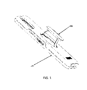
Fig.4 shows the position information of the AKR1C3 primer-probe in present
application.
Fig.5 shows the amplification results of 9 pairs of AKR1C3 primer-probes;
wherein
Fig.5a shows the amplification results of F1R1P1 primer-probe; Fig.5b shows
the
amplification results of F2R2P1 primer-probe; Fig.5c shows the amplification
results
of F2R6P1 primer-probe; Fig.5d shows the amplification results of F6R2P1
primer-probe; Fig.5e shows the amplification results of F6R6P1 primer-probe;
Fig.5f
shows the amplification results of F5R5P2 primer-probe; Fig.5g shows the
amplification results of F3R3P1 primer-probe; Fig.5h shows the amplification
results
of F4R3P2 primer-probe; and Fig.5i shows the amplification results of F7R7P3
primer-probe.
Fig.6 shows the amplification results and standard curves of 3 pairs of AKR1C3
primer-probes; wherein Fig.6a shows the amplification results of F2R6P1
primer-probe; Fig.6b shows the standard curve of F2R6P1 primer-probe ( 0
represents standard; E (amplification efficiency) =82.5%, R2=0.987, Slope=-
3.826,
y-int (y-intercept) =51.961); Fig.6c shows the amplification results of F6R2P1
primer-probe; Fig.6d shows the standard curve of F6R2P1 primer-probe ( 0
represents standard; E (amplification efficiency) =96.5%, R2=0.982, Slope=-
3.408,
y-int (y-intercept) =49.591); Fig.6e shows the amplification results of F6R6P1
primer-probe; and Fig.6f shows the standard curve of F6R6P1 primer-probe (0
represents standard; E (amplification efficiency) =98.0%, R2=0.992, Slope=-
3.371,
y-int (y-intercept) =49.385).
Fig.7 shows the amplification result and standard curve of AKR1C3 plasmid;
wherein
Fig.7a shows the amplification result of AKR1C3 plasmid; and Fig.7b shows the
standard curve of AKR1C3 plasmid ( 0 represents standards; E (amplification
efficiency) =100.7%, R2=0.999, Slope=-3.305, y-int (y-intercept) =42.336).
Fig.8 shows the amplification results of 4 types of reference gene primer-
probes;
wherein Fig.8a shows the amplification result of the reference gene GAP;
Fig.8b
shows the amplification result of the reference gene GOLGAl; Fig.8c shows the
amplification result of the reference gene ACTB; and Fig.8d shows the
amplification
result of the reference gene HPRT1.
Fig.9 shows the Western blot result of AKR1C3 expression in different cell
lines.
Fig.10 shows the results of detecting AKR1C3 expression in different cell
lines using
11
Date Recue/Date Received 2023-08-17
CA 03211243 2023-08-17
4 types of reference genes; wherein Fig. 10a shows the results of detecting
AKR1C3
expression in different cell lines using the reference gene HPRT1; Fig.10b
shows the
results of detecting AKR1C3 expression in different cell lines using the
reference
gene GAP; Fig.10c shows the results of detecting AKR1C3 expression in
different
cell lines using reference gene ACTB; and Fig. 10d shows the results of
detecting
AKR1C3 expression in different cell lines using the reference gene GOLGAL
Fig.11 shows the results of different cell lines detected by AKR1C3-ACTB
system.
Fig.12 shows the results of CCRF-CEM cell lines detected by AKR1C3-ACTB
system; wherein Fig.12a shows the amplification result of AKR1C3 in CCRF-CEM
cell lines; Fig.12b shows the standard curve of AKR1C3 in CCRF-CEM cell lines
(target: AKR1C3; Eff (amplification efficiency) %=94.19%, R2=0.998, Slope=-
3.47,
y-inter (y-intercept) =34.595); Fig.12c shows the amplification result of ACTB
in
CCRF-CEM cell lines; and Fig.12d shows the standard curve of ACTB in
CCRF-CEM cell lines (target: ACTB; Eff (amplification efficiency) %=95.302%,
R2=0.991, Slope=-3.44, y-inter (y-intercept) =28.854).
Fig.13 shows the results of plasmids detected by AKR1C3-ACTB system; wherein
Fig.13a shows the amplification result of AKR1C3 plasmid; Fig.13b shows the
standard curve of AKR1C3 plasmid (target: AKR1C3; Eff (amplification
efficiency) %=101.853%, R2=0.998, Slope=-3.278, y-inter (y-intercept)
=38.048);
Fig.13c shows the amplification result of ACTB plasmid; and Fig.13d shows the
standard curve of ACTB plasmid (target: ACTB; Eff (amplification
efficiency) %=94.134%, R2=1, Slope=-3.471, y-inter (y-intercept) =40.821).
Fig.14 shows the qPCR detection result ofJurkat cell lines.
Fig.15 shows the digital PCR detection result of cell lines (FAM and VIC were
detected simultaneously).
Fig.16 shows the results of AKR1C3 expression in 2 types of cell lines
detected by
digital PCR; wherein Fig.16a shows the result of AKR1C3 expression in CCRF-CEM
cell lines detected by digital PCR; and Fig.16b shows the result of AKR1C3
expression in MOLT-4 cell lines detected by digital PCR.
Fig.17 shows the results of ACTB expression in 2 types of cell lines detected
by
digital PCR; wherein Fig.17a shows the result of ACTB expression in CCRF-CEM
cell line detected by digital PCR; and Fig.17b shows the result of ACTB
expression in
MOLT-4 cell line detected by digital PCR.
Fig.18 shows the results of plasmids detected by AKR1C3-ACTB system in actual
samples testing; wherein Fig.18a shows the amplification result of AKR1C3
plasmid;
Fig.18b shows the standard curve of AKR1C3 plasmid (target: AKR1C3; Eff
12
Date Recue/Date Received 2023-08-17
CA 03211243 2023-08-17
(amplification efficiency) %=99.108%, R2=0.996, Slope=-3.344, y-inter (y-
intercept)
=38.128); Fig.18c shows the amplification result of ACTB plasmid; and Fig.18d
shows the standard curve of ACTB plasmid (target: ACTB; Eff (amplification
efficiency) %=93.445%, R2=0.998, Slope=-3.49, y-inter (y-intercept) =40.636).
Fig.19 is a histogram of FAM copy number/VIC copy number in actual samples.
Fig.20 shows the copy number results of 5-fold dilution of the cDNA of the
cell lines
detected with digital PCR.
DETAILED DESCRIPTION OF THE INVENTION
It is necessary to indicate that, unless otherwise defined, technical terms or
scientific
terms used in one or more examples of the present specification shall have the
ordinary meaning as understood by people having ordinary skill in the field to
which
the present disclosure belongs.
The experimental methods in the following examples are all conventional
methods
unless otherwise specified. The raw materials of the medicaments, the reagents
and
the like used in the following examples are all commercially available
products unless
otherwise specified.
"Administering" drug to patient refers to direct administration (which may be
administered to patient by a medical professional or self-administered) and/or
indirect
administration, which may be an operation of making drug prescriptions. For
example,
physicians who instruct patient to self-administer drugs and/or provide
patient with
drug prescriptions will administer drugs to patient.
"Cancer" refers to leukemia, lymphoma, cancer and other malignant tumors
(including solid tumors) that can spread locally by invasion and spread
systemically
by metastasis with potential unrestricted growth. Examples of cancers include
(but are
not limited to) cancers of adrenal gland, bone, brain, breast, bronchus, colon
and/or
rectum, gallbladder, head and neck, kidney, throat, liver, lung, nervous
tissue,
pancreas, prostate, accessory thyroid gland, skin, stomach and thyroid gland.
Certain
other examples of cancers include acute and chronic lymphocytic and
granulocytic
tumors, adenocarcinoma, adenoma, basal cell carcinoma, poorly differentiated
cervical epithelium and carcinoma in situ, Ewing's sarcoma, epidermoid
carcinoma,
giant cell tumor, glioblastoma multiforme, hairy cell tumor, intestinal
ganglion cell
tumor, proliferative corneal nerve tumor, islet cell carcinoma, Kaposi's
sarcoma,
leiomyoma, leukemia, lymphoma, malignant carcinoid, malignant melanoma,
malignant hypercalcemia, marfanoid habitus tumor, myeloid epithelial
carcinoma,
metastatic skin cancer, mucosal neuroma, myeloma, granuloma fungoide,
neuroblastoma, osteosarcoma, osteogenic and other sarcomas, ovarian tumors,
pheochromocytoma, true erythrocytosis, primary brain tumor, small cell lung
cancer,
13
Date Recue/Date Received 2023-08-17
CA 03211243 2023-08-17
squamous cell carcinoma of both ulcerative and papillary types, hyperplasia,
seminoma, soft tissue sarcoma, retinoblastoma, rhabdomyosarcoma, renal cell
tumor,
local skin lesion, reticulum cell sarcoma, and Wilm's tumor.
The terms "patient" and "individual" can be used interchangeably herein and
refer to
mammals in need of treatment for cancer. Generally, the patient is a human.
Generally,
the patient is a human diagnosed with cancer. In certain examples, "patient"
or
"individual" may refer to a non-human mammal used in screening,
characterizing, and
evaluating drugs and therapies, such as, a non-human primate, a dog, cat,
rabbit, pig,
mouse or a rat.
"Prodrug" refers to a compound that, after administration, is metabolized or
otherwise
converted to a biologically active or more active compound (or drug) with
respect to
at least one property. A prodrug, relative to the drug, is modified chemically
in a
manner that renders it, relative to the drug, less active or inactive, but the
chemical
modification is such that the corresponding drug is generated by metabolic or
other
biological processes after the prodrug is administered. A prodrug may have,
relative to
the active drug, altered metabolic stability or transport characteristics,
fewer side
effects or lower toxicity, or improved flavor (for example, see the reference
Nogrady,
1985, Medicinal Chemistry A Biochemical Approach, Oxford University Press, New
York, pages 388-392, incorporated herein by reference). A prodrug may be
synthesized using reactants other than the corresponding drug.
"Solid tumor" refers to solid tumors including (but not limited to) metastatic
tumor in
bone, brain, liver, lung, lymph node, pancreas, prostate, skin and soft tissue
(sarcoma).
"Treatment of' a condition or patient refers to taking steps to obtain
beneficial or
desired results, including clinical results. For purposes of this invention,
beneficial or
desired clinical results include, but are not limited to, alleviation or
improvement of
one or more symptoms of cancer; diminishment of extent of disease; delay or
slowing
of disease progression; alleviation, palliation, or stabilization of the
disease state; or
other beneficial results. Treatment of cancer may, in some cases, result in
partial
response or stable disease.
"Tumor cells" refers to tumor cells of any appropriate species, e.g.,
mammalian such
as murine, canine, feline, equine or human.
"Therapeutically effective amount" of a drug refers to an amount of a drug
that, when
administered to a patient with cancer, will have the intended therapeutic
effect, e.g.,
alleviation, amelioration, palliation or elimination of one or more
manifestations of
cancer in the patient. A therapeutic effect does not necessarily occur by
administration
of one dose, and may occur only after administration of a series of doses.
Thus, a
therapeutically effective amount may be administered in one or more
administrations.
14
Date Recue/Date Received 2023-08-17
CA 03211243 2023-08-17
In describing optically active compounds, the prefixes R and S are used to
represent
the absolute configuration of the molecule around its chiral center. (+) and (-
) are used
to represent the optical activity of the compound, i.e., the direction in
which the plane
of polarization rotates through the optically active compound. The prefix (-)
represents that the compound is levorotatory, i.e., the compound rotates the
direction
of the plane of polarization to left or anticlockwise. The prefix (+)
represents that the
compound is dextrorotary, i.e. the compound rotates the direction of the plane
of
polarization to right or clockwise. However, the optical signs (+) and (-) are
independent of the absolute configurations of R and S of the molecule.
The structure of compounds OBI-3424 (also referred to as AST-3424 and TH-
3424),
OBI-3423 and OBI-2870 are shown below:
o A
k-N
im = it> ip fliP.; Cr '14
ON 4111/VP 02N 02N
=
101 0 dist.
1.11
0 N
3424 3423 2870
wherein OBI-3424 is an S-enantiomer, OBI-3423 is an R-enantiomer, and OBI-2870
is a racemic mixture of OBI-3424 and OBI-3423 at a ratio of 1:1.
Previous studies have shown that compounds OBI-3424, OBI-3423 and OBI-2870
have good therapeutic effects on a variety of human cancers, especially
compound
OBI-3424 has good therapeutic effects on solid tumors and hematological
malignancies; wherein the therapeutic effects of compound OBI-3424 on solid
tumors
(including liver cancer, hepatocellular carcinoma (HCC), non-small cell lung
cancer,
melanoma, prostate cancer, mastocarcinoma, esophageal cancer, renal cancer,
gastric
cancer, colon cancer, cerebral cancer, bladder cancer, cervical cancer,
ovarian cancer,
head and neck cancer, endometrial cancer, pancreas cancer, sarcomatoid
carcinoma
and rectal cancer and the like.) are recited in patent CN108290911A; and the
therapeutic effects of compound OBI-3424 on hematological malignancies
(including
B lineage acute lymphoblastic leukemia (B-ALL) and T lineage acute
lymphoblastic
leukemia (T-ALL)) are recited in patent W02019/062919A1.
AKR1C3 is overexpressed at the protein level in many types of cancer,
especially in
liver cancer, gastric cancer, renal cancer, CRPC and non-small cell lung
cancer. Due
to the high expression of AKR1C3 in tumors, compound OBI-3424 is designed to
be
specifically activated in tumors, but compound OBI-3424 cannot be activated in
normal cells expressing low level of AKR1C3 to achieve tumor-specific
targeting.
Date Recue/Date Received 2023-08-17
CA 03211243 2023-08-17
OBI-3424 is developed as a highly potent DNA alkylation prodrug selectively
activated by AKR1C3. In the presence of NADPH, OBI-3424 is reduced from
AKR1C3 to an intermediate which is spontaneously hydrolyzed to OBI-2660, and
OBI-2660 is a DNA dialkylating agent similar to thioTEPA
(N,N',N"-triethylenethiophosphoramide) which leads to DNA cross-linking at N7
(or
06) position of guanine, and subsequently leads to cell death, as shown below.
0(,1
o o1-Ni _________
.4-02
11
f,
N 1
101 I
!,
0 pi V
DNA cross-linking
(4:04eato
cell death 4 __________________________________
140
Le"
activity
Further studies made by the applicant have confirmed that compound OBI-3424
activation is AKR1C3-dependent, and its cytotoxicity and antitumor efficacy
are
highly correlated with the expression level of AKR1C3 protein. Compound OBI-
3424
shows AKR1C3-dependent cytotoxicity ex vivo and antitumor activity in vivo in
a
variety types of human cancer, which supports the further development of
compound
OBI-3424 as an anticancer agent that can be used to treat different types of
cancer.
AKR1C3 is used as a biomarker to analyze the condition of cancer patients and
further guides patients to choose compound OBI-3424 for treatment.
Example 1 of the present application has confirmed that AKR1C3 enzyme and
AKR1C3 RNA content in solid tumors are linearly correlated with the amount
after
mathematical transformation for IC50 value of compound OBI-3424 on cancer
cells
and the linear correlation coefficient is high enough; Examples 2 and 3 have
confirmed that AKR1C3 enzyme and AKR1C3 RNA content in hematological tumors
are linearly correlated with the amount after mathematical transformation for
IC50
value of compound OBI-3424 on cancer cells, and the linear correlation
coefficient is
high enough. It is well known to those skilled in the art that the therapeutic
effect of a
drug on a patient can be directly confirmed only by association with IC50
value; in
theory and in practice, the lower the IC50 value, the better the efficacy of a
drug on a
patient. All of the experimental data of examples 1-3 detected a relevant
biometric
value that can be objectively measured to correlate the IC50 value, and then
predicted
the efficacy of a drug on a specific cancer patient and achieved targeted drug
delivery
(in fact, it is best to directly obtain the tumor tissue of a specific patient
and then
detect the IC50 value under the condition of cancer cell survival. This is the
best, most
intuitive and accurate, but it is not feasible in practice, so only relevant
biometric
values that can be objectively measured can be detected to correlate the IC50
value.).
After extensive detections, the present application has confirmed that AKR1C3
16
Date Recue/Date Received 2023-08-17
CA 03211243 2023-08-17
enzyme and AKR1C3 RNA content in solid tumors and hematological tumors are not
only linearly correlated with the IC50 value of compound OBI-3424 on cancer
cells,
but also the linear correlation coefficient is high enough, which is enough to
illustrate
that AKR1C3 enzyme and AKR1C3 RNA content in solid and hematological tumors
are highly correlated with the IC50 value of compound OBI-3424 on cancer
cells.
Therefore, in practical application, AKR1C3 enzyme content and AKR1C3 RNA
content of ex vivo sample of a patient can be detected, and then compound OBI-
3424
can be administrated to a patient for treatment according to the detection
results.
In the prior art, Western blotting or immunochemical staining is usually used
to detect
AKR1C3 enzyme content, however, there is no corresponding detection method for
detecting AKR1C3 RNA content in the prior art. Assuming that a corresponding
assay
can be used to assess AKR1C3 RNA content, patient with higher AKR1C3 RNA
content and most likely to respond to the prodrug thus can be selected to
administrate
compound OBI-3424 for achieving better cancer treatment effect.
Based on the above purpose, based on real-time fluorescence quantitative PCR
technology (qPCR), the present invention adopts RNA reverse transcription
reaction,
polymerase chain reaction and Taqman probe technology to detect the expression
of
AKR1C3 gene, and establishes a method and the corresponding kit of human
AKR1C3 gene expression which can be applied to LDT (Laboratory developed test)
system, finally they can be used for the drug detection of clinical trial
samples
companion diagnostics.
Basic principles for designing primer and probe: 1) length of primer-probe:
the length
of each primer is 15-30 bases. 2) GC% in the primer-probe is required to range
from
30 to 80%, and Tm value of primer ranges from between 55 to 60 C. 3) base
pairing
of the primer-probe itself and the 3' end between primers should be avoided as
much
as possible. 4) more than 6 consecutive base pairs of the primer-probe itself
and
between primer should be avoided as much as possible. 5) the length of the
target
fragment amplified by upstream and downstream primers ranges from 50 to 180bp,
and the probe is located between the upstream and downstream primers, as close
as
possible to the upstream primer, and the use of guanine should be avoided at
the 5'
end of the probe. When the annealing temperature of the probe is too low, LNA
probe
or MGB probe is considered for design. 6) The selected primers and probes
should be
designed in the conserved region of AKR1C3 gene which can specifically detect
the
expression of AKR1C3 gene.
Technical principles: RNA reverse transcription reaction, polymerase chain
reaction
and Taqman probe technology are used to design specific primers and probes
directing AKR1C3 gene expression. The target sequence is amplified by PCR
using
specific primers, and the Taqman probe bound to the template is cut by Taq
enzyme
(5'¨>3' exonuclease activity), the reporter is separated from the quencher,
generates
and accumulates fluorescent signals. The real-time amplification curve can be
17
Date Recue/Date Received 2023-08-17
CA 03211243 2023-08-17
obtained according to the relationship between the fluorescent signals and the
number
of amplification cycles, and then the detection for the expression level of
AKR1C3
RNA can be achieved.
The present invention designs 9 pairs of primers and probes for 3 target
regions of
AKR1C3 (exons 2-3, 4-5 and 5-6, respectively). The position information of the
primer-probes can be seen in Fig.l. Beacon Designer 8.12 software is used to
assist
the evaluation of primer quality which is in line with the basic principles
for designing
primer-probe, and Blast tool in NCBI database is used to ensure that each pair
of
primer-probe specifically amplified human gene sequences without common SNP
sites.
The first aspect of the present invention provides a primer-probe composition,
selected from any one of the following groups:
(i) upstream primer AKR1C3-F1, downstream primer AKR1C3-R1 and probe
AKR1C3 -P1, wherein the nucleotide sequences of the upstream primer AKR1C3-F1,
the downstream primer AKR1C3-R1 and the probe AKR1C3-P1 are shown as SEQ
ID NO:1, SEQ ID NO:2 and SEQ ID NO:3, respectively;
(ii) upstream primer AKR1C3-F2, downstream primer AKR1C3-R2 and probe
AKR1C3-P1, wherein the nucleotide sequences of the upstream primer AKR1C3-F2,
the downstream primer AKR1C3-R2 and the probe AKR1C3-P1 are shown as SEQ
ID NO:4, SEQ ID NO:5 and SEQ ID NO:3, respectively;
(iii) upstream primer AKR1C3-F2, downstream primer AKR1C3-R6 and probe
AKR1C3-P1, wherein the nucleotide sequences of the upstream primer AKR1C3-F2,
the downstream primer AKR1C3-R6 and the probe AKR1C3-P1 are shown as SEQ
ID NO:4, SEQ ID NO:6 and SEQ ID NO:3, respectively;
(iv) upstream primer AKR1C3-F6, downstream primer AKR1C3-R2 and probe
AKR1C3-P1, wherein the nucleotide sequences of the upstream primer AKR1C3-F6,
the downstream primer AKR1C3-R2 and the probe AKR1C3-P1 are shown as SEQ
ID NO:7, SEQ ID NO:5 and SEQ ID NO:3, respectively;
(v) upstream primer AKR1C3-F6, downstream primer AKR1C3-R6 and probe
AKR1C3-P1, wherein the nucleotide sequences of the upstream primer AKR1C3-F6,
the downstream primer AKR1C3-R6 and the probe AKR1C3-P1 are shown as SEQ
ID NO:7, SEQ ID NO:6 and SEQ ID NO:3, respectively;
(vi) upstream primer AKR1C3-F5, downstream primer AKR1C3-R5 and probe
AKR1C3-P2, wherein the nucleotide sequences of the upstream primer AKR1C3-F5,
the downstream primer AKR1C3-R5 and the probe AKR1C3-P2 are shown as SEQ
ID NO:8, SEQ ID NO:9 and SEQ ID NO:10, respectively;
18
Date Recue/Date Received 2023-08-17
CA 03211243 2023-08-17
(vii) upstream primer AKR1C3-F3, downstream primer AKR1C3-R3 and probe
AKR1C3-P1, wherein the nucleotide sequences of the upstream primer AKR1C3-F3,
the downstream primer AKR1C3-R3 and the probe AKR1C3-P1 are shown as SEQ
ID NO:11, SEQ ID NO: 12 and SEQ ID NO:3, respectively;
(viii) upstream primer AKR1C3-F4, downstream primer AKR1C3-R3 and probe
AKR1C3-P2, wherein the nucleotide sequences of the upstream primer AKR1C3-F4,
the downstream primer AKR1C3-R3 and the probe AKR1C3-P2 are shown as SEQ
ID NO:13, SEQ ID NO:12 and SEQ ID NO:10, respectively;
(ix) upstream primer AKR1C3-F7, downstream primer AKR1C3-R7 and probe
AKR1C3-P3, wherein the nucleotide sequences of the upstream primer AKR1C3-F7,
the downstream primer AKR1C3-R7 and the probe AKR1C3-P3 are shown as SEQ
ID NO:14, SEQ ID NO:15 and SEQ ID NO:16, respectively.
The present invention performed AKR1C3 amplification by using the above 9
groups
of primer-probe compositions, and selected groups (iii), (iv) and (v) from the
9 groups
of primer-probe compositions as candidate primer-probe compositions according
to
the amplification efficiency and specificity. In order to determine the best
primer-probe composition, further AKR1C3 amplification was performed in
different
samples by using primer-probe composition from groups (iii), (iv) and (v),
respectively. According to the amplification efficiency, the primer-probe
composition
of group (iv) (upstream primer AKR1C3-F6, downstream primer AKR1C3-R2 and
probe AKR1C3-P1) was finally determined as the best primer-probe composition
of
the present invention.
It should be noted that the 9 groups of primer-probe compositions of the
present
invention can be used not only for the detection of AKR1C3 RNA content by qPCR
method, but also for the detection of AKR1C3 RNA content by digital PCR
method.
The steps of the detection of AKR1C3 RNA content by qPCR method and digital
PCR method are described in detail below.
In a preferred embodiment of the present invention, wherein the 5'-end
reporters of
probes AKR1C3-P1, AKR1C3-P2 and AKR1C3-P3 are FAM, and the 3'-end
quenchers of probes AKR1C3-P1, AKR1C3-P2 and AKR1C3-P3 are MGB.
The preferred probe for the implementation of the present invention is the
probe
labeled according to the TaqMan system. The TaqMan system is available from
the
manufacturer, Life Technologies Inc. According to the TaqMan system,
oligonucleotide probes that are specifically designed to hybridize to the
amplified
target DNA are covalently linked to a reporter at 5' end and to a quencher at
3' end.
Examples of appropriate reporters for use in TaqMan system include
6-carboxyfluorescein (FAM) or tetrachlorofluorescein (TET). The typical
quencher is
19
Date Recue/Date Received 2023-08-17
CA 03211243 2023-08-17
minor groove binder (MGB). The principle of this TaqMan system is that the
quencher inhibits the fluorescence of the reporter as long as the position of
the
quencher and the reporter are very close to each other on the probe. Once the
oligonucleotide probe hybridizes to the target DNA during qPCR amplification,
it will
be degraded by Taq polymerase because the enzyme makes that the
oligonucleotide
primer extends along the DNA corresponding to the target DNA. This degradation
releases the reporter and the quencher, and the quencher becomes no longer
very close
to the reporter, thus enabling the reporter to emit its fluorescence, which
can then be
detected and measured by appropriate tools typically integrated in qPCR
equipment
(i.e. thermal cyclers).
Based on the same inventive concept, the second aspect of the present
invention
provides a kit comprising the primer-probe composition as described above.
In a preferred embodiment of the present invention, wherein the kit as
described
above further comprises a primer-probe composition of reference gene; and the
primer-probe composition of the reference gene comprises upstream primer ACTB-
F1,
downstream primer ACTB-R1 and probe ACTB-P1, and the nucleotide sequences of
the upstream primer ACTB-F1, the downstream primer ACTB-R1 and the probe
ACTB-P1 are shown as SEQ ID NO:17, SEQ ID NO:18 and SEQ ID NO:19,
respectively.
When using qPCR technology to measure the expression level of gene, the
absolute
expression level of the target gene is not often directly measured, but the
expression
levels of the target gene and the reference gene are measured, respectively.
Specifically, the expression level of the reference gene as a standard is
taken to
measure the relative expression level of the target gene, and then the
relative
expression levels between samples are compared. When the relative quantitative
method is used to measure the expression level of gene, the Ct value obtained
by
qPCR is an exponential relationship, not a linear relationship. Therefore, the
Ct value
cannot be directly used for statistical analysis methods that require normal
distribution
of data, such as t test and analysis of variance (ANOVA) method. Therefore,
the
original Ct value should be converted to 2-ct first to makes the data reach a
linear
relationship before statistical analysis. After the data are obtained by the
relative
quantitative method, the comparative CT value method (TAAct method) is often
used
for data analysis. This method is based on two assumptions: 0 the
amplification
efficiency is 100%, i.e., the amount of products in each PCR cycle is doubled,
which
can be solved by the verification of amplification efficiency; 0 there are
appropriate
reference genes to correct the error of the loading quantity of samples.
According to
the formula, calculating the fold change =2-AAct; AACt=(Ct of target gene - Ct
of
reference gene) of treatment group - (Ct of target gene - Ct of reference
gene) of
control group. According to the calculation formula, in relative quantitative
method,
the relative expression of the target gene cannot be calculated without the Ct
value of
the reference gene, suggesting that the reference gene plays a very important
role.
Date Recue/Date Received 2023-08-17
CA 03211243 2023-08-17
The reference gene refers to a known reference gene whose expression level is
not
affected by the study conditions and can be expressed consistently among
multiple
samples, and the expression level of this gene can be used to accurately
quantify the
loading of the initial material. Since the expression level of housekeeping
gene is less
affected by environmental factors and can be continuously expressed in almost
all
tissues and various growth stages of an organism, housekeeping gene is usually
used
as a reference gene in experiments.
According to the reference (DONG Enni, LIANG Qing, LI Li, et al., The
Selection Of
Reference Gene in Real-time Quantitative PCR [J]. China Animal Industry,
49(11):92-96. DOI:10.3969/j.issn.0258-7033.2013.11.025) and the reference
(ZHAO
Wen-Jing, XU Jie, BAO Qiuhua, et al., Selection of Reference Genes for Real-
time
Quantitative PCR [J]. Microbiology China,
2010(12):1825-1829.
DOI:CNKI:SUN:WSWTØ2010-12-019), the reference genes used in q-PCR are
shown in the following table:
Species Sample Type Reference Gene
Good stability Poor stability
Human Blood (pulmonaty tuberculosis patient)
HuP0 GAPDH, 13-actin, HPRT
Peripheral blood mononuclear cell HuPO, HPRT GAPDH, 13-actin, EF-1-a
Lung cancer tissue GUSB, 132-M 132-Microglobulin,
GAPDH
Liver cancer tissue HMBS, C-1BP UBC, HPRT, 1 8SrRNA
Mouse Brain (olfactory bulb, cerebellum, cortex,
GAPDH, HPRT1, 13-actin, NT-3
hypothalamus, hippocampus, brainstem Aequorin
and corpus striatum)
Small intestine SDHA, HRPT1 ARBP, ACTB,
GAPDH, B2M
Liver (immunostimulation) ACTB, GAPDH B2M, SDHA,
HPRT1, ARBP
Mammaty tissue GAPDH, HPRT1 ARBP, ACTB,
SDHA, B2M
Pig Piglet (heart, liver, spleen, lung, kidney,
LIMBS, HPRT1, RPL4 ACTB, GAPDH, SDHA,
brain, muscle, uterus, large intestine and 1BP1, B2M, YWHAZ
intestinal lymphoid tissue)
Bovine Endometrial tissue SUZ 1 2 GAPDH
Sheep Blood SDHA, YWHAZ GAPDH, PGK1
Goat Preantral follicle UBQ, 13-actin 1 8SrRNA
Embryo fibroblast (Newcastle disease 1 8SrRNA, GAPDH, SHDA ACTB, HPRT1,
HMBS
infected)
Chicken Blood (Pre-inflammatory phase) 0-actin HPRT,
GAPDH
Goose Gosling (liver, kidney, heart, muscle and
GAPDH, HPRT1 28SrRNA, TUB, SDH, ACT,
ovary) 1 8SrRNA
As for reference genes, 4 types of reference genes: HPRT1 (hypoxanthine
phosphoribosyl transferase), GAP (glyceraldehyde-3-phosphate dehydrogenase),
ACTB (also known as 13-actin) and GOLGA1 (Golgi protein, specifically Golgi
21
Date Recue/Date Received 2023-08-17
CA 03211243 2023-08-17
autoantigen Al) were screened out in present invention. The primer-probe
compositions corresponding to each of 4 types of reference genes (as shown in
Table
1) were used and detected with the cell line CCRF-CEM to evaluate the
fluorescence
intensity and amplification. Different samples (including healthy blood
samples and
bone marrow samples of leukemia patients) and cell lines with different
expression
levels (including cell lines with high AKR1C3 expression and cell lines with
low
AKR1C3 expression) were further detected to verify the expression stability
and
whether the samples and cell lines with high and low expression levels can be
distinguished. The detected results showed that cell lines RPMI8226 and CCRF-
CEM
with high expression levels can be better distinguished from cell line Jurkat
with low
expression level by using ACTB as reference as compared to using other three
reference genes as references. In healthy people, the values remained
relatively low
level, while high expression level of AKR1C3 was detected in bone marrow RNA
samples of leukemia patients. ACTB was selected as the reference gene based on
the
result of amplification curve. Accordingly, the primer-probe composition of
the
reference gene in the above kit comprises upstream primer ACTB-F1, downstream
primer ACTB-Rl and probe ACTB-Pl.
In a preferred embodiment of the present invention, wherein the 5'-end
reporter of the
probe ACTB-Pl is VIC (green fluorescent protein), and the 3 '-end quencher of
the
probe ACTB-Pl is BHQ1 (Black Hole Quencher 1).
The best AKR1C3 primer-probe composition and reference ACTB primer-probe
composition finally determined after screening are shown in Table 1.
Table 1 Sequence information table of AKR1C3 using primer-probes
Name Sequence Marker Description of Source
AKRIC 3-F1 CTCAACAAGCCAGGACTCAAGTACAAGC
AKR1C3-F2 AC AAGCC AGGAC TC AAGTAC AA
AKR1C3-F3 CAGTGTGAAGAGAGAAGAC ATATTC TAC A
AKR1C3-F4 GC AGATGGC AGTGTGAAGAG
AKR1C3-F5 TGGCAGTGTGAAGAGAGRAGAC
AKR1C3-F6 GATGATCCTCAACAAGCCAGGA
AKR1C3-F7 CACCAACAGATGAAAATGGAAAAGTAA
AKR1C3-R1 AC TTGCAGAAATC TAGC AAT TTAC TCC GGTTG
AKR1C3-R2 AATCTAGCAATTTACTCCGGTTGAAATAC
AKR1C3-R3 CAAGGCTGGTCGGACCAA
AKR1C3-R5 GGTCAACATAGTCCAATTGAGCT
AKR1C3-R6 AC TTGCAGAAATC TAGC AAT TTAC TCC
AKR1C3-R7 GGTTGAAGTTTGAC ACCCC AA
5'FAM,
AKR1C3-P1 CTGCAACCAGGTAGAATGTC A
3'MGB
5'FAM,
AKR1C3-P2 CTTTGGTCC AC TTTTC ATC GAC
3'MGB
22
Date Recue/Date Received 2023-08-17
CA 03211243 2023-08-17
5'FAM,
AKRIC 3-P3 TTGAC ATAGTGGATC TC TGT ACC A
3'MGB
AC TB -F 1 CACCTTCTACAATGAGCTGC G
AC TB -RI GTCTCAAAC ATGATCTGGGTCATC
5'VIC,
AC TB -P 1 CCTCGGTCAGCAGCACGGGGT
3'BHQ1
HPRT 1-Fl CAGCCTCAACATCCTGCACT from reference 1
HPRT 1-RI CACACAATAGCTCTTCAGTC TG from reference 1
HPRT 1 -P 1 TGACCTTGATTTATTTTGCATACC from reference 2
GAP-Fl AGGCTGGGGCTCATTTGC from reference 3
GAP -RI GIG CTCAGTGTAGCCCAGGATG from reference 3
GAP -P 1 GAGCCACACAAACACCAG from reference 4
upstream nucleic acid fragment of Golgi autoantigen nucleic acid sequence
GOLGA 1 -F 1
Al (GOLGAI) gene was not determined
downstream nucleic acid fragment of Golgi autoantigen nucleic acid sequence
GOLGAl-R1
Al (GOLGAI) gene was not determined
probe nucleic acid fragment of Golgi autoantigen Al nucleic acid sequence
GOLGA 1 -P 1
(GOLGAI) gene was not determined
Reference 1: corresponding nucleic acid sequences disclosed in Wu, X.,
Blackburn, P.,
Tschumper, R. et al. TALEN-mediated genetic tailoring as a tool to analyze the
function of acquired mutations in multiple myeloma cells. Blood Cancer Journal
4,
e210 (2014). haps://doi.org/10.1038/bcj.2014.32.
Reference 2: HPRT1 nucleic acid sequences disclosed in W02018123764A1.
Reference 3: corresponding nucleic acid sequences disclosed in Werner Kempf,
Marshall E. Kadin, Ann M. Dvorak, Carol C. Lord, Gunter Burg, Norman L.
Letvin,
Igor J. Koralnik, Endogenous retroviral elements, but not exogenous
retroviruses, are
detected in CD30-positive lymphoproliferative disorders of the skin,
Carcinogenesis,
Volume 24, Issue 2, February 2003, Pages 301-306,
https://doi.org/10.1093/carcin/24.2.301.
Reference 4: corresponding nucleic acid sequences disclosed in Stecker C,
Johann A,
Herzberg C, Averhoff B, Gottschalk G. Complete nucleotide sequence and genetic
organization of the 210-kilobase linear plasmid of Rhodococcus erythropolis
BD2. J
Bacteriol. 2003;185(17):5269-5274. doi:10.1128/jb.185.17.5269-5274.2003.
All the sequences without source indicated were new sequences or newly
developed
or selected sequences for the kit.
In a preferred embodiment of the present invention, wherein the kit as
described
above further comprises a polymerase mixture, and the polymerase mixture
mainly
comprises: DNA polymerase, MgCl2, buffer and dNTPs;
23
Date Recue/Date Received 2023-08-17
CA 03211243 2023-08-17
preferably, the polymerase mixture is selected as KAPA PROBE FAST RT-PCR
Master Mix(2x), which has been used in the laboratory for a long time and has
been
repeatedly verified to have good amplification efficiency.
In a preferred embodiment of the present invention, wherein the kit as
described
above further comprises a reverse transcriptase mixture;
preferably, the reverse transcriptase mixture is Superscript VILO MARSTER MIX.
In each PCR reaction, it must be detected and analyzed together with positive
control
and negative control (NC, nuclease-free water). In a preferred embodiment of
the
present invention, wherein the kit as described above further comprises a
negative
control and a positive control;
preferably, the negative control (NC) is nuclease-free water; and/or
preferably, the positive control is a reference with a known copy number.
The kit as described above may further comprises the elements necessary to
perform
qPCR, for example, reagents, as known to those skilled in the art. In addition
to
primer pairs and probes, the kit may further comprises one or more enzymes
(Taq
polymerase) or reagents used in qPCR reactions. The enzyme can be present in a
lyophilized form or in a suitable buffer. In addition, the kit may comprises
all the
additional elements necessary to perform qPCR, such as buffer, extraction
reagent,
enzyme, pipette, plate, nucleic acid, filter paper, gel material, transfer
material,
autoradiography equipment, instructions (recording relevant operation
methods), etc.
Based on the same inventive concept, the third aspect of the present invention
provides use of the primer-probe composition or the kit as described above in
the
preparation of a drug for treating cancers.
In a preferred embodiment of the present invention, wherein the AKR1C3 RNA
content in an ex vivo sample of a patient is obtained by using the primer-
probe
composition or the kit as described above;
patient with AKR1C3 RNA content greater than or equal to the predetermined
content
are administrated AKR1C3 activated anticancer drugs.
Accordingly, the present invention provides a method for treating a patient
suffering
from cancer, wherein the method includes:
obtaining the AKR1C3 RNA content in ex vivo sample of a patient by using the
primer-probe composition or the kit as described above;
24
Date Recue/Date Received 2023-08-17
CA 03211243 2023-08-17
administering AKR1C3 activated anticancer drugs to a patient with AKR1C3 RNA
content greater than or equal to the predetermined content ; preferably, a
therapeutically effective amount of compounds OBI-3424, OBI-3423 and OBI-2870
is administrated to the patient.
In a preferred embodiment of the present invention, wherein the AKR1C3 RNA
content is obtained according to the ratio of AKR1C3 copy number/reference
gene
copy number, for example, the ratio of AKR1C3 copy number/ACTB copy number is
calculated, if the ratio of AKR1C3 copy number/ACTB copy number > X, then it
is
determined that the expression of AKR1C3 is high, and the patient will be
given a
therapeutically effective amount of compounds OBI-3424, OBI-3423 and OBI-2870;
if the ratio of AKR1C3 copy number/ACTB copy number < X, then it is determined
that the expression of AKR1C3 is low, and the patient will not be given a
therapeutically effective amount of compounds OBI-3424, OBI-3423 and OBI-2870,
but other cancer drugs can be given to the patient.
The value of predetermined content X needs to be determined by detecting a
large
number of samples. According to a large number of clinical trial samples and
through
statistical methods, the inventors finally determined that the predetermined
content X
is 0.0001-1; more preferably, the predetermined content X is 0.00011-0.5; even
more
preferably, the predetermined content X is 0.00013-0.05; in particular
preferably, the
predetermined content X is 0.00014-0.015. For example, the predetermined
content X
is 0.0001-0.0005, 0.0001-0.001, 0.0001-0.005, 0.0001-0.01, 0.0001-0.05,
0.0001-0.1, 0.0001-0.5, 0.0001-0.9, 0.00011-
0.0005, 0.00011-0.001,
0.00011-0.005, 0.00011-0.01, 0.00011-0.05, 0.00011-0.1, 0.00011-0.5, 0.00011-
1,
0.00013-0.0005, 0.00013-0.001, 0.00013-0.01, 0.00013-0.05, 0.00013A.1,
0.00013-0.5, 0.00013-1, 0.0005-0.001, 0.0005-0.005, 0.0005-0.01, 0.0005-0.05,
0.0005-0.1, 0.0005-0.5, 0.0005-1, 0.00091-0.001, 0.00091-0.05, 0.00091-0.1,
0.00091-0.5, 0.00091-4, 0.001-0.005, 0.001-0.01, 0.001-0.05, 0.001-0.1, 0.001-
0.5,
0.001-4, 0.005-0.01, 0.005-0.05, 0.005-0.1, 0.005-0.5, 0.005-4, 0.01-0.05,
0.01-0.1, 0.01-0.5, 0.01-4, 0.05-0.1, 0.05-0.5 or 0.05-4; more preferably, the
predetermined content X is 0.0001, 0.00011, 0.00012, 0.00013, 0.00014,
0.00015,
0.000156, 0.00016, 0.000163, 0.000165, 0.00017, 0.000177, 0.00018, 0.000181,
0.00019, 0.000195, 0.0002, 0.000201, 0.000216, 0.000221, 0.00023, 0.000239,
0.00025, 0.00029, 0.0003, 0.00035, 0.0004, 0.00045, 0.0005, 0.00054, 0.0006,
0.00065, 0.0007, 0.00075, 0.0008, 0.00085, 0.0009, 0.00095, 0.001, 0.0015,
0.00177,
0.002, 0.00238, 0.0025, 0.00266, 0.00296, 0.003, 0.00315, 0.0035, 0.004,
0.0045,
0.005, 0.0055, 0.006, 0.0065, 0.00679, 0.007, 0.00710, 0.0075, 0.00778,
0.00791,
0.008, 0.0085, 0.009, 0.00939, 0.0095, 0.01, 0.011, 0.012, 0.01207, 0.013,
0.01365,
0.014, 0.015, 0.016, 0.017, 0.018, 0.019, 0.02, 0.025, 0.03, 0.035, 0.04,
0.045, 0.05,
0.055, 0.06, 0.065, 0.07, 0.075, 0.08, 0.085, 0.09, 0.095, 0.1, 0.15, 0.2,
0.25, 0.3, 0.35,
0.4, 0.45, 0.5, 0.55, 0.6, 0.65, 0.7, 0.75, 0.8, 0.85, 0.9, 0.95 or 1.
Date Recue/Date Received 2023-08-17
CA 03211243 2023-08-17
Based on the same inventive concept, the fourth aspect of the present
invention
provides a method for detecting AKR1C3 RNA content, including the following
steps:
(1) extracting RNA of an ex vivo sample to be detected and adding the
extracted RNA
to the reverse transcription system and the extracted RNA being reverse
transcribed to
synthesize cDNA;
(2) performing qPCR or digital PCR amplification with cDNA as template using
the
primer-probe composition or the kit as described above;
(3) obtaining AKR1C3 RNA content of ex vivo sample to be detected according to
qPCR or digital PCR amplification results.
Methodological principle of the method for detecting AKR1C3 RNA content of the
present invention: firstly, the sample RNA is reverse transcribed into cDNA,
and then
the cDNA is amplified by specific primers and probes, wherein the AKR1C3 gene
and
reference gene are indicated by FAM and VIC signals, respectively; and the
quantitative expression levels of AKR1C3 gene in peripheral blood RNA sample
is
calculated by samples and quality control products. At the same time, VIC
signal in
AKR1C3 reaction solution is used to monitor the sample quality.
In the present invention, two methods of qPCR and digital PCR are used to
conduct a
comparative experiment at the same time under the condition of sufficient
samples.
The same set of primer-probe is used for digital PCR and qPCR. Firstly, the
temperature gradient of qPCR is used to optimize the primer-probe and the most
appropriate temperature and primer-probe composition is selected for the
digital PCR
platform for quantitative reference materials and as a comparative method.
The detection process of digital PCR mainly includes two parts, i.e., PCR
amplification and fluorescence signal analysis. Before PCR reaction, samples
are
divided into tens of thousands of units (reaction chambers) so that only a
single DNA
molecule is present in each unit. The amplification procedure and system of
amplification stage of digital PCR are basically the same as those of ordinary
PCR,
but different from traditional qPCR method, digital PCR adopts direct counting
method for quantitative analysis, that is, after the end of PCR amplification,
if
fluorescence signal (product) is present, marked as 1, and if no fluorescence
signal
(product) is present, marked as 0. A reaction unit with a fluorescent signal
contains at
least one copy of the target molecule. Theoretically, in the case of the
concentration of
target DNA in the sample is very low, the number of reaction units with
fluorescence
signal is equal to the copy number of target DNA molecule. However, under
normal
circumstances, the reaction units of digital PCR may contain two or more
target
molecules, which can be calculated by Poisson distribution.
A ¨A,
p =
k !
26
Date Recue/Date Received 2023-08-17
CA 03211243 2023-08-17
In the above formula, where X, is the average copy number (concentration) of
target
DNA molecules contained in each reaction unit and p is the probability of k
copies of
target DNA molecules contained in each reaction unit under certain A.
conditions. X, is
determined by the dilution coefficient m of sample solution, with ?=cm, where
c is the
original copy number (concentration) of the sample. When k=0 (without the
target
DNA molecule), the above formula can be simplified as p=e=e, p can be regarded
as the ratio of the number of reaction units without fluorescence signal to
the total
number of reaction units, i.e,
n
= e
IL
where n is the total number of reaction units and f is the number of reaction
units with
fluorescence signal. Take the logarithm (1n) of both sides of the above
formula to
obtain
cnt = In (1 ¨1)
The initial copy number (concentration) of sample can be obtained from the
total
number of reaction units in the digital PCR and the number of units with
fluorescent
signals, as well as the dilution coefficient of sample. The quantitative
method of
digital PCR does not depend on the cyclic threshold of the amplification
curve, so it
will not affected by the amplification efficiency and does not need to use the
standard
curve. It has good accuracy and reproducibility, and absolute quantitative
analysis can
be achieved.
In the present invention, the specific reaction system of digital PCR differs
from the
specific reaction system of qPCR in that: in qPCR reaction system, a group of
primer-probe composition selected from groups (i) to (ix) is mixed with the
primer-probe composition of reference gene ACTB; whereas in digital PCR
reaction
system, due to the large difference in the expression levels of AKR1C3 and
ACTB in
samples and cell lines, the maximum difference in Ct may reach 10 Ct values.
Therefore, the copy number of VIC may be much larger than that of FAM during
the
digital PCR detection. In view of this, a group of primer-probe composition
selected
from groups (i) to (ix) is not mixed with the primer-probe composition of
reference
gene ACTB, but AKR1C3 digital PCR detection system and reference gene digital
PCR detection system are used for the amplification of AKR1C3 and reference
genes,
respectively.
PCR amplification results are affected by the changes in various factors of
reaction
system (including primer concentration, probe concentration, etc.) and
amplification
procedures. In order to obtain the best amplification efficiency and the least
non-specific products, the present application determines the reaction system
and
amplification procedure of digital PCR and qPCR based on years of research and
development experience. In combination with the selection of probe-primers,
the
27
Date Recue/Date Received 2023-08-17
CA 03211243 2023-08-17
correlation coefficient of standard curve is ensured to be greater than or
equal to 0.98
and the E value (amplification efficiency) is between 85% and 110%.
In a preferred embodiment of the present invention, wherein in step (3), the
step for
obtaining AKR1C3 RNA content of ex vivo sample to be detected according to
qPCR
or digital PCR amplification results includes:
(A) obtaining the copy numbers of AKR1C3 and reference gene of ex vivo sample
to
be detected according to qPCR or digital PCR amplification results,
respectively;
(B) calculating the ratio of AKR1C3 copy number/reference gene copy number to
obtain the AKR1C3 RNA content of ex vivo sample to be detected.
Wherein when the qPCR method is used, step (A) further includes:
(A1) plotting the standard curve of Ct value and initial copy number lg value
of
AKR1C3 and the standard curve of Ct value and initial copy number lg value of
reference gene, respectively;
(A2) according to the standard curves, obtaining the copy numbers of AKR1C3
and
reference gene of ex vivo sample to be detected by the detected Ct values of
AKR1C3
and reference gene, respectively (which can be calculated automatically by
software).
Another difference between digital PCR and qPCR is that the copy numbers of
AKR1C3 and reference gene of the ex vivo sample to be detected are obtained by
qPCR according to the standard curve, while the copy numbers of AKR1C3 and
reference gene of the ex vivo sample to be detected can be directly obtained
by digital
PCR according to the amplification result, without drawing the standard curve.
The Ct value of target gene AKR1C3 and the Ct value of reference gene should
be
interpreted. The Ct value of target gene AKR1C3 is the Ct value corresponding
to the
amplification signal (FAM signal) of target gene AKR1C3. The Ct value of
reference
gene is the Ct value corresponding to the amplification signal (VIC signal) of
reference gene. The threshold values of FAM and VIC are set during the
exponential
amplification stage.
In a preferred embodiment of the present invention, wherein the method is used
to
detect AKR1C3 RNA content in blood samples, bone marrow samples, or tissue
samples. Digital PCR and qPCR of the present invention can not only detect
blood
samples or bone marrow samples, but also detect tissue samples of solid
tumors,
except that the pre-processing process (such as RNA extraction process) is
different.
For example, as for blood samples, RNA extraction can be performed directly
using
the Qiagen made-up extraction kit; as for bone marrow samples or tissue
samples, the
mortar can be pre-cooled with liquid nitrogen, the bone marrow samples or
tissue
28
Date Recue/Date Received 2023-08-17
CA 03211243 2023-08-17
samples are ground to powder in the mortar, the dried tissue powder is placed
in a 1.5
ml EP tube, and then the RNA extraction kit is used for extraction.
The present application has conducted a performance test on the above kit and
detection method, and the test results are as follows:
1. minimum detection limit: the minimum detection limit was determined by
gradient
dilution of low-expression cell lines, which was used as the sample to be
detected and
repeated 10 times, and the detection results were 100% consistent;
2. precision: repeated detection by different time, instruments and
experimenters, with
CV value < 15%;
3. positive coincidence rate: the known positive samples and the standard
curve were
detected by qPCR, and the detection results were 100% consistent, E value of
the
standard curve was between 90%-110%, and R2 was >0.98;
4. negative coincidence rate: the known negative samples were detected by
qPCR, and
the consistent detection rate of detection results was 100%;
5. different loading template amount: 200ng and lOng RNA of known negative and
positive samples were selected respectively as the starting amounts of reverse
transcription to verify the range of loading amounts, and the consistent
detection rate
of detection results was 100%;
6. analysis specificity: two concentrations of negative references were
selected, and
each concentration level was repeated for three times, with a negative rate of
100%;
7. actual sample detection: 8 normal peripheral blood RNA samples were
selected for
actual sample detection, and the coincidence rate of detection results was
100%.
Through the performance verification of minimum detection limit, precision,
negative
coincidence rate, positive coincidence rate, template amount, analytical
specificity
and actual sample detection, all of the 7 performances met the verification
standards.
The performance of the detection method met the requirements of clinical
application
and can be used for clinical sample detection.
Based on the same inventive concept, the fifth aspect of the present invention
provides a method for detecting the expression level of AKR1C3 enzyme, wherein
the
method is used to detect the AKR1C3 RNA content of ex vivo sample to be
detected,
and then the expression level of the AKR1C3 enzyme of ex vivo sample to be
detected
is obtained according to the AKR1C3 RNA content of ex vivo sample to be
detected.
In a preferred embodiment of the present invention, wherein there is a linear
29
Date Recue/Date Received 2023-08-17
CA 03211243 2023-08-17
correlation between AKR1C3 RNA content ex vivo sample to be detected and the
expression level of AKR1C3 enzyme ex vivo sample to be detected.
In the prior art, administration of compounds OBI-3424, OBI-3423 and OBI-2870
are
usually judged based on whether the AKR1C3 reductase content is equal to or
greater
than the predetermined content, and example 4 of the present invention proves
that the
expression level of AKR1C3 enzyme is highly correlated with the expression
level of
AKR1C3 RNA. Therefore, in practical application, the AKR1C3 enzyme content can
be calculated according to AKR1C3 RNA content measured by the above method,
and
then administration of compounds OBI-3424, OBI-3423 and OBI-2870 are judged
based on whether the AKR1C3 enzyme content reaches the predetermined content.
The technical embodiments provided by the present invention are further
described in
the following examples. The following examples are only used to illustrate the
invention and do not limit the protection scope of the present invention.
The synthetic method for the AKR1C3 activated anticancer prodrug OBI-3424
(also
referred to as AST-3424 or TH-3424) used in the following examples can be seen
in
W02017087428A1 or CN108290911A.
Example 1. Correlation between the level of AKR1C3 protein and the level of
RNA
with IC50 value in liver cancer cell lines and non-small cell lung cancer
(NSCLC) cell
lines
1. Experimental materials and methods
1.1 Cell lines
All human cancer cell lines were from American Type Culture Collection (ATCC ,
Manassas, VA) or Japanese Collection of Research Bioresources (JCRB, Osaka,
Japan)
or Cobioer Biosciences (NanJing, China).
1.2 Ex vivo proliferation assay method
Exponentially growing cells were inoculated. After 24h, the test compound OBI-
3424
was added. After addition of the test compound OBI-3424, plates were incubated
in a
standard tissue incubator at 37 C for the indicated hours. At the end of the
experiment,
viable cells were detected using the CellTiter Glo (CTG) assay kit or
AlamarBlue.
Drug concentrations (IC50) resulting in 50% growth inhibition relative to
untreated
control were calculated using XLfit (IDBS, Boston, MA) or Prism 6 (GraphPad,
San
Diego, CA).
1.3 Western blotting
Date Recue/Date Received 2023-08-17
CA 03211243 2023-08-17
Human cell extract was prepared and protein concentration was determined.
Proteins
were detected using antibodies that recognize human AKR1C3 and tubulin or 13-
actin.
Odyssey Laser Imaging System and software (LI-COR Biosciences, Lincoln, NE) or
UVP ChemStudio imaging system and VisionWorks software (Analytik Jena AG)
were used to scan and quantify the band densities of AKR1C3 and tubulin or 13-
actin,
and the ratio of AKR1C3 to tubulin or 13-actin was calculated.
2. Experimental results
After exposing the hepatocellular carcinoma cell lines to compound OBI-3424
for 96h
and exposing the NSCLC cell lines to compound OBI-3424 for 72h, the IC50 value
was determined by ex vivo proliferation assay, and the expression of AKR1C3
protein
in hepatocellular carcinoma cell lines was determined by Western blotting
(tubulin
was used as loading control). The expression data of AKR1C3 RNA in the
hepatocellular carcinoma cell lines and in the NSCLC cell lines were from
CrownBio
database (https://db.crownbio.com/Crownbio/OncoExpress Login.aspx). The
results
were showed in Tables 2 and 3.
Table 2. Cytotoxicity of OBI-3424 to a group of human hepatocellular carcinoma
cell
lines after 96h of exposure
hepatocellular Normalized The expression The expression 3424
IC50 (nM)
carcinoma cell line AKR1C3 expression level of AKR1C3
level of AKR1C3
protein RNA (Log2 FPKM)
SNU-475 1.22 High 9.19 15
SNU-449 1.19 High 8.3 45
C3A 0.95 High 4.58 5
SNU-387 0.64 Medium 6.42 103
PLC/PRF/5 0.63 High 4.66 167
HLE 0.3 High 4.36 113
HuCCT1 0.21 Low 0.19 696
SNU-I82 0.17 Low -0.05 >1000
HLF 0.15 Low -1.69 >1000
S K-HEP- 1 0.13 Low -1.39 >1000
SNU-398 0.08 Low -1.33 >1000
Table 3. Cytotoxicity of OBI-3424 to a group of non-small cell lung cancer
(NSCLC)
cell lines after 72h of exposure
NSCLC cell line The expression level of AKR1C3 RNA 3424
(Log2 FPKM) IC50 (nM)
NCI-H1944 11.06 2.3
NCI-H2228 9.25 0.21
NCIH1755 9 8.2
NCI-H1563 8.61 2.5
NCI-H2110 8.23 1.1
31
Date Recue/Date Received 2023-08-17
CA 03211243 2023-08-17
NCI-H1792 8.07 4.5
CAL12T 3.86 29.1
NCIH2106 2.68 >1000
NCI-H23 -1.98 >1000
NCI-H522 -1.88 >1000
As shown in Tables 2 and 3, the hepatocellular carcinoma cell lines with high
AKR1C3 expression at both protein and RNA levels were more sensitive to OBI-
3424,
where IC50 values were in the low nanomolar range. On the other hand, cells
expressing low AKR1C3 were less sensitive to OBI-3424, where IC50 values were
higher than 1000 nM. Similarly, NSCLC cells also showed AKR1C3-dependent
cytotoxic properties after exposing NSCLC cell to compound OBI-3424 for 72h
(Table 3). 3424 IC50 in hepatocytes was highly correlated with the level of
AKR1C3
protein (R2=0.71, Fig.1, left), 3424 IC50 in hepatocytes was highly correlated
with the
expression level of AKR1C3 RNA (R2=0.87, Fig.1, middle), and 3424 IC50 in
NSCLC
was highly correlated with the expression level of AKR1C3 RNA (R2=0.80, FIG.
1,
right). These results showed that 3424-mediated cytotoxicity in hepatocyte
lines and
NSCLC cell lines was highly correlated with the expression level of AKR1C3.
Example 2. Correlation between the level of AKR1C3 protein and IC50 value in
leukemia cell lines
1. Experimental materials and methods
1.1 Cell lines and PDX ex vivo studies
All cell lines were purchased from HD Biosciences. All experimental work was
conducted under the approval of the respective institutional review board and
the
animal ethics committee of each institution, and the use of human relevant
tissue
samples was in accordance with the ethical and relevant legal regulations of
the
location of the experiments. Serial PDX previously established in 20-25 g
female
non-obese/SCID (NOD.CB17-Prkdcscid/SzJ, NOD/SCID) or NOD/SCID/IL2
receptor y-negative (NOD.Cg-Prkdcseid Il2relwil/SzJAusb, NSG) was used in the
experiments, as described elsewhere (Lock RB, Liem N, Farnsworth ML, Milross
CG,
Xue C, Tajbakhsh M, et al. The nonobese diabetic/severe combined
immunodeficient
(NOD/SCID) mouse model of childhood acute lymphoblastic leukemia reveals
intrinsic differences in biologic characteristics at diagnosis and relapse.
Blood 2002;
99:4100-8.). The development of lentivirus-transduced ALL-11 PDX [empty vector
(EV) and AKR1C3 overexpression] has been described previously (Jamieson SM, Gu
Y, Manesh DM, El-Hoss J, Jing D, Mackenzie KL, et al. A novel fluorometric
assay
for aldo-keto reductase 1C3 predicts metabolic activation of the nitrogen
mustard
prodrug PR-104A in human leukaemia cells. Biochem Pharmacol 2014; 88:36-45.).
1.2 Ex vivo cytotoxicity assay
32
Date Rectie/Date Received 2023-08-17
CA 03211243 2023-08-17
Leukemia cell lines were suspended in RPMI medium supplemented with FBS
(Biosera), while ALL PDX cells were cultured in QBSF medium (Quality
Biological
Inc.,) supplemented with Flt-3 ligand (20 ng/mL, BioNovus Life Sciences) or
IL7
(10-20 ng/mL, Jomar Life Research). Cells were inoculated according to the
optimal
cell density and incubated for 3h or overnight (37 C, 5%CO2). PDX cells and
leukemia cell lines were treated with OBI-3424 (10 mmol/L - 1 pmol/L) or
medium
control for 48 or 72h, respectively. Viability was determined using Alamar
Blue
reduction assay or Cell Titer-Glo Luminescent Cell Viability Assay (Promega).
The
half-maximal inhibitory concentration (IC50) was used to calculate the
interpolation of
nonlinear regression curves by GraphPad Prism 7 software.
1.3 Western blotting
Cryopreserved leukemia cells were thawed and lysed in RIPA lysis buffer, and
protein
concentrations were quantified by BCA assay. Each sample was loaded with 20
jig of
protein lysate in NuPAGE 4-12% Bis-Tris protein gel, then electrophoresed at
120V
and transferred to polyvinylenedifluoride membrane at 30V for lh. Mouse
anti-AKR1C3 (#A6229, Sigma-Aldrich, St. Louis, MO) or rabbit anti-actin
primary
antibody (#A2066, Sigma-Aldrich) was used, followed by horseradish peroxidase
conjugated anti-mouse or anti-rabbit IgG secondary antibody (GE Healthcare,
Buckingham, UK) respectively to probe the membrane. Immobilon Western
chemiluminescent HRP substrate (Merck Millipore, Billerica, MA) was used to
detect
conjugated secondary antibodies by quantifying the signals in the BioRad
Chemidoc
touch imaging system.
2. Experimental results
The ex vivo cytotoxicity of AKR1C3-related OBI-3424 was observed in 11 types
of
T-ALL cell lines, one B-ALL cell line transfected with granulocyte-colony
stimulating
factor, and one BCP-ALL cell line. The expression levels of AKR1C3 protein was
determined by Western blot analysis. The ex vivo cytotoxicity of OBI-3424 was
determined using CellTiter-Glo assay and calculated as 50% maximum inhibitory
concentration (IC50).
Ex vivo cytotoxicity demonstrated by OBI-3424, IC50 ranged from 3.0 to 30.0 nM
in 6
types of cell lines expressing high (strong) levels of AKR1C3. As for cell
lines with
medium AKR1C3 expression levels, IC50 ranged from 3.0 to 84.0 nM (Table 4).
Table 4. AKR1C3-dependent ex vivo cytotoxicity of OBI-3424 in ALL cell lines
Leukemia Cell line AKR1C3 IC 50(nM)
expression
T-ALL (child) PF-382 Strong 15.0
T-ALL (child) SUP-Ti Strong 3.0
33
Date Recue/Date Received 2023-08-17
CA 03211243 2023-08-17
T-ALL (child) TALL-1 Strong 30.0
T-ALL (19 years old) MOLT-4 Strong 10.0
T-ALL (child) CCRF-CEM Strong 3.7
T-ALL (child) Jurkat Medium 40.0
T-ALL (child) Jurkat, Clone E6-1 Medium 24.0
T-ALL (child) NOMO-1 Medium 11.0
T-ALL (Jurkat mutant) P116 Medium 84.0
B-ALL (transfected with G-CSF) GR-ST Strong 9.9
BCP-ALL Reh Medium 3.0
Note: G-CSF= granulocyte-colony stimulating factor; BCP-ALL= B-cell precursor
ALL
In order to evaluate the potential anti-leukemia activity of OBI-3424, ex vivo
cytotoxicity assays were performed on a variety of leukemia cell lines. It
proved that
OBI-3424 can treat T-ALL, B-ALL, acute myeloid leukemia, acute promyelocytic
leukemia (APL) and erythroleukemia. OBI-3424 showed potent cytotoxicity, in
particular against cell lines derived from T-ALL (T-lineage acute
lymphoblastic
leukemia) with high AKR1C3 expression, where IC50 values were in the low
nmol/L
range (see Table 5). The difference in IC50 values between cell lines with
high/medium
AKR1C3 expression and low expression was statistically significant (P=0.0016).
Table 5. Ex vivo cytotoxicity of OBI-3424 to leukemia cell lines after
exposing OBI-3424 to
leukemia cell lines for 72h
Cell line AKR1C3 expression* IC50(nM) Disease characteristic
CCRF-CEM High 3.7 T-ALL
KG-1 High 153
Erythroleuketnia (59 years old)
MOLT-4 Medium 10 T-ALL(19 years old)
NOMO-1 Medium 11 T-ALL (adult)
PF-382 Medium 15 T-ALL (pediatric)
SUP-T1 Medium 3 T-ALL (pediatric)
TALL-1 Medium 30 T-ALL (pediatric)
GR-ST Medium 9.9 B-ALL
(transfected with G-CSF)
TF-1 Medium 2.8
Erythroleuketnia (35 years old)
Jurkat Low 40 T-ALL (pediatric)
Jurkat, clone E6-1 Low 24 T-ALL
P116 Low 84 Jurkat mutant
P30/0HK Low > 1 uM T-ALL (pediatric)
HEL Low 224
Erythroleuketnia (30 years old)
Reh Low 3 ALL (non-T, non-B)
HL-60 Low 52.6 APL (36 years old)
HL-60, clone 15 Low 87 APL (36 years old)
K-562 Low > 1 uM CML (53 years old)
ATN-1 Low > 1 uM T-ALL (adult)
Mono-Mac-6 Low 29.8 AML-M5 (64 years old)
THP-1 Low > 1 uM AML (pediatric)
34
Date Recue/Date Received 2023-08-17
CA 03211243 2023-08-17
Kasumi-1 Low > 1 uM AML
P31/FUJ Low > 1 uM AML
AKR1C3 expression was evaluated by western blotting and relative to control I3-
tubulin expression: High,
AKR1C3/13-tubulin>5.0; medium, AKR1C3/13-tubulin is 2.0-5.0; low, AKR1C3/13-
tubulin <2Ø
Similar to the results obtained from leukemia cell lines, OBI-3424 exerted
potent
cell-killing effects on all leukemia cell lines. Fig.2a showed the
cytotoxicity of
OBI-3424 against 6 types of B-ALL cell lines, and Fig.2b showed the
cytotoxicity of
OBI-3424 against 7 types of T-ALL cell lines. It can be seen from Figs.2a and
2b that
AKR1C3-dependent activation of OBI-3424 is a DNA alkylation reagent. In
addition,
as shown in Fig.2c, at a concentration of 10 nmol/L of OBI-3424õ AKR1C3
protein
expression showed a significant negative correlation with the cell survival
rate ex vivo
of 18 types of ALL PDX (r=-0.53, P=0.023). Similarly, as shown in Fig.2d, at a
concentration of 100 nmol/L of OBI-3424, AKR1C3 protein expression showed a
significant negative correlation with the cell survival rate ex vivo of 18
types of ALL
PDX (r=-0.56, P=0.015).
Example 3. Correlation between the level of AKR1C3 RNA and IC50 value in
leukemia cell lines
After exposing the hematological cancer cell lines to compound OBI-3424 for
72h,
the IC50 value was determined by ex vivo proliferation assay. The expression
data of
AKR1C3 RNA in hematological cancer cell lines was from CrownBio database
(https://db.crownbio.com/Crownbio/OncoExpress Login.aspx). The results were
showed in Table 6.
Table 6. Cytotoxicity of AST-3424 to a group of human hematological cancer
cell
lines after 72h of exposure
Hematological cancer The expression level of AKR1C3 IC50(nM)
cell line RNA (Log2 FPKM)
CCRF-CEM 5.9566 1.7
SUP-T1 5.1574 3.9
PF-382 4.0971 6
TF-1 5.5893 16.8
KG-1 2.6621 153
P31/FUJ -1.2529 1000
Kasumi-1 -1.6608 1000
TALL-! 4.0126 24
Date Recue/Date Received 2023-08-17
CA 03211243 2023-08-17
HEL 2.1127 224
THP-1 -0.4299 1000
Jurkat 1.1763 51.1
The expression levels of AKR1C3 RNA (Log2 FPKM) and IC50 (nM) in Table 6
above were plotted as a curve graph, as shown in Fig.3d. A curve fitting
between 'Cm,
and the algebraic transformation value Log2 FPKM of AKR1C3 RNA expression
level was performed to obtain the corresponding curve fitting formula. The
correlation
coefficient R2=0.7889, which shows that the expression levels of AKR1C3 RNA
have
a significant linear correlation with the IC50 values.
Example 4. Correlation between the level of AKR1C3 protein and the level of
AKR1C3 RNA in leukemia cell lines
1. Experimental materials and methods
1.1 Cell lines and PDX ex vivo studies
All cell lines were purchased from HD Biosciences. All experimental work was
conducted under the approval of the respective institutional review board and
the
animal ethics committee of each institution. Serial PDX previously established
in
20-25 g female non-obese/SCID (NOD.CB17-Prkdcscid/SzJ, NOD/SCID) or
NOD/SCID/IL2 receptor y-negative (NOD.Cg-Prkdecid Il2relwil/SzJAusb, NSG)
was used in the experiments, as described elsewhere (Lock RB, Liem N,
Farnsworth
ML, Milross CG, Xue C, Tajbakhsh M, et al. The nonobese diabetic/severe
combined
immunodeficient (NOD/SCID) mouse model of childhood acute lymphoblastic
leukemia reveals intrinsic differences in biologic characteristics at
diagnosis and
relapse. Blood 2002; 99:4100-8.). The development of lentivirus-transduced ALL-
11
PDX [empty vector (EV) and AKR1C3 overexpression] has been described
previously (Jamieson SM, Gu Y, Manesh DM, El-Hoss J, Jing D, Mackenzie KL, et
al.
A novel fluorometric assay for aldo-keto reductase 1C3 predicts metabolic
activation
of the nitrogen mustard prodrug PR-104A in human leukaemia cells. Biochem
Pharmaco12014; 88:36-45.).
1.2 Western blotting
Cryopreserved leukemia cells were thawed and lysed in RIPA lysis buffer, and
protein
concentrations were quantified by BCA assay. Each sample was loaded with 20
jig of
protein lysate in NuPAGE 4-12% Bis-Tris protein gel, then electrophoresed at
120V
and transferred to polyvinylenedifluoride membrane at 30V for lh. Mouse
anti-AKR1C3 (#A6229, Sigma-Aldrich, St. Louis, MO) or rabbit anti-actin
primary
antibody (#A2066, Sigma-Aldrich) was used, followed by horseradish peroxidase
conjugated anti-mouse or anti-rabbit IgG secondary antibody (GE Healthcare,
36
Date Recue/Date Received 2023-08-17
CA 03211243 2023-08-17
Buckingham, UK) respectively to probe the membrane. Immobilon Western
chemiluminescent HRP substrate (Merck Millipore, Billerica, MA) was used to
detect
conjugated secondary antibodies by quantifying the signals in the BioRad
Chemidoc
touch imaging system.
1.3 RNA-Seq analysis
In order to analyze AKR1C3 expression in aspirates from primary patients, the
patients were divided into B-ALL and T-ALL and their related subgroups. Paired
end
readings were mapped by STAR (Dobin A, Davis CA, Schlesinger F, Drenkow J,
Zaleski C, Jha S, et al. STAR: ultrafast universal RNA-seq aligner.
Bioinformatics
2013;29:15-21.) to GRCh37 human genome reference via a recommended round-trip
mapping pipeline with default parameters, and Picard MarkDuplicates module was
used to mark the duplication rate. Gene annotation files were downloaded from
Ensembl (http://www.ensembl.org/) for STAR localization and subsequent
evaluation
of gene expression levels. In order to evaluate gene expression profiles, the
count of
readings of annotated genes was invoked by HTSeq (Anders S, Pyl PT, Huber W.
HTSeq¨a Python framework to work with highthroughput sequencing data.
Bioinformatics 2015;31:166-9.) and processed by the DESeq2 R program package
(Anders S, Huber W. Differential expression analysis for sequence count data.
Genome Biol 2010;11:R106.) to normalize gene expression to a regularized 10g2
value.
In order to analysis PDX samples, Illumina paired end RNA-seq data were
aligned to
human genome assembly (construct hg38) using STAR with the quantMode
parameters set to TranscriptomeSAM, and the alignment was converted to
transcript
coordinates. The alignment was run with rsem-calculate-expression commanded by
RSEM (version 1.2.31) to calculate raw gene counts, TPM, FPKM, and isoform
expression. All RNA-seq values were expressed as number of fragments per
kilobase
million (FPKM). The FPKM data were performed with 10g2 conversion.
2. Experimental results
The expression levels of AKR1C3 mRNA and protein in various types of ALL were
detected by RNA-Seq analysis (Fig.3a) and Western blotting (Fig.3b). The
analysis of
the results confirmed that T-ALL (n=25) had significantly higher AKR1C3
expression
as compared with B-ALL (n=65; P<0.0001). Moreover, there was a significant
correlation between AKR1C3 mRNA and protein expression (r=0.58; P=0.0003; See
Fig.3c).
Example 5. Design and screening of primers and probes
5.1 Experimental materials
37
Date Recue/Date Received 2023-08-17
CA 03211243 2023-08-17
5.1.1 Sample information
The cell lines used in this example were purchased from Nanjing Cobioer
Biosciences
Co., Ltd and National Infrastructure of Cell Line Resource; Leukemia samples
were
obtained from acute lymphoblastic leukemia patient volunteers (clinical) and
normal
samples were obtained from healthy volunteers (donors), and the volunteers had
signed informed consent. Fresh blood samples were preserved in PAXGENE tubes
and stored in a refrigerator at -20 C.
The collection, use and subsequent processing of all human samples were
conducted
in accordance with Chinese relevant regulatory regulations of the location of
the
experiments and in accordance with the relevant requirements of international
ethics.
Table 7. Sample information table
Sample type Sample number Sample type Sample source
Normal 1 Peripheral blood Donor
sample 2 Peripheral blood Donor
(11 cases) 3 Peripheral blood Donor
Peripheral blood Donor
6 Peripheral blood Donor
7 Peripheral blood Donor
8 Peripheral blood Donor
9 Peripheral blood Donor
Peripheral blood Donor
11 Peripheral blood Donor
12 Peripheral blood Donor
Cell line IF-1 Human erythroid leukemia cells National
Infrastructure of Cell
sample (5 Line Resource (NICR,
cases) Website of the platform:
http://www.cellresource.cM)
CCRF-CEM Human T-cell leukemia cells National Infrastructure
of Cell
Line Resource
RPMI8226 Multiple myeloma cells Nanjing Cobioer Biosciences
(Website of the company:
www.cobioer.com)
MOLT-4 Human acute T lymphoblastic leukemia National
Infrastructure of Cell
cells Line Resource
Jurkat Human T-cell leukemia cells Nanjing Cobioer
Biosciences
Leukemia GZJ ALL peripheral blood Clinical
sample (10 HCL B-ALL peripheral blood Clinical
cases) HXB B-ALL peripheral blood Clinical
JRZ B-ALL peripheral blood Clinical
LBJ T-ALL peripheral blood Clinical
38
Date Reel:le/Date Received 2023-08-17
CA 03211243 2023-08-17
LSX B-ALL peripheral blood Clinical
PS S B-ALL peripheral blood Clinical
ZYQ B-ALL peripheral blood Clinical
BZQ B-ALL bone marrow Clinical
QQY B-ALL bone marrow Clinical
Plasmid AKR1C3 Plasmid
(concentration: 16.7ng/ul, the Sarigon Biotech (Shanghai)
length of the inserted element: 355bp, the Co., Ltd.
total length of the plasmid: 3065bp)
ACTB Plasmid
(concentration: 57.8ng/ul, the Sarigon Biotech (Shanghai)
length of the inserted element: 500bp, the Co., Ltd.
total length of the plasmid: 2606bp)
5.1.2 Instrument information
Table 8. Instrument platform information
Instrument Manufacturer Model
Fluorescence quantitative PCR Thermo Fisher ABI 7500
instrument
Nucleic acid quantitative Life Qubit 3.0
instrument
Digital PCR instrument Thermo Fisher QuantStudio 3D Digital PCR
5.1.3 Reagent information
Table 9. Reagent information
Reagent Manufacturer Model Batch number Expiry date
QIAamp RNA Blood Mini Kit Qiagen 52304 166034744 13 months
PAXgene Blood RNA Kit-IVD Qiagen 762174 166011286 7 months
SuperScriptTM VILOTM Master Mix Thermo Fisher 11755050
2200704 6 months
KAPA PROBE FAST RT-PCR Master KAPA KK4702 0000100987 9 months
Mix(2x)
dUTP TAKARA 4020 B301E 12 months
UNG TAKARA 2820 AK40810A 12 months
5.2 Nucleic acid extraction
The RNA extraction kit was Qiagen made-up extraction kit: PAXGENE BloodRNA
Kit (catalog number: 762174, used for blood preserved in PAXGENE tubes) and
Qiagen made-up extraction kit: QIAamp RNA Blood Mini Kit (catalog number:
52304, used for blood preserved in EDTA tubes).
39
Date Recue/Date Received 2023-08-17
CA 03211243 2023-08-17
According to the instructions, RNA extraction was performed on blood preserved
in
PAXgene tubes using an extraction and purification Kit (PAXgene Blood RNA
Kit-IVD), and RNA extraction was performed on blood preserved in EDTA tubes
using QIAamp RNA Blood Mini Kit. Concentration detection was performed on the
extracted RNA using the recommended RNA nucleic acid quantification kit (Qubit
RNA HS Assay Kits, Thermo Fisher Scientific, Q32852).
5.3 Reverse Transcription
4 1_, of AKR1C3 reverse transcriptase mixture (Superscript VILO MARSTER ma
from Thermo Fisher (catalog number: 11755050)) was absorbed and added to 16
1_,
of RNA sample (replenish water to 16 1_, if the volume was insufficient),
slightly
vortexed and mixed, and then centrifuged transiently. PCR tubes containing the
reaction solution were placed in PCR instrument, the PCR program was set for
the
synthesis of cDNA.
5.4 Design and selection of AKR1C3 primers
9 pairs of primers and probes were designed for a total of 3 target regions of
AKR1C3.
The position information of primer-probes can be seen in Fig.4. AKR1C3 gene
primers were designed and synthesized by Contract Research Organization (CRO).
5.5 Selection of polymerase and reverse transcriptase
Polymerase mixture is a key component of PCR success or failure and affects
the
efficiency of amplification. The main components of polymerase mixture
comprise:
polymerase, MgCl2, buffer and dNTPs. The above components in commercially
available polymerase mixture have been optimized to the best state in a
certain
proportion to make concentrated mother liquor. When using, the corresponding
detection can be carried out as long as the polymerase buffer was added in
proportion
according to the multiplier. The polymerase KAPA PROBE FAST RT-PCR Master
Mix (2x) (catalog number: KK4702) which has been repeatedly verified to have
good
amplification efficiency, was used in this example.
In each PCR reaction, it must be detected and analyzed together with positive
reference and negative control (NC, nuclease-free water). qPCR detection
process was
as follows:
1) taking out AKR1C3 polymerase mixture, AKR1C3 reaction solution and
references,
mixing by shaking, transiently centrifuging for future use.
2) according to the number of samples to be detected and the total number of
reaction
tubes required for quality control, 11.1 IA of AKR1C3 qPCR polymerase mixture,
2.6
1_, of AKR1C3 reaction mixture and 4.3 IA of nuclease-free water were
respectively
Date Recue/Date Received 2023-08-17
CA 03211243 2023-08-17
included in each test, which were mixed by vortex and transiently centrifuged,
and
then divided into qPCR eight-row tubes with 18 L of each tube. (Note: both of
the
samples to be detected and the quality control need to be repeated twice, and
the
detection number of NTC should be>1.).
3) adding cDNA of samples to be detected, quality control and references in
corresponding qPCR eight-row tubes, respectively, covering eight-row caps,
mixing
by vortex and transiently centrifuging.
4) placing eight-row tubes into the sample tank of qPCR instrument and
recording the
order of placement, and setting the instrument amplification program according
to the
optimized reaction conditions for amplification.
5) after the completion of the experiment, wrapping the PCR reaction strips
with 2
layers of PE gloves and treating them as biological waste. The caps of PCR
tubes
were forbidden to open to prevent contamination.
5.6 Selection of primer-probes
5.6.1 Screening test of AKR1C3 primer-probes
The above steps 5.2-5.5 were adopted, 9 pairs of primer-probes were amplified
first,
the amplification results were shown in Fig.5.
Total RNA was extracted from the peripheral blood of healthy volunteers, and
cDNA
was synthesized via reverse transcription. It can be seen from the
amplification results
that F1R1P1, F2R2P1, F2R6P1, F6R2P1, F6R6P1 and F3R3P1 were higher than
other F5R5P2, F4R3P2 and F7R7P3 in terms of fluorescence intensity values.
Considering the specificity of the primer-probes, BLAST was performed on 9
pairs of
primer, and F2R6P1, F6R2P1 and F6R6P1 displayed better specificity, and no
non-specific products appeared.
5.6.2 Actual sample test of AKR1C3 primer-probes
cDNA was obtained by reverse transcription with total RNA extracted from
peripheral
blood of healthy volunteers using 3 sets of primer-probes (F2R6P1, F6R2P1 and
F6R6P1), and 6 samples were selected for testing. The results were shown in
the
following table.
Table 10. Actual sample test results of primer-probes
Primer pair F2R6 F6R2 F6R6
FAM FAM FAM FAM FAM FAM
Sample number
(Replicate 1) (Replicate 2) (Replicate 1) (Replicate 2) (Replicate 1)
(Replicate 2)
41
Date Recue/Date Received 2023-08-17
CA 03211243 2023-08-17
6 29.00 29.02 29.03 28.95 28.97 28.88
7 29.19 29.14 29.07 29.15 29.11 29.18
8 28.33 28.29 28.27 28.31 28.36 28.41
9 27.41 27.25 27.20 27.26 27.24 27.36
27.47 27.45 27.42 27.51 27.47 27.55
11 28.86 28.58 28.61 28.60 28.65 28.82
12 29.51 29.36 29.53 29.61 29.68 29.51
NTC N/A N/A N/A N/A N/A N/A
As can be seen from Table 10 that Ct value of the target gene AKR1C3
expression is
relatively stable in the range of 27 to 30 in healthy population.
5.6.3 Amplification efficiency test of AKR1C3 primer-probes
cDNA was obtained by reverse transcription with RNA extracted from peripheral
blood of one healthy volunteer using 3 sets of primer-probes (F2R6P1, F6R2P1
and
F6R6P1) for amplification efficiency test. The concentration of cDNA was
100ng/4,
after 5-fold dilution, it was further diluted 25-fold, 125-fold and 625-fold,
and there
were a total of four concentration gradients for 5 to 625-fold, with two
replicates for
each concentration for qPCR testing. The standard curve and amplification
efficiency
were determined based on the linear relationship between Ct value and log
value of
initial concentration. The results were shown in Fig.6.
It can be seen from the results that the amplification efficiency of F6R2P1
and
F6R6P1 was higher than that of F2R6P1, with greater than 95%. Therefore,
F6R2P1
and F6R6P1 primer-probes were selected for subsequent tests.
RNA samples extracted from the blood of 4 healthy volunteers were selected for
amplification efficiency test of F6R2P1 and F6R6P1, respectively. The results
were
shown in the following table:
Table 11. More sample standard curve tests of F6R2P1 and F6R6P1
Primer-probe pair F6R2P1 F6R6P1
Sample number 1 2 3 5 1 2 3 5
Target efficiency 81.90% 75.50% 88.50% 81.40% 83.20% 70.20% 89.50% 84%
R2 (squared correlation
coefficient of sample 0.998 0.993 1.000 0.999 0.999 0.995
0.996 0.996
standard curve fitting)
According to the results, it can be seen that there was little difference in
the
amplification efficiency of the 2 pairs of primer-probes, and there were
slight
fluctuations in different samples. However, R2 of F6R2P1 was better, and the
R2 was
42
Date Recue/Date Received 2023-08-17
CA 03211243 2023-08-17
equal to 1 in sample 3. Therefore, F6R2P1 was selected as the primer-probe of
AKR1C3 in this kit.
5.6.4 Standard curve test of AKR1C3 plasmids
After dissolving in 40jit nuclease-free water, the AKR1C3 plasmid dry powder
was
diluted 10-fold to a concentration of 5.78ng/jiL measured by Qubit. The
plasmid stock
solution was diluted by gradient, which was 105, 106, 107 and 108-fold,
respectively, to
obtain a total of 4 gradient concentrations of samples for qPCR test. Each
gradient
was repeated twice, and the standard curve of AKR1C3 was measured as shown in
Fig.7.
As can be seen from the standard curve in Fig.7, the amplification efficiency
of
AKR1C3 primer-probe reached 100%, and R2=0.999, suggesting the good
amplification efficiency.
Example 6. Screening of reference genes as well as design and screening of
corresponding primers and probes
As for the selection of reference genes, the references on leukemia gene
expression
([11 Yanlan Wang, Yue Liu, Changhua Thou et al. An AKR1C3-specific prodrug
with
potent antitumor activities against T-ALL. [J] .Leukemia&Lymphoma, 2020, Feb.
DOT: 10.1080/10428194.2020.1728746; [2] Donya Moradi Manesh, Jad El-Hoss,
Kathryn Evans et al. Growth factor AKR1C3 is a biomarker of sensitivity to PR-
104
in preclinical models of T-cell acute lymphoblastic leukemiaas treatment
targets in
clinical oncology.[J]. BLOOD, 2015, 125: 1193-1201; [3] Yuantong Tian, Lijing
Zhao,
Haitao Zhang et al. AKR1C3 overexpression may serve as a promising biomarker
for
prostate cancer progression. [J]. Diagnostic Pathology, 2014, 9: 42.) were
referred to,
and in combination with the primer-probes previously used, 4 reference genes
(HPRT1, GAP, ACTB and GOLGA1) were selected for testing with the cell line
CCRF-CEM to evaluate their fluorescence intensity and amplification. The
results
were shown in Fig.8.
In terms of fluorescence intensity: ACTB was the highest, followed by HPRT1.
In terms of amplification curves: the amplification curve of GOLGA1 are not
typical
S-shaped, and the other three are typical S-shaped amplification curves.
In terms of Ct: the Ct values of GAP and ACTB were low and their expression
levels
were relatively high, while the Ct values of HPRT1 and GOLGA1 were high and
their
expression levels were relatively low.
Samples and cell lines were tested to verify their expression stability and
whether
high and low expression samples could be distinguished.
43
Date Recue/Date Received 2023-08-17
CA 03211243 2023-08-17
RNA numbers 5, 6 and 7 of blood samples from healthy volunteers, bone marrow
RNA numbers BZQ and QQY of leukemia patients, and cell lines expressing
AKR1C3 (RPMI8226, CCRF-CEM and Jurkat) were used in this experiment.
According to the data and previous results, the cell lines RPMI8226 and CCRF-
CEM
were high-expression cell lines, and Jurkat was low-expression cell line (as
shown in
Fig.9).
AACt calculating method was adopted for the experimental results, i.e., AACt=
unknown sample FAM Ct- unknown sample VIC Ct- (quality control FAM Ct- quality
control VIC Ct), and CCRF-CEM was used as quality control in each group. The
results were shown in Fig.10.
It can be seen from the results that ACTB can better distinguish high-
expression cell
lines RPMI8226 and CCRF-CEM from low-expression cell line Jurkat when ACTB
was used as the reference gene compared with the other three reference genes.
In
healthy people, the values remained relatively low level, while high
expression of
AKR1C3 was detected in bone marrow RNA samples of leukemia patients. ACTB
was selected as the reference gene based on the results of the amplification
curve.
After ACTB was determined as the reference gene, 5 types of cell lines were
tested,
respectively, and the test results were shown in Fig.11.
It can be seen from the results that the test results of the cell lines were
consistent with
the validation results of Western blot (Fig. 9), which proved the accuracy of
the test
results of AKR1C3-ACTB primer-probes.
Example 7. Study of qPCR reaction system and conditions
The quality of qPCR reaction system was a necessary condition for successful
development and detection, and its main components must be optimized.
7.1 Determination of reaction system
The reverse transcription system was shown below:
Table 12. Reverse transcription system
Reverse transcription system Amount ( 200_, )
RNA Up to 2ug
Reverse transcriptase 40,
Nuclease-free water Replenish to 200,
The reverse transcription reaction conditions were shown below:
44
Date Recue/Date Received 2023-08-17
CA 03211243 2023-08-17
Table 13. Reverse transcription reaction conditions
Reaction temperature Reaction time
25 C 10min
42 C 60min
85 C 5min
C Hold
Polymerase mixture was the key component that affects PCR amplification
efficiency.
Contained components of the polymerase mixture KAPA PROBE FAST RT-PCR
Master Mix (2x) used in this example have been optimized to the best state at
a
certain ratio to make concentrated mother liquor. When in use, the
corresponding
detection can be carried out as long as the polymerase buffer was added in
proportion
according to the multiplier. According to the instructions, the recommended
amount
was half of the reaction system, i.e., in the reaction system of 20 L, the
amount
should be 10 L. The specific system was shown in the following table:
Table 14. Preparation of AKR1C3 qPCR reaction system
qPCR reaction system Amount (!IL)
AKRIC3-F6 (10[tM) 0.5
AKRIC3-R2 0.5
ACKR1C3-P1 0.3
ACTB-Fl 0.5
ACTB-R1 0.5
ACTB-P1 0.3
dUTP (2mM) 1
UNG enzyme 0.1
cDNA template (Converted according to the
20-50ng
amount of RNA)
Polymerase mixture 10
1120 Replenish to 20111
7.2 Optimization of qPCR reaction conditions
The reaction conditions used in this example were shown in the following table
15.
The reaction conditions were tested as the preferred reaction conditions for
RNA
reaction after long-term verification in the early stage.
Table 15. Amplification reaction conditions
System The total volume was 20 1
Collection Fluorescence signal was collected by AKR1C3 gene-FAM
channel. Fluorescence signal was collected by ACTB reference
gene-VIC channel. Both signals were collected at the same time.
Amplification Cycle number Temperature Time Fluorescence
procedure ( C) signal collection
Date Recue/Date Received 2023-08-17
CA 03211243 2023-08-17
1 1 cycle 37 C 3min NO
2 1 cycle 95 C 3min NO
3 40 cycle 95 C 15s NO
60 C 45s YES
Cycle number was one of the direct factors affecting the results of PCR, and
the cycle
number determined the degree of PCR amplification. The cycle number of qPCR
used
was usually between 30 and 45 cycles. The more cycles there were, the more
significant the non-specific amplification was, while the less cycles there
were, the
more significant was the effect on detection sensitivity.
7.3 Standard curve test of AKR1C3-ACTB system
7.3.1 Standard curve test of cell lines
In optimized reaction system, the primer-probes of AKR1C3 and ACTB were mixed,
and the test of standard curve was conducted to investigate the amplification
efficiency of the primer-probes. CCRF-CEM was used as the cell line, and
reverse
transcription was carried out according to the reverse transcription system of
section
7.1. After cDNA was obtained, four gradients of stock solution, 5, 25 and 125-
fold
dilution were obtained according to the 5-fold gradient dilution with two
replicates for
each concentration and the detection was performed by qPCR reaction system.
The
standard curve was obtained based on the linear relationship between the Ct
value and
initial copy number lg value, as shown in Fig.12.
It can be seen from Fig.12 that the amplification efficiency of AKR1C3 primer-
probe
was 94.2%, and that of ACTB primer-probe was 95.3%, both of them met the
requirements. R2 was greater than 0.99, suggesting that the linear
relationship was
good and met the requirements.
7.3.2 Standard curve test of plasmids
The AKR1C3 and ACTB plasmid dry powders used were dissolved in 40 L
nuclease-free water. After diluting 10-fold, the concentrations were measured
by
Qubit to be 1.67ng/4, 5.78ng/ L, respectively. The plasmid of AKR1C3 was
diluted
to 10-5, and the plasmid of ACTB was diluted to 10-3. 100 L of each plasmid
was
mixed, and then the gradient of 10, 100 and 1000-fold was diluted. Each
gradient was
repeated twice for qPCR test. The standard curve was made according to the
linear
relationship between Ct value and log value of initial concentration. The
results were
shown in Fig.13.
It can be seen from the result in the figures that the amplification
efficiency of
AKR1C3 reached 101.9% and that of ACTB reached 94.1%, with R2 greater than
0.99,
meeting the requirements.
46
Date Recue/Date Received 2023-08-17
CA 03211243 2023-08-17
Example 8. Digital PCR test of AKR1C3-ACTB primer-probes
8.1 Digital PCR detection system and reaction conditions
A digital PCR test using AKR1C3-ACTB primer-probes was tried in this example.
Since the expression levels of AKR1C3 and ACTB in samples and cell lines were
quite different, the maximum Ct difference may reach 10 Ct values. Fig.14
showed
the qPCR detection diagram of Jurkat cell line with low expression.
Therefore, in digital PCR detection, the copy number of VIC will be much
larger than
that of FAM, as shown in Fig.15.
In view of this, AKR1C3 and ACTB were detected separately. Two cell lines,
CCRF-CEM and MOLT-4, were taken as examples. After 2ug RNA was reverse
transcribed, the transcribed product was diluted 5-fold, and the copy number
of
AKR1C3 was detected by digital PCR using the following system:
Table 16. AKR1C3 digital PCR detection system
Reagent Amount (!IL)
AKR1C3-F6 (10uM) 1.2
AKR1C3-R2 1.2
AKR1C3-P1 0.3
2x3D LIFE MIX 7.5
cDNA 0.5
H20 4.3
The reaction procedure of the digital PCR was shown in the following table:
Table 17. Digital PCR reaction conditions
Cycle number Procedure Amount (!IL)
1 96 C 10min
39 60 C 2min
98 C 30s
1 60 C 3min
C co
The detection results were shown in Fig.16, respectively.
As for the detection of ACTB, 5-fold dilution of cDNA was required, followed
by
200-fold dilution, and then the copy number of ACTB was detected using the
47
Date Recue/Date Received 2023-08-17
CA 03211243 2023-08-17
following system:
Table 18. ACTB digital PCR detection system
Reagent Amount (!IL)
ACTB-F1 (10uM) 1.2
ACTB-R1 1.2
ACTB-P1 0.3
2x3D LIFE MIX 10
cDNA 0.5
I-120 4.3
The detection results were shown in Fig.17. It can be seen from the results
that FAM
copy number/VIC copy number of cell line CCRF-CEM was 0.0092, and FAM copy
number/VIC copy number of cell line MOLT-4 was 0.00129.
8.2 Actual sample test and determination of calculation method
Sample No. 9 of healthy volunteer, 8 cases of leukemia patients (numbered as
PSS,
GZJ, HXB, HCL, ZYQ, LBJ, JRZ, LSX), and 5 types of cell lines (TF-1, CCRF-CEM,
RPMI8226, MOLT-4, Jurkat) were used. The plasmid and concentration used in the
standard curve were as described in step 7.3.2 of Example 7. The preparation
method
was as follows: the plasmid of AKR1C3 was diluted to 10-4 and the plasmid of
ACTB
was diluted to 10-2; 1004 of each plasmid was mixed, and then the gradient of
10,
100 and 1000-fold was diluted. The copy numbers were quantified by digital
PCR.
qPCR tests were performed jointly, with two replicates for each sample. The
copy
numbers of AKR1C3 and ACTB of each unknown sample can be obtained by
standard curve, and the ratio of AKR1C3 copy number/ACTB copy number was used
as the way to measure the expression of AKR1C3. The measured standard curve
was
shown in Fig.18.
It can be seen from Fig.18 that the amplification efficiency of AKR1C3 reached
99.1%
and that of ACTB reached 93.4%, with R2 greater than 0.99, meeting the
requirements.
The sample test results were shown in the following table:
Table 19. Sample test results
Sample Name Target Name Cr Quantity The ratio of FAM/VIC copy
number
9 AKR1C3 25.87238503 4631
0.00394
9 ACTB 19.45120239 1176619
c-8226 AKR1C3 24.11899948 15491
0.00939
c-8226 ACTB 18.93831253 1650472
48
Date Recue/Date Received 2023-08-17
CA 03211243 2023-08-17
c-CCRF AKR1C3 21.87590218 72603
0.00710
c-CCRF ACTB 16.17395782 10227160
c-JUR AKR1C3 28.83884048 600
0.00029
c-JUR ACTB 18.57073402 2103491
c-MOLT-4 AKR1C3 26.2293644 3621
0.00177
c-MOLT-4 AC TB 18.61536598 2042448
c-IF-I AKR1C3 22.73562241 40163
0.01365
c-IF-I ACTB 18.06160355 2943309
GZJ AKR1C3 20.59411812 175517
0.00266
GZJ ACTB 13.34858036 65976364
HCL AKR1C3 27.93593407 1118
0.00014
HCL ACTB 16.5698204 7876216
HXB AKR1C3 25.07105827 8041
0.00023
HXB ACTB 14.28288174 35616992
JRZ AKR1C3 27.13578415 1940
0.00054
JRZ ACTB 17.76814651 3572138
LBJ AKR1C3 24.13855743 15283
0.01207
LBJ ACTB 19.34033203 1265921
LSX AKR1C3 20.65554237 168247
0.00238
LSX ACTB 13.24556828 70616680
PS S AKR1C3 20.97867584 134680
0.00315
PS S ACTB 14.00572395 42764056
ZYQ AKR1C3 20.80658913 151625
0.00296
ZYQ ACTB 13.72992897 51299136
The results of the FAM copy number /VIC copy number were shown in Fig.19.
As can be seen from Fig.19, the detection results of cell lines were
consistent with the
results of Western blot of cell lines, which verified the accuracy of the
calculation
method. At the same time, samples with high AKR1C3 expression were detected in
the samples of leukemia patients, as shown by LBJ in the figure. The 8 cases
of
patients were all patients who had received bone marrow transplantation.
Based on the studies of the above AKR1C3 primer-probe design, reaction system
and
reaction conditions, the detection method of AKR1C3 expression at RNA level
was
established, and certain sensitivity and accuracy were achieved.
Example 9. Performance test of the method for detecting AKR1C3 RNA content
According to examples 5-8, the best primer-probe composition, reference gene
and
corresponding primer-probe composition, reaction system and reaction
conditions in
qPCR method and digital PCR method were determined. Based on the above
conditions, the performance test was performed in this example by combining
qPCR
method and digital PCR method, including sensitivity, specificity,
repeatability,
49
Date Recite/Date Received 2023-08-17
CA 03211243 2023-08-17
accuracy and the like.
The qPCR reaction process included the following steps:
(1) Sample preparation
The concentration of RNA should be more than 5ng/ L, 20-200ng RNA was taken
and replenished to a certain volume with nuclease-free water;
(2) Reverse transcription
As shown in Table 12, the reverse transcription mixture was successively added
to the
above PCR tubes, and the mixture was mixed by vortex or by blowing with a
pipettor.
After short centrifugation, the mixture was placed on an ordinary PCR
instrument.
Reverse transcription was performed as shown in Table 13, and the reverse
transcription product cDNA was used as the template of qPCR reaction system
and
could be stored at -20 C.
(3) Preparation of primer working solution
Dry powders of primers and probes were dissolved in TE buffer for 2h at room
temperature (or dissolved overnight at 4 C). The concentration of primer
working
solution was 1004. After dissolving, the primers and probes were mixed
according to
a certain proportion and combination. After mixing thoroughly, it was stored
in
refrigerator at -20 C. The primer working solution can be stably stored at -
20 C for 1
year.
Before each use, the centrifuge tube containing DNA primer amplification
mixture
was shaken and mixed, then centrifuged transiently. According to the indicated
quantity (20 L/reaction), the required amount for single-use was dispensed and
reserved.
(4) Preparation of reaction system
Positive control product (STD), negative control product (Neg) and blank
control
product (NTC) should be used for quality control each time. The qPCR reaction
system was prepared according to Table 14, mixed and centrifuged transiently.
Then
qPCR amplification was performed under the amplification conditions as shown
in
Table 15.
The corresponding meanings of the sample numbers involved in the following
tests
are: W1 ____ standard with no AKR1C3 expression, P1 ______________ strong
positive cell lines
with AKR1C3 expression, P2 _______________________________________ gradient of
standards for AKR1C3-reference with
different copy numbers, which were used to detect standard curves, P3 weak
Date Recue/Date Received 2023-08-17
CA 03211243 2023-08-17
positive cell lines with AKR1C3 expression, Si ___________________ AKR1C3
standard with the
lowest detection limit, R1 _______________________________________ strong
positive cell lines with AKR1C3 expression,
R2 _____ standard with no AKR1C3 expression.
9.1 Lowest detection limit
9.1.1 Detection method
RNA extracted from cell lines with low expression was selected for reverse
transcription, and the amount of RNA was 21,tg. cDNA after reverse
transcription was
diluted 25-fold as the lowest detection limit reference, the copy numbers of
reference
were determined by digital PCR, and each reference was repeated for 10 times.
9.1.2. Confirmation process and result of the copy number of lowest detection
limit
reference
1) preparation and packaging of PCR mix: 7.5 4 of PCR master mix (per assay),
3
4 of Primer mix, and the total amount of water required to be added were
taken,
mixed by vortex, centrifuged, and dispensed into PCR reaction tubes.
2) sample addition: 0.5[tL of samples to be detected was added to the
corresponding
PCR reaction tube respectively, mixed by vortex, then centrifuged.
3) sample loading: the corresponding number of chips was prepared and 14.54 of
reaction liquid was taken for loading. Noted that do not touch the center of
chip and
the outer covers of chip during this process. The chip was pressed for at
least 20 s.
The mineral oil was added slowly and noted that do not spill the oil. The chip
was
sealed after adding mineral oil.
4) on-line testing: the chips were arranged on the shelf of digital PCR
instrument and
noted that the two-dimensional code and the number on the shelf should towards
yourself.
5) chips reading: after the chips were removed from the special PCR instrument
and
placed on room temperature, the chips were put into the chip scanner to scan
the
fluorescence signal. The computer calculated the mutation ratio according to
the
fluorescence signal.
6) detection results:
copy number calculation: target copy number =copies (target) x14.5/ loading
amount.
Fig. 20 showed the test results of 5-fold dilution of cDNA in this cell line,
which was
261.8 copies/4, and the copy number of detection limit reference was 52.4
51
Date Recue/Date Received 2023-08-17
CA 03211243 2023-08-17
copies/ L.
9.1.3 qPCR verification results
Table 20. Detection results of detection limit reference
Sample Target Target FAM/VIC copy
Name Name CT CV Quantity Name CT CV Quantity
number
Jurkat AKR1C3 31.97866 73 ACTB 21.659 303652 0.000239
Jurkat AKR1C3 32.12123 66 ACTB 21.36 372154
0.000177
Jurkat AKR1C3 32.34266 56 ACTB 21.626 310465 0.000181
Jurkat AKR1C3 32.17264 63 ACTB 21.562 324296 __ 0.000195
Jurkat AKR1C3 31.96986 73 ACTB 21.531 331262
0.000221
__________________ 0.584% _______________ 0.688% _______________
Jurkat AKR1C3 32.4185 53 ACTB 21.549 327325
0.000163
Jurkat AKR1C3 31.81558 81 ACTB 21.232 405964
0.000201
Jurkat AKR1C3 32.25435 60 ACTB 21.534 330640
0.000181
Jurkat AKR1C3 31.99182 72 ACTB 21.52 333726
0.000216
Jurkat AKR1C3 32.19634 62 ACTB 21.259 398636 0.000156
As can be seen from Table 20, the lowest detection limit reference was
detected by
qPCR with 10 times repeat. The positive detection rate was 100%, and the
lowest
detection limit met the verification standard.
9.2 Precision
Precision referred to the degree of closeness between multiple parallel test
results
under the same conditions. The closer the test values were to each other, the
higher
the precision. Precision reflected the repeatability of the kit operation. In
this example,
negative references, precision references with weak expression, and a
precision
references with high expression were selected to determine the intra-batch
precision.
The precision performance of the kit was evaluated by calculating the negative
coincidence rate of negative precision references, the coefficient of
variation (CV) of
the relative expression level of AKR1C3 for precision references with weak
expression and high expression.
9.2.1 Detection method
In this experiment, 2 precision references were used as detection samples, 2
instruments, 2 operators and 3 verification dates were used, and each
reference was
detected 24 times. All negative references were negative, and the positive CV
value
was <15%.
CV value = (standard deviation SD/ Mean) x100%
9.2.2 Experimental results
52
Date Recue/Date Received 2023-08-17
CA 03211243 2023-08-17
Table 21. AKR1C3 precision results
Experimental Instrument Sample Target Target
Experimenter CT CT CV CT
CT CV
date model Name Name Name
20201026 a.m. A 275053849 R1 AKR1C3 24.11411476
ACTB 19.088524
20201026 a.m. A 275053849 R1 AKR1C3 24.16817093
ACTB 19.390919
20201026 a.m. A 275053849 R1 AKR1C3 24.0476532
ACTB 19.034349
20201026 a.m. A 275053849 R1 AKR1C3 23.91945457
ACTB 18.850056
20201026 a.m. B 275051628 R1 AKR1C3 24.11411476
ACTB 19.088524
20201026 a.m. B 275051628 R1 AKR1C3 24.16817093
ACTB 19.390919
20201026 a.m. B 275051628 R1 AKR1C3 24.0476532
ACTB 19.034349
20201026 a.m. B 275051628 R1 AKR1C3 23.91945457
ACTB 18.850056
20201027 a.m. A 275053849 R1 AKR1C3 24.40969276
ACTB 19.163565
20201027 a.m. A 275053849 R1 AKR1C3 24.28497124
ACTB 18.652203
20201027 a.m. A 275053849 R1 AKR1C3 24.29683304
ACTB 18.56831
20201027 a.m. A 275053849 R1 AKR1C3 24.219944
ACTB 18.605289
______________________________________________________________________________
0.78% 1.73%
20201023 p.m. B 275051628 R1 AKR1C3 23.9898243
ACTB 19.041698
20201023 p.m. B 275051628 R1 AKR1C3 24.13328171
ACTB 19.094217
20201023 p.m. B 275051628 R1 AKR1C3 23.93481636
ACTB 18.961788
20201023 p.m. B 275051628 R1 AKR1C3 23.92903328
ACTB 19.025682
20201028 p.m. B 275053849 R1 AKR1C3 24.27304268
ACTB 18.652338
20201028 p.m. B 275053849 R1 AKR1C3 24.26483727
ACTB 18.751188
20201028 p.m. B 275053849 R1 AKR1C3 24.30420494
ACTB 19.012062
20201028 p.m. B 275053849 R1 AKR1C3 24.20526314
ACTB 19.029068
20201022 p.m. A 275051628 R1 AKR1C3 24.31328773
ACTB 19.504038
20201022 p.m. A 275051628 R1 AKR1C3 24.57431793
ACTB 19.716539
20201022 p.m. A 275051628 R1 AKR1C3 24.55712318
ACTB 19.723076
20201022 p.m. A 275051628 R1 AKR1C3 24.47789192
ACTB 19.65189
20201026 a.m. A 275053849 R2 AKR1C3
Undetermined ACTB 39.802425
20201026 a.m. A 275053849 R2 AKR1C3
Undetermined ACTB Undetermined
20201026 a.m. A 275053849 R2 AKR1C3
Undetermined ACTB Undetermined
20201026 a.m. A 275053849 R2 AKR1C3
Undetermined ACTB 39.324535
20201026 a.m. B 275051628 R2 AKR1C3 39.69240952
ACTB Undetermined
20201026 a.m. B 275051628 R2 AKR1C3
Undetermined ACTB 39.547302
20201026 a.m. B 275051628 R2 AKR1C3
Undetermined ACTB 39.802425
20201026 a.m. B 275051628 R2 AKR1C3 Undetermined
NA .. ACTB .. Undetermined .. NA
20201027 a.m. A 275053849 R2 AKR1C3
Undetermined ACTB Undetermined
20201027 a.m. A 275053849 R2 AKR1C3
Undetermined ACTB Undetermined
20201027 a.m. A 275053849 R2 AKR1C3
Undetermined ACTB Undetermined
20201027 a.m. A 275053849 R2 AKR1C3
Undetermined ACTB Undetermined
20201023 p.m. B 275051628 R2 AKR1C3
Undetermined ACTB Undetermined
20201023 p.m. B 275051628 R2 AKR1C3
Undetermined ACTB Undetermined
20201023 p.m. B 275051628 R2 AKR1C3
Undetermined ACTB 38.619129
53
Date Recue/Date Received 2023-08-17
CA 03211243 2023-08-17
20201023 p.m. B 275051628 R2 AKR1C3
Undetermined ACTB Undetermined
20201028 p.m. B 275053849 R2 AKR1C3
Undetermined ACTB Undetermined
20201028 p.m. B 275053849 R2 AKR1C3
Undetermined ACTB 39.244194
20201028 p.m. B 275053849 R2 AKR1C3
Undetermined ACTB Undetermined
20201028 p.m. B 275053849 R2 AKR1C3
Undetermined ACTB Undetermined
20201022 p.m. A 275051628 R2 AKR1C3
Undetermined ACTB 36.976463
20201022 p.m. A 275051628 R2 AKR1C3
Undetermined ACTB 36.03307
20201022 p.m. A 275051628 R2 AKR1C3
Undetermined ACTB 36.125805
20201022 p.m. A 275051628 R2 AKR1C3
Undetermined ACTB 36.806549
The CV value of AKR1C3 Ct for 24 times of positive precision reference was
0.78%,
and the CV value of ACTB CT was 1.73%. The 24 times of negative precision
reference were all negative.
As can be seen from Table 21, the samples of precision and repeatability
detection
were detected by qPCR for 24 times, and the CV values were all <5%, suggesting
that
precision and repeatability met the verification standards.
9.3 Negative coincidence rate
9.3.1 Detection method
Negative references (AKR1C3 homologous plasmid AKR1C4, 9.06x 10³copies) were
selected and detected by the kit for 3 times repeat to evaluate for false
positives.
9.3.2 Experimental results
Table 22. qPCR detection results of negative references
Target
Well Sample Name Target Name Reporter CT Name
Reporter CT
A3 WI AKR I C3 FAM Undetermined ACTB
VIC 39.62632
B3 WI AKR I C3 FAM Undetermined ACTB
VIC Undetermined
C3 WI AKR I C3 FAM Undetermined ACTB
VIC Undetermined
As can be seen from Table 22, the repeated detection results were all
negative, and the
negative coincidence rate was 100%, which met the verification standards.
9.4 Negative coincidence rate
9.4.1 Detection method
Two positive references (AKR1C3 cell lines verified by Western blot) were
selected
and detected by kit to evaluate the accuracy of the detection. AKR1C3 and ACTB
plasmids (quantified by digital PCR) were used for gradient dilution to make
standard
54
Date Recue/Date Received 2023-08-17
CA 03211243 2023-08-17
curves, which were detected by kit to evaluate amplification efficiency and
linear
relationship.
9.4.2 Experimental results
Date Recue/Date Received 2023-08-17
Table 23. qPCR detection results of positive references
Sample Target CT Target
CT FAM copy number /
Name Name CT CT Mean CT SD CV Quantity Name CT
CT Mean CT SD CV Quantity VIC copy number
PI AKR1C3 24.18057 11074.48 ACTB
18.75349 1423347 0.00778
PI AKR1C3 24.15725 24.17021 0.011876 0.05% 11254.97
ACTB 18.52806 18.68381 0.135132 0.72% 1657402 0.00679
PI AKR1C3 24.17281 11134.24 ACTB
18.76988 1407672 0.00791
P3 AKR1C3 29.13986 355.8989 ACTB
18.15344 2134538 0.00017
P3 AKR1C3 29.08711 29.04446 0.122427 0.42% 369.154
ACTB 18.2114 18.18871 0.030965 0.17% 2052591 0.00018
P3 AKR1C3 28.90641 418.4136 ACTB
18.2013 2066642 0.00020
P2 AKR1C3 22.36224 22.20885 0.21693 44200 ACTB
15.31071 15.21909 0.129568 15780000 P
.
P2 AKR1C3 22.05546 22.20885 0.21693 44200 ACTB
15.12747 15.21909 0.129568 15780000 ui
Iv
/
/
P2 AKR1C3 25.50587 25.48958 0.023043 4420 ACTB
18.49942 18.59612 0.136749 1578000 "
a8
ui
P2 AKR1C3 25.47328 25.48958 0.023043 4420 ACTB
18.69281 18.59612 0.136749 1578000 "
o
Iv
ui
P2 AKR1C3 28.66143 28.78446 0.173985 442 ACTB 21.82707
21.93377 0.150898 157800
O
a)
1
P2 AKR1C3 28.90748 28.78446 0.173985 442 ACTB 22.04047
21.93377 0.150898 157800 r
-4
P2 AKR1C3 32.32851 32.18291 0.205901 44.2 ACTB
25.45004 25.47176 0.030727 15780
P2 AKR1C3 32.03732 32.18291 0.205901 44.2 ACTB
25.49349 25.47176 0.030727 15780
56
Date Recue/Date Received 2023-08-17
CA 03211243 2023-08-17
As can be seen from Table 23, the selected positive references (cell lines
verified by
Western blot) were detected using the above detection method. P1 was a strong
positive standard and the ratio of FAM copy number/VIC copy number was high,
P3
was a weak positive standard and the ratio of FAM copy number/VIC copy number
was low, and CV met the requirements. The detection accuracy of the method met
the
requirements.
9.5 Specificity
9.5.1 Detection method
2 concentrations of negative references (AKR1C3 homologous plasmid AKR1C2 with
copy numbers of 1.14x10⁴ and 1.14x10³, respectively) were selected. Each
concentration level was repeated for 3 times to evaluate the specificity.
9.5.2 Detection results
Table 24. qPCR detection results of specific references
Target Target
Dilution Name Reporter CT Name Reporter CT
10-6 AKR1C3 FAM Undetermined ACTB VIC Undetermined
10-6 AKR1C3 FAM Undetermined ACTB VIC Undetermined
10-6 AKR1C3 FAM Undetermined ACTB VIC Undetermined
10-7 AKR1C3 FAM Undetermined ACTB VIC Undetermined
10-7 AKR1C3 FAM Undetermined ACTB VIC Undetermined
10-7 AKR1C3 FAM Undetermined ACTB VIC Undetermined
2 gradients of the negative references were repeated for 3 times, and the
results were
all negative. The specificity of kit test met the requirements.
9.6 Actual sample detection
9.6.1 Detection method
8 normal blood samples were selected for detection, and the loading amount was
40ng
cDNA of RNA reverse transcription. Each sample was detected twice, and the
accuracy of FAM copy number /VIC copy number ratio was calculated.
9.6.2 Detection results
57
Date Recue/Date Received 2023-08-17
The detection results were shown in the following table:
Table 25. qPCR detection results of normal blood samples
Sample Target Target
FAM copy number /
Name Name CT CT Mean CT SD CT CV Quantity Name CT
CT Mean CT SD CT CV Quantity VIC copy number
1 AKR1C3 29.19695 342.0897 ACTB 17.85256
2615471 0.00013
_______________________ 29.18378 0.018623 ______________________ 0.06%
17.7661 0.122284 0.69%
1 AKR1C3 29.17061 348.3924 ACTB 17.67963
2939487 0.00012
3 AKR1C3 27.59087 1041.478 ACTB 17.17691
4127791 0.00025
_______________________ 27.47804 0.159567 ______________________ 0.58%
17.13873 0.054002 0.32%
3 AKR1C3 27.36521 1217.828 ACTB 17.10054
4346273 0.00028
6 AKR1C3 28.04806 758.603 ACTB 18.27402
1967602 0.00039
_______________________ 28.00367 0.062777 ______________________ 0.22%
18.05006 0.316733 1.75%
6 AKR1C3 27.95928 806.755 ACTB 17.82609
2662646 0.00030
P
.
7 AKR1C3 27.65212 998.1864 ACTB 16.47845
6615674 0.00015 ui
Iv
_______________________ 27.55965 0.130763 ______________________ 0.47%
16.47723 0.001732 0.01% r
r
7 AKR1C3 27.46719 1134.708 ACTB 16.476
6626625 0.00017 19
a8
ui
8 AKR1C3 26.84116 1751.268 ACTB 17.08492
4392362 0.00040 Iv
o
_______________________ 26.84425 0.004378 ______________________ 0.02%
17.04245 0.060062 0.35% Iv
ui
8 AKR1C3 26.84735 1743.768 ACTB 16.99998
4651691 0.00037
O
a)
1
AKR1C3 25.80928 3580.959 ACTB 17.2306 3980807
0.00090 r
....1
_______________________ 25.76475 0.062972 ______________________ 0.24%
17.18045 0.070927 0.41%
10 AKR1C3 25.72022 3808.989 ACTB 17.13029
4259813 0.00089
11 AKR1C3 27.16312 1400.954 ACTB 16.77766
5405273 0.00026
_______________________ 27.22639 0.089475 ______________________ 0.33%
16.76808 0.013548 0.08%
11 AKR1C3 27.28966 1283.304 ACTB 16.7585
5475667 0.00023
12 AKR1C3 27.55924 1064.568 ACTB 16.55035
6302104 0.00017
_______________________ 26.98829 0.807451 ______________________ 2.99%
16.75941 0.295644 1.76%
12 AKR1C3 26.41733 2349.339 ACTB 16.96846
4751781 0.00049
58
Date Recue/Date Received 2023-08-17
CA 03211243 2023-08-17
8 normal blood samples were detected twice, the ratio of FAM copy number/VIC
copy number was <0.001, and the detection CV was <5%. The accuracy of the kit
met
the requirements.
9.7 Template amount verification
200ng and 'Ong RNA were used as the initial amount of reverse transcription,
and the
range of loading amount of bone marrow samples from 3 clinical leukemia
patients
was verified.
Table 26. qPCR detection results of negative and positive samples with
different
initiation amounts of reverse transcription
Sample Name Target Name CT Quantity
FAM copy number/VIC copy number
u8099-10ng AKR1C3 27.94 1249
u8099-10ng ACTB 21.16 415745 0.00300
u8099-10ng AKR1C3 28.22 1032
u8099-10ng ACTB 21.19 405838 0.00254
u8099-200ng AKR1C3 23.30 29525
u8099-200ng ACTB 16.48 10070348 0.00293
u8099-200ng AKR1C3 23.23 31032
u8099-200ng ACTB 16.43 10370063 0.00299
x9375-10ng AKR1C3 24.86 10204
x9375-10ng ACTB 20.16 820935 0.01243
x9375-10ng AKR1C3 24.87 10142
x9375-10ng ACTB 20.42 686215 0.01478
x9375-200ng AKR1C3 20.40 213177
x9375-200ng ACTB 15.36 21623626 0.00986
x9375-200ng AKR1C3 20.37 217756
x9375-200ng ACTB 15.22 23766040 0.00916
c5233-10ng AKR1C3 27.50 1689
c5233-10ng ACTB 20.51 645796 0.00262
c5233-10ng AKR1C3 27.48 1710
c5233-10ng ACTB 20.71 562264 0.00304
c5233-200ng AKR1C3 23.36 28377
c5233-200ng ACTB 16.07 13286656 0.00214
c5233-200ng AKR1C3 23.16 32533
c5233-200ng ACTB 16.13 12732719 0.00256
Conclusions: the detection results of 'Ong and 200ng loading samples were
consistent.
59
Date Recue/Date Received 2023-08-17
9.8 Verification Conclusions
Table 27. The summary table for verification conclusions
Verification Times of
Verification item Sample Standard
Detection result
type repeat
Negative
WI 3 All 3 results were
negative All 3 results were negative
coincidence rate
Pi 3 All 3 results were
positive All 3 results were positive
Accuracy
The amplification efficiency
verification Positive
The amplification efficiency of
P2 3 of standard
curve>90%,
reference
standard curve>90%, R2>0.99
R2>0.99
P3 3 All 3 results were
positive All 3 results were positive P
e,
The lowest The lowest
All 10 results were positive, and
s,
Si 10 Detection rate was 100%
detection limit limit detection limit detection rate was
100% 1-
s,
0.
µ.,
Negative reference (10-6
s,
3 All 3 results were
negative All 3 results were negative e,
s,
Analysis dilution)
,
Specificity
.3
' specificity
Negative reference (10-7 1-
3 All 3 results were
negative All 3 results were negative ...3
dilution)
RI 24 2 instruments, 2
operators and
3 verification dates were used,
The CV value of AKR1C3 Ct for 24
and each reference was tested
times of RI was 0.78%, and the CV
Precision Precision 24 times. All
negative
R2 24
value of ACTB CT was 1.73%. The 24
references were negative, and
times of R2 were all negative
the CV value for positive
references was<15%
Sample Actual sample
CV<15%, FAM copy numberNIC
8 normal blood samples 2 CV<15%
detection detection
copy number was at a low level
Date Recue/Date Received 2023-08-17
CA 03211243 2023-08-17
Conclusions: through the performance verification of the lowest detection
limit,
precision, negative coincidence rate, positive coincidence rate, template
amount,
analytical specificity and actual sample detection, the seven performances all
met the
verification standards. The performance of this detection method met the
requirements
of clinical application, and can be used for clinical sample detection.
Example 10. Kit for detecting AKR1C3 RNA content (qPCR Method)
In this example, the kit for detecting AKR1C3 RNA content comprises:
1) qPCR polymerase mixture, wherein the qPCR polymerase mixture mainly
comprises: DNA polymerase, MgCl2, buffer and dNTPs (plus UNG enzyme and
dUTP). Preferably, the polymerase mixture is KAPA PROBE FAST RT-PCR Master
Mix(2x);
2) Reverse transcriptase mixture, preferably, the reverse transcriptase
mixture is
Superscript VILO MARSTER MIX;
3) AKR1C3 reaction mixture, wherein the AKR1C3 reaction mixture comprises
primer-probe composition for detecting AKR1C3 gene and primer-probe
composition
for detecting reference gene; wherein the primer-probe composition for
detecting
AKR1C3 gene was selected from any one of the groups (i) to (ix) as described
above;
more preferably, it was selected from one group of group (iii), (iv) and (v);
even more
preferably, it was selected from group (iv). The 5'-end reporter was FAM and
the
3'-end quencher was MGB in the probe for detecting AKR1C3 gene. The
primer-probe composition for detecting reference gene comprises upstream
primer
ACTB-F1, downstream primer ACTB-R1 and probe ACTB-P1, the nucleotide
sequences of the upstream primer ACTB-F1, the downstream primer ACTB-R1 and
the probe ACTB-P1 are shown as SEQ ID NO:17, SEQ ID NO:18 and SEQ ID NO:19,
respectively. The 5'-end reporter of the probe of reference gene was VIC, and
the
3'-end quencher was BHQ1;
4) Negative control (NC), for example, nuclease-free water;
5) Positive control, for example, a reference with a known copy number;
6) Instructions recording relevant operation methods.
Example 11. Kit for detecting AKR1C3 RNA content (Digital PCR Method)
As described in example 8, separate detections were needed for AKR1C3 and ACTB
in digital PCR method. Therefore, the kit in this example differs from the kit
in
example 10 in that the AKR1C3 reaction mixture only comprises primer-probe
composition for detecting AKR1C3 gene, and does not comprise primer-probe
61
Date Recue/Date Received 2023-08-17
CA 03211243 2023-08-17
composition for detecting reference gene ACTB. The kit in this example
comprises
both AKR1C3 reaction mixture (comprising a primer-probe composition for
detecting
AKR1C3 gene) and reference gene ACTB reaction mixture (comprising a
primer-probe composition for detecting reference gene ACTB), the remaining
components being the same.
As described in example 10 or 11, the kit for detecting AKR1C3 RNA content was
used together with AKR1C3 activated anticancer drugs, usually screening
patients.
The use of the kit makes it easier for medical personnel to detect in
different
laboratories using a uniform detection kit standard operation procedure (SOP)
before
deciding to administer drugs to patients. In this way, the AKR1C3 detection
results
obtained by the same reagent and same procedure can match the recommended
detection results for a specific cancer in the drug instructions of the AKR1C3
activated cancer drug.
The specific operation method of the kit is recorded in the instructions,
i.e., the
specific operating conditions in the above instructions. Optionally or as a
preferred
embodiment, the instructions also provide a predetermined amount X value for
the
AKR1C3 activated anticancer drug for different cancer (tumor) types. For
example, as
for B lineage acute lymphoblastic leukemia (B-ALL) and T lineage acute
lymphoblastic leukemia (T-ALL), the predetermined X value was 0.00014 to
0.015,
and the ratio of AKR1C3 copy number/ACTB copy number of an ex vivo peripheral
blood sample from a patient detected by the above kit was 0.0092. According to
statistics, the ratio of AKR1C3 copy number/ACTB copy number of B-ALL patients
using AKR1C3 activated anticancer drugs should not be less than 0.00014.
Therefore,
doctors can prescribe AKR1C3 activated anticancer drugs for this patient.
Another
example, as for T lineage acute lymphoblastic leukemia (T-ALL), the ratio of
AKR1C3 copy number/ACTB copy number of an ex vivo peripheral blood sample
from a patient detected by the above kit was 0.00012. According to statistics,
the ratio
of AKR1C3 copy number/ACTB copy number of T-ALL patients using AKR1C3
activated anticancer drugs should not be less than 0.00014. Therefore, doctors
should
not prescribe AKR1C3 activated anticancer drugs for this patient.
Example 12. Detection method for AKR1C3 RNA content and the use of the kit for
detecting AKR1C3 RNA content in preparation of a drug for treating cancers
The ratio of AKR1C3 copy number/ACTB copy number of an ex vivo peripheral
blood sample from a B-ALL patient detected by the detection method for AKR1C3
RNA content established in examples 5-9 or the kit for detecting AKR1C3 RNA
content established in example 10 or 11 was 0.0092, greater than the
predetermined
content of 0.00014.
The B-ALL patient was administered with AKR1C3 activated anticancer drug.
62
Date Recue/Date Received 2023-08-17
CA 03211243 2023-08-17
Through the verification of existing experiments, AKR1C3 activated anticancer
drugs
selected from the following structure have better therapeutic effect.
'I 9k-4 0 A
16-N
=rf=-=.,Ord 7,..),NI> up 0 µN
I.>
02N'Y 02N ON
0 idõõL 0 rifil
IP IP 0**111 ' 0 N' 0 le
I I
-
63
Date Recue/Date Received 2023-08-17