Note: Descriptions are shown in the official language in which they were submitted.
WO 2022/193028
PCT/CA2022/050413
ASSAY MEMBRANE TEST REGION LOCALIZATION
CROSS-REFERENCE TO RELATED APPLICATIONS
[0001] This application claims priority to United States provisional patent
application
US63/163,529 filed 19 March 2021, which is hereby incorporated by reference
herein in its
entirety.
FIELD OF THE INVENTION
[0002] The present invention is directed to a method and device for increasing
accuracy of
flow assay membrane test results using labeling and localization of test
regions. The present
invention is also directed to localization of test and control regions of
interest on an assay
membrane where there are spatially separated signal detection regions in a
results area.
BACKGROUND
[0003] Analytical analyte binding assays are useful in diagnostic
applications, for example, in
human health, environmental assessment, and industrial food and drug
preparation. Lateral
flow membrane assays, being one type of these binding assays, are based on the
principles of
immunochromatography and exist for a wide array of target analytes. Assay
membranes are
commercially available for many applications including monitoring ovulation,
detecting
infectious disease organisms, analyzing drugs of abuse, and measuring other
analytes
important to human physiology, as well as for veterinary testing, agricultural
applications,
environmental testing, and product quality evaluation. While the assay
membrane tests provide
qualitative results based on the presence or absence of a signal line in a
test area, lateral flow
assay test design has progressed toward semiquantitative and quantitative
assays with the
integration of hand-held readers and high throughput analyzers.
[0004] Most lateral flow assay membranes are modeled after existing
immunoassay formats
and are typically sandwich assays in which an antigen or molecule of interest
is immobilized
between two layers of antibodies, a capture antibody immobilized at a test
region and a
mobile detection antibody having a bound detectable species. Other analyte
binding assays,
including immunoassays, utilize a broad range of test formats, such as
agglutination assays,
1
CA 03211910 2023- 9- 12
WO 2022/193028
PCT/CA2022/050413
precipitin assays, enzyme-linked immunoassays, direct fluorescence assays,
immuno-
histological tests, complement-fixation assays, serological tests, immuno-
electrophoretic
assays, and lateral flow and flow through tests. In blood-based assays,
proteins and other
molecules can be detected as indicators of various disease states and
immunological status,
and can detect the formation of one or more complexes between a detector
particle that is
free in the sample stream and a capture reagent or immobilized binding species
that is bound
to the membrane at a test region of interest.
[0005] The ability to obtain meaningful and accurate results in analyte
binding assays using
smaller sample volumes is important when testing samples that are difficult to
acquire in large
volume, such as point-of-care tests for human health. As the size of test
devices decreases and
the sample test volume decreases, detection methods for determining the
presence or
absence of a species of interest requires increased sensitivity compared to
inspection methods,
especially when the number of analytes of interest detected on a single assay
membrane is
high and/or when the concentration of analyte of interest in the sample is
low. In the use of
automated analyzers or point-of-care devices, ensuring accurate results during
high throughput
testing is critical to having reasonable confidence in the results of an assay
membrane test. In
addition, quantitation of results is increasingly being used to glean more
information from
tested samples, putting yet a greater burden on the accuracy requirements for
automated
detection systems.
[0006] In the manufacture of assay membranes there can be slight but
significant variation in
the location as well as the concentration of species applied to the membrane
which can affect
the results of the assay. Visualization of test and control areas using
automated visualization
can assist in improving accuracy of test results. In one example, United
States patent
10,254,232 to Yoo et al. describes a device and method for detecting an
analyzed object in a
specimen by comparing the reflectance signals before and after a lateral flow
test is run. In this
method, the background area, control area, and test area of a membrane is
illuminated by two
different illumination light sources, and the light emitted from the test
area, the control area,
and the background area, respectively, is detected by each of the light
receiving units to
calibrate the background noise.
2
CA 03211910 2023- 9- 12
WO 2022/193028
PCT/CA2022/050413
[0007] In an example of lot-to-lot calibration of assay membranes, United
States patent
9,671,401 to Irvin provides a method of adjusting a final signal value
measured on a lateral flow
assay test strip by adjusting the reflectance value measured on a test strip
to compensate for
variations in results exhibited among similar test strips to adjust the final
measured reflectance
value by comparison to test results exhibited by other test strips from the
same manufacturing
lot.
[0008] In high throughput automated analyzers, misalignment of the test and
control areas as
well as variation in concentration of species applied to the membrane can
result in variable and
therefore inaccurate interpretation of the assay results. There remains a need
for improving
detection and quantitation of species on assay membranes, in particular when
used in an
automated assay membrane analyzing device.
[0009] This background information is provided for the purpose of making known
information
believed by the applicant to be of possible relevance to the present
invention. No admission is
necessarily intended, nor should be construed, that any of the preceding
information
constitutes prior art against the present invention.
SUMMARY OF THE INVENTION
[0010] An object of the present invention is to provide a method of detection
of species of
interest from a sample using an analyte binding assay. It is another object of
the present
invention to provide a method for localizing a test region of interest on an
assay membrane
using a non-interfering localization label. It is another object of the
present invention to
provide a method and system for pre-labeling and pre-localization of test and
control regions
in areas on an assay membrane prior to an assay run such that detection at the
same location
can be done after the assay run. Control and test region localization provides
more accurate
automated signal detection by reducing the detection area and minimizing
background noise
in the detection of signal at the region of interest, in particular in the use
of automated signal
detection systems. The present invention has also been found to reduce
background noise
during assay results detection.
3
CA 03211910 2023- 9- 12
WO 2022/193028
PCT/CA2022/050413
[0011] In an aspect of the present invention there is provided a method for
localizing an
analyte of interest on a test region of an assay membrane comprising: imaging
a localization
species in the test region, the localization species having a molecular
property that, upon
imaging, differentiates the test region from a background of the assay
membrane; determining
contours of the test region by imaging the localization label and the
background around the
region of interest and comparing intensity of the background of the assay
membrane to
intensity at the region of interest; and imaging an analyte of interest inside
the contours of the
test region after exposing the assay membrane to a running buffer to run the
assay, the analyte
of interest bound to a detectable analyte label and an immobilized binding
species at the test
region.
[0012] In an embodiment of the method, imaging the localization label in the
test region is
performed prior to running the assay, and further comprising, before imaging
the analyte of
interest: applying a sample comprising the analyte of interest to the assay
membrane; and
applying a running buffer to the assay membrane to run the assay.
[0013] In an embodiment of the method, the assay membrane further comprises at
least one
control region of interest, the control region of interest comprising
localization label and an
additional immobilized binding species.
[0014] In another embodiment of the method, the localization label is an
organic dye,
inorganic dye, fluorescent molecule, phosphorescent molecule, radiating
molecule, or colored
bead.
[0015] In another embodiment of the method, the localization label is
brilliant blue FCF,
prussian blue, quinoline yellow WS, gold nanoparticles, europium
nanoparticles, Cu doped zinc
sulfide, glass beads, carbon nanotubes, HgTe quantum dots, phthalocyanine, or
a combination
thereof.
[0016] In another embodiment of the method, wherein the localization label or
the
immobilized detection species is conjugated with monoclonal anti-human IgE.
[0017] In another embodiment of the method, pre-localization imaging comprises
exposing
the test region of interest to an external stimulus to image a contrast
between the localization
label and the background.
4
CA 03211910 2023- 9- 12
WO 2022/193028
PCT/CA2022/050413
[0018] In another embodiment of the method, the external stimulus is white
light or
ultraviolet light.
[0019] In another embodiment of the method, the localization label comprises a
fluorescent
species, and the external stimulus comprises a light source in an absorbance
band of the
fluorescent species.
[0020] In another embodiment of the method, the molecular property of the
localization label
is wavelength, frequency, phase, amplitude, intensity, delay time, energy,
fluorescence
lifetime, refractive index, reflectance, absorbance, emissivity,
transmittance, polarization,
dispersion, scattering, or a combination thereof.
[0021] In another embodiment of the method, the localization label is free
flowing and
washed away from the test region of interest by the running buffer during the
assay run.
[0022] In another embodiment, the localization label is applied to the test
region before
manufacturing, and the localization label is soluble in the running buffer and
washed away from
the test region during the assay.
[0023] In another embodiment of the method, the localization label on the
assay membrane
is in an amount proportional to the immobilized binding species at the region
of interest in a
proportionality constant.
[0024] In another embodiment, the method further comprises using the
proportionality
constant to calculate a concentration of analyte of interest in the sample.
[0025] In another embodiment, the method further comprises housing the assay
membrane
in a cartridge.
[0026] In another embodiment of the method, the assay membrane is a lateral
flow assay
membrane.
[0027] In another embodiment, the method is carried out in an automated
analyzer.
[0028] In another aspect there is provided a method for identifying a region
of interest on an
assay membrane comprising: pre-localizing a region of interest on an assay
membrane, the
region of interest comprising a localization label and an immobilized binding
species, the
localization label having a molecular property that, upon imaging,
differentiates the region of
interest from a background of the assay membrane; determining contours of the
region of
CA 03211910 2023- 9- 12
WO 2022/193028
PCT/CA2022/050413
interest by imaging the localization label and the background around the
region of interest and
comparing intensity of the background of the assay membrane to intensity at
the region of
interest; applying a sample comprising an analyte of interest to the assay
membrane; applying
a running buffer to the assay membrane to run the assay; and after the assay
run, imaging the
pre-localized region of interest to detect binding of the analyte of interest
to the immobilized
binding species, wherein signal from the analyte of interest bound to the
immobilized binding
species is inside the contours of the region of interest.
[0029] In another aspect there is provided a method for manufacturing an assay
membrane
comprising: applying a localization label to a test region of interest on an
assay membrane, the
localization label having a molecular property that, upon imaging,
differentiates the test region
from a background of the assay membrane; and applying an immobilized binding
species to
the test region on the assay membrane, wherein the localization label does not
interfere with
binding of the immobilized binding species to an analyte of interest during an
assay run.
[0030] In an embodiment of the method, the localization label is soluble in
assay running
buffer.
[0031] In another embodiment of the method, the assay membrane is a lateral
flow assay
membrane.
[0032] In another embodiment, the method further comprises mixing the
localization label
and the immobilized binding species in a test solution and applying the test
solution to the
assay membrane during manufacturing.
[0033] In another embodiment of the method, the localization label and the
immobilized
binding species are present in a known ratio at the region of interest.
[0034] In another aspect there is provided a method for detecting an analyte
of interest on an
assay membrane comprising: providing a lateral flow assay membrane with a
sample addition
area and a results area downstream the sample addition area, the results area
comprising at
least one test region and at least one control region, the test region and the
control region
each comprising an immobilized binding species and an immobilized localization
label;
applying a sample comprising an analyte of interest to the sample addition
area; applying
running buffer to run the assay; visualizing the test region and control
region with an imaging
6
CA 03211910 2023- 9- 12
WO 2022/193028
PCT/CA2022/050413
system and a first imaging modality that locates the immobilized localization
label at the test
region and the control region to identify binding regions of interest; and
visualizing the test
region and control region with a second imaging system and a second imaging
modality at the
identified binding regions of interest, the immobilized localization label
having a molecular
property that differentiates a test region of interest around the test region
and a control region
of interest around the control region from background.
[0035] In an embodiment, the method further comprises determining contours of
the region
of interest by imaging the localization label and the background around the
region of interest
and comparing intensity of the background of the assay membrane to intensity
at the region of
interest.
[0036] In another aspect there is provided a lateral flow assay device
comprising: a sample
addition area; a results area downstream the sample addition area comprising
at least one test
region and at least one control region, the test region and the control region
each comprising
an immobilized binding species and a localization label, the localization
label having a
molecular property that, upon imaging, differentiates a region of interest
around the test
region and a region of interest around the control region from a background in
the results area.
[0037] In another aspect there is provided a lateral flow assay device
comprising: a sample
addition area; a results area downstream the sample addition area comprising
at least one test
region and at least one control region, the test region comprising an
immobilized binding
species and a localization label, the localization label having a molecular
property that, upon
imaging prior to assay run, differentiates a region of interest around the
test region from a
background in the results area.
[0038] In an embodiment of the device, the localization label on the test
region is in an
amount proportional to the immobilized binding species.
[0039] In another embodiment of the device the localization label is soluble
in assay running
buffer and washed away from the results area by the running buffer during the
assay run.
[0040] In another embodiment of the device the molecular property of the
localization label
is one or more of wavelength, color, frequency, phase, amplitude, intensity,
delay time, energy,
7
CA 03211910 2023- 9- 12
WO 2022/193028
PCT/CA2022/050413
fluorescence lifetime, refractive index, reflectance, absorbance, emissivity,
transmittance,
polarization, dispersion, and scattering.
BRIEF DESCRIPTION OF THE FIGURES
[0041] For a better understanding of the present invention, as well as other
aspects and
further features thereof, reference is made to the following description which
is to be used in
conjunction with the accompanying drawings, where:
[0042] Figure 1 is an isometric view of an example assay membrane;
[0043] Figure 2A is an illustration of the results area of an example assay
membrane;
[0044] Figure 2B is an illustration of one test region in a region of interest
on an assay
membrane;
[0045] Figure 3 illustrates a method for localization of regions of interest
on an assay
membrane;
[0046] Figure 4 is an example high signal result from a lateral flow assay
membrane assay
using a pre-localization method;
[0047] Figure 5 is an example low signal result from a lateral flow assay
membrane assay
using a pre-localization method;
[0048] Figure 6 is a flowchart of a method for pre-localization of a region of
interest on an
assay membrane;
[0049] Figure 7 is a flowchart of a visualization method for pre-localization
a region of interest
on an assay membrane;
[0050] Figure 8 is an illustration of a flow assay membrane with a
localization label after
manufacturing, before an assay run, and after an assay run;
[0051] Figure 9 is a flowchart of a method for detection of signal at a region
of interest after
localization and run of the assay;
[0052] Figure 10 is a flowchart of a method for manufacturing an assay
membrane with a
localization label for pre-localization of a region of interest;
[0053] Figure 11 is a panel of assay membranes with regions of interest during
a pre-
localization and post-localization method;
8
CA 03211910 2023- 9- 12
WO 2022/193028
PCT/CA2022/050413
[0054] Figure 12A is a flowchart of a method for test region localization on
an assay
membrane using a pre-run localization of the region of interest;
[0055] Figure 12B is a flowchart of a method for test region localization on
an assay
membrane using more than one imaging modalities in a post-run localization
method;
[0056] Figure 13 shows an assay membrane test region pre-localization with an
assay
membrane having a mobile localization label pre-applied to the test region;
[0057] Figure 14 shows an assay membrane test region pre-localization with an
assay
membrane having a non-mobile localization label pre-applied to the test
region; and
[0058] Figure 15 shows an assay membrane test region localization with an
assay membrane
having a localization label binding species pre-applied to the test region and
a localization
label in the assay running buffer.
DETAILED DESCRIPTION OF THE INVENTION
[0059] Unless defined otherwise, all technical and scientific terms used
herein have the same
meaning as commonly understood by one of ordinary skill in the art to which
this invention
belongs.
[0060] As used in the specification and claims, the singular forms "a", "an"
and "the" include
plural references unless the context clearly dictates otherwise.
[0061] As used herein, the terms "comprising," "having," "including," and
"containing," and
grammatical variations thereof, are inclusive or open-ended and do not exclude
additional,
unrecited elements, features, and/or method steps. These terms, when used
herein in
connection with a composition, device, article, system, use, or method, denote
that additional
elements, features, and/or method steps may be present. A composition, device,
article,
system, use, or method described herein as comprising certain elements and/or
steps may
also, in certain embodiments consist essentially of those elements and/or
steps, and in other
embodiments consist of those elements and/or steps, whether or not these
embodiments are
specifically referred to.
[0062] As used herein, the term "about" refers to an approximately +/-10%
variation from a
given value. It is to be understood that such a variation is always included
in any given value
9
CA 03211910 2023- 9- 12
WO 2022/193028
PCT/CA2022/050413
provided herein, whether or not it is specifically referred to. The recitation
of ranges herein is
intended to convey both the ranges and individual values falling within the
ranges, to the same
place value as the numerals used to denote the range, unless otherwise
indicated herein.
[0063] The use of any examples or exemplary language, e.g. such as",
"exemplary
embodiment", "illustrative embodiment" and for example" is intended to
illustrate or denote
aspects, embodiments, variations, elements or features relating to the
invention and not
intended to limit the scope of the invention.
[0064] As used herein, the terms "connect" and "connected" refer to any direct
or indirect
physical association between elements or features of the present disclosure.
Accordingly, these
terms may be understood to denote elements or features that are partly or
completely
contained within one another, attached, coupled to, disposed on, joined
together, in
communication with, operatively associated with, or fluidically coupled to,
etc., even if there
are other elements or features intervening between the elements or features
described as
being connected.
[0065] The term "sample" as used herein, refers to a volume of a liquid,
fluid, solution, or
suspension, intended to be subjected to qualitative or quantitative
determination of any of its
properties or components, such as the presence or absence of a component, the
concentration
of a component, etc. Typical samples used in the context of the present
invention as described
herein are biological or chemical samples derived from human or animal bodily
fluids such as
but not limited to blood, plasma, serum, lymph, urine, saliva, semen, amniotic
fluid, gastric
fluid, phlegm, sputum, mucus, tears, stool, etc. Other types of samples that
can be used with
the present invention can be derived from human or animal tissue samples where
the tissue
sample has been processed into a liquid, solution, or suspension to reveal
particular tissue
components for examination. Other non-limiting examples of samples that can be
used are
environmental samples, food industry samples, and agricultural samples.
[0066] The terms "analyte," "analyte of interest," and "species of interest"
in this disclosure
refer to any and all clinically, diagnostically, or relevant chemical or
biological analytes present
in a sample. Analytes of interest can include, but are not limited to
antibodies, hormones,
molecules, antigens, organic chemicals, biochemicals, and proteins. Some non-
limiting
CA 03211910 2023- 9- 12
WO 2022/193028
PCT/CA2022/050413
examples of antibodies include antibodies that bind food antigens, and
antibodies that bind
infectious agents such as virus and bacteria, for example anti-CCP, anti-
streptolysin-O, anti-
HIV, anti-hepatitis (anti-HBc, anti-HBs etc), antibodies against Borrelia, and
specific antibodies
against microbial proteins.
[0067] The term "analyzer" as used herein, refers to any apparatus enabling
the automated
processing of one or multiple analytical test assay membranes, and in which a
plurality of assay
membrane test devices may be processed. The analyzer can comprise a plurality
of
components configured for, for example, loading, incubating, testing,
transporting, imaging,
and evaluating a plurality of analytical test elements in an automated or semi-
automated
fashion, and in which sample and/or other fluids may be automatically
dispensed and
processed substantially without user intervention. Analyzers include but are
not limited to
clinical diagnostic apparatus and point-of-care type devices.
[0068] The term "reaction" as used herein, refers to any interaction which
takes place
between components of a sample and at least one reagent or reagents on or in,
or added to,
the substrate or membrane of the assay membrane device, or between two or more
components present in the sample. The term "reaction" is used to define the
interaction taking
place between an analyte and a reagent on the test device as part of the
qualitative or
quantitative determination of the analyte. The term "reaction" also includes
but is not limited
to reversible or irreversible binding of two or more molecules, one of which
is usually the
analyte of interest.
[0069] The term "region of interest" and the acronym "ROI" as used herein
refer to a region
on the assay membrane where a bound or immobilized species is localized. The
region of
interest can comprise one or more antibodies, antigens, detection agents,
conjugated
antibodies, tagging molecules, fluorophores, biomarker specific antibodies,
DNA molecules,
RNA molecules, aptamers, or probes, that independently or together with
another molecule,
are capable of binding to a species of interest in the sample that the assay
membrane is
designed to detect. The term region of interest is also used in context of a
"results area" where
a broader "result area" would include one or more "regions of interest".
11
CA 03211910 2023- 9- 12
WO 2022/193028
PCT/CA2022/050413
[0070] The terms "localization label" and "localization species" as used
herein refers to any
species on or applied to an assay membrane that can be detected by a detector
in advance of
the assay run to determine the location of a region of interest. The
localization label can be
bound to the region of interest, also referred to herein as 'immobile', or
unbound or mobile
such that it flows away from the region of interest during running of the
assay after the addition
of a running buffer. The localization label can also be the same or different
from the reporter,
also referred to as the immobilized binding molecule, which binds to the
analyte of interest.
[0071] The term "running buffer" as used herein refers to a solution, also
referred to as
mobile fluid or developing solution, which is applied to the sample addition
area of a flow
assay membrane to perform the assay. In a lateral flow assay the running
buffer flows along the
fluid flow path toward the reaction area or detection area on the assay
membrane. The running
buffer can contain the sample or be separate from the sample prior to
application to the
membrane. The running buffer is preferably aqueous and comprises one or more
buffers, salts,
and detergents.
[0072] Herein is provided a method of increasing precision of a lateral flow
test assay using
pre-localization of one or more region of interest on an assay membrane using
an automated
analyzer and imaging. Pre-localization of test regions on an assay membrane
enables
calibration of the location of the test region(s) of interest on the assay
membrane such that the
same region(s) can be localized after the assay has been run to detect the
presence of an
analyte of interest. Detection in the region(s) of interest after the assay
has been run limits the
detection region to only the region(s) of interest with a reasonable margin
such that
background noise received by the detector in the analyzer can be minimized. By
limiting the
region of detection after the assay run to the pre-localized region(s) of
interest improved
accuracy can be achieved, especially in automated analyzer systems. For
quantitative
automated high throughput lateral flow assay analysis reliable enough to
replace laboratory
results, an accurate and precise method of calibration must exist to get
similar results as a
laboratory. Pre-localization of regions of interest on the assay membrane has
been found to
12
CA 03211910 2023- 9- 12
WO 2022/193028
PCT/CA2022/050413
reduce the background noise captured by the optical detection system and
provide a broader
range of binding signal, resulting in robust and reliable automated assay
results.
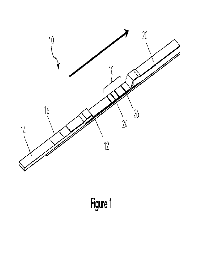
[0073] Figure 1 is an isometric view of an example flow assay membrane 10
which can be
used with the present method as a diagnostic test device. The assay membrane
10 comprises,
in series along a flow path: a sample addition area 16; a results area 18
comprising a test
region 24 and a control region 26, each of the test region and the control
region having an
immobilized binding molecule or species; and a wicking area 20. A sample
addition area 16 at
the upstream side of the lateral flow assay membrane 10 extends through one or
more fluidly
connected membrane to a results area 18 comprising the test region 24 and
control region 26,
and a wicking area 20. The arrow shows the direction of flow of running
buffer, also referred to
as the fluid flow path. The assay membrane 10 is preferably encased in a
cartridge for
protection and handling of the assay membrane in an automated analyzer.
[0074] A localization label is a molecule that is used in the assay to locate
the test region 24.
The localization label can be detected using a first imaging modality either
before or after the
assay run to determine the contours of the test region 24. After sample and
running buffer are
added to the assay membrane, an analyte label can be detected using the same
or different
imaging modality, where the analyte label binds an analyte of interest in the
sample and to an
immobilized binding species in the test region 24. By localizing the region of
interest at the test
region 24 using a localization label the imaging analysis of the analyte label
and analyte of
interest can be restricted to the known contours of the test region 24.
[0075] The localization label is a molecule or marker having a molecular
property that is
differentiated from the membrane background around it such that it can be
located by
imaging, optionally with a stimulus, at the region of interest where it has
been applied. The
assay membrane or fluid applied thereto comprises a detectable species, also
referred to as
the analyte label, that binds to an analyte of interest in a sample, either
directly or through a
coupling molecule, to visualize the presence of the analyte of interest by
binding with the
analyte of interest at one or more regions of interest through an immobilized
binding molecule
at one or more test regions, test lines, or test spots. The localization label
can be the same or
different than the analyte label, but does not interfere with the binding of
the analyte of
13
CA 03211910 2023- 9- 12
WO 2022/193028
PCT/CA2022/050413
interest to the immobilized binding species on the assay membrane. In one
assay membrane
design the localization label is applied to the test region 24 and is soluble
in running buffer
such that it is washed away by the running buffer. In another assay membrane
design the
localization label is immobilized on the test region 24 but does not impede
binding and
detection of any bound analyte of interest to the immobilized binding species
at the region of
interest.
[0076] The localization label or a localization label binding species that
binds the localization
label can be positioned inside, outside, or both inside and outside the test
regions of interest
on the assay membrane and can be positioned above, inside, or beneath any
layer of the assay
membrane. A localization label is a molecule that is used reversibly or
irreversibly bound to the
localization or test region and can be pre-localized or bound to the
localization region during
an assay run. Example of localization labels include but are not limited to
dyes or other
colorimetric molecules, fluorophores, radio labels, fluorochromes, or any
other molecule that
produces a signal detectable by an imaging system. The localization label can
be unbound to
the membrane and free flowing upon addition of running buffer. Alternatively,
the localization
label can bind to a localization label binding species, and the method of
binding of the
localization label to the localization label binding species can be, for
example, a direct covalent
or weaker non-covalent attachment method. Non-covalent methods could include
macromolecular anchoring molecules such as but not limited to antibodies,
avidin or
streptavidin, aptamers, nucleic acid with their appropriate binding pairs. The
molecular
property of the localization label can, for example, be such that the
localization label reflects
and/or absorbs and/or emits electromagnetic waves of wavelength between 10 nm
to 1 mm.
The molecular property of the localization label, in particular the
reflection, absorption, and/or
emission of electromagnetic waves, can be spontaneous or triggered by an
external excitation
or stimulus such as, for example, temperature variation, mechanical force,
electromagnetic
wave, chemical reaction, biochemical reaction, radiation, electron transfer,
filtration,
polarization, and light splitting.
[0077] The localization label can either be mobile or unbound and flow away
during the assay
run after application of running buffer, or be immobilized on the assay
membrane and remain
14
CA 03211910 2023- 9- 12
WO 2022/193028
PCT/CA2022/050413
in place during the assay run. A localization label that can flow away with
the running buffer
ensures that the localization label will not interfere with the post-run
molecular signal and with
analyte detection. Alternatively, a localization label that does not interfere
with the post-run
molecular signal can be immobile and can also be used, and the localization of
the localization
label at the test region 24 can be done in a different imaging modality than
that used to detect
analyte of interest bound to an analyte label in the same test region 24. Some
examples of
localization labels which can be used with the present device and method
include but are not
limited to organic dyes, inorganic dyes, fluorescent molecules, phosphorescent
molecules,
radiating molecules, and colored beads. Some specific examples of localization
labels include
Brilliant Blue FCF, Prussian blue, Quinoline Yellow WS, gold nanoparticles,
europium
nanoparticles conjugated with monoclonal anti-Human IgE, luminol, copper (Cu)
doped zinc
sulphide (ZnS), glass beads, carbon nanotubes, mercury telluride (HgTe)
quantum dots, and
phthalocyanine.
[0078] The localization label can also be water-soluble species that defines
and identifies the
region of interest for measuring the signal of a binding species and acts as a
proxy for
concentration of the analyte in question, and optionally washes away upon
assay run and does
not interfere with the reporter or analyte label that detects the analyte. In
addition, the
molecular property of the localization label can be proportional to a
concentration of binding
agent or immobilized binding species deposited at the test region of interest
to bind the
analyte of interest, and the molecular property and a proportionality constant
can be used to
calculate the concentration of the immobilized binding species which binds the
analyte of
interest at the test region 24. The comparison of the signal intensity of the
imaged region of
interest pre-labeled with the localization label before the assay run can also
provide an
indication of the age of the assay membrane, as binding species on the assay
membrane can
degrade with time and a decreased signal intensity of the pre-localization
label can be
indicative of an older, damaged, or less sensitive assay membrane. Pre-
labeling the assay
membrane using a localization label or localization label binding species that
binds a mobile
localization label and comparing it to signal from the analyte label and
analyte of interest
bound to the immobilized binding species at a known ratio and knowing the
degradation rate
CA 03211910 2023- 9- 12
WO 2022/193028
PCT/CA2022/050413
of each over time can also provide additional information on the integrity of
the assay
membrane and offers an opportunity of adjustment of the reported results based
on the
integrity or age of the assay membrane.
[0079] In use, once sufficient fluid is added to the sample addition area 16
on the assay
membrane or to an area partially overlapping with sample addition area 16, or
upstream of the
sample addition area 16 at the optional conjugate pad 14, the sample and
running buffer flows
along the defined fluid flow path (shown with an arrow) by capillary action
between the sample
addition area 16 and the wicking area 20. Fluid to run the sample can be a
sample fluid, i.e.
fluid containing the analyte of interest, running buffer, sample fluid mixed
with running buffer,
or a small amount of sample fluid followed by a sufficient amount of running
buffer to run the
assay. The sample addition area 16 on the diagnostic test device refers to the
area on the assay
membrane where a sample to be analysed is dispensed. Conjugate pad 14 can
further
comprise one or more conjugate detectable species and can also be used to
transfer sample or
running buffer upstream of the sample addition area 16 to the assay membrane.
The assay
membrane is preferably housed in a cartridge housing (not shown) to protect
the assay
membrane and compounds deposited thereon and to assist with receiving sample
and running
buffer. An optional solid support 12 can also provide structural support to
the components of
the assay membrane. The assay cartridge can further have one or more
identifier for identifying
the assay membrane, such as a barcode or other identifier, which can be any
textual or digital
data stored as an image that can be read by an optical reader or person.
Alternatively, the
assay cartridge can have one or more other identification tags such as, for
example, an RFID
tag or electromagnetic label.
[0080] In a typical lateral flow assay, a stationary or bound (immobilized)
binding molecule at
test region 24 binds to and indicates the presence (or absence) of an analyte
of interest, with
relative line intensity being correlated with the amount of analyte of
interest in the sample
applied to the assay membrane. Three test regions are shown as test lines,
however the assay
membrane can have one or more test regions. The control region 26 also
comprises a
stationary or bound (immobilized) reporter binding molecule which binds with a
reporter
molecule once the reporter passes the control region 26 to indicate a valid
test. The sample
16
CA 03211910 2023- 9- 12
WO 2022/193028
PCT/CA2022/050413
applied to an assay membrane is investigated to determine if it comprises the
species of
interest by running the assay with a developing solution or running buffer to
see if the species
of interest is present in the sample by binding to a region of interest at the
test region 24 on
the assay membrane. Each test and control region comprises a binding species,
and in many
standard lateral flow assay tests the binding species on the test region and
the control region is
usually invisible to optical systems prior to developing the assay. By
supplementing the applied
reagent on the regions of interest at the test region(s) with a localization
label that is detectable
by a visualization or imaging system, the region of interest on the membrane
in the test or
results area can be pre-localized. Upon interaction of an immobilized binding
species at the
region of interest on the assay membrane in the presence of the species or
analyte of interest
together with an analyte label the assay membrane can be visualized at the pre-
localized
region(s) of interest at the test region(s) to detect the presence of the
species of interest in the
sample.
[0081] In a standard immunoassay, a conjugate binding molecule binds an
analyte of interest,
which is then captured by an immobilized capture species at the test region.
In the present
pre-localization method the detectable species or detection conjugate can be
the same or
different from the localization label. The lateral flow or assay membrane is
referred to herein in
terms of the exemplary embodiment shown, however it will be readily apparent
that other
assay membrane device designs and possible variants of these designs could
also be similarly
configured for interrelationships with the presently described method and
device for sample
volume and concentration normalization and control and test region pre-
localization in a lateral
flow assay, particularly in an automated analyzer system, as herein described.
Other assay
membrane devices comprising various regions of interest including lines,
spots, and
comprising various membrane configurations can be used, for example for
chromatographic,
lateral flow, and enzyme-linked immunosorbent assay (ELISA) type assays.
[0082] To run the assay after pre-localization of a test region 24 having a
pre-applied mobile
localization label, sufficient running buffer is applied either directly to
the sample addition area
16 or to the conjugate pad 14 or into a running buffer reservoir in the
cartridge housing the
assay membrane 10 that is fluidly connected to the sample addition area 16. In
the
17
CA 03211910 2023- 9- 12
WO 2022/193028
PCT/CA2022/050413
embodiment shown, conjugate pad 14 at the first or upstream front end of the
fluid flow path
draws sample fluid and/or running buffer in the desired direction along the
assay membrane
optionally from a reservoir in a cartridge housing the assay membrane and
provides a capillary
force to draw up and move sample running buffer into the membrane of the assay
membrane
and through the sample addition area 16. The conjugate pad 14 is preferably
composed of a
glass fibre to allow mixing, and can optionally also include a porous material
such as, for
example, nitrocellulose, which can act as a size exclusion membrane and slow
fluid flow.
Conjugate pad 14 is optionally bendable, shown extending off from an optional
solid support
12, to accommodate a lowered buffer well or reservoir in the assay cartridge
base and further
positioned by an optional wick guide in the assay cartridge base and/or lid to
ensure fluid
contact with the running buffer. Obvious asymmetry in the design of the assay
membrane also
provides ease of assembly of the assay membrane within an assay cartridge and
provides a
directionality of the flow path so that the assay membrane is properly aligned
inside the
cartridge. Consistent alignment of the assay membrane in the cartridge can
also assist with
alignment of the cartridge and regions of interest on the assay membrane in
the analyzer.
Optionally a hydrophilic foil or layer (not shown) can be positioned onto at
least a portion of
the assay membrane to enhance the overall flow rate or process time of a
sample applied to
the flow assay membrane. The present membranes can be very small, and example
membranes used in this method are 3mm in width, providing an idea of the scale
of the
present membrane and its features. It is understood, however, that a variety
of assay
membrane sizes may be used.
[0083] The assay membrane can also optionally comprise one or more flow
channels,
optionally cut or pressed into the surface of the membrane substrate. The
fluid flow path may
also include additional separate areas containing one or more reagents,
antibodies, or
detection conjugates (detectable species), as well other areas or sites along
the fluid path that
can be used, for example, for washing of the sample and any bound or unbound
components
thereof. The assay membrane can also be optionally treated to adjust the
sample properties,
such as, for example, by pH level or viscosity. Additional reagents can be
located or applied on
or inside the assay membrane. Example optional reagents added to the assay
membrane can
18
CA 03211910 2023- 9- 12
WO 2022/193028
PCT/CA2022/050413
be any combination of, but not limited to, antibodies, salts, surfactants,
detergents,
macromolecules, small molecules, small molecules nanoparticles, microspheres,
and antigens,
where the reagents can be added or applied as liquid or solids to the assay
membrane.
[0084] Components of the assay membrane such as the physical structure of the
device
described herein can be prepared from, for example, copolymers, blends,
laminates,
metallized foils, metallized films or metals, waxes, adhesives, or other
suitable materials known
to the skilled person, and combinations thereof. Alternatively, device
components can be
prepared from copolymers, blends, laminates, metallized foils, metallized
films or metals
deposited on any one or a combination of the following materials or other
similar materials
known to the skilled person, examples include but are not limited to
paraffins, polyolefins,
polyesters, styrene containing polymers, polycarbonate, acrylic polymers,
chlorine containing
polymers, acetal homopolymers and copolymers, cellulosics and their esters,
nitrocellulose,
fluorine containing polymers, polyamides, polyimides,
polynnethylmethacrylates, sulfur
containing polymers, polyurethanes, silicon containing polymers, other
polymers, glass, and
ceramic materials. Alternatively, components of the device can be made with
plastic, polymer,
elastomer, latex, silicon chip, or metal. In one example, the elastonner can
comprise
polyethylene, polypropylene, polystyrene, polyacrylates, silicon elastomers,
or latex.
Alternatively, components of the device can be prepared from latex,
polystyrene latex or
hydrophobic polymers. In one example, a hydrophobic polymer can be used for
the cartridge
or membrane support comprising, for example, polypropylene, polyethylene, or
polyester.
Alternatively, components of the device can comprise TEFLON , polystyrene,
polyacrylate, or
polycarbonate. Alternatively, device components can be made from plastics
which are capable
of being embossed, milled or injection molded or from surfaces of copper,
silver and gold films
upon which may be adsorbed various long chain alkanethiols. The structures of
plastic which
are capable of being milled or injection molded can comprise, for example, a
polystyrene, a
polycarbonate, a polyacrylate, or cyclo-olefin polymer. The assay membrane can
also comprise
an optional filter material which can be placed within and/or downstream the
sample addition
area to filter particulates from the sample, for example to filter or trap
blood cells or particulate
matter from blood so that added plasma can travel through the device.
19
CA 03211910 2023- 9- 12
WO 2022/193028
PCT/CA2022/050413
[0085] Various configurations of diagnostic assay membranes and lateral flow
assay
membranes are known that can be used with the present method and prepared as
described,
including but not limited to variation in device dimensions, materials,
porosity of the substrate,
presence or absence of topographical features on the substrate, channel shape
and
configuration, and method of manufacturing the channel. The particular assay
membrane is
referred to throughout this description in terms of an exemplary embodiment,
however it will
be readily apparent that other device designs and possible variants of these
designs could also
be similarly configured. The described assay membrane 10 is particularly
useful for
immunoassay formats which are typically sandwich assays wherein the membrane
is coated
with an immobilized capture antibody or protein, sample is added, and any
analyte of interest,
either antigen or antibody, present in the sample binds to the immobilized
capture molecule at
a test region. In common immunoassays, a detecting antibody binds to antigen
in the sample,
an enzyme-linked secondary antibody binds to the detecting antibody or to the
antigen, and a
substrate in the fluid is converted by the enzyme into a detectable form.
[0086] In an automated system or analyzer, detection can be done automatically
using a
visualization or imaging system such as a camera or other detection system.
The visualization
system can further include one or more light sources emitting the same or
different
wavelengths of light, one or more lenses for focusing and enlargement of the
test area, and
one or more optical filters for eliminating or selecting specific wavelengths
of light. The
imaging device can also comprise one or more photodiode, photoresistor,
phototransistor,
camera, focal plane array, spectrometer, hall effect sensor, photomultiplier
tube, antennas, and
electrode.
[0087] Figure 2A is an illustration of the results area of an example assay
membrane, such as
the one shown in Figure 1. In results area 18 there are two test regions, 24a
and 24b, and
control region 26. In advance of adding running buffer to the assay membrane
the two test
regions, 24a and 24b, and optionally also the control region 26, which were
marked with a
localization label, are visualized using a visualization system.
Alternatively, other variations of
localization label addition and binding as well as variations on imaging
modalities can be used.
In all cases the visualization system pre-localizes the test region(s) and
also optionally the
CA 03211910 2023- 9- 12
WO 2022/193028
PCT/CA2022/050413
control region(s) on the assay membrane in the results area 18 by imaging a
localization label
to enable the analyte of interest to be found in the same region by imaging an
analyte label
using the same or different imaging modality. The dashed lines indicate the
contours of the
location of line pre-localization of the region of interest (ROI) around each
of the test and
control regions before the analyte of interest is detected, with a margin
around each line to
ensure that the luminosity of the analyte of interest bound to the test region
is properly read. In
a preferable embodiment the margin of the ROI is less than half of the
distance from one
region of interest to a different region of interest. In another preferable
embodiment the
margin on the side of each region of interest is less than 0.5mm, less than
0.3mm, or less than
0.2mm.
[0088] Figure 28 is an illustration of one test region in a region of interest
on a flow assay
membrane. The region of interest (ROI) 28 indicated by the dashed lines is
shown as between
the predetermined contours at the location of the pre-localized test region 24
which was
detected with a visualization system and imaging modality to localize the
localization label on
the test region with a reasonable margin. After the assay is run, the
visualization system in an
analyzer can locate the region of interest on the flow assay membrane based on
automated
membrane alignment, and optionally with the addition of external reference
features such as
cartridge feature location, cartridge port or opening alignment with the
imaging system,
and/or other location markers such as relative locations of the regions of
interest to other
detectable regions of interest such as the control region or other test
regions. The intensity of
the test region 24 at the pre-localized ROI on the assay membrane can then be
measured after
the assay is run to determine the presence or absence of the analyte of
interest in the sample.
In addition, the intensity of the test region at the pre-localized location
can be used to
quantitatively determine the concentration or concentration range of the
analyte of interest in
the sample based on the detected intensity of the localization label as
detected at the test
region 24.
[0089] The localization label has a molecular property used for detection of
the labeling
species that can be imaged in advance of running the assay or after running
the assay, and that
differentiates the region of interest where the localization label has been
applied from the
21
CA 03211910 2023- 9- 12
WO 2022/193028
PCT/CA2022/050413
background of the assay membrane. The differentiating molecular nature or
property of the
localization label can be, for example, wavelength or color, frequency, phase,
amplitude,
intensity, delay time, energy, fluorescence lifetime, refractive index,
reflectance, absorbance,
emissivity, transmittance, polarization, dispersion, and scattering. The
localization label
molecular properties can be observed using a detector or imaging modality
which may have
any combination of, but not limited to, one or more photodiodes,
photoresistors,
phototransistors, cameras, focal plane arrays, spectrometers, hall effect
sensors,
photomultiplier tubes, antennas, and electrodes. If needed, an external
stimulus to stimulate
the molecular property can be used, for example any combination of but not
limited to
temperature variation, mechanical force, electromagnetic wave, chemical
reaction, biochemical
reaction, radiation, electron transfer, light filtration, light polarization,
and light splitting.
[0090] Figure 3 illustrates one general method for pre-localization of regions
of interest on an
assay membrane by applying the localization label and immobilized binding
species to the
assay membrane during manufacture. In the assay membrane shown there are three
test
regions 24 and one control region 26, however it is understood that the pre-
labeled assay
membrane can have one or more test regions and preferably at least one control
region. In the
first step, the assay membrane is visualized using a visualization system to
provide a pre-assay
localization of the test and optionally the control regions and identification
of the region(s) of
interest on the assay membrane by detection of the localization label 102.
This process can be
generalized to other methods of localization where one or more result areas
are searched for
any shape, number, or size of regions of interest. After the locations of the
test regions have
been determined and the regions of interest identified, a running buffer is
added to the assay
membrane to run the assay 104. Pre-run analysis is then used to locate the one
or more test
region(s) 24 in the results area on the assay membrane in the region(s) of
interest after the
assay is run, which is used later to reduce the amount of background noise
captured post-run
by the imaging or visualization device. By pre-locating the test region at the
region(s) of
interest the visualization system can be programmed to ignore any area
surrounding the test
region(s), which are sources of background noise, and focus on the region(s)
of interest where
the immobilized binding species is localized. Background noise increases the
uncertainty of any
22
CA 03211910 2023- 9- 12
WO 2022/193028
PCT/CA2022/050413
system, especially near the limit of detection. In assay runs with low signal,
distinguishing the
background noise from regions of interest can be near impossible and can
result in
inconclusive assay results. Post-assay detection of the analyte label at the
test regions in the
results area of the assay membrane can then done based on the line pre-
localization 106
providing a meaningful signal even with a low signal result of the analyte of
interest. The signal
intensity of each region of interest, i.e. at each of the test and control
regions, can thus be
measured by limiting the use of the collected image data to the location of
each region of
interest, optionally with a reasonable margin, to reduce the noise
contribution to the signal
from the region of interest.
[0091] Figure 4 is an example high signal result from a lateral flow assay
membrane assay
after an assay run. Image A shows the results area from the lateral flow assay
with fluorescence
imaging of bound Europium to two test regions 24a, 24B and a control region
26. The images
were taken using a CMOS (complementary metal¨oxide¨semiconductor) camera, a UV
LED and
the Europium reporter. Prior to the assay run the same results area was imaged
to pre-localize
the contours of the regions of interest as shown by the dashed lines in image
B, and the
localization label on the test and control regions was imaged. After the assay
run the image
processing of the area at the test and control regions could be restricted to
the contour areas
of the region of interest identified by the presence of localization label
before the assay and
limited to the area between each set of dashed lines. In a high signal result
where the test and
control regions are bright and discernable in post-assay imaging the line pre-
localization can
be useful for excluding extraneous signal evidenced by gradient shading
adjacent the control
and test regions.
[0092] Figure 5 is an example low signal result from a lateral flow assay
membrane assay.
Image A shows the results area from the lateral flow assay with fluorescence
imaging of bound
Europium to two test regions 24a, 24B and a control region 26 where the test
regions are more
difficult to discern due to lower contrast in the image at the regions of
interest compared to
background. In the low signal case, determination of the test region(s)
location in an
automated system can be challenging as the signal to noise ratio is high
compared to
background. However, using line pre-localization the signal intensity can
still be meaningfully
23
CA 03211910 2023- 9- 12
WO 2022/193028
PCT/CA2022/050413
measured by restricting the quantified signal to the contour area at the
region of interest
around which the bound immobilized species is known to be on the results area
of the assay
membrane. Image B shows the same results area where the regions of interest at
the test and
control regions were pre-localized such that image analysis can be done only
on the pre-
localized regions of interest. In this way even low signal assay results can
be meaningfully read
and recorded, reducing the false negative incidence of the test. Further,
signal amplification in
the localized region(s) of interest can provide higher accuracy results for
calculation for samples
which have a low concentration analyte of the interest. Pre-localizing the
test region(s)
increases the limit of detection of the lateral flow assay by providing a
signal measurement in
the location where the immobilized species is known to be, and calibration of
the optical or
visualization system and data analysis method improves the accuracy of
analysis of the assay
results. Further, pre-determination of the location of the immobilized binding
species improves
high throughput automation results by limiting the data analysis to the known
region of
interest, thus limiting processing time and resulting in a more reliable
result. The process of
using a localization label is particularly useful when algorithms that search
test areas for regions
of interest for positive signals are used. These algorithms look for
significant signal:noise
changes and use these changes to identify a region of interest on an assay
membrane. In this
instance, any region with a high background in a negative sample run result
area may falsely be
labeled as the region of interest. This can also happen for very low positive
sample runs. This
"false localization" can increase the imprecision of a population of negative
and low positive
samples, and the increase in imprecision in turn decreases the limit of
detection of an assay by
making a low positive sample difficult to statistically distinguish from a
negative sample. Using
pre-localization of regions of interest ensures that image analysis is
performed at the location
where the bound species is known to be on the assay membrane.
[0093] Figure 6 is a flowchart of a method for pre-localization of a region of
interest on a flow
assay membrane with localization label and immobilized binding species pre-
applied to the
test region(s). A manufactured assay membrane necessarily has variation, and
the present
method reduces noise in the assay result and can normalize the variation using
pre-detection of
a localization species on the assay membrane before the assay is run. In an
automated assay
24
CA 03211910 2023- 9- 12
WO 2022/193028
PCT/CA2022/050413
reader or analyzer, the contrast in electromagnetic or molecular property of a
localization label
at a region of interest is determined using an imaging device before the assay
run. Molecular
properties at the region(s) of interest can be observed with or without an
external stimulus
depending on the localization label, and the molecular property can be
detected by one or
more detectors. In an example, the molecular property of the localization
label can be one or
more of wavelength (color), frequency, phase, amplitude, intensity, delay
time, energy,
fluorescence lifetime, refractive index, reflectance, absorbance, emissivity,
transmittance,
polarization, dispersion, and scattering, and the localization label can be
detected with or
without excitation prior to imaging, depending on the molecular property being
detected. If
the contrast in molecular property of the localization label is detectable
without external
stimulus 202 then the ROI locations can be found using the contrast of
molecular properties
206. If an external stimulus is required, as in the case of a fluorescent
detectable localization
species, an external stimulus is applied 204, such as a light at or near the
excitation wavelength
of the fluorescent detector, and the imaging device finds the ROI locations
using the contrast
of molecular properties 206 with stimulus. The analyzer then confirms that the
ROI locations
are correct in the assay reader 208, and if not found to be correct, the
position of the assay
membrane in the assay reader can be adjusted 210. The assay membrane can then
be imaged
again to localize the ROI locations on the assay membrane 208 by locating the
localization
label and the correct assay membrane position is recorded for that assay
membrane result area
212. The method for locating positions of regions of interest can be repeated
until the position
is located. This step ensures that an accurate position for region of interest
is located each run,
and controls for the positions of the regions of interests that are impacted
by, for
example, inconsistent human handling, error or drift in reader components,
assay
manufacturing and assay assembly. Once the assay membrane position and ROI
position(s) has
been recorded, the differential molecular properties between ROls and other
regions are also
recorded 214 and can be used for background subtraction calculation. The assay
is then run
216 by adding running buffer to the upstream end of the assay membrane.
[0094] The signal from the imaging or signal detection in the analyzer is
digitized and can be
transformed into, for example, a vector or multi-dimensional data array. The
molecular
CA 03211910 2023- 9- 12
WO 2022/193028
PCT/CA2022/050413
property data arrays are then processed to locate the region of interest based
on the contrast
between regions of interest and other regions. To find the location of test
and optionally also
control region(s) of interest using the molecular property of the localization
label, software
algorithms in the image processing system can use, for example, one or more of
cropping,
rotation, smoothening, color space transformation, time-frequency domain
transformation,
contrast enhancement, sharpening, thresholding, amplification, clipping,
averaging, feature
extraction, scaling, pattern recognition, projection, component analysis,
wavelet
transformation, filtering, algebra calculation, histogram operation, and
geometric
transformation. The software algorithm can also use the relative locations of
and the signal
intensity at regions of interest for any of the molecular properties of the
localization label for
signal correction after the assay has been run, which can be done using, for
example, any
combination of linear or non-linear algebra calculation or transformation.
[0095] An automated analyzer is preferably used to receive the assay membrane
and also
preferably to process the assay results including imaging and visualization or
detection of
regions of interest on the membrane. In one example, the analyzer can comprise
a fluid
dispense area comprising a sample conduit for dispensing a fluid volume onto
the assay
membrane, an imaging area comprising a light source for illuminating the assay
membrane and
an imaging device for imaging the assay membrane, and a processor for
analysing image data
collected from the imaging device.
[0096] Figure 7 is a flowchart of a visualization or detection method for pre-
localization of a
region of interest (ROI) on a pre-labeled assay membrane in an analyzer. The
assay membrane
can be pre-labeled with any localization label that can be detected by the
imaging device in
the analyzer or any localization label binding species that binds the
localization label. The
localization label can, for example, be colored or have molecular properties
that can be
changed with an external stimulus, such as a fluorescent species. The assay
membrane is put
into an imaging device or automated analyzer with an imaging device to take an
image of the
ROI. The imaging device can comprise, for example, one or more camera, charge-
coupled
device (CCD) sensor, or complementary metal oxide semiconductor (CMOS) sensor.
The light
or signal captured by the imaging device can be visible, infrared, of single
or multiple
26
CA 03211910 2023- 9- 12
WO 2022/193028
PCT/CA2022/050413
wavelengths. A fluorescent localization label is a species that is detectable
in a detection range
different from the fluid sample when excited by an illumination light source
and can re-emit
light of a different wavelength after light excitation from the illumination
source at a first
wavelength. When used with a fluorescent localization label as the pre-
labeling species or
localization label, imaging can comprise application of an external stimulus
such as light as a
suitable excitation wavelength to excite the fluorescent species. Fluorescent
species, such as
fluorescent dyes and fluorescent proteins, can offer several advantages
compared to
colorimetric labels, such as greater assay sensitivity, quantitative readout,
and the possibility of
multiplexing for simultaneous on-site measurement of different substances from
a single
sample. Commonly used labels include, for example, gold nanoparticles (GNP),
quantum dots,
and fluorescent microspheres. The localization label applied at the region of
interest has a
different molecular property from the rest of regions on the assay membrane
after the assay is
manufactured but before the assay is run. The differences can be any and/or
any combination
of wavelength, color, frequency, phase, amplitude, intensity, delay time,
energy, fluorescence
lifetime, refractive index, reflectance, absorbance, emissivity,
transmittance, polarization,
dispersion, and scattering.
[0097] For pre-localization of the ROI, an imaging or detection device is used
to receive array
image data of the assay membrane and a processor is used to process the imaged
data. The
pre- localization of the test region(s) can be done either before the assay is
run or after the
assay is run using an imaging modality that differentiates the localization
label from the analyte
label. The imaging system of the analyzer includes at least one illumination
light source or and
at least one light receiving unit connected to a microcontroller and computer
system for
recording and analyzing collected imaging data. A computer capable of data
analysis in the
present methods comprises a processor, memory, and at least one data storage
device or
connection thereto. The system memory typically contains data such as data
and/or program
modules such as an operating system and application software that are
accessible to and/or
are operated on by the processor. The computer may also include other
removable/non-
removable, volatile/non-volatile computer storage media. The computer may be
connected to
the imaging device or receive data from the imaging device for processing.
27
CA 03211910 2023- 9- 12
WO 2022/193028
PCT/CA2022/050413
[0098] Once the ROI has been imaged by the imaging device, a processor
receives arrays of
values from imaging device 302 and the image data is cropped to the region of
interest 304.
Methods such as adaptive thresholding can be used to define and crop the assay
region. The
intensity contrast between the region of interest and other regions 306 is
then enhanced and
the noise is removed as much as possible from region of interest 308. The
contours of regions
of interest in the data array are then found, and their locations and
dimensions located 310.
The analyzer finds contours in the image where the regions of interest may be
present by
comparing electromagnetic or pixel intensity in and around the ROI.
Corrections can also be
applied to increase the contrast of the image and noise is then removed from
the image. The
precise location and contours of the region of interest can be based on a
variety of metrics
such as, for example, size, aspect ratio, pixel intensity contrast, gradient
slope, and relative
distance of the contours. The contours that contain the location of the
regions of interest are
projected onto the original data array 312 and the intensity data of the
region of interest is
stored as well. The differential intensity data between the region(s) of
interest and other
regions 314 is also stored, as well as the location and size of the contours,
optionally in
combination with one or more other reference locations that can be
additionally used to
localize the regions of interest on the assay membrane.
[0099] Figure 8 is an illustration of a flow assay membrane with a
localization label after
manufacturing, before an assay run, and after an assay run. The pre-run
localization analysis
uses a proxy labeling or localization label to calibrate the location and also
optionally the
intensity of the test region(s) and optionally also control region(s) after
run of the lateral flow
assay. After assay manufacturing the regions that the optical device can
locate are region of
interest R1, region of interest R2, and background region R5 of the detection
area. The regions
of interest can exhibit different (region 1) or the same (region 2) molecular
properties from the
other regions (region 5) when no external excitation or stimulus is applied,
and the molecular
properties for the ROls can be the same or different with the use of different
localization label
or combinations of localization label, or different concentrations thereof.
The molecular
property of the localization label can be detected by one or multiple
detectors or detector
combinations of, for example, photodiodes, photoresistors, phototransistors,
cameras, focal
28
CA 03211910 2023- 9- 12
WO 2022/193028
PCT/CA2022/050413
plane arrays, spectrometers, hall effect sensors, photomultiplier tubes,
antennas, and
electrodes. The detected signals are then digitized and transformed into a
vector and/or multi-
dimensional data array. If needed, external excitations or stimulus can be
applied by any
combinations of temperature variation, mechanical force, electromagnetic wave,
chemical
reaction, biochemical reaction, radiation, electron transfer, filtration,
polarization, and light
splitting. With external excitation or stimulus, the molecular properties in
ROls (region 1-2) are
different from other background regions (region 5). The molecular properties
in ROls can be
the same (region 2) or different (region 1) from the situation without
external excitation or
stimulus, and can be the same or different from each other.
[0100] The molecular property data arrays are processed to locate the ROls
based on the
contrast between ROI and other regions. Between the assay membrane manufacture
date and
the time the assay is run, degradation, aging, fading, and chemical and
biological changes can
cause a change in localization label as well as the binding species in the
assay which can affect
the signal measured at the different regions. Further, after the lateral flow
array is run the
imaging of each region will have again changed compared to the pre-assay
region of interest
R1, region of interest R2, and background of detection area R5. In addition,
other test and
control regions may be visible in the test area of the assay membrane R3, R4
where there had
been no application of localization label during manufacturing but where
imaging can detect a
change in signal intensity after the assay run. Such regions of interest can
also be localized after
the assay run based on their known relative location from the regions of
interest that had been
pre-localized with localization label (R1/R2). In particular, the manufacture
of assay membranes
with multiple lines where only one or only two lines have pre-labeling may
also be useful in
localizing the other lines after the assay run based on the distance from one
line to a labeled
line or an unlabeled line. During the deposition of binding agent on the assay
membrane, the
quantity of the localization label can be proportional to the immobilized
binding reagent at the
region of interest, thus the signal from the localization label can be used as
a proxy for
concentration obtained from its molecular properties and can be used to
calculate the
concentration of immobilized binding species using a proportionality constant.
Calibration of
effective concentration immobilized binding species on the assay membrane to
determine the
29
CA 03211910 2023- 9- 12
WO 2022/193028
PCT/CA2022/050413
concentration of analyte of interest in the sample can thereby also be done
based on the
differential intensity of each region of interest before and after the assay
run. In particular, the
signal intensity of the molecular signal detected from the ROI on the assay
membrane is
related to the concentration of reagents on the lateral flow assay membrane
through a
proportionality constant. The proportionality constant can be used to
calibrate the amount or
concentration of bound reagent on the test region and/or control region
through calculating
the signal intensity of the localization label at the ROI.
[0101] After the assay test is run, the optical properties of each region of
interest can also
change. In one example, the molecular properties in the ROI may change based
on the
external excitations and/or stimulus used for signal reading. For example, the
molecular
properties in region 1 after running the assay can be the same or different
from other regions
(region 5), which are different from before running the assay, and different
from without
external excitation or stimulus. The molecular properties at region 2 (R2)
after running the assay
can be the same as other regions (such as background R5), and/or different
from the molecular
properties of the same region before running the assay, and the same or
different as without
external excitation or stimulus. Molecular properties in region 3 (R3) and
region 4 (R4) after
running the assay can be the same or different from other regions (R5), the
same or different as
before running the assay, or the same or different as without external
excitation or stimulus.
[0102] After the assay is run the assay membrane is moved back to the target
position in the
reader by the automated analyzer and the actual ROI locations are found and
compared with
the target value, with optional adjustment to the assay membrane position
until the alignment
is acceptable for imaging and signal processing. In particular, the analyzer
can move the assay
to the target position in the imaging system and the analyzer can find the
locations of the
region of interest using the pre-run localization data. Alternatively,
computer processing can
adjust the image taken after the assay run to align the received image to the
pre-localization
ROI locations. The device then reads the assay signal and applies signal
correction using pre-
run molecular properties. The signal correction can be done using any or any
combination of
linear or non-linear algebra calculation or transformation. The assay signal
is then read and
recorded, optionally with applied external stimulus, and a signal correction
is applied using the
CA 03211910 2023- 9- 12
WO 2022/193028
PCT/CA2022/050413
previously recorded differential molecular properties. The signal correction
can be done using
any or any combination of linear and/or non-linear algebra calculation and/or
transformation. In
this example, the signal data collected for regions 1-4 can be used to
indicate either a positive
or negative reaction for the test. Regions 1, 3, and 4 can also be used as a
control signal when
region 2 is used as a test region. After the post-run analysis is complete,
the device will output
results.
[0103] Figure 9 is a flowchart of a method for detection of signal at a region
of interest in an
automated analyzer after test region pre-localization and run of the assay by
addition of a
running buffer. After the lateral flow assay test has been completed, the
regions of interest i.e.
the test and region(s), are located using the pre-labeling information and
data recorded for the
assay membrane. To read the assay results the assay membrane is moved to the
recorded
position in the imaging device 350 of the analyzer. If the contrast in
molecular properties
requires an external stimulus 352 then an external stimulus 354 is applied,
such as appropriate
illumination. If no external stimulus or illumination is needed and the
contrast is sufficient then
none is required. The ROI locations on the assay membrane are then found using
the contrast
of molecular properties 356. The analyzer then refers to the stored ROI
locations and queries
whether the newly found ROI locations are correct in the assay reader 358. If
not, the assay
membrane position inside the assay reader is adjusted 360 and the ROI
locations are re-found
using the contrast of molecular properties 356 to ensure that the location of
the assay
membrane is correct. If the positioning is correct, then the assay signals at
the ROI are read
362 and signal correction is used to record the differential molecular
properties using
information related to molecular properties recorded before the test and after
the assay run
364. The result is then output for reading and reporting.
[0104] A variety of techniques can be used in the method of detection of a
region of interest
(ROI) post-localization. The pre-localization method consists of two parts: a
pre-localization
analysis to localize the test region(s) and a post-run analysis to determine
the presence and/or
amount of analyte of interest bound at the test region and optionally also to
verify the control
region. The data processing method shown for ROI locating is based on the
contrast in
molecular signal at the location of the localization label compared to the
background of the
31
CA 03211910 2023- 9- 12
WO 2022/193028
PCT/CA2022/050413
assay membrane. In one technique this is done using light illumination at a
particular
wavelength and light intensity to detect the contrast. ROls such as those at
test region and
control region can also have one or more binding species, such as immobilized
antibodies, in
addition to the localization label, and the localization label can be
deposited together with test
region and control region antibodies or in a separate step. When the
localization label is
deposited onto the assay membrane in the same process and/or same solution as
the
deposited testing reagents (e.g. test region antibodies), the quantity of the
localization labeling
species at each ROI will be proportional to the testing reagents on the assay.
In particular, a
more intense contrast in molecular signal of the localization label at the ROI
indicates that
more binding species have also been deposited in the same location. Thus, the
amount of
localization label at the ROI can be used as an indication of the amount of
test reagent(s), and
the recorded difference in observable molecular properties (such as light
intensity values in the
above example) can be used to correct for the assay signal reading after the
assay run. As such,
the contrast in molecular signal can be indicative of the concentration of
immobilized binding
species as the ROI. This step can improve the assay accuracy by compensating
for the variation
in assay manufacturing and different readers. Once the assay membrane has been
prepared
with the immobilized binding species and localization label at the ROI the
assay membrane can
then be loaded with sample and running buffer and the ROls can be found after
the assay run
using the pre-localization determination for the same assay membrane.
[0105] For location of the ROI (pre-localization) before running the assay,
the localization
label can be detected, for example, with a white light source and an RGB
camera, or a light
source capable of sufficient differentiation of the localization label
compared to the
background of the assay membrane. Other visualization techniques can also be
used, such as,
for example, fluorescence, to contrast the molecular properties of the
localization label at the
test regions compared to background. The contrast can then be used to generate
a data array
with different light intensities, indicating the relative location of the ROls
on the assay
membrane. In both cases of fluorescence detection as well as visible light
detection, data
processing methods are similar. In one example of data processing, the ROI is
digitized to an
array of intensity values. The array can then be extracted and cropped to
reduce data size. The
32
CA 03211910 2023- 9- 12
WO 2022/193028
PCT/CA2022/050413
data can also be cropped to the assay region by adaptive thresholding or image
thresholding.
The intensity contrast between the ROI and other regions can then be enhanced
and separated
from other regions. Noise areas can also be removed, for example by dilation
and erosion. The
contours in the data array can then be found, as well as their locations and
dimensions, and the
ROI positions and sizes can be determined by filtering by, for example,
contour location, size,
aspect ratio, and/or relative distance. The contour locations are then
projected to the original
data array.
[0106] In an example, if the detection conjugate bound to the immobilized
binding species
comprises a europium label, excitation of the ROI after assay run by a 365nm
light source will
generate a fluorescent signal at about 635nm at the pre-localized ROI. The
differential intensity
data between ROls and other regions is also stored and the analysis outputs
the assay ROI
location on the assay membrane. Other optical improvements and data analysis
techniques
that can be used to render the data more precise or accurate could be applied.
The ROI
position determination process can also be repeated in a closed loop until a
satisfactory
location can be obtained. This iterative step ensures the accurate position of
the ROls inside
the reader, which can be highly variable due to, for example, inconsistent
human handling,
error/drift in reader components, assay manufacturing, and assay assembly.
After the assay
test, if the localization label is washed away the immobilized binding species
remaining on the
assay membrane at the ROI can be detected using molecular contrast in
combination with the
pre-localized locations of the ROls. Pre-localization of the ROls is
especially useful in the case
where there is little or no molecular contrast visible at the test regions
after the run, where pre-
localization can provide the location contours of the pre-localized
immobilized binding species
to better differentiate the signal at the pre-localized ROls.
[0107] Figure 10 is a flowchart of a method for manufacturing an assay
membrane with a
localization label for pre-localization of a region of interest. In the
manufacture of assay
membranes, detectors, binding agents, and bound species are applied to the
membrane in an
industrial process, often sprayed to large sheets of assay membrane. To mass
manufacture
lateral flow assay membranes, the prepared membrane sheets are then cut into
the desired
size and placed into a protective cartridge for transport, handling, and assay
running. The
33
CA 03211910 2023- 9- 12
WO 2022/193028
PCT/CA2022/050413
manufacturing process of lateral flow membranes is necessarily imprecise, and
minor variations
are introduced in, for example, membrane size, concentration of applied
reagents and
materials, quality or age of applied reagents, and amount of applied reagents
and materials
assembled between assay membranes, as well as in their placement inside each
cartridge.
Uncontrollable variations are thereby present in the manufacturing of assay
membranes, which
can lead to uncertainty in the assay results, especially when the variations
cannot be corrected
or normalized. These minor variations can also have a detectable and
significant effect on test
results, and can significantly change the results during quantitative
investigations. During
manufacture of pre-labeled assay membranes, a control solution is prepared for
application to
the control region 402. If the control solution comprises a detectable species
that has
molecular properties whose contrast can be detected 404 then the detectable
species in the
control solution can act as the localization label. Otherwise, an additional
localization label can
be optionally added to the control solution 406 for creating the labeled
control region(s). The
test solution is prepared for the test region 408 in a similar manner, whereby
if the test solution
comprises a detectable species that can be detected without the addition of a
localization
label 410 no additional localization label is required, otherwise a
localization label or
localization label binding species is added to the test solution 412. By pre-
localizing the each
bound and/or labeled species the location, concentration, and amount of
sprayed species can
be detected either prior to the assay run or after the assay run and prior to
imaging the analyte
of interest, and the results with contoured test region can be used for more
accurate
quantitation of the analyte of interest in the sample after the membrane has
been exposed to a
sample and the assay developed. In addition, starting concentration of
detectors, and other
bound species can be incorporated into quantitative calculations of sample
concentration to
more accurately discern concentrations of species of interest in the sample.
The control
solution and test solution are then applied to the assay membrane 414, which
is often done
using spray application. The assay membrane is then prepared for use in the
analyzer 416,
which often involves stabilization and/or protection of the assay membrane in
a protective
cartridge.
34
CA 03211910 2023- 9- 12
WO 2022/193028
PCT/CA2022/050413
[0108] In another embodiment of the present invention, the assay membrane can
be pre-
labeled at the regions of interest with an immobile localization label that
does not interfere
with binding of the analyte of interest to the immobilized binding species or
detection of the
detectable species at the region of interest. This embodiment uses a
localization label that has
a molecular property that is detectable in a different imaging modality or,
for example, at a
different wavelength than the detection range of the detectable species that
indicated binding
of the analyte of interest to the immobilized binding species at the region of
interest. In one
example of this embodiment, sample and running buffer can be added to the
assay membrane
to develop the assay and allow any analyte of interest in the sample to bind
to the immobilized
binding species and detectable species at the region of interest. After the
assay is run a first
imaging method which can detect the presence of the immobilized localization
label at the ROI
is used to locate the ROI and assign the contours of the ROI. A second and
different imaging
method is then done to detect the presence of the detectable species inside
the contours of
the ROI to determine the binding of any analyte of interest in the ROI. For
example, the first
imaging method to image the localization label can use red light illumination
and a digital
camera, and the second imaging method can use fluorescent illumination where
the detectable
species is one that has fluorescent molecular properties. In this method the
assay membrane
only needs to be moved by the analyzer into an imaging area one time, after
the assay run, if
the analyzer has two modes of imaging and detection, one for each of the
localization label
and the detectable species.
[0109] Example 1
[0110] An experiment was done to demonstrate pre-localization of test and
control regions
and visualization thereof in a lateral flow assay. Brilliant blue FCF was
added as a localization
label to a control solution and to a test solution and the solutions were
applied to an assay
membrane at a control region and test region, respectively. Prior to running
the assay, an
optical system took images of the test region and control region using white
light through a
detection window before the lateral flow assay. The brilliant blue FCF was
used as a localization
label to indicate the locations of the test and control regions and the
location data was stored.
Sample containing an analyte of interest and a running buffer was then added
to the
CA 03211910 2023- 9- 12
WO 2022/193028
PCT/CA2022/050413
membrane and the assay was run. A fluorescence probe that binds to the analyte
of interest
was also present in the assay. After the lateral flow assay run the optical
system took a second
image of the detection area under UV light (to excite the fluorescence probe),
and the
reflectance values were recorded. The analysis algorithm, rather than
searching for the test and
control regions using intensity data, used the pre-localization, pre-run
localization data to
locate the contours of the test region and control region (ROls) based on
prior identification of
the location of the brilliant blue FCF. The reflectance values in the region
that were located
pre-run were then used to calculate the concentration of the analyte of
interest.
[0111] Figure 11 is a panel of assay membranes with regions of interest during
the pre-
localization and post-localization method as described in this experiment.
Panel A is an image
of a pre-localized test region (bottom) and control region (top) with the
localization label
contrast to background shown as observed under white light. Panel B is an
image of the pre-
localized test and control regions with localization margins identified based
on molecular
contrast under white light of the localization label and background. Panel C
is an image of a
post-assay lateral flow membrane with test region (bottom) and control region
(top) taken
under UV light showing the presence of bound fluorescent label. It is notable
that the bottom
band or test band is very diffuse, with blurry margins. Panel D is an image of
post-assay test
and control regions with pre-localized margins indicating the location of the
immobilized
binding species.
[0112] Test region localization on an assay membrane provides a boundary
region for
automated analysis such that the results region where binding of the species
of interest can be
analyzed with confidence. Localizing the test binding region, either before
the assay run or
afterwards, and doing so in a way that differentiates the imaging results of
the localization label
from those of the binding species, enables more accurate measurement of the
bound species
of interest at the binding region while excluding noise in the surrounding non-
binding region.
It has been found that this method enables lower threshold concentration
measurement of the
analyte of interest in a test sample and results in fewer erroneous automation
errors which can
provide false negative and false positive results when the concentration of
analyte of interest in
the test sample is below a noise threshold.
36
CA 03211910 2023- 9- 12
WO 2022/193028
PCT/CA2022/050413
[0113] Figure 12A is a flowchart of a method for test region localization on
an assay
membrane using a pre-run localization of the region of interest. In this
method the assay
membrane is pre-labeled with a localization label which is applied at the test
region 24,
preferably during assay membrane manufacture. In one method, the assay
membrane can be
prepared by mixing the localization label and immobilized binding species
together in a test
solution and applying the solution to the assay membrane in a test region or
test region.
Application of the test solution to the test region or test region is
preferably done with
spraying. After application to the membrane the localization label can be
either unbound to
the membrane and flow away with the running buffer during the assay, or can be
bound to the
test region and remain in place after the assay run. In use with this type of
localization system,
the test region in the region of interest can be localized by detection of the
pre-applied
localization label before the assay run. This is done by imaging the region of
interest and
localizing the localization label on the test region 452 or test region. The
test region 24 is also
preferably localized in relation to one or more features on the membrane, such
as another test
region or control region 26 or other recognizable feature that can be imaged,
or to a
recognizable feature on the membrane cartridge or housing. Using a reference
feature can
further assist in aligning the assay membrane during imaging of the test
region after the assay
has been run with sample. If the localization label is mobile, meaning the
localization label is
unbound to the membrane and soluble in the running buffer, the same imaging
modality can
be used to image the assay membrane after the assay run 456, provided that
there is no
interference with the detected signal at the test region from the localization
label. If the
localization label is bound or anchored to the assay membrane and remains at
the test region
after the assay run, a different imaging modality that images only the bound
species 454 can
be used, provided that the localization label does not interfere with anchored
binding species
at the binding modality selected for the binding species and analyte of
interest. The two
different imaging modalities can vary along, for example but not limited to,
imaging
wavelength, fluorescence excitation wavelength, fluorescence emission
wavelength, and light
excitation or emission polarization.
37
CA 03211910 2023- 9- 12
WO 2022/193028
PCT/CA2022/050413
[0114] Figure 12B is a flowchart of a method for test region localization on
an assay
membrane using more than one imaging modality in a post-run localization
method. In this
method the region of interest has two immobilized capture species: a first
immobilized binding
species to capture the localization label, and a second immobilized binding
species to capture
the molecule of interest and the binding species label. In an immunoassay, the
first
immobilized binding species is a first capture antibody for capturing the
localization label
where the localization label comprises a binding antigen attached to a
detectable label, and
the second immobilized binding species is a second capture antibody for
capturing the species
of interest and a mobile detection antibody having a bound detectable species.
The
localization label and detectable species which binds to the species of
interest are each
detectable at a different and non-interfering imaging modality such that the
presence of each
in the same region can be detected independently. Using this method, the
localization label
can be either applied to the region of interest during assay manufacture such
that it is
anchored to the test region or can be applied with the sample and/or the
running buffer and
have a conjugate that binds to an anchored species at the test region. In both
cases the
localization label can be localized at the test region after the assay run and
imaged using a
different imaging modality than that used to detect the species of interest.
After running buffer
and sample are added to the assay membrane and the assay is developed, the
localization of
the test region in the region of interest can be done by detection of the
localization label post-
run using a first imaging modality 460. A second imaging modality is then used
to detect the
binding species label which binds to the analyte of interest, if present in
the sample, in the
region of interest 462. In an immunoassay the binding species label forms a
sandwich with the
antigen (molecule of interest) and an immobilized capture antibody at the test
region. It is
understood that the second imaging modality is not significantly affected by
the presence of
the localization label in the same region as the binding species detected and
that the presence
of the localization label does not interfere with detection of the binding
species in the second
imaging modality. In one example use of the method, the localization label and
the binding
species label can be added together with the sample and/or the running buffer
and flow
together at the test region in the region of interest. In another example the
localization label
38
CA 03211910 2023- 9- 12
WO 2022/193028
PCT/CA2022/050413
and the binding species label can be added sequentially after the sample
containing the
binding species or analyte of interest. In either case two bound species at
the test region, one
to bind the localization label and the second to bind the binding species
label are non-
competitive and do not affect the binding of the other species.
[0115] Figure 13 shows an assay membrane test region pre-localization with an
assay
membrane having a mobile localization label pre-applied to the test region. In
this variation the
localization label 52 and immobilized binding species 50 are applied to the
assay membrane at
a test region 24 during manufacture of the assay membrane (A). The immobilized
binding
species 50 is preferably a capture antibody that has a high affinity to the
analyte of interest
which the assay membrane is designed to capture and measure from a test
sample. In this
variation the localization label 52 is mobile and soluble in the running
buffer in the assay such
that it will be substantially removed and carried downstream from the test
region 24 during the
assay run, leaving the analyte of interest 56 from an applied sample bound to
the immobilized
binding species 50 together with an analyte label at the test region 24 after
the assay run (B).
This assay membrane design can be used in the present method by localizing the
localization
label at the test region 24 with a first imaging modality before the assay is
run, and then
imaging the test region 24 after the assay is run with the same or different
imaging modality.
Since substantially all of the localization label 52 will have been carried
away from the test
region 24 by the assay running buffer it is possible to use the same imaging
modality to image
the conjugate of the analyte of interest with analyte label bound to the
immobilized binding
species 50.
[0116] Figure 14 shows an assay membrane test region pre-localization with an
assay
membrane having a non-mobile localization label pre-applied to the test
region. In this
variation the localization label 52 and immobilized binding species 50 are
applied to the assay
membrane at a test region 24 during manufacture of the assay membrane (A). The
immobilized
binding species 50 is preferably a capture antibody that has a high affinity
to the analyte of
interest which the assay membrane is designed to capture and measure from a
test sample. In
this variation the localization label 52 is non-mobile and immobilized or
bound to the test
region 24 during the assay run such that it is substantially not soluble in
the assay running
39
CA 03211910 2023- 9- 12
WO 2022/193028
PCT/CA2022/050413
buffer. After the running buffer is added to the assay membrane together with
the analyte of
interest in a sample solution, any analyte of interest 56 together with an
analyte label will bind
to the immobilized binding species 50 at the test region 24 (B). This assay
membrane design
can be used in the present method by localizing the localization label at the
test region 24 with
a first imaging modality before or the assay is run, and then imaging the test
region 24 after
the assay is run with a different imaging modality to image the analyte label
bound to the
analyte of interest 56 and immobilized binding species 50. It is noted that it
is also possible to
image the test region 24 before the assay run (A) using a first imaging
modality, and then after
the assay run (B) using the same imaging modality and interpret the signal of
the analyte of
interest by subtracting the pre-run signal from the post-run signal.
[0117] Figure 15 shows an assay membrane test region localization with an
assay membrane
having a localization label binding species pre-applied to the test region and
a localization
label in the assay running buffer. In this variation a localization label
binding species 54 that
binds a mobile localization label 52 is applied to the test region 24 during
manufacture of the
assay membrane along with the immobilized binding species 50 which binds an
analyte of
interest in the sample (A). The immobilized binding species 50 is preferably a
capture antibody
that has a high affinity to the analyte of interest which the assay membrane
is designed to
capture and measure from a test sample. In this variation the localization
label 52 is added to
the sample solution and/or running buffer, and upon reaching the test region
24 during the
assay run binds to the localization label binding species 54 (B). Concurrently
the analyte of
interest in a sample solution will bind to the immobilized binding species 50
at the test region
24 (B). This assay membrane design can be used in the present method by
localizing the
localization label at the test region 24 with a first imaging modality after
the assay is run to
localize the test region using the localization label 52, and then imaging the
same test region
24 with a different imaging modality to image the analyte label bound to the
analyte of interest
56 and immobilized binding species 50.
[0118] In cases where the localization label is applied to the test region
along with the
immobilized binding species during manufacture, it is also possible to gauge
the age and state
of degradation of the otherwise invisible immobilized binding species by
imaging the
CA 03211910 2023- 9- 12
WO 2022/193028
PCT/CA2022/050413
localization label and comparing the expected signal to the actual signal
obtained. It has been
found that deterioration of the localization label at the test region can
provide an indication of
overall assay membrane sensitivity and can be an indication of assay membrane
quality.
[0119] All publications, patents and patent applications mentioned in this
specification are
indicative of the level of skill of those skilled in the art to which this
invention pertains and are
herein incorporated by reference. The reference to any prior art in this
specification is not, and
should not be taken as, an acknowledgement or any form of suggestion that such
prior art
forms part of the common general knowledge.
[0120] The invention being thus described, it will be obvious that the same
may be varied in
many ways. Such variations are not to be regarded as a departure from the
scope of the
invention, and all such modifications as would be obvious to one skilled in
the art are intended
to be included within the scope of the following claims.
41
CA 03211910 2023- 9- 12