Note: Descriptions are shown in the official language in which they were submitted.
CA 03211971 2023-08-28
WO 2022/187115 PCT/US2022/018077
BIOCHEMICAL PROBES ATTACHED TO EPDXY-BASED RESINS
CROSS-REFERENCE TO RELATED APPLICATIONS
100011 This application claims the benefit of U.S. Provisional Application
No. 63/288,018 as
filed on January 17, 2021, and US Provisional Application No. 63/155,472 as
filed on March 4,
2021, the entire contents of which are incorporated herein.
STATEMENT REGARDING FEDERALLY SPONSORED
RESEARCH OR DEVELOPMENT
100021 Not applicable.
INCORPORATION-BY-REFERENCE OF MATERIAL SUBMITTED ON
A COMPACT DISC
100031 Not applicable.
BACKGROUND OF THE INVENTION
100041 The present invention is directed to a method for making a solid
substrate for
conducting biological and chemical assays and to the solid substrate made by
the method. In
particular, solid substrates for use in multiplex bioassays.
DESCRIPTION OF RELATED ART
[0005] Arrays for biological and chemical analysis can be created by
attaching probe
molecules to a solid substrate that has a surface comprising a resin material,
such as a
functionalized epoxy resin. The arrays permit rapid screening of a large
number of
biomolecules, such as nucleic acids and proteins, in very small sample
volumes. For example,
particles, known as microspheres or microbeads, bearing identifiable labels
and/or markings,
called barcoded microbeads, have been used in parallel multiplex analyses for
the identification
of disease-related targets, toxin-related targets, gene-related targets, and
the like. The
microbeads have a resin coating on their surface that is conjugated to one or
more probe
molecules that have an affinity for, and/or an ability to interact with, one
or more specific target
molecules. Each probe molecule is attached to a separate bead that is coded so
as to be uniquely
identifiable. In an assay, the microbead is contacted with a sample and
different target molecules
CA 03211971 2023-08-28
WO 2022/187115 PCT/US2022/018077
in the sample become bound to the microbead that has the corresponding probe
molecule
conjugated to it. The barcode enables identification of the target.
[0006] Microbead assays are now an important tool in biologicals assays and
diagnostics.
Microbead-based technologies represent an elegant and versatile approach for
conducting highly
parallel quantitative multiparameter assays. They form the basis for a variety
of techniques for
detecting and quantifying nucleic acids and proteins in a sample.
[0007] Epoxy-based resins have been used as the coating material to which
the probe
molecule is attached. Attaching the probe molecule to the surface of the epoxy-
based resin,
however, requires the additional step of having to first functionalize the
surface of the resin.
U.S. Patent No. 9,255,922 discloses a substrate, such as a microbead or micro
pellet, coated with
an epoxy-based resin to which a probe molecule is attached. The patent teaches
that epoxy-
based resins are hydrophobic, which presents a limitation to many biological
applications, so that
the epoxy-based resin must be modified with an additional functional monomer
before the probe
molecule can be efficiently attached to the resin. Thus, after the epoxy-based
resin is formed (or
while the epoxy-based resin is being formed), the epoxy-based resin needs to
be contacted with
an additional functional monomer to functionalize the epoxy-based resin so
that the biomolecule
probe can be efficiently attached to the surface of the epoxy-based resin.
This additional step of
contacting the epoxy resin with a functional monomer is laborious and time
consuming.
[0008] There is a need in the art, for simplified methods for making
substrates that have a
surface comprising an epoxy-based resin to which a biomolecule probe can be
efficiently
attached. The inventors have unexpectedly discovered that a biomolecule probe
can be
efficiently attached to a substrate coated with an epoxy-based resin without
having to first
functionalize the epoxy-based resin with an additional functional monomer.
[0009] These and other features and advantages of the present invention
will become apparent
from the remainder of the disclosure, in particular the following detailed
description of the
preferred embodiments, all of which illustrate by way of example the
principles of the invention.
[0010] Citation of any reference in this application is not to be construed
that such reference
is prior art to the present application.
2
CA 03211971 2023-08-28
WO 2022/187115 PCT/US2022/018077
SUMMARY OF THE INVENTION
[0011] The invention is directed to a substrate for biological analysis and
a method for
making the substrate for biological analysis. The substrate for biological
analysis comprises an
epoxy-based resin having a biomolecule probe that is directly bonded to the
polymerized resin.
[0012] The substrate for biological analysis is prepared by:
(i) providing a substrate that has a surface comprising an epoxy-based resin
and
(ii) contacting a biomolecule probe with the epoxy-based resin so that the
biomolecule
probe bonds directly to the epoxy-based resin.
[0013] The invention is also directed to a method of assaying for the
presence of an analyte in
a sample. The method comprises: contacting the sample with a substrate that
has a surface
comprisingan epoxy-based resin having a biomolecule probe directly bonded to
the epoxy-based
resin, wherein the biomolecule probe binds the analyte with specificity.
BRIEF DESCRIPTION OF THE DRAWINGS
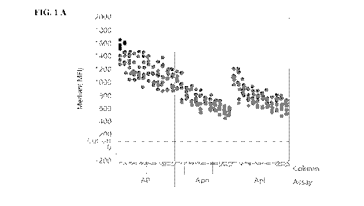
[0014] FIG. I is a plot of signal strength as fluorescence intensity on
BMBs measured as the
single median result from all the beads in a well (MFI) vs. cell column
position (i.e., 1-12) of a
96 well plate for an assay for antibodies specific for three Anaphisma derived
peptides
designated "AP", "Aph", and "Apl" as designated on the X axis of FIG. IA and
IB, as described
in Example 6. FIG. IA depicts signal strength vs. cell column position for a
standard read buffer
and FIG. 1B depicts signal strength vs. cell column position for a citrate
read buffer, as described
in Example 6.
DETAILED DESCRIPTION OF THE INVENTION
[0015] The invention is directed to a substrate for biological analysis and
a method for
making the substrate for biological analysis. The substrate for biological
analysis comprises an
epoxy-based resin having a biomolecule probe that is directly bonded to the
epoxy-based resin.
[0016] The phrase "biomolecule probe directly bonded to the epoxy-based
resin," and similar
phrases, as used herein, means that the biomolecule probe is attached to the
resin by simply
3
CA 03211971 2023-08-28
WO 2022/187115 PCT/US2022/018077
contacting the biomolecule probe with the resin without first contacting the
epoxy-based resin
with another reagent that coyalently reacts with the epoxy-based resin. The
biomolecule probe
can be passively attached to the epoxy-based resin or covalently attached to
the epoxy-based
resin.
[0017] The substrate for biological analysis is prepared by:
(i) providing a substrate that has a surface comprising an epoxy-based resin
and
(iii) contacting a biomolecule probe with the epoxy-based resin so that the
biomolecule
probe bonds directly to the epoxy-based resin.
[0018] In one embodiment, the substrate is the epoxy-based resin.
[0019] In one embodiment, the substrate is a solid support coated with the
epoxy-based resin.
Illustrative solid support materials upon which the epoxy-based resin can be
coated include, but
are not limited to, particles, beads and surfaces comprising glass, polymers,
latex, elemental
metals, metal composites, alloys, silicon, carbon, and hybrids thereof.
[0020] In one embodiment, a portion of the epoxy-based resin is polymerized
prior to
contacting the biomolecule probe with the epoxy-based resin.
[0021] Suitable epoxy-based resins include, but are not limited to, EPON SU-
8, EPON
1001F, 1002F, 1004F, 1007F, 1009F, 2002, and 2005 (commercially available from
Hexion
Specialty Chemicals of Fayetteville, NC). EPON SU-8 and EPON 1002F are
preferred resins.
[0022] SU-8 is a photo-curable epoxy-based resin. SU-8 is a formaldehyde,
polymer with
(chloromethyl)oxirane and 4,4-(1-methylethylidene)bisphenol (CAS: 28906-96-9).
SU-8 is a
polymeric solid epoxy novolac resin possessing an average epoxide group
functionality of
around eight. The structure of SU-8 epoxy resin is:
4
CA 03211971 2023-08-28
WO 2022/187115
PCT/US2022/018077
0- 9 .
e
1,0) t J
\
tP:
0
\ .......................... A: sc
\ ..
srs..õ,,o= 3 s
[0023] SU-8 is commercially available from Hexion Specialty Chemicals as a
solution
containing SU-8 and a photo acid generator under the traden.ame EPON SU-8.
[00.24] 1002F is a photo-curable epoxy resin (CAS: 25036-25-3). 1002F is
phenol, 44'41-
methylethylidene )bis-, polymer with 2,T-[(1-methylethylidene)bis(4,1-
phenyleneoxymethylene
)This( oxirane) and is commercially available from Hexion Specialty Chemicals
as a solution
containing 1002F and a photo acid generator under the tradename EPON 1002F:
0 CH 3 OH CH 3 0
H2C¨CHCH2-0 OCH2CHCH2-0
OCH2-CH¨CH2
CH3 CH3
- 2-3
[0025] Suitable, biomolecule probes include, but are not limited to,
lipids, polysaccharides,
amino acids, polypeptides, oligopeptides, peptides, antibodies and fragments
thereof,
polynucleotides (including single and double stranded DNA and RNA),
oligonucleotides,
aptamers, lectins, avidin, streptavidin, biotin, and polyethylene glycol.
Preferably, the
biomolecule probe is a polypeptide, oligopeptide, peptide, polynucleotide or
shorter
oligonucleotide. In one embodiment, the biomolecule is an antibody. In one
embodiment, the
biomolecule probe is a synthetic molecule, such as, for example, rhodamine.
Illustrative
biomolecule probes include SDMA, ADMA, T4, cortisol, progesterone, and enzymes
(e.g.,
lipases, such as pancreatic lipase).
[0026] Without wishing to be bound by theory, it is believed that the
biomolecule probe is
bonded to the epoxy-based resin by the reaction of an amine, th.iol, or
hydroxyl groups on the
biomolecule with epoxy groups on the resin. The biomolecule probe can also be
passively
CA 03211971 2023-08-28
WO 2022/187115 PCT/US2022/018077
bonded to the epoxy-based resin. The phrase "passively bonded," as used
herein, means bonding
by non-covalent interactions, such as Vander Walls, hydrophobic, hydrophilic,
or hydrogen
bonding interactions.
[0027] The substrate that has a surface comprising an epoxy-based resin can
be, but is not
limited to, a film, alone or adhered to another solid surface; a microbead; a
microparticle; a
micro pellet; a microwafer; a paramagnetic bead; a microparticle containing an
identifying
feature, such as a bar code; a paramagnetic microparticle; a paramagnetic
microparticle
containing a bar code; and a bead containing a nickel bar code.
[0028] In some embodiments, the biomolecule probe is bonded directly to the
epoxy resin by
simply contacting the epoxy-based resin with the biomolecule.
[0029] In one embodiment, the biomolecule probe is contacted with the epoxy-
based resin by
adding the substrate that has a surface comprising an epoxy-based resin to a
solution of the
biomolecule probe to provide a contact mixture. In one embodiment, the
solution of the
biomolecule probe is an aqueous solution. In one embodiment, the solution of
the biomolecule
probe is a buffered aqueous solution. In one embodiment, the solution of the
biomolecule probe
is a dimethyl sulfoxide (DMSO) solution.
[0030] In a preferred embodiment, the substrate that has a surface
comprising an epoxy-based
resin is washed with DMSO before the substrate that has a surface comprising
an epoxy-based
resin is added to the solution of the biomolecule probe to provide the contact
mixture. In one
embodiment, the substrate that has a surface comprising an epoxy-based resin
is washed with
DMSO immediately before it is contacted with the solution of the biomolecule
probe to provide
the contact mixture. It has been unexpectedly discovered that contacting the
epoxy-based resin
with DMSO before the epoxy resin is contacted with the biomolecule probe
provides a substrate
for biological analysis that exhibits less variability in how many biomolecule
probes are bound to
the epoxy resin and less variability in the signal obtained when detecting the
presence of the
analyte in the sample that binds to the biomolecule probe. It has been
unexpectedly found that
contacting the epoxy-based resin with DMSO before the epoxy resin is contacted
with the
biomolecule probe provides a substrate for biological analysis that exhibits a
better signal.
6
CA 03211971 2023-08-28
WO 2022/187115 PCT/US2022/018077
100311 By directly bonding the biomolecule probe to the epoxy-based resin,
the method
advantageously avoids the additional steps of having to functionalize the
epoxy-based resin by
(i) reacting the epoxy resin with another molecule before the biomolecule
probe is bonded to the
epoxy-based resin or (ii) mixing another molecule into the epoxy resin prior
to polymerization.
By avoiding this additional step, the method advantageously is faster, less
expensive, and
removes a step where errors or variability could potentially occur.
[0032] When a solution of the biomolecule probe is used to contact the
biomolecule probe
with the substrate that has a surface comprising an epoxy-based resin, the
concentration of the
biomolecule probe in the solution ranges from about 0.05 mglmt to about 5
mg/mL, preferably
about 0.01 mglinL to about 3.0 mg/mL, and more preferably about 0.15 to about
2.5 mg/mL, for
example about 1.5 mg/mL.
100331 The concentration of the substrate that has a surface comprising an
epoxy-based resin
in the contact mixture ranges from about 0.05 to about 5.0 million
substrates/mL, preferably
about 0.1 to about 3.0 million substrates/ML. In one embodiment, the
biomolecule probe is a
peptide and the concentration of the substrate that has a surface comprising
an epoxy-based resin
in the contact mixture ranges from about 0.1 to about 3.0 million
substrates/mL, for example
about 2 million substrates/mL. In one embodiment, the biomolecule probe is an
antibody and the
concentration of the substrate that has a surface comprising an epoxy-based
resin in the
contacting mixture ranges from about 0.1 to about 1.8 million substrates/mL,
for example about
1 million substrates/mL.
[0034] The solution of the biomolecule probe is typically contacted with
the substrate that has
a surface comprising an epoxy-based resin for a sufficient amount of time so
that the
biomolecule probe bonds to the epoxy-based resin. Typically the solution of
the biomolecule
probe is contacted with the substrate that has a surface comprising an epoxy-
based resin for at
least about 4 hours, preferably at least about 8 hours, more preferably at
least about 10 hours. In
one embodiment, the molecule probe is contacted with the substrate that has a
surface
comprising an epoxy-based resin for between about 4 hours and about 18 hours.
100351 The contact mixture (i.e., the substrate that has a surface
comprising an epoxy-based
resin and the solution of the biomolecule probe) is maintained at a sufficient
temperature so that
the biomolecule probe bonds to the epoxy-based resin. In one embodiment, the
contact mixture
7
CA 03211971 2023-08-28
WO 2022/187115 PCT/US2022/018077
is maintained at a temperature of between about 4 'C to about 65 C,
preferably between about
15 C and about 30 C, and more preferably between about 18 C and 27 C. In
one
embodiment, the contact mixture is stirred to assure that the surfaces of the
substrate that has a
surface comprising an epoxy-based resin are sufficiently contacted with the
solution of the
biomolecule probe.
[00361 In one embodiment, after the substrate that has a surface comprising
an epoxy-based
resin and the solution of the biomolecule probe are contacted so that the
biomolecule probe is
bonded to the epoxy-based resin, the solution of the biological probe is
removed and the
resulting biomolecule functionalized substrate is washed with a mixture of
about 1% bovine
serum albumin (BSA) (commercially available from Proliant Biologicals of
Ankany IA), about
0.05% Tween-20 (commercially available from Sigma Aldrich of St. Louis, MO),
and about
0.05% Proclin 950 (commercially available from Sigma Aldrich of St. Louis, MO)
in phosphate
buffered saline (PBS), at a pH of about 7.4. A suitable PBS solution includes
about 1.8 mM
sodium phosphate monobasic (commercially available from Sigma Aldrich of St.
Louis, MO),
about 8.4 mM sodium phosphate dibasic (commercially available from Sigma
Aldrich of St.
Louis, MO), and about 145 mM sodium chloride (commercially available from
Amresco of
Salon, OH). In one embodiment, the biomolecule functionalized substrate is
washed is washed
at least three times with at least about 200 !IL of the wash solution. In one
embodiment, the
biomolecule functionalized substrate is washed at least three times with about
200 1.1.1. to about
1,000 [IL of the wash solution.
[00371 Suitable buffers include, but are not limited to, phosphate , TRIS,
HEPES, MES,
EPPS, Bis-TRIS, Bis-TRIS propane, PIPES, ADA, MOPS, MOPSO, ACES, BES, Tricine,
TES,
Gly-Gly, DIPSO, inorganic buffers, organic buffers, acetic acid based, and
citric acid based
buffers.
[0038] The resulting washed biomolecule functionalized substrate can then
be added to a
solution of about 1% BSA, about 0.05% Tween-20, about 0.05% Proclin 950 in PBS
at pH about
7.4, for use in an assay.
[0039] In a first aspect of the method, the biomolecule probe is a protein,
such as an antibody,
an enzyme (e.g., streptavidin and avidin), or parts of an antibody (e.g., Fc
and FAB fragments),
and the solution of the biomolecule probe is an aqueous solution. In one
embodiment of the first
8
CA 03211971 2023-08-28
WO 2022/187115 PCT/US2022/018077
aspect of the method, the solution of the biomolecule probe is an aqueous
solution buffered with
about 100mM 2-(N-morpholino)ethanesulfonic acid (MES) and about 140mM
Guanidine-HCI at
a pH of about 5.5. In one embodiment of the first aspect of the method, the
solution of the
biomolecule probe is an aqueous solution buffered with about 100 mM 344-(2-
hydroxyethyl)piperazin-1-yl]propane-1-sulfonic acid (EPPS) and 140 mM
Guanidine-HC1 at a
pH of about 8 aqueous.
100401 In one embodiment of the first aspect of the method, the substrate
that has a surface
comprising an epoxy-based resin is washed with a solution of PBS containing
about 0.05%
Tween-20 before it is contacted with the solution of the biomolecule probe. In
one embodiment
of the first aspect of the method, the substrate that has a surface comprising
an epoxy-based resin
is washed at least three times with at least about 200 !,11_, of a solution of
PBS containing about
0.05% Tween-20 before it is contacted with the solution of the biomolecule
probe. In an
embodiment of the first aspect of the method, the substrate that has a surface
comprising an
epoxy-based resin is washed at least three times with about 200 [it to about
1,000 tL of a
solution of PBS containing about 0.05% Tween-20 before it is contacted with
the solution of the
biomolecule probe.
[0041] In a preferred embodiment of the first aspect of the method, the
substrate that has a
surface comprising an epoxy-based resin is then further washed with DMSO
before it is
contacted with the solution of the biomolecule probe. In one embodiment of the
first aspect of
the method, the substrate that has a surface comprising an epoxy-based resin
is washed at least
three times with at least about 200 III of the wash solution. In one
embodiment of the first
aspect of the method, the substrate that has a surface comprising an epoxy-
based resin is washed
at least three times with about 200 fit to about 1,000 pl. of DMSO before it
is contacted with the
solution of the biomolecule probe.
[0042] In one embodiment of the first aspect of the method, after the
substrate that has a
surface comprising an epoxy-based resin and the solution of the biomolecule
probe are contacted
so that the biomolecule probe is attached to the epoxy-based resin, the
solution of the biological
probe is removed and the resulting biomolecule functionalized substrate is
washed with a
mixture of about 1% BSA, about 0.05% Tween-20, and about 0.05% Proclin 950 in
PBS at a pH
of about 7.4. In one embodiment, the biomolecule functionalized substrate is
washed at least
9
CA 03211971 2023-08-28
WO 2022/187115 PCT/US2022/018077
three times with at least about 200 iL of the wash solution. In one
embodiment, the biomolecule
functionalized substrate is washed at least three times with about 200 pL to
about 1,0001..1L of
the wash solution.
[0043] The resulting washed biomolecule functionalized substrate can then
be added to a
solution of about 1% BSA, about 0.05% Tween-20, about 0.05% Proclin 950 in PBS
at pH about
7.4, for use in an assay.
[0044] In one embodiment of the first aspect of the method, the substrate
that has a surface
comprising an epoxy-based resin is a barcoded magnetic bead, such as a
barcoded magnetic bead
coated with SU-8 epoxy-based negative photoresist (commercially available from
Applied
BioCode of Santa Fe Springs, CA).
[0045] In a second aspect of the method, the biomolecule probe is a peptide
that has a
cysteine residue and the solution of the biomolecule probe is a solution in
DMSO. In one
embodiment of the second aspect of the method, the solution is DMSO containing
about 1%
Tween-20. In one embodiment of the second aspect of the method, the peptide
concentration
ranges from about 0.2 mM to about 1 inM peptide, for example, about 0.5 mM.
[0046] In one embodiment of the second aspect of the method, the substrate
that has a surface
comprising an epoxy-based resin is washed with a solution of DMSO containing
about 1%
Tween-20 before it is contacted with the solution of the biomolecule probe. In
one embodiment
of the second aspect of the method, the substrate that has a surface
comprising an epoxy-based
resin is washed at least three times with at least about 200 tL of a solution
of DMSO containing
about 1% Tween-20 before it is contacted with the solution of the biomolecule
probe. In an
embodiment of the second aspect of the method, the substrate that has a
surface comprising an
epoxy-based resin is washed at least three times with about 200 fiL, to about
1,000 [tr.. of a
solution of DMSO containing about 1% Tween-20 before it is contacted with the
solution of the
biomolecule probe.
[0047] In one embodiment of the second aspect of the method, after the
substrate that has a
surface comprising an epoxy-based resin and the solution of the biomolecule
probe are contacted
so that the biomolecule probe is bonded to the epoxy-based resin, the solution
of the biological
probe is removed and the resulting biomolecule functionalized substrate is
washed with a
CA 03211971 2023-08-28
WO 2022/187115 PCT/US2022/018077
mixture of about 1% BSA, about 0.05% Tween-20, and about 0.05% Proclin 950 in
PBS at a pHI
of about 7.4. In one embodiment, the biomolecule functionalized substrate is
washed at least
three times with at least about 200 uL of the wash solution. In one
embodiment, the biomolecule
functionalized substrate is washed at least three times with about 200 !IL to
about 1,000 ut, of
the wash solution.
[00481 The resulting washed biomolecule functionalized substrate can then
be added to a
solution of about 1% BSA, about 0.05% Tween-20, about 0.05% Proclin 950 in PBS
at a pH of
about 7.4, for use in an assay.
100491 In one embodiment of the second aspect of the method, the substrate
that has a surface
comprising an epoxy-based resin is a barcoded magnetic bead, such as a
barcoded magnetic bead
coated with SU-8 epoxy-based negative photoresist (commercially available from
Applied
BioCode of Santa Fe Springs, CA).
[0050] Without wishing to be bound by theory, the second aspect of the
method involves
reaction of a thiol group on the biomolecule probe with epoxide groups on the
epoxy-based resin.
This cysteine residue can occur anywhere in the peptide sequence. A cysteine
can also be spaced
from the peptide with a linker, such as a PEG linker. The PEG can be of a
defined length, such
as through the use of a discreet PEG. dPEG (commercially available from Quanta
Biodesitm,
Plain City, OH) is a particularly suitable PEG. Methods for linking a cysteine
residue to a
protein with a PEG linker are known in the art. See, fir example, I. Hamley,
PEG-Peptide
Conjugates, Biomacromolecules, 15:1543-59, 2014. Other linkers and spacers
include beta-
alanine, 4-aminobutyric acid (GABA), (2-aminoethoxy) acetic acid (AEA), 5-
aminovaleric acid
(Ava), 6-aminohexanoic acid (Ahx), Trioxatridecan-succinamic acid (Ttds) and
peptides.
[0051] In one embodiment, the peptides is capped at the N and C termini via
N-terminal
acetylation (Ac) or C-terminal amidation. When the biomolecule probe is an
antibody, the
antibody can be bonded to the epoxy-based resin via a thiol group by reducing
the antibody using
a reducing reagent, such as dithiothreitol (DTT), tris(2-
carboxyethyl)phosphine (TCEP), or 13-
mercaptoethanol (BME), to make thiol groups in the hinge region of the
antibody accessible for
bonding to the epoxy-based resin.
11
CA 03211971 2023-08-28
WO 2022/187115 PCT/US2022/018077
[00521 In one embodiment, the substrate that has a surface comprising an
epoxy-based resin
is prepared by coating a support with a solution containing SU-8 resin and a
photo acid
generator, such as triphenyl sulfonium hexafluoroantimonate, in a solvent,
such as 7-
butyrolactone or cyclopentanone, such as EPON SU-8 (commercially available
from Hexion
Specialty Chemicals of Fayetteville, NC). The support is then heated to remove
the solvent and
leave a coating of solid epoxy-based resin on the support. The thickness of
the solid epoxy-
based resin can be several hundred microns thick. Typically, the thickness of
the solid epoxy-
based resin ranges from about 1 nm to about 3 mm. Optionally, a portion of the
solid epoxy-
based resin is then polymerized by irradiation with UV light to provide a
polymerized epoxy-
based resin. In one embodiment, a photomask is placed on top of the solid
epoxy-based resin
before it is irradiated with UV light so that a pattern is left on the
polymerized epoxy-based resin.
In one embodiment, the support is separated from the solid epoxy-based resin
100531 In one embodiment, the substrate that has a surface comprising an
epoxy-based resin
is prepared by coating the support with a solution containing 1002F resin and
a photo acid
generator, such as triphenyl sulfonium hexafluoroantimonate, in a solvent,
such as y-
butyrolactone or cyclopentanone, such as EPON 1002F (commercially available
from Hexion
Specialty Chemicals of Fayetteville, NC). The support is then heated to remove
the solvent and
leave a coating of a solid epoxy-based resin on the support. The thickness of
the solid epoxy-
based resin can be several hundred microns thick. Typically, the thickness of
the solid epoxy-
based resin ranges from about 1 nm to about 3 mm. Optionally, a portion of the
solid epoxy-
based resin is then polymerized by irradiation with UV light to provide a
polymerized epoxy-
based resin. In one embodiment, a photomask is placed on top of the solid
epoxy-based resin
before it is irradiated with UV light so that a pattern is left on the
polymerized epoxy-based resin.
In one embodiment, the support is separated from the solid epoxy-based resin.
100541 In one embodiment, the sample is a fecal sample. The term "fecal
sample", as used
herein, includes feces, any sample containing feces, and fractions and
extracts of feces. Methods
for the preparation of fecal extracts are described in US Patent No.
8,367,808. In one
embodiment, the molecule being assayed for (i.e., the analyte) is a protein
generated by, for
example, an intestinal worm, such as, for example, a round worm, a whip worm,
a hookworm, a
tape worm, or a heart worm, or the parasite Giardia, and the biomolecule probe
is an antibody to
the protein. In one embodiment, the antibody is specific for coproantigens. In
one embodiment,
12
CA 03211971 2023-08-28
WO 2022/187115 PCT/US2022/018077
the biomolecule probe is selected from the group consisting of an antibody
that specifically binds
a coproantigen from roundworm, an antibody that specifically binds a
coproantigen from
whipworm, an antibody that specifically binds a coproantigen from hookworm, an
antibody that
specifically binds a coproantigen from tapeworm, an antibody that specifically
binds an antigen
from heartworm, and an antibody that specifically binds a coproantigen from
Giardia.
[0055] Examples of antibodies that specifically bind coproantigens from
hookworm are
disclosed in US Patent Nos. 9,239,326 and 8,895,294. Examples of antibodies
that specifically
bind coproantigens from roundworm are disclosed in US Patent Nos. 8,097,261;
9,212,220;
9,103,823; 8,105,795; and 8,895,294. Examples of antibodies that specifically
bind
coproantigens from whipworm are disclosed in US Patent Nos, 8,367,808 and
8,895,294.
Examples of antibodies that specifically bind coproantigens from tapeworm are
disclosed in US
Patent No. 11,001,626. Examples of antibodies that specifically bind
coproantigens from
Giardia are disclosed in H. Stibbs, Monoclonal antibody-based enzyme
immunoassay for
Giardia lamblia antigen in human stool, J. Clin. Microbiol., (11):2582-2588,
Nov. 1989 and H.
Stibbs et al., Identification of Giardia lamblia-specific antigens in injected
human and gerbil
feces by western immunoblotting, J. Clin. Microbiol., (10):2340-6, Oct. 1990.
100561 In one embodiment, the sample is a blood sample from a subject, the
analyte is an
antibody generated by the subject's immune response to a protein generated by
an infectious
agent and the biomolecule probe is a protein, polypeptide, or oligopeptide
that is capable of
specifically binding the antibody. Proteins, polypeptides, or oligopeptides
that are capable of
specifically binding circulating antibodies of an animal that has been
infected with bacteria of the
genus Ehrlichia, including Ehrlichia Canis, Ehrlichia chaffeensis and/or
Ehrlichia ewingii, are
disclosed in US Patent Nos. 7,087,372; 7,407,770; 7,445,788; 7449,191;
7,842,473; 7,888,054;
8,980,274; 7,183,060; 7,744,872; 8,409,817; 9,850,295; 8,158,751, and
9,605,032. Proteins,
polypeptides, or oligopeptides that are capable of specifically binding
circulating antibodies of
an animal that has been infected with bacteria of the genus Anaplasma.,
including Anapla.sma
phagocytophylum and Anaplasma platys, are disclosed in US Patent Nos.
6,964,855; 7,439,321;
8,303,959; 6,306,402; 6,204,252; 8,093,008, and 9,120,857. Proteins,
polypeptides, or
oligopetides that are capable of specifically binding circulating antibodies
of an animal that has
been infected with bacteria of the genus Borrelia, including Borrelia
burgdotteri, are disclosed
in US Patent Nos. 6,719,983; 6,740,744; 6,475,492, and 6,660,274.
13
CA 03211971 2023-08-28
WO 2022/187115 PCT/US2022/018077
[0057] in one embodiment, the analyte is an antibody resulting from the
subjects response to
a protein generated by a Borrella burgdorferi, that causes Lyme disease, and
the biomolecule
probe is the protein, or part of the protein, generated by Borrelia
burgdorferi.
[0058] In one embodiment, the sample is a blood sample from a subject, the
analyte is a
metabolite, and the biomolecule probe is an antibody that is capable of
specifically binding the
metabolite. The term "blood sample", as used herein, includes whole blood or
any portion or
fraction of whole blood, including but not limited to serum and plasma.
[0059] In one embodiment, the analyte is symmetric dimethylarginine (SDMA).
Antibodies
that specifically bind SDMA are disclosed in US Patent No. 8,481,690.
[0060] In one embodiment, the sample is a blood sample from a subject and
the analyte is an
antigen originating from a pathogen that is present in the blood of an
infected subject and the
biomolecule probe is an antibody against the antigen. In one embodiment, the
analyte is an
antibody that specifically bind circulating antigens from heartworm
(Dirofilaria
Antibodies that specifically bind circulating antigens from heartworm
(Dirofilaria itninitis) are
disclosed in US Patent No. 4,839,275.
[0061] In one embodiment, the substrate for biological assaying is prepared
by a method that
involves:
(i) providing a silicon/alumintun wafer;
(ii) coating the silicon/aluminum wafer with a first solution of a first epoxy-
based resin
dissolved in a first solvent to provide a silicon/aluminum wafer coated with a
first epoxy-based
resin coating;
(iii) heating the siliconlaluminum wafer coated with a first epoxy-based resin
coating to
remove at least a portion of the first solvent to provide a silicon/aluminum
wafer coated with a
first solid epoxy-based resin coating;
(iv) depositing a nickel bar code on the first solid epoxy-based resin coating
to provide a
silicon/aluminum wafer with a first solid epoxy resin coating and a nickel bar
code;
14
CA 03211971 2023-08-28
WO 2022/187115 PCT/US2022/018077
(v) coating the nickel bar code with a second solution of a second epoxy-based
resin
dissolved in a second solvent to provide a silicon/aluminum wafer coated with
a first epoxy-
based resin coating and a second epoxy-based resin coating;
(vi) heating the silicon/aluminum wafer with a first solid epoxy-based resin
coating and a
second epoxy-based resin coating to remove at least a portion of the second
solvent to provide a
silicon/aluminum wafer coated with a first solid epoxy-based resin coating and
a second solid
epoxy-based resin coating,
wherein the nickel bar code is between the first solid epoxy-based resin
coating and the second
solid epoxy-based resin coating;
(vii) optionally, polymerizing at least a portion of the first solid epoxy-
based resin
coating and the second solid epoxy-based resin coating to provide a
silicon/aluminum wafer
layered with the first solid epoxy-based resin, the nickel bar code, and the
second solid epoxy-
based resin coating, wherein at least a portion of the first solid epoxy-based
resin coating and the
second solid epoxy-based resin coating has been polymerized;
(viii) separating the silicon/aluminum wafer from the silicon/aluminum wafer
coated
with the first epoxy-based resin coating, the nickel bar code, and the second
epoxy-based resin
coating to provide a bar coded magnetic bead;
(iv) contacting the bar coded magnetic bead with a biornolecule so that the
biomolecule
bonds directly to the epoxy-based resin.
100621 In one embodiment, the first solution of a first epoxy-based resin
dissolved in a first
solvent is the same and the second solution of a second epoxy-based resin
dissolved in a second
solvent.
100631 In one embodiment, the silicon/aluminum wafer is separated from the
first solid epoxy
resin, the nickel bar code, and the second solid epoxy resin coating by
contacting the
silicon/aluminum wafer with sodium hydroxide.
[00641 The invention is also directed to a method of assaying for the
presence of an analyte
a sample. The method comprises: contacting the sample with a substrate that
has a surface
CA 03211971 2023-08-28
WO 2022/187115 PCT/US2022/018077
comprising an epoxy-based resin having a biomolecule probe directly bonded to
the epoxy-based
resin, wherein the biomolecule probe binds the analyte with specificity.
[0065] The phrases "binds the analyte with specificity," "specifically
binds," "with
specificity," "specific for the analyte," and similar phrases, as used herein,
have their art-
recognized meaning, i.e., that the biomolecule probe recognizes and binds to
the analyte (or a
class of analytes) with greater affinity than it binds to other non-specific
molecules. For
example, an antibody raised against an antigen that binds the antigen more
efficiently than other
non-specific molecules can be described as specifically binding to the
antigen. Binding
specificity can be tested using methodology known in the art such as, for
example, an enzyme-
linked immunosorbant assay (ELIS A), a radioimmunoassay (RIA), surface plasmon
resonance,
or a western blot assay.
[0066] In one embodiment, the sample is a fecal sample. In one embodiment,
the analyte is a
protein generated by an intestinal worm and the biomolecule probe is an
antibody against the
protein generated by the intestinal worm. In one embodiment, the antibody
against the protein
generated by the intestinal worm is selected from the group consisting of an
antibody that
specifically binds a coproantigen from roundworm, an antibody that
specifically binds a
coproantigen from whipworm, an antibody that specifically binds a coproantigen
from
hookworm, an antibody that specifically binds a coproantigen from tapeworm, an
antibody that
specifically binds an antigen from heartworm, and an antibody that
specifically binds a
coproantigen from Giardia.
[0067] In one embodiment, the sample is a blood sample from a subject, the
analyte is an
antibody generated by the subject's immune response to a protein generated by
an infectious
agent, and the biomolecule probe is a protein, polypeptide, or oligopeptide
that is capable of
specifically binding the antibody.
[0068] In one embodiment, the analyte is an antibody resulting from the
subjects response to
a protein generated by a Borrelia burgdorferi, that causes Lyme disease, and
the biomolecule
probe is the protein, or part of the protein, generated by Borrelia
burgdorferi.
16
CA 03211971 2023-08-28
WO 2022/187115 PCT/US2022/018077
100691 in one embodiment, the sample is a blood sample from a subject, the
analyte is a
metabolite, and the biomolecule probe is an antibody that is capable of
specifically binding the
metabolite. In one embodiment, the analyte is symmetric dimethylarginine
(SDMA).
[0070] In one embodiment, the sample is a blood sample from a subject and
the analyte is an
antigen originating from a pathogen that is present in the blood of an
infected subject and the
biomolecule probe is an antibody against the antigen. In one embodiment, the
analyte is an
antibody that specifically bind circulating antigens from heartworm
(Dirojilaria inunitis).
EXAMPLES
[0071] The present invention is not to be limited in scope by the specific
embodiments
disclosed in the examples which are intended as illustrations of a few aspects
of the invention
and any embodiments that are functionally equivalent are within the scope of
this invention.
Indeed, various modifications of the invention in addition to those shown and
described herein
will become apparent to those skilled in the art and are intended to fall
within the scope of the
appended claims. Such variations of the invention, including the substitution
of all equivalents
now known or later developed, which would be within the purview of those
skilled in the art, and
changes in formulation or minor changes in experimental design, are to be
considered to fall
within the scope of the invention incorporated herein.
Example 1: Passive coupling of a biomolecule probe to barcoded magnetic beads
[0072] Barcoded magnetic beads (BMBs) are manufactured from SU-8, an epoxy-
based
negative photoresist (commercially available from Applied BioCode Corp. of
SantaFe Springs,
CA). Coupling a biomolecule probe, such as a monoclonal antibody or protein,
to BMBs was
achieved via absorption of the biomolecule probe onto the BMB surface
according to the
following procedure.
[0073] A solution of the monoclonal antibody at a final concentration of
about 0.15-2.5
mg/mL antibody (typically about 1.5 mu/mL) was prepared in an aqueous buffer
containing
about 100mM MES (commercially available from Sigma Aldrich of St. Louis, MO)
and about
140mM Guanidine-HC1 (commercially available from Sigma Aldrich of St. Louis,
MO) at a pH
of about 5.5 or an aqueous buffer containing about 100 mM EPPS (commercially
available from
17
CA 03211971 2023-08-28
WO 2022/187115 PCT/US2022/018077
Sigma Aldrich of St. Louis, MO) and about 140 mM Guanidine-HCl at a pl-il of
about 8 to
provide an antibody coating solution.
100741 A sufficient amount of the BMBs was suspended in a BMB wash buffer
(about 1.8
mM sodium phosphate monobasic (commercially available from Sigma Aldrich of
St. Louis,
MO), about 8.4 m114 sodium phosphate dibasic (commercially available from
Sigma Aldrich of
St. Louis, MO), about 145 mM sodium chloride (commercially available from
Amresco LLC of
Salon, OH), and about 0.05% Tween-20 (commercially available from Sigma
Aldrich of St.
Louis, MO) at a pH of about 7.4) to provide a final concentration of about 0.1-
1.8 million
BMBs/rriL (typically about 1 million/mL for antibodies). The BMBs were washed
three times
with the BMB wash buffer. All BMB washes (for this and subsequent steps)
proceed as follows:
100751 First place the tube containing the BMBs in a magnetic stand and
allow the BMBs to
adhere to the magnet for 1-10 minutes. Then carefully aspirate off the
supernatant before
resuspending the BMBs in a volume of wash buffer about equal to the original
suspension
volume of the BMBs (about 200 p.L. to about 1,000 4). Repeat these steps a
total of three times
to provide a BMB pellet.
[0076] Following washing the BMBs with wash buffer, the BMBs were washed three
times
with a volume of DMSO (commercially available from Sigma Aldrich of St. Louis,
MO) about
equal to the original suspension volume to provide a pellet of DMSO washed
BMBs.
100771 Coating Reaction: Following the DMSO wash, the antibody coating
solution (about
1.5 mg/mL final concentration) was immediately combined with the DMSO washed
BMBs
(about 1 million BMBs/mL final concentration) and incubated for 4-18 hours at
room
temperature (18-27 C) with mixing. After the incubation was complete, the
antibody coupled
BMBs were washed three times with Assay Buffer (about 1% BSA (commercially
available
from Proliant biologicals of Ankany IA), about 0.05% Tween-20 (commercially
available from
Sigma Aldrich of St. Louis, MO), and about 0.05% Proclin 950 (commercially
available from
Sigma Aldrich of St. Louis, MO) in about 1.8 mM sodium phosphate monobasic
(commercially
available from Sigma Aldrich of St. Louis, MO), about 8.4 inIVI sodium
phosphate dibasic
(commercially available from Sigma Aldrich of St. Louis, MO), and about 145
mIVI sodium
chloride (commercially available from Amresco LLC of Salon, OH), at a pH of
about 7.4).
18
CA 03211971 2023-08-28
WO 2022/187115 PCT/US2022/018077
100781 The antibody coupled BMBs are then suspended in the Assay Buffer at
the desired
final concentration for use in an assay.
Example 2: Coupling a biomolecule probe to barcoded magnetic beads via a thiol
group
100791 Coupling a biomolecule probe, such as a monoclonal antibody or
protein, containing a
thiol group to barcoded magnetic beads (BMBs) that are coated with an epoxy-
based resin is
achieved via absorption of the biomolecule probe onto the BMB surface
according to the
following procedure.
100801 A solution of the peptide in DMSO (commercially available from Sigma
Aldrich of St
Louis, MO) containing about 1% Tween-20 (commercially available from Sigma
Aldrich of St.
Louis, MO) at a final peptide concentration of about 0.02 to about 1 mM
(typically about 0.1
niM) was prepared to provide a peptide coating solution. The peptide used for
this example was
acetylated-Cys(dPEG12)[peptide]-amide, wherein the peptide was an oligopeptide
of 25 amino
acid residues and dPEG12 is a discreet PEG12. The acetylated-
Cys(dPEG12)[peptide]-amide
was provided as a custom synthesis from New England Peptide of Gardner, MA:
100811 Suitable BMBs for use in the method are BMBs that are manufactured
from SU-8, an
epoxy-based negative photoresist (commercially available from Applied BioCode
Corp. of Santa
Fe Springs, CA).
100821 A sufficient amount of BMBs to provide a final concentration of
about 0.1-3 million
BMBs/mL (typically about 2 million/mL) in the coating reaction described below
were
suspended in a BMB wash buffer (about 1% Tween-20 in DMSO) at a volume of
about 200 viL
to about 1,000 viL. The BMBs were washed three times with the about 1% Tween-
20 in DMSO
as described below. All BMB washes (for this and subsequent steps) proceed as
follows:
100831 The tube containing the BMBs was placed in a magnetic stand and the
BMBs were
allowed to adhere to the magnet for 1-10 minutes. The supernatant was
carefully aspirated off
before resuspending the BMBs in a volume of wash buffer about equal to the
original suspension
volume of the BMBs. These steps were repeated a total of three times to
provide a BMB pellet.
100841 Immediately after washing the BMBs with wash buffer, the washed BMBs
were
combined with the peptide coating solution and incubated for about 4 hours at
room temperature
19
CA 03211971 2023-08-28
WO 2022/187115 PCT/US2022/018077
(18-27 'V) with mixing. Following the 4 hour incubation, the peptide-coupled
LBMBs were
washed three times with a volume of Assay Buffer (about 1% BSA, about 0.05%
Tween-20, and
about 0.05% Proclin 950 in about 1.8 mM sodium phosphate monobasic, about 8.4
mM sodium
phosphate dibasic, and about 145 niM sodium chloride, at a pH of about 7.4)..
[0085] The peptide coupled BMBs are then suspended in the Assay Buffer at
the desired final
concentration for use in an assay.
Example 3: Coupling a biomolecule probe to barcoded magnetic beads via a thiol
group
generated from a reduced disulfide-linked cysteine
[00861 Macromolecules, such as monoclonal antibodies, can be coupled via a
thiol group by
reacting the monoclonal antibody with a reducing reagent, such as
dithiothreitol (DTT), tris(2-
carboxyethyl)phosphine (TCEP), or 2-mercaptoethanol (BME) to reduce disulfide-
linked
cysteine side chains so as to make them accessible for bonding to the surface
of the BMBs.
[0087] For covalent coupling of reduced antibodies to epoxy groups on BMBs,
a reduced
antibody coating solution was prepared by diluting the antibody in a reducing
buffer (50 mM
sodium phosphate (commercially available from Sigma Aldrich of St. Louis, MO),
75 mM
sodium chloride (commercially available from Amresco LLC of Salon, OH), 2
ml\,4 EDTA
(commercially available from Sigma Aldrich of St. Louis, MO), and 5 mM DTT
(commercially
available from Thermo Fisher Scientific of Waltham, MA) at a pH of about 7.4)
to a
concentration of about 5 mg/mL. The resulting solution was incubated for about
0.5 hours at 18-
27 C.
[0088] Following the reduction, the reduced antibody was exchanged into a
solution
containing 50 mM sodium phosphate (commercially available from Sigma Aldrich
of St. Louis,
MO), 75 mM sodium chloride (commercially available from Amresco of Salon, OH),
and 2 mM
EDTA (commercially available from Sigma Aldrich of St. Louis, MO) at a pH of
about 7.4 using
a G25 Zeba Spin desalting column (commercially available from Thermo Fisher
Scientific of
Waltham, MA) according to the manufacturer's instructions. The antibody
solution was adjusted
to a concentration of about 0.5 mg/mL and the resulting solution immediately
added to DMSO
washed BMBs.
CA 03211971 2023-08-28
WO 2022/187115 PCT/US2022/018077
100891 DMSO washed BMBs were prepared by first suspending a sufficient amount
of the
BMBs to provide a final concentration of about 0.1-1.8 million BMBs/mL
(typically about 1
million/mL for antibodies) in the coating reaction described below in a volume
of about 200 !IL
to about 1,000 !IL of a wash buffer (about 1.8 mM sodium phosphate monobasic,
about 8.4 mM
sodium phosphate dibasic, about 145 nM sodium, and about 0.05% Tween-20, at a
pH of about
7.4). The BMBs were washed three times with the wash buffer as described
below.
[0090] All BMB washes (for this and subsequent steps) proceed as follows:
first place the
tube containing the BMBs in a magnetic stand and allow the BMBs to adhere to
the magnet for
1-10 minutes. Then carefully aspirate off the supernatant and resuspend the
BMBs in a volume
of wash buffer about equal to the original volume used to suspend the BMBs.
Repeat these steps
three times to provide a BMB pellet.
[0091] Following washing of the BMBs with wash buffer, the BMBs were washed
three
times with a volume of DNB (commercially available from Sigma Aldrich of St.
Louis, MO)
about equal to the original suspension volume to provide DMSO washed BMBs. The
BMBs
were then suspended in a volume of DMSO about equal to the original suspension
volume and
incubated for about 4 hours at 18-27 C with mixing. Following the incubation,
the tube
containing the BMBs were placed in a magnetic stand and the BMBs allowed to
adhere to the
magnet for 1-10 minutes. The supernatant was then carefully aspirated off to
provide DMSO
washed BMB pellets.
[0092] Suitable BMBs for use in the method are BMBs that are manufactured
from SU-8, an
epoxy-based negative photoresist (commercially available from Applied BioCode
Corp. of Santa
Fe Springs, CA).
[0093] Following the DMSO wash, the antibody coating solution (about 0.5
mg/mL final
concentration) was immediately combined with the DMSO washed BMBs (about 1
million
BMBs/mL final concentration) and incubated for about 18 hours at room
temperature (18-27 C)
with mixing.
[0094] After the incubation was completed, the tube containing the BMBs was
placed in a
magnetic stand and the BMBs allowed to adhere to the magnet for 1-10 minutes.
The
supernatant was then carefully aspirated off and the BMBs resuspended in a
volume of wash
21
CA 03211971 2023-08-28
WO 2022/187115 PCT/US2022/018077
buffer (about 1.8 mM sodium phosphate monobasic, about 8.4 triM sodium
phosphate dibasic,
about 145 mM sodium chloride, and about 0.05% Tween- at a pH of about 7.4)
about equal to
the original suspension volume and the resulting solution incubated for about
15 minutes at 18-
27 C.
[0095] After the incubation was complete, the tube containing the BMBs was
placed in a
magnetic stand and the BMBs allowed to adhere to the magnet for 1-10 minutes.
The
supernatant was then carefully aspirated off and the BMBs resuspended in a
volume of Assay
Buffer about equal to the original suspension volume. Assay Buffer: about 1%
BSA, about
0.05% Tween-20, and about 0.05% Proclin 950 in about L8 ni1V1 sodium phosphate
monobasic,
about 8.4 mM sodium phosphate dibasic, and about 145 nM sodium chloride, at a
pH of about
7.4. The resulting suspension was incubated for about 30 minutes at 18-27 C
with mixing. The
antibody coupled BMBs were then washed three times with a volume of Assay
Buffer about
equal to the original suspension volume.
[0096] The antibody coupled BMBs were then suspended in the Assay Buffer at
the final
desired concentration for use in an assay.
Example 4: Coupling rhodamine to barcoded magnetic beads:
[0097] Materials:
Material Number Barcode Concentration Molecular Supplier
weight (g/mol)
Epoxy BMBs 0.5 36 500,000 Applied BioCode
million beads/mL
Epoxy BMBs 0.5 61 500,000 Applied BioCode
million beads/mL
Epoxy BMBs 0.5 14 500,000 Applied BioCode
million beads/mL
Magnet stand 1
DMSO Sigma Aldrich
Rhodamine Lissamine 0.3 mg/mL 600.7 ThermoFisher
Sulforhodamine 2.8 mg/mL 558.7 ThermoFisher
PBS-Tween (0.05%) 0.05% Tween IDEXX-In house
EPPS buffer, pH = 9.0 150 mM EPPS IDEXX- In house
[0098] DMSO washing of the BMBs:
[0099] To 1 m1, centrifuge tubes on a magnetic rack was added 0.5 mL of BMBs
suspended
in a storage buffer to provide a concentration of 100,000 BMBs/mL.
22
CA 03211971 2023-08-28
WO 2022/187115 PCT/US2022/018077
10011001 Suitable BMBs for use in the method are BMBs that are manufactured
from SU-8, an
epoxy-based negative photoresist (commercially available from Applied BioCode
Corp. of Santa
Fe Springs, CA). The BMBs are provided in a storage buffer containing sodium
chloride (0.8
%), potassium chloride (0.02 %), disodium hydrogen phosphate (0.144 %),
potassium
dihydrogen phosphate (0.024 ()/O), Tween-20 (0.05 %), and ProClin-950 (0.1 %).
1001011 The liquid was removed from each of the centriftige tubes by pipette
and then about
0.5 mL of DMSO (commercially available from Sigma Aldrich of St. Louis, MO)
was added to
each tube. The tubes were vortexed vigorously for 10 seconds, placed back onto
the magnetic
rack, allowed to sit for 1 minute, and the DMSO removed by pipette. This
washing procedure
was repeated 2 more times. About 0.5 mL of DMSO was then added to each tube,
the tube
vortexed vigorously, and placed on mixer for 4 hours at room temperature.
1001021 Coating with Rhodamine-Lissarnine:
1001031 After mixing for 4 hours, the tubes were removed and placed on a
magnet stand,
allowed to sit for 1 minute, the DMSO removed by pipette, and about 0.5 mL of
about 150 mM
EPPS buffer, at a pH of about 9.0, was added to the tubes. The tubes
containing the BMBs in the
EPPS buffer were then vortexed vigorously for 10 seconds, placed back on the
magnet stand, and
the EPPS buffer removed by pipette. This washing procedure was repeated 2 more
times. After
the washing was completed, about 0.5 mL of EPPS buffer was added to each tube
and the tubes
vortexed. Then about 10.0 rhodamine-lissamine (9 !IL of a 0.3 mg/mL
solution in DMSO)
or sulfo-rhodamine (control, 1.2 [it of a 2.8 mg/mL solution in DMSO) was
added to the epoxy-
based BMBs in the EPPS buffer. The suspension of BMBs were allowed to rotate
end over end
for about 22 hours at room temperature, protected from light. After this time,
the coated BMBs
were placed on the magnetic stand and the solvent removed by pipette. The
coated BMBs were
washed with about 1.0 mL of a solution of about 1.8 mM sodium phosphate
monobasic, about
8.4 mM sodium phosphate dibasic, about 145 nIVI sodium chloride, and about
0.05% Tween-20
with brief vortexing, followed by removal of the solvent. This process was
repeated 5 times.
Finally, about 1.0 mL of a solution of about 1.8 mM sodium phosphate
monobasic, about 8.4
mM sodium phosphate dibasic, about 145 nM sodium chloride, and about 0.05%
Tween-20 was
added to the coated BMBs to provide a final concentration of about 50,000
BMBs/mL. About
5.0 !IL of the BMBs in the resulting suspension were then added to the wells
of a 96-well plate to
23
CA 03211971 2023-08-28
WO 2022/187115 PCT/US2022/018077
give a final count of about 250 BMBs/well. About 200 1iL of the solution of
about 1.8 mM
sodium phosphate monobasic, about 8.4 m11/1 sodium phosphate dibasic, about
145 nM sodium
chloride, and about 0.05% Tween-20 was added to each well to provide a
suspension of the
BMBs for use in an assay.
[00104] The fluorescence of each well was determined using a microplate reader
(commercially reader Applied BioCode Corp. of Santa Fe Springs, CA).
[00105] Epoxy BMBs coated with Rhodamine-Lissamine showed significant
fluorescence in
the microplate reader, indicating that the rhodamine probe had been
efficiently coated onto the
epoxy-based BMB surface. The epoxy-based BMBs incubated with the sulfo-
rhodamine control,
which does not have a reactive amine functional group, did not show any
observable
fluorescence. This demonstrates that nonspecific binding of the rhodamine
probes to the epoxy-
based BMB surface is very low, and indicates that the Rhodamine-Lissamine is
reacting with the
epoxy-based surface specifically through the primary amine functionality. It
can be concluded
that amine-functionalized small molecules are capable of covalently binding to
the epoxy-based
BMB surface.
Example 5: Coupling of amine-containing peptides to barcoded magnetic beads:
[00106] Materials:
Material Number Barcode Concentration Molecular Supplier
weight (g/mol)
Epoxy BMBs 200,000 14 200,000 Applied BioCode
beads/mL
Magnet stand 1
Biotin-Lyme Peptide 1.0 mM 3075 ThermoFisher
Lyme-Alexafluor555 1.0 mM 3948 New England
no Cysteine Peptide
DMSO Sigma Aldrich
PBS-Tween (0.05%) 0.05% Tween IDEXX-In house
EPPS buffer, pH = 9.0 150 mM EPPS IDEXX- In house
SA-PE 8 ug/mL IDEXX- In house
[00107] DMSO washing of the BMBs:
[00108] To 1 mL centrifuge tubes on a magnetic rack was added about 0.2 mL of
BMBs
suspended in a storage buffer to provide a concentration of 100,000 BMBs/mL.
24
CA 03211971 2023-08-28
WO 2022/187115 PCT/US2022/018077
[00109] Suitable BMBs for use in the method are BMBs that are manufactured
from SU-8, an
epoxy-based negative photoresist (commercially available from Applied BioCode
Corp. of Santa
Fe Springs, CA). The BMBs are provided in a storage buffer containing sodium
chloride (0.8
%), potassium chloride (0.02 %), disodium hydrogen phosphate (0.144 %),
potassium
dihydrogen phosphate (0.024 ()/O), Tween-20 (0.05 %), and ProClin-950 (0.1 %).
[00110] The liquid was removed from each of the centriftige tubes by pipette
and then about
0.2 mL of DMSO (commercially available from Sigma Aldrich of St. Louis, MO)
was added to
each tube. The tubes were vortexed vigorously for 10 seconds, placed back onto
the magnetic
rack, allowed to sit for 1 minute, and the DMSO removed by pipette. This
washing procedure
was repeated 2 more times. About 0.2 mL of DMSO was then added to each tube,
the tube
vortexed vigorously, and placed on mixer for 4 hours at room temperature.
[00111] After mixing for 4 hours, the tubes were removed and placed on a
magnet stand,
allowed to sit for 1 minute, the DMSO removed by pipette, and about 0.2 mL of
about 150 mM
EPPS buffer, at a pH of about 9.0, was added to the tubes. The tubes
containing the BMBs in the
EPPS buffer were then vortexed vigorously for 10 seconds, placed back on the
magnet stand, and
the EPPS buffer removed by pipette. This washing procedure was repeated 2 more
times.
[00112] About 0.1 rnM solutions of both biotin-lyme peptide and Lyme-
Alexafluor555 were
prepared from 1.0 mM stock solutions. Each peptide has multiple lysine
residues, but no
cysteine residues or other thiol groups. About 200 !IL of the biotin-lyme
peptide solution, the
Lyme-Alexafluor555 solution, or a control containing only EPPS buffer was
added to the tubes
containing the BMBs. The resulting suspension of BMBs was allowed to rotate
end over end for
about 2 hours at room temperature, protected from light. After this time, the
tubes containing the
BMBs were placed on the magnetic rack and the solvent removed by pipette. The
BMBs were
then washed with about 0.2 mL of a solution of about 1.8 mM sodium phosphate
monobasic,
about 8.4 mM sodium phosphate dibasic, about 145 nM sodium chloride, and about
0.05%
Tween-20 with brief vortexing, followed by removal of the solvent. This
process was repeated 2
times.
[00113] About 200 viL of SA-PE (streptavidin phycoerythrin)-containing
solution (8 f1g/mL
SA-PE, obtained by diluting SA-PE (commercially available as a 1 mg/mL
solution from Moss
Inc. of Pasadena, MD) with Multiplex Assay Buffer: 1.8 mM sodium phosphate
monobasic, 8.4
CA 03211971 2023-08-28
WO 2022/187115 PCT/US2022/018077
mM sodium phosphate dibasic, 145 nM sodium chloride, 0.05% Tween-20, 1% bovine
serum
albumin, 0.05% ProClin 950 was added to the BMBs and the beads incubated for
about 10
minutes. Following incubation the supernatant was removed and the BMBs were
washed with
about 0.2 mL of a solution of about 1.8 rriM sodium phosphate monobasic, about
8.4 mM sodium
phosphate dibasic, about 145 nle4 sodium chloride, and about 0.05% Tween-20 at
a pH of about
7.4 with brief vortexing, followed by removal of the solvent. This process was
repeated 5 times.
Finally, about 0.4 mL of a solution of about 1.8 mM sodium phosphate
monobasic, about 8.4
mM sodium phosphate dibasic, about 145 nM sodium chloride, and about 0.05%
Tween-20 at a
pH of about 7.4 was added to the coated BMBs to provide a final concentration
of about 50,000
BMBs/mL. 5.0 !IL of the resulting suspension of BMBs was added to the wells of
a 96-well
plate to give a final count of about 250 BMBs/well and about 200 uL of the
solution of about 1.8
mM sodium phosphate monobasic, about 8.4 mM sodium phosphate dibasic, about
145 nM
sodium chloride, and about 0.05% Tween-20 was added to each well to provide a
suspension of
the BMBs for use in an assay.
[00114] The fluorescence of each well was determined using a microplate reader
(commercially reader Applied BioCode Corp. of Santa Fe Springs, CA).
[00115] BMBs coated with Biotin-Lyme Peptide and the Lyme-Alexafluor555
peptide both
showed significant fluorescence in the microplate reader indicating that the
peptides had been
efficiently coated onto the BMB surface. The lack of fluorescence observed in
the control
demonstrates that nonspecific binding of the SA-PE analyte to the BMB surface
is very low, and
indicates that the Biotin-Lyme Peptide and the Lyme-Alexafluor555 peptide is
reacting with the
epoxy surface specifically through the primary amine functionality contained
in the lysine
residues of the peptide_ It can be concluded that lysine (amine functionality)
containing peptides
are capable of covalently binding to the epoxy BMB surface.
Example 6: Assay using barcoded magnetic beads followed by a citrate buffer
wash:
Coupling of biomolecule probe to barcoded magnetic beads:
[00116] To 1 mL centrifuge tubes on a magnetic rack was added about 0.2 mL
(volume can
vary from about 0.1 mL to about 500 mL) of BMBs suspended in a storage buffer
to provide a
26
CA 03211971 2023-08-28
WO 2022/187115 PCT/US2022/018077
concentration of about 100,000 BMBs/mL (concentration can range from about
100,000
BMBs/mL to about 3 million BMBs/mL).
1001171 Suitable BMBs for use in the method are BMBs that are manufactured
from SU-8, an
epoxy-based negative photoresist (commercially available from Applied BioCode
Corp. of Santa
Fe Springs, CA). The BMBs are provided in a storage buffer containing sodium
chloride (0.8
%), potassium chloride (0.02 %), disodium hydrogen phosphate (0.144 %),
potassium
dihydrogen phosphate (0.024 %), Tween-20 (0.05 %), and ProClin-950 (0.1 %).
1001181 The liquid was removed from each of the centrifuge tubes by pipette
and then about
0.2 mL of DMSO (commercially available from Sigma Aldrich of St. Louis, MO)
was added to
each tube. The tubes were vortexed vigorously for 10 seconds, placed back onto
the magnetic
rack, allowed to sit for 1 minute, and the DMSO removed by pipette. This
washing procedure
was repeated 2 more times. In one embodiment, about 0.2 mL of DMSO was then
added to each
tube, the tube vortexed vigorously, and placed on a mixer for 4 hours at room
temperature. This
4 hour mix at room temperature is optional.
1001191 After mixing, the tubes were removed and placed on a magnet stand,
allowed to sit for
1 minute, the DMSO removed by pipette, and about 0.2 mL of about 150 mM EPPS
buffer, at a
pH of about 9.0, was added to the tubes. The tubes containing the BMBs in the
EPPS buffer
were then vortexed vigorously for 10 seconds, placed back on the magnet stand,
and the EPPS
buffer was removed by pipette. This washing procedure was repeated 2 more
times. Following
the final wash with EPPS, BMBs were coated with peptides or antibodies as
described below in
parts (A) and (B), respectively, such that each peptide and antibody was
combined with BMBs of
a distinct barcode.
(A) Coating of BMBs with peptides
1001201 Peptides (biochemical probes) capable of specifically binding a
patients antibodies
against Anaplasma, Ehrlichia or Borrelia ( e.g., a peptide derived from a
protein sequence of
Anaplasina phagocytophyhun, Anaplasina platys, Ehrlichia canis, Ehrlichia
ewingii, or Borrelia
hurgdorferi, as described above) were synthesized with a cysteine linked to
each peptide's N-
terminus via a PEG12 linker as described above, except for the peptide derived
from Borrelia
which had an N-terminal cysteine, but no PEG linker. About 200 !IL of an about
0.1 mM
27
CA 03211971 2023-08-28
WO 2022/187115 PCT/US2022/018077
solution of each peptide was added to the tubes containing the EPPS-washed
BMBs. The
resulting suspension of BMBs was allowed to rotate end over end for about 2
hours at room
temperature, protected from light. After this time, the tubes containing the
BMBs were placed
on the magnetic rack and the solvent removed by pipette. The BMBs were then
washed with
about 0.2 mL of a solution of about 1.8 mM sodium phosphate monobasic, about
8.4 mM sodium
phosphate dibasic, about 145 TIM sodium chloride, and about 0.05% Tween-20
with brief
vortexing, followed by removal of the solvent. This process was repeated 2
times.
(B) Coating of BMBs with Antibody
1001211 About 200 ttL of an antibody (biochemical probe), at a concentration
of approximately
mg/mL (antibody concentration can range from about 1.5 mg/mL to about 12.0
mg/mL),
capable of specifically binding antigen from heartworm (Dirofilaria immitis),
as described
above, circulating in the blood of an infected animal was added to the tubes
containing the
BMBs. The resulting suspension of BMBs was allowed to rotate end over end for
about 2 hours
at room temperature, protected from light. After this time, the tubes
containing the BMBs were
placed on the magnetic rack and the solvent removed by pipette. The BMBs were
then washed
with about 0.2 mL of a solution of about 1.8 mM sodium phosphate monobasic,
about 8.4 mM
sodium phosphate dibasic, about 145 nM sodium chloride, and about 0.05% Tween-
20 with brief
vortexing, followed by removal of the solvent. This process was repeated 2
times.
Assay:
1001221 BMBs (65,000 BMBs/mL in assay buffer (1.0% BSA, 0.05% Tween, 0.05%
Proclin
950, in PBS)) coated with the biomolecule probes described above in (A) or (B)
were mixed to
build a multiplex BMB mixture. The multiplex BMB mixture was further diluted
in assay buffer
to achieve a concentration of 500 BMB/mL of each biomolecule probe.
1001231 About 100 tiL of this diluted multiplex BMB mixture was added to each
well (i.e.,
about 50 beads per biomolecule probe) of a 96 well plate using an Integra
automatic pipette
(commercially available from Integra Biosciences Corp., Hudson, NH). The BMBs
were washed
5 times with 300 !IL of 0.05% Tween-20 in PBS on a 405-TS plate washer
(commercially
available from BioTeke, Winooski, VT) with a 10 second soak. After the final
wash, excess
supernatant (approximately 30 fiL) was left on the plate after the final wash.
28
CA 03211971 2023-08-28
WO 2022/187115 PCT/US2022/018077
[00124] 50 t_tt of sample (serum or plasma, neat) was added to the BMBs in
each well on the
96 well plate. The plate was then placed on the plate mixer and mixed for 30
mm at 1000 rpm.
After the 30 minute incubation, the BMBs were washed with 300 tit of 0.05%
Tween-20 in PBS
with a 10 second soak.
[00125] 50 tt.1_, of each biotinylated peptide (2.0 [tg/mL, i.e., the same
peptide that was used as
the biomolecule probes) or biotinylated anti-heartworm antibody (1.0 tigimL)
in 1.0% BSA,
0.05% Tween, 0.05% Proclin 950, in PBS was added to each well of the 96 well
plate. The plate
was then placed on a plate mixer and mixed for 15 minutes. After the 15 minute
incubation, the
BMBs were washed with 300 jiL of 0.05% Tween-20 in PBS with a 10 second soak.
[00126] 50 tti_, of SA-PE, 8.0 tig/m1_, (commercially available from, MOSS
Inc., Pasadena, MD
catalog no. SAPERP01) was added to each well of the 96 well plate. The plate
was then placed
on a plate mixer and mixed for 10 minutes. After the 10 minute incubation, the
BMBs were
washed with 300 tiL of 0.05% Tween-20 in PBS with a 10 second soak.
[00127] The resulting BMBs were than treated by either of two procedures.
[00128] In the first procedure, to each well of the 96 well plate was added:
(i) about 200 tit of a buffer (commercially available from Applied BioCode
Inc., Santa
Fe Springs, CA, catalog no. 4/1-D0004-500) (standard read buffer).
[00129] In the second procedure, to each well of the 96 well plate was added:
(ii) about 200 itL of a solution of a citrate buffer containing sodium citrate
tribasic
dihydrate (0.485 M), citric acid (0.015 M), sodium chloride (0.1 M), Proclin
950 (0.5 mL/L), pH
6.1-6.3 (citrate read buffer).
[00130] Ionic strength is an important factor that allows for stability of the
immunocomplex
formed on the surface of the beads. Without wishing to be bound by theory, it
is believed that
the stabilizing effect is due to the high salt concentration making the
solution undesirable for
disassociation. Salts, other than citrate salts, at high ionic strength also
work, but they need to be
at a minimum concentration of about 0.5M. Other salts, however, are not ideal
for
manufacturability or shipping because they fall out of solution at
temperatures less than ambient
29
CA 03211971 2023-08-28
WO 2022/187115 PCT/US2022/018077
temperature. Advantageously, the mix of citrate buffer remains in solution
when stored or
shipped at refrigerated conditions.
[00131] The fluorescence of each well was determined using a BioCode 2500
Analyzer
(commercially available from Applied BioCode Corp. of Santa Fe Springs, CA).
[00132] The fluorescent intensity of each sample in each well of the 96-well
plate (i.e., a plate
with 12 columns (1-12) and 8 rows (A-H)) was determined. FIG. 1 illustrates
the signal strength
observed using the standard read buffer containing (FIG. 1 A) and using the
citrate read buffer
(FIG. 1 B). In the experiment, each of the wells of a 96-well plate (i.e., a
plate with 12 columns
(1-12) and 8 rows (A-H)) were filled with identical BMBs (i.e., everything
bound to the beads, as
described above), and the signals were read from column 1 (i.e., wells lA
through 1H) to column
12 (i.e., wells 12A through 12H). The time to read the fluorescence in all of
the cells of the 96
well plate (i.e., cells lA to 12H) takes approximately 37 minutes. As can be
seen in FIG. 1A,
when the standard read buffer (which is buffer that does not contain citrate)
is used, i.e.,
procedure (i), there is a gradual decrease in signal strength from the start
of the read cycle to the
end of the read cycle (approximately 37 minutes). In contrast, as can be seen
in FIG. 1B, when
the citrate read buffer is used, i.e., procedure (ii), no decrease in signal
strength was observed
from the start of the read cycle to the end of the read cycle (approximately
37 minutes). This
decrease in signal intensity during the read cycle when using a non-citrate-
containing buffer,
such as the standard read buffer, but not when using a citrate-containing
buffer, such as the
citrate read buffer, occurred in an assay for antibodies specific for each of
three Anaplasma
derived peptides designated "AP", "Aph", and "Apl" on the X axis of FIG. IA
and FIG. 1B.
[00133] FIG. 1 shows that contacting the BMBs with a citrate buffer before
reading the
fluorescence advantageously avoids a decrease in fluorescence as a function of
time compared to
other buffers.
[00134] Similar effects of standard and citrate read buffer on signal strength
as a function of
time was observed in the assays for antibodies specific for peptides derived
from Ehrlichia canis,
Ehrlichia ewingii, and in the assay for Borrelia burgdorferi (not shown).
[00135] The entire disclosure of all references that have been cited are
incorporated herein by
reference.