Note: Descriptions are shown in the official language in which they were submitted.
BASE ELEMENT OF A MULTI-CHAMBER BIOCHIP, PRODUCTION OF
THE MULTI-CHAMBER BIOCHIP, AND USE THEREOF FOR
ESTABLISHING ORGAN AND DISEASE MODELS AND SUBSTANCE
TESTS
[0001] The invention relates to a base element which can be used to produce
a multi-chamber biochip for a series of innovative applications.
[0002] Biochips in the sense of this application serve to simulate biological
systems, such as, for example, organs or tissues in an experimental setup, in
that biological structures and situations/environmental conditions are
recreated in reaction chambers artificially, but as close to reality as
possible.
For example, a biochip can be formed from a number of components arranged
one above the other, cooperating to form at least one culture chamber, but
more often two culture chambers. These can be separated from one another
by means of membranes of selected properties, wherein the culture chambers
are filled with one medium or several media. Optionally, various substances,
such as, for example, nutrients, active substances, and synthetic materials,
as
well as aerosols, cells, microorganisms, and/or spheroids, can be introduced
in a targeted manner into the media in order to simulate biological systems or
specific situations/environments under controlled conditions.
[0003] One possibility for the design of such biochips is known, for example,
from the publication by Raasch, M., et al. (Raasch, M., et al., 2016; An
integrative microfluidically supported in vitro model of an endothelial
barrier
combined with cortical spheroids simulates effects of neuroinflammation in
neocortex development; Biomicrofluidics 10; doi 10.1063/1.4955184). Two
base elements, each forming a preliminary culture chamber, are combined with
one separation membrane each, and subsequently joined together by
adhesive bonding, then sealed with closure films and partly sealed with
adhesives or connecting substances. Filling the three culture chambers, which
are arranged one above the other and separated by the two separation
1
Date Recue/Date Received 2023-09-15
membranes, takes place via channels likewise formed in the two base
elements. In this way, at least two culture chambers can be created in a small
space and can be operated and examined under laboratory conditions, i.e.,
while monitoring, for example, the biological structures used and the
composition, temperature, and, optionally, flow of the media. The use of such
generic biochips in a cell culture incubator additionally enables operation in
an
ambient atmosphere with, for example, a controlled temperature and
composition.
[0004] Detection of the processes in the interior of the culture chambers can
take place, for example, by means of an optical analysis through the
transparent windows of the biochip (for example, by means of transmitted light
microscopy analyses, live cell imaging). Furthermore, substances can be
introduced via the channels, through the separation membranes, in order to
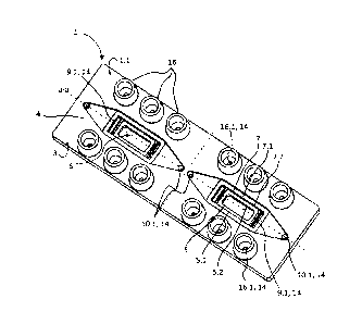
assess the barrier/seal/permeability of the biological structures or to
evaluate
transport processes on the biological structures, and/or to analyze the
reactions of proteins on the surfaces and/or inside of the biological
structures.
Furthermore, biological structures and introduced substances, such as media
(potentially enriched with the aforementioned substances) can be removed for
optical, molecular biological, and chemical analyses.
[0005] However, a biochip as described above must be constructed in layers
from a number of different elements, wherein each layer must be connected
tightly to the respective adjacent elements; in addition, the requirements for
the desired dimensions of the culture chambers and thus the determined flow
and physiological conditions must be reliably met. This means that, in
particular, the base elements have to be produced with high precision and
dimensional accuracy, as a result of which the production costs are high.
[0006] In the experiments of the inventors, it was further found that the
gluing
of the base elements induces stresses which lead to a deformation of the
biochip, and the adhesion is subject to aging processes which make the
2
Date Recue/Date Received 2023-09-15
biochip leaky, losing its function. In addition, it is disadvantageous that
the
separation membrane must be introduced before the bonding of the base
elements. This is associated with a risk of contamination of the membrane by,
for example, adhesive.
[0007] The object of the invention is to propose an option which makes it
possible to reduce or resolve the disadvantages known from the prior art, and
to furnish a multi-chamber biochip which can be used for a plurality of
experimental applications.
[0008] The object is achieved by the subject matter of the independent as well
as the subordinate claims. Advantageous developments are the subject matter
of the dependent claims.
[0009] The object of the invention is achieved by means of a base element of
a multi-chamber biochip which has a bottom with at least one frame which is
located thereon and which is open at least on its side facing away from the
bottom. In this case, an interior space is surrounded by the frame. The frame
can be designed as a step that surrounds the interior space. In an option
which
is advantageous because it is easier to produce, this can also be created by
means of a planar, and in particular rectangular, molding of the base element,
which has an accordingly high material thickness, corresponding in particular
to the height of the interior space. Several frames which delimit several
interior
spaces can be placed on the bottom. Alternatively, in the variant of the
planar
molding of the base element, several interior spaces can also be surrounded
by the planar molding.
[0010] Furthermore, at least one first web which is seated on the bottom
within
each interior space, and which extends around a first surface, is formed, the
height of which is less than the height of the frame. The first web can be
designed in this case to be free-standing, or can be designed as a step in the
frame. The web has a first lateral surface facing the interior space and a
first
3
Date Recue/Date Received 2023-09-15
support surface opposite the bottom. The first support surface is designed to
support a first separation membrane. A lower preliminary culture chamber
(first
chamber) is surrounded by the first lateral surface of the first web and the
surface surrounded by the first web. The remaining interior space between the
first support surface and the upper side of the frame forms an upper
preliminary
culture chamber (second chamber). The surface surrounded by the first web
can be polygonal, preferably square, and in particular rectangular, but also
round. Advantageously, the support surface of the web extends around the
enclosed area at a uniform height with respect to the bottom. The lower
preliminary culture chamber and the upper preliminary culture chamber are
each connected to the surrounding environment by at least one separate
channel that opens at an outer side of the base element.
[0011] In further embodiments of the base element according to the invention,
the lower preliminary culture chamber and/or the upper preliminary culture
chamber are connected to the surrounding environment by at least two
separate channels. In the assembled state of the multi-chamber biochip, the
channels advantageously allow for a supply and/or discharge of media into/out
of the culture chambers.
[0012] For example, to allow the cultivation of spheroids of approximately 1
mm size, at least one culture chamber and the associated channels are
designed with a construction having a clear height or a diameter of more than
1 mm-for example, 1.1 or 1.2 mm.
[0013] The base element according to the invention is formed (monolithically)
from a single workpiece, and preferably has the format of a microscopy slide
(76 mm x 26 mm 3 mm). The monolithic design allows easier handling
compared to solutions according to the prior art, since it is not necessary to
connect two or more base elements to one another, whose passages
(channels, interior spaces, culture chambers, or the like) need to be aligned
and glued before the connection. In addition, the base element is more stable
4
Date Recue/Date Received 2023-09-15
in a monolithic construction; it retains its original shape (no stress-related
bending of the body), is not leaky at adhesive points, and is therefore more
practical in handling and use. In particular, higher flow rates of the
supplied
media (perfusion speeds) can be achieved without leaks occurring. This is
advantageous in particular in the establishment of organ models, because
perfusion speeds matching those in vivo must be maintained in such cases
over the longest possible period of time. The resulting omission of glued
points
also advantageously has the consequence that the adhesives - which are
absolutely necessary in the prior art, and which could interact
disadvantageously with the biological structures - can be omitted. In
addition,
the requirements for dimensional accuracy of the base element according to
the invention can be lower than for the biochip composed of several elements.
[0014] In a further advantageous embodiment of the base element, a second
web is formed, which sits on the bottom within the interior space and extends
around a second surface. Its height is less than the height of the frame, but
greater than the height of the first web. The second web can also be designed
to be free-standing, or can be designed as a step in the frame, and has a
support surface lying opposite the bottom, hereafter referred to as the second
support surface. The second support surface is designed to support a second
separation membrane. Between the first support surface and the second
support surface, the second web forms a second lateral surface facing the
interior space. The second surface, surrounded by the second web, and the
second lateral surface define a middle preliminary culture chamber (third
chamber). The surface surrounded by the second web can be polygonal,
preferably square, and in particular rectangular, but also round. Preferably,
the
geometric shape of the surface enclosing the second web corresponds to that
of the surface surrounded by the first web. The middle preliminary culture
chamber is also connected by at least one channel to an outer side of the base
element.
5
Date Recue/Date Received 2023-09-15
[0015] As will be explained further below, a multi-chamber biochip with three
or more culture chambers arranged one above the other can advantageously
be produced by means of such an embodiment of the base element.
[0016] Advantageously, the material from which the base element is produced
comprises an injection-molded, biocompatible plastic. Examples of such
plastics are polyesters such as polyurethanes (PU), polyimides, styrenes
(SEBS), polypropylenes (PP), polystyrenes (PS), polycarbonate (PC),
polyethylene terephthalate (PET), and cyclic polyolefins (COP and COC). In a
manner corresponding to the material selection, the base element is produced
as a single piece in an injection molding or casting process, using the
corresponding injection molding or casting tools.
[0017] Biochips are often also produced from polydimethylsiloxanes (PDMS)
in the prior art. However, the use of PDMS-based chips for complex cell,
organ,
and disease models is complicated, since, in contrast to the aforementioned
plastics, the PDMS has to be activated in order to be suitable for biological
structures such as cell cultures. In order to influence the systems of
biological
structures and media to be investigated by means of a biochip as little as
possible, the materials of the biochip should ideally be inert. However, the
frequently-used material PDMS is known to have high bond-forming capacity
with some chemical compounds, for example (Auner et al., (2019): Chemical
PDMS binding kinetics and implications for bioavailability in microfluidic
devices; Lab Chip 19: 864-874). This bond-forming capacity can have a
difficult-to-estimate influence on experiments (e.g., substance tests) that
are
carried out with biochips made of PDMS. For example, substances with a logP
greater than 1.8 and a low hydrogen donor count are adsorbed strongly on
PDMS, which makes active substance testing and interpretation of the data
obtained considerably more difficult. For example, the substance
propiconazole has a logP of 3.72 and a hydrogen donor count of 0. It is very
hydrophobic and can only be detected at less than 30% of the starting
concentration in the culture medium after 24 hours in PDMS chips, since it
6
Date Recue/Date Received 2023-09-15
binds irreversibly to PDMS (Auner et al. 2019). This problem relates
primarily,
but not exclusively, to the classic active substance group of small molecules.
Many pharmaceutical active substances belong to this group of drugs, such
that PDMS-based biochips are not suitable as test systems. In addition, a one-
piece base element for a multi-chamber biochip made only of a single
workpiece is not producible by means of PDMS, since, with this substance, the
necessary separation membrane can be fixed only between two individual
components.
[0018] In an advantageous embodiment of the base element according to the
invention, it is therefore produced from polybutylene terephthalate (PBT).
[0019] PBT is a polymer which is commonly used to produce products that are
subject to a high mechanical load and/or which repeatedly come into contact
with hot media. Typical uses of PBT are, for example, plain bearings, valve
parts, screws, parts for household appliances such as coffee machines or hair
dryers, and parts for medical devices such as connectors for pulse oximeters,
tips for electrosurgical instruments, and clips for breathing masks. The
production of suitable PBT starting polymers can even be realized in a GMP-
compliant manner. PBT is very suitable for injection molding due to its
favorable cooling and process behavior.
[0020] This material, which is unusual for use in cell culture chambers,
showed
very low bond-forming capacity in tests, compared to a series of components
of the media used, such that an influence of the material of the multi-chamber
biochips, and in particular of the (preliminary) culture chambers, on the
tests
taking place in such a cell culture chamber can be advantageously reduced.
For example, the inventors have found in tests for active substance adsorption
with propiconazole and troglitazone that PBT is very suitable for active
substance tests of substances up to a logP of 3.72 (logP propiconazole: 3.72;
logP troglitazone: 3.60). After an incubation period of 24 h, at least 80% of
the
7
Date Recue/Date Received 2023-09-15
starting concentration of the propiconazole or the troglitazone is detectable
in
the culture medium.
[0021] In a further embodiment, the base element according to the invention
can be designed such that portions of the channels are formed in the lateral
surface, facing away from the frame, of the bottom and/or in the lateral
surface,
opposite the bottom, of the base element. Preferably, the channel portions in
the lateral surface or in the lateral surfaces can be formed completely or
partially as preliminary channel portions, due to the formation of
circumferential
channel boundary webs. These channel boundary webs are created in the
base element, adjoin it, or project out of the base element. The channel
boundary webs have inwardly-oriented lateral surfaces which bound a channel
space and thus act as a channel wall. Support surfaces, which are provided
for supporting a (closure) membrane or a channel cover, run on the upper side
(end face) of the channel boundary webs.
[0022] The channels are each connected to connectors (e.g., standard Luer
format) for supplying or removing media. These connectors are
advantageously formed opposite the bottom.
[0023] A window can be present in the bottom, for example, which allows the
visual detection of processes in at least one of the culture chambers.
[0024] In order to obtain a multi-chamber biochip according to the invention,
a
base element according to the invention is provided. This serves as the base
of the multi-chamber biochip and advantageously allows both efficient
production and flexible adaptation to the respective requirements.
[0025] In the case of a fully-assembled and ready-to-use multi-chamber
biochip, a first separation membrane is placed on and connected to the first
support surface of the first web. A lower culture chamber is provided by the
first
web and the first separation membrane. Depending upon the embodiment of
8
Date Recue/Date Received 2023-09-15
the base element, a second separation membrane is optionally placed on a
second support surface of a second web, and connected to the web. In this
case, a middle culture chamber is provided between the first separation
membrane and the second separation membrane. A closure membrane is
placed on the frame and is connected to the frame. This closure membrane
delimits an upper culture chamber which, depending upon the design of the
base element, is provided between the first separation membrane and the
closure membrane or between the second separation membrane and the
closure membrane.
[0026] Optionally, an additional closure membrane is provided, which sits on
the lateral surface, facing away from the frame, of the bottom, and is
connected
thereto.
[0027] The separation membranes used are preferably films which, depending
upon the material, thickness, and production thereof, can be flexibly
integrated
to prespecified degrees, and can be permeable as well as impermeable to
gases, liquids, particles, and/or more complex molecules (semi-permeable).
Preferred but non-exclusive materials for the separation membranes are
polyethylene terephthalate or polycarbonate. The membranes preferably have
pores with a size between 0.4 pm and 8 pm, and have a thickness between 5
and 50 pm, preferably 10 and 20 pm, and particularly preferably 12 pm. The
separation membranes can be at least translucent, and preferably transparent,
to at least one selected wavelength range, in order to enable improved optical
detection of processes in at least one of the culture chambers. The closure
membranes (sometimes also referred to as bonding films) can also preferably
be integrated in a flexible manner, and can be selected to be correspondingly
blocking or (semi-)permeable to certain classes of substance. The closure
membranes can also be transparent for at least one selected wavelength
range, in order to enable an optical detection of processes in at least one of
the culture chambers. In some embodiments, the closure membranes can be
designed as transparent closure films - for example, as polycarbonate films or
9
Date Recue/Date Received 2023-09-15
polyethylene terephthalate films. Glass or polystyrenes or COC/COP are also
possible materials from which the closure membranes can be made. The
closure membranes (or closure films) can also function as a channel cover,
since they extend over optionally existing channel boundary webs and rest on
the support surfaces thereof, and are connected to them in a gas-tight and
liquid-tight manner.
[0028] In a particular embodiment of the multi-chamber biochip, at least one
of
the separation membranes can have depressions, which are also referred to
as (micro)cavities. Cells, cell composites, spheroids, and/or organoids can be
colonized and/or cultured in these (micro)cavities.
[0029] An application example here is the improved/extended culture of
spheroids and organoids over a period of up to 4 days compared to static cell
culture.
[0030] In the design of the multi-chamber biochip with microcavities,
spheroids
and organoids can be cultured in an immobilized manner under (microfluidic)
cell culture conditions with flow-through, without an additional embedding in
(hydro)gels, which usually consist of proteins of the extracellular matrix in
individual form or mixed forms. The microcavities can be between 500 and
1,500 pm in diameter, and preferably 800 pm. The gel-free culture allows
better
optical analysis during the culture period. In addition, the gel-free culture
allows
a gentler and non-destructive recovery of the intact cell tissue - in
particular,
spheroids and organoids from the biochip - for further analyses such as tissue
sections, immunofluorescence stains, flow cytometry, ELISA-based assays, or
tissue lysis for DNA/RNA analyses and Western Blot analyses.
[0031] The gel-free immobilized culture of spheroids and organoids enables
further an easier co-culturing with blood vessel tissue arrangements
(vascularization) - either on different separation membranes (indirect
vascularization) or on the same (micro)cavity separation membrane or planar
Date Recue/Date Received 2023-09-15
separation membrane (direct vascularization). In addition, it is possible for
immobilized and vascularized spheroids and organoids with immune cells to
be directly rinsed in the culture medium. The gel-free culture of spheroids
and
organoids considerably facilitates the migration of immune cells into the
tissue
of the spheroid and organoids.
[0032] The culture under flow-through and/or gel-free cell culture conditions
allows better maintenance of the vitality of biological structures, due among
other things to improved nutrient and oxygen conditions - in particular, for
spheroids -than under comparable static cell culture conditions. This makes it
possible to maintain the function of such biological structures for a longer
time
for test purposes.
[0033] In order to provide a user with a wide selection of possibilities of
use, a
set for a multi-chamber biochip according to the invention can be provided,
which comprises a base element as described above. Furthermore, at least
one first separation membrane is present in such a set, which serves for
placement on the first web and closing off the lower culture chamber.
Optionally, at least one second separation membrane is included in the set,
which serves for placement on the second web and closing off the middle
culture chamber if the base element included in the set has a second web. In
addition, a closure membrane is included in the set, which serves for
placement on the frame and closing off the remaining interior space as an
upper culture chamber.
[0034] In addition, an additional closure membrane can be included, which is
intended for placement on the lateral surface, facing away from the frame, of
the bottom.
[0035] A set according to the invention can, for example, be provided directly
to a user or delivered to a service provider who performs the assembly of the
11
Date Recue/Date Received 2023-09-15
components of the set on behalf of and according to the specifications of, for
example, the user.
[0036] Advantageously, the provision of such a set also enables the
introduction
of biological material, e.g., larger organoids or cell clusters or tissue
pieces or
multi-cellular organisms, e.g., parasites, which, due to their size, cannot be
flushed into the chambers by the channels present. Such material can be
applied directly in a sterile environment into a still-open preliminary
culture
chamber, which is subsequently closed off with a separation membrane or a
closure membrane - for example, by means of an adhesive method.
[0037] The multi-chamber biochip is produced by providing a base element,
and by the first separation membrane being placed on the first web. This is
connected to the first web, forming a seal. If necessary, a biological
material
can be introduced into the lower preliminary culture chamber before the first
separation membrane is applied, as described further above.
[0038] In the sense of this description, a sealed connection is understood to
mean that a planar or linear connection is created, which is in particular
liquid-
and gas-tight, so that a culture chamber bounded by the membrane or a
channel bounded by the membrane reliably withstands the flow of a medium
under a certain static or dynamic working pressure.
[0039] A connection is advantageously - but not exclusively - made by guiding
a beam of directed and controlled high-energy radiation along the joining seam
or connecting surface to be produced. The high-energy radiation is in
particular
laser radiation of a wavelength and intensity which are matched to the
materials to be connected.
[0040] Once the first separation membrane is attached to the first web, the
second separation membrane is optionally placed on the second web and
connected thereto, forming a seal. Optionally, a biological material can again
12
Date Recue/Date Received 2023-09-15
be introduced into the middle preliminary culture chamber before the second
separation membrane is applied. In a corresponding manner, the closure
membrane is placed on the frame, and the closure membrane and frame are
connected, forming a seal. Optionally, the additional closure membrane is
placed on the lateral surface, facing away from the frame, of the bottom, and
is connected thereto, forming a seal.
[0041] Since each preliminary culture chamber, and, as a result, each of the
resulting culture chambers, is contacted by at least one channel, a medium
can be supplied and/or discharged into each of the culture chambers of the
multi-chamber biochip.
[0042] The above-described plurality of specific embodiments of the multi-
chamber biochip according to the invention advantageously allows a
considerable number of possibilities for using such a multi-chamber biochip.
All of the intended uses comprise at least the following steps. First, a multi-
chamber biochip according to the invention is selected and provided. The
selection can take place, for example, with regard to the existing number of
culture chambers and/or with regard to the choice and/or the combination of
the separation membrane(s). In order to avoid undesirable interactions and
contamination during use, the multi-chamber biochip can subsequently be
sterilized. An assembly of sterile components under elevated purity
conditions is also equivalent to a sterilization. Subsequently, for example,
media, cells, microorganisms, spheroids and/or organoids and/or cell clusters
and/or tissue pieces, or multi-cellular organisms can be introduced into the
existing culture chambers.
[0043] In a specific embodiment of a use of the multi-chamber biochip or in
one
of its provided configurations, a hydrogel - preferably consisting of
components
of the extracellular matrix, such as, for example, collagens, fibronectin,
laminins, etc. - can be introduced into at least one of the culture chambers.
13
Date Recue/Date Received 2023-09-15
[0044] The multi-chamber biochip according to the invention can be used, for
example, to generate and/or culture spheroids and/or organoids in at least one
of its culture chambers by colonizing them in the (micro)cavities of one of
the
membranes.
[0045] The multi-chamber biochip according to the invention can also be used
for testing cells, cell cultures, organoids, or spheroids with various
substances,
active substances, nanomaterials, microorganisms, vectors, antibodies, etc.
[0046] Due to its structure on the basis of the base element according to the
invention, the invention is easy to use for a user. The assembly of a multi-
chamber biochip is considerably simplified and significantly less prone to
error
compared to the prior art. Moreover, due to the one-piece design of the base
element, an efficient production and a versatile combinability with a wide
variety of separation membranes and/or closure membranes is possible. The
use, for example, of the method of laser bonding to connect the membranes,
forming a seal, makes it possible to dispense with glues or other adhesives.
[0047] A multi-chamber biochip according to the invention can, for example, be
used, depending upon the specific embodiment, for active substance tests or
the establishment and characterization of organ or organoid models and
disease and infection models. For active substance tests, it is conceivable,
for
example, to examine an immune response of the cultured cells to a substance
administration. In this case, there are different possibilities; the substance
can
be put into the chamber in which the cells to be tested grow, for example. The
influence of the substance on the cells can then be determined, for example,
by microscopic observation of the cells or by examination of the cell culture
medium - for example, for messenger substances, markers, etc., discharged
from the cells into the medium. Alternatively, the substance can also be added
to the chamber opposite to the cells to be tested in order, for example, to
examine an influence of cells growing in this chamber, located opposite to the
cells to be tested, upon the effect of the substance in the cells to be tested
of
14
Date Recue/Date Received 2023-09-15
the other chamber, which could, for example, weaken or potentiate a
substance effect (gradient formation). Furthermore, spheroids and/or
organoids with and without immune cell populations can be rinsed/perfused.
The immune cells can be rinsed/perfused into the chamber with the spheroids
or organoids, or can preferably be flushed/perfused via a blood vessel
structure in one of the adjacent chambers. In addition, the immune cells can
be permanently integrated into the blood vessel structure and/or the
spheroid/organoids. In addition, spheroids or organoids can be vascularized
by introducing blood vessel cells such as endothelial cells alone or
endothelial
cells in combination with - but not exclusively - pericytes and smooth muscle
cells. In order, for example, to simulate a tissue traversed by blood vessels,
the upper and lower walls of the middle culture chamber can be lined with
endothelial cells alone or in combination with pericytes and smooth muscle
cells, which are introduced through the inlet channel, whereas organ-specific
epithelial cells are introduced into the lower and into the upper culture
chambers. In a further embodiment, however, the endothelial cells can also be
introduced on the upper and lower walls of the upper or lower chamber by the
given inlet channel, alone or in combination with pericytes and smooth muscle
cells, and, in the middle chamber, epithelial tissue can be integrated in the
form
of layered cell layers or in the form of spheroids and organoids.
[0048] The invention is explained in more detail below with reference to
exemplary embodiments and figures. In the drawings:
Fig. 1 is a schematic perspectival view of an exemplary embodiment of a base
element according to the invention;
Fig. 2 is a schematic view of an exemplary embodiment of channels in the
bottom of a base element according to the invention;
Fig. 3 is a schematic view of an exemplary embodiment of a set according to
the invention for providing a multi-chamber biochip (exploded view);
Date Recue/Date Received 2023-09-15
Fig. 4 is a lateral section through a multi-chamber biochip according to the
invention, with three culture chambers, and a schematic illustration of an
apparatus for operating the multi-chamber biochip; and
Fig. 5 shows a possible use of a multi-chamber biochip according to the
invention.
[0049] In the following, the invention is described by means of exemplary
embodiments in which two interior spaces 6 are respectively bounded by a
rectangular planar molding of a frame 4 on a bottom 3 of a base element 1. As
will be explained in more detail below, the interior spaces 6 are divided in a
stepped fashion in such a way that they each form a (preliminary) multi-
chamber cavity 2.1. A multi-chamber biochip 2 can thus be provided by the
base element 1 of Figs. 1, 2, and 3, or a multi-chamber biochip 2 is provided,
which has two multi-chamber cavities 2.1 (wherein both multi-chamber cavities
2.1 functionally form a complete multi-chamber biochip). To improve the
clarity
of the figures, both existing multi-chamber cavities 2.1 are used in Figs. 1,
2,
and 3 to indicate the elements of a single multi-chamber cavity 2.1 or of a
multi-
chamber biochip 2 with only one multi-chamber cavity 2.1. The description
relates here only to one multi-chamber cavity 2.1.
[0050] A base element 1 according to the invention is formed as a single piece
from a biocompatible material, and in particular from a biocompatible
injection-
molded plastic (Fig. 1). Starting from a bottom 3 (see also Fig. 2), a frame 4
is
formed, which is open on its side facing away from the bottom. The frame 4 is
planar and encloses an interior space 6.
[0051] Within the interior space 6, a first web 5, which extends around a
first
surface, is made in the form of a step present in the material of the frame 4.
The height of the first web 5 is less than the height of the frame 4. The
first
web 5 has a first lateral surface 5.2, facing the interior space 6, and a
first
support surface 5.1 opposite the bottom 3. The first support surface 5.1 is
designed to support a first separation membrane 11 (see Figs. 3, 4). A lower
16
Date Recue/Date Received 2023-09-15
preliminary culture chamber 8 is bounded by the first lateral surface 5.2 and
the surface enclosed by the first web 5. In the exemplary embodiment of Figs.
1 through 3, the surface enclosed by the first web 5 is rectangular.
[0052] In addition, a second web 7 which is placed inside the interior space 6
on the bottom 3 and surrounds a second surface is provided, which is likewise
designed as a step made of material of the base element 1 (Fig. 1). The second
surface enclosed by the second web 7 is larger than the first surface enclosed
by the first web 5, and also rectangular. The height of the second web 7 is
less
than the height of the frame 4, but greater than the height of the first web
5. A
second support surface 7.1 is formed on the second web 7. The second
support surface 7.1 is designed to support a second separation membrane 12
(see Fig. 3). In the portion between the first support surface 5.1 of the
first web
Sand the second support surface 7.1 of the second web 7, the second web 7
forms a second lateral surface 7.2 facing the interior space 6. A middle
preliminary culture chamber 9 is bounded by the second surface, surrounded
by the second web 7, and the second lateral surface 7.2. The remaining
interior
space 6 between the second support surface 7.1 and an upper side 1.1 of the
base element 1 forms an upper preliminary culture chamber 10. The upper
side 1.1 is formed by the lateral surface, opposite the bottom 3, of the base
element 1.
[0053] In order to be able to supply the culture chambers 8, 9, and 10
resulting
from the preliminary culture chambers 8, 9, 10 in the assembled state of the
multi-chamber biochip 2 with media 18 (see Fig. 4), two channels 14 are
formed between the culture chambers 8, 9, and 10 and the upper side 1.1 of
the base element I. In Fig. 1, the channels 14 can be seen as round passages
10.1, 16.1 and as passages with rectangular cross-sections 9.1. The channels
14 lead from the culture chambers 8, 9, and 10 in the direction of the bottom
3, where a distribution to the connectors 16 takes place (cf. Figs. 1, 3, 4).
17
Date Recue/Date Received 2023-09-15
[0054] In the exemplary embodiment shown, the connectors 16 sit on the
upper side 1.1 of the base element 1. In the exemplary embodiment, each of
the connectors 16 is configured for the supply and discharge of media 18
through each of the culture chambers 8, 9, 10. Each of the culture chambers
8, 9, 10 of a multi-chamber biochip 2 ready for operation can therefore allow
passage of a medium 18 independently of the other culture chambers 8, 9, 10.
In particular, each medium 18 can be selected individually for the respective
culture chambers 8, 9, 10 and can be applied to the respective culture
chambers 8, 9, 10 by an individually controllable volume flow.
[0055] To enable an optical detection of processes at least in the lower
culture
chamber 8 during the operation of the multi-chamber biochip 2, a window 17
is formed in the bottom 3 (Fig. 4). The surface of the window 17 is congruent
with the surface enclosed by the first web 5.
[0056] The exemplary course of the channels 14 and the connection thereof to
the respective connectors 16 is shown in Fig. 2 with a representation of the
outwardly-pointing lateral surface of the bottom 3. For the supply and
discharge of a medium 18 through the lower culture chamber 8, two lower
channels 14.2 are present which open into the lower culture chamber 8 through
two supply openings 8.1 arranged diagonally opposite one another. To supply
the middle culture chamber 9 with a further medium 18, two middle channels
14.3 are provided, which are formed in portions as opposite rectangular supply
passages 9.1 arranged between the first web 5 and the second web 7 (see
also Fig. 1). The upper culture chamber 10 is supplied with a further medium
18 by two upper channels 14.4, in that the latter contact the upper culture
chamber 10 via the respectively outermost, round supply passages 10.1.
[0057] The connector passages 16.1 shown in each case with a round cross-
section then produce the respective connections to the connectors 16.
18
Date Recue/Date Received 2023-09-15
[0058] In the exemplary embodiment of Fig. 2, portions 14.1 of the channels
14 between the supply openings 8.1 and the supply passages 9.1, 10.1, on
the one hand, are present, as are, on the other, the associated connector
passages 16.1 which are fashioned as preliminary channel portions 14.1. The
preliminary channel portions 14.1 are formed by depressions incorporated into
the base element 1. In the exemplary embodiment, the depressions are
arranged in the shape of a groove with a rectangular cross-section. Of course,
other types of depressions are also possible with other cross-sections - such
as a semicircular cross-section. The depressions have inner channel walls
14.5, each enclosing a connector passage 16.1 and a supply passage 9.1,
10.1, or, in the case of the lower culture chamber 8, the two associated
connector passages 16.1 and the window 17 with the two, diagonally opposite
supply passages 8.1. The channel walls 14.5 sit flush with the lateral
surface,
pointing away from the frame 4, of the bottom 3. By applying a channel cover,
the preliminary channel portions 14.1 can be closed, and thus the functional
state of these channel portions 14.1 can be produced. In the exemplary
embodiment of Figs. 3 and 4, a lower closure membrane 15 is applied to the
lateral surface, pointing away from the frame 4, of the bottom 3, and, in the
manner of a seam, along the channel walls 14.5 is connected in a liquid-tight
manner to the bottom 3. The connection is preferably carried out by means of
laser welding along the connection seam to be produced, but can also be
effected, for example, by gluing or solvent welding. Due to the liquid-tight
connection of the closure membrane 15, the preliminary channel portions 14.1
attain their functional state.
[0059] In Fig. 3, a set for a multi-chamber biochip 2 is shown by way of
example, wherein the illustration can also be regarded as an exploded
illustration of the components of an exemplary embodiment of a multi-chamber
biochip 2. A base element 1 according to the invention is present as a central
element. A first flexible separation membrane 11 is provided for placement on
the first support surface 5.1 of the first web 5. If the first separation
membrane
11 is attached there, it closes off a lower culture chamber 8. The first
separation
19
Date Recue/Date Received 2023-09-15
membrane 11 is provided, for example, with microcavities 19, into which, for
example, spheroids or organoids can be introduced, and/or can be cultivated
there (see Fig. 5). A second flexible separation membrane 12 serves for
placement on the second support surface 7.1 of the second web 7, and closes
off a middle culture chamber 9. A transparent closure membrane 13, which is
also provided, can be placed on the frame 4, which membrane in the
assembled state serves to close off the remaining interior space 6 as an upper
culture chamber 10 between the second separation membrane 12 and the
closure membrane 13.
[0060] In order also to close off the channel portions 14.1, formed in the
bottom
3, as well as the window 17, an additional transparent closure membrane 15
is present, which, as already explained above, is applied on the bottom 3 and
spans and seals the respective channel portions 14.1 and the window 17.
[0061] A multi-chamber biochip 2 according to the invention, with a lower
culture chamber 8, a middle culture chamber 9, and an upper culture chamber
10, is shown in Fig. 4 in a lateral sectional view. The drawing plane of Fig.
4
corresponds to the line a-a of Fig. 3, wherein only the left half of the multi-
chamber biochip of Fig. 3 is shown in Fig. 4 for reasons of clarity.
[0062] The monolithic structure of the base element 1 of the multi-chamber
biochip 2 can be clearly seen in Fig. 4. Starting from a bottom 3, a flat
frame 4
is formed which is open on its side facing away from the bottom 3. Within the
frame 4, a first web 5, which extends around a first surface, is made in the
form
of a step present in the material of the frame 4. The height of the first web
5 is
less than the height of the frame 4. The first web 5 has a first lateral
surface
5.2 and a first support surface 5.1 opposite the bottom 3. In the region of
the
surface enclosing the first web 5, the bottom has a window 17. Furthermore,
there is a second web 7 which is placed on the bottom 3 and surrounds a
second surface, and is likewise designed as a step made of material of the
base element 1. The height of the second web 7 is less than the height of the
Date Recue/Date Received 2023-09-15
frame 4, but greater than the height of the first web 5. A second support
surface
7.1 is formed on the second web 7. Between the first support surface 5.1 of
the first web 5 and the second support surface 7.1 of the second web 7, the
second web 7 forms a second lateral surface 7.2 facing the interior space 6.
[0063] A first separation membrane 11 is applied to the first support surface
5.1, and a second separation membrane 12 is applied to the second support
surface 7.1 - in both cases in a liquid-tight manner. The first separation
membrane 11 has microcavities 19. On the lateral surface, facing away from
the frame 4, of the bottom 3, a lower closure membrane 15 is applied, and an
upper closure membrane 13 is applied to the upper side of the frame - in both
cases in a liquid-tight manner. A lower culture chamber 8 is bounded by the
first lateral surface 5.2, the first separation membrane 11, and the lower
closure
membrane 15. A middle culture chamber 9 is bounded by the second lateral
surface 7.2, the first separation membrane 11, and the second separation
membrane 12, and an upper culture chamber 10 is bounded by the remaining
frame 4, the second separation membrane 12, and the upper closure
membrane 13.
[0064] The functions of the membranes 11, 12, 13, and 15 are clearly visible -
both to delimit the culture chambers 8, 9, and 10 from one another or from the
surrounding environment, and to provide desired options with regard to the
exchange of molecules and/or cells between the culture chambers 8, 9, and 10.
[0065] In the examples of Figs. 3 and 4, the multi-chamber biochip 2 has three
chambers. The lower chamber 8 has a usable base area of approximately 42
mm2, a height of 0.5 mm, and a volume of 60 mm3, including the volume of the
inlet and discharge channels 14. In the region of the supply openings 8.1, the
inlet and discharge channels 14 have a diameter of approximately 0.5 mm.
The middle chamber 9 has a usable base area of 160 mm2, a height of 1.1
mm, and a volume of just 200 mm3, including the volume of the inlet and
discharge channels 14. The inlet and discharge channels 14 are, in the region
21
Date Recue/Date Received 2023-09-15
of the supply passages 9.1, rectangular and 2 mm wide. The upper chamber
has a usable base area of 216 mm2, a height of 0.7 mm, and a volume of
around 150 mm3, including the volume of the inlet and discharge channels 14.
The inlet and discharge channels 14 have a diameter of 0.8 mm in the region
5 of the supply passages 10.1. All mentioned single-dimensional
specifications
can vary and, for example, have a variability of 0.5 mm, which can result in
changes in the size of the surfaces and volumes.
[0066] Various media 18 (shown by arrows) can flow along the associated
10 channels 14 into the respective culture chambers 8, 9, 10 and out again,
wherein the supply of media 18 into the culture chambers 8, 9, 10 can take
place independently of one another (Fig. 4). The connection principle is shown
in Fig. 4 using the example of the lower culture chamber 8: the associated
connectors 16, via which a supply line 23 and a discharge line 24 are arranged
in each case, are connected to lower channels 14.2, which open into the lower
culture chamber 8 via supply openings 8.1 and supply the lower chamber 8
with a medium 18. The lower channels 14.2 extend in part offset to the drawing
plane and are therefore drawn with partially dashed lines.
[0067] In an analogous manner, the middle culture chamber 9 can be supplied
with medium 18 via the middle channels 14.3, and the upper culture chamber
10 via the upper channels 14.4. The multi-chamber biochip 2 can be operated,
for example, by means of a device such as a reading device or a microscope
which has a lens 21 which is oriented towards the window 17, and which can
be monitored and optionally detected, stored, and evaluated by means of the
processes in the multi-chamber biochip 2. For this purpose, a light source 22
can also be present in order to illuminate the multi-chamber biochip 2 in the
desired manner. In addition, a pump 25 can be arranged which is connected
to the supply lines 23 and discharge lines 24, which in turn are attached to
the
corresponding connectors 16. The pump 25 and optionally the light source 22
can be controlled by means of a controller 20 so that, for example, a
perfusion
of the culture chambers 8, 9, 10 can be carried out in a controlled manner and
22
Date Recue/Date Received 2023-09-15
can be monitored optically. The controller 20, which is implemented, for
example, by a computer, can optionally also store and/or evaluate optically-
detected data - in addition to the generation of control commands. It is
possible, for example, to control the pumping rates for the individual culture
chambers 8, 9, 10 as a function of the optically-detected data by means of the
controller 20.
[0068] The invention advantageously enables the construction of complex
biological models. For example, microfluidic cultures of spheroids 26 and/or
organoids 26 with integrated blood vessel and immune cell circulation can be
realized.
[0069] Thus, a model for studying pancreatic cancer (PDAC, pancreatic ductal
adenocarcinoma) can be created (Fig. 5). For this purpose, the lower culture
chamber 8 is provided with a clear height of 0.5 mm between the lower closure
membrane 15 and the first separation membrane 11. The first separation
membrane 11 is porous and is provided with microcavities 19 on its lateral
surface facing the middle culture chamber 9, in which cavities the spheroids
26 can be colonized and cultured. The middle culture chamber 9 has a clear
height of 1.1 mm, so that spheroids 26 can be introduced in a non-destructive
manner up to a size of approximately 1 mm. The channels 14 are also
dimensioned to be correspondingly large.
[0070] In the exemplary embodiment of Fig. 5, the second separation
membrane 12 is designed as a porous PET film and covers the middle culture
chamber 9. A cell layer 27 consisting of microvascular, pancreatic endothelial
cells 28 with macrophages 29 is formed in the upper culture chamber 10. For
the sake of improved clarity, the cell layer 27 is shown at a distance from
the
second separation membrane 12. In reality, the cell layer 27 grows adherently
on the second separation membrane 12. Through the second separation
membrane 12, perfused monocytes 30 and T-cells 31, for example, can pass
into the middle culture chamber 9.
23
Date Recue/Date Received 2023-09-15
[0071] The upper culture chamber 10 has a clear height of 0.7 mm between
the second separation membrane 7 and the closure membrane 13.
24
Date Recue/Date Received 2023-09-15
List of reference signs
1 base element
1.1 upper side
2 multi-chamber biochip
2.1 multi-chamber cavity
3 bottom
4 frame
5 first web
5.1 first support surface (first end face)
5.2 first lateral surface
6 interior space
7 second web
7.1 second support surface (second end face)
7.2 second lateral surface
8 lower (preliminary) culture chamber
8.1 supply opening
9 middle (preliminary) culture chamber
9.1 supply passage
10 upper (preliminary) culture chamber
10.1 supply passage
11 first separation membrane
12 second separation membrane
13 upper closure membrane (or upper bonding film)
14 channel
14.1 preliminary channel portion
14.2 lower channel
14.3 middle channel
14.4 upper channel
14.5 channel wall
15 additional (lower) closure membrane (or lower bonding film)
16 connector
Date Recue/Date Received 2023-09-15
16.1 connector passage
17 window
18 medium
19 microcavity
20 controller
21 lens
22 light source
23 inlet
24 discharge
25 pump
26 spheroid/organoid
27 cell layer
28 (pancreatic) endothelial cell
29 macrophages
30 monocytes
31 T-cells
26
Date Recue/Date Received 2023-09-15