Note: Descriptions are shown in the official language in which they were submitted.
CA 03213500 2023-09-13
1
PROCESS FOR PRODUCING COMPLEX ARRAYS
Prior art
In biology, complexes can consist of various components. They are usually
combinations of at
least two molecules that interact with each other in a non-covalent way. The
molecular complex
usually has a different function than the individual molecules. Typical
examples are protein-
protein complexes or RNA-protein complexes or DNA-protein complexes. Examples
are
ribosomes or nucleosomes. MHC/HLA molecules, for example, form complexes with
different
peptides. Usually 8 to 11 peptides are incorporated, thereby stabilizing the
complex. The complex
is presented on a cell and binding with a T-cell receptor can occur.
The analysis or testing of different complexes can be relevant for very many
questions.
Therefore, microarrays containing such complexes are of interest.
Microarrays are a collection of many different, small points (spots) with
molecules on a solid
substrate. In the production of microarrays, a basic distinction is made
between 4 different types
of production:
1. Spotted microarrays
a. Microarray Spotter [1]
2. In situ synthesized microarrays
a. Spot synthesis; Inkjet printing [2]
b. Photolithography by means of photomasks [3]
c. Photolithography by means of micromirrors [4]
3. Synthesis using DNA polymerase
A relatively new method for the production of DNA microarrays consists of
synthesizing
the DNA on the surface using a polymerase based on a DNA template
(W02009034181A2_stellacci, W02010100265A1_roth). In this process, a solid
surface is
provided with primers (synthesis starting points for the DNA polymerase). A
mix
consisting of the individual synthesis components, the DNA polymerase and the
template
is then applied to this surface. The synthesis proceeds in a massively
parallel manner up
to several thousand spots. The reaction spaces for each of these spots were
physically
separated from each other to ensure an independent synthesis reaction. This
can be
achieved by means ranging from spatial separation via microcavities to
limiting diffusion.
Date Recue/Date Received 2023-09-13
CA 03213500 2023-09-13
2
4. Synthesis using an in vitro translation mix
A DNA microarray can be converted into a protein microarray by using a cell-
free
expression mix, first translating the expressible DNA into RNA and then
translating the
RNA into proteins. This principle has already been demonstrated in a plurality
of different
applications and models, which at their core, however, always consist of a
cell-free
expression of proteins. Only the technical implementation and capture of the
proteins on
the surface is different [5, 16]. In every application described, the aim is
to create a
protein microarray with the purest possible monoclonal protein spots.
The basic differences between the production methods are that the molecules
are produced in
advance in the first mentioned method 1 and during the production of the
microarray in the other
methods.
There are also approaches and methods whose purpose is to replicate existing
microarrays.
Examples of this are:
5. The amplification of DNA microarrays by hybridization [6-10].
6. The amplification of DNA microarrays by hybridization and extension by DNA
polymerase
[11-13].
7. Amplification by means of a master cavity chip and subsequent PCR [14, 15].
The aim of all the methods described above for the production of microarrays
is to create spots of
the target molecule that are as monoclonal as possible. The target molecule
does not form any
interactions with other molecules. In all known synthesis methods, an
interaction of various
molecules is necessary to synthesize the target molecule (synthesis building
blocks, DNA, RNA,
proteins). In most cases, these molecules are no longer present on the final
microarray. If they
are, they are only to be considered as accessories and no longer interact in
any relevant way with
the target molecules. This means that these methods are very well suited for
creating microarrays
with the purest possible target molecules.
In nature, however, it often happens that certain molecules must first be
activated by others or
form so-called complexes with other molecules in order to reach an activated
state themselves.
Microarrays containing molecules activated in this way cannot be produced with
the current state
of the art, or can only be produced using complex processes.
In biology and industry, pipetting robots are often used to present molecules
in so-called reaction
chambers (micro to macro). Traditionally, microplates with 6, 12, 24, 48, 96,
384, 1536 or 3456
reaction chambers (wells) are used. This is particularly necessary when the
number of samples
to be analyzed is very high. Here it is common and state of the art for
molecules also to be mixed
in such reaction chambers in order to realize a plurality of biological tests,
such as ELISA, activity
Date Recue/Date Received 2023-09-13
CA 03213500 2023-09-13
3
tests, enzyme tests and many more. Molecular complexes can also be generated
and measured
in this way in a high-throughput process.
However, it is common for the individual reactions to be measured separately.
It is possible to
create complexes of molecules in the reaction chambers and then print them
onto a surface using
traditional microarray production. This type of production is time-consuming
and expensive. In
addition, it has been shown that particularly complex molecules, such as
receptors or enzymes,
are damaged due to the long transfer process and become partially or
completely inactive or
exhibit artificial behavior. In general, attempts are made to add the more
complex molecules as
late as possible or, preferably, even to rinse them in solution over a ready-
made array. Therefore,
there are many more antigen arrays (because they are less complex) than
antibody arrays
(because they are more complex) for measuring an antigen-antibody interaction.
Document U58105845B2 is prior art and describes a method for producing and
measuring an
array of complexes. The method is relatively complicated and uses a channel
system. A surface
is coated with a molecule via 6 channels. The setup is then rotated 90 degrees
and a second
coating is made via the same channels, resulting in the complexation of the
molecules. An
analyte can then be passed through the channels to measure the interaction
between the analyte
and the complex on the surface. Using this setup, potentially 36 molecular
complexes can be
measured on the surface.
The published documents US 8211382 B2 and US 9682396 B2 belong to the prior
art and
describe the so-called flow printing method. In this method, a print head is
pressed onto a surface
to create many small, closed microfluidic channels. Molecules are then
injected through these
channels to specifically bring them into contact with the surface. Also with
this system, the
number of channels in the print head represents a limitation.
A prior art manufacturing method for microarrays involves the simultaneous
transfer of molecules
from a cavity chip with many small reaction chambers to a surface. Such a
method is disclosed,
for example, in WO 2010100265 Al. Here, molecules are presented in a carrier
system (e.g.
cavity chip) and amplified in the reaction chambers. The molecules or
derivatives formed are then
captured on a capture surface. The generation of complexes is neither
described nor envisaged
in this context. In addition, an essential component of the method is an
amplification step.
WO 2013174942 Al is also prior art and describes how, within a carrier system
(e.g. cavity chip),
another molecule can be produced from a template molecule in order to then
capture the product
on a capture surface. The aim is to produce a microarray that is as pure as
possible, consisting of
monoclonal, pure spots. A specific mixture of two types of molecules with the
aim of forming a
complex was not considered.
Date Recue/Date Received 2023-09-13
CA 03213500 2023-09-13
4
WO 2013 045700 Al also belongs to the prior art and describes how another
molecule can be
generated from exactly one template molecule present in a cavity. For this
purpose, an
amplification mix is filled in. The resulting product is then captured on a
capture surface. The
method is intended to produce a microarray that is as pure as possible,
consisting of monoclonal,
pure spots. In the method described, it is necessary to amplify the molecules
and a specific
mixture of molecules is not provided. It is therefore not possible to generate
a microarray of
molecular complexes with this method.
WO 2013186359 Al belongs to the prior art and describes a method for the
analysis of molecular
properties or reaction conditions, whereby an array with monoclonal molecular
spots is first
produced. In this process, product molecules are produced and transferred.
Complexation is not
included in the intended reaction spectrum.
DE 102018122546 B3 is also prior art. This publication describes the possible
uses of an MHC
complex array, whereby specially stabilized MHCs are used. The measurement is
performed by
BLI (bio-layer interferometry). However, array production is not disclosed.
Therefore, the prior art does not yet provide a method for producing a
microarray with molecular
complexes in a simple and cost-saving way.
Description of the invention
It was therefore the objective of the invention to provide a method for the
production of a
molecular complex array which overcomes the disadvantages of the prior art and
is thus able to
provide different arrays for analyses in a simple, inexpensive and rapid
manner. The objective is
solved by the independent claims. Particularly advantageous embodiments can be
found in the
dependent claims.
In a first preferred embodiment, the invention relates to a method for the in-
situ production of a
molecular complex microarray comprising the following steps:
= Providing a first surface comprising a plurality of separate active
regions,
= Introducing first molecules to a plurality of active regions,
= Adding a second molecule to each active region with the first molecule
present
= Closing the active regions with a second surface,
= Complexation between the molecules,
= Immobilization of the formed complex on a capture surface.
Date Recue/Date Received 2023-09-13
CA 03213500 2023-09-13
Particularly preferred is the method for in-situ production of a molecular
complex microarray
comprising the following steps:
a) Providing a first surface comprising a plurality of separate active
regions,
b) Introducing first molecules to a plurality of active regions,
5 c) Fixing the present molecules to the surface,
d) Adding a second molecule to each active region with the first molecule
present,
e) Closing the active regions with a second surface,
f) Complexation between the molecules,
g) Immobilization of the formed complex on a capture surface, preferably
the surface
from e).
In the method according to the invention, the molecular complexes formed can
thus be
transferred simultaneously to the capture surface without them having to be
removed individually
from reaction chambers (microfluidically or via a carrier medium) and then
transferred to the final
surface. This represents a considerable simplification compared to prior art
methods and leads to
time and cost savings as well as very accurate results.
Thus, a substantial aspect of the invention is that the molecules that are to
form the complex or
are to be examined for their complex-forming properties are not premixed. That
is, no complex is
spotted onto an array, rather the complexation takes place only on the
surface. This has the
advantage that no premixes have to be created, which would be complex and
whereby a
relatively large amount of both material and resources are consumed.
Especially with a plurality
of possible combinations, the prior art methods quickly reach their limits. If
a large number of
different complexes are to be contained on an array, a large amount of
premixing would have to
take place, which is not required by the method according to the invention. In
contrast, the
method according to the invention is significantly faster and consumes less
materials, resources
and personnel time.
In a complex, two or more molecules typically enter into a non-covalent
interaction. It is preferred
in the sense of the invention that the resulting complex fulfils a task and/or
functions that the
individual molecules themselves would not have been able to perform.
Different first molecules can be used on one surface. If more than one type of
first molecule is
used on a surface, these can either be present separately in individual active
regions, so that only
one type of molecule is presented in each active region. However, it is also
possible that a
Date Recue/Date Received 2023-09-13
CA 03213500 2023-09-13
6
plurality of types of first molecules are presented within one active region.
It is also possible to
introduce the different types of first molecules one after the other.
If more than one type of first molecule is introduced into an active region,
it is possible that more
than one type of first molecule will be used in the formed complex. It is
preferred that the method
according to the invention does not comprise an amplification step and/or that
the first molecules
are not subjected to derivatization. Therefore, it is also not necessary to
provide a reaction mix.
With the method according to the invention, it is possible to significantly
facilitate and accelerate
the production of a complex microarray.
It is preferred that the capture surface is the second surface. This makes it
possible for the
complexes already to attach during complexation. The method is particularly
suitable if the same
second molecule is used on the entire array.
However, the second surface can itself also be a microarray containing, for
example, the second
molecules.
The active regions are preferably cavities and/or spots. It is important that
the active regions on
the first surface are separate from each other and that the molecules cannot
mix.
The surfaces can be made of different materials, e.g..: glass or PDMS.
Preferably, the first surface, the second surface and/or the capture surface
has the following
dimensions: 5 mm - 75 mm x 3 mm -25 mm, more preferably 10 mm -25 mm x 10 mm -
25 mm,
most preferably 15 mm x 15 mm.
The number of active regions per surface is preferably 50 - 20,000,
particularly preferably 300 -
10,000.
The active regions can have completely different sizes. Preferably, they are
round areas,
although other shapes are also possible. The diameter of the individual active
regions is
preferably 50 pm to 1000 pm, particularly preferably 100 pm to 700 pm, very
particularly
preferably 15 pm to 500 pm. The distance between the active regions can also
vary. Preferred
are distances between 10 pm and 200 pm, particularly preferred 20 pm to 100
pm, most
preferred 50 pm.
If the active regions are cavities, these have a preferred volume of 500 pl to
100 nl, particularly
preferably 350 pl to 30 nl, most preferably 500pIto 5 nl.
The depth of the cavities is preferably 5 pm to 100 pm, more preferably 10 pm
to 50 pm, most
preferably 30 pm.
Date Recue/Date Received 2023-09-13
CA 03213500 2023-09-13
7
Specific embodiments have the following dimensions, for example:
= 4,104 (54x76) active regions on an area of 16 mm x 10 mm with an active
region
diameter of 150 pm and a distance of 50 pm between the active regions. When
these are
cavities, they are 30 pm deep and have a volume of 530 pl.
= 1,188 (27x44) active regions on an area of 16 mm x 10 mm with an active
region
diameter of 300 pm and a distance of 50 pm between the active regions. When
these are
cavities, they are 30 pm deep and have a volume of 2.12 nl.
= or 476 (28x17) active regions on an area of 16 mm x 10 mm, with an active
region
diameter of 500 pm and a distance of 50 pm between the active regions. When
these are
cavities, they are 30 pm deep and have a volume of 5.8 nl.
The invention is by no means limited to these embodiments. In principle, all
possible dimensions,
numbers, shapes and arrangements of surfaces and active regions are
conceivable. It is also
possible to use common chips, such as those with 1188 cavities.
Furthermore, it is preferred that the first molecules introduced are fixed to
the surface in step c)
via an immobilization tag, by adsorption, by ionic interaction, by van der
Weals forces and/or by
drying.
If an immobilization tag is used, it does not necessarily have to bind
covalently to the surface.
Binding via e.g. intermolecular interactions is also possible.
Therefore, it may be preferred that the first molecules comprise
immobilization tags.
It is preferred that the surfaces with the molecules are durable for a long
time after this step,
which is a crucial advantage of this process. The durability also depends on
the molecules used.
It is particularly preferred that the surfaces produced in this way can be
stored for any length of
time. Depending on the molecule, several weeks or months can therefore easily
elapse between
step c) and step d). It is best to store the surfaces in an area that is dry
and below room
temperature, preferably below 10 C, particularly preferably at 4 C.
It is often the case that complexes consist of a stable and an unstable
complex partner. The
invention is therefore particularly advantageous because it is possible to
introduce the stable
complex partner as the first molecule (e.g. a peptide) and to store it in this
way over a long period
of time. The less stable complex partner is then added as a second molecule
(e.g. an MHC) only
shortly before a planned analysis or examination.
Complexes can also be used as first or second molecules. However, these then
form a new
complex with the first or second molecule, which is then captured on the
surface as a complex in
the sense of the invention. It is therefore not only a matter of binding
complexes to a surface, but
also of specifically allowing complexes to form and then capturing them.
Date Recue/Date Received 2023-09-13
CA 03213500 2023-09-13
8
It is preferred that
= the second molecules are added to the first molecules or wherein
= the second molecules are present on the second surface and contact is
established between the active regions comprising the first molecules and the
second molecules via a liquid bridge.
The second molecules can be added in different ways. It is important that as
little air as possible
remains in the active regions between the two surfaces, as this can make it
more difficult to
capture the molecules on the capture surface. In addition, cross-contamination
should be avoided
as far as possible and the active regions should be kept separate. This is
primarily important
when working with different first molecules on one surface.
It is preferred that the second molecules comprise immobilization tags. The
same immobilization
tags as for the first molecules are possible here.
It is possible that the second molecules are applied to the surface in a large
droplet. This
procedure has the advantage that the individual active regions can be filled
almost without air.
Depending on the filling level, however, it can happen that molecules are
flushed out when the
second surface is applied, so that this method is not suitable for every
application or must be
implemented with particular precision.
Another method is filling with small droplets. This can be performed using a
printer, for example.
In this case, the second molecules are applied to the active regions in small
droplets. If cavities
are used as active regions, it can be advantageous to select a droplet volume
that is larger than
the volume of the cavity in order to exclude as many air bubbles as possible.
However, overfilling
the active regions can lead to cross-contamination, as molecules can penetrate
into the
neighboring active regions.
However, it has proven to be particularly preferable to use small droplets
whose volume is
smaller than that of the active regions. The excess air can be removed, e.g.
after applying the
second surface, preferably by applying overpressure. This procedure has the
advantage that no
air bubbles are present and no cross-contamination occurs. With current
measurement
equipment, this method therefore provided the best results.
If the second molecules are present on a second surface, they can be present
either in separate
active regions or in a planar manner. It is preferred that only one type of
second molecule is used
per array, especially if they are applied in a planar manner to the second
surface. If the second
surface is a microarray or a cavity array, different second molecules can also
be used, in which
case the different second molecules are spatially separated by active regions,
preferably spots or
cavities.
Date Recue/Date Received 2023-09-13
CA 03213500 2023-09-13
9
The active regions on the first surface are thus brought into contact with the
second molecule.
This can be done either simultaneously or active region by active region.
After or during filling, the
active regions are sealed with a capture surface, which can then specifically
capture the resulting
molecular complex.
Advantageously, the complexation takes place in the closed active regions.
These can be, for
example, closed cavities. A liquid bridge that forms between the two surfaces
can also entail
closed active regions in the sense of the invention.
It is preferred that the complexation is enabled by unfixing the first
molecules. This can be done
in different ways depending on the type of fixing, e.g. by releasing the
immobilization tag,
rehydration or by dissolution of the intermolecular interactions. A person
skilled in the art is able
to select a suitable method without having to be inventive. Depending on the
tag and the bond,
different methods can be considered for releasing the immobilization tag.
Thus, the release can
be effected via light of various wavelengths, e.g. UV light, chemical
cleavage, enzymatic
cleavage, electrical fields, magnetic fields or also electrochemical cleavage.
It is further preferred that the introduction of the first molecules into the
active regions of the first
surface is achieved by one of the following methods:
a. spotting liquid comprising the first molecules,
b. synthesizing the first molecules,
c. applying particles comprising the first molecules, and/or
d. establishing contact between the active regions of the first surface and a
DNA
microarray comprising spots of DNA, wherein the DNA encodes the first
molecules.
It is preferred that the first molecules are selected from the group
comprising proteins, peptides,
DNA, RNA, small molecules, cells, preferably CRISPR-associated proteins and
mutations
thereof, gRNA, proteins from the class of major histocompatibility complexes
and mutations
thereof, proteins from the class of antibodies, T lymphocytes, B lymphocytes.
In this context,
therefore, cells can also be called molecules. When cells are used as first
molecules, the
complex partner usually represents a surface protein or other molecular
structure on the surface
of the cell, usually referred to in biology as a receptor, interactor, marker
or complex of diversity
(CD). Lipids, phospholipids, sugar residues or other surface structures can
also serve as complex
partners. It is particularly preferred that molecules used as first molecules
are stable enough to
be fixed and stored on the surface. Therefore, proteins, peptides, DNA, RNA,
small molecules
are particularly preferred first molecules.
Date Recue/Date Received 2023-09-13
CA 03213500 2023-09-13
It is preferred that the second molecules are selected from the group
comprising proteins,
peptides, DNA, RNA, small molecules, cells, preferably CRISPR-associated
proteins and
mutations thereof, gRNA, proteins from the class of major histocompatibility
complexes and
mutations thereof, proteins from the class of antibodies, T-lymphocytes, B-
lymphocytes. In this
5 context, cells can therefore also be called molecules. When cells are used
as second molecules,
the complex partner usually represents a surface protein or other structure on
the surface of the
cell.
It is preferred that protein-protein or protein-peptide complexes are formed.
Complexes are also
preferred, whereby one complex partner is located on a cell surface. This can
be the case, for
10 example, if a cell is used as the first or second molecule.
A preferred protein-peptide complex is, for example, an MHC-peptide complex.
Antibody-antigen
complexes are also possible.
The formation of RNA-protein complexes is also preferred. For example, gRNA
and Cas9 can
each be used as the first or second molecule. This creates an RNA-protein
complex whose
function would be to specifically cut and/or bind DNA. The gRNA provides the
specificity and
Cas9 the enzymatic activity of the cutting process.
DNA-protein complexes are also preferred.
Preferably, the capture surface comprises capture molecules selected from the
group comprising
proteins, peptides, DNA, RNA, small molecules, preferably silanes, sugars,
protein immobilization
tags.
It is possible that the capture molecules specifically capture a first
molecule, a second molecule
and/or the complex formed. For example, a formed complex may have a tertiary
structure that
does not occur in the individual molecules and which is specifically
recognized by the capture
molecule, for example an antibody.
In another preferred embodiment, the invention relates to a described method,
wherein the
molecular complex microarray is analyzed, measured and/or characterized. This
may involve, for
example, an interaction measurement or an examination of the complex
functions. The analysis
of the interaction may concern the complexation itself, or an output
interaction with one or more
other molecules.
An important application of the method according to the invention is MHC or
HLA screening. The
presentation of peptides on the cell surface by MHC/HLA molecules is an
important component in
the immune response against infections and also cancer cells. Adaptive cell
therapies offer new
effective ways for direct and personalized treatment of diseases. For example,
a patients T-cells
can be genetically modified with a specific T-cell receptor (TCR) that can
specifically recognize a
Date Recue/Date Received 2023-09-13
CA 03213500 2023-09-13
11
particular cancer and thereby trigger an immune response to target the
patients tumor. Another
way to achieve the same result is to deliver a designed "TCR-bispecific"
molecule to the patient
that establishes contact between an abnormal cell type and a T-cell. In both
therapeutic
approaches, it must be ensured that the administered new TCR does not interact
with healthy
cells and thus trigger an autoimmune reaction.
With the method described above, it is possible to produce an MHC or HLA assay
that is
specifically designed to screen thousands of different MHC or HLA peptide
combinations. These
MHC or HLA peptide combinations are the key to distinguishing the body's own
cells from foreign
or abnormal cells. They are also the binding sites for the TCR molecules.
Prior to TCR-based
therapies, TCRs need to be screened in a high-throughput manner to ensure that
they only bind
to the specific HLA-peptide combination present on the cancer cell and not
those found on
healthy cells. [17] Screening can be used, for example, to examine the
efficacy and specificity of
TCR candidates.
To perform such a screening, thousands of different peptides are specifically
and separately
mixed with the same MHC or HLA molecule. Usually, the peptides are the first
molecules
introduced. However, it is also possible that the MHCs/HLAs are the first
molecules and the
peptides are added as second molecules. This leads to a complexation of
MHC/HLA and peptide.
The individual complexes formed are then immobilized on a capture surface to
generate a
microarray. The microarray is then brought into contact with the TCR molecules
to be analyzed.
These can be present solubly as an analyte or on a cell or parts of a cell.
Finally, the interactions
between the TCR and the HLA-peptide complexes can be analyzed.
For the method according to the invention, the HLAs or MHCs do not have to be
specially
stabilized. The screens can be performed with native, modified, mutated or
stabilized MHC/HLA
molecules. This is also possible, inter alia, because the invention allows
spatially separated pre-
storage of the stable and long-term storable complex partners. Less stable
partners can be
added as a second molecule immediately before the array is used, so that the
overall complex
forms immediately without exhibiting signs of degradation due to storage.
It is further preferred that the MHC/HLA screen is performed with T cells or
parts thereof instead
of TCRs, whereby these T cells have a corresponding TCR on their surface.
In a further embodiment, complexation is initially prevented because the first
or the second
molecule is present in a complex with a temporary molecule. Preferably, an MHC
is already
linked to a temporary peptide. This temporary peptide is bound to the peptide-
binding pit or
pocket of the MHC, so that the MHC cannot accept another peptide. A signal is
used to separate
this binding and the MHC is ready to form a complex with the desired peptide.
Date Recue/Date Received 2023-09-13
CA 03213500 2023-09-13
12
Preferably, it is possible to use MHCs comprising a UV-cleavable peptide which
act as a
placeholder. This peptide is then replaced by a desired peptide in the method
of the invention.
For this purpose, a UV light source is used to illuminate the chip once both
molecules (MHC and
desired peptide) have been provided. The UV light cleaves the placeholder and
the position
becomes free for the desired peptide to form a complex with the MHC. In this
case, complexation
is activated by an additional signal, in this case the UV signal. This
embodiment is particularly
well suited for the use of non-stabilized MHCs.
In another embodiment, MHCs are used that do not fold correctly. Folding only
occurs in the
presence of the peptides that bind to the peptide-binding pit/pocket.
All embodiments of the invention are suitable for use with both MHC Class I
and MHC Class II.
Another possible field of application of the invention is, for example,
research in the field of gene
therapy. The Cas proteins (e.g. Cas9) offer the possibility of very precise
genome editing, which
plays a major role especially in the field of gene therapies. In the case of
Cas9, the protein is
programmed by means of two specific RNA molecules (tracrRNA and crRNA). This
programming
gives Cas9 the specificity to bind to a particular gene locus. In this
process, tracrRNA and crRNA
can also be fused to form the so-called guide or gRNA. The advantage is that
Cas9 only needs to
be linked to one molecule to give it the corresponding specificity. Especially
in the field of
personalized gene therapy, it may be necessary to test many different gRNA
molecules to
investigate their specificity and off-target activity to the corresponding
gene locus. The aim is to
minimize the side effects of gene therapy for each patient [18].
When many different gRNAs are combined with corresponding Cas proteins, this
is referred to as
multiplexed CRISPR applications. Very broad areas of application have already
been described
in the prior art. A distinction is always made between gene editing and
transcription regulation. In
the former, targeted cutting (either single or double strand breaks) is
popular, and in the latter,
Cas proteins bind to corresponding loci to exert an effect on gene regulation
[18].
With the new method according to the invention, it is possible to generate a
microarray on which
many different gRNA-Cas protein complexes are present. With such an array, on
the one hand,
the binding to specific DNA regions can be investigated (e.g. for off-target
analyses). On the other
hand, the individual active regions can also be combined with cells in order
to specifically modify
or regulate genes in a high-throughput format. Arrays in which a large number
of Cas mutations
are combined with the same gRNA are also possible, e.g. in order to generate /
screen an
improved Cas mutation or a protein with modified PAM (Protospacer Adjacent
Motif) sequence
recognition.
Date Recue/Date Received 2023-09-13
CA 03213500 2023-09-13
13
All methods known in the prior art in the CRISPR field are based on all-in
libraries in tubes for
pull-down approaches or in cells with cell-based readouts. CRISPR microarrays,
on the other
hand, are not described in the prior art.
The invention therefore provides, for the first time, a simple production
method for microarrays of
the complexes, which does not require an amplification reaction and in which
more unstable
complex partners can also be used.
Figure description
In the following, we will outline the invention with the aid of figures and
examples, without being
limited to these.
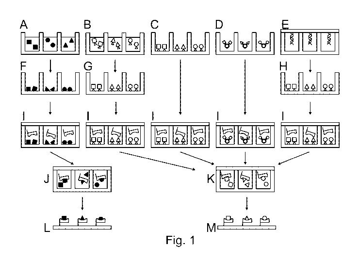
Figure 1 shows a preferred embodiment of the invention. In the figure shown, a
first surface is
used with separate cavities as active regions. A to E show how the first
molecules are present or
can be introduced. This can be performed either by spotting liquid containing
the pure molecules
(A), spotting liquid containing the molecules with a specific immobilization
tag (B), synthesizing
the molecules with a specific immobilization tag (C), spotting / applying
particles (beads) on which
the molecules with a specific immobilization tag are anchored (D) or by
closing the cavities with a
DNA microarray (spotting, synthesizing ...) containing spots of DNA which in
turn encode the first
complex partners (E).
If it is not already the case (C and D), the first molecules are applied to
the surface of the cavities
in the next step and fixed thereon. This can be achieved by drying the liquid
present (F), by
specific immobilization via the immobilization tag and subsequent washing or
drying of the chip
(G), by expression of the DNA molecules and subsequent specific immobilization
via the
immobilization tag and subsequent washing or drying of the chip (H).
In (I) the cavities are filled with the second molecule.
Complexation occurs within the closed cavities either by rehydration of the
molecules from step 1
(J) or by specific splitting-off of the immobilization tags of the first
molecules from step 1(K).
By capturing the resulting complexes on the capture surface and washing the
surface, a
microarray is formed, which can be further measured and characterized (L + M).
The capture
surface can be the second surface from step I or another surface.
Figure 2 shows a further preferred embodiment of the process according to the
invention.
One array is produced by synthesis or spotting with a plurality of different
first molecules, in this
example peptides (A). Another array is produced by spotting with a plurality
of second molecules
Date Recue/Date Received 2023-09-13
CA 03213500 2023-09-13
14
(in this case MHC complexes) (B). The two arrays are then brought into closer
contact in such a
way that a liquid bridge is created between the individual arrays. It is
important that the individual
liquid bridges do not touch each other, such that the active regions remain
separate (C). The
molecules of the first array (A) are either rehydrated or specifically split
off from the surface, e.g.
by means of light. The two molecules of the respective arrays are then mixed
together via this
contact and an MHC-peptide complex is formed (D). The MHC-peptide complexes
can then be
captured. The result is a microarray of the MHC-peptide complexes (E).
Figure 3
Figure 3 shows the application of the method according to the invention in
combination with an
MHC screening. To carry out such a screening, thousands of different peptides
are specifically
and separately mixed with the same MHC molecule (A). This leads to a
complexation of MHC
and peptide. For better illustration, the figure shows this process in
simplified form, not in closed
active regions. The individual complexes are then immobilized on a surface to
generate a
microarray (B). The microarray is then brought into contact with the TCR
molecule to be analyzed
(C). Finally, the interactions between the TCR and the MHC-peptide complexes
can be analyzed
(D).
Figure 4
Figure 4 shows a preferred embodiment of the method according to the
invention. The first
molecules, in this case peptides, are spotted onto a chip, e.g. a PDMS chip
(A). In step B it can
be seen how the peptides have been fixed by drying. In this case, storage at 4
C for a long period
of time is possible (C). In step D, the second molecules are added, in this
case MHC complexes.
In step E, the cavities of the first surface are closed with a capture surface
and closed active
regions are created in which MHC-peptide complexes are formed. These are
captured by the
capture molecules on the capture surface. In step F, in this case, T cell
receptors are added to
analyze the binding properties.
Figure 5
Figure 5 shows different embodiments of the method according to the invention.
In step A, the
first molecules are introduced into active regions (in this case cavities).
This takes place in the
form of droplets. The fixing can be seen in step B, which in this case is
achieved by drying. In this
example, the surfaces loaded in this way can be stored for a long time at
preferably 4 C (C).
The second row shows different ways of applying the second molecules. In this
example, MHCs
are used as second molecules. 1 shows that the second molecules can be applied
by means of
large droplets, so that multiple active regions are filled at the same time.
In this example, the
cavities are overfilled to avoid air pockets. In 2, the MHCs are applied in
smaller droplets to the
individual active regions in a more targeted manner. Here, too, the cavities
are overfilled in this
Date Recue/Date Received 2023-09-13
CA 03213500 2023-09-13
example. In 3, the MHCs are applied in smaller droplets to the individual
active regions in a more
targeted manner, whereby the volume of the droplets is smaller here than that
of the cavities.
Complexation takes place in the active regions. Subsequently, a capture
surface is applied in all
three examples. The last row shows how the complexes are bonded to the capture
surface and in
5 this case are examined for their binding properties to T cell receptors.
Figure 6
Figure 6 shows different results of the methods according to Figure 5, whereby
Figure 6.3.2
shows a very good result when the method according to the invention is carried
out correctly.
Figure 6.2.2 also shows an evaluable result, although there was cross-
contamination with the
10 neighboring cavities. Nevertheless, an interaction with the T-cell
receptors is already measurable
here. Figure 6.3.1 and 6.3.2 show a desirable result when the method according
to the invention
is carried out properly. Here, clean cavities can be seen, such that no cross-
contamination
occurred. The interaction with the T-cell receptors can be measured well.
Different experiments were carried out with MHCs as the second molecule. For
this purpose,
15 different MHCs were used and peptide-MHC (pMHC) complex arrays were
prepared using the
method of the invention. The arrays were then rinsed over with T cell
receptors and binding to the
pMHCs was displayed. The examples shown below are intended to illustrate the
invention and
are not intended to limit the subject matter of the application. In
particular, both MHC class 1 and
MHC class 2 molecules are suitable. The analysis with soluble T-cell receptor
analytes shown
here is one example of the scope of application. It is also possible to bring
the arrays into contact
with T cells or parts thereof and determine their interaction. Of course,
completely different
analyses are also possible, in which case the arrays are brought into contact
with the respective
other components or analysis partners.
In detail:
Example 1
Experiments were conducted with stabilized MHCs (source: Tetramershop) that do
not include
peptides in the peptide-binding pocket.
A streptavidin-coated glass slide and a cavity chip are provided.
Streptavidin-coated glass slides are used for immobilization of biotin-tagged
ligands.
The cavity chips (BioCopy cavity chip) comprise small cavities that are used
as reagent
containers for pMHC complexation.
Date Recue/Date Received 2023-09-13
CA 03213500 2023-09-13
16
The peptides used for the pMHC complexes are printed into the prepared cavity
chips. These can
now be stored until further use.
In the next step, the MHC molecules are printed into the prepared peptide
chips. This is followed
by binding of the peptide in the binding pocket of the MHC. The complexes
formed in this way are
captured on the streptavidin-coated surface and form a microarray formation.
After an incubation step, the glass slide-chip sandwich can be separated and
the pMHC
microarray is ready for use.
The arrays produced in this way were tested and rinsed with T cell receptors
for this purpose.
The binding of the pMHC spots was displayed and gave good results.
Example 2
Experiments were conducted with non-stabilized MHCs (source: e.g. Sanquin,
Biolegend)
comprising UV-cleavable or UV-sensitive peptides.
A streptavidin-coated glass slide and a cavity chip are provided.
Streptavidin-coated glass slides are used for immobilization of biotin-tagged
ligands.
The cavity chips (BioCopy cavity chip) comprise small cavities that are used
as reagent
containers for pMHC complexation.
The peptides used for the pMHC complexes are printed into the prepared cavity
chips. These can
now be stored until further use.
In the next step, the MHC molecules are printed into the prepared peptide
chips. For the
exchange of a UV-cleavable peptide localized in the non-stabilized MHC, a UV
light source is
used and the chip is illuminated. UV cleavage causes an exchange of the
cleaved peptide with
the provided (printed) peptide.
After peptide exchange, the complexes formed in this way are captured on the
streptavidin-
coated surface and form a microarray formation.
After an incubation step, the glass slide-chip sandwich can be separated and
the pMHC
microarray is ready for use.
The array produced in this way was rinsed with T cell receptors and binding to
the pMHCs could
be displayed and gave good results.
Date Recue/Date Received 2023-09-13
CA 03213500 2023-09-13
17
Example 3
Experiments have been carried out with non-stabilized HLAs (source: E.g.
Immundex) which
need to be folded. The unloaded MHCs are not folded correctly. Folding takes
place in the
presence of the peptides.
A streptavidin-coated glass slide and a cavity chip are provided.
Streptavidin-coated glass slides are used for immobilization of biotin-tagged
ligands.
The cavity chips (BioCopy cavity chip) comprise small cavities that are used
as reagent
containers for pMHC complex formation.
The peptides used for the pMHC complexes are printed into the prepared cavity
chips. These can
now be stored until further use.
In the next step, the MHC molecules are printed into the prepared peptide
chips. Now the folding
takes place and the peptides bind in the pockets of the MHC molecules, forming
a pMHC
complex.
The formed complexes are captured on the streptavidin-coated surface and form
a microarray
formation.
After an incubation step, the glass slide-chip sandwich can be separated and
the pMHC
microarray is ready for use.
The array produced in this way was also tested by rinsing it with T cell
receptors. The bonded
pMHC spots could be display and show good results.
Date Recue/Date Received 2023-09-13
CA 03213500 2023-09-13
18
Bibliography:
[1] Rays, M., Chen, Y., & Su, Y. A. (1996). Use of a cDNA microarray to
analyse gene
expression patterns in human cancer. Nature genetics, 14.
[2] Blanchard, A. P., Kaiser, R. J., & Hood, L. E. (1996). High-density
oligonucleotide
arrays. Biosensors and bioelectronics, 11(6-7), 687-690.
[3] Pease, A. C., Soles, D., Sullivan, E. J., Cronin, M. T., Holmes, C. P.,
& Fodor, S. P.
(1994). Light-generated oligonucleotide arrays for rapid DNA sequence
analysis. Proceedings of
the National Academy of Sciences, 91(11), 5022-5026.
[4] Nuwaysir, E. F., Huang, W., Albert, T. J., Singh, J., Nuwaysir, K.,
Pitas, A., ... & Green, R.
D. (2002). Gene expression analysis using oligonucleotide arrays produced by
maskless
photolithography. Genome research, 12(11), 1749-1755.
[5] Kilb, N., Burger, J., & Roth, G. (2014). Protein microarray generation
by in situ protein
expression from template DNA. Engineering in Life Sciences, 14(4), 352-364.
[6] U520060141245A1
[7] W02006112815A2
[8] Lin, H., Sun, L., & Crooks, R. M. (2005). Replication of a DNA
Microarray. Journal of the
American Chemical Society, 127(32), 11210-11211.
[9] Kim, J., & Crooks, R. M. (2007). Parallel fabrication of RNA
microarrays by mechanical
transfer from a DNA master. Analytical chemistry, 79(23), 8994-8999.
[10] Lin, H., Kim, J., Sun, L., & Crooks, R. M. (2006). Replication of DNA
microarrays from zip
code masters. Journal of the American Chemical Society, 128(10), 3268-3272.
[11] Kim, J., & Crooks, R. M. (2007). Replication of DNA microarrays
prepared by in situ
oligonucleotide polymerization and mechanical transfer. Analytical chemistry,
79(19), 7267-7274.
[12] U520100256017A1
[13] W02008022332A2
[14] W02010100265A1
[15] Kramer, S. D., Wohrle, J., Meyer, P. A., Urban, G. A., & Roth, G.
(2019). How to copy and
paste DNA microarrays. Scientific reports, 9(1), 1-10.
Date Recue/Date Received 2023-09-13
CA 03213500 2023-09-13
19
[16] Kilb, N., Herz, T., Burger, J., Woehrle, J., Meyer, P. A., & Roth, G.
(2019). Protein
Microarray Copying: Easy on-Demand Protein Microarray Generation Compatible
with
Fluorescence and Label-Free Real-Time Analysis. ChemBioChem, 20(12), 1554-
1562.
[17] Moritz, A., Anjanappa, R., Wagner, C., Bunk, S., Hofmann, M., Pszolla,
G., ... & Maurer,
D. (2019). High-throughput peptide-MHC complex generation and kinetic
screenings of TCRs
with peptide-receptive HLA-A* 02: 01 molecules. Science immunology, 4(37).
[18] McCarty, N. S., Graham, A. E., Studena, L., & Ledesma-Amaro, R.
(2020). Multiplexed
CRISPR technologies for gene editing and transcriptional regulation. Nature
communications,
11(1), 1-13.
Date Recue/Date Received 2023-09-13