Note: Descriptions are shown in the official language in which they were submitted.
WO 2022/208327 PCT/IB2022/052862
1
SPATIAL MAPPING BY SERIAL PRIMER EXTENSION
CROSS-REFERENCING
This application claims the benefit of U.S. provisional application serial no.
63/168,132, filed on March 30, 2021, which application is incorporated by
reference herein in
its entirety.
BACKGROUND
Sample and molecule indices are commonly employed in many of today's genomics
workflows. Sample indexing is a commonly used approach that enables samples to
be
sequenced and analyzed in a multiplex way. In sample indexing methods, all
nucleic acids in a
particular sample are labeled with the same sequence tag, and the tagged
library is pooled with
other libraries that are tagged with different barcodes, and the pool is
sequenced in parallel in a
single sequencing run. Then, during analysis, the sample-specific indexes
allow software to
separate the multiplexed sequence data into sample-specific data sets. The
goal of molecular
indexing, on the other hand, is to tag each molecule in a sample a unique
sequence before PCR
amplification. In these methods, each nucleic acid in the starting sample is
tagged with a
unique molecule index, and the sequence analysis software filters out
duplicate reads (i.e.,
reads from copes of the same molecule) and eliminate PCR/sequencing errors
using the index.
This disclosure employs indices to provide spatial information, which is
believed to be
a new use for molecular indices.
SUMMARY
This disclosure provides, among other things, a probe system comprising: (a) a
population of nucleic acid molecules that have an extendible end; (b) a first
set of barcoded
particles that each have a nucleotide sequence comprising: (i) a binding
sequence that is
complementary to the extendible end of the nucleic acid molecules of (a), (ii)
a unique particle
identifier sequence, and (iii) a first template sequence; (c) a second set of
barcoded particles
that each have a nucleotide sequence comprising: (i) the first template
sequence, and (ii) a
unique particle identifier sequence. In this probe system, extension of the
nucleic acid
molecules of (a) using the first set of barcoded particles of (b) as a
template produces
CA 03213800 2023- 9- 27
WO 2022/208327 PCT/1B2022/052862
2
extensions products that contain the complement of a unique particle
identifier sequence of a
particle of (a)(ii) and the complement of the first template sequence.
In the description that follows below, the nucleic acid molecules that have an
extendible end are primers, and the method is implemented using primer
extension. In
alternate embodiments (illustrated in Figs. 12-14) the method may be
implemented using gap-
fill/ligation or ligation-based method. As such, the present system and method
should not be
limited to primer extension-based methods only.
In some embodiments, the method comprises: hybridizing the first set of
barcoded
particles with the first population of primer molecules extending the
hybridized primer
molecules using the nucleotide sequence of the first set of barcoded particles
as a template to
produce first primer extension products that contain the complement of a
unique particle
identifier sequence from a barcoded particle in the first set of barcoded
particles and the
complement of the first template sequence; removing the first set of barcoded
particles;
hybridizing the first primer extension products with the second set of
barcoded particles,
IS wherein the complement of the first template sequence in the first
primer extension products
hybridizes to the first template sequence in the second set of barcoded
particles; and extending
the first primer extension products using the nucleotide sequence of the
second set of barcoded
particles as a template to produce second primer extension products that
contain: a unique
particle identifier sequence from a barcoded particle in the first set of
barcoded particles, the
first template sequence, and a unique particle identifier sequence from a
barcoded particle in
the second set of barcoded particles.
The probe system may be employed in a method of making a map of binding events
on
a cellular sample. In the embodiments, this method may comprise: (a)
obtaining: i. a sample
containing primer molecules that are bound to sites in or on cells; ii. a
first set of barcoded
particles that each have a nucleotide sequence comprising: (i) a primer
binding sequence that is
complementary to the 3' end of the primer molecules, (ii) a unique particle
identifier sequence,
and (iii) a first template sequence; iii. a second set of barcoded particles
that each have a
nucleotide sequence comprising: (i) the first template sequence, and (ii) a
unique particle
identifier sequence; (b) specifically hybridizing the first set of barcoded
particles with the
sample, wherein the nucleotide sequence of at least some of the first set of
barcoded particles
CA 03213800 2023- 9- 27
WO 2022/208327 PCT/1B2022/052862
3
hybridizes to at least two primer molecules; (c) extending the primers that
are hybridized to
barcoded particles in step (b) using the nucleotide sequences to which the
primers are
hybridized as a template to produce first primer extension products that each
comprise a first
unique particle identifier sequence; (d) removing the first set of barcoded
particles from the
sample; (e) specifically hybridizing the second set of barcoded particles with
the first primer
extension products of (c), wherein the nucleotide sequences of at least some
of the second set
of barcoded particles hybridizes to at least two molecules of the primer
extension products: (f)
extending the first primer extension products that are hybridized to a
barcoded particle in step
(e) using the nucleotide sequences to which the primers are hybridized to as a
template to
produce second primer extension products that comprise the two unique particle
identifier
sequences; (g) determining which unique particle identifier sequence or
complements thereof
are in second primer extension products; and (h) making a map of the relative
positions of the
primers using the unique particle identifier sequences that is in the second
primer extension
products.
IS Alternative embodiments involve a probe system comprising: (a) a
population of
primer molecules; (b) a set of barcoded particles that each have a nucleotide
sequence
comprising: (i) a primer binding sequence that is complementary to the 3' end
of the primer
molecules of (a), (ii) a unique particle identifier sequence, and (iii) a
first template sequence;
and (c) a ligation splint comprising a first oligonucleotide and a second
oligonucleotide,
wherein the first oligonucleotide comprises a first sequence and the first
template sequence;
and the second oligonucleotide comprises a second sequence that is
complementary to the first
sequence, and the first template sequence.
The alternative probe system may be used in a method that comprises: i.
hybridizing
the set of barcoded particles of (b) with the population of primer molecules
of (a), ii. extending
the hybridized primer molecules using the nucleotide sequences as a template
to produce first
primer extension products that contain i. the complement of a unique particle
identifier
sequence from a barcoded particle and ii. the complement of the first template
sequence;
iii. removing the barcoded particles; iv. hybridizing the first primer
extension products with
the ligation splint, wherein the complements of the first template sequence in
two proximal
first primer extension products hybridize to the first and second sequences of
the ligation
CA 03213800 2023- 9- 27
WO 2022/208327 PCT/1B2022/052862
4
splint; and ligating at least one of the first or the second oligonucleotide
of the hybridized
ligation splint to the first primer extension products and extending the 3'
end of the ligated
first or second oligonucleotide in the splint using the first primer extension
product in the
ligation product as a template, thereby adding two unique particle identifier
sequences to a
primer.
BRIEF DESCRIPTION OF THE FIGURES
The skilled artisan will understand that the drawings, described below, are
for
illustration purposes only. The drawings are not intended to limit the scope
of the present
teachings in any way.
Fig_ 1 schematically illustrates a probe system of the present disclosure.
Fig. 2 schematically illustrates a method in which the probe system can be
used.
Fig. 3 schematically illustrates a probe system that has primer binding sites,
thereby
allowing the extension products to be amplified.
Fig. 4 schematically illustrates an embodiment of the method in which the
primers are
teghered.
Fig. 5 schematically illustrates how the probe system can be used to produce
an array.
Fig. 6 also illustrates how the probe system can be used to produce an array.
Fig. 7 illustrates how a sample can be analyzed using the array illustrated in
Figs. 5 and
6.
Fig. 8 schematically illustrates how binding sites can be spatially
reconstructed.
Figs. 9A-9C schematically illustrate further details of an embodiment of the
present
method.
Fig. 10 schematically illustrates an alternative probe system and method of
using the
same.
Fig. 11 schematically illustrates a method in which the alternative probe
system shown
in Fig. 10 can be used.
CA 03213800 2023- 9- 27
WO 2022/208327 PCT/IB2022/052862
Fig. 12 schematically illustrates a gap-fill/ligation implementation of the
present
method.
Fig. 13 schematically illustrates a ligation-based version of the present
method, where
the nucleic acid molecules are attached to "pre-made" barcodes.
5 Fig. 14 schematically illustrates a gap-fill/ligation version of the
present method that
can be used to add barcodes to cDNA.
Fig. 15 schematically illustrates how the method can be implemented using
rolling
circle amplification products using a hairpin in each of the repeats.
Fig. 16 follows on from Fig. 15 and illustrates one example of the assay can
be
designed so that the primer extension product end at a defined nucleotide,
thereby allowing it
to be used as a primer in the next step of the method.
Fig. 17 schematically illustrates an alternative example of how the assay can
be
designed so that the primer extension product end at a defined nucleotide,
thereby allowing it
to be used as a primer in the next step of the method.
Figs. 18A-18C show correlation plots of marker counts within each cluster.
Poor
correlation between CD3 and HLA-DR suggests each cluster represents a single
cell.
Fig. 19 is a visualization of a graph component (cluster) using a force-
generated layout
algorithm. Different antibody types can be distinguished.
DEFINITIONS
Before describing exemplary embodiments in greater detail, the following
definitions
are set forth to illustrate and define the meaning and scope of the terms used
in the description.
Numeric ranges are inclusive of the numbers defining the range. Unless
otherwise
indicated, nucleic acids are written left to right in 5 to 3' orientation;
and, amino acid
sequences are written left to right in amino to carboxy orientation,
respectively.
Unless defined otherwise, all technical and scientific terms used herein have
the same
meaning as commonly understood by one of ordinary skill in the art to which
this invention
belongs. Singleton, et al., DICTIONARY OF MICROBIOLOGY AND MOLECULAR
CA 03213800 2023- 9- 27
WO 2022/208327
PCT/IB2022/052862
6
BIOLOGY, 2D ED., John Wiley and Sons, New York (1994), and Hale & Markham, THE
HARPER COLLINS DICTIONARY OF BIOLOGY, Harper Perennial, N.Y. (1991) provide
one of skill with the general meaning of many of the terms used herein. Still,
certain terms are
defined below for the sake of clarity and ease of reference.
It must be noted that as used herein and in the appended claims, the singular
forms -a",
"an", and "the" include plural referents unless the context clearly dictates
otherwise. For
example, the term "a primer" refers to one or more primers, i.e., a single
primer and multiple
primers. It is further noted that the claims can be drafted to exclude any
optional element. As
such, this statement is intended to serve as antecedent basis for use of such
exclusive
terminology as "solely," "only" and the like in connection with the recitation
of claim
elements, or use of a "negative" limitation.
The term "nucleotide" is intended to include those moieties that contain not
only the
known purine and pyrimidine bases, but also other heterocyclic bases that have
been modified.
Such modifications include methylated purines or pyrimidines, acylated purines
or
pyrimidines, alkylated riboses or other heterocycles. In addition, the term
"nucleotide"
includes those moieties that contain hapten or fluorescent labels and may
contain not only
conventional ribose and deoxyribose sugars, but other sugars as well. Modified
nucleosides or
nucleotides also include modifications on the sugar moiety, e.g., wherein one
or more of the
hydroxyl groups are replaced with halogen atoms or aliphatic groups, are
functionalized as
ethers, amines, or the likes.
The term "nucleic acid" and "polynucleotide" are used interchangeably herein
to
describe a polymer of any length, e.g., greater than about 2 bases, greater
than about 10 bases,
greater than about 100 bases, greater than about 500 bases, greater than 1000
bases, up to
about 10,000 or more bases composed of nucleotides, e.g., deoxyribonucleotides
or
ribonucleotides, and may be produced enzymatically or synthetically (e.g., PNA
as described
in U.S. Patent No. 5,948,902 and the references cited therein) which can
hybridize with
naturally occurring nucleic acids in a sequence specific manner analogous to
that of two
naturally occurring nucleic acids, e.g., can participate in Watson-Crick base
pairing
interactions. Naturally-occurring nucleotides include guanine, cytosine,
adenine, thymine,
CA 03213800 2023- 9- 27
WO 2022/208327
PCT/IB2022/052862
7
uracil (G, C, A, T and U respectively). DNA and RNA have a deoxyribose and
ribose sugar
backbone, respectively, whereas PNA's backbone is composed of repeating N-(2-
aminoethyl)-
glycine units linked by peptide bonds. In PNA, various purine and pyrimidine
bases are linked
to the backbone by methylene carbonyl bonds. A locked nucleic acid (LNA),
often referred to
as inaccessible RNA, is a modified RNA nucleotide. The ribose moiety of an LNA
nucleotide
is modified with an extra bridge connecting the 2 oxygen and 4' carbon. The
bridge "locks"
the ribose in the 3'-endo (North) conformation, which is often found in the A-
form duplexes.
LNA nucleotides can be mixed with DNA or RNA residues in the oligonucleotide
whenever
desired. The term "unstructured nucleic acid", or "UNA", is a nucleic acid
containing non-
natural nucleotides that bind to each other with reduced stability. For
example, an unstructured
nucleic acid may contain a G' residue and a C' residue, where these residues
correspond to
non-naturally occurring forms, i.e., analogs, of G and C that base pair with
each other with
reduced stability, but retain an ability to base pair with naturally occurring
C and G residues,
respectively. Unstructured nucleic acid is described in US20050233340, which
is incorporated
by reference herein for disclosure of UNA.
The term "oligonucleotide" as used herein denotes a single-stranded multimer
of
nucleotides of from about 2 to 200 nucleotides, up to 500 nucleotides in
length.
Oligonucleotides may be synthetic or may be made enzymatically, and, in some
embodiments,
are 30 to 150 nucleotides in length. Oligonucleotides may contain
ribonucleotide monomers
(i.e., may be oligoribonucleotides) or deoxyribonucleotide monomers. An
oligonucleotide may
be 10 to 20,21 to 30, 31 to 40,41 to 50, 51to 60, 61 to 70, 71 to 80, 80 to
100, 100 to 150 or
150 to 200 nucleotides in length, for example.
The term "primer" as used herein refers to an oligonucleotide that is capable
of acting
as a point of initiation of synthesis when placed under conditions in which
synthesis of a
primer extension product, which is complementary to a nucleic acid strand, is
induced, i.e., in
the presence of nucleotides and an inducing agent such as a DNA polymerase and
at a suitable
temperature and pH. The primer may be single-stranded and must be sufficiently
long to
prime the synthesis of the desired extension product in the presence of the
inducing agent. The
exact length of the primer will depend upon many factors, including
temperature, source of
primer and use of the method. For example, for diagnostic applications,
depending on the
CA 03213800 2023- 9- 27
WO 2022/208327 PCT/IB2022/052862
8
complexity of the target sequence or fragment, the oligonucleotide primer
typically contains
15-25 or more nucleotides, although it may contain fewer nucleotides. The
primers herein are
selected to be substantially complementary to different strands of a
particular target DNA
sequence. This means that the primers must be sufficiently complementary to
hybridize with
their respective strands. Therefore, the primer sequence need not reflect the
exact sequence of
the template. For example, a non-complementary nucleotide fragments may be
attached to the
5' end of the primer, with the remainder of the primer sequence being
complementary to the
strand. Alternatively, non-complementary bases or longer sequences can be
interspersed into
the primer, provided that the primer sequence has sufficient complementarity
with the
sequence of the strand to hybridize therewith and thereby form the template
for the synthesis
of the extension product.
The term "primer extension products" refer to the product of extension of a
primer or
the product of extension of a molecule that is itself a primer extension
product. The term "first
primer extension product" refers to molecule that are the product of extension
of a primer. The
term "second primer extension products" refers to the product obtained by
extending first
primer extension products. If the second primer extension products are
sequenced, then the
entire molecule (or most of it) may be sequenced, which sequence includes at
least the
sequence added onto the primer in the first primer extension reaction and
sequence added onto
the first primer extension product in the second primer extension reaction.
The term "hybridization" or "hybridizes" refers to a process in which a
nucleic acid
strand anneals to and forms a stable duplex, either a homoduplex or a
heteroduplex, under
normal hybridization conditions with a second complementary nucleic acid
strand and does
not form a stable duplex with unrelated nucleic acid molecules under the same
normal
hybridization conditions. The formation of a duplex is accomplished by
annealing two
complementary nucleic acid strands in a hybridization reaction. The
hybridization reaction can
be made to be highly specific by adjustment of the hybridization conditions
(often referred to
as hybridization stringency) under which the hybridization reaction takes
place, such that
hybridization between two nucleic acid strands will not form a stable duplex,
e.g., a duplex
that retains a region of double-strandedness under normal stringency
conditions, unless the two
nucleic acid strands contain a certain number of nucleotides in specific
sequences which are
CA 03213800 2023- 9- 27
WO 2022/208327
PCT/IB2022/052862
9
substantially or completely complementary. "Normal hybridization or normal
stringency
conditions" are readily determined for any given hybridization reaction. See,
for example,
Ausubel et al., Current Protocols in Molecular Biology, John Wiley & Sons,
Inc., New York,
or Sambrook et al., Molecular Cloning: A Laboratory Manual, Cold Spring Harbor
Laboratory
Press. As used herein, the term "hybridizing" or "hybridization" refers to any
process by which
a strand of nucleic acid binds with a complementary strand through base
pairing.
A nucleic acid is considered to be "selectively hybridizable" to a reference
nucleic acid
sequence if the two sequences specifically hybridize to one another under
moderate to high
stringency hybridization and wash conditions. Moderate and high stringency
hybridization
conditions are known (see, e.g., Ausubel, et al., Short Protocols in Molecular
Biology, 3rd ed.,
Wiley & Sons 1995 and Sambrook et al., Molecular Cloning: A Laboratory Manual,
Third
Edition, 2001 Cold Spring Harbor, N.Y.). One example of high stringency
conditions includes
hybridization at about 42 C in 50% formamide, 5X SSC, 5X Denhardt's solution,
0.5% SDS
and 100 ug/ml denatured carrier DNA followed by washing two times in 2X SSC
and 0.5%
SDS at room temperature and two additional times in 0.1 X SSC and 0.5% SDS at
42 C.
The term "sequencing", as used herein, refers to a method by which the
identity of at
least 10 consecutive nucleotides (e.g., the identity of at least 20, at least
50, at least 100 or at
least 200 or more consecutive nucleotides) of a polynucleotide are obtained.
The term "next-generation sequencing" refers to the so-called parallelized
sequencing-
by-synthesis or sequencing-by-ligation platforms currently employed by, e.g.,
Illumina, Life
Technologies, B GI Genomics (Complete Genomics technology), and Roche etc.
Next-
generation sequencing methods may also include nanopore sequencing methods or
electronic-
detection based methods such as, e.g., Ion Torrent technology commercialized
by Life
Technologies.
The term "duplex," or "duplexed," as used herein, describes two complementary
polynucleotides that are base-paired, i.e., hybridized together.
The terms "determining," "measuring," "evaluating," "assessing," "assaying,"
and
"analyzing" are used interchangeably herein to refer to forms of measurement,
and include
determining if an element is present or not. These terms include both
quantitative and/or
qualitative determinations. Assessing may be relative or absolute.
CA 03213800 2023- 9- 27
WO 2022/208327
PCT/IB2022/052862
The term "ligating", as used herein, refers to the enzymatically catalyzed
joining of the
terminal nucleotide at the 5' end of a first DNA molecule to the terminal
nucleotide at the 3'
end of a second DNA molecule.
The terms "plurality", "set" and "population" are used interchangeably to
refer to
5 something that contains at least 2 members. In certain cases, a plurality
may have at least 10, at
least 100, at least 1000, at least 10,000, or at least 100,000 members.
A "primer binding site" refers to a site to which an oligonucleotide
hybridizes in a
target polynucleotide or fragment. If an oligonucleotide "provides" a binding
site for a primer,
then the primer may hybridize to that oligonucleotide or its complement.
10 The term "strand" as used herein refers to a nucleic acid made up of
nucleotides
covalently linked together by covalent bonds, e.g., phosphodiester bonds.
The term "extending", as used herein, refers to the extension of a nucleic
acid by
ligation or the addition of nucleotides using a polymerase. If a nucleic acid
that is annealed to a
polynucleotide is extended, the polynucleotide acts as a template for an
extension reaction. In
these embodiments, the nucleic acid may be extended by a template-dependent
polymerase or
by ligation to an oligonucleotide that is complementary to the polynucleotide,
where the
polynucleotide acts as a splint.
The term "extending" includes extension at the 3' end or the 5' end. Primer
extension,
ligation and gap-fill ligation reactions are types of extending.
The term "extendible 5 or 3' end" refers to a 5' phosphate and 3' hydroxyl,
respectively,
both of which are extensible by ligation. 3' hydroxyls are also extendible by
a polymerase.
The term "as a template" as used herein, refers to: (a) a primer extension
reaction in one strand acts as a
template for the addition of nucleotides by a polymerase, (b) a splinted
ligation, where one strands acts a template
(or "splint") for ligating two nucleic acid molecules together. In ligation
reactions, both molecules hybridize to
the template and become ligated. Ligation can be at the 5' end of a nucleic
acid molecule or at the 3' end of a
nucleic acid molecule, at the 3' end or the 5' end; and (c) gap-fill/ligation
reactions. In gap-fill/ligation reactions,
two nucleic acids are hybridized to a template with a gap inbetween. One
nucleic acid molecule is extended
towards the other nucleic acid molecule by primer extension and then the 3'
end of product is ligated to the other
nucleic acid. As used herein, the term "rolling circle amplification" or "RCA"
for short refers to
an isothermal amplification that generates linear concatemerized copies of a
circular nucleic
acid template using a strand-displacing polymerase. RCA is well known in the
molecular
CA 03213800 2023- 9- 27
WO 2022/208327
PCT/IB2022/052862
11
biology arts and is described in a variety of publications including, but not
limited to Lizardi et
al (Nat. Genet. 1998 19:225-232), Schweitzer et al (Proc. Natl. Acad. Sci.
2000 97:10113-
10119), Wiltshire et al (Clin. Chem. 2000 46:1990-1993) and Schweitzer et al
(Curr. Opin.
Biotech 2001 12:21-27), which are incorporated by reference herein.
As used herein, the term "rolling circle amplification products" refers to the
concatemerized products of a rolling circle amplification reaction.
As used herein, the term "surface" refers to any solid material (e.g. glass,
metal,
ceramics, organic polymer surface or gel) that may contain cells or any
combinations of
biomolecules derived from cells, such as proteins, nucleic acids, lipids,
oligo/polysaccharides,
biomolecule complexes, cellular organelles, cellular debris or excretions
(exosomes,
microvesicles), etc. Tissue blots, western blots and glass slides are examples
of solid materials
that have a surface. Cells, e.g., suspensions of mammalian cells, are another
example of a
surface.
As used herein, the term "splint" refers to an oligonucleotide that hybridize
to the ends
of two other oligonucleotides and brings those ends together to produce a
ligatable junction or
a gap that can be filled by a gap-fill/ligation reaction.
As used herein, the term "barcoded particles" is intended to refer to both
barcoded
RCA products and barcoded nanoparticles, wherein the particles in a population
of barcoded
particles are each separately barcoded with a unique particle identifier
sequence, i.e., a
sequence that is unique to each particle such that the particles can be
distinguished from one
another by their unique identifier sequences.
Other definitions of terms may appear throughout the specification.
DESCRIPTION OF EXEMPLARY EMBODIMENTS
Before the various embodiments are described, it is to be understood that the
teachings
of this disclosure are not limited to the particular embodiments described,
and as such can, of
course, vary. It is also to be understood that the terminology used herein is
for the purpose of
describing particular embodiments only, and is not intended to be limiting,
since the scope of
the present teachings will be limited only by the appended claims.
CA 03213800 2023- 9- 27
WO 2022/208327
PCT/IB2022/052862
12
The section headings used herein are for organizational purposes only and are
not to be
construed as limiting the subject matter described in any way. While the
present teachings are
described in conjunction with various embodiments, it is not intended that the
present
teachings be limited to such embodiments. On the contrary, the present
teachings encompass
various alternatives, modifications, and equivalents, as will be appreciated
by those of skill in
the art.
Unless defined otherwise, all technical and scientific terms used herein have
the same
meaning as commonly understood by one of ordinary skill in the art to which
this disclosure
belongs. Although any methods and materials similar or equivalent to those
described herein
can also be used in the practice or testing of the present teachings, some
exemplary methods
and materials are now described.
The citation of any publication is for its disclosure prior to the filing date
and should
not be construed as an admission that the present claims are not entitled to
antedate such
publication by virtue of prior invention. Further, the dates of publication
provided can be
different from the actual publication dates which can need to be independently
confirmed.
As will be apparent to those of skill in the art upon reading this disclosure,
each of the
individual embodiments described and illustrated herein has discrete
components and features
which can be readily separated from or combined with the features of any of
the other several
embodiments without departing from the scope or spirit of the present
teachings. Any recited
method can be carried out in the order of events recited or in any other order
which is logically
possible.
All patents and publications, including all sequences disclosed within such
patents and
publications, referred to herein are expressly incorporated by reference.
Before certain aspects of the present invention are described in greater
detail, it is
important to note that the figures illustrate embodiments of present probe
system and method
that employ RCA products. As noted below, the principles illustrated in the
figures can be
readily applied to barcoded nanoparticles and, as such, the present invention
should not be
limited to what is described in the figures. In addition, only one of the RCA
product repeats is
shown in the figures. As is well known, barcoded RCA products (and barcoded
nanoparticles)
CA 03213800 2023- 9- 27
WO 2022/208327
PCT/IB2022/052862
13
may contain at least 10, at least 50, at least 100, at least 500 or at least
1,000 repeats of the
molecule (which are either concatenated in the RCA product) or tethered to the
surface of a
nanoparticle).
Moreover, it is recognized that the present probe system and method does not
need to
be implemented as a primer extension assay (as illustrated in Fig. 2). As
illustrated in Figs. 12-
14, the method may be implemented using gap-fill/ligation or ligation assay,
which can both
involve extending the 5' end of the nucleic acid molecules using the barcoded
particles as a
template. As such, while the description below is focused on embodiments, that
employ primer
extension, it should be recognized that the primer extension-based embodiments
described
below are just an example of how the method can be implemented.
Some principles of the present probe system are illustrated in Fig. 1. With
reference to
Fig. 1, the probe system may comprise: a population of primer molecules 2
(which, as shown,
have a 3' end of sequence C-BS1'). As will be described in greater detail
below, the primer
molecules may be synthetic oligonucleotides or cDNA molecules that have a
primer sequence
at the 3' end, for example. In some embodiments, the primer molecule may be
linked to a
binding agent (e.g., an oligonucleotide probe, antibody, aptamer, etc.) and in
other
embodiments, the primer molecules may be linked to a planar substrate as a
lawn in which the
3' ends of the oligonucleotides are distal to the substrate and capable of
being extended by a
polymerase. As illustrated, the probe system also comprises first set of
barcoded particles 4
and second set of barcoded particles 12. As shown, the particles in the first
set of barcoded
particles 4 each have a nucleotide sequence comprising: a primer binding
sequence 6 (C-BS1)
that is complementary to the 3' end of the primer molecules 2, as well as a
unique particle
identifier sequence 8 (UMI1), and first template sequence 10 (C-BS2). The
particles in the
second set of barcoded particles 12 each have a nucleotide sequence
comprising: the first
template sequence 10 (i.e., the same template sequence as is in the first set
of particles, C-
BS2), and a unique particle identifier sequence 14 (UMI2). In the embodiment
shown, the
nucleotide sequence of the second set of barcoded particles does not contain
primer binding
sequence 6 (CBS1). However, in other embodiments, the nucleotide sequence of
the second
set of barcoded particles may contain primer binding sequence 6 (CBS1). In
this probe system
(as shown in Fig. 2), extension of the primer molecules 2 using the first set
of barcoded
CA 03213800 2023- 9- 27
WO 2022/208327
PCT/IB2022/052862
14
particles 4 as a template produces first primer extensions products 16 that
contain the
complement of a unique particle identifier sequence 18 of a particle of the
first set of barcoded
particles (i.e., the complement of UMI1, or UMI1') and the complement 20 of
the first
template sequence (C-BS2', which is the complement of C-BS2). As will be
described in
greater detail below, the 3' end of the primer extension product 16 can
hybridize to the first
template sequence 10 of the second set of particles. As shown, extension of
the 3' end of the
primer extension product 16 using a particle from the second set of barcoded
particles as a
template results in second primer extension products 22 that contain the
complement 18 of a
unique particle identifier sequence from the first set of particles, the
complement 20 of the first
template sequence, and 24 the complement of a unique particle identifier
sequence from the
second set of particles. In some embodiments, there may be an internal hairpin
immediately
downstream of first template sequence.
In some embodiments, the first and second sets of barcoded particles may be
rolling
circle amplification (RCA) products or barcoded nanoparticles. For example, in
some
embodiments, the first and second sets of barcoded particles may be RCA
products, the first
and second sets of barcoded particles may be barcoded nanoparticles or the
first set of
barcoded particles may be RCA products or barcoded nanoparticles and the
second set of
barcoded particles may be the other type.
In embodiments in which the barcoded particles are RCA products, the RCA
products
each contain a unique sequence that is in the repeated sequence. In other
words, if there are
1,000 RCA products, each product will have a unique sequence (referred to
herein as a unique
molecular identifier "UMI- or unique identifier "UID"). The UID for one
particle is different
to the UIDs for other particles. The RCA product can be made by, e.g.,
synthesizing initial
oligonucleotides that have a degenerate sequence, circularizing the initial
oligonucleotides
using a splint, and amplifying the circularized oligonucleotides by RCA. In
some
embodiments, the initial oligonucleotides may contain a degenerate (e.g.,
random) sequence of
6-10 nucleotides, or even more random nucleotides dependent on the number of
unique RCA
products required. Amplification of circularized oligonucleotides that have a
degenerate
sequence should produce a population of RCA products that each have a unique
identifier (i.e.,
a sequence that is different from the other RCA products in the population).
Methods for
CA 03213800 2023- 9- 27
WO 2022/208327
PCT/IB2022/052862
generating RCA products that have unique identifiers are described in Wu et al
(Nat. Comm.
2019 10: 3854) and US20160281134, for example, and are readily adapted for use
herein. In
some embodiments, the different oligonucleotides that are used to make the
first and second
sets of RCA products are made separately and then mixed together. In other
embodiments, the
5 different oligonucleotides may be made in parallel on a planar support in
the form of an array
and then cleaved from the array. Examples of such methods are described in,
e.g., Cleary et al.
(Nature Methods 2004 1: 241-248) and LeProust et al. (Nucleic Acids Research
2010 38:
2522-2540). In some embodiments, one or both sets of RCA products may contain
uracil of
thymine, thereby allowing the RCA products to be degraded enzymatically, by
USER (see,
10 e.g., Bitinaite et al Nucleic Acids Res. 2007 35: 1992-2002), which
contains UDG (uracil
DNA glycosylase) and an AP lyase which cleaves the phophodiester backbone at
apurinic
sites.
In embodiments in which the barcoded particles are barcoded nanoparticles, the
barcoded nanoparticles are small beads or metallic particles the like, that
are coated in
15 oligonucleotides, where the surface-tethered oligonucleotides on each
particle have a unique
sequence that is different to the sequence that is in the oligonucleotides
that are tethered to
other particles in the population. In other words, if there are 1,000 barcoded
particles, the
oligonucleotides that are tethered to each particle will have a unique
sequence (referred to
herein as a unique molecular identifier "UMI" or unique identifier "UID". The
UID for one
particle is different to the UIDs for other particles. These particles can be
of any suitable size,
material and shape. In many embodiments, the particles have a size of 10nm-
200nm. Gold
particles (that can be readily made to any diameter in the range of 1.8 nm to
1500 nm, for
example) can be used, although the particles can also be made from silver,
silica, titanium
dioxide, carbon, polymers (like polystyrene, polyacrylate, etc), agarose, etc.
Magnetic particles
of iron and various alloys could also be used (Creative Diagnostics, Shirley,
NY, USA). The
particles do not need to be magnetic, but magnetic nanospheres could be used
in some cases
(Creative Diagnostics, Shirley, NY, USA). There are several surface
chemistries for
functionalizing metal surfaces so that they can be joined to nucleic acid. For
example, the
particles may be modified to contain reactive groups, including, but not
limited to, N-
hydroxysuccinimidyl ester, sulfo-N-hydroxysuccinimidyl ester, a halo-
substituted phenol
CA 03213800 2023- 9- 27
WO 2022/208327
PCT/IB2022/052862
16
ester, pentafluorophenol ester, a nitro-substituted phenol ester, an
anhydride, isocyanate,
isothiocyanate, an imidoester, maleimide, iodoacetyl, hydrazide, an aldehyde,
or an epoxide.
Other suitable groups are known in the art and may be described in, e.g.,
Hermanson,
"Bioconjugate Techniques" Academic Press, 2nd Ed., 2008. The most commonly
used
capture-agent reactive groups are NHS (which is amine-reactive) and maleimide
(which is
sulfhydryl-reactive), although many others may be used. The particles can also
be coated in
streptavidin, which can bind to biotinylated nucleic acids. In some
embodiments, the barcoded
particles may be made by emulsion PCR, which method has been successfully used
for other
applications and is described in, e.g., Kanagal-Shamanna et al (Methods Mol
Biol 2016 1392:
33-42) and Shao et al (PlosOne 2011 0024910). In some embodiments, the nucleic
acids of the
barcoded particles may contain uracil, thereby allowing them to be cleaved
using USER, as
discussed above.
In any embodiment, the primer molecules 2 may contain a 5' tail that does not
hybridize to any of the nucleic acids of the barcoded particles. The 5' tail
may contain a primer
binding site (e.g., for a forward primer) and a target identifier sequence
which may be used
later in the protocol. As noted above, the primer molecules may be synthetic,
man-made
oligonucleotides that are in the 10-200 nucleotides in length. In some of
these embodiments,
the primer molecules may be linked to a binding agent, (e.g., an
oligonucleotide probe,
antibody, aptamer, etc.). In these embodiments, the primer molecules are not
cDNA. In some
embodiments, the primer molecules may contain cDNA molecules that have a
primer sequence
at the 3' end, i.e., an appended sequence that can serve as a primer. In these
embodiments, the
cDNA can be made by, e.g., hybridizing a reverse transcription primer (e.g., a
primer that has
a 3' end made of oligo(dT), a random sequence or gene-specific sequence, that
may optionally
have a 5' end that does not hybridize to the RNA and may contain a sequence
that provides a
binding site for a PCR primer) to RNA, e.g., RNA that is in sample in situ.
The reverse
transcription primer can be extended in situ (in a reaction that contains
NTPs, reverse
transcriptase and any other necessary reagents) to produce cDNA products to
produce the first
strand cDNA. The primer sequence may be added to the cDNA by template
switching (see,
e.g., Zhu et al BioTechniques 2001 30: 892-7), ligation a 3' adapter or by
tailing, e.g., by a
terminal transferase. As indicated above, in cDNA embodiments, the cDNA may be
made in
CA 03213800 2023- 9- 27
WO 2022/208327
PCT/IB2022/052862
17
situ and, as such, the primer may be in or on a tissue section. In other
embodiments, the primer
molecules may linked to a planar substrate, e.g., via their 5' ends such that
they form a "lawn-.
In embodiments in which the primer molecules may be linked to a binding agent,
the
binding agent may be an oligonucleotide probe, an antibody, or an aptamer, for
example. In
these embodiments, the primer molecules may additionally contain a sequence
that identifies
the target to which the binding agent binds. For example, if the primer is
linked to an antibody,
the primer may additionally contain a target identifier sequence that
identifies the antibody
name or the target (e.g., epitope) to which the antibody binds to in the 5'
end of the primer
(downstream from any primer sequences). As such, in some embodiments, the
primer may be
part of a capture agent-primer conjugate in which a capture agent, e.g., an
antibody or aptamer
and primer that are linked non-covalently (e.g., via a streptavidin/biotin
interaction) or
covalently (e.g., via a click reaction or the like) linked to a single-
stranded primer in a way that
the capture agent can still bind to its binding site. The oligonucleotide and
the capture agent
may be linked via a number of different methods, including those that use
maleimide or
halogen-containing group, which are cysteine-reactive. The capture agent and
the
oligonucleotide may be linked proximal to or at the 5' end of the
oligonucleotide, proximal to
or at the 3' end of the oligonucleotide, or anywhere in-between. In some
embodiments, the
oligonucleotides may be linked to the capture agents by a linker that spaces
the oligonucleotide
from the capture agents. Oligonucleotides may be linked to capture agents
using any
convenient method (see, e.g., Gong et al., Bioconjugate Chem. 2016 27: 217-225
and
Kazane et al. Proc Natl Acad Sci 2012 109: 3731-3736). As noted above, the
sequence of a
primer that is conjugated to a binding agent uniquely identifies the epitope
or sequence to
which the binding agent binds. For example, if the method is performed using
10 different
antibodies, then each antibody is tethered to a different primer that contains
a sequence that
identifies the epitope to which the antibody binds. This feature allows the
method to be
multiplexed and, in some embodiments, at least 5, at least 10, at least 20 or
at least 50 different
antibodies that bind to different markers in or on the surface of a cell can
be used in the
method. Each antibody is conjugated to a different target identifier sequence,
and the antibody
identifier sequences allow the binding events for a particular antibody to be
mapped. Such
CA 03213800 2023- 9- 27
WO 2022/208327
PCT/IB2022/052862
18
tagged antibodies are described in, e.g., Wu et al (Nat. Comm. 2019 10: 3854)
and
US20160281134, and others.
In embodiments in which the primer molecules may be linked to a planar
substrate, the
substrate may be made by, e.g., making the primer molecules synthetically,
e.g., using
phosphoramidite chemistry and tethering the primer molecules to a planar
support, e.g., a glass
slide or the like, by the 5' end, the chemistry for which is well known.
In any embodiment, the primer molecules 2 may have a 5' tail that has a
forward
primer sequence 30 and the nucleotide sequence of the second set of barcoded
particles 12 has
a reverse primer sequence 32 downstream of the unique particle identifier
sequence 14, as
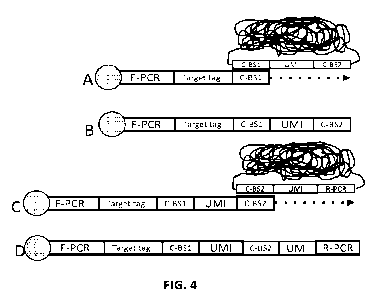
illustrated in Fig. 3. In this embodiment (and as illustrated in Fig. 4) the
second primer
extension product has forward and reverse primer sequences, thereby allowing
the product to
be amplified by PCR.
The lengths of the component parts of a nucleic acid molecule described herein
may
vary. In some embodiments, the part of the primer that hybridizes to the
primer binding
sequence 6 (C-BS1) may be at least 10 nucleotides in length, e.g., 12-50
nucleotides in length,
the UMI sequences may be at least 5 nucleotides in length, e.g., 6-20
nucleotides in length, and
the first template sequence 10 may be at least 10 nucleotides in length, e.g.,
12-50 nucleotides
in length.
In any embodiment, the first and second sets of barcoded particles may each
comprise
at least 10 members (e.g., at least 100, at least 1,000, at least 10,000, at
least 100,000, at least
1M at least 10 M, at least 100 M, at least 1B or at least 10B members), which
are each
uniquely identifiable by their particle identifier sequence. In some
embodiments, the nucleic
acid sequences of the barcoded particles in the first and second sets of
barcoded particles may
be identical to one another except for their particle identifier sequences.
A method for adding unique particle identifier sequences to a primer using the
present
probe system is also provided. The principle of this method is illustrated in
Fig. 2. In these
embodiments the method may comprise hybridizing the first set of barcoded
particles 4 with
the population of primer molecules 2, extending the hybridized primer
molecules using the
nucleotide sequence of the first set of barcoded particles as a template to
produce first primer
extension products 16 that contain i. the complement 18 of a unique particle
identifier
CA 03213800 2023- 9- 27
WO 2022/208327
PCT/IB2022/052862
19
sequence from a barcoded particle in the first set of barcoded particles and
ii. the complement
20 of the first template sequence. Next, the first set of barcoded particles
are removed, e.g.,
denaturation and washing, or e.g. by having incorporated uracil residues in
place of thymidine
in the first barcoded particle and treating the product enzymatically, e.g.,
using USER (NEB),
which contains uracil-n-glycosylase enzyme and an AP lyase, which is capable
of cleaving
phosphodiester bonds specifically at apurinic sites, thereby degrading the
first barcoded
particle liberating the extended primer, In these embodiments, primer 2 (and
thus primer
extension product 16) may be tethered to a support or a sample (e.g., via a
capture agent) and,
as such, the first set of barcoded particles can be subjected to stringent
wash conditions
without removing the primer or primer extension product from the sample or
support. Further,
the first primer extension product may be terminated at the correct position
(i.e., at the end of
the first template sequence) by either engineering a site for a restriction
enzyme at that position
and then digesting the primer extension products (which will be double
stranded) with the
restriction enzyme after they have been produced, or hybridizing a blocking
oligonucleotide to
a sequence immediately downstream from the first template sequence, or by
designing an
internal hairpin immediately downstream from the first template sequence,
which will cause
the polymerase to stall. In these latter embodiments, the polymerase (which
should be a non-
strand displacing polymerase) should terminate synthesis when it runs into the
blocking
oligonucleotide or hairpin. Alternative ways for terminating nucleic acid
synthesis at defined
sites are schematically illustrated in Figs. 15-17. Fig. 15 illustrates RCA
products that contain
hairpin loops. These hairpin loops can cleaved by a restriction enzyme to
liberate a 5' and 3'
end either as blunt ends, or with overhang. Fig. 16 illustrates how a primer
extension can be
terminated at a defined site in the product. In this embodiment, the hairpins
are digested to
produce a blunt end or an overhang, but the RCA product stays together as a
complex. The
RCA products are then hybridize to the conjugated oligonucleoti des and a
strand displacing
DNA polymerase (e.g. Klenow fragment of Ecoli DNA polymerase I) copies the
barcode from
the RCA product, displaces the hairpin, and terminates naturally as the
template ends. That
primer extension product can then be used as a primer on the next set of
pixels. The restriction
enzyme cleavage of the hairpin may be performed before or after the
hybridization of the RCA
products to the oligonucleotides. Fig. 17 illustrates how defined ends can be
produced by
CA 03213800 2023- 9- 27
WO 2022/208327
PCT/IB2022/052862
digesting the extension products after they have been made, while they are
still double-
stranded.
Next, the method may comprise hybridizing the first primer extension products
16 with
the second set of barcoded particles 12, wherein the complement 20 of the
first template
5 sequence in the first primer extension products hybridizes to the first
template sequence in the
second set of barcoded particles. Next, the method comprises extending the
first primer
extension products using the nucleotide sequence of the second set of barcoded
particles as a
template to produce second primer extension products that contain: a unique
particle identifier
sequence 18 from a barcoded particle in the first set of barcoded particles,
the first template
10 sequence 20, and a unique particle identifier sequence 20 from a
barcoded particle in the
second set of barcoded particles.
As illustrated in Fig. 3, the primer molecules 2 may have a forward primer
sequence
and the nucleotide sequence of the second set of barcoded particles 12 may
have a reverse
primer sequence downstream of the unique particle identifier sequence. In
these embodiments,
15 the first primer extension product 16 should contain the forward primer
sequence at the 5' end,
the second primer extension product 22 should contain the forward primer
sequence at the 5'
end and the reverse primer sequence at the 3' end. In this embodiment, the
method may
comprise amplifying the second primer extension products 22 by PCR using
primers that
target the forward and reverse primer sequences. Fig. 4 illustrates an
embodiment of the
20 method in which the primers are either tethered to a support or to a
sample.
In some embodiments, the method may comprise sequencing the second primer
extension products, or an amplified copy thereof. In these embodiments, each
second primer
extension product should contain the unique identifiers for two barcoded
particles, thereby
identifying which barcoded particles hybridized with the primer in the
different hybridization
steps. In some embodiments, and as will be described in greater detail below,
the method may
comprise mapping the relative positions of the primers using the pairs of
unique particle
identifier sequences that are in the second primer extension products.
As noted above, in some embodiments, the primers may be attached to a cellular
sample via a binding agent. In these embodiments, the unique particle
identifiers in the second
primer extension products indicate the relative position of the binding agents
on the cellular
CA 03213800 2023- 9- 27
WO 2022/208327
PCT/IB2022/052862
21
sample. This embodiment is illustrated in Fig. 4. As can be seen in Fig. 4, in
these
embodiments, the primer may include a target identification sequence. In the
embodiments
shown in Fig. 4, in steps A and B, the primer molecules contain a target
identifier and are
immobilized to the sample, e.g., via a binding agent. In this method, the
first set of barcoded
particles is hybridized to the sample and complements of the UMIs and C-BS2
sequence from
the particles to which the primers are hybridized are copied onto the end of
the primer
molecules using the hybridized particles as templates. In steps C and D, the
first set of
barcoded particles is removed and the second set of particles are hybridized.
After
hybridization, the primer extension products are extended to add UMIs from the
second set of
particles onto the ends of the primer extension products. The pairs of
barcodes that are copied
onto the ends of the primers can be analyzed to determine the relative
positions of different
primers in the sample.
As indicated above, in alternative embodiments, the method may be implemented
using
a gap-fill/ligation and/or a ligation-based approach where, in these
embodiments, the 5' end of
a nucleic acid may be extended using the barcoded particles of (b) as a
template by a gap-
fill/ligation or ligation reaction. These embodiments are schematically
illustrated in Figs. 12-
14. With reference to Fig. 12, instead of copying the barcodes from a barcoded
particle by
polymerizing from a free 3'end of an primer that is conjugated to a binding
agent (as described
above), a gap-fill/ligation reaction can be used to copy the barcode onto the
5' end of an
oligonucleotide conjugated to the antibody, where the oligonucleotide has a 5'
phosphate.
These embodiments can be done using a combination of T4 DNA polymerase and T4
DNA
ligase, for example. Fig. 13 shows how the 5' end or 3' end of an
oligonucleotide that is
conjugated to a binding agent can be ligated to a "pre-made" molecule that
contains the
barcode of a particle. In these embodiments, the barcodes are first copied by
gap-fill/ligation
reaction. During the assay itself, the oligonucleotide of the antibody is
ligated to the 5' end or
the 3' end of the pre-made copy of the barcode. In this figure, the 5' end of
the oligonucleotide
is ligated to the copy of the barcode, however this can be done the other way
around, i.e., the
oligonucleotides coupled to the antibody carry a free 3'end that is ligated to
the 5'phosphate
end of the UMI copy sequence. Fig. 14 illustrates how the 5' end of a cDNA can
be added to a
barcode via a gap-fill/ligation reaction. In this method, mRNA is copied in a
a reverse
CA 03213800 2023- 9- 27
WO 2022/208327
PCT/IB2022/052862
22
transcription reaction (using an oligond(T) primer, a random primer or a gene-
specific primer)
and once the cDNA has been produced, the barcode is added to the 5' end of the
cDNA via a
gap-fill ligation reaction. These and other implementations can be envisioned.
As noted above, in some embodiments the primer molecules may be attached to a
planar substrate, e.g., a glass slide or the like. In these embodiments, at
least 106, 107, 108 or
109 primer molecules may be attached to a planar support and extended using
the method
described above. This method is schematically illustrated in Fig. 5. In this
embodiment, the
first and second sets of barcoded particles hybridize sequences that are in
overlapping areas on
the support 50. The first primer extension reaction adds the molecular
identifiers to the primers
that hybridize to the first set of barcoded particles to result in a first
array of features 52 that
correspond to the first primer extension products. In the example shown, there
are six features
58, each corresponding to a barcoded particle and each containing a different
molecular
identifier. After the first primer extension reaction is completed, the first
set of barcoded
particles are removed and the second set of barcoded particles are hybridized
to the substrate.
In this step, the second set of barcoded particles hybridize with the first
primer extension
products, but in an overlapping manner to the first set of barcoded particles.
Extension of the
hybridized second set of barcoded particles adds the molecular identifiers
from the second set
of barcoded particles to the first primer extension products, to produce a
second array of
features 56 that that correspond to the second primer extension products. By
way of example,
in array 56, features 60 and 62 have the same unique identifier from a first
barcoded particle
(corresponding to feature 58) but different unique identifiers from the second
barcoded
particles.
This embodiment of the method, which results in an array, is also illustrated
in Fig. 6.
With reference to Fig. 6, the method may start with a solid surface with an
immobilized
oligonucleotide. In Step 1, the free 3' ends of the immobilized
oligonucleotides are extended
into the first set of barcoded particles (e.g., RCA products) that have unique
molecular
identifiers. This extension step copies the complement of the unique molecular
identifiers from
the first set of barcoded particles onto the oligonucleotides. The barcoded
particles are then
removed. In Step 2, a second set of barcoded particles is added to the
surface, effectively
adding a second random barcode to the surface. The primer extension products
are then
CA 03213800 2023- 9- 27
WO 2022/208327
PCT/IB2022/052862
23
extended =, which incorporate the identifiers of the second set of particles
into the initial
extension products. The second identifiers provide information on which first
identifiers are in
proximity. As the second addition has only partial overlap with the first, it
will result in a
spatial map of linked identifiers, illustrated at the bottom of Fig. 6. The
array can then be used
to capture biomolecules bound in a cell or a tissue sample, thereby providing
spatial
information of the biomolecules. The resolution will likely be at the size of
the particle, which
may be 50-500 nm, vastly surpassing the resolution of other methods.
Alternatively, the
capture of biomolecules in the sample (for example mRNA binding probes or poly
A primers
for cDNA) can be done on the surface containing the immobilized
oligonucleotides (before
primer extension). An example of the use of such an array is shown in Fig. 7.
The present probe system may be used to map binding events that are in or on a
cellular sample. This method is schematically illustrated in Fig. 8. In these
embodiments, the
method may comprise obtaining a sample containing primer molecules that are
bound to sites
in or on cells. For example, as described above, this sample may be made by
binding primers
that are attached to binding agents (e.g., oligonucleotide probes, antibodies
or aptamers) to
sites that are in or on the cells, or by hybridizing a first primer to RNA in
the cell, extending
the primer to make cDNA, and appending a second primer onto the 5' end of the
cDNA, where
the cDNA becomes the primer molecules that are used in the present method.
Other
components used in the method include a first set of barcoded particles as
discussed above,
i.e., particles that that each have a nucleotide sequence comprising: a primer
binding sequence
that is complementary to the 3' end of the primer molecules, a unique particle
identifier
sequence, and a first template sequence, as illustrated in Fig. 1, as well as
a second set of
barcoded particles that each have a nucleotide sequence comprising: the first
template
sequence, and a unique particle identifier sequence, as illustrated in Fig. 1.
In any embodiment
and as illustrated in Fig. 1, the nucleotide sequence of the second set of
barcoded particles may
lack the primer binding sequence that is in the nucleotide sequence of the
first set of barcoded
particles. As such, in any embodiment, the nucleotide sequence of the second
set of barcoded
particles may lack the primer binding sequence that is in the nucleotide
sequence of the first
set of barcoded particles may comprise the first template sequence and a
unique particle
CA 03213800 2023- 9- 27
WO 2022/208327
PCT/IB2022/052862
24
identifier sequence, but not the primer binding sequence that is in the
nucleotide sequence of
the first set of barcoded particles.
As described above, this method involves specifically hybridizing the first
set of
barcoded particles with the sample, wherein the nucleotide sequence of at
least some of the
first set of barcoded particles hybridizes to at least two primer molecules.
The number of
primer molecules that hybridize to each barcoded particle may vary, as shown
in Fig. 8. Some
of the first set of barcoded particles may hybridize to one primer molecule,
but many should
hybridize to at least two, at least 5, at least 10, or at least 20 primer
molecules, depending on
the density of the primer molecules and the size of the particles used. As
with the method
described above, the method comprises extending the primers that are
hybridized to the
barcoded particles using the nucleotide sequences to which the primers are
hybridized as a
template to produce first primer extension products that each comprise a first
unique particle
identifier sequence. Next, the method may comprise removing the first set of
barcoded
particles from the sample, as described above, and specifically hybridizing
the second set of
barcoded particles with the first primer extension products. In this step of
the method the
nucleotide sequences of at least some of the second set of barcoded particles
hybridizes to at
least two molecules of the primer extension products. Likewise, extending the
first primer
extension products that are hybridized to a barcoded particle in the prior
using the nucleotide
sequences to which the primers are hybridized as a template to produces second
primer
extension products that comprise the two unique particle identifier sequences,
one from a
particle in the first set, and another from a particle in the second set.
Next, the method
comprises determining which unique particle identifier sequence or complements
thereof are
in second primer extension products and making a map of the relative positions
of the primers
of using the unique particle identifier sequences that are in the second
primer extension
products.
The steps of this method are laid out in detail in Fig. 9A-C. As can be seen
from Fig.
9A-C, the primers may contain target identifier sequences, thereby allowing
the assay to be
multiplexed. In this method, a population of second primer extension products
may be
amplified and sequenced en masse. Each sequence read should contain a target
identifier as
well as two particle identifiers, one from the first set of particles and the
other from the second
CA 03213800 2023- 9- 27
WO 2022/208327
PCT/IB2022/052862
set of particles. Because the sites to which the first and second sets of
particles bind overlap in
the separate hybridization reactions, the primers can be mapped to sites in
the sample.
Specifically, a relational map of the barcoded particles can be produced by
figuring out which
pairs of identifiers are added to a primer. The target sequences can be mapped
onto the
5 relational map, thereby providing a map of the binding sites of the
primers (or the sites, e.g.,
the epitopes or sequences, to which they are bound).
Alternative ligation-based embodiments are described below. These embodiments
avoid the use of two sets of barcoded particles and, instead only 1 primer
extension step is
required. In these embodiments, the pairs of identifiers can be created by
connecting nearby
10 primer extension products via a ligation splint. These embodiments are
schematically
illustrated in Figs. 10 and 11. In these embodiments, the probe system may
comprise: a
population of primer molecules (which, as shown, may contain a sequence that
provides a
primer binding site, a target identifier and primer sequence C-BS1); a set of
barcoded particles
that each have a nucleotide sequence comprising: (i) a primer binding sequence
(C-B S1) that is
15 complementary to the 3' end of the primer molecules, (ii) a unique
particle identifier sequence
(UMI), and (iii) a first template sequence (C-BS2). Instead of a second set of
barcoded
particles, this probe system comprises a ligation splint. As illustrated in
Fig. 10, the ligation
splint is composed of a first oligonucleotide and a second oligonucleotide,
wherein the first
oligonucleotide comprises a first sequence and the first template sequence (C-
BST'); and the
20 second oligonucleotide comprises a second sequence that is complementary
to the first
sequence, and the first template sequence (C-BST'). The two oligonucleotides
in the splint
may have the same sequence and hybridize to one another via sequences at the
5' ends.
In general terms, the components of this system are similar to the serial
extension
system components described above, except that the alternative system has a
ligation splint
25 instead of the second set of barcoded particles. For example, the
barcoded particles may be
rolling circle amplification (RCA) products, or the barcoded particles may be
barcoded
nanoparticles, wherein nucleotide sequence is tethered to the surface of the
barcoded particles.
Likewise, the primer molecules of (a) are synthetic oligonucleotides that are
10-200 nt in
length, which may be linked to a binding agent, (e.g., an oligonucleotide
probe, antibody,
CA 03213800 2023- 9- 27
WO 2022/208327
PCT/IB2022/052862
26
aptamer, etc). In some embodiments, the primer molecules may be cDNA molecules
that have
a primer sequence at the 3' end.
As with the serial extension embodiments, the set of barcoded particles may
contain at
least 10 particles, each having a unique particle identifier sequence. In this
embodiment, the
set of barcoded particles may each comprise at least 10 members (e.g., at
least 100, at least
1,000õ at least 10,000, at least 100,000, at least 1M at least 10 M, at least
100 M, at least 1B
or at least 10B members), which are each uniquely identifiable by their
particle identifier
sequence. In some embodiments, the nucleic acid sequences of the barcoded
particles may be
identical to one another except for their particle identifier sequences.
The alternative probe system may be used to map binding events that are in or
on a
cellular sample in a similar way to the serial probe system described above. A
method for
adding unique particle identifier sequences to a primer using the alternative
probe system is
provided. This method, which is illustrated in Figs. 10 and 11, may comprise
hybridizing the
set of barcoded particles with the population of primer molecules (which may
be immobilized
on a support, as shown), and extending the hybridized primer molecules using
the nucleotide
sequences as a template. This step results in the production of first primer
extension products
that contain i. the complement of a unique particle identifier sequence (UMI)
from a barcoded
particle and ii. the complement of the first template sequence (C-BS2). After
the barcoded
particles are removed, the method comprises hybridizing the first primer
extension products
with the ligation splint, wherein the complements of the first template
sequence in two
proximal first primer extension products hybridize to the first and second
sequences of the
ligation splint, as shown. Next, the method comprises ligating at least one of
either the first
and second oligonucleotides of the hybridized ligation splint to the first
primer extension
products and extending the 3' end of at least one of the first and second
oligonucleotides in the
splint using the first primer extension product in the ligation product as a
template, thereby
adding two unique particle identifier sequences to a primer. As illustrated in
Fig. 10, the
primers can be engineered to forward and reverse primer sequences and target
identification
sequences, thereby providing a product that contains PCR primer sites, a pair
of target
identification sequences and two particle identifiers. These sequences can be
analyzed to
determine the relative positions of the barcoded particles and primers (or the
binding agents,
CA 03213800 2023- 9- 27
WO 2022/208327
PCT/IB2022/052862
27
probes or cDNA molecules to which they are tethered), as schematically
illustrated in Fig. 11.
As would be apparent, the method may comprise sequencing the second primer
extension,
thereby identifying which the pairs of unique particle identifier sequences
are in the second
primer extension products. The method may further comprise mapping the
relative positions of
the primers of i. using the pairs of unique particle identifier sequences that
are in the second
primer extension products.
As with the serial extension embodiments described above, the primers
molecules may
be attached to a cellular sample via a binding agent (e.g., an antibody,
aptamer or probe), and
the unique particle identifiers in the second primer extension products may
indicate the relative
position of the binding agents on the cellular sample. Likewise, the primers
may be the 3' ends
of cDNAs that are made in the cellular sample in situ, wherein the unique
particle identifiers in
the second primer extension products indicate the relative position of cDNAs
in the cellular
sample.
In any embodiment, barcoded particles may be mapped relative to one another if
two
complex identifiers are added to the same molecule. The map produced by the
method may be
a three-dimensional map or a two-dimensional map, depending on how the method
is
implemented. For example, if the complexes products are immobilized within
cells (e.g.,
produced in situ in cells) then the map produced may be three dimensional. In
other
embodiments, e.g., if the complexes are immobilized on one or more surfaces
(e.g., the surface
of one or more cells that may be in suspension or mounted on a support), then
the map
produced by the method may be two dimensional or potentially three dimensional
because, in
theory, the map may be spherical. While the method can be applied to cells (as
described
below) the method can be adapted to map adjacent complexes that are
immobilized on any
surface, e.g., a glass slide that may have a tissue blot, or a western blot,
etc. Likewise, although
the complexes can be anchored to sites in or on cells or on a surface via an
antibody (e.g., an
antibody that is conjugated to an oligonucleotide that has a sequence that is
complementary to
a sequence in a complex), the complexes can be immobilized via any type of
interaction, e.g.,
covalent or non-covalent interactions, directly or indirectly. For example, in
some
embodiments, the complexes may be bound to the cell via a binding agent (e.g.,
an aptamer, an
antibody or an oligonucleotide, etc.), where the binding agent binds to a
sequence in the
CA 03213800 2023- 9- 27
WO 2022/208327
PCT/IB2022/052862
28
complexes and a site in a cell or on the surface of the one or more cells. In
some
embodiments, the complexes may be immobilized via hybridization to an
oligonucleotide that
also hybridizes to a nucleic acid (e.g., to a cellular RNA) or the RCA
products may be
immobilized non-covalently to a site via electrostatic interactions, via a
streptavidin/biotin
interaction, or by a covalent linkage (e.g., via a click coupling).
In any embodiment, the complexes may be immobilized in or on cells that are in
solution, cells that are on a support (e.g., a slide), cells that in a three-
dimensional sample of
tissue, or cells that are in a tissue section. A sample containing cells that
are in solution may
be a sample of cultured cells that have been grown as a cell suspension, for
example. In other
embodiments, disassociated cells (which cells may have been produced by
disassociating
cultured cells or cells that are in a solid tissue, e.g., a soft tissue such
as liver of spleen, using
trypsin or the like) may be used. In particular embodiments, the complexes may
be
immobilized on cells that can be found in blood, e.g., cells that in whole
blood or a sub-
population of cells thereof. Sub-populations of cells in whole blood include
platelets, red blood
cells (erythrocytes), and white blood cells (i.e., peripheral blood
leukocytes, which are made
up of neutrophils, lymphocytes, eosinophils, basophils and monocytes). These
five types of
white blood cells can be further divided into two groups, granulocytes (which
are also known
as polymorphonuclear leukocytes and include neutrophils, eosinophils and
basophils) and
mononuclear leukocytes (which include monocytes and lymphocytes). Lymphocytes
can be
further divided into T cells, B cells and NK cells. Peripheral blood cells are
found in the
circulating pool of blood and not sequestered within the lymphatic system,
spleen, liver, or
bone marrow. If cells that are immobilized on a support are used, then then
the sample may be
made by, e.g., growing cells on a planar surface, depositing cells on a planar
surface, e.g., by
centrifugation, by cutting a three dimensional object that contains cells into
sections and
mounting the sections onto a planar surface, i.e., producing a tissue section.
In alternative
embodiments, the surface may be made by absorbing cellular components onto a
surface.
In any embodiment, the method may comprise immobilizing thousands, tens of
thousands, hundreds of thousands or at least a million barcoded particles
(each having a unique
identifier), to a population of cells (e.g., via an antibody) so that on each
cell the barcoded
particles effectively coat the cell. The barcoded particles may hybridize to
other
CA 03213800 2023- 9- 27
WO 2022/208327
PCT/IB2022/052862
29
oligonucleotides that are tethered to sites in or on a cell to produce a
matrix comprising the
barcoded particles. After hybridization, the unique identifier sequences of
adjacent complexes
can be copied from one barcoded particle to another. A physical map of the
complexes, as well
as the sites to which the barcoded particles bind to the cell, can be
constructed based on the
sequences that have been copied.
In addition to making a map of the barcoded particles, the method may involve
performing a proximity assay between one or more binding agents that are bound
to sites in
the cells or on the surface of the cells (e.g., antibodies that are bound to
cell surface markers on
the cells). In these embodiments, a product may contain a pair of unique
complex identifier
sequences as well as a binding agent identifier sequence. In some embodiments,
the binding
agent may be an antibody-oligonucleotide conjugate and in other embodiments,
the capture
agent may be an oligonucleotide probe or cDNA. The oligonucleotide and the
capture agent
may be linked via a number of different methods, including those that use
maleimide or
halogen-containing groups, which are cysteine-reactive. The capture agent and
the
oligonucleotide may be linked proximal to or at the 5' end of the
oligonucleotide, proximal to
or at the 3' end of the oligonucleotide, or anywhere in-between. In some
embodiments, the
oligonucleotides may be linked to the capture agents by a linker that spaces
the oligonucleotide
from the capture agents. Oligonucleotides may be linked to capture agents
using any
convenient method, as described above. In many embodiments, the sequence of an
oligonucleotide that is conjugated to a binding agent uniquely identifies the
epitope or
sequence to which the binding agent binds. For example, if the method is
performed using 10
different antibodies, then each antibody is tethered to a different sequence
that identifies the
epitope to which the antibody binds. This feature allows the method to be
multiplexed and, in
some embodiments, at least 5, at least 10, at least 20 or at least 50
different antibodies that
bind to different markers in or on the surface of a cell can be used in the
method. Each
antibody is conjugated to a different antibody identifier sequence, and the
antibody identifier
sequences allow the binding events for a particular antibody to be mapped.
Such tagged
antibodies are described in, e.g., Wu et al (Nat. Comm. 2019 10: 3854) and
US20160281134,
and others.
CA 03213800 2023- 9- 27
WO 2022/208327
PCT/IB2022/052862
EMBODIMENTS
Embodiment 1. A probe system comprising: (a) a population of primer molecules;
(b)
a first set of barcoded particles that each have a nucleotide sequence
comprising: (i) a
primer binding sequence that is complementary to the 3' end of the primer
molecules of (a),
(ii) a unique particle identifier sequence, and (iii) a first template
sequence; (c) a second set of
5 barcoded particles that each have a nucleotide sequence comprising: (i)
the first template
sequence, and (ii) a unique particle identifier sequence; wherein extension of
the primer
molecules of (a) using the first set of barcoded particles of (b) as a
template produces primer
extension products that contain the complement of a unique particle identifier
sequence of a
particle of (b)(ii) and the complement of the first template sequence.
10 Embodiment 2. The probe system of embodiment 1, wherein the barcoded
particles of
(b) and/or (c) are rolling circle amplification (RCA) products.
Embodiment 3. The probe system of embodiment 1, wherein the barcoded particles
of
(b) and/or (c) are barcoded nanoparticles, wherein the nucleotide sequences
are tethered to the
surface of the barcoded particles.
15 Embodiment 4. The probe system of any prior embodiment, wherein
primer molecules
of (a) are synthetic oligonucleotides that are 10-200 nt in length.
Embodiment 5. The probe system of any prior embodiment, wherein the primer
molecules of (a) are cDNA molecules that have a primer sequence at the 3' end.
Embodiment 6. The probe system of any prior embodiment, wherein the primer
20 molecules of (a) have a forward primer sequence and the nucleotide
sequence of the second set
of barcoded particles has a reverse primer sequence downstream of the unique
particle
identifier sequence.
Embodiment 7. The probe system of any prior embodiment, wherein the primer
molecules of (a) are linked to a binding agent, (e.g., an oligonucleoti de
probe, antibody,
25 aptamer, etc).
Embodiment 8. The probe system of embodiment 7, wherein the primer molecules
further comprise target identifier sequences that indicate the binding agent
to which they are
linked.
CA 03213800 2023- 9- 27
WO 2022/208327
PCT/IB2022/052862
31
Embodiment 9. The probe system of any of embodiments 1-6, wherein the primer
molecules are linked to a planar substrate.
Embodiment 10. The probe system of any prior embodiment, wherein the first and
second sets of barcoded particles independently contain at least 10,000
particles, each having a
unique particle identifier sequence.
Embodiment 11. The probe system of any prior embodiment, wherein the
nucleotide
sequence of the second set of barcoded particles lacks the primer binding
sequence of (b)(i).
Embodiment 12. A method for adding unique particle identifier sequences to a
primer
using the probe system of any of embodiments 1-11, comprising;
I. hybridizing the first set of barcoded particles of (b) with the
population of
primer molecules of (a),
i i . extending the hybridized primer molecules using the
nucleotide sequence of the
first set of barcoded particles as a template to produce first primer
extension
products that contain i. the complement of a unique particle identifier
sequence
from a barcoded particle in the first set of barcoded particles and ii. the
complement of the first template sequence;
i i i . removing the first set of barcoded particles;
iv. hybridizing the first primer extension products with the second set of
barcoded
particles, wherein the complement of the first template sequence in the first
primer extension products hybridizes to the first template sequence in the
second set of barcoded particles; and
v. extending the first primer extension products using the nucleotide
sequence of
the second set of barcoded particles as a template to produce second primer
extension products that contain:
a unique particle identifier sequence from a barcoded particle in the first
set of barcoded particles,
the first template sequence, and
a unique particle identifier sequence from a barcoded particle in the
second set of barcoded particles.
CA 03213800 2023- 9- 27
WO 2022/208327
PCT/IB2022/052862
32
Embodiment 13. The method of embodiment 12, wherein the primer molecules have
a
forward primer sequence and the nucleotide sequence of the second set of
barcoded particles
has a reverse primer sequence downstream of the unique particle identifier
sequence, and the
method comprises amplifying the second primer extension products of v.
Embodiment 14. The method of embodiment 12 or 13, where the method comprises
sequencing the second primer extension products of v.
Embodiment 15. The method of embodiment 14, mapping the relative positions of
the
primers of i. using the pairs of unique particle identifier sequences that are
in the second
primer extension products.
Embodiment 16. The method of any of embodiments 12-15, wherein the primers are
attached to a planar substrate, and the method results in an array of the
second primer
extension products on the substrate.
Embodiment 17. The method of any of embodiments 12-15, wherein the primers are
attached to a cellular sample via a binding agent, and the unique particle
identifiers in the
second primer extension products indicate the relative position of the binding
agents on the
cellular sample.
Embodiment 18. The method of any of embodiments 12-15, wherein the primers are
cDNA s that are made in the cellular sample in situ, wherein the unique
particle identifiers in
the second primer extension products indicate the relative position of cDNAs
in the cellular
sample.
Embodiment 19. A method for making a map of binding events in or on a cellular
sample, comprising: (a) obtaining: i. a sample containing primer molecules
that are bound to
sites in or on cells; ii. a first set of barcoded particles that each have a
nucleotide sequence
comprising: (i) a primer binding sequence that is complementary to the 3' end
of the primer
molecules of (a), (ii) a unique particle identifier sequence, and (iii) a
first template sequence;
a second set of barcoded particles that each have a nucleotide sequence
comprising: (i)
the first template sequence, and (ii) a unique particle identifier sequence;
(b) specifically
hybridizing the first set of barcoded particles of (a)(ii) with the sample,
wherein the nucleotide
sequence of at least some of the first set of barcoded particles hybridizes to
at least two primer
molecules; (c) extending the primers that are hybridized to barcoded particles
in step (b) using
CA 03213800 2023- 9- 27
WO 2022/208327
PCT/IB2022/052862
33
the nucleotide sequences to which the primers are hybridized as a template to
produce first
primer extension products that each comprise a first unique particle
identifier sequence: (d)
removing the first set of barcoded particles from the sample; (e) specifically
hybridizing the
second set of barcoded particles of (a)(iii) with the first primer extension
products of (c),
wherein the nucleotide sequences of at least some of the second set of
barcoded particles
hybridizes to at least two molecules of the primer extension products; (f)
extending the first
primer extension products that are hybridized to a barcoded particle in step
(e) using the
nucleotide sequences to which the primers are hybridized as a template to
produce second
primer extension products that comprise the two unique particle identifier
sequences; (g)
determining which unique particle identifier sequence or complements thereof
are in second
primer extension products; and (h) making a map of the relative positions of
the primers of
(a)(i) using the unique particle identifier sequences that are in the second
primer extension
products.
Embodiment 20. The method of embodiment 19, wherein the sample of (a) is made
by
binding primers that are attached to binding agents to sites that are in or on
the cells.
Embodiment 21. The method of embodiment 20, wherein the binding agents are
oligonucleotide probes, antibodies or aptamers.
Embodiment 22. The method of embodiment 20 or 21, wherein the primers further
comprise target identifier sequences that indicate the binding agent to which
they are linked.
Embodiment 23. The method of embodiment 19, wherein the sample of (a) is made
by
hybridizing a first primer to RNA in the cell, extending the primer to make
cDNA, and
appending a second primer onto the 5 end of the cDNA.
Embodiment 24. The probe system of any prior embodiment, wherein the
nucleotide
sequence of the second set of barcoded particles lacks the primer binding
sequence of the first
set of barcoded particles_
Embodiment 25. A probe system comprising: (a) a population of primer
molecules; (b)
a set of barcoded particles that each have a nucleotide sequence comprising:
(i) a primer
binding sequence that is complementary to the 3' end of the primer molecules
of (a), (ii) a
unique particle identifier sequence, and (iii) a first template sequence; and
(c) a ligation splint
comprising a first oligonucleotide and a second oligonucleotide, wherein the
first
CA 03213800 2023- 9- 27
WO 2022/208327
PCT/IB2022/052862
34
oligonucleotide comprises a first sequence and the first template sequence;
and the second
oligonucleotide comprises a second sequence that is complementary to the first
sequence, and
the first template sequence.
Embodiment 26. The probe system of embodiment 25, wherein the barcoded
particles
of (b) are rolling circle amplification (RCA) products.
Embodiment 27. The probe system of embodiment 25, wherein the barcoded
particles
of (b) are barcoded nanoparticles, wherein nucleotide sequence of (b) is
tethered to the surface
of the barcoded particles.
Embodiment 28. The probe system of any of embodiments 25-27, wherein primer
molecules of (a) are synthetic oligonucleotides that are 10-200 nt in length.
Embodiment 29. The probe system of any of embodiments 25-27, wherein the
primer
molecules of (a) are cDNA molecules that have a primer sequence at the 3' end.
Embodiment 30. The probe system of any of embodiments 25-28, wherein the
primer
molecules of (a) are linked to a binding agent, (e.g., an oligonucleotide
probe, antibody,
aptamer, etc).
Embodiment 31. The probe system of any of embodiments 25-30, wherein the set
of
barcoded particles may contain at least 10,000 particles, each having a unique
particle
identifier sequence.
Embodiment 32. A method for adding unique particle identifier sequences to a
primer
using the probe system of any of embodiments 25-31, comprising:
hybridizing the set of barcoded particles of (b) with the population of primer
molecules of (a),
i i . extending the hybridized primer molecules using the
nucleotide sequences as a
template to produce first primer extension products that contain i. the
complement of a unique particle identifier sequence from a barcoded particle
and ii. the complement of the first template sequence;
i i i . removing the barcoded particles;
iv. hybridizing the first primer extension products with
the ligation splint, wherein
the complements of the first template sequence in two proximal first primer
CA 03213800 2023- 9- 27
WO 2022/208327
PCT/IB2022/052862
extension products hybridize to the first and second sequences of the ligation
splint;
v. ligating at least one of the first or the second
oligonucleotide of the hybridized
ligation splint to the first primer extension products and extending the 3'
end of
5 the
ligated first or second oligonucleotide in the splint using the first primer
extension product in the ligation product as a template, thereby adding two
unique particle identifier sequences to a primer.
Embodiment 33.
The method of embodiment 32, where the method comprises
sequencing the second primer extension products of v.
10 Embodiment 34. The method of embodiment 32 or 33, further comprising
mapping the
relative positions of the primers of i. using the pairs of unique particle
identifier sequences that
are in the second primer extension products.
Embodiment 35. The method of any of embodiments 32-34, wherein the primers
molecules of i. are attached to a cellular sample via a binding agent, and the
unique particle
15 identifiers in the second primer extension products indicate the
relative position of the binding
agents on the cellular sample.
Embodiment 36. The method of any of embodiments 32-34, wherein the primers are
cDNAs that are made in the cellular sample in situ, wherein the unique
particle identifiers in
the second primer extension products indicate the relative position of cDNAs
in the cellular
20 sample.
EXAMPLES
The following examples are put forth so as to provide those of ordinary skill
in the art
with additional disclosure and description of how to make and use the present
invention, and
are not intended to limit the scope of what the inventors regard as their
invention nor are they
25 intended to represent that the experiments below are all or the only
experiments performed.
METHODS
Probe circularization and Rolling circle amplification: Probe oligo (1 or 2)
was
circularized using template oligo (3 or 4) in 50 ul ligation reactions
containing 100 nM probe
CA 03213800 2023- 9- 27
WO 2022/208327
PCT/IB2022/052862
36
oligo, 100 nM template oligo, 1 mM ATP, 200 U T4 DNA ligase and a reaction
buffer
containing 33 mM Tris-acetate, 10mM Mg-acetate, 66mM K-acetate, 1 mM DTT and
0.1%
Tween20. The reaction was incubated at 37 C for 30 minutes.
Non-circularized probes were digested by adding 10 U Exonuclease I and 100 U
exonuclease 111 to each reaction following ligation. The exonuclease digestion
was performed
for 30 min at 37 C, followed by heat inactivation at 85 C for 20 min.
RCA primer oligo 3 or 4 was added to each reaction after exonuclease digestion
to a
concentration of 100 nM and was allowed to hybridize to the circularized
probes by incubation
at 37 C for 20 min.
Rolling circle amplification was performed in 75 al reactions containing 2.5
nM
circularized probe, 0.75 mM of each d(AUGC)TP and 7.5 U pili29 DNA polymerase,
in the
same reaction buffer as used for probe circularization. The reaction was
incubated for 20 min
at 37 C, followed by heat inactivation at 65 C for 10 min.
Cell staining and fixation: Raji and Jurkat cells were aspirated from separate
T75
flasks and spun down at 300 x g for 5 mM. Cells were counted and cell
suspension
corresponding to 1 M cells were taken. Cells were washed twice in FACS buffer
(2% FBS, 2
mM EDTA in lx PBS). Cells were blocked with Fc blocking agent and spun down to
remove
the supernatant. A pool of 2 TotalSeq B (Biolegend inc) antibody-
oligonucleotide conjugates
(oligo 8-27) targeting various immune cell markers (CD3, CD4, etc) at a
concentration of 5
ag/m1 of each conjugate was added to the cells and incubated for 30 mM on ice.
The conjugate
stained cells were washed twice with 300 al FACS buffer after which the cells
were spun
down, supernatant removed and 250 IA of 1% PFA was added to fix the sample.
After 10
minute incubation at RT, the fixation was quenched by addition of 12.5 al of
2.5 M Glycine,
followed by a wash with 250 IA of 125 mM Glycine in PBS. After another wash in
PBS cells
were resuspended in PBS and stored at +4 C until use.
Primer extension assay: Conjugate-stained and fixated Raji and Jurkat cells
were
mixed at a 1:1 ratio and an aliquot corresponding to approximately 20 000
cells in total was
put in a PCR tube.
Hybridization of RCA products to antibody-conjugate oligos bound to cells were
performed in a 40 p1 reaction containing 2.5 nM RCA products (originating from
oligo 1), 0.5
CA 03213800 2023- 9- 27
WO 2022/208327
PCT/IB2022/052862
37
1.1M of blocking oligo (5), in a buffer with 300 mM NaC1, 15 mM MgCl2, 20 mM
Tris-HC1
(pH 8). The reaction was incubated for 15 minutes at 55 C.
The sample were centrifuged at 500 x g for 2 min to pellet the cells,
supernatant
removed and 100 pi of a wash buffer (50 mM NaCl, 1mM EDTA, 20mM Tris-HC, pH
81) was
added and cells resuspended. The sample was again centrifuged to pellet cells
and supernatant
removed.
A 50 pl DNA polymerase extension reaction was performed to incorporate pixel
barcode sequence by adding a mastermix containing 0.2 mM dNTPs, 1 pi of
Klentaq DNA
polymerase in a buffer comprising 50 mM Na-acetate, 10 mM Mg-acetate, 20 mM
Tris-
acetate, 100 pg/m1 BSA. The reaction was incubated for 15 min at 30 C. A
washing step was
again performed under the same conditions as previously described after which
a USER
digestion (Uracil DNA glycosylase + Endonuclease VII) reaction was performed
to degrade
DNA pixels by adding 50 pi of a buffer comprising 50 mM NaCl, 1mM EDTA, 20 mM
Tris-
HC and 1 U of USER enzyme. The reaction was incubated for 30 min at 37 C.
A wash step was performed before adding a 50 tl USER inactivation mastermix
consisting of 1 U UGI protein in 50 mM NaCl, 1 mM EDTA, 20 mM Tris-HC1, pH 8.
The
reaction was incubated for 15 mM at 37 C.
After a wash step a second RCA product hybridization reaction was performed,
using
RCA products originating from oligo 2, using otherwise the same conditions as
previously
described for hybridization of RCA product 1. Similarly, the same protocols
for extension and
USER inactivation were performed following the 2nd hybridization reaction.
Following the
2nd USER digestion step, a washing step was performed before resuspending the
cells in 50
of wash buffer. The resulting cell suspension was quantified and diluted to a
concentration of
20 cells/pl.
After the serial extension assay, each conjugate oli gonucl eoti de bound to
cells via
antibody-binding had two DNA pixel barcode sequences and a reverse PCR primer
motif
incorporated through the extension steps of the assay.
PCR was performed for 15 cycles in a reaction containing 5 pl of diluted
sample, 0.2
mM dNTPs, 0.4 pM each of fwd and rev primers containing Illumina adapter
sequences (6, 7),
1 gl of Phusion DNA polymerase, in lx of Phusion HF reaction buffer. The PCR
reaction
CA 03213800 2023- 9- 27
WO 2022/208327
PCT/IB2022/052862
38
consisted of: 98 C denaturation for 1 min, followed by 15 cycles of 10 s
denaturation at 98 C,
30s annealing at 60 C, 40 s at 72 C before a final extension at 72 C for 5
min.
The PCR product was finally purified using Ampure XP beads following the
manufacturer's instructions, but using a bead-to-sample ratio of 1.2. The
purified PCR product
was quantified using a Qubit fluorometer and sequenced on an 111umina NextSeq
2000
sequencer.
SEQ ID NO name seq
1
GCTTTAAGGCCGGTCCTAGCAANNNNNNNNNNNNNNNNNNNNCAACATCAGTATTCC
D12_PLA CAGGCTACCTGCAGGTTAAGCGGATTG
2
CAACATCAGTATTCCCAGGCTAAAANNNNNNNNNNNNNNNNNNNNAGATCGGAAGA
D15_PL-B GCGTCGTGTAGGGAAAGACCTGCAGGTTAAGCGGATTG
3 D12 PLT A TTGCTAGGACCGGCCTTAAAGCCAATCCGCTTAACCTGCAGG
4 D14-PLTB TAGCCTGGGAATACTGATGTTGCAATCCGCTTAACCTGCAGG
5 D2_b locker CCTGCAGGTTAAGCGGATTGmUmUmUmUmU
6 Fwd primer
CAAGCAGAAGACGGCATACGAGATCGAGTAATGTGACTGGAGTTCAGAC*G*T*G
7 Rev primer
AATGATACGGCGACCACCGAGATCTACACTATAGCCTACACTCTTTCCCTACACGACG*C
*T*C
8
GTGACTGGAGTTCAGACGTGTGCTCTTCCGATCTNNNNNNNNNNTCCCTTGCGATTTAC
CD45 TSeqB NNNNNNNNNGCTTTAAGGCCGGTCCTAGC*A*A
9
GTGACTGGAGTTCAGACGTGTGCTCTTCCGATCTNNNNNNNNNNTATCCCTTGGGATG
CD3_TSeqB GNNNNNNNNNGCTTTAAGGCCGGICCTAGC*A*A
GTGACTGGAGTTCAGACGTGTGCTCTTCCGATCTNNNNNNNNNNCTGGGCAATTACTC
CD19_TSeqB GNNNNNNNNNGCTTTAAGGCCGGTCCTAGC*A*A
11
GTGACTGGAGTTCAGACGTGTGCTCTTCCGATCTNNNNNNNNNNGCCGGACGACATTA
IgGictri_TSeqB ANNNNNNNNNGCTTTAAGGCCGGTCCTAGC*A*A
12
GTGACTGGAGTTCAGACGTGTGCTCTTCCGATCTNNNNNNNNNNTTCTGGGTCCCTAG
CD2O_TSeqB ANNNNNNNNNGCTTTAAGGCCGGTCCTAGC*A*A
13
GTGACTGGAGTTCAGACGTGTGCTCTTCCGATCTNNNNNNNNNNGTCTCTTGGCTTAAA
CD69_TSeqB NNNNNNNNNGCTTTAAGGCCGGTCCTAGC*A*A
14
GTGACTGGAGTTCAGACGTGTGCTCTTCCGATCTNNNNNNNNNNAATAGCGAGCAAGT
HLA-DR_TSeqB ANNNNNNNNNGCTTTAAGGCCGGTCCTAGC*A*A
CD8 TSB GTGACTGGAGTTCAGACGTGTGCTCTTCCGATCTNNNNNNNNNNGCTGCGCTTTCCATT
CA 03213800 2023- 9- 27
WO 2022/208327
PCT/IB2022/052862
39
NNNNNNNNNGCTTTAAGGCCGGTCCTAGC*A*A
16
GTGACTGGAGTTCAGACGTGTGCTCTTCCGATCTNNNNNNNNNNCAATCAGACCTATG
CD14_TSB ANNNNNNNNNGCTTTAAGGCCGGTCCTAGC*A*A
17
GTGACTGGAGTTCAGACGTGTGCTCTTCCGATCTNNNNNNNNNNATATGTATCACGCG
IgG2isoarl_TSB ANNNNNNNNNGCTTTAAGGCCGGTCCTAGC*A*A
18
GTGACTGGAGTTCAGACGTGTGCTCTTCCGATCTNNNNNNNNNNTCAATCCTTCCGCTT
CD45RA_TSB NNNNNNNNNGCTTTAAGGCCGGTCCTAGC*A*A
19
GTGACTGGAGTTCAGACGTGTGCTCTTCCGATCTNNNNNNNNNNCTCCGAATCATGTT
CD45RO_TSB GNNNNNNNNNGCTTTAAGGCCGGICCTAGC*A*A
20
GTGACTGGAGTTCAGACGTGTGCTCTTCCGATCTNNNNNNNNNNGTCCCTGCAACTTG
CD62L_TSB ANNNNNNNNNGCTTTAAGGCCGGTCCTAGC*A*A
21
GTGACTGGAGTTCAGACGTGTGCTCTTCCGATCTNNNNNNNNNNTCCCACTTCCGCTTT
CD82_TSB NNNNNNNNNGCTTTAAGGCCGGTCCTAGC*A*A
22
GTGACTGGAGTTCAGACGTGTGCTCTTCCGATCTNNNNNNNNNNTGGATTCCCGGACT
CD7 TSB TNNNNNNNNNGCTTTAAGGCCGGTCCTAGC*A*A
23
GTGACTGGAGTTCAGACGTGTGCTCTTCCGATCTNNNNNNNNNNCGCGAACATAAGAA
CD70_TSB GNNNNNNNNNGCTTTAAGGCCGGICCTAGC*A*A
24
GTGACTGGAGTTCAGACGTGTGCTCTTCCGATCTNNNNNNNNNNCAGTCGTGGTAGAT
CD72_TSB ANNNNNNNNNGCTTTAAGGCCGGTCCTAGC*A*A
25
GTGACTGGAGTTCAGACGTGTGCTCTTCCGATCTNNNNNNNNNNATATGTCAGAGCAC
CD162_TSB CNNNNNNNNNGCTITAAGGCCGGTCCTAGC*A*A
26
GTGACTGGAGTTCAGACGTGTGCTCTTCCGATCTNNNNNNNNNNGGTGGCTAGATAAT
CD26_TSB GNNNNNNNNNGCTTTAAGGCCGGICCTAGC*A*A
27
GTGACTGGAGTTCAGACGTGTGCTCTTCCGATCTNNNNNNNNNNGAGATGTCTGCAAC
CD63_TSB TNNNNNNNNNGCTTTAAGGCCGGTCCTAGC*A*A
RESULTS
The combination of 2 DNA pixel barcodes incorporated onto each conjugate oligo
represents two DNA pixels that were in proximity to the molecule during the
serial extension
assay since each pixel only occupies a distinct region of space. This,
together with the fact that
multiple neighboring molecules will thus share the same pixel barcode allows
for a spatial
reconstruction of relative positions of each molecule, using a graph theory
approach by
CA 03213800 2023- 9- 27
WO 2022/208327
PCT/IB2022/052862
considering the DNA pixel barcodes as nodes in a graph and the combination of
2 pixel
barcodes of each molecule represents an edge (link) in the graph.
A series of data filtering steps were performed on the 40 million reads
generated. In
short, the filtering steps consisted of removal of reads that were shorter
than the expected
5 length, did not contain common sequence motifs expected at certain
positions, and only
observed once in the data. The average read depth was 7.99 i.e., on average
there were 7.99
copies of each unique molecule.
Of the reads remaining after filtering, a total of 3.4 M unique molecules were
identified
based on a unique molecular identifier (UMI) sequence that was part of the
conjugation oligo
10 sequence. A total of 361575 DNA pixel sequences from set A and 451177
DNA pixel
sequences from set B were found. The average number of unique conjugate oligos
associated
with each pixel from set A was 7.47 and 9.31 for pixel set B.
After filtering away small graph components, the generated graph, or physical
map,
consisted of a set of approximately 100 graph components (clusters), each
composing at least
15 1000 nodes. An induced subgraph was generated for each of the components
and the count of
each surface marker type was summarized for each cluster. The correlation of
marker counts
within each cluster was compared using scatter plots (Figs. 18A-18C). A linear
correlation in
marker counts was observed if plotting two B-cell markers against each other
(HLA-DR,
CD19) or two T-cell markers (CD3, CD7). No such correlation was observed if
instead
20 plotting a B-cell marker (HLA-DR) against a T-cell marker (CD3).
Together, these results
show that each separate graph cluster was generated from and represent a
single cell in this
experiment that contained a sample with a mixture of B and T-cells, Raji and
Jurkat.
A representative cluster was selected and visualized using the Kamada-Kawai
force-
generated graph layout algorithm (Fig. 19). Each edge (link) of the graph in
Fig. 19 can be
25 represented in a different color which identifies the different antibody
types, where the
information has been decoded from the barcode sequence of each the
oligonucleotides
conjugated to an antibody, and each node of the graph represents a DNA pixel
sequence.
CA 03213800 2023- 9- 27