Note: Descriptions are shown in the official language in which they were submitted.
WO 2022/226402
PCT/US2022/026159
INHIBITORS OF UBIQUITIN SPECIFIC PEPTIDASE 22 (U5P22) AND USES
THEREOF FOR TREATING DISEASES AND DISORDERS
CROSS-REFERENCE TO RELATED APPLICATIONS
This application claims benefit of priority to U.S. Patent Application Ser.
No.
63/201,330, filed April 23, 2021, the contents of which are incorporated by
reference in its
entirety.
STATEMENT REGARDING FEDERALLY SPONSORED RESEARCH OR
DEVELOPMENT
This invention was made with government support under CA232347 and CA220801
awarded by the National Institutes of Health. The government has certain
rights in the
invention.
REFERENCE TO A SEQUENCE LISTING
This application is being filed electronically via EFS-Web and includes an
electronically submitted Sequence Listing in .txt format. The .txt file
contains a sequence
listing entitled "702581 02132 ST25.txt" created on April 25, 2022 and is
18,669 bytes in
size. The Sequence Listing contained in this .txt file is part of the
specification and is hereby
incorporated by reference herein in its entirety.
BACKGROUND
The field of the invention relates to small molecule inhibitors of ubiquitin
specific
peptidase 22 (USP22) and the use thereof in treating diseases and disorders
associated with
USP22 biological activity. In particular, the field of the invention relates
to small molecule
inhibitors of the peptidase activity of USP22 which may be formulated as
pharmaceutical
compositions for treatment of cell proliferative diseases and disorders such
as cancer.
The expression of ubiquitin specific peptidase 22 (USP22) is often increased
in
many, if not all types of human cancers. USP22 functions as a potential
oncogene in
tumorigenesis and progression in lung and colon cancer in part through
diminishing the
tumor suppressor p53 transcriptional activity and promoting cell cycle
progression. Mice
with genetic USP22 suppression in immune cells have better tumor rejection
using multiple
syngeneic tumor models including lung cancer, lymphoma, melanoma, and colon
cancers.
1
CA 03216296 2023- 10- 20
WO 2022/226402
PCT/US2022/026159
These results indicate that USP22 is an ideal therapeutic target in antitumor
therapy because
that, on one hand, inhibition of USP22 in tumor cells can directly induces
their apoptosis
and blocks cell cycle progression, on the other hand, USP22 suppression in
immune cells
enhances antitumor immunity.
SUMMARY OF THE INVENTION
Disclosed herein are inhibitors of ubiquitin specific peptidase 22 (USP22) and
uses
for treating diseases and disorders thereof One aspect of the technology
provides for a
method of treating a subject in need of treatment for a disease or disorder
associated with
ubiquitin specific peptidase 22 (USP22) activity, the method comprising
administering to
the subject an effective amount of a therapeutic agent that inhibits the
biological activity of
USP22. In some embodiments, the disease or disorder is a cell proliferative
disease or
disorder. In some embodiments, the disease or disorder is a cancer. In some
embodiments,
the cancer may be selected from the group consisting of lung cancer, gastric
carcinoma,
pancreatic cancer, melanoma, lymphoma, colon cancer, breast cancer, ovarian
cancer,
bladder cancer, prostate cancer. glioma, mesothelioma, neuroblastoma, mantle
cell
lymphoma, and acute myeloid leukemia.
Another aspect of the technology provides for a method of suppressing Treg
cell
activity in a subject in need thereof, the method comprising administering to
the subject an
effective amount of a therapeutic agent that inhibits the activity of USP22.
In some
embodiments, the subject has an infectious disease. In some embodiments, the
subject has
sudden acute respiratory syndrome coronavirus 2 (SARS-CoV2) infection.
Another aspect of the technology provides for a method for inhibiting
ubiquitin
specific peptidase activity (E.C. 3.4.19.12) of USP22 in a subject in need
thereof, the method
comprising administering to the subject an effective amount of a therapeutic
agent that
inhibits the biological activity of USP22.
For the disclosed methods, the therapeutic agent is an inhibitor of ubiquitin
specific
peptidase 22 (USP22). In some embodiments, the therapeutic agent comprises one
or more
compounds selected from Table Si. In some embodiments, the therapeutic agent
is 11-
anilino-7, 8,9,10-tetrahy drobenzimidazo [1,2-b] isoquinoline-6-carbonitrile.
Pharmaceutical compositions comprising the therapeutic agents described herein
and a suitable pharmaceutical carrier. In some embodiments, the therapeutic
agent is 11-
2
CA 03216296 2023- 10- 20
WO 2022/226402
PCT/US2022/026159
anilino-7,8,9,10-tetrahydrobenzimidazo[1,2-blisoquinoline-6-carbonitrile.
In some
embodiments, the composition comprises an effective amount of the compound for
inhibiting biological activity of USP22 when administered to a subject in need
thereof In
some embodiments, the composition comprises an effective amount of the
compound for
suppressing Treg cell activity in a subject in need thereof In some
embodiments, the
composition comprises an effective amount of the compound for inhibiting
ubiquitin
specific peptidase activity (E.C. 3.4.19.12) of USP22 in a subject in need
thereof
BRIEF DESCRIPTION OF THE FIGURES
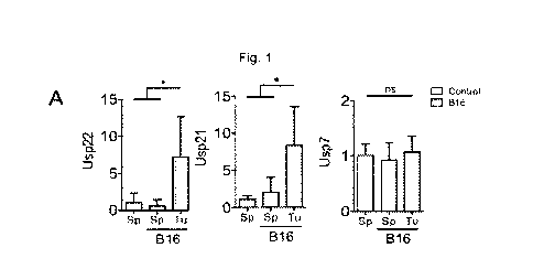
Fig. 1. Intratumoral Treg cells have increased mRNA expression of Usp22 and
Usp21. A-C, mRNA level of YFP+ sorted Treg cells from control mice spleens,
and tumor-
challenged mice spleens and tumor cells. All mRNA values calculated relative
to WT Treg
cell levels of unchallenged mice. spleens B16) Usp22: n=5-6, Usp21: n=3-5,
Usp7: n=3-5.
LLC1) Usp22: n=5-6, Usp21: n=3-6, Usp7: n=3-6. EG7) Usp22: n=4-5, Usp21 n=3-4,
Usp7: n=3-7. D, mRNA level of Usp22, Usp21 and Foxp3 in CD4 CD25 CD127- Treg
cells
isolated from human lung cancer tissues from patients relative to Treg cells
recovered from
the cancer-adjacent healthy lung tissue isolated from the same patient. AHL:
adjacent
healthy lung; LTu: lung tumor. Usp22: n=8, Usp21: n=3, FoxP3: n=11. E, mRNA
level of
Usp21 and FoxP3 in Treg cells isolated from human lung cancer patients. AHL:
adjacent
healthy lung; LTu: lung tumor n=9. A-C, Two-tailed unpaired t-test was done to
determine
statistical significance. D, Two-tailed paired t-test was performed to
determine statistical
significance of FoxP3 and Usp22 in Ltu vs. AHL. E, Linear regression was
calculated for
the correlation between Usp22 and FoxP3 within Ltu. All data are presented as
mean + stdev. NS, not significant. *P < 0.05, **P < 0.01, ***P < 0.001, ****P
< 0.0001.
Fig. 2: Tumor cell secreted TGF-I3 increases Usp22 and Usp21 level in iTreg
cells. A,
USP mRNA level in iTreg cells in control T cell media compared to addition of
tumor cell
treated media at 50/50 with T cell media for 24 hours. Usp22) Control: n=14,
B16: n=10,
LLC1: n=5, EG7: n=4. Usp21) Control: n=12, B16: n=8, LLC1: n=3, EG7: n=3.
Usp7)
Control: n=10, B16: n=7, LLC1: n=4, EG7: n=5 B, USP protein level in iTreg
cells in control
T cell media compared to addition of tumor cell treated media at 50/50 with T
cell media
for 24 hours. C, USP mRNA level in iTreg cells with the addition of a TGF-I3
inhibitor in
tumor cell media (Usp22) Control: n=22, B16: n=15, B16+Inh: n=5, LLC1: n=10,
3
CA 03216296 2023- 10- 20
WO 2022/226402
PCT/US2022/026159
LLC1+inh: n=5, EG7: n=7, EG7+inh: n=5. Usp21) Control: n=20, B16: n=13,
B16+Inh:
n=5, LLC1: n=8, LLC1+inh: n=4, EG7: n=7, EG7+inh: n=5. Usp7) Control: n=14,
B16:
n=10, B16+Inh: n=5, LLC1: n=8, LLC1+inh: n=3, EG7: n=8, EG7+inh: n=6. D,
SMAD2,
SMAD3, and SMAD4 binding capacity along the Usp22 promoter under TGFb
inhibition.
SMAD2: n=4-5; SMAD3 n=3; SMAD4: n=3. A-C, All mRNA values calculated relative
to
untreated WT iTreg cells. A-D, Ordinary one-way ANOVA with multiple
comparisons was
performed to determine significance. All data are presented as mean stdev.
NS, not
significant. *P < 0.05, **P < 0.01, ***P < 0.001, .. P<0.0001.
Fig. 3: Usp22 and Usp21 are required for FOXP3 stability in nTreg cells under
environmental and metabolic stress found in the TME. All mRNA values
calculated relative
to unchallenged WT Treg cells. A, nTreg USP mRNA level in normoxic (21% 02)
verses
hypoxic (1% 02) conditions after 24 hours (n=6-13). B, FOXP3 MFI change in
22K0 nTreg
cells relative to WT nTreg cells after 72 hours in normoxic (21% 02) verses
hypoxic (1% 02)
conditions (n=5). C, nTreg USP mRNA level after treatment with dMOG for 24
hours (n=6).
D, USP mRNA level in nTreg cells after exposure to glucose-restricted (0.5mM)
conditions
after 24 hours relative to normal media (11mM glucose) (n=7-18). E, Relative
FOXP3 MFI
change in nTreg cells from control and cells cultured under low glucose
conditions after 48
hours (n=3). F, nTreg USP mRNA level under amino acid starvation for 24 hours
(n=5-9).
C, FOXP3 MFI stability in Usp22- or Usp21-null nTreg cells cultured in normal
media
conditions verses amino acid starvation after 48 hours in (n=3). H, nTreg USP
mRNA level
after treatment with luM oligomycin A for 24 hours (n=5-7). I, nTreg USP mRNA
level
after treatment with 250nM Torinl for 24 hours (n=5-7). A, C-D, F and H-I,
Ordinary one-
way ANOVA with multiple comparisons was performed to determine significance.
B, E
and C, Two-tailed unpaired t-test was performed to determine statistical
significance. All
data are presented as mean stdev. NS, not significant. *P < 0.05, **P <0.01,
***P <0.001, ****P <0.0001
Fig. 4: Loss of Usp22 and Usp21 in Treg cells differentially impairs FoxP3
expression and cell function. A, Usp22 and Usp21 levels in WT, 21K0, 22K0 and
dKO
mice (n=5-8). All mRNA values calculated relative to WT Treg cells. B, Mice
weights over
a 2-month period (n=2-9). C, Peripheral activation of CD4+ and CD8+ T cells as
measured
by CD44h1CD62L10 expression (n=7-9). D, Representative histogram (left) and
4
CA 03216296 2023- 10- 20
WO 2022/226402
PCT/US2022/026159
quantification (right) of FOXP3 MFI in splenic Treg cells of WT and KO animals
(n=6-8).
E, Heat map of Treg cell signature genes (*significance is adjusted P <0.01)
in 21K0 (n=2),
22K0 (n=3), and dKO (n = 3) versus WT (n=3) mice. F, Venn Diagram of DEGs
(adj.
p<0.01) between 22K0, 21K0 and dKO (n=2-3). G, Normalized enrichment scores
from
gene set enrichment analysis (False Discovery Rate, FDR<25%) from the hallmark
gene set
in the molecular signatures database comparing the gene set generated from RNA
sequencing of Wt, 21K0, 22K0, and dKO mice (n=2-3). A-C, Two-way ANOVA with
multiple comparisons between rows was performed to determine statistical
significance. D,
One-way ANOVA with multiple comparisons between rows was performed to
determine
statistical significance. All data are presented as mean stdev. NS, not
significant.
*P < 0.05, **P < 0.01, ***P < 0.001, ****P < 0.0001.
Fig. 5: Deletion of Usp21 and Usp22 in Treg cells synergize to enhance
antitumor
immunity A, Tumor growth curve of B16 cells subcutaneously injected in the
flank of WT,
21K0, 22K0 and dKO mice (n=13-14). B, Percent activation as defined by
CD44hiCD62L10
of CD4 and CD8 T cells in the spleens of B16 challenged mice (n=5-6). C,
Percent IFN-
y and Granzvme B (GZMB) production of peripheral CD8+ T cells (n=3). D, FOXP3
MFI
of peripheral Treg cells relative to WT (n=7-9). E, PD-1 MFI of peripheral
'Leg cells relative
to WT (n=3). F, GITR MFI of peripheral Treg cells relative to WT (n=6-8). G,
LAG3 MFI
of peripheral Treg cells relative to WT (n=3). H, Representative flow
cytometry plot and
graphical representation of % infiltration of CD4+ and CD8+ T cells within the
tumor (n=5-
6). I-J, Percentage IFN-y and GZMB production of intratumoral CD8+ and
CD4+cells (n=5-
6). K, Representative FOXP3+ percentage of CD4+ cells relative to WT in itTreg
cells (n=6).
L, Representative flow plot (left) and quantitative representation of FOXP3
MFI within
tumor Treg cells relative to WT (n=6-9). M, Tumor growth curve of B16 cells
treated with
TGFf3 shRNA or scramble control shRNA subcutaneously injected in the flank of
WT and
dKO mice (n=3-4). A-C, H-J, and M Two-way ANOVA with multiple comparisons
between rows was performed to determine statistical significance. All data are
presented as
mean + stdev. NS, not significant. *P < 0.05, **P < 0.01, ***P < 0.001, ****P
< 0.0001.
D-C and K and L, One-way ANOVA with multiple comparisons between rows was
performed to determine statistical significance. All data are presented as
mean stdev. NS,
not significant. *P < 0.05,
5
CA 03216296 2023- 10- 20
WO 2022/226402
PCT/US2022/026159
Fig. 6: Usp22 inhibitor administration enhances antitumor immunity. A,
Structure
of compound CS30 (Usp22i-S02). B, FOXP3 MFI in WT and 22K0 of Treg cells after
treatment with 20mg/kg of Usp22i-S02 in vivo (n=3). C, Representative flow
cytometry plot
of FOXP3+CD25+ MFI of CD4+ peripheral cells of mice treated with 20mg/kg of
Usp22i-
502 relative to control (n=5). D, Graphical representation of Foxp3 MFI upon
1Jsp22i-502
administration (n=5). E, Tumor growth curve of LLC1 cells subcutaneously
injected in the
flank of WT mice with or without the addition of 20mg/kg/time of the Usp22
inhibitor
starting at day 15, in 100 !IL of oil (n=4). F-G, Representative flow
cytometry plot and
graphical representation of % infiltration of CD4+ and CD8+ T cells within the
tumor (n=4).
H, Representative histogram plot and graphical representation of itTreg Foxp3
MFI (n=4).
I, MFI of itTreg suppressive markers (n=3-4). J, Percent Foxp3+IFNg+ itTreg
cells in
control and Usp22-502 treated mice (n=3-4). B, D-E, G, H-I Two-way ANOVA with
multiple comparisons between rows was performed to determine statistical
significance. J,
Unpaired two tailed T test was performed to determine significance. All data
are presented
as mean stdev. NS, not significant. *P <0.05, **P <0.01, ***P <0.001,****P
<0.0001.
Figure 7: Intratumoral Treg cells have increased Foxp3 and activation markers.
A,
Representative CD4+ FOXP3+ percentage by flow cytometry of cells from non-
tumor
challenged controls and B16-, LLC1-, and EG7- challenged mice. B16: n=3-4,
EG7: n=2-
6, and LLC1: n=5-6. B, Representative overlay of FOXP3 MFI in tumor and spleen
of
CD45+ CD4+ FOXP3+ (Treg) cells of control and tumor-challenged mice. C,
Quantification
of FOXP3 MFI in control spleen and tumor-challenged spleen and tumor Treg
cells (n=4-
11). D-G, MFI of Treg cell-associated markers under control Treg cells
isolated from the
spleen and splenic and tumor Treg cells from B16, EG7, or LLC1 challenged
animals. CD25:
n=4-11; GITR: n=3-11; CTLA-4: n=4-11; PD-1: n=4-11. All MFI values calculated
relative
to WT Treg cell levels of non-challenged mice spleens. C-G, Two-tailed
unpaired t-test was
performed to determine statistical significance. All data are presented as
mean stdev. NS,
not significant. *P <0.05, **P <0.01, ***P <o=00, ****P <0.0001.
Fig 8: TGF-f3 induces expression of Usp22 and Usp21 in Treg cells. A, Visual
representation of Tumor Conditioned Media (TCM) experiments. B, iTreg USP mRNA
level
under TGF-13 induction post-polarization. Usp22: n=18-19; Usp21: n=7-8; Usp7:
4-5. C,
iTreg USP mRNA level under TGF-fi with and without a TGF-13 inhibitor. Usp22:
n=3-11;
6
CA 03216296 2023- 10- 20
WO 2022/226402
PCT/US2022/026159
Usp21: n=8-18; Usp7: 3-8. D, Treg FoxP3 mRNA level with TGF-fl induction
(n=10). B-D,
All mRNA values calculated relative to WT untreated iTreg cells. E, TGF-I3
level in B16,
LLC1 and EG7 tumor conditioned media (n=3). F-H, Correlation between USP
induction
and TGF-I3 level in the tumor conditioned media. Usp22: n=3-10; Usp21: n=3-8;
Usp7: n=3-
6. 1, TGF-fl mRNA level of control or TGF-fl shRNA treated B16 cells (n=3).
.1, mRNA
level of Usp22 in sorted itTreg cells from mice injected with shRNA treated
B16 cells (n=4).
B, D, and I, Two-tailed unpaired t-test was performed to determine statistical
significance.
C and E Ordinary one-way Anova with multiple comparisons between groups was
performed to determine statistical significance. All data are presented as
mean + stdev. NS,
not significant. *P <0.05, **P <o0, ***P <o00, ****P <0.0001.
Figure 9: SMAD3 and SMAD4 bind to conserved SBE on the Usp22 promoter while
Usp21 is upregulated by non-canonical TGF-I3 signaling. A, Usp22 promoter
region
overlaid with plausible SMAD binding elements (SBE) and placement of primers
created
for ChIP. B and C, Binding of SMAD2, SMAD3 and SMAD4 along the Usp22 and Usp21
promoter regions using ChIP-qPCR Usp22: n=2-7; Usp21: n=1-2. D-F,
Representative flow
plot and graphical representation of Foxp3 MFI and percentage in WT and Usp21-
null iTreg
cells polarized in 5ng/p.1 of TGF-13 (n=3). G, iTreg Usp21 mRNA level under
TGF-I3 with
and without a TGF-fl non-canonical pathway p38 kinase inhibitor (p38i) (n=4).
E and F,
Two-tailed unpaired t-test was performed to determine statistical
significance. C, Ordinary
one-way Anova with multiple comparisons between groups was performed to
determine
statistical significance. All data are presented as mean + stdev. NS, not
significant.
*P <005 **P <0.01, ***-13 <0,001, ****P <0.0001.
Figure 10: USP22 reciprocally enhances TGF-I3 signaling through SMAD protein
stabilization in positive feedback loop. A, Representative protein level of
SMAD2, SMAD3,
and SMAD4 in USP22 WT and KO iTreg cells. B, mRNA level of SMAD2. SMAD3, and
SMAD4 in USP22 WT and KO iTreg cells (n=3). All mRNA values calculated
relative to
unchallenged WT iTreg cells. Two-tailed unpaired t-test was performed to
determine
statistical significance. All data are presented as mean + stdev. NS, not
significant.
*P < 0.05, **P < 0.01, ***P < 0.001, ****P < 0.0001. C, USP22 endogenous IP
with
SMAD 2, SMAD 3, and SMAD 4 proteins within iTreg cells. D-F, Overexpression
DUB
assay IP in 2931 cells of USP22 with SMAD2, SMAD3, and SMAD4. G, SMAD2 and
7
CA 03216296 2023- 10- 20
WO 2022/226402
PCT/US2022/026159
SMAD4 protein degradation in WT and KO iTreg cells under cycloheximide
treatment for
2, 4 and 6 hours. H, SMAD2 and SMAD4 protein degradation in WT and KO iTreg
cells
under cycloheximide treatment with or without MG132 protease inhibitor at 4
hours.
Figure 11: HIF-a and the AMPK/mTOR balance modulates Treg cell FoxP3 stability
through IJSP22 and IJSP21. All mRNA values calculated relative to unchallenged
WT Treg
cells. A, iTreg USP mRNA level in normoxic and hypoxic conditions after 4
hours (n=4-5).
B, iTreg USP protein level in normoxic and hypoxic conditions after 24 hours.
C, Visual
representation of stability assay calculations of Foxp3 MFI level. %02 is the
percentage of
oxygen, Glu is glucose, and AA is amino acids. One variable was changed at a
time, the
others kept at baseline control. D, iTreg cell USP mRNA level after treatment
with DMOG
for 24 hours (n=6). E, nTreg cell Foxp3 MFI change treated with DMOG for 48
hours relative
to untreated nTreg (n=9-10). F, Foxp3 MFI change of iTreg after 72h in hypoxia
compared
to untreated cells (n=3). G, Foxp3 MFI change of iTreg treated with DMOG for
72 hours
relative to untreated iTreg cells (n=4) H, iTreg cell USP mRNA level under low
glucose
conditions after 24 hours (n=3-8). I, iTreg cell USP protein level under low
glucose
conditions after 24 hours. J, iTreg Foxp3 MFI change low glucose conditions
after 72 hours
relative to complete media (n=7). K, iTreg cell USP mRNA level in amino acid
starvation
relative to complete media after 24 hours (n=6). L, Foxp3 MFI change of iTreg
cells under
amino acid starvation for 72 hours relative to untreated iTreg cells (n=6). M,
iTreg cell USP
mRNA level after treatment with laM oligomycin for 24 hours (n=6). N, iTreg
USP mRNA
level after treatment with 250nM Torinl for 24 hours (n=5-9). A, D, H, and K-
M, Ordinary
one-way Anova with multiple comparisons between groups was performed to
determine
statistical significance. E-G and J, Two-tailed unpaired t-test was performed
to determine
statistical significance. All data are presented as mean stdev. NS, not
significant.
*P < 0.05, **P < 0.01, ***P < 0.001, ****13 < 0.0001.
Figure 12: Loss of Usp22 and Usp21 in Treg cells differentially alter Treg
metabolic
pathways. A, T and B cell percentages in peripheral organs of KO and WT
animals (n=5-
9). B, Percent of CD4 and CD 8 T cells in peripheral organs of CD45+ cells?
(n=4-10). C-
E, Heat map of metabolic pathways (significance is noted by * adjusted P
<0.01) in U21K0
(n=2), U22K0 (n=3), and dKO (n = 3) versus WT (n=3) mice. Genes chosen based
on
differential expression (adj. p<0.01) in the dKO mice. F, Basal mitochondria'
OCR and G,
8
CA 03216296 2023- 10- 20
WO 2022/226402
PCT/US2022/026159
Basal ECAR of 21K0 (n=5), 22K0 (n=5) and dKO (n=4-5) relative to WT (n=5) nTi-
eg cells.
A-B, Two-way ANOVA with Sidak's multiple comparisons between rows was
performed
to determine statistical significance. F-G, One-way ANOVA with Tukey's
multiple
comparisons between rows was performed to determine statistical significance.
All data are
presented as mean stdev. NS, not significant *I' <005, **13 <001, ***13
<0001,
****P <0.0001.
Figure 13: Usp21-deletion alters cell. A, Normalized enrichment scores from
gene
set enrichment analysis (False Discovery Rate, FDR<1%) from the hallmark gene
set in the
molecular signatures database comparing the gene set generated from RNA
sequencing of
22K0 and dKO mice (n=3). B, Representative graph of percent Ki67 positive
cells within
the CD4+Foxp3+ Treg compartment (n=7). One-way ANOVA with Tukey's multiple
comparisons between rows was performed to determine statistical significance.
All data are
presented as mean stdev. NS, not significant. *P <0.05, **P <0.01, ***P
<0.001,
****P <0.0001.
Figure 14. Development and validation of Usp22-specific inhibitor through
structure-based hierarchical virtual screening. A, Flowchart of structure-
based virtual
screening. B, The overall conformation of USP22-m (USP22 model generated using
SWISS
MODEL) is represented by cartoon which are colored by conservation using the
color-code
bar. Catalytic centre of USP22 was defined as docking position. C,
Ramachandran plot
statistics of USP22-m generated by PROCHEK progress (left). The Displacement
of the
catalytic centre loop in USP22-md (the MD optimized model) compared to UBP8
(PDB:
3MHS) and USP22-m (right). D, S02 displayed in green stick binding in the
pocket of
USP22-md structure (left). Ligplot showing hydrogen bonding and hydrophobic
contacts of
SO2 with USP22-md (middle). The best ranked position of SO2 (shown in green)
in the
binding pocket of USP22-md is presented, generated by docking. E, Individual
energy
contributions of amino acid residues after MD simulations and PBSA
calculations. F-G,
Binding free energies of compound S02 to USP22 Model.
Figure 15: Usp22i-S02 halts Usp22-mediated Foxp3 deubiquitination. A,
Graphical
representation of Foxp3 MFI change in WT versus 221(0 iTreg cells treated with
various
doses of Usp22i-S02 (n=3). B, Representative histogram of Foxp3 MFI level in
iTreg cells
as Usp22 inhibitor concentration increases from 0-20[Ig/mL. C, Cell survival
of iTreg cells
9
CA 03216296 2023- 10- 20
WO 2022/226402
PCT/US2022/026159
treated with various doses of Usp22i-S02 (n=3). D, FOXP3 and USP22 protein
level in WT
and 22K0 mice treated with 101,1g/mL Usp22i-S02. E-F, Graphical and
representative data
of Foxp3 MFI of human Treg cells treated with various doses of Usp22i-S02
(n=3). G,
Graphical representation of Foxp3 MFI in WT, Usp21-null, and Usp22-null Treg
cells
treated with Usp22i-S02 at 10p.g/mL (n=3). H, FOXP3 and IJSP22 protein
degradation of
cycloheximide (101,1g/mL) treated iTreg cells with or without the addition of
101.1g/mL of
Usp22i-S02. I-J, Endogenous DUB assay IP in iTreg cells of USP22 with FOXP3
under
increasing concentrations of Usp22i-S02. K, Foxp3 mRNA level in iTreg cells as
Usp22
inhibitor concentration increases from 0-20 g/mL (n=3). L, FOXP3 and USP22
level in
WT iTreg cells with or without 201.tg/mL Usp22 inhibitor treated with 201,IM
MG132. M,
Graphical representation of the percentage of decrease of Foxp3 MFI in either
WT or
Usp22-null nTreg cells placed in low glucose conditions with or without the
addition of
101,tg/mL of Usp22i-S02 (n=7-8). N, Graphical representation of the percentage
of decrease
of Foxp3 MFI in WT nTreg cells placed in hypoxic or low amino acid conditions
with or
without the addition of 101.1.g/mL of Usp22i-S02 (n=3-5). G, Two-tailed
unpaired t-test
comparing within groups was performed to determine statistical significance.
K, One-way
ANOVA with Dunnet's multiple comparisons between rows relative to control was
performed to determine statistical significance. M-N, Two-way ANOVA with
Sidak's
multiple comparisons between rows was performed to determine statistical
significance. All
data are presented as mean stdev. NS, not significant. *P < 0.05, **P <0.01,
***P <0.001, ****P <0.0001.
Figure 16: Usp22i-S02 has little effect on naïve mice, yet enhances anti-tumor
immunity in LLC1-challenged mice. A, Body weight of Usp22i-S02 treated mice
verses
DMSO treated controls over the course of treatment (n=4). A-F. Injections were
twice a day
for 3 consecutive days at 10mg/kg (n=4). B, Percent of cell populations in
naïve mice treated
with Usp22i-S02 relative to DMSO control (n=3-4). C, Percent of 1<I-67+ cells
in various
compartments in naive mice treated with Usp22i-S02 relative to DMSO control
(n=3-4). D,
Percent CD44111CD62LI0 in T cell populations gated on CD45+ cells in naïve
mice treated
with Usp22i-S02 relative to DMSO control (n=3). E, Percent Annexin+PI+ T cells
gates on
CD45+ cells in naive mice treated with Usp22i-S02 relative to DMSO control
(n=4). F-H,
Organ toxicity panel (VetScan VS2 Comprehensive Diagnostic Rotor lot 1061AA2)
of
CA 03216296 2023- 10- 20
WO 2022/226402
PCT/US2022/026159
naive mice treated with Usp22i-S02 relative to DMSO control (n=2-3). I, Growth
curve of
subcutaneously injected LLC1 in WT mice treated with Usp22i-S02 at 10mg/kg
(n=5-10).
I-Q, Injections were twice a day for 5 consecutive days at 10mg/kg. J, Weights
of resected
tumors at day 16 from I (n=10). K-L, Representative flow plot and graphical
representation
of infiltrating T cells gated on CD45+ cells (n=5). M-P, Characterization of
intratumoral
CD8+ T cells frome mice treated with Usp22i-S02 (n=3-5). Q, Percent of
intratumoral Treg
cells from mice treated with Usp22i-S02 verses DMSO control, gated on CD4
Foxp3+ from
mice (n=5). A-E, I and L, Two-way ANOVA with Sidaks's multiple comparisons
between
rows relative to control was performed to determine statistical significance.
F-H, J, and M-
Q, Two-tailed unpaired t-test was performed to determine statistical
significance. All data
are presented as mean stdev. NS, not significant. *P <0.05, **P < 0.01, ***P
<0.001,
****P <0.0001.
Figure 17: Usp22i-S02 inhibits tumor growth in vitro and in vivo. A, In vitro
counts
of LLCI after treatment with Usp22i-S02 under various concentrations for 24
hours (n=2).
B, In vitro viability of LLC1 after treatment with Usp22i-S02 under various
concentrations
for 24 hours (n=2). C, In vitro relative growth of LLC1 cells treated with
lOug/m1 of Usp22i-
S02 relative to DMSO treated control for 7 days via 0D600 (n=4). D, Growth
curve of 1
million subcutaneously injected LLCI cells into RAG-/- mice treated for 3 days
Usp22i-
S02 relative to DMSO control injections at day 15 of tumor growth (n=4). C-D,
Two-way
ANOVA with Sidaks's multiple comparisons between rows relative to control was
performed to determine statistical significance. All data are presented as
mean stdev. NS,
not significant. *P <0.05, **P <0.01, ***P <0.001, ****P <0.0001.
Figure 18: TME-specific factors can drive increased levels of Usp22 and Usp21
potentially through modulation of TGF-I3 signaling, HIFI a, AMPK, and mTOR
activity to
render Tieg cells more stable in the tumor microenvironment.
DETAILED DESCRIPTION
Disclosed herein are inhibitors of ubiquitin specific peptidase 22 (USP22) and
uses
for treating diseases and disorders thereof. Computer-based and biological
approaches were
used to identify small molecule specific inhibitors. As demonstrated in the
Examples,
treatment of regulatory T cells (Tregs), both mouse and human, with inhibitors
of USP22
significantly reduced the protein expression of FoxP3, a substrate of USP22.
In contrast,
11
CA 03216296 2023- 10- 20
WO 2022/226402
PCT/US2022/026159
treatment did not further inhibit FoxP3 expression in USP22-null Tregs,
indicating that the
inhibitors of USP22 may be a highly specific inhibitor of USP22. In addition,
treatment
inhibited USP22 activity in lung cancer cells and consequently suppressed lung
cancer cell
growth. More importantly, treatment of lung cancer-bearing mice largely
diminished the
tumor mass. These results indicate that inhibitors of IJSP22 can be used as a
potent drug in
antitumor therapy. In addition, the fact that suppression of USP22 diminishes
Treg
suppressive functions, also allows for these inhibitors to be used to treat
diseases associated
to immune deficiency as well as to boost the immune response to combat
infectious diseases
such as SARS-CoV2 infection.
The present invention is described herein using several definitions, as set
forth below
and throughout the application.
Definitions
The disclosed subject matter may be further described using definitions and
terminology as follows. The definitions and terminology used herein are for
the purpose of
describing particular embodiments only and are not intended to be limiting.
As used in this specification and the claims, the singular forms "a," "an,"
and "the"
include plural forms unless the context clearly dictates otherwise. For
example, the term "a
substituent" should be interpreted to mean "one or more substituents," unless
the context
clearly dictates otherwise.
As used herein, "about-, "approximately,- "substantially,- and "significantly-
will
be understood by persons of ordinary skill in the art and will vary to some
extent on the
context in which they are used. If there are uses of the term which are not
clear to persons
of ordinary skill in the art given the context in which it is used, "about"
and "approximately"
will mean up to plus or minus 10% of the particular term and "substantially"
and
"significantly- will mean more than plus or minus 10% of the particular term.
As used herein, the terms "include" and -including" have the same meaning as
the
terms "comprise" and "comprising." The terms "comprise" and -comprising"
should be
interpreted as being "open" transitional terms that permit the inclusion of
additional
components further to those components recited in the claims. The terms
"consist- and
"consisting of" should be interpreted as being "closed" transitional terms
that do not permit
the inclusion of additional components other than the components recited in
the claims. The
12
CA 03216296 2023- 10- 20
WO 2022/226402
PCT/US2022/026159
term "consisting essentially of' should be interpreted to be partially closed
and allowing the
inclusion only of additional components that do not fundamentally alter the
nature of the
claimed subject matter.
The phrase -such as" should be interpreted as -for example, including."
Moreover,
the use of any and all exemplary language, including but not limited to "such
as", is intended
merely to better illuminate the invention and does not pose a limitation on
the scope of the
invention unless otherwise claimed.
Furthermore, in those instances where a convention analogous to "at least one
of A,
B and C, etc." is used, in general such a construction is intended in the
sense of one having
ordinary skill in the art would understand the convention (e.g., "a system
having at least one
of A, B and C" would include but not be limited to systems that have A alone,
B alone, C
alone, A and B together, A and C together, B and C together, and/or A, B, and
C together.).
It will be further understood by those within the art that virtually any
disjunctive word and/or
phrase presenting two or more alternative terms, whether in the description or
figures,
should be understood to contemplate the possibilities of including one of the
terms, either
of the terms, or both terms. For example, the phrase "A or B" will be
understood to include
the possibilities of "A" or 13 or "A and B."
All language such as "up to," "at least," "greater than," "less than," and the
like,
include the number recited and refer to ranges which can subsequently be
broken down into
ranges and subranges. A range includes each individual member. Thus, for
example, a
group having 1-3 members refers to groups having 1, 2, or 3 members.
Similarly, a group
having 6 members refers to groups having 1, 2, 3, 4, or 6 members, and so
forth.
The modal verb "may" refers to the preferred use or selection of one or more
options
or choices among the several described embodiments or features contained
within the same.
Where no options or choices are disclosed regarding a particular embodiment or
feature
contained in the same, the modal verb "may" refers to an affirmative act
regarding how to
make or use and aspect of a described embodiment or feature contained in the
same, or a
definitive decision to use a specific skill regarding a described embodiment
or feature
contained in the same. In this latter context, the modal verb "may- has the
same meaning
and connotation as the auxiliary verb "can."
13
CA 03216296 2023- 10- 20
WO 2022/226402
PCT/US2022/026159
A "subject in need thereof" as utilized herein may refer to a subject in need
of
treatment for a disease or disorder associated with ubiquitin specific
peptidase 22 (USP22)
activity and/or expression. A subject in need thereof may include a subject
having a cancer
that is characterized by the activity and/or expression of USP22. The
disclosed compounds,
pharmaceutical compositions, and methods may be utilized to treat diseases and
disorders
associated with USP22 activity and/or expression.
In some embodiments, a subject in need thereof may include a subject having a
cancer that is treated by administering a therapeutic agent that inhibits the
biological activity
of USP22, and/or that inhibits dissemination of cancer cells.
The disclosed compounds, pharmaceutical compositions, and methods may be
utilized to treat diseases and disorders associated with USP22 activity and/or
expression
which may include cell proliferative diseases and diseases and disorders such
as cancers.
Suitable cancers for treatment by the disclosed compounds, pharmaceutical
compositions,
and methods may include, but are not limited to lung cancer, gastric
carcinoma, pancreatic
cancer, melanoma, lymphoma, colon cancer, breast cancer, ovarian cancer,
bladder cancer,
prostate cancer, glioma, mesothelioma, neuroblastoma, mantle cell lymphoma,
and acute
myeloid leukemia.
In some embodiments, a subject in need thereof may include a subject in need
of
treatment of infection. In some embodiments, the infection is a viral
infection, such as an
infection by a corona virus. In some embodiments, the subject in need thereof
is in need of
a treatment for infection by sudden acute respiratory syndrome coronavirus 2
(SARS-CoV2)
and COVID. In some embodiments, a subject in need thereof may refer to a
subject in need
of augmenting the immune response to an infection. In some embodiments, a
subject in need
thereof may refer to a subject in need of augmenting the immune response to
sudden acute
respiratory syndrome coronavirus 2 (SARS-CoV2) infection.
The disclosed compounds, pharmaceutical compositions, and methods may be
utilized to treat diseases and disorders associated with USP22 activity and/or
expression
which may include infections and diseases and disorders such as respiratory
infections,
including sudden acute respiratory syndrome coronavirus 2 (SARS-CoV2)
infection.
The term "subject" may be used interchangeably with the terms "individual" and
"patient" and includes human and non-human mammalian subjects.
14
CA 03216296 2023- 10- 20
WO 2022/226402 PCT/US2022/026159
The disclosed compounds may be utilized to modulate the biological activity of
USP22, including modulating the peptidase activity of USP22. The term -
modulate" should
be interpreted broadly to include "inhibiting" USP22 biological activity
including peptidase
activity.
Ubiquitin specific peptidase (IJSP22) refers to the protein also referred to
by the
name ubiquitin carboxyl-terminal hydrolase 22. USP22 has been shown to have
enzyme
activities that include catalyzing the thiol-dependent hydrolysis of ester,
thioester, amide,
peptide and isopeptide bonds formed by the C-terminal glycine of ubiquitin.
USP22 has
ENZYME entry: EC 3.4.19.12. The compounds disclosed herein may inhibit one or
more
of the activities of USP22 accordingly.
Human USP22 is known to have two isoforms and the disclosed compounds may
inhibit one or more activities of isoform 1 and/or isoform 2.
Human USP22 Isoform 1 has the following amino acid sequence:
10 20 30 40 50
MVSRPEPEGE AMDAELAVAP PGCSHLGSFK VDNWKQNLRA IYQCFVWSGT
60 70 80 90 100
AEARKRKAKS CICHVCGVHL NRLHSCLYCV FFGCFIKKHI HEHAKAKRHN
110 120 130 140 150
LAIDLMYGGI YCFLCQDYIY DKDMEIIAKE EQRKAWKMQG VGEKFSTWEP
160 170 180 190 200
TKRELELLKH NPKRRKITSN CTIGLRGLIN LGNTCFMNCI VQALTHTPLL
210 220 230 240 250
RDFFLSDRHR CEMQSPSSCL VCEMSSLFQE FYSGHRSPHI PYKLLHLVWT
260 270 280 290 300
HARHLAGYEQ QDAHEFLIAA LDVLHRHCKG DDNGKKANNP NHCNCIIDQI
CA 03216296 2023- 10- 20
WO 2022/226402 PCT/US2022/026159
310 320 330 340 350
FTGGLQSDVT CQVCHGVSTT IDPFWDISLD LPGSSTPFWP LSPGSEGNVV
360 370 380 390 400
NGESHVSGTT TLTDCLRRFT RPEHLGSSAK IKCSGCHSYQ ESTKQLTMKK
410 420 430 440 450
LPIVACFHLK RFEHSAKLRR KITTYVSFPL ELDMTPFMAS SKESRMNGQY
460 470 480 490 500
QOPTDSLNND NKYSLFAVVN HOGTLESGHY TSFIROHKDO WFKCDDAIIT
510 520
KASIKDVLDS EGYLLFYHKQ FLEYE (SEQ ID NO: 1)
Isoform 2 has the following sequence:
10 20 30 40 50
MAPGWPSLSA GSRQEAPQLA AGGSAYQAVG RQFQPRATAL QGPSQAKSCI
60 70 80 90 100
CHVCGVHLNR LHSCLYCVFF GCFTKKHIHE HAKAKRHNLA IDLMYGGIYC
110 120 130 140 150
FLCQDYIYDK DMEIIAKEEQ RKAWKMQGVG EKFSTWEPTK RELELLKHNP
160 170 180 190 200
KRRKITSNCT IGLRGLINLG NTCFMNCIVQ ALTHTPLLRD FFLSDRHRCE
210 220 230 240 250
16
CA 03216296 2023- 10- 20
WO 2022/226402 PCT/US2022/026159
MQSPSSCLVC EMSSLFQEFY SGHRSPHIPY KLLHLVWTHA RHLAGYEQQD
260 270 280 290 300
AHEFLIAALD VLHRHCKGDD NGKKANNPNH CNCIIDQIFT GGLQSDVTCQ
310 320 330 340 350
VCHGVSTTID PFWDISLDLP GSSTPFWPLS PGSEGNVVNG ESHVSGTTTL
360 370 380 390 400
TDCLRRFTRP EHLGSSAKIK CSGCHSYQES TKQLTMKKLP IVACFHLKRF
410 420 430 440 450
EHSAKLRRKI TTYVSFPLEL DMTPFMASSK ESRMNGQYQQ PTDSLNNDNK
460 470 480 490 500
YSLFAVVNHQ GTLESGHYTS FIRQHKDQWF KCDDAIITKA SIKDVLDSEG
510
YLLFYHKQFL EYE (SEQ ID NO: 2)
Pharmaceutical Compositions
The compounds employed in the compositions and methods disclosed herein may
be administered as pharmaceutical compositions and, therefore, pharmaceutical
compositions incorporating the compounds are considered to be embodiments of
the
compositions disclosed herein. Such compositions may take any physical form
which is
pharmaceutically acceptable; illustratively, they can be orally administered
pharmaceutical
compositions. Such pharmaceutical compositions contain an effective amount of
a disclosed
compound, which effective amount is related to the daily dose of the compound
to be
administered. Each dosage unit may contain the daily dose of a given compound
or each
dosage unit may contain a fraction of the daily dose, such as one-half or one-
third of the
17
CA 03216296 2023- 10- 20
WO 2022/226402
PCT/US2022/026159
dose. The amount of each compound to be contained in each dosage unit can
depend, in
part, on the identity of the particular compound chosen for the therapy and
other factors,
such as the indication for which it is given. The pharmaceutical compositions
disclosed
herein may be formulated so as to provide quick, sustained, or delayed release
of the active
ingredient after administration to the patient by employing well known
procedures.
The compounds for use according to the methods of disclosed herein may be
administered as a single compound or a combination of compounds. For example,
a
compound that inhibits the biological activity of ubiquitin specific peptidase
22 (USP22)
may be administered as a single compound or in combination with another
compound
inhibits the biological activity of USP22 or that has a different
pharmacological activity.
As indicated above, pharmaceutically acceptable salts of the compounds are
contemplated and also may be utilized in the disclosed methods. The term
"pharmaceutically acceptable salt" as used herein, refers to salts of the
compounds, which
are substantially non-toxic to living organisms. Typical pharmaceutically
acceptable salts
include those salts prepared by reaction of the compounds as disclosed herein
with a
pharmaceutically acceptable mineral or organic acid or an organic or inorganic
base. Such
salts are known as acid addition and base addition salts. It will be
appreciated by the skilled
reader that most or all of the compounds as disclosed herein are capable of
forming salts
and that the salt forms of pharmaceuticals are commonly used, often because
they are more
readily crystallized and purified than are the free acids or bases.
Acids commonly employed to form acid addition salts may include inorganic
acids
such as hydrochloric acid, hy drobromi c acid, hy droi odi c acid, sulfuric
acid, phosphoric
acid, and the like, and organic acids such as p-toluenesulfonic,
methanesulfonic acid, oxalic
acid, p-bromophenylsulfonic acid, carbonic acid, succinic acid, citric acid,
benzoic acid,
acetic acid, and the like. Examples of suitable pharmaceutically acceptable
salts may include
the sulfate, pyrosulfate, bisulfate, sulfite, bisulfate, phosphate,
monohydrogenphosphate,
dihydrogenphosphate, metaphosphate, pyrophosphate, bromide, iodide, acetate,
propionate,
decanoate, capry I ate, acry I ate, formate, hydrochloride, di hy drochl ori
de, i sobutyrate,
caproate, heptanoate, propiolate, oxalate, malonate, succinate, suberate,
sebacate, fumarate,
maleat-, butyne-.1,4-dioate, hexyne-1,6-dioate, benzoate, chlorobenzoate,
methylbenzoate,
hy droxy benzo ate, methoxy b enzo ate, phthalate,
xylenesulfonate, phenylac elate,
18
CA 03216296 2023- 10- 20
WO 2022/226402
PCT/US2022/026159
phenylpropionate, phenylbutyrate, citrate, lactate, a-hydroxybutyrate,
glycolate, tartrate,
methanesulfonate, propanesulfonate, naphthalene-1 -sulfonate, naphthalene-2-
sulfonate,
mandelate, and the like.
Base addition salts include those derived from inorganic bases, such as
ammonium
or alkali or alkaline earth metal hydroxides, carbonates, bicarbonates, and
the like. Bases
useful in preparing such salts include sodium hydroxide, potassium hydroxide,
ammonium
hydroxide, potassium carbonate, sodium carbonate, sodium bicarbonate,
potassium
bicarbonate, calcium hydroxide, calcium carbonate, and the like.
The particular counter-ion forming a part of any salt of a compound disclosed
herein
is may not be critical to the activity of the compound, so long as the salt as
a whole is
pharmacologically acceptable and as long as the counter-ion does not
contribute undesired
qualities to the salt as a whole. Undesired qualities may include undesirably
solubility or
toxicity.
Pharmaceutically acceptable esters and amides of the compounds can also be
employed in the compositions and methods disclosed herein. Examples of
suitable esters
include alkyl, aryl, and aralkyl esters, such as methyl esters, ethyl esters,
propyl esters,
dodecyl esters, benzyl esters, and the like. Examples of suitable amides
include
unsubstituted amides, monosubstituted amides, and disubstituted amides, such
as methyl
amide, dimethyl amide, methyl ethyl amide, and the like.
In addition, the methods disclosed herein may be practiced using solvate forms
of
the compounds or salts, esters, and/or amides, thereof Solvate forms may
include ethanol
solvates, hydrates, and the like.
The pharmaceutical compositions may be utilized in methods of treating a
disease
or disorder associated with the biological activity of ubiquitin specific
peptidase 22
(USP22). As used herein, the terms "treating- or "to treat- each mean to
alleviate symptoms,
eliminate the causation of resultant symptoms either on a temporary or
permanent basis,
and/or to prevent or slow the appearance or to reverse the progression or
severity of resultant
symptoms of the named disease or disorder. As such, the methods disclosed
herein
encompass both therapeutic and prophylactic administration.
As used herein the term "effective amount" refers to the amount or dose of the
compound, upon single or multiple dose administration to the subject, which
provides the
19
CA 03216296 2023- 10- 20
WO 2022/226402
PCT/US2022/026159
desired effect in the subject under diagnosis or treatment. The disclosed
methods may
include administering an effective amount of the disclosed compounds (e.g., as
present in a
pharmaceutical composition) for treating a disease or disorder associated with
biological
activity of ubiquitin specific peptidase 22 (USP22).
An effective amount can be readily determined by the attending diagnostician,
as
one skilled in the art, by the use of known techniques and by observing
results obtained
under analogous circumstances. In determining the effective amount or dose of
compound
administered, a number of factors can be considered by the attending
diagnostician, such as:
the species of the subject; its size, age, and general health; the degree of
involvement or the
severity of the disease or disorder involved; the response of the individual
subject; the
particular compound administered; the mode of administration; the
bioavailability
characteristics of the preparation administered; the dose regimen selected;
the use of
concomitant medication; and other relevant circumstances.
A typical daily dose may contain from about 0.01 mg/kg to about 100 mg/kg
(such
as from about 0.05 mg/kg to about 50 mg/kg and/or from about 0.1 mg/kg to
about 25
mg/kg) of each compound used in the present method of treatment.
Compositions can be formulated in a unit dosage form, each dosage containing
from
about 1 to about 500 mg of each compound individually or in a single unit
dosage form,
such as from about 5 to about 300 mg, from about 10 to about 100 mg, and/or
about 25 mg.
The term "unit dosage form- refers to a physically discrete unit suitable as
unitary dosages
for a patient, each unit containing a predetermined quantity of active
material calculated to
produce the desired therapeutic effect, in association with a suitable
pharmaceutical carrier,
diluent, or excipient.
Oral administration is an illustrative route of administering the compounds
employed in the compositions and methods disclosed herein. Other illustrative
routes of
administration include transdermal, percutaneous, intravenous, intramuscular,
intranas al,
buccal, intrathecal, intracerebral, or intrarectal routes. The route of
administration may be
varied in any way, limited by the physical properties of the compounds being
employed and
the convenience of the subject and the caregiver.
As one skilled in the art will appreciate, suitable formulations include those
that are
suitable for more than one route of administration. For example, the
formulation can be one
CA 03216296 2023- 10- 20
WO 2022/226402
PCT/US2022/026159
that is suitable for both intrathecal and intracerebral administration.
Alternatively, suitable
formulations include those that are suitable for only one route of
administration as well as
those that are suitable for one or more routes of administration, but not
suitable for one or
more other routes of administration. For example, the formulation can be one
that is suitable
for oral, tran s derm al , percutaneous, intravenous, intramuscular,
intranasal, buccal, and/or
intrathecal administration but not suitable for intracerebral administration.
The inert ingredients and manner of formulation of the pharmaceutical
compositions
are conventional. The usual methods of formulation used in pharmaceutical
science may be
used here. All of the usual types of compositions may be used, including
tablets, chewable
tablets, capsules, solutions, parenteral solutions, intranasal sprays or
powders, troches,
suppositories, transdermal patches, and suspensions. In general, compositions
contain from
about 0.5% to about 50% of the compound in total, depending on the desired
doses and the
type of composition to be used. The amount of the compound, however, is best
defined as
the "effective amount", that is, the amount of the compound which provides the
desired dose
to the patient in need of such treatment. The activity of the compounds
employed in the
compositions and methods disclosed herein are not believed to depend greatly
on the nature
of the composition, and, therefore, the compositions can be chosen and
formulated primarily
or solely for convenience and economy.
Capsules are prepared by mixing the compound with a suitable diluent and
filling
the proper amount of the mixture in capsules. The usual diluents include inert
powdered
substances (such as starches), powdered cellulose (especially crystalline and
microcrystalline cellulose), sugars (such as fructose, mannitol and sucrose),
grain flours,
and similar edible powders.
Tablets are prepared by direct compression, by wet granulation, or by dry
granulation. Their formulations usually incorporate diluents, binders,
lubricants, and
disintegrators (in addition to the compounds). Typical diluents include, for
example, various
types of starch, lactose, mannitol, kaolin, calcium phosphate or sulfate,
inorganic salts (such
as sodium chloride), and powdered sugar. Powdered cellulose derivatives can
also be used.
Typical tablet binders include substances such as starch, gelatin, and sugars
(e.g., lactose,
fructose, glucose, and the like). Natural and synthetic gums can also be used,
including
21
CA 03216296 2023- 10- 20
WO 2022/226402
PCT/US2022/026159
acacia, alginates, methylcellulose, polyvinylpyrrolidine, and the like.
Polyethylene glycol,
ethylcellulose, and waxes can also serve as binders.
Tablets can be coated with sugar, e.g., as a flavor enhancer and sealant. The
compounds also may be formulated as chewable tablets, by using large amounts
of pleasant-
tasting substances, such as mannitol, in the formulation. Instantly dissolving
tablet-like
formulations can also be employed, for example, to assure that the patient
consumes the
dosage form and to avoid the difficulty that some patients experience in
swallowing solid
obj ects.
A lubricant can be used in the tablet formulation to prevent the tablet and
punches
from sticking in the die. The lubricant can be chosen from such slippery
solids as talc,
magnesium and calcium stearate, stearic acid, and hydrogenated vegetable oils.
Tablets can also contain disintegrators. Disintegrators are substances that
swell when
wetted to break up the tablet and release the compound. They include starches,
clays,
celluloses, algins, and gums. As further illustration, corn and potato
starches,
methylcellulose, agar, bentonite, wood cellulose, powdered natural sponge,
cation-exchange
resins, alginic acid, guar gum, citrus pulp, sodium lauryl sulfate, and
carboxymethylcellulose can be used.
Compositions can be formulated as enteric formulations, for example, to
protect the
active ingredient from the strongly acid contents of the stomach. Such
formulations can be
created by coating a solid dosage form with a film of a polymer which is
insoluble in acid
environments and soluble in basic environments. Illustrative films include
cellulose acetate
phthalate, polyvinyl acetate phthalate, hydroxypropyl methylcellulose
phthalate, and
hydroxypropyl methylcellulose acetate succinate.
Transdermal patches can also be used to deliver the compounds. Transdermal
patches can include a resinous composition in which the compound will dissolve
or partially
dissolve; and a film which protects the composition, and which holds the
resinous
composition in contact with the skin. Other, more complicated patch
compositions can also
be used, such as those having a membrane pierced with a plurality of pores
through which
the drugs are pumped by osmotic action.
As one skilled in the art will also appreciate, the formulation can be
prepared with
materials (e.g., actives excipients, carriers (such as cyclodextrins),
diluents, etc.) having
22
CA 03216296 2023- 10- 20
WO 2022/226402
PCT/US2022/026159
properties (e.g., purity) that render the formulation suitable for
administration to humans.
Alternatively, the formulation can be prepared with materials having purity
and/or other
properties that render the formulation suitable for administration to non-
human subjects, but
not suitable for administration to humans.
Inhibitors of IJbiquitin specific Peptidase 22 (IJSP22) IJses Thereof
Disclosed are compounds, pharmaceutical compositions comprising the compounds,
and methods of using the compounds and pharmaceutical compositions for
treating a subject
having or at risk for developing a disease or disorder associated with
ubiquitin specific
peptidase 22 (USP22) biological activity. The disclosed compounds may inhibit
the
biological activity of USP22. As such, the disclosed compounds and
pharmaceutical
compositions may be utilized in methods for treating a subject having or at
risk for
developing a disease or disorder that is associated with USP22 activity which
may be cell
proliferative diseases and disorders, such as cancer, or an infection
associated disease or
disorder, such as sudden acute respiratory syndrome, such as SARS-CoV2.
In some embodiments, the disclosed methods include treating a subject in need
of
treatment for a disease or disorder associated with ubiquitin specific
peptidase 22 (USP22)
activity. In the disclosed methods, the subject may be administered an
effective amount of
a therapeutic agent that inhibits the biological activity of USP22.
The disclosed methods may be performed in order to treat a cell proliferative
disease
or disorder, which may include cancer. Suitable cancers that may be treated by
the disclosed
methods may include, but are not limited to, lung cancer, gastric carcinoma,
pancreatic
cancer, melanoma, lymphoma, colon cancer, breast cancer, ovarian cancer,
bladder cancer,
prostate cancer, glioma, mesothelioma, neuroblastoma, mantle cell lymphoma,
and acute
myeloid leukemia.
In some embodiments, the disclosed methods may be performed in order to treat
lung cancer, for example, non-small cell lung cancer (NSCLC).
In some embodiments, the disclosed methods may be performed in order to treat
skin cancer, for example, melanoma.
In the disclosed methods, a subject in need thereof typically is administered
a
therapeutic agent that inhibits the biological activity of ubiquitin specific
peptidase 22
23
CA 03216296 2023- 10- 20
WO 2022/226402 PCT/US2022/026159
(USP22). In some embodiments, the therapeutic agent inhibits ubiquitin
specific peptidase
activity (E.C.: 3.4.19.12) of USP22.
Suitable therapeutic agents for use in the disclosed methods may include, but
are not
limited to, a compound having a formula selected from the group consisting of:
/
...----i...
t.'f., /
t),.
1 N j\=,,,...,. ¨.44H t
?
....................................................................
=\µµ,.,*=== '''''
µ,..."I:fs; ' Nr.0 '''= =-es- \
f >
L..,.õ.....,N,..4
1 t:
ii
, .s..
Fs. ... F
.... õ.- ..._
l''''''\ 1 FI:iu li '1
P----.. ------------------ <1 . (2-- -"'"'
''`cs .9. is4f.sµii." ..........,,,,,..,
..........k, ........,,,;:j....
\r.,,,,--.....< .1, 0 11 ' r ' 0
, V ,.,....,,,, .... = %kw....."-
µ i D
..., ,µ ..... NH
,... _________________________ ., 0 rse 11
e
\ 0
O'''''.
..
\
\ 1
,,,C)
.,S:' .......,....>,--
=
0#' \ /*-----\
p,....;
N
,
\.......0, 0,
¨.......
K. ,,e: .
-......,,,,,, ...,:.., \ /
....._..........?
0, 7¨W 11
\-.... . ...---4-.:>.. ...----=-. / =-=-:. ---::--
-0'
/ 0
..,....tsi
24
CA 03216296 2023- 10- 20
WO 2022/226402
PCT/US2022/026159
CH3
-0_ 0
HN ' N-0
........-p-",, i
t
r `µ...
C,\ i
o.--- ,..,....
c)........_ z
o, .o, I., ......õ =
...,õ--- ,..,.....- -.......,õ.0
\ '
\,....._
',,,,...õ,....õ.....'"',...õ ...,õ,.........<::`",....., CH3
,........, ,
,
ii
...,.,:::....,,
......1.>.--
,...-__:...--,=!..0 `Ny....--- =-...>õ,
t i
.--57'.. ---"µ (,,,,,-..--,, õ--= k--, õ... NI
N
i HN
...,.....õ...., ....,s,
fif
,
n
n
'......µ (.... ....,..,
_IL
o.--, .., ...
,
-------; õ..-.:,
/o ,,, µ =
il I,- , NH
..., if¨,
/ o . ,P
. =
;,=1-4...
rtz=-=-.--,--N .. ,...., \
\ / , __ i \ __
,
/ ,- .... .......õ. ,
,.....õ, .., \
,
i
S
i \ I/
k\., $ 0 __ %
'.\ ,e, \ 1/
...y
ti==== õ--''. __ >\ __ t
, /
...._d \ __ õ
CA 03216296 2023- 10- 20
WO 2022/226402
PCT/US2022/026159
_..-----
O. f
0 N
\1/4,s, Ci -'"=='k ,..k.
/
Tt N'
N
0
!! µ)
0
....,;õ,.....0
(Di-,
II P
-,,,.....,,,,-;-'==¨,,,...õ.....S.,,,,........S.\_ _. I...sji\
=
I C
1
) .._.,,
I
/ 0
\ ===:...--
,N,,,, ,,N...õ....-tõ...,.. if
01 H %.---
-----1
,
'
Nz.----,/
.-----nr;'\._ = ----
i
, \ 'N ";:=)-----
------,41/7
/
'.. - ----/
-... ../.
0
hiss
0 /
"_. \
,,,...,,,,,s,, _....s,
%.õ........../
N"!--
,,, =- 0- b
d=., /
H3e
,
,
0
=%,
Vk
1....õ (,. ,,,,,r. ...--- ."-
=:".= N .. ., i
= ,i7 ` i
H ,N . I
,,:i
...........--,,,,,,,s,....., / It
- -
I -N......,,,..1 \ / I
'1
N -
I
! = = -,.. ..----
<:-.-- ".--..,,.
)--
I
C 0-
,
'
26
CA 03216296 2023- 10- 20
WO 2022/226402 PCT/US2022/026159
H .0
y,-----------
0 H 0
Ci
1 s----k,
1 1
S _ H0' ''II I/ 0
Isit.......,
...., >
0 ,
NH4 0
N
----,,,--0 ....------õ..---",i
II
H 0õ,-. -.,...,..,,,--;--- 0,...,-;-- .s.,0,-----N--,,,....--:;-'''''
t ....--; ==.',.., ,
-..õ:..--µ -
.....z.õ..---
,
F.,:aa,--.--- = . --;";\
\ it / \.$-.----.\ 0
...
, , ....
/ .0 = Z
.õ..:;.. I%
\o'
.....,..---:.>õ..õ..,.N H
1
,,----,.Ø,-- = ..õ.......7
,
In some embodiments of the disclosed methods, the subject is administered a
compound selected from the group consisting of:
7-(difluoromethyl)-N-(3,4-dimethylpheny1)-5-phenylpyrazolo11,5-alpyrimidine3-
carboxamide,
11-Anilino-7,8,9,10-tetrahydrobenzimidazo[1,2-blisoquinoline-6-carbonitrile,
2,7-bis(4-methoxyphenyl) 9-oxo9H-fluorene-2,7-disulfonate,
6-(2,5-dimethoxypheny1)-2-oxo-1,2-dihydropyridine-3-carbonitrile,
2,4-dimethanesulfony1-8-methoxy5H,6H-benzo WI quinazoline,
4,5-bis(4-methoxyphenoxy)benzene-1,2-dicarbonitrile,
27
CA 03216296 2023- 10- 20
WO 2022/226402
PCT/US2022/026159
9- [(3-methylbut-2-en-1-yDoxyl-7Hfuro[3,2-g]chromen-7-one,
N-(2- I [5-(ethanesulfony1)-3-nitrothiophen-2-yll sulfanyllphenypacetami de,
1- [4-nitro-5-(pyri din-4-yls ulfanyl)thi ophen-2-yl] ethan-1-one,
bis[(4-methoxyphenyl)aminolpyrazine2,3-dicarbonitrile,
5- { [(2,4-dimethylphenyl)sulfonyllamino}-2-methyl-N-phenylnaphtho[1,2-blfuran-
3-carboxamide,
8-0xotetrahydropalmatine,
1- {5- [(4-chlorophenyl)amino] -4-nitrothiophen-2-yllethan-1-one,
ethyl
6-cy ano-7-(4-methoxypheny1)-5-oxo-l-phenyl-1,5-
dihydro[1,2,4]triazolo[4,3-a]pyrimidine-3-carboxylate,
1-(5- {[(4-chlorophenypmethyll sulfanyl} -4-nitrothiophen-2-yDethan-1-one,
bis[(3-chlorophenyDaminolpyrazine-2,3-dicarbonitrile,
1- {5- [(4-methoxyphenyl)sulfanyll -4-nitrothiophen-2-yllethan-1-one,
4-(4-methoxypheny1)-2-methyl-5-oxo-5H-indeno[1,2-blpyridine-3-carbonitrile,
1- {5-[(2.3-dichlorophenyOsulfanyll- 4-nitrothiophen-2-ylf ethan-l-one,
1-(1H-benzimidazol-2-yDethanone (6-methyl-4-phenyl-2-quinazolinyl) hydrazone,
1- {5-[(4-chlorophenyl)sulfany11-4- nitrothiophen-2-yllethan-1-one,
Cryptochrysin,
2-amino-4-(4-hydroxypheny1)-5- oxo-4H,5H-pyrano [3,2-c]
chromene-3-
carbonitrile,
alpha-naphthoflavanone, and
ethyl 2-(4-ethoxyanilino)-5{3- methoxy-4-(2-propynyloxy) benzylidenel -4-ox o-
4,5-dihy dro-3- thiophenecarboxylate.
In some embodiments of the disclosed methods, the therapeutic agent
administered
to the subject may be the compound having the formula:
28
CA 03216296 2023- 10- 20
WO 2022/226402
PCT/US2022/026159
Ct4
( =-=...,,r...,,,i,, or,N.,,,,,,,NN.
1 _____________________________________ il i
..,..,.õ,...
t i
,
otherwise referred to as 11-Anilino-7,8,9,10-tetrahydrobenzimidazo[1,2-
blisoquinoline-6-
carbonitrile.
The disclosed methods also may be performed in order to suppress Treg cell
activity
in a subject in need thereof For example, in the disclosed methods the subject
may be
administered an effective amount of a therapeutic agent that inhibits the
activity of USP22,
thereby suppressing Treg cell activity in the subject.
In some embodiments, the disclosed methods may also be performed in order to
augment the immune response of the subject to an infectious disease in a
subject in need
thereof
In some embodiments, the disclosed methods are used to augment the immune
response to sudden acute respiratory syndrome coronavirus 2 (SARS-CoV2)
infection in a
subject in need thereof
In some embodiments, the disclosed methods are used to augment the immune
response of the subject to an infectious disease, in a subject in need thereof
In some
embodiments, the therapeutic agent inhibits ubiquitin specific peptidase
activity (E. C.:
3.4.19.12) of USP22.
In some embodiments, the disclosed methods of augmenting a subject's immune
response to an infectious disease. For example, the therapeutic agent
administered to a
subject in a need thereof may be a compound having a formula selected from any
of the
compounds described herein.
Also disclosed are pharmaceutical compositions. In some embodiments, the
disclosed pharmaceutical compositions comprise an effective amount of a
therapeutic agent
having a formula chosen from any of the compounds described herein and a
suitable
pharmaceutical carrier.
29
CA 03216296 2023- 10- 20
WO 2022/226402
PCT/US2022/026159
In some embodiments of the disclosed pharmaceutical compositions, the
pharmaceutical compositions may comprise an effective amount of a compound is
selected
from any of the compounds described herein and a suitable pharmaceutical
carrier.
In some embodiments, the disclosed pharmaceutical composition may comprise an
effective amount of 11- Anilino-7,8,9,10-tetrahy drobenzimi dazo [1,2-b] i
soquinolin e-6-
carbonitrile and a suitable pharmaceutical carrier.
In some embodiments, the disclosed pharmaceutical compositions comprise an
effective amount of a therapeutic agent that inhibits the biological activity
of ubiquitin
specific peptidase 22 (USP22).
In some embodiments, the disclosed pharmaceutical compositions comprise an
effective amount of the compound for suppressing Treg cell activity.
In some embodiments, the disclosed pharmaceutical compositions comprise an
effective amount of the compound for inhibiting ubiquitin specific peptidase
activity (E.C.
3.4.19.12) of USP22.
In some embodiments, the disclosed pharmaceutical compositions comprise an
effective amount of the compound for inhibiting the biological activity of
USP22 when
administered to a subject in need thereof
In some embodiments, the disclosed pharmaceutical compositions comprise an
effective amount of the compound for suppressing Treg cell activity when
administered to
a subject in need thereof
In some embodiments, the disclosed pharmaceutical compositions comprise an
effective amount of the compound for inhibiting ubiquitin specific peptidase
activity (E.C.
3.4.19.12) of USP22 when administered to a subject in need thereof
EXAMPLES
The following Examples are illustrative and should not be interpreted to limit
the
scope of the claimed subject matter.
Example 1: Identification of a deubiquitination module essential for Treg
fitness in the
tumor microenvironment
The highly immunosuppressive tumor microenvironment (TME) favors T regulatory
(Treg) cell stability and function, while diminishing the anti-tumor activity
of effector T cells.
Here, we characterized previously unknown TME-specific cellular and molecular
CA 03216296 2023- 10- 20
WO 2022/226402
PCT/US2022/026159
mechanisms that promote intratumoral Treg adaptation. We uncovered the
critical role of
FOXP3 deubiquitinases, ubiquitin specific peptidase 22 (Usp22) and 21 (Usp21)
in Treg
stabilization under TME. Specifically, TME stressors including elevated TGF-
I3, hypoxia,
and nutrient deprivation upregulate Usp22 and Usp21 to maintain optimal Foxp3
expression
in response to alterations in HIF, AMPK and mTOR activity. The simultaneous
loss of both
USPs synergizes to alter Treg metabolic signatures and impair suppressive
mechanisms,
resulting in enhanced anti-tumor activity. Finally, we developed the first
Usp22-specific
small molecule inhibitor, which significantly reduced intratumoral Treg cells
and
consequently enhanced anti-tumor immunity. Our findings unveil new mechanisms
underlying the functional uniqueness of intratumoral Treg cells and identify
Usp22 as an
antitumor therapeutic target that inhibits Treg adaptability in the TME.
Tumors have long been recognized as having distinctive properties of growth,
invasion, and metastasis, but their ability to evade immune recognition and
destruction has
recently attracted attention. While neoplastic cells have sufficient
antigenicity to promote
an anti-tumor immune response, tumors evade the immune system through a
variety of
mechanisms including the production of immune suppressive mediators and
cytokines,
defective antigen presentation, and recruitment of immune regulatory cells
such as T
regulatory (Treg) cells (1, 2). Furthermore, the disorganized vascular system
and enhanced
rate of proliferation observed in tumors creates a hostile microenvironment
depleted of
oxygen, glucose, and amino acids while enriched with cytokines and lactic acid
(3). Many,
if not all, of these alterations in the tumor microenvironment (TME) are known
to inhibit
anti-tumor immune responses through a variety of mechanisms. Particularly,
these TME-
derived pressures favorably alter intratumoral (it)Treg cells, resulting in
heightened survival
and suppressive abilities, while diminishing the anti-tumor effects of
effector T (Teff) cells
(4-7). Moreover, itTreg cells themselves are known to aid in metastasis, and
their increased
number correlates with poor clinical outcomes (1, 6).
The exact composition of itTreg cells, and whether the majority of this
population
consists of natural (n)Treg or tumor-induced Treg cells, remains unknown and
may differ
between tumor types (8). However, it is likely that both populations, although
epigenetically
distinct, thrive in the TME and further aid in dampening anti-tumor immunity.
Interestingly,
itTreg cells display upregulated expression of the lineage-defining Treg
transcription factor,
31
CA 03216296 2023- 10- 20
WO 2022/226402
PCT/US2022/026159
Forkhead Box P3 (FOXP3) (9, 10), which functions to enhance Treg fitness by
augmenting
Treg cell stability and suppressive molecular function. Importantly, Foxp3
expression is
essential for proper Treg development and function (11). However, the
molecular
mechanisms underlying how and which TME factors upregulate Foxp3 expression to
potentiate itTreg suppressive function remain unknown_
The presence of itTreg cells plays a pivotal role in inhibiting anti-tumor
immunity,
and is a major hurdle for current tumor-targeting immunotherapies. As Treg
depletion
through a Treg-specific marker remains challenging (12, 13), the particular
pathways that
enhance Treg suppressive capabilities within the TME are attractive candidates
for new
therapeutic targets to diminish itTreg suppressive function. Although Foxp3 is
uniquely
important for Treg identify and function, it is an intracellular protein whose
targeting would
require great care as complete inhibition would likely drive significant
autoimmunity (11).
In addition, specifically targeting a transcription factor like FOXP3 remains
technically
challenging. Therefore, superior therapeutic candidates will be those that
control the
expression and stability of Foxp3 specifically in the TME.
Foxp3 expression and stability can be regulated from the transcriptional to
the post-
translational level, with each layer independently controlling the stability
and overall
function of Treg cells. Particularly, a newly appreciated layer of Foxp3
regulation and Treg
functional modulation is through ubiquitination (14, 15). Ubiquitination of
histones on the
Foxp3 promoter and conserved non-coding DNA sequence (CNS) regions via E3
ubiquitin
ligases results in chromatin condensation and lack of Foxp3 transcription
(16). Furthermore,
direct ubiquitination of the FOXP3 protein can result in proteasomal
degradation.
Importantly, ubiquitin may be removed from these sites by deubiquitinating
enzymes
(DUBs), functioning to both open the chromatin at the transcriptional level,
and to stabilize
FOXP3 at the protein level (14). The balance between E3 Ligases and DUBs on
Foxp3
expression results in an equilibrium state that regulates Foxp3 levels within
Treg cells. We
and others have discovered three members of the ubiquitin specific peptidase
(USP) family
as direct modulators of FOXP3 deubiqutination at the transcriptional and/or
post-
translational level: Usp7, Usp21, and Usp22 (14, 17, 18). However, the broad
environmental
cues and cellular regulation of these deubiquitinases remain unknown. Here, we
investigate
the role of the TME on the USP-FOXP3 axis, and develop the first Usp22-
specific inhibitor
32
CA 03216296 2023- 10- 20
WO 2022/226402
PCT/US2022/026159
capable of antitumor activity. Our study has identified specific TME factors
selectively
induce FOXP3 deubiquitinases Usp22 and Usp21, but not Usp7 expression to
control Treg
stability and adaptation.
Results
Selective upregulation of FoxP3 deubiquitinases in itTreg cells
Since tumors create a hostile microenvironment where immune cell function is
greatly altered, we began by characterizing the suppressive profiles of murine
itTreg cells
(Fig. 7). Upon subcutaneous injection of B16 melanoma, LLC1 Lewis Lung
Carcinoma,
and EG7 Lymphoma into WT Foxp3'-c" (WT) mice, itTreg displayed both increased
percentages of FOXP3 + cells and FOXP3 protein levels relative to splenic Treg
cells within
the same mouse as well as against a non-challenged control mouse (Fig. 7A-C).
Furthermore, itTreg cells in each tumor type exhibited increased surface
expression of
multiple known Treg suppressive markers including CTLA-4 and PD-1 (Fig. S1D-
G). These
data suggest that itTreg cells have elevated immune suppressive functions
through the
upregulation of FOXP3 and surface inhibitory receptors, which is consistent
with previous
studies demonstrating that human intratumor Treg cells display enhanced
suppressive
function (4).
As the three FOXP3-targetting USPs aid in maintaining FOXP3 stability (16-18),
we hypothesized that modulation of their expression may drive the FOXP3
upregulation in
itTreg cells. Interestingly, the mRNA level of Usp22 was consistently
increased within itTreg
cells in comparison to the peripheral Treg cells harvested from same mouse or
non-
challenged controls, but Usp7 mRNA level was unchanged. In contrast, Usp21
mRNA level
was only increased under B16 challenge, suggesting that Usp21 upregulation in
Treg cells
occurs only under certain TME conditions (Fig. 1A-C). These data indicate that
one or many
factors in the TME upregulate both Usp22 and Usp21 transcription to
potentially stabilize
FOXP3, leading to stabilized itTreg function. To support this notion, we
further confirmed
that Usp22 is upregulated in Treg cells isolated from human tumor lung tissue
patient samples
(LTu) in comparison to adjacent healthy lung tissue (AHL) (Fig. 1D). This
upregulation
shows a strong positive correlation with FOXP3 upregulation within the LTu
patient
samples, suggesting Usp22 promotes Foxp3 expression in itTreg cells in human
tumors (Fig.
1D-E). Similar to our observation from the syngeneic lung cancer model, Usp21
was not
33
CA 03216296 2023- 10- 20
WO 2022/226402
PCT/US2022/026159
increased in human lung tumor itTreg cells nor did it have a significantly
positive correlation
with Foxp3 (Fig. ID and F), suggesting that Usp22 is the more dominant USP in
Treg cells
within the tumor at least in lung cancer.
Tumor-derived TGF-f3 selectively induces Usp22 and Usp21 in Treg cells
As soluble factors secreted by the tumors are known to alter immune cell
function
(19, 20), we investigated the role of TME-soluble factors in regulating Usp22
and Usp21 in
itTreg cells. We exposed in vitro induced (i)Treg cells to media obtained from
cultured tumor
cells (tumor conditioned media or TCM) (Fig. 8A). Interestingly, TCM from B16
and LLC1,
but not EG7 cells, enhanced Usp22 and Usp21 mRNA levels (Fig. 2A). In
contrast, the
levels of Usp7 remained unchanged, recapitulating the results in Fig. 1.
Similar to the
mRNA levels, USP22 and USP21 protein levels were increased upon incubation
with LLC1
TCM (Fig. 2B). Consistently, the addition of EG7 cultured media was not able
to enhance
any of the USPs at the protein level (Fig. 2B), indicating that specific tumor
types selectively
inducing Foxp3 deubiquitinases in Treg cells.
Many types of tumors secrete large amounts of TGF-13, which dampens immune
responses and promotes metastasis (21, 22). Together with the fact that TGF-I3
is
particularly important for iTreg generation and stability (23), we speculated
that TGF-I3 could
aid in enhancing Foxp3 expression in itTreg cells through induction of Usp22
and Usp21.
Indeed, mRNA levels of both Foxp3-targetting USPs were increased when TGF-13
was
added to the media of iTreg cells, while Usp7 showed no such increase (Fig.
8B). This
increase of both Usp22 and Usp21 expression was largely diminished by the
addition of a
TGF-I3 inhibitor (LY 3200882) (Fig. 8C). Importantly, the level of Foxp3 mRNA
rose
concurrently with the levels of Usp22 and Usp21 (Fig. 8D), demonstrating that
the TGF-r3
can further enhance FoxP3 expression through Usp22 and Usp21 induction.
To further determine if TGF-I3 is implicated in TCM-driven Usp22 and Usp21
upregulation, we added the TGF-l3 inhibitor to the TCM from each of the
aforementioned
tumor cell lines. Indeed, the TGF-f3 inhibitor completely diminished the mRNA
enhancement of Usp22 (Fig. 2C), signifying that TGF-r3 is the primary factor
in the B16 and
LLC1 TCM that enhances Usp22 expression. Interestingly, the Usp21 level was
also
diminished when the inhibitor was added to the LLC1 TCM, but was not under B16
TCM
34
CA 03216296 2023- 10- 20
WO 2022/226402
PCT/US2022/026159
condition (Fig. 2C). It is possible that this difference could be due to the
quantity of TGF-P
secreted by the tumor cell lines into the medias. Indeed, LLC1 cells secreted
significantly
higher amounts of TGF-P than both B16 and EG7 cells (Fig. 2SE), which
positively
correlates with observed increase in Usp22 and Usp21 mRNA expression (Fig. 8F
and G).
The levels of Usp7 remain unchanged under all treatment groups and displayed
no
correlation to the increasing level of TGF-I3 in the various tumor types (Fig.
2C and Fig.
8B-C and H). Therefore, our data identify TGF-P as a critical soluble factor
to selectively
induce Usp22 and Usp21 in Treg cells.
To determine if tumor derived TGF-I3 is important in upregulating Usp22 and
Usp21
within the TME proper, we determined whether the levels of Usp22 and Usp21 in
itTreg
cells infiltrating B16 melanomas lacking TGF-P are still upregulated. shRNA
knockdown
largely diminished the TGF-P expression in B16 cells (Fig. 81). However, Usp22
levels,
while with a trend of reduction in itTregs from B16 melanoma lacking TGF-I3
comparing to
those in tumors treated with scramble shRNA, did not achieve any statistical
significance,
indicating that although tumor derived TGF-P is sufficient to induce Usp22 and
Usp21 both
in vitro, other TME factors are also at play which overcome the effect of TGF-
P knockdown
at the current experimental setting.
TGF-p signaling upregulates Usp22 and Usp21 through distinctive pathways
To uncover the mechanism by which TGF-13 acts on Usp22 and Usp21
transcription,
we first investigated the canonical TGF-P signaling pathway, which works
through the co-
activating SMAD transcription factors (homologues of the Drosophila protein,
mothers
against decapentaplegic (Mad) and the Caenorhabditis el egans protein Sma)
including
SMAD2, SMAD3 and SMAD4 through specifically binding to the SMAD-binding
element
(SBE) (24, 25). We scanned along the promoter regions of both Usp22 and Usp21
for
sequences of conserved SBE. Along the Usp22 promoter, we found three promising
regions
for which we made primers and assessed the SMAD binding capacity (Fig. 9A and
B).
Chromatin immunoprecipitation (ChIP) analysis detected that SMAD3 and SMAD4,
but
not SMAD2, bind to Usp22 promoter at around 300 and 1200 base pairs upstream
of the
transcription start site (Fig. 9B). SMAD binding at both sites was ablated
upon the addition
of the TGF-I3 inhibitor, demonstrating that SMAD3 and SMAD4 binding to the
Usp22
CA 03216296 2023- 10- 20
WO 2022/226402
PCT/US2022/026159
promoter is due directly to TGF-13 signaling (Fig. 2D). SMAD2 showed no
binding capacity
to any regions of the Usp22 promoter (Fig. 2D; Fig. 9B); likely due to steric
hinderance
blocking its direct DNA interaction (26).
We have recently observed that, although Usp22-null iTreg cells polarize
normally
with high levels of TGF-13, sub-optimal polarization conditions resulted in a
significant
decrease in FOXP3 MFI and percentage relative to the WT iTrcg cells (16). This
suggests an
important function of Usp22 in perpetuating TGF-I3 signaling within iTreg
polarization.
Indeed, Usp22-null iTreg cells display a significant deficiency in both SMAD2
and SMAD4
protein levels compared to WT iTreg cells, with no difference in their mRNA
levels (Fig.
10A and B), suggests Usp22 functions as a positive regulator for the TGF-13
signaling
pathway through stabilizing SMADs at the protein level. This decrease is
possibly due to
enhanced SMAD ubiquitination and proteasomal degradation upon Usp22 deletion,
as
Usp22 is a DUB. Indeed, Usp22 interacts with and deubiquitinates both SMAD2
and
SMAD4 (Fig. 10C-D and F). Although Usp22 interacts with SMAD3, it does not act
as a
DUB of SMAD3 (Fig. 10C and E), suggesting it acts specifically through
stabilizing
SMAD2 and SMAD4. Particularly, USP22-null iTreg cells displayed enhanced
degradation
of SMAD2 and SMAD4 upon cycloheximide treatment relative to WT, which is
rescued
upon proteasomal inhibition with MG132 treatment (Fig. 10G and H). Therefore,
our data
suggests that USP22 functions to reciprocally enhance TGF-13 signaling through
SMAD2
and SMAD4 protein stabilization. This act ensures upregulati on of itself
through a positive
feedback loop, further ensuring Foxp3 expression in itTreg cells.
Unlike with Usp22, no SBEs were found when scanning the Usp21 promoter,
implying that TGF-I3 induces Usp21 expression independent of SMADs. Indeed,
none of
the regions showed binding capacity of any of the tested SMAD proteins,
confirming that
Usp21 expression is not induced through canonical TGF-I3 signaling (Fig. 9C).
However,
lack of Usp21 in iTreg cells results in diminished Foxp3 expression in vitro,
leaving the
opportunity for non-canonical TGF-13 signaling to drive Usp21 induction and
result in
stabilization of Foxp3 (Fig. 9D-F). Indeed, inhibition of p38, a Smad-
independent TGF-I3
mediated MAP kinase, restrained TGF-13-mediated Usp21 induction (Fig. 9G).
Altogether,
36
CA 03216296 2023- 10- 20
WO 2022/226402
PCT/US2022/026159
these data indicate that Usp22 and Usp21 are mediated through distinct TGF-13
signaling
pathways.
Hypoxia selectively induces Treg Usp22, which supports Foxp3 expression
Although tumor derived TGF-I3 was central to upregulating Treg Usp22 and Usp21
in vitro, TGF- 13 suppression was insufficient to abolish Usp22 upregulation
in itTreg cells
(Fig. 8I-K), implying that additional TME factors may influence itTreg
stability and function
through USPs. In addition to tumor cell secreted factors, tumor-driven hypoxia
has been
repeatedly implicated in FOXP3 stability and Treg cell function (27, 28). A
known negative
prognostic factor in solid tumors (3, 29), hypoxia preferentially
downregulates T cell
proliferation, receptor signal transduction, and effector function while
increasing Treg cell
suppressive capabilities (27, 30, 31). We, therefore, investigated the effects
of hypoxia on
USP levels in Treg cells. Surprisingly, only Usp22 expression was enhanced
under hypoxic
conditions at both the mRNA and protein levels (Fig. 3A and Fig. 11A and B).
Therefore,
we speculated that Usp22 could function as a stabilizer of FOXP3 under the
hypoxic
conditions in the TME and performed a FOXP3 stability assay (Fig. 11C).
Indeed, Usp22-
deficient nTreg cells show a reduced ability to sustain FOXP3 expression under
hypoxic
conditions (Fig. 3B), signifying that Usp22 is required for FOXP3
stabilization under the
hypoxic conditions found within the TME.
Under hypoxic conditions, hypoxia inducible factors a (HIF-a) are stabilized
resulting in the activation of a transcriptional program that promotes
cellular adaptation to
low oxygen levels (32). HIF-a are known to have two functional binding sites
on the Usp22
promoter (33), suggesting that hypoxic induction of Usp22 may be HIF-a-
dependent.
Indeed, incubation with hypoxia-independent HIF-a activator,
dimethyloxalylglycine
(dMOG), increased Usp22 mRNA level in both nTreg and iTreg cells (Fig. 3C;
Fig. 11D),
indicating that hypoxia-induced Usp22 expression is involved in FoxP3
stabilization. To
support this, we further showed that Usp22-deficient nTreg cells displayed
decreased
stability of FOXP3 following treatment with dMOG, confirming that Usp22-
dependent
FOXP3 stabilization under hypoxic conditions is HIF-a dependent (Fig. 11E). In
contrast,
this FOXP3 stabilization was not observed in iTreg cells under hypoxic
conditions or with
dMOG treatment (Fig. 11F and G). This could be due to the lack of TGF-P
present in the
37
CA 03216296 2023- 10- 20
WO 2022/226402
PCT/US2022/026159
experimental conditions, which is pivotal for Foxp3 expression stabilization
in iTreg cells.
Regardless, these results demonstrate the importance of Usp22 in hypoxia-
mediated Treg cell
FOXP3 expression within the TME.
Metabolic alterations in the TME induce Usp22 and Usp21 to promote Foxp3
stability
In addition to oxygen, glucose levels in the TME are often decreased, in part
through
its enhanced uptake by tumor cells which compete with the glucose necessity of
the highly
glycolytic Teff cells (34, 35). Conversely, FOXP3 promotes oxidative
phosphorylation over
glycolysis in Treg cells, potentially giving them a functional advantage
within the TME (5,
36, 37). Therefore, we hypothesized the observed Treg cell advantage in
nutrient deprived
environments could exist partially as a consequence of USPs mediated
stabilization of
Foxp3 expression. Indeed, Usp22 mRNA and protein levels were increased in Treg
cells
upon glucose deprivation (Fig. 3D and Fig. 11H and I). Additionally, Usp22-
deficient Treg
cells have significantly lower FOXP3 maintenance under glucose deprivation
compared to
WT Treg cells, demonstrating that Usp22 functions to stabilize FOXP3 under
glucose-
restricted conditions (Fig. 3E and Fig. 11J).
Along with the competition for glucose, a scarcity of amino acids within
tumors may
also alter immune cell function (35). Importantly, amino acid starvation is
known to enhance
Treg cell induction (38). To investigate the role of USPs in amino acid
starvation induced
Foxp3 expression we cultured Treg cells in media lacking amino acids. Indeed,
amino acid
starvation led to increased expression of Usp22 and Usp21, but not Usp7, in
nTreg and iTreg
cells (Fig. 3F; Fig. 11K). Furthermore, the stability of FOXP3 in amino acid
starved nTreg,
but not iTreg, cells is reduced by the deficiency of Usp22 or Usp21 (Fig. 3G;
Fig. 11L). In
environments deplete of both glucose and amino acids, activation of adenosine
monophosphate-activated protein kinase (AMPK) suppresses anabolic metabolism
while
upregulating oxidative metabolism to promote cellular survival (39),
suggesting AMPK
activation is involved in Usp22 or Usp21 upregulation. So, we treated Treg
cells with an
inhibitor of mitochondrial ATP synthase, Oligomycin A, and measured USP mRNA
levels.
Indeed, oligomycin treatment increased both Usp22 and Usp21, but not Usp7,
mRNA level
in nTreg (Fig. 3H), further supporting our observation that glucose
deprivation and
subsequent energy stress induces Usp22 expression in Treg cells.
38
CA 03216296 2023- 10- 20
WO 2022/226402
PCT/US2022/026159
It is well known that AMPK functions in balance with mammalian target of
rapamycin (mTOR) signaling to regulate the cellular metabolic state (39).
Intriguingly,
pharmacologic inhibition of mTOR also resulted in increased Usp22 and Usp21,
but not
Usp7, expression in nTreg cells (Fig. 31). In iTreg cells, however, Usp21 was
not upregulated
at the mRNA level upon AMPK activation or mTOR inhibition (Fig. 11M and N),
suggesting cell-type specificity of the response. Collectively, these findings
suggest that the
global metabolic state as determined by the balance of AMPK and mTOR activity,
act to
modulate Foxp3 expression and stability through Usp22, and to a lesser extent
Usp21, in
Treg cells.
It has been proposed that itTreg cells better adapt to the metabolically
stressful
conditions of the TME, which offers them a functional advantage over Teff
cells (5, 19).
Combined, our data suggests that alterations in the microenvironment can drive
increased
levels of Usp22 and Usp21 potentially through modulation of HIFcc, AMPK, and
mTOR
activity to enhance Treg stability in the tumor microenvironment.
USP22 and USP21 modulate Treg fitness through distinct pathways
Our discoveries thus far have suggested that Usp22, and to a lesser extent
Usp21,
are important in maintaining FOXP3 expression and thus Tmg fitness in the TME
through
multiple pathways. To study their combined functionality in vivo, we generated
a strain of
Treg-specific Usp22 and Usp21 double knockout (dKO) mice by breeding Usp21'mi
ce with
Usp22"FoxP3YFPcre single knockout mice. This breeding strategy gave us the
Treg-specific
knockout of Usp22 (221(0), Usp21 (21K0), and the dKO, all of which were
confirmed via
qPCR (Fig. 4A). Deletion of either Usp22, Usp21, or both in Treg cells did not
alter the
frequency of either B or T cells in the spleens of 6-week-old mice (Fig. 12A
and B).
Importantly, while the mice display similar weights early in life, by 24 weeks
of age the
221(0 and dKO animals are consistently smaller in size compared to WT (Fig.
4B).
Unsurprisingly, all three KO groups showed significant increase in
CD4411`CD62'
activated splenic Teff cells in comparison to age matched WT mice, consistent
with the
development of low level, progressive inflammation with age (Fig. 4C).
Importantly, only
the 221(0 and dKO mice showed decreases in Foxp3 expression and significant
reductions
in Treg cell-associated suppressive markers (Fig. 4D and E). Although a
previous study
reported that the 211(0 mice develop age-related impairments in Treg cell
function and
39
CA 03216296 2023- 10- 20
WO 2022/226402
PCT/US2022/026159
number secondary to impaired Foxp3 expression (18), our 8-week-old mice showed
no
changes in Foxp3 expression, suggesting that Usp21-mediated Foxp3
stabilization may be
dispensable outside of the TME.
Interestingly, transcriptional profiling revealed more Treg cell suppressive
markers
were differentially expressed in the dKO mice than in either single KO animal
when
compared to WT gene expression (Fig. 4E), suggesting a possible synergism
between the
loss Usp22 and Usp21 on Treg cell stability and function. Furthermore,
differentially
expressed genes (DEGs) between the 21K0 and the 22K0 were relatively distinct
(Fig. 4F).
Although gene set enrichment analysis (GSEA) of both single KO mice showed
changes in
many cell cycle and proliferative pathways, such as G2M checkpoints and E2F
targets, as
well as changes in oxidative phosphorylation (Fig. 4G), there were only a
total of 32
overlapping differentially expressed genes between the 21K0 and the 22K0 (Fig.
4F).
Importantly, Treg cells from the dKO animals displayed a GSEA and bulk gene
expression
signature that merged the changes found in each of the single KO mice,
suggesting that the
loss of both Usp22 and Usp21 synergize to diminish Treg cell function (Fig.
4G).
As we demonstrated that both Usp22 and Usp21 are regulated by metabolic
alterations in the TME, it was particularly interesting to identify disruption
of multiple
metabolic pathways in each of the KO animals. In fact, Treg cells from dKO
mice had
profound changes in lipid metabolic processes, one carbon metabolism, and
ribosomal
biogenesis (Fig. 12C-E). Interestingly, Usp22-null Treg cells, but not Usp21
deficient cells,
displayed similar alterations in both lipid metabolism and one-carbon
metabolism to the
dKO Treg cells (Fig. 12C and D). In contrast, Treg cells from 21K0 and dKO
mice showed
profound decreases in ribosomal gene expression, which was not identified in
the Usp22-
null Treg cells (Fig. 12E), suggesting distinct pathways by which Usp22 and
Usp21 modulate
Treg fitness. Our in vitro metabolic flux analysis further demonstrated that,
unlike the 21K0,
both 22K0 and the dKO display enhanced mitochondrial oxygen consumption (OCR)
and
extracellular acidification rates (ECAR) (Fig. 12F and G), suggesting that
Usp22 may play
an essential role in modulating the metabolic state of regulatory T cells.
As Usp21 seemed to have a Foxp3-independent role in Treg function, we compared
the DEGs from the 22K0 and the dKO mice in order to determine the contribution
of Usp21
to the dKO phenotype. Interestingly, we noticed significant changes in cell
cycle pathways
CA 03216296 2023- 10- 20
WO 2022/226402
PCT/US2022/026159
and effector differentiation pathways (Fig. 13A), suggesting a loss of
homeostasis in the Treg
compartment. Indeed, Ki67 staining revealed an increase in proliferation of
the 21K0 and
dKO Treg cells in comparison to WT Treg cells, but not in the 22K0 (Fig. 13B),
indicating
that the normally highly suppressive effector Treg has an increased
proliferative capacity
with a downregul ati on of functional genes.
Collectively, these data imply that both Usp22 and Usp21 modulate Treg cell
metabolism although seemingly through unique pathways to maintain Treg
stability and
function.
Usp22 and Usp21 deletion in Treg cells synergize to enhance anti-tumor
immunity
To test the importance of Treg cell Usp22 and Usp21 in tumor conditions in
vivo, we
used the B16 melanoma syngeneic tumor model. Mice with Leg-specific ablation
of Usp22
showed increased tumor rejection compared to the deletion of Usp21.
Importantly, though,
mice harboring the joint deletion of both Usp22 and Usp21 in Treg cells grew
the smallest
tumors (Fig. 5A). Additionally, the dKO and 22K0 animals showed greater
proportions of
effector memory CD4 and CD8 T cells in the spleens. In contrast, deletion of
Usp21 in
Treg cells was insufficient to enhance the B16 tumor rejection (Fig. 5A).
Consistently, 21K0
mice cytokine levels were on par with WT mice, while the 22K0 mice displayed
an increase
of CD8+ Granzyme B (GZMB) production. Notably, the tumor-bearing dKO mice had
significant increases of both interferon-y (IFN-y) and GZMB producing CD8+ T
cells in the
spleens, and each cytokine was enhanced even in comparison to single KO
animals (Fig.
5C). Furthermore, both the 22K0 and dKO had significant drops in FOXP3 and
Treg
suppressive marker MFI in peripheral Treg cells, which was not observed in
21K0 Treg cells
(Fig. 5D-G). Collectively, these data suggest that the combined loss of Usp21
and Usp22 in
Treg cells results in enhanced activation of Teff cells effect compared to
individual loss of
Usp21 or Usp22 alone.
Further analysis of tumor infiltrating lymphocytes indicated a significant
increase in
CD4+ and CD8+ T cell frequencies in the dKO mice, with each compartment in the
dKO
secreting higher amounts of both IFN-y and GZMB than WT mice (Fig. 5H-J).
Notably, the
dKO mice had significantly higher levels of Teff cell infiltration than both
the 22K0 and the
21K0 mice, as well as the having the highest levels of IFN-y secretion.
Consistent with
splenic Treg cells, itTreg cells in the 22K0 and dKO mice had significantly
lower Treg
41
CA 03216296 2023- 10- 20
WO 2022/226402
PCT/US2022/026159
infiltration and FOXP3 MFI than in the WT and 21K0 (Fig. 5K and L). As loss of
Usp22
and Usp21 downregulated many Treg suppressive genes, as shown by our RNAseq
data (Fig.
4E), we conclude that Treg fragility due to Usp22 and Usp21 loss perpetuates
anti-tumor
immunity by alleviating Treg suppression on cytotoxic CD8+ T cells, shown by
the loss of
anti -tumor response post-C D8 depletion (Fig. 5N).
Although the loss of USP22 alone displayed significant anti-tumor immunity,
the
loss of both Usp22 and Usp21 in Treg cells displayed a more vigorous anti-
tumor response,
as documented by the dKO mice having a dramatically increased cytokine
production, the
highest infiltrating T cell number, and the smallest tumor sizes.
Collectively, this data
suggests that Usp21 and Usp22 cooperate to maintain Foxp3 expression and Treg
cell
function in the TME.
Identification of a Usp22-specific small molecule inhibitors
Although deletion of Usp21 in addition to Usp22 in Treg cells enhances
antitumor
immunity, Usp22 deletion alone is sufficient in diminishing tumor burden. To
assess
whether pharmacologic inhibition of Usp22 could modulate Treg function, we
aimed to
identify Usp22-specific inhibitors. It has been suggested that in vitro
purified USP22 protein
lacks catalytic activity (40, 41), leading to difficulties for high-throughput
screening.
Therefore, we used the computer-aided drug design (CADD) to develop a Usp22-
specific
small molecule inhibitor (Fig. 14A). As Usp22 contains a highly conserved
putative
catalytic domain (Cys, His, and Asp) from yeast to human, a homology modeling
study was
performed to obtain a model of human Usp22 for use in structure-based virtual
screening
(Fig. 14B). Of three validated structural models of Usp22, the yeast 11BP8
structure (PDB
code 3MHS) was chosen as a template protein to construct the Usp22 model by
Swiss Model
(Usp22-m) (Fig. 14C and D). In order to obtain conformation at the lowest
potential, the
structure of Usp22-m was further subjected to molecular dynamics simulation
and clustering
analysis using Gromacs5.15, and the distance between Cys 185 and His 479 was
increased
from 3.6 A to 4.8 A in the position of catalytic site of USP22 (Usp22-md)
(Fig. 14C). We
further compared the predicted amino acid sequence of USP22 with 150
homologous full
sequences. The conservation grades are mapped onto the structure and show the
Cys domain
was highly conserved. This study not only provides basis for the accuracy of
homology
modeling, but also provides favorable conditions for drug selectivity
screening.
42
CA 03216296 2023- 10- 20
WO 2022/226402
PCT/US2022/026159
We then used both Lipinski's Rule and Veber's Rule to filter through the Specs
database and found a total of 240K compounds binding to the catalytic pocket
of our Usp22
model. We then filtered the top 100 compounds ranked by docking affinity by MD
and
MM/PBSA methods and were left with 25 compounds (Table 1). This limited number
of
compounds allowed us for further biological screening. As IJSP22 suppression
leads to
dramatic reduction in FOXP3 expression levels, we utilized FOXP3 MEI reduction
as a
readout for the biological validation of USP22 inhibitory efficacy by each of
the 25
chemicals. As indicated in Table 1, the chemical SO2 (11-anilino-7,8,9,10-
tetrahydrobenzimidazo[1,2-blisoquinoline-6-carbonitrile) showed strong
efficacy in
downregulating FOXP3 expression. The compound S02, structure shown in Fig. 6A,
bound
stably in the USP22 catalytic domain pocket shown by the RMSD trajectory (Fig.
14E) with
strong binding energies to our USP22-md model (Fig. 14F). Furthermore,
analysis of SO2
interaction with each residue of Usp22 indicated that the side chain negative
residues (Glu,
Asp) make a favorable contribution to the binding of the inhibitor and
protein, however, the
positively charged residues such as Arg and Lys, play a detrimental role (Fig.
14G).
Therefore, future generations of Usp22 inhibitors should take into
considerations not only
the hydrophobic residues, but also the interaction between the inhibitor and
the charged and
polar residues on the surface of the binding pocket.
Usp22i-S02 holds great preclinical efficacy in enhancing anti-tumor immunity
After initial screening, we ran an in vitro dose response study on compound
S02,
now dubbed Usp22i-S02, in both WT and Usp22-null iTreg cells (Fig. 15A-C). A
concentration of 10 g/mL showed decreases in Foxp3 MFI and protein level
comparable to
Usp22-null iTreg cells with little effect on viability, indicating a near
complete suppression
of Usp22 activity in stabilizing Foxp3 (Fig. 15A-D). Importantly, low doses of
Usp22i-S02
administration to human Treg cells significantly decreased Foxp3 MFI with
little effect to
cell viability, showing the relevance of this inhibitor to human cells (Fig.
15E and F). In
contrast, Usp22i-S02 had minimal effect on FOXP3 levels in murine Treg cells
already
lacking Usp22 both in vivo (Fig. 6B) and in vitro, while having full effect on
iTreg cells
lacking Usp21 (Fig. 15A and G). Functionally, Usp22i-S02 administration had
similar
effects to Usp22 deletion in iTreg cells, resulting in enhanced FOXP3
degradation in
cycloheximide (CHX) treated cells, increased FOXP3 ubiquitination, and
decreased Foxp3
43
CA 03216296 2023- 10- 20
WO 2022/226402
PCT/US2022/026159
transcription (Fig. 15H-K). Furthermore, Usp22i-S02-mediated FOXP3 degradation
was
halted by MG132 protease inhibition, indicating that Usp22i-S02 enhances
proteasomal-
specific degradation of FOXP3 (Fig. 15L). Importantly, Usp22i-S02
significantly
diminished Foxp3 stability in Treg cells under glucose starvation, while
having no effect on
Foxp3 in Treg cells lacking IJsp22 (Fig. 15M). This trend was also seen under
hypoxic and
amino acid starvation conditions, indicating that Usp22i-S02 reduces Foxp3
stability under
TME factors (Fig. 15L). Therefore, these results indicate that Usp22i-S02 is a
potent
USP22-specific small molecule inhibitor that downregulates Foxp3 expression in
Treg cells.
An important aspect of a potential immunotherapeutic is its antitumor
functionality
paired with low immune toxicity. To determine the toxicity of Usp22i-S02 in
vivo, we first
determined its effects in naive mice. We found little alteration in the
weights, B cell and Teff
cell percentages and proliferation, and Teff cell activation of treated mice
compared to
DMSO-treated control mice (Fig. 16A-D). Unlike Teff cells, Treg cell death was
significantly
increased (Fig. 16E), resulting in a decrease in Treg percentage (Fig. 16B).
Interestingly, Treg
proliferation was also increased (Fig. 16C), potentially indicating a
dysfunctional Treg
population within the tumor as shown in Fig 14B. Importantly, administration
of Usp22i-
S02 to WT mice mimicked a genetic deletion of Usp22 in Treg cells, showing a
significant
drop in FOXP3 MFI in Treg cells from the spleen and lymph nodes without any
alteration in
Treg cell frequency, with no additional decrease to FOXP3 MFI in the Usp22-K0
mice (Fig.
6B-D). Furthermore, a comprehensive tissue panel showed no organ toxicity
differences
form control DMSO treated mice (Fig. 16F-H). These data indicate that
administration of
Usp22i-S02 results in a Treg-specific phenotype in naïve mice with little
effects on other
immune cell types and tissue toxicity.
To determine the functionality of Usp22i-S02 as a potential therapeutic, we
tested
the inhibitor on established tumors. Following initial LLC1 tumor
establishment, WT mice
administered Usp22i-S02 showed striking tumor rejection compared to untreated
mice, as
well as a significant increase in Teff cell tumor infiltration (Fig. 6E-G).
Importantly,
intratumoral, but not splenic, Foxp3 Tmg percentage significantly decreased
following
administration of Usp22i-S02 (Fig. 6H). Furthermore, itTreg cells had lower
levels of GITR
and PD-1, and also expressed significantly higher levels of IFN-7, a marker of
Treg
dysfunction and fragility(42), suggesting the importance of Usp22i-S02 on Treg
cells
44
CA 03216296 2023- 10- 20
WO 2022/226402
PCT/US2022/026159
specifically within the tumor (Fig. 6J). Further analysis of tumor
infiltrating lymphocytes
was done on mice treated immediately following tumor implantation (Fig. 161).
Along with
decreased tumor burden and increased Teff cell infiltration, intratumoral CD8
T cells
displayed a less exhausted phenotype, with an increase in CD44+ cells and a
decrease in T-
het, Blimpr, and Annexin V+ cells compared to non-treated mice (Fig. 16H-P).
Importantly, intratumoral Foxp3 + Treg percentage significantly decreased
following
administration of Usp22i-S02 (Fig. 16Q).
As Usp22 is also an important oncogene (43, 44), we were interested in the
potential
dual-therapeutic function of Usp22i-S02. Indeed, administration of Usp22i-S02
to LLC1
cells in vitro resulted in decreased tumor cell counts, viability, and growth
(Fig. 17A-C).
Furthermore, treatment of Rag 4- mice with established tumors resulted in a
small but
statistically significant decrease in tumor growth, in line with previous
observations that
tumor cell intrinsic Usp22 is required for tumor growth (Fig. 17D)(45).
Together, our data
show the critical role of Usp22 in Treg cell stability and adaptation within
the TME, and that
specifically targeting Usp22 with a small molecule inhibitor enhances anti-
tumor immunity
through both tumor and immune intrinsic mechanisms.
Discussion
Emerging data suggests that the TME, which is deprived of nutrients and
oxygen,
likely offers a metabolic advantage to Treg cells over Teff cells to further
promote an
immunosuppressive microenvironment. However, the TME-specific factors and
their
cellular targets that potentiate Treg cell suppressive function and adaptation
remain largely
unidentified. Our study illustrates a previously unappreciated role of Foxp3-
specific DUBs,
Usp22 and Usp21, as environmentally-sensitive factors that enhance Foxp3
stability in the
TME. We identified several TME factors that upregulate Usp22 and Usp21,
ultimately
stabilizing Foxp3: (1) tumor-secreted TGF-I3; (2) hypoxia; (3) glucose-
restriction; and (4)
amino acid-deprivation (Fig. 18). Our findings unveil new mechanisms behind
the
metabolic and functional uniqueness of itTreg cells, providing evidence on how
these cells
adapt in response to environmental cues to support their function.
As it has been well-documented that itTreg cells arc more suppressive and
often have
high Foxp3 expression (9, 10, 46), we first confirmed these findings in
various murine tumor
models. Interestingly, we found that itTreg cells in these models and in lung
cancer patients
CA 03216296 2023- 10- 20
WO 2022/226402
PCT/US2022/026159
upregulate Usp22, and sometimes Usp21, when compared to non-tumor-residing
Treg cells.
Furthermore, Usp22 upregulation is con-elated with higher Foxp3 expression in
human lung
cancer itTreg cells, suggesting that TME factors selectively induce these USPs
to protect
Foxp3 from ubiquitin-mediated degradation while simultaneously promoting Foxp3
transcription. The fact that we observed increased IJsp22 in human itTreg
cells broadens the
relevance of this pathway to human tumor therapies. Although Usp7 in Treg
cells is known
to control Foxp3 expression and Treg suppressive function in a model of
colitis, we did not
observe an increase in Usp7 expression in itTregs, suggesting Usp7 may
primarily regulate
Treg function during homeostatic conditions.
TGF-I3 is a major player in iTreg conversion and stability and is broadly
secreted by
many tumor types. We found that tumor secreted TGF-13 is sufficient in
upregulating Usp22
through canonical TGF-r3 signaling. Furthermore, Usp22 partakes in a feedback
loop to
further upregulate itself and Foxp3 through SMAD protein stabilization.
Although Usp21
was not functioning through the canonical TGF-13 pathway, it is possible that
the non-
canonical TGF-13 JNK/P38 signaling pathway could be at play (47). As TGF-13 is
widely
implicated in Foxp3 expression and stability, and iTreg function, our data
adds a new level
of complexity to already known systems (23, 48). These novel mechanisms
potentially
function to ensure Treg cell stabilization through alternate pathways,
strengthening their
ability to maintain their suppressive capacity in diverse microenvironments.
However, tumor-secreted TGF-I3 is not the only factor capable of upregulating
USPs, since Treg cells treated with EG7 TCM could not recapitulate the
increase of Usp22
seen in itTreg cells isolated from EG7 tumors. Therefore, we hypothesized that
other
environmental factors within the TME are also implicated in Treg stabilization
through USPs.
As hypoxia is a major hallmark of solid tumors (3, 29), we investigated how
low oxygen
conditions influence Usp22 levels in Treg cells. Hypoxia induced Usp22 in a
HIF-dependent
manner. Also, upon Usp22 deletion, nTreg cells under hypoxic stress could not
sustain stable
FOXP3 expression. Our findings are in line with previous data that
demonstrated heightened
proliferation and suppressive capabilities of nTreg cells under hypoxic
conditions (27). These
data, paired with the knowledge of two functioning HIF binding sites along the
Usp22
46
CA 03216296 2023- 10- 20
WO 2022/226402
PCT/US2022/026159
promoter, imply that hypoxia can enhance Treg suppressive function through
Usp22-
dependent stabilization of FOXP3 (33).
Along with a decrease in oxygen availability, the competition for nutrients
that
occurs within the TME influences immune cell growth, survival, and function.
Classically,
Treg cells are thought to have a significantly lower reliance on glycolysis
than Teff cells,
potentially providing another advantage (5, 34, 37). Our data identifies Usp22
as an
important mediator in this process, functioning to stabilize FOXP3 under
glucose- and
amino acid-deprivation. In part the enhanced stability of FOXP3 appears
secondary to
AMPK activation, which likely occurs under glucose restriction within the TME.
Interestingly, AMPK activation in Treg cells is accompanied by a shift towards
oxidative
metabolism, which may further enhance Treg survival in the TME (49). We show
that AMPK
activation is sufficient to upregulate Usp22 and Usp21, implicating their
involvement in
FOXP3 stabilization for Treg cell function under energy stress. The promotion
of AMPK
signaling via nutrient deficiency also suppresses mTOR activity within T cells
(35, 50). As
the balance of AMPK and mTOR signaling functions as an environmental sensor
for
nutrient availability, it is possible that AMPK activation primarily increases
Usp22 and
Usp21 expression thru inhibition of mTOR signaling. Indeed, mTOR inhibition
was capable
of upregulating Usp22 and Usp21 in Treg cells.
The metabolic status of an immune cell is highly important within the TME for
their
cell survival and function. As Treg cells can adapt to low-oxygen, low
nutrient environments,
this gives them a metabolic advantage compared to Teff cells. Importantly,
FOXP3 is
essential to this process as it is known to promote oxidative phosphorylation
within Treg
cells. We show that Usp22- and Usp21-deficient Treg cells have significantly
altered
expression of metabolic genes and impaired OCR and ECAR. In addition, RNA
sequencing
analysis demonstrated that loss of Usp22 and Usp21 in Treg cells resulted in
the upregulation
of multiple pathways associated with cell growth and proliferation.
Collectively, these data
raise the intriguing possibility that Usp22 and Usp21 work to promote Treg
cell quiescence
in nutrient-restricted environments in part through modulating Treg cell
metabolic programs.
Together, our data indicate that microenvironmental stress within the TME
upregulates Treg
USP levels, which then function to stabilize FOXP3. Enhanced FOXP3 stability
further
supports Treg cell adaptation to the TME; thus, identifying Usp22 and Usp21 as
important
47
CA 03216296 2023- 10- 20
WO 2022/226402
PCT/US2022/026159
environment-sensitive factors that regulate Treg cell identity, metabolism and
function in the
TME.
Additionally, we and others have demonstrated that both Usp22 and Usp21 are
upregulated in many cancer types, such as gastric carcinoma, pancreatic cancer
and
melanoma, and have been correlated with poor prognosis (51, 52). Usp22
promotes
oncogenic c-Myc activation as well as indirectly antagonizes the tumor
suppressive function
of p53, while Usp21 functions as an oncogene by stabilizing a group of
transcription factors
including Fral, FoxMl and Wnt (52-54). Importantly, Usp22 and Usp21 also
function to
maintain Foxp3 expression through DUB function at the transcriptional (Usp22)
and post-
translational (both) levels. This duality makes Usp22 and Usp21 highly
attractive potential
therapeutics that can target both tumor cell intrinsic and immunosuppressive
pathways
simultaneously. Indeed, their combined loss resulted in the most significant
impairment in
Treg tumor-promoting functions, suggesting that Usp22 and Usp21 play distinct
roles in
modulating Treg cell adaption and function in the TME.
However, loss of Usp22 in Treg cells resulted in enhanced anti-tumor immunity
relative to the loss of Usp21, suggesting a dominance of Usp22 in itTmg cells.
Therefore,
specifically targeting Usp22 may be sufficient in eliminating the advantage
Treg cells have
over Teff within the TME. To test this, we developed and tested the first ever
Usp22-specific
inhibitor. Administration of the inhibitor resulted in a dramatic decrease in
itTreg number,
resulting in strong in vivo anti-tumor effects. Our data demonstrate that
Usp22 is a targetable
protein, and that the inhibitor Usp22i-S02 has the potential of being
incorporated into tumor
immune therapies. Furthermore, many current therapeutics focus on promoting
Teff cell
function, as such the addition of Usp22 inhibition with current therapies
could further
enhance anti-tumor immunity through synergistic effects.
Materials and Methods
Tumor models
EG7 Lymphoma, LLC1 lung carcinoma, and B16-F10 melanoma cell lines were
provided by the Zhang laboratory at Northwestern and used for tumor models as
previously
reported (14). The cells lines were cultured in DMEM with 10% FBS, and were
tested for
mycoplasma using LookOut Mycoplasma PCR detection kit (Sigma, MP0035-1KT).
Cultured cancer cells were trypsinized and washed once with PBS. LLC1 lung
carcinoma
48
CA 03216296 2023- 10- 20
WO 2022/226402
PCT/US2022/026159
tumor cells were subcutaneously administered to the right flank of 8- to 10-
week-old mice
at 1 x 10⁶ tumor cells per mouse, and B16 melanoma at 5 >< 104 tumor cells per
mouse.
Tumors were measured every 2-3 days by measuring along 3 orthogonal axes (x, y
and z)
and tumor volume was calculated as (xyz)/2. The tumor size limit agreed by 1RB
was 2 cm3.
In vitro iTreg cell TCM and TGF-13 assays
Previously generated iTreg cells were washed and rested for 7 hours in OPTImem
media containing 5 ng/ml of IL-2 to maintain survival. OPTImem was used to
avoid any
TGF-I3 contamination found in serum. After resting, the cells were incubated
in OPTImem
containing IL-2 with or without the addition of 20ng/m1 TGF-I3 or the various
tumor cell
medias (B16, LLC1, and EG7). TCM was obtained by plating B16, EG7, or LLC1
cell lines
at 50% confluency for 16 hours. TCM was then mixed 50:50 with fresh OPTImem
and
incubated on iTreg cells for 24 hours. TGF-I3 inhibitor LY 3200882 (Med Chem
Express:
Cat. No.: HY-103021) was added at 25 g/mL where indicated.
In vitro Treg cell hypoxia culture
nTieg cells were isolated as described above and cultured at 37 C in either
normoxic
(21% 02) or in a hypoxic condition (1%02) for 24 hours. Hypoxia was induced
using (Name
of hypoxia chamber and company). T cell medium was incubated at 37'C at
normoxia or
hypoxia for 3 hours prior to usage. Cells were then collected and RNA was
extracted as
described above. For iTreg cells, cells were isolated and polarized as
described above.
Subsequently, cells were rested in optiMEM overnight and then plated in
optiMEM
containing 5ng/m1 IL-2 in either normoxic or hypoxic conditions. optiMEM media
was
incubated at 37 C at normoxia or hypoxia overnight prior to usage. Hypoxia
stability assay
was conducted as described above but cells were cultured in normoxia or
hypoxia for 72
hours, then collected and stained for FOXP3 for flow cytometry.
Glucose and amino acid restriction assays
nTmg cells were isolated as described above and cultured in either normal T
cell
medium, T cell medium lacking glucose (Thermo Fisher Catalog# 11879020), or T
cell
medium lacking amino acids including glutamine (US Biological Catalog# R9010-
02)
substituted with dialyzed FBS (GIBCO Catalog# A3382001) for 24 hours at 1 x
105 cells
per well. T cell media included with 2000U of IL-2 and CD3/CD28 beads as
described
49
CA 03216296 2023- 10- 20
WO 2022/226402
PCT/US2022/026159
above. iTreg cells were isolated and polarized as described above for 3 days.
Following
polarization, iTreg cells were cultured in normal T cell media or T cell media
lacking glucose
or amino acids for 24 hours. Both nTreg and iTreg cells were then collected
and RNA was
extracted as described above. For stability assays, cells were cultured as
described above for
48 hours, then collected and stained for FOXP3 for flow cytometry.
In vitro inhibitor assays
All nTreg and iTreg cells were plated as described above DMOG (Sigma Catalog
#D3695) was administered to the cells in relevant experiments at 1mM for 24
hours.
Oligomycin (Sigma Catalog# 75351) was administered at luM to the media of the
cells in
relevant experiments for 24 hours. Torin 1 (Millipore Catalog #475991) was
administered
to the relevant cells at 250 nM for 24 hours. FOXP3 protein level was assessed
via flow
cytometry following 48 hours of treatment of inhibitors described above. In
vitro
administration of Usp22i-S02 was at lOug/mL.
Usp22i-S02 In Vivo Inhibitor Experiments.
LLC1 cells were transplanted into 6-to-8-week-old C57BL/6 male mice.
Subcutaneous injections were performed in the right flank of mice in a final
volume of 100
itIL using 1^6 cells per injection. The USP22i-S02 was injected
intraperitoneally (i.p) at a
concentration of 20mg/kg/time, in 100 [1.1_, of oil, twice a day for 5 days
beginning on the
day of the LLC1 cells injection. Control animals received 100 u1_, of oil
alone. Subcutaneous
tumor diameters were measured daily with calipers until any tumor in the mouse
cohort
reached 2.5 cm in its largest diameter. Cells were processed and analyzed as
stated above.
Statistics and Data Availability
No statistical methods were used to predetermine sample size. The experiments
were
not randomized. The investigators were not blinded to allocation during
experiments and
outcome assessment. All statistical analyses were computed with GraphPad and
tests used
for each experiment are listed in the Fig. legends. ANOVAs with multiple
comparisons
between rows were corrected with Tukey's test to determine statistical
significance. Two-
tailed unpaired t tests were performed with Welch's correction.
References:
1. T. J. Curiel, el al., Specific recruitment of regulatory T cells in ovarian
carcinoma
fosters immune privilege and predicts reduced survival. Nal Med 10, 942-949
(2004).
CA 03216296 2023- 10- 20
WO 2022/226402
PCT/US2022/026159
2. G. P. Dunn, A. T. Bruce, H. Ikeda, L. J. Old, R. D. Schreiber, Cancer
immunoediting: from immunosurveillance to tumor escape. Nat Immunol 3, 991-998
(2002).
3. D. Hanahan, R. Weinberg, Hallmarks of Cancer: The Next Generation. Cell
144,
646-674 (2011).
4. M. De Simone, et al., Transcriptional Landscape of Human Tissue Lymphocytes
Unveils Uniqueness of Tumor-Infiltrating T Regulatory Cells. Immunity 45, 1135-
1147
(2016).
5. A. Angelin, et al., Foxp3 Reprograms T Cell Metabolism to Function in Low-
Glucose, High-Lactate Environments. Cell Metab 25, 1282-1293.e7 (2017).
6. C. M. Paluskievicz, et al., T Regulatory Cells and Priming the Suppressive
Tumor
Microenvironment. Front Immunol 10, 2453 (2019).
7. N. E. Scharping, et al., The Tumor Microenvironment Represses T Cell
Mitochondrial Biogenesis to Drive Intratumoral T Cell Metabolic Insufficiency
and
Dysfunction. Immunity 45, 374-388 (2016).
8. G. Deng, Tumor-infiltrating regulatory T cells: origins and features. Am J
Clin
Exp Immunol 7,81-87 (2018).
9. M. De Simone, et al., Transcriptional Landscape of Human Tissue Lymphocytes
Unveils Uniqueness of Tumor-Infiltrating T Regulatory Cells. Immunity 45, 1135-
1147
(2016).
10. G. Plitas, et al., Regulatory T Cells Exhibit Distinct Features in Human
Breast
Cancer. Immunity 45, 1122-1134(2016).
11. J. D. Fontenot, M. A. Gavin, A. Y. Rudensky, Foxp3 programs the
development
and function of CD4-PCD25+ regulatory T cells. Nat Immunol 4, 330-336 (2003).
12. P. Attia, A. V. Maker, L. R. Haworth, L. Rogers-Freezer, S. A. Rosenberg,
Inability of a Fusion Protein of IL-2 and Diphtheria Toxin (Denileukin
Diftitox, DAB389IL-
2, ONTAK) to Eliminate Regulatory T Lymphocytes in Patients With Melanoma. .1-
Immunother 28, 582-592 (2005).
13. A. Tanaka, S. Sakaguchi, Targeting Treg cells in cancer immunotherapy. Eur
Immunol 49, 1140-1146 (2019).
51
CA 03216296 2023- 10- 20
WO 2022/226402
PCT/US2022/026159
14. J. Barbi, D. M. Pardo11, F. Pan, Ubiquitin-dependent regulation of Foxp3
and
Treg function. Immunol Rev 266, 27-45 (2015).
15. E. Montauti, D. Fang, Regulation of Treg Functions by the Ubiquitin
Pathway.
Adv Exp Med Biol 1278, 47-62 (2020).
16. J. T. Cortez, et al., CRISPR screen in regulatory T cells reveals
modulators of
Foxp3. Nature 582, 416-420 (2020).
17. L. Wang, et al., Ubiquitin-specific Protease-7 Inhibition Impairs Tip60-
dependent Foxp3+ T-regulatory Cell Function and Promotes Antitumor Immunity.
Ebiomedicine 13, 99-112 (2016).
18. Y. Li, etal., USP21 prevents the generation of T-helper-1-like Treg cells.
Nat
Commun. 7,13559 (2016).
19. C. A. Crane, B. J. Ahn, S. J. Han, A. T. Parsa, Soluble factors secreted
by
glioblastoma cell lines facilitate recruitment, survival, and expansion of
regulatory T cells:
implications for immunotherapy. Neuro-oncology 14, 584-595 (2012).
20. M. D. Wellenstein, K. E. de Visser, Cancer-Cell-Intrinsic Mechanisms
Shaping
the Tumor Immune Landscape. Immunity 48, 399-416 (2018).
21. Z. Yang, et al., Notchl signaling in melanoma cells promoted tumor-induced
immunosuppression via upregulation of TGF-131. JExp Clin Cane Res 37, 1(2018).
22. K. de Visser, W. Kast, Effects of TGF-I3 on the immune system:
implications for
cancer immunotherapy. Leukemia 13, 1188-1199 (1999).
23. S. Fu, etal., TGF-beta Induces Foxp3 + T-Regulatory Cells from CD4 + CD25
- Precursors. Am .I Transplant 4, 1614-1627 (2004).
24. T. Takimoto, etal., Smad2 and Smad3 Are Redundantly Essential for the TGF-
I3¨Mediated Regulation of Regulatory T Plasticity and Thl Development. J
Immunol 185,
842-855 (2010).
25. L. Lu, etal., Synergistic effect of TGF-I3 superfamily members on the
induction
of Foxp3+ Treg. Eur Immunol 40, 142-152 (2010).
26. S. Dennler, S. Huet, J.-M. Gauthier, A short amino-acid sequence in MH1
domain is responsible for functional differences between 5mad2 and 5mad3.
Oncogene 18,
1643-1648 (1999).
52
CA 03216296 2023- 10- 20
WO 2022/226402
PCT/US2022/026159
27. A. M. Westendorf, et al., Hypoxia Enhances Immunosuppression by Inhibiting
CD4+ Effector T Cell Function and Promoting Treg Activity. Cell Physiol
Biochem 41,
1271-1284 (2017).
28. V. Kumar, D. 1. Gabrilovich, Hypoxia-inducible factors in regulation of
immune
responses in tumour microenvironment. Immunology 143, 512-519 (2014).
29. S. Chouaib, M. Z. Noman, K. Kosmatopoulos, M. A. Curran, Hypoxic stress:
obstacles and opportunities for innovative immunotherapy of cancer. Oncogene
36, 439-
445 (2017).
30. G. L. Semenza, Targeting HIF-1 for cancer therapy. Nat Rev Cancer 3, 721-
732
(2003).
31. E. T. Clambey, et al., Hypoxia-inducible factor-1 alpha¨dependent
induction of
FoxP3 drives regulatory T-cell abundance and function during inflammatory
hypoxia of the
mucosa. Proc National Acad Sci 109, E2784¨E2793 (2012).
32. B. Keith, R. S. Johnson, M. C. Simon, HIF la and HIF2a: sibling rivalry in
hypoxic tumour growth and progression. Nat Rev Cancer 12, 9-22 (2012).
33. T. H. Kim, et al., Ga12 ablation exacerbates liver steatosis and obesity
by
suppressing USP22/SIRT1-regulated mitochondrial respiration. J Clin Invest
128, 5587-
5602 (2018).
34. K. E. Beckermann, S. 0. Dudzinski, J. C. Rathmell, Dysfunctional T cell
metabolism in the tumor microenvironment. Cytokine Growth FR 35, 7-14 (2017).
35. C.-H. Chang, et al., Metabolic Competition in the Tumor Microenvironment
Is
a Driver of Cancer Progression. Cell 162, 1229-1241 (2015).
36. R. D. Michalek, et al., Cutting Edge: Distinct Glycolytic and Lipid
Oxidative
Metabolic Programs Are Essential for Effector and Regulatory CD4+ T Cell
Subsets. .1-
Immunol 186, 3299-3303 (2011).
37. S. E. Weinberg, et al., Mitochondrial complex III is essential for
suppressive
function of regulatory T cells. Nature 565, 495-499 (2019).
38. S. P. Cobbold, et al., Infectious tolerance via the consumption of
essential amino
acids and mTOR signaling. Proc National Acad Sci 106, 12055-12060 (2009).
39. L. A. J. O'Neill, D. G. Hardie, Metabolism of inflammation limited by AMPK
and pseudo-starvation. Nature 493, 346-355 (2013).
53
CA 03216296 2023- 10- 20
WO 2022/226402
PCT/US2022/026159
40. A. Kohler, E. Zimmerman, M. Schneider, E. Hurt, N. Zheng, Structural Basis
for Assembly and Activation of the Heterotetrameric SAGA Histone H2B
Deubiquitinase
Module. Cell 141, 606-617 (2010).
41. M. T. Morgan, et al., Structural basis for histone H2B deubiquitination by
the
SAGA DUB module. Science 351, 725-728 (2016).
42. A. E. Overacre-Delgoffe, et al., lnterferon-y Drives Treg Fragility to
Promote
Anti-tumor Immunity. Cell 169, 1130-1141. e 1 1 (2017).
43. J. Melo-Cardenas, Y. Zhang, D. D. Zhang, D. Fang, Ubiquitin-specific
peptidase
22 functions and its involvement in disease. Ohcotarget 7,44848-44856 (2016).
44. R. S. Schrecengost, et al., USP22 Regulates Oncogenic Signaling Pathways
to
Drive Lethal Cancer Progression. Cancer Res 74, 272-286 (2014).
45. L. Lv, et al., Silencing USP22 by asymmetric structure of interfering RNA
inhibits proliferation and induces cell cycle arrest in bladder cancer cells.
Mol Cell Biochem
346, 11-21 (2011).
46. C. Procaccini. et al., The Proteomic Landscape of Human Ex Vivo Regulatory
and Conventional T Cells Reveals Specific Metabolic Requirements. Immunity 44,
406-421
(2016).
47. M. E. Engel, M. A. McDonnell, B. K. Law, H. L. Moses, Interdependent SMAD
and JNK Signaling in Transforming Growth Factor-I3-mediated Transcription. J
Biol Chem
274, 37413-37420 (1999).
48. S. Budhu, et al., Blockade of surface-bound TGF-I3 on regulatory T cells
abrogates suppression of effector T cell function in the tumor
microenvironment. Sc! Signal
10, eaak9702 (2017).
49. G. A. Gualdoni, et al., The AMP analog AICAR modulates the Treg/Th17 axis
through enhancement of fatty acid oxidation. Faseb J30, 3800-3809 (2016).
50. N. M. Chapman, H. Chi, mTOR signaling, Tregs and immune modulation.
Immunotherapy 6, 1295-1311(2014).
51. G. V. Glinsky, 0. Berezovska, A. B. Glinskii, Microarray analysis
identifies a
death-from-cancer signature predicting therapy failure in patients with
multiple types of
cancer. J Clin Invest 115, 1503-1521 (2005).
54
CA 03216296 2023- 10- 20
WO 2022/226402
PCT/US2022/026159
52. P. Hou, et al., USP21 deubiquitinase promotes pancreas cancer cell
sternness via
Wnt pathway activation. Gene Dev 33, 1361-1366 (2019).
53. Z. Lin, et at., USP22 Antagonizes p53 Transcriptional Activation by
Deubiquitinating Sirt1 to Suppress Cell Apoptosis and Is Required for Mouse
Embryonic
Development. Mol Cell 46, 484-494 (2012).
54. L. Peng, et at., Ubiquitin specific protease 21 upregulation in breast
cancer
promotes cell tumorigenic capability and is associated with the NOD-like
receptor signaling
pathway. Oncol Lett 12, 4531-4537 (2016).
Conclusion
Our studies identify 11-anilino-7,8,9,10-tetrahydrobenzimidazo11,2-
biisoquinoline-
6-carbonitrile, or USP22i-S02, as a USP22-specific inhibitor. This inhibitor
appears to be
an ideal antitumor therapeutic drug because: (i) it inhibits Treg suppressive
functions and (ii)
inhibit tumor cell expression of PD-L1, both of which enhances antitumor
immune response.
In addition, (iii) USP22i-S02 can directly inhibit tumor cell proliferation
through USP22
suppression.
CA 03216296 2023- 10- 20
o
TABLES
0
Table 1. Compounds
MM/PBSA
Efficacy in
\
Chemical
Chemical name Binding Free
Chemical structure USP22
number
Energy(kJ/mol)
inhibition
7-(difluoromethyl)-N-(3,4-
dimethylphenyI)-5-
NH
SO1 -342.23 01
phenyl pyrazolo[1, 5- 7N 4,
a]pyrimidine-3-carboxamide
N
C1
F F
CN
11-anilino-7,8,9,10- \ N
SO2 tetrahydrobenzimidazo[1,2- -300.51
b]isoquinoline-6-carbonitrile
HN
\
Pli
0
o
0 S
I I
0
-201.50
0
//
SO3
2,7-bis(4-methoxyphenyl) 9-
oxo-9H-fluorene-2 7-disulfonate
H3Co
6-(2,5-dimethoxyphenyI)-2-oxo-
.7CH3
0
SO4 1,2-dihydropyridine-3- -190.83
carbonitrile NH
0
2,4-dimethanesulfony1-8-
o/
SO5 methoxy-5H,6H- -187.88 N
N
benzo[h]quinazoline
\
Pli
0
o
4,5-bis(4-
\
SO6 methoxyphenoxy)benzene-1,2- -175.87
0
dicarbonitrile
0
0
'N
Ge 9-[(3-methylbut-2-en-1-yl)oxy]-
-164.09 SO7
7H-furo[3,2-g]chromen-7-one
0 0
0
\
Pli
o
CH3
0
HNV0
N
N-(2-{[5-(ethanesulfonyI)-3-
S08 nitrothiophen-2- -147.32
yl]sulfanyllphenyl)acetamide
(
0h-13
H3C
0
1-[4-nitro-5-(pyridin-4- S
S09 ylsulfanyl)thiophen-2-yl]ethan-1- -141.38
one
0
0
o
0
(I)
N
bis[(4-
\
Si0 methoxyphenyl)amino]pyrazine- -139.07
2,3-d icarbonitrile
HN
0
5-11(2,4-
NH
dimethylphenypsulfonyl]amino}-
S11 -134.24
2-methyl-N-phenylnaphtho[1,2- HN
0=S=0
b]furan-3-carboxamide
\
Pli
c?'
0 o
S12 8-0xotetrahydropalmatine -133.44 0
=N
¨0
CI
N
S13 +-0-
145-[(4-chlorophenyl)amino]-4- _121.93
nitrothiophen-2-yl}ethan-1-one
0
0
r---
0 XO
N
ethyl 6-cyano-7-(4-
methoxypheny1)-5-oxo-1-
N \
S14 phenyl-1,5-dihydro[1,2,4] -119.36
triazolo[4,3-a] pyrimidine-3-
carboxylate
Pli
CI o
0
1-(5-{[(4-
S15s
s
chlorophenyl)methyl]sulfany11-4- -118.92
nitrothiophen-2-yl)ethan-1-one
CI
bis[(3-
S16 chlorophenyl)amino]pyrazine- -117.91
Cl
2,3-d icarbonitrile =N/N'=\/NH
c,
=
N
H3C
0
1-{5-[(4-
Fi3c
s
0
S17 methoxyphenyl)sulfanyI]-4- -115.11
nitrothiophen-2-yl}ethan-1-one
0
Pli
o
CH3
0
\
4-(4-methoxyphenyI)-2-methyl-
5-oxo-5H-indeno[1,2-b]pyridine-
S18 -112.41
3-carbonitri le 0
0
H3C
1-{5-[(2,3-
S19 dichlorophenyl)sulfanyI]-4- -112.36
nitrothiophen-2-yl}ethan-1-one
CI
\o
-
CI
NN
1- (1H-benzimidazol-2-y1)
N S20 ethanone (6-methyl-4-phenyl-2- -109.21
HN N
quinazolinyl) hydrazone N
\
Pli
o
H3C 0
0
1-{5-[(4-chlorophenyl)sulfany1]-
C
S21 4-nitrothiophen-2-yllethan-1- -109.19
one
o-
OH 0
S22 Cryptochrysin -108.43
HO 0
NH2
N
2-amino-4- (4-hydroxyphenyl) -
0
S23 5-oxo-4H,5H-pyrano[3,2-c] -105.05
chromene-3-carbonitrile
HO
0 0
0
Pli
5
9
o
0
0
S24 alpha-naphthoflavanone -104.67
\
0
0
ethyl 2- (4-ethoxyanilino) -5- [3-
o/
methoxy-4- (2-propynyloxy)
S25 -103.73
benzylidene] -4-oxo-4,5-
0
dihydro-3-thiophenecarboxylate
NH
ni
1 1
\
Pli
n
>
o
L.
r.,
,
o
,
Lo
0
r.,
o
r.,
';'
N,
o
Table 2. Antibodies
0
N
0
Application Target Clone Fluorophore
Vendor Catalog No. N
N
Flow Cytometry Antibodies
i...)
N
Viability Dye .;:leail cells N/A
BrillianiVinlet 510 'Tenho 13-0870-T100 o
.6.
Viability Dye dead cells N/A. Alexa kluer
506 ABiost knee 65-0(i6- 14 o
k.)
Mouse I Cell Sarrace Stalftlag CD9tfr.1 (Tby1,1.) N.ISSI
APC ,eBioscience 17,.,00042
4iiw...1',U11 Snri:.ac Snijni4.. .c,,...1.?3,..e. , , ,
14572C11 ...,. .?,E , ..:g91pN:44... , 100 307
mwse T. f.:411 SttriaC 5113 :if* .c.-1õ).:: .... 1744.. .?.L..7cy:7,
.p.4cRe., 0. 1Ø0219 ,
..mol4q T 011 SiitifAcc Staii0g c'f1),.e: 1454C11
, . ... Apt.";
Iliolere nil 100311
..
, .
Mouge T ex II Snifati.: Staining CIA (MI ,5 Paeille Blue
i.]Bk3I e(,end '.:100428
, ,...
Mouse T Cll Stirfaci: Stang C".1)4 GK 1.5 PerCP-Cy5,5
]Iiicilesle ad 10043.1 . .
lie/ouse T Cell Su:fiioi Staining C,'D4 GK LS .5 AFC Cy?
/6k-o1i,s.f.intd 1004l3'.]
ktotie. T all Sin-face Staining CD4 GK.1.5 PE.Cy7
13io1egend 100421
Mouse T Cell Serft.to Staining CD1la 53-6.7 Pacific Blue
13i.olef.tead 100725
c= M.0 T Cell S'iirtli<T Staining CD25 PC61
AK. 4,3ioLegend 102012
o ,
MotiK. '...r... cc it Siirf4..t. Staining CD/5 PC61
PE. Cv7 !.golegot.1.... 102016
....,.....,......
,............. ......,..
Men*: I Cell Stirfne Stiiriir,1 CD25 FC:61 : PE
J5foleend 10200S
.14<nm I. Ce ll Stu 'ai,e Stairtiag. .i.(7.)44 .. IM7 .F.11,:-
...Cy.7 iP'.0o4. . !.10RIP .
Motm T Cell Surface Staining CD44 1M7 APC
Sole t.,,end 101011
Mouse T Cell &trace Staining C.1)(i21.: MEld-14 APC
iBiol..e:genil 104412
Mouse T Cell Surrace Salling CD62L MEL-14 PE
4biescienet 12-0621-82
.
:
Mouse T Cell Sarface Staining CT)35.7 OHM. DIA.., I FstrCP7CO3-.5
13iiii.,egetul .. 126315
Mmse T (cil Siirrace Stiiiinny.. Ci)357 (<31Tiq., .MA71
, M.. iigØ,,,M.,0
Mmio T Q.li Sarae Siiiinigg (,:1).12$ (1(X).5) 7f.3,17c,i9.
Ilt .c.1"leszierice: 12:-99.42781 .
"Mouw....I...g . q $3,r4c-c, .$404..g . . .(71,1;.79 y.p.7.3.)..,
291 1Al2 ...,. gg. , 1p....i.,0,qc.14.. , -.1..g205.
n
14:ir,eg T.Ø11 &31 fac:(' 5.1.001..õ õ õ
.(:.1.274...f..!.P.7..K4..)...,õ õ... . lifil,X4 õ ...,. õ .. õr.g .. õ
.. õ õ .. Ik1,cup.4..õ .. . '.1"..44.1Ø...õ
Mouse I Cell Sierface Stainin CD103 5E7 PE
4; Bicae knee 12-103142
cp
N
0
N
N
k..e
c,
PJI
=0
9
o
Mouse T Cell tote/cellular Staining iFon. pc; FRC- Ifis FT C
41instierion I 1-5773-82 0
McTCdiIntraceilitinr Staining iFottp3. Filc-16s PE
i*Binse: ie nee 12-577342
Meuse T Cell rcdllu Staiin hp22 Mintz Fluor
ti.47 Satita Cruz Bio 's,e-390.585
Mouse T C:e '1 intracelluint Staining JES6-51.14 AFC
i*Biostignce
Mo'Ttd1Inv ace iluiur Stan IrNy XMCI 2 VIC
BwLnd 5.05$0Ø
Nj.Quw T 1 mace "Win:: Staining 1L-4
1:10.11. iNKI3O1 nd50410.5 Mouse T 11 In (r i3C nfr4 I L- t 7A
x11.18/-1.10 1 AFC
= i5t16915
Intr3egilinat qt,t t 1 F1TC
F.31.0Legqt4.
Mouse. TColl fr r JktSininingõ
'117;p1.
Mouse T Intact Hular Staining CD 52 (cTLA-4)
ucto¨tiri AC illnLegend '106309
Mouse T CIImmo:. 0ular Sng CD152 (C11,A4) 13C104B9. PE.Cy7
1 ()6313
Mousn T Intraeg Mar g (.130304
(Mtropilin- I 1E12 APC BoLud 145203
Vi'emern, CUP, awl OAF Blot Antibodies
1IItp-coni3;=,gate4 Mye (Se" (.40 .0S)
FIRP-coniogiiied HA (Stung crIgs'Ay I4931 )
co,
HRP c d FLAG (Sigma, Catti: AS592)
upti,CA1.01.1 c.:,544T4,.(4111. grs.45).
anVncin
anti4oxg3 (eFtioguience., 11-5773-12)
aStria12 (Abu ab7 109)
anti-Stnad3 (Ax, Ct ab2S379)
atitl-Stnan4 cCell Signaling CAA 3F$454)
anti-FLA(' ?' Sigma Ca A3592)
at p22 r Novi-4s Noi.egietias (aft NBPI-49644)
rabbit a nti,US.P22 (Abtarn. atf195289)
17.1
ts.)
WO 2022/226402 PCT/US2022/026159
Table 3. Primers
PRIMER NAME SEQUENCE USAGE SEQ ID NO:
USP22Chp1F TGTATTCTTGCCACGCCCAA Smad ChIP 3
USP22ChpiR TCCTAGTGTGGGCGTTTCTG Smad ChIP 4
USP22Chp2F ATTGCGGTACCCAACACAGT Smad ChIP 5
USP22Chp2R GCGTCTGCGAGTTCTCTGAA Smad ChIP 6
USP22Chp3F TTCAGAGAACTCGCAGACGC Smad ChIP 7
USP22Chp3R GCGTGCTGAGGATTGGGTAA Smad ChIP 8
USP22Chp4F TTACCCAATCCTCAGCACGC Smad ChIP 9
U SP22Chp4R ArIGGTGGTTTGCCGGTCTA Smad ChIP 10
USP22Chp5F CTTAGACCGGCAAACCACCA Smad ChIP 11
USP22Chp5R GGCTCCAGAGAAAAGCCGAA Smad ChIP 12
USP22Chp6F TCTTAGACCGGCAAACCACC Smad ChIP 13
USP22Chp6R TGTCCGCGGGAAAGGATAAC Smad ChIP 14
USP22Chp7F TCCCACCTGTGTTGGATTGC Smad ChIP 15
U SP22Chp7R GGGCTTCCCAAGACAATGACT Smad ChIP 16
USP22Chp8F AAGCCAAGGGCTTCCCAAG Smad ChIP 17
USP22Chp8R ACTCAGGGCATATTGTGAGGG Smad ChIP 18
USP22Chp9F TGTCGGCAATTTTTCTCGGC Smad ChIP 19
I JSP22Chp9R CCCATGATGTGGACTCAGTGA Smad ChIP 20
USP21ProlF TGCATCGGCTAGGAATGGTC Smad ChIP 21
1JSP21ProlR ACCAATCAGGTCACCAAGCC Smad ChIP 22
USP21Pro2F A GGC TT GGT GA C C T GATT GG
Smad ChIP 23
USP21Pro2R GCTTGTTCC GC A GATTCC AC Smad ChIP 24
USP21Pro3F AGCTCTCCTCTGTCAAGCCT Smad ChIP 25
LISP21Pro3R AACGTAGAGCAGCCTC-1"17GG Smad ChIP 26
Li SP21Pro4F AGTGGAAGTCCCCGATCTGA Smad ChIP 27
USP21Pro4R GGC C4TA GTC CT TC ATT GGC,T Smad ChIP
28
USP21Pro5F AGCCAATGAAGGACTACGCC Smad ChIP 29
USP21Pro5R CCTCC AG G GCTCTACTTGGA Smad ChIP 30
USP2 I Pro617 CC TGGT Aciccr GTGGTICTC Smad ChIP 31
LISP21Pro6R CTCC (-3C,GTITTGCTI GTICA Smad ChIP 32
USP21Pro7F GGATCTCCCCACCCTTAGGT Smad ChIP 33
LISP21Pro7R GGAAGCAAGAGGGATGCAGT qPCR 34
Usp7F AA G TCTCAAGGTTATA G GGA qPCR 35
Usp7R CC A T GCT T GT CT GGGTAT AGT Gr qPCR 36
U SP2 1 F tgcatgaagaacctgagitga qPCR 37
USP21R acaggtccacaatettgetgt qPCR 38
USP22ex2 IF gcttcaaggtggacaactgg qPCR 39
U SP22ex2 1R acatg gea gacacag gac a qPCR 40
RI SP22F 1 GGAAAATGCAAGGCGTIGGAG qPCR 41
liUSP22R 1 GT CFCA.GTT GGAGGT GATC TTT qPCR 42
hUSP22F 2 CTGGGACATCACiCTTGGATCT qPCR 43
68
CA 03216296 2023- 10- 20
WO 2022/226402 PCT/US2022/026159
hUSP22R___ 2 CTTTCCCCGTTTACCACGTTG qPCR 44
ULTSP21F 1 GCCACCCACTITCiAGACGTAG qPCR 45
hLISP2 IR I TCCGTATGCTGAACAGGGTAG qPCR 46
hIJSP7F CCCfCCGTGTTTIGT(ICGA qPCR 47
hUSP7R AGA CC A TCTACCITGGA ATC AGA qPCR 48
h18S F GAGGATGAGGTGGAACGTG-T3 qPCR 49
hi SS R. AGAAGT G AC GCAGCCCICIA3 qPCR 50
69
CA 03216296 2023- 10- 20