Note: Descriptions are shown in the official language in which they were submitted.
WO 2022/245820
PCT/US2022/029628
VASOACTIVE INTESTINAL PEPTIDE (VIP) RECEPTOR ANTAGONISTS
I. CROSS REFERENCE TO RELATED APPLICATIONS
1. This application claims the priority benefit of U.S. Provisional
Application No.
63/189,507, filed May 17, 2021, which is expressly incorporated herein by
reference in its entirety.
II. BACKGROUND
2. Vasoactive intestinal peptide (VIP) is produced in a variety of cells,
including
immune cells, neurons, and endocrine cells in the central nervous system.
Endogenous VIP is
present in nerves innervating smooth muscles of airway and pulmonary vessels
within the lung,
and VIP functions as a bronchodilator in this organ. VIP also can alter
cellular proliferation and
the production of inflammatory signals through the VIP receptors VPAC1 and
VPAC2. A
chimeric peptide, referred to as VIPhyb, was developed with the six N-terminal
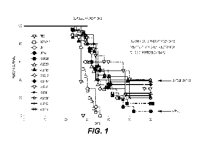
amino acids of
native VIP replaced with six highly polar N -terminal six amino acids from the
sequence of the
neurotensin peptide followed by the C-terminal 22 amino acid sequence of VIP.
With changes in
the N-terminal amino acids, VIPhyb has altered biological activity compared
with VIP and acts as
an antagonist to the VIP receptor (VIP-R) by competitively binding to the
receptor but not
signaling. .
3. Cancer treatments typically utilize surgery, chemotherapy, and radiation
therapy.
However, alternative methods of treatment that strengthen the immune system to
attack cancerous
cells are reported. These methods include collecting, amplifying, and altering
T cells in order to
target and stimulate the immune system to aggressively eliminate cancerous
cells. In chimeric
antigen receptor (CAR) T cell therapy, isolated T cell are engineered to
express chimeric protein
and are administered back into the patient. However, there is a need to
identify therapies that
improve the functional properties of T cells.
III. SUMMARY
4. Disclosed are methods and compositions related to vasoactive intestinal
peptide
(VIP) receptor antagonists for uses in managing the treatment or prevention of
cancer and viral
infections. In certain embodiments, this disclosure relates to chimeric
variants of VIP-R
antagonists, as peptides disclosed herein, and pharmaceutical composition
comprising the same.
In certain embodiments, this disclosure contemplates methods of stimulating
immune cells to
target cancer by mixing immune cells in vitro with peptides disclosed herein
and further
¨ 1 -
CA 03216694 2023- 10- 25
WO 2022/245820
PCT/US2022/029628
administering an effective amount of stimulated immune cells to a subject in
need of cancer
treatment.
5. In one aspect disclosed herein are vasoactive intestinal peptide (VIP)
receptor
antagonists comprising the amino acid
sequence
KPRRPYX1X2X3X4TX5LRKQX6AVX7X8KYLX9X10ILN (SEQ ID NO: 3), wherein Xl is T or A;
X2 is D, V, or S; X3 is N or D; X4 is Y or C;
R or S; X6 M or I; X7 is K or N; X8 is K, X9 is N
or M; and Xl is S or L; and
provided that the peptide is not
KPRRPYTDNYTRLRKQMAVKKYLNSILN (SEQ ID NO: 1) or the combination wherein X1 is
T, X2 is D, X3 is N; X4 is Y, X5 is R, X6 is M, X7 is K, X9 is N, and X10 is
S. In one aspect disclosed
herein are vasoactive intestinal peptide (VIP) receptor antagonists comprising
the amino acid
sequence KPRRPYX1X2X3X4TX5LRKQX6AVX7KYLVX9ILN (SEQ ID NO: 21), wherein Xl is
T or A; X2 is D, V. or S; X3 is N or D; X4 is Y or C; X5 R or S; X6 M or I; X7
is K or N; X8 is N
or M; and X9 is S or L; and
provided that the peptide is not
KPRRPYTDNYTRLRKQMAVKKYLNSILN (SEQ ID NO: 1) or the combination wherein X1 is
T, X2 is D, X3 is N; X4 is Y, X5 is R, X6 is M, X7 is K, X8 is N, and X9 is S.
For example, disclosed
herein are VIP-R antagonist comprising the amino acid sequence
KPRRPY ADN Y TRLRKQMA VN KYLN LILN (SEQ ID NO:
6),
KPRRPYAVNYTRLRKQIAVKKYLMSILN (SEQ ID NO:
7),
KPRRPYAVNYTRLRKQMAVNKYLMSILN (SEQ ID NO:
8),
KPRRPYADNCTRLRKQIAVNKKYLNSILN (SEQ ID NO:
9),
KPRRPYTVNYTSLRKQIAVKKYLMLILN (SEQ ID NO:
10),
KPRRPYTDNCTSLRKQIAVNKYLNLILN (SEQ ID NO:
11),
KPRRPYAVNCTSLRKQIAVNKYLNSILN (SEQ ID NO:
12),
KPRRPYAVNCTSLRKQIAVKKYLMSILN (SEQ ID NO:
13),
KPRRPYTVNCTSLRKQIAVKKYLMLILN (SEQ ID NO:
14),
KPRRPYTSDYTRLRKQMAVKKYLNSILN (SEQ ID NO:
15),
KPRRPYTSDYTRLRKQMAVKKYLNLILN (SEQ ID NO: 16), a fragment thereof, or an analog
thereof.
6. Also, disclosed herein are VIP-R antagonists of any preceding aspect,
wherein an
amino, carboxyl, hydroxyl, or thiol group in the VIP-R antagonist is
substituted.
7. In some aspects, the VIP-R antagonist is conjugated to and/or
encapsulated within
a nanoparticle.
8. Also disclosed herein are VIP-R antagonists of any preceding aspect
wherein the
VIP-R antagonist further comprises a label, e.g., fluorescent or radioactive.
¨ 2 -
CA 03216694 2023- 10- 25
WO 2022/245820
PCT/US2022/029628
9. In one aspect, disclosed herein are pharmaceutical compositions
comprising the
VIP-R antagonist of any preceding aspect and a pharmaceutically acceptable
carrier.
10. In one aspect, disclosed herein are nucleic acids encoding the VIP-R
antagonists of
any preceding aspect. Also disclosed herein are recombinant vectors comprising
said nucleic acids.
In one aspect, disclosed herein are expression systems or cells comprising a
recombinant vector
of any preceding aspect.
11_
Also disclosed herein are methods of treating, decreasing, inhibiting,
reducing,
ameliorating, and/or preventing a cancer and/or metastasis in a subject or
enhancing the immune
response to cancer and/or metastasis in a subject comprising administering to
the subject a
therapeutically effective amount of the VIP-R antagonist (such as, for
example, SEQ ID NO: 6,
SEQ ID NO: 7, SEQ ID NO: 8, SEQ ID NO: 9, SEQ ID NO: 10, SEQ ID NO: 11, SEQ ID
NO:
12, SEQ ID NO: 13, SEQ ID NO: 14, SEQ ID NO: 15, and/or SEQ ID NO: 16, a
fragment thereof,
or an analog thereof) or pharmaceutical composition of any preceding aspect.
In certain
embodiments, the VIP-R antagonist or the pharmaceutical composition is
administered in
combination with another anti-cancer agent. In some aspect, the method can
further comprise
exposing the subject to radiation and/or transplanting allogeneic
hematopoietic stem cells into the
subject and/or other adoptive cellular therapies (such as, for example,
administration of CAR '1'
cells, TCR Modified T Cells, CAR NK cells, TILs, TINKs, and/or MILs). In
certain embodiments,
the method further comprises administering to the subject a therapeutically
effective amount of a
phosphatidylinositol 3-kinase (PI3K) inhibitor (including, for example, a
PI3K3c inhibitor, a PI3KI3
inhibitor, a PI3Ko inhibitor, or a PI3Ky inhibitor). In certain embodiments,
the method further
comprises administering to the subject a therapeutically effective amount of
an immune checkpoint
blockade. In some embodiments, the immune checkpoint blockade is a PD-1
inhibitor, a PD-Li
inhibitor, or a CTLA-4 inhibitor.
12. In certain
embodiments, this disclosure relates to methods of ex vivo augmenting T
cell activation and/or expansion comprising mixing T cells with a VIP-R
antagonist of any
preceding aspect (such as, for example, SEQ ID NO: 6, SEQ ID NO: 7, SEQ ID NO:
8, SEQ ID
NO: 9, SEQ ID NO: 10, SEQ ID NO: 11, SEQ ID NO: 12, SEQ ID NO: 13, SEQ ID NO:
14, SEQ
ID NO: 15, and/or SEQ ID NO: 16, a fragment thereof, or an analog thereof). In
certain
embodiments, mixing T cells is in combination with an anti-CD3 antibody and/or
anti-CD28
antibody. In certain embodiments, the mixing T cells is in combination with a
phosphatidylinositol
3-kinase (PI3K) inhibitor (including, but not limited to, fimepinostat,
rigosertib, buparlisib,
CH5132799, pilaralisib, ZSTK474, sonolisib, pictilisib, copanlisib, B591, TG-
100-115, RIDR-PI-
103, dactolisib, apitolisib, gedatolisib, SF1126, omipalisib, samotolisib,
bimiralisib, paxalisib,
¨ 3 -
CA 03216694 2023- 10- 25
WO 2022/245820
PCT/US2022/029628
voxtalisib, G5K1059615, MEN1611, ZSTK474, as well as, isoform-specific
inhibitiors such as a
PI3Ka inhibitor (such as, for example, inavolisib, alpelisib, AZD8835,
PWT33597, taselisib,
and/or serabelisib), a PI3K13 inhibitor (such as, for example, AZD8186 and/or
GSK2636771), a
PI3K6 inhibitor (such as, for example, AZD8835, AZD8186, nemiralisib,
seletalisib, acalisib,
CAL263, TG100-115, duvelisib, idelalisib, tenalisib, taselisib, zandelisib,
AMG319, linperlisib,
parsaclisib, umbralisib, and/or leniolisib), and/or a PI3Ky inhibitor (such
as, for example,
eganelisib, tenalisib, taselisib, and/or duvelisib)). In certain embodiments,
the mixing T cells is in
combination with an immune checkpoint blockade. In some embodiments, the
immune checkpoint
blockade is a PD-1 inhibitor, a PD-Li inhibitor, or a CTLA-4 inhibitor.
13. Also
disclosed herein is a kit comprising the VIP-R antagonist of any preceding
aspect (such as, for example, SEQ ID NO: 6, SEQ ID NO: 7, SEQ ID NO: 8, SEQ ID
NO: 9, SEQ
ID NO: 10, SEQ ID NO: 11, SEQ ID NO: 12, SEQ ID NO: 13, SEQ ID NO: 14, SEQ ID
NO: 15,
and/or SEQ ID NO: 16, a fragment thereof, or an analog thereof) or the
pharmaceutical
composition of any preceding aspect and an anti-CD3 antibody and/or anti-CD28
antibody. In
certain embodiments, the kit further comprises a phosphatidylinositol 3-kinase
(PI3K) inhibitor
(including, but not limited to, fimepinostat, rigosertib, buparlisib,
CH5132799, pilaralisib,
ZSTK474, sonolisib, pictilisib, copanlisib, B591, TG-100-115, R1DR-P1-103,
dactolisib,
apitolisib, gedatolisib, SF1126, omipalisib, samotolisib, bimiralisib,
paxalisib, voxtalisib,
GSK1059615, MEN1611, ZSTK474, as well as, isoform-specific inhibitiors such as
a PI3Ka
inhibitor (such as, for example, inavolisib, alpelisib AZD8835, PWT33597,
taselisib, and/or
serabelisib), a PI3K13 inhibitor (such as, for example, AZD8186 and/or
G5K2636771), a P131(6
inhibitor (such as, for example, AZD8835, AZD8186, nemiralisib, seletalisib,
acalisib, CAL263,
TG100-115, duvelisib, idelalisib, tenalisib, taselisib, zandelisib, AMG319,
linperlisib, parsaclisib,
umbralisib, and/or leniolisib), and/or a PI3Ky inhibitor (such as, for
example, eganelisib, tenalisib,
taselisib, and/or duvelisib)). In certain embodiments, the kit further
comprises an immune
checkpoint blockade. In some embodiments, the immune checkpoint blockade is a
PD-1 inhibitor,
a PD-Li inhibitor, or a CTLA-4 inhibitor.
14. Also
disclosed herein is an in vitro cell culture composition, comprising one or
more T cells and the VIP-R antagonist of any preceding aspect (such as, for
example, SEQ ID NO:
6, SEQ ID NO: 7, SEQ ID NO: 8, SEQ ID NO: 9, SEQ ID NO: 10, SEQ ID NO: 11, SEQ
ID NO:
12, SEQ ID NO: 13, SEQ ID NO: 14, SEQ ID NO: 15, and/or SEQ ID NO: 16, a
fragment thereof,
or an analog thereof) or the pharmaceutical composition of any preceding
aspect. In some
embodiments, the in vitro cell culture composition further comprises an anti-
CD3 antibody and/or
anti-CD28 antibody. In certain embodiments, the in vitro cell culture
composition further
- 4 -
CA 03216694 2023- 10- 25
WO 2022/245820
PCT/US2022/029628
comprises a phosphatidylinositol 3-kinase (PI3K) inhibitor (including, but not
limited to,
fimepinostat, rigosertib, buparlisib, CH5132799, pilaralisib, ZSTK474,
sonolisib, pictilisib,
copanlisib, B591, TG-100-115, RIDR-PI-103, dactolisib, apitolisib,
gedatolisib, SF1126,
omipalisib, samotolisib, bimiralisib, paxalisib, voxtalisib, GSK1059615,
MEN1611, ZSTK474,
as well as, isoform-specific inhibitiors such as a PI3Ka inhibitor (such as,
for example, inavolisib,
AZD8835, PWT33597, taselisib, and/or serabelisib), a PI3Kf3 inhibitor (such
as, for
example, AZD8186 and/or GSK2636771), a PI3K6 inhibitor (such as, for example,
AZD8835,
AZD8186, nemiralisib, seletalisib, acalisib, CAL263, TG100-115, duvelisib,
idelalisib, tenalisib,
taselisib, zandelisib, AMG319, linperlisib, parsaclisib, umbralisib, and/or
leniolisib), and/or a
PI3K7 inhibitor (such as, for example, eganelisib, tenalisib, taselisib,
and/or duvelisib)). In certain
embodiments, the in vitro cell culture composition further comprises an immune
checkpoint
blockade. In some embodiments, the immune checkpoint blockade is a PD-1
inhibitor, a PD-Li
inhibitor, or a CTLA-4 inhibitor.
15. In certain
embodiments, this disclosure relates to methods of treating or preventing
graft versus host disease in a subject comprising administering an effective
amount of any VIP-R
antagonist of any preceding aspect (such as, for example, SEQ ID NO: 6, SEQ ID
NO: 7, SEQ ID
NO: 8, SEQ 11) NO: 9, SEQ Ill NO: 10, SEQ Ill NO: 11, SEQ 11) NO: 12, SEQ ID
NO: 13, SEQ
ID NO: 14, SEQ ID NO: 15, and/or SEQ ID NO: 16, or a fragment thereof) to a
subject that is to
receive or received transplanted allogeneic tissue or cells.
16. In certain
embodiments, this disclosure relates to methods of treating, reducing,
inhibiting, decreasing, ameliorating, managing, and/or preventing a microbial
infection (including,
but not limited to viral, bacterial, fungal, and/or parasitic infections)
comprising administering to
a subject infected with a microbe or at risk for a microbial infection a
therapeutically effective
amount of the VIP-R antagonist of any preceding aspect (such as, for example,
SEQ ID NO: 6,
SEQ ID NO: 7, SEQ ID NO: 8, SEQ ID NO: 9, SEQ ID NO: 10, SEQ ID NO: 11, SEQ ID
NO:
12, SEQ ID NO: 13, SEQ ID NO: 14, SEQ ID NO: 15, and/or SEQ ID NO: 16, a
fragment thereof,
or an analog thereof) or the pharmaceutical composition of any preceding
aspect.
17. Also
disclosed herein is a method of treating a cancer or a chronic infection in a
subject in need, comprising providing one or more T cells; mixing the one or
more T cells with
the VIP-R antagonist (such as, for example, SEQ ID NO: 6, SEQ ID NO: 7, SEQ ID
NO: 8, SEQ
ID NO: 9, SEQ ID NO: 10, SEQ ID NO: 11, SEQ ID NO: 12, SEQ ID NO: 13, SEQ ID
NO: 14,
SEQ ID NO: 15, and/or SEQ ID NO: 16, a fragment thereof, or an analog thereof)
or the
pharmaceutical composition of any preceding aspect thereby expanding the one
or more T cells;
and administering a therapeutically effective amount of the expanded T cells
to the subject
- 5 -
CA 03216694 2023- 10- 25
WO 2022/245820
PCT/US2022/029628
18. In some embodiments, the method of any preceding aspect comprises
mixing the
one or more T cells is in combination with an anti-CD3 antibody and/or an anti-
CD28 antibody.
In some embodiments, the method of any preceding aspect comprises mixing the
one or more T
cells is in combination with an immune checkpoint blockade. In some
embodiment, the method of
any preceding aspect comprises mixing one or more T cells in combination with
a
phosphatidylinositol 3-kinase (PI3K) inhibitor (including, but not limited to,
fimepinostat,
rigosertib, buparlisib, CH5132799, pilaralisib, ZSTK474, sonolisib,
pictilisib, copanlisib, B591,
TG-100-115, RIDR-PI-103, dactolisib, apitolisib, gedatolisib, SF1126,
omipalisib, samotolisib,
bimiralisib, paxalisib, voxtalisib, GSK1059615, MEN1611, ZSTK474, as well as,
isoform-
specific inhibitiors such as a PI3Kcc inhibitor (such as, for example,
inavolisib, alpelisib
AZD8835, PWT33597, taselisib, and/or serabelisib), a PI3KI3 inhibitor (such
as, for example,
AZD8186 and/or GSK2636771), a P131(8 inhibitor (such as, for example, AZD8835,
AZD8186,
nemiralisib, seletalisib, acalisib, CAL263, TG100-115, duvelisib, idelalisib,
tenalisib, taselisib,
zandelisib, AMG319, linperlisib, parsaclisib, umbralisib, and/or leniolisib),
and/or a PI3K1
inhibitor (such as, for example, eganelisib, tenalisib, taselisib, and/or
duvelisib)).
19. In some embodiments, the method of any preceding aspect further
comprises
administering to the subject a PI3 kinase inhibitor, a VIP receptor
antagonist, or an immune
checkpoint blockade, or a combination thereof before, during, or after
administering the expanded
T cells. In some embodiments, the one or more T cells are derived from the
subject. In some
embodiments, the one or more T cells are engineered T cells. In some
embodiments, the one or
more T cells comprises a chimeric antigen receptor.
IV. BRIEF DESCRIPTION OF THE DRAWINGS
20. The accompanying drawings, which are incorporated in and constitute a
part of this
specification, illustrate several embodiments and together with the
description illustrate the
disclosed compositions and methods.
21. Figure 1 shows treatment with the VIP-derived novel peptides prolonged
survival
of mice engrafted with acute myeloid leukemia. B6 (CD45.2. H-2K") mice were
administered
C1498 1 x 10⁶ /mouse through tail vein. VIP novel peptides were subcutaneously
injected 10
microgram per mouse daily from day 6 following leukemia administration,
totally 7 doses.
Survival of the mice was observed daily. Data were pooled from 3 replicated
experiments.
22. Figure 2 shows treatment with the VIP-derived novel peptides prolonged
survival
of the leukemic mice. B6 (CD45.2, H-2Kb) mice were administered C1498 1 x
106/mouse through
tail vein. VIP novel peptides were subcutaneously injected 10 microgram per
mouse daily from
¨ 6 -
CA 03216694 2023- 10- 25
WO 2022/245820
PCT/US2022/029628
day6 following leukemia administration, totally 7 doses. Survival of the mice
was observed daily.
Survival of the mice were pooled from 3 replicated experiments and analyzed by
log-rank test
compared with the survival of mice treated with scrambled peptide.
23. Figure 3 shows predicted affinity and potency of competitive binding
VPAC by
VIP-derived novel peptides correlated with increasing percentages of survival
in leukemia-bearing
mice treated with VIP-derived novel peptides. We make a logarithm of the sum
of the absolute
values of the predicted binding affinity to human VPAC1 and VPAC2 for each of
the distinct VIP
novel peptides and then plot logarithm of absolute value of binding affinity
against respective
percentages of survival. Panel A: Correlation of survival with the logarithm
of the absolute value
of the predicted binding affinity to human VPAC1 , (logarithm of absolute
value of binding affinity
for VPAC1), [percentage of survival]. ANT293(scramb) (1.64)
[0], ANTO8 (1.78) [16],
VIPhyb
(1.782) [5], ANT197 (1.803)1135], ANT219 (1.841)1120], ANT195
(1.848)11401,
ANT107
(1.864)1120], ANT114 (1.867) [20], ANT203 (1.877) [25]. ANT58
(1.883)1130),
Panel B: Correlation of survival with the logarithm of the absolute value of
the predicted binding
affinity to human VPAC2. (logarithm of absolute value of binding affinity for
VPAC2)
[percentage of survival]. ANT293(scramb) (1.573) [0], ANT203 (1.707)1125],
VIPhyb (1.708)
[5], ANT107 (1.719) [20], ANTO8 (l.732)[16], AN'1'114 (1.734) [20], AN'1'58
(1.782)
[30], ANT197 (1.839)1135], ANT219 (1.839) [20], ANT195 1.854 [40]
Panel C: shows the logarithms of the absolute value of the sum of binding
affinities to VPAC1 and
VPAC2 plotted against respective survival percentages in leukemic mice treated
with each VIP-
derived novel peptide.
24. Figure 4 shows treatment with VIP derived novel peptides reduced levels
of
leukemia cells in blood of the leukemic mice. B6 SJL (CD45.1, H-21(6) mice
were administered
C1498 (CD45.2 H-2Kb)1 x 10⁶ /mouse through tail vein. VIP novel peptides were
subcutaneously
injected 10 microgram per mouse daily from day6 following leukemia
administration, totally 7
doses. The mice were bled weekly from the submandibular vein, starting from
day 6, prior to the
administration of peptides. Antibodies to CD45.2 identified myeloid leukemia
cells and antibodies
to CD45.1 identified murine leukocytes of the host.
25. Figure 5 shows that mice treated with the VIP-derived novel peptides
had a
prolonged survival from re-challenged with myeloid leukemia cells. B6 mice
were administered
either Luc-1498 1 x 10⁶ /mouse for Luc-C198 positive control or C1498 1 x 10⁶
/mouse for C1498
(luciferase) negative control through tail vein. Tumor-free residual survival
mice after day 68 were
re-challenged with Luc-1498 1 x 10⁶ /mouse through tail vein. 16 days
following leukemia re-
challenge, the mice were luminescence-imaged bi-weekly. The image exposure
time were 15
¨ 7 -
CA 03216694 2023- 10- 25
WO 2022/245820
PCT/US2022/029628
seconds except 3 minutes at day16 and day23. Data were showed with Average
Radiance
[p/s/cm2/sr]. Dead mice are signified by the white X.
26. Figure 6 shows aggregate data from mice treated with the VIP-derived
novel
peptides had a prolonged survival from re-challenge with acute myeloid
leukemia. B6 mice were
administered either Luc-1498 1 x 10⁶ /mouse for Luc-C198 positive control or
C1498 1 x 10⁶
/mouse for C1498 (luciferase-negative) positive-control through the tail vein.
Tumor-free mice
that had been previously inoculated with C1498, treated with novel VIP-R
antagonist peptides and
remained cancer-free for more than 60 days, were re-challenged with Luc-C1498.
Survival of the
mice was measured daily. The eleven mice that had survived following initial
inoculation with
C1498 leukemia and then treatment with a VIP-derived novel peptide were pooled
as follows:
ANTO8 one mouse, ANT58 three mice, ANT107 two mice, ANT195 two mice, ANT197
one
mouse, ANT300 two mice. Luciferase positive C1498 was administered to eight
control mice that
had not previously been exposed to leukemia or VIP-derived novel peptides (Luc-
C1498 control),
and luciferase-negative C1498 was administered to seven control mice that had
not previously
been exposed to leukemia or VIP-derived novel peptides (C1498 control).
27. Figure 7 shows decreased tumor volume upon ANT308+aPD-1 treatment of
C57BL/6 mice with subcutaneously implanted KPC tumors. Day 22 after tumor
implantation, 3
days after completing a 10-day course of treatment. *p<0.05 Wilcoxon signed
rank test
28. Figures 8A shows relative changes differences in the volume of Panc02
tumors
growing as a sub-cutaneous tumor, from the time that treatment was initiated
on day 10 post-
implantation to euthanasia on day 22 post-implantation, following 10 days of
treatment with daily
injections of ANT308 or scrambled peptide and injection every 3 days of anti-
PD1 monoclonal
antibody or iso- type matched antibody, from day 10 through day 19 post-
implantation. Mice were
euthanized and tumor volume measured Day 22 after tumor implantation. and 3
days after
completing a 10-day course of treatment. Figures 8B shows actual tumor volume
of the Panc02
tumors that had grown sub-cutaneously following 10 days of treatment with
daily injections of
ANT308 or scrambled peptide and every 3 days injection of anti-PD1 monoclonal
antibody or iso-
type matched antibody from day 10 through day 19 post-implantation. Mice were
euthanized and
subjected to necropsy on day 22 after tumor implantation, and 3 days after
completing a 10-day
course of treatment.
29. Figure 9 shows tumor size and growth rates at 37 days following sub-
cutaneous
KPC tumor implantation. Shown are scrambled peptide plus IgG, Ant308 plus anti-
PD1,
scrambled peptide plus anti-PD-1, Ant308 plus IgG, and Ant308 plus AMD3100.
Tumor volume
¨ 8 -
CA 03216694 2023- 10- 25
WO 2022/245820
PCT/US2022/029628
was measured with calipers. 'Star' (*) indicates mice euthanized either due to
large tumor volume
(>500mm3) or ulceration of the skin overlying the tumor.
30. Figures 10A, 10B, and 10C show that treatment of human T cells with VIP-
R
antagonists (Ant08, Ant308, Ant195) augments T cell activation, as measured
through CD69
expression. Figure OA shows the total percent of T cells positive for CD69
expression at 6hrs with
respective peptide treatments at 3uM. Figure 10B shows the total percent of
CD4+ and CD8+
subset T cells positive for CD69 expression at 24hrs with respective peptide
treatments. 'Resting'
represents group maintained in culture for 6 or 24 hours without activation.
'Activated' reflects T
cells activated with CD3 antibody coated plates with no corresponding peptide
treatment. VS1'
denotes group activated in the presence of VIP scrambled 1 peptide as a
peptide control. Mean
value from 'Activated' group shown with dotted line (---) for comparison.
Responses from same
healthy donors are shown with same color dots. Error bars are computed as the
standard error of
the mean (SEM) from 6 healthy donor samples. Figure 10C shows a representative
flow plot
showing higher proportion of CD4+CD69+ T cells in antagonist-treated group
relative to control
groups (row 1) at 24hrs from one donor (blue dots).
31. Figure 11 shows the expression of TIM3 in CD4+ and CD8+ T cells at 24
hours
post treatment.
32. Figure 12 shows the expression of CXCR4 in CD4+ and CD8+ T cells at 6
and 24
hours post treatment.
33. Figures 13A, 13B, 13C, 13D, and 13E show that mouse pancreatic cancer
cells
(KPC) express VIP receptors but their growth is not affected by treatment with
VIP-R antagonist.
Figure 13A shows a western blot showing expression of VPAC1, VPAC2, and PAC1
in human
and mouse pancreatic cancer cells. Figures 13B, 13C, and 13D show the
expression ratio of
VPAC1 (Figure 13B), VPAC2 (Figure 13C), and PAC1 (Figure 13D) relative to
GAPDH in each
cell line. Figure 13E shows the viability of the cells relative to a control
with increasing
concentration of a CIP antagonist.
34. Figures 14A, 14B, and 14C show that the VIP-R antagonist ANT308 plus
anti-PD1
antibody treatment promoted infiltration of adoptively transferred T cells
into pancreatic tumors.
Figure 14A shows the experimental scheme. Figures 14B and 14C show
immunohistochemistry
images from tumors in untreated (Figure 14B) and treated (Figure 14C) mice.
35. Figure 15 shows a survival curve in mice with and without treatment
following
implantation of pancreatic cancer (KPC) tumors.
36. Figures 16A, 16B, 16C, 16D, 16E, 16F, and 16G shows that VIP is over-
expressed
by PDAC. (Figure 16A) VIP mRNA expression levels in various solid
malignancies, as obtained
¨ 9 -
CA 03216694 2023- 10- 25
WO 2022/245820
PCT/US2022/029628
from TCGA. (Figure 16B) Representative images of human PDAC tumor stained with
antibodies
to VIP (green) and CK19 (red), showing higher VIP expression in cancer
epithelial cells compared
to adjacent normal epithelial cells. Scale bars represent 20itim. Levels of
VIP in (Figure 16C)
culture supernatants collected from murine and human PDAC cell hues cultured
for 24 hours (n=3
per cell line) were compared to culture supernatants from B 16F10 and D4M
melanoma cells;
(Figure 16D) plasma of mice bearing melanoma or PDAC tumors (n=5) compared to
plasma of
non-tumor- bearing mice; (Figure 16E) plasma of PDAC patients (n=19) compared
to that from
healthy volunteers (n=26); (Figure 16F) plasma from one C57BL/6 mice bearing
subcutaneous
KPC.Luc tumor isolated at different tumor volumes; and (Figure 16G) culture
supernatants from
primary CAFs isolated from human PDAC tumors (n=9) and PSCL-12 cell line
(n=3). p values in
Figures 16C and 16D were calculated using ANOVA and Dunnett's post-hoc test,
where the means
were compared to Bl6F10. p values in e were calculated by student t-test.
Error bars show mean
SEM. **p<0.01, ***p<0.001 and ****p<0.0001.
37. Figures 17A, 17B, 17C, 17D, 17E, 17F, 17G, 17H, and 171 show that
inhibition of
VIP-R signaling decreases T cell exhaustion marker expression in cultured
human T cell. (Figure
17A) Representative western blot and (Figure 17B) quantified expression levels
of VPAC1 and
VPAC2; and (Figure 17C) PD-1 and CTLA-4 in lysates of healthy human '1' cells
expanded with
plate bound human anti-CD3 antibodies for 0, 3, 6, 12, 24, 48 and 72 hours.
Lower molecular
weight band of 25kD shown for VPAC2-specific expression as confirmed from our
VPAC2 KO
model. Percentage of (Figure 17D) CD69 expression at 24 h post activation
(Figure 17E)
phosphorylation of CREB (phospho-CREB) downstream of VPAC1/2 receptor at 6 h
and in CD4+
and CD8+ T cells normalized to levels in control with no peptide. PDAC patient
peripheral blood
T cells were expanded for 9 days with plate bound human anti-CD3 antibodies +/-
ANT008 and
the (Figure 17F) percentage of Tregs was quantified using the (Figure 17G)
gating strategy shown.
Percentage of PD 1+, Tim- 3+, Lag3 +, PD1+Tim-3+, PD1+Lag3+ and PDI+Tim-3+Lag -
3+ (triple
positive) in (Figure 17H) CD4+ and (Figure 171) CD8+ subsets is shown.
Statistical differences in
Figures 17D and 17E were calculated via repeated measures ANOVA followed by
Dunnett's post-
test where each sample in the treatment group was compared to the matched
sample in the control
group (scrambled treated). Statistical differences in Figures 17F, 17H, and
171 were calculated via
paired student T-test. Error bars show mean SEM. *p<0.50, **p<0.01 and
***p<0.001.
38. Figures 18A, 18B, 18C, 18D, and 18E show improved survival in PDAC-
bearing
mice treated with the combination of VIP-R antagonists and anti-PD-1 is T cell
dependent. (Figure
18A) Kaplan-Meier survival plots of C57BL/6 mice with subcutaneously implanted
KPC.Luc,
MT5 or Panc02 tumors stratified by treatment. (Figure 18B) Spider plots for
KPC.Luc
¨ 10 -
CA 03216694 2023- 10- 25
WO 2022/245820
PCT/US2022/029628
corresponding to results in a. as measured by Vernier calipers following
subcutaneous tumor
implantation in 4 different treatment groups. Median tumor volumes represented
as dashed gray
line (----). In a and b, tumor cells were implanted in female or male mice
with males receiving 20 mg
of ANT308 due to higher body weight compared to female mice. Kaplan-Meier
survival plots of
(Figure 18C) C57BL/6 mice receiving monoclonal CD4 and/or CD8 monoclonal
antibodies
(Figure 18D) CD4K0 or (Figure 18E) CD8K0 mice compared to wild-type CD57BL/6
mice with
subcutaneously implanted KPC.Luc tumors, stratified by treatment. Statistical
differences for
Kaplan-Meier curves are calculated via Log-rank test. *p<0.05, **p<0.01 and
***p<0.001,
****p<0.0001.
39. Figures 19A, 19B, 19C, 19D, 19E, 19F, 19G, 19H and 191 show increased T
cell
activation and reduced frequency of Tregs in KPC.Luc tumors treated with a
combination of VIP-
R antagonist and anti-PD-1. Subcutaneous KPC.Luc tumors in C57BL/6 mice
treated with
ANT008 and/or anti-PD-1 (n=5 per treatment group), were analyzed via flow
cytometry 10 days
after treatment for proportion of (Figure 19A) CD4+ and (Figure 19B) CD8+ T
cells expressing
Ki67, IFN gamma, IL-4, PD-1 and Tim-3. (Figure 19C) Representative flow plots
showing the
gating strategy used to quantify CD25+ FoxP3+ Tregs. (Figure 19D) Percentage
of Tregs in tumors
of the different treatment groups (n=5 per treatment group). Volcano plot
showing differential
expression of genes in T cells from (Figure 19E) ANTON+ isotype IgG (IgG) vs
scrambled
peptide (Scram) + isotype IgG, (Figure 19F) scrambled peptide +anti-PD-1 vs
scrambled peptide
+ isotype IgG and (Figure 19G) ANT008+anti-PD-1 vs scrambled peptide + isotype
IgG (n=3
mice per treatment group). Horizontal black line represents false discovery
rate (FDR) < 0.1.
Genes that are associated with TCR activation and co-stimulation and are at
levels significantly
higher when compared to Scram+ isotype IgG (FDR<0.1) are labeled in red.
(Figure 19H) Heat
map showing gene expression changes in genes associated with TCR activation
and co-
stimulation. (Figure 191) TCR activation and co-stimulation pathway score
between the T cells in
tumors of mice from the different treatment groups. Statistical differences in
Figure 19A, 19B,
19D and 191 were calculated via ANOVA followed by Dunnett's post-test. Error
bars show mean
SEM *p<0.05, **p<0.001, ***p<0.0001.
40. Figures 20A,
20B, 20C, 20D, 20E, 20F, and 20G show that combination therapy
with VIP-R antagonist and anti-PD-1 increased frequency of tetramer+, CD8+ T
cells within the
tumor and provide protective immunity to tumor re- challenge. (Figure 20A) Box
and whiskers
plot showing Shannon's Entropy in T cells of KPC.Luc tumors in each treatment
group. (Figure
20B) List showing TCR-13 amino acid sequences shared between samples of each
treatment group
and (Figure 20C) the frequencies of the shared clones in each treatment group_
Sequences are color
¨ 11 -
CA 03216694 2023- 10- 25
WO 2022/245820
PCT/US2022/029628
coded to represent number of mice per group (n=4) that share the specific TCR-
I3 clone. CD8+ in
subcutaneous KPC .Luc tumors were stained with MuLV p15E-H2Kb tetramer after
10 days of
treatment with ANT308 and/or anti-PD-1 (n=3 per treatment group) using the
(Figure 20D) gating
strategy and (Figure 20E) quantified. (Figure 20F) Kaplan-Meier survival
curves of subcutaneous
KPC.Luc bearing mice treated with ANT008/ANT308 and/or anti-PD-1 from day 3-12
after tumor
implantation (n=16 per scrambled peptide + isotype IgG, n=20 in ANT008/ANT308+
isotype IgG
and in scrambled peptide + anti-PD-1 treatment groups; n=23 in ANT008/ANT308 +
anti-PD-1
treatment group). (Figure 20G) Kaplan-Meier survival curves of tumor free mice
from Figure 20F
that were rechallenged with KPC.Luc tumors on the opposite flank (n=6 in
scrambled peptide +
anti-PD-1 treatment group; n=8 in ANT008/ANT308+anti-PD-1 treatment group).
Naive
C57BL/6 mice were inoculated with tumor cells at the same time of rechallenge
(n=7). Statistical
differences in a and e were calculated via ANOVA followed by Dunnett's post-
test and in Figure
20F and Figure 20G were calculated using Log-rank test. Error bars show mean
SEM *p<0.05,
**p<0.01, ****p<0.0001.
41. Figures 21A, 21B, 21C, 21D, 21E, 21F, 21G, 21H and 211 show that
synergism
between ANT008 and anti-PD-1 increases T cell infiltration and proliferation
and decreases tumor
burden in orthotopic KPC.Luc murine PDAC. KPC.Luc cells were orthotopically
implanted in the
tail of the pancreas of C57BL/6 mice and treated with ANT008 and/or anti-PD-1
with n=9, 10, 8
and 11 in scrambled+IgG, ANT008+IgG, scrambled+anti-PD-1 and ANT008+anti-PD-1,
respectively. (Figure 21A) Schematic showing orthotopic implantation of
KPC.Luc cells and
treatment strategy with ANT008 and/or anti-PD-1. (Figure 21B) Waterfall plot
showing % change
in tumor flux on day 22 relative to day 7 prior to start of treatment. (Figure
21C) Total flux as
measured by IVIS bioluminescent imaging in the different treatment groups.
Isoflurane was used
for anesthesia for bioluminescent imaging. Median flux represented as dashed
gray line (----). Cross
symbol (+) represents mice that were euthanized before day 25 due to
ulceration of the tumor and
circle symbol(o) represent mouse that were imaged on day 26 via MRI imaging
shown in Figure
30. (Figure 21D) Bar graph showing weight of pancreas on day 25 when the mice
were euthanized.
'Star' shaped (*) data points indicate tumor free mice and dotted horizontal
line (----) represents
the average weight of healthy pancreas from naïve mice. (Figure 21E)
Representative multiplex
IHC images (right) showing pancreatic tumors stained for DAPI (blue), CD4
(yellow), CD8 (red)
and Ki67 (cyan) and trichrome staining (left) with black arrows showing blue
collagen stain in the
tissue. Bar plot showing number of (Figure 21F) CD4+ or (21G) CDR+ T
cells/mm2; and (Figure
21H) Ki67+ CD4+ or (Figure 211) Ki67+ CD8+ T cells/mm2. P values in Figure 21D
were
¨ 12 -
CA 03216694 2023- 10- 25
WO 2022/245820
PCT/US2022/029628
calculated using student ANOVA followed by Dunnett's post hoc test (comparing
each treatment
group with Scram+IgG). Error bars show mean SEM. *p<0.05, **p<0.01.
42. Figures 22A, 22B, 22C, 22E, 22F show that combination therapy with VIP-
R
antagonist and anti-PD-1 promotes intratumoral T cell infiltration and
decreases CXCR4
expression on T cells in tumor draining lymph nodes. KPC.Luc tumors were
subcutaneously
implanted in C57BL/6 mice. On day 15 after tumor implantation, GFP+ T cells
were adoptively
transferred (via tail vein injections) treated with ANT308+/- aPD-1 for 3
days. (Figure 22A)
Schematic showing GFP+ T cell transfer and treatment strategy in mice with
subcutaneous
KPC.Luc tumors. (Figure 22B) Representative Hoescht (blue for nucleus) stained
tumor tissues
from tumors of each treatment group. Zoom in of two regions of interest (R01)
labelled as ROI-1
and ROI-2 in the original image of tumors of mice treated with ANT308+aPD- is
also shown.
Percentage of (Figure 22C) CXCR4+CD69+ and (Figure 22D) CXCR4+Ki67+ cells in
CD4+
(left) and CD8+ (right) subsets of T cells. (Figure 22E) Tumor growth rate and
(Figure 22F)
survival curves generated from mice with subcutaneous KPC.Luc tumors that were
treated with
scrambled peptide, IgG and PBS or ANT308 and aPD-1 or AMD3100 and aPD-1 or
ANT308 and
AMD3100 or a combination of ANT308, aPD-1 and AMD3100. Median tumor volume
represented as dashed gray line (----). Statistical differences in c and d
were determined via repeated
measures ANOVA and Dunnett's post-test with n=4-5 mice per group. Statistical
differences in
22E were determined via Log-rank test (n = 9-10 mice per group). Straight
lines in c and d show
mean. *p<0.05, **p<0.01, **p<0.001, p<0.0001.
43. Figures 23A, 23B, 23C, and 23D show that PDAC cell lines and human PDAC
tissues express VIP and receptors for VIP (Figure 23A) Representative images
of one human
PDAC tumor stained with antibodies to VIP (green), CK19 (red) and merged
(yellow) showing
VIP expression in cancer epithelial cells. Scale bars represent 2001.1m.
(Figure 23B) Representative
western blot of lysates from murine melanoma; and murine and human PDAC cell
lines probed
for VPAC1, VPAC2 and GAPDH as control. (Figure 23C) VPAC1, (Figure 23D) VPAC2
protein
bands from western blot were analyzed by densitometric analysis and normalized
against the
intensity of GAPDH. Results are the mean SEM of three independent
experiments. P values in
Figure 23C were determined by ANOVA followed by Dunnett's post-test. *p<0.05,
***p<0.001.
44. Figures 24A, 24B, 24C, 24D, 24E, 24F, 24G, and 24H show that absence of
VPAC2 receptor on PDAC cells confer limited autocrine effect on the growth of
cancer cells in
vitro and in vivo. (Figure 24A) Percentage viability of murine (MT5, KPC.Luc,
Panc02) and
human (Capan02, BxPC3) PDAC cell lines cultured in the presence of different
concentrations
ranging from 0-5jiM of ANT008 for 72 hours is plotted. Confirmation of CRISPR-
Cas9 KO of
¨ 13 -
CA 03216694 2023- 10- 25
WO 2022/245820
PCT/US2022/029628
VIPR2 encoding VPAC2 receptor via (Figure 24B) western blot; (Figure 24C) RT-
PCR using
primers targeting exon 9-12 downstream of targeted site and (Figure 24D)
Sanger Sequencing
showing the validation of in-del mutation in exon 2. In vitro MTT assay
showing (Figure 24E)
Proliferation of WT and KO cells over 72 hours; (Figure 24F) Percent viability
of wild type (WT)
and VPAC2 KO (KO) Panc02 cells treated with ANT008 and ANT308 at 3[1,M for 72
hours.
(Figure 24G) Tumor growth curve of WT versus KO Panc02 cells in C57BL/6 mice
following
subcutaneous tumor implantation. Values represent median tumor volume 95%
confidence
interval. (Figure 24H) Kaplan-Meier survival plots corresponding to results in
Figure 24G. Median
survival time for WT is 21 days and 28 days for VPAC2 KO. Error bars represent
mean and
standard deviation. *p<0.05, **p<0.01.
45. Figures 25A,
25B, and 25C show gating strategy for flow cytometric analysis of
healthy human T cells. (Figure 25A) Cells were gated as 111' by plotting
forward scatter height
(FSC-H) and side scatter height (SSC-H). Singlet from P1 was selected by
gating along the
diagonal on forward scatter height (FSC-H) versus forward scatter area (FSC-A)
plot. Live cells
from singlets were selected by plotting live/dead versus FSC-A. The live cells
were then plotted
on CD4 versus CD8 plot, to identify CD4+ and CD8+ T cells. CD69 expressing T
cells in (Figure
2511) CD4+ and (Figure 25C) CD8+ subsets were then identified by plotting each
subset on
CD4/CD8 versus CD69 flow plots. Percentage of CD69+ T cells within each subset
is shown in
shown in red.
46. Figures 26A,
2611, 26C, and 26D show gating strategy for flow cytometric analysis
of CREB phosphorylation in T cells. (Figure 26A) Plots for Forskolin-treated
human T cells used
as positive control for gating phospho-CREB positive cells. T cells were
treated with forskolin at
301LIM on ice for 30 mins and stained for the surface expression of CD4 and
CD8, followed by
intracellular staining with anti-phospho-CREB (S133) antibody. Representative
plots for phospho-
CREB expression in (Figure 26B) CD4+ and (Figure 26C) CD8+ human T cells when
treated with
scrambled peptide (Scram), ANT008 and ANT308 at 31.(M for 6h (Figure 26D)
Percentage of
CD3+phospho-CREB+ in murine T cells under similar conditions as in Figure 26C.
47. Figure 27A,
27B, 27C, and 27D show gating strategy for PD-1, Tim-3 or Lag-3
expression on PDAC patient CD4+ or CD8+ T cells expanded ex-vivo over 9 days.
(Figure 27A)
Cells were gated as `131' by plotting forward scatter area (FSC-A) and side
scatter area (SSC-A).
Singlet from P1 was selected by gating along the diagonal on forward scatter
height (FSC-H)
versus forward scatter area (FSC-A) plot. Live cells from singlets were
selected by plotting
live/dead versus FSC-A. The live cells were then plotted on CD3 versus FSC-A
plot, and the cells
that are positive for CD3 were gated as T cells. CD4+ and CD8+ T cells were
then discriminated
¨ 14 -
CA 03216694 2023- 10- 25
WO 2022/245820
PCT/US2022/029628
by plotting T cells on CD4 versus CD8 plot. (Figure 27B) PD-1+, (Figure 27C)
Tim-3+ and (Figure
27D) Lag-3+ cells were gated on CD4+ (top) or CD8+ (bottom) T cells based on
FNIO controls.
48. Figures 28A, 28B, 28C, 28D, and 28E show that combination therapy with
VIP-R
antagonist and anti-PD-1 reduces tumor burden and improves survival in male
and female
C57BL/6 mice with KPC tumors. Boxplot showing tumor volumes of MT5 (Figure
28A); KPC-
Luc (Figure 28B) and Panc02 (Figure 28C) tumor volumes as measured by Vernier
calipers on
day 22 for MT5 and day 22 for KPC and Panc02 after subcutaneous tumor
implantation. Kaplan-
Meir survival curve of (Figure 28D) female or (Figure 28E) male C57BL/6 mice
subcutaneously
implanted with KPC.Luc tumors and treated with ANT308 (female: 10mg, male:
2011g) and/or anti-
PD-1. Statistical differences in a-c were calculated by ANOVA followed by
Dunnett's post-test.
Solid line shows median with in each treatment group. Statistical differences
in d and e are
calculated via Log-rank test. *p<0.05, **p<0.01 and ***p<0.001.
49. Figures 29A, 29B, 29C, 29D, 29E, 29F, and 29G show that administration
of
ANT008 or ANT308 showed no adverse toxicity in C57BL/6 mice. C57BL/6 mice
received daily
subcutaneous injection of ANT008 or ANT308 for 10 days (n=5 per group) and
analyzed for
evidence of toxicity on day 11. (Figure 29A) Body weight in grams during the
duration of drug
administration; (Figure 29B) Number of WBCs, RBCs and platelets in blood as
per complete blood
count (left) proportions of T cells, B cells, NK cells, DCs and MDSCs in
spleen as identified by
flow cytometry (right) are plotted. Representative H&E stained sections of
(Figure 29C) colon
(top), lungs (bottom) and (Figure 29D) liver are shown. Arrows in Figure 29D
show the focal
hepatic lesions in liver. The focal hepatic necrosis that was observed in one
of five mice in each
group is not considered as drug-related toxicity, as these lesions are
commonly observed in several
in-bred mice strains at the Jackson Laboratory. C57BL/6 mice received daily
subcutaneous
injection of 30ug of ANT308 (n=6) or a combination of 30ug of ANT308 daily
along with 200ug
of anti-PD I every 3 days (n=6), for a duration of 4 days. Mice receiving
scrambled peptide and
isotype IgG served as control (n=4). (Figure 29E) Weight of the mice, (Figure
29F) complete blood
count (CBC) and (Figure 29G) serum chemistries after 4 days of treatment are
plotted. P values in
Figures 29B, 29E, 29F, and 29G were calculated by ANOVA followed by Dunnett's
post-test.
Error bars represent mean and standard deviation. *p<0.05, **p<0.01.
50. Figures 30A, 30B, 30C, 30D, 30E, 30F, and 30G show that bioluminescent
signal
from orthotopically implanted KPC.Luc tumors positively correlated with tumor
burden and
demonstrates histologic desmoplasia. (Figure 30A) On day 26 after orthotopic
KPC.Luc tumor
implantation in C57BL/6 mice, tumor burden in representative mice indicated by
'circle' symbol
in Figure 21C, were compared via bioluminescent imaging, 1VIS imaging and H&E
staining of
¨ 15 -
CA 03216694 2023- 10- 25
WO 2022/245820
PCT/US2022/029628
formalin fixed pancreas isolated after euthanasia. For bioluminescent imaging,
isoflurane was used
for anesthesia. (Figure 30B) Total flux (p/s) as measured by bioluminescent
imaging on day 26
after tumor implantation was plotted with respect to weight of the isolated
pancreas after
euthanasia. Data points are color coded to represent mice in different
treatment groups with n=9,
10, 8 and 11 in scrambled+IgG, ANT008+IgG, scrambled+anti-PD-1 and ANT008+anti-
PD-1,
respectively. (Figure 30C) Trichrome staining showing blue collagen stains in
the tissue for
arthotopically implanted KPC.Luc tumors in all treatment groups.
Representative images for
scrambled+IgG, ANT008+IgG, scrambled+anti-PD-1 shown; ANT008+anti-PD-1 shown
in
Figure 21E. XY plot showing the correlation between number of (Figure 30D)
CD4+ or (Figure
30E) CD8+ T cells/mm2; and (Figure 30F) Ki67+ CD4+ or (Figure 30G) Ki67+ CD8+
T cells/mm2
with weight of the pancreas with n = 4 to 6 mice per group.
51. Figures 31A, 31B, and 31C show increased frequency of GFP+ T cells in
tumors
of mice treated with the combination of VIP-R antagonist and anti-PD-1 as
confirmed by flow
cytometry. (Figure 31A) Singlet from single cell suspensions prepared from
tumors of mice from
Figure 22A were gated by plotting forward scatter area (FSC-A) versus forward
scatter height
(FSC-H). Live CD45+ cells were gated by selecting CD45 positive, followed by
gating for CD3
positive cells in CD3 versus SSC-A plots. GFP+ cells were then selected by
gating UPP positive
cells based on mixed population of unstained splenocytes from naïve C57BL/6
mice and spleen
samples from GFP transgenic mice. (Figure 31B) Representative plot for
CD3+GFP+ cells for
four treatment groups (Scram+IgG, ANT308+IgG, Scram+anti-PD1, ANT308+ant-PD1)
(Figure
31C) Summary data from b showing percentage of GFP+ T cells over live CD45+ T
cells. Percent
GFP+ T cells were computed as percentage of total CD3+GFP+ events divided by
total live CD45+
events enumerated from FlowJo.
52. Figure 32 shows the experiment design of testing the effect of ANT308
alone or in
combination with anti-PD-1 on liver metastases in an intraocular melanoma
mouse model.
53. Figure 33 shows that ANT308 in combination with anti-PD-1 reduced
hepatic
metastases from intraocular melanoma mice in 2 weeks (n=4).
54. Figure 34 shows that ANT308 alone or combined with anti-PD-1 decreased
hepatic
metastases from intraocular melanoma mice in 3 weeks (n=6).
55. Figure 35 shows hepatic metastases in 2 and 3 weeks after ANT308/anti-
PD-1
(n=10).
56. Figure 36 shows that ANT308 suppressed growth of liver metastases in 3
weeks
after tumor inoculation (N=6).
¨ 16 -
CA 03216694 2023- 10- 25
WO 2022/245820
PCT/US2022/029628
57. Figure 37 shows that ANT308 inhibited angiogenesis (arrow) and growth
of liver
metastases (N=10).
58. Figure 38 shows that the size of intraocular melanoma was not affected
by either
ANT308 alone or combined anti-PD-1.
59. Figure 39 shows that VIP-R antagonist ANT308 induced dose-dependent
clearance
of C1498 leukemia and long-term survival in mice with AML.
60_ Figure 40 shows that VIP-R antagonist ANT308 induced
schedule-dependent
clearance of C1498 leukemia and long-term survival in mice.
V. DETAILED DESCRIPTION
61. Before the present compounds, compositions, articles, devices, and/or
methods are
disclosed and described, it is to be understood that they are not limited to
specific synthetic
methods or specific recombinant biotechnology methods unless otherwise
specified, or to
particular reagents unless otherwise specified, as such may, of course, vary.
It is also to be
understood that the terminology used herein is for the purpose of describing
particular
embodiments only and is not intended to be limiting.
A. Definitions
62. As used in the specification and the appended claims, the singular
forms "a," -an"
and "the" include plural referents unless the context clearly dictates
otherwise. Thus, for example,
reference to "a pharmaceutical carrier" includes mixtures of two or more such
carriers, and the
like.
63. Ranges can be expressed herein as from "about" one particular value,
and/or to
"about" another particular value. When such a range is expressed, another
embodiment includes
from the one particular value and/or to the other particular value. Similarly,
when values are
expressed as approximations, by use of the antecedent "about," it will be
understood that the
particular value forms another embodiment. It will be further understood that
the endpoints of
each of the ranges are significant both in relation to the other endpoint, and
independently of the
other endpoint. It is also understood that there are a number of values
disclosed herein, and that
each value is also herein disclosed as "about" that particular value in
addition to the value itself.
For example, if the value "10" is disclosed, then "about 10- is also
disclosed. It is also understood
that when a value is disclosed that "less than or equal to" the value, -
greater than or equal to the
value" and possible ranges between values are also disclosed, as appropriately
understood by the
skilled artisan. For example, if the value "10" is disclosed the "less than or
equal to 10" as well
as "greater than or equal to 10" is also disclosed. It is also understood that
the throughout the
application, data is provided in a number of different formats, and that this
data, represents
¨ 17 -
CA 03216694 2023- 10- 25
WO 2022/245820
PCT/US2022/029628
endpoints and starting points, and ranges for any combination of the data
points. For example, if
a particular data point "10" and a particular data point "15" are disclosed,
it is understood that
greater than, greater than or equal to, less than, less than or equal to, and
equal to 10 and 15 are
considered disclosed as well as between 10 and 15. It is also understood that
each unit between
two particular units are also disclosed. For example, if 10 and 15 are
disclosed, then 11, 12, 13,
and 14 are also disclosed.
64_ In this specification and in the claims which follow,
reference will be made to a
number of terms which shall be defined to have the following meanings:
65. "Administration" to a subject or "administering" includes any route of
introducing
or delivering to a subject an agent. Administration can be carried out by any
suitable route,
including intravenous, intraperitoneal, and the like. Administration includes
self-administration
and the administration by another. "Administration" to a subject includes any
route of introducing
or delivering to a subject an agent. Administration can be carried out by any
suitable route,
including oral, topical, intravenous, subcutaneous, transcutaneous,
transdermal, intramuscular,
intra-joint, parenteral, intra-arteriole, intradermal, intraventricular,
intracranial, intraperitoneal,
intralesional, intranasal, rectal, vaginal, by inhalation, via an implanted
reservoir, or via a
transdermal patch, and the like. Administration includes self-administration
and the administration
by another.
66. "Optional" or "optionally" means that the subsequently described event
or
circumstance may or may not occur, and that the description includes instances
where said event
or circumstance occurs and instances where it does not.
67. The term -comprising" in reference to a peptide having an amino acid
sequence
refers a peptide that may contain additional N-terminal (amine end) or C-
terminal (carboxylic acid
end) amino acids, i.e., the term is intended to include the amino acid
sequence within a larger
peptide. The term "consisting of' in reference to a peptide having an amino
acid sequence refers
a peptide having the exact number of amino acids in the sequence and not more
or having not more
than a rage of amino acids expressly specified in the claim. In certain
embodiments, the disclosure
contemplates that the "N-terminus of a peptide may consist of an amino acid
sequence," which
refers to the N-terminus of the peptide having the exact number of amino acids
in the sequence
and not more or having not more than a rage of amino acids specified in the
claim however the C-
terminus may be connected to additional amino acids, e.g., as part of a larger
peptide.
the disclosure contemplates that the "C-terminus of a peptide may consist of
an amino acid
sequence,- which refers to the C-terminus of the peptide having the exact
number of amino acids
in the sequence and not more or having not more than a rage of amino acids
specified in the claim
¨ 18 -
CA 03216694 2023- 10- 25
WO 2022/245820
PCT/US2022/029628
however the N-terminus may be connected to additional amino acids, e.g., as
part of a larger
peptide.
68. An "increase" can refer to any change that results in a greater amount
of a symptom,
disease, composition, condition or activity. An increase can be any
individual, median, or average
increase in a condition, symptom, activity, composition in a statistically
significant amount. Thus,
the increase can be a 1, 2, 3, 4, 5, 6, 7, 8, 9, 10, 15, 20, 25, 30, 35, 40,
45, 50, 55, 60, 65, 70, 75,
80, 85, 90, 95, or 100% increase so long as the increase is statistically
significant.
69. A "decrease" can refer to any change that results in a smaller amount
of a symptom,
disease, composition, condition, or activity. A substance is also understood
to decrease the genetic
output of a gene when the genetic output of the gene product with the
substance is less relative to
the output of the gene product without the substance. Also, for example, a
decrease can be a change
in the symptoms of a disorder such that the symptoms are less than previously
observed. A
decrease can be any individual, median, or average decrease in a condition,
symptom, activity,
composition in a statistically significant amount. Thus, the decrease can be a
1, 2, 3, 4, 5, 6, 7, 8,
9, 10, 15, 20, 25, 30, 35, 40, 45, 50, 55, 60, 65, 70, 75, 80, 85, 90, 95, or
100% decrease so long
as the decrease is statistically significant.
70. "Inhibit," "inhibiting," and "inhibition" mean to decrease an activity,
response,
condition, disease, or other biological parameter. This can include but is not
limited to the complete
ablation of the activity, response, condition, or disease. This may also
include, for example, a 10%
reduction in the activity, response, condition, or disease as compared to the
native or control level.
Thus, the reduction can be a 10, 20, 30, 40, 50, 60, 70, 80, 90, 100%, or ally
amount of reduction
in between as compared to native or control levels.
71. "Inhibitors" or "antagonists" of expression or of activity are used to
refer to
inhibitory molecules, respectively, identified using in vitro and in vivo
assays for expression or
activity of a described target protein, e.g., ligands, antagonists, and their
homologs and mimetics.
Inhibitors are agents that, e.g., inhibit expression or bind to, partially or
totally block stimulation
or enzymatic activity, decrease, prevent, delay activation, inactivate,
desensitize, or down regulate
the activity of the described target protein, e.g., antagonists. A control
sample (untreated with
inhibitors) are assigned a relative activity value of 100%. Inhibition of a
described target protein
is achieved when the activity value relative to the control is about 80%,
optionally 50% or 25,
10%, 5% or 1%. As used herein, the terms "VIP antagonist" or "VIP receptor
antagonist" are used
interchangeably.
72. By "reduce" or other forms of the word, such as "reducing" or -
reduction," is meant
lowering of an event or characteristic (e.g., tumor growth). It is understood
that this is typically
- 19 -
CA 03216694 2023- 10- 25
WO 2022/245820
PCT/US2022/029628
in relation to some standard or expected value, in other words it is relative,
but that it is not always
necessary for the standard or relative value to be referred to. For example,
"reduces tumor growth"
means reducing the rate of growth of a tumor relative to a standard or a
control.
73. By "prevent" or other forms of the word, such as "preventing" or
"prevention," is
meant to stop a particular event or characteristic, to stabilize or delay the
development or
progression of a particular event or characteristic, or to minimize the
chances that a particular
event or characteristic will occur. Prevent does not require comparison to a
control as it is typically
more absolute than, for example, reduce. As used herein, something could be
reduced but not
prevented, but something that is reduced could also be prevented. Likewise,
something could be
prevented but not reduced, but something that is prevented could also be
reduced. It is understood
that where reduce or prevent are used, unless specifically indicated
otherwise, the use of the other
word is also expressly disclosed.
74. The term "subject" refers to any individual who is the target of
administration or
treatment. The subject can be a vertebrate, for example, a mammal. In one
aspect, the subject can
be human, non-human primate, bovine, equine, porcine, canine, or feline. The
subject can also be
a guinea pig, rat, hamster, rabbit, mouse, or mole. Thus, the subject can be a
human or veterinary
patient. The term "patient" refers to a subject under the treatment of a
clinician, e.g., physician.
75. The term "therapeutically effective amount" refers to the amount of the
composition used is of sufficient quantity to ameliorate one or more causes or
symptoms of a
disease or disorder. Such amelioration only requires a reduction or
alteration, not necessarily
elimination.
76. The term "treatment" refers to the medical management of a patient with
the intent
to cure, ameliorate, stabilize, or prevent a disease, pathological condition,
or disorder. This term
includes active treatment, that is, treatment directed specifically toward the
improvement of a
disease, pathological condition, or disorder, and also includes causal
treatment, that is, treatment
directed toward removal of the cause of the associated disease, pathological
condition, or disorder.
In addition, this term includes palliative treatment, that is, treatment
designed for the relief of
symptoms rather than the curing of the disease, pathological condition, or
disorder; preventative
treatment, that is, treatment directed to minimizing or partially or
completely inhibiting the
development of the associated disease, pathological condition, or disorder;
and supportive
treatment, that is, treatment employed to supplement another specific therapy
directed toward the
improvement of the associated disease, pathological condition, or disorder.
¨ 20 -
CA 03216694 2023- 10- 25
WO 2022/245820
PCT/US2022/029628
77. "Biocompatible" generally refers to a material and any metabolites or
degradation
products thereof that are generally non-toxic to the recipient and do not
cause significant adverse
effects to the subject.
78. "Comprising" is intended to mean that the compositions, methods, etc.
include the
recited elements, but do not exclude others. "Consisting essentially or when
used to define
compositions and methods, shall mean including the recited elements, but
excluding other
elements of any essential significance to the combination. Thus, a composition
consisting
essentially of the elements as defined herein would not exclude trace
contaminants from the
isolation and purification method and pharmaceutically acceptable carriers,
such as phosphate
buffered saline, preservatives, and the like. "Consisting of' shall mean
excluding more than trace
elements of other ingredients and substantial method steps for administering
the compositions
provided and/or claimed in this disclosure. Embodiments defined by each of
these transition terms
are within the scope of this disclosure.
79. "Composition" refers to any agent that has a beneficial biological
effect. Beneficial
biological effects include both therapeutic effects, e.g., treatment of a
disorder or other undesirable
physiological condition, and prophylactic effects, e.g., prevention of a
disorder or other
undesirable physiological condition. The terms also encompass pharmaceutically
acceptable,
pharmacologically active derivatives of beneficial agents specifically
mentioned herein, including,
but not limited to, a vector, polynucleotide, cells, salts, esters, amides,
proagents, active
metabolites, isomers, fragments, analogs, and the like. When the term
"composition" is used, then,
or when a particular composition is specifically identified, it is to be
understood that the term
includes the composition per se as well as pharmaceutically acceptable,
pharmacologically active
vector, polynucleotide, salts, esters, amides, proagents, conjugates, active
metabolites, isomers,
fragments, analogs, etc.
80. A "control"
is an alternative subject or sample used in an experiment for
comparison purposes. A control can be "positive" or "negative."
81. "Effective
amount- of an agent refers to a sufficient amount of an agent to provide
a desired effect. The amount of agent that is "effective- will vary from
subject to subject,
depending on many factors such as the age and general condition of the
subject, the particular
agent or agents, and the like. Thus, it is not always possible to specify a
quantified "effective
amount." However, an appropriate "effective amount" in any subject case may be
determined by
one of ordinary skill in the art using routine experimentation. Also, as used
herein, and unless
specifically stated otherwise, an "effective amount" of an agent can also
refer to an amount
covering both therapeutically effective amounts and prophylactically effective
amounts. An
¨ 21 -
CA 03216694 2023- 10- 25
WO 2022/245820
PCT/US2022/029628
"effective amount" of an agent necessary to achieve a therapeutic effect may
vary according to
factors such as the age, sex, and weight of the subject. Dosage regimens can
be adjusted to provide
the optimum therapeutic response. For example, several divided doses may be
administered daily
or the dose may be proportionally reduced as indicated by the exigencies of
the therapeutic
situation.
82. A "pharmaceutically acceptable" component can refer to a component that
is not
biologically or otherwise undesirable, i.e., the component may be incorporated
into a
pharmaceutical formulation provided by the disclosure and administered to a
subject as described
herein without causing significant undesirable biological effects or
interacting in a deleterious
manner with any of the other components of the formulation in which it is
contained. When used
in reference to administration to a human, the term generally implies the
component has met the
required standards of toxicological and manufacturing testing or that it is
included on the Inactive
Ingredient Guide prepared by the U.S. Food and Drug Administration.
83. "Pharmaceutically acceptable carrier" (sometimes referred to as a
"carrier") means
a carrier or excipient that is useful in preparing a pharmaceutical or
therapeutic composition that
is generally safe and non-toxic and includes a carrier that is acceptable for
veterinary and/or human
pharmaceutical or therapeutic use. The terms "carrier" or "pharmaceutically
acceptable carrier"
can include, but are not limited to, phosphate buffered saline solution,
water, emulsions (such as
an oil/water or water/oil emulsion) and/or various types of wetting agents. As
used herein, the
term "carrier" encompasses, but is not limited to, any excipient, diluent,
filler, salt, buffer,
stabilizer, solubilizer, lipid, stabilizer, or other material well luiown in
the art for use in
pharmaceutical formulations and as described further herein.
84. "Pharmacologically active" (or simply "active"), as in a
"pharmacologically
active" derivative or analog, can refer to a derivative or analog (e.g., a
salt, ester, amide, conjugate,
metabolite, isomer, fragment, etc.) having the same type of pharmacological
activity as the parent
compound and approximately equivalent in degree.
85. "Therapeutic agent- refers to any composition that has a beneficial
biological
effect. Beneficial biological effects include both therapeutic effects, e.g.,
treatment of a disorder
or other undesirable physiological condition, and prophylactic effects, e.g.,
prevention of a
disorder or other undesirable physiological condition (e.g., a non-immunogenic
cancer). The terms
also encompass pharmaceutically acceptable, pharmacologically active
derivatives of beneficial
agents specifically mentioned herein, including, but not limited to, salts,
esters, amides, proagents,
active metabolites, isomers, fragments, analogs, and the like. When the terms
"therapeutic agent"
is used, then, or when a particular agent is specifically identified, it is to
be understood that the
¨ 22 -
CA 03216694 2023- 10- 25
WO 2022/245820
PCT/US2022/029628
term includes the agent per se as well as pharmaceutically acceptable,
pharmacologically active
salts, esters, amides, proagents, conjugates, active metabolites, isomers,
fragments, analogs, etc.
86. The term "prodrug" refers to an agent that is converted into a
biologically active
form in vivo. Prodrugs are often useful because, in some situations, they may
be easier to
administer than the parent compound. The prodrug may also have improved
solubility in
pharmaceutical compositions over the parent drug. A prodrug may be converted
into the parent
drug by various mechanisms, including enzymatic processes and metabolic
hydrolysis. Typical
prodrugs are pharmaceutically acceptable esters. Prodrugs include compounds
wherein a hydroxy,
amino or mercapto (thiol) group is bonded to any group that, when the prodrug
of the active
compound is administered to a subject, cleaves to form a free hydroxy, free
amino or free mercapto
group, respectively. Examples of prodrugs include, but are not limited to,
acetate, formate and
benzoate derivatives of an alcohol or acetamide, formamide and benzamide
derivatives of an
amine functional group in the active compound and the like.
87. "Therapeutically effective amount" or "therapeutically effective dose"
of a
composition (e.g. a composition comprising an agent) refers to an amount that
is effective to
achieve a desired therapeutic result. In some embodiments, a desired
therapeutic result is the
control of tumor growth. In some embodiments, a desired therapeutic result is
the control of
metastasis. In some embodiments, a desired therapeutic result is prevention of
relapse.
Therapeutically effective amounts of a given therapeutic agent will typically
vary with respect to
factors such as the type and severity of the disorder or disease being treated
and the age, gender,
and weight of the subject. The term can also refer to an amount of a
therapeutic agent, or a rate of
delivery of a therapeutic agent (e.g., amount over time), effective to
facilitate a desired therapeutic
effect, such as pain relief. The precise desired therapeutic effect will vary
according to the
condition to be treated, the tolerance of the subject, the agent and/or agent
formulation to be
administered (e.g., the potency of the therapeutic agent, the concentration of
agent in the
formulation, and the like), and a variety of other factors that are
appreciated by those of ordinary
skill in the art. In some instances, a desired biological or medical response
is achieved following
administration of multiple dosages of the composition to the subject over a
period of days, weeks,
or years.
88. The term "isolating" as used herein refers to isolation from a
biological sample,
i.e., blood, plasma, tissues, exosomes. or cells. As used herein the term
"isolated," when used in
the context of, e.g., a nucleic acid, refers to a nucleic acid of interest
that is at least 60% free, at
least 75% free, at least 90% free, at least 95% free, at least 98% free, and
even at least 99% free
from other components with which the nucleic acid is associated with prior to
purification.
¨ 23 -
CA 03216694 2023- 10- 25
WO 2022/245820
PCT/US2022/029628
89.
"Encoding" refers to the inherent property of specific sequences of
nucleotides in a
polynucleotide, such as a gene, a cDNA, or an mRNA, to serve as templates for
synthesis of other
polymers and macromolecules in biological processes having either a defined
sequence of
nucleotides (i.e., rRNA, tRNA and mRNA) or a defined sequence of amino acids
and the biological
properties resulting therefrom, Thus, a gene encodes a protein if
transcription and translation of
mRNA.
90_
The term as used herein "engineered" and other grammatical forms thereof
may
refer to one or more changes of nucleic acids, such as nucleic acids within
the genome of an
organism. The term "engineered- may refer to a change, addition and/or
deletion of a gene.
Engineered cells can also refer to cells that contain added, deleted, and/or
changed genes.
91. "Expression vector" refers to a vector comprising a recombinant
polynucleotide
comprising expression control sequences operatively linked to a nucleotide
sequence to be
expressed. An expression vector comprises sufficient cis-acting elements for
expression; other
elements for expression can be supplied by the host cell or in an in vitro
expression system.
Expression vectors include all those known in the art, such as cosmids,
plasmids (e.g., naked or
contained in liposomes) and viruses (e.g., lentiviruses, retroviruses,
adenoviruses, and adeno-
associated viruses) that incorporate the recombinant polynucleotide.)
92. The "fragments," whether attached to other sequences or not, can
include
insertions, deletions, substitutions, or other selected modifications of
particular regions or specific
amino acids residues, provided the activity of the fragment is not
significantly altered or impaired
compared to the nonmodified peptide or protein. These modifications can
provide for some
additional property, such as to remove or add amino acids capable of disulfide
bonding, to increase
its bio-longevity, to alter its secretory characteristics, etc. In any case,
the fragment must possess
a bioactive property, such as regulating the transcription of the target gene.
93. The term
"gene" or "gene sequence" refers to the coding sequence or control
sequence, or fragments thereof. A gene may include any combination of coding
sequence and
control sequence, or fragments thereof. Thus, a "gene" as referred to herein
may be all or part of a
native gene. A polynucleotide sequence as referred to herein may be used
interchangeably with
the term "gene", or may include any coding sequence, non-coding sequence or
control sequence,
fragments thereof, and combinations thereof. The term "gene" or ''gene
sequence" includes, for
example, control sequences upstream of the coding sequence (for example, the
ribosome binding
site).
94.
The terms "identical" or percent "identity," in the context of two or
more nucleic
acids or polypeptide sequences, refer to two or more sequences or subsequences
that are the same
¨ 24 -
CA 03216694 2023- 10- 25
WO 2022/245820
PCT/US2022/029628
or have a specified percentage of amino acid residues or nucleotides that are
the same (i.e., about
60% identity, preferably 61%, 62%, 63%, 64%, 65%, 66%, 67%, 68%, 69%, 70%,
71%, 72%,
73%, 74%, 75%, 76%, 77%, 78%, 79%, 80%, 81%, 82%, 83%, 84%, 85%, 86%, 87%,
88%, 89%,
90%, 91%, 92%, 93%,94%, 95%, 96%, 97%, 98%, 99% or higher identity over a
specified region
when compared and aligned for maximum correspondence over a comparison window
or
designated region) as measured using a BLAST or BLAST 2.0 sequence comparison
algorithms
with default parameters described below, or by manual alignment and visual
inspection (see, e.g.,
NCBI web site or the like). Such sequences are then said to be "substantially
identical." This
definition also refers to, or may be applied to, the compliment of a test
sequence. The definition
also includes sequences that have deletions and/or additions, as well as those
that have
substitutions. As described below, the preferred algorithms can account for
gaps and the like.
Preferably, identity exists over a region that is at least about 10 amino
acids or 20 nucleotides in
length, or more preferably over a region that is 10-50 amino acids or 20-50
nucleotides in length.
As used herein, percent (%) nucleotide sequence identity is defined as the
percentage of amino
acids in a candidate sequence that are identical to the nucleotides in a
reference sequence, after
aligning the sequences and introducing gaps, if necessary, to achieve the
maximum percent
sequence identity. Alignment for purposes of determining percent sequence
identity can be
achieved in various ways that are within the skill in the art, for instance,
using publicly available
computer software such as BLAST, BLAST-2, ALIGN, ALIGN-2 or Megalign (DNASTAR)
software. Appropriate parameters for measuring alignment, including any
algorithms needed to
achieve maximal alignment over the full-length of the sequences being compared
can be
determined by known methods.
95. For sequence
comparisons, typically one sequence acts as a reference sequence, to
which test sequences are compared. When using a sequence comparison algorithm,
test and
reference sequences are entered into a computer, subsequence coordinates are
designated, if
necessary, and sequence algorithm program parameters are designated.
Preferably, default
program parameters can be used, or alternative parameters can be designated.
The sequence
comparison algorithm then calculates the percent sequence identities for the
test sequences relative
to the reference sequence, based on the program parameters.
96. One example of an algorithm that is suitable for determining percent
sequence
identity and sequence similarity are the BLAST and BLAST 2.0 algorithms, which
are described
in Altschul et al. (1977) Nuc. Acids Res. 25:3389-3402, and Altschul et al.
(1990) J. Mol. Biol.
215:403-410, respectively. Software for performing BLAST analyses is publicly
available through
the National Center for Biotechnology Information
(http://vvvvw.ncbi.nlm.nih.gov/). This
- 25 -
CA 03216694 2023- 10- 25
WO 2022/245820
PCT/US2022/029628
algorithm involves first identifying high scoring sequence pairs (HSPs) by
identifying short words
of length W in the query sequence, which either match or satisfy some positive-
valued threshold
score T when aligned with a word of the same length in a database sequence. T
is referred to as
the neighborhood word score threshold (Altschul et al. (1990) .1. Mol. Biol.
215:403-410). These
initial neighborhood word hits act as seeds for initiating searches to find
longer HSPs containing
them. The word hits are extended in both directions along each sequence for as
far as the
cumulative alignment score can be increased. Cumulative scores are calculated
using, for
nucleotide sequences, the parameters M (reward score for a pair of matching
residues; always >0)
and N (penalty score for mismatching residues; always <0). For amino acid
sequences, a scoring
matrix is used to calculate the cumulative score. Extension of the word hits
in each direction are
halted when: the cumulative alignment score falls off by the quantity X from
its maximum
achieved value; the cumulative score goes to zero or below, due to the
accumulation of one or
more negative-scoring residue alignments; or the end of either sequence is
reached. The BLAST
algorithm parameters W, T, and X determine the sensitivity and speed of the
alignment. The
BLASTN program (for nucleotide sequences) uses as defaults a word length (W)
of 11, an
expectation (E) or 10, M=5, N=-4 and a comparison of both strands. For amino
acid sequences,
the BLASTP program uses as defaults a word length of 3, and expectation (E) of
10, and the
BLOSUM62 scoring matrix (see Henikoff and Henikoff (1989) Proc. Natl. Acad.
Sci. USA
89:10915) alignments (B) of 50, expectation (E) of 10, M=5, N=-4, and a
comparison of both
strands.
97. The BLAST algorithm also performs a statistical analysis of the
similarity between
two sequences (see, e.g., Karlin and Altschul (1993) Proc. Natl. Acad. Sci.
USA 90:5873-5787).
One measure of similarity provided by the BLAST algorithm is the smallest sum
probability
(P(N)), which provides an indication of the probability by which a match
between two nucleotide
or amino acid sequences would occur by chance. For example, a nucleic acid is
considered similar
to a reference sequence if the smallest sum probability in a comparison of the
test nucleic acid to
the reference nucleic acid is less than about 0.2, more preferably less than
about 0.01.
98. The term "nucleic acid" as used herein means a polymer composed of
nucleotides,
e.g. deoxyribonucleotides (DNA) or ribonucleotides (RNA). The terms
''ribonucleic acid" and
"RNA" as used herein mean a polymer composed of ribonucleotides. The terms
"deoxyribonucleic
acid" and "DNA" as used herein mean a polymer composed of
deoxyribonucleotides. (Used
together with "polynucleotide" and "polypeptide".)
99. Unless otherwise specified, a "nucleotide sequence encoding an amino
acid
sequence" includes all nucleotide sequences that are degenerate versions of
each other and that
¨ 26 -
CA 03216694 2023- 10- 25
WO 2022/245820
PCT/US2022/029628
encode the same amino acid sequence. The phrase nucleotide sequence that
encodes a protein or
an RNA may also include introns to the extent that the nucleotide sequence
encoding the protein
may in some version contain an intron(s).
100. As used herein, "operatively linked" can indicate that the regulatory
sequences
useful for expression of the coding sequences of a nucleic acid are placed in
the nucleic acid
molecule in the appropriate positions relative to the coding sequence so as to
effect expression of
the coding sequence_ This same definition is sometimes applied to the
arrangement of coding
sequences and/or transcription control elements (e.g. promoters, enhancers,
and termination
elements), and/or selectable markers in an expression vector. The term
"operatively linked" can
also refer to the arrangement of polypeptide segments within a single
polypeptide chain, where the
individual polypeptide segments can be, without limitation, a protein,
fragments thereof, linking
peptides, and/or signal peptides. The term operatively linked can refer to
direct fusion of different
individual polypeptides within the single polypeptides or fragments thereof
where there are no
intervening amino acids between the different segments as well as when the
individual
polypeptides are connected to one another via one or more intervening amino
acids.
101. The term "polynucleotide" refers to a single or double stranded polymer
composed
of nucleotide monomers.
102. The term "polypeptide" refers to a compound made up of a single chain of
D- or L-
amino acids or a mixture of D- and L-amino acids joined by peptide bonds.
103. The terms "peptide," "protein," and "polypeptide" are used
interchangeably to refer
to a natural or synthetic molecule comprising two or more amino acids linked
by the carboxyl
group of one amino acid to the alpha amino group of another.
104. The term "promoter" as used herein is defined as a DNA sequence
recognized by
the synthetic machinery of the cell, or introduced synthetic machinery,
required to initiate the
specific transcription of a polynucleotide sequence.
105. As used herein, the term "promoter/regulatory sequence" means a nucleic
acid
sequence which is required for expression of a gene product operably linked to
the
promoter/regulatory sequence. In some instances, this sequence may be the core
promoter
sequence and in other instances, this sequence may also include an enhancer
sequence and other
regulatory elements which are required for expression of the gene product. The
promoter/regulatory sequence may, for example, be one which expresses the gene
product in a
tissue specific manner.
106. The term "variant" as used herein refers to a polypeptide or
polynucleotide that
differs from a reference polypeptide or polynucleotide, but retains essential
properties. A typical
¨ 27 -
CA 03216694 2023- 10- 25
WO 2022/245820
PCT/US2022/029628
variant of a polypeptide differs in amino acid sequence from another,
reference, polypeptide.
Generally, differences are limited so that the sequences of the reference
polypeptide and the variant
are closely similar overall (homologous) and, in many regions, identical. A
variant and reference
polypeptide may differ in amino acid sequence by one or more modifications
(e.g., substitutions,
additions, and/or deletions).
107. The term "cancer" as used herein is defined as disease characterized by
the rapid
and uncontrolled growth of aberrant cells. Cancer cells can spread locally or
through the
bloodstream and lymphatic system to other parts of the body, Examples of
various cancers include
but are not limited to, breast cancer, prostate cancer, ovarian cancer,
cervical cancer, skin cancer,
pancreatic cancer, colorectal cancer, renal cancer, liver cancer, brain
cancer, lymphoma, leukemia,
lung cancer and the like.
108. As used herein, the term "metastasis" is meant to refer to the process in
which
cancer cells originating in one organ or part of the body, with or without
transit by a body fluid,
and relocate to another part of the body and continue to replicate.
Metastasized cells can
subsequently form tumors which may further metastasize. Metastasis thus refers
to the spread of
cancer, from the part of the body where it originally occurred, to other parts
of the body.
109. 'throughout this application, various publications are referenced. 'Me
disclosures
of these publications in their entireties are hereby incorporated by reference
into this application
in order to more fully describe the state of the art to which this pertains.
The references disclosed
are also individually and specifically incorporated by reference herein for
the material contained
in them that is discussed in the sentence in which the reference is relied
upon.
B. Compositions
110. Disclosed are the components to be used to prepare the disclosed
compositions as
well as the compositions themselves to be used within the methods disclosed
herein. These and
other materials are disclosed herein, and it is understood that when
combinations, subsets,
interactions, groups, etc. of these materials are disclosed that while
specific reference of each
various individual and collective combinations and permutation of these
compounds may not be
explicitly disclosed, each is specifically contemplated and described herein.
For example, if a
particular VIP-R antagonist is disclosed and discussed and a number of
modifications that can be
made to a number of molecules including the VIP-R antagonist are discussed,
specifically
contemplated is each and every combination and permutation of VIP-R antagonist
and the
modifications that are possible unless specifically indicated to the contrary.
Thus, if a class of
molecules A, B, and C are disclosed as well as a class of molecules D, E, and
F and an example
of a combination molecule, A-D is disclosed, then even if each is not
individually recited each is
¨ 28 -
CA 03216694 2023- 10- 25
WO 2022/245820
PCT/US2022/029628
individually and collectively contemplated meaning combinations, A-E, A-F, B-
D, B-E, B-F, C-
D, C-E, and C-F are considered disclosed. Likewise, any subset or combination
of these is also
disclosed. Thus, for example, the sub-group of A-E, B-F, and C-E would be
considered disclosed.
This concept applies to all aspects of this application including, but not
limited to, steps in methods
of making and using the disclosed compositions. Thus, if there are a variety
of additional steps
that can be performed it is understood that each of these additional steps can
be performed with
any specific embodiment or combination of embodiments of the disclosed
methods.
111. The terms "vasoactive intestinal peptide" and "VIP" refer to
HSDAVFTDNYTRLRKQMAVKKYLNSILN (SEQ ID NO: 2) unless the context suggests
otherwise. VIP is a multifunctional endogenous polypeptide that modulates both
innate and
adaptive immunity at multiple levels of immune cell differentiation and
activation. VIP is typically
secreted by a variety of cells such as neurons (in both the central and
peripheral nervous systems)
B-cells, T-cells, and accessory cells. VIP and the closely related
neuropeptide pituitary adenylyl
cyclase-activating polypeptide (PACAP) bind to three known receptors-VPAC1,
VPAC2, and
PAC. It is believed that T-cells and dendritic cells (DC) express VPAC1 and
VPAC2, but not
PAC. PAC1 is mainly expressed on neuron and endocrine cells in the brain and
pituitary and
adrenal glands, and in most forms selectively binds PACAP.
112. Some cancers are caused by viruses, and traditional vaccines against
those viruses,
such as HPV vaccine and Hepatitis B vaccine, will prevent those cancers. It is
contemplated that
peptide disclosed herein can be administered in combination with these
vaccines to improve
treatment efficacy.
113. It is believed that cancer cells arise and are destroyed by the immune
system, and
that cancer forms when the immune system fails to destroy them. One approach
to cancer
vaccination is to separate proteins from cancer cells and immunize cancer
patients against those
proteins, stimulating an immune reaction that kills the cancer cells. Cancer
vaccines are
contemplated for the treatment of acute myeloid leukemia, multiple myeloma,
lymphoma, breast,
lung, colon, skin, kidney, prostate, and other cancers.
114. In certain embodiments, the disclosure relates to treating cancers by
administering
any vasoactive intestinal peptide receptor (VIP-R) antagonist disclosed herein
(such as, for
example. SEQ ID NO: 6, SEQ ID NO: 7, SEQ ID NO: 8, SEQ ID NO: 9, SEQ ID NO:
10, SEQ
ID NO: 11, SEQ ID NO: 12, SEQ ID NO: 13, SEQ ID NO: 14, SEQ ID NO: 15, and/or
SEQ ID
NO: 16, a fragment thereof, or an analog thereof) in combination with cancer
antigens. Other VIP-
R antagonists or VIP antagonists are also reported in U.S. Patent Nos.
6,630,124 and 5,217,953,
which are incorporated herein by reference in their entireties.
¨ 29 -
CA 03216694 2023- 10- 25
WO 2022/245820
PCT/US2022/029628
115. Prevention of the action of microorganisms may be controlled by addition
of any
of various antibacterial and antifungal agents, for example, parabens,
chlorobutanol, phenol, sorbic
acid, and the like. It may also be desirable to include isotonic agents, for
example sugars, sodium
chloride, and the like. Prolonged absorption of the injectable pharmaceutical
form can be brought
about by the use of agents delaying absorption, for example. aluminum
monostearate and gelatin.
116. It is contemplated that any of the peptides disclose herein may be
modified with
hydrocarbon or polyethylene glycol groups in order to provide improve
properties such as
solubility, bioavailability, and/or biological degradation.
117. In certain embodiments, this disclosure relates to methods of coupling
any
vasoactive intestinal peptide receptor (VIP-R) antagonist disclosed herein
(such as, for example,
SEQ ID NO: 6, SEQ ID NO: 7, SEQ ID NO: 8, SEQ ID NO: 9, SEQ ID NO: 10, SEQ ID
NO: 11,
SEQ ID NO: 12, SEQ ID NO: 13, SEQ ID NO: 14, SEQ ID NO: 15, and/or SEQ ID NO:
16, a
fragment thereof, or an analog thereof, as well as, any VIP-R antagonist/VIP
antagonist disclosed
in U.S. Patent Nos. 6,630,124 and 5,217,953, which are incorporated herein by
reference in their
entireties) or the nucleic acid encoding the VIP-R antagonist disclosed herein
to a nanoparticle.
Accordingly, in some embodiments, the VIP-R antagonist disclosed herein or the
nucleic acid
encoding the VIP-R antagonist disclosed herein is conjugated to and/or
encapsulated within a
nanoparticle. In certain embodiments, the nanoparticle is comprised of
poloxamer-stabilized
polypropylene sulfide. The term "nanoparticle" as used herein refers to a
particle or structure
which typically ranges from about 1 nm to about 1000 nm in size. In certain
embodiments, the
nanoparticle has a diameter of between about 10 and about 100 nm. In certain
embodiments, the
nanoparticle has a diameter between about 20 and about 50 nm, preferably about
30 nm. In certain
embodiments, the nanoparticle has a diameter from about 50 nm to about 500 nm
size, more
preferably from about 50 nm to about 350 nm size, more preferably from about
100 nm to about
250 nm size.
118. In certain embodiments, the disclosure contemplates using particles
disclosed
herein when the peptide sequence couple to the nanoparticle is contains any
VIP-R antagonist
disclosed herein (such as, for example, SEQ ID NO: 6, SEQ ID NO: 7, SEQ ID NO:
8, SEQ ID
NO: 9, SEQ ID NO: 10, SEQ ID NO: 11, SEQ ID NO: 12, SEQ ID NO: 13, SEQ ID NO:
14, SEQ
ID NO: 15, and/or SEQ ID NO: 16, as well as, any VIP-R antagonist disclosed in
U.S. Patent Nos.
6,630,124 and 5,217,953, which are incorporated herein by reference in their
entireties) plus a C-
terminal linker peptide, GGGGSC (SEQ ID NO: 22). In certain embodiments, the
chemical linkage
between the peptides disclosed herein and the nanoparticles is a disulfide
bond.
¨ 30 -
CA 03216694 2023- 10- 25
WO 2022/245820
PCT/US2022/029628
119. In certain embodiments, the disclosure relates to recombinant peptides
comprising
sequences disclosed herein or fusions thereof wherein the amino terminal end
or the carbon
terminal end of the amino acid sequence are optionally attached to a
heterologous amino acid
sequence, label, or reporter molecule. A "label" refers to a detectable
compound or composition
that is conjugated directly or indirectly to another molecule, such as an
antibody or a protein, to
facilitate detection of that molecule. Specific, non-limiting examples of
labels include fluorescent
tags, enzymatic linkages, and radioactive isotopes. In one example, a "label
receptor" refers to
incorporation of a heterologous polypeptide in the receptor. A label includes
the incorporation of
a radiolabeled amino acid or the covalent attachment of biotinyl moieties to a
polypeptide that can
be detected by marked avidin (for example, streptavidin containing a
fluorescent marker or
enzymatic activity that can be detected by optical or colorimetric methods).
Various methods of
labeling polypeptides and glycoproteins are known in the art and may be used.
Examples of labels
for polypeptides include, but are not limited to, the following: radioisotopes
or radionuclides (such
as 35S or 1311) fluorescent labels (such as fluorescein isothiocyanate (FITC),
rhodamine, lanthanide
phosphors), enzymatic labels (such as horseradish peroxidase, beta-
galactosidase, luciferase,
alkaline phosphatase), chemiluminescent markers, biotinyl groups,
predetermined peptide
epitopes recognized by a secondary reporter (such as a leucine zipper pair
sequences, binding sites
for secondary antibodies, metal binding domains, epitope tags), or magnetic
agents, such as
gadolinium chelates. In some embodiments, labels are attached by spacer arms
of various lengths
to reduce potential steric hindrance.
1. Peptides
a) Protein variants
120. As disclosed herein there are numerous variants of the vasoactive
intestinal peptide
receptor (VIP-R) antagonists that are known and herein contemplated. In
addition, to the known
functional variants there are derivatives of the VIP-R antagonists which also
function in the
disclosed methods and compositions. Protein variants and derivatives are well
understood to those
of skill in the art and in can involve amino acid sequence modifications. For
example, amino acid
sequence modifications typically fall into one or more of three classes:
substitutional, insertional
or deletional variants. Insertions include amino and/or carboxyl terminal
fusions as well as
intrasequence insertions of single or multiple amino acid residues. Insertions
ordinarily will be
smaller insertions than those of amino or carboxyl terminal fusions, for
example, on the order of
one to four residues. Deletions are characterized by the removal of one or
more amino acid
residues from the protein sequence. Typically, no more than about from 2 to 6
residues are deleted
at any one site within the protein molecule. These variants ordinarily are
prepared by site specific
¨ 31 -
CA 03216694 2023- 10- 25
WO 2022/245820
PCT/US2022/029628
mutagenesis of nucleotides in the DNA encoding the protein, thereby producing
DNA encoding
the variant, and thereafter expressing the DNA in recombinant cell culture.
Techniques for making
substitution mutations at predetermined sites in DNA having a known sequence
are well known,
for example M13 primer mutagenesis and PCR mutagenesis. Amino acid
substitutions are
typically of single residues, but can occur at a number of different locations
at once; insertions
usually will be on the order of about from 1 to 10 amino acid residues; and
deletions will range
about from 1 to 30 residues_ Deletions or insertions preferably are made in
adjacent pairs, i.e. a
deletion of 2 residues or insertion of 2 residues. Substitutions, deletions,
insertions or any
combination thereof may be combined to arrive at a final construct. The
mutations must not place
the sequence out of reading frame and preferably will not create complementary
regions that could
produce secondary protein structure. Substitutional variants are those in
which at least one residue
has been removed and a different residue inserted in its place. Such
substitutions generally are
made in accordance with the following Tables 1 and 2 and are referred to as
conservative
substitutions.
TABLE 1:Amino Acid Abbreviations
Amino Acid Abbreviations
Alanine Ala A
allosoleucine AIle
Arginine Arg
asparagine Asn
aspartic acid Asp
Cysteine Cys
glutamic acid Glu
Glutamine Gln
Glycine Gly
Hi stidine His
Isolelucine Ile
Leuci ne Leu
Lysine Lys
phenylalanine Phe
proline Pro
pyroglutamic acid pGlu
Serine Ser
Threonine Thr
Tyrosine Tyr
Tryptophan Trp
Valine Val V
TABLE 2:Amino Acid Substitutions
Original Residue Exemplary Conservative Substitutions,
others are known in the art.
Ala Ser
Arg Lys; Gin
Asn Gin; His
Asp Glu
Cys Ser
Gin Asn, Lys
¨ 32 -
CA 03216694 2023- 10- 25
WO 2022/245820
PCT/US2022/029628
Glu Asp
Gly Pro
His Asn;Gln
Ile Leu; Val
Leu Ile; Val
Lys Arg; Gin
Met Leu; Tie
Phe Met; Leu; Tyr
Ser Thr
Thr Ser
Trp Tyr
Tyr Tip; Phe
Val Ile; Leu
121.
Substantial changes in function or immunological identity are made by
selecting
substitutions that are less conservative than those in Table 2, i.e.,
selecting residues that differ
more significantly in their effect on maintaining (a) the structure of the
polypeptide backbone in
the area of the substitution, for example as a sheet or helical conformation,
(b) the charge or
hydrophobicity of the molecule at the target site or (c) the bulk of the side
chain. The substitutions
which in general are expected to produce the greatest changes in the protein
properties will be
those in which (a) a hydrophilic residue, e.g. seryl or threonyl, is
substituted for (or by) a
hydrophobic residue, e.g. leucyl, isoleucyl, phenylalanyl, valyl or alanyl;
(b) a cysteine or proline
is substituted for (or by) any other residue; (c) a residue having an
electropositive side chain, e.g.,
lysyl, arginyl, or histidyl, is substituted for (or by) an electronegative
residue, e.g., glutamyl or
aspartyl; or (d) a residue having a bulky side chain, e.g., phenylalanine, is
substituted for (or by)
one not having a side chain, e.g., glycine, in this case, (e) by increasing
the number of sites for
sulfation and/or glycosylation.
122. For example, the replacement of one amino acid residue with another that
is
biologically and/or chemically similar is known to those skilled in the art as
a conservative
substitution. For example, a conservative substitution would be replacing one
hydrophobic residue
for another, or one polar residue for another. The substitutions include
combinations such as, for
example. Gly, Ala; Val, Ile, Leu; Asp, Glu; Asn, Gln; Ser, Thr; Lys, Arg; and
Phe, Tyr. Such
conservatively substituted variations of each explicitly disclosed sequence
are included within the
mosaic polypeptides provided herein.
123. Substitutional or deletional mutagenesis can be employed to insert sites
for N-
glycosylation (Asn-X-Thr/Ser) or 0-glycosylation (Ser or Thr). Deletions of
cysteine or other
labile residues also may be desirable. Deletions or substitutions of potential
proteolysis sites, e.g.
Arg, is accomplished for example by deleting one of the basic residues or
substituting one by
glutaminyl or histidyl residues.
¨ 33 -
CA 03216694 2023- 10- 25
WO 2022/245820
PCT/US2022/029628
124. Certain post-translational derivatizations are the result of the action
of recombinant
host cells on the expressed polypeptide. Glutaminyl and asparaginyl residues
are frequently post-
translationally deamidated to the corresponding glutamyl and aspartyl
residues. Alternatively,
these residues are deamidated under mildly acidic conditions. Other post-
translational
modifications include hydroxylation of proline and lysine, phosphorylation of
hydroxyl groups of
seryl or threonyl residues, methylation of the o-amino groups of lysine,
arginine, and histidine side
chains (T.E. Creighton, Proteins: Structure and Molecular Properties, W. H.
Freeman & Co., San
Francisco pp 79-86 119831), acetylation of the N-terminal amine and, in some
instances, amidation
of the C-terminal carboxyl.
125. It is understood that one way to define the variants and derivatives of
the disclosed
proteins herein is through defining the variants and derivatives in terms of
homology/identity to
specific known sequences. For example, SEQ ID NO: 6, SEQ ID NO: 7; SEQ ID NO:
8, SEQ ID
NO: 9, SEQ ID NO: 10, SEQ ID NO: 11, SEQ ID NO: 12, SEQ ID NO: 13, SEQ ID NO:
14, SEQ
ID NO: 15, and SEQ ID NO: 16 set forth a particular sequence of VIP-R
antagonist. Specifically
disclosed are variants of these and other proteins herein disclosed which have
at least, 70% or 75%
or 80% or 85% or 90% or 95% homology to the stated sequence. Those of skill in
the art readily
understand how to determine the homology of two proteins. For example, the
homology can be
calculated after aligning the two sequences so that the homology is at its
highest level.
126. Another way of calculating homology can be performed by published
algorithms.
Optimal alignment of sequences for comparison may be conducted by the local
homology
algorithm of Smith and Waterman Adv. Appl. Math. 2: 482 (1981), by the
homology alignment
algorithm of Needleman and Wunsch, J. MoL Biol. 48: 443 (1970), by the search
for similarity
method of Pearson and Lipman, Proc. Natl. Acad. Sci. U.S.A. 85: 2444 (1988),
by computerized
implementations of these algorithms (GAP, BESTFIT, FASTA, and TFASTA in the
Wisconsin
Genetics Software Package, Genetics Computer Group, 575 Science Dr., Madison,
WI), or by
inspection.
127. The same types of homology can be obtained for nucleic acids by for
example the
algorithms disclosed in Zuker, M. Science 244:48-52, 1989, Jaeger et al. Proc.
Natl. Acad. Sci.
USA 86:7706-7710, 1989, Jaeger et al. Methods Enzyrnol. 183:281-306, 1989.
128. It is
understood that the description of conservative mutations and homology can
be combined together in any combination, such as embodiments that have at
least 70% homology
to a particular sequence wherein the variants are conservative mutations.
129. As this specification discusses various proteins and protein sequences it
is
understood that the nucleic acids that can encode those protein sequences are
also disclosed. This
¨ 34 -
CA 03216694 2023- 10- 25
WO 2022/245820
PCT/US2022/029628
would include all degenerate sequences related to a specific protein sequence,
i.e. all nucleic acids
having a sequence that encodes one particular protein sequence as well as all
nucleic acids,
including degenerate nucleic acids, encoding the disclosed variants and
derivatives of the protein
sequences. Thus, while each particular nucleic acid sequence may not be
written out herein, it is
understood that each and every sequence is in fact disclosed and described
herein through the
disclosed protein sequence. It is also understood that while no amino acid
sequence indicates what
particular DNA sequence encodes that peptide or protein within an organism,
where particular
variants of a disclosed VIP-R antagonist are disclosed herein, the known
nucleic acid sequence
that encodes peptide are known and herein disclosed and described.
130. It is understood that there are numerous amino acid and peptide analogs
which can
be incorporated into the disclosed compositions. For example, there are
numerous D amino acids
or amino acids which have a different functional substituent then the amino
acids shown in Table
1 and Table 2. The opposite stereo isomers of naturally occurring peptides are
disclosed, as well
as the stereo isomers of peptide analogs. These amino acids can readily be
incorporated into
polypeptide chains by charging ERNA molecules with the amino acid of choice
and engineering
genetic constructs that utilize, for example, amber codons, to insert the
analog amino acid into a
peptide chain in a site-specific way.
131. Molecules can be produced that resemble peptides, but which are not
connected via
a natural peptide linkage. For example, linkages for amino acids or amino acid
analogs can include
CH2NH--, CH7S , CH2 CH2 , CH=CH (cis and trans), COCH2 , CH(OH)CH2--, and
--CHH2S0¨(These and others can be found in Spatola, A. F. in Chemistry and
Biochemistry of
Amino Acids, Peptides, and Proteins, B. Weinstein, eds., Marcel Dekker, New
York, p. 267
(1983); Spatola, A. F., Vega Data (March 1983), Vol. 1, Issue 3, Peptide
Backbone Modifications
(general review); Morley, Trends Pharm Sci (1980) pp. 463-468; Hudson, D. et
al., Int J Pept Prot
Res 14:177-185 (1979) (--CH2NH--, CH2CH2--); Spatola et al. Life Sci 38:1243-
1249 (1986) (--
CH H2--S); Hann J. Chem. Soc Perkin Trans. I 307-314 (1982) (--CH--CH--, cis
and trans);
Almquist et al. J. Med. Chem. 23:1392-1398 (1980) (--COCH9--); Jennings-White
et al.
Tetrahedron Lett 23:2533 (1982) (--COCH7--); Szelke et al. European Appin, EP
45665 CA
(1982): 97:39405 (1982) (--CH(OH)CH2--); Holladay et al. Tetrahedron. Lett
24:4401-4404
(1983) (--C(OH)CH2--); and Hruby Life Sci 31:189-199 (1982) (--CH2--S--); each
of which is
incorporated herein by reference. A particularly preferred non-peptide linkage
is --CH2NH--. It
is understood that peptide analogs can have more than one atom between the
bond atoms, such as
b-alanine, g-aminobutyric acid, and the like.
¨ 35 -
CA 03216694 2023- 10- 25
WO 2022/245820
PCT/US2022/029628
132. Amino acid analogs and analogs and peptide analogs often have enhanced or
desirable properties, such as, more economical production, greater chemical
stability, enhanced
pharmacological properties (half-life, absorption, potency, efficacy, etc.),
altered specificity (e.g.,
a broad-spectrum of biological activities), reduced antigenicity, and others.
133. D-amino acids can be used to generate more stable peptides, because D
amino acids
are not recognized by peptidases and such. Systematic substitution of one or
more amino acids of
a consensus sequence with a D-amino acid of the same type (e.g., D-lysine in
place of L-lysine)
can be used to generate more stable peptides. Cysteine residues can be used to
cyclize or attach
two or more peptides together. This can be beneficial to constrain peptides
into particular
conformations.
134. In certain embodiments, this disclosure contemplates derivatives of the
VIP-R
antagonists disclose herein wherein one or more amino acids are substituted
with chemical groups
to improve pharmacokinetic properties such as solubility and serum half-life,
optionally connected
through a linker. In certain embodiments, such a derivative may be a prodrug
wherein the
substituent or linker is biodegradable, or the substituent or linker is not
biodegradable. In certain
embodiments, contemplated substituents include a saccharide, polysaccharide,
acetyl, fatty acid,
lipid, and/or polyethylene glycol. The substituent may be covalently bonded
through the formation
of amide bonds on the C-terminus or N-terminus of the peptide optionally
connected through a
linker. In certain embodiments, it is contemplated that the substituent may be
covalently bonded
through an amino acid within the peptide, e.g. through an amine side chain
group such as lysine
or an amino acid containing a carboxylic acid side chain group such as
aspartic acid or glutamic
acid, within the peptide comprising a sequence disclosed herein. In certain
embodiments, it is
contemplated that the substituent may be covalently bonded through a cysteine
in a sequence
disclosed herein optionally connected through a linker. In certain
embodiments, a substituent is
connected through a linker that forms a disulfide with a cysteine amino acid
side group.
135. The term "substituted" refers to a molecule wherein at least one hydrogen
atom is
replaced with a substituent. When substituted, one or more of the groups are
"substituents." The
molecule may be multiply substituted. In the case of an oxo substituent
("=0"), two hydrogen
atoms are replaced. Example substituents within this context may include
halogen, hydroxy, alkyl,
alkoxy, nitro, cyano, oxo, carbocyclyl, carbocycloalkyl, heterocarbocyclyl,
heterocarbocycloalkyl,
aryl, arylalkyl, heteroaryl, heteroarylalkyl, -NRaRb, -NRaC(=0)Rb, -
NRaC(=0)NRaNRb,-
NRaC(=0)0Rb, - NRaSO2Rb, -C(-0)Ra. -C(=0)0Ra, -C(=0)NRaRb, -0C(=0)NRaRb, -0Ra,
-
SRa, -SORa, - S(=0)2Ra, -0S(=0)2Ra and -S(=0)20Ra. Ra and Rb in this context
may be the
same or different and independently hydrogen, halogen hydroxyl, alkyl, alkoxy,
alkyl. amino,
¨ 36 -
CA 03216694 2023- 10- 25
WO 2022/245820
PCT/US2022/029628
alkylamino dialkylamino, carbocyclyl, carbocycloalkyl,
heterocarbocyclyl,
heterocarbocycloalkyl, aryl, arylalkyl, heteroaryl, and heteroarylalkyl. The
substituents may
further optionally be substituted.
136. As used herein, a "lipid" group refers to a hydrophobic group that is
naturally or
non-naturally occurring that is highly insoluble in water. As used herein a
lipid group is considered
highly insoluble in water when the point of connection on the lipid is
replaced with a hydrogen
and the resulting compound has a solubility of less than 0.63 x 10⁻⁴ % w/w (at
25 C) in water,
which is the percent solubility of octane in water by weight. See Solvent
Recovery Handbook, 2nd
Ed, Smallwood, 2002 by Blackwell Science, page 195. Examples of naturally
occurring lipids
include saturated or unsaturated hydrocarbon chains found in fatty acids,
glycerolipids,
cholesterol, steroids, polyketides, and derivatives. Non-naturally occurring
lipids include
derivatives of naturally occurring lipids, acrylic polymers, aromatic, and
alkylated compounds and
derivatives thereof.
137. For example, if a disclosed peptide or a pharmaceutically acceptable form
of the
peptide contains a carboxylic acid functional group, a prodrug can comprise a
pharmaceutically
acceptable ester formed by the replacement of the hydrogen atom of the acid
group with a group
such as (Ci-C8)alkyl, (C2-C12)alkanoyloxymethyl, 1-(alkanoyloxy)ethyl having
from 4 to 9 carbon
atoms, 1 -methyl-1 -
( alkanoyloxy)-ethyl having from 5 to 10 carbon atoms,
alkoxycarbonyloxymethyl having from 3 to 6 carbon atoms, 1-
(alkoxycarbonyloxy)ethyl having
from 4 to 7 carbon atoms, 1-methyl-1-(alkoxycarbonyloxy)ethyl having from 5 to
8 carbon atoms,
N-(alkoxycarbonyeaminomethyl having from 3 to 9 carbon atoms, 1-(N-
(alkoxycarbonyl)amino)ethyl having from 4 to 10 carbon atoms, 3-phthalidyl, 4-
crotonolactonyl,
g amma-butyrolac ton-4 -yl, di-N,N- (Ci-
C2) alkylamino(C2-C3) alkyl (such as beta-
dimethylaminoethyl), carbamoy1-(Ci-C2)alkyl. N,N-di(Ci-C2)alkylcarbamoy1-(Ci-
C2)alkyl and
piperidino-, pyrrolidino- or morpholino(C2-C3)alkyl.
138. If a disclosed peptide or a pharmaceutically acceptable form of the
peptide contains
an alcohol functional group, a prodrug can be formed by the replacement of the
hydrogen atom of
the alcohol group with a group such as (Ci-C6)alkanoyloxymethyl, 1-(( Ci-
C6)alkanoyloxy) ethyl,
1 -methyl- 1((Ci-C6)alkanoyloxy )ethyl (Ci-C6)alkoxycarbonyloxymethyl,
-N- (Ci-
C6)alkoxycarbonylaminomethyl, succinoyl, (Ci -C6) alkanoyl, alpha- amino (Ci-
C4)alkanoyl ,
arylacyl and alpha-aminoacyl, or alpha-aminoacyl-alpha-aminoacyl, where each
alpha-aminoacyl
group is independently selected from naturally occurring L-amino acids
P(0)(OH)2, -P(0)(0(Ci-
C6)alky1)2, and glycosyl (the radical resulting from the removal of a hydroxyl
group of the
hemiacetal form of a carbohydrate).
¨ 37 -
CA 03216694 2023- 10- 25
WO 2022/245820
PCT/US2022/029628
139. If a disclosed peptide or a pharmaceutically acceptable form of the
peptide
incorporates an amine functional group, a prodrug can be formed by the
replacement of a hydrogen
atom in the amine group with a group such as R-carbonyl, RO-carbonyl, NRR'-
carbonyl where R
and R' are each independently (Ci-Cto)alkyl, (C3-C7)cycloalkyl, benzyl, a
natural alpha-aminoacyl,
-C(OH)C(0)0Y I wherein Y1 is H, (Ci-C6)alkyl or benzyl, -C(0Y2)Y3 wherein Y2
is (C1-C4) alkyl
and Y3 is (C1-C6)alkyl. carboxy(C1-C6)alkyl, amino(C1-C4)alkyl or mono-Nor di-
N,N-(Ci-
C6)alkylaminoalkyl, -C(Y4)Y5 wherein Y4 is H or methyl and Y5 is mono-N- or di-
N,N-( 1-
C6)alkylamino, morpholino, piperidin-l-yl or pyrrolidin-l-yl.
140. As used herein, "pharmaceutically acceptable esters" include, but are not
limited
to, alkyl, alkenyl, alkynyl, aryl, arylalkyl, and cycloalkyl esters of acidic
groups, including, but not
limited to, carboxylic acids, phosphoric acids, phosphinic acids, sulfonic
acids, sulfinic acids, and
boronic acids.
141. As used herein, "pharmaceutically acceptable enol ethers" include, but
are not
limited to, derivatives of formula -C=C(OR) where R can be selected from
alkyl, alkenyl, alkynyl,
aryl, aralkyl, and cycloalkyl. Pharmaceutically acceptable enol esters
include, but are not limited
to, derivatives of formula -C=C(OC(0)R) where R can be selected from hydrogen,
alkyl, alkenyl,
alkynyl, aryl, aralkyl, and cycloalkyl.
142. A "linking group" refers to any variety of molecular arrangements that
can be used
to bridge to molecular moieties together. An example formula may be -Rm-
wherein R is selected
individually and independently at each occurrence as: -CRmRm-, -CHRm-, -CH-, -
C-, -CH2-,
-C(OH)Rm, -C(OH)(OH)-, -C(OH)H. -C(Hal)Rm-, -C(Hal)(Hal)-, -C(Hal)H-, -C(N3)Rm-
,
-C(CN)Rm-, -C(CN)(CN)-, -C(CN)H-, -C(N3)(N3)-, -C(N3)H-, -0-, -S-, -N-, -NH-, -
NRm-,
-(C=0)-, -(C=NH)-, -(C=S)-, -(C=CH2)-, which may contain single, double, or
triple bonds
individually and independently between the R groups. If an R is branched with
an Rm it may be
terminated with a group such as -CH3, -H, -CH=CH2, -CCH, -OH, -SH, -NH2, -N3, -
CN, or -Hal,
or two branched Rs may form a cyclic structure. It is contemplated that in
certain instances, the
total Rs or "rn- may be less than 100, or 50, or 25, or 10. Examples of
linking groups include
bridging alkyl groups and alkoxyalkyl groups. Linking groups may be
substituted with one or more
substituents.
2. Homology/identity
143. It is understood that one way to define any known variants and
derivatives or those
that might arise, of the disclosed genes and proteins herein is through
defining the variants and
derivatives in terms of homology to specific known sequences. For example, any
of SEQ ID NO:
6, SEQ ID NO: 7; SEQ ID NO: 8, SEQ ID NO: 9, SEQ ID NO: 10, SEQ ID NO: 11, SEQ
ID NO:
¨ 38 -
CA 03216694 2023- 10- 25
WO 2022/245820
PCT/US2022/029628
12, SEQ ID NO: 13, SEQ ID NO: 14, SEQ ID NO: 15, and SEQ ID NO: 16 sets forth
a particular
sequence of a VIP-R antagonist disclosed herein. Specifically disclosed are
variants of these and
other genes and proteins herein disclosed which have at least, 70, 71, 72, 73,
74, 75, 76, 77, 78,
79, 80, 81, 82, 83, 84, 85, 86, 87, 88, 89, 90, 91, 92, 93, 94, 95. 96, 97,
98, 99 percent homology
to the stated sequence. Those of skill in the art readily understand how to
determine the homology
of two proteins or nucleic acids, such as genes. For example, the homology can
be calculated after
aligning the two sequences so that the homology is at its highest level.
144. Another way of calculating homology can be performed by published
algorithms.
Optimal alignment of sequences for comparison may be conducted by the local
homology
algorithm of Smith and Waterman Adv. Appl. Math. 2: 482 (1981), by the
homology alignment
algorithm of Needleman and Wunsch, J. MoL Biol. 48: 443 (1970), by the search
for similarity
method of Pearson and Lipman, Proc. Natl. Acad. Sci. U.S.A. 85: 2444 (1988),
by computerized
implementations of these algorithms (GAP, BESTFIT, FASTA, and TFASTA in the
Wisconsin
Genetics Software Package, Genetics Computer Group, 575 Science Dr., Madison,
WI), or by
inspection.
145. The same types of homology can be obtained for nucleic acids by for
example the
algorithms disclosed in Zuker, M. Science 244:48-52, 1989, Jaeger et al. Proc.
Natl. Acad. Sci.
USA 86:7706-7710, 1989, Jaeger et al. Methods Enzymol. 183:281-306, 1989 which
are herein
incorporated by reference for at least material related to nucleic acid
alignment.
3. Pharmaceutical carriers/Delivery of pharmaceutical products
146. In certain embodiments, the disclosure contemplates pharmaceutical
composition
comprising peptide disclosed herein, or nanoparticle thereof, or optionally
other pharmaceutical
agent, or pharmaceutically acceptable salts thereof, and a pharmaceutically
acceptable excipient.
147. In certain embodiments, this disclosure relates to compositions such as
pharmaceutical compositions and cell growth media comprising peptides
disclosed herein. In
certain embodiments, this disclosure relates to pharmaceutical compositions
comprising any
yasoactive intestinal peptide receptor (VIP-R) disclosed herein (such as, for
example, SEQ ID NO:
6, SEQ ID NO: 7, SEQ ID NO: 8, SEQ ID NO: 9, SEQ ID NO: 10, SEQ ID NO: 11, SEQ
ID NO:
12, SEQ ID NO: 13, SEQ ID NO: 14, SEQ ID NO: 15, and/or SEQ ID NO: 16, a
fragment thereof,
or an analog thereof, as well as, any VIP-R antagonist disclosed in U.S.
Patent Nos. 6,630,124
and 5,217,953, which are incorporated herein by reference in their entireties)
and pharmaceutically
acceptable excipient. In certain embodiments, the pharmaceutical composition
is in the form of a
capsule, tablets, pill, powder, or granule. In certain embodiments, the
pharmaceutical composition
is in the form of a sterilized pH buffered aqueous salt solution. In certain
embodiments, the
- 39 -
CA 03216694 2023- 10- 25
WO 2022/245820
PCT/US2022/029628
pharmaceutical composition is in the form of a container configured to spray a
liquid or sealed
container with a propellant.
148. As described above, the compositions can also be administered in vivo in
a
pharmaceutically acceptable carrier. By "pharmaceutically acceptable" is meant
a material that is
not biologically or otherwise undesirable, i.e., the material may be
administered to a subject, along
with the nucleic acid or vector, without causing any undesirable biological
effects or interacting
in a deleterious manner with any of the other components of the pharmaceutical
composition in
which it is contained. The carrier would naturally be selected to minimize any
degradation of the
active ingredient and to minimize any adverse side effects in the subject, as
would be well known
to one of skill in the art.
149. The compositions may be administered orally, parenterally (e.g.,
intravenously), by
intramuscular injection, by intraperitoneal injection, transdermally,
extracorporeally, topically or
the like, including topical intranasal administration or administration by
inhalant. As used herein,
"topical intranasal administration" means delivery of the compositions into
the nose and nasal
passages through one or both of the flares and can comprise delivery by a
spraying mechanism or
droplet mechanism, or through aerosolization of the nucleic acid or vector.
Administration of the
compositions by inhalant can be through the nose or mouth via delivery by a
spraying or droplet
mechanism. Delivery can also be directly to any area of the respiratory system
(e.g., lungs) via
intubation. The exact amount of the compositions required will vary from
subject to subject,
depending on the species, age, weight and general condition of the subject,
the severity of the
allergic disorder being treated, the particular nucleic acid or vector used,
its mode of administration
and the like. Thus, it is not possible to specify an exact amount for every
composition. However,
an appropriate amount can be determined by one of ordinary skill in the art
using only routine
experimentation given the teachings herein.
150. Parenteral administration of the composition, if used, is generally
characterized by
injection. Injectables can be prepared in conventional forms, either as liquid
solutions or
suspensions, solid forms suitable for solution of suspension in liquid prior
to injection, or as
emulsions. A more recently revised approach for parenteral administration
involves use of a slow
release or sustained release system such that a constant dosage is maintained.
See, e.g., U.S. Patent
No. 3,610,795, which is incorporated by reference herein.
151. The materials may be in solution, suspension (for example, incorporated
into
microparticles, liposomes, or cells). These may be targeted to a particular
cell type via antibodies,
receptors, or receptor ligands. The following references are examples of the
use of this technology
to target specific proteins to tumor tissue (Senter, et al., Bioconjugate
Chem., 2:447-451, (1991);
¨ 40 -
CA 03216694 2023- 10- 25
WO 2022/245820
PCT/US2022/029628
Bagshawe, K.D., Br. J. Cancer, 60:275-281, (1989); Bagshawe, et al., Br. J.
Cancer, 58:700-703,
(1988); Senter, et al., Bioconjugate Chem., 4:3-9, (1993); Battelli, et al.,
Cancer Immunol.
Immunother., 35:421-425, (1992); Pietersz and McKenzie, Immunolog. Reviews,
129:57-80,
(1992); and Roffler, et al., Biochem. Pharmucol, 42:2062-2065, (1991)).
Vehicles such as
"stealth" and other antibody conjugated liposomes (including lipid mediated
drug targeting to
colonic carcinoma), receptor mediated targeting of DNA through cell specific
ligands, lymphocyte
directed tumor targeting, and highly specific therapeutic retroviral targeting
of murine glioma cells
in vivo. The following references are examples of the use of this technology
to target specific
proteins to tumor tissue (Hughes etal., Cancer Research, 49:6214-6220, (1989);
and Litzinger and
Huang. Biochimica et Biophy,sica Acta, 1104:179-187, (1992)). In general,
receptors are involved
in pathways of endocytosis, either constitutive or ligand induced. These
receptors cluster in
clathrin-coated pits, enter the cell via clathrin-coated vesicles, pass
through an acidified endosome
in which the receptors are sorted, and then either recycle to the cell
surface, become stored
intracellularly, or are degraded in lysosomes. The internalization pathways
serve a variety of
functions, such as nutrient uptake, removal of activated proteins, clearance
of macromolecules,
opportunistic entry of viruses and toxins, dissociation and degradation of
ligand, and receptor-
level regulation. Many receptors follow more than one intracellular pathway,
depending on the
cell type, receptor concentration, type of ligand, ligand valency, and ligand
concentration.
Molecular and cellular mechanisms of receptor-mediated endocytosis has been
reviewed (Brown
and Greene, DNA and Cell Biology 10:6, 399-409 (1991)).
a) Pharmaceutically Acceptable Carriers
152. The compositions, including antibodies, can be used therapeutically in
combination
with a pharmaceutically acceptable carrier.
153. Suitable carriers and their formulations are described in Remington: The
Science
and Practice of Pharmacy (19th ed.) ed. A.R. Gennaro, Mack Publishing Company,
Easton, PA
1995. Typically, an appropriate amount of a pharmaceutically-acceptable salt
is used in the
formulation to render the formulation isotonic. Examples of the
pharmaceutically-acceptable
carrier include, but are not limited to, saline, Ringer's solution and
dextrose solution. The pH of
the solution is preferably from about 5 to about 8, and more preferably from
about 7 to about 7.5.
Further carriers include sustained release preparations such as semipermeable
matrices of solid
hydrophobic polymers containing the antibody, which matrices are in the form
of shaped articles,
e.g., films, liposomes or microparticles. It will be apparent to those persons
skilled in the art that
certain carriers may be more preferable depending upon, for instance, the
route of administration
and concentration of composition being administered.
¨ 41 -
CA 03216694 2023- 10- 25
WO 2022/245820
PCT/US2022/029628
154. Pharmaceutical carriers are known to those skilled in the art. These most
typically
would be standard carriers for administration of drugs to humans, including
solutions such as
sterile water, saline, and buffered solutions at physiological pH. The
compositions can be
administered intramuscularly or subcutaneously. Other compounds will be
administered
according to standard procedures used by those skilled in the art.
155. Pharmaceutical compositions may include carriers, thickeners, diluents,
buffers,
preservatives, surface active agents and the like in addition to the molecule
of choice. Pharmaceutical
compositions may also include one or more active ingredients such as
antimicrobial agents, anti-
inflammatory agents, anesthetics, and the like.
156. The pharmaceutical composition may be administered in a number of ways
depending
on whether local or systemic treatment is desired, and on the area to be
treated. Administration may
be topical (including ophthalmic, vaginal, rectal, intranasal), orally, by
inhalation, or parenterally, for
example by intravenous drip, subcutaneous, intraperitoneal or intramuscular
injection. The disclosed
antibodies can be administered intravenously, intraperitoneally,
intramuscularly, subcutaneously,
intracavity, or transdermal.
157. Preparations for parenteral administration include sterile aqueous or non-
aqueous
solutions, suspensions, and emulsions. Examples of non-aqueous solvents are
propylene glycol,
polyethylene glycol, vegetable oils such as olive oil, and injectable organic
esters such as ethyl
oleate. Aqueous carriers include water, alcoholic/aqueous solutions, emulsions
or suspensions,
including saline and buffered media. Parenteral vehicles include sodium
chloride solution,
Ringer's dextrose, dextrose and sodium chloride, lactated Ringer's, or fixed
oils. Intravenous
vehicles include fluid and nutrient replenishers, electrolyte replenishers
(such as those based on
Ringer's dextrose), and the like. Preservatives and other additives may also
be present such as, for
example, antimicrobials, anti-oxidants, chelating agents, and inert gases and
the like.
158. Formulations for topical administration may include ointments, lotions,
creams, gels,
drops, suppositories, sprays, liquids and powders. Conventional pharmaceutical
carriers, aqueous,
powder or oily bases, thickeners and the like may be necessary or desirable.
159. Compositions for oral administration include powders or granules,
suspensions or
solutions in water or non-aqueous media, capsules, sachets, or tablets.
Thickeners, flavorings,
diluents, emulsifiers, dispersing aids or binders may be desirable.
160. Some of the compositions may potentially be administered as a
pharmaceutically
acceptable acid- or base- addition salt, formed by reaction with inorganic
acids such as
hydrochloric acid, hydrobromic acid, perchloric acid, nitric acid, thiocyanic
acid, sulfuric acid,
and phosphoric acid, and organic acids such as formic acid, acetic acid,
propionic acid, glycolic
¨ 42 -
CA 03216694 2023- 10- 25
WO 2022/245820
PCT/US2022/029628
acid, lactic acid, pyruvic acid, oxalic acid, malonic acid, succinic acid,
maleic acid, and fumaric
acid, or by reaction with an inorganic base such as sodium hydroxide, ammonium
hydroxide,
potassium hydroxide, and organic bases such as mono-, di-, trialkyl and aryl
amines and
substituted ethanolamines.
161. In certain embodiment, this disclosure contemplates pharmaceutical
compositions
comprising peptide disclosed herein or nanoparticle thereof, and agents
disclosed herein and
pharmaceutically acceptable excipient. In certain embodiments, this disclosure
contemplates the
production of a medicament comprising peptide disclosed herein, or
nanoparticle thereof, or agents
disclosed herein and uses for methods disclosed herein.
b) Therapeutic Uses
162. Effective dosages and schedules for administering the compositions may be
determined empirically, and making such determinations is within the skill in
the art. The dosage
ranges for the administration of the compositions are those large enough to
produce the desired
effect in which the symptoms of the disorder are affected. The dosage should
not be so large as
to cause adverse side effects, such as unwanted cross-reactions, anaphylactic
reactions, and the
like. Generally, the dosage will vary with the age, condition, sex and extent
of the disease in the
patient, route of administration, or whether other drugs are included in the
regimen, and can be
determined by one of skill in the art. The dosage can be adjusted by the
individual physician in
the event of any counterindications. Dosage can vary, and can be administered
in one or more
dose administrations daily, for one or several days. Guidance can be found in
the literature for
appropriate dosages for given classes of pharmaceutical products. For example,
guidance in
selecting appropriate doses for antibodies can be found in the literature on
therapeutic uses of
antibodies, e.g., Handbook of Monoclonal Antibodies, Ferrone et al., eds.,
Noges Publications,
Park Ridge, N.J., (1985) ch. 22 and pp. 303-357; Smith et al., Antibodies in
Human Diagnosis and
Therapy, Haber et al., eds., Raven Press, New York (1977) pp. 365-389. A
typical daily dosage
of the antibody used alone might range from about 1 pig/kg to up to 100 mg/kg
of body weight or
more per day, depending on the factors mentioned above. For peptides disclosed
herein or
nanoparticle thereof, or other agents, the dosage administered to a patient is
typically 0.0001 mg/kg
to 100 mg/kg of the patient's body weight. Preferably, the dosage administered
to a patient is
between 0.0001 mg/kg and 20 mg/kg, 0.0001 mg/kg and 10 mg/kg, 0.0001 mg/kg and
5 mg/kg,
0.0001 and 2 mg/kg, 0.0001 and 1 mg/kg, 0.0001 mg/kg and 0.75 mg/kg, 0.0001
mg/kg and 0.5
mg/kg, 0.0001 mg/kg to 0.25 mg/kg, 0.0001 to 0.15 mg/kg, 0.0001 to 0.10 mg/kg,
0.001 to 0.5
mg/kg, 0.01 to 0.25 mg/kg or 0.01 to 0.10 mg/kg of the patient's body weight.
Further, the dosage
and frequency of administration of peptides disclosed herein or nanoparticle
thereof or agent may
¨ 43 -
CA 03216694 2023- 10- 25
WO 2022/245820
PCT/US2022/029628
be reduced by enhancing uptake and tissue penetration by modifications such
as, for example,
lipidation and the inclusion of natural or artificial pulmonary surfactants.
4. Nucleic acids
163. The term "nucleic acid" refers to a polymer of nucleotides, or a
polynucleotide.
The term is used to designate a single molecule, or a collection of molecules.
Nucleic acids may
be single stranded or double stranded and may include coding regions and
regions of various
control elements, as described below_
164. There are a variety of molecules disclosed herein that are nucleic acid
based,
including for example the nucleic acids that encode, for example, any of the
vasoactive intestinal
peptide receptor (VIP-R) set forth in SEQ ID NO: 6, SEQ ID NO: 7; SEQ ID NO:
8, SEQ ID NO:
9, SEQ ID NO: 10, SEQ ID NO: 11, SEQ ID NO: 12, SEQ ID NO: 13, SEQ ID NO: 14,
SEQ ID
NO: 15, and/or SEQ ID NO: 16, fragments thereof, or analogs thereof, as well
as various functional
nucleic acids or any VIP-R antagonist/VIP antagonist disclosed in U.S. Patent
Nos. 6,630,124 and
5,217,953, which are incorporated herein by reference in their entireties. The
disclosed nucleic
acids are made up of for example, nucleotides, nucleotide analogs, or
nucleotide substitutes. Non-
limiting examples of these and other molecules are discussed herein. It is
understood that for
example, when a vector is expressed in a cell, that the expressed mRN A will
typically be made up
of A, C, G, and U. Likewise, it is understood that if, for example, an
antisense molecule is
introduced into a cell or cell environment through for example exogenous
delivery, it is
advantageous that the antisense molecule be made up of nucleotide analogs that
reduce the
degradation of the antisense molecule in the cellular environment.
a) Nucleotides and related molecules
165. A nucleotide is a molecule that contains a base moiety, a sugar moiety
and a
phosphate moiety. Nucleotides can be linked together through their phosphate
moieties and sugar
moieties creating an internucleoside linkage. The base moiety of a nucleotide
can be adenin-9-y1
(A), cytosin-1-y1 (C), guanin-9-y1 (G), uracil-1-y1 (U), and thymin-1-y1 (T).
The sugar moiety of
a nucleotide is a ribose or a deoxyribose. The phosphate moiety of a
nucleotide is pentavalent
phosphate. A non-limiting example of a nucleotide would be 3'-AMP (3'-
adenosine
monophosphate) or 5'-GMP (5'-guanosine monophosphate). There are many
varieties of these
types of molecules available in the art and available herein.
166. The terms "a nucleic acid sequence encoding" a specified peptide refers
to a nucleic
acid sequence comprising the coding region of peptide or in other words the
nucleic acid sequence
that encodes peptide product. The coding region may be present in either a
cDNA, genomic DNA
or RNA form. When present in a DNA form, the oligonucleotide, polynucleotide,
or nucleic acid
¨ 44 -
CA 03216694 2023- 10- 25
WO 2022/245820
PCT/US2022/029628
may be single-stranded (i.e., the sense strand) or double-stranded. Suitable
control elements such
as enhancers/promoters, splice junctions, polyadenylation signals, etc. may be
placed in close
proximity to the coding region if needed to permit proper initiation of
transcription and/or correct
processing of the primary RNA transcript. Alternatively, the coding region
utilized in expression
vectors may contain endogenous enhancers/promoters, splice junctions,
intervening sequences,
polyadenylation signals, etc. or a combination of both endogenous and
exogenous control
elements.
167. A nucleotide analog is a nucleotide which contains some type of
modification to
either the base, sugar, or phosphate moieties. Modifications to nucleotides
are well known in the
art and would include for example, 5-methylcytosine (5-me-C), 5-hydroxymethyl
cytosine,
xanthine, hypoxanthine, and 2-aminoadenine as well as modifications at the
sugar or phosphate
moieties. There are many varieties of these types of molecules available in
the art and available
herein.
168. Nucleotide substitutes are molecules having similar functional properties
to
nucleotides, but which do not contain a phosphate moiety, such as peptide
nucleic acid (PNA).
Nucleotide substitutes are molecules that will recognize nucleic acids in a
Watson-Crick or
Hoogsteen manner, but which are linked together through a moiety other than a
phosphate moiety.
Nucleotide substitutes are able to conform to a double helix type structure
when interacting with
the appropriate target nucleic acid. There are many varieties of these types
of molecules available
in the art and available herein.
169. It is also possible to link other types of molecules (conjugates) to
nucleotides or
nucleotide analogs to enhance for example, cellular uptake. Conjugates can be
chemically linked
to the nucleotide or nucleotide analogs. Such conjugates include but are not
limited to lipid
moieties such as a cholesterol moiety. (Letsinger et al., Proc. Natl. Acad.
Sci. USA, 1989, 86,
6553-6556). There are many varieties of these types of molecules available in
the art and available
herein.
170. A Watson-Crick interaction is at least one interaction with the Watson-
Crick face
of a nucleotide, nucleotide analog, or nucleotide substitute. The Watson-Crick
face of a
nucleotide, nucleotide analog, or nucleotide substitute includes the C2, Ni,
and C6 positions of a
purine based nucleotide, nucleotide analog, or nucleotide substitute and the
C2, N3, C4 positions
of a pyrimidine based nucleotide, nucleotide analog, or nucleotide substitute.
171. A Hoogsteen interaction is the interaction that takes place on the
Hoogsteen face
of a nucleotide or nucleotide analog, which is exposed in the major groove of
duplex DNA. The
¨ 45 -
CA 03216694 2023- 10- 25
WO 2022/245820
PCT/US2022/029628
Hoogsteen face includes the N7 position and reactive groups (NH2 or 0) at the
C6 position of
purine nucleotides.
b) Sequences
172. There are a variety of sequences related to the VIP-R antagonists
disclosed herein,
all of which are encoded by nucleic acids or are nucleic acids. The sequences
for the human
analogs of these genes, as well as other analogs, and alleles of these genes,
and splice variants and
other types of variants, are available in a variety of protein and gene
databases, including Genbank.
Those of skill in the art understand how to resolve sequence discrepancies and
differences and to
adjust the compositions and methods relating to a particular sequence to other
related sequences.
Primers and/or probes can be designed for any given sequence given the
information disclosed
herein and known in the art.
5. Nucleic Acid Delivery
173. In the methods described above which include the administration and
uptake of
exogenous DNA into the cells of a subject (i.e., gene transduction or
transfection), the disclosed
nucleic acids can be in the form of naked DNA or RNA, or the nucleic acids can
be in a vector for
delivering the nucleic acids to the cells, whereby the antibody-encoding DNA
fragment is under
the transcriptional regulation of a promoter, as would be well understood by
one of ordinary skill
in the art. The vector can be a commercially available preparation, such as an
adenovirus vector
(Quantum Biotechnologies, Inc. (Laval, Quebec, Canada). Delivery of the
nucleic acid or vector
to cells can be via a variety of mechanisms. As one example, delivery can be
via a liposome, using
commercially available liposome preparations such as LIPOFECTIN, LIPOFECTAMINE
(GIBCO-BRL, Inc., Gaithersburg, MD), SUPERPECT (Qiagen, Inc. Hilden, Germany)
and
TRANSFECTAM (Promega Biotec, Inc., Madison, WI), as well as other liposomes
developed
according to procedures standard in the art. In addition, the disclosed
nucleic acid or vector can
be delivered in vivo by electroporation, the technology for which is available
from Genetronics,
Inc. (San Diego, CA) as well as by means of a SONOPORATION machine (ImaRx
Pharmaceutical Corp., Tucson, AZ).
174. As one example, vector delivery can be via a viral system, such as a
retroviral
vector system which can package a recombinant retroviral genome (see e.g.,
Pastan et al., Proc.
Natl. Acad. Sci. U.S.A. 85:4486, 1988; Miller et al., Mol. Cell. Biol. 6:2895,
1986). The
recombinant retrovirus can then be used to infect and thereby deliver to the
infected cells nucleic
acid encoding a broadly neutralizing antibody (or active fragment thereof).
The exact method of
introducing the altered nucleic acid into mammalian cells is, of course, not
limited to the use of
retroviral vectors. Other techniques are widely available for this procedure
including the use of
¨ 46 -
CA 03216694 2023- 10- 25
WO 2022/245820
PCT/US2022/029628
adenoviral vectors (Mitani et al., Hum. Gene Ther. 5:941-948, 1994), adeno-
associated viral
(AAV) vectors (Goodman et al., Blood 84:1492-1500, 1994), lentiviral vectors
(Naidini et al.,
Science 272:263-267, 1996), pseudotyped retroviral vectors (Agrawal et al.,
Exper. Hematol.
24:738-747, 1996). Physical transduction techniques call also be used, such as
liposome delivery
and receptor-mediated and other endocytosis mechanisms (see, for example,
Schwartzenberger et
al., Blood 87:472-478, 1996). This disclosed compositions and methods can be
used in conjunction
with any of these or other commonly used gene transfer methods.
175. As one example, if the antibody-encoding nucleic acid is delivered to the
cells of a
subject in an adenovirus vector, the dosage for administration of adenovirus
to humans can range
from about 107 to 109 plaque forming units (pfu) per injection but can be as
high as 1012 pfu per
injection (Crystal, Hum. Gene Ther. 8:985-1001, 1997; Alvarez and Curiel, Hum.
Gene Ther.
8:597-613, 1997). A subject can receive a single injection, or, if additional
injections are
necessary, they can be repeated at six month intervals (or other appropriate
time intervals, as
determined by the skilled practitioner) for an indefinite period and/or until
the efficacy of the
treatment has been established.
176. Parenteral administration of the nucleic acid or vector, if used, is
generally
characterized by injection. lnjectables can be prepared in conventional forms,
either as liquid
solutions or suspensions, solid forms suitable for solution of suspension in
liquid prior to injection,
or as emulsions. A more recently revised approach for parenteral
administration involves use of
a slow release or sustained release system such that a constant dosage is
maintained. For additional
discussion of suitable formulations and various routes of administration of
therapeutic compounds,
see, e.g., Remington: The Science and Practice of Pharmacy (19th ed.) ed. A.R.
Gennaro, Mack
Publishing Company, Easton, PA 1995.
6. Expression systems
177. The nucleic acids that are delivered to cells typically contain
expression controlling
systems. For example, the inserted genes in viral and retroviral systems
usually contain promoters,
and/or enhancers to help control the expression of the desired gene product. A
promoter is
generally a sequence or sequences of DNA that function when in a relatively
fixed location in
regard to the transcription start site. A promoter contains core elements
required for basic
interaction of RNA polymerase and transcription factors and may contain
upstream elements and
response elements.
178. Protein "expression systems" refer to in vivo and in vitro (cell free)
systems.
Systems for recombinant protein expression typically utilize cells
transfecting with a DNA
expression vector that contains the template. The cells are cultured under
conditions such that
¨ 47 -
CA 03216694 2023- 10- 25
WO 2022/245820
PCT/US2022/029628
they translate the desired protein. Expressed proteins are extracted for
subsequent purification. In
vivo protein expression systems using prokaryotic and eukaryotic cells are
well known. Also,
some proteins are recovered using denaturants and protein-refolding
procedures. In vitro (cell-
free) protein expression systems typically use translation-compatible extracts
of whole cells or
compositions that contain components sufficient for transcription, translation
and optionally post-
translational modifications such as RNA polymerase, regulatory protein
factors, transcription
factors, ribosomes, tRNA cofactors, amino acids and nucleotides_ In the
presence of an expression
vectors, these extracts and components can synthesize proteins of interest.
Cell-free systems
typically do not contain proteases and enable labeling of the protein with
modified amino acids.
Some cell free systems incorporated encoded components for translation into
the expression
vector. See, e.g., Shimizu et al., Cell-free translation reconstituted with
purified components,
2001, Nat. Biotechnol., 19, 751-755 and Asahara & Chong, Nucleic Acids
Research, 2010,
38(13): e141, both hereby incorporated by reference in their entirety.
a) Viral Promoters and Enhancers
179. Preferred promoters controlling transcription from vectors in mammalian
host cells
may be obtained from various sources, for example, the genomes of viruses such
as: polyoma,
Simian Virus 40 (S V40), adenovirus, retroviruses, hepatitis-B virus and most
preferably
cytomegalovirus, or from heterologous mammalian promoters, e.g. beta actin
promoter. The early
and late promoters of the SV40 virus are conveniently obtained as an SV40
restriction fragment
which also contains the SV40 viral origin of replication (Piers et al.,
Nature, 273: 113 (1978)).
The immediate early promoter of the human cytomegalovirus is conveniently
obtained as a HindIII
E restriction fragment (Greenway. P.J. et al., Gene 18: 355-360 (1982)). Of
course, promoters
from the host cell or related species also are useful herein.
180. Enhancer generally refers to a sequence of DNA that functions at no fixed
distance
from the transcription start site and can be either 5' (Laimins, L. et al.,
Proc. Natl. Acad. Sci. 78:
993 (1981)) or 3' (Lusty, M.L., et al., Mol. Cell Bio. 3: 1108 (1983)) to the
transcription unit.
Furthermore, enhancers can be within an intron (Banerji, J.L. et al., Cell 33:
729 (1983)) as well
as within the coding sequence itself (Osborne, T.F., et al., Mol. Cell Bio. 4:
1293 (1984)). They
are usually between 10 and 300 bp in length, and they function in cis.
Enhancers f unction to
increase transcription from nearby promoters. Enhancers also often contain
response elements
that mediate the regulation of transcription. Promoters can also contain
response elements that
mediate the regulation of transcription. Enhancers often determine the
regulation of expression of
a gene. While many enhancer sequences are now known from mammalian genes
(globin, elastase,
albumin, -fetoprotein and insulin), typically one will use an enhancer from a
eukaryotic cell virus
¨ 48 -
CA 03216694 2023- 10- 25
WO 2022/245820
PCT/US2022/029628
for general expression. Preferred examples are the SV40 enhancer on the late
side of the
replication origin (bp 100-270), the cytomegalovirus early promoter enhancer,
the polyoma
enhancer on the late side of the replication origin, and adenovirus enhancers.
181. The promotor and/or enhancer may be specifically activated either by
light or
specific chemical events which trigger their function. Systems can be
regulated by reagents such
as tetracycline and dexamethasone. There are also ways to enhance viral vector
gene expression
by exposure to irradiation, such as gamma irradiation, or alkylating
chemotherapy drugs.
182. In certain embodiments the promoter and/or enhancer region can act as a
constitutive promoter and/or enhancer to maximize expression of the region of
the transcription
unit to be transcribed. In certain constructs the promoter and/or enhancer
region be active in all
eukaryotic cell types, even if it is only expressed in a particular type of
cell at a particular time. A
preferred promoter of this type is the CMV promoter (650 bases). Other
preferred promoters are
SV40 promoters, cytomegalovirus (full length promoter), and retroviral vector
LTR.
183. It has been shown that all specific regulatory elements can be cloned and
used to
construct expression vectors that are selectively expressed in specific cell
types such as melanoma
cells. The glial fibrillary acetic protein (GFAP) promoter has been used to
selectively express
genes in cells of glial origin.
184. Expression vectors used in eukaryotic host cells (yeast, fungi, insect,
plant, animal,
human or nucleated cells) may also contain sequences necessary for the
termination of
transcription which may affect mRNA expression. These regions are transcribed
as
polyadenylated segments in the untranslated portion of the mRNA encoding
tissue factor protein.
The 3 untranslated regions also include transcription termination sites. It is
preferred that the
transcription unit also contains a polyadenylation region. One benefit of this
region is that it
increases the likelihood that the transcribed unit will be processed and
transported like mRNA.
The identification and use of polyadenylation signals in expression constructs
is well established.
It is preferred that homologous polyadenylation signals be used in the
transgene constructs. In
certain transcription units, the polyadenylation region is derived from the
SV40 early
polyadenylation signal and consists of about 400 bases. It is also preferred
that the transcribed
units contain other standard sequences alone or in combination with the above
sequences improve
expression from, or stability of, the construct.
b) Markers
185. The viral vectors can include nucleic acid sequence encoding a marker
product.
This marker product is used to determine if the gene has been delivered to the
cell and once
¨ 49 -
CA 03216694 2023- 10- 25
WO 2022/245820
PCT/US2022/029628
delivered is being expressed. Preferred marker genes are the E. Coli lacZ
gene, which encodes
B-galactosidase, and green fluorescent protein.
186. In some embodiments the marker may be a selectable marker. Examples of
suitable selectable markers for mammalian cells are dihydrofolate reductase
(DHFR), thymidine
kinase, neomycin. neomycin analog G418, hydromycin, and puromycin. When such
selectable
markers are successfully transferred into a mammalian host cell, the
transformed mammalian host
cell can survive if placed under selective pressure. There are two widely used
distinct categories
of selective regimes. The first category is based on a cell's metabolism and
the use of a mutant
cell line which lacks the ability to grow independent of a supplemented media.
Two examples are:
CHO DHFR- cells and mouse LTK- cells. These cells lack the ability to grow
without the addition
of such nutrients as thymidine or hypoxanthine. Because these cells lack
certain genes necessary
for a complete nucleotide synthesis pathway, they cannot survive unless the
missing nucleotides
are provided in a supplemented media. An alternative to supplementing the
media is to introduce
an intact DHFR or TK gene into cells lacking the respective genes, thus
altering their growth
requirements. Individual cells which were not transformed with the DHFR or TK
gene will not
be capable of survival in non-supplemented media.
187. the second category is dominant selection which refers to a selection
scheme used
in any cell type and does not require the use of a mutant cell line. These
schemes typically use a
drug to arrest growth of a host cell. Those cells which have a novel gene
would express a protein
conveying drug resistance and would survive the selection. Examples of such
dominant selection
use the drugs neomycin, (Southern P. and Berg, P., J. Molec. Appl. Genet. 1:
327 (1982)),
mycophenolic acid, (Mulligan, R.C. and Berg, P. Science 209: 1422 (1980)) or
hygromycin,
(Sugden, B. et al., Mol. Cell. Biol. 5: 410-413 (1985)). The three examples
employ bacterial genes
under eukaryotic control to convey resistance to the appropriate drug G418 or
neomycin
(geneticin), xgpt (mycophenolic acid) or hygromycin, respectively. Others
include the neomycin
analog G418 and puramycin.
C. Kits
188. In some aspects, disclosed herein is a kit comprising the VIP-R
antagonist disclosed
herein (for example any of the VIP-R antagonists set forth in SEQ ID NO: 6,
SEQ ID NO: 7; SEQ
ID NO: 8, SEQ ID NO: 9, SEQ ID NO: 10, SEQ ID NO: 11, SEQ ID NO: 12, SEQ ID
NO: 13,
SEQ ID NO: 14, SEQ ID NO: 15, and/or SEQ ID NO: 16, a fragment thereof, or an
analog thereof
, as well as, any VIP-R antagonist/VIP antagonist disclosed in U.S. Patent
Nos. 6,630,124 and
5,217,953, which are incorporated herein by reference in their entireties). In
some embodiments,
the kit further comprises an anti-CD3 antibody and/or anti-CD28 antibody. In
some embodiments,
¨ 50 -
CA 03216694 2023- 10- 25
WO 2022/245820
PCT/US2022/029628
the kit further comprises a phosphatidylinositol 3-kinase (PI3K) inhibitor
(for example, a PI3Ka
inhibitor, a PI3KI3 inhibitor, a P13 K5 inhibitor, or a PI3Ky inhibitor).
189. Phosphatidylinosito1-3-kinase (PI3K)/AKT/mammalian target of rapamycin
(mTOR) signaling is one of the most important intracellular pathways, which
regulates cell
growth, motility, survival, metabolism, and angiogenesis. PI3K is a group of
plasma membrane-
associated lipid kinases, consisting of three subunits: p85 regulatory
subunit, p55 regulatory
subunit, and p110 catalytic subunit. PI3K can be divided into 3 classes:
classes I, II, and III. Class
I PI3Ks comprised of class IA and class TB PI3Ks. Class IA PI3K is a
heterodimer of p58
regulatory subunit and p110 catalytic subunit. Class IA PI3K contains p110a,
pnop and p1106
catalytic subunits produced from different genes PIK3CA, PIK3CB and PIK3CD,
respectively.
Subunit p 1 lOy produced by PIK3CG represents the catalytic subunit in class
IB PI3K. PI3K
inhibitors can inhibit one or more p110 isoforms of the class I PI3Ks. In some
embodiments, the
PI3K inhibitor described herein is a PI3Ka inhibitor, a PI3K13 inhibitor, a
PI3K6 inhibitor, or a
PI3Ky inhibitor. In some embodiments, the PI3K inhibitor described herein is a
Pan-PI3K
inhibitor, an isoform-specific inhibitor, or a dual PI3K inhibitor. Examples
of the PI3K inhibitor
described herein include, but not limited to, fimepinostat, rigosertib,
buparlisib, CH5132799,
pilaralisib, ZSTK474, sonolisib, pictilisib, copanlisib. B591, 1O-100-115,
RIDR-P1-103,
dactolisib, apitolisib, gedatolisib, SF1126, omipalisib, samotolisib,
bimiralisib, paxalisib,
voxtalisib, GSK1059615, MEN1611, ZSTK474, as well as, isoform-specific
inhibitiors such as a
PI3Ka inhibitor (such as, for example, inavolisib, alpelisib AZD8835,
PWT33597, taselisib,
and/or serabelisib), a PI3KI3 inhibitor (such as, for example, AZD8186 and/or
GSK2636771), a
PI3Ko inhibitor (such as, for example, AZD8835, AZD8186, nemiralisib,
seletalisib, acalisib,
CAL263, TG100-115, duvelisib, idelalisib, tenalisib, taselisib, zandelisib,
AMG319, linperlisib,
parsaclisib, umbralisib, and/or leniolisib), and/or a PI3Ky inhibitor (such
as, for example,
eganelisib, tenalisib, taselisib, and/or duvelisib). In some embodiments, the
PI3K inhibitor
described herein is a PI3K6 inhibitor (e.g., idelalisib).
190. In some embodiments, the kit disclosed herein comprising the VIP-R
antagonist
disclosed herein (for example, any of the VIP-R antagonists set forth in SEQ
ID NO: 6, SEQ ID
NO: 7; SEQ ID NO: 8, SEQ ID NO: 9, SEQ ID NO: 10, SEQ ID NO: 11, SEQ ID NO:
12, SEQ
ID NO: 13, SEQ ID NO: 14, SEQ ID NO: 15, and/or SEQ ID NO: 16, a fragment
thereof, or an
analog thereof) further comprises an immune checkpoint blockade. In some
embodiments, the
immune checkpoint blockade is a PD-1 inhibitor, a PD-Li inhibitor, or a CTLA-4
inhibitor.
191. As used herein, the term "PD-1 inhibitor" refers to a composition that
binds to PD-
1 and reduces or inhibits the interaction between the bound PD-1 and PD-Li. In
some
¨ 51 -
CA 03216694 2023- 10- 25
WO 2022/245820
PCT/US2022/029628
embodiments, the PD-1 inhibitor is a monoclonal antibody that is specific for
PD-1 and that
reduces or inhibits the interaction between the bound PD-1 and PD-Li. Non-
limiting examples of
PD-1 inhibitors are pembrolizumab, nivolumab, and cemiplimab. In some
embodiments, the
pembrolizumab is KEYTRUDA or a bioequivalent. In some embodiments, the
pembrolizumab is
that described in U.S. Pat. No. 8952136, U.S. Pat. No. 8354509, or U.S. Pat.
No. 8900587, all of
which are incorporated by reference in their entireties. In some embodiments,
the pembrolizumab
has the Unique Ingredient Identifier (UNII) of the U.S. Food and Drug
Administration of
DPT003T46P. In some embodiments, the nivolumab is OPDIVO or a bioequivalent.
In some
embodiments, the nivolumab has the Unique Ingredient Identifier (UNII) of the
U.S. Food and
Drug Administration of 31Y063LBSN. In some embodiments, the nivolumab is that
described in
U.S. Pat. No. 7595048. U.S. Pat. No. 8738474, U.S. Pat. No. 9073994, U.S. Pat.
No. 9067999,
U.S. Pat. No. 8008449, or U.S. Pat. No. 8779105, all of which are incorporated
by reference in
their entireties. In some embodiments, the cemiplimab is LIBTAYO or a
bioequivalent. In some
embodiments, the cemiplimab has the Unique Ingredient Identifier (UNII) of the
U.S. Food and
Drug Administration of 6QVL057INT. In some embodiments, the cemiplimab is that
described in
U.S. Pat. No. 10844137, which is incorporated by reference in its entirety. In
some embodiments,
the PD-1 inhibitor described herein is spartalizumab, J TX-4014, camrelizumab,
sintilimab,
tislelizumab, toripalimab, INCMGA00012 (MGA012), AMP-224, or AMP-514
(MEDI0680).
192. The term "PD-Li inhibitor" refers to a composition that binds to PDL-1
and
reduces or inhibits the interaction between the bound PD-Li and PD-1. In some
embodiments,
the PD-Li inhibitor is a monoclonal antibody that is specific for PD-Li and
that reduces or inhibits
the interaction between the bound PD-Li and PD-1. Non-limiting examples of PD-
Li inhibitors
are atezolizumab, avelumab and durvalumab. In some embodiments, the
atezolizumab is
TECENTRIQ or a bioequivalent. In some embodiments, the atezolizumab has the
Unique
Ingredient Identifier (UNII) of the U.S. Food and Drug Administration of
52CMIOWC3Y. In some
embodiments, the atezolizumab is that described in U.S. Pat. No. 8217149,
which is incorporated
by reference in its entirety. In some embodiments, the avelumab is BAVENCIO or
a
bioequivalent. In some embodiments, the avelumab has the Unique Ingredient
Identifier (UNII) of
the U.S. Food and Drug Administration of KXG2PJ551I. In some embodiments, the
avelumab is
that described in U.S. Pat. App. Pub. No. 2014321917, which is incorporated by
reference in its
entirety. In some embodiments, the durvalumab is IMFINZI or a bioequivalent.
In some
embodiments, the durvalumab has the Unique Ingredient Identifier (UNII) of the
U.S. Food and
Drug Administration of 28X28X90KV. In some embodiments, the durvalumab is that
described
in U.S. Pat. No. 8779108, which is incorporated by reference in its entirety.
In some embodiments,
¨ 52 -
CA 03216694 2023- 10- 25
WO 2022/245820
PCT/US2022/029628
the PD-Li inhibitor described herein is atezolizumab, avelumab, durvalumab, CK-
301. or BMS-
986189.
193. In some embodiments, the immune checkpoint blockade comprises a PD-1
inhibitor (e.g., pembrolizumab, nivolumab, cemiplimab, dostarlimab,
spartalizumab, JTX-4014,
camrelizumab, sintilimab, tislelizumab, toripalimab. INCMGA00012 (MGA012), AMP-
224, or
AMP-514 (MEDI0680)), a PD-Li inhibitor (e.g.. atezolizumab, avelumab,
durvalumab, CK-301,
or BMS-986189), or a CTLA-4 inhibitor (e.g., ipilimumab or Tremelimumab). In
some
embodiments, the immune checkpoint blockade comprises pembrolizumab,
nivolumab,
cemiplimab, dostarlimab, atezolizumab, avelumab, durvalumab, or ipilimumab.
194. In some embodiments, the immune checkpoint blockade comprises a PD-1
inhibitor, a PD-L1 inhibitor, a CTLA-4 inhibitor, an anti-TIM3 inhibitor, an
anti-LAG3 inhibitor,
an anti-CD47 inhibitor, imiquimod, polyinosinic-polycytidylic acid-poly-1-
lysine
carboxymethylcellulose (poly-ICLC), pexidartinib, an anti-TIGIT inhibitor, an
anti-B7-H3
inhibitor, an anti-B7-H4 inhibitor, an anti-A2aR inhibitor, an anti-CD73
inhibitor, an anti-NKG2A
inhibitor, an anti-PVRIG/PVRL2 inhibitor, an anti-CEACAM1 inhibitor, an anti-
CEACAM5
inhibitor, an anti-CEACAM6 inhibitor, an focal adhesion kinase (FAK)
inhibitor, a CCL2/CCR2
inhibitor, an anti-leukemia inhibitory factor (LIP) inhibitor, an anti-
CD47/S1RPa inhibitor, an anti-
colony-stimulating factor (CSF)-1 inhibitor, an anti-IL-1 inhibitor, an anti-
IL-1R3 inhibitor, an
anti-IL-8 inhibitor, an anti-semaphorin 4D (Sema4D) inhibitor, an
angiopoietin(Ang)-2 inhibitor,
a CLEVER-1 inhibitor, Axl-targeted enapotamab vedotin (EnaV), or an anti-
phosphatidylserine
inhibitor.
195. In some embodiments, disclosed herein is a kit comprising the VIP-R
antagonist
disclosed herein (for example any of the VIP-R antagonists set forth in SEQ ID
NO: 6, SEQ ID
NO: 7; SEQ ID NO: 8, SEQ ID NO: 9, SEQ ID NO: 10, SEQ ID NO: 11, SEQ ID NO:
12, SEQ
ID NO: 13, SEQ ID NO: 14, SEQ ID NO: 15, and/or SEQ ID NO: 16, a fragment
thereof, or an
analog thereof), an immune checkpoint blockade (e.g., pembrolizumab,
nivolumab, cemiplimab,
dostarlimab, atezolizumab, avelumab, durvalumab, or ipilimumab), and/or a PI3K
inhibitor
(including, but not limited to, fimepinostat, rigosertib, buparlisib,
CH5132799, pilaralisib,
ZSTK474, sonolisib, pictilisib, copanlisib, B591, TG-100-115, RIDR-PI-103,
dactolisib,
apitolisib, gedatolisib, SF1126, omipalisib, samotolisib, bimiralisib,
paxalisib, voxtalisib,
GSK1059615, MEN1611, ZSTK474, as well as, isoform-specific inhibitiors such as
a PI3Ka
inhibitor (such as, for example, inavolisib, alpe1isib AZD8835, PWT33597,
taselisib, and/or
serabelisib), a PI3KI3 inhibitor (such as, for example, AZD8186 and/or
GSK2636771), a P131(45
inhibitor (such as, for example, AZD8835, AZD8186, nemiralisib, seletalisib,
acalisib, CAL263,
- 53 -
CA 03216694 2023- 10- 25
WO 2022/245820
PCT/US2022/029628
TG100-115, duvelisib, idelalisib, tenalisib, taselisib, zandelisib, AMG319,
linperlisib, parsaclisib,
umbralisib, and/or leniolisib), and/or a PI3Ky inhibitor (such as, for
example, eganelisib, tenalisib,
taselisib, and/or duvelisib)).
D. Methods of Use
196. In certain embodiments, the disclosure relates to expanding T cells,
activating T
cells, expanding or reversing senescence in T cells, or reversing exhaustion
in T cells with a
naturally occurring reactivity to cancer can be found infiltrated in tumors of
the subject. The tumor
can be harvested, and these tumor-infiltrating lymphocytes (TIL) can be
isolated from the tumor
and then expanded using methods discloses herein.
197. In certain embodiments, this disclosure relates to compositions and
methods of
reversing senescence in T cells or reversing exhaustion in T cells by
interrupting vasoactive
intestinal peptide (VIP) signaling and/or inhibiting phosphatidylinosito1-3-
kinase (PI3 kinase)
inhibitor signaling and uses in managing cancer and chronic viral infections.
In certain
embodiments, the disclosure contemplates methods of reversing T cell
senescence or reversing
exhaustion in T cells by mixing T cell in vitro with any VIP-R antagonist
disclosed herein (such
as, for example, SEQ ID NO: 6, SEQ ID NO: 7, SEQ ID NO: 8, SEQ ID NO: 9, SEQ
ID NO: 10,
SEQ ID NO: 11, SEQ Ill NO: 12, SEQ ID NO: 13, SEQ ID NO: 14, SEQ ID NO: 15,
and/or SEQ
ID NO: 16, fragment thereof, or an analog thereof) or a nanoparticle
comprising any VIP-R
antagonist disclosed herein that prevents VIP from interacting with VIP
receptors and/or the
addition of a PI3 kinase inhibitor. In some embodiments, the method further
comprises mixing T
cell with an immune checkpoint blockade (for example, a PD-1 inhibitor, a PD-
Li inhibitor, or a
CTLA-4 inhibitor). In certain embodiments, the disclosure contemplates the
expansion of
senescent T cells by mixing with a PI3 kinase inhibitor, a nanoparticle, or a
polypeptide disclosed
herein, a VIP degrading enzyme, an immune checkpoint blockade, and
combinations thereof.
198. In certain embodiments, the disclosure contemplates methods of
stimulating
isolated T cells or expanding senescent T cells by in vitro exposure of T
cells to antibodies that
bind CD3 and/or CD28 in combination with the PI3 kinase inhibitor (e.g.,
idelalisib), any VIP-R
antagonist disclosed herein (such as, for example, SEQ ID NO: 6, SEQ ID NO: 7,
SEQ ID NO: 8,
SEQ ID NO: 9, SEQ ID NO: 10, SEQ ID NO: 11, SEQ ID NO: 12, SEQ ID NO: 13, SEQ
ID NO:
14, SEQ ID NO: 15, and/or SEQ ID NO: 16, a fragment thereof, or an analog
thereof) or
nanoparticle disclosed herein, a VIP degrading enzyme, and combinations
thereof. In certain
embodiments, the disclosure contemplates using anti-CD3 and anti-CD28
antibodies or binding
agents optionally linked to a solid substrate such as magnetic beads.
¨ 54 -
CA 03216694 2023- 10- 25
WO 2022/245820
PCT/US2022/029628
199. In certain embodiments, the disclosure contemplates methods of
proliferating T
cells that are negative for CD28 and/or CD27 using an in vitro cell culture as
disclosed herein
providing replicated T cells that have increased expression of CD28 and/or
CD27 compared with
levels prior to replication.
200. In certain embodiments, the disclosure contemplates methods of
proliferating T
cells wherein prior to, during, or after proliferating the T cells, the T
cells are mixed with a vector
having a nucleic acid sequence encoding a chimeric antigen receptor, wherein
the chimeric
antigen receptor comprises cancer targeting sequence, a transmembrane domain,
a T cell
costimulatory molecule domain, and a signal-transduction component of a T-cell
antigen receptor
domain under conditions such that the cells express a chimeric antigen
receptor on the surface of
the cells.
201. In certain embodiments, the disclosure relates to in vitro cell culture
compositions
comprising a minimal essential medium and T cells and any VIP-R antagonist
disclosed herein
(such as, for example, SEQ ID NO: 6, SEQ ID NO: 7, SEQ ID NO: 8, SEQ ID NO: 9,
SEQ ID
NO: 10, SEQ ID NO: 11, SEQ ID NO: 12, SEQ ID NO: 13, SEQ ID NO: 14, SEQ ID NO:
15,
and/or SEQ ID NO: 16, a fragment thereof, or an analog thereof) or
nanoparticle comprising any
of the VIP-R antagonists disclosed herein and a phosphatidylinosito1-3-kinase
inhibitor, VIP-
degrading enzyme, and combinations thereof and optionally further comprising
anti-CD3
antibodies and anti-CD28 antibodies optionally immobilized on a solid
substrate such as beads. In
certain embodiments, the T cells are purified from bone marrow cells or blood
cells, peripheral
blood.
202. In some embodiments, the PI3K inhibitor described herein is a P13 Ka
inhibitor, a
PI3K13 inhibitor, a PI3K6 inhibitor, or a PI3K7 inhibitor. In some
embodiments, the PI3K inhibitor
described herein is a Pan-PI3K inhibitor, an isoform-specific inhibitor, or a
dual PI3K inhibitor.
Examples of the PI3K inhibitor described herein include, but not limited to,
fimepinostat,
rigosertib, buparlisib, CH5132799, pilaralisib, ZSTK474, sonolisib,
pictilisib, copanlisib, B591,
TG-100-115, RIDR-PI-103, dactolisib, apitolisib, gedatolisib, SF1126,
omipalisib, samotolisib,
bimiralisib, paxalisib, voxtalisib, GSK1059615, MEN1611, ZSTK474, as well as,
isoform-
specific inhibitiors such as a PI3Ka inhibitor (such as, for example,
inavolisib, alpelisib,
AZD8835, PWT33597, taselisib, and/or serabelisib), a PI3K13 inhibitor (such
as, for example,
AZD8186 and/or G5K2636771), a PI3K6 inhibitor (such as, for example, AZD8835,
AZD8186,
nemiralisib, seletalisib , acalisib, CAL263, TG100- 115, duvelisib, idelalisib
, tenali sib , taselisib,
zandelisib, AMG319, linperlisib, parsaclisib, umbralisib, and/or leniolisib),
and/or a PI3Ky
inhibitor (such as, for example, eganelisib, tenalisib, taselisib, and/or
duvelisib). In certain
¨ 55 -
CA 03216694 2023- 10- 25
WO 2022/245820
PCT/US2022/029628
embodiments, the phosphatidylinosito1-3-kinase inhibitor is selected from
idelalisib. wortmannin,
demethoxyviridin, perifosine, buparlisib, duvelisib, copanlisib, and
alpelisib. In certain
embodiments, the phosphatidylinosito1-3-kinase inhibitor is in a culture at a
concentration of
greater than about 0.001 nM, 0.1 nM, 1 nM, 10 nM, 100 aM or between about 10
nM and about
10 micromolar or between about 10 nM and about 500 nM, or between about lOnM
and about 1
micromolar. In certain embodiments, the phosphatidylinosito1-3-kinase
inhibitor is selected from
idelalisib in a culture at a concentration of greater than about 0.001 nM, 0.1
nM, 1 nM, 10 nM,
100 nM or between about 10 nM and about 10 micromolar or between about 10 nM
and about 500
nM, or between about 10 nM and about 1 micromolar.
203. In some embodiments, the immune checkpoint blockade comprises a PD-1
inhibitor, a PD-L1 inhibitor, a CTLA-4 inhibitor, an anti-TIM3 inhibitor, an
anti-LAG3 inhibitor,
an anti-CD47 inhibitor, imiquimod, polyinosinic-polycytidylic acid-poly-1-
lysine
carboxymethylcellulose (poly-ICLC) , pexidartinib, an anti-TIGIT inhibitor, an
anti-B7-H3
inhibitor, an anti-B7-H4 inhibitor, an anti-A2aR inhibitor, an anti-CD73
inhibitor, an anti-NKG2A
inhibitor, an anti-PVRIG/PVRL2 inhibitor, an anti-CEACAM1 inhibitor, an anti-
CEACAM5
inhibitor, an anti-CEACAM6 inhibitor, an focal adhesion kinase (FAK)
inhibitor, a CCL2/CCR2
inhibitor, an anti-leukemia inhibitory factor (LIP) inhibitor, an anti-
CD47/S1RPa inhibitor, an anti-
colony-stimulating factor (CSF)-1 inhibitor, an anti-IL-1 inhibitor, an anti-
IL-1R3 inhibitor, an
anti-IL-8 inhibitor, an anti-semaphorin 4D (Sema4D) inhibitor, an
angiopoietin(Ang)-2 inhibitor,
a CLEVER-1 inhibitor, Axl-targeted enapotamab vedotin (EnaV), or an anti-
phosphatidylserine
inhibitor.
204. In certain embodiments, the culture comprises an enzyme that hydrolyses
VIP. In
certain embodiments, the culture comprises a VIP degrading enzyme such as a
peptidase, serine
peptidase, a tryptase, chymase, or human chymase 1 (CMA1). In certain
embodiments, the culture
has at least at least about 0.001 microgram per mL, about 0.01 microgram per
mL, about 0.1
microgram per mL, or about 1 microgram per mL of the VIP degrading enzyme such
as a mast
cell chymase. In certain embodiments, the disclosure contemplates a T cells
culture comprising a
minimal essential medium and isolated cells that express CD3 and/or CD4 and/or
CD8 and are
negative for CD27 and/or CD28 and a PI3 kinase inhibitor, any VIP-R antagonist
disclosed herein
(such as, for example, SEQ ID NO: 6, SEQ ID NO: 7, SEQ ID NO: 8, SEQ ID NO: 9,
SEQ ID
NO: 10, SEQ ID NO: 11, SEQ ID NO: 12, SEQ ID NO: 13, SEQ ID NO: 14, SEQ ID NO:
15,
and/or SEQ ID NO: 16, a fragment thereof, or an analog thereof) or a nanop
article comprising any
of the VIP-R antagonists disclosed herein, and combinations thereof. The cells
may be isolated by
¨ 56 -
CA 03216694 2023- 10- 25
WO 2022/245820
PCT/US2022/029628
negative or positive selection using binding agents attached to solid supports
such as beads,
magnetic beads, or particles of fluorescent binding agents.
205. In certain embodiments, the anti-CD3 antibodies and anti-CD28 antibodies
are
immobilized on a bead, magnetic bead, or solid surface. In certain
embodiments, more than 5.0
% or 10 % or 15 % of the total cells in the culture express CD3 and/or CD4
and/or CD8. In certain
embodiments, more than 20 %, 25 % or 50 % of the total cells express CD3
and/or CD4 and/or
CD8. In certain embodiments, more than 15 % or 20% or 30% of the T cells in
the culture are
negative for CD28 and/or CD27. In certain embodiments, more than 20 %, 25 % or
50 % of the T
cells are negative for CD28 and/or CD27.
206. In certain embodiments, the purified T cells are obtained from
centrifuging blood
under conditions such that plasma and red blood cells separate providing
purified T cells in a
mixture of white blood cells between the plasma and red blood cells. In
certain embodiments, the
purified T cells are obtained by bone marrow aspirates or a bone marrow
biopsy.
207. In certain embodiments, the purified T cells are obtained by mixing cells
with a
fluorescent marker that binds CD3 and purifying cells by fluorescent activated
cell sorting. In
certain embodiments, the purified T cells are obtained by mixing cells with a
magnetized marker
that binds CD3 and purifying cells by magnetic sorting. In certain
embodiments, the purified '1'
cells are obtained by mixing cells with a fluorescent marker that binds CD3
and/or CD4 and/or
CD8 and purifying cells by fluorescent activated cell sorting. In certain
embodiments, the purified
T cells are obtained by mixing cells with a magnetized marker that binds CD3
and/or CD4 and/or
CD8 and purifying cells by magnetic sorting.
208. In certain embodiments, the disclosure contemplates a solid substrate,
such as
beads, with anti-CD3 and anti-CD28 antibodies and having a VIP-degrading
enzyme coupled to
the surface. In certain embodiments, it is contemplated that the beads are
arranged in the medium
and the T cells are expanded on top of the medium such that the beads are sub-
cellular.
209. In certain embodiments, the VIP degrading enzyme comprises human CMAI
Accession number GenB ank:
AAI03975.1:
MLLKLKEKASLTLAVGTLPFPS QFNFVPPGRMCRVAGWGRTGVLKPGSDTLQEVKLRL
MDPQACSHFRDFDHNLQLCVGNPRKTKSAFKGDS GGPLLCAGVAQGIVSYGRSDAKPP
AVFTRISHYRPWINQILQAN (SEQ ID NO: 19).
210. In certain embodiments, the VIP-degrading enzyme is human recombinant
enkephalinase (neutral endopeptidase, EC 3.4.24.11) having the sequence:
DGICKS SDCIKSAARLIQNMDATTEPCTDFFKYACGGWLKRNVIPETS SRYGNIUILRDE
LEV V LKD V LQEPKTED1VAVQKAKAL YRS CINES AIDSRGGEPLLKLLPDIY GWPV ATEN
¨ 57 -
CA 03216694 2023- 10- 25
WO 2022/245820
PCT/US2022/029628
WEQKYGASWTAEKAIAQLNSKYGKKVLINLFVGTDDKNS VNHVIHIDQPRLGLPSRDY
YEC TGIYKEACTAYVDFMISVARLIRQEERLPIDENQLALEMNKVMELEKEIANATAKPE
DRNDPMLLYNKMTLAQIQNNFSLEINGKPFSWLNFTNEIMS TVNISITNEEDVVVYAPEY
LTKLKPILTKYSARDLQNLMSWRFIMDLVSSLSRTYKESRNAFRKALYGTTSETATWRR
CANYVNGNMENAVGRLYVEAAFAGESKHVVEDLIAQIREVFIQTLDDLTWMDAETKK
RAEEKALAIKERIGYPDD IVSNDNKLNNEYLELNYKEDEYFENIIQNLKFS QS KQLKKLR
EKVDKDEWISGAAVVNAFYS SGRNQIVFPAGILQPPFFS A QQ S NS LNYGGIGMVIGHEIT
HG1-D DNGRNFNKD GDLVDWWTQQS AS NFKEQS QCMVYQYGNF SWDLAG GQHLNG IN
TLGENIADNGGLGQAYRAYQNYIKKNGEEKLLPGLDLNHKQLFFLNFAQVWCGTYRPE
YAVNSIKTDVESPGNFRIIGTLQNSAEFSEAFHCRKNSYMNPEKKCRVW (SEQ ID NO:
20).
211. In certain embodiments, cell cultures and methods described herein
further include
IL-12. In certain embodiments, the IL-12 is contemplated to enhance the effect
of peptide disclosed
herein or nanoparticle thereof on T cell proliferation stimulated in vitro
with antibodies to CD3
and CD28.
212. In certain embodiments, the disclosure relates to methods of enhancing
the immune
response to a cell therapy comprising administering any VIP-R antagonist
disclosed herein (such
as, for example, SEQ ID NO: 6, SEQ ID NO: 7, SEQ ID NO: 8, SEQ ID NO: 9, SEQ
ID NO: 10,
SEQ ID NO: 11, SEQ ID NO: 12, SEQ ID NO: 13, SEQ ID NO: 14, SEQ ID NO: 15,
and/or SEQ
ID NO: 16, a fragment thereof, or an analog thereof) to a subject in
combination with a cell. In
certain embodiments, the subject is diagnosed with leukemia or lymphoma. In
certain
embodiments, the cell is a blood cell, bone marrow cell, leukocyte, T-cell,
natural killer cell, a
hematopoietic stem cell, a G-CSF mobilized or non-mobilized blood mononuclear
cell.
213. In certain embodiments, the cell is selected from the group consisting of
autologous
T-cells, allogeneic cells from a HLA matched donor, or allogeneic cells from a
HLA mis-matched
donor. In certain embodiments, the cell is a bone marrow cell. In certain
embodiments, the cell
is a blood mononuclear cell comprising/expressing granulocyte colony-
stimulating factor. The
cell therapy may be conducted with non-mobilized blood mononuclear cells.
214. In certain embodiments, it is contemplated that any VIP-R antagonist
disclosed
herein (such as, for example, SEQ ID NO: 6, SEQ ID NO: 7, SEQ ID NO: 8, SEQ ID
NO: 9, SEQ
ID NO: 10, SEQ ID NO: 11, SEQ ID NO: 12, SEQ ID NO: 13, SEQ ID NO: 14, SEQ ID
NO: 15,
and/or SEQ ID NO: 16, a fragment thereof, or an analog thereof) may be
administered to subjects
before, during, or after a cell-based immunotherapy including the recipient or
donor. The
immunotherapy may be performed in combinations with chemotherapy and/or a
radiation therapy.
¨ 58 -
CA 03216694 2023- 10- 25
WO 2022/245820
PCT/US2022/029628
It is contemplated that peptide may be used in combination with other immune
stimulators
including, but not limited to, CpG oligonucleotides, granulocyte colony
stimulating factor,
granulocyte-macrophage colony stimulating factor, interferon alpha, pegylated
interferon,
interleukin-12, interleukin-2, and pegfilgrastim.
215. In certain embodiments, this disclosure relates to methods of treating or
preventing
graft versus host disease in a subject comprising administering an effective
amount of any VIP-R
antagonist disclosed herein (such as, for example, SEQ ID NO: 6, SEQ ID NO: 7,
SEQ ID NO: 8,
SEQ ID NO: 9, SEQ ID NO: 10, SEQ ID NO: 11, SEQ ID NO: 12, SEQ ID NO: 13, SEQ
ID NO:
14, SEQ ID NO: 15, and/or SEQ ID NO: 16, a fragment thereof, or an analog
thereof) to a subject
after a hematopoietic stem cell transplant or a subject that is to receive or
received transplanted
allogeneic tissue or cells. In certain embodiments, the subject received
transplanted allogeneic
hematopoietic stem cells. In certain embodiments, the subject received
transplanted allogeneic
hematopoietic stem cells separated from peripheral blood. In certain
embodiments, the subject
received chemotherapy to radiation treatments prior to receiving transplanted
allogeneic
hematopoietic stem cells.
1. Methods of Treating Infectious Disease
216. the disclosed VIP-R antagonists can be used in the
treatment of infectious disease.
In one aspect, disclosed herein are methods of treating, decreasing,
inhibiting, reducing,
ameliorating, and/or preventing a microbial infection, wherein the microbial
infection is a viral
infection, and wherein the viral infection is an infection with a virus
selected from the group
consisting of Herpes Simplex virus- 1, Herpes Simplex virus-2, Varicella-
Zoster virus, Epstein-
Barr virus, Cytomegalovirus, Human Herpes virus-6, herpes lymphotropic virus,
roseolovirus,
Kaposi's sarcoma-associated herpesvirus, Variola virus, Vesicular stomatitis
virus, Hepatitis A
virus, Hepatitis B virus, Hepatitis C virus, Hepatitis D virus, Hepatitis E
virus, Rhinovirus,
Coronavirus (including, but not limited to avian coronavirus (IBV), porcine
coronavirus HKU15
(PorCoV HKU15), Porcine epidemic diarrhea virus (PEDV), HCoV-229E, HCoV-0C43,
HCoV-
HKUl, HCoV-NL63, SARS-CoV, SARS-CoV-2, or MERS-CoV), Influenza A virus
(including,
but not limited to HIN1), Influenza B virus, Influenza C virus, Measles virus,
Polyomavirus,
Human Papillomavirus (HPV) Types 16, 18, 31, 33, 35, 39, 45, 51, 52, 56, 58,
59, parvovirus B19,
molluscum contagiosum virus, JC virus (JCV), BK virus, Merkel cell
polyomavirus, Respiratory
syncytial virus, Adenovirus, Coxsackie virus, Chikungunya virus, Dengue virus,
Mumps virus,
Poliovirus. Rabies virus, Rous sarcoma virus, Reovirus, Yellow fever virus,
human adenovirus
types (HAdV-1 to 55), norovirus, rinderpest virus, California encephalitis
virus, Friend spleen
focus-forming virus (SFFV) or Xenotropic MuLV-Related Virus (XMRV), Ebola
virus, Marburg
- 59 -
CA 03216694 2023- 10- 25
WO 2022/245820
PCT/US2022/029628
virus, Lassa fever virus, Eastern Equine Encephalitis virus, Japanese
Encephalitis virus. St. Louis
Encephalitis virus, Murray Valley fever virus, West Nile virus, Rift Valley
fever virus, Rotavirus
A, Rotavirus B, Rotavirus C, Rotavirus D, Rotavirus E, Sindbis virus, Simian
Immunodeficiency
virus, Human T-cell Leukemia virus type-1, Hantavirus, Rubella virus, Simian
Inamunodeficiency
virus, Human Immunodeficiency virus type-1, and Human Immunodeficiency virus
type-2. In
some embodiments, the method comprises further administering to the subject a
therapeutically
effective amount of a phosphatidylinositol 3-kinase (PI3K) inhibitor (for
example, a PI3Ka
inhibitor, a PI3Kfl inhibitor, a PI3K6 inhibitor, or a PI3Ky inhibitor). In
some embodiments, the
method comprises further administering to the subject a therapeutically
effective amount of an
immune checkpoint blockade (e.g., a PD-1 inhibitor, a PD-Li inhibitor, or a
CTLA-4 inhibitor).
217. In some embodiments, the immune checkpoint blockade comprises a PD-1
inhibitor, a PD-Li inhibitor, a CTLA-4 inhibitor, an anti-TIM3 inhibitor, an
anti-LAG3 inhibitor,
an anti-CD47 inhibitor, imiquimod, polyinosinic-polycytidylic acid-poly-1-
lysine
carboxymethylcellulose (poly-ICLC) , pexidartinib, an anti-TIGIT inhibitor, an
anti-B7-H3
inhibitor, an anti-B7-H4 inhibitor, an anti-A2aR inhibitor, an anti-CD73
inhibitor, an anti-NKG2A
inhibitor, an anti-PVRIG/PVRL2 inhibitor, an anti-CEACAM1 inhibitor, an anti-
CEACAM5
inhibitor, an anti-CEACAM6 inhibitor, an focal adhesion kinase (PAK)
inhibitor, a CCL2/CCR2
inhibitor, an anti-leukemia inhibitory factor (LIF) inhibitor, an anti-
CD47/SIRPa inhibitor, an anti-
colony-stimulating factor (CSF)-1 inhibitor, an anti-IL-1 inhibitor, an anti-
IL-1R3 inhibitor, an
anti-IL-8 inhibitor, an anti-semaphorin 4D (Sema4D) inhibitor, an
angiopoietin(Ang)-2 inhibitor,
a CLEVER-1 inhibitor, Axl-targeted enapotamab vedotin (EnaV), or an anti-
phosphatidylserine
inhibitor. In some embodiments, the immune checkpoint blockade comprises
pembrolizumab,
nivolumab, cemiplimab, dostarlimab, atezolizumab, avelumab, durvalumab, or
ipilimumab.
218. In some embodiments, the disclosure relates to the use of a VIP-R
antagonist
disclosed herein (such as, for example, SEQ ID NO: 6, SEQ ID NO: 7, SEQ ID NO:
8, SEQ ID
NO: 9, SEQ ID NO: 10, SEQ ID NO: 11, SEQ ID NO: 12, SEQ ID NO: 13, SEQ ID NO:
14, SEQ
ID NO: 15, and/or SEQ ID NO: 16, a fragment thereof, or an analog thereof) in
the production of
an anti-viral medicament for the treatment of a viral infection. In some
embodiments, the anti-viral
medicament further comprises a phosphatidylinositol 3-kinase (PI3K) inhibitor
(for example, a
PI3Ka inhibitor, a PI3KI3 inhibitor, a PI3K6 inhibitor, or a PI3Ky inhibitor).
In some embodiments,
the anti-viral medicament further comprises an immune checkpoint blockade
(e.g., a PD-1
inhibitor, a PD-Li inhibitor, or a CTLA-4 inhibitor). In some embodiments, the
subject is
diagnosed with a chronic viral infection. In certain embodiments, the subject
undergoes serological
monitoring. In some embodiments, the administration is under conditions such
that the viral
¨ 60 -
CA 03216694 2023- 10- 25
WO 2022/245820
PCT/US2022/029628
infection is no longer detected. In some embodiments, the subject is diagnosed
with a RNA virus,
DNA virus, or retroviruses. In some embodiments, the subject is diagnosed with
a virus that is
double stranded DNA virus, sense single stranded DNA virus, double stranded
RNA virus, sense
single stranded RNA virus, antisense single stranded RNA virus, sense single
stranded RNA
retrovirus or a double stranded DNA retrovirus. In some embodiments, the
subject is diagnosed to
have a rotavirus, an influenza virus, a herpes virus, a hepatitis virus, or a
lentivirus. In some
embodiments, titer of the virus in the subject is reduced after the treatment
as compared to pre-
treatment.
219. In certain embodiments, the disclosure relates to methods of treating,
decreasing,
inhibiting, reducing, ameliorating, and/or preventing a viral infection
comprising administering
any of the VIP-R antagonists disclosed herein (such as, for example, SEQ ID
NO: 6, SEQ ID NO:
7, SEQ ID NO: 8, SEQ ID NO: 9, SEQ ID NO: 10, SEQ ID NO: 11, SEQ ID NO: 12,
SEQ ID
NO: 13, SEQ ID NO: 14, SEQ ID NO: 15, and/or SEQ ID NO: 16) to a subject at
risk of, exhibiting
symptoms of, or diagnosed with a viral infection. In some embodiments, the
method comprises
further administering to the subject a therapeutically effective amount of a
phosphatidylinositol 3-
kinase (PI3K) inhibitor (for example, a PI3Ka inhibitor, a PI3Kf3 inhibitor, a
PI3Ko inhibitor, or a
P13Ky inhibitor). In some embodiments, the method comprises further
administering to the subject
a therapeutically effective amount of an immune checkpoint blockade (e.g., a
PD-1 inhibitor, a
PD-Li inhibitor, or a CTLA-4 inhibitor). In certain embodiments, the subject
is immune
compromised or the subject is an allogeneic bone marrow transplant donor or
recipient. In typical
embodiments, the subject is an organ transplant recipient, undergoing
hemodialysis, diagnosed
with cancer, receiving an immunosuppressive drug, and/or diagnosed with an HIV-
infection. In
certain embodiments, the disclosure relates to preventing a viral infection in
an
immunocompromised subject at risk of infection by administering any of the VIP-
R antagonists
disclosed herein and optionally one or more antiviral agents.
220. In some embodiments, the immune checkpoint blockade comprises a PD-1
inhibitor, a PD-Li inhibitor, a CTLA-4 inhibitor, an anti-TIM3 inhibitor, an
anti-LAG3 inhibitor,
an anti-CD47 inhibitor, imiquimod, polyinosinic-polycytidylic acid-poly-1-
lysine
carboxymethylcellulose (poly-ICLC) , pexidartinib, an anti-TIGIT inhibitor, an
anti-B7-H3
inhibitor, an anti-B7-H4 inhibitor, an anti-A2aR inhibitor, an anti-CD73
inhibitor, an anti-NKG2A
inhibitor, an anti-PVRIG/PVRL2 inhibitor, an anti-CEACAM1 inhibitor, an anti-
CEACAM5
inhibitor, an anti-CEACAM6 inhibitor, an focal adhesion kinase (FAK)
inhibitor, a CCL2/CCR2
inhibitor, an anti-leukemia inhibitory factor (LIF) inhibitor, an anti-
CD47/SIRPa inhibitor, an anti-
colony-stimulating factor (CSF)-1 inhibitor, an anti-1L-1 inhibitor, an anti-
1L-1R3 inhibitor, an
¨ 61 -
CA 03216694 2023- 10- 25
WO 2022/245820
PCT/US2022/029628
anti-IL-8 inhibitor, an anti-semaphorin 4D (Sema4D) inhibitor, an
angiopoietin(Ang)-2 inhibitor,
a CLEVER-1 inhibitor, Axl-targeted enapotamab vedotin (EnaV), or an anti-
phosphatidylserine
inhibitor. In some embodiments, the immune checkpoint blockade comprises
pembrolizumab,
nivolumab, cemiplimab, dostarlimab, atezolizumab, avelumab, durvalumab, or
ipilimumab.
221. The disclosed VIP-R antagonists (such as, for example, SEQ ID NO: 6, SEQ
ID
NO: 7; SEQ ID NO: 8, SEQ ID NO: 9, SEQ ID NO: 10, SEQ ID NO: 11, SEQ ID NO:
12, SEQ
ID NO: 13, SEQ ID NO: 14, SEQ ID NO: 15, and/or SEQ ID NO: 16) are not limited
to the
treatment of viral infections, but are also useful in the treatment of other
microbial infections,
including, but not limited to bacterial, fungal, and parasitic infections.
Accordingly, also disclosed
herein are methods of treating, decreasing, inhibiting, reducing,
ameliorating, and/or preventing a
microbial infection, wherein the microbial infection is a bacterial infection,
and wherein the
bacterial infection is an infection with a bacteria selected from the group
consisting of
Mycobacterium tuberculosis, Mycobacterium bovis, Mycobacterium bovis strain
BCG, BCG
substrains, Mycobacterium avium, Mycobacterium intracellular, Mycobacterium
africanum,
Mycobacterium kansasii, Mycobacterium marinum, Mycobacterium ulcerans,
Mycobacterium
avium subspecies paratuberculosis, Nocardia asteroides, other Nocardia
species, Legionella
pneumophila, other Legionella species, Acetinobacter baumanii, Salmonella
typhi, Salmonella
enterica, other Salmonella species, Shigella boydii, Shigella dysenteriae,
Shigella sonnei, Shigella
flexneri, other Shigella species, Yersinia pestis, Pasteurella haemolytica,
Pasteurella multocida,
other Pasteurella species, Actinobacillus pleuropneumoniae, Listeria
monocytogenes, Listeria
ivanovii, Brucella abortus, other BruceIla species, Cowdria ruminantium,
Borrelia burgdorferi,
Bordetella avium, Bordetella pertussis, Bordetella bronchiseptica, Bordetella
trematum,
Bordetella hinzii, Bordetella 'Melt Bordetella parapertussis, Bordetella
ansorpii other Bordetella
species, Burkholderia mallei, Burkholderia psuedomallei, Burkholderia
cepacian, Chlamydia
pneumoniae, Chlamydia trachomatis, Chlamydia psittaci, Coxiella burnetii,
Rickettsial species,
Ehrlichia species, Staphylococcus aureus, Staphylococcus epidermidis,
Streptococcus
pneumoniae, Streptococcus pyogenes, Streptococcus agalactiae, Escherichia
coli, Vibrio cholerae,
Campylobacter species, Neiserria meningitidis, Neiserria gonorrhea,
Pseudomonas aeruginosa,
other Pseudomonas species, Haemophilus influenzae, Haemophilus ducreyi, other
Hemophilus
species, Clostridium tetani, other Clostridium species, Yersinia enterolitica,
and other Yersinia
species.
222. In one aspect, disclosed herein are methods of treating, decreasing,
inhibiting,
reducing, ameliorating, and/or preventing a microbial infection, wherein the
microbial infection is
a fungal infection, and wherein the fungal infection is an infection with a
fungus selected from the
¨ 62 -
CA 03216694 2023- 10- 25
WO 2022/245820
PCT/US2022/029628
group consisting of Candida albicans, Cryptococcus neoformans, Histoplasma
capsulatum,
Aspergillus fumigatus, Coccidiodes immitis, Paracoccidiodes brasiliensis,
Blastomyces
dermitidis, Pneumocystis camii, Penicillium mameffi, and Alternaria alternata.
21 7. Also disclosed herein are methods of treating, decreasing, inhibiting,
reducing,
ameliorating, and/or preventing a microbial infection, wherein the microbial
infection is a parasitic
infection, and wherein the parasitic infection is an infection with a parasite
selected from the group
consisting of Toxoplasma gondii, Plasmodium falciparum, Plasmodium vivax,
Plasmodium
malariae, other Plasmodium species, Entamoeb a histolytica, Naegleria fowleri,
Rhinosporidium
seeberi, Giardia lamblia, Enterobius vermicularis, Enterobius gregorii,
Ascaris lumbricoides,
Ancylostoma duodenale, Necator americanus, Cryptosporidium spp., Trypanosoma
brucei,
Trypanosoma cruzi, Leishmania major, other Leishmania species,
Diphyllobothrium latum,
Hymenolepis nana, Hymenolepis diminuta, Echinococcus granulosus, Echinococcus
multilocularis, Echinococcus vogeli, Echinococcus oligarthrus,
Diphyllobothrium latum,
Clonorchis sinensis; Clonorchis viverrini, Fasciola hepatica, Fasciola
gigantica, Dicrocoelium
dendriticum, Fasciolopsis buski, Metagonimus yokogawai, Opisthorchis
viverrini, Opisthorchis
felineus, Clonorchis sinensis, Trichomonas vaginalis, Acanthamoeba species,
Schistosoma
intercalatum, Schistosoma haematobium, Schistosoma japonicum. Schistosoma
mansoni, other
Schistosoma species, Trichobilharzia regenti, Trichinella spiralis,
Trichinella britovi, Trichinella
nelsoni, Trichinella nativa, and Entamoeba histolytica.It is understood and
herein contemplated
that despite the ability of the disclosed VIP-R antagonists (such as, for
example, SEQ ID NO: 6,
SEQ ID NO: 7, SEQ ID NO: 8, SEQ ID NO: 9, SEQ ID NO: 10, SEQ ID NO: 11, SEQ ID
NO:
12, SEQ ID NO: 13, SEQ ID NO: 14, SEQ ID NO: 15, and/or SEQ ID NO: 16, a
fragment thereof,
or an analog thereof) to inhibit microbial virulence and effectuate microbial
clearance in tissue
without the addition of an anti-microbial agent, there can be instances where
the addition (either
in the composition itself or as a separate administration) of an anti-
microbial is desired.
Accordingly, disclosed herein are methods of treating, decreasing, inhibiting,
reducing,
ameliorating, and/or preventing a microbial infection, autoimmune disease,
autoinflammatory
disease, or cancer, further comprising administering to the subject an anti-
microbial agent. Anti-
microbial agents can comprise any antibiotics, antibodies, small molecules,
and functional nucleic
acids (siRNA, RNAi, anti-sense oligonucleotides), that directly attack the
infecting microbe or
alter host conditions rendering the host system inhospitable to the microbe.
Such agents include,
but are not limited to Abacavir, Acyclovir, Adefovir, Amantadine, Amprenavir,
Ampligen,
Arbidol, Atazanavir, Atripla, Balavir, Beta-D-N4-hydroxycitidine (NHC, EIDD-
1931), Cidofovir,
Combivir, Dolutegravir, Darunavir, Delavirdine, Didanosine, Docosanol,
Edoxudine, Efavirenz,
¨ 63 -
CA 03216694 2023- 10- 25
WO 2022/245820
PCT/US2022/029628
Emtricitabine, Enfuvirtide, Entecavir, Ecoliever, Famciclovir, Fomivirsen,
Fosamprenavir,
Foscarnet, Fosfonet, Ganciclovir, Hydroxy-chloroquine, Ibacitabine, Imunovir,
Idoxuridine,
Imiquimod, Indinavir, Inosine, Lamivudine, Lopinavir, Loviride, Maraviroc,
Moroxydine,
Methisazone, Nelfinavir, Nevirapine, Nexavir, Nitazoxanide, Norvir,
Oseltandvir, Peginterferon
alfa-2a, Penciclovir, Peramivir, Pleconaril, Podophyllotoxin, Raltegravir,
Remdecivir, Ribavirin,
Rimantadine, Ritonavir, Pyramidine, Saquinavir, Sofosbuvir, Stavudine,
Telaprevir, Tenofovir,
Tenofovir disoproxil, Tipranavir, Trifluridine, Trizivir, Tromantadine,
Truvada, Valaciclovir,
Valganciclovir, Vicriviroc, Vidarabine, Viramidine, Zalcitabine, Zanamivir,
Zidovudine,
Clofazimine; Dapsone; Capreomycin; Cycloserine; Ethambutol(Bs); Ethionamide;
Isoniazid;
Pyrazinamide ; Rifampicin; Rifab u tin;
Rifapentine; Streptomycin; Arsphenamine;
Chloramphenicol(Bs); Fosfornycin; Fusidic acid; Metronidazole; Mupirocin;
Platensimycin;
Qu inupri stin/D alfopris tin; Thiamphenicol ; Tigecycline(Bs); Tinidazole;
Trimethoprim(Bs);
aminoglycosides such as, for example, Amikacin, Gentamicin, Kanamycin,
Meropenem,
Neomycin, Netilmicin, Tobramycin, Paromomycin, Streptomycin, Spectinomycin,
Nitazoxanide,
Melarsoprol Eflornithine, Metronidazole, Tinidazole, Miltefosine, Mebendazole,
Pyrantel
p amoate , Thiabendazole, Diethylcarbamazine, Ivermectin, Niclos amide,
Praziquantel,
Albendazole, Praziquantel, Rifampin, Amphotericin B, Fumagillin, Amphotericin
B, Candicidin,
Filipin, Hamycin, Natamycin, Nystatin, Rimocidin, Bifonazole, Butoconazole,
Clotrimazole,
Econazole, Fenticonazole, Isoconazole, Ketoconazole, Luliconazole, Miconazole,
Omoconazole,
Oxiconazole, Sertaconazole, Sulconazole, Tioconazole, Albaconazole,
Efinaconazole,
Epoxiconazole, Fluconazole, Is av uconazol e, Itraconazole, Pos aconazole,
Propiconazole,
Ravuconazole, Terconazole, Voriconazole, Abafungin, Anidulafungin,
Caspofungin, Micafungin,
A urones Benzoic acid, Ciclopirox, Fl ucy tosine, Gri seoful v in, Haloprogin,
Tolnaftate,
Undecylenic acid, Crystal violet, Balsam of Peru, Orotomide, Miltefosine, ;
ansamycins, such as,
for example, geldanamycin, rifaximin, herbimycin; Carbapenems, such as, for
example,
Ertapenem, Doripenem, Imipenem/Cilastatin, and Meropenem; Cephalosporins, such
as, for
example, Cefaclroxil, Cefazolin, Cephradine, Cephapirin, Cephalothin,
Cefalexin, Cefaclor,
Cefoxitin, Cefotetan, Cefamandole, Cefmetazole, Cefonicid, Loracarbef,
Cefprozil, Cefuroxime,
Cefixime, Cefdinir, Cefditoren, Cefoperazone, Cefotaxime, Cefpodoxime,
Ceftazidime,
Ceftibuten, Ceftizoxime, Moxalactam, Ceftriaxone, Cefepime, Ceftaroline
fosamil, and
Ceftobiprole; Glycopeptides, such as, for example Teicoplanin, Vancomycin,
Telavancin,
Dalbavancin, and Oritavancin; Lincosamides(Bs), such as, for example,
Clindamycin and
Lincomycin; Lipopeptides, such as, for example, Daptomycin; Macrolides(Bs),
such as, for
example, Azithromycin, Clarithromycin, Erythromycin, Roxithromycin,
Telithromycin, and
¨ 64 -
CA 03216694 2023- 10- 25
WO 2022/245820
PCT/US2022/029628
Spiramycin; Monobactams, such as, for example, Aztreonam; Nitrofurans, such
as, for example,
Furazolidone and Nitrofurantoin(Bs); Oxazolidinones(Bs), such as, for example,
Linezolid,
Posizolid, Radezolid, and Torezolid; Penicillins, such as, for example,
Amoxicillin, Ampicillin,
Azlocillin, Dicloxacillin, Flucloxacillin, Mezlocillin, Methicillin,
Nafcillin, Oxacillin, Penicillin
G, Penicillin V, Piperacillin, Penicillin G, Temocillin, and Ticarcillin;
Polypeptides, such as, for
example. Bacitracin, Colistin. and Polymyxin B; Quinolones/Fluoroquinolones,
such as, for
example. Ciprofloxacin, Enoxacin, Gatifloxacin, Gemifloxacin, Levofloxacin,
Lomefloxacin,
Moxifloxacin, Nadifloxacin, Nalidixic acid, Norfloxacin, Ofloxacin,
Trovafloxacin,
Grepafloxacin, Sparfloxacin, and Temafloxacin; Sulfonamides(Bs), such as, for
example,
Mafenide, Sulfacetamide, S ulfadiazine, Silver s ulfadiazine, S
ulfadimethoxine, S ulfamethizole,
Sulfamethoxazole, Sulfanilamide (archaic), Sulfasalazine, Sulfisoxazole,
Trimethoprim-
Sulfamethoxazole (Co-trimoxazole) (TMP-SMX), and Sulfonamidochrysoidine
(archaic);
Tetracyclines(Bs), such as, for example, Demeclocycline, Doxycycline,
Metacycline,
Minocycline, Oxytetracycline, and Tetracycline; monoclonal antibodies such as,
for example,
Actoxumab, Atidortoxumab, Berlimatoxumab,' Bezlotoxurnab, Cosfroviximab.
Edobacomab,
Felvizumab, Firivumab, Foravirumab, Larcaviximab, Motavizumab, Navivumab,
Panobacumab,
Palivizumab, Porgaviximab, CR6261, Rafivirumab, Pagibaximab, Obiltoxaximab,
lbalizumab,
Regavirumab, Rmab, Sevirumab, Rivabazumab pegol, Tefibazumab, Suvratoxumab,
and
Tuvirumab; and checkpoint inhibitors; Pembrolizumab, Nivolumab, Atezolizumab,
Avelumab,
Durvalumab, pidilizumab, AMP-224, AMP-514, PDR001, cemiplimab, and Ipilimumab.
In
certain embodiments, the subject is administered a pharmaceutical composition
comprising a VIP-
R antagonist disclosed herein (such as, for example, SEQ ID NO: 6, SEQ ID NO:
7, SEQ ID NO:
8, SEQ ID NO: 9, SEQ ID NO: 10, SEQ ID NO: 11, SEQ ID NO: 12, SEQ ID NO: 13,
SEQ ID
NO: 14, SEQ ID NO: 15, and/or SEQ ID NO: 16, a fragment thereof, or an analog
thereof) and a
second antiviral agent.
218. In certain embodiments, the disclosure relates to treating a subject with
a viral
infection after infection by administering a VIP-R antagonist disclosed herein
(such as, for
example. SEQ ID NO: 6, SEQ ID NO: 7, SEQ ID NO: 8, SEQ ID NO: 9, SEQ ID NO:
10, SEQ
ID NO: 11, SEQ ID NO: 12, SEQ ID NO: 13, SEQ ID NO: 14, SEQ ID NO: 15, and/or
SEQ ID
NO: 16, a fragment thereof, or an analog thereof) and an immunoglobulin.
219. In certain embodiments, the disclosure relates to treating or preventing
a viral
infection by administering a VIP-R antagonist disclosed herein (such as, for
example, SEQ ID
NO: 6, SEQ ID NO: 7, SEQ ID NO: 8, SEQ ID NO: 9, SEQ ID NO: 10, SEQ ID NO: 11,
SEQ ID
- 65 -
CA 03216694 2023- 10- 25
WO 2022/245820
PCT/US2022/029628
NO: 12, SEQ ID NO: 13, SEQ ID NO: 14, SEQ ID NO: 15, and/or SEQ ID NO: 16, a
fragment
thereof, or an analog thereof) and a viral vaccine or in the absence of a
viral vaccine.
220. In certain embodiments, the disclosure relates to enhancing the immune
response
to a vaccine comprising administering a VIP-R antagonist disclosed herein
(such as, for example,
SEQ ID NO: 6, SEQ ID NO: 7, SEQ ID NO: 8, SEQ ID NO: 9, SEQ ID NO: 10, SEQ ID
NO: 11,
SEQ ID NO: 12, SEQ ID NO: 13, SEQ ID NO: 14, SEQ ID NO: 15, and/or SEQ ID NO:
16, a
fragment thereof, or an analog thereof) to a subject in need thereof.
Typically, the vaccine is
selected from the group of vaccines consisting of herpes zoster vaccine,
smallpox vaccine, polio
vaccine, pertussis vaccine, influenza vaccine, diphtheria vaccine, tetanus
vaccine, meningococcal
vaccine, influenza A vaccine including subtype H1N1 vaccine, influenza B
vaccine, influenza C
vaccine, rotavirus A vaccine, rotavirus B vaccine, rotavirus C vaccine,
rotavirus D vaccine,
rotavirus E vaccine, SARS coronavirus vaccine, human adenovirus types (HAdV-1
to 55) vaccine,
human papillomavirus (HPV) vaccine, parvovirus B19 vaccine, molluscum
contagiosum vaccine,
JC vaccine, BK vaccine, Merkel cell polyomavirus vaccine, coxsackie A vaccine,
norovirus
vaccine, Rubella vaccine, lymphocytic choriomeningitis vaccine, yellow fever
vaccine, measles
vaccine, mumps vaccine, respiratory syncytial vaccine, rinderpest vaccine,
California encephalitis
vaccine, hantavirus vaccine, rabies vaccine, Ebola vaccine, marburg vaccine,
herpes simplex virus-
1 (HSV-1) vaccine, herpes simplex virus-2 (HSV-2) vaccine, varicella zoster
vaccine, Epstein-
Barr virus (EBV) vaccine, cytomegalovirus (CMV) vaccine, herpes lymphotropic
vaccine,
roseolovirus vaccine, Kaposi's sarcoma-associated herpesvirus vaccine,
hepatitis A (HAY)
vaccine, hepatitis B (HBV) vaccine, hepatitis C (HCV) vaccine, hepatitis D
(HDV) vaccine,
hepatitis E (HEY) vaccine, human immunodeficiency virus (HIV) vaccine, The
Human T-
lymphotropic virus Type I (HTLV-1) vaccine, Friend spleen focus-forming virus
(SFFV) vaccine,
and Xenotropic MuLV-Related Virus (XMRV) vaccine. In certain embodiments, the
vaccine for
a subject diagnosed with a chronic viral infection.
221. In certain embodiments, the vaccine comprises a protein or peptide,
carbohydrate,
sugar, polysaccharide, or nucleic acid. Typically, the vaccine is an
attenuated replication
competent virus or an inactivated virus. In certain embodiments, the vaccine
comprises a live or
a killed or inactivated prokaryotic or eukaryotic cell.
222. In certain embodiments, the human T cells are activated in vitro by co-
incubation
with viral antigens. In certain embodiments, the viral antigens are presented
on microvesicles. In
certain embodiments, the viral antigens are presented on dendritic cells.
223. Nucleic acid vaccines, typically a DNA plasmid or RNA vaccine, are
genetically
engineered to encode and/or produce one or more antigens from a pathogen. The
nucleic acid
¨ 66 -
CA 03216694 2023- 10- 25
WO 2022/245820
PCT/US2022/029628
transfects or infects host cells, where the inner machinery of the cells
expresses the proteins.
Because these proteins are recognized as foreign, when they are processed by
the host cells and
displayed on their surface immune response is triggered. Cytotoxic T
lymphocytes responses can
also be enhanced by co-inoculation with co-stimulatory molecules such as GM-
CSF, B7-1, or B7-
2. In certain embodiments, a VIP-R antagonist disclosed herein (such as, for
example, SEQ ID
NO: 6, SEQ ID NO: 7, SEQ ID NO: 8, SEQ ID NO: 9, SEQ ID NO: 10, SEQ ID NO: 11,
SEQ ID
NO: 12, SEQ ID NO: 13, SEQ ID NO: 14, SEQ ID NO: 15, and/or SEQ ID NO: 16, a
fragment
thereof, or an analog thereof) may be administered in combination with nucleic
acid vaccines or
other co-stimulatory molecules.
224. In certain embodiments, the disclosure relates to vaccine compositions
comprising
a VIP-R antagonist disclosed herein (such as, for example, SEQ ID NO: 6, SEQ
ID NO: 7, SEQ
ID NO: 8, SEQ ID NO: 9, SEQ ID NO: 10, SEQ ID NO: 11, SEQ ID NO: 12, SEQ ID
NO: 13,
SEQ ID NO: 14, SEQ ID NO: 15, and/or SEQ ID NO: 16, a fragment thereof, or an
analog thereof)
and methods of administering a VIP-R antagonist disclosed herein in
combination with a vaccine.
In certain embodiments, the vaccine contains an antigen from a pathogen and is
presented to the
immune system from weakened or killed forms of the microbe or its toxins. The
antigen stimulates
the immune system. Vaccines may be prophylactic (e.g. to prevent or ameliorate
the effects of a
future infection by any pathogen), or therapeutic by being administered after
infection or diagnosis
of the disease.
225. Some vaccines contain killed, but previously virulent, microorganisms
that have
been destroyed with chemicals or heat. The influenza vaccine, cholera vaccine,
bubonic plague
vaccine, polio vaccine, hepatitis A vaccine, and rabies vaccine are examples
of a killed vaccine
that are contemplated by this disclosure.
226. Some vaccines contain live, attenuated microorganisms. Typically, these
are live
viruses that have been cultivated under conditions that disable certain
virulent properties, or which
use closely related but less dangerous organisms to produce a broad immune
response; however,
some are bacterial in nature.
227. In certain embodiments, the vaccine is a protein subunit. Rather than
introducing
an inactivated or attenuated microorganism to an immune system, a fragment of
it can be used to
create an immune response. Examples include the subunit vaccine against
Hepatitis B virus that
is composed of only the surface proteins of the virus, the virus-like particle
(VLP) vaccine against
human papillomavirus (HPV) that is composed of the viral major capsid protein,
and the
hemagglutinin and neuraminidase subunits of the influenza virus.
¨ 67 -
CA 03216694 2023- 10- 25
WO 2022/245820
PCT/US2022/029628
228. In certain embodiments, the vaccine comprises a polysaccharide. Certain
bacteria
have polysaccharide outer coats that are typically immunogenic. By linking
these polysaccharides
to proteins (e.g. toxins), the immune system can be led to recognize the
polysaccharide as if it were
a protein antigen.
229. Toxoid vaccines are made from inactivated toxic compounds. Examples of
toxoid-
based vaccines include diphtheria and tetanus toxoid. In certain embodiments,
a VIP-R antagonist
disclosed herein (such as, for example, SEQ ID NO: 6, SEQ ID NO: 7, SEQ ID NO:
8, SEQ ID
NO: 9, SEQ ID NO: 10, SEQ ID NO: 11, SEQ ID NO: 12, SEQ ID NO: 13, SEQ ID NO:
14, SEQ
ID NO: 15, and/or SEQ ID NO: 16, a fragment thereof, or an analog thereof) is
administered in
combination with DPT. DPT (also DTP and DTwP) refers to a class of combination
vaccines
against three infectious diseases in humans: diphtheria, pertussis (whooping
cough) and tetanus.
The vaccine components include diphtheria, tetanus toxoids, and killed whole
cells of the
organism that causes pertussis (wP). DTaP (also known as Tdap, DTPa, and TDaP)
refers to
similar combination vaccines in which the pertussis component is acellular.
Also contemplated is
the DT or TD vaccine, which lacks the pertussis component.
230. Other specific vaccines contemplated by the disclosure include the
anthrax vaccine,
e.g., culture filtrates of an avirulent, nonencapsulated strain known as V770-
NP1-R, Bacille
Calmette-Guerin (BCG), e.g., a strain of the attenuated live bovine
tuberculosis bacillus,
haemophilus influenzae type B vaccine, e.g., Hib polysaccharide-protein
conjugate vaccine,
hepatitis A vaccine, e.g., inactivated Hepatitis A virus, hepatitis B vaccine,
e.g., hepatitis B surface
antigen, human papillomavirus (HPV) vaccine, e.g., non-infectious virus-like
particles assembled
from the Li proteins of HPV types 6, 11, 16 and 18, meningococcal vaccine,
e.g., capsular
polysaccharide antigens of Neisseria meningitides serogroups A, C, Y, and W-
135 strains
individually conjugated to diphtheria toxoid protein.
231. In certain embodiments, this disclosure relates to methods of treating an
active
cytomegalovirus infection comprising administering an effective amount of a
vasoactive intestinal
peptide antagonist disclosed herein to a subject diagnosed with and exhibiting
signs or symptoms
of an active cytomegalovirus infection, wherein the vasoactive intestinal
peptide antagonist
comprises a peptide having a C-terminal amide and is optionally modified with
hydrocarbon or
polyethylene glycol groups. In some embodiments, the method comprises further
administering to
the subject a therapeutically effective amount of a phosphatidylinositol 3-
kinase (PI3K) inhibitor
(for example, a PI3Ka inhibitor, a PI3Kfl inhibitor, a P13 K3 inhibitor, or a
PI3Ky inhibitor). In
some embodiments, the method comprises further administering to the subject a
therapeutically
¨ 68 -
CA 03216694 2023- 10- 25
WO 2022/245820
PCT/US2022/029628
effective amount of an immune checkpoint blockade (e.g., a PD-1 inhibitor, a
PD-L1 inhibitor, or
a CTLA-4 inhibitor).
232. In certain embodiments, this disclosure relates to methods of reducing an
active
cytomegalovirus infection comprising administering an effective amount of a
vasoactive intestinal
peptide antagonist disclosed herein to a subject suffering from an active
cytomegalovirus infection,
wherein the vasoactive intestinal peptide antagonist comprises a peptide
having a C-terminal
amide and is optionally modified with hydrocarbon or polyethylene glycol
groups. In some
embodiments, the method comprises further administering to the subject a
therapeutically effective
amount of a phosphatidylinositol 3-kinase (P13 K) inhibitor (for example, a
PI3Ka inhibitor, a
PI3K13 inhibitor, a PI3Ko inhibitor, or a PI3K7 inhibitor). In some
embodiments, the method
comprises further administering to the subject a therapeutically effective
amount of an immune
checkpoint blockade (e.g., a PD-1 inhibitor, a PD-Li inhibitor, or a CTLA-4
inhibitor). In certain
embodiments, a titer of cytomegalovirus in the subject is reduced after
administering the
vasoactive intestinal peptide antagonist as compared to pretreatment.
233. In some embodiments, the immune checkpoint blockade comprises a PD-1
inhibitor, a PD-Li inhibitor, a CTLA-4 inhibitor, an anti-TIM3 inhibitor, an
anti-LAG3 inhibitor,
an anti-CD47 inhibitor, imiquimod, polyinosinic-polycytidylic acid-poly-1-
lysine
carboxymethylcellulose (poly-ICLC) , pexidartinib, an anti-TIGIT inhibitor, an
anti-B7-H3
inhibitor, an anti-B7-H4 inhibitor, an anti-A2aR inhibitor, an anti-CD73
inhibitor, an anti-NKG2A
inhibitor, an anti-PVRIG/PVRL2 inhibitor, an anti-CEACAM1 inhibitor, an anti-
CEACAM5
inhibitor, an anti-CEACAM6 inhibitor, an focal adhesion kinase (FAK)
inhibitor, a CCL2/CCR2
inhibitor, an anti-leukemia inhibitory factor (LIF) inhibitor, an anti-
CD47/SIRPa inhibitor, an anti-
colony-stimulating factor (CSF)-1 inhibitor, an anti-IL-1 inhibitor, an anti-
IL-1R3 inhibitor, an
anti-IL-8 inhibitor, an anti-semaphorin 4D (Sema4D) inhibitor, an
angiopoietin(Ang)-2 inhibitor,
a CLEVER-1 inhibitor, Axl-targeted enapotamab vedotin (EnaV), or an anti-
phosphatidylserine
inhibitor. In some embodiments, the immune checkpoint blockade comprises
pembrolizumab,
nivolumab, cemiplimab, dostarlimab, atezolizumab, avelumab, durvalumab, or
ipilimumab.
2. Method of treating cancer
234. In certain embodiments, it is contemplated that any VIP-R antagonist
disclosed
herein (such as, for example, SEQ ID NO: 6, SEQ ID NO: 7, SEQ ID NO: 8, SEQ ID
NO: 9, SEQ
ID NO: 10, SEQ ID NO: 11, SEQ ID NO: 12, SEQ ID NO: 13, SEQ ID NO: 14, SEQ ID
NO: 15,
and/or SEQ ID NO: 16, a fragment thereof, or an analog thereof) is used in
certain cellular
immunotherapies that are effective for treating cancer such as lymphocyte
infusions or allogeneic
bone marrow transplantations. Donor immune cells, particularly NK cells and T-
cells, cells have
¨ 69 -
CA 03216694 2023- 10- 25
WO 2022/245820
PCT/US2022/029628
anti-cancer cytotoxic activity. VIP antagonism of the peptide enhances
cellular immune responses
in vivo. VIP antagonism increases the cytotoxic activity of antigen-specific T-
cells and NK cells.
VIP antagonism is predicted to increase the anti-cancer activity of NK cells
or antigen-specific T-
cells. VIP antagonism in conjunction with cellular immunotherapy is predicted
to increase the
efficacy of said therapy. It is believed that the absence of VIP does not
increase the "off-target"
graft versus host disease activity of donor lymphocytes in recipients of
allogeneic bone marrow
transplantation. Thus, administration of VIP-R antagonists to subjects with
cancer receiving
cellular therapies, e.g., donor lymphocyte infusions or allogeneic bone marrow
transplantation,
will increase the anti-cancer activity of said therapy. In some embodiments,
the method comprises
further administering to the subject a therapeutically effective amount of a
phosphatidylinositol 3-
kinase (PI3K) inhibitor (for example, a PI3Ka inhibitor, a PI3Kf3 inhibitor, a
PI3Ko inhibitor, or a
PI3Ky inhibitor). In some embodiments, the method comprises further
administering to the subject
a therapeutically effective amount of an immune checkpoint blockade (e.g., a
PD-1 inhibitor, a
PD-Li inhibitor, or a CTLA-4 inhibitor).
235. In some embodiments, the immune checkpoint blockade comprises a PD-1
inhibitor, a PD-Li inhibitor, a CTLA-4 inhibitor, an anti-TIM3 inhibitor, an
anti-LAG3 inhibitor,
an anti-CD47 inhibitor, imiquimod, polyinosinic-polycytidylic acid-poly-1-
lysine
carboxymethylcellulose (poly-ICLC) , pexidartinib, an anti-TIGIT inhibitor, an
anti-B7-H3
inhibitor, an anti-B7-H4 inhibitor, an anti-A2aR inhibitor, an anti-CD73
inhibitor, an anti-NKG2A
inhibitor, an anti-PVRIG/PVRL2 inhibitor, an anti-CEACAM1 inhibitor, an anti-
CEACAM5
inhibitor, an anti-CEACAM6 inhibitor, an focal adhesion kinase (FAK)
inhibitor, a CCL2/CCR2
inhibitor, an anti-leukemia inhibitory factor (LIF) inhibitor, an anti-
CD47/SIRPa inhibitor, an anti-
colony-stimulating factor (CSF)-1 inhibitor, an anti-IL-1 inhibitor, an anti-
IL-1R3 inhibitor, an
anti-IL-8 inhibitor, an anti-semaphorin 4D (Sema4D) inhibitor, an
angiopoietin(Ang)-2 inhibitor,
a CLEVER-1 inhibitor, Axl-targeted enapotamab vedotin (EnaV), or an anti-
phosphatidylserine
inhibitor. In some embodiments, the immune checkpoint blockade comprises
pembrolizumab,
nivolumab, cemiplimab, dostarlimab, atezolizumab, avelumab, durvalumab, or
ipilimumab.
236. "Cancer" refers any of various cellular diseases with malignant neoplasms
characterized by the proliferation of cells. It is not intended that the
diseased cells must actually
invade surrounding tissue and metastasize to new body sites. Cancer can
involve any tissue of the
body and have many different forms in each body area. Within the context of
certain embodiments,
whether "cancer is reduced" may be identified by a variety of diagnostic
manners known to one
skill in the art including, but not limited to, observation the reduction in
size or number of tumor
masses or if an increase of apoptosis of cancer cells observed, e.g., if more
than a 5 % increase in
¨ 70 -
CA 03216694 2023- 10- 25
WO 2022/245820
PCT/US2022/029628
apoptosis of cancer cells is observed for a sample compound compared to a
control without the
compound. It may also be identified by a change in relevant biomarker or gene
expression profile,
such as PSA for prostate cancer, HER2 for breast cancer, serum levels of VIP
in patients with
pancreatic cancer, or others.
237. The disclosed compositions can be used to treat, inhibit, decrease,
reduce,
ameliorate, and/or prevent any disease where uncontrolled cellular
proliferation occurs such as
cancers. A representative but non-limiting list of cancers that the disclosed
compositions can be
used to treat is the following: malignancies located in the colon, abdomen,
bone, breast, digestive
system, liver, pancreas, peritoneum, endocrine glands (adrenal, parathyroid,
hypophysis, testicles,
ovaries, thymus, thyroid), eye, head and neck, nervous system (central and
peripheral), lymphatic
system, pelvis, skin, soft tissue, spleen, thorax and genitourinary apparatus
and, more particularly,
childhood acute lymphoblastic leukemia, acute lymphoblastic leukemia, acute
lymphocytic
leukemia, acute myeloid leukemia, adrenocortical carcinoma, adult (primary)
hepatocellular
cancer, adult (primary) liver cancer, adult acute lymphocytic leukemia, adult
acute myeloid
leukemia, adult Hodgkin's disease, adult Hodgkin's lymphoma, adult lymphocytic
leukemia, adult
non-Hodgkin's lymphoma, adult primary liver cancer, adult soft tissue sarcoma,
AIDS-related
lymphoma, AIDS-related malignant tumors, anal cancer, astrocytoma, cancer of
the biliary tract,
cancer of the bladder, bone cancer, brain stem glioma, brain tumors, breast
cancer, cancer of the
renal pelvis and ureter, primary central nervous system lymphoma, central
nervous system
lymphoma, cerebellar astrocytoma, brain astrocytoma, cancer of the cervix,
childhood (primary)
hepatocellular cancer, childhood (primary) liver cancer, childhood acute
lymphoblastic leukemia,
childhood acute myeloid leukemia, childhood brain stem glioma, childhood
cerebellar
astrocytoma, childhood brain astrocytoma, childhood extracranial germ cell
tumors, childhood
Hodgkin's disease, childhood Hodgkin's lymphoma, childhood visual pathway and
hypothalamic
glioma, childhood lymphoblastic leukemia, childhood medulloblastoma, childhood
non-Hodgkin's
lymphoma, childhood supratentorial primitive neuroectodermal and pineal
tumors, childhood
primary liver cancer, childhood rhabdomyosarcoma, childhood soft tissue
sarcoma, childhood
visual pathway and hypothalamic glioma, chronic lymphocytic leukemia, chronic
myeloid
leukemia, cancer of the colon, cutaneous T-cell lymphoma, endocrine pancreatic
islet cells
carcinoma, endometrial cancer, ependymoma, epithelial cancer, cancer of the
esophagus, Ewing's
sarcoma and related tumors, cancer of the exocrine pancreas, extracranial germ
cell tumor,
extragonadal germ cell tumor, extrahepatic biliary tract cancer, cancer of the
eye, breast cancer in
women, Gaucher's disease, cancer of the gallbladder, gastric cancer,
gastrointestinal carcinoid
tumor, gastrointestinal tumors, germ cell tumors, gestational trophoblastic
tumor, head and neck
¨ 71 -
CA 03216694 2023- 10- 25
WO 2022/245820
PCT/US2022/029628
cancer, hepatocellular cancer, Hodgkin's disease,
Hodgkin's lymphoma,
hypergammaglobulinemia, hypopharyngeal cancer, intestinal cancers, intraocular
melanoma, islet
cell carcinoma, islet cell pancreatic cancer, Kaposi's sarcoma, cancer of
kidney, cancer of the
larynx, cancer of the lip and mouth, cancer of the liver, cancer of the lung,
lymphoproliferative
disorders, macroglobulinemia, breast cancer in men, malignant mesothelioma,
malignant
thymoma, medulloblastoma, melanoma, mesothelioma, Merkel cell carcinoma,
occult primary
metastatic squamous neck cancer, primary metastatic squamous neck cancer,
metastatic squamous
neck cancer, multiple myeloma, multiple myeloma/plasmatic cell neoplasia,
myelodysplastic
syndrome, myelogenous leukemia, myeloid leukemia, myeloproliferative
disorders, paranasal
sinus and nasal cavity cancer, nasopharyngeal cancer, neuroblastoma, non-
Hodgkin's lymphoma
during pregnancy, non-melanoma skin cancer, non-small cell lung cancer,
metastatic squamous
neck cancer with occult primary, buccopharyngeal cancer, malignant fibrous
histiocytoma,
malignant fibrous osteosarcoma/histiocytoma of the bone, epithelial ovarian
cancer, ovarian germ
cell tumor, ovarian low malignant potential tumor, pancreatic cancer,
paraproteinemias, purpura,
parathyroid cancer, cancer of the penis, pheochromocytoma, hypophysis tumor,
neoplasia of
plasmatic cells/multiple myeloma, primary central nervous system lymphoma,
primary liver
cancer, prostate cancer, rectal cancer, renal cell cancer, cancer of the renal
pelvis and ureter,
retinoblastoma, rhabdomyosarcoma, cancer of the salivary glands, sarcoidosis,
sarcomas, skin
cancer, small cell lung cancer, small intestine cancer, soft tissue sarcoma,
squamous neck cancer,
stomach cancer, pineal and supratentorial primitive neuroectodermal tumors, T-
cell lymphoma,
testicular cancer, thymoma, thyroid cancer, transitional cell cancer of the
renal pelvis and ureter,
transitional renal pelvis and ureter cancer, trophoblastic tumors, cell cancer
of the renal pelvis and
ureter, cancer of the urethra, cancer of the uterus, uterine sarcoma, vaginal
cancer, optic pathway
and hypothalamic glioma, cancer of the vulva, Waldenstrom's macroglobulinemia,
Wilms' tumor
and any other hyperproliferative disease, as well as neoplasia, located in the
system of a previously
mentioned organ.
238. In one aspect, it is understood the treatment of cancer does not need to
be limited
to the administration of VIP-R antagonists but can include the further
administration of anti-cancer
agents to treat, inhibit, reduce, decrease, ameliorate, and/or prevent a
cancer or metastasis. Anti-
cancer therapeutic agents (such as checkpoint inhibitors, chemotherapeutics,
immunotoxins,
peptides, and antibodies) that can be used in the methods of treating,
inhibiting, reducing,
decreasing, ameliorating, and/or preventing a cancer and/or metastasis and in
combination with
any of the disclosed VIP-R antagonists can comprise any anti-cancer
therapeutic agent known in
the art, the including, but not limited to Abemaciclib, Abiraterone Acetate,
Abitrexate
¨ 72 -
CA 03216694 2023- 10- 25
WO 2022/245820
PCT/US2022/029628
(Methotrexate), Abraxane (Paclitaxel Albumin-stabilized Nanoparticle
Formulation), ABVD,
ABVE, ABVE-PC, AC, AC-T, Adcetris (Brentuximab Vedotin), ADE, Ado-Trastuzumab
Emtansine, Adriamycin (Doxorubicin Hydrochloride), Afatinib Dimaleate,
Afinitor (Everolimus),
Akynzeo (Netupitant and Palonosetron Hydrochloride), Aldara (Imiquimod),
Aldesleukin,
Alecensa (Alectinib), Alectinib, Alemtuzumab, Alimta (Pemetrexed Disodium),
Aliqopa
(Copanlisib Hydrochloride), Alkeran for Injection (Melphalan Hydrochloride),
Alkeran Tablets
(Melphalan), Aloxi (Palonosetron Hydrochloride), Alunbrig (Brigatinib),
Ambochlorin
(Chlorambucil), Amboclorin Chlorambucil), Amifostine, Aminolevulinic Acid,
Anastrozole,
Aprepitant, Aredia (Pamidronate Disodium), Arimidex (Anastrozole), Aromasin
(Exemestane),Arranon (Nelarabine), Arsenic Trioxide, Arzerra (Ofatumumab),
Asparaginase
Erw ini a chry santhemi, Atezoliz umab, A v as tin (Bev acizumab), A v el umab
, Axitinib, Azacitidine,
Bavencio (Avelumab), BEACOPP, Becenum (Carmustine), Beleodaq (Belinostat),
Belinostat,
Bendamustine Hydrochloride, BEP, Besponsa (Inotuzumab Ozogamicin) ,
Bevacizumab,
Bexarotene, Bexxar (Tositumomab and Iodine I 131 Tositumomab), Bicalutamide,
BiCNU
(Carmustine), Bleomycin, Blinatumomab, Blincy to (Blinatumomab), Bortezomib,
Bosulif
(Bosutinib), Bosutinib, Brentuximab Vedotin, Brigatinib, BuMel, Busulfan,
Busulfex (Busulfan),
Cabazitaxel, Cabometyx (Cabozantinib-S-Malate), Cabozantinib-S-Malate, CAP',
Campath
(Alemtuzumab). Camptosar , (frinotecan Hydrochloride), Capecitabine, CAPDX.
Carac
(Fluorouracil¨Topical), Carboplatin, CARBOPLATIN-TAXOL, Carfilzomib, Carmubris
(Carmustine), Carmustine, Carmustine Implant, Casodex (Bicalutamide), CEM,
Ceritinib,
Cerubidine (Daunorubicin Hydrochloride), Cervarix (Recombinant HPV Bivalent
Vaccine),
Cetuximab, CEV, Chlorambucil, CHLORAMBUCIL-PREDNIS ONE, CHOP, Cisplatin,
Cladribine, Clafen (Cyclophosphamide), Clofarabine, Clofarex (Clofarabine),
Clolar
(Clofarabine), CMF, Cobimetinib, Cometriq (Cabozantinib-S-Malate), Copanlisib
Hydrochloride,
COPDAC, COPP, COPP-ABV, Cosmegen (Dactinomycin), Cotellic (Cobimetinib),
Crizotinib,
CVP, Cyclophosphamide, Cyfos (Ifosfamide), Cyramza (Ramucirumab), Cytarabine,
Cytarabine
Liposome, Cytosar-U (Cytarabine), Cy toxan (Cyclophosphamide), Dabrafenib,
Dacarbazine,
Dacogen (Decitabine), Dactinomycin, Daratumumab. Darzalex (Daratumumab),
Dasatinib,
Daunorubicin Hydrochloride, Daunorubicin Hydrochloride and Cytarabine
Liposome, Decitabine,
Defibrotide Sodium, Defitelio (Defibrotide Sodium), Degarelix, Denileukin
Diftitox, Denosumab,
DepoCyt (Cy tarabine Liposome), Dexamethasone, Dexrazoxane Hydrochloride,
Dinutuximab,
Docetaxel, Doxil (Doxorubicin Hydrochloride Liposome), Doxorubicin
Hydrochloride,
Doxorubicin Hydrochloride Liposome, Dox-SL (Doxorubicin Hydrochloride
Liposome), DTIC-
Do me (Dacarbazine), Durvalumab, Duvelisib, Efudex (Fluorouracil- -Topical),
Elitek
¨ 73 -
CA 03216694 2023- 10- 25
WO 2022/245820
PCT/US2022/029628
(Ras buricase), Ellence (Epirubicin Hydrochloride), Elotuzumab, Eloxatin
(Oxaliplatin),
Eltrombopag Olamine, Emend (Aprepitant), Empliciti (Elotuzumab), Enasidenib
Mesylate,
Enzalutamide, Epirubicin Hydrochloride, EPOCH, Erbitux (Cetuximab), Eribulin
Mesylate,
Erivedge (Vismodegib), Erlotinib Hydrochloride, Erwinaze (Asparaginase Erwinia
chrysanthemi), Ethyol (Amifostine), Etopophos (Etoposide Phosphate),
Etoposide, Etoposide
Phosphate, Evacet (Doxorubicin Hydrochloride Liposome), Everolimus, Evista,
(Raloxifene
Hydrochloride), Evomela (Melphalan Hydrochloride), Exemestane, 5-FU
(Fluorouracil Injection),
5-FU (Fluorouracil-- Topic al), Fareston (Toremifene), Farydak (Panobinostat),
Faslodex
(Fulvestrant), FEC, Femara (Letrozole), Filgrastim. Fludara (Fludarabine
Phosphate), Fludarabine
Phosphate, Fluoroplex (Fluorouracil--Topical), Fluorouracil Injection,
Fluorouracil--Topical,
Flutamide, Folex (Methotrexate), Folex PFS (Methotrexate), FOLFIRI, FOLFIRI-
BEVACIZUMAB , FOLFIRI-CETUXIMAB. FOLFIRINOX, FOLFOX, Folotyn (Pralatrexate),
FU-LV, Fulvestrant, Gardasil (Recombinant HPV Quadrivalent Vaccine), Gardasil
9
(Recombinant HPV Nonavalent Vaccine), Gazyva (Obinutuzumab), Gefitinib,
Gemcitabine
Hydrochloride, Gemcitabine-cisplatin, Gemcitabine-oxaliplatin, Gemtuzumab
Ozogamicin,
Gemzar (Gemcitabine Hydrochloride), Gilotrif (Afatinib Dimaleate), Gleevec
(Imatinib
Mesylate), Gliadel (Carmustine Implant), Gliadel wafer (Carmustine Implant),
Glucarpidase,
Goserelin Acetate, Halaven (Eribulin Mesylate), Hemangeol (Propranolol
Hydrochloride),
Herceptin (Trastuzumab), HPV Bivalent Vaccine, Recombinant, HPV Nonavalent
Vaccine,
Recombinant, HPV Quadrivalent Vaccine, Recombinant, Hycamtin (Topotecan
Hydrochloride),
Hydrea (Hydroxyurea), Hydroxyurea, Hyper-CVAD, Ibrance (Palbociclib),
Ibritumomab
Tiuxetan, Ibrutinib, ICE, Iclusig (Ponatinib Hydrochloride), Idamycin
(Idarubicin Hydrochloride),
Idarubicin Hydrochloride, Idelalisib, Idhifa (Enasidenib Mesylate), Ifex
(Ifosfamide), Ifosfamide,
Ifosfamidum (Ifosfamide), IL-2 (Aldesleukin), Imatinib Mesylate, Imbruvica
(lbrutinib), Imfinzi
(Durvalumab), Imiquimod, Imlygic (Talimogene Laherparepvec), Inlyta
(Axitinib), Inotuzumab
Ozogamicin, Interferon Alfa-2b, Recombinant, Interleukin-2 (Aldesleukin),
Intron A
(Recombinant Interferon Alfa-2b), Iodine I 131 Tositumomab and Tositumomab,
Ipilimumab,
Iressa (Gefitinib), Irinotecan Hydrochloride, Irinotecan Hydrochloride
Liposome, Istodax
(Romidepsin), Ixabepilone, Ixazomib Citrate, Ixempra (Ixabepilone), Jakafi
(Ruxolitinib
Phosphate), JEB, Jevtana (Cabazitaxel), Kadcyla (Ado-Trastuzumab Emtansine),
Keoxifene
(Raloxifene Hydrochloride), Kepi v ance (Palifermin), Key truda
(Pembrolizumab), Kisqali
(Ribociclib), Kymriah (Tisagenlecleucel). Kyprolis (Carfilzomib), Lanreotide
Acetate, Lapatinib
Ditosylate, Lartruvo (Olaratumab), Lenalidomide, Lenvatinib Mesylate, Lenvima
(Lenvatinib
Mesylate), Letrozole, Leucovorin Calcium, Leukeran (Chlorambucil), Leuprolide
Acetate,
¨ 74 -
CA 03216694 2023- 10- 25
WO 2022/245820
PCT/US2022/029628
Leustatin (Cladribine), LevuIan (Aminolevulinic Acid), Linfolizin
(Chlorambucil), LipoDox
(Doxorubicin Hydrochloride Liposome), Lomustine, Lonsurf (Trifluridine and
Tipiracil
Hydrochloride), Lupron (Leuprolide Acetate), Lupron Depot (Leuprolide
Acetate), Lupron Depot-
Ped (Leuprolide Acetate), Lynparza (Olaparib), Margibo (Vincristine Sulfate
Liposome),
Matulane (Procarbazine Hydrochloride), Mechlorethamine Hydrochloride,
Megestrol Acetate,
Mekinist (Trametinib), Melphalan, Melphalan Hydrochloride, Mercaptopurine,
Mesna, Mesnex
(Mesna), Methazolastone (Temozolomide), Methotrexate, Methotrexate LPF
(Methotrexate),
Methylnaltrexone Bromide, Mexate (Methotrexate), Mexate-AQ (Methotrexate),
Midostaurin,
Mitomycin C, Mitoxantrone Hydrochloride, Mitozytrex (Mitomycin C), MOPP,
Mozobil
(Plerixafor), Mustargen (Mechlorethamine Hydrochloride) , Mutamycin (Mitomycin
C), Myleran
(Bus ulfan), Mylosar (Azacitidine), Mylotarg (GemtuLumab Ozogamicin),
Nanoparticle Paclitaxel
(Paclitaxel Albumin-stabilized Nanoparticle Formulation), Navelbine
(Vinorelbine Tartrate),
Necitumumab, Nelarabine, Neosar (Cyclophosphamide), Neratinib Maleate, Nerlynx
(Neratinib
Maleate), Netupitant and Palonosetron Hydrochloride, Neulasta (Pegfilgrastim),
Neupogen
(Filgrastim), Nexavar (Sorafenib Tosylate), Nilandron (Nilutamide), Nilotinib,
Nilutamide,
Ninlaro (Ixazomib Citrate), Niraparib Tosylate Monohydrate, Nivolumab,
Nolvadex (Tamoxifen
Citrate), Nplate (Romiplostim), Obinutuzumab, Odomzo (Sonidegib), OEPA,
Ofatumumab, OFF,
Olaparib, Olaratumab, Omacetaxine Mepesuccinate, Oncaspar (Pegaspargase),
Ondansetron
Hydrochloride, Onivyde (rinotecan Hydrochloride Liposome), Ontak (Denileukin
Diftitox),
Opdivo (Nivolumab), OPPA, Osimertinib, Oxaliplatin, Paclitaxel, Paclitaxel
Albumin-stabilized
Nanop article Formulation, PAD, Palbociclib, Palifermin, Palonosetron
Hydrochloride,
Palonosetron Hydrochloride and Netupitant, Pamidronate Disodium, Panitumumab,
Panobinostat,
Paraplat (Carboplatin), Paraplatin (Carboplatin), Pazopanib Hydrochloride,
PCV, PEB,
Pegaspargase, Pegfilgrastim, Peginterferon Alfa-2b, PEG-Intron (Peginterferon
Alfa-2b),
Pembrolizumab, Pemetrexed Disodium, Perj eta (Pertuzumab), Pertuzumab,
Platinol (Cisplatin),
Platinol-AQ (Cisplatin), Plerixafor, Pomalidomide, Pomalyst (Pomalidomide),
Ponatinib
Hydrochloride, Portrazza (Necitumumab), Pralatrexate, Prednisone, Procarbazine
Hydrochloride
, Proleukin (Aldesleulcin), Prolia (Denosumab), Promacta (Eltrombopag
Olamine), Propranolol
Hydrochloride, Provenge (Sipuleucel-T), Purinethol (Mercaptopurine), Purixan
(Mercaptopurine),
Radium 223 Dichloride, Raloxifene Hydrochloride, Ramucirumab, Rasburicase, R-
CHOP, R-
CVP, Recombinant Human Papillomavirus (HPV) Bivalent Vaccine, Recombinant
Human
Papillomavirus (HPV) Nonavalent Vaccine, Recombinant Human Papillomavirus
(HPV)
Quadrivalent Vaccine, Recombinant Interferon Alfa-2b, Regorafenib, Relistor
(Methylnaltrexone
Bromide), R-EPOCH, Revlimid (Lenalidomide), Rheumatrex (Methotrexate),
Ribociclib, R-ICE,
¨ 75 -
CA 03216694 2023- 10- 25
WO 2022/245820
PCT/US2022/029628
Rituxan (Rituximab), Rituxan Hycela (Rituximab and Hyaluronidase Human),
Rituximab,
Rituximab and , Hyaluronidase Human, Rolapitant Hydrochloride, Romidepsin,
Romiplostim,
Rubidomycin (Daunorubicin Hydrochloride), Rubraca (Rucaparib Camsylate),
Rucaparib
Camsylate, Ruxolitinib Phosphate, Rydapt (Midostaurin). Sclerosol Intrapleural
Aerosol (Talc),
Siltuximab, Sipuleucel-T, Somatuline Depot (Lanreotide Acetate), Sonidegib,
Sorafenib Tosylate,
Sprycel (Dasatinib), STANFORD V, Sterile Talc Powder (Talc), Steritalc (Talc),
Stivarga
(Regorafenib). Sunitinib Malate, Sutent (Sunitinib Malate), Sylatron
(Peginterferon Alfa-2b),
Sylvant (Siltuximab), Synribo (Omacetaxine Mepesuccinate), Tabloid
(Thioguanine), TAC,
Tafinlar (Dabrafenib), Tagrisso (Osimertinib), Talc, Talimogene Laherparepvec,
Tamoxifen
Citrate, Tarabine PFS (Cy tarabine), Tarcev a (Erlotinib Hydrochloride),
Targretin (Bexarotene),
Tasigna (Nilotinib), Taxol (Paclitaxel), Taxotere (Docetaxel), Tecentriq ,
(Atezolizumab),
Temodar (Temozolomide), Temozolomide, Temsirolimus, Thalidomide, Thalomid
(Thalidomide), Thioguanine, Thiotepa, Tisagenlecleucel. Tolak
(Fluorouracil¨Topic al),
Topotecan Hydrochloride, Toremifene, Torisel (Temsirolimus), Tositumomab and
Iodine I 131
Tositumomab, Toted. (Dexrazoxane Hydrochloride), TPF, Trabectedin, Trametinib,
Trastuzumab,
Treanda (Bendamustine Hydrochloride), Trifluridine and Tipiracil
Hydrochloride, Trisenox
(Arsenic Trioxide), Tykerb (Lapatinib Ditosylate), Unituxin (Dinutuximab),
Uridine Triacetate,
VAC, Vandetanib, VAMP, Varubi (Rolapitant Hydrochloride), Vectibix
(Panitumumab), VeIP,
Velban (Vinblastine Sulfate), Velcade (Bortezomib), Velsar (Vinblas tine
Sulfate), Vemurafenib,
Venclexta (Venetoclax), Venetoclax, Verzenio (Abemaciclib), Viadur (Leuprolide
Acetate),
Vidaza (Azacitidine), Vinblastine Sulfate, Vincasar PFS (Vincristine Sulfate),
Vincristine Sulfate,
Vincristine Sulfate Liposome, Vinorelbine Tartrate, VIP, Vismodegib, Vistogard
(Uridine
Triacetate), Voraxaze (Glucarpidase), Vorinostat, Votrient (Pazopanib
Hydrochloride), Vyxeos
(Daunorubicin Hydrochloride and Cytarabine Liposome), Wellcovorin (Leucovorin
Calcium),
Xalkori (Crizotinib), Xeloda (Capecitabine), XELIRI, XELOX, Xgeva (Denosumab),
Xofigo
(Radium 223 Dichloride), Xtandi (Enzalutamide), Yervoy (Ipilimumab), Yondelis
(Trabectedin),
Zaltrap (Ziv-Aflibercept), Zarxio (Filgrastim), Zej ula (Niraparib Tosylate
Monohydrate), Zelboraf
(Vemurafenib), Zevalin (lbritumomab Tiuxetan), Zinecard (Dexrazoxane
Hydrochloride), Ziv-
Aflibercept, Zofran (Ondansetron Hydrochloride), Zoladex (Goserelin Acetate),
Zoledronic Acid,
Zolinza (Vorinostat), Zometa (Zoledronic Acid), Zydelig (Idelalisib), Zykadia
(Ceritinib), and/or
Zytiga (Abiraterone Acetate). Anti-cancer agents and immune regulators can
also include
checkpoint inhibitors. Checkpoint inhibitors include, but are not limited to,
antibodies that block
PD-1 (JTX-4014, cemiplimab, Camrelizumab, dostarlimab, Toripalimab,
Tislelizumab,
Spartalizumab, S intilimab , Nivolumab (B MS -936558 or MDX1106), CT-011,
pembrolizumab
¨ 76 -
CA 03216694 2023- 10- 25
WO 2022/245820
PCT/US2022/029628
(MK-3475)), PD-Li (atezolizumab, avelumab, durvalumab, CK-301, MDX-1105 (BMS-
936559),
MPDL3280A, MSB0010718C), PD-L2 (rHIgM12B7), CTLA-4 (Ipilimumab (MDX-010) and
Tremelimumab (CP-675,206)), IDO, B7-H3 (MGA271), B7-H4, TEV13, LAG-3
(relatimab, BMS-
986016).
239. In certain embodiments, the method of administration is in a subject with
a
lymphodepleted environment. In certain embodiments, lymphodepleting agents are
cyclophosphamide and fludarabine.
240. As used herein the term "idelalisib" refers to the compound (S)-2-(1-(9H-
purin-6-
ylamino)propy1)-5-fluoro-3-phenylquinazolin-4(3H)-one or alternative salts
thereof.
241. The disclosed VIP-R antagonists can further be combined with radiotherapy
and/or
adoptive cell transfer therapies including but not limited to the
administration of expanded,
modified, or cultured tumor infiltrating lymphocytes (TILs); expanded,
modified, or cultured
marrow infiltrating lymphocytes (MILs); expanded, modified, or cultured Tumor
infiltrating
Natural killer cells (TINKs); chimeric antigen receptor (CAR) T cells; TCR
Modified T Cells,
and/or CAR NK cells. In certain embodiments, this disclosure relates to
methods of treating a
subject diagnosed with cancer comprising administering a cell in combination
with any of the VIP-
R antagonists disclosed herein to a subject in need thereof. In certain
embodiments, the subject is
diagnosed with leukemia. In certain embodiments, the subject is diagnosed with
lymphoma. In
certain embodiments, the cell is a blood mononuclear cell. In certain
embodiments, the cell is a
bone marrow cell. In certain embodiments, the cell is a leukocyte. In certain
embodiments, the cell
is a T-cell. In certain embodiments, the cell is a natural killer cell. In
certain embodiments, the cell
is a hematopoietic stem cell. In certain embodiments, the cell is a G-CSF
mobilized blood
mononuclear cell. In certain embodiments, the cell is an HLA matched or mis-
matched allogeneic
cell. In certain embodiments, the cell is syngeneic cell. In certain
embodiments, the cell is an
autologous cell. In certain embodiments, the peptide has a C-terminal amide
and/or is optionally
modified with hydrocarbon or polyethylene glycol groups. In one aspect,
disclosed herein are
methods of treating, inhibiting, reducing, decreasing, ameliorating, and/or
preventing leukemia
comprising administering any of the VIP-R antagonists disclosed herein (such
as, for example,
SEQ ID NO: 6, SEQ ID NO: 7; SEQ ID NO: 8, SEQ ID NO: 9, SEQ ID NO: 10, SEQ ID
NO: 11,
SEQ ID NO: 12, SEQ ID NO: 13, SEQ ID NO: 14, SEQ ID NO: 15, and/or SEQ ID NO:
16, a
fragment thereof, or an analog thereof) to a subject in combination with
transplanting
hematopoietic stem cells. In certain embodiments, this disclosure relates to
methods comprising
expanding lymphocytes in vitro providing expanded cells and exposing the
expanded cells with
any of the V1P-R antagonists disclosed herein.
¨ 77 -
CA 03216694 2023- 10- 25
WO 2022/245820
PCT/US2022/029628
242. In certain embodiments, the disclosure relates to methods of treating
cancer by
performing a stem cell transplantation comprising administering any VIP-R
antagonist disclosed
herein (such as, for example, SEQ ID NO: 6, SEQ ID NO: 7, SEQ ID NO: 8, SEQ ID
NO: 9, SEQ
ID NO: 10, SEQ ID NO: 11, SEQ ID NO: 12, SEQ ID NO: 13, SEQ ID NO: 14, SEQ ID
NO: 15,
and/or SEQ ID NO: 16, a fragment thereof, or an analog thereof) to the subject
in combination
with transplanting a multipotent hematopoietic stem cell derived from the
subject (self) or a donor.
The stem cells may be collected from peripheral blood such as cord blood or
placenta-derived stem
cells or from the bone marrow. To limit the risks of transplanted stem cell
rejection or of severe
graft-versus-host disease, the donor will typically have the substantially the
same human leukocyte
antigens (HLA) as the recipient; however, the donor may have mis-matches for
certain antigens.
In some embodiments, the method comprises further administering to the subject
a therapeutically
effective amount of a phosphatidylinositol 3-kinase (PI3K) inhibitor (for
example, a PI3Kci
inhibitor, a P131(13 inhibitor, a P131(.5 inhibitor, or a PI3Ky inhibitor). In
some embodiments, the
method comprises further administering to the subject a therapeutically
effective amount of an
immune checkpoint blockade (e.g., a PD-1 inhibitor, a PD-L1 inhibitor, or a
CTLA-4 inhibitor).
243. In certain embodiments, the disclosure relates to methods of providing
lymphocyte
infusions after a hematopoietic progenitor cell transplant to treat a
hematologic malignancy (e.g.,
cancer of the blood or bone marrow, such as leukemia or lymphoma). A
transplant recipient is
typically infused with lymphocytes obtained in a leukapheresis procedure from
the original
allogeneic stem cell (hematopoietic progenitor cell) donor.
244. In certain embodiments, the disclosure relates to extraction of
lymphocytes from
the blood and expanding in vitro against tumor antigen(s) and optionally
exposing the cells with
an appropriate stimulatory cytokine and/or any VIP-R antagonist disclosed
herein (such as, for
example. SEQ ID NO: 6, SEQ ID NO: 7, SEQ ID NO: 8, SEQ ID NO: 9, SEQ ID NO:
10, SEQ
ID NO: 11, SEQ ID NO: 12, SEQ ID NO: 13, SEQ ID NO: 14, SEQ ID NO: 15, and/or
SEQ ID
NO: 16, a fragment thereof, or an analog thereof).
245. In certain embodiments, the disclosure relates to methods of enhancing
topical
immunotherapies comprising administering any VIP-R antagonist disclosed herein
(such as, for
example, SEQ ID NO: 6, SEQ ID NO: 7, SEQ ID NO: 8, SEQ ID NO: 9, SEQ ID NO:
10, SEQ
ID NO: 11, SEQ ID NO: 12, SEQ ID NO: 13, SEQ ID NO: 14, SEQ ID NO: 15, and/or
SEQ ID
NO: 16, a fragment thereof, or an analog thereof) in combination with
providing an immune
enhancement cream, such as imiquimod, comprising an interferon-producing drug
that causes the
activation of T-cells. In some embodiments, the method comprises further
administering to the
subject a therapeutically effective amount of a phosphatidylinositol 3-kinase
(P13K) inhibitor (for
- 78 -
CA 03216694 2023- 10- 25
WO 2022/245820
PCT/US2022/029628
example. a PI3Ka inhibitor, a PI3K13 inhibitor, a PI3K6 inhibitor, or a PI3Ky
inhibitor). In some
embodiments, the method comprises further administering to the subject a
therapeutically effective
amount of an immune checkpoint blockade (e.g., a PD-1 inhibitor, a PD-Li
inhibitor, or a CTLA-
4 inhibitor).
246. In certain embodiments, it is contemplated that peptides disclosed herein
can be
used in combination with adoptive cell therapies. For example, T cells with a
naturally occurring
reactivity to cancer can be found infiltrated in tumors of the subject_ The
tumor can be harvested,
and these tumor-infiltrating lymphocytes (TIL) can be expanded, or made more
effective, in vitro
using interleukin-2 (IL-2), anti-CD3 and allo-reactive feeders. These T cells
can then be
transferred back into the subject along with administration of a VIP-R
antagonist. Before
reinfusion, lymphodepletion of the recipient is typically done to eliminate
regulatory T cells as
well as normal endogenous lymphocytes that compete with the transferred cells.
It is also
contemplated that the adoptive cell transfer of lymphocytes may be transduced
with a vector
encoding T cell receptors (TCRs) that recognize a cancer antigen. In some
embodiments, the
method comprises further administering to the subject a therapeutically
effective amount of a
phosphatidylinositol 3-kinase (PI3K) inhibitor (for example, a PI3Ka
inhibitor, a PI3K13 inhibitor,
a P131(.5 inhibitor, or a PI3Ky inhibitor). In some embodiments, the method
comprises further
administering to the subject a therapeutically effective amount of an immune
checkpoint blockade
(e.g., a PD-1 inhibitor, a PD-Li inhibitor, or a CTLA-4 inhibitor).
247. In certain embodiments, this disclosure relates to methods of augmenting
T-cell
activation and ex vivo expansion by co-incubation of human T cells with a
nanoparticle containing
a small molecule antagonist of VIP signaling. In certain embodiments, the
human T cells are
activated with anti-CD3 antibody bound to a plate. In certain embodiments, the
human T cells are
activated in a mixed lymphocyte reaction. In certain embodiments, the human T
cells are activated
in vitro by co-incubation with tumor-associated antigens. In certain
embodiments, the tumor
associate antigens are presented on tumor microvesicles. In certain
embodiments, the activated
human T cells are infused into a human patient with cancer.
248. In certain embodiments, the activated human T cells are infused into a
human
patient with cancer. In certain embodiments, the human patient with cancer has
leukemia. In
certain embodiments, the human patient with cancer has lymphoma. In certain
embodiments, the
human patient with cancer has multiple myeloma. In certain embodiments, the
human patient with
cancer has an epithelial cancer. In certain embodiments, the human patient has
lung cancer. In
certain embodiments, the human patient has breast cancer. In certain
embodiments, the human
patient has colon cancer. In certain embodiments, the human patient has
prostate cancer. In certain
¨ 79 -
CA 03216694 2023- 10- 25
WO 2022/245820
PCT/US2022/029628
embodiments, the human patient has malignant melanoma. In certain embodiments,
the human
patient has brain cancer.
249. In certain embodiments, this disclosure relates to methods of augmenting
anti-
cancer immune responses by infusion of any of the VIP-R antagonists disclosed
herein (such as,
for example. SEQ ID NO: 6, SEQ ID NO: 7; SEQ ID NO: 8, SEQ ID NO: 9, SEQ ID
NO: 10,
SEQ ID NO: 11, SEQ ID NO: 12, SEQ ID NO: 13, SEQ ID NO: 14, SEQ ID NO: 15,
and/or SEQ
ID NO: 16, a fragment thereof, or an analog thereof) or nanoparticle
expressing a VIP-R antagonist
disclosed herein. In certain embodiments, the activated T-cells are infused
into a patient with
chronic CMV infection. In certain embodiments, the activated T-cells are
infused into a patient
with chronic EBV infection. In certain embodiments, the activated T-cells are
infused into a
patient with chronic BK virus infection. In certain embodiments, the activated
T-cells are infused
into a patient with chronic adenovirus infection.
VI. EXAMPLES
250. The following examples are put forth so as to provide those of ordinary
skill in the
art with a complete disclosure and description of how the compounds,
compositions, articles,
devices and/or methods claimed herein are made, evaluated, and are intended to
be purely
exemplary and are not intended to limit the disclosure. Efforts have been made
to ensure accuracy
with respect to numbers (e.g., amounts, temperature, etc.), but some errors
and deviations should
be accounted for. Unless indicated otherwise, parts are parts by weight,
temperature is in C or is
at ambient temperature, and pressure is at or near atmospheric.
1. Example 1
251. Whether tumor specific expression of vasoactive intestinal polypeptide
represents
a mechanism of tumor-mediated immune escape was evaluated. There is a spectrum
of VIP
expression across tumors with highest expression being seen in pancreatic
exocrine cancer and
lowest expression seen in melanoma. In general, levels of VIP expression by
tumors are inversely
proportional to the expression of other co-inhibitory pathway molecules such
as PDLl. Some
tumors expressing and secreting VIP may have mutations in VIP coding sequence,
such that the
resultant peptide molecule secreted by the cancer has improved
pharmacokinetics or
pharmacodynamics in the tumor microenvironment. The pharmacokinetic advantage
to the cancer
cell secreting VIP might be that the mutation renders the mutant VIP less
susceptible to proteases,
improving its half-life. A pharmacodynamic advantage to the cancer in more
potently inhibiting
anti-cancer immunity might be the consequence of a mutation in VIP that
enhances binding affinity
to the receptor, thus enhancing inhibitory signaling on the T cell that
expresses VAPCI and/or
VPAC2.
¨ 80 -
CA 03216694 2023- 10- 25
WO 2022/245820
PCT/US2022/029628
252. Experiments were performed to determine whether mutated VIP produced by
tumors would lead to more sustained suppression of anticancer T-cells in the
tumor
microenvironment. Analyzing mutations in specific genes curated from deposited
tumor
sequences, there are multiple cancers with mutations in the coding sequence of
VIP. In particular,
there were 140 missense mutations and 17 truncating mutations within the VIP
gene cluster listed
in the Cancer Genome Atlas. Within the coding sequence of the 28 amino acid
VIP peptide,
mutations were identified in VIP coding sequence present in breast cancer,
prostate
adenocarcinoma, esophageal adenocarcinoma, cutaneous melanoma, small-cell lung
cancer,
stomach adenocarcinoma, uterine endometrial carcinoma, cutaneous melanoma,
esophageal
adenocarcinoma, colorectal adenocarcinoma, uterine carcinosarcoma,
hepatocellular adenoma,
lung adenocarcinoma, stomach adenocarcinoma. Eight specific mutations that
were present in the
C-terminal amino acids including the alpha helix of VIP that binds the
receptor.
253. VIP-related peptides tested in silico for predicted binding affinities to
human
VPAC1 and VPAC2 (Creative Biolabs). Docking scores show predicted free energy
change
associated with peptide binding. Note that a greater negative score is
predicted to be associated
with higher binding affinity. Based upon an initial screen of 300 peptides for
binding affinity to
VPAC1 and screening a subset of 100 peptides for binding affinity to VPAC2, a
select group of
peptides were synthesized and tested for their ability to stimulate
proliferation of luciferase+
mouse T cells in vitro. The lowest concentration of peptide that led to
maximal luminescence is
shown. Proliferation data represents the relative level of luminescence from
quadruplicate wells
containing 1 x 10E5 T cells/well in a 96 well tissue culture plate stimulated
for 96 hours with
plate-bound anti-CD3 antibodies. Confirmatory in vivo testing of select
peptides was then
performed based upon their ability to induce autologous anti-leukemia
responses in C57B1/6 mice
that had been previously injected with 1 x 10E6 C1498 acute myeloid leukemia
cells. Leukemia
cells were injected on day 0, 10 ug peptide was injected s.c. daily on days 6-
12. The predicted
binding affinity to VPAC1 and/or VPAC2 was associated with enhanced activity
in stimulating
mouse T cell proliferation and anti-leukemia activity in mice. Sequence
differences between VIP
and tested peptides are shown in red font (Table 3).
254. To test the ability of the disclosed VIP-R antagonists to effect survival
to acute
myeloid leukemia survival, B6 (CD45.2, H-2K') mice were administered C1498 1 x
106 /mouse
through tail vein. VIP novel peptides were subcutaneously injected 10
microgram per mouse daily
from day 6 following leukemia administration, totally 7 doses. Survival of the
mice was observed
daily. As shown if Figures 1 and 2, in each case, survival was increased
relative to negative
controls and VIP (SEQ ID NO: 1).
¨ 81 -
CA 03216694 2023- 10- 25
WO 2022/245820
PCT/US2022/029628
255. Next, we looked at the correlation of increasing survival with the
affinity and
potency of competitive binding VPAC by each of the VIP-R antagonists. As shown
in Figure 3
there is direct correlation between the affinity and potency of competitive
binding for both human
VPAC1 (Panel A) and VPAC2 (Panel B), and the sum of binding affinity to VPAC1
and VPAC2
(Panel C).
256. Figure 4 shows treatment with VIP derived novel peptides reduced
levels of
leukemic cells in blood of the leukemic mice up to 20 days following
administrations C1498 1 x
106 /mouse through tail vein. VIP novel peptides were subcutaneously injected
10 microgram per
mouse daily from day6 following leukemia administration, totally 7 doses.
257. In a rechallenge model of leukemia, mice previously inoculated with C1498
leukemia cells that achieved complete clearance of leukemia after treatment
with the VIP-derived
novel peptide(s) and had prolonged survival without detectable leukemia were
re-challenged with
Luc-1498 1 x 10⁶ /mouse through tail vein. 16 days following leukemia re-
challenge, the mice
were luminescence-imaged by weekly (Figure 5). Additionally, as shown in
Figure 6, mice
previously treated with the VIP-derived novel peptides had a prolonged
survival when re-
challenged with acute myeloid leukemia, indicating that prior treatment with
the VIP-derived
novel peptide(s) had developed immunological memory that allowed the mice to
reject the newly
injected leukemia cells.
258. Additionally, we have tested the ANT308 peptide in two models of
pancreatic
cancer as a single agent and in combination with anti-PD1 MoAb and found that
ANT308 has
single agent activity in controlling tumor growth as well as synergy when it
is combined with anti-
PD1 therapy. As shown in Figure 7, ANT308 treatment reduced tumor volume in of
C57BL/6
mice with subcutaneously implanted KPC tumors. This decease in tumor volume
was more
pronounced in combination with anti-PD1 treatment. We also examined the effect
of ANT308 in
combination with Iso IgG or anti-PD1 antibody had on tumor volume as measured
as a percent
change in volume (Figure 8A) and in measured volume (Figure 8B).
Interestingly, treatment with
the combination of VIP-R antagonist and anti-PD1 had a synergistic effect on
decreasing tumor
volume measurements and controlling the growth of subcutaneous pancreatic
tumors in mice
(Figure 8C). Investigating this effect more completely, we observed that
combination treatment
with both anti-PD1 and VIP-R antagonist was able to slow tumor growth relative
to control mice
treated with scrambled peptide and iso-type matched IgG, with the combination
showing a
statistically significant improvement in the rate of tumor growth (Figure 8D).
Examining viability
and regression of each of the various treatment groups further, we observed
that the 20% of mice
treated with ANT308 and anti-PD1 were tumor free with an additional 50% of
tumors showing
¨ 82 -
CA 03216694 2023- 10- 25
WO 2022/245820
PCT/US2022/029628
regression. In comparison, only 10% of anti-PD1 treated mice were tumor free,
with 20% showing
tumor regression, with 30% of the remainder having progressing tumors, and 40%
of mice
euthanized due to the growth of large tumors. The growth of tumors in mice
treated with ANT308
alone was not statistically different from the growth of tumors in control
mice that received
scrambled peptide and isotype-matched IgG (Figure 9).
2. Example 2: Treatment of human T cells with VIP-R antagonists (Ant08,
Ant308, Ant195)
259. This study adopted an ex-vivo system using human T cells isolated from
healthy
donors to screen for novel VIP-R antagonist peptides. Human T cells were
cultured over 6 or 24
hours with activation in the presence or absence of peptides and were assessed
for percent CD69
surface expression by flow cytometry to measure the state of T cell
activation. The activity of the
peptides was determined by percent increase in CD69 expression relative to no
peptide control.
VIP scrambled 1 (VS1) was used as a peptide control. Two novel peptides,
Ant308 and Ant195
were tested for activity when compared to Ant08, which has demonstrated high
activity in our
previous screens. Thus far, all screenings were performed using murine T
cells, however, this
study reports an alternative screening method using human T cells, which
allows robust results
within 24 hours that informs the activity of peptides on different human '1
cell subtypes. Using
this method, this study shows that treatment of human T cells activated in
vitro in the presence of
VIP-R antagonists, Ant08, Ant308, and Ant195, demonstrate increase activation
as early as 6
hours after treatment with approximately 10 to 15% increase in the mean CD69
expression relative
to T cells in no-peptide control cultures (---) (Figure 10A). All donors,
represented with dots of
same color, show an increase in CD69 expression at 6 hours in the antagonist-
treated group despite
the donor-to-donor variability. Of the three antagonists, Ant195 shows highest
activity across
multiple donors. The activity of the antagonists to stimulate T cells is
further shown to be
maintained at 24 hour, an effect which is more prominent in the CD4+ T cells
compared to CD8+
T cells (Figure 10B). However, the results also indicate that VS1 peptide
stimulates T cells
compared to no-peptide control by -3%, although lower than the three
antagonists on question.
We also observed that amongst the donors, most were classified as average
responders, with
measured value for CD69 expression within the error bars while two others had
measured values
outside the error bars, one sample reflecting a super-responder (purple dots)
and one sample
reflecting a slow-responder (green dots) (Figures 10A and 10B). A
representative flow plot from
one average responder (blue dots) showing higher percentage of CD4+CD69+ in
the presence of
VIP-R antagonists at 24hrs compared to control, as shown by increasing
intensity of red signal in
the gated population (Figure 10C). Overall, this study shows that all VIP-R
antagonists can
¨ 83 -
CA 03216694 2023- 10- 25
WO 2022/245820
PCT/US2022/029628
augment activation of human T cells, with data supporting that Ant195 had the
highest activity
when compared to Ant08 and Ant308.
260. Using the same T cells, the effect of treatment on T cell activation was
assessed as
indicated by TIM3 expression (Figure 11). As with CD69, the percentage of
CD4+TIM3+ and
CD8+TIM3+ cells increased at 24 hours in treated groups. By contrast CXCR4
expression
decreased at 24 hours in treated mice (Figure 12).
261. This study also investigated the expression VIP receptors in the
pancreatic cancer
model using various pancreatic cancer cells (KPC, Panc02, PANC01, Capan02,
BxPC3, and
MT5). As measured by western blot, VPAC1, VPAC2, and PAC1 were all expressed
in the cancer
cells (Figures 13A-13D). Additionally, viability and growth of the cells was
unaffected by
treatment with VIP-R antagonists.
262. The next experiment assessed the ability of ANT308 and anti-PD-1
treatment to
effect adoptively transferred T cell homing and infiltration into established
pancreatic tumors.
Mice were injected with 5x10⁵ KPC cells subcutaneously and 15 days post
inoculation were treated
with an infusion of immunologically naïve GFP+ T cells along with ANT308 and
anti-PD-1
antibody. While mock-treated control mice showed minimal infiltration of GFP+
T cells, mice
treated with ANT308 and anti-PD-1 showed significant infiltration of GFP+ '1
cells (Figures 14A-
14C). Lastly, this study measured the effect of the combination of ANT308 and
anti-PD-1 on
survival. While mock-treated animals inoculated with pancreatic cancer cells
all died by 30 days,
the treated group had 90% survival at least to 35 days post inoculation
(Figure 15). Unlike many
solid tumor malignancies, pancreatic ductal adenocarcinoma (PDAC) is generally
unresponsive to
immune checkpoint blockade (ICB) therapies that target molecules such as
programmed death-1
(PD-1), programmed death-ligand 1 (PD-L1) or cytotoxic T lymphocyte antigen-4
(CTLA-4).
Therapeutic resistance of PDAC to ICB is thought to be partly due to low tumor
mutational burden,
with the exception of the very small fraction of PDCA patients with tumors
that have high
micros atellite instability. In addition, the tumor microenvironment (TME) in
PDAC is
characterized by cancer-associated fibroblasts (CAF) that secrete
immunosuppressive proteins and
metabolites, abundant regulatory T cells (Tregs), immunosuppressive tumor-
associated
macrophages (TAMs) and dendritic cells, and limited numbers of functional T
cells. While several
clinical strategies have targeted immunosuppressive cells in some cancers,
there has been limited
improvement in the clinical management of PDAC over the past few years.
Recently, pre-clinical
and translational clinical studies have shown that the combination of
treatment with agnostic CD40
antibody and gemcitabine/nab-paclitaxel can induce potent immune-mediated
control of PDAC
but these promising pre-clinical results were not recapitulated in an early
phase clinical trial. This
¨ 84 -
CA 03216694 2023- 10- 25
WO 2022/245820
PCT/US2022/029628
study identified overexpression of VIP, an immunosuppressive neuropeptide, as
a novel target for
immune checkpoint therapy in PDAC.
263. VIP is a 28-amino acid long neuropeptide present in the brain, pancreas,
colon and
lung. Overexpression of VIP and its receptors has been previously reported in
breast, prostate. and
lung cancers, where it was noted to promote growth and metastasis of tumors.
Immune cells
including T cells have VIP receptors that are upregulated upon T cell
activation and respond to
VIP receptor signaling by inhibiting activation and proliferation as well as
promoting the
generation of Tregs and Th2 cells. Inhibition of VIP-R signaling by treating
mice with a VIP-R
antagonist improved T cell-dependent antitumor response in preclinical models
of acute leukemia,
and augmented adaptive anti-viral immunity. This study explores VIP-R
antagonists in solid tumor
cancer models.
264. This study shows that VIP is robustly expressed in murine and human PDAC
in
comparison to most other solid tumor malignancies. Paracrine production of VIP
production by
tumor cells within the PDAC TME constitutes an immune checkpoint pathway that
limits the
antitumor activity of VIP-receptor-expressing T cells. Inhibition of VIP
receptor signaling can
improve T cell-dependent responses to checkpoint therapy and improve survival
in preclinical
models of PDAC. This study investigated these concepts by treating tumor-
bearing mice with more
potent VIP-R antagonists designed to have higher binding affinities to VIP
receptors compared to
VIPhyb. Shown herein is that the combination of VIP-R antagonist and anti-PD-1
significantly
enhances activation, decreases exhaustion and recruits tumor-infiltrating T
cells in murine PDAC
(TIL). Furthermore, combination drug therapy decreases tumor growth rates,
resulting in
regression of tumors.
265. VIP is overexpressed in human and murine pancreatic cancer. Expression
levels of VIP mRNA across different human tumors were compared using the
Cancer Genome
Atlas (TCGA). Pancreatic and gastrointestinal cancers had the highest VIP mRNA
expression
when compared to other solid tumor malignancies (Fig. 16a). Immunofluorescence
(IF) staining
of human PDAC tumors showed increased expression of VIP in pancreatic ductal
carcinoma cells
with co-expression of cytokeratin- 19 (CK19) when compared to adjacent normal
tissues (Figs.
16b and 23a). Additionally, analysis of culture supernatants obtained from
human and murine
PDAC cell lines showed that most PDAC cell lines secrete VIP (Fig. 16c). On
the other hand,
supernatants from murine melanoma cell lines Bl6F10 and D4M had low to
undetectable VIP
(Fig. 16c). The potential for tumor-secreted VIP to have systemic effects on
the immune system
is supported by the observation that immunocompetent C57BL/6 mice implanted
with murine
PDAC tumors had significantly elevated levels of plasma VIP when compared to
mice with
¨ 85 -
CA 03216694 2023- 10- 25
WO 2022/245820
PCT/US2022/029628
comparable tumor volumes of B16F10 melanoma (Fig. 16d). These findings are in
accordance
with the human VIP mRNA expression data in which melanoma had low expression
of VIP, while
PDAC had high levels of VIP mRNA. Similarly, human PDAC patients with
pancreatic cancer
had significantly higher plasma VIP levels than healthy volunteers (Fig. 16e),
indicating that
plasma VIP is a biomarker for PDAC. Corroborating this notion, plasma VIP
increased linearly
with increased volume of KPC-Luc tumors in mice (Fig. 16f).
266. Orthotopic implantation of KPC-Luc cells resulted in higher plasma levels
of VIP
than both culture supernatants of KPC-Luc and plasma from mice with
subcutaneous KPC-Luc
tumors, suggesting that the desmoplastic TME created in the orthotopic model
contributes to
higher blood VIP levels (Fig. 16d). In support of this, supernatants from
primary CAFs from
PDAC patients and that from h-iPSC-PDAC-1, a human pancreatic CAF cell line,
secreted high
levels of VIP (Fig. 16g). These data confirm expression of high levels of VIP
by tumor cells and
stromal cells within the TME of human and murine PDAC.
267. Inhibiting VIP-R signaling promoted T cell activation while decreasing
exhaustion in vitro. Expression of VIP by PDAC and the TME showed that VIP may
function as
a paracrine and/or autocrine factor for cancer cell survival. Human PDAC
tissues express both
VPAC1 and VPAC2, the two VIP receptors that appear most relevant to VIP-R
signaling on
immune cells. Additionally, VPAC1 and VPAC2 expression is confirmed in both
murine and
human PDAC cell lines via western blot (Figures 23b-23d). Thus, to test the
autocrine effect of
VIP made by tumor cells on VIP-receptor expressing tumor cells, we first
evaluated whether
inhibiting VIP-R signaling affects the growth of PDAC cells in vitro. These
and subsequent studies
used peptide VIP-R antagonists predicted to have greater receptor affinity to
human VPAC1 and
VPAC2 than VIPhyb based upon in silky modeling. Treatment of PDAC cells with
increasing
concentrations of ANT008 did not affect viability of PDAC cell lines in vitro,
except a transient
effect in MT5 (Figure 24a). Notably, growth of KPC-Luc. Panc02, Capan02 and
BxPC3 was not
affected by addition of VIP-R antagonists (Figure 24a). To test whether VIP
produced by PDAC
can have an autocrine effect on PDAC growth through VIP-R, VPAC2 was knocked
out from the
PDAC cell line Panc02 (Figures 24b-24d). VPAC2-knockout Panc02 cells had
similar growth
rates in vitro compared to the wild-type parenteral cell line (Figure 24e).
Treatment of VPAC2
knockout or wild-type Panc02 cells with the VIP-R antagonist ANT308 did not
inhibit in vitro
growth (Figure 241). VPAC2 KO cells had a slight in vivo growth delay as
compared to wild-type
cells (Figure 24g) and improved survival (Figure 24h), suggesting a modest
direct effect of VIP-
R signaling on tumor growth mediated by the VPAC2 receptor in the Panc02 cell
line.
¨ 86 -
CA 03216694 2023- 10- 25
WO 2022/245820
PCT/US2022/029628
268. It has been previously reported that inhibiting VIP-R signaling with
VIPhyb
decreases phosphorylation of CREB (phospho-CREB) and enhances T cell
proliferation. This
study evaluated the effect of ANT008 or ANT308 on downstream phospho-CREB
signaling
leading to higher T cell activation and proliferation. Human T cells activated
with anti-CD3
antibody up-regulated VPAC1 and VPAC2 expression within 48 hours of activation
(Figures 17a
and 17b), with kinetics that are slightly delayed relative to that of PD-1 and
CTLA-4, the two
targetable immune checkpoint molecules (Figures 17a and 17c). To determine the
effect of VIP-
R antagonists on T cell activation, this study measured CD69 expression after
treatment with
scrambled-VIP-sequence control peptide (Scram), ANT008, or ANT308 using the
gating strategy
shown in Figure 25. Inhibiting VIP-R signaling with the addition of VIP-R
antagonists
significantly increased CD69 expression (Figure 17d). Notably, ANT308, which
has higher
predicted binding affinity to VPAC1 and VPAC2 compared to ANT008,
significantly increased
levels of CD69 in both CD4 and CD8 human T cells (Figure 17d). Similarly,
addition of ANT308
to the cultures more potently inhibited activation-induced phosphorylation of
CREB and increased
T cell activation, when compared to treatment with a control scrambled peptide
sequence (Figure
17e, Figure 26).
269. This study next investigated the effect of VIP-R antagonist on ex vivo
expansion of
human T cells isolated from PDAC patient peripheral blood. In vitro treatment
with ANT008
significantly decreased the proportion of human Tregs (CD4+ CD25+ FoxP3+)
assessed after 9
days of T cell expansion (Figures 17f and 17g). Additionally, ANT008 also
significantly decreased
T cells with an "exhausted" phenotype, as measured by the proportion of T
cells co-expressing
PD1 and Tim-3 or PD1 and Lag-3 or PD1, Tim-3 and Lag-3 in both CD4+ and CD8+ T
cell subsets
(Figures 17h and 17i, Figure 27).
270. Antitumor effects of combined blockade of VIP receptor with anti-PD-1
inhibition is T cell-dependent in murine PDAC. To evaluate the effect of VIP-R-
mediated
signaling inhibition on the growth of PDAC tumors in vivo, three different
murine PDAC tumors
(KPC-Luc, MT5 and Panc02) were implanted in syngeneic immunocompetent C57BL/6
mice.
After tumors were palpable, mice were randomly allocated to treatment with VIP-
R antagonists
and/or anti-PD-1 monoclonal antibody, or control groups that received
scrambled peptide or
isotype-matched IgG. Single agent VIP-R antagonist treatment in MT5 cells
produced a modest
decrease in growth in vitro (Figure 28a), and mice bearing MT5 tumors had
significantly improved
survival and decreased tumor burden following monotherapy with VIP-R
antagonist (Figure 18a
and Figure 28a), indicating direct inhibition of autocrine signaling of VIP
through VIP-R on MT5.
In all mice bearing either MT5, KPC- Luc, or Panc02 tumors, the combination of
VIP-R antagonist
¨ 87 -
CA 03216694 2023- 10- 25
WO 2022/245820
PCT/US2022/029628
with anti-PD-1 significantly decreased tumor burden (Figures 28a-28c) and
improved survival
when compared to mice treated with scrambled peptide and isotype matched IgG
(Figures 18a and
18b). Notably in all three tumor models, combination therapy resulted in tumor
eradication in a
significant proportion of mice (40% in KPC and MT5 tumor-bearing mice and 30%
in mice with
Panc02 tumors) with no difference in outcome by sex of the mice (Figures 28d
and 28e). In
addition to the antitumor efficacy, the safety of VIP- R antagonists in
immunologically naïve mice
was confirmed (Figures 29a-29g). Specifically, daily subcutaneous
administration of ANT008 or
ANT308 at the same dose and frequency as used in the anti-cancer treatment
protocol did not
affect the overall survival, activity levels or weight of mice (Figure 29a).
Analysis of blood
samples showed a modest decrease in total leukocytes with treatment (Figure
29b), while analysis
of splenocytes did not show any significant differences in frequencies of T.
B, NK, dendritic cells
(DCs), or myeloid derived suppressor cells (MDSCs) (Figure 29b). Furthermore,
H&E staining of
sections of colon and lung tissue in ANT008- and ANT308-treated mice did not
show lymphocytic
infiltrates or histopathology to suggest auto-immunity (Figures 29c-29d).
271. The study next tested whether the enhanced survival in KPC-Luc tumors
treated
with VIP-R antagonist and anti-PD-1 is T cell-dependent. The enhanced survival
seen with VIP-
R antagonist and anti-PD-1 combination therapy was abrogated by depletion of
either CD4+ T
cells, or CD8+ T cells (Figure 18c). Combination therapy failed to improve
survival in both CD4-
/- and CD8-/- tumor-bearing mice (Figures 18d and 18e, respectively)
indicating the enhanced
antitumor response seen with the combination therapy is both CD4+ and CD8+ T
cell-dependent.
272. Increased T cell activation in tumors of mice treated with a combination
of
VIP-R antagonist and anti-PD-1. To investigate whether inhibiting VIP-R
signaling promotes T
cell activation in vivo, the study analyzed tumor infiltrating T cells in
subcutaneous KPC-Luc
tumors for differences in activation markers. While there were no differences
in the proportions
of Ki67-, IFN-y¨ or IL-4-expessing CD4+ or CD8+ T cells, monotherapy with
ANT008 or anti-
PD-1 significantly altered the levels of PD-1- and/or Tim-3-expressing T
cells. ANT008
monotherapy increased levels of PD-1+ Tim-3- T cells in CD4+ and CD8+ T cell
subsets,
indicating enhanced T cell activation (Figures 19a and 19b). On the other
hand, anti-PD-1
monotherapy increased the frequency of PD-1+ Tim-3+ CD4+ T cells, consistent
with T cell
exhaustion (Figures 19a and 19b). Furthermore, ANT008 or anti-PD-1
monotherapy, as well as
combination therapy, significantly decreased the frequency of Tregs within the
PDAC tumors
(Figures 19c and 19d). These findings are in accordance with the effects of
VIP-R antagonist on
T regs in vitro (Figure 17f). This experiment further confirmed the effect of
ANT008 and anti-
PD-1 on T cell activation by analyzing mRNA expression in T1L with Nanostring.
While there
¨ 88 -
CA 03216694 2023- 10- 25
WO 2022/245820
PCT/US2022/029628
were no genes significantly upregulated in TIL from mice treated with single-
agent ANT008
(Figure 19e) or anti-PD-1 (Figure 191), combination therapy significantly
upregulated expression
of genes associated with TCR activation and co-stimulation (>4-fold change,
FDR<0.1) (Figure
19g). Notably, several markers of T cell activation and co-stimulation such as
CD27, CD28,
CD247, ICOS, TIGIT and CTLA4, were upregulated in the combination group.
Cytokines such as
TNF- a and IL2, that are expressed by activated T cells were also expressed at
significantly
higher levels in TIL from the combination group (Figure 19h). Overall, the TCR
activation and
co-stimulatory pathway score was significantly higher in T cells in the tumors
of mice treated with
combination therapy when compared to control mice treated with scrambled
peptide + isotype IgG
(Figure 19i).
273. Combination therapy with VIP-R antagonist and anti-PD-1 induces a tumor
specific T cell response and confers protective immunity to tumor re-
challenge. The next
experiment evaluated if combination therapy increased the frequencies of
specific T cell clones
and/or antigen-specific T cells within the tumor. DNA was extracted from
tumors followed by
amplification of TCRI3 genes. Deep sequencing of the TCRB genes showed
increased TCR
diversity in tumors from the ANT008 and anti-PD-1 treatment groups (n=4) when
compared to
control-treated mice (scrambled peptide and isotype IgG; n=4) as shown by
Shannon's entropy
(Figure 20a), showing more unique TCR responses in the combination group. Upon
analyzing the
top 50 highest frequency clones in each group, the combination treatment group
had the largest
numbers of clones shared by at least 2 samples when compared to all other
treatment groups
(ANT008 and anti-PD-1: 12, control peptide and anti-PD-1: 8, ANT008 and
isotype IgG: 6, control
peptide and isotype IgG: 6) (Figure 20b). No significant differences were seen
in the frequencies
of the shared clones in each treatment group (Figure 20c).
274. Next investigated was whether combination therapy with VIP-R antagonist
and
anti- PD-1 promotes tumor-specific T cell responses. MuLV pl5E is an antigen
expressed on KPC-
Luc, Panc02 and MC38 tumors that is considered a tumor-specific antigen due to
lack of
expression of MuLVp15E in C57BL/6 mice. Antigen-specific CD8+ TILs were
enumerated with
flow cytometry using a MuLV p 15E-H2Kb tetramer reagent. It was found that
when the tumors
were analyzed after 10 days of treatment, tumors from ANT308 and anti-PD-1
treated mice had
significantly increased frequencies of tumor-antigen-specific tetramer+ CD8+ T
cells compared
to control treatment tumors (2.85% versus 0.72%, p<0.01; Figure 20d and Figure
20e). Together,
these data show that combination therapy with VIP-R antagonist promotes tumor-
antigen-specific
T cell responses in KPC-Luc tumors.
¨ 89 -
CA 03216694 2023- 10- 25
WO 2022/245820
PCT/US2022/029628
275. The KPC-Luc model has been shown to be partially responsive to single-
agent anti-
PD1 antibody and some mice were observed without evident KPC-Luc tumors after
single agent
anti-PD1 treatment (Figure 18a). To test whether treatment with the
combination of VIP-R
antagonists with anti-PD1 antibodies enhanced anti-cancer inummological memory
more than
treatment with anti-PD1 antibodies alone, tumor-free mice that had no
detectable tumor by
palpation or BLI following treatment with anti-PD1 monotherapy or the
combination of anti-PD1
and either ANT008 or Ant308 were rechallenged with a second inoculation of KPC-
Luc. These
mice were 80-100 days after initial treatment with either anti-PD1 alone (n=6)
or the combination
of anti-PD1 antibody with either ANT008 (n=5) or ANT308 (n=3) (Figure 201).
Mice previously
treated with the combination of VIP-R antagonist and anti-PD1 had 100%
survival after tumor
rechallenge versus 0% long-term survival among mice that had been treated
initially with single
agent anti-PD1 antibody (Figure 20g). These results substantiate the
generation of long-term
protective anti-cancer immunological memory following treatment with only the
combination of
VIP-R antagonist peptides and anti-PD-1 and not with anti-PD1 monotherapy.
Synergism between
ANT008/ANT308 and anti-PD-1 decreased tumor burden and increased intra-tumoral
T cell
frequency in orthotopic murine PDAC
276. The next experiment tested the efficacy of combination ANT008 and anti-PD-
1
therapy in a more clinically-relevant orthotopic KPC-Luc model, in which PDAC
cells are
implanted directly into the pancreas, recapitulating some elements of the TME
in clinical PDAC.
Bioluminescent imaging (BLI) confirmed successful engraftment of KPC-Luc cells
into the tail of
the pancreas in wild type mice 6 to 7 days after implantation. On Day 7 post
orthotopic
implantation, tumor-bearing mice were randomly assigned to treatment with
combinations of
control peptide, isotype control antibody, ANT008, or anti-PD-1 MoAb (Figure
21a). Antitumor
responses were the greatest in the combination ANT008 plus anti- PD-1 group,
such that tumors
regressed in 7 of 11 mice (63%) versus 5/9 mice in the group receiving anti-PD-
1 monotherapy
(55%) and 4/10 mice in the ANT008 monotherapy group (40%) (Figure 21b). In
addition,
combination of ANT008 and anti-PD-1 had a synergistic effect (Table 4) and led
to slower growth
of tumors and the lowest tumor burden (measured by the weight of the pancreas
on day 25) when
compared with control mice (Figure 21c and 21d). Also, one out of 10 mice each
in the anti- PD-
1 monotherapy and ANT008/anti-PD-1 combination treatment groups were tumor-
free as judged
by histological analysis of serial H&E-stained tissue sections. The accuracy
of the bioluminescent
signal (tumor flux) from the tumors was validated by comparing IVIS and MRI
images with cross
sectional images of H&E-stained paraffin embedded pancreatic tissue, obtained
at the time of
necropsy (Figure 30a) and by correlating tumor BL1 flux with weight of the
pancreas (Figure 30b).
¨ 90 -
CA 03216694 2023- 10- 25
WO 2022/245820
PCT/US2022/029628
277. Finally, immunohistochemical analysis of pancreas at Day 25 of therapy
evaluated
collagen and infiltrating T cells across tumors from mice in the different
treatment groups (Figure
21e). Bands of collagen were visualized in all tumors, consistent with
evidence of desmoplastic
TME characteristic of human PDAC (Figure 21e, Figure 30c). Tumors from mice
that received
the combination therapy had significantly higher intra-tumoral levels of CD4+
and CD8+ T cells
(Figures 21f and 21g), as well as a higher proportion of Ki67+ CD4+ and CD8+ T
cells (Figure
21h and 21i). Furthermore, there was an inverse correlation between T cell
density and pancreas
weight, such that the smallest tumors in the combination group had higher
numbers of proliferating
T cells (Figures 30d-30g). These findings indicated that combination therapy
with ANT008 and
anti-PD-1 not only leads to enhanced T cell activation, but also promotes T
cell infiltration in the
collagen-rich TME of orthotopic murine PDAC tumors.
278. Combination therapy with VIP-R antagonist and anti-PD-1 promotes T cell
homing into tumors and decreases CXCR4 expression on T cells in tumor draining
lymph
nodes. To test whether the enhanced anti-tumor response with combination
therapy was due to
increased infiltration of T cells into the TME, immunologically naive GFP+ T
cells were
adoptively transferred from C57B1/6 EGFP-transgenic mice to wild-type C57BL/6
mice bearing
subcutaneously-implanted KPC-Luc tumors, and measured infiltration of GFP+ '1
cells into the
tumor (Figure 22a). Following three days of treatment with ANT308 and/or anti-
PD1 MoAb, mice
treated with combination therapy had increased numbers of GFP+ T cells within
the tumor when
compared to all other treatment groups (Figures 31a-31c). Fluorescent
microscopy of DAPI
stained frozen tumor sections further confirmed significantly increased GFP+ T
cell infiltration
with combination therapy, that infiltrated the entire tumor (Figure 22b).
279. Preclinical and clinical studies have previously shown that
downregulation of
CXCR4 promotes mobilization and increases intra-tumoral T cell infiltration in
human and murine
PDAC tumors. The next experiment tested whether combination therapy with VIP-R
antagonist
and anti-PD1 modulates expression levels of CXCR4 on T cells. While anti-PD-1
monotherapy
increased proportions of Ki67 or CD69 expressing CD4+ and CD8+ T cells, a
significant
proportion of activated or proliferating cells also expressed CXCR4 (Figure
22c and 22d). On the
other hand, treatment with the combination of anti-PD-1 plus VIP-R antagonist
increased the
proportion of activated CD69+ CD4+ or CD8+ cells with decreased CXCR4
expression (Figure
22c and 22d). As AMD3100, a CXCR4 antagonist has been clinically evaluated as
a strategy to
mobilize CD8+ T cells into PDAC tumors, the next experiment tested treatment
with AMD3100
combined with anti-PD-1 and/or VIP-R antagonists. Combination therapy with VIP-
R antagonist
and anti-PD-1 was superior to the combination of AMD3100 and anti-PD-1 (Figure
22e), resulting
¨ 91 -
CA 03216694 2023- 10- 25
WO 2022/245820
PCT/US2022/029628
in complete regression of tumors in 20% and 10% of the mice, respectively
(p=NS) (Figure 22f).
When all three drugs (VIP-R antagonist, anti-PD-1, and AMD3100) were used in
combination,
survival was not significantly better than in control mice receiving scrambled
peptide, isotype
matched IgG and PBS (Figure 22e). These findings show that down-regulation of
CXCR4, but not
complete CXCR4 blockade, can be a superior therapeutic strategy to promote T
cell trafficking
and cytotoxicity within the PDAC TME.
280. The clinical efficacy of immune checkpoint blockade in pancreatic cancer
targeting
PD-1 and CTLA-4 has been modest, despite the remarkable success of ICB in
treatment of patients
with other solid tumor malignancies. Clinical and preclinical studies have
shown that the poor
responsiveness of PDAC to ICB is largely due to an immunologically cold TME
characterized by
limited numbers of T cells in the tumor parenchyma, and multiple mechanisms
that restrict intra-
tumoral T cell activation. Therefore, strategies that 'boost' T cell priming
or activation can
promote enhanced T cell-mediated anti-tumor responses and improve
responsiveness to anti-PD-
1 or anti- CTLA-4 ICB. Using preclinical models of murine PDAC the study
tested whether
treatment with VIP-R antagonists promote activation and proliferation of
antitumor T cells and
whether these drugs synergize with anti-PD-1.
281. the results herein with selective depletion of C114+ or CDS+ '1 cells
(Figures 18c-
18e) and the generation of cancer-antigen specific immunological memory
(Figures 19e and 19g)
show that the dominant effect of VIP-R antagonists is via inhibition of
paracrine signaling of VIP
produced by tumor cells on T cells that express the VIP-R. While knock-out of
VPAC2 in Panc02
cells indicated modest direct effects of VIP-signaling on the growth of cancer
cells in vivo, with
mice given injections of VPAC2 knock-out Panc02 tumors having a median
survival benefit of 7
days compared to mice given VPAC2 wild type tumors (Figure 24h). In contrast,
the combination
of VIP-R antagonist and anti-PD-1 synergistically improved T cell-dependent
antitumor responses
in mice with PDAC, resulting in tumor elimination in up to 40% of treated
tumor-bearing mice.
Furthermore, since rechallenging the tumor free mice resulted in complete
tumor rejection in mice
that received the combination of VIP-R antagonist and anti-PD-1, it further
emphasized the role
of the enhanced adaptive and long-lasting anti-tumor immunity generated in
response to inhibiting
VIP-R signaling. Recipients of VIP-R antagonist peptide/anti-PD-1 combination
therapy had
increased homing, activation and proliferation of intra-tumoral CD4+ and CD8+
T cells and
marked increases in tumor-antigen-specific T cells within the TME. Induction
of a robust T-cell
response via CD40-signaling overcomes refractoriness of PDAC tumors to ICB.
282. Exclusion of T cells from the TME is an important characteristic of PDAC
tumors
that is considered a consequence of a dense desmoplastic stroma containing
robust
¨ 92 -
CA 03216694 2023- 10- 25
WO 2022/245820
PCT/US2022/029628
immunosuppressive cells and soluble factors that limit T cell activation. In
multiple studies
components of the stroma such as cancer associated fibroblasts (and the
cytokines and chemokines
that they secrete), suppressing effector functions of T cells results in an
immunologically "cold"
tumor. The role of the CXCR4/CXCL12 axis in T cell infiltration in PDAC tumors
is complex.
CXCR4 expression facilitates homing of naive T cells to lymph nodes with high
CXCL12 levels
where priming to tumor antigens can occur. However, CXCR4+ T cells can be
"trapped" in the
peri-tumoral extracellular matrix by binding to CXCL12 expressed by CAF and
tethered onto
KRT19. Thus, high expression of CXCR4 is a predictive marker for poor survival
in PDAC
patients, and treatment with CXCR4 antagonists increase CD8+ T cell
infiltration in the TME.
Consistent with those data, the current findings of synergy between VIP-R
antagonists and anti-
PD1 align with modulation of CXCR4 levels. Treatment with anti-PD-1 increases
expression of
CXCR4 on intra-tumoral T cells while the addition of VIP-R antagonist to anti-
PD-1 significantly
decreased CXCR4 expression on T cells potentially preventing T cells from
being "trapped" in the
extracellular matrix. Treatment with a triple combination of VIP-R antagonist,
anti-PD-1 and a
CXCR4 antagonist resulted in similar tumor growth rates and survival to
control mice receiving
no drugs (Figure 22e) indicating that down-regulation of CXCR4, but not
complete blockade, can
be a superior therapeutic strategy to promote '1 cell trafficking and
cytotoxicity within the TME.
283. TIL in mice receiving combination drug therapy expressed significantly
higher
levels of transcripts associated with TCR signaling and activation, and
increased mRNA levels of
Thl cytokines TNF-a, IFN-y, and IL-2. Furthermore, upregulation of mRNA levels
for
chemokines CCL2, CCL4 and CCL5 in T cells and the chemokine receptor CXCR6
indicate that
intra-tumoral T cells activated by combination VIP-R antagonist/anti-PD1
therapy can promote
recruitment of additional T cells from blood into the TME. This is supported
by increased
accumulation of immunologically naive GFP+ T cells into tumors of mice treated
with
combination therapy.
284. These data show VIP-R antagonists act by blocking an inhibitory signaling
pathway
that limits the activation, proliferation, and survival of immune cells.
Decreased CREB
phosphorylation following in vitro treatment of human T cells with VIP-R
antagonists suggests
that enhanced NF-KB signaling is responsible for the enhanced T cell
activation seen in the murine
PDAC models. One of the safety concerns in the use of any ICB is induction of
autoimmunity.
Notably, treatment of wild type mice with 10 days of daily subcutaneous
injections of the ANT008
or ANT308 VIP-R antagonists was not associated with histopathological evidence
of
autoimmunity, consistent with the absence of auto-immune disease in VIP
knockout mice and no
evident effect on behavior. Thus, the salubrious effect of VIP-R antagonist
treatment on anti-
- 93 -
CA 03216694 2023- 10- 25
WO 2022/245820
PCT/US2022/029628
cancer immunity in the PDAC models is likely due to local effects of VIP in
the TME where VIP
is pathologically overexpressed.
285. This study shows that mouse T cells penetrated deep into orthotopically-
implanted
tumors treated with the combination of VIP-R antagonists and anti-PD1,
apparently crossing dense
stromal bands of collagen. While the current study focused on the treatment of
PDAC, several
other cancers can also be potential targets for VIP-R antagonists. Published
studies indicate
antitumor activity of VIP-R antagonists in murine models of myeloid leukemia
and lymphoma,
and other cancers over-express VIP. The focus and conclusions of the current
study are orthogonal
to previous studies that examined the suppression of autoimmunity by native
VIP, or studies that
examined the activity of VIP-R antagonists as tumor cytostatic drugs.
286. Finally, why hasn't VIP signaling been previously identified as a
targetable ICB
pathway? Pharmacologic antagonism of VIP receptors was explored as a strategy
to block
autocrine signaling of VIP expressed by tumor cells that stimulates tumor
growth. The human
pancreatic cancer cell line CAPAN-2 expresses VIP receptors and growth is
stimulated by VIP.
VIP-R antagonists inhibit c-fos mRNA induction by VIP and retard the growth of
CAPAN-2 cells
in nude mice indicating that VIP receptor antagonists have a tumor-intrinsic
cytostatic effect. Pre-
clinical studies explored VIP-R antagonists as cytostatic anti-cancer drugs
but did not lead to
clinical trials of VIP-R antagonists in humans, or to studies testing the
potential of VIP-R
antagonists to augment adaptive immunity. While VIP was identified more than
four decades ago,
the effects of VIP on immunity were studied. While the focus of the current
study has been on the
effect of anti-PD1 antibody and VIP-R antagonists on anti-tumor T cells, this
therapy also has the
potential to affect immune-suppressive myeloid cells in the TME and block the
generation of
tolerogenic dendritic cells and affect antigen presentation by tumor-
infiltrating macrophages and
dendritic cells in tertiary lymphoid structures within the tumor.
287. The current study shows that VIP-R antagonists represent a novel and
tractable
approach in the treatment of PDAC. Of note, VIP amino acid sequence is
identical between
humans and mice, and VIP-R sequences are highly conserved, showing the VIP-R
antagonists
with immunological activity in tumor-bearing mice can have comparable
properties in human
patients. In support of this notion, ex vivo treatment of human T cells
isolated from the blood of
PDAC patients with VIP-R antagonists promoted T cell activation, down-
regulation of PD-1, Tim-
3 and Lag-3 immune checkpoint molecules associated with immunological
senescence and
decreased the frequencies of regulatory T cells (Fig. 17f). Furthermore, over-
expression of VIP
may represent a biomarker useful to identify patients with PDAC and other
cancers sensitive to
¨ 94 -
CA 03216694 2023- 10- 25
WO 2022/245820
PCT/US2022/029628
the ICB activity of VIP-R antagonists (Fig. 16b and 16e), analogous to the use
of PD-Li staining
of cancer as a predictive biomarker for response to anti-PD1 ICB.
3. Example 3: Materials and Methods
288. Cell lines and reagents. MT5 and KPC-Luc cells were generous gifts from
Dr.
Tuveson (Cold Spring Harbor Laboratory, Cold Spring Harbor, NY) and Dr.
Logsdon (MD
Anderson Cancer Center, Houston, Texas), respectively and Panc02 cells were
provided by Dr.
Pilon-Thomas (H. Lee Moffitt Cancer Center, Tampa, FL). EXPC3, Pancl and
Bl5F10 were from
ATCC (Manassass, VA) and SM1 were from Dr. Antoni Ribas (UCLA, Los Angeles,
CA). MT5
and BXPC3 cells were cultured in Roswell Park Memorial Institute (RPMI) medium
supplemented
with 5% and 10% Fetal Bovine Serum (FBS), respectively, in addition to 10mM L-
glutamine and
antibiotics. KPC-Luc, Panc02 and Pancl cells were cultured in Dulbecco' s
Modified Eagle
Medium (DMEM) supplemented with 10% PBS, 10mM L-glutamine and antibiotics.
Synthego
(Redwood City, CA) provided CRISPR/Cas9 VPAC2 knockout pools of Panc02 cell
line. Single
clones of VPAC2 KO Panc02 cells were selected by limit-dilution and expanded
in same media
as wild type Panc02 cells. Bl6F10 cells were cultured in DMEM with L-glutamine
and sodium
pyruvate, supplemented with 10% FBS. 100ug/mL streptomycin, and 1500mg/L
sodium
bicarbonate. The human pancreatic cancer associate stellate (PSC) cell line h-
iPSC-PDAC-1 was
generated and maintained as previously described.
289. Pharmaceutical grade murine antibodies to PD-1 (Clone RMP1-14) or isotype
control (Clone 2A3) were purchased from BioXcell (West Lebanon, NH).
Pharmaceutical grade
AMD3100 was purchased from Selleck Chemicals LLC (Houston, Texas). Scrambled
peptide,
ANT008 and ANT308 were purchased from RS synthesis (Louisville, KY) at a
purity of >95%.
200ftM stock solutions were prepared in DEPC-Treated pyrogen free water from
IBI Scientific
(Dubuque, IA) and stored at -80 C until use. Creative Biolabs (Shirley, NY)
performed in-silico
modeling to predict the binding affinity of ANT008 and ANT308 to VPAC1 and
VPAC2
receptors.
290. Patients and samples. Primary human PSC/CAF were isolated from resected
pancreatic tumors in accordance with an Institutional Review Board (IRB)-
approved protocol at
the Winship Cancer Institute of Emory University on de-identified tumor
tissues. Briefly, freshly
resected pancreatic tissue was dissected into lmm3 pieces, plated in a culture
dish and incubated
with DMEM + 10% 1-"BS+ antibiotics for 2-3 weeks until a coalesce was
observed. Cell free
supernatant was then collected to quantify levels of VIP via VIP specific
enzyme immunoassay as
described below. Blood samples from consented pancreatic cancer patients and
healthy volunteers
were collected in EDTA coated vacutainer tubes from Becton Dickinson and
Company (Franklin
¨ 95 -
CA 03216694 2023- 10- 25
WO 2022/245820
PCT/US2022/029628
Lakes, NJ). Plasma was isolated as previously described, where in the EDTA
vacutainer tubes
were centrifuges at 2000xg for 15 minutes and stored at -80 C until used for
analysis of levels of
VIP. Patient demographics is described Table 3. Peripheral blood mononuclear
cells (PBMCs)
were isolated from consented patients with PDAC or healthy volunteers (IRB
00087397 and IRB
00046063, respectively) by ficoll-hypaque density-gradient centrifugation as
previously described
and cryopreserved in CryoStor cell cryopreservation media CS10 (STEMCELL
Technologies,
Vancouver, Canada), until required for T cell isolation and ex-vivo expansion.
291. Antibodies. For western blot analysis of VPAC1, VPAC2, PD1 and CTLA4
expression, anti-VPAC1 (1:500). anti-VPAC2 (1:500) monoclonal antibodies from
Sigma Aldrich
(St. Louis, MO) and anti-PD1 (1:1000) and anti-CTLA-4 (1:500) monoclonal
antibodies from Cell
Signaling Technology (Danvers. MA) were used. For IF, anti-VIP monoclonal
antibody at 1:50
from OriGene (Clone 0T15B5) and anti-CK18 monoclonal antibody at 1:400 from
Abcam (Clone
EP1580Y) was used. Details of fluorochrome conjugated antibodies for flow
cytometric analysis
are provided Table 4. Fixable Aqua live/dead stain from Thermo Fisher
Scientific (Waltham, MA)
was used to detect and gate for live cells in all samples analyzed via flow
cytometric analysis. To
identify tumor specific T cells, APC conjugated MHC Tetramer H-2kb MuLV p 15E
from MBL
International Corporation (Woburn, MA) was used.
292. Mice. All experimental procedures were approved by the Institutional
Animal Care
and Use Committee (IACUC) at Emory University. Female or male C57BL/6, CD4K0
(B6.129S2-Cd4tmlMak/j) and CD8K0 (B6.129S2-Cd8Plimak/J) were obtained from the
Jackson
Laboratory (Bar Harbor, ME) at 6-8 weeks of age and housed in micro-isolator
cages. Transgenic
mice expressing enhanced green fluorescence protein (EGFP) from on a C57BL/6
background
(strain designation: C57BL/6-Tg(Act-EGFP)C14-Y01-FM131 Osb) were a gift from
Dr. Masaru
Okabe (Osaka University, Osaka, Japan) and were bred and maintained at the
Emory University
Animal Care Facility (Atlanta, GA) Experiments were performed when mice were 8-
10 weeks old
and animal care and maintenance was provided following The Guide for Care and
Use of
Laboratory Animals (National Research Council). For CD4+ and/or CD8+ T cell
depletion studies,
antibody to deplete CD4+ T cells (Clone 6K1.5) or CD8+ T cells (Clone 2.43)
from BioXcell
(West Lebanon, NH) were injected intraperitoneally at 200ug per mouse on days -
3, -1, +1, +3,
+7 with respect to tumor implantation and twice a week until the completion of
the experiment.
293. Preparation of single cell suspensions. Harvested tumor tissues or tumor
draining
lymph nodes (TDLNs) from murine KPC-Luc or Panc02 bearing mice were cut into
small pieces
using a scalpel and treated triple enzyme digestion cocktail containing
10mg/m1 collagenase, 1
mg/ml hyaluronidase and 200 mg/ml DNase in HBSS at 37 C for 20 minutes (TDLNs)
or 1 hour
¨ 96 -
CA 03216694 2023- 10- 25
WO 2022/245820
PCT/US2022/029628
(tumors) and vortexed every 15 minutes. The tissue pieces were then
mechanically dissociated,
washed, centrifuged, and passed through a 70nm nylon mesh filter to obtain a
single cell
suspension for staining and analysis via flow cytometry. For spleen samples,
the single cell
suspension obtained by mechanical dissociation, was passed through 70nm nylon
mesh filter and
depleted of red blood cells using ammonium chloride lysis buffer and washed
twice. Blood
samples were collected in tubes with 0.1m1 diluted heparin (500 USP units/ml)
followed by red
blood cell depletion using ammonium chloride lysis buffer and washed twice.
294. VIP specific enzyme immunoassay. 3 x 10⁵ B 16F10, KPC-Luc, MT5, Panc02,
BXPC3 and Panel cells were cultured in 6 well plates with 3 ml of respective
media. Cell free
media was collected 24 hours after culture and stored at -80 C until tested
for VIP levels.
Peripheral blood collected from consented PDAC patients and healthy volunteers
in EDTA tubes
were centrifuged at 2000g for 10 minutes to isolate plasma that was also
stored in -80 C until
analysis. VIP levels in cell free supernatant and plasma were quantified via
VIP specific enzyme
immunoassay (ETA) kit following manufacturer's protocol (RayBiotech, Peachtree
Corners,
Georgia). Absorbance was measured at 450nm using synergy plate reader (BioTek,
Winooski,
Vermont), a standard curve generated and used to determine the concentration
of VIP in the
samples.
295. Cell viability assay. To determine the effect of Ant-08 on the growth of
PDAC cell
lines in-vitro, MT5, KPC- Luc, Panc02, BXPC3, Capan-02 and cells were plated
on a 96 well plate
and treated with varying concentrations of Ant-08 (0-5 nM) in respective
media. for 24-72 hours.
Cell viability was assessed using cell proliferation kit I from Roche (Basel,
Switzerland), following
the manufacturer's instructions. Briefly, at the end of the incubation time,
lOul of 0.5mg/m1 MTT
labeling reagent was added and the plate was incubated in a humidified CO2
incubator for 4 h. This
was followed by incubation with solubilization buffer overnight and reading
the absorbance at
570nm. Percentage of cell viability with respect to control (0 jiM ANT008) was
plotted. Similar
assay was performed to for wild type and VPAC2 KO Panc02 cells following
treatment with Ant-
08 or Ant-308 at 3nm for 72 hours.
296. In vivo efficacy studies. For the subcutaneous model, 5x10⁵ KPC-Luc cells
were
injected subcutaneously near the right flank of female or male C57BL/6 mice.
For the MT5 or
Panc02 models, 5x10⁵ were injected subcutaneously near the right flank of
female C57B L/6 mice.
For the orthotopic KPC-Luc model, mice were anesthetized and the KPC-Luc cells
were
suspended in Matrigel and injected in the tail of the pancreas following
laparotomy. 6-7 days after
tumor implantation mice were randomized into 4 treatment groups and treated
with VIP-R
antagonist and/or anti-PD-1. While scram+1gG control mice received scrambled
peptide and
¨ 97 -
CA 03216694 2023- 10- 25
WO 2022/245820
PCT/US2022/029628
isotype IgG, the VIP-R antagonist, anti-PD-1 and VIP-R antagonist and anti-PD-
1 groups received
VIP-R antagonist and IgG; scrambled peptide and anti-PD-1; and VIP-R
antagonist and anti-PD-
1, respectively. The treatment regimen involved administering 1011g of
scrambled or VIP-R
antagonist: ANT008 or ANT308, subcutaneously every day and 200jtg of IgG or
anti-PD-1
intraperitoneally once every three days, for a total of 10 days. In
experiments involving male mice,
201_tg of VIP-R antagonist was used due to the higher body weight when
compared to female mice.
In experiments were mice received AMD3100, 5mg/kg of AMD3100 in PBS was
administered
subcutaneously every day for 10 days. In the KPC -Luc models, the tumor growth
rate was plotted
using the tumor flux measurements quantified using IVIS bioluminescent
imaging. In the
orthotopic KPC-Luc model, one mouse per group with biggest non-ulcerated tumor
was sacrificed
on day 28 and placed supine within a custom- built cradle and imaged with 9.4
Tesla MRI scanner.
Rapid acquisition with relaxation enhancement (RARE) imaging sequence was used
with a slice
thickness of 0.4mm and a total of 40 slices per mouse (RARE factor = 8,
Average = 25 and Field
of view = 30.7X 30.7 mm2). In experiments with subcutaneous tumors, Vernier
calipers were used
to measure the tumor dimensions and the tumor volume was calculated using the
formula: tumor
volume = 1/2(length x width x height).
297. Nanostring analysis of Tumor infiltrating T cells. Singlet cell
suspensions of tumors
were subjected to magnetic T cell isolation using EasySep TIVI Mouse CD90.2
Positive Selection kit
II from STEMCELL technologies (Vancouver, Canada). Qiagen RNeasy Micro kit was
used to
extract RNA, and quantity and quality was assessed using the Nanodrop and
Agilent 2100. RNA
analyzed using the nCounter Metabolic Pathways Panel (Nanostring Technologies,
Seattle, WA).
298. TCR deep sequencing Subcutaneously implanted KPC-Luc tumors treated with
ANT008 and/or anti-PD-1 were harvested on day 21 after tumor implantation.
Qiagen RNeasy
Micro kit was used to extract RNA, and quantity and quality was assessed using
the Nanodrop and
Agilent 2100. Sequencing of TCR-13 CDR3 V and J sequences performed by
Adaptive
Biotechnologies (Seattle, WA).
299. Human T cell activation and expansion. One day before in vitro T cell
expansion,
cryopreserved PBMCs were thawed and rested by culturing at 37 C in a 5% CO?
humidified
incubator overnight in complete RPMI media supplemented with 10% FBS, 100U/mL
penicillin
and 100ug/mL streptomycin, MEM nonessential amino acids, 20mM N-2-
hydroxyethylpiperazine-N-2-ethane sulfonic acid (HEPES), and 50uM 2-
mercaptoethanol. T cells
were isolated via negative magnetic isolation using EasySep Human T cell
isolation kit from
STEMCELL Technologies (Vancouver, Canada). 50,000 to 1 million T cells were
seeded on each
well of a 96 well plate that was coated with bug/m1 of Ultra-LEAF Purified
anti-human CD3
¨ 98 -
CA 03216694 2023- 10- 25
WO 2022/245820
PCT/US2022/029628
antibody (clone: UCHT1) from Biolegend (San Diego, CA) and cultured with
30U/mlrecombinant
human IL-2 (Peprotech, Inc., Cranbury, NJ) with or without 31.tM scrambled
peptide, ANT008 or
ANT308. Cells were counted using Trypan blue dye and phenotyped via flow
cytometry on day
9. Every three or four days, the cells were split to 6 or 48 well or plates
coated with anti-human
CD3.
300. Peptides. ANT008 has the peptide sequence of VIPhyb modified by replacing
serine at amino acid position 25 with leucine. ANT308 is a further
modification of ANT008
sequence in which the aspartic acid and asparagine residues at positions 8 and
9 are replaced with
serine and aspartic acid, respectively. (ANT008: -60.17 kcal/mol free binding
energy for VPAC1
and -51.07 for VPAC2, ANT308: -71.56 for VPAC1 and -56.27 for VPAC2 as per in
,silieo
analysis).
301. Adoptive transfer of GFP+ T cells One million KPC-Luc cells were
subcutaneously
implanted into C57BL/6 mice. On day 15 after tumor implantation, spleen from
EGFP transgenic
mice were harvested and processed as above and T cells were magnetically
isolated using Pan T
cell isolation kit II from Miltenyi Biotech (Auburn, CA). 10 million GFP+ T
cells were then
injected intravenously to the KPC-Luc tumor bearing C57BL/6 mice. Mice were
randomized into
4 treatment groups: scram+IgG, AN T308+IgG, scram+aPD-1, ANT308+aP1)-1 and
treated
subcutaneously with lOug of scrambled peptide/ANT308 on day 1, 2 and 3 and/or
200ug IgG or
anti-PD-1 intraperitoneally on day 1 after T cell transfer. Mice were
sacrificed on day 18 after
tumor implantation and harvested tumors were flash frozen in OCT compound
(Sakura Finetek,
Torrance, CA) for embedding and cryosectioning. Tissue slides were then
stained with 2ug/m1
Hoescht 33342 (Abeam, Cambridge, MA) and imaged on a BZ-X810 epifluorescence
microscope
(Keyence Corp, Itasca, IL) using DAPI (359nm/461nm) and GFP (488nm/510nm)
filter sets
(Chroma Technology Corp, Bellows Falls, VT). Plan Fluor 40X 1.3 NA oil
immersion objective
from Nikon Inc. (Melville, NY) were used with 100% of the light from
excitation source reaching
the sample, 1/1.2 second camera exposure and image aspect ratio of 1920 x
1440. Multiple images
were acquired spanning the entire tissue section, which were then stitched
using Keyence' image
stitcher function and merged into a composite image using Fiji image analysis
software.
302. lininunofluorescence. Paraffin-embedded PDAC tissues and adjacent normal
tissues were deparaffinized, hydrated and antigen retrieved by boiling with
1XTrilogy for 15
minutes (Cell Marque- Trilogy Buffer) and subsequent washing with distilled
water.
Permeabilization was performed using 0.3% Triton-X-100, followed by blocking
step with
eBioscienceTM low protein blocking buffer for 1 hour at room temperature.
Mouse anti-VIP
(OriGene Technologies, Inc. Rockville, MD) diluted at 1:50 and rabbit anti-
cytokeratin-19
¨ 99 -
CA 03216694 2023- 10- 25
WO 2022/245820
PCT/US2022/029628
(Abeam, Cambridge, MA) diluted at 1:400 was applied and incubated overnight at
4 C. Secondary
antibodies for anti-mouse IgG (H+L) conjugated with Alexa Fluor 647 and anti-
rabbit IgG (H+L)
conjugated with TRITC was applied and incubated for 1 hour at room
temperature. Tissue slides
were then stained with 2ug/m1 Hoescht 33342 (Abeam, Cambridge, MA) and imaged
on a BZ-
X810 epifluorescence microscope (Keyence Corp, Itasca, IL) using DAPI
(359nm/461nm) and
Alexa Fluor 647 (594nm/633nm) and TRITC (579nm/599nm) filter sets (Chroma
Technology
Corp, Bellows Falls, VT). Plan Fluor 40X 1.3 NA oil immersion objective from
Nikon Inc.
(Melville, NY) were used with 100% of the light from excitation source
reaching the sample, 1/1.7
second camera exposure and image aspect ratio of 1920 x 1440. Multiple images
were acquired
spanning the entire tissue section, which were then stitched using Keyence'
image stitcher function
and merged into a composite image using Fiji image analysis software.
303. Histology. All tissues for histology were fixed at 4 C for 2-3 days in 4%
paraformaldehyde in PBS, embedded in paraffin and cut into 5-gm thick
sections. Slides were
deparaffinized with EZ-Prep (# 05279771001, Ventana, Tucson, AZ) and then were
antigen
retrieved for 64 minutes with CC1 reagent (#950-500, Ventana, Tucson, AZ).
Mouse anti-VPAC1
and anti-VPAC2 from Sigma Aldrich (St. Louis, MO) diluted at 1:500 or rabbit
anti-cytokeratin-
19 (Abeam, Cambridge, MA) diluted at 1:500 were applied and incubated for 40
minutes.
DISCOVERY OmniMap anti-mouse or anti-rabbit HRP was applied and incubated for
12 minutes.
The detection was completed in combination with DISCOVERY ChromoMap DAB kit as
per
manufacturer's recommendations. Pancreas harvested from mice with
orthotopically implanted
KPC-Luc tumors or colon, liver and inflated lungs harvested from naïve C57BL/6
mice receiving
ANT008 or ANT308 were formalin-fixed and paraffin embedded before being
stained with H&E
(Leica 560 MX, Wetzlar, Germany) or Masson's Trichrome (Polyscientific Inc.,
Bay Shore, NY).
All slides were dehydrated, cover-slipped and scanned on Hamamatsu Nanozoomer
2.0 HT at 40x
and reviewed by a pathologist.
304. Multiplex Immunohistochemistry Multiplex IHC staining was performed on
the
Roche Ventana DISCOVERY automated immunostainer from Ventana Medical Systems
(Tucson,
AZ). The Ventana DISCOVERY uses a sequential staining procedure with a
denaturation step
between each staining sequence. Slides were deparaffinizekl with EZ-Prep (#
05279771001,
Ventana) and then were antigen retrieved for 64 minutes with CC1 reagent (#950-
500, Ventana).
Cell Conditioning 2 buffer (CC2, #950-123, Ventana) was used for deactivation
of the bound
primary antibody and secondary anti-horse radish peroxide between each
staining sequence. Four
pre-diluted primary antibodies were sequentially applied in the following
order using the indicated
chromogenic detection: rabbit anti-Ki67 (#ab833 from Abeam, Cambridge, MA)
with Opal 570,
¨ 100 -
CA 03216694 2023- 10- 25
WO 2022/245820
PCT/US2022/029628
rabbit anti-Foxp3 (#NB100-39002 from Novus Biologicals, Littleton, CO) with
Opal 480, rabbit
monoclonal CD4 (#ab133616 from Abcam) with Opal 620 and rabbit anti-CD8
(#ab4055 from
Abeam) with Opal 690 at dilutions of 1:300, 1:500, 1:500 and 1:250,
respectively. Slides were
cover slipped with VECTASHIELD Antifade mounting medium (Vector Laboratories)
and
stained slides were stored at 4 C. Numbers of CD4+, CD8+, Ki67+CD4+ and
K167+CD8+ T cells
were quantified using QuPath, an open source software for digital pathology
image analysis.
305. Statistics. For survival data, Kaplan-Meier method with log-rank tests
was
performed to determine statistical differences between treatment groups. For
comparison of tumor
volume and differences in immune cell subsets where 4 treatment groups were
compared, one-
way ANOVA followed by Dunnett's multiple comparison post-hoc test was used.
For data from
ex-viva expansion of T cells from healthy volunteers, repeated measures ANOVA
was used
followed by Dunnett's post-hoc test. For data from expansion of T cells from
PDAC patients were
T cell phenotype and characteristics between scrambled or ANT008 treated were
compared, pair
wise student t-test was utilized. In Figures 30d-30g, R-squared values were
generated from a linear
regression model. P values less than 0.05 were considered significant. All
statistical analyses were
conducted using GraphPad Prism software, version 8.2 (GraphPad Software, Inc.,
San Diego, CA,
USA).
Table 3. Table depicting demographics of PDAC patients tested for serum VIP
levels.
¨ 101 -
CA 03216694 2023- 10- 25
WO 2022/245820
PCT/US2022/029628
1 ................................................
tPati ant # Age Gender Race
Ethnicity
1
1 71 Female Caucasian
'Non- Hispanic
_____________ 2 66 Male Caucasian
Non-Hispanic 1
.
3 61 Female African American.
'N on- H i spa=
=s.
4 7 I Male Caucasian
Hispanic
73 Ntale Caucasian Non-Hispanic I
6 76 Male Caucasian
Non-Hispanic 1
3
7 59 Female Caucasian
Non-Hispanic 1
............. 8 47 Female African American
Non-Hispanic 1
9 69 Male Caucasian
'Non-Hispanic 1
69 Male Caucasian Non-Hispanic 1
11 64 Male African American
Non-Hispanic 1
.................õ...... ...õ .
.
.....õ.õ..õ.................................................................õ
. . . . ........ . . . ...... . ........................ ............... i.
12 71 Fern ale Caucasian
'N on-il ispailic j
13 59 Female en.
.on-Hispanic 1
14 69 Female Caucasian
Non -1-tispani c 1
71 Female Caucasian Non-Hispanic' 1
16 67 Male Caucasian
Non-Hispanic 1
17 69 Male Caucasian
Non-Hispanic
18 52 Male African American.
Non-Hispanic j.
19 63 Male Caucasian
Non-Hispanic
,
¨ 102 -
CA 03216694 2023- 10- 25
WO 2022/245820
PCT/US2022/029628
Table 4. Details of fluorescent conjugated antibodies used in this study.
Human/mouse Fluoroclirome target Clone Vendor
,
PE-C-1;594 03 UCITFI BD
APC-C.'-y7 ;(.7.D4 RP,A.-T4 BD
'
Alex a Fkior 700t CD8 RPA-T8 131)
13V650 tD69 FN.513 olegend
PE-eFluer 611) eXCR4 1265 eBioscienee
Human
'RTC :PD-1 FRI 12147 tiolegimd
PE-Cy7 Tim-3 703 BD
APC 1 = g-3 7112C65 Bi ()legend
13V605 CD25 2A.3 131)
PerCFey 5.5 tFt_ixP3 236A/E7 BD
BV480 ...:D45 30-F11 BD
,
firc tD3 17A2 an
Ale ma Fluor 70( tD4 R.M4-5 13:13
PerCP cy5,5. eD$
=t== ,= = .5...4-0,7
.13D
Mouse PE-cy7 1i67 16A8 Biolegend
APC-Cy7 ka-)25 3C7 Biolegend
PE PexF3 FJK- I 6s eB ioseleace
BV785 00-1 29F,1Al2 fli ()legend
PE tin-3 88.2C12 Biolegend
Human And 0-CRE13
MOUSt Alexa(R) 647 iS133) 8763 Cell Signaling
..
- 103 -
CA 03216694 2023- 10- 25
n
>
o
L.
r.,
,
cn
0
0
.o.
NJ
0
NJ
F-.
CP
NJ
Ul
Table 5: VPAC Binding Affinity
0
'
r4
Docking scores from in In vitro
daily ,
Leukemia-bearing mice with 1 x 10E6 C1498 i.v. on day
silico screening (Creative addition of 3
uM is)
Peptide
0 10 ug peptide administered s.c. daily on days 6-12 t--,7-4
amino acid sequence SEQ ID NO Biolabs) peptide
C
r Name
=t-
luc+ T cell
Percentage Median survival number of animals p ,1 4
VPAC1-R VPAC2-R
- N
proliferation
alive day 60 time (days) value c/w SRAM1 ,' =
F
HSDAVFTDNYTRLRKQ 77% + 13%
(0.1
VIP SEQ ID NO: 2 -65.8 -52.61
0% 30 days n=10, p=0.1001 E
MAVKKYLNSILN uM)
12
C KPRRPYTDNYTRLRKQ
VIPhyb SEQ ID NO: 1 -60.62 -51.007 197% + 38% (1 uM) 5% 34
days n=20, p<0.0001 k5,
MAVKKYLNSILN
KPRRPYTDNCTRLRKQ
O
ANT005 SEQ ID NO: 4 -64.27 -64.45 187% + 1% (3 uM)
MAVKKYLNSILN
KPRRPYTDNYTRLRKQ
ANT008 SEQ ID NO: 5 -60.17 -53.978 185% + 24% (1 uM) 16% 35
days n=25, p<0.0001
MAVKKYLNLILN
KPRRPYADNYTRLRKQ
ANT058 SEQ ID NO: 6 -76.3 -60.602 30% 35 days n=20, p<0.0001
MAVNKYLNLILN
'C:)- KPRRPYAVNYTRLRKQI
-I, ANT105 SEQ ID NO: 7 -68.19 -55.355 121% +
6% (3 uM)
AVKKYLMSILN
KPRRPYAVNYTRLRKQ
ANT107 SEQ ID NO: 8 -73.12 -52.346 30% 38 days n=10, p=0.0025
MAVNKYLMSILN
KPRRPYADNCTRLRKQI
ANT114 SEQ ID NO: 9 -73.57 -54.216 20% 34 days n=5, p=0.0018
AVNKKYLNSILN
KPRRPYTVNYTSLRKQI SEQ ID NO:
ANT195 -70.44 -71.439 161% + 23%
(3 uM) 40% 38 days n=25, p<0.0001
AVKKYLMLILN 10
KPRRPYTDNCTSLRKQI SEQ ID NO:
ANT197 -63.6 -69.074 161% + 13%
(3 uM) 35% 37 days n=20, p<0.0001
AVNKYLNLILN 11
KPRRPYAVNCTSLRKQI SEQ ID NO:
ANT202 -56.35 -67.02 171% + 8% (3
uM)
AVNKYLNSILN 12
KPRRPYAVNCTSLRKQI SEQ ID NO:
ANT203 -75.34 -50.942 185% +8% (3
uM) 25% 34 days n=20, p<0.0001
AVKKYLMSILN 13
t
KKPRRPYTVNCTSLRK SEQ ID NO:
ANT219 -69.37 -69.116 189% + 8%
(3 uM) 20% 35 days n=10, p=0.0002 -3
QTAVKKYLMLILN 14
p=1--
KPRRPYTSDYTRLRKQ SEQ ID NO: 216% + 20%
(0.3 u)
ANT300 -72.36 -61.60
33.30% 47 days n=15, p<0.0001 (,.)
MAVKKYLNSILN 15 uM)
=
(s.)
KPRRPYTSDYTRLRKQ SEQ ID NO:
N
ANT308 -71.56 -56.27 109% + 8% (3
uM) 40% 34 days n=15, p=0.0009 -..'
MAVKKYLNLILN 16
i,..)
s:
a
N
00
9
Docking scores from in In vitro
daily
Leukemia-bearing mice with 1 x 10E6 C1498 i.v. on day
silico screening (Creative addition of 3
uM E
Peptide
0 10 ug peptide administered s.c. daily on days 6-12
amino acid sequence SEQ ID NO Biolabs) peptide
,S
Name
t.õ)
luc+ T cell
Percentage Median survival number of animals p
VPAC I -R VPAC2-R
c --
proliferation
alive day 60 time (days) value c/w SRAM1 tv)
HSDAVFINTKLDKLNTR SEQ ID NO:
oe
SCRAM1 -42.84 -37.102
0% 28 days n=30, n=NS
LVSAQNYMKYR 17
KPRRPYINTKLDKLNTR SEQ ID NO:
SCRAM2 -43.69 -37.423 108.74%
0%
LVSAQNYMKYR 18
C
CONTROL
S- NO
PEPTIDE
PBS Wild-type mice n/a n/a
0% 27 days n=30, n=NS
PBS VIP KO-type mice n/a n/a
10%
I Note:- ANT 300 and ANT 308 have SD at ail positions 9,9 according to
the sequence of PHI. ANT 308 has the resides S,D at aa positions 8,9 from PHI
plus L at aa position 25
I from ANT 008.
ri
-o
t=.)
00
WO 2022/245820
PCT/US2022/029628
References:
1. Kabacaoglu, D., et al., Immune Checkpoint Inhibition for Pancreatic Ductal
Adenocarcinoma: Current Limitations and Future Options. Front Immunol, 2018.
9: p. 1878.
2. Saung, M.T. and L. Zheng, Current Standards of Chemotherapy for Pancreatic
Cancer.
Clin Ther, 2017. 39(11): p. 2125-2134.
3. Le, D.T., et al., Mismatch repair deficiency predicts response of solid
tumors to PD-1
blockade. Science, 2017. 357(6349): p. 409-413.
4. Young. K., et al., Immunotherapy and pancreatic cancer: unique challenges
and
potential opportunities. Ther Adv Med Oncol, 2018. 10: p. 1758835918816281.
5. Hosein, A.N., R.A. Brekken, and A. Maitra, Pancreatic cancer stroma: an
update on
therapeutic targeting strategies. Nat Rev Gastroenterol Hepatol, 2020. 17(8):
p. 487-505.
6. Lin, J.H. and R.H. Vonderheide, Dendritic cell dysfunction precedes tumor
formation
during pancreatic oncogenesis. Cancer Research, 2020. 80(16).
7. Johnson, B.A., 3rd, et al., Strategies for Increasing Pancreatic Tumor
Immunogenicity.
Clin Cancer Res, 2017. 23(7): p. 1656-1669.
8. Vonderheide, R.H., The Immune Revolution: A Case for Priming, Not
Checkpoint.
Cancer Cell, 2018. 33(4): p. 563-569.
9. Khalil, M. and R.H. Vonderheide, Anti-CD40 agonist antibodies: preclinical
and
clinical experience. Update Cancer Ther, 2007. 2(2): p. 61-65.
10. Vonderheide, R.H., CD40 Agonist Antibodies in Cancer Immunotherapy. Annu
Rev
Med, 2020. 71: p. 47-58.
11. O'Hara, M.H., O'Reilly, E.M., Wolff, R.A., Wainberg, Z.A., Ko,A.H., Rahma,
0.E.,
Fisher, G.A., Lyman, J.P, Cabanski,C.R., Karakunnel, J.J., Gherardini, P.F.,
Kitch, L.J.,
Bucktrout, S., Christopher, E., Mick,R., Chen,R., Trifan, 0.C., Salvador, L.,
O'Donnell-Tormey,J.
and Vonderheide, R.H., Gemcitabine (Gem) and nab-paclitaxel (NP) nivolumab
(nivo) CD40
agonistic monoclonal antibody APX005M (sotigalimab), in patients (Pts) with
untreated
metastatic pancreatic adenocarcinoma (mPDAC): Phase (Ph) 2 final results.
Journal of Clinical
Oncology 2021.
12. Delgado, M. and D. Ganea, Vasoactive intestinal peptide: a neuropeptide
with
pleiotropic immune functions. Amino Acids, 2013. 45(1): p. 25-39.
13. Fernandez-Martinez, A.B., et al., Multifunctional role of VIP in prostate
cancer
progression in a xenograft model: suppression by curcumin and COX-2 inhibitor
NS-398.
Peptides, 2009. 30(12): p. 2357-64.
14. Moody, T.W. and I. Gozes, Vasoactive intestinal peptide receptors: a
molecular target
in breast and lung cancer. Curr Pharm Des, 2007. 13(11): p. 1099-104.
15. Moody, T.W., et al., VIP receptor antagonists and chemotherapeutic drugs
inhibit the
growth of breast cancer cells. Breast Cancer Res Treat, 2001. 68(1): p. 55-64.
16. Moody, T.W., B. Nuche-Berenguer, and R.T. Jensen, Vasoactive intestinal
peptide/pituitary aclenylate cyclase activating polypeptide, and their
receptors and cancer. Curr
Opin Endocrinol Diabetes Obes, 2016. 23(1): p. 38-47.
17. Moody, T.W., et al., A Vasoactive-Intestinal-Peptide Antagonist Inhibits
Nonsmall
Cell Lung-Cancer Growth. Proceedings of the National Academy of Sciences of
the United States
of America, 1993. 90(10): p. 4345-4349.
- 106 -
CA 03216694 2023- 10- 25
WO 2022/245820
PCT/US2022/029628
18. Anderson, P. and E. Gonzalez-Rey, Vasoactive intestinal peptide induces
cell cycle
arrest and regulatory functions in human T cells at multiple levels. Mol Cell
Biol, 2010. 30(10):
p. 2537-51.
19. Gonzalez-Rey, E. and M. Delgado, Vasoactive intestinal peptide and
regulatory T-cell
induction: a new mechanism and therapeutic potential for immune homeostasis.
Trends Mol Med,
2007. 13(6): p. 241-51.
20. Delgado, M. and D. Ganea, Cutting edge: is vasoactive intestinal peptide a
type 2
cytokine? J Immunol, 2001. 166(5): p. 2907-12.
21. Petersen, C.T., J.M. Li, and E.K. Waller, Inhibition of CREB signaling
through
antagonism of vasoactive intestinal peptide enhances CD8 T cell function in a
murine model of
AML. Journal of Immunology, 2016. 196.
22. Petersen, C.T., J.M. Li, and E.K. Waller, Administration of a vasoactive
intestinal
peptide antagonist enhances the autologous anti-leukemia T cell response in
murine models of
acute leukemia. Oncoimmunology, 2017. 6(5): p. e1304336.
23. Li, J.M., et al., VIPhyb, an antagonist of vasoactive intestinal peptide
receptor,
enhances cellular antiviral immunity in murine cytomegalovirus infected mice.
PLoS One, 2013.
8(5): p. e63381.
24. Li, J.M., et al., Pharmacological inhibition of VIP signaling enhances
antiviral
immunity and improves survival in murine cytomegalovirus-infected allogeneic
bone marrow
transplant recipients. Blood, 2013. 121(12): p. 2347-51.
25. Zia, H., et al., Breast cancer growth is inhibited by vasoactive
intestinal peptide (VIP)
hybrid, a synthetic VIP receptor antagonist. Cancer Res, 1996. 56(15): p. 3486-
9.
26. Reubi, J.C., et al., Vasoactive intestinal peptide/pituitary adenylate
cyclase-activating
peptide receptor subtypes in human tumors and their tissues of origin. Cancer
Res, 2000. 60(11):
p.3105-12.
27. Schulz, S., et al., lmmunocytochemical identification of VPAC1, VPAC2, and
PAC]
receptors in normal and neoplastic human tissues with subtype-specific
antibodies. Clin Cancer
Res, 2004. 10(24): p. 8235-42.
28. Schulz, S., et al., VPAC2 receptor expression in human normal and
neoplastic tissues:
evaluation of the novel MAB SP235. Endocr Connect, 2015. 4(1): p. 18-26.
29. Buchbinder, E.I. and A. Desai, CTLA-4 and PD-1 Pathways: Similarities,
Differences,
and Implications of Their Inhibition. Am J Clin Oncol, 2016. 39(1): p. 98-106.
30. Jin, H.T., et al., Cooperation of Tim-3 and PD-1 in CD8 T-cell exhaustion
during
chronic viral infection. Proc Natl Acad Sci U S A, 2010. 107(33): p. 14733-8.
31. Dong, Y., et al., CD4(+) T cell exhaustion revealed by high PD-1 and LAG-3
expression and the loss of helper T cell function in chronic hepatitis B. BMC
Immunol, 2019.
20(1): p. 27.
32. Zarour, H.M., Reversing T-cell Dysfunction and Exhaustion in Cancer. Clin
Cancer
Res, 2016. 22(8): p. 1856-64.
33. Bronte, V., et al., Effective genetic vaccination with a widely shared
endogenous
retroviral tumor antigen requires CD40 stimulation during tumor rejection
phase. J Immunol,
2003. 171(12): p. 6396-405.
- 107 -
CA 03216694 2023- 10- 25
WO 2022/245820
PCT/US2022/029628
34. Burrack, A.L., et al., Combination PD-1 and PD-Li Blockade Promotes
Durable
Neoantigen-Specific T Cell-Mediated Immunity in Pancreatic Ductal
Adenocarcinoma. Cell Rep,
2019. 28(8): p. 2140-2155 e6.
35. Biasci, D., et al., CXCR4 inhibition in human pancreatic and colorectal
cancers
induces an integrated immune response. Proc Natl Acad Sci U S A, 2020.
117(46): p. 28960-
28970.
36. Seo, Y.D., et al., Mobilization of CD8(+)T Cells via CXCR4 Blockade
Facilitates PD-
1 Checkpoint Therapy in Human Pancreatic Cancer. Clin Cancer Res, 2019.
25(13): p. 3934-
3945.
37. Fearon DT, J.T., AMD3100/Plerixafor overcomes immune inhibition by the
CXCL12-
KRT19 coating on pancreatic and colorectal cancer cells. . Br J Cancer, 2021.
38. Taube, J.M., et al., Association of PD-1, PD-1 ligands, and other features
of the tumor
immune microenvironment with response to anti-PD-1 therapy. Clin Cancer Res,
2014. 20(19): p.
5064-74.
39. Royal, R.E., et al., Phase 2 trial of single agent Ipilimumab (anti-CTLA-
4) for locally
advanced or metastatic pancreatic adenocarcinorna. J Innnaunother, 2010.
33(8): p. 828-33.
40. Saka, D., et al., Mechanisms of T-Cell Exhaustion in Pancreatic Cancer.
Cancers
(Basel), 2020. 12(8).
41. Balachandran, V.P., G.L. Beatty, and S.K. Dougan, Broadening the Impact of
Immunotherapy to Pancreatic Cancer: Challenges and Opportunities.
Gastroenterology, 2019.
156(7): p. 2056-2072.
42. Martinez-Bosch, N., J. Vinaixa, and P. Navarro, Immune Evasion in
Pancreatic
Cancer: From Mechanisms to Therapy. Cancers (Basel), 2018. 10(1).
43. Watt, J. and H.M. Kocher, The desmoplastic stroma of pancreatic cancer is
a barrier
to immune cell infiltration. Oncoimmunology, 2013. 2(12).
44. Gorchs, L., et al., Human Pancreatic Carcinoma-Associated Fibroblasts
Promote
Expression of Co-inhibitory Markers on CD4(+) and CD8(+) T-Cells. Front
Immunol, 2019. 10:
p. 847.
45. Barrett, R.L. and E. Pure, Cancer-associated fibroblasts an their
influence on tumor
immunity and irnmunotherapy. Elife, 2020. 9.
46. Ware, M.B., B.F. El-Rayes, and G.B. Lesinski, Mirage or long-awaited
oasis:
reinvigorating T-cell responses in pancreatic cancer. J Immunother Cancer,
2020. 8(2).
47. Sackstein, R., T. Schatton, and S.R. Barthel, T-lymphocyte homing: an
underappreciated yet critical hurdle for successful cancer immunotherapy. Lab
Invest, 2017.
97(6): p. 669-697.
48. Fearon, D.T. and T. Janowitz, AMD3100/Plerixafor overcomes immune
inhibition by
the CXCL12-KRT19 coating on pancreatic and colorectal cancer cells. Br J
Cancer, 2021.
49. Marechal, R., et al., High expression of CXCR4 may predict poor survival
in resected
pancreatic adenocarcinowt. British Journal of Cancer, 2009. 100(9): p. 1444-
1451.
50. Zhang, J.B., et al., Mechanisms by which CXCR4/CXCL12 cause metastatic
behavior
in pancreatic cancer. Oncology Letters, 2018. 15(2): p. 1771-1776.
51. Wei, S.C., C.R. Duffy, and J.P. Allison, Fundamental Mechanisms of Immune
Checkpoint Blockade Therapy. Cancer Discov, 2018. 8(9): p. 1069-1086.
- 108 -
CA 03216694 2023- 10- 25
WO 2022/245820
PCT/US2022/029628
52. He, X. and C. Xu, Immune checkpoint signaling and cancer immunotherapy.
Cell Res,
2020. 30(8): p. 660-669.
53. Wen, A.Y., K.M. Sakamoto, and L.S. Miller, The role of the transcription
factor CREB
in immune function. J Immunol, 2010. 185(11): p. 6413-9.
54. von Euw, E, et al., CTL44 blockade increases Th17 cells in ',talents with
metastatic
melanoma. J Transl Med, 2009. 7: p. 35.
55. Abad, C., et al., Vasoactive intestinal peptide loss leads to impaired CNS
parenchymal
T-cell infiltration and resistance to experimental autoimmune
encephalomyelitis. Proc Natl Acad
Sci U S A, 2010. 107(45): p. 19555-60.
56. Colwell, C.S., et al., Disrupted circadian rhythms in VIP- and PHI-
deficient mice. Am
Physiol Regul Integr Comp Physiol, 2003. 285(5): p. R939-49.
57. Chaudhury, D., et al., Select cognitive deficits in vasoactive intestinal
peptide deficient
mice. BMC Neurosci, 2008. 9: p. 63.
58. Pham, T.N.D., et al.. Precl inical Models of Pancreatic Ductal
Adenocarcinoma and
Their Utility in Immunotherapy Studies. Cancers (Basel), 2021. 13(3).
59. Dow, S., A Role for Dogs in Advancing Cancer Immunotherapy Research. Front
Immunol, 2019. 10: p. 2935.
60. Abad, C., et al., VPACI receptor (Vipr1)-deficient mice exhibit
ameliorated
experimental autoimmune encephalomyelitis, with specific deficits in the
effector stage. J
Neuroinflammation, 2016. 13(1): p. 169.
61. Moody, T.W., et al., A vasoactive intestinal peptide antagonist inhibits
non-small cell
lung cancer growth. Proc Natl Acad Sci U S A, 1993. 90(10): p. 4345-9.
62. Sharma, A., et al., A vasoactive intestinal peptide antagonist inhibits
the growth of
glioblastoma cells. J Mol Neurosci, 2001. 17(3): p. 331-9.
63. Moody, T.W., et al., VIP receptor antagonists inhibit mammary
carcinogenesis in
C3( 1 )SV4OT antigen mice. Life Sci, 2004. 74(11): p. 1345-57.
64. Moody, T.W., et al., Vasoactive intestinal peptide-caraptothecin
conjugates inhibit the
proliferation of breast cancer cells. Peptides, 2007. 28(9): p. 1883-90.
65. Plonowski, A., et al., Inhibition of PC-3 human prostate cancers by
analogs of growth
hormone-releasing hormone (GH-RH) endowed with vasoactive intestinal peptide
(VIP)
antagonistic activity. hit J Cancer, 2002. 98(4): p. 624-9.
66. Zia, H., et al., (N-stettryl, norleucine 1 7) VIP hybrid inhibits the
growth of pancreatic
cancer cell lines. Life Sci, 2000. 66(5): p. 379-87.
67. Jiang, S., et al., Vasoactive intestinal peptide (VIP) stimulates in vitro
growth of VIP-
/ receptor-bearing human pancreatic adenocarcinoma-derived cells. Cancer Res,
1997. 57(8): p.
1475-80.
68. Said, S.I. and V. Mutt, Polypeptide with broad biological activity:
isolation from small
intestine. Science, 1970. 169(3951): p. 1217-8.
69. Delgado, M., et al., VIP and PACAP inhibit IL-12 production in LPS-
stimulated
macrophages. Subsequent effect on IFN gamma synthesis by T cells. Journal of
Neuroimmunology, 1999. 96(2): p. 167-181.
- 109 -
CA 03216694 2023- 10- 25
WO 2022/245820
PCT/US2022/029628
70. Delgado, M., et al., Vasoactive intestinal peptide (VIP) and pituitary
adenylate
cyclase-activating polyp eptide (PA CAP) inhibit IL-12 production in LPS-
stimulated
macrophages. Faseb Journal, 1999. 13(4): p. A320-A320.
71. Smalley, S.G., P.A. Barrow, and N. Foster, Immttnomodulation of innate
immune
responses by vasoactive intestinal peptide (VIP): its therapeutic potential in
inflammatory disease.
Clin Exp lmmunol, 2009. 157(2): p. 225-34.
72. Chorny, A., et al., Vasoactive intestinal peptide induces regulatory
dendritic cells that
prevent acute graft-versus-host disease while maintaining the graft-versus-
tumor response.
Blood, 2006. 107(9): p. 3787-3794.
73. Delgado, M., E. Gonzalez-Rey, and D. Ganea, The neuropeptide vasoactive
intestinal
peptide generates tolerogenic dendritic cells. J Immunol, 2005. 175(11): p.
7311-24.
74. Tsukamoto, M., et al., PD-Li expression enhancement by infiltrating
macrophage-
derived tumor necrosis factor-alpha leads to poor pancreatic cancer prognosis.
Cancer Sci, 2019.
110(1): p. 310-320.
75. Jansen, C.S., et al., An intra-tumoral niche maintains and differentiates
stem-like CD8
Tee/is. Nature, 2019. 576(7787): p. 465-470.
76. Adams, S., et al., Preoperative concurrent paclitaxel-radiation in locally
advanced
breast cancer: pathologic response correlates with five-year overall survival.
Breast Cancer Res
Treat, 2010. 124(3): p. 723-32.
77. Sena, M., et al., High conservation of upstream regulatory sequences on
the human
and mouse vasoactive intestinal peptide (VIP) genes. DNA Seq, 1994. 5(1): p.
25-9.
78. Komar, H.M., et al., Inhibition of Jak/STAT signaling reduces the
activation of
pancreatic stellate cells in vitro and limits caerulein-induced chronic
pancreatitis in vivo. Sci Rep,
2017. 7(1): p. 1787.
79. Mace, T.A., et al., Pancreatic cancer-associated stellate cells promote
differentiation
of myeloid-derived suppressor cells in a STAT3-dependent manner. Cancer Res,
2013. 73(10): p.
3007-18.
80. Thavasu, P.W., et al., Measuring cytokine levels in blood. Importance of
anticoagulants, processing, and storage conditions. J Immunol Methods, 1992.
153(1-2): p. 115-
24.
81. Petersen, C.T., et al., Improving T-cell expansion and function for
adoptive T-cell
therapy using ex vivo treatment with PI3Kdelta inhibitors and VIP antagonists.
Blood Adv, 2018.
2(3): p. 210-223.
82. Nakanishi, T., et al., FISH analysis of 142 EGFP transgene integration
sites into the
mouse genome. Genomics, 2002. 80(6): p. 564-74.
- 110 -
CA 03216694 2023- 10- 25
WO 2022/245820
PCT/US2022/029628
B. Sequences
SEQ ID NO: 1 amino acid sequence for VIPhyb
KPRRPYTDNYTRLRKQMAVKKYLNSILN
SEQ ID NO: 2 amino acid sequence for native vasoactive intestinal peptide
(VIP)
HSDAVFTDNYTRLRKQMAVKKYLNSILN
SEQ ID NO: 3 amino acid VIP-R antagonist consensus sequence
KPRRPYX1X2X3X4TX5LRKQX6AVX7X8KYLX9X1 1LN
SEQ ID NO: 4 amino acid sequence for antagonistic peptide ANT005
KPRRPYTDNCTRLRKQMAVKKYLNS ELN
SEQ ID NO: 5 amino acid sequence for antagonistic peptide ANT008
KPRRPYTDNYTRLRKQMAVKKYLNLILN
SEQ ID NO: 6 amino acid sequence for antagonistic peptide ANT058
KPRRPYADNYTRLRKQMAVNKYLNLILN
SEQ ID NO: 7 amino acid sequence for antagonistic peptide ANT105
KPRRPYAVNYTRLRKQIAVKKYLMSILN
SEQ ID NO: 8 amino acid sequence for antagonistic peptide ANT107
KPRRPYAVNYTRLRKQMAVNKYLMSILN
SEQ ID NO: 9 amino acid sequence for antagonistic peptide ANT114
KPRRPYADNCTRLRKQIAVNKKYLNSILN
SEQ ID NO: 10 amino acid sequence for antagonistic peptide ANT195
KPRRPYTVNYTSLRKQIAVKKYLMLILN
SEQ ID NO: 11 amino acid sequence for antagonistic peptide ANT197
KPRRPY TDNCTSLRKQIA V N KYLN LILN
¨ 111 -
CA 03216694 2023- 10- 25
WO 2022/245820
PCT/US2022/029628
SEQ ID NO: 12 amino acid sequence for antagonistic peptide ANT202
KPRRPYAVNCTSLRKQIAVNKYLNSILN
SEQ ID NO: 13 amino acid sequence for antagonistic peptide ANT203
KPRRPYAVNCTSLRKQIAVKKYLMSILN
SEQ ID NO: 14 amino acid sequence for antagonistic peptide ANT219
KKPRRPYTVNCTSLRKQIAVKKYLMLILN
SEQ ID NO: 15 amino acid sequence for antagonistic peptide ANT300
KPRRPYTSDYTRLRKQMAVKKYLNSILN
SEQ ID NO: 16 amino acid sequence for antagonistic peptide ANT308
KPRRPYTSDYTRLRKQMAVKKYLNLILN
SEQ ID NO: 17 amino acid sequence for scrambled peptide SCRAM1
HSDAVFINTKLDKLNTRLVSAQNYMKYR
SEQ ID NO: 18 amino acid sequence for scrambled peptide SCRAM2
KPRRPYINTKLDKLNTRLVSAQNYMKYR
SEQ ID NO: 19 human CMA1 Accession number GenBank: AAI03975.1:
MLLKLKEKASLTLAVGTLPFPS QFNFVPPGRMCRVAGWGRTGVLKPGSDTLQEVKLRL
MDPQACSHFRDFDHNLQLCVGNPRKTKSAFKGDS GGPLLCAGVAQGIVSYGRSDAKPP
AVFTRISHYRPWINQILQAN
SEQ ID NO: 20 amino acid sequence of human recombinant enkephalinase (neutral
endopeptidase, EC 3.4.24.11)
DGICKSSDCIKSAARLIQNMDATTEPCTDFFKYACGGWLKRNVIPETS SRY GNI ____________ DILRDE
LEVVLKD VLQEPKTEDIVAVQKAKALYRSCINES AIDS RGGEPLLKLLPDIYGWPVATE
NWEQKYGASWTAEKAIAQLNSKYGKKVLINLFVGTDDKNS VNHVIHIDQPRLGLPSRD
YYECTGIYKEACTAYVDFMIS VARLIRQEERLPIDENQLALEMNKVMELEKEIANATAK
PEDRN DPMLLYN KMTLAQIQN N FSLEIN GKPFS WLNFTNEIMS TV N ISITNEED V V V YAP
- 112 -
CA 03216694 2023- 10- 25
WO 2022/245820
PCT/US2022/029628
EYLTKLKPILTKYS ARDLQNLMSWRFIMDLVS S LS RTYKES RNAFRKALYGTTS ETATW
RRCANYVNGNMENAVGRLYVEAAFAGES KHVVEDLIAQIREVFIQTLDDLTWMDAET
KKRAEEKALAIKERIGYPDDIVSNDNKLNNEYLELNYKEDEYFENIIQNLKES QS KQLKK
LREKVDKDEWIS GAAVVNAFYS S GRNQIVFPAGILQPPFFSAQQSNSLNYGGIGMVIGHE
ITHGEDDNGRNENKD GDLVDWWTQQS AS NEKEQS QCMVYQYGNFSWDLAGGQHLNG
INTLGENIADNG GLGQAYRAYQNYIKKNGEEKLLPGLDLNHKQLFFLNFAQVWCGTYR
PEYAVNS IKTDVESPGNFRIIGTLQNSAEFS EAFHCRKNS YMNPEKKCRVW
SEQ ID NO: 21
KPRRPYX1X2X3X4TX5LRKQX6AVX7KYLXsX9ILN
SEQ ID NO: 22
GGGGSC
¨ 113 -
CA 03216694 2023- 10- 25