Note: Descriptions are shown in the official language in which they were submitted.
CA 03217652 2023-10-23
WO 2022/226353
PCT/US2022/026014
CAR NKTS EXPRESSING ARTIFICIAL MICRO RNA-EMBEDDED SHRNA FOR
DOWNREGULATION OF MHC CLASS I & II EXPRESSION
STATEMENT REGARDING FEDERALLY SPONSORED RESEARCH OR
DEVELOPMENT
[0001] This invention was made with government support under Grant No. 5
P50
CA126752 awarded by the National Institutes of Health. The government has
certain rights in
the invention.
CROSS-REFERENCE TO RELATED APPLICATIONS AND INCORPORATION OF
SEQUENCE LISTING
[0002] This application claims the benefit of U.S. Provisional Application
No.
63/179,104, filed April 23, 2021, which is incorporated by reference in its
entirety herein. A
sequence listing contained in the file named "P35062U500 SL.TXT" which is
7,255 bytes
(measured in MS-Windows ) and created on April 22, 2022, is filed
electronically herewith
and incorporated by reference in its entirety.
FIELD
[0003] The present disclosure relates to at least the fields of cell
biology, molecular
biology, immunology, and medicine.
BACKGROUND
[0004] Type-I NKT cells (NKTs) are an evolutionary conserved subset of
innate
lymphocytes that express invariant TCRa-chain Va24-Ja18 and react to self- or
microbial-
derived glycolipids presented by monomorphic HLA class-I like molecule CD1d
(Gene ID
912) (Porcelli etal. Analysis of T cell antigen receptor (TCR) expression by
human
peripheral blood CD4-8- alpha/beta T cells demonstrates preferential use of
several V beta
genes and an invariant TCR alpha chain. lExp.Med.1993;178(1):1-16); Lantz and
Bendelac,
"An invariant T cell receptor alpha chain is used by a unique subset of major
histocompatibility complex class I-specific CD4+ and CD4-8- T cells in mice
and humans,"
lExp.Med. 1994; 180(3): 1097- 1106; Bendelac A, Lantz 0, Quimby ME, Yewdell
JVV,
Bermink JR, Brutkiewicz RR. CD1 recognition by mouse NK1+ T lymphocytes.
Science
1995;268(5212):863-865.; Kim EY, Lynch L, Brennan PJ, Cohen NR, Brenner MB.
The
transcriptional programs of iNKT cells. Semin. Immunol. 2015;27(l):26-32).
1
CA 03217652 2023-10-23
WO 2022/226353
PCT/US2022/026014
[0005] Global transcriptional profiling studies demonstrate that NKTs,
though they share
properties with T and NK cells, are a distinct population of lymphocytes
(Cohen et a/.,2013).
Both in mice and humans, NKTs diverge from conventional T cells at the stage
of
CD4+CD8+ (double positive, DP) thymocytes (CD8, Gene ID 925). Unlike
conventional T
cells, which are positively selected by thymic epithelial cells, NKTs are
selected by CD1d-
expressing DP thymocytes (Gapin L, Matsuda JL, Surh CD, Kronenberg M. NKT
cells derive
from double-positive thymocytes that are positively selected by CD1d.
Nat.lmmunol.
2001;2(10):971-978 ). The expression of promyelocytic leukemia zinc finger
transcription
factor (PLZF) immediately after positive selection enables intrathymic
expansion and
effector/memory-like differentiation of NKTs (Savage AK, et al. The
transcription factor
PLZF directs the effector program of the NKT cell lineage. Immunity.
2008;29(3):391-403).
[0006] NKT cells have numerous anti-tumor properties and their numbers have
been
reported to correlate with good outcome in several types of cancer. Heczey A.
et al. and Tian
G. et al. demonstrated that NKT cells can be isolated from peripheral blood,
transduced with
a CAR and expanded to clinical scale for adoptive cell therapy applications.
Several studies
have shown that donor-derived NKTs do not mediate GvHD and even may suppress
it.
Therefore, allogeneic healthy donor-derived CAR-NKT cells could be used to
treat cancer
patients without a risk of GvHD that, in contrast to T cells, does not require
additional genetic
manipulation.
[0007] All normal nucleated cells however express HLA class I and therefore
adoptively
transferred therapeutic cells from HLA mismatched donors will be eliminated by
the host
immune system. T and NKT cells can also transiently express HLA class II when
activated,
and HLA class II mismatch triggers donor cell elimination by host CD4 T cells.
A common
approach to delay such rejection is to use of immunosuppressive host
conditioning to allow a
therapeutic window for effector cells to mediate anti-tumor activity before
recovery of the
host immune system. However, such approach is toxic to patients and may not
allow
complete tumor control due to insufficient persistence of the therapeutic
effector cells.
[0008] There is therefore a need for off-the-shelf CAR-based cellular
immunotherapies
that can be rapidly expanded to clinical scale, do not induce graft-versus-
host disease
(GvHD), and are tolerated by patients. Due to restriction by monomorphic CD1d,
NKT cells
do not produce GvHD.
2
CA 03217652 2023-10-23
WO 2022/226353
PCT/US2022/026014
[0009] To limit rejection of CAR-NKT cells by the immune system of an
allogeneic host,
the instant disclosure provides constructs that incorporate shRNA sequences
against132-
microglobulin (B2M) and the invariant chain (Ii) (a.k.a. CD74) or the class II
transactivator
(CIITA) to achieve knock-down of HLA class I and class II, respectively, in
NKT cells. In
particular, the instant disclosure provides constructs comprising embedded
shRNA sequences
within an artificial microRNA (amiR) scaffold integrated into the CAR
construct.
[0010] Here it is shown that optimized CAR-amiR constructs mediate
effective
knockdown of HLA class I and II in transduced NKT cells. NKT cells expressing
these
constructs demonstrate potent in vivo anti-tumor activity in a lymphoma NSG
mouse model
and resist rejection by allogeneic immune cells both in vitro and in vivo.
SUMMARY
[0011] The present disclosure provides for, and includes, a recombinant
construct for
suppressing the expression of an endogenous major histocompatibility complex
(MHC) gene,
comprising a DNA sequence encoding a chimeric antigen receptor (CAR)
recognizing a
tumor antigen and a DNA sequence encoding a small hairpin RNA (shRNA) sequence
targeting an MHC class I or MHC class II gene, where the shRNA sequence is
embedded in
an artificial microRNA (amiR) scaffold.
[0012] In one aspect, the recombinant construct as disclosed herein
further comprises a
DNA sequence encoding a cytokine. In some aspects, the cytokine is interleukin-
15 (IL-15),
IL-7, IL-12, IL-18, IL-21, IL-27, IL-33, or a combination thereof In one
aspect, the cytokine
is IL-15. In one aspect, the IL-15 is a human IL-15. In one aspect, the DNA
sequence
encoding an IL-15 is codon-optimized. In another aspect, the IL15 comprises an
IL-2 signal
peptide.
[0013] In some aspects, the amiR is amiR155. In other aspects, the amiR
is amiR30.
[0014] In some aspects, the MHC class I gene encodes a 132-microglobulin
(B2M).
[0015] In some aspects, the MHC class II gene encodes an invariant chain
(Ii) or a class II
transactivator (CIITA).
[0016] In some aspects, the recombinant constructs as disclosed herein
comprise a first
shRNA sequence embedded in a first amiR scaffold and a second shRNA sequence
3
CA 03217652 2023-10-23
WO 2022/226353
PCT/US2022/026014
embedded in a second amiR scaffold. In some aspects, the first shRNA sequence
targets a
MHC class I gene and the second shRNA sequence targets a MHC class I gene. In
one
aspect, the first amiR scaffold and the second amiR scaffold are from the same
amiR
sequence. In other aspects, the first amiR scaffold and the second amiR
scaffold are from
different amiR sequences.
[0017] The present disclosure also provides for, and includes, a method
for limiting
rejection of an engineered natural killer T (NKT) cell by the immune system of
an allogeneic
host, comprising transducing an NKT cell with the recombinant constructs
disclosed herein,
where the expression of the endogenous MHC gene in the NKT cell is suppressed
by the
shRNA.
[0018] In some aspects, the expression level of the endogenous MHC gene
is decreased by
at least 10% 2 days post-transduction.
[0019] In some aspects, the expression level of the endogenous MHC gene
is decreased by
at least 10% 7 days post-transduction.
[0020] In some aspects, the expression level of the endogenous MHC gene is
decreased by
at least 10% 14 days post-transduction.
[0021] In some aspects, the NKT cell is a CD id-restrictive NKT cell.
[0022] The present disclosure further provides for, and includes, an
engineered NKT cell
transduced with the recombinant constructs as disclosed herein, or produced by
a method
disclosed herein, where the expression of the endogenous MHC gene in the NKT
cell is
significantly suppressed compared with a control NKT cell not transduced with
the
recombinant construct.
[0023] In some aspects, the engineered NKT cell has improved resistance
to rejection by
allogeneic T cells or PBMCs.
[0024] In some aspects, the engineered NKT cell has improved resistance to
destruction
by allogeneic natural killer cells.
4
CA 03217652 2023-10-23
WO 2022/226353
PCT/US2022/026014
BRIEF DESCRIPTION OF THE DRAWINGS
[0025] The present disclosure is disclosed with reference to the
accompanying drawings,
wherein:
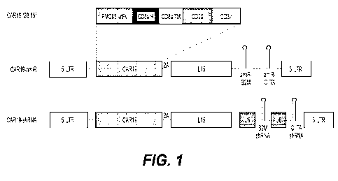
[0026] Figure 1 presents a diagram of CAR19 expression constructs with
artificial
microRNA (amiR) (CAR19-amiR) or pol III promoter-based expression of short
hairpin
RNA or small hairpin RNA (shRNA) (CAR19-shRNA) sequences against 02-
microglobulin
(B2M) and the invariant chain (Ii) (a.k.a. CD74) or the class II
transactivator (CIITA). LTR=
long terminal repeat, scFv= single chain variable fragment, H= hinge, TM=
transmembrane.
In some embodiments, the U6 promoter is replaced with an H1 or 7SK promoter.
[0027] Figure 2 presents representative results of CAR19 expression in NKTs
are
transduced with CAR19 constructs containing scrambled (scr.) or B2M-specific
shRNA
driven by the U6, H1, or 7SK promoter or embedded in the miR155 scaffold. CAR
expression is evaluated 2 days post-transduction.
[0028] Figure 3 presents a representative dot plot of intracellular flow
cytometry of a
donor gating the cells into CAR19 and HLA-A,B,C of NKTs transduced with CAR19
constructs containing scrambled (scr.) or B2M-specific shRNA driven by the H1,
7SK, or U6
promoters or embedded in amiR155 as indicated. Representative histograms of
HLA-A,B,C
expression for transduced and non-transduced samples is shown for each. B2M
shRNA
expression supported by amiR155 from within CAR19 is shown to result in the
greatest level
of knockdown of HLA-A,B,C (bottom right). CAR and HLA-A,B,C expression is
evaluated
2 days post-transduction.
[0029] Figure 4 presents another representative dot plot of intracellular
flow cytometry of
a donor gating the cells into CAR19 and HLA-A,B,C of NKTs transduced with
CAR19
constructs containing scrambled (scr.) or B2M-specific shRNA driven by the U6
promoter or
embedded in amiR155 as indicated. CAR and HLA-A,B,C expression is evaluated 14
days
post-transduction.
[0030] Figure 5 presents a representative dot plot of intracellular flow
cytometry of a
donor gating the cells into CAR19 and HLA-A,B,C of NKTs transduced with CAR19
constructs containing scrambled (scr.) or B2M-specific shRNA embedded in
amiR30 as
indicated. CAR and HLA-A,B,C expression is evaluated 7 days post-transduction.
5
CA 03217652 2023-10-23
WO 2022/226353
PCT/US2022/026014
[0031] Figure 6 presents representative dot plots of intracellular flow
cytometry of a
donor gating the cells into CAR19 and HLA-A,B,C of NKTs transduced with CAR19
constructs containing 5 different B2M-specific shRNA sequences (SEQ ID NOs:1
to 5)
embedded in amiR155 and previously evaluated shRNA sequence (SEQ ID NO:6) used
in
ANCHOR product. CAR and HLA-A,B,C expression is evaluated 12 days post-
transduction.
The results are quantified and presented in Table 4.
[0032] Figures 7A to 7C present representative dot plots of intracellular
flow cytometry
of a donor gating the cells into CAR19 and HLA-DR,DP,DQ of NKTs transduced
with ten
CAR19 constructs containing CIITA-specific shRNA (SEQ ID NOs:7 to 16
corresponding to
graphs 1 to 10 respectively) embedded in amiR155. CAR and HLA-DR,DP,DQ
expression is
evaluated 12 days post-transduction. The results are quantified and presented
in Table 4.
[0033] Figures 8A to 8C present representative dot plots of intracellular
flow cytometry
of a donor gating the cells into CAR19 and HLA-DR,DP,DQ of NKTs transduced
with ten
CAR19 constructs containing CD74-specific shRNA (SEQ ID NOs:17 to 26
corresponding to
graphs 1 to 10 respectively) embedded in amiR155. CAR and HLA-DR,DP,DQ
expression is
evaluated 12 days post-transduction. The results are quantified and presented
in Table 4.
[0034] Figure 9 presents a representative plot of the percent knockdown
of NKTs
transduced with CAR19.15 constructs containing single amiR-embedded shRNA
targeting
B2M (SEQ ID NO:X or CIITA (SEQ ID NO:12) as indicated. Knockdown efficiency
was
evaluated four days post-transduction. N = 4 donors.
[0035] Figure 10 presents a graph of IL-15 secretion from representative
donor NKT cells
transduced with the indicated constructs using the BioLegend ELISA MAX Tm
Deluxe Set
Human IL-15 kit (BioLegend #435104) and expression of CAR19. Figure 10 panel A
presents NKT cells transduced with CAR19.15, CAR19.15.u6-b2m, car19.15.miR155-
b2m,
or non-transduced (NT) and either cultured alone or co-cultured with CD19-
positive Raji
lymphoma cells for 48 hours. Figure 10 panel B presents NKT cells transduced
with
CAR19 constructs containing B2M-specific shRNA driven by the U6 promoter or
embedded
in the miR155 scaffold. CAR expression is evaluated two days post-
transduction. N= 1
donor, three technical repeats.
[0036] Figure 11 presents diagrams of constructs designed to boost IL15
expression from
knockdown constructs by incorporation of codon-optimized IL15 sequence, IL15
receptor
6
CA 03217652 2023-10-23
WO 2022/226353
PCT/US2022/026014
alpha (IL15Ra), and IL15Ra Sushi domain (extracellular N terminal portion of
IL15Ra,
essential for binding IL15).
[0037] Figure 12 panel A and panel B present graphs of IL-15 expression
of NKTs
transduced with the indicated constructs or non-transduced and either cultured
alone or co-
cultured with CD19-postive Raji lymphoma cells for 72 hours. Culture
supernatant are
processed using the BioLegend ELISA MAX Tm Deluxe Set Human IL-15 kit
(BioLegend
#435104) to detect IL15 secretion. A) N= 1 donor, three technical repeats. B)
N=3 donors.
[0038] Figure 13 presents representative dot plots of intracellular flow
cytometry of a
donor gating the cells into CAR19 and IL-15 of NKTs transduced with CAR19.15-
15Ra-
amiR-B2M construct (Figure 11) and IL15 expression is evaluated four days
later. Data
shown from three donors.
[0039] Figure 14 presents a diagram of a double knockdown construct of a CAR19
and
codon-optimized IL15 expression paired amiR30-B2M shRNA and amiR155-CIITA
shRNA
to mediate HLA class I and II knockdown, respectively.
[0040] Figures 15A and 15B present representative dot plots (A) of
intracellular flow
cytometry of a donor gating the cells into CAR19 and HLA-A,B,C or HLA-DR,DP,DQ
of
NKTs transduced with the CAR19 construct shown in Figure 14 and a graph of
knockdown
percentage (B) for three donors(BL# 81, 82, 83).
[0041] Figures 16A and 16B present representative dot plots from four
donors of
intracellular flow cytometry of a donor gating the cells into CAR19 and HLA-
A,B,C or HLA-
DR,DP,DQ of NKTs transduced with the CAR19 construct shown in Figure 14. CAR,
HLA-
A,B,C, and HLA-DR,DP,DQ expression are evaluated at day 19 of expansion.
Labels
indicate MFI for each population and knock-down percentage between cell
populations
connected by arrows.
[0042] Figure 17 presents a representative graph of IL15 secretion in NKT
cells
transduced with the indicated constructs or non-transduced and either cultured
alone or co-
cultured with CD19-positive Raji lymphoma cells for 48 hours. The culture
supernatant is
processed using the BioLegend ELISA MAX II'l Deluxe Set Human IL-15 kit
(BioLegend
#435104) to detect IL15 secretion. N= 3 donors (BL #81, 82, 83).
7
CA 03217652 2023-10-23
WO 2022/226353
PCT/US2022/026014
[0043] Figure 18 presents a representative graph of in vitro cytotoxicity
against CD19-
positive target cells compared with CAR19 and CAR19.IL15 NKT cells. NKT cells
are
transduced with indicated constructs and co-cultured for six hours with CD19-
positive Raji
lymphoma cells engineered to express high levels of firefly luciferase at
specified effector-to-
.. target ratios. Luciferin was added at the conclusion of the assay for
detection of
bioluminescence.
[0044] Figures 19A and 19B present results of NKT cells transduced with
CAR19.opti-
IL15 double knockdown constructs to control CD19-positive tumors in vivo and
promote
survival of NSG mice comparably to CAR19.15 NKT cells. Figure 19A presents
imaging of
NSG mice injected intravenously with 2x10⁵ firefly luciferase-positive Daudi
lymphoma cells
on day 0 followed by intravenous injection of 5x10⁶ NKT cells transduced with
indicated
constructs or no construct (non-transduced, NT) on day 3. Just prior to
imaging, each mouse
receives 100 pi luciferin at 30 mg/mL via intraperitoneal injection and are
imaged under a
bioluminescent channel. (Bioluminescent counts scale 600 - 30,000) Figure 19B
presents a
Kaplan Meier survival curve for the mice shown in Figure 19A.
[0045] Figure 20 presents a diagram of a double knockdown construct of a CAR19
and
codon-optimized IL15 containing a fused IL2 signal peptide (IL2SP) to boost
IL15 secretion.
SD/SA = splice donor/splice acceptor
[0046] Figure 21 presents a representative graph of IL15 secretion by NKT
cells
expressing the double knockdown construct of Figure 20. NKT cells are
transduced with the
indicated constructs or non-transduced and either cultured alone or co-
cultured with CD19-
positive Raji lymphoma cells for 48 hours. The culture supernatant is
processed using the
BioLegend ELISA MAX Tm Deluxe Set Human IL-15 kit (BioLegend #435104) to
detect
IL15 secretion.
[0047] Figure 22 presents results of NKT cells transduced with the IL2SP-
opti IL15
CAR19 construct with double amiR knockdown of Figure 20 to control CD19-
positive
tumors in vivo and promote survival of NSG mice comparably to CAR19.15 NKT
cells. NSG
mice are injected intravenously with 2x10⁵ firefly luciferase-positive Daudi
lymphoma cells
on day 0 followed by intravenous injection of 1x10⁶ or 5x10⁶NKTs transduced
with
indicated constructs or no construct (non-transduced, NT) on day 4. Just prior
to imaging,
8
CA 03217652 2023-10-23
WO 2022/226353
PCT/US2022/026014
each mouse receives 1004 luciferin at 30 mg/mL via intraperitoneal injection
and are
imaged under a bioluminescent channel. Bioluminescent counts scale 600 -
30,000.
[0048] Figures 23A and 23B present results of tumor progression in NSG
mice treated
with CAR NKT cells expressing double knockdown construct and a Kaplan Meier
survival
curve respectively. NSG mice are injected intravenously with 2x10⁵ firefly
luciferase-
positive Daudi lymphoma cells on day 0 followed by intravenous injection of
5x10⁶NKTs
transduced with indicated constructs or no construct (non-transduced, NT) on
day 3. Just
prior to imaging, each mouse receives 100 pi luciferin at 30 mg/mL via
intraperitoneal
injection and are imaged under a bioluminescent channel. Bioluminescent counts
scale 2000 -
30,000. B) Kaplan Meier survival curve for mice shown in A). Tumor progression
is delayed
and survival is unchanged.
[0049] Figure 24 presents a representative graph of NKT cells expressing
1) CAR19.15
containing two scrambled shRNA sequences in place of B2M and CIITA
(CAR19.IL2SP-
opti15.amiR-SCR-amiR-SCR, scramble), 2) CAR19.15 with amiR-embedded B2M and
CIITA shRNA sequences (CAR19.IL2SP-opti15.amiR-B2M-amiR-CIITA, knockdown), and
the B2M/CIITA double knockdown construct (knockout) evaluated by flow
cytometry daily
for CAR and HLA expression, gated on HLA I- cells. Recipient NK cells (HLA-
A2+) are
isolated using the NK cell isolation kit (Miltenyi Biotech) and co-cultured
with donor NKT
cells (HLA-A2-) at a 1:1 ratio for three days. NKT cells expressing the
B2M/CIITA double
knockdown construct persist in the presence of allogeneic NK cells while
double knock-out
leaves NKT cells vulnerable to NK cell killing.
[0050] Figure 25 presents representative graphs of flow cytometry of NKT
cells
transduced with scrambled, knockdown, and knockout constructs of Figure 24
every 2 to 3
days. Pan T cells are isolated from recipient PBMCs using the naive pan T cell
isolation kit,
human (Miltenyi Biotech. Recipient T cells (HLA-A2+) are co-cultured with
donor NKT
cells (HLA-A2-) at a 2:1 (T:NKT) ratio for seven days. NKT cells expressing
the
B2M/CIITA double knockdown construct resist rejection by allogeneic T cells
compared to
NKT cells carrying scrambled shRNA control construct.
[0051] Figure 26 presents representative graphs of flow cytometry results
of transduced
NKT cells evaluated every 2 to 3 days in co-culture with allogenic PBMCs.
Recipient
PBMCs (HLA-A2+) are co-cultured with donor NKT cells (HLA-A2-) at a 10:1
9
CA 03217652 2023-10-23
WO 2022/226353
PCT/US2022/026014
(PBMC:NKT) ratio for seven days. NKT cells are transduced with 1) CAR19.15
with
scrambled shRNA control, or 2) CAR19.15 with double knockdown.
[0052] Figures 27A and 27B present representative graphs of flow
cytometry of NKT
cells transduced with 1) CAR19.IL2SP-optil5 with scrambled shRNA sequences in
place of
B2M control (Scr), 2) CAR19.IL2SP-optil5 with double knockdown (I(D), 3)
CAR19.IL2SP-optil5 with double knockout (KO) and co-cultured with recipient NK
cells
(HLA-A2+) isolated using the NK cell isolation kit (Miltenyi Biotech) at a 2:1
(NK:NKT)
ratio for two days. Panel A of Figure 27A presents representative flow plots
showing total
frequency of donor NKT cells on day 0 and day 2 of co-culture (top, Figure
27B). Panel B
of Figure 27A presents absolute cell counts of donor NKT cells and Panel C of
Figure 27B
present recipient NK cells on day 0 and day 2 of co-culture. All data denote
mean s.d., three
unique donor¨recipient pairs are used. P values are determined using two-way
ANOVA with
Sidak's correction for multiple comparisons and nonsignificant (P > 0.05)
values are not
shown. P values are determined using the two-tailed, paired Student's t-test.
[0053] Figures 28A and 28B present representative graphs of flow cytometry
of
transduced NKT cells and absolute cell counts of donor NKT cells and recipient
T cells in
another aspect. Pan T cells are isolated from recipient PBMCs (HLA-A2+) using
the naive
pan T cell isolation kit, human (Miltenyi Biotech). Purified T cells are then
stimulated with
OKT3/aCD28 for 24 hours, in vitro expanded for 5-10 days, and co-cultured with
donor
NKT cells (HLA-A2-) at a 2:1 (T:NKT) ratio for two days. NKTs are transduced
with 1)
CAR19.IL2SP-optil5 with scrambled shRNA sequences (Scr), 2) CAR19.IL2SP-optil5
with
double knockdown (I(D), 3) CAR19.IL2SP-optil5 with double knockout (KO). Panel
A of
Figure 28A presents representative flow plots showing total frequency of donor
NKT cells
on day 0 and day 2 of co-culture (top, Figure 28B). Absolute cell counts of
donor NKT cells
are shown in Panel B of Figure 28A and Panel C of Figure 28B presents absolute
cell
counts of recipient T cells on day 2 of co-culture. All data denote mean
s.d., five unique
donor¨recipient pairs are used. P values are determined using two-way ANOVA
with Sidak's
correction for multiple comparisons and nonsignificant (P > 0.05) values are
not shown.
[0054] Figure 29A and 29B present representative graphs of flow cytometry
of
transduced NKT cells and absolute cell counts of donor NKT cells and recipient
T cells in
another aspect. Recipient whole PBMCs (HLA-A2+) are co-cultured with donor
NKTs
(HLA-A2-) at a 10:1 (PBMC:NKT) ratio for nine days. NKTs are transduced with
1)
CA 03217652 2023-10-23
WO 2022/226353
PCT/US2022/026014
CAR19.IL2SP-optil5 with scrambled shRNA sequences in place of B2M control
(Scr), 2)
CAR19.IL2SP-optil5 with double knockdown (KD), 3) CAR19.IL2SP-optil5 with
double
knockout (KO). Panel A of Figure 29A presents representative flow plots
showing total
frequency of donor NKT cells on day 0 and day 9 of co-culture (top, Figure
29B). Panel B
of Figure 29A shows absolute cell counts of donor NKT cells and Panel C of
Figure 29B
shows absolute cell counts of recipient T cells on days 0, 3, 6, and 9 of co-
culture. All data
denote mean s.d., three unique donor¨recipient pairs are used. P values are
determined
using two-way ANOVA with Sidak's correction for multiple comparisons and
nonsignificant
(P > 0.05) values are not shown. P values are determined using the two-tailed,
paired
Student's t-test.
[0055] Figure 30 presents a representative results of in vivo persistence
in an in vivo T
cell-mediated rejection model in vivo of NKT cells expressing the B2M/CIITA
double
knockdown construct. Panel A presents the experimental procedure. NSG mice are
irradiated at 1.2 Gy on day -1, and on the following day receive 7 x 10⁶ in
vitro expanded
human T-cells (day 5-10 post initial OKT3/aCD28 stimulation) from an HLA-A2-
recipient.
Four days later, mice receive 2 x 10⁶ control construct (CAR19.IL2SP-
opti15.amiR-SCR-
amiR-SCR) or knockdown construct (CAR19.IL2SP-opti15.amiR-b2m-amiR-ciita)
transduced NKT cells from an HLA-A2+ donor intravenously. RTC= recipient T
cells. Panel
B presents representative flow plots showing frequencies of donor HLA-A2+ Scr
control or
double KD NKT cells in peripheral blood on days 6 and 28. Panel C presents the
frequency
of donor HL-A2+ NKT cells and recipient HLA-A2-T-cells (Panel D) at specified
time
points. Data denote mean SD with 7-8 mice per group.
[0056] Figure 31 presents representative results of in vivo persistence
in an in vivo PBMC
cell-mediated rejection model in vivo of NKT cells expressing the B2M/CIITA
double
knockdown construct in the presence of allogeneic PBMCs compared to scrambled
control
NKTs. Panel A presents the experimental procedure. NSG (MHCK ) mice re
irradiated at
1.2 Gy on day -1, and then receive intravenously 5 x 10⁶ freshly isolated PBMC
from an
HLA-A2- recipient on day 0. Four days later, 5 x 10⁶ scrambled control or
double
knockdown transduced NKTs from an HLA-A2+ donor are administered
intravenously.
Panel B presents representative flow plots showing frequencies of donor HLA-
A2+ Scr
control or double KD NKT cells in peripheral blood on days 6 and 20. Panel C
presents the
11
CA 03217652 2023-10-23
WO 2022/226353
PCT/US2022/026014
frequency donor HL-A2+ NKT cells and Panel D present the frequency of
recipient HLA-
A2-T cells at specified time points. Data denote mean SD with 7-8 mice per
group.
[0057] Figures 32A and 32B present representative results of anti-tumor
activity in vivo
in the presence of allogeneic T cells compared to scrambled control NKT cells
in an In vivo
T cell-mediated rejection model with B cell lymphoma xenograft of NKT cells
expressing the
B2M/CIITA double knockdown construct. Panel A presents the experimental
procedure.
NSG mice are irradiated at 1.2 Gy and receive intravenously 7 x10⁶ in vitro
expanded human
T cells (days 8-10 postinitial OKT3/aCD28 stimulation) from an HLA-A2 -
recipient on the
following day. One day later, 2x10⁵ firefly luciferase-positive Daudi cells
are injected
intravenously, followed three days later by 5 x10⁶ scrambled control or
knockdown
transduced NKT cells generated from an HLA-A2+ donor. RTC= recipient T cells.
Panel B
presents a representative flow plot showing frequencies of donor HLA-A2+
scrambled
control (Scr) or double KD NKT cells in peripheral blood of mice on days 6 and
28.
Frequencies of HLA-A2+ donor CAR NKT cells (Panel C) and HLA-A2- RTCs in
peripheral
blood (Panel D) after tumor injection. Panel E presents lymphoma progression
measured
using IVIS imaging at specified time points. Panel F presents Kaplan¨Meier
curve showing
survival of mice in each experimental group. P values are determined using two-
sided log-
rank test.
[0058] Figure 33 presents examples of CAR.GPC3.opti-IL15 double knockdown
constructs. The constructs comprise sequences encoding either the GPC3-
specific scFv from
GC33 or the scFv from the humanized YP7.
[0059] Figure 34 presents levels of HLA class I or class II gene
knockdown are observed
in CAR-GPC3 NKT cells expressing either the humanized GPC3 scFv (YP7) or
murine
GPC3 scFv (GC33)
[0060] Figure 35 presents expression levels of IL15 in NKT cells expressing
humanized
GPC3 scFv (YP7) and NKT cells expressing murine GPC3 scFv.
[0061] Figure 36 presents the cytotoxicity levels in cells expressing
humanized GPC3
scFv (YP7) and NKT cells expressing murine GPC3 scFv, as measured by the
xCelligence
assay.
12
CA 03217652 2023-10-23
WO 2022/226353
PCT/US2022/026014
[0062] Figure 37 presents experimental design and the expected anti-tumor
activity of
CAR.GPC3 NKT cells in an HCC xenograft model.
[0063] Figure 38 presents the expression level of B2M, CIITA, or native
IL-15 in
CAR.GPC3 NKT cells expressing amiR constructs targeting B2M and CIITA and
CAR.GPC3 NKT cells comprising IL15 constructs.
[0064] Figure 39 presents a comparison of IL-15 expression levels in NKT
cells
expressing constructs having IL-15 coding sequence upstream or downstream of
CAR.GPC3.
[0065] Figure 40 presents a heat map illustrating the HLA-specific genes
downregulated
in G.28BBz.15.miR-expressing NKT cells in comparison with 15G28BBz-expressing
NKT
cells. Adjusted P value is less than 0.05 and fold change is greater than 2.
[0066] Figure 41 presents a heat map illustrating the HLA-specific and
immune effector
genes downregulated in YP7.28BBz.15.miR-expressing NKT cells in comparison
with
15G28BBz expressing NKT-cells. Adjusted P value is less than 0.05 and fold
change is
greater than 2.
[0067] Figure 42 presents a heat map illustrating that no significant
pathways are
enriched in humanized YP7.28BBz.15.miR-expressing NKT cells in comparison with
murine
G.28BBz.15.miR-expressing NKT cells. Adjusted P value is less than 0.05 and
fold change is
greater than 2.
[0068] Corresponding reference characters indicate corresponding parts
throughout the
several views. The example(s) set out herein illustrate(s) [one/several]
embodiment(s) of the
present disclosure but should not be construed as limiting the scope of the
present disclosure
in any manner.
DETAILED DESCRIPTION
[0069] The present application is directed to methods and compositions
related to
genetically modified natural killer T cells (NKT cells). NKT cells are a
distinct cell type that
share some features of both T and NK cells but are distinct from both
conventional T cells
and also NK cells. NKT cells have divergent development from conventional T
cells and NK
cells and different functions driven by a unique set of transcriptional
regulators. See
Kronenberg M, Gapin L. The unconventional lifestyle of NKT cells.
NatRev.Immunol.
13
CA 03217652 2023-10-23
WO 2022/226353
PCT/US2022/026014
2002;2(8):557-568; Godfrey, JCI, 2004, Cohen NR, etal. Shared and distinct
transcriptional
programs underlie the hybrid nature of iNKT cells. Natdmmunol. 2013;14(1):90-
99.).
Godfrey et al., identify transcription factors, signal-transduction factors,
cell surface
molecules, cytokines, and other factors that selectively influence NKT cell
development
reflecting the unique programming associated with the NKT cell lineage.
(Godfrey etal.,
"Raising the NKT cell family," Nat. Immunol., 11(3):197-206 (2010) ("Godfrey
etal.")
hereby incorporated by reference in its entirety. See also Engel and
Kronenberg,
"Transcriptional control of the development and function of Va4i NKT cells,"
Current Topics
in Microbiology and Immunology, Volume 381, 2014). Many transcription factors
and
signaling molecules that affect NKT cells differentiation in the thymus do not
affect other
conventional T cell populations that develop there. As used throughout the
present
disclosure, the term "T cell" is limited to conventional T cells that are
distinguishable from
NKT cells. These differences result in different responses to stimuli and
genetic changes
such as engineered gains and losses of gene expression that make results in
non-NKT cells
unpredictable.
[0070] NKT cells are distinguishable based on whole genome transcription
analysis and
are equally distant from conventional and NK cell lineages. See Cohen et al.
supra.
Conventional T cells, also known as T lymphocytes, are an important cell type
with the
function of fighting pathogens and regulating the immune response. Two hall
marks of these
cells are expression of an antigen receptor encoded by segments of DNA that
rearrange
during cell differentiation to form a vast array of receptors. A number of
cells fall within this
generic definition of a T cell, for example: T helper cells (CD4+ cells)
including the sub-
types TH1, TH2, TH3, TH17, TFH; cytotoxic T cells (mostly CD8+ cells, also
referred to a
CTLs); memory T cells (including central memory T cells, effector memory T
cells, and
resident memory T cells); regulatory T cells, and mucosal associated invariant
T cells. Cell
surface markers of T cells include the T cell receptor and CD3. Generally T
cells do not
express CD56 (i.e. are CD56 negative).
[0071] NK cells and NKT cells are CD56+. In humans NK cells usually
express the cell
surface marker CD56, CD161, CD11 b, NKp46, NKp44, CD158 and IL-12R. NK cells
express a limited repertoire of receptors with an entirely different
structure, some of which
are also found on NKT cells. Most NK receptors are not highly conserved
comparing
humans and rodents. NK cells express members of the family of killer-cell-
immunoglobulin-
14
CA 03217652 2023-10-23
WO 2022/226353
PCT/US2022/026014
like receptors (KIRs), which can be activating or inhibiting, as well as
receptors that are
members of the lectin (carbohydrate-binding) family of proteins such as NKG2D
and
CD94NKG2A/C. KIRs are not expressed on NKT cells. NK cells are activated by a
number
of cell surface receptors, such as KIRs in humans or Ly49 in mice, natural
cytotoxic receptors
(NCRs), NKG2D and CD94:NKG2 heterodimers. In addition cytokines and
chemokines,
such as IL-12, IL-15, IL-18, IL-2 and CCLS, play a significant role in NK cell
activation.
[0072] NKT cells generally can be identified as CD3+CD56+ cells and
express a T cell
receptor. NKT cells express a T cell receptor and CD3 chains like T cells, but
also have
markers such CD56 and CD161, like NK cells. Having said that, it is now
commonly
accepted by experts that they are a distinct lineage of cells. That is they
are very different
from other T cells and their behavior and properties cannot be predicted from
analysis of
other T cells, nor are they NK cells. NKT cells are completely different cells
to conventional
T cells and to NK cells. Due to the unique properties of the NKT cell lineage,
observations
made with other populations of lymphocytes, such as T cells, NK cells, and B
cells, may not
predict functional consequences of NKT cell activation.
[0073] NKT cells can be identified from other cell types including CD4 T
cells, CD8 T
cells, regulatory T cells, y.5 T cells, B cells, NK cells, monocytes and
dendritic cells based on
the expression of cell surface markers. See Park etal., "OMIP-069: Forty-Color
Full
Spectrum Flow Cytometry Panel for Deep Immunophenotyping of Major Cell Subsets
in
Human Peripheral Blood," Cytometry Part A 97A:1044-1051 (2020); Hertoghs
etal., OMIP-
064: A 27-Color Flow Cytometry Panel to Detect and Characterize Human NK Cells
and
Other Innate Lymphoid Cell Subsets, MAIT Cells, and y.5 T Cells, Cytometry
Part A
97A:1019-1023 (2020); Sahir etal., Development of a 43 color panel for the
characterization
of conventional and unconventional T-cell subsets, B cells, NK cells,
monocytes, dendritic
cells, and innate lymphoid cells using spectral flow cytometry, Cytometry
2020:1-7.
[0074] NKT cells are divided into two main types, Type I and Type II. The
most
significant form of NKT cells, known as type I NKT cells or invariant NKT
cells ("iNKT"),
have an invariant T cell receptor alpha chain (Va4i mouse or Va24i human).
Type I NKT
(iNKT) cells can be readily detected by the binding of CD id-based tetramers
loaded with
aGalCer analogs. The form of the antigen receptor is a limited repertoire due
to an invariant
alpha chain paired with one of a relatively small number of beta chains,
inhibition, or
therapeutic use. The antigens recognized by this invariant receptor are
glycolipids, for
CA 03217652 2023-10-23
WO 2022/226353
PCT/US2022/026014
example those found in bacterial cells. The invariant receptor recognizes
alpha-
galatosylceramide (a-GalCer) a glycolipid originally derived from marine
sponges. This
compound is similar to microbial glycolipids, and it is now generally assumed
to be derived
from a microbial symbiont associated with the sponge. NKT cells require
antigen presented
on a molecule CD1d.
[0075] Type II NKT cells also require antigen presentation from CD1d but
have a more
diverse but still limited TCR repertoire. Type II NKT cells express low levels
of the
transcription factor PLZF. While Type I NKT cells only recognize a-GalCer,
Type II NKT
cells recognize sulfatide, lyso-sulfatide, Lyso-PC and Lyso-GL1. Type II NKT
cells are
more prevalent in humans, but less prevalent in mice. See Dhodpkar and Kumar,
"Type II
NKT Cells and Their Emerging Role in Health and Disease," J Immunol.
198(3):1015-1021
(2017).
[0076] Two pathways are known for NKT cell activation. NKT cells respond
stimulation
through their T cell receptor via antigen presented on CD1d molecules. This
does not depend
upon the involvement of a CD4 or CD8 co-receptor to generate a TCR signal, and
the
response of these cells is somewhat less dependent on a co-stimulatory signal.
In addition, a
mechanism for activation of NKT cells exists in the absence of antigen
engaging the T cell
receptor, via innate inflammatory stimuli, such as IL-12 and IL-18. Once
activated T cells are
found in the peripheral blood. Similarly NK cells are found in the peripheral
blood. In
contrast the majority of NKT cells are found in tissues and they migrate away
from peripheral
blood to the site of tumors, for example as mediated via a two-step process
involving CCR2
and CCR6. The mechanisms involved in this migration are specific to NKT cells
and not
general mechanisms that apply to other lymphocytes.
[0077] iNKT cells are readily distinguishable from other T-cell types.
See Table 1. Only
a small fraction of expanded T cells (a subset of CD4 T cells) can produce
tumor-protective
Th2 cytokines (IL-4, IL-5, IL-13, IL-10) upon activation either via the T cell
receptor (TCR).
The majority of T cells (including all CD8+ T cells) and all NK cells produce
only anti-tumor
Thl cytokines (i.e. IFN-gamma, GM-CSF, TNF-alpha). In contrast, NKT cells
simultaneously produce Thl and Th2 cytokines." Depending on the balance of Thl
and Th2
cytokines produced after T cell receptor (TCR) activation, NKT cells can
either activate or
suppress the immune response. Thus NKT cells have an intriguing paradoxical
dual function
of immune activation and immune suppression. In contrast other immune cells
usually have
16
CA 03217652 2023-10-23
WO 2022/226353
PCT/US2022/026014
one primary function, for example fighting pathogens, whilst other subsets of
cells are
dedicated to regulating the immune response.
Table 1: Distinguishing features of iNKT cells
T CELLS iNKT CELLS
TCR specificity varies TCR specificity does not vary
TCR binds peptides presented on MHC TCR binds certain glycolipids, for
example
molecules
natural products and derivatives from
bacterial cell walls, presented on CD1d
TCR/MHC/peptide complex formed TCR
has unique docking strategy with CD1d
Part of the reactive immune system Part of the innate immune system
Take time to react to a "threat" React very quickly to a "treat"
Involved in tissue rejection Not involved in tissue rejection
Tolerant to self-antigens Can react to self-antigens
Non-specifically activated by anti-CD3 Can be activated by the cytokines IL-
12 and
agonistic antibody IL-18
Primarily located in blood Generally resident in tissue
Do not co-located with tumor associated Co-located with tumor associated
macrophages macrophage in hypoxic tumor
microenvironment
Does not migrate to tumor Migrates to the tumor microenvironment
via
a unique CCR2 and CCR6 mechanism
Have a clear hierarchy of naïve-central- Have mostly effector-memory
phenotype
effector differentiation when freshly isolated from peripheral
blood,
but can generate CD62L+ central memory-
like cells upon certain conditions of ex vivo
culture (G. Tian et al.)
Developmental pathway is distinct for the two cell types
In vitro stimulation/culture of the T cell and NKT cells require different
protocols
[0078] NKT cells also develop in the thymus, however, the positive
selection of Type I
NKT cells is mediated by CD id positive thymocytes. NKT cells are also subject
to negative
17
CA 03217652 2023-10-23
WO 2022/226353
PCT/US2022/026014
selection by dendritic cells. See Godfrey et al., at Figure 2 summarizing the
development and
maturation of T cells and NKT cells in the thymus.
[0079] Unless defined otherwise, all technical and scientific terms used
herein have the
meaning commonly understood by a person skilled in the art to which this
invent ion belongs.
The following references provide one of skill with a general definition of
many of the terms
used in this invention: Singleton et al., Dictionary of Microbiology and
Molecular Biology
(2nd ed. 1994); The Cam bridge Dictionary of Science and Technology (Walker
ed., 1988);
The Glossary of Genetics, 5th Ed., R. Rieger etal. (eds.), Springer Verlag
(1991); and Hale
& Marham, The Harper Collins Dictionary of Biology (1991). As used herein, the
following
terms have the meanings ascribed to them below, unless specified otherwise.
[0080] As used herein the term "about" refers to plus/minus 10 %.
[0081] The terms "comprises", "comprising", "includes", "including",
"having" and their
conjugates mean "including but not limited to."
[0082] The term "consisting of" means "including and limited to."
[0083] The term "consisting essentially of" means that the composition,
method or
structure may include additional ingredients, steps and/or parts, but only if
the additional
ingredients, steps and/or parts do not materially alter the basic and novel
characteristics of the
claimed composition, method or structure.
[0084] As used herein, the singular form "a", "an" and "the" include
plural references
unless the context clearly dictates otherwise. For example, the term "a cell"
or "at least one
cell" may include a plurality of cells, including mixtures thereof
[0085] The terms "comprises", "comprising", and are intended to have the
broad meaning
ascribed to them in U.S. Patent Law and can mean "includes", "including" and
the like.
[0086] By "increase" is meant to alter positively by at least 5%. An
alteration may be by
5%, 10%, 25%, 30%, 50%, 75%, or even by 100%.
[0087] By "decrease" or "reduce" is meant to alter negatively by at least
5%. An alteration
may be by 5%, 10%, 25%, 30%, 50%, 75%, or even by 100%.
18
CA 03217652 2023-10-23
WO 2022/226353
PCT/US2022/026014
[0088] By "modulate" is meant positively or negatively alter. Exemplary
modulations
include a 1%, 2%, 5%, 10%, 25%, 50%, 75%, or 100% change.
[0089] Throughout this application, various embodiments of this
disclosure may be
presented in a range format. It should be understood that the description in
range format is
merely for convenience and brevity and should not be construed as an
inflexible limitation on
the scope of the disclosure. Accordingly, the description of a range should be
considered to
have specifically disclosed all the possible subranges as well as individual
numerical values
within that range. For example, description of a range such as from 1 to 6
should be
considered to have specifically disclosed subranges such as from 1 to 3, from
1 to 4, from 1
to 5, from 2 to 4, from 2 to 6, from 3 to 6 etc., as well as individual
numbers within that
range, for example, 1, 2, 3, 4, 5, and 6. This applies regardless of the
breadth of the range.
[0090] Whenever a numerical range is indicated herein, it is meant to
include any cited
numeral (fractional or integral) within the indicated range. The phrases
"ranging/ranges
between" a first indicate number and a second indicate number and
"ranging/ranges from" a
first indicate number "to" a second indicate number are used herein
interchangeably and are
meant to include the first and second indicated numbers and all the fractional
and integral
numerals therebetween.
[0091] As used herein, a "genetically engineered natural killer T (NKT)
cell" or
"engineered NKT cell" is an NKT cell that comprises at least one recombinant
nucleic acid
encoding exogenous protein or a endogenous protein downstream of a non-native
promoter.
In aspects, genetically engineered NKT cells comprise a recombinant nucleic
acid encoding a
chimeric antigen receptor.
[0092] By "endogenous" is meant a nucleic acid molecule or polypeptide
that is normally
expressed in a cell or tissue.
[0093] By "exogenous" is meant a nucleic acid molecule or polypeptide that
is not
endogenously present in the cell, or not present at a level sufficient to
achieve the functional
effects obtained when over-expressed. The term "exogenous" would therefore
encompass any
recombinant nucleic acid molecule or polypeptide expressed in a cell, such as
foreign,
heterologous, and over-expressed nucleic acid molecules and polypeptides.
19
CA 03217652 2023-10-23
WO 2022/226353
PCT/US2022/026014
[0094] As used herein, the term "artificial microRNAs (amiRNAs)" are
molecules that
have been developed to promote gene silencing in a similar manner to naturally
occurring
miRNAs. amiRNAs are generally constructed by replacing the mature miRNA
sequence in
the pre-miRNA stem-loop with a sequence targeting a gene of interest. These
molecules offer
a great alternative to silencing approaches that are based on shRNAs and
siRNAs because
they present the same efficiency as these options and are less cytotoxic. As
used herein, the
term "embedded" in an artificial microRNA scaffold" refers to the process of
replacing a
mature miRNA sequence in the pre-miRNA stem-loop with a sequence targeting a
gene of
interest. In some aspects, the amiR used in the instant disclosure is amiR155.
Lagos-Quintana
etal., "Identification of tissue-specific microRNAs from mouse." Curr Biol.
2002 Apr
30;12(9):735-9. In another aspect, the amiR used in the instant disclosure is
amiR30.
Fellmann etal., "An optimized microRNA backbone for effective single-copy
RNAi." Cell
Rep. 2013 Dec 26;5(6):1704-13. In further aspects, the amiR used in the
instant disclosure is
an artificial microRNA scaffold known in the art.
[0095] A "short hairpin RNA," "small hairpin RNA" or "shRNA" is an
artificial RNA
molecule with a tight hairpin turn that can be used to silence target gene
expression via RNA
interference (RNAi). They typically consist of a stem of 19-29 base pairs
(bp), a loop of at
least 4 nucleotides (nt), and a dinucleotide overhang at the 3' end. In some
aspects, the term
"shRNA" in the instant disclosure may refer to the sense strand or the
antisense strand of the
"stem" part of a small hairpin RNA. In other aspects, the term "shRNA" may
include the
sense strand, the antisense strand, and the loop in between.
[0096] As used herein, a small hairpin RNA (shRNA) "targeting" a gene of
interest refers
to an shRNA comprising a sequence of at least 19 contiguous nucleotides that
is essentially
identical to, or is essentially complementary to, a gene of interest. Aspects
of shRNAs
functional in this disclosure have sequence complementarily that need not be
100% but is at
least sufficient to permit hybridization to RNA transcribed from the target
gene to form a
duplex under physiological conditions in a cell to permit cleavage by a gene
silencing
mechanism. Thus, in aspects the segment is designed to be essentially
identical to, or
essentially complementary to, a sequence of 19 or more contiguous nucleotides
in either the
target gene or messenger RNA transcribed from the target gene. By "essentially
identical" is
meant having 100% sequence identity or at least about 80, 81, 82, 83, 84, 85,
86, 87, 88, 89,
90, 91, 92, 93, 94, 95, 96, 97, 98, or 99% sequence identity when compared to
the sequence
CA 03217652 2023-10-23
WO 2022/226353
PCT/US2022/026014
of 19 or more contiguous nucleotides in either the target gene or RNA
transcribed from the
target gene; by "essentially complementary" is meant having 100% sequence
complementarity or at least about 80, 81, 82, 83, 84, 85, 86, 87, 88, 89, 90,
91, 92, 93, 94, 95,
96, 97, 98, or 99% sequence complementarity when compared to the sequence of
19 or more
contiguous nucleotides in either the target gene or RNA transcribed from the
target gene. In
some aspects of this disclosure shRNAs are designed to comprise a sequence
having 100%
sequence identity with or complementarity to one allele of a given target
gene; in other
aspects the shRNAs are designed to comprise a sequence having 100% sequence
identity
with or complementarity to multiple alleles of a given target gene.
[0097] Sequence identity is typically measured using sequence analysis
software that are
widely available in the art. Such software matches identical or similar
sequences by
assigning degrees of homology to various substitutions, deletions, and/or
other modifications.
Conservative substitutions typically include substitutions within the
following groups:
glycine, alanine; valine, isoleucine, leucine; aspartic acid, glutamic acid,
asparagine,
glutamine; serine, threonine; lysine, arginine; and phenylalanine, tyrosine.
In an exemplary
approach to determining the degree of identity, a BLAST program may be used,
with a
probability score between e-3 and e-100 indicating a closely related sequence.
[0098] Major histocompatibility complex (MHC) class I and class II
proteins play a
pivotal role in the adaptive branch of the immune system. Both classes of
proteins share the
task of presenting peptides on the cell surface for recognition by T cells.
Immunogenic
peptide¨MHC class I (pMHCI) complexes are presented on nucleated cells and are
recognized by cytotoxic CD8+ T cells. The presentation of pMHCII by antigen-
presenting
cells (e.g., dendritic cells (DCs), macrophages, or B cells), on the other
hand, can activate
CD4+ T cells, leading to the coordination and regulation of effector cells. In
all cases, it is a
clonotypic T cell receptor that interacts with a given pMHC complex,
potentially leading to
sustained cell: cell contact formation and T cell activation. Wieczorek et
al., "Major
Histocompatibility Complex (MHC) Class I and Class II Proteins: Conformational
Plasticity
in Antigen Presentation." Frontiers in Immunology, 2017, Mar 17;8:292.
[0099] Major histocompatibility complex class I and class II share an
overall similar fold.
The binding platform is composed of two domains, originating from a single
heavy a-chain
(HC) in the case of MHC class I and from two chains in the case of MHC class
II (a-chain
21
CA 03217652 2023-10-23
WO 2022/226353
PCT/US2022/026014
and (3-chain). The two domains evolved to form a slightly curved 13-sheet as a
base and two a-
helices on top, which are far enough apart to accommodate a peptide chain in-
between. Two
membrane-proximal immunoglobulin (Ig) domains support the peptide-binding
unit. One Ig
domain is present in each chain of MHC class II, while the second Ig-type
domain of MHC
class I is provided by non-covalent association of the invariant light chain
beta-2
microglobulin (B2M) with the HC. Transmembrane helices anchor the HC of MHC
class I
and both chains of MHC class II in the membrane. Id. Class II transactivator
(CIITA) is a
transcriptional coactivator that regulates y-interferon-activated
transcription of MHC class I
and II genes.
[00100] The human leukocyte antigen (HLA) system or complex is a group of
related
proteins that are encoded by the MHC gene complex in humans. These cell-
surface proteins
are responsible for the regulation of the immune system.
[00101] As used herein, the term "method" refers to manners, means, techniques
and
procedures for accomplishing a given task including, but not limited to, those
manners,
means, techniques and procedures either known to, or readily developed from
known
manners, means, techniques and procedures by practitioners of the chemical,
pharmacological, biological, biochemical and medical arts.
[00102] As used herein, "treatment" refers to clinical intervention in an
attempt to alter the
disease course of the individual or cell being treated, and can be performed
either for
prophylaxis or during the course of clinical pathology. Therapeutic effects of
treatment
include, without limitation, preventing occurrence or recurrence of disease,
alleviation of
symptoms, diminishment of any direct or indirect pathological consequences of
the disease,
preventing metastases, decreasing the rate of disease progression,
amelioration or palliation
of the disease state, and remission or improved prognosis. By preventing
progression of a
disease or disorder, a treatment can prevent deterioration due to a disorder
in an affected or
diagnosed subject or a subject suspected of having the disorder, but also a
treatment may
prevent the onset of the disorder or a symptom of the disorder in a subject at
risk for the
disorder or suspected of having the disorder.
[00103] As used herein, the terms "cell," "cell line," and "cell culture" may
be used
interchangeably. All of these terms also include their progeny, which is any
and all
subsequent generations. It is understood that all progeny may not be identical
due to
22
CA 03217652 2023-10-23
WO 2022/226353
PCT/US2022/026014
deliberate or inadvertent mutations. The cells disclosed herein can be
autologous cells,
syngeneic cells, allogenic cells and even in some cases, xenogeneic cells.
[00104] By "isolated cell" is meant a cell that is separated from the
molecular and/or
cellular components that naturally accompany the cell.
[00105] The term "chimeric antigen receptor" or "CAR," as used herein, refers
to an
artificial T cell receptor that is engineered to be expressed on an immune
effector cell and
specifically bind an antigen. In aspects, CARs comprise and ectodomain, a
transmembrane
domain, and an endodomain. In certain aspects, a CAR can comprise an
ectodomain and
transmembrane domain without an endodomain, but more CARs of the present
application
include the endodomain and provide for intracellular signaling.
[00106] By "receptor" is meant a polypeptide, or portion thereof, present on a
cell
membrane that selectively binds one or more ligands.
[00107] As used herein, an "antigen recognition domain" generally comprises a
single
chain variable fragment (scFv) specific for a particular cancer antigen. In
some aspects,
where there are two or more CARs in the same cell, the second CAR may comprise
an scFv
specific for another particular antigen.
[00108] As used herein, the term "single-chain variable fragment" or "scFv" is
a fusion
protein of the variable regions of the heavy (VH) and light chains (VL) of an
immunoglobulin covalently linked to form a VH: :VL heterodimer. The heavy (VH)
and light
chains (VL) are either joined directly or joined by a peptide-encoding linker
(e.g., 10, 15, 20,
amino acids), which connects the N-terminus of the VH with the C-terminus of
the VL, or
the C-terminus of the VH with the N-terminus of the VL. The linker is usually
rich in glycine
for flexibility, as well as serine or threonine for solubility. Despite
removal of the constant
regions and the introduction of a linker, scFv proteins retain the specificity
of the original
25 immunoglobulin. Single chain Fv polypeptide antibodies can be expressed
from a nucleic
acid including VH- and VL-encoding sequences as described by Huston, et al.
(Proc. Nat.
Acad. Sci. USA, 85:5879-5883, 1988). See, also, U.S. Pat. Nos. 5,091,513,
5,132,405 and
4,956,778; and U.S. Patent Publication Nos. 20050196754 and 20050196754.
[00109] As used herein, a "transmembrane domain" is a region of predominantly
of
nonpolar amino acid residues that when the protein is expressed, traverses the
bilayer at least
23
CA 03217652 2023-10-23
WO 2022/226353
PCT/US2022/026014
once. Generally, the transmembrane domain is encoded by 18 to 21 amino acid
residues and
adopts an alpha helical configuration. As used herein, the transmembrane
domain may be of
any kind known in the art. In aspects the transmembrane domain is although in
some cases it
is CD28. Other sources include CD3-C, CD4, or CD8. An exemplary combination of
an
ectodomain is shown in Figure 27b of PCT/US2022/015525. Other suitable
transmembrane
regions can be obtained from CD16, NKp44, NKp46, and NKG2d.
[00110] As used herein, the term "endodomain" refers to the intracellular
domain of a CAR
that provides for signal transmission in a cell. Generally, the endodomain can
be further
divided into two parts, a stimulatory domain and optionally, a co-stimulatory
domain. The
co-stimulatory domain is shown to be arranged amino-terminal to the
stimulatory in Figure
27a of PCT/US2022/015525, but the present specification also provides for an
amino
terminal stimulatory domain and followed by a co-stimulatory domain when
present. The
most commonly used endodomain component is CD3-zeta that contains 3 ITAMs and
that
transits an activation signal to the NKT cell after the antigen is bound.
Other suitable
stimulatory domains can be obtained from 2B4 (CD244), TNF receptor superfamily
member
9 (Gene ID 3604, e.g., 4-1BB or CD137), Interleukin 21 (IL-21, Gene ID 59067),
hematopoietic cell signal transducer (HCST, Gene ID 10870 e.g., DAP10), and
transmembrane immune signaling adaptor (TYROBP, Gene ID 7305; DAP12).
[00111] As used herein, the term "ectodomain" refers to the extracellular
portion of a CAR
and encompasses a signal peptide, an antigen recognition domain, and a spacer
or hinge
region that links the antigen recognition domain to the transmembrane domain.
When
expressed, the signal peptide may be removed.
[00112] The term "tumor antigen" as used herein refers to an antigen (e.g., a
polypeptide,
glycoprotein, or glycolipid) that is uniquely or differentially expressed on a
tumor cell
compared to a normal or non-neoplastic cell. With reference to the invention,
a tumor antigen
includes any polypeptide expressed by a tumor that is capable of being
recognized by an
antigen recognizing receptor (e.g., CD19, Muc-1) or capable of suppressing an
immune
response via receptor-ligand binding (e.g., CD47, PD-L1/L2, 87.112).
[00113] By "tissue antigen" is meant an antigen (e.g., a polypeptide or
glycoprotein or
glycolipid) that is uniquely or differentially expressed on a normal or non-
neoplastic cell or
tissue compared to a tumor cell.
24
CA 03217652 2023-10-23
WO 2022/226353
PCT/US2022/026014
[00114] The terms "subject," "individual," and "patient," are used
interchangeably herein
and refer to any vertebrate subject, including, without limitation, mammals,
preferably a
humans and other primates, including non-human primates such as laboratory
animals
including rodents such as mice, rats and guinea pigs; The term does not denote
a particular
age. Thus, both adult and newborn individuals are intended to be covered.
[00115] By "effective amount" is meant an amount sufficient to have a
therapeutic effect.
In one embodiment, an "effective amount" is an amount sufficient to arrest,
ameliorate, or
inhibit the continued proliferation, growth, or metastasis (e.g., invasion, or
migration) of a
neoplasia.
[00116] By a "heterologous nucleic acid molecule or polypeptide" is meant a
nucleic acid
molecule (e.g., acDNA, DNA or RNA molecule) or polypeptide that is not
normally present
in a cell or sample obtained from a cell. This nucleic acid may be from
another organism, or
it may be, for example, an mRNA molecule that is not normally expressed in a
cell or sample.
[00117] By "immunoresponsive cell" is meant a cell that functions in an immune
response
or a progenitor, or progeny thereof
[00118] The terms "isolated," "purified," or "biologically pure" refer to
material that is free
to varying degrees from components which normally accompany it as found in its
native
state. "Isolate" denotes a degree of separation from original source or
surroundings. "Purify"
denotes a degree of separation that is higher than isolation. A "purified" or
"biologically
pure" protein is sufficiently free of other materials such that any impurities
do not materially
affect the biological properties of the protein or cause other adverse
consequences. That is, a
nucleic acid or peptide of this invention is purified if it is substantially
free of cellular
material, viral material, or culture medium when produced by recombinant DNA
techniques,
or chemical precursors or other chemicals when chemically synthesized. Purity
and
homogeneity are typically determined using analytical chemistry techniques,
for example,
polyacrylamide gel electrophoresis or high performance liquid chromatography.
The term
"purified" can denote that a nucleic acid or protein gives rise to essentially
one band in an
electrophoretic gel. For a protein that can be subjected to modifications, for
example,
phosphorylation or glycosylation, different modifications may give rise to
different isolated
proteins, which can be separately purified.
CA 03217652 2023-10-23
WO 2022/226353
PCT/US2022/026014
[00119] The term "obtaining" as in "obtaining the agent" is intended to
include purchasing,
synthesizing or otherwise acquiring the agent (or indicated substance or
material).
[00120] By "neoplasia" is meant a disease characterized by the pathological
proliferation of
a cell or tissue and its subsequent migration to or invasion of other tissues
or organs.
Neoplasia growth is typically uncontrolled and progressive, and occurs under
conditions that
would not elicit, or would cause cessation of, multiplication of normal cells.
Neoplasias can
affect a variety of cell types, tissues, or organs, including but not limited
to an organ selected
from the group consisting of bladder, bone, brain, breast, cartilage, glia,
esophagus, fallopian
tube, gallbladder, heart, intestines, kidney, liver, lung, lymph node, nervous
tissue, ovaries,
pancreas, prostate, skeletal muscle, skin, spinal cord, spleen, stomach,
testes, thymus, thyroid,
trachea, urogenital tract, ureter, urethra, uterus, and vagina, or a tissue or
cell type thereof
Neoplasias include cancers, such as sarcomas, carcinomas, or plasmacytomas
(malignant
tumor of the plasma cells). Illustrative neoplasms for which the invention can
be used
include, but are not limited to leukemias (e.g., acute leukemia, acute
lymphocytic leukemia,
acute myelocytic leukemia, acute myeloblastic leukemia, acute promyelocytic
leukemia,
acute myelomonocytic leukemia, acute monocytic leukemia, acute
erythroleukemia, chronic
leukemia, chronic myelocytic leukemia, chronic lymphocytic leukemia),
polycythemia vera,
lymphoma (Hodgkin's disease, non-Hodgkin's disease), Waldenstrom's
macroglobulinemia,
heavy chain disease, and solid tumors such as sarcomas and carcinomas (e.g.,
fibrosarcoma,
myxosarcoma, liposarcoma, chondrosarcoma, osteogenic sarcoma, chordoma,
angiosarcoma,
endotheliosarcoma, lymphangiosarcoma, lymphangioendotheliosarcoma, synovioma,
mesothelioma, Ewing's tumor, leiomyosarcoma, rhabdomyosarcoma, colon
carcinoma,
pancreatic cancer, breast cancer, ovarian cancer, prostate cancer, squamous
cell carcinoma,
basal cell carcinoma, adenocarcinoma, sweat gland carcinoma, sebaceous gland
carcinoma,
papillary carcinoma, papillary adenocarcinomas, cystadenocarcinoma, medullary
carcinoma,
bronchogenic carcinoma, renal cell carcinoma, hepatoma, nile duct carcinoma,
choriocarcinoma, seminoma, embryonal carcinoma, Wilm's tumor, cervical cancer,
uterine
cancer, testicular cancer, lung carcinoma, small cell lung carcinoma, bladder
carcinoma,
epithelial carcinoma, glioma, astrocytoma, medulloblastoma, craniopharyngioma,
ependymoma, pinealoma, hemangioblastoma, acoustic neuroma, oligodenroglioma,
schwannoma, meningioma, melanoma, neuroblastoma, and retinoblastoma).
26
CA 03217652 2023-10-23
WO 2022/226353
PCT/US2022/026014
[00121] By "operably linked", as used herein, is meant the linking of two or
more
biomolecules so that the biological functions, activities, and/or structure
associated with the
biomolecules are at least retained. In reference to polypeptides, the term
means that the
linking of two or more polypeptides results in a fusion polypeptide that
retains at least some
of the respective individual activities of each polypeptide component. The two
or more
polypeptides may be linked directly or via a linker. In reference to nucleic
acids, the term
means that a first polynucleotide is positioned adjacent to a second
polynucleotide that directs
transcription of the first polynucleotide when appropriate molecules (e.g.,
transcriptional
activator proteins) are bound to the second polynucleotide.
[00122] By "promoter" is meant a control sequence that is a region of a
nucleic acid
sequence at which initiation and rate of transcription are controlled. It may
contain genetic
elements at which regulatory proteins and molecules may bind, such as RNA
polymerase and
other transcription factors, to initiate the specific transcription a nucleic
acid sequence.
[00123] By "reference" or "control" is meant a standard of comparison. For
example, the
immune response of a cell expressing a CAR and an additional protein may be
compared to
the immune response of a corresponding non-engineered cell expressing CAR
alone.
[00124] By "analog" is meant a structurally related polypeptide or nucleic
acid molecule
having the function of a reference polypeptide or nucleic acid molecule.
[00125] By "disease" is meant any condition or disorder that damages or
interferes with the
normal function of a cell, tissue, or organ. Examples of diseases include
neoplasia or
pathogen infection of cell.
[00126] As used herein, the term "engineering" refers to the genetic
modification of a cell
to introduce one or more exogenous nucleic acid sequences. Preferably,
engineering
introduced exogenous nucleic acid sequences that are transcribed and
translated to express a
protein. Introducing exogenous nucleic acid sequences can be performed using
methods
known in the art including transformation, transfection and transduction.
[00127] The present disclosure provides for, and includes, a recombinant
construct for
suppressing the expression of an endogenous major histocompatibility complex
(MHC) gene,
comprising a DNA sequence encoding a chimeric antigen receptor (CAR)
recognizing a
tumor antigen and a DNA sequence encoding a small hairpin RNA (shRNA) sequence
27
CA 03217652 2023-10-23
WO 2022/226353
PCT/US2022/026014
targeting an MHC class I or MHC class II gene, where the shRNA sequence is
embedded in
an artificial microRNA (amiR) scaffold.
[00128] In one aspect, the recombinant construct as disclosed herein further
comprises a
DNA sequence encoding a cytokine. In some aspects, the cytokine is interleukin-
15 (IL-15),
.. IL-7, IL-12, IL-18, IL-21, IL-27, IL-33, or a combination thereof In one
aspect, the cytokine
is IL-15. In one aspect, the IL-15 is a human IL-15. In one aspect, the DNA
sequence
encoding an IL-15 is codon-optimized. In another aspect, the IL15 comprises an
IL-2 signal
peptide. In one aspect, the DNA sequence encoding an IL-15 in conjunction with
IL15Ra. In
another aspect, the DNA sequence encoding an IL-15 in conjunction with the
IL15Ra Sushi
domain. In some aspects, the DNA sequence encoding an IL-15 is upstream of the
DNA
sequence encoding a CAR. In other aspects, the DNA sequence encoding an IL-15
is
downstream of the DNA sequence encoding a CAR.
[00129] In some aspects, the amiR used in the instant disclosure is amiR155.
In another
aspect, the amiR used in the instant disclosure is amiR30. In further aspects,
the amiR used in
the instant disclosure is an artificial microRNA scaffold known in the art.
[00130] In some aspect, the MHC class I and class II genes are human leukocyte
antigen
(HLA) class I and class II genes.
[00131] In some aspects, the MHC class I gene encodes a 02-microglobulin
(B2M).
[00132] In some aspects, the MHC class II gene encodes an invariant chain (Ii)
or a class II
.. transactivator (CIITA).
[00133] In some aspects, the recombinant constructs as disclosed herein
comprise a first
shRNA sequence embedded in a first amiR scaffold and a second shRNA sequence
embedded in a second amiR scaffold. In some aspects, the first shRNA sequence
targets a
MHC class I gene and the second shRNA sequence targets a MHC class I gene. In
one
aspect, the first amiR scaffold and the second amiR scaffold are from the same
amiR
sequence. In other aspects, the first amiR scaffold and the second amiR
scaffold are from
different amiR sequences.
[00134] In some aspects, the recombinant constructs as disclosed herein are
suitable for
expression in different types of immune cells. In certain other embodiments,
the tumor
28
CA 03217652 2023-10-23
WO 2022/226353
PCT/US2022/026014
antigen-specific CARs described herein are expressed in different types of
immune cells.
Examples of immune cells include, but are not limited to, T cells, NK cells,
dendritic cells,
NKT cells, MAFF cells, y6-T cells, or a mixture thereof The T cells may be
CD4+ T cells,
CD8+ T cells, or Treg cells, Thl T cells, Th2 T cells, Th17 T cells,
unspecific T cells, or a
population of T cells that comprises a combination of any of the foregoing.
The immune cells
may harbor a polynucleotide that encodes the CAR, and the polynucleotide may
further
comprise a suicide gene.
[00135] The present disclosure also provides for, and includes, a method for
limiting
rejection of an engineered natural killer T (NKT) cell by the immune system of
an allogeneic
host, comprising transducing an NKT cell with the recombinant constructs
disclosed herein,
where the expression of the endogenous MHC gene in the NKT cell is suppressed
by the
shRNA.
[00136] In some aspects, the present disclosure also provides for, and
includes, a method
for limiting rejection of an engineered immune cell by the immune system of an
allogeneic
host, comprising transducing an immune cell with the recombinant constructs
disclosed
herein, where the expression of the endogenous MHC gene in the immune cell is
suppressed
by the shRNA.
[00137] In some aspect, the immune system of an allogeneic host comprise
immune cells
including, but are not limited to, T cells, NK cells, dendritic cells, NKT
cells, MAFF cells,
y6-T cells, or a mixture thereof The T cells may be CD4+ T cells, CD8+ T
cells, or Treg
cells, Thl T cells, Th2 T cells, Th17 T cells, unspecific T cells, or a
population of T cells that
comprises a combination of any of the foregoing.
[00138] In some aspects, the expression level of the endogenous MHC gene in
the
engineered immune cell is decreased by at least 10%, at least 15%, at least
20%, at least 25%,
at least 30%, at least 35%, as least 40%, as least 45%, at least 50%, at least
55%, at least
60%, at least 65%, at least 70%, at least 75%, at least 80%, at least 85%, at
least 90%, or at
least 95% at 2 days post-transduction. In some aspects, the expression level
of the
endogenous MHC gene in the engineered immune cell is decreased by 10% to 15%,
10% to
20%, 10% to 25%, 10% to 30%, 10% to 35%, 10% to 40%, 10 to 45%, 10% to 50%,
10% to
55%, 10% to 60%, 10% to 65%, 10% to 70%, 10% to 75%, 10% to 80%, 10% to 85%,
10%
to 90%, 10% to 95%, 15% to 20%, 15% to 25%, 15% to 30%, 15% to 35%, 15% to
40%,
29
CA 03217652 2023-10-23
WO 2022/226353
PCT/US2022/026014
15% to 45%, 15% to 50%, 15% to 55%, 15% to 60%, 15% to 65%, 15% to 70%, 15% to
75%, 15% to 80%, 15% to 85%, 15% to 90%, 15% to 95%, 20% to 25%, 20% to 30%,
20%
to 35%, 20% to 40%, 20% to 45%, 20% to 50%, 20% to 55%, 20% to 60%, 20% to
65%,
20% to 70%, 20% to 75%, 20% to 80%, 20% to 85%, 20% to 90%, 20% to 95%, 25% to
30%, 25% to 35%, 25% to 40%, 25% to 45%, 25% to 50%, 25% to 55%, 25% to 60%,
25%
to 65%, 25% to 70%, 25% to 75%, 25% to 80%, 25% to 85%, 25% to 90%, 25% to
95%,
30% to 35%, 30% to 40%, 30% to 45%, 30% to 50%, 30% to 55%, 30% to 60%, 30% to
65%, 30% to 70%, 30% to 75%, 30% to 80%, 30% to 85%, 30% to 90%, 30% to 95%,
35%
to 40%, 35% to 45%, 35% to 50%, 35% to 55%, 35% to 60%, 35% to 65%, 35% to
70%,
35% to 75%, 35% to 80%, 35% to 85%, 35% to 90%, 35% to 95%, 40% to 45%, 40% to
50%, 40% to 55%, 40% to 60%, 40% to 65%, 40% to 70%, 40% to 75%, 40% to 80%,
40%
to 85%, 40% to 90%, 40% to 95%, 45% to 50%, 45% to 55%, 45% to 60%, 45% to
65%,
45% to 70%, 45% to 75%, 45% to 80%, 45% to 85%, 45% to 90%, 45% to 95%, 50% to
55%, 50% to 60%, 50% to 65%, 50% to 70%, 50% to 75%, 50% to 80%, 50% to 85%,
50%
to 90%, 50% to 95%, 55% to 60%, 55% to 65%, 55% to 70%, 55% to 75%, 55% to
80%,
55% to 85%, 55% to 90%, 55% to 95%, 60% to 65%, 60% to 70%, 60% to 75%, 60% to
80%, 60% to 85%, 60% to 90%, 60% to 95%, 65% to 70%, 65% to 75%, 65% to 80%,
65%
to 85%, 65% to 90%, 65% to 95%, 70% to 75%, 70% to 80%, 70% to 85%, 70% to
90%,
70% to 95%, 75% to 80%, 75% to 85%, 75% to 90%, 75% to 95%, 80% to 85%, 80% to
90%, 80% to 95%, 85% to 90%, 85% to 95%, or 90% to 95% at 2 days post-
transduction.
[00139] In some aspects, the expression level of the endogenous MHC gene in
the
engineered immune cell is decreased by at least 10%, at least 15%, at least
20%, at least 25%,
at least 30%, at least 35%, as least 40%, as least 45%, at least 50%, at least
55%, at least
60%, at least 65%, at least 70%, at least 75%, at least 80%, at least 85%, at
least 90%, or at
least 95% at 7 days post-transduction. In some aspects, the expression level
of the
endogenous MHC gene in the engineered immune cell is decreased by 10% to 15%,
10% to
20%, 10% to 25%, 10% to 30%, 10% to 35%, 10% to 40%, 10 to 45%, 10% to 50%,
10% to
55%, 10% to 60%, 10% to 65%, 10% to 70%, 10% to 75%, 10% to 80%, 10% to 85%,
10%
to 90%, 10% to 95%, 15% to 20%, 15% to 25%, 15% to 30%, 15% to 35%, 15% to
40%,
.. 15% to 45%, 15% to 50%, 15% to 55%, 15% to 60%, 15% to 65%, 15% to 70%, 15%
to
75%, 15% to 80%, 15% to 85%, 15% to 90%, 15% to 95%, 20% to 25%, 20% to 30%,
20%
to 35%, 20% to 40%, 20% to 45%, 20% to 50%, 20% to 55%, 20% to 60%, 20% to
65%,
20% to 70%, 20% to 75%, 20% to 80%, 20% to 85%, 20% to 90%, 20% to 95%, 25% to
CA 03217652 2023-10-23
WO 2022/226353
PCT/US2022/026014
30%, 25% to 35%, 25% to 40%, 25% to 45%, 25% to 50%, 25% to 55%, 25% to 60%,
25%
to 65%, 25% to 70%, 25% to 75%, 25% to 80%, 25% to 85%, 25% to 90%, 25% to
95%,
30% to 35%, 30% to 40%, 30% to 45%, 30% to 50%, 30% to 55%, 30% to 60%, 30% to
65%, 30% to 70%, 30% to 75%, 30% to 80%, 30% to 85%, 30% to 90%, 30% to 95%,
35%
to 40%, 35% to 45%, 35% to 50%, 35% to 55%, 35% to 60%, 35% to 65%, 35% to
70%,
35% to 75%, 35% to 80%, 35% to 85%, 35% to 90%, 35% to 95%, 40% to 45%, 40% to
50%, 40% to 55%, 40% to 60%, 40% to 65%, 40% to 70%, 40% to 75%, 40% to 80%,
40%
to 85%, 40% to 90%, 40% to 95%, 45% to 50%, 45% to 55%, 45% to 60%, 45% to
65%,
45% to 70%, 45% to 75%, 45% to 80%, 45% to 85%, 45% to 90%, 45% to 95%, 50% to
55%, 50% to 60%, 50% to 65%, 50% to 70%, 50% to 75%, 50% to 80%, 50% to 85%,
50%
to 90%, 50% to 95%, 55% to 60%, 55% to 65%, 55% to 70%, 55% to 75%, 55% to
80%,
55% to 85%, 55% to 90%, 55% to 95%, 60% to 65%, 60% to 70%, 60% to 75%, 60% to
80%, 60% to 85%, 60% to 90%, 60% to 95%, 65% to 70%, 65% to 75%, 65% to 80%,
65%
to 85%, 65% to 90%, 65% to 95%, 70% to 75%, 70% to 80%, 70% to 85%, 70% to
90%,
70% to 95%, 75% to 80%, 75% to 85%, 75% to 90%, 75% to 95%, 80% to 85%, 80% to
90%, 80% to 95%, 85% to 90%, 85% to 95%, or 90% to 95% at 7 days post-
transduction.
[00140] In some aspects, the expression level of the endogenous MHC gene is
decreased by
at least 10%, at least 15%, at least 20%, at least 25%, at least 30%, at least
35%, as least 40%,
as least 45%, at least 50%, at least 55%, at least 60%, at least 65%, at least
70%, at least 75%,
at least 80%, at least 85%, at least 90%, or at least 95% at 14 days post-
transduction. In some
aspects, the expression level of the endogenous MHC gene in the engineered
immune cell is
decreased by 10% to 15%, 10% to 20%, 10% to 25%, 10% to 30%, 10% to 35%, 10%
to
40%, 10 to 45%, 10% to 50%, 10% to 55%, 10% to 60%, 10% to 65%, 10% to 70%,
10% to
75%, 10% to 80%, 10% to 85%, 10% to 90%, 10% to 95%, 15% to 20%, 15% to 25%,
15%
to 30%, 15% to 35%, 15% to 40%, 15% to 45%, 15% to 50%, 15% to 55%, 15% to
60%,
15% to 65%, 15% to 70%, 15% to 75%, 15% to 80%, 15% to 85%, 15% to 90%, 15% to
95%, 20% to 25%, 20% to 30%, 20% to 35%, 20% to 40%, 20% to 45%, 20% to 50%,
20%
to 55%, 20% to 60%, 20% to 65%, 20% to 70%, 20% to 75%, 20% to 80%, 20% to
85%,
20% to 90%, 20% to 95%, 25% to 30%, 25% to 35%, 25% to 40%, 25% to 45%, 25% to
50%, 25% to 55%, 25% to 60%, 25% to 65%, 25% to 70%, 25% to 75%, 25% to 80%,
25%
to 85%, 25% to 90%, 25% to 95%, 30% to 35%, 30% to 40%, 30% to 45%, 30% to
50%,
30% to 55%, 30% to 60%, 30% to 65%, 30% to 70%, 30% to 75%, 30% to 80%, 30% to
85%, 30% to 90%, 30% to 95%, 35% to 40%, 35% to 45%, 35% to 50%, 35% to 55%,
35%
31
CA 03217652 2023-10-23
WO 2022/226353
PCT/US2022/026014
to 60%, 35% to 65%, 35% to 70%, 35% to 75%, 35% to 80%, 35% to 85%, 35% to
90%,
35% to 95%, 40% to 45%, 40% to 50%, 40% to 55%, 40% to 60%, 40% to 65%, 40% to
70%, 40% to 75%, 40% to 80%, 40% to 85%, 40% to 90%, 40% to 95%, 45% to 50%,
45%
to 55%, 45% to 60%, 45% to 65%, 45% to 70%, 45% to 75%, 45% to 80%, 45% to
85%,
45% to 90%, 45% to 95%, 50% to 55%, 50% to 60%, 50% to 65%, 50% to 70%, 50% to
75%, 50% to 80%, 50% to 85%, 50% to 90%, 50% to 95%, 55% to 60%, 55% to 65%,
55%
to 70%, 55% to 75%, 55% to 80%, 55% to 85%, 55% to 90%, 55% to 95%, 60% to
65%,
60% to 70%, 60% to 75%, 60% to 80%, 60% to 85%, 60% to 90%, 60% to 95%, 65% to
70%, 65% to 75%, 65% to 80%, 65% to 85%, 65% to 90%, 65% to 95%, 70% to 75%,
70%
to 80%, 70% to 85%, 70% to 90%, 70% to 95%, 75% to 80%, 75% to 85%, 75% to
90%,
75% to 95%, 80% to 85%, 80% to 90%, 80% to 95%, 85% to 90%, 85% to 95%, or 90%
to
95% at 14 days post-transduction.
[00141] In some aspects, the NKT cell is a CD1d-restrictive NKT cell.
[00142] The present disclosure further provides for, and includes, an
engineered NKT cell
transduced with the recombinant constructs as disclosed herein, or produced by
a method
disclosed herein, where the expression of the endogenous MHC gene in the NKT
cell is
significantly suppressed compared with a control NKT cell not transduced with
the
recombinant construct.
[00143] The present disclosure also provides for, and includes, an engineered
immune cell
transduced with the recombinant constructs as disclosed herein, or produced by
a method
disclosed herein, where the expression of the endogenous MHC gene in the
immune cell is
significantly suppressed compared with a control immune cell not transduced
with the
recombinant construct. Examples of immune cells include, but are not limited
to, T cells, NK
cells, dendritic cells, NKT cells, MAFF cells, y.5-T cells, or a mixture
thereof The T cells
may be CD4+ T cells, CD8+ T cells, or Treg cells, Thl T cells, Th2 T cells,
Th17 T cells,
unspecific T cells, or a population of T cells that comprises a combination of
any of the
foregoing.
[00144] In some aspects, the engineered NKT cell has improved resistance to
rejection by
allogeneic T cells or PBMCs.
[00145] In some aspects, the engineered NKT cell has improved resistance to
destruction
by allogeneic natural killer cells.
32
CA 03217652 2023-10-23
WO 2022/226353
PCT/US2022/026014
[00146] As provided herein, in aspects, a genetically engineered NKT cell is a
Type I NKT
cell. In an aspect the Type I NKT cell is a CD62L positive (CD62L+) NKT cell.
Generally,
the NKT cells of the present disclosure are isolated from human peripheral
blood and have
undergone less than 20 days of culture prior to introducing a gene construct
to produce a
genetically engineered NKT cell.
[00147] In aspects, the genetically engineered NKT cell of the present
disclosure are further
characterized by the expression of the cell markers CD4, CD28, 4-1BB, CD45R0
(Gene
ID5788), 0X40, CCR7, and combinations thereof The expression of these markers
is
closely associated with trafficking of the NKT cells to the tumor site where
they can mediate
anti-tumor responses. In further aspects, the genetically engineered NKT cells
express
markers of NKT cell survival and memory such as, but not limited to, S1PR1, IL-
7Ra,
IL21R. In aspects, the genetically engineered NKT cells of the present
disclosure express
low levels of the exhaustion markers TIM-3, LAG3, and PD-1.
[00148] The present disclosure provides for and includes CAR proteins that
comprise
antibody recognition domains that recognize a cancer antigen. In aspects, the
CAR
comprises an antibody recognition domain for a cancer antigen, a spacer or
hinge region, a
transmembrane domain, and an endodomain. In an aspect, the antibody
recognition domain
is a single-chain variable fragment (scFv). In certain aspects the antibody
recognition domain
is directed at cancer antigens on the cell surface of cancer cells that
express an antigen of
interest, for example. In aspects, the endodomain includes a stimulatory
domain, such as
those derived from the T cell receptor z-chain. In other aspect, the
stimulatory domains of
the present specification include, but are not limited to, endodomains from co-
stimulatory
molecules such as CD27, CD28, 4-IBB, and 0X40 or the signaling components of
cytokine
receptors such as IL7 and IL15. In aspects, co-stimulatory molecules are
employed to
enhance the activation, proliferation, and cytotoxicity of the NKT cells
produced by the CAR
after antigen engagement. In specific aspects, the co-stimulatory molecules
are CD28, 0X40,
or 4-1BB.
[00149] Included, and provided by the present disclosure are cancer antigens
such as
Melanoma-associated antigen (MAGE), Preferentially expressed antigen of
melanoma
(PRAME), CD19, CD20, CD22, K-light chain, CD30, CD33, CD123, CD38, CD138,
ROR1,
ErbB2, ErbB3/4, EGFr vIII, carcinoembryonic antigen, EGP2, EGP40, HER2,
mesothelin,
TAG72, PSMA, NKG2D ligands, B7-H6, IL-13 receptor a2, MUC1, MUC16, CA9, GD2,
33
CA 03217652 2023-10-23
WO 2022/226353
PCT/US2022/026014
GD3, HMW-MAA, CD171, Lewis Y, G250/CAIX, HLA-AI MAGE Al, HLA-A2 NY-ESO-
1, PSC1, folate receptor-a, CD44v6, CD44v7/8, 8H9, NCAM, VEGF receptors, 5T4,
Fetal
AchR, or CD44v6. In an aspect, the cancer antigen is selected from the group
consisting of
CD19, GD2, and glypican-3 (GPC3). In another aspect, the cancer antigen is
CD19. In an
aspect, the cancer antigen is GD2. In yet another aspect, the cancer antigen
is GPC3.
[00150] Also included and provided for by the present disclosure are
genetically engineered
NKT cells comprising two or more CAR molecules that recognize cancer antigens
selected
from the group consisting of MAGE, PRAME, CD19, CD20, CD22, K-light chain,
CD30,
CD33, CD123, CD38, CD138, ROR1, ErbB2, ErbB3/4, EGFr viii, carcinoembryonic
antigen,
EGP2, EGP40, HER2, mesothelin, TAG72, PSMA, NKG2D ligands, B7-H6, IL-13
receptor
a2, MUC1, MUC16, CA9, GD2, GD3, HMW-MAA, CD171, Lewis Y, G250/CAIX, HLA-
AI MAGE Al, HLA-A2 NY-ESO-1, PSC1, folate receptor-a, CD44v6, CD44v7/8, 8H9,
NCAM, VEGF receptors, 5T4, Fetal AchR, and CD44v6.
[00151] In certain aspect, the antigen recognition domain comprises a single-
chain variable
fragment (scFv). In certain aspect, the antigen recognition domain recognizes
a cancer
antigen on the cell surface of cancel cells. Non-limiting examples of cancer
antigens include
any one of Melanoma-associated antigen (MAGE), Preferentially expressed
antigen of
melanoma (PRAME), CD19, CD20, CD22, K-light chain, CD30, CD33, CD123, CD38,
CD138, ROR1,ErbB2,ErbB3/4, EGFr viii, carcinoembryonic antigen, EGP2, EGP40,
HER2,
mesothelin, TAG72, PSMA, NKG2D ligands, B7-H6, IL-13 receptor a2, MUC1, MUC16,
CA9, GD2, GD3, HMW-MAA, CD171, Lewis Y, G250/CAIX, HLA-AI MAGE Al, HLA-
A2 NY-ESO-1, PSC1, folate receptor-a, CD44v6, CD44v7/8, 8H9, NCAM, VEGF
receptors,
5T4, Fetal AchR, NKG2D ligands, or CD44v6. In some cases, the antigen
recognition
domain recognizes CD19, CD22, CD30, GD2, GPC3, CSPG4, HER2, CEA, or
Mesothelin.
In one particular aspect, the antigen recognition domain comprises a single-
chain variable
fragment (scFv) from the CD19-specific antibody FMC-63. In another particular
aspect, the
antigen recognition domain comprises a single-chain variable fragment (scFv)
from the GD2-
specific antibody 14G2a. In another particular aspect, the antigen recognition
domain
comprises a single-chain variable fragment (scFv) from the GPC3-specific
antibody GC33 or
YP7.
[00152] In one aspect, the endodomain sequence in the expression construct
according to
the present disclosure comprises a cytoplasmic signaling domain, such as those
derived from
34
CA 03217652 2023-10-23
WO 2022/226353
PCT/US2022/026014
the T cell receptor -chain, in order to produce stimulatory signals for NKT
cell proliferation
and effector function following engagement of the antigen recognition domain
with the target
antigen. Non-limiting examples of the endodomain sequences include endodomains
from co-
stimulatory molecules such as CD27, CD28, 4- IBB, and 0X40 or the signaling
components
of cytokine receptors such as IL7 and IL15. In certain aspects, co-
stimulatory molecules are
employed to enhance the activation, proliferation, and cytotoxicity of the NKT
cells after
antigen engagement. In specific aspects, the co-stimulatory molecules are
CD28, 0X40, and
4-1BB. In one aspect, the endodomain of the CAR according to the present
disclosure is
utilized for signal transmission in the cell after antigen recognition and
cluster of the
receptors. In one aspect, the endodomain comprises a CD3-zeta that contains 3
ITAMs and
that transmits an activation signal to the NKT cell after the antigen is
bound. In certain
aspects, additional co-stimulatory signaling is utilized, such as CD3-zeta in
combination with
CD28, 4-IBB, and/or 0X40. In one particular aspect, the endodomain sequence
comprises the
signal sequence of 4-1BB fused in-frame to a CD3-zeta chain.
[00153] The transmembrane domain may be of any kind. In one aspect, the
transmembrane
domain comprises the transmembrane domain of CD28. In another aspect, the
transmembrane domain comprises the transmembrane domain of CD8.
[00154] In one particular aspect, the CAR,CD19, CAR.GD2, and CAR.GPC3
constructs
are made as previously described (Heczey etal., 2014; Pule etal., A chimeric T
cell antigen
receptor that augments cytokine release and supports clonal expansion of
primary human T
cells. Mol.Ther. 2005;12(5):933-941) and contain a say from the CD19-specific
antibody
FMC-63 or the GD2-specific antibody 14G2a or the GPC3-specific antibody GC33,
or YP7,
connected via a short spacer derived from the laGI hinge region to the
transmembrane
domain derived from CD8a, followed by signaling endodomain sequences of 4-IBB
fused
with chain.
[00155] Expression constructs according to the present disclosure can be
introduced into
the cells as one or more DNA molecules or constructs, where there may be at
least one
marker that will allow for selection of host cells that contain the
construct(s). The constructs
can be prepared in conventional ways, where the genes and regulatoy regions
may be
.. isolated, as appropriate, ligated, cloned in an appropriate cloninf2, host,
analyzed by restriction
or sequencing, or other convenient means. The constructs once completed and
demonstrated
to have the appropriate sequences may then be introduced into the CTI: by any
convenient
CA 03217652 2023-10-23
WO 2022/226353
PCT/US2022/026014
means. The constructs may be integrated and packaged into non-replicating,
defective viral
genomes like Adenovirus, Adeno-associated virus (AAV), or Herpes simplex virus
(HSV) or
others, including retroviral vectors, for infection or transduction into
cells. The constructs
may include viral sequences for transfection, if desired. Alternatively, the
construct may be
introduced by fusion, electroporation, biolistics, transfection, lipofection,
or the like. The host
cells may be grown and expanded in culture before introduction of the
construct(s), followed
by the appropriate treatment for introduction of the construct(s) and
integration of the
construct(s). The cells are then expanded and screened by virtue of a marker
present in the
construct. Various markers that may be used successfully include hprt,
neomycin resistance,
thy midine lcinase, hygromycin resistance, etc.
[00156] In particular aspects, there are methods of generating cells
encompassed by the
disclosure, including cells that have downregulation of B2M, CIITA, or both.
Such cells also
may express one or more types of engineered receptors.
[00157] In some aspects, the method of producing the cells includes the step
of obtaining
cells to be manipulated, although in other cases the obtaining step is not
included in the
method. The donor cells may be obtained from a healthy subject, including one
that does not
have cancer, for example. The cells may or may not be expanded prior to
recombinant
manipulation to downregulate B2M and/or CIITA. In some methods, the cells may
be
selected to express or lack expression of a marker, for example whereupon such
selection
allows for enhanced expansion of the cells. For example, part of the method of
producing the
cells may include steps for selecting for expression of CD62L, expression of
CD4, and/or
reduced or absent expression of PD1.
[00158] In particular embodiments, cells of the disclosure are manipulated to
express an
entity other than the agent that downregulates B2M and/or CIITA, and the
entity may be an
engineered receptor, a cytokine, or another gene product. In specific
embodiments, the entity
is a chimeric antigen receptor (CAR). In some cases, the step that renders the
cell to
downregulate B2M and/or CIITA is a concomitant step that renders the cells
capable of
expressing the other entity, although in alternative cases these are different
steps. In specific
embodiments, when the cells are simultaneously engineered to downregulate B2M
and/or
CIITA and to express a CAR, it is because the agent that downregulates B2M
and/or CIITA
36
CA 03217652 2023-10-23
WO 2022/226353
PCT/US2022/026014
and the CAR are expressed on the same vector. However, in other cases the
agent that
downregulates B2M and/or CIITA and the CAR are expressed from different
vectors.
[001591 Methods of the disclosure may or may not include steps of generating
vectors to be
introduced to the donor cells or expanded progeny thereof. Production of
recombinant vectors
is well-known in the art, and a variety of vectors may be utilized, including
viral or non-viral
vectors. In cases where a single vector encompasses both an agent that
downregulates B2M
and/or CIITA and an engineered receptor such as a CAR, the skilled artisan
recognizes that
design of the vector will take size constraints (for example) for the cells
into consideration.
[001601 In cases wherein the cells to be manipulated are T cells, the
endogenous T cell
receptor of the cells may be downregulated or knocked out, such as using
routine methods in
the art.
[00161J Aspects of the disclosure include a cell or cells encompassed by the
disclosure for
use in the treatment of a medical condition, such as cancer or a premalignant
condition, in a
subject. The cells may be used for any type of cancer, including
neuroblastoma, breast
cancer, cervical cancer, ovary cancer, endometrial cancer, melanoma, bladder
cancer, lung
cancer, pancreatic cancer, colon cancer, prostate cancer, hematopoietic tumors
of lymphoid
lineage, leukemia, acute lymphocytic leukemia, chronic lymphocytic leukemia, B-
cell
lymphoma, Burkitt's lymphoma, multiple myeloma, Hodgkin's lymphoma, Non-
Hodgkin's
lymphoma, myeloid leukemia, acute myelogenous leukemia (AML), chronic
myelogenous
leukemia, thyroid cancer, thyroid follicular cancer, tumors of mesenchymal
origin,
fibrosarcoma, rhabdomyosarcomas, melanoma, meal melanoma, teratocarcinoma,
neuroblastoma, glioma, glioblastoma, benign tumor of the skin, renal cancer,
anaplastic large-
cell lymphoma, esophageal squamous cells carcinoma, hepatocellular carcinoma
(HCC),
follicular dendritic cell carcinoma, intestinal cancer, muscle-invasive
cancer, seminal vesicle
tumor, epidermal carcinoma, spleen cancer, bladder cancer, head and neck
cancer, stomach
cancer, liver cancer, bone cancer, brain cancer, cancer of the retina, biliary
cancer, small
bowel cancer, salivary gland cancer, cancer of uterus, cancer of testicles,
cancer of
connective tissue, prostatic hypertrophy, myelodysplasia, Waldenstrom's
macroglobinaemia,
nasopharyngeal, neuroendocrine cancer myelodysplastic syndrome, mesothelioma,
angiosarcoma, Kaposi's sarcoma, carcinoid, oesophagogastric, fallopian tube
cancer,
peritoneal cancer, papillary serous mullerian cancer, malignant ascites,
gastrointestinal
37
CA 03217652 2023-10-23
WO 2022/226353
PCT/US2022/026014
stromal tumor (GIST), or a hereditary cancer syndrome selected from Li-
Fraumeni syndrome
and Von Hippel-Lindau syndrome (VHL). In specific embodiments, the
premalignant
condition is myelodysplastic syndrome (MDS).
[00162] In particular aspects of the disclosure there are methods of treating
a disease with
cells encompassed in the disclosure. Although the disease may be of any kind,
in specific
embodiments the disease is cancer. Any type of cancer may be treated,
including
neuroblastoma, breast cancer, cervical cancer, ovaiy cancer, endometrial
cancer, melanoma,
bladder cancer, lung cancer, pancreatic cancer, colon cancer, prostate cancer,
hematopoietic
tumors of lymphoid lineage, leukemia, acute lymphocytic leukemia, chronic
lymphocytic
leukemia, B-cell lymphoma, Burkitt's lymphoma, multiple myeloma, Hodgkin's
lymphoma,
Non-Hodgkin's lymphoma, myeloid leukemia, acute myelogenous leukemia (AML),
chronic
myelogenous leukemia, thyroid cancer, thyroid follicular cancer,
myelodysplastic syndrome
(MDS), tumors of mesenchymal origin, fibrosarcoma, rhabdomyosarcomas,
melanoma, uveal
melanoma, teratocarcinoma, neuroblastoma, glioma, glioblastoma, benign tumor
of the skin,
renal cancer, anaplastic large-cell lymphoma, esophageal squamous cells
carcinoma,
hepatocellular carcinoma, follicular dendritic cell carcinoma, intestinal
cancer, muscle-
invasive cancer, seminal vesicle tumor, epidermal carcinoma, spleen cancer,
bladder cancer,
head and neck cancer, stomach cancer, liver cancer, bone cancer, brain cancer,
cancer of the
retina, biliary cancer, small bowel cancer, salivary gland cancer, cancer of
uterus, cancer of
testicles, cancer of connective tissue, prostatic hypertrophy, myelodysplasia,
Waldenstrom's
inacroglobinaemia, nasophaiyngeal, neuroendocrine cancer myelodysplastic
syndrome,
mesothelioina, angiosarcoma, Kaposi's sarcoma, carcinoid, oesophagogastric,
fallopian tube
cancer, peritoneal cancer, papillary serous mullerian cancer, malignant
ascites,
gastrointestinal stromal tumor (GIST), or a hereditary cancer syndrome
selected from Li-
Fraumeni syndrome and Von Hippel-Lindau syndrome (VHL). In specific
embodiments, the
disease is myelodysplastic syndrome (MDS).
[00163i An effective amount of cells of the disclosure having reduced
expression of B2M,
CIITA, or both, are provided to a subject in need of therapy with the cells.
The amount may
be of any quantity as long as at least one symptom of the disease is
ameliorated. In specific
embodiments, the cells are provided in a range of at least from about 1x10⁶ to
about 1x10⁹
cells, even more desirably, from about 1x10⁷ to about 1x10⁹ cells, although
any suitable
amount can be utilized either above, e.g., greater than 1x10⁹ cells, or below,
e.g., less than
38
CA 03217652 2023-10-23
WO 2022/226353
PCT/US2022/026014
lx107 cells. In specific embodiments, one or more doses of the cells are
provided to the
subject, and subsequent doses may be separated on the order of minutes, hours,
days, weeks,
months or years. In some cases, separate deliveries of the cells have
different amounts of
cells. For example, an initial dose of the cells may be greater or lower than
one or more
subsequent doses.
[00164] The individual being treated may be an adult, adolescent, child,
infant or animal.
The individual may be a mammal, including a human, dog, cat, horse, cow,
sheep, pig, and so
forth. The individual may be of any gender, race, genetic background, and so
forth. The
individual may or may not have a personal and/or family history of cancer. The
cells to be
manipulated for downregulation of expression of B2M and/or CIITA may or may
not be
obtained from a family member. In cases wherein the individual has cancer, the
cancer may
be of any stage or grade, and the cancer may be primary, metastatic,
recurrent, sensitive,
refractory, and so forth.
[00165] In some aspects, one or more therapies in addition to the
immunotherapy of the
disclosure may be provided to the subject, such as surgery, radiation, hormone
therapy,
another, nonidentical immunotherapy, chemotherapy, or a combination thereof.
[00166] In some aspects, the cells are employed for prevention of cancer in a
subject,
including, for example, a subject with a personal and/or family history of
cancer.
[00167] Cells may be delivered to the subject in any suitable manner,
including by
injection, for example. It is in particular envisaged that the cells are
administered to the
subject via infusion or injection. Administration of the suitable compositions
may be effected
by different ways, e.g., by intravenous, subcutaneous, intraperitoneal,
intramuscular, topical,
parenteral, transdennal, intraluminal, intra-arterial, intrathecal or
intradermal administration.
The cells may be provided b),,' direct injection into a cancer. Administration
of the cells may
be systemic or local.
[00168] The cells may or may not be targeted to a hypoxic environment
associated with the
cancer. In such cases, any regulatory element(s) to effect expression from an
expression
construct(s) in the cell may be effective in hypoxic environments.
39
CA 03217652 2023-10-23
WO 2022/226353
PCT/US2022/026014
[001691 In some aspects, compositions comprising allogeneic NKTs as described
herein for
use in the treatment of a medical condition, such as cancer or a premalignant
condition in an
individual are provided. Such compositions are off-the shelf products which
can be
administered to any individual, regardless whether the HLA matches or not.
Such
composition has significant advantages for patients with regards to immediate
availability,
safety and therapeutic potential. Further to the cells described herein, said
compositions may
comprise, without being limited to, suspending agents, anti-oxidants, buffers,
bacteriostats
and solutes.
[00170] Any of the cell compositions described herein and/or reagents to
produce and/or
use the cell compositions may be comprised in a kit. In a non-limiting
example, cells or
reagents to manipulate cells may be comprised in a kit. In certain
embodiments, cells that
have reduced expression of B2M and/or CIITA, or a population of cells that
comprises NKT
cells that have reduced expression of B2M and /or CIITA, may be comprised in a
kit. Such a
kit may or may not have one or more reagents for manipulation of cells. Such
reagents
include small molecules, proteins, nucleic acids, antibodies, buffers,
primers, nucleotides,
salts, and/or a combination thereof, for example. Nucleic acids (DNA or RNA)
or other
agents that are capable of directly or indirectly reducing expression of B2M
and/or CIITA
may be included in the kit, such as shRNA or CRTSPR guide RNA. Nucleic acids
that encode
one or more cytokines, or cytokines themselves, may be included in the kit.
Proteins, such as
cytokines or antibodies, including agonistic monoclonal antibodies, may be
included in the
kit. Substrates that comprise the antibodies, or naked substrates themselves,
may be included
in the kit. Cells that comprise antigen presenting cell activity or reagents
to generate same
may be included in the kit. Nucleotides that encode engineered receptors, such
as chimeric
antigen receptors or chimeric qtolcine receptors or engineered T-cell
receptors, may be
included in the kit, including one or more reagents to generate same.
[00171] In particular aspects, the kit comprises the cell therapy of the
disclosure and also
another therapy for a particular medical condition, such as a cancer therapy.
In some cases,
the kit, in addition to the cell therapy embodiments, also includes a second
cancer therapy,
such as chemotherapy, hormone therapy, and/or immunotherapy, for example. The
kit(s) may
be tailored to a particular cancer for a subject and comprise respective
second cancer
therapies for the subject.
CA 03217652 2023-10-23
WO 2022/226353
PCT/US2022/026014
[00172] The kits may comprise suitably aliquoted compositions of the present
disclosure.
The components of the kits may be packaged either in aqueous media or in
lyophilized form.
The container means of the kits will generally include at least one vial, test
tube, flask, bottle,
syringe or other container means, into which a component may be placed, and
preferably,
suitably aliquoted. Where there are more than one component in the kit, the
kit also may
generally contain a second, third or other additional container into which the
additional
components may be separately placed. However, various combinations of
components may
be comprised in a vial. The kits of the present disclosure also will typically
include a means
for containing the composition and any other reagent containers in close
confinement for
commercial sale. Such containers may include injection or blow-molded plastic
containers
into which the desired vials are retained.
EXAMPLES
EXAMPLE 1: MATERIALS AND METHODS
NKT-cell isolation, expansion and in vivo injection.
[00173] Isolate PBMCs via apheresis. Buffy Coats (Gulf Coast Regional Blood
Center)
are obtained. Samples are diluted with equal volume of PBS. 15ml Ficoll-Paque
is placed in
50m1 centrifugation tube, and is carefully overlayed with 35m1 of the
peripheral blood/ PBS
onto Ficoll-Paque without disturbance of the interface. The tubes are
centrifuged at 800xg for
30 min at RT with no brake. The upper PBS layer is carefully aspirated,
leaving about 10 mls
of PBS. The PBMCs are carefully harvested at the PBS/Ficoll-Paque using a
serological
pipette. The harvested PBMCs are washed 3 times with 50m1 PBS by
centrifugation at 800xg
for 5 mins at RT. PBMCs are resuspended in 50 ml MACS buffer and count using
trypan
blue. Proceed to iNKT isolation.
[00174] Isolate NKT cells with Miltenyi microbeads. Cell number is first
determined
from the previous step. Cell suspension is centrifuged at 300xg for 10
minutes. The
supernatant is aspirated completely. Cell pellet is resuspended in 400 u.L of
MACS buffer per
10 total cells. 100 u.L of Anti-iNKT MicroBeads (Miltenyi Biotec) is added per
10 total cells.
The cells and the MicroBeads are mixed well and incubated for 15 minutes in
the refrigerator
(2-8 C). The cells are washed by adding 1-2 mL of MACS buffer per 10 cells and
centrifuged at 300xg for 10 minutes. The supernatant is aspirated completely.
Up to 10 cells
are resuspended in 500 uL of MACS buffer. The column is placed in the magnetic
field of a
41
CA 03217652 2023-10-23
WO 2022/226353
PCT/US2022/026014
suitable MACS Separator. The column is prepared by rinsing with the
appropriate amount of
MACS buffer: LS: 3 mL. Cell suspension is applied onto the column. Flow-
through
containing unlabeled cells is collected. The column is washed with the
appropriate amount of
MACS buffer. Unlabeled cells that pass through: LS: 3x3 mL are collected. The
column is
then removed from the separator and placed on a suitable collection tube. The
appropriate
amount of MACS buffer is pipetted onto the column. The magnetically labeled
cells are
immediately flushed out by firmly pushing the plunger into the column. LS: 5
mL.
[00175] NKT primary stimulation including transduction. NKT cells are
centrifuged at
400g for 5 mins at RT and resuspended in 1 ml complete RPMI media and plated
in 1 well of
24-well plate. Cells are counted and small aliquot is taken for purity
staining at this step.
PBMCs are counted. An appropriate amount of PBMCs is irradiated with 2.5 Gy by
setting
irradiator to Level 5, and irradiated for 10 minutes, 40 seconds. After
irradiation, PBMCs are
washed and resuspended at 5x10⁶ cells/mL. lml of PBMCs (5 million cells) are
added to
NKT cells in 24-well plate. 10Ong/m1 (24) aGalCer (stock: 100[1g/mL), 2001U/mL
(24)
.. IL-2 (Stock: 200 IU/4), and lOng/mL IL-21 are added. Cells are incubated at
37 C, 5%
CO2 for 10 days, and are fed with 2001U/ml IL-2 and 10 ng/mL IL-21 every other
day. Media
is changed and/or wells are split as necessary. On day 8 of primary expansion,
NKT cell
transduction is performed as follows. After the transduction, cells are
transferred to a 6-well
G-Rex plate once NKT number exceeds 10x106 cells and continue to expand for 10-
12 days
total. At the end of primary expansion, NKT cells can either be frozen or
proceed to
secondary stimulation.
[00176] NKT cell transduction. Retronectin-coated plate is prepared: i).
Determine the
number of wells needed for transduction; ii). Make a suspension of Retronectin
at 7 ug/ml in
PBS for each well and add 1 ml of Retronectin suspension to each well of a non-
tissue culture
coated plate; iii) Seal the edges of the plate with Parafilm and incubate
overnight at 4 C.
Alternatively, for same-day use, incubate Retronectin-coated plate for 4 hours
at 37 C. The
Retronectin-coated plate is then removed from 4 C and warmed in hood for about
10 min. At
the same time, retroviral supernatant(s) are thawed. Retronectin suspension is
aspirated and
discarded. lml of retroviral supernatant is added to each well. The plate is
centrifuged at
.. 4600G for lhr, 30 C. NKT cells are collected and prepared at a
concentration of 0.25x10⁶
cells/ml. IL-2 2001U/ml and IL-21 lOng/m1 are added to NKT suspension.
Retroviral
supernatant is aspirated. NKT suspension is plated into each well for a final
concentration of
42
CA 03217652 2023-10-23
WO 2022/226353
PCT/US2022/026014
0.5x10⁶NKTs per well. The plate is spun at 400g for 10 minutes. The plate is
then incubated
at 37 C, 5% CO2 for 48 hours. On day 9 of primary expansion, transfer NKT
cells into a 24-
well tissue culture plate with fresh media. Wells are generally pooled
together in order to
maintain approximately lx106 cells/ml concentration.
[00177] NKT secondary expansion. Following end of primary
stimulation/transduction, or
working with primary-expanded frozen NKT cells, NKT cells are resuspended at
2x10⁶
cells/ml. If using PBMCs for secondary stimulation, frozen aliquot is thawed
and irradiated at
Level 5 for 10 minutes and 40 seconds. If using artificial APC (B-8-2), cells
are resuspended
at 1x10⁶ cells/ml and irradiated at Level 5 for 27 minutes. Irradiated cells
are washed and co-
.. cultured with NKT cells at a 1:5 NKT:PBMC or a2:1 NKT:aAPC ratio in a24
well plate.
100 ng/ml (24) aGalCer (stock: 100u.g/mL), 200 IU/mL (24) IL-2 (Stock: 200
IU/4),
and 10 ng/mL IL-21 are added. Cells are incubated at 37 C, 5% CO2 for 10 days,
and are fed
with 200 IU/ml IL-2 and 10 ng/mL IL-21 every other day. Media is changed
and/or wells are
split as necessary. Cells are transferred to G-Rex 10 once NKT number exceeds
10x106 cells
.. and continue to expand for 10-12 days total.
[00178] Evaluate CAR.CD19 transduction efficiency and NKT cell purity. Single-
color
compensation controls are set up using 0.5-1x10⁶ NKT cells per FACS tube for
each
individual antibody, and are stained in a final volume of 50u1. Cells are
incubated for 20
minutes at 4 C, and are washed once with 2 ml 1xPBS, spun at 400xg for 5
minutes, and
resuspended in 300u1 1xPBS. For unstained control, 0.S-1x10⁶ NKT cells are set
aside in an
additional FACS tube. For experimental samples, 0.S-1x10⁶ NKT cells are
transferred from
culture into a FACS tube. Non-transduced cells are used as negative control.
Cells are washed
with 2m1 1xPBS. Sul Alexa 647 anti-CAR.CD19 antibody is added and the cells
are
incubated at 4 C off-light for 20 minutes. Cells are then washed thoroughly.
[00179] Day 0: Establish lymphoma xenografts using firefly luciferase/GFP+
CD19+
Daudi cells. NOD/SCID/IL2ynull (NSG) mice are maintained at the Small Animal
Core
Facility of Texas Children's Hospital and are treated according to the
protocols approved by
Baylor College of Medicine's Institutional Biosafety Committee and
Institutional Animal
Care and Use Committee (IACUC)¨refer to animal research protocol number AN-
5194. On
Day 0, NSG mice are injected via tail vein with 2x10⁵ firefly luciferase/GFP+
Daudi cells to
establish disease. Cells are washed with PBS. 300u1 PBS is added and the
samples are run on
43
CA 03217652 2023-10-23
WO 2022/226353
PCT/US2022/026014
LSRII or iQue. First, gate on live lymphocytes in the FSC vs SSC plot. Gate
directly on
CAR/CD19+ positive cells, using non-transduced NKT cells to set up the CAR+
gate.
[00180] Day 3: Inject CAR.CD19 transduced NKTs. Three days after injecting
Daudi
xenografts, NSG mice carrying Daudi tumors are injected via tail vein with
5x10⁶
CAR.CD19 NKT cells followed by intraperitoneal injection of IL-2 (2000
U/mouse) every
other day for two weeks. Tumor size/distribution is monitored every week using
bioluminescence imaging as follows. Just prior to imaging, each mouse is
injected with 100
luciferin at 30 mg/mL via intraperitoneal injection. After 5 min, the mice are
imaged
using an IVISO Lumina II Quantitative Fluorescent and Bioluminescent imaging
system
under a bioluminescent channel at Texas Children's Hospital, Small Animal
Imaging
Facility. Bioluminescence counts are then analyzed using Living Image
software.
In vitro cytotoxicity assay
[00181] Cultures of luciferase positive Daudi or Raji cells are established in
RPMI-
1640/GlutaMAX/10% (v/v) FBS. Luciferase expression is confirmed prior to
beginning
experiment and the number of target cells is determined to use in cytotoxicity
assay (A
standard curve is set up with 200,000 cells at the highest concentration, then
1:2 serial
dilutions are performed and evaluated for luciferase expression. Ensure that
the number of
target cells used in assay falls within linear range of standard curve.). A
suspension of Daudi
cells is prepared at 0.2x10⁶ cells/mL (or number of cells calculated based on
standard curve)
in RPMI/20% (v/v) FBS medium. 100 uL (20,000 cells) is plated in appropriate
wells of
black clear bottom 96-well plates. At least three wells are set up with target
cells only and
three wells are set up for media only controls. The wells are placed in 37 C
in a 5% CO2-in-
air, fully humidified atmosphere while effector cells are processed. Effector
cells are
harvested and counted. The cells are diluted to appropriate concentration for
10:1, 5:1, 2.5:1,
and 1.25:1 effector:target ratios, ensuring that transduction rate is
normalized across all CAR-
transduced NKT cells. Effector cells are added to target for each
concentration in triplicate.
Cells are cultured for 6 hours at 37 C in a 5% CO2-in-air, fully humidified
atmosphere. Tecan
Spark 10M plate reader is set up to warm to 37 C, bioluminescence signal is
read, and an
acquisition template is set up. 100 IA of medium is carefully removed from all
wells of each
plate while avoiding contact with base of wells. Immediately prior to use,
required amount of
1.5 mg/ml working stock of luciferin is prepared. 100 ul of luciferin is added
to all wells of
each plate. The plates are incubated for 5 minutes at 37 C in a 5% CO2-in-air,
fully
44
CA 03217652 2023-10-23
WO 2022/226353
PCT/US2022/026014
humidified atmosphere. Plates are removed from incubator, the lid are then
removed, and
bioluminescence is read using Tecan Spark 10M plate reader. For data analysis:
acquire data
and calculate percentage killing/lysis as:
(Total luciferase) ¨ (x) x 100%
(Total luciferase) ¨ (Spontaneous luciferase)
Retroviral constructs and retrovirus production.
[00182] CAR.CD19, CAR.GD2, and CAR.GPC3 constructs are made as previously
described (Heczey etal., 2014; Pule etal., 2005) and contained a scFv from the
CD19-
specific antibody FMC-63 or the GD2-specific antibody 14G2a connected via a
short spacer
derived from the IgG1 hinge region to the transmembrane domain derived from
CD8a,
followed by signaling endodomain sequences of 4-1BB fused with z chain.
Cloning and sequence information for CAR19.IL2SP-Opti15.amiR construct
[00183] The primer sequences are from Sigma-Aldrich and are designed using
"Primer
BLAST" tool from the NCBI. Template is the CAR19.15 vector. Table 2 below
shows the
cloning primers and DNA fragments synthesized. Table 3 is the sequencing
primers.
Table 2. Cloning primers and DNA fragments synthesized
Name SEQ ID Sequence
NO:
F-car19 27 TGCCATGGAGTTTGGGCTGAGCTGGC
R-zeta 28 GCGAGGGGGCAGGGCCTGCAT
Opti-15 29 CAGTGTACTAATTATGCTC:TCT TGAAAT TL,GCTGGAGATGT
TGAGAGC,AATCCCGGGCCC,
(synthesized
ATGAG.AATCAGCAAGC:CCCACCTGAGATCC:ATC:AGCATCCAGTGCTACCTGTGCCTGCTG
b IDT)
CTGAACAGCCACTTTC:TGACAGAGGCCGGC:ATC:CAC:GTGTTCATCCTGGGCTGTTTTTC
y T
GC:CGGCCTGCCTAAGACCGAGGCCAACTGGGTTAAC:GT GAT CAGCGACCTGAAGAAGAT C
GAGGACCTGATCCAGAGCATGCACATCGACGCCACACTGTAC:ACCGAGAGCGACGTGCAC
CC:TAGCTGTAAAGTGACCGCCATGAAGTGCTTTCTGCTGGAACTGCAAGTGATCAGCCTG
GAAAGCGGCGACGCCAGCATCCACGACACCGTGGAAAACCTGATCATC,'CTGGCCAACAAC
AGCC T GAGCAGCAACGGCAAT GT GACCGAGTCC:GGC:T GCAAAGAGT GCGAGGAACT GGIAA
GAGAAGAATATCAAAGAGTTCCTGCAGAGCTTC:GT GCACAT CGTGCAGAT GTTCATCAAC,
AC:CAGCTGAGAGCGCTTG
miR30- 30
AGAGCGCTIGTITGAATGAGGCTICAGIACITTACAGAATCGITGCCIGCACATCITGGA
AACACTIGCTGGGATTACTITGACITCTTAACCCAACAGUlGGCTCGAGAAGGIATAITG
B2M
CIGTTGACAGTGAGCGAAGGTITGAAGATGCCGCATTITAGTGAAGCCACAGATGTAAAA
fragment
I'GC.GGCAT'CTICAAACcTuGCCIACIGCCTCGGACITCAAGGGGCTAGAATICGAGCAA
(synthesized
TTATCTIGTITACTAAAACTGAATACCITGCTATCTCITTGATACATTITTACAAAGCIG
AATTAAAATGGIATAAATTAAATCACITTGITAACATGATGICGACCT
by IDT,
underlined
text =
CA 03217652 2023-10-23
WO 2022/226353 PCT/US2022/026014
shRNA
sequence)
miR155- 31 AT GT CGACC 1-GGAGGC. TTGCT
GAAGGCIGTATGCTUTIGTAGGCACCCAGSTCAGTGIT
CIITA
TIGGCCACTGACTGACACTGACCTGIGCCIACAAACAGGACACAAGGCCTGITACTAGCA
fragment CTCACATGGAACAAATGGCCGTCGACACCICGAGAT
(synthesized
by IDT;
underlined
text =
shRNA
sequence)
Table 3. Sequencing primers:
Name SEQ ID Sequence
NO:
F-CAR19 32 CACCGCCCTCAAAGTAGAC
F-Z 33 ATGGCCTTTACCAGGGTCTCAG
F-OPTI-15 34 CGAGGAACTGGAAGAGAAGAAT
R- 35 TCGTACTCTATAGGCTTCAGC
VECTOR
Proliferation and apoptosis assays
[00184] NKTs are labeled with CellTrace Violet (CTV; Thermo Fisher, Waltham,
MA) and
stimulated with aGalCer-pulsed B-8-2 cells. Cell proliferation is examined on
day 6 by
measuring CTV dilution using flow cytometry. Early and late apoptosis is
measured on day 3
post-NKT stimulation by staining for annexin-V and 7-AAD (BD Biosciences,
Franklin
Lakes, NJ), respectively, followed by flow cytometry.
[00185] Multiplex cytokine quantification assay CD19-CAR-NKTs are stimulated
for 24
hours by Daudi lymphoma cells at a 1:1 ratio. Supernatants are collected and
analyzed using
the MILLIPLEX MAP Human Cytokine/ Chemokine Immunoassay panel (Millipore) for
Luminex0 analysis according to the manufacturer's protocol.
Flow cytometry.
[00186] Immunophenotyping is performed using the following mAbs to: HLA-C EMR8-
5,
CD id CD1d42, CD86 2331, 4-1BBL C65-485, OX4OL ik-1, CD3 OKT, Va24-Ja18 6B11,
46
CA 03217652 2023-10-23
WO 2022/226353
PCT/US2022/026014
CD4 SK3, CD62L DREG-56, CD134 ACT35, CD137 4B4-1, PD-1 EH12.1, GATA3 L50-
823 (BD Biosciences), LAG- 3 Polyclonal, TEVI-3 344823 (R&D System), and
rabbit anti-
LEF1 EP2030Y mAb (ABCAM). BD or R&D-suggested fluorochrome and isotype-
matching Abs is used as negative controls. The expression of CAR.CD19 on NKTs
is
determined using anti-Id (clone 136.20.1) CD19-CAR specific mAb (Torikai H,
etal.
Toward eliminating HLA class I expression to generate universal cells from
allogeneic
donors. fi/oo<i.2013;122(8):1341-1349) and goat anti-mouse IgG (BD
Biosciences).
NKT-cell phenotypic analysis
[00187] NKT-cell phenotype is assessed using monoclonal antibodies (mAbs) for
CD3
(UCHT1), Va24-Ja18 (6B11), CD4 (RPA-T4), granzyme B (GB11), CD62L (DREG-56; BD
Biosciences, San Jose, CA), Vr311 (C21; Beckman Coulter, Brea, CA), and IL-21R
(17Al2;
BioLegend, San Diego, CA and BD Biosciences). CD19-CAR expression by
transduced
NKTs is detected using anti-Id mAb (clone 136.20.1) (25), a gift from Dr. B.
Jena (MD
Anderson Cancer Center, Houston, TX). Intracellular staining is performed
using a
fixation/permeabilization solution kit (BD Biosciences) with mAbs for Bc12
(N46-467; BD
Biosciences) and BIM (Y36; Abcam, Cambridge, MA) followed by staining with a
secondary
goat anti-rabbit IgG-AF488 mAb (Abcam). Phosflow staining is performed using
Cytofix
buffer (BD Biosciences) and Perm buffer III (BD Biosciences) with mAb for
5tat3 (pY705;
Clone 4; BD Biosciences). Detection of 5tat3 phosphorylation is performed
after 15 minutes
.. of treatment with IL-21. Fluorochrome- and isotype-matching antibodies
suggested by BD
Biosciences or R&D Systems is used as negative controls.
[00188] Analysis is performed on an LSR-II 5-laser flow cytometer (BD
Biosciences)
using BD FACSDiva software version 6.0 and FlowJo 10.1 (Tree Star, Ashland,
OR).
Gene expression analysis
[00189] Total RNA is collected using the Direct-zolTM RNA MiniPrep Kit (Zymo
Research, Irvine, CA). Gene expression analysis is performed using the
Immunology Panel
version 2 (NanoString, Seattle, WA) with the nCounter Analysis System by the
BCM
Genomic and RNA Profiling Core. Data is analyzed using nSolver 3.0 software
(NanoString). Differences in gene expression levels between CD62L+ and CD62L¨
subsets
in the two culture conditions are evaluated using the paired moderated t-
statistic of the Linear
Models for Microarray Data (Limma) analysis package (26).
47
CA 03217652 2023-10-23
WO 2022/226353
PCT/US2022/026014
In vivo experiments
[00190] NSG mice are obtained from the Jackson Laboratory and maintained at
the BCM
animal care facility. Mice are injected intravenously (IV) with 2 x 10⁵
luciferase-transduced
Daudi lymphoma cells to initiate tumor growth. On day 3, mice are injected IV
with 4 x 10><
106 CD19-CAR-NKTs followed by intraperitoneal (IP) injection of IL-2 (1,000 U/
mouse)
only or a combination of IL-2 (1,000 U/mouse) and IL-21 (50 ng/mouse) every
other day for
two weeks. Tumor growth is assessed once per week by bioluminescent imaging
(Small
Animal Imaging core facility, Texas Children's Hospital).
Statistics
[00191] The Shapiro-Wilk test is used to assess normality of continuous
variables.
Normality is rejected when the P value is less than 0.05. For non-normally
distributed data,
the Mann-Whitney U test is used to evaluate differences in continuous
variables between two
groups. To evaluate differences in continuous variables, a two-sided paired
Student's t-test is
used to compare two groups, one-way ANOVA with post-test Bonferroni correction
is used
to compare more than two groups, and two-way ANOVA with Sidak's post-hoc test
is used
to compare in a two-by-two setting. Survival is analyzed using the Kaplan-
Meier method
with the log-rank (Mantel-Cox) test to compare two groups. Statistics are
computed using
GraphPad Prism 7 (GraphPad Software, San Diego, CA). Differences are
considered
significant when the P value was less than 0.05.
EXAMPLE 2: AMIR VERSUS POL III PROMOTER-DRIVEN SHRNA FOR HLA
CLASS VII KNOCKDOWN AND CO-EXPRESSION WITH CAR19 IN NKTS
[00192] To limit rejection of NKT cells by the immune system of an allogeneic
host,
recombinant constructs that incorporate U6 promoter-driven shRNA sequences
against 132-
microglobulin (B2M) and the invariant chain (Ii) (a.k.a. CD74) or the class II
transactivator
(CIITA) are designed to achieve knock-down of HLA class I and class II,
respectively, in
NKT cells. Constructs comprising the 7SK and the H1 polymerase III promoters
instead of
the U6 promoter are also designed and evaluated.
[00193] Meanwhile, experiments are carried out to evaluate the feasibility of
using amiR
scaffolds (e.g., amiR155 and amiR30) to support expression of B2M-shRNA
sequences from
within CAR19. The CAR19 construct is shown in Figure 1. The goal is to
evaluate how this
approach compares to use of polymerase III promoter-driven shRNA in terms of
impact on
48
CA 03217652 2023-10-23
WO 2022/226353
PCT/US2022/026014
CAR expression and ability to effectively suppress expression of HLA class I
and/or II in
transduced NKTs.
[00194] In Figure 2, NKT cells are transduced with CAR19 constructs containing
scrambled (scr.) or B2M-specific shRNA driven by the U6, H1, or 7SK promoter
or
embedded in the miR155 scaffold. CAR expression was evaluated 2 days post-
transduction.
Figure 2 shows that in NKT cells from a representative donor, incorporation of
either
promoter- or miR-driven shRNA at the 3' end of the CAR19 construct similarly
reduced the
level of CAR expression regardless of shRNA specificity.
[00195] In Figure 3, NKT cells are transduced with CAR19 constructs containing
scrambled (scr.) or B2M-specific shRNA driven by the H1, 7SK, or U6 promoter
or
embedded in amiR155 as indicated. CAR and HLA-A,B,C expression are evaluated 2
days
post-transduction. Figure 3 shows that B2M shRNA expression supported by
amiR155 from
within CAR19 yields the greatest level of HLA-A,B,C knockdown compared to the
three
polymerase III-driven promoters evaluated.
[00196] In Figure 4, NKT cells are transduced with CAR19 constructs containing
scrambled (scr.) or B2M-specific shRNA driven by the U6 promoter or embedded
in
amiR155 as indicated. CAR and HLA-A,B,C expression are evaluated 14 days post-
transduction. Figure 4 shows that the amiR155-B2M shRNA construct mediates
effective
long term (14 days post-transduction) suppression of HLA-A,B,C expression,
demonstrating
a greater degree of knockdown than the U6-B2M shRNA construct.
[00197] In Figure 5, NKT cells are transduced with CAR19 constructs containing
scrambled (scr.) or B2M-specific shRNA embedded in amiR30 as indicated. CAR
and HLA-
A,B,C expression are evaluated seven days post-transduction. Figure 5 shows
that the
amiR30-B2M shRNA construct mediates effective suppression of HLA-A,B,C
expression as
assessed seven days post-transduction, demonstrating a comparable degree of
knockdown to
the amiR155-B2M shRNA construct.
[00198] Taken together, these experiments demonstrate that incorporation of
either
promoter- or miR-driven shRNA at the 3' end of the CAR19 construct similarly
reduces the
level of CAR expression regardless of shRNA specificity. B2M shRNA expression
supported
by amiR155 from within CAR19 yields the greatest level of HLA-A,B,C knockdown
compared to the U6, H1, and 7SK polymerase III-driven promoters. The amiR155-
B2M
49
CA 03217652 2023-10-23
WO 2022/226353
PCT/US2022/026014
shRNA construct mediates more effective and stable suppression of HLA-A,B,C
expression
compared to the U6-B2M shRNA construct. The amiR30-B2M shRNA construct
mediates
effective suppression of HLA-A,B,C expression as assessed seven days post-
transduction,
demonstrating a comparable degree of knockdown to the amiR155-B2M shRNA
construct.
EXAMPLE 3: SCREENING AMIR-SHRNA TARGET SEQUENCES FOR B2M,
CIITA, AND CD74
[00199] In this example, different shRNA candidate sequences targeting B2M,
CIITA, and
CD74 are screened as detailed below. The shRNA sequences are either selected
from a set of
validated shRNAs available through Sigma (1 in lists below) or designed using
the Invitrogen
RNAi tool (2 in lists below). This screening approach allows for selection of
the shRNA
sequence in each case that, in conjunction with amiR155 within CAR19, mediated
the most
efficient knockdown of HLA-A,B,C (for B2M shRNA) and HLA-DR,DP,DQ (for CIITA
and
CD74 shRNA) in transduced NKT cells.
[00200] Table 4 provides the sequences for the shRNA candidates:
Table 4. Sequences for the shRNA candidates
Target shRNA SEQ ID NO Sequence
HLA class I B2M #1 1 ctggtctuctatctcttgtal
B2M #2 2 cagcagagaatggaaagtcaal
B2M #3 3 ccgtgtgaaccatgtgactal
B2M #4 4 agttaagcgtgcataagttaal
B2M #5 5 tagagtaggctcacagtgtal
B2M #6 6 aggtagaagatgccgcatal
HLA class II CIITA #1 7 ttgtacaagcttagcctgagcl
CIITA #2 8 tagggtactttgatgtctgcgi
CIITA #3 9 gttaagaagctccaggtagcci
CIITA #4 10 ttccatgtcacacaacagcct2
CIITA #5 11 taggaagcttgaggagacc2
CA 03217652 2023-10-23
WO 2022/226353
PCT/US2022/026014
CIITA #6 12 thgtaggcacccaggtcagt2
CIITA #7 13 atctcaggctgatccgtgaat2
CIITA #8 14 tggagaagtactttctctgtg2
CIITA #9 15 ttagctgtttccctgctaagg2
CIITA #10 16 tgaactcaaaccctggacctg2
HLA class II CD74 #1 17 ccaccaagtatggcaacatgal
CD74 #2 18 ccacacagctacagctactti
CD74 #3 19 caagtcggaacagcagataaci
CD74 #4 20 cgcgaccttatctccaacaati
CD74 #5 21 gaccatagactggaaggtctti
CD74 #6 22 cctttgtagctttcacttcca2
CD74 #7 23 gaacctgagacaccttaagaa2
CD74 #8 24 gcaccattggctcctgtttga2
CD74 #9 25 tcacagcagcctccaacacaa2
CD74 #10 26 caacacaaggctccaagacct2
[00201] In Figure 6, NKT cells are transduced with CAR19 constructs containing
B2M-
specific shRNA (5 distinct candidate sequences and previously evaluated shRNA
sequence
used in ANCHOR product) embedded in amiR155. CAR and HLA-A,B,C expression are
evaluated 12 days post-transduction. The results show variation in HLA-A,B,C
knockdown
level depending on the specific shRNA sequence used to target B2M.
[00202] In Figure 7, NKT cells are transduced with CAR19 constructs containing
CIITA-
specific shRNA (10 distinct candidate sequences) embedded in amiR155. CAR and
HLA-
DR,DP,DQ expression are evaluated 12 days post-transduction. The results show
variation in
HLA-DR,DP,DQ knockdown level depending on the specific shRNA sequence used to
target
CIITA.
[00203] In Figure 8, NKT cells are transduced with CAR19 constructs containing
CD74-
specific shRNA (10 distinct candidate sequences) embedded in amiR155. CAR and
HLA-
51
CA 03217652 2023-10-23
WO 2022/226353 PCT/US2022/026014
DR,DP,DQ expression are evaluated 12 days post-transduction. The results show
variation in
HLA-DR,DP,DQ knockdown level depending on the specific shRNA sequence used to
target
CD74.
[00204] Table 5 summarizes the quantification of HLA class I or II knockdown
efficiency
for the shRNA candidates evaluated in Figures 6-8.
Table S.
Target shRNA SEQ ID Ql: CAR19-, HLA I Q2: CAR19+, HLA I HLA I
or II KD
NO orII+ or II+ efficiency
Mean HLA I or II Mean HLA I or II
HLA class II CD74 #1 17 18293 15206 0.168753075
CD74 #2 18 16258 12099 0.255812523
CD74 #3 19 16811 15061 0.104098507
CD74 #4 20 15112 9676 0.359714134
CD74 #5 21 13606 12063 0.11340585
CD74 #6 22 20337 17670 0.131140286
CD74 #7 23 13519 10142 0.249796583
CD74 #8 24 13605 9599 0.29445057
CD74 #9 25 18403 11205 0.391131881
CD74 #10 26 17837 14136 0.207490049
HLA class II CIITA #1 7 16997 13119 0.22815791
CIITA #2 8 12275 5915 0.518126273
CIITA #3 9 14039 9201 0.34461144
CIITA #4 10 12166 7987 0.343498274
CIITA #5 11 13939 7670 0.449745319
CIITA #6 12 11472 5084 0.556834031
CIITA #7 13 12501 6707 0.463482921
CIITA #8 14 16581 13645 0.177070141
52
CA 03217652 2023-10-23
WO 2022/226353
PCT/US2022/026014
CIITA #9 15 15062 9191
0.389788873
CIITA #10 16 14499 7443
0.486654252
HLA class I B2M #1 1 9158 4449
0.514195239
B2M #2 2 4777 743
0.844463052
B2M #3 3 7853 3262
0.584617344
B2M #4 4 9236 3356
0.636639238
B2M #5 5 11634 10858
0.066701049
B2M #6 6 11281 2128 0.81
[00205] In Figure 9, NKT cells are transduced with CAR19.15 constructs
containing single
amiR-embedded shRNA targeting B2M (using shRNA sequence from ANCHOR) or CIITA
(using candidate sequence #6) as indicated. Knockdown efficiency is evaluated
four days
post-transduction. N = 4 donors (BL # 62, 80, 81, 83). Table 6 below presents
the data
corresponding to Figure 9.
Table 6.
KD % Donor 1 KD% Donor 2 KD % Donor 3 KD
% Donor 4
miR155-B2M 0.81 0.745552 0.822627
0.736333
miR30-B2M 0.88 0.825983 0.848333
0.779514
miR155-CIITA 0.55 0.338715 0.48215
0.304001
[00206] Taken together, these experiments demonstrates the selection of the
best shRNA
candidates for B2M, CIITA, and CD74. For HLA class II knockdown, CIITA is
selected over
CD74 for shRNA targeting.
EXAMPLE 4: IMPROVING IL15 PRODUCTION BY NKTS EXPRESSING CAR19-
AMIR CONSTRUCTS
[00207] Efficient co-expression of IL15 from the CAR19 construct is important
for
promoting survival and anti-tumor activity of transduced NKTs. An IL15 ELISA
is
performed and the results indicate that NKTs expressing CAR19.15 with either
U6-driven
B2M shRNA or miR155-embedded B2M shRNA produce significantly reduced levels of
53
CA 03217652 2023-10-23
WO 2022/226353 PCT/US2022/026014
IL15 compared to NKTs expressing the original CAR19.15 (Figure 10 panel A).
This
reduction in IL15 levels also corresponds to a lower level of CAR expression
from NKTs
expressing these constructs (Figure 10 panel B). Table 7 below presents the
data
corresponding to Figure 10, panel A.
Table 7.
NKT only Lymphoma cell co-
culture
NT
15.46921 10.30059 14.22287 12.82991 13.37977 17.48534
CAR19.15
26.8695 23.71701 23.60704 159.0543 156.4516 159.1642
CAR19.15.U6-B2M 11.91349
12.5 10.99707 19.83138 20.60117 20.71114
CAR19.15.miR155-B2M
12.31672 11.1437 11.84018 31.70821 34.42082 39.8827
[00208] In order to address this issue, a set of three constructs (Figure 11)
are designed
with modifications aimed to improve IL15 expression (codon-optimized IL15) or
biological
potency/activity (IL15 expressed in conjunction with IL15Ra or the IL15Ra
Sushi domain).
.. The NKT cells are transduced with the new constructs and evaluated for the
impact on IL15
production.
[00209] Figure 12 shows that expression of codon-optimized (opti) IL15 from
CAR19
construct with amiR155-driven B2M shRNA boosts secretion of IL15 following co-
culture
with CD19+ tumor cells. Table 8 below presents the data corresponding to
Figure 12, panel
A. Table 9 below presents the data corresponding to Figure 12, panel B.
Table 8.
NKT only Lymphoma cell co-
culture
CAR19
5.45675 5.315279 5.153597 5.315279 5.537591 5.537591
CAR19.15
5.638642 5.921584 4.769604 15.40016 14.67259 15.46079
CAR19.15.miR155-B2M
5.578011 6.669361 5.194018 7.134196 6.729992 7.881973
CAR19.15.miR30-B2M
5.881164 6.5481 6.042846 10.85287 12.20695 14.12692
CAR19.opti15.miR155-B2M
6.063056 6.143897 7.396928 41.81487 43.06791 43.75505
CAR19.15.1115Ra.miR155-B2M
5.679062 5.820534 5.840744 6.50768 6.204527 5.679062
CAR19.15.IL15Ra(Sushi).miR155-
B2M
5.638642 5.679062 5.699272 5.941795 6.042846 5.901374
54
CA 03217652 2023-10-23
WO 2022/226353 PCT/US2022/026014
Table 9.
NKT only Lymphoma cell co-
culture
CAR19 12.95922 12.19217 15.30077 14.61445 13.5648
11.62697
CAR19.15 40.16956 26.96811 32.86233 150.5854 54.50141
84.77998
CAR19.15.miR155-B2M 27.33145 24.30359 23.25394 23.98062 24.78805
27.85628
CAR19.15.miR30-B2M 25.43399 24.86879 24.22285 25.03028 26.76625
29.06742
CAR19.opti15.miR155-B2M 66.69358 33.14493 69.03512 344.3682 97.69883
194.025
[00210] Figure 13 shows that co-expression of IL15-IL15Ra from CAR19.15
promotes
surface expression of IL15 by transduced NKTs via binding to IL15Ra. The data
is from
three donors.
[00211] Taken together, these experiments demonstrate that IL15 secretion and
CAR
expression are lower in NKTs expressing CAR19.15 construct with U6- or amiR155-
driven
B2M shRNA versus original CAR19.15. Expression of codon-optimized (opti) IL15
from
CAR19 construct with amiR155-driven B2M shRNA boosts secretion of IL15 in
NKTs.
However, the effect was variable in three donors tested. Co-expression of IL15-
IL15Ra from
CAR19.15 promotes surface expression of IL15 by transduced NKTs via binding to
IL15Ra.
This binding may promote effective trans-presentation of IL15 to
neighboring/target cells
expressing the IL2R-beta and common gamma chains.
EXAMPLE 5: EVALUATION OF DOUBLE KNOCKDOWN CONSTRUCTS:
AMIR-EMBEDDED SHRNA SEQUENCES CO-EXPRESSED WITH CAR19 AND
OPTIMIZED IL15
[00212] To minimize rejection of CAR19 NKTs in an allogeneic patient, a
construct is
designed to knock down HLA class I and II simultaneously using amiR-embedded
shRNA
sequences to target B2M (class I) and CIITA (class II). The best performing
B2M and
CIITA-specific shRNAs are selected and evaluated in the single knockdown
screening for
inclusion in the double knockdown construct: the B2M shRNA target sequence is
the same as
the one used in the ANCHOR product and is embedded within amiR30, and CIITA
shRNA
candidate #6 is embedded within amiR155. Codon-optimized IL15 is also
integrated to
maximize IL15 secretion by NKT cells transduced with this construct based on
findings from
the previous experiments.
CA 03217652 2023-10-23
WO 2022/226353 PCT/US2022/026014
[00213] The efficacy of HLA class I and II knockdown mediated by this double
knockdown construct (Figure 14) is evaluated in transduced NKT cells. An IL15
ELISA is
also performed to determine whether the presence of the additional amiR-shRNA
impacts
IL15 expression or secretion. Additionally, the anti-tumor activity of NKT
cells expressing
this construct is also evaluated in relevant in vitro and in vivo models.
[00214] Figure 15 shows that CAR19.opti-IL15 double knockdown construct
mediates
effective HLA class I and II knockdown in NKTs from three donors 10 days post-
transduction. NKT cells are transduced with CAR19 construct shown in Figure
14. CAR,
HLA-A,B,C, and HLA-DR,DP,DQ expression are evaluated 10 days post-
transduction.
Knockdown percentage results for the three donors (BL# 81, 82, 83) are
summarized in
Figure 15, panel B. Table 10 below presents the data corresponding to Figure
15B.
Table 10.
Donor 1 Donor 2 Donor 3
MHC class I KD % 0.814455 0.836503 0.772071
MHC class II KD % 0.67364 0.686854 0.678659
[00215] Figure 16 shows that CAR19.opti-IL15 double knockdown construct
mediates
effective HLA class I and II knockdown in NKTs from four healthy donors at day
19 post-
transduction.
[00216] Figure 17 shows that L15 secretion remains lower in NKT cells
expressing
CAR19.opti-IL15 double knockdown construct versus the original CAR19.15
construct. The
NKT cells are transduced with the indicated constructs or non-transduced and
either cultured
alone or co-cultured with CD19+ Raji lymphoma cells for 48 hours. The culture
supernatant
is then processed using the BioLegend ELISA MAXTM Deluxe Set Human IL-15 kit
(BioLegend #435104) to detect IL15 secretion. N= 3 donors (BL #81, 82, 83).
Table 11
below presents data corresponding to Figure 17.
Table 11.
NKT only Lymphoma cell co-
culture
CAR19 14.93776 15.11065 15.47372 14.29806 15.52559
14.64385
CAR19.15 40.88866 224.6369 19.31189 424.5678 929.6162
200.4495
CAR19.opti15.miR double 15.73306 24.44675 15.16252 170.2974 339.6611
24.03181
56
CA 03217652 2023-10-23
WO 2022/226353 PCT/US2022/026014
[00217] Figure 18 indicates that NKT cells transduced with CAR19.opti-IL15
double
knock-down construct show similar level of in vitro cytotoxicity against CD19-
positive target
cells compared with CAR19 and CAR19.IL15 NKT cells. The NKT cells are
transduced with
indicated constructs and co-cultured for six hours with CD19+ Raji lymphoma
cells
engineered to express high levels of firefly luciferase at specified effector-
to-target ratios.
Luciferin is added at the conclusion of the assay for detection of
bioluminescence. Table 12
below presents data corresponding to Figure 18.
Table 12.
to 1 5 to 1
CAR19
0.935568 0.917244 0.909342 0.854247 0.814848 0.821503
CAR19.15
0.917881 0.910696 0.905255 0.823626 0.803791 0.800438
CAR19.opti15.miR double
0.905981 0.901736 0.897573 0.759216 0.773896 0.767962
2.5 to 1 1.25 to 1
CAR19
0.626282 0.56297 0.61185 0.129573 0.094069 0.166452
CAR19.15 0.593768 0.52453 0.5276
0.105881 0.086567 0.056699
CAR19.opti15.miR double 0.444 0.467793 0.469963
0.06229 0.072074 0.073339
[00218] Figure 19 demonstrates that NKT cells transduced with CAR19.opti-IL15
double
knockdown construct control CD19+ tumors in vivo and promote survival of NSG
mice
comparably to CAR19.15 NKTs. NSG mice are injected intravenously with 2x10⁵
firefly
luciferase-positive Daudi lymphoma cells on day 0 followed by intravenous
injection of
5x10⁶NKTs transduced with indicated constructs or no construct (non-
transduced, NT) on
day 3. Just prior to imaging, each mouse receive 100 1_, luciferin at 30
mg/mL via
intraperitoneal injection and are imaged under a bioluminescent channel.
Bioluminescent
counts scale 600 - 30,000. Panel B is the Kaplan Meier survival curve for mice
shown in
Panel A. Table 13 below presents data corresponding to Figure 19B.
57
CA 03217652 2023-10-23
WO 2022/226353 PCT/US2022/026014
Table 13.
Experimental Day (Death)
37 37 37 37 36 36 36 36
NT 1 1 1 1 1 1 1 1
CAR19
CAR19.15
CAR19.15.miR-B2M
CAR19.15.miR-B2M-CIITA
64 93 65 74 66 66 74 61
NT
CAR19 1 0 1 1 1 1 1 1
CAR19.15
CAR19.15.miR-B2M
CAR19.15.miR-B2M-CIITA
93 93 93 60 93 93 93 93
NT
CAR19
CAR19.15 0 0 0 1 0 0 0 0
CAR19.15.miR-B2M
CAR19.15.miR-B2M-CIITA
93 93 93 93 93 93 93 74
NT
CAR19
CAR19.15
CAR19.15.miR-B2M 0 0 0 0 0 0 0 1
CAR19.15.miR-B2M-CIITA
93 80 93 93 93 93 93 93
NT
CAR19
CAR19.15
CAR19.15.miR-B2M
CAR19.15.miR-B2M-CIITA 0 1 0 0 0 0 0 0
58
CA 03217652 2023-10-23
WO 2022/226353 PCT/US2022/026014
[00219] Taken together, these experiments demonstrate that CAR19.opti-IL15
double
knockdown construct mediates effective HLA class I and II knockdown in NKTs.
NKTs
transduced with CAR19.opti-IL15 double knockdown construct show similar level
of in vitro
cytotoxicity against CD19-positive target cells compared with CAR19 and
CAR19.IL15
NKTs. NKTs transduced with CAR19.opti-IL15 double knockdown construct control
CD19+
tumors in vivo and promote survival of NSG mice comparably to CAR19.15 NKTs.
IL15
secretion remains lower in NKTs expressing CAR19.opti-IL15 double knockdown
construct
versus original CAR19.15.
EXAMPLE 6: REPLACING IL15 SIGNAL PEPTIDE WITH IL2 SIGNAL PEPTIDE
TO BOOST IL15 SECRETION FROM NKTS EXPRESSING DOUBLE
KNOCKDOWN CONSTRUCT
[00220] In order to enhance secretion of IL15 by NKT cells expressing the
double knock-
down construct, the IL 15 signal peptide is replaced with the IL2 signal
peptide, which is
commonly used to mediate secretion of fusion proteins (Figure 20). IL 15
secretion by NKTs
expressing the modified construct versus the original construct is compared.
Anti-tumor
activity experiments in NSG mice are also performed to evaluate any impact on
in vivo
function.
[00221] Figure 21 indicates that the IL2 signal peptide boosts IL 15 secretion
by NKT cells
expressing double knockdown construct. NKT cells are transduced with the
indicated
constructs or non-transduced and either cultured alone or co-cultured with
CD19+ Raji
lymphoma cells for 48 hours. The culture supernatant is then processed using
the BioLegend
ELISA MAXTM Deluxe Set Human IL-15 kit (BioLegend #435104) to detect IL15
secretion. Table 14 presents the data corresponding to Figure 21.
Table 14.
NKT cells NKT + tumor
CAR19
9.390671 9.026692 9.053653 8.784039 9.148018 9.458075
CAR19.15
66.54894 69.09679 70.8358 485.5433 494.4136 506.5732
CAR19.15.miRs
28.30413 27.58965 27.8997 102.6099 84.01995 89.74926
CAR19.IL2SP-15.miRs
26.0124 38.05069 37.78107 314.9447 317.7892 298.66
59
CA 03217652 2023-10-23
WO 2022/226353 PCT/US2022/026014
[00222] Figure 22 shows the in vivo evaluation of NKTs expressing IL2SP-opti
IL15
CAR19 construct with double amiR knockdown. NSG mice are injected
intravenously with
2x10⁵ firefly luciferase-positive Daudi lymphoma cells on day 0 followed by
intravenous
injection of 1x10⁶ or 5x10⁶ NKTs transduced with indicated constructs or no
construct (non-
transduced, NT) on day 4. Just prior to imaging, each mouse receives 100 [IL
luciferin at 30
mg/mL via intraperitoneal injection and are imaged under a bioluminescent
channel.
Bioluminescent counts scale 600 - 30,000.
[00223] Figure 23 indicates that IL2SP appears to delay tumor progression in
NSG mice
albeit without extending survival of mice treated with CAR NKTs expressing
double
knockdown construct. NSG mice are injected intravenously with 2x10⁵ firefly
luciferase-
positive Daudi lymphoma cells on day 0 followed by intravenous injection of
5x10⁶ NKTs
transduced with indicated constructs or no construct (non-transduced, NT) on
day 3. Just
prior to imaging, each mouse receives 100 [IL luciferin at 30 mg/mL via
intraperitoneal
injection and are imaged under a bioluminescent channel. Bioluminescent counts
scale 2000 -
30,000. Panel B is the Kaplan Meier survival curve for mice shown in Panel A.
Table 15
below presents the data corresponding to Figure 23B.
Table 15.
33 33 33 33 32 32 32 32
NT 1 1 1 1 1 1 1 1
CAR19.15
CAR19.15-opt-amiR-B2M-
CIITA
CAR19.IL2SP-15-opti.amiR-
B2M-CIITA
61 65 61 65 53 65 61 53
NT
CAR19.15 1 0 1 0 1 0 1 1
CAR19.15-opt-amiR-B2M-
CIITA
CAR19.IL2SP-15-opti.amiR-
B2M-CIITA
56 65 65 56 53 60 53 53
NT
CAR19.15
CAR19.15-opt-amiR-B2M-
CIITA 1 0 0 1 1 1 1 1
CAR19.IL2SP-15-opti.amiR-
B2M-CIITA
CA 03217652 2023-10-23
WO 2022/226353 PCT/US2022/026014
65 65 65 60 59 53 65
NT
CAR19.15
CAR19.15-opt-amiR-B2M-
CIITA
CAR19.IL2SP-15-opti.amiR-
B2M-CIITA 0 0 0 1 1 1 0
[00224] Taken together, these experiments show that IL2 signal peptide boosts
IL15
secretion by NKTs expressing double knockdown construct. IL2SP may delay tumor
progression in mice treated with CAR NKTs expressing double knockdown
construct.
EXAMPLE 7: EVALUATING ALLOGENICITY OF NKTS EXPRESSING DOUBLE
KNOCKDOWN CONSTRUCT VIA MIXED LYMPHOCYTE REACTIONS (MLR)
[00225] The ultimate goal of knocking down HLA class I and II expression is to
reduce the
allogenicity of transduced NKTs, thereby preventing or delaying rejection and
increasing the
therapeutic time window for these cells within an allogeneic patient.
[00226] To determine how HLA knock-down mediated by the amiR construct impacts
NKT allogenicity, several mixed lymphocyte reactions (MLRs) are performed by
co-
culturing CAR19.IL2SP-optil5 double knockdown (CAR19.IL2SP-opti15.amiR-B2M-
amiR-
CIITA) NKTs with HLA-mismatched NK cells, T cells, or PBMCs. At multiple time-
points
during co-culture, NKT cell numbers, CAR expression, and HLA expression are
evaluated to
determine whether the NKTs are able to persist in the presence of allogeneic
immune cells. In
parallel, the same co-cultures are performed using CAR19.IL2SP-optil5 NKTs
with
scrambled shRNA sequences in place of B2M and CIITA shRNA sequences
(CAR19.IL2SP-
opti15.amiR-SCR-amiR-SCR), as well as NKTs with B2M and CIITA knocked out
(mediated by specific guide RNAs via CRISPR/Cas9).
[00227] In addition, several in vivo MLR rejection assays are performed,
including
allogeneic T cell and PBMC rejection. In these experiments, HLA-mismatched
recipient T
cells or PBMCs are infused into NSG mice (MHC null in the case of PBMCs)
followed four
days later by donor NKT cells expressing the amiR double knockdown construct
with
B2M/CIITA shRNAs or scrambled sequence shRNAs. The T cell rejection model is
also
evaluated in the context of mice with CD19+ Daudi lymphoma tumors.
61
CA 03217652 2023-10-23
WO 2022/226353
PCT/US2022/026014
[00228] As shown in Figure 24, NKTs expressing the B2M/CIITA double knockdown
construct persist in the presence of allogeneic NK cells while double knock-
out leaves NKTs
vulnerable to NK cell killing in the in vitro MLR. Recipient NK cells (HLA-
A2+) are
isolated using the NK cell isolation kit (Miltenyi Biotech) and co-cultured
with donor NKTs
.. (HLA-A2-) at a 1:1 ratio for three days. NKTs are transduced with 1)
CAR19.15 containing
two scrambled shRNA sequences in place of B2M and CIITA (CAR19.IL2SP-
opti15.amiR-
SCR-amiR-SCR, scramble), 2) CAR19.15 with amiR-embedded B2M and CIITA shRNA
sequences (CAR19.1L2SP-opti15.amiR-B2M-amiR-CIITA, knockdown), 3) NKTs with
B2M/CIITA double knockout. NKTs are evaluated by flow cytometry daily for CAR
and
HLA expression, gated on HLA I- cells. Table 16 below presents the data
corresponding to
Figure 24.
Table 16.
NKT % Scramble KD KO
Day 0 53.8 80.7 79.5
Day 1 54 69.6 52
Day 2 51.1 64 18.7
Day 3 46.2 56.9 9.98
NK count Scramble KD KO
Day 0 100000 100000 100000
Day 1 115266.3 72228.1 25968.8
Day 2 215024.7 143390.7
21524.45
Day 3 256026.4 194198.6 14602
[00229] As shown in Figure 25, NKTs expressing the B2M/CIITA double knockdown
construct resist rejection by allogeneic T cells compared to NKTs carrying
scrambled shRNA
control construct in the in vitro MLR. Pan T cells are isolated from recipient
PBMCs using
the naive pan T cell isolation kit, human (Miltenyi Biotech. Recipient T cells
(HLA-A2+) are
co-cultured with donor NKTs (HLA-A2-) at a 2:1 (T:NKT) ratio for seven days.
NKTs are
transduced with 1) CAR19.15 scrambled shRNA control, 2) CAR19.15 with double
knockdown, 3) NKTs with B2M/CIITA double knockout. NKTs are evaluated by flow
cytometry every 2-3 days. Tables 17 and 18 below present the data
corresponding to Figure
25.
62
CA 03217652 2023-10-23
WO 2022/226353
PCT/US2022/026014
Table 17.
NKT % Scramble KD KO
Day 0 50.8 74.4 76.7
Day 3 33 71.3 80.9
Day 5 30.4 64.6 82.8
Day 7 36.6 71.2 83.5
Table 18.
T count Scramble KD KO
Day 0 100000 100000 100000
Day 3 123337.5 271054.1
390310.1
Day 5 159463.8 310390.1
499668.2
Day 7 186958.7 407170 358134.8
[00230] As shown in Figure 26, NKTs expressing the B2M/CIITA double knockdown
construct resist rejection by allogeneic PBMCs compared to NKTs carrying
scrambled
shRNA control construct in the in vitro MLR. Recipient PBMCs (HLA-A2+) are co-
cultured
with donor NKTs (HLA-A2-) at a 10:1 (PBMC:NKT) ratio for seven days. NKTs are
transduced with 1) CAR19.15 with scrambled shRNA control, or 2) CAR19.15 with
double
knockdown. NKT cells are evaluated by flow cytometry every 2-3 days. Tables 19
and 20
below present the data corresponding to Figure 26.
Table 19.
NKT % Scramble KD
Day 0 44.1 61.1
Day 3 49.6 84.2
Day 5 46.8 81.5
Day 7 52.5 80.8
Table 20.
PBMC count Scramble KD
Day 0 100000 100000
Day 3 66154.5 74733.23
Day 5 298614 439500.2
Day 7 363283.2 603981.6
[00231] As shown in Figure 27, NKTs expressing the B2M/CIITA double knockdown
construct resist killing by allogeneic NK cells while double knockout leaves
NKTs vulnerable
to NK cell killing in the in vitro MLR. Recipient NK cells (HLA-A2+) are
isolated using the
63
CA 03217652 2023-10-23
WO 2022/226353 PCT/US2022/026014
NK cell isolation kit (Miltenyi Biotech) and co-cultured after isolation with
donor NKTs
(HLA-A2-) at a 2:1 (NK:NKT) ratio for two days. NKTs are transduced with 1)
CAR19.IL2SP-optil5 with scrambled shRNA sequences (Scr), 2) CAR19.IL2SP-optil5
with
double knockdown (KD), 3) CAR19.IL2SP-optil5 with double knockout (KO). A)
Representative flow plots showing total frequency of donor NKT cells on day 0
and day 2 of
co-culture. Absolute cell counts of B) donor NKT cells and C) recipient NK
cells on day 0
and day 2 of co-culture. All data denote mean s.d., three unique
donor¨recipient pairs are
used. P values are determined using two-way ANOVA with Sidak's correction for
multiple
comparisons and nonsignificant (P > 0.05) values are not shown. P values are
determined
using the two-tailed, paired Student's t-test. Table 21 below presents the
data corresponding
to Figure 27A. Table 22 below presents the data corresponding to Figure 27B.
Table 21.
Scramble KO KD
Day 0 53404.05 58479.33 55945.89 37043.16
65688 53830.98 37441.95 57170.4 55380.78
Day 2 172533.1 111520.1 178017.8
21791.27 31399.89 23076 107980.6 157472.7 94897.83
Table 22.
Scramble KO KD
Day 0 138600 114660 144060 157920 113190
143220 136920 109200 128520
Day 2 212940 259560 173880 429580 421400
442080 243100 192525 231200
[00232] As shown in Figure 28, NKTs expressing the B2M/CIITA double knockdown
construct resist rejection by allogeneic T cells compared to NKTs carrying
scrambled shRNA
control construct in the in vitro MLR. Pan T cells are isolated from recipient
PBMCs (HLA-
A2+) using the naive pan T cell isolation kit, human (Miltenyi Biotech).
Purified T cells are
then stimulated with OKT3/aCD28 for 24 hours, in vitro expanded for 5-10 days,
and co-
cultured with donor NKTs (HLA-A2-) at a 2:1 (T:NKT) ratio for two days. NKTs
are
transduced with 1) CAR19.IL2SP-optil5 with scrambled shRNA sequences (Scr), 2)
CAR19.IL2SP-optil5 with double knockdown (KD), 3) CAR19.IL2SP-optil5 with
double
knockout (KO). A) Representative flow plots showing total frequency of donor
NKT cells on
day 0 and day 2 of co-culture. Absolute cell counts of B) donor NKT cells and
C) recipient T
cells on day 2 of co-culture. All data denote mean s.d., five unique
donor¨recipient pairs are
64
CA 03217652 2023-10-23
WO 2022/226353 PCT/US2022/026014
used. P values are determined using two-way ANOVA with Sidak's correction for
multiple
comparisons and nonsignificant (P > 0.05) values are not shown. Table 23 below
presents the
data corresponding to Figure 28A. Table 24 below presents the data
corresponding to Figure
28B.
Table 23.
Scramble KO KD
Day 0 37956.24 44163 40578.3 30635.85
38024.28 68668.32 44427.6 61103.7 46393.62
Day 2
29457.12 28420.6 61302.36 136986.7 83705.39 133276.1 118738.6 108431.1
67381.2
Table 24.
Scramble KO KD
Day 0 163800 144060 147630 168420 152250 115080
150570 127050 132510
Day 2 372120 360400 336840 243600 282130 215100
254150 265625 315775
[00233] As shown in Figure 29, NKTs expressing the B2M/CIITA double knockdown
construct resist rejection by allogeneic PBMCs compared to NKTs carrying
scrambled
shRNA control construct in the in vitro MLR. Recipient whole PBMCs (HLA-A2+)
are co-
cultured with donor NKTs (HLA-A2-) at a 10:1 (PBMC:NKT) ratio for nine days.
NKTs are
transduced with 1) CAR19.IL2SP-optil5 with scrambled shRNA sequences (Scr), 2)
CAR19.IL2SP-optil5 with double knockdown (KD), 3) CAR19.IL2SP-optil5 with
double
knockout (KO). A) Representative flow plots showing total frequency of donor
NKT cells on
day 0 and day 9 of co-culture. Absolute cell counts of B) donor NKT cells and
C) recipient
cells on days 0, 3, 6, and 9 of co-culture. All data denote mean s.d., three
unique donor¨
recipient pairs are used. P values are determined using two-way ANOVA with
Sidak's
correction for multiple comparisons and nonsignificant (P > 0.05) values are
not shown. P
values are determined using the two-tailed, paired Student's t-test. Table 25
below presents
the data corresponding to Figure 29A. Table 26 below presents the data
corresponding to
Figure 29B.
CA 03217652 2023-10-23
WO 2022/226353 PCT/US2022/026014
Table 25.
Scramble KO KD
Day 0
9744.042 5721.276 5442.228 6684.579 8381.252 6684.579 8378.095 8040.461
8385.685
Day 3 8452.44 9467.82 25905.24 4319.055
8339.31 11970 12051.59 11163.83 15810.39
Day 6 28594.72 95.942 15760.5 72765
76387.5 73040 80778.88 55125.84 46390.4
Day 9 41807.1 205.2 45659.08 161406.5
78926.4 141746 101154.2 264001.5 259461.2
Table 26.
Scramble KO KD
Day 0 95040 102300 102740 100650 97680 100320
80410 99440 97350
Day 3 106800 68220 90840 139950 128400 92250
115830 117045 110295
Day 6 312200 340200 322105 178850 244300
115600 207100 242060 204400
Day 9 443190 441900 505620 195320 273240
242800 266400 112200 317200
[00234] As shown in Figure 30, NKTs expressing the B2M/CIITA double knockdown
construct persist in vivo in the presence of allogeneic T cells compared to
scrambled control
NKTs in the in vivo T cell-mediated rejection model. A) NSG mice are
irradiated at 1.2 Gy
on day -1, and on the following day received 7 x 10⁶ in vitro expanded human T-
cells (day 5-
post initial OKT3/aCD28 stimulation) from an HLA-A2- recipient. Four days
later, mice
10 received 2 x 10⁶ control construct (CAR19.IL2SP-opti15.amiR-SCR-amiR-
SCR) or
knockdown construct (CAR19.IL2SP-opti15.amiR-b2m-amiR-ciita) transduced NKTs
from
an HLA-A2+ donor intravenously. RTC= recipient T cells. B) Representative flow
plot
showing frequencies of donor HLA-A2+ Scr control or double KD NKT cells in
peripheral
blood on days 6 and 28. Frequency of C) donor HL-A2+ NKT cells and D)
recipient HLA-
A2-T-cells at specified time points. Data denote mean SD with 7-8 mice per
group. Table
27 below presents the data corresponding to Figure 30, panel C. Table 28 below
presents
the data corresponding to Figure 30, panel D.
Table 27.
Scramble
Day 6 0.89 1.1 4.06 2.13 2.1 4.62 1.59
0
Day 13 0.13 0.49 0.078 0.076 0.39 0.067
0.53 0.69
Day 19 0.44 0.27 0 0 0 0 0 0.092
Day 28 0 0 0 0 0 0.024 0 0
KD
66
CA 03217652 2023-10-23
WO 2022/226353 PCT/US2022/026014
Day 6 6.88 1.17 1.1 5.59 2.44 1.98 1.31
0.98
Day 13 0.75 1.18 0.067 0.059 0.042 0.12 0.59
0.27
Day 19 11 7.95 5.59 4.29 5.69 5.46 9.36
5.89
Day 28 17.4 14.4 5.03 3.11 2.88 3.37 5.6 3.11
Table 28.
Scramble
Day 5 9.15 4.72 4.11 6.17 6.37 3.75 18.3 24
Day 13 5.94 9.22 1.17 9.88 9.03 0.86 8.31
13.1
Day 19 77.7 19.4 8.02 5.19 8.97 5.43 3.46
52.7
Day 27 25.9 17 8.43 23.3 55.2 44 12 23
KD
Day 5 11.2 6.77 7.51 10.2 15.6 12.9 6.55
5.85
Day 13 5.12 8.98 5.37 0.68 1.02 2.56 7.32
5.89
Day 19 8.59 4.53 5.9 1.14 17.8 8.92 6.88
7.01
Day 27 13.4 11.2 33.1 23.5 16.7 38.7 14 37.3
[00235] As shown in Figure 31, NKTs expressing the B2M/CIITA double knockdown
.. construct persist in vivo in the presence of allogeneic PBMCs compared to
scrambled control
NKTs in the in vivo PBMC-mediated rejection model. A) NSG (MHCK ) mice are
irradiated
at 1.2 Gy on day -1, and then received intravenously 5 x 10⁶ freshly isolated
PBMC from an
HLA-A2- recipient on day 0. Four days later, 5 x 10⁶ scrambled control or
double
knockdown transduced NKTs from an HLA-A2+ donor are administered
intravenously. B)
Representative flow plot showing frequencies of donor HLA-A2+ Scr control or
double KD
NKT cells in peripheral blood on days 6 and 20. Frequency of C) donor HL-A2+
NKT cells
and D) recipient HLA-A2-T cells at specified time points. Data denote mean SD
with 7-8
mice per group. Table 29 below presents the data corresponding to Figure 31,
panel C.
Table 30 below presents the data corresponding to Figure 31, panel D.
Table 29.
Scramble
Day 6 3.5 1.38 1.77 0.99 0.39 1.03 0.61
0.31
Day 13 0.038 0.059 0.26 0.065 0.031 0
0.12 0.077
Day 20 0.12 0.085 0.23 0.69 0.2 0.2 1.02
0.98
KD
Day 6 0.59 0.53 1.03 1.41 2.3 0.41 3.73
0.27
Day 13 0.034 0.044 0.2 0.16 0.034 0.053 0.08
0.22
67
CA 03217652 2023-10-23
WO 2022/226353
PCT/US2022/026014
1 Day 20 1 2.87 4.34 5.44 1 4.86 1 4.9 1 1.53
1 1.68 1 0.29
Table 30.
Scramble
Day 5 0.87 0.57 0.41 1.06 0 1.03 0.26 0.012
Day 13 0.79 0.18 0.25 1.02 0.41 0.57 0.23 0.19
Day 20 17.8 0.9 64.6 41.9 3.29 2.16 1.52 1.97
KD
Day 5 0.15 0.43 1.2 0.59 0.57 0.091 1.69 2.96
Day 13 0.068 0.22 0.23 0.2 0.3 0.25 0.4 0.62
Day 20 1.71 26.6 50.4 37.1 15.6 3.86 42.6 4.21
[00236] As shown in Figure 32, NKTs expressing the B2M/CIITA double knockdown
construct persist and mediate potent anti-tumor activity in vivo in the
presence of allogeneic T
cells compared to scrambled control NKTs in the in vivo T cell-mediated
rejection model
with B cell lymphoma xenograft. NSG mice are irradiated at 1.2 Gy and received
intravenously 7 x10⁶ in vitro expanded human T cells (days 8-10 postinitial
OKT3/aCD28
stimulation) from an HLA-A2 - recipient on the following day. One day later,
2x10⁵ firefly
luciferase-positive Daudi cells are injected intravenously, followed three
days later by 5x10⁶
scrambled control or knockdown transduced NKTs generated from an HLA-A2+
donor.
RTC= recipient T cells. B) Representative flow plot showing frequencies of
donor HLA-A2+
scrambled control (Scr) or double KD NKT cells in peripheral blood of mice on
days 6 and
28. Frequencies of C) HLA-A2+ donor CAR NKT cells and D) HLA-A2- RTCs in
peripheral
blood after tumor injection. E) Lymphoma progression measured using IVIS
imaging at
specified time points. F) Kaplan-Meier curve showing survival of mice in each
experimental
group. P values are determined using two-sided log-rank test. Table 31 below
presents the
data corresponding to Figure 32A, panel C. Table 32 below presents the data
corresponding
to Figure 32A, panel D. Table 33 presents the data corresponding to Figure
32B, panel F.
Table 31.
Scramble
Day 6 1.1 0.31 1.93 1.2 2.26 2.48 3.96 0.76
Day 13 0.5 0.48 0.37 0.076 1.55 0.61 1.45
1.32
Day 19 0 0 0.51 0 0 0.11 0.056 0.3
Day 28 0 0.021 0.023 0.39 0 0.79
0.59 0
68
CA 03217652 2023-10-23
WO 2022/226353 PCT/US2022/026014
KD
Day 6 0.02 0.16 1.44 0.25 1.65 1.02 1.24
0.76
Day 13 0.38 2.11 0.017 0.45 0.25 0.3 0.04
0.17
Day 19 3.37 5.18 6.2 4.46 1.53 1.14 6.63 4.83
Day 28 1.64 6.01 3.97 6.97 0.43 6.93 5.42
6.24
Table 32.
Scramble
Day 6 18.9 4.8 15.3 6.61 6.58 3.85 10.7
24.7
Day 14 8.3 12 7.42 0.73 2.28 1.46 3.19 2.98
Day 20 51.3 9.94 10.7 45.5 5.39 16 30.1
12.3
Day 29 51.2 42.9 42.5 43.2 26.6 40.5 44.3
4.2
KD
Day 6 3.8 7.77 5.38 2.97 6.65 10.9 6.55
5.85
Day 14 8.45 5 1.09 7.95 3.96 5.6 0.5 0.46
Day 20 28.4 13.3 31.6 8.46 10.4 10.8
Day 29 6.07 53 54.1 51.9 55.1 49.8 40.8
43.8
Table 33.
20 20 20 20 19 19 19 19
RTC + NT 1 1 1 1 1 1 1 1
RTC +
Scramble
RTC + KD
27 40 40 27 33 40 27 27
RTC + NT
RTC +
Scramble 1 1 1 1 1 1 1 1
RTC + KD
33 49 49 40 49 49 40 40
RTC + NT
RTC +
Scramble
RTC + KD 1 0 0 1 0 0 1 1
[00237] Taken together, these experiments demonstrate that NKTs expressing the
B2M/CIITA double knockdown construct (CAR19.1L2SP-opti15.amiR-B2M-amiR-CIITA)
resist killing by allogeneic NK cells while B2M knockout leaves NKTs
vulnerable to NK cell
killing. NKTs expressing the B2M/CIITA double knockdown construct resist
rejection by
69
CA 03217652 2023-10-23
WO 2022/226353
PCT/US2022/026014
allogeneic T cells compared to NKTs carrying the scrambled shRNA control
construct
(CAR19.1L2SP-opti15.amiR-scr-amiR-scr). NKTs expressing the double knockdown
construct persist significantly better than scrambled shRNA control NKTs in
both T cell and
PBMC-mediated in vivo rejection models. NKTs expressing the double knockdown
construct
retain potent anti-tumor activity in an in vivo T cell-mediated rejection
model with Daudi cell
xenograft.
EXAMPLE 8: AMIR VERSUS POL III PROMOTER-DRIVEN SHRNA FOR HLA
CLASS VII KNOCKDOWN AND CO-EXPRESSION WITH CAR.GPC3 IN NKTS
[00238] Experiments are carried out to evaluate the feasibility of using amiR
scaffolds
(e.g., amiR155 and amiR30) to support expression of B2M-shRNA sequences from
within
CAR.GPC3. A few representative CAR.GPC3 constructs are described, for example,
in
Figure 33. The goal is to evaluate how this approach compares to use of
polymerase III
promoter-driven shRNA in terms of impact on CAR expression and ability to
effectively
suppress expression of HLA class I and/or II in transduced NKTs.
[00239] These experiments are predicted to demonstrate that incorporation of
either
promoter- or miR-driven shRNA at the 3' end of the CAR.GPC3 construct
similarly reduces
the level of CAR expression regardless of shRNA specificity. B2M shRNA
expression
supported by amiR155 from within CAR.GPC3 yields the greatest level of HLA-
A,B,C
knockdown compared to the U6, H1, and 7SK polymerase III-driven promoters. The
amiR155-B2M shRNA construct mediates more effective and stable suppression of
HLA-
A,B,C expression compared to the U6-B2M shRNA construct. The amiR30-B2M shRNA
construct mediates effective suppression of HLA-A,B,C expression as assessed
seven days
post-transduction, demonstrating a comparable degree of knockdown to the
amiR155-B2M
shRNA construct.
EXAMPLE 9: EVALUATION OF DOUBLE KNOCKDOWN CONSTRUCTS: AMIR-
EMBEDDED SHRNA SEQUENCES CO-EXPRESSED WITH CAR.GPC3 AND
OPTIMIZED IL15
[00240] To minimize rejection of CAR.GPC3 NKT cells in an allogeneic patient,
a
construct is designed to knock down HLA class I and II simultaneously using
amiR-
embedded shRNA sequences to target B2M (class I) and CIITA (class II). The
best
performing B2M and CIITA-specific shRNAs are selected and evaluated in the
single
CA 03217652 2023-10-23
WO 2022/226353
PCT/US2022/026014
knockdown screening for inclusion in the double knockdown construct: the B2M
shRNA
target sequence is the same as the one used in the ANCHOR product and is
embedded within
amiR30, and CIITA shRNA candidate #6 is embedded within amiR155. Codon-
optimized
IL15 is also integrated to maximize IL15 secretion by NKT cells transduced
with this
construct based on findings from the previous experiments.
[00241] The efficacy of HLA class I and II knockdown mediated by this double
knockdown construct is evaluated in transduced NKT cells. An IL15 ELISA is
also
performed to determine whether the presence of the additional amiR-shRNA
impacts IL15
expression or secretion. Additionally, the anti-tumor activity of NKT cells
expressing this
construct is also evaluated in relevant in vitro and in vivo models.
[00242] These experiments are predicted to demonstrate that CAR.GPC3.opti-IL15
double
knockdown construct mediates effective HLA class I and II knockdown in NKT
cells. NKT
cells transduced with CAR.GPC3.opti-IL15 double knockdown construct show
similar level
of in vitro cytotoxicity against GPC3-positive target cells compared with
CAR.GPC3 and
CAR.GPC3.IL15 NKTs. NKTs transduced with CAR.GPC3.opti-IL15 double knockdown
construct control GPC3+ tumors in vivo and promote survival of NSG mice
comparably to
CAR.GPC3.15 NKTs. IL15 secretion remains lower in NKTs expressing
CAR.GPC3.opti-
IL15 double knockdown construct versus original CAR.GPC3.15.
EXAMPLE 10: EVALUATING ALLOGENICITY OF CAR.GPC3 NKTS
EXPRESSING DOUBLE KNOCKDOWN CONSTRUCT VIA MIXED
LYMPHOCYTE REACTIONS (MLR)
[00243] To determine how HLA knock-down mediated by the amiR construct impacts
NKT allogenicity, several mixed lymphocyte reactions (MLRs) are performed by
co-
culturing CAR.GPC3.IL2SP-opti15 double knockdown (CAR.GPC3.IL2SP-opti15.amiR-
B2M-amiR-CIITA)NKTs with HLA-mismatched NK cells, T cells, or PBMCs. At
multiple
time-points during co-culture, NKT cell numbers, CAR expression, and HLA
expression are
evaluated to determine whether the NKTs are able to persist in the presence of
allogeneic
immune cells. In parallel, the same co-cultures are performed using
CAR.GPC3.IL2SP-
optil5 NKTs with scrambled shRNA sequences in place of B2M and CIITA shRNA
sequences (CAR.GPC3.IL2SP-opti15.amiR-SCR-amiR-SCR), as well as NKTs with B2M
and CIITA knocked out (mediated by specific guide RNAs via CRISPR/Cas9).
71
CA 03217652 2023-10-23
WO 2022/226353
PCT/US2022/026014
[00244] In addition, several in vivo MLR rejection assays are performed,
including
allogeneic T cell and PBMC rejection. In these experiments, HLA-mismatched
recipient T
cells or PBMCs are infused into NSG mice (MHC null in the case of PBMCs)
followed four
days later by donor NKT cells expressing the amiR double knockdown construct
with
.. B2M/CIITA shRNAs or scrambled sequence shRNAs. The T cell rejection model
is also
evaluated in the context of mice with GPC3+ Daudi lymphoma tumors.
[00245] These experiments are predicted to demonstrate that NKTs expressing
the
B2M/CIITA double knockdown construct (CAR.GPC3.1L2SP-opti15.amiR-B2M-amiR-
CIITA) resist killing by allogeneic NK cells while B2M knockout leaves NKTs
vulnerable to
.. NK cell killing. NKTs expressing the B2M/CIITA double knockdown construct
resist
rejection by allogeneic T cells compared to NKTs carrying the scrambled shRNA
control
construct (CAR.GPC3.1L2SP-opti15.amiR-scr-amiR-scr). NKTs expressing the
double
knockdown construct persist significantly better than scrambled shRNA control
NKTs in
both T cell and PBMC-mediated in vivo rejection models. NKTs expressing the
double
knockdown construct retain potent anti-tumor activity in an in vivo T cell-
mediated rejection
model with Daudi cell xenograft.
EXAMPLE 11: EVALUATING NKT CELLS EXPRESSING CAR.GPC3.0PTI-IL15
DOUBLE KNOCKDOWN CONSTRUCTS
[00246] Examples of CAR.GPC3.opti-IL15 double knockdown constructs are shown
in
Figure 33. The constructs comprise sequences encoding either the GPC3-specific
scFv from
GC33 or the scFv from the humanized YP7. Figure 34 indicates that similar
levels of HLA
class I or class II gene knockdown are observed in CAR. GPC3 NKT cells
expressing either
the humanized GPC3 scFv (YP7) or murine GPC3 scFv (GC33).
[00247] IL-15 production by the CAR. GPC3 NKT cells is measured at baseline
(unstimulated) or after stimulation with GPC3-positive Huh-7, HepG2, or A549
cells. Figure
shows that in one experiment, NKT cells expressing murine GPC3 scFv (GC33)
double
knockdown construct secret more IL-15 than NKT cells expressing humanized GPC3
scFv
(YP7) double knockdown construct. Table 34 below presents the data
corresponding to
Figure 35. Figure 36 indicates that in another experiment, NKT cells
expressing GC33
30 double knockdown construct show higher cytotoxicity levels than NKT
cells expressing YP7
double knockdown construct, as measured by the xCelligence assay. As indicated
in Figure
72
CA 03217652 2023-10-23
WO 2022/226353 PCT/US2022/026014
37, experiments are carried out to evaluate NKT cells expressing humanized
scFv YP7
double knockdown construct or GC33 double knockdown construct in an HCC
xenograft
model. Table 35 below presents the data corresponding to Figure 37.
Table 34.
YP7.28BBz.15.miR G28BBz.15.miR
HUH7 26.82456 40.34211 20.67544 21.67544 147.5351 290.3158
103.3684 295.114
HepG2 90.03509 46.95614 24.00877 46.09649 287.614 233.5 208.8421
607.8421
A549 25.99123 19.89474 19.37719 28.94737 41.03509 45.40351
29.42982 222.9123
Unstimulated 20.05263 21.24561 19.86842 21.24561 31.26316 59.18421
22.66667 37.15789
15G28BBz NT
HUH7 133.7193 34.70175 34.48246 60.36842 20.55263 20.25439
20.39474 20.75439
HepG2 148.2193 37.77193 50.33333 72.52632 20.2193 19.51754
19.99123 19.57018
A549 45.09649 21.00877 25.2193 42.17544 19.88596 20.64035
19.27193 19.14912
Unstimulated 53.51754 24.46491 33.29825 31.48246 19.9386 20.35088
20.74561 20.2807
Table 35
41 41 41 48
NT 1 1 1 1
15.G28BBz
GC33CAR.15.amiR
YP7CAR.15.amiR
105 105 105 105 105 105 90 105
NT
15.G28BBz 0 0 1 0 0 0 1
1
GC33CAR.15.amiR
YP7CAR.15.amiR
105 105 98 56 105 55 55 55
NT
15.G28BBz
GC33CAR.15.amiR 0 0 1 1 0 1 1
1
YP7CAR.15.amiR
50 45 45 45 43 43 43 43
NT
15.G28BBz
GC33CAR.15.amiR
YP7CAR.15.amiR 1 1 1 1 1 1 1
1
73
CA 03217652 2023-10-23
WO 2022/226353 PCT/US2022/026014
EXAMPLE 12: POSITIONING IL-15 UPSTREAM OF CAR ENHANCES
TRANSGENIC IL-15 GENE EXPRESSION
[00248] Figure 38 shows that CAR.GPC3 NKT cells expressing amiR constructs
targeting
B2M and CIITA express lower levels of these targeted genes, but CAR.GPC3 NKT
cells
comprising IL15 constructs express higher levels of native IL15. Table 36
below presents the
data corresponding to Figure 38.
Table 36.
Gene Symbol ID CIITA B2M IL15
Si A 3247 230091 74
S5 A 5997 288113 129
S9 A 5278 119725 132
512 A 4918 138546 279
S2 B 7483 206228 210
S6 B 5584 259397 303
510 B 8812 209223 165
513 B 11638 198667 258
S3 C 4248 154674 153
S7 C 8690 299664 276
S14 C 5656 153397 337
S4 D 3300 85375 114
S8 D 2783 168190 160
Sll D 2582 139451 205
S15 D 5051 159156 178
[00249] Alternative constructs are prepared to test the effect of positioning
IL-15 upstream
of CAR.GPC3 on the level of transgenic IL-15 gene expression. Figure 39
indicates that
positioning IL-15 coding sequence upstream of CAR.GPC3 enhances the expression
level of
transgenic IL-15 gene. Table 37 below presents the data corresponding to
Figure 39.
Table 37.
GeneSymbol Group IL15-B IL15-CD
Si A 1 0
S5 A 0 0
S9 A 110 41
512 A 0 0
S2 B 8285 3599
S6 B 5258 2482
74
CA 03217652 2023-10-23
WO 2022/226353 PCT/US2022/026014
S10 B 9898 4675
S13 B 9877 4220
S3 C 87 267
S7 C 559 1232
S14 C 530 1358
S4 D 91 245
S8 D 236 576
Sll D 51 137
S15 D 98 274
EXAMPLE 13: EVALUATING THE EFFECT OF B2M AND CIITA KNOCKDOWNS
ON GLOBAL GENE EXPRESSION IN CAR.GPC3 NKT CELLS
[00250] Global differential gene expression is analyzed to examine the effect
of HLA class
I and class II double knockdown by amiRs on humanized YP7 or murine GPC3-
expressing
CAR.GPC3 NKT cells. Table 38 below summaries the numbers of unregulated or
downregulated genes as analyzed in 4 donors.
Table 38.
15.G28BBz vs. G.28BBz.15.miR
YP7.28BBz.15.miR G.28BBz.15.miR vs.
NT vs. 15.G28BBz vs. 15.G28BBz
YP7.G28BBz.15.miR
Up-regulated 159 6 36
8
Down-regulated 2 43 119
4
[00251] Figure 40 is a heat map illustrating the HLA-specific genes
downregulated in
G.28BBz.15.miR-expressing NKT cells in comparison with 15G28BBz-expressing NKT
cells. Table 39 below summarizes the negatively regulated genes. Table 40
presents the data
corresponding to Figure 40.
Table 39.
pathway members_input_overlap p-value q-value
Asthma - Homo sapiens (human) HLA-DOA; HLA-DOB; HLA- 4.26E-05
0.001424807
DQA1
Thl and Th2 cell differentiation - CD247; HLA-
DOA; HLA- 6.64E-05 0.001424807
Homo sapiens (human) DOB; HLA-DQA1
Allograft rejection - Homo sapiens HLA-DQA1; HLA-DOB; 8.42E-05
0.001424807
(human) HLA-DOA
Graft-versus-host disease - Homo HLA-DQA1; HLA-
DOB; 9.96E-05 0.001424807
sapiens (human) HLA-DOA
Th17 cell differentiation - Homo CD247; HLA-
DOA; HLA- 0.000121137 0.001424807
sapiens (human) DOB; HLA-DQA1
CA 03217652 2023-10-23
WO 2022/226353
PCT/US2022/026014
Type I diabetes mellitus - Homo HLA-DQA1; HLA-DOB;
0.000135696 0.001424807
sapiens (human) HLA-DOA
Intestinal immune network for IgA HLA-DQA1; HLA-DOB;
0.000179432 0.001614891
production - Homo sapiens (human) HLA-DOA
Autoimmune thyroid disease - HLA-DOA; HLA-DOB; HLA-
0.000245788 0.001933526
Homo sapiens (human) DQA1
Staphylococcus aureus infection - HLA-DQA1; HLA-DOB;
0.000276218 0.001933526
Homo sapiens (human) HLA-DOA
Viral myocarditis - Homo sapiens HLA-DQA1; HLA-DOB;
0.000344162 0.002077158
(human) HLA-DOA
Table 40.
GeneSymbol S2 S6 S10 S13 S3 S7 S14
CDH17 0 0 0 0 9 19 4
TM7SF2 239 72 74 64 552 1019 646
BANK1 4 8 8 19 27 93 66
PLEKHN1 62 66 103 51 146 680 313
DRAXIN 2063 2070 620 2004 7288 4732
5162
MPZL3 2535 2141 1322 1990 3867 7004
7809
DUSP4 19607 17684 28254 25238 10657 11759
13226
CD247 51712 35620 47633 60685 14951 35234
26019
PPP2R3C 2223 3000 2683 2738 1567 1697 617
ELL2 16092 7184 11880 5736 3884 6076
3045
SUOX 390 352 580 324 137 277 108
LGALS9 1413 745 2264 1147 406 614 662
NEIL3 1247 831 903 1161 365 306 536
BATF3 7245 3707 7222 4663 2373 2963
1523
ANKHD1-EIF4EBP3 3129 6854 4695 2767 1723 1848
1540
MYB 5221 3329 7780 6913 1278 2686
2754
ANK2 103 55 89 88 27 54 18
PMS2 555 153 330 330 99 172 115
PAPD7 1718 2468 3235 3620 861 1162 965
PPARG 1103 513 716 560 376 205 145
UHRF2 1116 2341 2234 1805 411 1086 544
IGSF3 145 43 133 75 32 27 31
HOXB9 126 201 159 239 45 75 43
CCDC181 24 21 51 16 7 7 6
ZBED2 14613 2244 14743 20461 1309 6107
2519
HLA-DQA1 21103 6398 26714 21053 3342 4573
5088
GNAL 384 316 458 344 67 95 89
FMNL2 269 123 91 85 25 28 33
HHLA2 205 101 154 18 15 51 13
HLA-DOA 1884 784 2092 2235 155 772 170
HLA-DOB 97 62 104 71 17 16 12
HSPA4L 40 14 34 46 6 0 5
HLX 5391 1226 2504 934 451 180 131
76
CA 03217652 2023-10-23
WO 2022/226353 PCT/US2022/026014
CYP19A1 103 98 128 34 4 17
8
PTGIR 749 247 746 477 37 14
85
SIM1 378 203 976 146 29 18
16
BHLHE22 60 238 369 198 4 21
10
VTN 35 4 11 4 0 0
0
MFAP3L 18 11 4 29 0 0
0
THY1 1074 44 273 246 4 8
1
PARD3 12 2 7 53 0 0
0
RP 11-468E2.1 24 17 25 12 0 0
0
CALD1 27 13 31 12 0 0
0
CDH13 53 11 11 13 0 0
0
SPATC1 37 23 33 5 0 0
0
FMOD 30 42 43 19 0 0
0
FREM2 109 14 33 4 0 0
0
C2CD4D 154 75 0 25 0 0
0
SP5 168 0 238 4 0 0
0
[00252] Figure 41 is a heat map illustrating the HLA-specific and immune
effector genes
downregulated in YP7.28BBz.15.miR-expressing NKT cells in comparison with
15G28BBz
expressing NKT-cells. Table 41 below summarizes the negatively regulated
genes. Table 42
below summarizes the positively regulated genes. Table 43 shows presents
the data
corresponding to Figure 41.
Table 41.
pathway members_input_overlap p-value q-value
IL2RA; HLA-DRA; IL23A; IFNG;
Th17 cell differentiation - Homo NFKBIA; NFKBIE; IL21; CD247;
sapiens (human) HLA-DOA; HLA-DQA1 5.98E-09
1.26E-06
TICAM2; BCL2A1; TAB3;
NF-kappa B signaling pathway - CXCL8; NFKBIA; BIRC3; LTA;
Homo sapiens (human) VCAM1; ICAM1 3.12E-08
3.06E-06
IL2RA; HLA-DRA; IL23A; IL3;
IFNG; CIITA; NFKBIA; BIRC3;
CSF2; TAB3; LTA; TNFRSF9;
Cytokine Signaling in Immune ICAM1; DUSP4; VCAM1; HLA-
system DQA1; IL9 4.36E-08
3.06E-06
HLA-DRA; IL23A; IFNG; CXCL8;
Rheumatoid arthritis - Homo sapiens CSF2; HLA-DOA; ICAM1; HLA-
(human) DQA1 2.59E-07
1.36E-05
IL2RA; HLA-DRA; IFNG;
Thl and Th2 cell differentiation - NFKBIA; NFKBIE; CD247; HLA-
Homo sapiens (human) DOA; HLA-DQA1 3.69E-07
1.56E-05
77
CA 03217652 2023-10-23
WO 2022/226353 PCT/US2022/026014
IL3; HLA-DRA; IL9; HLA-DOA;
Asthma - Homo sapiens (human) HLA-DQA1 1.91E-06
6.70E-05
Viral myocarditis - Homo sapiens HLA-DRA; DMD; HLA-DOA;
(human) ICAM1; MYH6; HLA-DQA1 3.82E-06
0.000115012
Inflammatory bowel disease (IBD) - HLA-DRA; IL23A; IFNG; IL21;
Homo sapiens (human) HLA-DOA; HLA-DQA1 7.65E-06
0.000201841
Antigen processing and presentation - CD74; HLA-DRA; IFNG; CIITA;
Homo sapiens (human) HLA-DOA; HLA-DQA1 1.20E-05
0.000248875
IL2RA; CCL1; IL23A; IL3; IFNG;
Cytokine-cytokine receptor CXCL8; CSF2; IL21; LTA;
interaction - Homo sapiens (human) TNFRSF9; IL9 1.21E-05
0.000248875
Table 42.
pathway members_input_overlap p-value q-value
Endocytosis - Homo sapiens (human) CXCR1;
BIN1; LDLRAP1 0.009491706 0.151867301
Table 43.
GeneSymbol S2 S6 S10 S13 S4 S8 Sll S15
CDH17 0 0 0 0 12 12 34 36
BORC S7-ASMT 0 0 0 0 47 18 0 14
DPY19L1 126 4 5 4 2415 2039 140
1267
ALS2CL 0 0 8 9 94 12 66
266
PTCHD2 0 0 4 4 32 56 12 14
MAP3K2 9 10 10 27 22 706 14 58
ABLIM1 6 5 38 13 26 28 117
294
CXCR1 73 34 65 95 422 286 564
425
CREG2 12 14 19 7 131 61 39 47
KCNH3 19 66 9 42 159 76 75
411
CALHM1 15 12 0 18 57 32 69 64
SYNE1 252 969 370 715 1751 5292 2605
1805
GALNT6 212 250 188 110 204 1560 145
1795
RHOU 187 804 344 633 553 3852 1825
3158
WNT9A 26 79 18 83 192 329 151
192
LIME1 783 1290 455 964 3441 4105 2360
4283
SMAD6 144 86 57 41 287 359 174
425
ROPN1L 155 147 77 102 758 314 237
269
PPFIBP2 314 129 176 278 923 562 616
807
KIAA1161 258 250 191 350 362 1091 487
1469
DRAXIN 2063 2070 620 2004 5183 5485 2793
6940
ZMYND10 71 86 37 32 229 220 98
116
RASA3 5373 2654 2746 3478 12886 7941 5542
13030
CRIP1 8447 8580 5222 4614 25459 14290 15545
13572
BIN1 1975 3441 1440 2082 4006 7502 6805
4579
78
CA 03217652 2023-10-23
WO 2022/226353 PCT/US2022/026014
ZFP36L2 7709 7238 6065 9888 18900 23452 12636
25712
TREML2 652 1023 647 1393 1487 2729 2103
3264
SAMD3 1518 2075 1669 921 3746 4847 3989
2602
CCDC74B 207 81 108 135 216 335 215 576
FAM227B 47 25 19 49 73 99 58 94
ADD1 7049 3068 3968 5837 12100 13632 6838
13396
RIMBP3 103 85 45 68 150 257 101 187
CDC42EP4 209 109 104 128 212 507 184 355
LDLRAP1 1249 560 935 758 2266 1316 1658
2585
ANXA2R 511 728 285 404 894 1204 676
1400
GPC1 534 682 456 554 857 1946 794
1208
TAB3 400 435 316 460 247 198 105 279
OAF 0 3 4 4 0 0 0 0
TICAM2 227 403 347 325 161 213 141 139
NAMPT 9663 13886 12885 11718 5099 7594
5532 5841
MAMLD1 2898 1841 2660 2549 1701 940 889
1246
AKAP5 987 947 1491 1501 478 539 598 797
WARS 24766 14365 25449 17206 10344 9047
11869 7277
SERTAD2 6971 5747 3912 6029 2555 3121 1676
2822
GPR137 681 1444 554 965 483 447 286 392
CCDC6 4393 4811 6413 8606 2024 2128 2519
4053
NFE2L3 12865 10202 11739 14729 3915 6624
4013 6250
MAP1LC3A 812 775 675 564 257 324 274 311
CIITA 7483 5584 8812 11638 3300 2783 2582
5051
RI\IF19B 7244 4981 5595 5980 2026 2707 1726
3166
ANKHD1-
EIF4EBP3 3129 6854 4695 2767 2530 992 1001
2522
ZNF629 258 192 210 376 58 150 91 119
ARID5A 10976 4993 9514 8795 1751 3157 2311
6811
MAL 10493 11520 7313 8301 3180 4249 2530
5156
RND1 327 299 239 209 65 177 89 95
CD74 290136 323828 242139 257827 76961 185208
75921 105828
DUSP5 14074 17487 11744 11812 4620 8062
4270 4529
CD247 51712 35620 47633 60685 11656 28131
14750 22492
SUOX 390 352 580 324 118 104 230 168
HLA-DRA 48324 50992 37979 42365 13244 28314
13381 12193
ANK2 103 55 89 88 36 35 29 21
BCL2A1 497 860 968 485 207 391 236 179
ZC3H12A 2954 3605 2365 2721 540 1030 1153
1477
RDH10 5035 6521 9933 5347 1875 2756 3076
1720
CD83 1465 2081 2255 1362 451 1076 492 518
ENTPD1 7017 12878 6229 6319 2220 3565 2220
3450
DUSP4 19607 17684 28254 25238 8356 6108
7346 9524
ADAP1 1772 1782 3148 3100 732 538 1052
1028
GK 5150 3558 3616 3497 1968 1213 1040
1023
79
CA 03217652 2023-10-23
WO 2022/226353 PCT/US2022/026014
TTC8 159 123 201 136 89 32 27 56
RHOB 1197 817 1124 1116 395 343 153 481
C17orf96 2027 1639 2887 3050 582 973 386
1195
IL23 A 1816 1463 886 2073 489 513 277 704
IL2RA 93734 65613 73811 64818 28866 22177
20536 19521
RGS1 5172 2986 4385 2412 1494 732 870
1419
BATF3 7245 3707 7222 4663 1622 2187 1792
1197
SDC4 15444 13101 16091 19612 3822 5355
3449 6958
BIRC3 39671 22881 26471 24648 10949 12355
3885 6282
HOXB9 126 201 159 239 58 35 32 92
UCP3 920 207 324 110 109 180 56 91
NFKBIA 31278 37481 26306 26912 5043 14484
6235 9266
ICAM1 12109 10015 7496 6918 2534 4095 1823
1472
ZMIZ2 9418 12861 7177 8129 3770 2279 2025
1699
ZFHX2 501 212 208 332 118 65 75 49
DUSP2 1060 1131 2242 1569 220 380 441 431
POLR1B 872 506 1170 539 213 177 108 255
AFAP1L2 1389 1873 2090 1985 438 583 251 538
PPARG 1103 513 716 560 288 154 104 124
ELL2 16092 7184 11880 5736 2913 1890 1637
2793
GOS2 857 269 264 522 185 109 23 115
ABTB2 676 306 1523 396 289 69 190 81
CXorf21 564 658 316 641 69 167 51 225
INSIG1 8566 1623 3828 3481 766 1434 727 931
MYB 5221 3329 7780 6913 1363 984 1185
1587
HMSD 291 252 116 174 49 88 23 10
MY01B 503 220 207 153 65 67 36 46
LTA 7883 11312 21272 8862 1035 2930 2872
2947
CTTNBP2NL 761 391 251 466 70 190 8 72
NR4A1 952 655 1084 1368 226 203 111 163
GHRL 32 59 73 190 11 15 7 29
GGT1 2971 367 830 1364 342 93 221 203
IQCG 2816 543 578 241 205 107 207 94
NTRK1 445 390 631 779 52 67 58 188
NFKBIE 1312 1652 2230 694 108 137 95 633
HLA-DOA 1884 784 2092 2235 53 414 157 490
SPR 64 35 74 43 20 8 0 4
SMPDL3A 211 140 125 166 30 21 11 34
CPM 639 162 407 308 91 23 28 67
DMD 641 2689 840 3201 190 136 62 699
AD GRE1 727 130 624 268 85 27 34 89
ZBED2 14613 2244 14743 20461 1268 1974
1658 2062
HLA-DQA1 21103 6398 26714 21053 2874 1206
2229 3609
SGPP2 317 570 718 561 73 78 49 72
TRIB1 926 492 1062 1510 124 85 87 199
CA 03217652 2023-10-23
WO 2022/226353 PCT/US2022/026014
PT GIR 749 247 746 477 95 7 20 154
NR4A2 641 691 648 957 131 97 49 73
USP9X 859 393 2461 9343 325 395 304 558
CSF2 16823 11511 16042 14243 2698 1080
1879 597
SIM1 378 203 976 146 32 27 55 78
CD200 1531 194 591 166 175 24 39 8
EGR2 1801 2304 4272 3959 256 449 328 288
HLX 5391 1226 2504 934 629 76 47 245
HIVEP1 1089 947 706 961 43 65 42 229
PMCH 23 68 55 65 0 4 8 9
POU2AF1 1107 232 1316 594 96 22 119 62
RASD2 43 102 89 124 5 11 4 12
GNE 94 832 473 359 21 17 65 43
IFNG 8790 40287 17188 15533 405 3431 1253
1416
TNFRSF9 2575 2616 3220 1891 187 245 98 268
MB 111 104 73 335 8 26 2 11
RRAD 435 369 839 439 45 32 49 13
CCDC3 1613 176 402 1052 152 0 23 35
PIP5K1B 493 226 186 782 21 34 48 5
C3 688 5818 637 1240 55 274 22 246
NR4A3 1132 1341 1737 1857 85 134 59 77
KIAA0226L 677 65 317 213 35 0 0 24
CXCL8 3731 5098 4341 5793 172 160 231 338
CYP 7B1 64 80 65 114 0 5 0 10
CYP19A1 103 98 128 34 4 3 4 4
CRTAM 3040 2084 5071 1948 30 157 98 142
DACT3 85 48 163 59 11 0 0 0
CCL1 9251 9069 8890 1933 126 405 105 65
BHLHE22 60 238 369 198 8 12 1 0
BEST1 6 19 1322 18 10 1 2 19
IL21 406 146 249 155 10 0 5 4
THY1 1074 44 273 246 22 0 0 0
MYH6 481 109 183 100 4 3 0 5
VCAM1 37 79 19 151 0 0 0 4
IL3 9379 3195 3245 1302 96 45 11 60
CA12 177 32 91 70 3 0 0 0
XIRP1 400 178 302 189 0 4 0 4
TIE1 330 109 515 39 0 0 0 7
FMOD 30 42 43 19 0 0 0 0
HEY1 92 18 26 10 0 0 0 0
IL9 45 7 103 28 0 0 0 0
81
CA 03217652 2023-10-23
WO 2022/226353
PCT/US2022/026014
[00253] Figure 42 is a heat map illustrating that no significant pathways are
enriched in
humanized YP7.28BBz.15.miR-expressing NKT cells in comparison with murine
G.28BBz.15.miR-expressing NKT cells. Table 44 presents the data corresponding
to Figure
42.
Table 44.
GeneSymbol S3 S7 S14 S4 S8 Sll S15
RP 11-468E2.1 0 0 0 30 262 104
20
C2CD4D 0 0 0 78 79 38
63
VTN 0 0 0 10 3 12
8
DPY19L1 37 10 84 2415 2039 140
1267
GALNT6 123 208 101 204 1560 145
1795
PAPD7 861 1162 965 2201 3844 3238
4182
NEIL3 365 306 536 489 1141 1128
1484
UBQLN2 1514 4931 3002 5510 6156 5323
7835
CMIP 4259 5880 6117 1516 1373 1287
2467
NR4A3 81 677 1264 85 134 59
77
HIVEP1 866 1122 855 43 65 42
229
BEST1 1034 46 851 10 1 2
19
[00254] Taken together, these data show lower levels of HLA-specific gene
expression in
15G28BBz NKT cells in comparison with NKT cells expressing CARs with B2M/CIITA-
specific amiR-shRNAs.
82