Note: Descriptions are shown in the official language in which they were submitted.
CA 03217824 2023-10-24
WO 2022/232521 PCT/US2022/026951
SYSTEMS AND METHODS FOR ORTHOGONAL INTRA VENTRICULAR
ACCESS
CROSS-REFERENCE TO RELATED APPLICATIONS
This application claims priority to U.S. Provisional Application Serial No.
63/182,229, filed on April 30, 2021, which is incorporated by reference herein
in its entirety.
BACKGROUND
Safe and reliable access to the ventricular system is important for successful
neurosurgery. In the operating room, access to the ventricular system can be
achieved by
way of internal ventricular shunts. For example, extra ventricular drainage
(EVD) is a
common neurosurgical procedure often performed under emergent conditions at
the
bedside. Certain techniques can be used to place a catheter into the
ipsilateral frontal horn
for EVD. For example, a catheter can be placed at Kocher's point (i.e., 2-3
centimeters
lateral to the midline and 11 centimeters posterior to the nasion) in the
trajectory of the
ipsilateral medial canthus, and the tragus can cannulate the anterior lateral
ventricle, given
normal ventricular and calvarial anatomy. Furthermore, placement of a catheter
at Kocher's
point directed in a trajectory at a right angle (orthogonal) to the cranial
surface can cannulate
the anterior lateral ventricle, given normal ventricular and calvarial
anatomy. Even though
certain freehand techniques using superficial anatomical landmarks (e.g.,
medial canthus
and tragus) can be used for EVD placement, the accuracy rate of EVD catheter
placement
can be from 39.9% to 84%, demonstrating a need for improvement. Finding an
accurate
trajectory can be challenging under bedside conditions without pinning of the
head and
control of general anesthesia. Accordingly, there remains a need for an
improved technique
for orthogonal intraventricular access.
1
CA 03217824 2023-10-24
WO 2022/232521 PCT/US2022/026951
SUMMARY
The disclosed subject matter provides devices and methods for stereotactic
placement of a catheter.
An example device can include a conical component comprising a first opening
and
a second opening, a cylindrical rod for receiving the catheter, a footplate
for securing the
device underneath a skin of a subject, and a clip for holding the catheter.
The first opening
and second opening can form a lumen therethrough. The cylindrical rod can be
coupled to
the first opening, and the footplate can be coupled to the conical component.
In certain embodiments, the conical component can be a 180-degree hollow
truncated conical component. The second opening can be located at a base of
the conical
component, and the base can be configured to contact a target tissue. In non-
limiting
embodiments, the target tissue can be a calvarial surface anterior to a burr
hole. The
diameter of the second opening can be from about 0.5 cm to about 1.5 cm. The
first opening
can be located at a top of the conical component. The diameter of the first
opening can be
from about 0.25 cm to about 0.75 cm. In non-limiting embodiments, the height
of the
conical component can be about 1 cm.
In certain embodiments, the footplate can be configured to be located at a
calvarial
surface anterior to a burr hole to secure the device underneath a skin of a
subject. The length
of the footplate can be from about 0.1 cm to about 2 cm.
In certain embodiments, a portion of the cylindrical rod can be located in the
lumen.
The diameter of the cylindrical rod can be from about 0.1 cm to about 0.75 cm.
In certain
embodiments, the cylindrical rod can include a lumen that can be configured to
receive a
catheter so that the catheter can be aligned parallel to the cylindrical rod
and placed through
a frustum of the conical component into a ventricle.
2
CA 03217824 2023-10-24
WO 2022/232521 PCT/US2022/026951
In certain embodiments, the clip can be configured to hold a catheter in place
during
a surgical procedure.
In certain embodiments, the device can be configured to be placed on the
calvarial
surface anterior to a burr hole in a semi-circular way so that an entire burr
hole is free for
catheter placement.
The disclosed subject matter provides methods for placing a catheter. An
example
method can include placing a device on a target hole for inserting the
catheter, wherein the
device can include a conical component comprising a first opening and a second
opening, a
cylindrical rod for receiving the catheter, a footplate for securing the
device underneath of
a skin of a subject, and a clip for holding the catheter. The first opening
and second opening
can form a lumen therethrough. The cylindrical rod can be coupled to the first
opening, and
the footplate can be coupled to the conical component. The method can further
include
aligning the catheter parallel to the cylindrical rod and placing the catheter
through a frustum
of the conical component into a target tissue.
In certain embodiments, the method can further include holding the catheter
using
the clip during a surgical procedure. In non-limiting embodiments, the device
can be printed
using a three-dimensional printer.
In certain embodiments, the subject shows an indication. The indication can
include
subarachnoid hemorrhage, intraventricular hemorrhage, traumatic brain injury
(TBI),
hydrocephalus, pseudotumor and post-surgical wound drainage, or a combination
thereof.
BRIEF DESCRIPTION OF THE DRAWINGS
Further features and advantages of the present disclosure will become apparent
from
the following detailed description taken in conjunction with the accompanying
figures
showing illustrative embodiments of the present disclosure, in which:
3
CA 03217824 2023-10-24
WO 2022/232521 PCT/US2022/026951
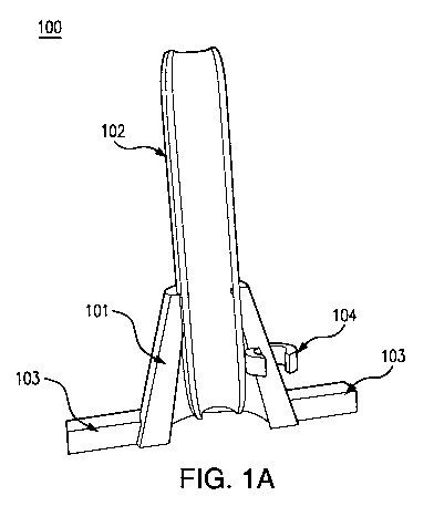
Fig. 1A is an illustration of a front view of an exemplary device in
accordance with
the present disclosure. Fig. 1B is an illustration of a top view of an
exemplary device in
accordance with the present disclosure.
Fig. 2A is a photo image of a front view of an exemplary device with a
cylindrical
rod coupled to a conical component in accordance with the present disclosure.
Fig. 2B is a
photo image of a back view of an exemplary device with a cylindrical rod
coupled to a
conical component in accordance with the present disclosure.
Fig. 3A is a photo image of a front view of an exemplary device with a
cylindrical
rod located in a lumen of a conical component in accordance with the present
disclosure.
.. Fig. 3B is a photo image of a side view of an exemplary device with a
cylindrical rod located
in a lumen of a conical component in accordance with the present disclosure.
Fig. 4A is a photo image of a side view of a skull model with an exemplary
device
implanted under the skin in accordance with the present disclosure. Fig. 4B is
a photo image
of a front view of a skull model with an exemplary device implanted under the
skin in
accordance with the present disclosure
Fig. 5 is a compilation of photo images demonstrating the exemplary devices
with
the footplates under the skin and the conical component functioning as a skin
retractor in
accordance with the present disclosure.
Fig. 6 demonstrates patient head computed tomography (CT) scan images
demonstrating ventricular placement with the disclosed device assistance in
accordance
with the present disclosure.
Fig. 7 is an illustration of an exemplary device with an alternative design in
accordance with the present disclosure.
Throughout the figures, unless otherwise stated, are used to denote like
features,
elements, components, or portions of the illustrated embodiments. Moreover,
while the
4
CA 03217824 2023-10-24
WO 2022/232521 PCT/US2022/026951
present disclosure will now be described in detail with reference to the
figures, it is done so
in connection with the illustrative embodiments.
DETAILED DESCRIPTION
The disclosed subject matter relates to devices and methods for stereotactic
placement of catheters.. The disclosed subject matter can be used with
moderate or local
sedation without the use of general anesthesia.
Unless otherwise defined, all technical and scientific terms used herein have
the
same meanings as commonly understood by one of ordinary skill in the art.
An "individual," "patient," or "subject," as used interchangeably herein, can
be a
human or non-human animal. Non-limiting examples of non-human animal subjects
include
non-human primates, dogs, cats, mice, rats, guinea pigs, rabbits, pigs, fowl,
horses, cows,
goats, sheep, and cetaceans.
The term "about" or "approximately" means within an acceptable error range for
the particular value as determined by one of ordinary skill in the art, which
will depend, in
part, on how the value is measured or determined, i.e., the limitations of the
measurement
system. For example, "about" can mean within 3 or more than 3 standard
deviations, per the
practice in the art. Alternatively, "about" can mean a range of up to +1-20%,
up to +/-10%,
up to +1-5%, and up to +/-1% of a given value. Alternatively, particularly
with respect to
biological systems or processes, the term can mean within an order of
magnitude, e.g.,
within 5-fold or within 2-fold, of a value.
The term "coupled," as used herein, refers to the connection of a device
component
to another device component by any means known in the art. The type of
coupling used to
connect two or more device components can depend on the scale and operability
of the
device. For example, and not by way of limitation, a coupling of two or more
components
5
CA 03217824 2023-10-24
WO 2022/232521 PCT/US2022/026951
of a device disclosed herein can include one or more joints, valves, fittings,
couplings,
transfer lines, or sealing elements.
In certain embodiments, the disclosed subject matter provides a device for
stereotactic placement of catheters. For example, the disclosed device can be
used by
surgeons to gain intraventricular access for drains and shunts. In non-
limiting embodiments,
the disclosed device can be used as a stand-alone adjunct for ventricular
catheter navigation.
As shown in Figs. 1A and 1B, an exemplary device 100 can include a conical
101, a
cylindrical rod 102, a footplate 103, and a clip 104.
In certain embodiments, the conical component can include a first opening and
a
second opening that form a lumen therethrough. As shown in Fig. 2A, the first
opening 201
can be located on the top of the conical component 202. In non-limiting
embodiments, the
diameter of the first opening can be from about 0.01 cm to about 1 cm, from
about 0.01 cm
to about 0.9 cm, from about 0.01 cm to about 0.8 cm, from about 0.01 cm to
about 0.75
cm, from about 0.02 cm to about 0.75 cm, from about 0.03 cm to about 0.75 cm,
from about
0.04 cm to about 0.75 cm, from about 0.05 cm to about 0.75 cm, from about 0.06
cm to
about 0.75 cm, from about 0.07 cm to about 0.75 cm, from about 0.08 cm to
about 0.75 cm,
from about 0.09 cm to about 0.75 cm, from about 0.1 cm to about 0.75 cm, from
about 0.2
cm to about 0.75 cm, from about 0.25 cm to about 0.75 cm, from about 0.3 cm to
about 0.75
cm, from about 0.4 cm to about 0.75 cm, or from about 0.5 cm to about 0.75 cm.
The size
of the first opening can vary depending on the size of the catheter. In non-
limiting
embodiments, the cylindrical rod can be coupled to the first opening (Figs 2A
and 2B). As
shown in Fig. 2A, the second opening 203 can be located on the base of the
conical
component 202. In non-limiting embodiments, the diameter of the second opening
can be
from about 0.01 cm to about 2 cm, from about 0.01 cm to about 1.9 cm, from
about 0.01
cm to about 1.8 cm, from about 0.01 cm to about 1.7 cm, from about 0.01 cm to
about 1.6
6
CA 03217824 2023-10-24
WO 2022/232521 PCT/US2022/026951
cm, from about 0.01 cm to about 1.5 cm, from about 0.05 cm to about 1.5 cm,
from about
0.03 cm to about 1.5 cm, from about 0.04 cm to about 1.5 cm, from about 0.05
cm to about
1.5 cm, from about 0.06 cm to about 1.5 cm, from about 0.07 cm to about 1.5
cm, from
about 0.08 cm to about 1.5 cm, from about 0.09 cm to about 1.5 cm, from about
0.1 cm to
about 1.0 cm, from about 0.1 cm to about 1.5 cm, from about 0.2 cm to about
1.5 cm, from
about 0.3 cm to about 1.5 cm, from about 0.4 cm to about 1.5 cm, from about
0.5 cm to
about 1.5 cm, from about 0.6 cm to about 1.5 cm, from about 0.7 cm to about
1.5 cm, from
about 0.8 cm to about 1.5 cm, from about 0.9 cm to about 1.5 cm, or from about
1 cm to
about 1.5 cm.
In certain embodiments, the thickness of the wall of the conical component can
be
from about 0.01 cm to about 3 cm, from about 0.02 cm to about 3 cm, from about
0.05 cm
to about 3 cm, from about 0.1 cm to about 3 cm, from about 0.1 cm to about 2.5
cm, from
about 0.1 cm to about 2 cm, from about 0.01 cm to about 1.5 cm, or about from
about 0.01
cm to about 1 cm.
In certain embodiments, the base of the conical component can be configured to
contact a target area. For example, the base of the conical component can be
contacted with
or be located on a calvarial surface anterior to a burr hole. In non-limiting
embodiments,
the first opening and the second opening can form a hollow lumen through which
a user can
view the burrhole. In non-limiting embodiments, the size of the openings can
be varied as
long as the burrhole lies at the midpoint of the conical base.
In certain embodiments, the conical component can be configured to be placed
on
the calvarial surface anterior to the burrhole in a semi-circular fashion,
leaving the entire
burrhole free for catheter placement. For example, as shown in Figs. 1A and
2A, the conical
component can be a 180-degree hollow truncated conical component. In non-
limiting
embodiments, the height of the conical component can be at least about 0.1 cm,
at least
7
CA 03217824 2023-10-24
WO 2022/232521 PCT/US2022/026951
about 0.2 cm, at least about 0.3 cm, at least about 0.4 cm, at least about 0.5
cm, at least about
0.6 cm, at least about 0.7 cm, at least about 0.8 cm, at least about 0.9 cm,
or at least about 1
cm.
In non-limiting embodiments, the conical component can be a 180-degree hollow
truncated conical component, with a diameter of about 1.5 cm at its base,
which can make
contact with the calvarial surface, anterior to the burrhole. The half conical
component can
be about 1 cm in height and about 0.75 cm in diameter at the apex. The half
conical
component can include a hollow lumen through which the burrhole can be
observed. The
disclosed measurements can be varied as long as the burrhole lies at the
midpoint of the
conical base.
In certain embodiments, the cylindrical rod can be coupled to the conical
component.
For example, as shown in Figs. 2A and 2B, the cylindrical rod can be coupled
to the conical
component adjacent to the first opening. The diameter of the cylindrical rod
can be from
about 0.01 cm to about 1 cm, from about 0.01 cm to about 0.9 cm, from about
0.01 cm to
about 0.8 cm, from about 0.01 cm to about 0.75 cm, from about 0.02 cm to about
0.75 cm,
from about 0.03 cm to about 0.75 cm, from about 0.04 cm to about 0.75 cm, from
about
0.05 cm to about 0.75 cm, from about 0.06 cm to about 0.75 cm, from about 0.07
cm to
about 0.75 cm, from about 0.08 cm to about 0.75 cm, from about 0.09 cm to
about 0.75 cm,
from about 0.1 cm to about 0.75 cm, from about 0.2 cm to about 0.75 cm, from
about 0.25
cm to about 0.75 cm, from about 0.3 cm to about 0.75 cm, from about 0.4 cm to
about 0.75
cm, pr from about 0.5 cm to about 0.75 cm. In non-limiting embodiments, the
diameter of
the cylindrical rod can be about 0.75 cm. In non-limiting embodiments, the
length of the
cylindrical rod can be from about 1 cm to about 10 cm. In non-limiting
embodiments, as
shown in Figs. 1 and 3, a portion of the cylindrical rod can be located in the
lumen of the
conical component.
8
CA 03217824 2023-10-24
WO 2022/232521 PCT/US2022/026951
In certain embodiments, the cylindrical rod can include a lumen. The lumen of
the
cylindrical rod can be configured to receive a catheter. For example, the
catheter can be
inserted into the lumen of the cylindrical rod and be aligned parallel to the
cylindrical rod.
Then, the catheter can be placed through a frustum of the conical component
into a ventricle.
In certain embodiments, the diameter of the lumen of the cylindrical rod can
be from
about 0.01 cm to about 2 cm, from 0.01 cm to about 1.5 cm, from 0.01 cm to
about 1 cm,
from 0.02 cm to about 1 cm, from 0.03 cm to about 1 cm, from 0.04 cm to about
1 cm, from
0.05 cm to about 1 cm, from 0.06 cm to about 1 cm, from 0.07 cm to about 1 cm,
from 0.08
cm to about 1 cm, from 0.09 cm to about 1 cm, or from 0.1 cm to about 1 cm.
In certain embodiments, the length of the cylindrical rod can be from about
0.1 cm
to about 10 cm, from about 0.1 cm to about 9 cm, from about 0.1 cm to about 8
cm, from
about 0.1 cm to about 7 cm, from about 0.1 cm to about 6 cm, from about 0.1 cm
to about 5
cm, from about 0.2 cm to about 5 cm, from about 0.3 cm to about 5 cm, from
about 0.4 cm
to about 5 cm, from about 0.5 cm to about 5 cm, from about 0.6 cm to about 5
cm, from
about 0.7 cm to about 5 cm, from about 0.8 cm to about 5 cm, from about 0.9 cm
to about 5
cm, about 1.0 cm to 5.0 cm, about 1.0 cm to 4.0 cm, or about 1.0 cm to 3.0 cm.
In certain embodiments, the footplate can be coupled to the conical component.
In
non-limiting embodiments, the footplate can be coupled to the base of the
conical
component for securing the position of the disclosed device. For example, as
shown in Figs.
4A and 4B, the footplate can be configured to be located at a calvarial
surface anterior to a
burr hole to secure the device underneath a skin of a subject. In non-limiting
embodiments,
the length of the footplate can be about from 0.1 cm to about 2 cm, from 0.1
cm to about
1.75 cm, from 0.1 cm to about 1.5 cm, from 0.1 cm to about 1 cm, from 0.2 cm
to about 1
cm, from 0.3 cm to about 1 cm, from 0.4 cm to about 1 cm, or from 0.5 cm to
about 1 cm.
9
CA 03217824 2023-10-24
WO 2022/232521 PCT/US2022/026951
In certain embodiments, the clip can be coupled to the conical component. As
shown
in Figs. 1-3, the clip 104 can be coupled to the wall of the conical component
101. The clip
can be configured to hold a catheter in place during a surgical procedure
(e.g., tunneling the
catheter, suturing the catheter securely to the scalp). This can allow the
proceduralist to
secure the catheter to the device so they do not have to physically hold it
into place while
completing the rest of the procedure. This enables the catheter to stay at the
right position
in the ventricle and not inadvertently fall deeper or be pulled out.
In certain embodiments, the inner diameter of the clip can be from 0.1 cm to
about
1 cm, from 0.2 cm to about 1 cm, from 0.2 cm to about 0.9 cm, from 0.2 cm to
about 0.8
cm, or from 0.2 cm to about 0.7 cm. In non-limiting embodiments, the outer
diameter of
the clip can be from about 0.2 cm to about 2 cm, from about 0.3 cm to about 2
cm, from
about 0.3 cm to about 1.5 cm, from about 0.3 cm to about 1 cm, from about 0.3
cm to about
0.9 cm, or from about 0.3 cm to about 0.8 cm. In some embodiments, the height
of the clip
can be from 0.1 cm to about 3 cm, 0.1 cm to about 2 cm, or 0.1 cm to about 1
cm.
In certain embodiments, the disclosed comical component, the cylindrical rod,
footplate, and the clip can include autoclavable resin, plastic, or any
sterilizable materials.
In non-limiting embodiments, the disclosed device, including the plastic, can
be autoclaved
for sterility. The use of plastic can allow mass production of a cheap and
disposable device.
In non-limiting embodiments, each compartment or the whole device can be
manufactured
using a three-dimensional printer.
Fig. 5 demonstrates the disclosed device in use. The footplates sit under the
skin,
and the conical component functions as a stand-alone skin retractor, allowing
the
proceduralist to have a full view of the surgical site and burrhole. Fig. 5
also demonstrates
the clip in use, allowing for secure holding of the catheter so the
proceduralist can have both
hands free for additional parts of the procedure. Fig. 6 demonstrates head CT
scans from 6
CA 03217824 2023-10-24
WO 2022/232521 PCT/US2022/026951
patients who underwent ventricular placement using device assistance,
demonstrating
successful ventricular cannulation.
Fig. 7 demonstrates the disclosed device with an alternative design. The
disclosed
device can include a burr hole adaptor to allow hand-free operation. In non-
limiting
embodiments, the disclosed device can include a silicone or a silicone-like
material to fit
snug within the burr hole and avoid bony obstructions.
In certain embodiments, the disclosed subject matter provides a method for
placing
a catheter using the disclosed device. An example method can include placing
the disclosed
device on a target burrhole for inserting the catheter, aligning the catheter
parallel to the
cylindrical rod, and placing the catheter through a frustum of the conical
component into a
target tissue. In non-limiting embodiments, the method can further include
holding the
catheter using the clip during a surgical procedure.
In certain embodiments, the disclosed device can increase the accuracy of
external
ventricular drain (EVD)/shunt catheter placement allowing for less
malpositioned catheters.
.. In non-limiting embodiments, the disclosed device can be a disposal device
and/or an
autoclavable device so that the infection during a surgical procedure can
decrease.
In non-limiting embodiments, the disclosed device can include a footprint to
be used
in bedside procedures without extending incision. In non-limiting embodiments,
the
disclosed device can allow for improved two-handed surgeon ergonomics. The
disclosed
device can allow the trajectory modification without locking down the user
trajectory. The
footplates sit under the skin holding the base of the conical component flush
with the
calvarial surface without having to be held in place by the proceduralist.
Additionally, the
clip allows for the catheter to be held in place securely prior to final
securement by tunneling
and suture.
11
CA 03217824 2023-10-24
WO 2022/232521 PCT/US2022/026951
In certain embodiments, the disclosed device can be used for a subject with
various
indications. For example, the indication can include subarachnoid
hemorrhage,
intraventricular hemorrhage, traumatic brain injury (TBI), hydrocephalus,
pseudotumor and
post-surgical wound drainage, or a combination thereof. In non-limiting
embodiments, the
disclosed device can provide reliable and accurate ventricular access with no
clinical
complications (e.g., tract hemorrhages or post-procedural infections).
In certain embodiments, the disclosed device can be used as a stand-alone
adjunct
for ventricular catheter navigation. In non-limiting embodiments, the
disclosed subject
matter provides a kit for extra-ventricular puncture/drainage or ventricular
shunting
systems, including the disclosed device. The disclosed subject matter can be
used by
neurosurgeons or other providers accessing the ventricular system that can be
used at the
bedside and in the operation room.
In certain embodiments, the disclosed device can allow the adjustment based on
the
patient's scan. For example, when the patient's scan demonstrates the shift of
the ventricles
based on the measurement of the pre-procedure CT scan, the proceduralist is
able to adjust
the angle of the catheter accordingly. In this instance, the catheter can be
aligned with the
trajectory demonstrated by the cylindrical component, and then the catheter
angle can be
adjusted slightly based on the preoperative assessment of the ventricular
location.
EXAMPLES
Example 1:
The effects of the disclosed subject matter on the accuracy of catheter
placement
were assessed by giving a ninety-degree trajectory for external ventricular
drain (EVD) or
shunt placement with the disclosed device. The disclosed device (i.e., DIVE
guide) was
sterilized and placed on the top of the skull as a guide that shows a 90-
degree angle between
12
CA 03217824 2023-10-24
WO 2022/232521 PCT/US2022/026951
the skull to the ventricles, which can be used for catheter placement. The
device was placed
without going inside the skull, brain, or ventricular system.
16 total patients have been enrolled for ventricular access using the DIVE
guide. 7
patients underwent ventriculoperitoneal shunting, and 9 patients underwent
bedside
extraventricular drain placement. 100% (16/16) of procedures had successful
ventricular
cannulation in an average of 1.12 passes with 0% (0/0) requiring repositioning
following
confirmatory head computed tomography (HCT). On post-procedure head CT, the
tip of
the catheter was located in the ipsilateral frontal horn or 3rd ventricle in
87.5% (14/16) of
patients and in the contralateral lateral ventricle in 12.5% (2/16) of
patients. See Table 1.
Catheter accuracy N=16
Intraventricular 100% (16/16)
Kakarla Grade 1 87.5% (14/16)
(ipsilateral frontal horn/3rd)
Kakarla Grade 2 12.5% (2/16)
(contralateral frontal horn)
Kakarla Grade 3 0% (0/16)
(eloquent ti s sue/ci sterns)
Table 1. Effects of the DIVE guide on the accuracy of catheter placement.
Compared to the standard freehand ventricular access, the DIVE guided
procedures
are superior in both the average number of passes and accuracy of catheter
placement. The
device clip secured the catheter during tunneling, and no catheters were
inadvertently
dislodged. No patients suffered clinical complications or infection.
Example 2:
Safe and reliable access to the ventricular system can be an important skill
in the
neurosurgical armamentarium. While the freehand technique has remained an
accepted
method for ventricular access, the accuracy rate of catheter placement has
been reported as
low as 40%, pass attempts as high as 3 per procedure, and complications
between 10-40%.
13
CA 03217824 2023-10-24
WO 2022/232521 PCT/US2022/026951
Certain devices have been developed to assist with catheter placement.
However, these
devices often prove cumbersome, necessitating a large incision, or require
expensive
navigation technology.
The disclosed subject matter provides a low-profile device for reliable and
accurate
ventricular access with improved safety, efficacy and accuracy.
A novel device for ventricular entry, the DIVE guide, was designed. 50
patients
undergoing extra ventricular drain (EVD) or ventricular shunt placement were
prospectively
enrolled for DIVE assisted catheter placement with a non-significant risk
(NSR) device
designation. The primary outcome is the location of the catheter tip on post-
operative CT
scan and secondary outcome measures include a total number of catheter passes,
clinically
important tract hemorrhages and post-procedural infections.
50 total patients were prospectively enrolled for ventricular access with DIVE
assistance. Indications included sub arachnoi d hemorrhage, intraventricular
hemorrhage,
TBI, hydrocephalus, pseudotumor and post-surgical wound drainage. 76% (38/50)
underwent right sided catheter placement and 24% (12/20) underwent left. 100%
(50/50)
of procedures had successful cannulation in an average of 1.06 passes. On post-
procedure
head CT, the tip of the catheter was located in the ipsilateral frontal horn
or 3rd ventricle
(Kakarla Grade 1) in 92% (46/50) and in the contralateral lateral ventricle
(Kakarla Grade
2) in 8% (4/50). See Table 2. There were no clinically significant tract
hemorrhages or post
procedural infections.
Catheter accuracy N=50
Intraventricular 100% (50/50)
Kakarla Grade 1 92% (46/50)
(ipsilateral frontal horn/3rd)
14
CA 03217824 2023-10-24
WO 2022/232521 PCT/US2022/026951
Kakarla Grade 2 8% (4/50)
(contralateral frontal horn)
Kakarla Grade 3 0% (0/16)
(eloquent tissue/cisterns)
Table 2. Improved accuracy of catheter placement with the DIVE guide.
As shown in Table 2, 100% of DIVE procedures had successful ventricular
cannulation, with 92% achieving Kakarla Grade 1, in an average of 1.06 passes.
The DIVE
provides reliable and accurate ventricular access with no clinical
complications.
All patents, patent applications, publications, product descriptions, and
protocols,
cited in this specification are hereby incorporated by reference in their
entireties. In case of
a conflict in terminology, the present disclosure controls.
While it will become apparent that the subject matter herein described is well
calculated to achieve the benefits and advantages set forth above, the
presently disclosed
subject matter is not to be limited in scope by the specific embodiments
described herein.
It will be appreciated that the disclosed subject matter is susceptible to
modification,
variation, and change without departing from the spirit thereof. Those skilled
in the art will
recognize or be able to ascertain using no more than routine experimentation,
many
equivalents to the specific embodiments described herein. Such equivalents are
intended to
be encompassed by the following claims.