Note: Descriptions are shown in the official language in which they were submitted.
WO 2022/238411
PCT/EP2022/062653
1
MICRON EEDLE BASED DELIVERY SYSTEM
Field of the invention
This invention relates to a microneedle based delivery system, in particular
such a system for drug or
vaccine delivery to target tissue such as skin tissue with accuracy and ease
of use.
Background of the invention
Microneedles are gaining increased use in various medical applications in view
of the many
documented benefits for both patients and medical professionals, for example
reduced tissue
trauma, surgery times and patient recovery, reduced risk of infection, and
minimising the surgical or
medical equipment needed in procedures involving microneedle based devices.
Such microneedles
allow for extremely accurate, and shallow, deployment into tissue, which can
be beneficial in various
applications, for example in drug delivery, and more particularly in drug
delivery to sensitive or
delicate tissue such as skin, ocular tissue and oral mucosa.
International patent applications W02018/069543 and W02019/201903 provide
detailed disclosures
of the configuration and operation of microneedles and opposing microneedle
arrays which may be
provided in the form of a device for application to a tissue substrate for
various surgical and
therapeutic uses, one particular use being drug delivery directly from or
through the microneedles
provided. The disclosures of W02018/069543 and W02019/201903 are incorporated
herein in their
entirety.
Intradermal delivery of drugs and vaccines offers significant advantages over
conventional
intramuscular and oral delivery routes for vaccines and drugs. Systemic uptake
of therapeutics via
the dermal blood capillaries and lymphatic system offers a host of benefits,
including avoiding the
deleterious effects of first-pass metabolism, rapid drug onset and improved
bioavailability of APIs,
such as biologics, that are not readily absorbed across the mucosal layers of
the gastrointestinal
tract. Additionally, the dermal layer of skin is replete with antigen
presenting cells and therefore
represents an optimal location for the delivery of vaccines in order to elicit
an enhanced immune
response, and in some cases, using lower dosages relative to standard
intramuscular administration.
The viscoelastic and highly deformable nature of skin poses significant
clinical challenges in the
delivery of liquid formulations of both low and high volume and low and high
viscosity (such as can
be the case for certain of biologics) to specific depths in skin using hollow
microneedle and
hypodermic needle-based approaches. Uncontrolled deformation of the skin
significantly limits
accuracy of depth targeting, which is further compounded by natural inter- and
intra-subject
CA 03218075 2023- 11- 6
WO 2022/238411
PCT/EP2022/062653
2
anatomical variability in thickness and biomechanical properties of skin. Skin
deformation and
compression caused by placement and application of these devices increases the
injection pressure
requirements, impacts flow-rate, significantly limits the physical volumes and
viscosities that can be
delivered and also impairs intradermal bleb/bolus diffusion kinetics.
Furthermore, the absence of
intrinsic anchorage of microneedle technologies, in particular, during
intradermal injection requires
additional mechanical work be manually applied to the injection device or
patch to counter the
injection pressure. Unless balanced, this component of injection pressure is
driving to eject and
cause relative migration of skin and/or needle tips, affecting the site of
delivery but which can also
result in formulation leakage, efflux and spray back. Appreciably, operator
movement(s) during
handling and injection is another factor further impacting depth targeting
accuracy and dose delivery,
where the absence of intrinsic anchoring can facilitate gross movements of the
needle tips relative to
the skin and distribute the injection away from the intended target depth in
skin.
The layered structure of the eye, in addition to the delicate nature of the
ocular tissue and the high
potential for damage or complications during any form of ocular surgery or
therapy, has presented
difficulties in the targeted delivery of drugs to the eye, in particular where
it is required to deliver the
drug to a particular layer within the eye in order to improve efficacy and
potentially avoid side effects
which may arise when a drug is delivered to an unintended part of the eye.
Conventional drug
delivery to the eye is complicated through natural and, significant
variability in the thickness of the
sclera (outermost layer) of the eye. In addition, the eye readily rotates
during injection, requiring
stabilisation, and there is deformation of the eye tissue during injection
which limits the ability to
target specific regions of the eye. Relative movement of the injector and drug
efflux during injection
also occur, further complicating the procedure and compromising the efficiency
of payload delivery.
It is therefore an object of the present invention to provide a self-anchoring
microneedle based
delivery system which is operable to quickly and easily deploy microneedles
into a tissue substrate
such as the skin and eye, for the purposes of targeted drug delivery of both
low and high volumes of
low and high viscosity solutions (such as biologics).
Summary of the invention
According to the present invention there is provided a microneedle based
delivery system comprising
a body having a first section and a second section displaceable relative to
one another; at least one
first hollow microneedle provided on the first section and at least one second
hollow microneedle
provided on the second section, the first and second sections being
displaceable relative to one
another in order to transition the microneedles between a disengaged state and
an engaged state;
and a delivery manifold in fluid communication with the first and second
hollow microneedles.
Preferably, a longitudinal axis of the at least one first microneedle extends
at a first oblique angle
relative to the direction of displacement between the first and second
sections, and a longitudinal
CA 03218075 2023- 11- 6
WO 2022/238411
PCT/EP2022/062653
3
axis of the at least one second microneedle extends at a second oblique angle
relative to the
direction of displacement between the first and second sections.
Preferably, the first oblique angle extends away from the second oblique
angle.
Preferably, the at least one first microneedle is transversely offset to the
at least one second
microneedle relative to the direction of displacement between the first and
second sections.
Preferably, the first and the second sections are slidably and/or hingedly
displaceable relative to one
another.
Preferably, the delivery manifold is captured between the first and second
sections at least when the
microneedles are in the engaged state.
Preferably, the delivery manifold is clamped against the body when the
microneedles are in the
engaged state such as to establish a fluid tight seal between the delivery
manifold and the body.
Preferably, the delivery manifold comprises an inlet adapted for connection
with a fluid reservoir and
an outlet engagable with the body such that the inlet is in fluid
communication with the hollow
microneedles.
Preferably, the body defines a chamber between the first and second sections
in which the outlet of
the delivery manifold is captured and which chamber is arranged to bias the
outlet into sealing
engagement with the body at least when the system is in the engaged state.
Preferably, the body defines an enclosure at least partially surrounding the
delivery manifold.
Preferably, the delivery manifold defines a first fluid flow path in fluid
communication with the at least
one first microneedle and an independent second fluid flow path in fluid
communication with the at
least one second microneedle.
Preferably, the first and second section of the body each define an elongate
arm at a free end of
which the respective at least one microneedle is provided.
Preferably, each arm defines an upper end opposite the lower end and a fluid
flow path extending
through the arm between the upper and lower end, the delivery manifold being
in fluid
communication with the fluid flow path at the upper end of each arm.
Preferably, the delivery manifold is in fluid communication with the end of
the fluid flow paths when
the microneedles are in both the engaged and disengaged state.
CA 03218075 2023- 11- 6
WO 2022/238411
PCT/EP2022/062653
4
Preferably, the microneedle based delivery system comprises a release
mechanism defined by a
trigger on each of the first and second sections and arranged to facilitate
manual displacement of the
system into the disengaged state.
Preferably, the microneedle based delivery system comprises a locking
mechanism to releasably
secure the system in the engaged state.
Preferably, the locking mechanism is integrated with at least one of the
triggers and is releasable
through actuation of the trigger.
Preferably, the at least one trigger with which the locking mechanism is
integrated is resiliently
deformable.
Preferably, the microneedle based delivery system comprises a stop releasably
engageable with the
body to limit relative displacement between the first and second sections.
Preferably, the delivery system comprises a retention lock operable to prevent
the first and second
sections from being separated from one another beyond the disengaged state.
Preferably, the delivery system comprises a pair of tissue contacting feet
each defining a tissue
contacting surface which is substantially longitudinally aligned with the
first and second hollow
microneedles.
Preferably, the microneedle based delivery system comprises interlocking
elements operable to
prevent the first and second sections from being displaced from the disengaged
to the engaged state
when the elements are interlocked, and which are separable in response to
downward pressure and
reactive forces applied to the feet by contacted tissue.
Brief description of the drawings
The present invention will now be described with reference to the accompanying
drawings, in which:
Figure 1 illustrates a perspective view of a microneedle based delivery system
according to an
embodiment of the present invention, coupled with a syringe, and in a
disengaged state;
Figure 2 illustrates a front elevation of the delivery system as shown in
Figure 1:
Figure 3 illustrates a front elevation of the delivery system in an engaged
state;
Figure 4 illustrates a cut away perspective view of the delivery system in the
disengaged state;
CA 03218075 2023- 11- 6
WO 2022/238411
PCT/EP2022/062653
Figure 5 illustrates a cut away perspective view of the delivery system in the
engaged state;
Figure 6 illustrates an exploded perspective view of the delivery system from
a first side;
5
Figure 7 illustrates the exploded perspective view of Figure 6 from the
reverse side;
Figure 8a illustrates a sectioned front elevation revealing a release
mechanism when the system is in
the engaged state;
Figure 8b illustrates the arrangement of Figure 8a with the release mechanism
being activated;
Figure 8c illustrates the arrangement of Figure 8a when the system is in the
disengaged state;
Figure 9 illustrates a perspective view of a microneedle based delivery system
according to an
alternative embodiment of the present invention and in a disengaged state;
Figure 10 illustrates a front elevation of the delivery system as shown in
Figure 9;
Figure 11 illustrates a front elevation of the delivery system of Figures 9
and 10 in an engaged state;
Figure 12 illustrates a cut away view of the delivery system of Figures 9 to
11 in the engaged state;
Figure 13 illustrates a front elevation of a microneedle based delivery system
according to a further
alternative embodiment of the present invention and in a disengaged state;
Figure 14 illustrates a front elevation of the delivery system as shown in
Figure 13 in a partially
engaged state;
Figure 15 illustrates a front elevation of the delivery system of Figures 13
and 14 in a fully engaged
state;
Figure 16 illustrates a perspective view of a stop for location within the
delivery system of Figures 13
to 15 to facilitate selective limitation of displacement between first and
second sections of the
system;
Figure 17 illustrates a perspective view of a microneedle based delivery
system according to a
further embodiment of the present invention in a disengaged state;
Figure 18 illustrates a side elevation of the delivery system as shown in
Figure 17:
CA 03218075 2023- 11- 6
WO 2022/238411
PCT/EP2022/062653
6
Figure 19 illustrates a perspective view of the microneedle based delivery of
Figure 17 in an
engaged state;
Figure 20 illustrates a side elevation of the delivery system as shown in
Figure 19;
Figure 21 illustrates an exploded perspective view of the delivery system of
Figures 17 to 20;
Figure 22 illustrates a side elevation of the delivery system of Figures 17 to
21 with a pair of halves
substantially separated from one another;
Figure 23 illustrates an end elevation of the delivery system of Figures 17 to
22;
Figure 24 illustrates a perspective view of one half or section of the
delivery system of Figures 17 to
23 as seen from one side thereof;
Figure 25 illustrates an alternative perspective view of the section shown in
Figure 24;
Figure 26 illustrates a perspective view of an other section of the delivery
system for interlocking
engagement with the section of Figure24;
Figure 27 illustrates the halves or sections shown in Figure 24-26 in a
designed for manufacturing
form factor;
Figure 28 illustrates the section of Figure 27 separated into two constituent
parts;
Figure 29 illustrates a perspective view of a microneedle based delivery
system according to another
embodiment of the present invention in a partially disassembled or separated
state;
Figure 30 illustrates the delivery system as shown in Figure 29 advanced into
a disengaged state:
Figure 31 illustrates an end elevation of the microneedle based delivery of
Figures 29 and 30; and
Figure 32 illustrates a plan view of the delivery system of Figures 29 to 31
in an engaged state.
Detailed description of the drawings
Referring now to Figures 1 to 8 of the accompanying drawings there is
illustrated a microneedle
based delivery system, generally indicated as 10, for use in delivering one or
more fluids, in
particular a dose of a drug or therapeutic agent in liquid form, to a target
area of tissue. The delivery
system 10 is suitable for use with a wide range of tissue, for example skin or
muscle, but is
CA 03218075 2023- 11- 6
WO 2022/238411
PCT/EP2022/062653
7
particularly suited for use in delivering a drug to ocular tissue such as the
subchoroidal or
suprachoroidal regions of the eye (not shown). The delivery system 10 is also
preferably adapted to
be coupled to an external fluid supply, most preferably a conventional syringe
S as will be described
hereinafter, to allow a fluid to be dispensed from the syringe S or other
external fluid supply to the
target tissue.
The delivery system 10 comprises a body 12 having a first section 14 and a
second section 16 which
are respectively provided with at least one first hollow microneedle 18 and at
least one second
hollow microneedle 20 which are adapted to be reversibly insertable into the
target tissue to allow for
drug delivery through the hollow microneedle 18, 20, the operation of which
will be described in
detail hereinafter. The number of microneedles 18, 20 may be varied, for
example to suit a particular
application, target area, drug to be delivered and/or delivery rate, and it is
also envisaged that one or
more solid microneedles (not shown) may be provided to securely anchor the
hollow microneedles
18, 20 to the target tissue to achieve reliable drug delivery. The material,
dimensions, orientation,
and relative positioning of the microneedles 18, 20 may also be varied as
necessary. For example
the dimension and/or orientation of the microneedles 18, 20 may be arranged to
provide a desired
depth of insertion into the target tissue, which can therefore facilitate
accurate drug delivery to
specific locations or layers of tissue.
In a preferred embodiment the body 12 is substantially formed from one or more
polymers and the
delivery system 10 is intended as a single use product. The microneedles 18,
20 may also be
formed from a polymer, or may be metal or another material and suitably
secured to the first and
second section 14, 16. However it is also envisaged that the system 10 could
be substantially
formed from a metal such as stainless steel, titanium, etc. or a hard wearing
polymer and may be
reusable following sterilisation, for example in an autoclave. The
microneedles 18, 20 could be
provided as a modular component releasably securable to the body 12 and thus
single use while the
body 12 is reusable.
The first and second sections 14, 16 of the body 12 are secured together but
displaceable relative to
one another by a fixed distance, and in the embodiment illustrated are
slidably displaceable relative
to one another to transition the system 10, and in particular the microneedles
18,20 between a
disengaged state, for example as illustrated in Figures 1, 2 and 4, and an
engaged state, for
example as illustrated in Figures 3 and 5. In the disengaged stage the
microneedles 18, 20 are in a
first orientation relative to one another prior to application to the target
tissue, and in the engaged
state are in a second orientation relative to one another. In use the
microneedles 18, 20 are applied
against the target tissue in the disengaged state and the system 10 is then
displaced into the
engaged state by manually advancing the first and second section 14, 16
towards one another,
which action effects the relative displacement of the microneedles 18, 20 in a
manner which draws
and anchors the microneedles 18, 20 into the target tissue to allow drug
delivery as hereinafter
described.
CA 03218075 2023- 11- 6
WO 2022/238411
PCT/EP2022/062653
8
The underlying methodology of this deployment technique is described in detail
in the above
mentioned international applications W02018/069543 and W02019/201903. For ease
of reference,
hereinafter directions or dimensions in a direction between the first and
second sections 14, 16 of the
delivery system 10 and the microneedles 18,20 will be referred to as being an
"X" coordinate or
direction, transversely or along the width will be referred to as being a "Y"
coordinate and along the
depth will be referred to as being a "Z" coordinate, and as represented
schematically in Figure 4
relative to the delivery system 10.
In order to allow the microneedles 18, 20 to be accurately positioned,
particularly in the case of
tissue that is relatively difficult to accurately engage or pierce, the body
12 comprises a first arm 22
extending from the first section 14 and an adjacent second arm 24 extending
from the second
section 16, both of which are elongate in form in the "Z" or depth direction,
which is beneficial for
engaging with relatively inaccessible tissue such as ocular tissue. The at
least one first microneedle
18 is provided on a lower end of the first arm 22 while the at least one
second microneedle 20 is
provided on a lower end of the second arm 24. In the embodiment illustrated
the arms 22, 24 taper
inwardly as they extend away from the body 12 towards the microneedles 18, 20,
thereby defining a
relatively small footprint at the lower end on which the microneedles 18, 20
are located, allowing both
accurate location and good visibility of the microneedles 18, 20 and
surrounding tissue during
application to the target tissue. It will of course be appreciated that the
shape, orientation and
dimensions of the arms 22, 24 may be varied as required, in particular
depending on the application
of the delivery system 10 and/or the type and/or location of the target
tissue..
In the embodiment illustrated the first and second arms 22, 24 are located
adjacent one another, but
offset in the "Y" direction, transverse to the "X" direction in which the
first and second sections 14, 16
are displaceable relative to one another. In this way the arms 22, 24 can move
into overlapping
alignment as the system 10 is transitioned into the engaged state, drawing the
microneedles 18, 20
into the tissue, as can be seen in particular in Figures 4 and 5. However
other geometries and
arrangements are also possible, for example one arm could be provided with a
channel to at least
partially receive the other arm when in the engaged state, or the arms may
remain separated or
spaced from one another in both the disengaged and engaged states.
In order to allow drug delivery through the hollow microneedles 18, 20 at
least the first arm 22 is
provided with a first delivery conduit 26 extending internally along the
length of the arm 22 (in the "Z"
direction) and in fluid communication with the at least one first microneedle
18. In the preferred
embodiment illustrated the second arm 24 is provided with a second delivery
conduit 28 extending
internally along the length of the second arm 24 and in fluid communication
with the at least one
second microneedle 20. It is to be understood that the system 10 could
function with a single
delivery conduit to supply only the first or second microneedles 18, 20, but
it is preferred that both
the hollow microneedles 18, 20 can be supplied with and used for drug delivery
to the target tissue.
CA 03218075 2023- 11- 6
WO 2022/238411
PCT/EP2022/062653
9
The first and second arms 22, 24 each terminate at an upper end or face 30
onto which the delivery
conduits 26, 28 open, thereby establishing a fluid flow path from the upper
face 30 through the arms
22, 24 to the microneedles 18, 20. Referring to Figure 4 it can be seen that
when the system 10 is in
the disengaged state the upper ends of the delivery conduits 26, 28 are spaced
from one another in
the "X" direction, and when the system 10 is transitioned into the engaged
stage as illustrated in
Figure 5 the upper ends of the delivery conduits 26, 28 are adjacent one
another and thus
overlapping or aligned in the "X" direction. Located above and enclosing the
upper ends of the
deliver conduits 26,28 is a delivery manifold 32 which comprises an inlet 34,
an outlet 36 and a
lumen 38 extending therebetween. The outlet 36 is in register with the upper
faces 30 such as to
enclose the upper ends of the delivery conduits 26, 28, thereby allowing fluid
delivery through the
delivery manifold 32, into the delivery conduits 26, 28, and ultimately to the
microneedles 18, 20.
The inlet 34 is adapted to be releasably secured to a convention luer lock
connector or head H of the
syringe S, but may be adapted for a fluid tight connection to any other
desired external fluid supply
(not shown). It will therefore be understood that the syringe S, containing a
drug or other fluid to be
delivered, can be connected to the system 10 via the delivery manifold 32, and
fluid can thus be
dispensed from the syringe S, through the microneedles 18, 20 to the target
tissue in a reliable and
accurate manner.
In order to secure the delivery manifold 32 to the body 12 the delivery
manifold 32, and most
preferably the outlet 36, is captured between the first and second sections
14, 16 at least when the
microneedles 18,20 are in the engaged state. In the embodiment of Figures 1 to
8 the body 12
defines a chamber 40 located directly above and partially defined by the upper
faces 30, one part or
side of the chamber 40 formed in the first section 14 and the other side of
the chamber 40 being
formed in the second section 16 opposite one another. The chamber 40 is shaped
and dimensioned
to encapsulate the outlet 36, with an opening 42 being provided in an upper
wall of the chamber 40
through which the delivery manifold 32 extends into an enclosure 44 defined by
the body 12. The
enclosure 44 is preferably shaped and dimensioned to surround and effectively
enclose the delivery
manifold 32 such that, in use, the delivery manifold 32, and therefore the
connection to the syringe S,
is inaccessible. A mouth 46 is formed in an upper region of the enclosure 44
to accommodate the
syringe S. As the mouth 46 extends across the interface between the first and
second sections 14,
16 it will be appreciated that with the system 10 in the disengaged state the
mouth 46 will be
enlarged to allow the syringe S to be advanced into the chamber 44 to couple
the head H with the
inlet 34 of the delivery manifold 32. When the system 10 is transitioned into
the engaged state the
mouth 46 will partially close about the body of the syringe S to avoid any
unintentional movement or
uncoupling of the syringe S.
At the interface between the outlet 36 of the delivery manifold 32 and the
upper face 30 of the arms
22, 24, as seen in particular in Figures 4 and 5, the outlet 36 is dimensioned
to enclose the upper
ends of the delivery conduits 26, 28 when the system 10 is in both the
disengaged state of Figure 4
and the engaged state of Figure 5, although the system 10 could be adapted
such that the outlet 36
only encloses the upper ends of the delivery conduits 26, 28 when the system
10 is in the engaged
CA 03218075 2023- 11- 6
WO 2022/238411 PC
T/EP2022/062653
state. However, it is often necessary to prime a syringe prior to dispensing a
fluid, in order to ensure
there is no air in the syringe that would then be injected into the subject.
Thus by having the outlet
36 dimensioned to enclose the upper ends of the delivery conduits 26, 28 when
the system 10 is in
the disengaged state the syringe S can be primed or purged of air before
application of the
5 microneedles 18, 20 to the target tissue, as a fluid flow path exists
between the syringe S and the
microneedles 18, 20. When the microneedles 18,20 are secured to the target
tissue the system 10
will be in the engaged state, and as the outlet 36 also encloses the upper
ends of the delivery
conduits 26, 28 in this state the contents of the syringe S can then be
delivered to the target tissue
via the microneedles 18, 20.
It is envisaged that the manifold 32 could be replaced with a modified
manifold (not shown) defining
a pair of independent fluid flow paths each arranged in fluid communication
with one of the delivery
conduits 26, 28, thereby allowing two different fluids to be delivered to the
first and second
microneedles 18, 20 via a modified syringe (not shown) or pair of syringes
(not shown) or other fluid
supplies.
In order to prevent unwanted leakage of the contents of the syringe S at the
interface between the
outlet 36 and the upper face 30 of the arm 22, 24 a suitable fluid tight seal
is established at the
interface. This seal may be achieved in a number of ways, for example using
gaskets, deformable
rubber/elastomer seals, coatings, sealing geometries, mechanical interlocking
or an interference fit
between the outlet 36 and the upper face 30 of the arms 22, 24. In the
embodiment of Figures 1 to 8
this seal is established by complimentary dimensioning of the outlet 36 and
the chamber 40, in
particular in the "Z" direction whereby the height or "Z" dimension of the
chamber 40 is such that the
upper wall of the chamber 40 contacts the upper wall of the outlet 36 and
biases the outlet 36
against the upper face 30 of the arms 22, 24 such as to establish a seal
therebetween. In a
particularly preferred arrangement, the upper wall of the chamber 40 and the
upper wall of the outlet
36, which are in face to face engagement, have a corresponding draft or
incline, for example in the
region of 1 or 2 , optionally up to 5 or 10 or more in the "X" direction of
relative displacement
between the first and second sections 14, 16. In this way, as the first and
second sections 14, 16
are displaced towards one another in transitioning the system 10 into the
engaged state, the bias
applied to the outlet 36 will increase, thereby increasing the seal at the
interface between the outlet
36 and the upper face 30 of the arms 22, 24. This arrangement increases the
mechanical
compressive force acting on the outlet 36 from nominal in the disengaged state
to maximal in the
engaged state. This allows for the above described priming of the syringe Sand
microneedles 18,20
prior to injection into tissue, where pressures are low, to allowing higher
pressures associated with
the injection once the microneedles 18, 20 have been deployed into the tissue,
without fear of
leakage.
In use the delivery system 10 is transitioned from the disengaged state to the
engaged state by
manually applying pressure to the first and second sections 14, 16 of the body
12, preferably via the
outer walls of the enclosure 44. As seen in Figures 6 and 7 the walls of body
12 which define the
CA 03218075 2023- 11- 6
WO 2022/238411
PCT/EP2022/062653
11
enclosure 44 include extensions 48 on the first section 14 and corresponding
guideways 50 on the
second section 16 which interlock to secure the sections 14, 16 together while
permitting limited
relative displacement. Tabs 52 are provided in the extensions 48 which will be
visible through
corresponding windows 54 when the system 10 has been correctly transitioned
into the engaged
state to provide a visual indication to the user signifying that the
microneedles 18, 20 are embedded
and fluid delivery from the syringe S can be undertaken. The tabs 52 further
serve to ensure that the
first and second sections 14, 16 can not disengage from one another. Graphical
indicia 56 may be
provided about the body 12 to indicate the correct direction in which the
first and second sections 14,
16 should be displaced to transition the system 10 into the engaged state. The
system 10 may be
provided with a stop (not shown) releasably engageable with the body 12, for
example located within
the enclosure 44, to limit relative displacement between the first and section
sections 14, 16 as a
means of limiting the depth of penetration of the microneedles 18, 20 to suit
a particular application
or anatomical consideration.
Once delivery of fluid to the target tissue has been completed it is necessary
to disengage the
system 10, in particular the microneedles 18, 20, from the tissue. It is
therefore necessary for the
user to displace the first and second sections 14, 16 away from one another to
transition the system
10 into the disengaged state and thus affect withdrawal of the microneedles
18, 20. In order to
facilitate this action the system 10 comprises a release mechanism in the form
of first and second
triggers 58, 60 respectively provided on the first and second sections 14, 16,
in the region of the
upper face 30 of the arms 22, 24 and positioned beneath the enclosure 44. Each
trigger 58, 60
extends in cantilever form towards and marginally beyond the wall of the
enclosure 44 of the
opposite section 14, 16 in order to be accessible by the user. Each trigger
58, 60 terminates in an
enlarged paddle 62 which is shaped and dimensioned for operative engagement
with a finger or
thumb of the user. The triggers 58, 60 are therefore positioned side by side
and slide relative to one
another when the system 10 is displaced between the disengaged and engaged
states without
hindering said displacement of the system 10. Referring to Figure 6 the system
10 is preferably
provided with a locking mechanism to secure the system 10 in the engage and or
disengaged state,
in particular to avoid any inadvertent withdrawal of the microneedles 18, 20,
for example while fluid is
being delivered to the tissue. In the embodiment illustrated the locking
mechanism is provided in the
form of a protrusion or detent 64 on an upper face of the first trigger 58 and
a correspondingly
shaped and dimensioned first socket 66 and second socket 68 on the underside
of the enclosure 44
which is in face to face with the upper face of the first trigger 58, and
visible in Figures 8a, 8b and 8c.
The first and second sockets 66, 68 are spaced apart from one another by the
distance through
which the first and second sections 14, 16 are displaceable. The first socket
66 is located to receive
the detent 64 when the system 10 is in the engaged state as illustrated in
Figure 8a, to releasably
secure the system 10 in the engaged state.
Referring in particular to Figures 8a, 8b and 8c the sequence of steps to
effect displacement of the
system 10 from the engaged to disengaged state is illustrated. Figure 8a shows
the system 10 in
the engaged state with the detent 64 captured in the first socket 66. In order
to begin displacing the
CA 03218075 2023- 11- 6
WO 2022/238411
PCT/EP2022/062653
12
first and second sections 14, 16 away from one another the system 10 is
gripped by the triggers 58,
60, for example between thumb and forefinger. At least the first trigger 58 is
resiliently deformable,
and the user therefore applies pressure to the first trigger 58 via the paddle
62 to deform the first
trigger 58 in a manner which will draw the detent 64 out of the first socket
66 as illustrated in Figure
8b. At this point the user can apply pressure to both triggers 58, 60 to
effectively push the triggers
58, 60 towards one another. This will effect relative displacement between the
first and second
sections 14, 16 to transition the system 10 into the disengaged state as shown
in Figure 8c, drawing
the microneedles 18, 20 out of the tissue. In this configuration the detent 64
will be aligned with the
second socket 68 and once pressure is released the first trigger 58 will
return and the detent 64 will
enter the second socket 68 to lock the system 10 in the disengaged state. The
system 10 can then
be withdrawn from the tissue.
Referring now to Figures 9 to 12 there is illustrated an alternative
embodiment of a microneedle
based delivery system according to the present invention, generally indicated
as 110. In this
alternative embodiment like components have been accorded like reference
numerals and unless
otherwise stated perform a like function.
The delivery system 110 comprises a body 112 having a first section 114 and a
second section 116
which are displaceable relative to one another to transition the system 110
between disengaged and
engaged states. The system 110 comprises first hollow microneedles 118
provided on the first
section 114 and second hollow microneedles 120 provided on the second section
116. The body
112 defines elongate first and second arms 122, 124 at a free end of which the
microneedles 118,
120 are provided. Although not illustrated, first and second delivery conduits
(not shown) extend
through the arms 122, 124 into fluid communication with the microneedles 118,
120. A delivery
manifold 132 is captured between the first and second sections 114, 116 to
facilitate the coupling of
a syringe (not shown) or other fluid supply to the system 110 for delivery
through the microneedles
118, 120 in the same manner as described above in relation to the system 10
shown in Figures 1 to
8. The body 112 defines an enc10sure144 above the arms 122, 124 in which the
delivery manifold
132 is contained and which is accessible through a mouth 146 at an upper
portion of the enclosure
144.
The overall composition, appearance and general operation of the system 110 is
the same as
described above with reference to the system 10 shown in Figures 1 to 8.
However, unlike the
system 10 the first and second sections 114,116 of the system 110 are
pivotally displaceable
relative to one another in order to transition the system 110 between the
disengaged and engaged
states. The first and second sections 114, 116 are secured to one another by
means of a pair of
hinges 180 which are located on opposite sides of the mouth 146, although it
will be appreciated that
numerous other configurations may be employed to enable this pivoting relative
displacement.
Thus as the first and second sections 114, 116 are displaced towards the
engaged state the
microneedles 118, 120 will move relative to one another along an arc,
advancing the microneedles
CA 03218075 2023- 11- 6
WO 2022/238411
PCT/EP2022/062653
13
118, 120 into the target tissue. Once the system 110 is in the engaged stage
with the microneedles
118, 120 embedded in the tissue a fluid can be delivered into the tissue from
the coupled syringe
(not shown) in the same manner as described above for the system 10. The
system 110 may be
provided with a stop (not shown) releasably engageable with the body 112, for
example located
within the enclose 144, to limit relative displacement between the first and
section sections 114, 116
as a means of limiting the depth of penetration of the microneedles 118, 120.
Similarly the system 110 may be disengaged from the tissue following fluid
delivery by utilising a
release mechanism in the form of a pair of triggers 158, 160 to displace the
first and second sections
114, 116 away from one another along an arcuate path about the pair of hinges
180 in a similar
manner as described above.
Referring to Figures 13 to 16 there is illustrated a further alternative
embodiment of a microneedle
based delivery system according to the present invention, generally indicated
as 210. In this
alternative embodiment like components have been accorded like reference
numerals and unless
otherwise stated perform a like function.
The delivery system 210 is effectively a variant of the system 110, comprising
a body 212 having a
first section 214 and a second section 216 which are again hingedly
displaceable relative to one
another to transition the system 210 between disengaged and engaged states.
The system 210
comprises first hollow microneedles 218 provided on the first section 214 and
second hollow
microneedles 220 provided on the second section 216. In this embodiment the
microneedles 218,
220 are of increased length relative to the previous embodiments in order to
permit increased tissue
penetration.
The body 212 defines elongate first and second arms 222, 224 at a free end of
which the
microneedles 218, 220 are provided. The first and second sections 214, 216 of
the system 210 are
secured to one another by means of a pair of hinges 280, although it will
again be appreciated that
numerous other configurations may be employed to enable this pivoting relative
displacement. Tabs
252 provided on the first section 214 are located within windows 254 on the
second section 216 to
both provide a visual indication as to the level of displacement of the first
and second sections 214,
216 and thus deployment of the microneedles 218, 220, and to prevent the first
and second sections
214, 216 from disengaging from one another.
The system 210 is optionally provided with a stop 290 shown in isolation in
Figure 16, and which in
use is located within a chamber (not shown) defined between the first and
second sections 214, 216
of the body 212 and arranged to selectively limit relative displacement
between the first and second
sections 214, 216 as a means of limiting the depth of penetration of the
microneedles 218, 220. The
stop 290 comprises first, second and third steps 292, 294, 296 of increasing
height, whereby the
stop 290 may be selectively positioned such that one of the steps 292, 294,
296 projects outwardly
though the window 254 such as to be captured between the tab 252 and the
window 254. The depth
CA 03218075 2023- 11- 6
WO 2022/238411
PCT/EP2022/062653
14
of the step 292, 294, 296 located in the window 254 will determine to what
extent the first and
second sections 214, 216 may be displaced together and thus to what extent the
system 210 can be
displaced into the engaged state. For example Figure 14 shows the system 210
displaced into a
partially engaged state with the microneedles 218, 220 in a first relative
position while Figure 15
shows the system 210 displaced into a fully engaged state with the
microneedles 218, 220 in a
second relative position which will achieve a greater tissue penetration than
that of Figure 14. These
two states may be permitted by locating different steps 292, 294, 296 of the
stop 290 within the
window 254. The stop 290 may also be positioned with the largest step 296
projecting through the
window 254 which may be dimensioned to effectively lock the system 210 in the
disengaged state to
prevent accidental deployment of the microneedles 218, 220, for example during
handling,
particularly when applying a torque to a syringe (not shown) being coupled to
the system 210.
Referring to Figures 17 to 28 there is illustrated a further alternative
embodiment of a microneedle
based delivery system according to the present invention, generally indicated
as 310. In this
alternative embodiment like components have been accorded like reference
numerals and unless
otherwise stated perform a like function.
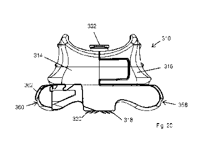
The delivery system 310 comprises a body 312 having a first section 314 and a
second section 316
which are displaceable relative to one another to transition the system 310
between disengaged and
engaged states substantially as hereinbefore described with reference to the
preceding
embodiments. The outer end wall of the first and second sections 314, 316 may
be contoured
and/or otherwise arranged to provide an ergonomic and/or retentive form factor
with which a user's
finger and/or thumb may be engaged during operation of the delivery system
310, in order to
establish a secure hold during deployment onto the target tissue. In this way
a user can securely
grip the first and second sections 314, 316 between a thumb and forefinger in
order to then squeeze
the sections 314, 316 together to move the delivery system 310 from the
disengaged to the engaged
state as hereinbefore described.
The system 310 comprises first hollow microneedles 318 provided on the first
section 314 and
second hollow microneedles 320 provided on the second section 316. As with
previous
embodiments, the material, dimensions, orientation, and relative positioning
of the microneedles 318,
320 may also be varied as necessary. For example the dimension and/or
orientation of the
microneedles 318, 320 may be arranged to provide a desired depth of insertion
into the target tissue,
which can therefore facilitate accurate drug delivery to specific locations or
layers of tissue.
Particular orientations of the microneedles 318, 320 may additionally improve
the manufacturability
of the delivery system 310, for example by rotation about a plane normal to
the axis of the respective
needle 318, 320, or a series of Euler rotations about a body-fixed coordinate
system of the needle
318, 320 (where one of the axes of this coordinate system is aligned with the
longitudinal axis of the
needle 318, 320). Such an orientation may provide additional performance
improvements whereby,
during deployment additional shear strain or deformation is applied to the
skin or other tissue in a
plane perpendicular to the direction of motion of the first and second
sections 314, 316. This can
CA 03218075 2023- 11- 6
WO 2022/238411
PCT/EP2022/062653
serve to further increase insertion efficiency by creating a shear strain
gradient along the vertical
depth of the needle 318, 320 in the skin, provide additional scratch fixation
on insertion and
potentially reduce pain and/or discomfort experienced by the user. Furthermore
by orienting the
needles 318, 320 in this way usability can be improved as deployment of the
needles 318, 320 is
5 less sensitive to slight deviation from the ideal normal orientation
during insertion whereby the
syringe (not shown) connected to the delivery system 310 is perpendicular to
the skin.
The first and second sections 314, 316 of the body 312 are secured together
but slidably
displaceable relative to one another by a fixed distance to transition the
system 310 between a
10 disengaged state, for example as illustrated in Figures 17 and 18, and an
engaged state, for
example as illustrated in Figures 19 and 20. In the disengaged stage the
microneedles 318, 320 are
in a first orientation relative to one another prior to application to the
target tissue, and in the
engaged state are in a second orientation relative to one another. As with
previously described
embodiments the microneedles 318, 320 are applied against the target tissue in
the disengaged
15 state and the system 310 is then displaced into the engaged state by
manually advancing the first
and second section 314, 316 towards one another to draw and anchor the
microneedles 318, 320
into the target tissue to allow drug delivery as hereinafter described.
The body 312 comprises an first arm 322 extending from the first section 314
and an adjacent
second arm 324 extending from the second section 316 on a lower end of each of
which the
respective microneedles 318, 320 are provided. The shape, orientation and
dimensions of the arms
322, 324 may be varied as required. The arms 322, 324 define internal delivery
conduits (not
shown) providing fluid communication between a delivery manifold 332 and the
respective
microneedles 318, 320 as hereinbefore described. The delivery manifold 332
comprises an upper
inlet 334 operable for connection with a syringe (not shown) or comparable
reservoir of a medium to
be delivered, such as a liquid drug composition or the like, and a lower
outlet 336 extending between
which is a lumen 338, the lower outlet 336 and lumen 338 preferably being
bifurcated (not shown) to
supply both sets of microneedles 318, 320 through the respective arm 322, 324.
The manifold 332 is
received within an enclosure 344 defined between the first and second sections
314, 316 but which
is open and thus allows a user visual access to the manifold 332 which may aid
in connection or
disconnection of the syringe (not shown). As described above the outlet 336,
in use, is captured
between the first and second sections 314, 316 in fluid tight contact with the
arms 322, 324 in order
to ensure a leak free delivery of fluid from the manifold 332 into the arms
322, 324 and ultimately the
microneedles 318, 320. This may be achieved as hereinbefore described or by
any other suitable
functional alternative arrangement. The manifold comprises a baseplate 339
which articulates with
tapered surfaces of enclosure 344 in such a way so as provide counter torque
resistance during
engagement of a leur lock syringe (not shown) to the inlet 334 and thereby
isolates the outlet 336
from torque loading and thus possible misalignment with the arms 322, 324.
Furthermore, the
clockwise nature of the torque is strongly resisted by the interaction of the
interlocked extensions 348
and guideways 350 and preventing the device 310 from being wedged apart.
Another important
CA 03218075 2023- 11- 6
WO 2022/238411
PCT/EP2022/062653
16
function of the manifold 332 is to provide an abutting surface against the
inner surfaces of enclosure
of 344 to prevent the coming apart of the two sections 314, 316 in the "Y"
direction.
In the embodiment illustrated the first and second sections 314, 316 are the
same component, but
arranged so as to be inter-engagable with one another as illustrated. In
particular each section 314,
316 defines a lateral extension 348 projecting from the right hand side, and a
correspondingly
shaped and dimensioned guideway 350 on the left hand side for receiving the
extension 348 in
sliding engagement therewith. In this way, with two identical parts 314, 316
facing one another the
right hand extension of each section 314, 316 is receivable in the left hand
guideway 350 of the
opposite section 314, 316. This arrangement provides a significant improvement
in manufacturing
the delivery system 310 as only a single part is required to provide both
sections 314, 316 but it is of
course understood that the opposed sections may have different
forms/geometries which still
providing the above described functionality. Figures 27 and 28 illustrate an
exemplary but non-
limiting form of the first section 314 as designed for manufacture, whereby
the section 314 is formed
from two parts which may be suitable secured together.
As with previous embodiments, the first and second arms 322, 324 are located
adjacent one
another, but offset in the "Y" direction on each section 314, 316, transverse
to the "X" direction in
which the first and second sections 314, 316 are displaceable relative to one
another. In the
embodiment illustrated each arm 324, 326 is offset to the right hand side so
that again, when
identical sections 314, 316 are facing and interlocked with one another the
arms 324, 326 sit
alongside one another in face to face engagement. In this way the arms 322,
324 can move
alongside one another as the system 310 is transitioned into the engaged
state, drawing the
microneedles 318, 320 into the tissue.
In order to allow a user to withdraw the microneedles 318, 320 once a drug or
the like has been
dispensed into the tissue the delivery system 310 is preferably provided with
a release mechanism in
the form of first and second triggers 358, 360 respectively provided on the
first and second sections
314, 316. Each trigger 358, 360 extends in cantilever form towards and
marginally beyond the wall
of the enclosure 344 of the opposite section 314, 316 in order to be
accessible by the user. Each
trigger 358, 360 terminates in a paddle 362 which is shaped and dimensioned
for operative
engagement with a finger or thumb of the user and which is independently
deformable relative to the
surrounding trigger 358, 360 as detailed hereinafter, in particular being
formed as an independent
cantilevered component captured within the cantilevered trigger 358, 360 and
thus being capable of
deflection independently of deflection of the trigger 358, 360.. The system
310 is preferably provided
with a locking mechanism to secure the system 310 in the engaged state, in
particular to avoid any
inadvertent withdrawal of the microneedles 318, 320, for example while fluid
is being delivered to the
tissue. In the embodiment illustrated the locking mechanism is provided in the
form of a protrusion
or detent 364 on underside of the first and second sections 314, 316 which is
in face to face
engagement with the upper face of the respective trigger 358, 360 and a
correspondingly shaped
and dimensioned first socket 366 and second socket 368 on an upper face of the
paddle 362. The
CA 03218075 2023- 11- 6
WO 2022/238411
PCT/EP2022/062653
17
first and second sockets 366, 368 are spaced apart from one another by the
distance through which
the first and second sections 314, 316 are displaceable between the disengaged
and engaged
states. The first socket 366 is located to receive the detent 364 when the
system 310 is in the
disengaged state, the socket 366 and detent 364 being arranged to provide a
relatively low
resistance to relative movement of the sections 314, 316, for example by
having complimentary
faces which can slide past/over one another with relative ease and which may
be accommodated by
deformation of the paddle 362 away from the underside of the respective
section 314, 316. In this
way the location of the detent 364 within the first socket 366 provides a low
level of retention of the
system 310 in the disengaged state which is nonetheless sufficient though to
prevent inadvertent
deployment of the system 310 during routine handling. When the system 310 is
displaced from the
disengage state to the engaged state each trigger 358, 360 and integral paddle
362 will move
relative to the respective detent 364, with the paddle 362 being deflected
away from the underside of
the section 314, 316 to disengage the detent 364 and the first socket 366. The
detent 364 will then
be brought into register with the second socket 368 as the system 310 reaches
the fully engaged
state and will snap into engagement with the second socket 368 in order to
retain the system 310 in
the engaged state. The detent 364 and second socket 368 are arranged to
prevent any reversal of
the system 310 from the engaged back to the disengaged state without a
positive action from the
user as detailed hereinafter.
The system 310 may also be provided with an additional safety features to
prevent inadvertent
deployment into the engaged state, in particular when a syringe (not shown) is
being connected. A
pin (not shown) or the like could be located in a chamber or opening (not
shown) provided in the
space between bottom edge of the extension 348 and the upper surface of the
respective trigger
358, 360 which would prevent inadvertent deployment of the system 310 until
the pin is actively
removed by the user. Additional or alternative safety features may of course
be employed to provide
this functionality.
In order to enable the first and second sections 314, 316 to be displaced away
from one another to
return the system 310 to the disengaged state the system 310 is gripped by the
paddle 362 of each
trigger 358, 360, for example between thumb and forefinger. Each paddle 362 is
resiliently
deformable, and the user therefore applies pressure to deform the paddles 362
in a manner which
will draw the second socket 368 downwardly out of register with the detent
364. At this point the
user can apply pressure to both triggers 358, 360 to effectively push the
triggers 358, 360 towards
one another. This will effect relative displacement between the first and
second sections 314, 316 to
transition the system 310 into the disengaged state, drawing the microneedles
318, 320 out of the
tissue. In this configuration the detent 364 will again be aligned with the
first socket 366 and once
pressure is released the paddles 362 will return and the detent 364 will enter
the first socket 366 to
hold the system 310 in the disengaged state. The system 310 can then be
withdrawn from the
tissue.
CA 03218075 2023- 11- 6
WO 2022/238411
PCT/EP2022/062653
18
The delivery system 310 may also be adapted to prevent the first and second
sections 314, 316 from
being pulled apart or away from one another beyond the disengaged state, in
order to ensure that a
user does not inadvertently effect such a displacement. The system 310 may
therefore comprise a
retention lock comprising a second projection or detent 370 provided on the
underside of the
respective section 314, 316 and a corresponding third socket 372 provided in
the respective trigger
358, 360. The second detent 370 and third socket 372 are positioned to be
engaged when the
system 310 is in the disengaged state, for example as seen in Figure 18, and
move away from one
another as the sections 314, 316 are displaced into the engaged state, thus
providing no resistance
to this movement. However, the shape and configuration of the detent 370 and
socket 372 is such
that they engage when the sections 314, 316 are in the disengaged state and
prevent the sections
314, 316 from being further separated from one another.
However in order to allow the two identical halves or sections 314, 316 to be
initially brought into
register with one another, for example from the initial position of engagement
shown in Figure 22,
each trigger 358, 360 must be able to pass over the second detent 370 as the
sections 314, 316 are
brought together towards the initial disengaged state from being completely
separated from one
another. In the embodiment illustrated this is facilitated by providing
complementary surfaces, the
second detent 370 with a sloping or ramped surface 374 while the respective
contacting portion of
the trigger 358, 360 comprises a curved or sloping surface 376. These surfaces
contact as the
sections 314, 316 are initially brought together, and as the trigger 358, 360
is of cantilevered form it
will be forced to deflect downwardly away from the second detent 370 as the
sections 314, 316 are
advanced towards the disengaged state, thus effectively defining a single
stage ratchet arrangement.
In the particularly preferred arrangement illustrated the portion of the
trigger 358, 360 defining the
sloped surface 376 is cantilevered in both a longitudinal or "X" direction in
addition to a transverse or
"Y" direction and is therefore readily deflected to allow the trigger 358, 360
to pass the second detent
370. As the first and second sections 314, 316 reach the disengaged position
or state the sloped
surface 376 of the trigger 358, 360 will have passed the second detent 370 and
will then return to the
pre-deflected position, causing the second detent 370 to be captured in the
third socket 372 and
thereby preventing the two sections 314, 316 from being reversely separated
beyond the disengaged
state. At this point the detent 364 will also be located in the first socket
366 thereby lightly retaining
the delivery system 310 in the disengaged state ready for use.
As detailed hereinbefore, and in greater detail in International patent
applications W02018/069543
and W02019/201903, the relative displacement of the microneedles 318; 320 when
engaged with
tissue effects a particular deformation of the target tissue, applying a shear
force in order to improve
penetration and anchoring of the microneedles 318, 320. In order to further
improve this action, the
delivery device 310 is provided with a pair of tissue contacting feet 380
which are conveniently
formed on or defined by an underside or tissue contacting face of the triggers
358, 360. The feet
may however be provided as separate components. The feet 380 are preferably
arranged such as
to have a lower surface that is approximately aligned with the microneedles
318, 320, namely to be
at approximately the same depth or "Z" dimension. In this manner, when the
delivery device 310 is
CA 03218075 2023- 11- 6
WO 2022/238411
PCT/EP2022/062653
19
initially applied to the skin in the disengaged state, the pair of feet 80
will also contact the skin,
preferably at locations longitudinally spaced from one another in the "X"
direction and beyond the
microneedles 318, 320. Then as the sections 314, 316 are displaced towards the
engaged state the
pair of feet 380 will be displaced longitudinally away from one another and
from the microneedles
318, 320 in the "X" direction, which will act to apply tension to the
intervening skin and thereby
improving the penetrating efficacy of the microneedles 318, 320. The skin
contact made by the pair
of feet 380 protects the skin surrounding and beneath the microneedles 318,
320 from over
compression by dissipating any excess load applied to the delivery system 310
by the user, reducing
the injection pressure requirements and improving injectability. The tissue
contacting surface of the
feet 380 may be conditioned or otherwise adapted to increase friction with the
skin or other tissue in
order to further increase the functionality thereof.
The delivery system 310 may also be provided with an additional safety system
to prevent
inadvertent deployment into the engaged state, in particular when the system
310 is being handled
and introduced onto the skin surface. The feet 380 and triggers 358, 360 may
comprise a
cantilevered element (not shown) that in response to the downwards pressure
and reactive forces
applied by the skin to the feet 380 moves an incorporated detent (not shown)
out of register with a
recess on the underside of the respect section 314, 316 thereby allowing the
system 310 to be
transitioned from the disengaged to engaged states in a load-responsive
manner.
Figures 29 to 32 illustrate a further embodiment of a microneedle based
delivery system according to
the present invention and generally indicated as 410. In this embodiment like
components have
been accorded like reference numerals and unless otherwise stated perform a
like function. The
delivery system 410 mirrors the configuration and general operation of the
system 310 shown in
Figures 17 to 28, with one modification to improve the operability thereof.
Specifically, in this
embodiment, the delivery system 410 comprises a second detent 470 and first
and second triggers
458, 460 which are modified to define angled/tapered surfaces 482 adjacent and
perpendicular to a
sloped surface 476 and which flare inwardly in the "Y" direction. During the
initial engagement of first
and second sections 414, 416 the second detent 470 contacts both the sloped
surface 476 and the
tapered surface 482 in order to force the trigger 458, 460 to deflect
outwardly in the both the "Y"
direction and partially downwardly in the "Z" direction during displacement of
the first and second
sections 414, 416 from the initial separated state towards the disengaged
state as hereinbefore
described. The second detent includes an outer flared surface 484 which meshes
with the tapered
surface 482 to allow the second detent 470 and trigger 458, 460 to slide past
one another in contact
and effecting the lateral deformation of the trigger 458, 460. In this way,
the extent of cantilever
bending of the trigger 458, 460 in the sagittal plane to produce the required
clearance for the second
detent 470 is significantly reduced. The flexural rigidity of the triggers
458, 460 in the removal plane
can therefore be increased, improving manufacturability but also reducing the
extent of trigger
deflection during removal, and ensuring that the second detent 470 engages the
third socket 472 to
prevent the system 410 from being pulled apart. Furthermore, as shown most
clearly in Figure 32,
the cut-out on the trigger 458, 460 defined by the sloped and tapered surfaces
476, 482 is mirrored
CA 03218075 2023- 11- 6
WO 2022/238411
PCT/EP2022/062653
about a central plane conveniently creating a pair of triangle- or arrow-
shaped features. These will by
virtue of the configuration and operation of the device 410 be hidden from the
user in the detached
configuration, but come into view only when the device 410 is deployed into
the engaged state.
Coloured indicia such as arrow heads (not shown) or the like may be provided
on the sloped
5 surfaces 476 to further highlight this aspect. With or without colour
coding, this can improve usability
by intuitively indicating the manner of removal to the user, in addition to
improving the grip, feel and
handling of the device 410.
Examples of Experimental Results
Prototypes of the above described embodiments of the hollow microneedle
delivery system 10; 110;
210; 310; 410 according to the invention were produced to assess the initial
manufacturability,
function and injectability of the delivery system in a series of in vitro and
in vivo experiments, the
results of which are set out hereinafter.
Example 1 ¨ Production of Prototype of an Embodiment of the Invention
Fully functional, high-fidelity prototypes of an above embodiment of the
current invention were
manufactured using commercially-available stainless steel 31G hypodermic
needles (MicrofineTm,
Becton Dickinson & Company, USA) embedded into individual rapid-prototyped
parts produced
using a resin-based 3D printing system (Photon Mono, AnyCubic, China; z-axis
resolution of 25pm
and x-y spot size of 48pm). The main body and manifold were produced in an ABS-
like
Photopolymer Grey Resin (Elegoo, China), whilst a flexible resin (eResin-Flex,
eSUN, China) was
used to produce deformable seals integrated into the underside of the manifold
for achieving a fluid-
tight junction. The prototyped devices exhibited an array of six 31G
microneedles in a 2 x 3
configuration, each forming a 27 angle with the substrate, with an 820p.m
vertical tip height from the
substrate and 1,500um interspacing. Spacing between the lateral rows was
2,00011m and relative
linear travel between them of no more than 4,600prn permitted, corresponding
to displacement to
transition the system from the detached (disengaged) to the attached (engaged)
state in skin. Initial
inspection and testing was used to confirm that all hypodermic needles were in
fluid communication
with the manifold and that a fluid-tight seal was achieved prior to release
for testing as described
below.
Example 2 ¨ In vitro Injectability Assessment of a Low Viscosity Preparation
in Porcine Skin
The following study was performed to assess the injectability of prototypes of
the invention prepared
according to the above compared to a control Mantoux technique (using a 27G
hypodermic needle)
and comparator device (NanoSofthr, NanoPass, Israel) for delivering low and
high volumes of a low
viscosity material into ex vivo skin samples. (The NanoSoftTM is an injection
device with three
0.6mm, hollow, pyramidal-shaped silicon crystal microneedles (approximately
80p.m lumen diamater)
which is applied to the skin at a 45 angle). Freshly harvested full-thickness
porcine skin samples
(approximately 20cm x 20cm) were placed over a 1.5cm thick silicone suturing
model and secured to
an underlying cork board using pins. A low viscosity (approximately 1
centipoise (cp)) solution for
CA 03218075 2023- 11- 6
WO 2022/238411
PCT/EP2022/062653
21
injection was prepared by dissolving Methylene Blue (1%) in Phosphate Buffered
Saline (PBS)
Solution and adding flurescent beads (in a 1-;1 0 dilution). N=3 injections
per material volume (0.1m1
and 0.5m1) per injection method (Mantoux, NanoSoftTM device and present
invention) were
administered to the skin and evaluated. An additional 1m1 group was performed
for the control
Mantoux technique and present invention only, due to challenges encountered
with delivering this
volume of material with the NanoSoftn" device in pilot testing. Devices were
used to perform a single
injection only and not re-used. Injectability (including ease of deployment
and removal, leakage and
skin bleb formation) was qualitatively assessed, whilst tissue disruption and
injection distribution
were qualitatively assessed using Optical Coherence Tomography (OCT),
cryosectioning and
histological analysis.
Results
The injection process was straightforward for 0.1m1 of a lcp solution for all
devices. However, with
increasing injection volume a commensurate increase in injection backpressure
was encountered for
the NanoSoftTM and Mantoux injections, but not the devices according to the
present invention. This,
combined with the anchorage during injection achieved by the present invention
were noted as
differentiating attributes which contributed to an overall improved injection
experience for the user
over the control and comparator devices. Macroscopic images show the
distribution in the
epidermis/upper dermis for NanoSoftn" devices and more dermal distribution for
Mantoux and
injections using the present invention. There was no significant damage to the
skin surface at the
injection sites for any of the devices. Skin sectioning and histological
analysis confirmed confined
distribution of injected material in the upper dermis/epidermis for the
NanoSoftTm, and greater dermal
distribution for injections using the present invention and Mantoux
injections, which is most likely
attributable to the combined effects of a relatively smaller vertical
microneedle height (i.e. 60011m)
and steeper application angle (i.e. 45 to the skin surface) of the NanoSoftTM
device resulting in a
relatively shallow injection deposition . Microdisruptions were noticed in the
upper dermis when high
volumes (i.e. 0.5m1) were injected with NanoSoftTM. Tissue damage at the
injection site and in the
lower dermis were seen for Mantoux injections. Minimal microdisruptions were
observed at the
injection sites for the present invention and in the dermis, even when the
high volumes were injected
(500u1 and 1m1) although there was some tissue damage, as expected. The
injected solution was
observed to distribute further away from the injection site for the current
invention compared to both
the Mantoux and NanoSoftTM injections.
Example 3 ¨ In vitro Injectability Assessment of a High Viscosity Preparation
in Porcine Skin
A further set of experiments were conducted using the devices and materials
from Example 2 to
deliver 3 volumes (0.1m1, 0.5m1 and 1mI) of a high viscosity preparation
(average zero shear
viscosity of 3 samples using the Cross Model determined to be 3830 610cp (mean
SD)) to porcine
skin, in vitro. This solution (Formulation B) was prepared in the following
way; 20mIs of Formulation
A (prepared by mixing 45m1 Viscosity Standard (18.8 cPs at 20 C) (VVVR
Chemicals), 5m1 Methylene
Blue Solution and 1 ml Brij 30 surfactant) mixed with 2m1 of 30nm yellow-green
fluorescent beads.
N=1 injections per injection volume (0.1m1, 0.5m1 and 1mI) per device (Mantoux
Technique,
CA 03218075 2023- 11- 6
WO 2022/238411
PCT/EP2022/062653
22
NanoSoftTmand the current invention) were administed to porcine skin, in
vitro. Fresh devices were
used to perform each single injection only and not re-used. lnjectability
(including ease of
deployment and removal, leakage and skin bleb formation) was qualitatively
assessed.
Results
The present invention was associated with superior injectability (most
noticeably in the form of
overall injection success, significantly reduced back pressure as well as
speed of injection) at all
three injection volumes. Whilst the Mantoux technique (with a 27G hypodermic
needle) was able to
successfully deliver at all three volumes, it was associated with relatively
increased back pressure
and all injections had to be performed slowly. The NanoSoftTM device was
routinely associated with
the highest backpressure and slowest injection time at all injection levels,
and was not able to be
used to administer more than 0.5m1 of the high viscosity solution. For all
injection techniques and
devices, a commensurate increase in injection backpressure with increasing
injection volumes was
observed. Additionally, the resulting bleb formations in the skin did not
appear to disperse as far as
the low viscosity formulations from the low viscosity experiments (Example 2),
and it is postulated
that this likely had an influence on back pressure generation. As in Example
2, superior injectability
coupled with anchorage during injection achieved by the current invention were
noted as
differentiating attributes which contributed to an overall improved injection
experience for the user
over the control and comparator devices.
Example 4 ¨ In vivo Injection of a Sterile Solution into a Healthy Volunteer
A prototype of an embodiment of the current invention prepared according to
the procedure above
was sterilised in a 70% alcohol solution in a first step. A sterile syringe
containing 3mIs of 0.9%
Sodium Chloride for injection BP (BBraun, Germany) was attached to the syringe
coupling. Using
their non-dominant hand the volunteer self-applied the device using a clicking
action to a site on the
dominant (right) forearm, which had been cleaned and sterilised in advance
using an alcohol-based
preparation. With the device anchored to the skin, the non-dominant hand was
then transferred to
and used to grip the syringe flange and plunger and administer the injection.
The volunteer
experience and macroscopic images of the site taken at regular intervals for
up to 2 hours following
injection were qualitatively assessed as measures of injectability of an
embodiment of the current
invention.
Results
The volunteer was able to manipulate the device and attach the device to their
right forearm,
administer the full 3mIs of sterile solution and remove the device using their
non-dominant hand
without any issues. The volunteer reported only mild pain on application of
the device (1 on a visual
analogue scale (VAS) of 0 - 10) and likewise reported a very mild and
transient 'heat' sensation
during the very initial injection phase (1 on the VAS). Resistance to
injection was observed to drop
off dramatically following this initial, immediate phase of injection and the
injection could be
performed with minimal effort and sensation (0 -1 on the VAS), the entire
volume of 3mIs taking less
than 20s to administer in this initial demonstration (although the volunteer
reported that it was felt
CA 03218075 2023- 11- 6
WO 2022/238411
PCT/EP2022/062653
23
that this could have been performed more rapidly). The injection was
associated with a bleb
formation that was observed to radiate in all directions away from the
microneedle attachment site,
resulting in a bleb approximately 25-30mm in diameter and approximately 2-4mm
in height (directly
above the microneedle insertion site). No leakage at the manifold of the
device or at the injection site
was noted. Macroscopic images showed significant reduction in size of the bleb
between 35 and 80
minutes, with the bleb being virtually indistinguishable from the surrounding
skin by 2 hours following
the injection, save for the slight erythema associated with the micro-
disruptions created by the
inserted microneedles. No adverse reactions to the application of the
microneedles or administration
of the sterile solution were noted, and the erythema associated with the
microneedle insertion was
observed to resolve within 1-2 days.
The microneedle based delivery system 10, 110; 210; 310; 410 of the present
invention thus
provides a means of effecting the highly targeted delivery of a fluid such as
a drug to a target site,
allowing accurate surface placement and depth selection, while being simple to
operate and
permitting connection to a conventional syringe, and having particular utility
in ocular treatment by
facilitating ease of engagement and anchoring while also allowing the accurate
delivery of a drug or
vaccine to a desired depth in the skin or layer of the eye such as the
subchoroidal or suprachoroidal
regions.
The invention is not limited to the embodiments described herein but can be
amended or modified
without departing from the scope of the present invention.
CA 03218075 2023- 11- 6