Note: Descriptions are shown in the official language in which they were submitted.
W02022/249123
PCT/1B2022/054960
"METHOD AND MICROFLUIDIC SYSTEM FOR THE ISOLATION OF
PARTICLES"
Cross-Reference to Related Applications
This patent application claims priority from Italian
patent application no. 102021000013715 filed on May 26, 2021,
the entire disclosure of which is incorporated herein by
reference.
Technical Field of the Invention
The present invention relates to a method and
microfluidic system for the manipulation and/or analysis of
particles.
Background of the Invention
In the field of manipulation and/or analysis of
particles, the microfluidic systems are known which comprise
an inlet, through which, in use, the sample is inserted in
the microfluidic system; and a moving assembly, which in
turn comprises a microfluidic chamber and is adapted to move
the particles inside the microfluidic chamber. Typically,
the moving assembly comprises: a plurality of actuators,
which are adapted to displace the particles; a detection
device to acquire images of the microfluidic chamber; and a
control device to control the actuators so as to move the
particles inside the microfluidic chamber as a function of
the images acquired by the detection device. Normally, the
images are acquired by fluorescence in order to have a
brighter representation of the shapes and/or of the positions
of the particles.
This type of microfluidic systems has some drawbacks
including as follows: the risk, in certain circumstances, of
not being able to correctly identify and/or recognize some
particles; of not being able to recover some particles; an
operating speed that is not always optimal; the risk that
some particles are damaged or contaminated.
1
CA 03220045 2023- 11- 22
WO 2022/249123
PCT/IB2022/054960
Aim of the present invention is to provide a method and
a microfluidic system for the manipulation and/or analysis
of particles, which make it possible to overcome, at least
partially, the drawbacks of the prior art and are, at the
same time, easy and economical to implement.
Subject and Summary of the Invention
According to the present invention there are provided
a method and a microfluidic system as set forth in the
following independent claims and, preferably, in any of the
claims directly or indirectly dependent on the independent
claims.
Unless explicitly stated otherwise, the following terms
have the meaning set forth hereinbelow in this text.
The equivalent diameter of a section is defined as the
diameter of a circle having the same area as the section.
A microfluidic system is defined as a system comprising
a microfluidic circuit, itself provided with at least one
microfluidic channel and/or at least one microfluidic
chamber. Advantageously but not necessarily, the
microfluidic system comprises at least one valve (more in
particular, a plurality of valves). Additionally or
alternatively, the microfluidic system comprises at least
one pump (more in particular, a plurality of pumps) and
possibly at least one seal (more in particular, a plurality
of seals).
In particular, a microfluidic channel is defined as a
channel having a section with an equivalent diameter lower
than 0.5 mm. In other words, a microfluidic channel has at
least one stretch with section with equivalent diameter lower
than 0.5 mm.
In particular, the microfluidic chamber has a height
lower than 0.5 mm. More in particular, the microfluidic
chamber has a width and length that are greater than the
height (more precisely but not necessarily, at least five
2
CA 03220045 2023- 11- 22
WO 2022/249123
PCT/IB2022/054960
times the height).
A particle is defined as a corpuscle having the largest
dimension lower than 500 m (advantageously, lower than 150
m; in particular, up to 40 pm; in particular, starting from
10 pm). According to some non-limiting examples, the
particles are selected from: cells, cell debris (in
particular, cell fragments; e.g., nuclei), exosomes,
extracellular vesicles (such as, for example, extracellular
vesicles of tumour origin), cell aggregates (such as, for
example, small clusters of cells deriving from stem cells
such as neurospheres or mammals), bacteria, lipospheres,
micro-beads (in polystyrene and/or magnetic), nano-beads
(e.g., nano-beads up to 100 nm,) complexes formed by micro-
beads and/or nano-beads bound to cells (and a combination
thereof). Advantageously, the particles are cells.
According to some non-limiting embodiments, the
particles (advantageously cells and/or cell debris) have the
largest dimension lower than 60 m.
According to some specific, non-limiting embodiments,
particles are chosen from the group consisting of: tumour
cells, white blood cells (WBC), stromal cells, spermatozoa,
circulating tumour cells (CC), circulating myeloid cells
(CC), nuclei, spores, foetal cells, micro-beads,
liposomes, exosomes, extracellular vesicles (EV - e.g.
extracellular vesicles of tumour origin - tdEVs), epithelial
cells, erythroblasts, trophoblasts,
erythrocytes,
endothelial cells, stem cells (and combinations thereof).
Particle dimensions can be measured in a standard manner
with graduated-scale microscopes or normal microscopes used
with graduated-scale slides (on which the particles are
deposited).
In this text, the dimensions of a particle is defined
as the length, the width and the thickness of the particle.
The term "in a substantially selective manner" is used
3
CA 03220045 2023- 11- 22
WO 2022/249123
PCT/IB2022/054960
to identify a displacement (or other similar terms indicating
a movement) of particles relative to other particles (which
typically do not move). In particular, the particles that
are displaced and/or separated are mostly particles of one
or more given types. Advantageously but not necessarily, a
substantially selective displacement (or other analogous
terms indicating a movement and/or separation) envisages
displacing particles with at least 90% (advantageously 95%)
of particles of the given type(s).
In this text, the expressions "downstream" and
"upstream" are to be interpreted referring to the direction
of the fluid flow and/or of the movement of the particles
(from the inlet to an outlet of the microfluidic system).
In this text, when reference is made to a microfluidic
system and/or a method for the manipulation and/or analysis
of particles of a sample, it is not excluded that the sample
comprises a single particle that is manipulated/analysed.
Brief Description of the Drawings
The invention will now be described with reference to
the accompanying drawings, which show some non-limiting
examples of embodiments, in which:
Figure 1 schematically shows a system in
accordance with the present invention;
- Figure 2 schematically shows a further (and more
detailed) embodiment of the system of Figure 1;
- Figure 3 schematically shows a detail of the
operation of the system of Figure 1 or 2 in two subsequent
instants - t(0) and t(1);
- Figure 4 is a flowchart of an operating process of
the system of Figure 1 or 2;
Figure 5 is a photograph taken in the visible range
of a part of the system of Figure 1 or 2;
- Figure 6 is an image showing what was obtained by
using (according to n an operation mode with subtraction
4
CA 03220045 2023- 11- 22
WO 2022/249123
PCT/IB2022/054960
between subsequent photographs) the system of Figure 1 or 2;
Figure 7 shows parts of Figure 6 on an enlarged
scale;
- Figure 8 is a photograph showing what was obtained
by using the system of Figure 1 or 2 according to an operating
mode (with difference between subsequent photographs) which
is different from that implemented to obtain Figures 6 and
7;
- Figure 9 is a detail on enlarged scale of Figure
5;
- Figure 10 is the detail of Figure 9 in the
subsequent instant;
- Figure 11 is an image obtained from the combination
(difference) of Figures 9 and 10 (corresponds to a detail on
an enlarged scale of Figure 8);
Figure 12 is an image obtained by inverting the
image of Figure 11;
- Figure 13 is a flowchart of an operating process
of the system of Figure 1 or 2;
Figures 14 and 15 are photographs showing the
operating process shown by the flowchart of Figure 13;
Figures 16 to 18 are flowcharts of respective
operating processes of the system of Figure 1 or 2;
- Figure 19 is a photograph used to verify the
correct results obtained by the system of Figure 1 or 2
operating according to the operating process of Figure 18;
Figures 20 to 22 are flow diagrams of respective
operating processes of the system of Figure 1 or 2;
- Figure 23 is a block diagram showing schematically
the operation of a neural network;
Figure 24 schematically shows results obtained
using the system of Figure 1 or 2;
Figure 25 is a flowchart of an operating process
of the system of Figure 1 or 2;
5
CA 03220045 2023- 11- 22
WO 2022/249123
PCT/IB2022/054960
Figure 26 is a flowchart of an operating process
of the system of Figure 1 or 2.
Detailed Description of Preferred Embodiments of the
Invention
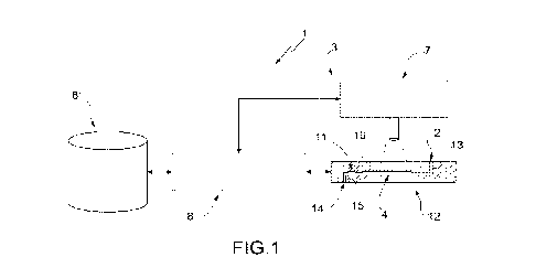
In Figure 1, in accordance with a first aspect of the
present invention, 1 denotes as a whole a microfluidic system
for the manipulation (in particular, for the isolation)
and/or analysis of particles of a sample. Advantageously,
but not necessarily, the microfluidic system 1 is for the
manipulation (in particular, for the isolation) of particles
of a sample.
The microfluidic system 1 comprises at least one inlet
2, through which, in use, the sample is inserted in the
microfluidic system 1; and a moving assembly 3, which
comprises at least one microfluidic chamber 4 and is
configured to move at least one specific particle 5 (see
e.g. Figure 3) inside the microfluidic chamber 4.
The moving assembly 3 comprises at least one actuator
6, which is configured to displace the specific particle 5
(and other particles in the sample); a detection device 7
(Figure 1) which is configured to acquire (at least partial)
images of (in particular, of the entire) microfluidic chamber
4; and a control device 8, which is configured to control
the actuator 6 (in particular, the actuators 6) so as to
move the specific particle 5 (in particular, along a given
path P) inside the microfluidic chamber 4.
Images of the microfluidic chamber 4 are defined as the
images of the entire microfluidic chamber 4 or of one or
more portions of the microfluidic chamber 4.
Note that the path P may have different lengths. For
example, the path P may also be the path between two adjacent
actuators 6 (and thus extremely short). Alternatively, but
not necessarily, the path P extends through a plurality of
actuators (e.g., so as to arrive as far as a recovery chamber
6
CA 03220045 2023- 11- 22
WO 2022/249123
PCT/IB2022/054960
11 - described further below).
Advantageously, but not necessarily, the moving
assembly 3 comprises a plurality of actuators 6 (Figure 3),
which are configured to displace the specific particle 5
(inside the microfluidic chamber 4; in particular, along the
path P). In particular, the control device 8 is configured
(more precisely but not necessarily, a control unit thereof
9 is configured - Figure 2) to control the actuators 6 so as
to move the specific particle 5 inside the microfluidic
chamber 4 (more in particular, along the given path P).
Advantageously, but not necessarily, the moving
assembly 3 is configured to move the specific particle 5
(and the other particles of the sample) in a deterministic
manner (i.e. in a deliberate manner from an initial given
position to a subsequent given position). In particular, the
moving assembly 3 is configured to move the specific particle
5 (and the other particles of the sample) in a substantially
selective manner relative to the other particles of the
sample inside the microfluidic chamber 4.
In particular, the moving assembly 3 is configured (in
particular, the actuator(s) is/are configured) to exert a
force directly on the specific particle 5 (more in
particular, without the force being exerted on the fluid
which transfers the movement to the specific particle 5 -
and to the other particles). For example, each actuator 6
comprises (in particular, is) a respective electrode.
According to some non-limiting embodiments, the moving
assembly 3 comprises a displacing system for displacing
particles chosen from the group consisting of: travelling
waves, thermal flow, local fluid movements generated by
electro thermal flow, local fluid movements generated by
electro hydrodynamic forces, dielectrophoresis, optical
tweezers, opto-electronic tweezers,
light-induced
dielectrophoresis, magnetophoresis, acoustophoresis (and a
7
CA 03220045 2023- 11- 22
WO 2022/249123
PCT/IB2022/054960
combination thereof).
In particular, the displacing system for displacing
particles is chosen from the group consisting of:
dielectrophoresis, optical tweezers, magnetophoresis, light-
induced dielectrophoresis (and a combination thereof).
Advantageously, but not necessarily, the displacing system
for displacing particles is dielectrophoresis.
According to specific non-limiting embodiments, the
moving assembly 3 comprises a dielectrophoresis unit (or
system) like for example described in at least one of the
patent applications WO-A-0069565, WO-A-2007010367, WO-A-
2007049120. More in particular, the moving assembly 3
operates in accordance with what is described in the patent
applications with publication number W02010/106434 and
W02012/085884.
As better shown in Figure 3, the control device 8 is
configured (more precisely but not necessarily, the control
unit thereof 9 is configured - Figure 2) to control the
detection device 7 so that the detection device 7 acquires
a first image of the aforementioned part of the microfluidic
chamber 4 in a first instant t(0), when the specific particle
5 is arranged in a first position IP (of the given path P)
inside the part of the microfluidic chamber 4, and a second
image of an area of the microfluidic chamber 4 in a second
instant t(1) subsequent to the first instant, when the
specific particle 5 is arranged in a second position IIP (of
the given path P) inside the mentioned area of the
microfluidic chamber 4.
In particular, the control device 8 is configured (more
precisely but not necessarily, a control unit 9 thereof is
configured - Figure 2) to control the actuators 6 so as to
move the specific particle 5 (the specific particles 5)
inside the microfluidic chamber 4 from the first position IP
to the second position (more in particular, along the path
8
CA 03220045 2023- 11- 22
WO 2022/249123
PCT/IB2022/054960
P). More in particular, the first and the second position IP
and IIP are intermediate points of the path P.
In other words, the control device 8 is configured (more
precisely, but not necessarily, a control unit 9 thereof is
configured - Figure 2) to control the actuators 6 so as to
move the specific particle 5 (the specific particles 5) from
a start position to an end position of the path P (passing
through the first and the second position IP and TIP); where
the first and the second position IP and TIP are intermediate
points between the start position and the end position.
In some non-limiting cases, the second image is only
about an area of the microfluidic chamber 4. Alternatively,
the second image is about the entire microfluidic chamber 4.
According to some non-limiting embodiments, the first
image is only about a part of the microfluidic chamber 4.
Alternatively, the first image is about the entire
microfluidic chamber 4.
By way of example, Figure 3 shows the specific particle
5 in the first position IP in the first instant - t(0) - and
in the second position IIP in the second instant - t(1).
According to different embodiments, the area of the
microfluidic chamber 4 acquired with the second image
coincides with or is different from the part of the
microfluidic chamber 4 acquired with the first image.
Advantageously but not necessarily, the area of the
microfluidic chamber 4 acquired with the second image
coincides with the part of the microfluidic chamber 4
acquired with the first image (i.e., the second image is
about the part of the microfluidic chamber 4 that is also of
the first image).
The control device 8 is configured (more precisely, but
not necessarily, a process unit thereof 10 is configured -
Figure 2) to process at least one derived image (examples of
such a derived image are shown in Figures 6, 7, 8, 11 and
9
CA 03220045 2023- 11- 22
WO 2022/249123
PCT/IB2022/054960
12) as a function of (at least) the first image and the
second image.
By way of non-limiting example, note that Figure 5 shows
an example photograph taken in the first instant. Figure 9
is an enlargement of this photograph and shows the first
position IP (of the specific particle 5) in the first
instant. Figure 10 is an enlargement of the first position
IP of a photograph taken in the second instant. As can easily
be seen, the specific particle 5 is arranged in the first
instant in the first position IP while in the second instant
it is no longer in the first position IP.
By comparing Figure 5, which is a simple photograph of
the part of the microfluidic chamber 4, with Figures 6, 7
and 8, which are non-limiting examples of derived images, it
is also evident that the particles (and, more precisely, the
specific particle 5) are significantly and surprisingly more
visible and identifiable thanks to the microfluidic system
1 in accordance with the present invention. Furthermore,
thanks to the microfluidic system 1 (and to the method)
according to the present invention it is possible to follow
the specific particle 5 (each particle) continuously by
verifying its position and/or movements throughout the time
of interest. It should be noted that, until now, the
particles (and their positions) were identifiable with a
certain degree of accuracy through detections by
fluorescence. These detections are
intrinsically
discontinuous (after excitation, in just a few instants the
particles are no longer visible due to the photochemical
degradation phenomenon of the fluorophore) and for some
wavelengths (e.g. ultraviolet) are harmful to cells and DNA.
More precisely, it has been experimentally observed
that by using the microfluidic system 1 it is surprisingly
possible to determine the position and the morphological
characteristics of the particles with greater speed,
CA 03220045 2023- 11- 22
WO 2022/249123
PCT/IB2022/054960
precision and ease. It should be noted, in fact, that not
only the particles are highlighted but also the background
(and its confusing effect on detection) is practically
eliminated, making detection more precise and brighter.
Thus, the microfluidic 1 system has, among other things, a
reduced risk of losing and/or damaging particles and its
operating speed is higher than that of state-of-the-art
systems. In this regard, it should be noted that, in order
to identify the type and/or group (in particular, type)
and/or the position of the particles, it is, among other
things, no longer necessary to carry out detections by
fluorescence.
Advantageously but not necessarily, the control device
8 is configured (in particular, the process unit thereof 10
is configured) to process the derived image as a function of
the difference and/or subtraction between the first image
and the second image.
More precisely, but not necessarily, the derived image
is the difference and/or subtraction between the first image
and the second image.
According to some non-limiting embodiments, the control
device 8 is configured to process the derived image as a
function the difference between the first image and the
second image; in particular, the derived image is the
difference between the first image and the second image.
As is known in the field of image processing,
subtraction is defined as the superposition of the first
image and the inverse (the negative) of the second image. In
particular, to perform a subtraction among images, (the value
of) each pixel of the second image is subtracted from (the
value of) a corresponding pixel of the first image.
Examples of subtraction are shown in Figures 6 and 7,
wherein the first positions IP of each particle (i.e. the
position of each particle in the first instant) are depicted
11
CA 03220045 2023- 11- 22
WO 2022/249123
PCT/IB2022/054960
darker and the second positions IIP (i.e. the position of
each particle in the second instant) of each particle are
depicted lighter.
As is known in the field of image processing, a
difference is defined as a subtraction, the result of which
is reported as an absolute value. In particular, in order to
carry out a difference among images, (the value of) each
pixel of the second image is subtracted from (the value of)
a corresponding pixel of the first image; the result (value)
obtained is reported as an absolute value.
Examples of the difference are shown in Figure 8,
wherein the first positions IP of each particle (i.e. the
position of each particle in the first instant) and the
second positions IIP (i.e. the position of each particle in
the second instant) of each particle are depicted lighter
(than the background). Figure 11 is a detail on an enlarged
scale (of a first position IP) of Figure 8.
Advantageously but not necessarily, the control device
8 is configured (in particular, the process unit thereof 10
is configured) to estimate the second position IIP of the
specific particle 5 based on (as a function of) the derived
image.
In particular, said second position IIP is different
from the first position IP.
Note that in this text estimating refers to measuring
(determining, particularly as precisely as possible)
something (e.g. the position of the specific particle 5).
Advantageously, but not necessarily, the control device
8 is configured (in particular, the control unit thereof 9
is configured) to control at least the actuator 6 (in
particular, the actuators 6) in a third instant, which is
subsequent to the first instant and prior to the second
instant, so as to move at least the specific particle 5 from
the first position IP (in particular, to the second position
12
CA 03220045 2023- 11- 22
WO 2022/249123
PCT/IB2022/054960
IIP).
Advantageously, but not necessarily, the moving
assembly 3 is configured to exert a force on the specific
particle 5 (on the specific particles 5) while the first
image and the second image are acquired, in particular, so
that the specific particle 5 (the specific particles 5)
remains (remain) substantially in the first and,
respectively, in the second position IP and TIP.
It has been observed experimentally that, unexpectedly,
in this way the first and the second image are of better
quality.
More precisely but not necessarily, the control device
8 is configured to control the actuator 6 (in particular,
the actuators 6) and the detection device 7 so that the
actuator 6 (in particular, the actuators 6) exerts (exert)
a force on the specific particle 5 (on the specific
particles) while the first image and the second image are
acquired by the detection device 7.
Advantageously, but not necessarily, the moving
assembly 3 is configured to exert a force on the specific
particle 5 (on the specific particles 5) so as to keep the
specific particle 5 (the specific particles 5) suspended
(them suspended) while the first image and the second image
are acquired.
It has been experimentally observed that, surprisingly,
in this way the specific particle 5 is made better visible
(and therefore the first and the second image and,
consequently, also the derived image are of better quality).
It was subsequently hypothesised that this is due to the
fact that, in this way, the background (more precisely, the
base wall of the microfluidic system 1 - in particular, of
the microfluidic chamber 4) turns out to be out of focus
with respect to the particle(s).
More specifically, but not necessarily, the control
13
CA 03220045 2023- 11- 22
WO 2022/249123
PCT/IB2022/054960
device 8 is configured to control the actuator 6 (in
particular, the actuators 6) and the detection device 7 so
that the actuator 6 (in particular, the actuators 6) exerts
(exert) a force on the specific particle 5 (on the specific
particles) so as to keep the specific particle 5 (the
specific particles 5) suspended (them suspended) while the
first image and the second image are acquired by the
detection device 7.
Where in this text reference is made to one or more
particle(s) being "suspended", it is meant that such
particle(s) levitate(s) in (inside) the contained fluid. In
other words, the particle(s) is/are kept spaced apart from
a base wall of the microfluidic system 1 (in particular, of
the microfluidic chamber 4), and optionally, where present,
from an upper wall of the microfluidic system 1 (in
particular, of the microfluidic chamber 4).
With regard to how to achieve the above, reference is
made to the provisions of the aforementioned documents WO-
A-0069565, WO-A-2007010367, WO-A-2007049120, W02010/106434
and W02012/085884, taking particularly into consideration
WO-A-0069565.
In this context, advantageously but not necessarily,
the moving assembly 3 comprises an electrode assembly
(actuators 6) comprising a first electrode array formed on
a support (base wall of the microfluidic chamber 4) and a
second electrode array comprising at least one electrode.
The second electrode array is turned towards and spaced apart
from the first electrode array. The particles (the specific
particle(s) 5) and the fluid in which they are immersed
(inside the microfluidic chamber 4) are arranged in a region
between the first electrode array and the second electrode
array. The moving assembly further comprises means for
establishing an electric field of constant amplitude on at
least one closed imaginary surface located entirely in said
14
CA 03220045 2023- 11- 22
WO 2022/249123
PCT/IB2022/054960
fluid. Such means for establishing an electric field of
constant amplitude comprise means for applying first
periodic signals having a frequency and a first phase to a
first sub-set of electrodes of the first electrode array and
to the second electrode array and at least another periodic
signal having the mentioned frequency and a second phase,
opposite to said first phase, to at least another subset of
electrodes of the first electrode array.
Referring in particular to Figure 1, according to some
non-limiting embodiments, the moving assembly 3 is
configured to transfer at least part of the particles (in
particular, including at least the specific particle 5) of
a first given type and/or group (in particular, type) of the
sample from the microfluidic chamber 4 to a recovery chamber
11 (it also being microfluidic) of the microfluidic system
1 in a substantially selective manner relative to further
particles of the sample.
More precisely, but not necessarily, the microfluidic
system 1 (more precisely, the moving assembly 3) comprises
a microfluidic device 12 (schematically shown in lateral
section in Figure 1), which in turn comprises the
microfluidic chamber 4 (and possibly the recovery chamber
11).
According to some non-limiting embodiments, the
microfluidic device 12 also comprises a (microfluidic)
channel 13, which connects the inlet 2 to the microfluidic
chamber, an outlet 14, through which, in use, the specific
particle 5 (and/or other particles of interest) can be (is)
recovered, a (microfluidic) channel 15 which connects the
recovery chamber 11 (arranged between the outlet 14 and the
microfluidic chamber 4) to the outlet 14.
In particular, the microfluidic device 12 comprises a
channel 16 that connects the microfluidic chamber 4 to the
recovery chamber 11.
CA 03220045 2023- 11- 22
WO 2022/249123
PCT/IB2022/054960
Advantageously, but not necessarily, the microfluidic
device 12 is like the one described in patent applications
with publication numbers W02010/106434 and W02012/085884 (in
these cases, the microfluidic chamber 4 corresponds to the
main chamber described therein). In certain non-limiting
cases, also the entire microfluidic system 1 is as described
in the patent applications with publication numbers
W02010/106434 and W02012/085884, except as directly
indicated in this text.
According to some non-limiting embodiments, the control
device 8 is configured (in particular, the control unit
thereof 9 is configured) to control at least the actuator 6
(in particular, the actuators 6) so as to move at least the
specific particle 5 (and the other particles of the sample)
inside the microfluidic chamber 4 (along the given path P)
as a function of the data acquired by the detection device
7, more in particular as a function of the aforementioned
derived image.
Advantageously, but not necessarily, the microfluidic
system 1 comprises a source 17 (in particular, a light
source) which is configured to emit at least one given
wavelength (in particular, at given wavelengths; in
particular, in the visible range).
In particular, the detection device 7 is configured to
acquire the first and the second image at least at the given
wavelength (in particular, at the given wavelengths; in
particular, in the visible range).
Referring in particular to Figures 14 and 15, according
to some non-limiting embodiments, the control device 8 is
configured (in particular, the process unit thereof 10 is
configured) to define at least one further given path PP for
at least one further particle of the sample as a function of
the derived image. In particular, in such cases, the control
device 8 is configured (more in particular, the control unit
16
CA 03220045 2023- 11- 22
WO 2022/249123
PCT/IB2022/054960
thereof 9 is configured) to operate at least actuator 6 (in
particular, the actuators 6) so that said further particle
is moved (and the other particles of the sample are moved)
along said further path PP so as not to hit said at least
one specific particle 5.
In particular, when the second position TIP coincides
with the first position IP (or does not coincide with an
expected position), the control device 8 is configured (more
in particular, the process unit thereof 10 is configured) to
determine the second position IIP as a function of the
derived image and to define the further given path PP so
that the further given path does not go through the second
position IIP.
It was experimentally observed that, in this way, the
yield, the efficiency and the operating speed of the
microfluidic system were surprisingly improved. In this
regard, it should be noted that where the specific particle
5 is blocked inside the microfluidic chamber 4 (or otherwise
no longer responds correctly to the controls of the control
device 8 through the actuator(s) 6), it is possible to
prevent the further particle (or in any case other particles)
from being blocked in their movement by the specific particle
5 and/or by a part of the moving assembly 3 that is not
functioning correctly in the area of the position IIP and/or
IP. In this regard, it should be noted that it is, for
example, possible that an actuator 6 is faulty (or stops
functioning correctly); in these cases, in the absence of
what is described above, the particles may accumulate in the
area of the faulty actuator 6, severely altering the results
obtained and/or obtainable from the microfluidic system 1.
By way of example, Figure 14 shows a hypothesized path
PPP previously identified by the control device 8 for the
aforementioned further particle. Figure 15 shows instead the
further path PP obtained on the basis of (as function of)
17
CA 03220045 2023- 11- 22
WO 2022/249123
PCT/IB2022/054960
the derived image. In particular, in the example shown, the
second position IIP is identified as corresponding to the
first position IP, and the further path PP (modified with
respect to the path PPP) does not pass in the area of the
second position IIP.
Advantageously but not necessarily, as for example can
be seen from Figure 15, the path PP is determined by the
control device 8 (in particular, by the process unit thereof
10) so that it does not extend even through positions
adjacent to the second position IIP.
It has been experimentally observed that, in this way,
the performance of the microfluidic system 1 is surprisingly
further improved. It is, for example, possible that the
problem that has led to the blockage of the specific particle
5 in the position IP may in some way prevent the movements
also in neighbouring positions (e.g. when in any case the
specific particle has displaced itself slightly, in practice
blocking a neighbouring position, as well).
According to some non-limiting embodiments (in
particular, when the displacing system of the moving assembly
3 is dielectrophoresis - e.g. as described in WO-A-0069565,
WO-A-2007010367 and/or WO-A-2007049120) each position is
defined by a respective actuator 6 (e.g. an electrode).
In particular, the control device 8 is configured (in
particular, the control unit thereof 9 is configured) to
control at least the actuator 6 (more in particular, the
actuators 6) so that the further particle follows the further
path PP.
Advantageously but not necessarily, the control device
8 is configured (in particular, the process unit thereof 10
is configured) to estimate a detected speed at least of the
specific particle 6 (in particular, of the particles) as a
function of the derived image based on (as a function of)
the distance between the first position IP and the second
18
CA 03220045 2023- 11- 22
WO 2022/249123
PCT/IB2022/054960
position IIP and on the time difference between the first
instant and the second instant.
According to some non-limiting embodiments, the control
device 8 is configured (in particular, the control unit
thereof 9 is configured) to control the detection device 7
so that the detection device 7 acquires a plurality of
supplementary images of the (part of - or of the entire)
microfluidic camera 4 in respective supplementary instants
that are subsequent to said first instant (and prior to said
second instant). In particular, the supplementary instants
are subsequent to one another. More in particular, they are
spaced apart from each other by a given time interval At
(and, even more in particular, constant). Alternatively, the
time interval between two supplementary instants can be
variable.
Advantageously but not necessarily, the control device
8 is configured (in particular, the process unit thereof 10
is configured) to estimate the time needed by the specific
particle 5 to displace itself from the first position IP to
the second position IIP on the basis of (as a function of)
the supplementary images.
More precisely but not necessarily, the control device
8 is configured (in particular, the process unit thereof 10
is configured) to estimate the second instant when one of
the first of the supplementary images (which is thus to be
considered as corresponding to the aforementioned second
image) shows the specific particle 5 in the second position
IIP.
In this way, it has been experimentally observed that
it is surprisingly possible to reduce the risk of particles
being lost (i.e. not being properly displaced by the
actuator(s) 6) inside the microfluidic chamber 4 (along the
respective paths P and/or PP) and/or to improve the
efficiency and/or the yield of the microfluidic system.
19
CA 03220045 2023- 11- 22
WO 2022/249123
PCT/IB2022/054960
Advantageously but not necessarily, the control device
8 is configured (in particular, the control unit thereof 9
is configured) to operate at least the actuator 6 (in
particular, the actuators 6) to displace the specific
particle 5 as a function of the detected speed.
In fact, in certain non-limiting cases, for example
where the displacing system of the moving assembly 3 is
dielectrophoresis (e.g. as described in WO-A-0069565, WO-A-
2007010367 and/or WO-A-2007049120), the control device 8 is
configured (in particular, the control unit thereof 9 is
configured) to activate and deactivate the actuators 6
(arranged along the path P) in sequence as a function of the
detected speed.
More precisely, but not necessarily, in use, when the
control device 8 (in particular, the process unit thereof
10) estimates that the specific particle 5 has arrived at
the first position IP (from a previous position) on the basis
of (as a function of) the derived speed, the control device
8 (in particular, the control unit thereof 9) deactivates
the actuator 6 (electrode) arranged in the area of the
position IP and activates the actuator 6 (electrode) arranged
in the second position IIP. In this way, the specific
particle 5 displaces itself from the first position IP to
the second position IIP.
At this point, when the control device 8 (in particular,
the process unit thereof 10) estimates that the specific
particle 5 has arrived at the second position TIP on the
basis of the derived speed, the control device 8 (in
particular, the control unit thereof 9) deactivates the
actuator 6 (electrode) arranged in the area of the position
IIP and activates the actuator 6 (electrode) arranged in the
area of a further position arranged downstream of the second
position (along the path P).
Advantageously, but not necessarily, the control device
CA 03220045 2023- 11- 22
WO 2022/249123
PCT/IB2022/054960
8 is configured (in particular, the process unit thereof 10
is configured) to determine the type (e.g. whether it is a
spermatozoon, a white blood cell, an epithelial cell, a
tumour cell, an endothelial cell or a stem cell) of at least
the specific particle 5 (in particular each particle) as a
function of said derived image.
Alternatively or additionally, the control device 8 is
configured (in particular, the process unit thereof 10 is
configured) to determine the group of at least the specific
particle 5 (in particular, each particle) as a function of
said derived image.
In certain non-limiting cases, the control device 8 is
configured to identify the type and/or group (in particular,
the type) of the specific particle 5 (in particular, using
supervised or non-supervised automated learning), for
example based on reference images (and/or derived image (s)
According to some advantageous but not limiting
embodiments, the control device 8 is configured (in
particular, the process unit thereof 10 is configured) to
extract parameters (in particular, morphological parameters)
of at least the specific particle 5 on the basis of (as a
function of) the derived image and to determine the type
and/or group (in particular, type) of at least one specific
particle 5 by using automated learning (in particular,
supervised - more in particular, a neural network; or non-
supervised - more in particular, clustering).
In particular, the control device 8 is configured (more
in particular, the process unit thereof 10 is configured) to
determine the respective type and/or group (in particular,
type) of each particle of a plurality of particles (of the
sample) as a function of the derived image (in particular,
on the basis of (as a function of) the - morphological -
parameters of each particle obtained from the derived image).
More in particular, the control device 8 is configured
21
CA 03220045 2023- 11- 22
WO 2022/249123
PCT/IB2022/054960
(in particular, the process unit thereof 10 is configured)
to determine the respective type and/or group (in particular,
type) of the specific particle 5 (and possibly of each
particle) on the basis of (as a function of) the derived
image and of further derived images (obtained in the same
manner as the aforementioned derived image - by combining
two different images of the microfluidic chamber 4 or of a
part thereof taken subsequently).
More details regarding the operation of the control
unit 8 (more precisely, of the process unit thereof 10) are
given below in relation to the method in accordance with the
present invention.
Advantageously, but not necessarily, the microfluidic
system 1 comprises a storage unit 8' (Figure 1), which is
configured to store, for example, what is detected by the
detection device 7 and/or what is processed by the control
device 8 and/or reference parameters (on the basis of - as
a function of - which the type and/or group (in particular,
type) of the specific particle 5 and/or the other particles
is determined).
The embodiment of the microfluidic system I shown in
Figure 2 differs from the microfluidic system 1 of Figure 1
in that it comprises some further components. For example,
it is made explicit in Figure 2 that the control device 8
comprises the control unit 9 and the process unit 10, which
can be separated from each other and (simply) connected or
can be fully integrated into a single unit.
In particular, the microfluidic system 1, according to
some non-limiting embodiments (Figure 2), also comprises: an
operator interface 18 (HMI - e.g. a screen, a keyboard and/or
a pointer - mouse); a temperature control unit 19 for
adjusting (maintaining within a desired interval) the
temperature of part of (or all of) the microfluidic device
12; a fluidic control device 20 (in particular, controlled
22
CA 03220045 2023- 11- 22
WO 2022/249123
PCT/IB2022/054960
by the control device 8) for adjusting the flows of the
fluids within the microfluidic device 12; and a recovery
unit 21 for collecting the specific particle 5 (and/or other
particles) exiting the microfluidic device 12 (in
particular, from the outlet thereof 14).
According to some non-limiting embodiments, the
detection device 7 (in accordance with what is shown in
Figure 2) comprises: a video camera 22 (in particular, a
digital one - or camera); a microscope 23; and the light
source 17.
Advantageously but not necessarily, the microfluidic
system 1 also comprises a moving device 24, which is
configured to move the microfluidic device 12 and/or the
detection device 7 relative to each other.
In accordance with a second aspect of the present
invention, there is provided a use of the microfluidic system
1 (as defined above) for selectively collecting cells of one
or more specific types. For example, there is provided a use
of the microfluidic system 1 (as defined above) for
(substantially) selectively collecting cells selected from
the group consisting of: tumour cells, white blood cells
(WBCs), stromal cells, spermatozoas, circulating tumour
cells (CTCs), circulating myeloid cells (CMMCs), foetal
cells, epithelial cells, erythroblasts, trophoblasts,
erythrocytes, endothelial cells, stem cells (and a
combination thereof).
In some non-limiting cases, there is provided the use
of the microfluidic system 1 (as defined above) for
(substantially) selectively collecting cells chosen from the
group consisting of: spermatozoa, white blood cells,
epithelial cells, tumour cells, endothelial cells, stem
cells, foetal cells, nuclei, extracellular vesicles, plant
cells (and a combination thereof).
In addition or as an alternative, there is provided a
23
CA 03220045 2023- 11- 22
WO 2022/249123
PCT/IB2022/054960
use of the microfluidic system 1 (as defined above) for
forensic medicine. In addition or as an alternative, there
is provided a use of the microfluidic system 1 (as defined
above) for diagnostics (of pathologies - e.g. for tumour
diagnosis). In addition or as an alternative, there is
provided a use of the microfluidic system 1 for oncology. In
addition or as an alternative, there is provided a use of
the microfluidic system 1 for prenatal diagnosis.
In the case of a use for oncology, more precisely but
not necessarily, there is provided a use for counting and/or
the analysis and/or the isolation of Circulating Tumour Cells
(CTCs).
In accordance with a third aspect of the present
invention, there is provided a method for the manipulation
(in particular, for the isolation) and/or analysis of
particles of a sample by means of a microfluidic system 1.
The microfluidic system 1 comprises at least one inlet
2, through which the sample is inserted in the microfluidic
system 1; a moving assembly 3, which comprises at least one
microfluidic chamber 4 and is configured to move at least
one specific particle 5 inside the microfluidic chamber 4.
More precisely, but not necessarily, the moving
assembly 3 comprises a microfluidic device 12, which, in
turn, comprises the microfluidic chamber 4 (and possibly, a
recovery chamber 11, and channels 13, 15 and 16).
Advantageously but not necessarily, the moving assembly
3 further comprises: at least one actuator (e.g. an electrode
- in particular, a plurality of actuators), which is
configured to displace at least the specific particle 5; a
detection device 7 which is configured to acquire images (at
least partial images) of the microfluidic chamber 4; and a
control device 8, which is configured to control at least
one actuator 6 so as to move said at least one specific
particle (along a given path P inside the microfluidic
24
CA 03220045 2023- 11- 22
WO 2022/249123
PCT/IB2022/054960
chamber 4).
Advantageously, but not necessarily, the microfluidic
system 1 is as described above in accordance with the first
aspect of the present invention.
The method comprises: a first detection step, during
which the detection device 7 acquires a first image of at
least a part of the microfluidic chamber in a first instant,
when at least the specific particle 5 is arranged in a
respective first position IP (in particular, of the given
path P) inside the mentioned part of the microfluidic chamber
4; and a second detection step, during which the detection
device 7 acquires a second image of at least one area of the
microfluidic chamber in a second instant which is subsequent
to the first instant, in particular when at least the
specific particle 5 is arranged in a respective second
position IIP (more in particular, of the given path P) inside
the mentioned at least one area of the microfluidic chamber.
In some non-limiting cases, the second image is only
about an area of the microfluidic chamber 4. In other words,
the second image is a partial image of the microfluidic
chamber 4. Alternatively, the second image is about the
entire microfluidic chamber 4.
According to some non-limiting embodiments, the first
image is only about a part of the microfluidic chamber 4. In
other words, the first image is a partial image of the
microfluidic chamber 4. Alternatively, the first image is
about the entire microfluidic chamber 4.
According to different embodiments, the area of the
microfluidic chamber 4 acquired during the second detection
step coincides with or is different from the part of the
microfluidic chamber 4 acquired during the first detection
step. Advantageously but not necessarily, the area of the
microfluidic camera 4 acquired during the second detection
step coincides with the part of the microfluidic camera 4
CA 03220045 2023- 11- 22
WO 2022/249123
PCT/IB2022/054960
acquired during the first detection step (i.e., the first
and the second image are about the same part of the
microfluidic camera 4).
According to mutually alternative and non-limiting
situations, the first position IP and the second position
TIP may be different from one another or coincide.
The method further comprises a processing step, during
which the control device processes at least one derived image
as a function of at least the first image and the second
image.
As already indicated above with reference to Figures 5
to 12, in this way it has been experimentally observed that
the particles (and, more precisely, the specific particle 5)
are significantly and surprisingly more visible and
identifiable (both as type and/or group (in particular, type)
and as position); they can, moreover, be followed
continuously (since it is possible to verify their movements
and/or position throughout the time span of interest).
Advantageously, but not necessarily, the method also
comprises an identification step, during which the control
device estimates (i.e. determines as precisely as possible)
the second position IIP of at least the specific particle 5
(in particular, of the particles) on the basis of (as a
function of) the derived image.
In particular, the second position IIP is different
from the first position IP.
Advantageously, but not necessarily, the method further
comprises a moving step, during which the control device 8
(in particular, a control unit thereof) controls at least
the actuator 6 (in particular, the plurality of actuators 6)
in a third instant, which is subsequent to the first instant
and prior to the second instant, so as to move at least the
specific particle 5 (in particular, to the second position
IIP) from the first position IP (in particular, along the
26
CA 03220045 2023- 11- 22
WO 2022/249123
PCT/IB2022/054960
given path P) .
By way of example, Figure 3 shows the specific particle
in the first position IP in the first instant - t(0) - and
in the second position IIP in the second instant - t(1).
5 In
particular, during the moving step, the control
device 8 controls at least the actuator 6 (more in
particular, the actuators 6) to displace the specific
particle 5 and a plurality of other particles (more in
particular, all the particles present in the microfluidic
chamber 4).
More in particular, the control device 8 controls at
least the actuator 6 (more in particular, the actuators 6)
so as to displace the specific particle 5 and the other
particles (even more in particular, all the particles present
in the microfluidic chamber 4) in a deterministic manner.
Alternatively or additionally, the control device 8
controls at least the actuator 6 (more in particular, the
actuators 6) so as to displace the specific particle 5 and
the other particles (more in particular, all the particles
in the microfluidic chamber 4) in a substantially selective
manner relative to the other particles of the sample inside
the microfluidic chamber 4.
Even more precisely but not necessarily, during the
moving step substantially all the actuators 6 are activated
and deactivated in a coordinated manner in order to
substantially displace each particle that is placed
substantially in any position of the fluidic chamber
(assuming the correct operation of each actuator 6).
Advantageously, but not necessarily, particularly
during the moving step, the control device 8 (more precisely,
but not necessarily, the control unit thereof 9 - Figure 2)
controls the actuators 6 so as to move the specific particle
5 (the specific particles 5) inside the microfluidic chamber
4 along the path P. In particular, the first and the second
27
CA 03220045 2023- 11- 22
WO 2022/249123
PCT/IB2022/054960
position IP and IIP are intermediate points of the path P.
In other words, the control device 8 (more precisely,
but not necessarily, the control unit thereof 9 - Figure 2)
controls the actuators 6 so that the actuators themselves
move, in particular during the moving step, the specific
particle 5 (the specific particles 5) from a start position
to an end position of the path P (passing through the first
and the second position IP and TIP); where the first and the
second position IP and TIP are intermediate points between
the start position and the end position.
Advantageously but not necessarily, the moving assembly
3 exerts, in particular during the moving step, a force on
the specific particle 5 (on the specific particles 5) while
the first image and the second image are acquired (during
the first and the second detection step), in particular so
that the specific particle 5 (the specific particles 5)
remains (remain) (in particular substantially fixed) in the
first position IP (during the first detection step) and,
respectively, in the second position IIP (during the second
detection step).
More precisely but not necessarily, the control device
8 controls the actuator 6 (in particular, the actuators 6)
and the detection device 7 so that the actuator 6 (in
particular, the actuators 6) exerts (exert) a force on the
specific particle 5 (on the specific particles) while the
first image and the second image are acquired by the
detection device 7, in particular so that the specific
particle 5 (the specific particles 5) remains (remain) (in
particular substantially fixed) in the first position IP
(during the first detection step) and, respectively, in the
second position IIP (during the second detection step).
Advantageously, but not necessarily, the moving
assembly 3 exerts a force on the specific particle 5 (on the
specific particles 5) so as to keep the specific particle 5
28
CA 03220045 2023- 11- 22
WO 2022/249123
PCT/IB2022/054960
(the specific particles 5) suspended (them suspended) while
the first image and the second image are acquired (during
the first and the second detection step).
More precisely, but not necessarily, the control device
8 controls the actuator 6 (in particular, the actuators 6)
and the detection device 7 so that the actuator 6 (in
particular, the actuators 6) exerts (exert) a force on the
specific particle 5 (on the specific particles) so as to
keep the specific particle 5 (the specific particles 5)
suspended (them suspended) while the first image and the
second image are acquired by the detection device 7.
In this context, according to some non-limiting
embodiments, the method provides for manipulating particles
immersed in a fluid placed in a region between a first and
a second array of electrodes belonging to a group of
electrodes. The second electrode array comprises at least
one electrode and is facing and spaced apart from the first
electrode array. The method provides for applying first
periodic signals having a frequency and a first step to a
first subset of electrodes of the first electrode array and
to the second electrode array and at least a second periodic
signal having the mentioned frequency and a second step,
which is opposite said first step, to at least another subset
of electrodes of the first electrode array, thereby
establishing an electric field of constant amplitude on at
least one imaginary closed surface arranged entirely in the
fluid, whereby the particles are attracted or repelled by a
portion of the region enclosed by the at least one imaginary
closed surface, depending on the electrical properties of
the particles and the fluid.
Advantageously but not necessarily, the first image
also contains the other particles in the respective initial
positions; the second image also contains the other particles
in respective subsequent positions.
29
CA 03220045 2023- 11- 22
WO 2022/249123
PCT/IB2022/054960
Figure 4 schematically shows a flowchart of a specific
and non-limiting example of a procedure implemented in
accordance with the aforementioned method for the
manipulation (in particular, for the isolation) and/or
analysis of particles.
The procedure, advantageously but not necessarily,
provides for a start (start - step A) ; the first detection
step (step B); the moving step (step C); the second detection
step (step D); the processing step (step E); the
identification step (step F); and possibly an end step (end
- step G).
Optionally, these steps (more precisely, steps B to F)
can be repeated one or more times after the particles 6 have
been returned to their original positions (e.g., the specific
particle 5 has been returned to the first position IP) (step
H) and/or the part and/or the area of the microfluidic
chamber 4 that is acquired during the first and the second
detection steps is changed (e.g., by displacing the detection
device 7 and/or the microfluidic chamber 4) (step I).
Advantageously, but not necessarily (during the moving
step), the moving assembly 3 moves (is configured to move)
at least the specific particle 5 in a deterministic manner
(i.e. in a deliberate manner from an initial given position
to a subsequent given position).
In particular (during the moving step), the moving
assembly 3 moves (is configured to move) said at least one
specific particle in a substantially selective manner
relative to (all - to all the) other particles of the sample
inside the microfluidic chamber.
For example, the moving assembly 3 exerts a force
directly on the specific particle 5 (more in particular,
without the force being exerted on the fluid, which transfers
the movement to specific particle 5 - and to the other
particles). In some specific and non-limiting cases, each
CA 03220045 2023- 11- 22
WO 2022/249123
PCT/IB2022/054960
actuator 6 comprises (in particular, is) a respective
electrode.
Advantageously, but not necessarily, the moving
assembly 3 is defined as described above in relation to the
first aspect of the present invention.
Additionally or alternatively, the control device 8
and/or the detection device 7 and/or the microfluidic device
12 are defined as described above in relation to the first
aspect of the present invention.
In particular, (all of) the microfluidic system 1 is
defined as described above in relation to the first aspect
of the present invention.
Advantageously, but not necessarily, during the
processing step, the control device 8 (in particular, a
process unit thereof 10) processes the derived image as a
function of the difference and/or subtraction between the
first image and the second image. In some specific and non-
limiting cases, during the processing step, the control
device 8 (in particular, a process unit thereof 10) processes
the derived image as a function of the difference between
the first image and the second image.
In particular, the derived image is the difference
(and/or subtraction) between the first image and the second
image.
Advantageously, but not necessarily, the processing
step comprises an alignment sub-step, during which the first
and the second images are aligned (with each other). In such
cases, during the processing step, the control device 8 (in
particular, a process unit thereof 10) processes the derived
image as a function of the (difference and/or subtraction
between the) first image and the second image, after the
first and the second images have been aligned with each
other. Note, that since the first and the second image that
were subjected to alignment are a function of the first and
31
CA 03220045 2023- 11- 22
WO 2022/249123
PCT/IB2022/054960
of the second image (as acquired), the derived image is also
in this case (at least indirectly) a function of the first
and of the second image (as acquired).
Thanks to this alignment step, it is possible to obtain
brighter derived images and thus reduce the incidence of
false positives.
According to some non-limiting embodiments, the
alignment sub-step is performed by means of an algorithm of
known type, for example Optical Flow or FFT (Fast Fourier
Transform).
According to some non-limiting embodiments, the method
transfers at least part of the particles (in particular,
including at least the specific particle 5) of a first given
type and/or group (in particular, type) of the sample from
the microfluidic chamber 4 to a recovery chamber 11 (it also
being microfluidic) of the microfluidic system 1 (more
precisely, of the microfluidic device 12) in a substantially
selective manner relative to (all) further particles of the
sample.
Advantageously, but not necessarily (during the moving
step), the control device 8 (in particular, the control unit
thereof 9) controls (is configured to control) at least the
actuator 6 (in particular, the actuators 6) so as to move at
least the specific particle 5 (in particular, the particles)
inside the microfluidic chamber 5 (in particular, along said
given path P) as a function of the data acquired by the
detection device 7, in particular as a function of the
derived image.
According to some non-limiting embodiments, the method
comprises an adaptation step, during which the control device
8 defines at least one further given path PP for at least
one further specific particle of the sample as a function of
the derived image; the moving assembly 3 moves said further
specific particle (in particular, the control device 8 -
32
CA 03220045 2023- 11- 22
WO 2022/249123
PCT/IB2022/054960
more in particular, the control unit 9 - operates at least
the actuator 6 - more in particular, the actuators 6 - so
that the further specific particle is moved), in particular
along the further path PP, so as not to hit said at least
one specific particle.
In particular, when the second position IIP coincides
with the first position IP or does not coincide with an
expected position, the control device 8 (in particular, a
process unit thereof) determines the second position IIP as
a function of the derived image and defines the further given
path PP so that the further given path PP does not go through
(in the area of) the second position TIP (and/or through
positions near the second position IIP).
According to some non-limiting embodiments, the method
comprises a third detection step, during which the detection
device 7 acquires a third image of the microfluidic chamber
4 in a further instant subsequent to the second instant,
when (at least) the specific particle 6 is arranged in a
third position (of the given path P) inside (the part of)
the microfluidic chamber 4. The control device 8 traces an
actual path followed by at least the specific particle 5 as
a function of the derived image and of a further derived
image obtained on the basis of (as a function of) the third
image and of the second image (e.g. the further derived image
is the difference and/or the subtraction of the third image
and of the second image).
Advantageously but not necessarily, the further
specific particle (and any further particles) is also the
subject of the first detection step, the moving step, the
second processing step, the identification step (and
possibly a verification step as described hereinbelow).
Figure 13 schematically shows a flowchart of a specific
and non-limiting example of a procedure implemented in
accordance with the aforementioned method for the
33
CA 03220045 2023- 11- 22
WO 2022/249123
PCT/IB2022/054960
manipulation (in particular, for the isolation) and/or
analysis of particles.
The procedure provides steps A to G as described above
(in particular, with reference to Figure 4) and a
verification step (step L), during which the control device
8 (in particular, the process unit 10) verifies whether the
specific particle 5 has moved (and the other particles have
moved) correctly.
In particular, if this is the case (i.e. if the control
device 8 verifies that the specific particle 5 has moved
correctly), the procedure starts again according to a
repeatable cycle from the moving step (step C), in other
words, the specific particle 5 is moved from the second
position IIP (to the aforementioned third position - along
the path P) and it is (again) proceeded with the second
detection step (D), the processing step (E), the
identification step (F) and the verification step (L).
According to some non-limiting embodiments, this cycle
is repeated until the specific particle 5 reaches a desired
end position and/or the verification step (L) yields a
negative result (i.e., following the verification step based
on the processing step it is determined that the specific
particle 5 has not moved correctly).
In the event that the verification step (L) yields a
negative result, in particular the aforementioned adaptation
step (step M) is implemented.
In particular, the adaptation step (M) comprises an
obstacle creation sub-step (step N), during which the control
device 8 creates a (virtual) obstacle in the area of the
position where the specific particle 5 is (is blocked); and
a redefinition sub-step (step 0), during which the further
path PP is determined (in particular, during the redefinition
sub-step, a respective further path PP is determined for
each of the further particles to be moved) which avoids the
34
CA 03220045 2023- 11- 22
WO 2022/249123
PCT/IB2022/054960
(virtual) obstacle.
Advantageously, but not necessarily, after the
adaptation step, the cycle (steps C to L, in sequence) is
repeated (e.g. for the further particle; in particular, for
the other particles - the particles that have moved
correctly), in particular until the further particle (in
particular, each of the other particles) reaches a desired
final position and/or the verification step (L) yields a
negative result.
Advantageously, but not necessarily, during the first
detection step and during the second detection step, the
part of the microfluidic chamber 4 and the area of the
microfluidic chamber 4, respectively, are lighted with
radiations having given wavelengths (in particular, in the
visible range).
In particular, the first and the second image are
acquired at the aforementioned given wavelengths; more in
particular, the first and the second image are acquired in
the visible range (even more in particular, they are not
acquired at wavelengths outside the visible range).
According to some non-limiting embodiments, the method
comprises a speed estimation step, during which the control
device 8 estimates a detected speed of at least the specific
particle 5 (and of the other particles) as a function of the
distance between the first position IP and the second
position IIP (in particular, obtained on the basis of - as
a function of - the derived image) and the time needed by
the specific particle 5 to displace itself from the first
position IP to the second position IIP. In particular, the
time needed by said at least one specific particle 5 to
displace itself from the first position IP to the second
position IIP is the difference between said first and said
second instant
According to some non-limiting embodiments, the speed
CA 03220045 2023- 11- 22
WO 2022/249123
PCT/IB2022/054960
estimation step is performed before the specific particle 5
is transferred towards the recovery chamber 11.
Alternatively, the speed estimation step is performed while
the specific particle is transferred towards the recovery
chamber 11.
In particular, the detected speed is estimated as a
function of the distance between the first position IP and
the second position TIP that are obtained on the basis of
(as function of) the derived image and the time between the
first and the second instant. Note that, according to some
non-limiting embodiments, the distance between the first
position IP and the second position TIP corresponds to the
distance between two successive actuators 6 (electrodes)
(and is, therefore, known).
More in particular, the detected speed is estimated as
a function of the distance between the first position IP and
the second position IIP, which in turn are estimated on the
basis of (as a function of) the derived image obtained as a
function of the first and of the second images subjected to
alignment (i.e. after the aforementioned alignment sub-step
has been performed).
Advantageously, but not necessarily, the image
processing step comprise a derived image manipulation step,
by which a derived manipulated image is obtained, as a
function of which the aforementioned distance between the
first position IP and the second position IIP is estimated.
According to some non-limiting embodiments, the image
manipulation step comprises a binarisation sub-step, during
which each pixel of the derived image is transformed (from
grey tones) to black or white (as a function of a threshold
grey tone) in order to obtain a binarised derived image.
Alternatively or additionally, the image manipulation
step comprises a morphological manipulation sub-step, during
which the (advantageously binarised) derived image is
36
CA 03220045 2023- 11- 22
WO 2022/249123
PCT/IB2022/054960
subjected to opening, dilating and/or closing operations in
order to obtain the manipulated derived image.
During the opening operation, the outermost edges (more
precisely, the relative corners) of the representation(s) of
the specific particle 5 in the derived image are eroded.
During the dilating operations, the outer edges of the
representation(s) of the specific particle 5 in the derived
image are dilated.
During the closing operations, the inner edges of the
representation(s) of the specific particle 5 in the derived
image are dilated. In particular, as a (macroscopic) effect,
the closure of any holes inside the image, the filling of
any cavities are obtained.
Advantageously, but not necessarily, the distance
between the first position IP and the second position TIP is
estimated by evaluating the distance between the barycentres
(centroids) of the representations (in the first position IP
and in the second position TIP) of the specific particle 5
in the derived image (more advantageously, of the manipulated
derived image).
Figure 26 schematically shows a flowchart of a specific
and non-limiting example of an implemented procedure for
measuring the distance between the first position IP and the
second position IIP.
The procedure provided implementing in succession: the
first detection step (step B); the moving step (step C), the
second detection step (step D); the alignment sub-step (step
AL) ; a derived image processing (in particular, as a function
of the difference and/or subtraction between the first image
and the second image - step DIF) the binarisation sub-step
(step BIN); the opening operations (step OP); the dilating
operations (step DIL); the closing operations (step CLO); an
estimation of the distance between the first position IP and
the second position IIP (step EXT) on the basis of (as a
37
CA 03220045 2023- 11- 22
WO 2022/249123
PCT/IB2022/054960
function of) the manipulated derived image (obtained as a
result of the steps: B, C, D, AL, DIF, BIN, OP, DIL and CLO).
For merely explanatory and non-limiting purposes, it
should be noted that, in this case, the processing step
comprises the steps AL, DIF, BIN, OP, DIL and CIO.
Advantageously, but not necessarily, the method
comprises a plurality of supplementary detection steps,
during each of which the detection device 7 acquires a
respective supplementary image of the microfluidic chamber
4 (in particular, of the aforementioned at least one part of
the microfluidic chamber 4; additionally or alternatively,
of the aforementioned at least one area of the microfluidic
chamber 4) in a respective supplementary instant subsequent
to the first instant (and, in particular, prior to said
second instant). During the speed estimation step, the time
needed by the specific particle 5 to displace itself from
the first position IP to the second position IIP is measured
on the basis of (as a function of) the supplementary images.
In particular, the second instant is estimated when a first
one of the supplementary images (which is thus to be
considered as corresponding to the aforementioned second
image) shows at least the specific particle 5 in the second
position IIP.
In particular, the supplementary instants are
subsequent to one another (i.e. spaced apart by a given -
and constant - time interval At). For example, each interval
At can be from about 5ms to about 15ms (in particular, about
10ms).
Figure 16 schematically shows a flowchart of a specific
and non-limiting example of a procedure implemented in
accordance with the aforementioned method for the
manipulation (in particular, for the isolation) and/or
analysis of particles.
The procedure envisages steps A to C, E, F, G and L as
38
CA 03220045 2023- 11- 22
WO 2022/249123
PCT/IB2022/054960
described above (in particular, with reference to Figure 4)
and the supplementary detection step DD. Steps DD, E, F and
L, in sequence, are repeated in a cycle at successive
supplementary instants (tj+1=ti+At) (in particular, spaced
apart by the given time interval - At) until the specific
particle arrives at the second position IIP (and, therefore,
the verification step (L) yields a positive result). At this
point, the control device 8 estimates the detected speed
(step Q).
Advantageously, but not necessarily, the method
comprises a conveying step, during which the moving assembly
3 displaces itself (in particular, the control device
controls at least the actuator 6 - more in particular, the
actuators 6 - so as to displace) the specific particle 5 (in
particular, along the given path P) as a function of the
detected speed. This makes it possible to optimise the speed
at which each particle moves in a personalised manner.
According to certain non-limiting forms of actuation
(during the conveying step), the actuators 6 (electrodes)
are activated in succession along the given path P so that,
when the specific particle 5 is arranged in the area of a
first actuator 6 of the moving assembly 3, the first actuator
6 is deactivated and a second actuator 6 of the moving
assembly, which is arranged downstream of the first actuator
6 along the given path P, is activated.
In particular, when the specific particle 5 is arranged
in the area of the second actuator 6, the second actuator 6
is deactivated and a third actuator 6 of the moving assembly
3, which is arranged downstream of the second actuator 6
along the given path P, is activated.
More precisely, but not necessarily, the moments in
which the first actuator 6 and the second actuator 6 (and
possibly the third actuator 6) are activated and deactivated
are determined by the control device 8 on the basis of (as
39
CA 03220045 2023- 11- 22
WO 2022/249123
PCT/IB2022/054960
a function of) the detected speed.
According to some non-limiting embodiments, the method
comprises at least one further first detection step, during
which the detection device 7 acquires a further first image
(of the aforementioned part) of the microfluidic chamber 4
in a further first instant, when a second specific particle
is arranged in a further first position of a second given
path inside (the aforementioned part) the microfluidic
chamber 4; at least one further second detection step, during
which the detection device 7 acquires a further second image
(of the aforementioned part) of the microfluidic chamber 4
in a further second instant subsequent to the further first
instant, when said further specific particle is arranged in
a further second position of the second given path inside
(the aforementioned part) the microfluidic chamber 4; a
further processing step, during which the control device 8
develops at least one further derived image as a function of
said further first image and said further second image (in
particular, as a function of the difference and/or
subtraction between said further first image and said further
second image); and a further speed estimation step, during
which the control device 8 estimates a further detected speed
of the second specific particle as a function of the distance
between the first further position and the second further
position, which are obtained on the basis of (as a function
of) said further derived image, and of the time needed by
the second specific particle to displace itself from the
further first position to the further second position.
More precisely, but not necessarily, the method
comprises a further conveying step, during which the moving
assembly 3 displaces (in particular, said control device 8
controls at least the actuator 6, more in particular the
actuators 6, to displace) the second specific particle as a
function of said further speed detected along said further
CA 03220045 2023- 11- 22
WO 2022/249123
PCT/IB2022/054960
given path.
Advantageously but not necessarily, the first detection
step coincides with the further first detection step, the
second detection step coincides with the further second
detection step, the further processing step coincides with
said processing step, the further derived image coincides
with the aforementioned derived image, the further first
image and the further second image coincide with the first
image and the said second image, respectively.
In particular, said conveying step and said further
conveying step are at least partly simultaneous.
Figure 17 schematically shows a flowchart of a non-
limiting variant of the procedure of Figure 16. Also in this
case, the procedure envisages steps A to C, DD, E, F, G, L
and Q. There IS further provided a verification step (step
R), during which it is verified whether the total time
elapsed since the moving step (C) is higher than a limit
time. If this is not the case, a cycle envisaging, in
sequence, steps DD, E, F, L and R is repeated. If this the
case, this cycle is not repeated and step Q is completed for
all the particles.
In particular (as shown in Figure 17), during the
verification step (L), the control device 8 assesses whether
all the particles (or supposed particles) have reached their
destination. If this is not the case, the verification step
(R) is carried out. If this is the case, step Q is completed
for all the particles.
Optionally, this procedure also comprises a step for
acquiring an image by fluorescence (step S).
Optionally, in the variant of Figure 17, the procedure
envisages a cycle that is repeated (starting from step Q or
step R) until during a control step (step U) it is verified
that the entire (relevant) microfluidic chamber 4 has been
subjected to steps B and DD.
41
CA 03220045 2023- 11- 22
WO 2022/249123
PCT/IB2022/054960
This cycle envisages steps B (possibly S), C, DD, E, F,
L, in sequence, and also step H (as described above),
subsequent to step Q or to step R and prior to step U; and
step I (as described above), subsequent to step U and prior
to step B.
In these cases, in other words, steps B, (possibly S)
C, DD, E, F, L, Q (and/or R), H, U and I are repeated in a
cycle until step U yields a positive result (i.e. when the
entire (relevant) microfluidic chamber 4 has been subjected
to steps B and DD).
Advantageously, but not necessarily, the method
comprises a characterisation step, during which the type
and/or group (in particular, type) of at least the specific
particle 5 (in particular, of each particle) is determined
(in particular, by the control device 8; more in particular,
by the control unit 9) as a function of the derived image
(in particular, on the basis - as a function - of parameters
- e.g. morphological parameters - of said at least one
specific particle obtained from said derived image). More
precisely, but not necessarily, during said characterisation
step, the respective type and/or group (in particular, type)
of each particle of a plurality of particles is determined
as a function of said derived image (in particular, on the
basis - as a function - of parameters - e.g. morphological
parameters - of said each particle obtained from said derived
image).
It should be noted that in this text, when reference is
made to the "characterisation step" or "characterisation",
this means: a classification or grouping.
In particular, "classification" (as used in the sector)
is defined as an operation that on the basis of analysis of
previously labelled data makes it possible to predict the
labelling of future data classes.
In particular, "grouping" (as used in the sector) is
42
CA 03220045 2023- 11- 22
WO 2022/249123
PCT/IB2022/054960
defined as an aggregation of unlabelled or unstructured data
starting from common characteristics automatically
identified by the machine.
In particular, "clustering" (as used in the sector) is
defined as a set of multivariate data analysis techniques
aimed at selecting and grouping homogeneous elements in a
data set.
In particular, "neural network" (as used in the sector)
is defined as a computational model composed of artificial
"neurons", which is vaguely inspired by the simplification
of a biological neural network. It is therefore a
mathematical-computer model consisting of interconnections
of information.
In particular, "type" (as used in the sector) is defined
as an identification of a data item in a category having a
label defined a priori.
In particular, "group" (as used in the sector) is
defined as an identification of data as belonging to a
category that is recognised starting from common elements
without (the need for) a priori identification.
In addition to or as an alternative (to determining the
type and/or group (in particular, type) of at least the
specific particle 5 as a function of the derived image),
during the characterisation step, the type and/or group (in
particular, type) of at least the specific particle 5 is
determined (in particular, by the control device) as a
function of the detected speed (during the aforementioned
speed estimation step). In some non-limiting cases, the type
and/or group (in particular, type) of at least the specific
particle 5 is determined (in particular, by the control
device) as a function of a combination of detected speed and
derived image and/or morphological parameters.
In particular, when the specific particle 5 is a cell,
the viability or integrity of the specific particle 5 (more
43
CA 03220045 2023- 11- 22
WO 2022/249123
PCT/IB2022/054960
precisely, if the particle is a living and/or intact or dead
and/or apoptotic and/or damaged cell) is determined (in
particular, by the control device) as a function of the
detected speed (during the aforementioned speed estimation
step). More in particular, if the detected speed is below a
given threshold speed, the specific particle 5 is considered
dead (or apoptotic or damaged); if the detected speed is
above a given threshold speed, the specific particle 5 is
considered alive and/or intact.
Advantageously but not necessarily, during the
characterisation step, the control device 8 determines the
type and/or group (in particular, type) of at least the
specific particle 5 (in particular, of each particle) using
an automated learning (in particular, supervised or non-
supervised).
According to some non-limiting embodiments, during the
characterisation step, the control device 8 determines the
type of at least the specific particle 5 (in particular, of
each particle) using a supervised automated learning (in
particular, a neural network or convolutional neural
network) or the control device 9 determines the belonging
group of at least the specific particle 5 (in particular, of
each particle) using a non-supervised automated learning (in
particular, using the clustering or a non-supervised neural
network).
Non-limiting examples of (non-supervised) automated
learning are: k-means clustering, DBSCAN clustering,
Autoencoder and self-organizing maps.
Non-limiting examples of supervised automated learning
are: decision trees, neural networks, convolutional neural
networks, support-vector machines, etc.
Patent US6463438 and the article Single-Cell Phenotype
Classification Using Deep Convolutional Neural Networks
(Oliver Durr and Beate Sick; Journal of Biomolecular
44
CA 03220045 2023- 11- 22
WO 2022/249123
PCT/IB2022/054960
Screening 2016, Vol. 21(9) 998-
1003; DOI
10.1177/187057116631284) describe
particle/cell
classification/identification systems in more detail.
Advantageously but not
necessarily, the
characterisation step comprises an extraction sub-step,
during which parameters (e.g. morphological parameters) of
the specific particle 5 (in particular, of the particles)
are extracted (in particular, by the control device 8) on
the basis of (as a function of) the derived image; and an
identification sub-step, during which the control device 8
determines the type and/or group (in particular, type) of at
least the specific particle 5 (in particular, of each
particle) on the basis of (as a function of) the extracted
(e.g. morphological) parameters.
Advantageously, but not necessarily, the
characterisation step of the specific particle 5 (in
particular, of the particles) is performed (in particular,
by the control device 8) on the basis of (as a function of)
the image derived using convolutional neural networks.
Figure 18 schematically shows a flowchart of a specific
and non-limiting example of a procedure implemented in
accordance with the aforementioned method for the
manipulation (in particular, for the isolation) and/or
analysis of particles.
The procedure envisages steps A to F and G as described
above and the extraction sub-step (step X).
Optionally, the procedure comprises the identification
sub-step (step Y), between step X and step G to verify if
correct in consideration of the figure.
Additionally or alternatively, the procedure comprises
steps H and I (and the repetition of the relative cycle, B,
C, D, E, H and I in sequence) as described above.
Additionally or alternatively, steps F, X and Y could
be performed at least partially simultaneously with steps H
CA 03220045 2023- 11- 22
WO 2022/249123
PCT/IB2022/054960
and I.
In addition or as an alternative, the loop of steps I
and H can start from step D instead of E.
By way of example, Figure 19 shows how the results
obtained experimentally following the method described above
are surprisingly reliable, matching the results that were
achieved when analysing the sample by carrying out detections
by fluorescence. For example, the method described above (in
a fully automated way - without the intervention of an
operator) has succeeded in, among other things,
discriminating between white blood cells (WBC) and
epithelial cells (EPT).
Advantageously, but not necessarily, the method
comprises at least one re-orientation (e.g., rotation)
and/or deformation step, during which the moving assembly 3
re-orients (e.g., rotates) and/or deforms (the actuators 6
are operated - in particular, by the control device 8 - so
as to re-orient and/or deform) at least the specific particle
5 (the particles) so that at least the specific particle 5
assumes (the particles assume) a different conformation.
More precisely, but not necessarily, the method
comprises an additional detection step, during which, the
detection device 7 acquires an additional image of the
microfluidic chamber 4 (in particular, of the aforementioned
at least one part of the microfluidic chamber 4; additionally
or alternatively, of the aforementioned at least one area of
the microfluidic chamber 4), when at least the specific
particle 5 has assumed the different conformation (the
particles have assumed respective different conformations).
During the processing step, the control device 8 develops an
additional derived image as a function of the additional
image and one between the first image and the second image
(and possibly a further additional image, which is acquired
by the detection device 7 prior to the re-orientation and/or
46
CA 03220045 2023- 11- 22
WO 2022/249123
PCT/IB2022/054960
deformation step). During the characterisation step, the
type and/or group (in particular, type) of at least the
specific particle 5 (and also of the other particles) is
determined (in particular, by the control device 8) (also)
as a function of said additional derived image.
According to some non-limiting embodiments, the
additional detection step corresponds to the second
detection step and (thus) the additional image corresponds
to (is the) aforementioned second image.
Alternatively, the additional detection step is
different from the first and the second detection step and
(therefore) the additional image does not correspond to (is
the) aforementioned second image and/or first image.
Figure 20 schematically shows a non-limiting variant of
the procedure of Figure 18 in which the re-orientation step
(step Z) is also envisaged (essentially as the only
difference). The repetition of the second detection step (D)
serves as an additional detection step.
According to some non-limiting embodiments, the method
comprises a plurality of further first detection steps,
during which the detection device 7 acquires further first
images of the microfluidic chamber 4 in further first
instants, when specific second particles are arranged in
respective further first positions of given second paths
inside the microfluidic chamber; a plurality of further
second detection steps, during which the detection device 7
acquires further second images of the microfluidic chamber
4 in further second instants subsequent to the further first
instant, when the second specific particles are arranged in
respective further second positions of the given second paths
inside the microfluidic chamber 4; a plurality of further
processing steps, during which the control device 8 develops
a plurality of further derived images, each, as a function
of a respective one of the further first images and a
47
CA 03220045 2023- 11- 22
WO 2022/249123
PCT/IB2022/054960
respective one of the further second images (in particular,
as a function of the difference and/or subtraction between
the respective further first image and the respective further
second image); and a characterisation step, during which
said specific particle 5 and said second specific particles
are classifiably divided into at least two types and/or
groups. In particular, the types and/or groups enclose
particles with similar characteristics.
Typically, after the characterisation (grouping) step,
an operator identifies the different types, dividing the
particles, for example, between lymphocytes, platelets,
epithelial cells, etc.
Advantageously but not necessarily, the particles are
associated with different types (e.g. lymphocytes,
circulating tumour cells, epithelial cells, nuclei, etc.)
automatically, e.g. during the
characterisation
(classification) step.
Advantageously, but not necessarily, where there has
been previous training, the particles are associated with
different types (e.g. lymphocytes, circulating tumour cells,
epithelial cells, nuclei, etc.) automatically, e.g. during
the characterisation (classification) step.
In some non-limiting cases, the first detection step
and the further first detection steps coincide (with each
other), the second detection step and the further first
detection steps coincide (with each other), the further
processing step and the further processing steps coincide
(with each other), the further derived images and the
aforementioned derived image coincide (with each other), the
further first images and the first image coincide (with each
other), the further second images and the second image,
respectively coincide (with each other).
Advantageously, but not necessarily, the method further
comprises a further moving step, during which the control
48
CA 03220045 2023- 11- 22
WO 2022/249123
PCT/IB2022/054960
device 8 controls a plurality of actuators 6 (each after a
further first detection step and before a further second
detection step) in order to move the aforementioned second
specific particles 5 from the respective first positions (in
particular, to the respective second positions) along said
given path P.
According to some non-limiting embodiments, the moving
step and the further moving step are (at least partially)
simultaneous.
In particular, it should be noted that the further first
images include a representation of the second specific
particles in the first instant; the further second images
include a representation of the second specific particles in
the second instant.
Advantageously but not necessarily, the method
comprises a learning step (step AP - Figure 21) which
comprises: at least a first detection sub-step, during which
the detection device 7 acquires a first learning image of at
least a part of a microfluidic test chamber in a first test
instant, when a test particle of a known type is arranged in
a first test position (in particular, of a given test path)
inside the mentioned part of the microfluidic test chamber;
at least one second detection sub-step, during which the
detection device 7 acquires a second learning image of an
area of the microfluidic test chamber in a second test
instant subsequent to the first test instant, when said test
particle is arranged in a second test position (in
particular, of the given test path) inside the mentioned
area of the microfluidic test chamber; and at least one
processing sub-step, during which the control device 8 (in
particular, the process unit thereof 10) develops a derived
test image as a function of the first learning image and of
the second learning image and, on the basis of (as a function
of) the derived test image, configures (in particular,
49
CA 03220045 2023- 11- 22
WO 2022/249123
PCT/IB2022/054960
determines parameters of) an automated learning algorithm
(in particular, supervised) in order to train the aforesaid
algorithm to identify the (known) type of particles.
In particular, during the characterisation step, the
type and/or group (in particular, type) of the specific
particle 5 is determined (more precisely, by the control
device 8; even more precisely, by the process unit thereof
10) as a function of the derived image using the
aforementioned automated learning algorithm.
According to some non-limiting embodiments, during the
processing sub-step, the control device 8 (in particular,
the process unit thereof 10) extracts (identifies and
selects) the parameters of the test particle on the basis
(as a function) of (from) the derived test image (step AA)
and configures (i.e. trains) the automated learning
algorithm with these parameters (step AB), in particular, by
correlating them with the known type. For example, step AA
is realised using a neural network (particularly
convolutional - CNN; or image processing algorithms).
Alternatively or additionally, the parameters of the
test particle are morphological parameters (in particular,
whose type has been selected by an operator - non-limiting
examples of morphological parameters are dimensions, shape,
colour etc.). According to some non-limiting embodiments, in
such cases, the control device extracts the parameters of
the test particle on the basis of (as a function of) the
derived test image (step AA) and configures (i.e. trains)
the automated learning algorithm with these parameters (step
AB), in particular, correlating them with the known type.
For example, step AA is realised using a neural network
(particularly convolutional - CNN; or image processing
algorithms).
More precisely but not necessarily, the method (in
particular, the learning step) comprises a labelling sub-
CA 03220045 2023- 11- 22
WO 2022/249123
PCT/IB2022/054960
step (step AC), during which an operator determines the
correlation between the particle and the type using the
available information (for example, the morphological
parameters and/or the bright field and/or fluorescence
images and/or the fluorescence measurements); during the
processing sub-step, the control device 8 (in particular,
the process unit thereof 10) extracts (in particular, by
image processing) the parameters of the types selected during
the selection step from the derived test image (step AA) and
configures (i.e. trains) the automated learning algorithm
with these parameters (step AB), in particular using the
correlation with the known type performed by the operator.
In particular, the second test position is different
from the first test position.
In some non-limiting cases, the second learning image
is only about an area of the microfluidic test chamber. In
other words, the second learning image is a partial image of
the microfluidic test chamber. Alternatively, the second
learning image is about the entire microfluidic test chamber.
According to some non-limiting embodiments, the first
learning image only about a part of the microfluidic test
chamber. In other words, the first learning image is a
partial image of the microfluidic test chamber.
Alternatively, the first learning image is about the entire
microfluidic test chamber.
According to different embodiments, the area of the
microfluidic test chamber acquired during the second
detection sub-step coincides with or is different from the
part of the microfluidic test chamber acquired during the
first detection sub-step. Advantageously but not
necessarily, the area of the test microfluidic chamber
acquired during the second detection sub-step coincides with
the part of the test microfluidic chamber acquired during
the first detection sub-step (i.e. the first and the second
51
CA 03220045 2023- 11- 22
WO 2022/249123
PCT/IB2022/054960
learning images are about the same part of the test
microfluidic chamber).
Advantageously, but not necessarily, during the first
detection sub-step, the detection device 7 acquires the first
learning image in the visible range. Alternatively or
additionally, during the second detection sub-step, the
detection device 7 acquires the second learning image in the
visible range.
According to some non-limiting embodiments, the known
type is determined (step AE) on the basis of (as a function
of) a fluorescence image and/or on the basis of (as a
function of) a genetic analysis and/or by an operator on the
basis of (as a function of) the derived test image and/or
the first learning image and/or the second learning image
(acquired in bright field).
Advantageously, but not necessarily, the first
detection sub-step, the second detection sub-step and the
processing sub-step are repeated a plurality of times, each
with a different test particle; more in particular, the first
detection sub-step, the second detection sub-step and the
processing sub-step are repeated a plurality of times, each
with a test particle of a different known type; in
particular, said microfluidic test chamber coincides with
the aforementioned microfluidic chamber 4.
According to some non-limiting embodiments, the first
and the second detection sub-steps may be coincident for
multiple particles.
Figure 21 schematically shows a flowchart of a specific
and non-limiting example of a procedure implemented in
accordance with the aforementioned method for the
manipulation (in particular, for the isolation) and/or
analysis of particles.
In particular, the procedure of Figure 21 envisages (in
addition to steps A, B (possibly S), C, D, E, F, G and the
52
CA 03220045 2023- 11- 22
WO 2022/249123
PCT/IB2022/054960
optional H and I with the repetition of the optional cycle
B (possibly S), C, D, H and I), a step of selection (crop -
AD) of an image of a single particle (in particular, the
specific particle 5), steps AA, AC and AB, which involve the
definition of correlation data (DC) that relate the derived
images, in particular the parameters (e.g. morphological
parameters), of the particles with the different types. In
particular, the sequence of steps AA, AC and AB are repeated
for each particle (of interest), for example during the
training step of the algorithm or can be performed only once
with all the particles.
According to some non-limiting embodiments, the
procedure also provides for a particle characterisation step
(step VY - e.g. following the procedure of Figure 20 or 18).
In particular, classification takes place using automated
learning algorithms (e.g. supervised, in particular neural
networks or convolutional neural networks) configured with
the parameters (step DC) generated during the learning step.
Figure 23 schematically shows an example of a
convolutional neural network (CNN) that can be used during
the characterisation step. In this figure, the
aforementioned first image is identified with II; the
aforementioned second image is identified with III; the
(optional) fluorescence images are identified with IF; the
derived image is identified with ID; FF denotes the function
that combines the first and the second images; IFM denotes
the map of the characteristics of the particle(s) obtained
by convolution (CON); the selected maps of the
characteristics of the particle(s) obtained by reduction
(pooling - PO) are identified with FM; FCL denotes completely
connected layers; OL denotes the output layer (OL - output
layer) that leads to the classification e.g. for white blood
cells (WBC), epithelial cells (EPT) and spermatozoa (SC).
Figure 24 schematically shows experimental results
53
CA 03220045 2023- 11- 22
WO 2022/249123
PCT/IB2022/054960
obtained by the method in accordance with the present
invention using a neural network. White blood cells (WBC),
epithelial cells (EPT) and spermatozoa (SC) were identified
by means of the method. The empty rectangle denotes the
processing step (with the processing of the derived image);
the arrow denotes the characterisation.
According to some non-limiting embodiments, the method
comprises the step of acquiring an image by fluorescence
(step S) and a step of identifying the position of all the
particles (step AE) on the basis (as a function) of the
fluorescence image.
Additionally or alternatively, the method comprises a
step of identifying (the position) of all the particles that
have moved (step AF - these particles are assumed to be live
or intact cells).
Advantageously but not necessarily,
the
characterisation step (step V) is implemented on the basis
(as a function) of what is inferred (also) from the steps of
acquisition of a fluorescence image (step S) and of
identification of all the particles that have moved (step
AF). This makes it possible to identify, among other things,
the particles that have not moved.
Figure 22 schematically shows a flowchart of a specific
and non-limiting example of a procedure implemented in
accordance with the aforementioned method for the
manipulation (in particular, for the isolation) and/or
analysis of particles. In Figure 22, the list (of the
positions) of the fluorescence-positive cells is denoted by
LF and the list (of the positions) of the cells that have
moved is denoted by LM.
Advantageously, but not necessarily, the aforementioned
speed estimation and classification steps are combined.
Figure 25 schematically shows a flowchart of a specific
and non-limiting example of a procedure implemented in
54
CA 03220045 2023- 11- 22
WO 2022/249123
PCT/IB2022/054960
accordance with the aforementioned method, wherein the speed
estimation and classification steps are combined between
them.
The method according to this procedure, advantageously
but not necessarily, envisages steps A-E, X and Y as defined
above to identify the classes (types - CL) of particles and
that the speed estimation step (step SC) is implemented for
each class. In particular, the method also comprises a
parameter identification step (RP - Routing Parameters) for
the displacement of the particle(s) (step RPS) as a function
of the detected speed; and the aforementioned conveying step
(TR).
According to some non-limiting embodiments, it is
further provided that a check of the arrival of the particles
at the desired position (e.g. in the recovery chamber) (CC
step) is carried out and that upon arrival of the last
particle (LCA) the procedure ends (step G).
Optionally, an operating cycle can be provided that
provides for correcting the parameters (RP - Routing
Parameters) for the displacement of the particle(s) (step
ADJ) of a given class on the basis (as a function) of the
yield for each class (YL) obtained by a calculation (step
CAL) as a function of the data detected during the check of
the arrival of the particles at the desired position (step
CC).
In accordance with a further aspect of the present
invention, in addition to or as an alternative to the method
referred to in the third aspect of the present invention,
there is provided a method for the manipulation (in
particular, for the isolation) and/or analysis of particles
of a sample by means of a microfluidic system 1 (in
particular, as described above in accordance with the first
aspect of the present invention).
The microfluidic system 1 comprises at least one inlet
CA 03220045 2023- 11- 22
WO 2022/249123
PCT/IB2022/054960
2, through which the sample is inserted in the microfluidic
system 1; a moving assembly 3, which comprises at least one
microfluidic chamber 4 and is configured to move at least
one specific particle 5 inside the microfluidic chamber 4.
More precisely, but not necessarily, the moving
assembly 3 comprises a microfluidic device 12, which, in
turn, comprises the microfluidic chamber 4 (and possibly, a
recovery chamber 11, and channels 13, 15 and 16).
Advantageously but not necessarily, the moving assembly
3 further comprises: at least one actuator 6 (e.g. an
electrode - in particular, a plurality of actuators), which
is configured to displace at least the specific particle 5;
a detection device 7 which is configured to acquire images
(at least partial images) of the microfluidic chamber 4; and
a control device 8, which is configured to control at least
the actuator 6 so as to move said at least one specific
particle (along a given path P inside the microfluidic
chamber 4).
In particular, the method comprises:
a plurality of the first detection steps, during each
of which the detection device 7 acquires a respective first
image of a respective part of the microfluidic chamber 4 so
that the first images contain a representation of said
plurality of particles;
a characterisation step, during which the control
device 8 identifies which particles of said plurality of
particles are of a given type and/or group as a function of
said further first images;
a transfer step, during which at least one particle of
the given type and/or group, which has been identified as
such during the characterisation step, is transferred by
means of the moving assembly 3 (in particular, through
operation of the at least one actuator (6); more in
particular, of the plurality of actuators) from said
56
CA 03220045 2023- 11- 22
WO 2022/249123
PCT/IB2022/054960
microfluidic chamber 4 to a recovery chamber 11 of the
microfluidic system 1 in a substantially selective manner
relative to further particles of the sample.
Advantageously, but not necessarily, at least part of
the characterisation step and at least part of the transfer
step take place simultaneously with at least part of the
plurality of the first detection steps.
Alternatively or in addition, at least part of the
characterisation step and at least part of the transfer step
take place before at least part of the plurality of the
detection steps.
It has been experimentally observed that in this manner
particle recovery takes place in a particularly fast and
efficient manner.
According to some non-limiting embodiments, the at
least one particle of the given type and/or group is
transferred towards the recovery chamber 11 by the moving
assembly 3 (in particular, through operation of said at least
one actuator 6; more in particular, of said plurality of
actuators) during (or before) one of said first detection
steps.
Advantageously, but not necessarily, the method
comprises a plurality of second detection steps, each of
which is subsequent to a respective first detection step and
during each of which the detection device 7 acquires a
respective second image of the part of the microfluidic
chamber 4 acquired during the respective first detection
step so that the second images contain a representation of
said plurality of particles;
a plurality of moving steps, each of which is subsequent
to a respective first detection step and prior to a
respective second detection step and during which the control
device 8 controls said at least one actuator 6 (in
particular, said plurality of actuators) so as to move at
57
CA 03220045 2023- 11- 22
WO 2022/249123
PCT/IB2022/054960
least part of said plurality of particles arranged in the
area of the part of the microfluidic chamber 4 acquired
during the respective first detection step; and a processing
step, during which the control device 8 develops a plurality
of derived images, each as a function of one of the first
images and of a corresponding one of the second images.
In particular, during the characterisation (in
particular, classification) step, the control device 8
identifies which particles of said plurality of particles
are of a given type and/or group (in particular, type) as a
function of said first images.
A second image corresponds to a first image when said
second image and said first image are about the same part of
the microfluidic chamber 4.
Unless the contrary is explicitly indicated, the
content of the references (articles, books, patent
applications, etc.) cited in this text is referred to in
full herein. In particular, the aforementioned references
are incorporated herein by reference.
58
CA 03220045 2023- 11- 22