Note: Descriptions are shown in the official language in which they were submitted.
DESCRIPTION
System for the treatment of biological material
Scope
The present invention refers to a system for the treatment of biological
material that comprises a device for the
extraction of biological material, such as blood, bone marrow, adipose tissue,
semen, synovial fluid or other body
liquids and fluids, and equipment for processing the biological material
contained in said medical-veterinary
device, by means of centrifugation, agitation, filtering, temperature,
emulsification or fragmentation.
Prior state of the art
Some systems used for the processing of biological material are currently
known, which, among others, include
syringes for the extraction of the referred biological material from the
patient, as well as several containers
suitable to contain said biological material and to carry out its processing
in a suitable machine in order to carry
out its processing, centrifugation, agitation, filtering, temperature,
emulsification or fragmentation, for example, in
order to separate it from the different fractions of whole blood, select them,
and then extract them and apply the
desired fraction.
A general problem with these systems is that the syringes have suitable
characteristics in order to carry out the
extraction of the biological material from the patient, making it necessary to
transfer said biological material to
other containers with different characteristics, and suitable for treating it
in a specific centrifugal, filtering,
fragmentation, thermal, ultrasonic or agitating machine.
Given the type of biological material to be processed, in order to obtain
satisfactory results safely and without risk,
it is essential that said material is not contaminated from the moment it is
extracted until the end of its processing,
due to the fact that the risk of contamination increases while being processed
in the number of containers used
and transfers from one container to another, as well as inputs and outputs of
connections between them.
The technical problem that arises is to minimize the number of containers,
devices, transfers and equipment to be
used in order to carry out the extraction, processing and subsequent
application of the processed biological
material or a fraction extracted from it and to optimize the size, time,
volume and operation of the processing
equipment.
Explanation of the invention
The system of the invention comprises a medical-veterinary device for the
extraction of fluids/liquids and/or
biological tissues, for example: blood, adipose tissue, bone marrow, semen,
synovial fluid or other fluids and body
fluids, in addition to equipment for processing the biological material
contained in said medical-veterinary device;
featuring the particularity that said medical-veterinary device has technical
characteristics that allow, through the
use and exchange of a reduced number of parts/components, to alternately form:
- a syringe suitable for aspirating fluids or biological tissues of different
densities,
- an airproof, inert, aseptic and sterile container that holds the extracted
biological material inside and
that is suitable to be introduced into the processing equipment to process
that biological material by
means of centrifugation / agitation / filtration / temperature / ultrasound /
fragmentation, in a very
small space and in the volumes of interest, according to the criteria of the
user or,
- a syringe for the individualized extraction of the fractions of biological
material of interest separated
during its processing as well as its subsequent use or application by
injection, spraying, mixing with
autologous or homologous biomaterials or synthetic and natural substitutes or
applying it topically,
according to the needs and the criteria of the user.
This container makes it possible to reattach the necessary parts/components to
once again convert the
veterinary-medical device into a syringe in order to inject its contents or
separate fractions of the processed
biological material.
The device of the invention comprises a cylindrical, transparent tube, which
has a conical front end with a
connector, preferably a male Luer Lock type, and an open rear end, both ends
being provided with reversible
coupling and fixing means for the interchangeable parts that allow its
conversion into a syringe or into a container
protected by end caps.
The device comprises, inside, a plunger that can be moved inside the
cylindrical tube, comprising: - a rigid rear
part, with a tubular configuration and a section which is smaller than the
interior of the cylindrical tube, and - a
front part made of flexible material with a diameter fitted to the rigid
tailpiece and the internal diameter of the
2
CA 03223451 2023- 12- 19
cylindrical tube, coupled to the rigid, tubular-configured tailpiece. Said
front part is defomiable between: a convex
initial position coinciding and in contact with the conical front end of the
cylindrical tube, and a concave position
automatically adopted during the suction/aspiration of the biological material
to be processed and its possible
future application/use in the patient.
The flexible front part of the plunger includes an orifice with a coupled
connector in its central area, optionally a
valved female Luer Lock cone type, accessible through the rear mouth of the
cylindrical tube. In order to avoid
adhesions between the rigid material of the cylindrical tube and the flexible
material in the flexible front part of the
piston, this front part is positively embossed in the form of radially
arranged lines or points, integrated in the same
flexible material and made of the same material.
Said medical-veterinary device also comprises a first piston actuation rod,
provided with a front connector that can
be coupled to the female Luer Lock cone-type valved connector, integrated into
the component of the flexible part
of the piston and whose displacement causes the change of initial convex
position to the concave position of use
of the flexible front part of the piston during the suction of blood or
biological material and its possible injection or
subsequent application.
In an embodiment of the invention, the medical-veterinary device comprises a
second, transparent stem, with an
axial channel, provided with: a male Luer cone type back connector that can be
coupled to the female Luer cone
type valved connector of the flexible component of the plunger and secured by
a bayonet-type fixation to the rigid
component of the plunger and provided with a male Luer cone-type tip for
actuation of the female Luer Lock cone-
type valved connector, and another valved back connector, both connected by a
fine axial conduit and
extraction/introduction of blood fractions, biological material or liquids
externally or contained in the cylindrical
processing tube and separated by centrifugation or by suitable processing.
The change of the flexible front part of the piston, from a convex initial
position to a concave position of use,
provides an essential technical advantage for the invention, since in said
initial position it features a shape that
coincides with the conical shape of the front end of the tube and is kept in
contact with said conical front end of
the cylindrical tube on the positive radial reliefs of the flexible component,
thus assuring the minimum initial
existence of air inside the cylindrical tube, and allowing the desired amount
of volume of tissues or fluids to be
aspirated without subtracting any volume of fluids due to the presence of air.
The change of position from convex to concave of the flexible front part of
the plunger in the position of use,
prevents the formation of turbulence during the extraction of the separated
fractions of processed biological
material through the female Luer Lock cone-type connector of the plunger, and
of the second rod with axial
channel connected to the female Luer cone connector and secured and fixed to
the rigid part of the plunger by
means of a bayonet-type thread, and minimizes the risk of mixing of the
fractions when performing suction
through the central area, more backward, of the concave shape of the flexible
front part of the piston.
Alternatively, standard syringes attached to the plunger valve can be used in
order to carry out fraction
withdrawal.
The use of the hollow and transparent cylindrical tube in combination with the
rigid part of the transparent plunger
and the axial conduit of the transparent rod, for the extraction and
separation of the fractions of biological material
contained in the cylindrical tube and separated during processing, allows, for
example, the visualization of the
color of each fraction that is being extracted, and the change in the color
indicates how far to continue or stop in
order to select the optimum fraction to be used by the user. When the next
fraction enters said fine transparent
axial conduit, it allows to obtain a great precision in the separate
extraction of each fraction and to avoid the
accidental extraction of a part of the following fraction, without mixing them
and with a full visual control by the
user.
Optionally, the detection of a variation in the composition of the extracted
fractions can be carried out by optical,
digital, impedance, laser or other methods that can make very precise readings
of the biological material without
mixing them and with fully automatic control.
Advantageously, the rear connector of the second rod with axial channel is a
valved connector, for example, a
female Luer Lock type, or of another type suitable for the connection of a
syringe with a male Luer or Luer Lock
cone for the extraction and separation of the fraction of biological material
closest to the plunger inside the
cylindrical tube, with a syringe, or with an extension tube provided with a
suitable connector for the introduction or
extraction into/from the interior of the cylindrical tube, through the second
rod with axial channel, of a possible
auxiliary liquid or other body fluid or tissue through the transparent
plunger, that is to say, through the rear part of
the plunger coupled to the stem and as desired, without breaking the closed
circuit. Both connectors of the device
are used in order to withdraw or add fluids, liquids, tissues, fractions or
medications, depending on the protocol
and the interest of the user.
3
CA 03223451 2023- 12- 19
The cylindrical tube comprises removable closure caps with fixation, at the
anterior and posterior ends of the
cylindrical tube and which, together with said cylindrical tube and its
plunger, make up a container that can be
attached to equipment for processing fluids and biological tissues and for
obtaining of concentrates or biological
products and by-products by differential centrifugation, agitation,
filtration, fragmentation, ultrasound or
temperature.
Said cylindrical tube also comprises a removable rear stop, provided with:
coupling and fixing means with the rear
end of the cylindrical tube, lateral support fins for the user's fingers, and
a hole for passage and retention in
different longitudinal positions of the rods and consequently of the plunger
connected to them, for vacuum or
removal of fractions.
The aforementioned first and second rods have an ergonomic rear wing for their
actuation (in use by pulling or
pushing them) and longitudinal wings with longitudinally spaced teeth, which
interact with the rear stop with tabs
in the different retention positions, for its segmentation in steps of a
predetermined volume, for example, in 5 ml
steps.
In a first angular position of the first stem, without axial channel, said
lateral toothed tabs in segments of
predetermined volume pass through the removable abutting stopper hole coupled
to the rear end of the cylindrical
tube, allowing its longitudinal displacement; while in a second angular
position of the first rod, the tabs are hooked
in the removable stop, thus preventing the longitudinal movement of said
plunger.
This feature makes it possible to move the plunger towards the rear area of
the cylindrical tube, keeping the valve
at the front end of the cylindrical tube closed, and to fix it to the
removable stopper by means of the side tabs,
creating a predetermined vacuum inside the cylindrical tube for the suction of
the biological material during its
extraction.
With the characteristics mentioned, this medical-veterinary device features a
series of advantages compared to
those currently existing, among which it is worth mentioning, that it is:
- Safe: like a syringe, but it can be disassembled and can be reassembled,
always in a closed
system.
- Flexible: the same medical-veterinary device is used for any fluid or
biological tissue and for any
volume between 1 ml. and 100 ml. and in any medical, veterinary, or analytical
setting (ambulatory,
surgical, or laboratory).
- Simple: It is used as a syringe that allows aspiration with a self-vacuum
system, if required, it
becomes a container to process in centrifugation, temperature, agitation,
filtration, fragmentation,
ultrasound, laser, grinding, digestion or other processors, in addition to
accurately selecting the
biological product and finally allowing the concentrate to be injected
directly, if required.
- Practical: The same device is at the same time a syringe to extract, a
container to process and is
once again a syringe to infiltrate or extract the separated fractions of
biological material, from small to
large volumes (between lml. and 100m. per device).
- Easily mountable and removable. It is transformed into a cylindrical tube
with 2 outlets and which
can be processed in 2 different positions, in almost any processing equipment
currently available in
the market by centrifugation, filtration, agitation, fractionation,
temperature, ultrasound, laser, grinding
or digestion which admits tubes with the size of the device and it is also an
exclusive device for the
automated processing equipment of the invention.
According to the invention, the processing equipment comprises: a
microprocessor control structure and
electronics, a structural chassis, an outer casing provided with a dosing lid
with a safety lock, a touchpad, a
motor-driven turntable maintenance-free electric bidirectional rotation, and
it is provided with at least two fixings
for placing the tubes/cylindrical containers of the biological material to be
processed and other cylindrical
tubes/containers for collecting a separate fraction of the biological material
processed.
In order to transfer a fraction of the processed biological material from one
container to another container, the
processing equipment comprises a transparent flexible tube (for example
silicone) with connectors that can be
attached to the Luer Lock connectors located at the front ends of the
cylindrical tubes and / or of the internal
pistons of the respective tubes/cylindrical tanks; and of one or more
peristaltic or other pump(s) for the transfer of
a volume or a separate fraction of biological material from one container to
another, by said flexible tube, and
alternatively for any liquid or compound necessary for the different
techniques and uses, at the discretion of the
user.
This processing equipment also includes a control unit with specific software
to work with or without Internet
connection, as independent equipment, carrying out predefined processing
cycles and to work "on line/off line"
interlinked with a counter/analyzer (for example, a hematology analyzer) and
to carry out personalized processing
cycles based on the individual characteristics of the biological material to
be processed, said means of
4
CA 03223451 2023- 12- 19
communication being suitable for receiving information on data relating to the
patient, date, diagnosis, therapy,
clinical history, process, initial product to be processed, the handling
and/or sending of data related to the final
product obtained from the processing and the connection with other equipment,
with apps and with the cloud.
Statistics, protocols, instructions and other data obtained will make up Big
Data in order to provide improvements
and optimizations of applications and therapies with equipment, systems and
devices for the benefit of patients.
According to the invention, the aforementioned processing equipment comprises
at least one support for holding
an intermediate portion of the transparent tube, preferably flexible and made
of silicone, used to transfer the
product from one cylindrical tube to another cylindrical tube and a sensor
(optical, digital, by impedance,
ultrasonic, by laser or others) that detects changes in color, density,
components of the final product transferred
through the transparent flexible tube and that causes speed variation,
reversal/inversion of flow direction or
stoppage of the peristaltic pump(s) when it detects a change (for example in
color) of the product circulating
through the flexible tube.
Also optionally the sensor in the flexible tube line can be a detector,
reader, counter and identifier of particles,
their size, their number and their geometry/morphology or biological, chemical
or biochemical composition, by
means of imaging technology, impedance, laser or other technologies that
conform to these readings, counts and
identifications.
Brief description of the content of the drawings
In order to complement the description that is being made and in order to
facilitate the understanding of the
characteristics of the invention, a set of drawings is attached to this
specification in which, for illustrative and non-
limiting purposes, the following has been represented:
- Figure 1 shows a schematic view of an embodiment of the system for
processing biological material
object of the invention.
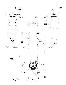
- Figure 2 shows an exploded view in elevation of the different components of
the medical-veterinary
device intended to be coupled alternatively with said cylindrical tube, in
order to form a syringe for
extracting biological material, a container for processing biological
material, or an extraction syringe
from the container of the processed biological material fractions and
alternatively an
injection/application syringe of the biological material and/or its fractions.
- Figure 2a shows a front view of the plunger, in which lines and points in
relief can be seen defined
in the flexible front part thereof.
- Figures 3 and 4 show perspective views of the medical-veterinary device
respectively forming a
syringe and a container
- Figure 5 shows a perspective view of the stopper with detachable fins
designed to fit onto the rear
end of the cylindrical tube in order to form the syringe configuration.
- Figure 6 shows an elevation view, sectioned by a vertical plane of the
syringe ready for the
extraction of biological material; with the front part of the piston in a
convex initial position, in contact
with the conical front end of the cylindrical tube; with the suction plunger
connected to the first rod,
without axial channel, and with a straight needle, mounted on the male Luer
Lock cone connector or
similar, at the anterior end of the cylindrical tube.
- Figure 7 shows a view of the syringe of figure 6, once an extraction of
biological material has been
carried out, specifically blood, with the plunger displaced and the flexible
part of the plunger modified
in a concave position and the plunger assembly moved towards the zone back of
the cylindrical tube.
- Figure 8 shows a view of the syringe of Figure 7, once the blood extraction
has been carried out,
and the first driving rod has been removed, and with the front and rear covers
attached to the ends of
the tube, forming a container for the processing of biological material in
processing equipment. In
case of processing the device in the equipment designed to carry out the
separations of fractions in
an automated way, the stoppers are not necessary.
- Figure 9 shows a profile view of the syringe with the plunger in the
position of figure 8, containing
the blood fractions obtained by processing, specifically centrifuging in this
case. In this figure, the
second rod, with an axial channel, is coupled to the valved connector and
fixed by means of a
bayonet-type closure to the rigid plunger for the extraction of the blood
fractions obtained and with a
syringe, for the collection of the fraction obtained above the cylindrical
tube connected to the rear
connector of said second axial channel.
- Figure 10 shows a schematic elevation view of an embodiment of the equipment
for processing the
biological material contained in the cylindrical tube of Figures 4 and 8, and
in which the different
elements housed inside the equipment have been represented.
- Figures 11, 12 and 13 show schematic plan views of the processing equipment
of the previous
Figure during the separation into fractions of a biological sample housed in
the cylindrical tube that
functions as a container for the medical-veterinary device and the transfer
from one of the separated
fractions to a second cylindrical tube that also functions as a container.
5
CA 03223451 2023- 12- 19
- Figure 14 shows a plan view of a variant embodiment of the processing
equipment working
simultaneously with two pairs of cylindrical tubes.
- Figure 15 shows a valiant embodiment of the system shown in Figure 1,
provided in this case with
means of intercommunication for its connection with other external equipment.
Detailed description of embodiments of the invention
In Figure 1, the medical-veterinary device (100) has been schematically
represented, using the format of a
syringe (100a) and the format of a container (100b) conformable with the same
device, and the appropriate
processing equipment (600) in order to process the biological samples inside
the container format (100b) of the
medical-veterinary device (100).
The medical-veterinary device (100) comprises a cylindrical tube (10) that has
a conical front end (101), equipped
with a connector (102), in this case a male Luer Lock cone, for coupling a
needle, trocar, cannula or tube,
depending on the biological material to be processed, (blood, bone marrow,
adipose tissue, etc.) provided with a
complementary connection and an open rear end (103).
The front and rear ends (101, 103) of the cylindrical tube (10) comprise
coupling means (104, 105) for assembling
a rear stop (30) at the rear end of the cylindrical tube during use of the
medical-veterinary device as a syringe
(100a), or the assembly at the anterior and posterior ends of the cylindrical
tube (10) of respective closure caps
(106,107) that together with the cylindrical tube (10) form the container
(100b) for the processing of biological
material in the processing equipment (600),
The two-component piston (20) with the possibility of longitudinal
displacement is assembled inside the cylindrical
tube (10)
Said piston (20) comprises a rigid rear part (201), of tubular configuration,
and a front part (202) made of flexible
material coupled to the rear part (201). Said front part (202) includes
perimeter sealing lips (203) against the wall
of the cylindrical tube and has a valved connector (204) in its central area,
accessible through the rear mouth of
the tube and suitable for optional connection of a first rod (40) actuating
the piston, a second transparent rod (50)
provided with an axial channel, or a standard syringe with a suitable
connector for the separate extraction of
fractions of biological material once processed. The removable rear stop (30)
includes lateral tabs (301) to
support the fingers and a hole (302) with radial openings (303) for through-
mounting and retention in different
positions of the first rod (40) drive or the second stem (50) with axial
channel.
The first drive rod (40) comprises a front connector (401) that can be
mechanically coupled to the valved
connector (204) of the flexible component of the piston (20), ergonomic wings
(402) for its drive and a row of
lateral teeth (403) spaced in the longitudinal direction, which in a first
angular position of the first drive rod (40)
pass through the radial openings (303) of the rear stop (30) thus allowing the
longitudinal displacement of said
first drive rod (40) and in a second position interact with the rear stop (30)
preventing the longitudinal
displacement of said first rod (40)
The second rod (50), with an axial channel, comprises a conical front
connector (501) that is an extension of the
axial channel that can be attached to the piston valve connection, fixed in
this case to the rigid component of the
piston by means of bayonet or thread-type mechanical elements and optionally a
female Luer Lock cone-type
valved rear connector (502) connected by a fine axial conduit (503) for
extracting fractions of blood or biological
material contained in a cylindrical tube and separated by centrifugation or
processing. This second rod (50) also
includes ergonomic tabs (504) for manual operation and longitudinal tabs with
a row of teeth (505) spaced apart
in the longitudinal direction, similar to those of the first drive piston
(40), for its locking and unlocking with respect
to the rear stop (30) of the cylindrical tube (10).
As shown in Figure 2a, the front part (202) of the plunger features radial
lines or dots (205) in positive relief.
Figure 3 shows an example of the embodiment of the medical-veterinary device
forming a syringe with the
cylindrical tube (10), the rear stopper (30) coupled to the tube and the first
rod (40) mounted through the rear
stopper (30) and mechanically coupled to the plunger (20) -not shown- housed
in the cylindrical tube (10).
Figure 4 shows an embodiment of the medical-veterinary device forming a
container for the processing of
biological material, made up of the tube cylinder (10), the closure caps (106,
107) and the piston housed, in a
hidden position, in the cylindrical tube (10).
The perspective of Figure 5 shows an example of the rear stopper (30) with the
hole (302) and the radial
openings (303) for through mounting and locking the stems (40, 50) in
different positions.
6
CA 03223451 2023- 12- 19
As shown in Figures 1 and 6, in an initial position the front part (202)
presents a convex shape, with a tampering
which coincides with the front end of the cylindrical tube and is initially
kept in contact with the conical front end of
the tube by means of the radial lines or dots (205) in relief, avoiding the
presence of air between the piston (20)
and the conical front end (101) of the cylindrical tube.
In said Figure 6, the medical-veterinary device forms a syringe for the
extraction of a biological material, for
example, blood, with the first actuation rod (40) being mechanically coupled
to the plunger (20) that is in the initial
convex position.
Figure 7 shows the syringe of the previous Figure once the first drive rod
(40) and consequently plunger (20) have
been displaced towards the rear area, the front part (202) changing its
morphology from convex to concave and
once the extraction of the biological material contained in the device now
configured as a syringe has been
carried out.
This displacement of the first rod (40), pulling the valved connector (204) of
the piston (20) causes the front part of
said piston (20) to adopt the concave shape.
Once the biological material has been extracted, the medical-veterinary device
becomes a container, represented
in Figure 8, suitable for keeping the biological material (M) inside during
its processing in processing equipment
(600) as will be described later.
In order to convert the syringe of Figure 7 into the container of Figure 8, it
is enough to disassemble the first
actuation rod (40) and the rear stop (30) from the cylindrical tube (10), and
optionally close the front and rear ends
of the cylindrical tube through the corresponding caps (106 and 107).
In case of using the processor in an automated way, it is not essential to use
the caps (106, 107), connecting
some flexible tubes with fixing elements both to the male Luer Lock cone
connection of the cylinder and to the
female Luer Lock cone valved connection plunger which, in turn, through
peristaltic or other pumps and
element/composition sensors of the biological material, will speed up, slow
down, stop, or reverse the flow
direction through the sensor of the processor to the tank, where the process
of the device will collect the separate
fractions of interest.
Once the biological material (M) has been processed and its different
fractions (for example Fl, F2, F3) have
been separated, the container of the medical-veterinary device shown in Figure
8 is transformed into a syringe
represented in Figure 9 and suitable for the independent extraction of the
successive fractions of biological
material, simply removing the caps (106, 107) from the cylindrical tube (10),
if optionally used, and mounting the
rear stop 30 on the same cylindrical tube used for the example of extraction
and, in this case, the second stem
(50) with axial channel for the extraction of said fractions through its fine
axial duct (503) in order of higher to
lower fraction.
In Figure 8 said extraction of the fraction (F1) is optionally carried out by
connecting a standard syringe (not
referenced) to the rear valved connection (502) of the axial channel rod (50)
or by means of a syringe directly
coupled to the plunger without the second coupled stem.
Optionally, a flexible tube can also be coupled directly to the plunger,
provided with suitable connectors to
motorize the extraction of the fractions, optionally using peristaltic pumps
or any device that allows the fractions
obtained to be aspirated in an automated and continuous manner, without the
manual use of elements
/components such as those described at the beginning of this chapter.
The concave shape of the front part (202) of the piston (20) is especially
advantageous during the extraction of
the fraction (F1) and all the following fractions of biological material,
through the interior of the second rod (50)
since the suction is carried out through the most rearward central area of the
front part of the piston, thus avoiding
the formation of turbulence and, for example, preventing the first fraction
(F1) from mixing during its extraction
with the next fraction (F2) and so on with all the fractions.
The processing equipment (600) shown in Figure 10 and used for processing the
biological material inside the
container of the medical-veterinary device described above, comprises an outer
casing (601) provided with a lid
(602) and housing a turntable (603) driven by a motor (606) for bidirectional
rotation connected to a frequency
inverter (609) controlled by a control unit (607).
Said cap (602) has some ultraviolet light lamps (614) inside for the
disinfection of the interior of the equipment
and/or its use as a trigger and trainer of biological processes in the samples
(for example, blood or plasma
coagulation).
7
CA 03223451 2023- 12- 19
The turntable (603) comprises fixings (604) for holding a container (100b)
containing a biological material to be
processed and an empty container (100b) for collecting a separate fraction of
the biological material.
In this embodiment, the processing equipment also comprises: - a cold or heat
generator (616) in order to
eventually control the temperature of the sample with different aims such as
the reduction of processing times, an
increase of biological processes, an optimal maintenance of the samples, etc.;
- a transformer (617) for power
supply and - an internal or external ultrasonic generator (618).
Figures 11 to 13 show a pair of containers (100b) connected by a transparent
tube (611), preferably flexible and
made of silicone, provided with connectors (not referenced) coupled to the
Luer Lock connectors (102 and 204)
the respective containers (100b) and a peristaltic pump (605) for the transfer
from one container (100b) to another
of a fraction of the biological material, separated during a processing cycle,
optionally by one connector or by
another or both at the same time, depending on the fraction of interest.
The processing equipment (600) comprises a support (610) for holding an
intermediate portion of the transparent
tube (611) and a laser sensor (612) that detects, for example, the color (also
optionally the sensor in the flexible
tube line can be a detector, reader, counter and identifier of particles,
their size, their number and their
geometry/morphology or their biological, chemical or biochemical composition,
using optical or digital imaging
technology, impedance, laser or other technologies that conform to these
readings, counts and identifications), of
the final product transferred through the transparent tube (611) and that
causes different actions/reactions of the
pump such as acceleration, braking, change of direction of rotation and the
stoppage of the peristaltic pump (605)
when, for example, it detects the change in color of the product, due to the
change in the fraction that circulates
through said tube, or the activation of other functions or processes such as
ultrasound, temperature, agitation or
similar.
Said transparent tube (611) comprises an air purge filter (613) to prevent air
from being transferred together with
the separated fraction of the processed biological material.
Figure 14 shows a plan view of a variant embodiment of the processing
equipment working simultaneously with
two pairs of containers (100b).
The connections with the transparent tube (611) can also be made at the rear
of the cylindrical tube, particularly
connected to the second plunger (204) in order to remove fractions from this
part of the device or simultaneously
from the front (102) and rear (204) of the device depending on the needs of
the user.
This processing equipment (600) includes a touchpad (608) connected to the
control unit (607) and has software
that allows it to work independently, without Internet connection, carrying
out predefined processing cycles, or to
work "on line" with internet connection as shown in Figure 15.
In this embodiment, the processing equipment (600) is connected to a counting
or analysis equipment, for
example, a hematology analyzer (700) that determines the characteristics of
the biological material to be
processed and sends it to the processing equipment (600) in such a way that it
performs in each case, with each
sample from each patient, a customized processing cycle based on the
characteristics of the biological material to
be processed. Said hematological analyzer (700) can also determine the
characteristics of the final product
obtained from processing,
In this embodiment, the processing equipment (600) includes a communication
module (615) for receiving
information related to the characteristics of the product to be processed,
sending data related to the final product
obtained from processing, and connecting to a computer (800) and to remote
devices, an App, or the cloud.
This communication module allows the control and traceability of the
tubes/cylindrical tanks through barcodes,
QR or RF microchip among others, for control of quality, for analysis of
samples and pre- and post-processed
biological material by means of particle identifier, their size, their number
and their geometry/morphology or
biological, chemical or biochemical composition, by means of optical, digital
imaging technology, also by
impedance, laser or other technologies that adjust to these readings, counts
and identifications, as well as to
coordinate and execute variable processes of the biological samples in the
processor according to readings, for
optimization of the final biological product as well as the collection of
individual data, protocols and identification
and history of the sample and/or patients, for remote control and diagnosis
and telemedicine/veterinary/analysis,
data export and reading use on any current and future platform (PC, Mac,
laptops, phones and smart terminals, in
relation to and in use with Al (Artificial Intelligence), loT (Internet of
Things), IT (Information Technology), ICT
(Information and Communication Technologies), social networks, among others.
Once the nature of the invention has been sufficiently described, as well as a
preferred embodiment, it is stated
for the appropriate purposes that the materials, shape, size and arrangement
of the elements described may be
8
CA 03223451 2023- 12- 19
modified, as long as this does not imply an alteration of the essential
characteristics of the invention that are
claimed below.
9
CA 03223451 2023- 12- 19