Note: Descriptions are shown in the official language in which they were submitted.
CA 03224175 2023-12-13
WO 2022/264109
PCT/IB2022/055655
Multivalent Influenza Vaccines
RELATED APPLICATIONS
[001] This application claims the benefit of U.S. Provisional Application
No. 63/212,523,
filed on June 18, 2021; U.S. Provisional Application No. 63/276,243, filed
November 5, 2021;
PCT International Application No. PCT/U52021/058250, filed November 5, 2021;
and
European Patent Application No. 21315198.8, filed October 13, 2021; which are
incorporated
by reference in their entirety for all purposes.
BACKGROUND OF THE INVENTION
[002] Messenger RNA (mRNA) based vaccines provide a promising alternative to
traditional subunit vaccines, which contain antigenic proteins derived from a
pathogen.
Antigen proteins are usually recombinantly made and require bacterial
fermentation and/or cell
culture, as well as complex purification. Vaccines based on mRNA allow de novo
expression
of complex antigens in the vaccinated subject, which in turn allows proper
post-translational
modification and presentation of the antigens in its natural conformation.
Unlike traditional
technologies, the manufacture of mRNA vaccines does not require complex and
costly bacterial
fermentation, tissue culture, and purification processes. Moreover, once
established, the
manufacturing process for mRNA vaccines can be used for a variety of antigens,
enabling rapid
development and deployment of mRNA vaccines. Further, mRNA vaccines are
inherently safe
delivery vectors as they express the antigens only transiently and do not
integrate into the host
genome. Because antigens encoded by mRNAs are produced in vivo in the
vaccinated
individual, mRNA vaccines are especially effective in eliciting both humoral
and T cell
mediated immunity.
[003] Current approved influenza vaccines are either live attenuated
influenza vaccines or
inactivated influenza vaccines, which are often produced in cell culture or
eggs. Moreover,
multiple strains of influenza may circulate within populations each year,
making it difficult for
a single influenza vaccine to offer robust protection against multiple
strains. Accordingly,
there exists a need for mRNA-based influenza vaccines, including multivalent
mRNA-based
influenza vaccines that target multiple influenza strains.
SUMMARY OF THE INVENTION
1
CA 03224175 2023-12-13
WO 2022/264109
PCT/IB2022/055655
[004] The present disclosure provides an influenza vaccine composition,
comprising eight
messenger RNA (mRNA), each mRNA comprising an open reading frame (ORF)
encoding a
different influenza antigen.
[005] In certain embodiments, the composition comprises eight mRNA encoding
(i) one or
more hemagglutinin (HA) antigens, (ii) one or more neuraminidase (NA)
antigens, or (iii) at
least one HA antigen and at least one NA antigen.
[006] In certain embodiments, the composition comprises one or more mRNA
encoding
antigens of influenza A, B and/or C viruses.
[007] In certain embodiments, the antigens are HA and/or NA antigens of
influenza A and
influenza B viruses.
[008] In certain embodiments, the NA antigens of influenza A viruses are
selected from
subtypes Ni, N2, N3, N4, N5, N6, N7, N8, N9, NiO, and N11.
[009] In certain embodiments, the HA and NA antigens of influenza B viruses
are from the
influenza B/Yamagata lineage or the influenza BNictoria lineage.
[0010] In certain embodiments, the HA antigen and NA antigen is selected from
the group
consisting of H1N1, H3N2, H2N2, H5N1, H7N9, H7N7, H1N2, H9N2, H7N2, H7N3,
H5N2,
and Hi 0N7 subtypes and/or B/Yamagata and B/Victoria lineages.
[0011] In certain embodiments, the composition comprises one mRNA encoding an
H3 HA
antigen, one mRNA encoding an H1 HA antigen, one mRNA encoding an HA antigen
from the
Influenza B/Yamagata lineage, and one mRNA encoding an HA antigen from the
Influenza
BNictoria lineage.
[0012] In certain embodiments, the composition comprises one mRNA encoding an
H3 HA
antigen, one mRNA encoding an N2 NA antigen, one mRNA encoding an H1 HA
antigen, one
mRNA encoding an Ni NA antigen, one mRNA encoding an HA antigen from the
Influenza
B/Yamagata lineage, one mRNA encoding an NA antigen from the Influenza
B/Yamagata
lineage, one mRNA encoding an HA antigen from the Influenza BNictoria lineage,
and one
mRNA encoding an NA antigen from the Influenza BNictoria lineage.
[0013] In certain embodiments, the ORF is codon optimized.
[0014] In certain embodiments, the mRNA molecule comprises at least one 5'
untranslated
region (5' UTR), at least one 3' untranslated region (3' UTR), and at least
one polyadenylation
(poly(A)) sequence.
[0015] In certain embodiments, the mRNA comprises at least one chemical
modification.
2
CA 03224175 2023-12-13
WO 2022/264109
PCT/IB2022/055655
[0016] In certain embodiments, at least 20%, at least 30%, at least 40%, at
least 50%, at least
60%, at least 70%, at least 80%, at least 85%, at least 90%, at least 95%, or
100% of the uracil
nucleotides in the mRNA are chemically modified.
[0017] In certain embodiments, at least 20%, at least 30%, at least 40%, at
least 50%, at least
60%, at least 70%, at least 80%, at least 85%, at least 90%, at least 95%, or
100% of the uracil
nucleotides in the ORF are chemically modified.
[0018] In certain embodiments, the chemical modification is selected from the
group
consisting of pseudouridine, Nl-methylpseudouridine, 2-thiouridine, 4'-
thiouridine, 5-
methylcytosine, 2-thio-l-methy1-1-deaza-pseudouridine, 2-thio-l-methyl-
pseudouridine, 2-
thio-5-aza-uridine, 2-thio-dihydropseudouridine, 2-thio-dihydrouridine, 2-thio-
pseudouridine,
4-methoxy-2-thio-pseudouridine, 4-methoxy-pseudouridine, 4-thio-l-methyl-
pseudouridine,
4-thio-pseudouridine, 5-aza-uridine, dihydropseudouridine, 5-methyluridine, 5-
methyluridine,
5-methoxyuridine, and 2' -0-methyl uridine.
[0019] In certain embodiments, the chemical modification is selected from the
group
consisting of pseudouridine, Nl-methylpseudouridine, 5-methylcytosine, 5-
methoxyuridine,
and a combination thereof
[0020] In certain embodiments, the chemical modification is Nl-
methylpseudouridine.
[0021] In certain embodiments, the mRNA is formulated in a lipid nanoparticle
(LNP).
[0022] In certain embodiments, the LNP comprises at least one cationic lipid.
[0023] In certain embodiments, the cationic lipid is biodegradable. In certain
embodiments,
the cationic lipid is not biodegradable.
[0024] In certain embodiments, the cationic lipid is cleavable. In certain
embodiments, the
cationic lipid is not cleavable.
[0025] In certain embodiments, the cationic lipid is selected from the group
consisting of OF-
02, cKK-E10, GL-HEPES-E3-E10-DS-3-E18-1, GL-HEPES-E3-E12-DS-4-E10, and GL-
HEPES -E3 -E12-D S -3 -E14 .
[0026] In certain embodiments, the LNP further comprises a polyethylene glycol
(PEG)
conjugated (PEGylated) lipid, a cholesterol-based lipid, and a helper lipid.
[0027] In certain embodiments, the LNP comprises:
a cationic lipid at a molar ratio of 35% to 55%;
a polyethylene glycol (PEG) conjugated (PEGylated) lipid at a molar ratio of
0.25%
to 2.75%;
a cholesterol-based lipid at a molar ratio of 20% to 45%; and
a helper lipid at a molar ratio of 5% to 35%,
3
CA 03224175 2023-12-13
WO 2022/264109
PCT/IB2022/055655
wherein all of the molar ratios are relative to the total lipid content of the
LNP.
[0028] In certain embodiments, the LNP comprises:
a cationic lipid at a molar ratio of 40%;
a PEGylated lipid at a molar ratio of 1.5%;
a cholesterol-based lipid at a molar ratio of 28.5%; and
a helper lipid at a molar ratio of 30%.
[0029] In certain embodiments, the PEGylated lipid is dimyristoyl-PEG2000 (DMG-
PEG2000) or 2-[(polyethylene glycol)-20001-N,N-ditetradecylacetamide (ALC-
0159).
[0030] In certain embodiments, the cholesterol-based lipid is cholesterol.
[0031] In certain embodiments, the helper lipid is 1,2-dioleoyl-SN-glycero-3-
phosphoethanolamine (DOPE) or 1,2-distearoyl-sn-glycero-3-phosphocholine
(DSPC).
[0032] In certain embodiments, the LNP comprises:
a cationic lipid is selected from the group consisting of OF-02, cKK-E10, GL-
HEPES-
E3-E10-DS -3-E18-1, GL-HEPES-E3-E12-DS-4-E10, and GL-HEPES-E3-E12-DS-3-E14, at
a molar ratio of 40%;
DMG-PEG2000 at a molar ratio of 1.5%;
cholesterol at a molar ratio of 28.5%; and
DOPE at a molar ratio of 30%.
[0033] In certain embodiments, the LNP has an average diameter of 30 nm to 200
nm. In
certain embodiments, the LNP has an average diameter of 80 nm to150 nm.
[0034] In certain embodiments, the influenza vaccine composition comprises
between 1
mg/mL to 10 mg/mL of the LNP.
[0035] In certain embodiments, the LNP comprises between 1 and 20 mRNA
molecules. In
certain embodiments, the LNP comprises 5-10 or 6-8 mRNA molecules.
[0036] In certain embodiments, the LNP comprises two or more mRNA, wherein
each mRNA
encodes a different influenza antigen.
[0037] In certain embodiments, the composition comprises eight LNPs, wherein
each LNP
comprises an mRNA encoding a different influenza antigen.
[0038] In certain embodiments, the composition is formulated for intramuscular
injection.
[0039] In certain embodiments, the composition comprises a phosphate-buffer
saline.
[0040] In one aspect, the disclosure provides a method of eliciting an immune
response in a
subject in need thereof, comprising administering to the subject, optionally
intramuscularly,
intranasally, intravenously, subcutaneously, or intradermally, a
prophylactically effective
amount of the influenza vaccine composition described above.
4
CA 03224175 2023-12-13
WO 2022/264109
PCT/IB2022/055655
[0041] In one aspect, the disclosure provides a method of preventing influenza
infections or
reducing one or more symptoms of influenza infections, comprising
administering to the
subject, optionally intramuscularly, intranasally, intravenously,
subcutaneously, or
intradermally, a prophylactically effective amount of the influenza vaccine
composition
described above.
[0042] In certain embodiments, the influenza vaccine composition elicits an
immune
response against one or more seasonal and/or pandemic influenza strains.
[0043] In certain embodiments, the method comprises administering to the
subject one or
more doses of the influenza vaccine composition, each dose comprising about 1
jig to about
250 jig of mRNA.
[0044] In certain embodiments, the method comprises administering to the
subject one or
more doses of the influenza vaccine composition, each dose comprising about
2.5, 5, 15, 45,
or 135 jig of mRNA.
[0045] In certain embodiments, the method comprises administering to the
subject two doses
of the influenza vaccine composition with an interval of 2-6, optionally 4,
weeks.
[0046] In another aspect, the disclosure provides for the use of the influenza
vaccine
composition described above for the manufacture of a medicament for use in
treating a subject
in need thereof
[0047] In certain embodiments, the influenza vaccine composition is for use in
treating a
subject in need thereof
[0048] In another aspect, the disclosure provides a kit comprising a container
comprising a
single-use or multi-use dosage of the composition described above, optionally
wherein the
container is a vial or a pre-filled syringe or injector.
[0049] In certain embodiments, the influenza antigens comprise an influenza
virus HA
antigen and/or an influenza virus NA antigen having a molecular sequence
identified or
designed from a machine learning model.
BRIEF DESCRIPTION OF THE DRAWINGS
[0050] FIG. 1A is a pair of graphs showing the expression of human
erythropoietin (hEPO)
in mice treated with various LNP formulations of hEPO mRNA. Panel a): LNP
formulations
"Lipid A" and "Lipid B" compared to MC3. Bars represent means and standard
deviations.
Panel b): Formulation made with cationic lipid OF-02. PEG: DMG-PEG2000.
Cholest:
cholesterol. "Lipid A": LNP composition containing OF-02, DMG-PEG2000,
cholesterol, and
DOPE, in this order, at a molar ratio of 40:1.5:28.5:30, unless otherwise
indicated. "Lipid B":
CA 03224175 2023-12-13
WO 2022/264109
PCT/M2022/055655
LNP composition containing cKK-E10, DMG-PEG2000, cholesterol, and DOPE, in
this order,
at a molar ratio of 40:1.5:28.5:30.
[0051] FIG. 1B is a pair of graphs showing expression of hEPO in mice and non-
human
primates (NHPs) using LNP formulations Lipid A and Lipid B.
[0052] FIG. 2A and 2B are a pair of graphs showing that Lipid A and Lipid B
LNP
formulations with mRNA encoding hemagglutinin (HA) of strain
A/California/7/2009 (H1N1)
(CA09) induced robust functional antibodies (FIG. 2A) and protected mice
against death or
severe weight loss (more than 20%) when challenged with a pandemic strain of
influenza virus
(FIG. 2B). Hemagglutinin inhibition (HAT) titers are reported as log10 for
serum samples
taken at study days 0, 14, 28, 42, 56, 92, and 107. Bars are geometric means
and geometric
standard deviations. Daily weights were measured after intranasal challenge
(day 93) with
4LD50 of A/Belgium/2009 (H1N1) (Belgium09). Weights are presented as the
percentage of
weight lost from the day of challenge. Euthanasia occurred for mice losing
more than 20% of
their starting body weight and for all mice 14 days post-infection (day 107).
rHA: recombinant
hemagglutinin. AF03: an oil-in-water emulsion adjuvant. Diluent = PBS. LLOQ =
lower limit
of quantitation. 1/40 = 1/40 minimum target, which refers to HAT antibody
titers associated
with 50% reduction in the risk of influenza infection or disease in healthy
adults (Coudeville
et al., BMC Med Res Methodol. (2010) 10:18). Dashed line in FIG. 2B = 20%
weight loss cut
off with respect to weight on the day of challenge.
[0053] FIG. 3A and 3B are a pair of graphs showing that A/Michigan/45/2015
(Mich15)
neuraminidase (NA) mRNA formulated with Lipid A LNP induced robust functional
antibodies (FIG. 3A) and protected mice against weight loss and death when
challenged with
a pandemic strain of influenza virus (FIG. 3B). Neuraminidase inhibition (NAT)
titers are
reported as log10 for serum samples taken at study days 14, 28, 42, 56, 88,
and 114. Daily
weights were observed after intranasal challenge (day 89 for the one-dose
groups or day 117
for the two-dose groups) with 4LD50 of Belgium09. Weights are presented as the
percentage
of weight lost from the day of challenge. Euthanasia occurred for mice losing
more than 20%
of their starting body weight and for all mice 14 days post-infection (day 103
for the 1 dose
groups or day 131 for the 2 dose groups). Bars are means and standard
deviations. Upper
dashed line in FIG. 3A = upper limit of quantitation. Lower dashed line in
FIG. 3A = lower
limit of quantitation. Dashed line in FIG. 3B = 20% weight loss cut off with
respect to weight
on the day of challenge. mRNA dosed: 0.4 or 0.016 jig mRNA encoding Mich15 NA.
Control:
0.6 jig mRNA encoding hEPO or diluent (PBS).
6
CA 03224175 2023-12-13
WO 2022/264109
PCT/IB2022/055655
[0054] FIG. 4 is a graph showing that Lipid A and Lipid B LNP formulations
with CA09 HA
mRNA (10 jt.g) induced robust functional antibodies in cynomolgus macaque
monkeys. HAT
titers are reported as 1og2 for serum samples taken at study days 0, 14, 28,
42, and 56.
[0055] FIGs. 5A-C show the MRT1400 mRNA encoding for influenza virus
A/Singapore/
INFIMH160019/2016 (5ing16; H3N2) HA hemagglutinin. FIG. 5A: an alignment of
the
wildtype (WT) gene and a codon-optimized gene (MRT10279) for the HA antigen.
FIG. 5B:
the structure of the mRNA. FIG. 5C: the sequence of the mRNA.
[0056] FIG. 6 is a pair of graphs showing that Lipid A and Lipid B LNP
formulations with
MRT1400 or NA mRNA induced robust functional antibodies in mice. First
injection was
given at study day 0 and second injection was given at study day 28. Left
Panel: HAT titers are
reported as log10 for serum samples taken at study days 14, 28, 42, and 56.
Right Panel: NAT
titers are reported as log10 for serum samples taken at study days 14, 28, 42,
and 56. Bars are
geometric means and geometric standard deviations. Dashed line = lower limit
of quantitation.
[0057] FIG. 7A is a graph showing that Lipid A and Lipid B LNP formulations
with MRT
1400 induced robust functional antibodies in NHPs. HAT titers are reported as
1og2 for serum
samples taken at study days 0, 14, 28, 42, and 56. First injection was given
at study day 0 and
second injection was given at study day 28. Bars are means and standard
deviation. Upper
dashed line = 1/40 minimum target. Lower dashed line = lower limit of
detection.
[0058] FIG. 7B and 7C are a pair of graphs showing that a Lipid A LNP
formulation
(MRT5400) containing MRT1400 mRNA induced functional antibodies (FIG. 7B) and
robust
ELISA titers (FIG. 7C) in cynomolgus macaque monkeys at four dose levels: 15,
45, 135 and
250 lag of mRNA. HAT and ELISA titers are reported as 1og2 for serum samples
taken at study
days 0, 14, 28, 42, and 56. First injection was given at study day 0 and
second injection given
at study day 28. Bars are means and standard deviations. Dash line = 1/40
minimum target.
[0059] FIGs. 8A and 8B are panels of graphs showing the T cell cytokine
response of
cynomolgus macaques after a second vaccination with Lipid A LNP formulation
MRT5400 in
three dose level groups (250 jig, 135 jig, and 45 jtg of mRNA). IFN-y and IL-
13 induced by
re-stimulation with either the recombinant HA (rHA) protein (left panel) or
the pooled peptides
(right panel) were assessed in peripheral blood mononuclear cells (PMBC) on
day 42 by
ELISPOT assays. The frequencies of PBMC secreting IFN-y (FIG. 8A) or IL-13
(FIG. 8B)
were calculated as spots forming cells (SFC) per million PBMC. Each symbol
represents an
individual sample, and the bar represents the standard deviation.
[0060] FIG. 9A is a pair of graphs showing that Lipid A LNP formulations
containing
modified and unmodified CA09 HA mRNA were comparable as indicated by HAT
titers in
7
CA 03224175 2023-12-13
WO 2022/264109
PCT/IB2022/055655
vaccinated mice. HAT titers are reported as 10g2 for serum samples taken at
study days 14, 28,
42, and 56. First injection was given at study day 0 and second injection was
given at study
day 28. Bars are means and standard deviation. Upper dashed line = 1/40
minimum target.
Lower dashed line = lower limit of quantitation.
[0061] FIG. 9B is a pair of graphs showing that Lipid A LNP formulations
containing
modified and unmodified CA09 HA mRNA were comparable as indicated by ELISA
titers in
mice. Total IgG ELISA titers are reported as log10 for serum samples taken at
study days 14,
28, 42, and 56. First injection was given at study day 0 and second injection
was given at study
day 28. Dashed line = lower limit of quantitation.
[0062] FIGs. 10A and 10B are a pair of graphs showing that bivalent Lipid A
LNP
formulations with CA09 HA mRNA and Sing16 HA mRNA induced robust functional
antibodies as assessed by HAT titers (CA09 (FIG. 10A) and Sing16 (FIG. 10B))
in Balb/c mice
at a dose of 0.4 lag of total mRNA. 0.4 lag mRNA was dosed as a co-
encapsulated mRNA-
LNP formulation, or each HA mRNA was separately administered with 0.2 lag
going into each
leg. Each HA mRNA was also co-encapsulated into a formulation with non-coding
mRNA to
control for total mRNA packing into the LNP. The diluent group received mRNA-
LNP diluent
buffer. HAT titers are reported for serum samples taken at study days -2
(baseline), 14, 28, and
42. FIG. 10B only shows study days -2 (baseline from pooled sera) and 42.
First injection
was given at study day 0 and second injection given at study day 28. Bars are
geometric means
and geometric standard deviations. Dashed line = lower limit of quantitation.
[0063] FIG. 11 shows the functional verification of mRNA-LNP Formulations.
Panel (a) is
a graph showing the expression of firefly (FF) luciferase in BALB/c mice: a
single dose of
Luciferase FF mRNA-LNP (5, 1, 0.1, 0.05 [tg) was injected in mice (n=4) by IM
route.
Luciferin (3 mg) was injected at the time of whole animal imaging, using IVIS
Spectrum,
Perkin Elmer recording bioluminescence intensity. Images of whole animal
average radiance
at 6, 24, 48 and 72h after injection were taken. Radiance recorded for 1, 0.5,
0.1 and 0.05 [tg
dose administrations of Luc mRNA-LNP are shown in the graph. Panel (b) shows
whole
animal images indicating total flux of luminescence, at 6 to 72 hours. Total
flux of
luminescence in groups of mice (n=4) receiving 0.1 g dose of FF-LNP are
shown. Panel (c)
shows the expression of hEPO in BALB/c mice. A single dose of hEPO mRNA-LNP
(0.1 g)
was injected in BALB/c mice by IM route. hEPO expression was quantified in
serum at 6
hours and 24 hours after administration using ELISA. Bars represent means and
standard
deviations. Panel (d) shows the expression of hEPO in NHP. A single dose of
hEPO mRNA-
LNP (10 g) was injected in Cynomolgus macaques by IM route. hEPO expression
was
8
CA 03224175 2023-12-13
WO 2022/264109
PCT/IB2022/055655
quantified in serum at 6, 24, 48, 72, and 96 hours after administration, using
ELISA. Bars
represent means and standard deviations.
[0064] FIG. 12 shows the serological evaluation of HA mRNA-LNP vaccine in
mice.
BALB/c mice (n=8 per group) were immunized twice IM, 4 weeks apart with 2,
0.4, 0.08, and
0.016 jig of either Ca109 HA mRNA-LNP or Sing16 HA mRNA-LNP. ELISA titers
recorded
for sera collected at days 14, 28, 42, 56 against CA09 (Ca109) H1N1 influenza
virus
recombinant HA (left panel) and Sing16 H3N2 influenza virus recombinant HA
(right panel)
are shown.
[0065] FIG. 13 shows the serological evaluation of HA mRNA-LNP vaccine in
mice.
BALB/c mice (n=8 per group) were immunized twice IM, 4 weeks apart with 2,
0.4, 0.08 and
0.016 jig of either CA09 HA mRNA-LNP or 5ing16 HA mRNA-LNP. Logio HAT titers
recorded against CA09 H1N1 influenza virus (left panel) and 5ing16 H3N2
influenza virus
(right panel) are shown.
[0066] FIG. 14 shows the serological evaluation of NA mRNA-LNP vaccine in
mice.
BALB/c mice (n=8 per group) were immunized twice IM 4 weeks apart with 2, 0.4,
0.08, and
0.016 jig of either Mich15 NA mRNA-LNP or 5ing16 NA mRNA-LNP. Total IgG titers
recorded for sera collected at days 0, 14, 28, 42, 56 against Mich15 Ni
influenza virus
recombinant NA (left panel) and Sing16 N2 virus recombinant NA (right panel)
are shown.
[0067] FIG. 15 shows the serological evaluation of NA mRNA-LNP vaccine in
mice.
BALB/c mice (n=8 per group) were immunized twice IM 4 weeks apart with 2, 0.4,
0.08 and
0.016 jig of either Mich15 NA mRNA-LNP or 5ing16 NA mRNA-LNP. Logic) NAT
(ELLA)
titers recorded for sera against Mich2015 (Ni): A/Mallard/Sweden/2002 (H6)
chimeric
influenza virus (left panel) and Sing16 (N2): A/Mallard/5weden/2002 (H6)
chimeric virus
(right panel) are shown.
[0068] FIGs. 16A and 16B show the protective efficacy of CA09 HA mRNA-LNP
vaccine
in mice after lethal A/Belgium/2009 H1N1 virus challenge. Mice (n=8) received
two IM doses
of CA09 HA mRNA-LNP (0.4 jig each) on day 0 and day 28. Control animals
received two
IM doses of diluent on day 0 and day 28. FIG. 16A shows the HAT titers
reported as Logic) for
serum samples taken at study days 0, 14, 28, 42, 56, 92, and 107. FIG. 16B
shows daily
weights after intranasal challenge on day 93 with 4LD50 of A/Belgium/2009 H1N1
strain.
Weights are presented as the percentage of weight lost from the day of
challenge. Individual
lines represent each animal.
[0069] FIGs. 17A-B show the protective efficacy of a single dose of unmodified
Mich15 NA
mRNA-LNP in mice after lethal A/Belgium/2009 H1N1 virus challenge. Mice (n=16)
were
9
CA 03224175 2023-12-13
WO 2022/264109
PCT/IB2022/055655
injected by the IM route with 0.4 [tg or 0.016 [tg of Mich15 NA mRNA-LNP. Half
of the mice
only received one injection (1 dose) on study day 0, while the other half (2
doses) received two
injections given at study day 0 and day 28. Control animals received two IM
doses of hEPO
mRNA-LNP (0.6 g) on day 0 and day 28. FIG. 17A shows the NAT titers are
reported as
Logic) for serum samples taken at study days 0, 14, 28, 42, 56, 88, and 114.
FIG. 17B shows
the daily weight change after intranasal challenge on day 89 for single dose
group and day 117
(89 days after second dose) for two dose group with 4LD50 of Belgium09 H1N1.
Weights are
presented as the percentage of weight lost from the day of challenge.
Individual lines represent
each animal.
[0070] FIG. 18 shows the serological evaluation of HA Sing16 HA mRNA-LNP
vaccine in
NHP. Cynomolgus macaques (n=6 per group) were injected twice, 4 weeks apart by
IM route,
with 15, 45 or 135 g of Sing16 HA mRNA-LNP. Serum samples were collected at
days -6,
14, 28, 42, and 56. Logic) IgG titers against recombinant HA protein of Sing16
virus are shown.
[0071] FIGs. 19A and 19B show the serological evaluation of HA Sing16 HA mRNA-
LNP
vaccine in NHP. Cynomolgus macaques (n=6 per group) were injected twice, 4
weeks apart
by IM route, with 15, 45 or 135 g of Sing16 HA mRNA-LNP. Serum samples were
collected
at days 0, 14, 28, 42, and 56. Logio HAT titers (FIG. 19A) and Logio micro-
neutralization
(MN) titers (FIG. 19B) against 5ing2016 virus are shown.
[0072] FIGs. 20A and 20B show T cell responses in NHP vaccinated with Sing16
HA
mRNA-LNP vaccine. Cynomolgus macaques (n=6 per group) were injected twice, 4
weeks
apart by IM route, with 45, 135, or 250 g of Sing16 HA mRNA-LNP. T cells were
determined
by ELISPOT on day 42 in PBMC stimulated in vitro with peptide pools to
represent the entire
HA open reading frame. The responses of PBMC secreting IFN-y (FIG. 20A) or IL-
13 (FIG.
20B) calculated as spots forming cells (SFC) per million PBMC are shown. Each
symbol
represents an individual sample, and the bar represent the geometric mean for
the group.
[0073] FIG. 21 shows the secretion of Sing16 H3-specific IgG by memory B cells
on day
180 in NHP vaccinated with Sing16 HA mRNA-LNP vaccine. Cynomolgus macaques
(n=6
per group) were injected twice, 4 weeks apart by IM route, with 15 or 45 g of
Sing16 HA
mRNA-LNP. The Human IgG single-color memory B cell ELISPOT kit (CAT#
NC1911372,
CU) was used to measure Sing16/H3-specific and total IgG+ antibody-secreting
cells (ASCs).
Differentiation of MBCs into ASCs was performed in PBMC collected at day 180
by using a
stimulation cocktail provided by the kit. The number of IgG+ and number of
Sing16/H3-
specific ASCs was calculated per million of PBMCs for each animal and the
frequency of
antigen-specific ASCs is shown.
CA 03224175 2023-12-13
WO 2022/264109
PCT/IB2022/055655
[0074] FIG. 22 shows the delivery of bivalent combinations of influenza
vaccine in mice.
BALB/c mice (n=8 per group) were immunized twice IM, 4 weeks apart with a
total 0.4 jig of
bivalent combinations co-encapsulated mRNA transcripts (1:1 wt/wt, half volume
per each leg)
or 0.2 jig each monovalent which was separately formulated and immunized
different legs.
H1H3 combo constituting CA09 HA mRNA-LNP, Sing16 HA mRNA-LNP; H3N2 combo of
5ing16 HA mRNA-LNP and 5ing16 NA mRNA-LNP and N1N2 combo of Mich15 NA
mRNA-LNP and Perth09 NA mRNA-LNP were tested in sera collected a day 0, 14,
28, 42,
against corresponding virus. Panel (a) shows HAT titers recorded against CA09
H1N1
influenza virus and Sing2016 H3N2. Panel (b) shows the HAT and NAT titers
recorded against
5ing2016 H3N2 and A/Mallard/Sweden/2002 (H6) chimeric influenza virus and H6N2
A/Perth/09 virus F1919D (N2) virus, respectfully. Panel (c) shows NAT titers
recorded against
Mich15 (Ni): A/Mallard/Sweden/2002 (H6) chimeric influenza virus and H6N2
A/Perth/09
virus F1919D (N2) virus.
[0075] FIG. 23 shows the delivery of quadrivalent combinations of influenza
vaccines in
NHP. Cynomolgus macaques (n=6 per group) were immunized twice IM, 4 weeks
apart with
a total 10 jig of quadrivalent combinations of co-encapsulated mRNA
transcripts (1:1:1:1
wt/wt). H2H3N1N2 combo consisting of CA09 HA mRNA, Sing16 HA mRNA, Mich15 NA
mRNA, and Perth09 NA mRNA. H1H3 combo constituting CA09 HA mRNA, 5ing16 HA
mRNA and 2x non-coding mRNA (ncmRNA); H3N2 combo of 5ing16 HA mRNA and
Perth09 NA mRNA and 2x non-coding mRNA. N1N2 combo of Mich15 NA mRNA, Perth09
NA mRNA-LNP, and 2x non-coding mRNA. H1 consisting of CA09 HA mRNA and 3x non-
coding mRNA. H3 consisting of 5ing16 HA mRNA and 3x non-coding mRNA. Ni
consisting
of Mich15 NA mRNA and 3x non-coding mRNA. N2 consisting of Perth09 NA mRNA and
3x non-coding mRNA. Inhibitory titers were tested in sera collected a day 0,
14, 28, 42, against
corresponding virus. Panel (a) shows the HAT titers recorded against CA09 H1N1
influenza
virus and 5ing16 H3N2. Panel (b) shows the NAT titers recorded against Mich15
(Ni):
A/Mallard/5weden/2002 (H6) chimeric influenza virus and H6N2 Perth/09 virus
F1919D (N2)
virus.
[0076] FIG. 24 depicts a graph showing the expression of human erythropoietin
(hEPO) in
mice treated with various LNP formulations of hEPO mRNA. LNP formulations
"Lipid A,"
"Lipid B," "Lipid C," "Lipid D," and "Lipid E" are shown. Bars represent means
and standard
11
CA 03224175 2023-12-13
WO 2022/264109
PCT/IB2022/055655
deviations. The LNP compositions contain the cationic lipid, DMG-PEG2000,
cholesterol, and
DOPE, in this order, at a molar ratio of 40:1.5:28.5:30.
[0077] FIG. 25 depicts a graph showing the expression of hEPO in non-human
primates
(NHPs) treated with various LNP formulations of hEPO mRNA. LNP formulations
"Lipid A,"
"Lipid B," "Lipid C," "Lipid D," and "Lipid E" are shown. Bars represent means
and standard
deviations. The LNP compositions contain the cationic lipid, DMG-PEG2000,
cholesterol, and
DOPE, in this order, at a molar ratio of 40:1.5:28.5:30.
[0078] FIG. 26 depicts a graph showing HAT titers at day 28 and day 42 post
injection with
various LNP formulations of HA mRNA. LNP formulations "Lipid A," "Lipid B,"
"Lipid C,"
"Lipid D," and "Lipid E" are shown. Bars represent means and standard
deviations. The LNP
compositions contain the cationic lipid, DMG-PEG2000, cholesterol, and DOPE,
in this order,
at a molar ratio of 40:1.5:28.5:30.
[0079] FIG. 27 depicts a graph showing Ca109 H1 HAT titers at day 28 and day
42 post
injection with various LNP formulations of HA mRNA. LNP formulations "Lipid
A," "Lipid
B," "Lipid C," "Lipid D," and "Lipid E" are shown. Bars represent means and
standard
deviations. The LNP compositions contain the cationic lipid, DMG-PEG2000,
cholesterol, and
DOPE, in this order, at a molar ratio of 40:1.5:28.5:30.
[0080] FIG. 28 depicts a graph showing Sing16 H3 HAT titers at day 28 and day
42 post
injection with various LNP formulations of HA mRNA. LNP formulations "Lipid
A," "Lipid
B," "Lipid C," "Lipid D," and "Lipid E" are shown. Bars represent means and
standard
deviations. The LNP compositions contain the cationic lipid, DMG-PEG2000,
cholesterol, and
DOPE, in this order, at a molar ratio of 40:1.5:28.5:30.
[0081] FIG. 29 depicts HAT titers for quadrivalent and octavalent mRNA-LNP
vaccines
administered to mice for 4 different influenza strains.
[0082] FIG. 30 depicts HINT values for quadrivalent and octavalent mRNA-LNP
vaccines,
administered to ferrets for 4 different influenza strains.
[0083] FIG. 31 depicts NAT titers for quadrivalent and octavalent mRNA-LNP
vaccines,
administered to mice for 4 different influenza strains.
[0084] FIG. 32 depicts NAT titers for quadrivalent and octavalent mRNA-LNP
vaccines,
administered to ferrets for 4 different influenza strains. Samples were
obtained on day 20
(D20) after the second dose of vaccine.
[0085] FIG. 33 depicts NAT titers for quadrivalent and octavalent mRNA-LNP
vaccines,
administered to ferrets for 4 different influenza strains. Samples were
obtained on day 42
(D42) after the second dose of vaccine.
12
CA 03224175 2023-12-13
WO 2022/264109
PCT/IB2022/055655
DETAILED DESCRIPTION OF THE INVENTION
[0086] The present disclosure provides novel lipid nanoparticle (LNP)
formulations for
delivering mRNA vaccines in vivo and methods of making the vaccines. The LNPs
are made
of a mixture of four lipids: a cationic lipid, a polyethylene glycol (PEG)-
conjugated lipid, a
cholesterol-based lipid, and a helper lipid. The LNPs encapsulate mRNA
molecules. The
encapsulated mRNA molecules can be comprised of naturally-occurring
ribonucleotides,
chemically modified nucleotides, or a combination thereof, and can each or
collectively code
for one or more proteins.
[0087] The inventors have discovered the present formulations through
screening
combinatorial libraries of lipid components. The present LNPs encapsulate and
protect the
mRNA payload from degradation and facilitate cellular uptake of the
encapsulated mRNA.
The LNPs described herein have enhanced transfection efficiency, promote
endosomal escape
of the mRNA, and consequently have improved potency as demonstrated by
enhanced
expression in vivo and in vitro when compared to industrial formulations
described in literature.
For example, the LNPs disclosed herein have superior stability and/or potency
profiles
compared to known LNPs, e.g., heptatriaconta-6,9,28,31-tetraen-19-y1 4-
(dimethylamino)butanoate (aka. DLin-MC3-DMA or MC3; Semple et al., Nat
Biotechnot
(2010) 28:172-6) or di((Z)-non-2-en-1-y1) 9-((4-(dimethylamino)butanoyl)oxy)
heptadecanedioate (aka. L319; Maier et al., Mol Ther. (2013) 21(8):1570-8). As
further
described below, the present formulations encapsulating an mRNA encoding hEPO,
when
delivered in vivo, led to high levels of erythropoietin circulating in blood
at 6 hours and 24
hours, with an up to 12-fold increase, relative to the industrial standard,
the MC3 LNP
formulation. Similarly, high potency has been found with other mRNAs, such as
those
encoding influenza antigens, in both murine and non-human primate models.
[0088] The mRNA vaccines as formulated herein can be used to induce a balanced
immune
response comprising both cellular and humoral immunity. Because the advantages
of the
present LNP formulations are not sequence-specific, these formulations can be
used to deliver
mRNAs that encode a variety of antigens, allowing rapid deployment in epidemic
or pandemic
situations. Further, the present LNP-formulated mRNA vaccines are highly
immunogenic and
therefore provide significant dose sparing possibility.
I. Lipid Nanoparticle (LNP)
13
CA 03224175 2023-12-13
WO 2022/264109
PCT/IB2022/055655
[0089] The LNPs of the disclosure comprise four categories of lipids: (i) an
ionizable lipid
(e.g., a cationic lipid); (ii) a PEGylated lipid; (iii) a cholesterol-based
lipid, and (iv) a helper
lipid.
A. Ionizable Lipids
[0090] An ionizable lipid facilitates mRNA encapsulation and may be a cationic
lipid. A
cationic lipid affords a positively charged environment at low pH to
facilitate efficient
encapsulation of the negatively charged mRNA drug substance.
[0091] In some embodiments, the cationic lipid is OF-02:
Th
HO 0
NH HO
HN1N OH
0 OH
\
OF-02
Formula (I)
OF-02 is anon-degradable structural analog of OF-Deg-Lin. OF-Deg-Lin contains
degradable
ester linkages to attach the diketopiperazine core and the doubly-unsaturated
tails, whereas OF-
02 contains non-degradable 1,2-amino-alcohol linkages to attach the same
diketopiperazine
core and the doubly-unsaturated tails (Fenton et al., Adv Mater. (2016)
28:2939; U.S. Pat.
10,201,618). An exemplary LNP formulation herein, Lipid A, contains OF-2.
[0092] In some embodiments, the cationic lipid is cKK-E10 (Dong et al., PNAS
(2014)
111(11):3955-60; U.S. Pat. 9,512,073):
14
CA 03224175 2023-12-13
WO 2022/264109 PCT/IB2022/055655
OH
c HN
HO C;31-i 17
0
OH
i
cKK-E10
Formula (II)
An exemplary LNP formulation herein, Lipid B, contains cKK-E10.
[0093] In some embodiments, the cationic lipid is GL-HEPES-E3-E10-DS-3-E18-1
(24442-
((3 -(B i s((Z)-2 -hydroxyoctadec-9-en-1 -yl)amino)propyl)di
sulfaneyl)ethyl)piperazin-1 -yl)ethyl
4-(bis(2-hydroxydecyl)amino)butanoate), which is a HEPES-based disulfide
cationic lipid
with a piperazine core, having the Formula III:
--.. .--..
OH
LN.r(:)N
0-
Y
OH
HO
Formula (III)
An exemplary LNP formulation herein, Lipid C, contains GL-HEPES-E3-E10-DS-3-
E18-1.
Lipid C has the same composition as Lipid A or Lipid B but for the difference
in the cationic
lipid.
[0094] In some embodiments, the cationic lipid is GL-HEPES-E3-E12-DS-4-E10 (2-
(4-(2-
((4-(bis(2-hydroxydecyl)amino)butyl)disulfaneyl)ethyl)piperazin- 1-yl)ethyl
4-(bis(2-
CA 03224175 2023-12-13
WO 2022/264109 PCT/IB2022/055655
hydroxydodecyl)amino)butanoate), which is a HEPES-based disulfide cationic
lipid with a
piperazine core, having the Formula IV:
0
HO
\¨N
HO N
OH _N
, S S\ H __
Formula (IV)
An exemplary LNP formulation herein, Lipid D, contains GL-HEPES-E3-E12-DS-4-
E10.
Lipid D has the same composition as Lipid A or Lipid B but for the difference
in the cationic
lipid.
[0095] In some embodiments, the cationic lipid is GL-HEPES-E3-E12-DS-3-E14
(24442-
((3 -(B i s(2-hydroxytetradecyl)amino)propyl)di sulfaneypethyl)pipe razin-1 -
yl)ethyl 4 -(bi s (2 -
hydroxydodecyl)amino)butanoate), which is a HEPES-based disulfide cationic
lipid with a
piperazine core, having the Formula V:
OH
OH r_-0),r_PN
0
N HO
\--N
Formula (V)
16
CA 03224175 2023-12-13
WO 2022/264109 PCT/IB2022/055655
An exemplary LNP formulation herein, Lipid E, contains GL-HEPES-E3-E12-DS-3-
E14.
Lipid E has the same composition as Lipid A or Lipid B but for the difference
in the cationic
lipid.
[0096] The cationic lipids GL-HEPES-E3-E 1 0-D S-3 -E18-1 (III), GL-HEPES-E3-E
1 2-D S-
4-E10 (IV), and GL-HEPES-E3-E12-DS-3-E14 (V) can be synthesized according to
the
general procedure set out in Scheme 1:
[0097] Scheme 1: General Synthetic Scheme for Lipids of Formulas (III), (IV),
and (V)
r.:t=7-. Ho..,.,-....N..Th
HO ,,,N ..,..\
i /4 ,
,.
-... -....--- -at 4 1 , ,, s= 0 N
k. .,-.t= NI)
N...,;.,
R.
.1,
TWO' =z. 9
- ii 6:=:-.'"
rp-k.....-- -...--- - f.' 1
,
¨ - s .3,...
or- '?1- =-=-=
li' )
r..... ...,, µ.0 ,....
T0'...NR`
fk' r...t..g..\,..
HO'' ) 9. ,,...-
.õ.s...,,,,,....S,..s..)...y
1 7
.1
Hot = Rµ (V111)
R..
ileN) R. CH
9 re,...,.NSANPI
=
=
140"'''''R" si ;....,.....Oli
.................... .,,- ,
R'
HO' R
17
CA 03224175 2023-12-13
WO 2022/264109 PCT/IB2022/055655
[0098] In some embodiments, the cationic lipid is MC3, having the Formula VI:
0
Formula (VI)
[0099] In some embodiments, the cationic lipid is SM-102 (9-heptadecanyl 8-{(2-
hydroxyethyl)[6-oxo-6-(undecyloxy)hexyllaminoloctanoate), having the Formula
VII:
0
N
Formula (VII)
[00100] In some embodiments, the cationic lipid is ALC-
0315 [(4-
hydroxybutypazanediylldi(hexane-6,1-diy1) bis(2-hexyldecanoate), having the
Formula VIII:
0 0
Formula (VIII)
[00101] In some embodiments, the cationic lipid may be selected from the group
comprising
[ckkE10] / [OF-02], [(6Z,9Z,28Z,31Z)-heptatriaconta-6,9,28,31-tetraen-19-yl] 4-
(dimethylamino)butanoate (D-Lin-MC3 -DMA); 2,2-dilinoley1-4-
dimethylaminoethy141,31-
dioxolane (DLin-KC2-DMA); 1,2-dilinoleyloxy-N,N-dimethy1-3-aminopropane (DLin-
DMA); di ((Z)-non-2-en- 1-y1) 9-((4-(dimethylamino)butanoyl)oxy)heptade cane
dioate (L319);
9-heptadecanyl 8- { (2-hydroxyethyl) [6-oxo-6-(undecyloxy)hexyl] amino
octanoate (S M-102);
[(4-hydroxybutypazanediyll di(hexane-6, 1 -diyl) bis(2-hexyldecanoate) (ALC-
0315); 113 -
(dimethylamino)-24(Z)-octadec-9-enoylloxypropyll (Z)-octadec-9-enoate (DODAP);
2,5-
bis(3-aminopropylamino)-N424di(heptadecyl)amino1-2-oxoethyllpentanamide
(DOGS);
[(3S,85,95,10R,13R,14S,17R)-10,13-dimethy1-174(2R)-6-methylheptan-2-y11-
2,3,4,7,8,9,11,12,14,15,16,17-dodecahydro-1H-cyclopenta[alphenanthren-3-yll
N42-
(dimethylamino)ethyllcarbamate (DC-Chol); tetraki s
(8-m ethylnonyl) 3,3 ',3 ",3"' -
18
CA 03224175 2023-12-13
WO 2022/264109
PCT/IB2022/055655
(((methylazanediy1) bis(propane-3,1 diy1))bis (azanetriy1))tetrapropionate
(3060i10); decyl (2 -
(dioctylammonio)ethyl) phosphate (9A1P 9); ethyl 5,5 -di ((Z)-heptadec-8-en-1 -
y1)-1 -(3 -
(pyrrolidin-1 -yl)propy1)-2,5 -dihydro-1H-imidazole-2-carboxylate (A2-Iso 5-2D
C 18); bis(2-
(dodecyldisulfanyl)ethyl) 3,3 '-((3
-methyl-9 -oxo-10-oxa-13,14-dithia-3 ,6-
diazahexaco syl)azanediyOdipropionate (BAME-
016B); 1,1 '-((2-(4-(2-((2-(bi s(2-
hydroxydodecyl)amino)ethyl) (2-hydroxydodecyl)amino)ethyl) piperazin-
l-
ypethypazanediy1) bis(dodecan-2-ol) (C12-200); 3 ,6-
bis(4-(bis(2-
hydroxydodecyl)amino)butyl)piperazine -2,5 -dione (cKK-
E12); hexa(octan-3 -y1)
9,9',9 ",9"1,9 " ",9" "- ((((benzene-1,3,5-tricarbonyl)yris(azanediy1)) tris
(propane-3, 1 -diyl))
tris(azanetriy1))hexanonanoate (FTT5); (43,6-dioxopiperazine-2,5-
diy1)bis(butane-4, 1-
diy1))bis(azanetriy1))tetrakis(ethane-2,1 -diyl)
(9Z,9'Z,9"Z,9"Z,12Z,12'Z,12"Z,12"Z)-tetrakis
(octadeca-9,12-dienoate) (OF-Deg-Lin); TT3;
N1,N3,N5-tris(3-
(didodecylamino)propyl)benzene-1,3,5-tricarboxamide ; Ni- [2,-
((1 S)-14(3 -
aminopropyl)amino] -44di (3 -aminopropyl)amino] bu tylcarboxamido)ethyl] -3 ,4-
di [oleyloxy] -
benzamide (MVL5); heptadecan-9-y1 8-((2-
hydroxyethyl)(8-(nonyloxy)-8-
oxooctyl)amino)octanoate (Lipid 5); and combinations thereof
[00102] In some embodiments, the cationic lipid is biodegradable.
[00103] In some embodiments, the cationic lipid is not biodegradable.
[00104] In some embodiments, the cationic lipid is cleavable.
[00105] In some embodiments, the cationic lipid is not cleavable.
[00106] Cationic lipids are described in further detail in Dong et al. (PNAS.
111(11):3955-60.
2014); Fenton et al. (Adv Mater. 28:2939. 2016); U.S. Pat. No. 9,512,073; and
U.S. Pat. No.
10,201,618, each of which is incorporated herein by reference.
B. PEGylated Lipids
[00107] The PEGylated lipid component provides control over particle size and
stability of the
nanoparticle. The addition of such components may prevent complex aggregation
and provide
a means for increasing circulation lifetime and increasing the delivery of the
lipid-nucleic acid
pharmaceutical composition to target tissues (Klibanov et al. FEBS Letters
268(1):235-7.
1990). These components may be selected to rapidly exchange out of the
pharmaceutical
composition in vivo (see, e.g., U.S. Pat. No. 5,885,613).
[00108] Contemplated PEGylated lipids include, but are not limited to, a
polyethylene glycol
(PEG) chain of up to 5 kDa in length covalently attached to a lipid with alkyl
chain(s) of C6-
C20 (e.g., C8, Cm, C12, C14, C16, or C18) length, such as a derivatized
ceramide (e.g., N-octanoyl-
19
CA 03224175 2023-12-13
WO 2022/264109
PCT/IB2022/055655
sphingosine- 14succinyl(methoxypolyethylene glycol)] (C8 PEG ceramide)). In
some
embodiments, the PEGylated lipid is 1,2-dimyristoyl-rac-glycero-3-
methoxypolyethylene
glycol (DMG-PEG); 1,2-distearoyl-sn-glycero-3-phosphoethanolamine-polyethylene
glycol
(DSPE-PEG); 1,2-dilauroyl-sn-glycero-3-phosphoethanolamine-polyethylene glycol
(DLPE-
PEG); or 1,2-distearoyl-rac-glycero-polyethelene glycol (DSG-PEG), PEG-DAG;
PEG-PE;
PEG-S-DAG; PEG-S-DMG; PEG-cer; a PEG-dialkyoxypropylcarbamate; 2-
[(polyethylene
glycol)-20001-N,N-ditetradecylacetamide (ALC-0159); and combinations thereof
[00109] In certain embodiments, the PEG has a high molecular weight, e.g.,
2000-2400 g/mol.
In certain embodiments, the PEG is PEG2000 (or PEG-2K). In certain
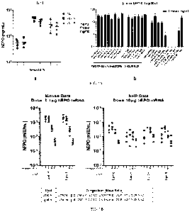
embodiments, the
PEGylated lipid herein is DMG-PEG2000, DSPE-PEG2000, DLPE-PEG2000, DSG-
PEG2000, C8 PEG2000, or ALC-0159 (2-[(polyethylene glycol)-20001-N,N-
ditetradecylacetamide). In certain embodiments, the PEGylated lipid herein is
DMG-
PEG2000.
C. Cholesterol-Based Lipids
[00110] The cholesterol component provides stability to the lipid bilayer
structure within the
nanoparticle. In some embodiments, the LNPs comprise one or more cholesterol-
based lipids.
Suitable cholesterol-based lipids include, for example: DC-Choi (N,N-dimethyl-
N-
ethylcarboxamidocholesterol), 1,4-bis(3-N-oleylamino-propyl)piperazine (Gao et
al., Biochem
Biophys Res Comm. (1991) 179:280; Wolf et al., BioTechniques (1997) 23:139;
U.S. Pat.
5,744,335), imidazole cholesterol ester ("ICE"; W02011/068810), sitosterol
(22,23-
dihydrostigmasterol), 0-sitosterol, sitostanol, fucosterol, stigmasterol
(stigmasta-5,22-dien-3-
ol), ergosterol; desmosterol (3B-hydroxy-5,24-cholestadiene); lanosterol (8,24-
lanostadien-3b-
ol); 7-dehydrocholesterol (A5,7-cholesterol); dihydrolanosterol (24,25-
dihydrolanosterol);
zymo sterol (5 a-chole sta-8,24-dien-3B-ol); latho sterol (5 a-chole st-7-en-
3B-ol) ; diosgenin
((313,25R)-spirost-5-en-3-ol); campesterol (campest-5-en-3B-ol); campestanol
(5a-campestan-
3b-ol); 24-methylene cholesterol (5,24(28)-cholestadien-24-methylen-3B-ol);
cholesteryl
margarate (cholest-5-en-3B-y1 heptadecanoate); cholesteryl oleate; cholesteryl
stearate and
other modified forms of cholesterol. In some embodiments, the cholesterol-
based lipid used in
the LNPs is cholesterol.
D. Helper Lipids
[00111] A helper lipid enhances the structural stability of the LNP and helps
the LNP in
endosome escape. It improves uptake and release of the mRNA drug payload. In
some
embodiments, the helper lipid is a zwitterionic lipid, which has fusogenic
properties for
CA 03224175 2023-12-13
WO 2022/264109
PCT/IB2022/055655
enhancing uptake and release of the drug payload. Examples of helper lipids
are 1,2-dioleoyl-
SN-glycero-3-phosphoethanolamine (DOPE); 1,2-distearoyl-sn-glycero-3-
phosphocholine
(DSPC); 1,2-dioleoyl-sn-glycero-3-phospho-L-serine (DOPS); 1,2-dielaidoyl-sn-
glycero-3-
phosphoethanolamine (DEPE); and 1,2-dioleoyl-sn-glycero-3-phosphocholine
(DPOC),
dipalmitoylphosphatidylcholine (DPPC), DMPC, 1,2-dilauroyl-sn-glycero-3-
phosphocholine
(DLPC), 1,2-Distearoylphosphatidylethanolamine (DSPE), and 1,2-dilauroyl-sn-
glycero-3-
phosphoethanolamine (DLPE).
[00112] Other exemplary helper lipids are dioleoylphosphatidylcholine (DOPC),
dioleoylphosphatidylglycerol (DOPG),
dipalmitoylphosphatidylglycerol (DPPG),
palmitoyloleoylphosphatidylcholine (POP C), palmitoyloleoyl-
phosphatidylethanolamine
(POPE), dioleoyl-phosphatidylethanolamine 4-(N-
maleimidomethyl)-cyclohexane-l-
carboxylate (DOPE-mal), dipalmitoyl phosphatidyl ethanolamine (DPPE),
dimyristoylphosphoethanolamine (DMPE),
phosphatidylserine, sphingolipids,
sphingomyelins, ceramides, cerebrosides, gangliosides, 16-0-monomethyl PE, 16-
0-dimethyl
PE, 18-1-trans PE, 1-stearoy1-2-oleoyl-phosphatidyethanolamine (SOPE), or a
combination
thereof In certain embodiments, the helper lipid is DOPE. In certain
embodiments, the helper
lipid is DSPC.
[00113] In various embodiments, the present LNPs comprise (i) a cationic lipid
selected from
OF-02, cKK-E10, GL-HEPES-E3-E10-DS-3-E18-1, GL-HEPES-E3-E12-DS-4-E10, or GL-
HEPES-E3-E12-DS-3-E14; (ii) DMG-PEG2000; (iii) cholesterol; and (iv) DOPE.
E. Molar Ratios of the Lipid Components
[00114] The molar ratios of the above components are important for the LNPs'
effectiveness
in delivering mRNA. The molar ratio of the cationic lipid, the PEGylated
lipid, the cholesterol-
based lipid, and the helper lipid is A: B: C: D, where A+B+C+D= 100%. In some
embodiments, the molar ratio of the cationic lipid in the LNPs relative to the
total lipids (i.e.,
A) is 35-55%, such as 35-50% (e.g., 38-42% such as 40%, or 45-50%). In some
embodiments,
the molar ratio of the PEGylated lipid component relative to the total lipids
(i.e., B) is 0.25-
2.75% (e.g., 1-2% such as 1.5%). In some embodiments, the molar ratio of the
cholesterol-
based lipid relative to the total lipids (i.e., C) is 20-50% (e.g., 27-30%
such as 28.5%, or 38-
43%). In some embodiments, the molar ratio of the helper lipid relative to the
total lipids (i.e.,
D) is 5-35% (e.g., 28-32% such as 30%, or 8-12%, such as 10%). In some
embodiments, the
(PEGylated lipid + cholesterol) components have the same molar amount as the
helper lipid.
In some embodiments, the LNPs contain a molar ratio of the cationic lipid to
the helper lipid
that is more than 1.
21
CA 03224175 2023-12-13
WO 2022/264109
PCT/IB2022/055655
[00115] In certain embodiments, the LNP of the disclosure comprises:
[00116] a cationic lipid at a molar ratio of 35% to 55% or 40% to 50% (e.g., a
cationic lipid at
a molar ratio of 35%, 36%, 37%, 38%, 39%, 40%, 41% 42%, 43%, 44%, 45%, 46%,
47%,
48%, 49%, 50%, 51%, 52%, 53%, 54%, or 55%);
[00117] a polyethylene glycol (PEG) conjugated (PEGylated) lipid at a molar
ratio of 0.25%
to 2.75% or 1.00% to 2.00% (e.g., a PEGylated lipid at a molar ratio of 0.25%,
0.50%, 0.75%,
1.00%, 1.25%, 1.50%, 1.75%, 2.00%, 2.25%, 2.50%, or 2.75%),
[00118] a cholesterol-based lipid at a molar ratio of 20% to 50%, 25% to 45%,
or 28.5% to
43% (e.g., a cholesterol-based lipid at a molar ratio of 20%, 21%, 22%, 23%,
24%, 25%, 26%,
27%, 28%, 29%, 30%, 31%, 32%, 33%, 34%, 35%, 36%, 37%, 38%, 39%, 40%, 41% 42%,
43%, 44%, 45%, 46%, 47%, 48%, 49%, or 50%); and
[00119] a helper lipid at a molar ratio of 5% to 35%, 8% to 30%, or 10% to 30%
(e.g., a helper
lipid at a molar ratio of 5%, 6%, 7%, 8%, 9%, 10%, 11%, 12%, 13%, 14%, 15%,
16%, 17%,
18%, 19%, 20%, 21%, 22%, 23%, 24%, 25%, 26%, 27%, 28%, 29%, 30%, 31%, 32%,
33%,
34%, or 35%),
[00120] wherein all of the molar ratios are relative to the total lipid
content of the LNP.
[00121] In certain embodiments, the LNP comprises: a cationic lipid at a molar
ratio of 40%;
a PEGylated lipid at a molar ratio of 1.5%; a cholesterol-based lipid at a
molar ratio of 28.5%;
and a helper lipid at a molar ratio of 30%.
[00122] In certain embodiments, the PEGylated lipid is dimyristoyl-PEG2000
(DMG-
PEG2000).
[00123] In various embodiments, the cholesterol-based lipid is cholesterol.
[00124] In some embodiments, the helper lipid is 1,2-dioleoyl-SN-glycero-3-
phosphoethanolamine (DOPE).
[00125] In certain embodiments, the LNP comprises: OF-02 at a molar ratio of
35% to 55%;
DMG-PEG2000 at a molar ratio of 0.25% to 2.75%; cholesterol at a molar ratio
of 20% to
50%; and DOPE at a molar ratio of 5% to 35%.
[00126] In certain embodiments, the LNP comprises: cKK-E10 at a molar ratio of
35% to
55%; DMG-PEG2000 at a molar ratio of 0.25% to 2.75%; cholesterol at a molar
ratio of 20%
to 50%; and DOPE at a molar ratio of 5% to 35%.
[00127] In certain embodiments, the LNP comprises: GL-HEPES-E3-E10-DS-3-E18-1
at a
molar ratio of 35% to 55%; DMG-PEG2000 at a molar ratio of 0.25% to 2.75%;
cholesterol at
a molar ratio of 20% to 50%; and DOPE at a molar ratio of 5% to 35%.
22
CA 03224175 2023-12-13
WO 2022/264109
PCT/IB2022/055655
[00128] In certain embodiments, the LNP comprises: GL-HEPES-E3-E12-DS-4-E10 at
a
molar ratio of 35% to 55%; DMG-PEG2000 at a molar ratio of 0.25% to 2.75%;
cholesterol at
a molar ratio of 20% to 50%; and DOPE at a molar ratio of 5% to 35%.
[00129] In certain embodiments, the LNP comprises: GL-HEPES-E3-E12-DS-3-E14at
a
molar ratio of 35% to 55%; DMG-PEG2000 at a molar ratio of 0.25% to 2.75%;
cholesterol at
a molar ratio of 20% to 50%; and DOPE at a molar ratio of 5% to 35%.
[00130] In certain embodiments, the LNP comprises: SM-102 at a molar ratio of
35% to 55%;
DMG-PEG2000 at a molar ratio of 0.25% to 2.75%; cholesterol at a molar ratio
of 20% to
50%; and DSPC at a molar ratio of 5% to 35%.
[00131] In certain embodiments, the LNP comprises: ALC-0315 at a molar ratio
of 35% to
55%; ALC-0159 at a molar ratio of 0.25% to 2.75%; cholesterol at a molar ratio
of 20% to
50%; and DSPC at a molar ratio of 5% to 35%.
[00132] In certain embodiments, the LNP comprises: OF-02 at a molar ratio of
40%; DMG-
PEG2000 at a molar ratio of 1.5%; cholesterol at a molar ratio of 28.5%; and
DOPE at a molar
ratio of 30%. This LNP formulation is designated "Lipid A" herein.
[00133] In certain embodiments, the LNP comprises: cKK-E10 at a molar ratio of
40%; DMG-
PEG2000 at a molar ratio of 1.5%; cholesterol at a molar ratio of 28.5%; and
DOPE at a molar
ratio of 30%. This LNP formulation is designated "Lipid B" herein.
[00134] In certain embodiments, the LNP comprises: GL-HEPES-E3-E10-DS-3-E18-1
at a
molar ratio of 40%; DMG-PEG2000 at a molar ratio of 1.5%; cholesterol at a
molar ratio of
28.5%; and DOPE at a molar ratio of 30%. This LNP formulation is designated
"Lipid C"
herein.
[00135] In certain embodiments, the LNP comprises: GL-HEPES-E3-E12-DS-4-E10
(at a
molar ratio of 40%; DMG-PEG2000 at a molar ratio of 1.5%; cholesterol at a
molar ratio of
28.5%; and DOPE at a molar ratio of 30%. This LNP formulation is designated
"Lipid D"
herein.
[00136] In certain embodiments, the LNP comprises: GL-HEPES-E3-E12-DS-3-E14at
a
molar ratio of 40%; DMG-PEG2000 at a molar ratio of 1.5%; cholesterol at a
molar ratio of
28.5%; and DOPE at a molar ratio of 30%. This LNP formulation is designated
"Lipid E"
herein.
[00137] In certain embodiments, the LNP comprises: 9-heptadecanyl 8-{(2-
hydroxyethy1)6-
oxo-6-(undecyloxy)hexyllaminoloctanoate (SM-102) at a molar ratio of 50%; 1,2-
distearoyl-
sn-glycero-3-phosphocholine (DSPC) at a molar ratio of 10%; cholesterol at a
molar ratio of
23
CA 03224175 2023-12-13
WO 2022/264109
PCT/IB2022/055655
38.5%; and 1,2-dimyristoyl-rac-glycero-3-methoxypolyethylene glycol-2000 (DMG-
PEG2000) at a molar ratio of 1.5%.
[00138] In certain embodiments, the LNP comprises: (4-
hydroxybutypazanediylldi(hexane-
6,1-diy1) bis(2-hexyldecanoate) (ALC-0315) at a molar ratio of 46.3%; 1,2-
distearoyl-sn-
glycero-3-phosphocholine (DSPC) at a molar ratio of 9.4%; cholesterol at a
molar ratio of
42.7%; and 2-[(polyethylene glycol)-20001-N,N-ditetradecylacetamide (ALC-0159)
at a molar
ratio of 1.6%.
[00139] In certain embodiments, the LNP comprises: (4-
hydroxybutypazanediylldi(hexane-
6,1-diy1) bis(2-hexyldecanoate) (ALC-0315) at a molar ratio of 47.4%; 1,2-
distearoyl-sn-
glycero-3-phosphocholine (DSPC) at a molar ratio of 10%; cholesterol at a
molar ratio of
40.9%; and 2-[(polyethylene glycol)-20001-N,N-ditetradecylacetamide (ALC-0159)
at a molar
ratio of 1.7%.
[00140] To calculate the actual amount of each lipid to be put into an LNP
formulation, the
molar amount of the cationic lipid is first determined based on a desired N/P
ratio, where N is
the number of nitrogen atoms in the cationic lipid and P is the number of
phosphate groups in
the mRNA to be transported by the LNP. Next, the molar amount of each of the
other lipids is
calculated based on the molar amount of the cationic lipid and the molar ratio
selected. These
molar amounts are then converted to weights using the molecular weight of each
lipid
F. Active Ingredients of the LNPs
[00141] The active ingredient of the present LNP vaccine composition is an
mRNA that
encodes an influenza antigen.
[00142] Where desired, the LNP may be multi-valent. In some embodiments, the
LNP may
carry mRNAs that encode more than one influenza antigen, such as two, three,
four, five, six,
seven, or eight antigens. For example, the LNP may carry multiple mRNA, each
encoding a
different influenza antigen; or carry a polycistronic mRNA that can be
translated into more
than one influenza antigen (e.g., each antigen-coding sequence is separated by
a nucleotide
linker encoding a self-cleaving peptide such as a 2A peptide). An LNP carrying
different
mRNA typically comprises (encapsulate) multiple copies of each mRNA. For
example, an
LNP carrying or encapsulating two different mRNA typically carries multiple
copies of each
of the two different mRNA.
[00143] In some embodiments, a single LNP formulation may comprise multiple
kinds (e.g.,
two, three, four, five, six, seven, eight, nine, ten, or more) of LNPs, each
kind carrying a
different mRNA.
24
CA 03224175 2023-12-13
WO 2022/264109
PCT/IB2022/055655
[00144] In some embodiments, the multi-valent LNP vaccines contain mRNA
molecules
encoding polypeptides derived from eight influenza viral proteins selected
from hemagglutinin
(e.g., hemagglutinin 1 (HA1) and hemagglutinin 2 (HA2)), neuraminidase (NA),
nucleoprotein
(NP), matrix protein 1 (M1), matrix protein 2 (M2), nonstructural protein 1
(NS1), and non-
structural protein 2 (NS2). In further embodiments, the multi-valent LNP
vaccines containing
eight mRNA encoding antigenic polypeptides derived from an HA protein, from an
NA protein,
and from both HA and NA proteins. In some embodiments, the mRNA encoding
antigenic
polypeptides are derived from different influenza strains.
[00145] In certain embodiments, the composition may comprise one or more mRNA
encoding
antigens of influenza A, B and C viruses. In one embodiment, the composition
may comprise
one or more mRNA encoding HA and/or NA antigens of influenza A and influenza B
viruses.
In one embodiment, the HA antigens of influenza A viruses are selected from
subtypes H1,
H2, H3, H4, H5, H6, H7, H8, H9, H10, H11, H12, H13, H14, H15, H16, H17, and
H18. In
one embodiment, the NA antigens of influenza A viruses are selected from
subtypes Ni, N2,
N3, N4, N5, N6, N7, N8, N9, N10, and N11. In one embodiment, the HA and NA
antigens of
influenza B viruses are from the influenza B/Yamagata lineage. In one
embodiment, the HA
and NA antigens of influenza B viruses are from the influenza BNictoria
lineage. In some
embodiments, the one or more HA and NA antigens are from influenza virus
strains
recommended by the World Health Organization (WHO) in their annual
recommendation for
influenza vaccine formulations.
[00146] In certain embodiments, at least one of the one or more influenza
virus proteins
comprises an influenza virus HA protein and/or an influenza virus NA protein
having a
molecular sequence identified or designed from a machine learning model, and
in certain
embodiments, at least one of the one or more ribonucleic acid molecules encode
one or more
influenza virus proteins having a molecular sequence identified or designed
from a machine
learning model.
[00147] In one embodiment, the composition comprises one mRNA encoding an H3
HA
antigen, one mRNA encoding an H1 HA antigen, one mRNA encoding an HA antigen
from
the influenza B/Yamagata lineage, and one mRNA encoding an HA antigen from the
influenza
BNictoria lineage.
[00148] In one embodiment, the composition comprises one mRNA encoding an H3
HA
antigen, one mRNA encoding an N2 NA antigen, one mRNA encoding an H1 HA
antigen, one
mRNA encoding an Ni NA antigen, one mRNA encoding an HA antigen from the
influenza
CA 03224175 2023-12-13
WO 2022/264109
PCT/IB2022/055655
B/Yamagata lineage, one mRNA encoding an NA antigen from the influenza
B/Yamagata
lineage, one mRNA encoding an HA antigen from the influenza BNictoria lineage,
and one
mRNA encoding an NA antigen from the influenza B/Victoria lineage.
1001491 In an embodiment, the composition comprises further comprise one or
more InRNA
encoding a machine learning influenza virus HA having a molecular sequence
identified or
designed from a machine learning model, wherein the one or more machine
learning influenza
virus HA may be selected from an H1 HA, an H3 HA, an HA from a BNictoria
lineage, an
HA from a B/Yamagata lineage, or a combination thereof
1001501 When selecting one or more machine learning influenza virus HAs, any
machine
learning algorithm may be used. For example, envisioned herein are any of the
machine
learning algorithms and methods disclosed in PCT Application Nos. WO
2021/080990 Al,
entitled Systems and Methods for Designing Vaccines, and WO 2021/080999 Al,
entitled
Systems and Methods for Predicting Biological Responses, both of which are
incorporated by
reference in their entireties herein.
[00151] The mRNA may be unmodified (i.e., containing only natural
ribonucleotides A, U, C,
and/or G linked by phosphodiester bonds), or chemically modified (e.g.,
including nucleotide
analogs such as pseudouridines (e.g., N-1-methyl pseudouridine), 2'-fluoro
ribonucleotides,
and 2'-methoxy ribonucleotides, and/or phosphorothioate bonds). The mRNA
molecule may
comprise a 5' cap and a polyA tail.
G. Buffer and Other Components
[00152] To stabilize the nucleic acid and/or LNPs (e.g., to prolong the shelf-
life of the vaccine
product), to facilitate administration of the LNP pharmaceutical composition,
and/or to enhance
in vivo expression of the nucleic acid, the nucleic acid and/or LNP can be
formulated in
combination with one or more carriers, targeting ligands, stabilizing reagents
(e.g.,
preservatives and antioxidants), and/or other pharmaceutically acceptable
excipients.
Examples of such excipients are parabens, thimerosal, thiomersal,
chlorobutanol,
benzalkonium chloride, and chelators (e.g., EDTA).
[00153] The LNP compositions of the present disclosure can be provided as a
frozen liquid
form or a lyophilized form. A variety of cryoprotectants may be used,
including, without
limitations, sucrose, trehalose, glucose, mannitol, mannose, dextrose, and the
like. The
cryoprotectant may constitute 5-30% (w/v) of the LNP composition. In some
embodiments,
the LNP composition comprises trehalose, e.g., at 5-30% (e.g., 10%) (w/v).
Once formulated
26
CA 03224175 2023-12-13
WO 2022/264109
PCT/IB2022/055655
with the cryoprotectant, the LNP compositions may be frozen (or lyophilized
and
cryopreserved) at -20 C to -80 C.
[00154] The LNP compositions may be provided to a patient in an aqueous
buffered solution
¨ thawed if previously frozen, or if previously lyophilized, reconstituted in
an aqueous buffered
solution at bedside. The buffered solution preferably is isotonic and suitable
for e.g.,
intramuscular or intradermal injection. In some embodiments, the buffered
solution is a
phosphate-buffered saline (PBS).
II. RNA
[00155] The present LNP vaccine compositions of the disclosure may comprise an
RNA
molecule (e.g., mRNA) that encodes an antigen of interest. The RNA molecule of
the present
disclosure may comprise at least one ribonucleic acid (RNA) comprising an ORF
encoding an
antigen of interest. In certain embodiments, the RNA is a messenger RNA (mRNA)
comprising an ORF encoding an antigen of interest. In certain embodiments, the
RNA (e.g.,
mRNA) further comprises at least one 5' UTR, 3' UTR, a poly(A) tail, and/or a
5' cap.
II. A. 5' Cap
[00156] An mRNA 5' cap can provide resistance to nucleases found in most
eukaryotic cells
and promote translation efficiency. Several types of 5' caps are known. A 7-
methylguanosine
cap (also referred to as "m7G" or "Cap-0"), comprises a guanosine that is
linked through a 5'
¨ 5' - triphosphate bond to the first transcribed nucleotide.
[00157] A 5' cap is typically added as follows: first, an RNA terminal
phosphatase removes
one of the terminal phosphate groups from the 5' nucleotide, leaving two
terminal phosphates;
guanosine triphosphate (GTP) is then added to the terminal phosphates via a
guanylyl
transferase, producing a 5 '5 '5 triphosphate linkage; and the 7-nitrogen of
guanine is then
methylated by a methyltransferase. Examples of cap structures include, but are
not limited to,
m7G(5')ppp, (5'(A,G(5')ppp(5')A, and G(5')ppp(5')G. Additional cap structures
are
described in U.S. Publication No. US 2016/0032356 and U.S. Publication No. US
2018/0125989, which are incorporated herein by reference.
[00158] 5'-capping of polynucleotides may be completed concomitantly during
the in vitro-
transcription reaction using the following chemical RNA cap analogs to
generate the 5'-
guanosine cap structure according to manufacturer protocols: 3'-0-Me-
m7G(5')ppp(5')G (the
ARCA cap); G(5 ')ppp(5 ')A; G(5 ')ppp(5 ')G; m7G(5')ppp(5')A; m7G(5')ppp(5')G;
m7G(5')ppp(5')(2'0MeA)pG; m7G(5')ppp(5')(2'0MeA)pU; m7G(5')ppp(5')(2'0MeG)pG
27
CA 03224175 2023-12-13
WO 2022/264109 PCT/IB2022/055655
(New England BioLabs, Ipswich, MA; TriLink Biotechnologies). 5'-capping of
modified RNA
may be completed post-transcriptionally using a vaccinia virus capping enzyme
to generate the
Cap 0 structure: m7G(5')ppp(5')G. Cap 1 structure may be generated using both
vaccinia virus
capping enzyme and a 2'-O methyl-transferase to generate: m7G(5')ppp(5')G-2' -
0-methyl.
Cap 2 structure may be generated from the Cap 1 structure followed by the 2'-0-
methylation
of the 5'-antepenultimate nucleotide using a 2'-0 methyl-transferase. Cap 3
structure may be
generated from the Cap 2 structure followed by the 2'-0-methylation of the 5'-
preantepenultimate nucleotide using a 2'-0 methyl-transferase.
[00159] In certain embodiments, the mRNA of the disclosure comprises a 5' cap
selected from
the group consisting of 3'-0-Me-m7G(5')ppp(5')G (the ARCA cap), G(5')ppp(5')A,
G(5 ')ppp(5 ')G, m7G(5')ppp(5')A, m7G(5 ')ppp(5')G,
m7G(5')ppp(5')(2'0MeA)pG,
m7G(5')ppp(5')(2'0MeA)pU, and m7G(5')ppp(5')(2'0MeG)pG.
[00160] In certain embodiments, the mRNA of the disclosure comprises a 5' cap
of:
0
D X1
OH OH
0 0 0 N N NH2
II II II
I I I
H2N N N 0- 0- 0
1-711H
FT, 0 F
0
N+ (:)p=c, CH3
CH3 0
II. B. Untranslated Region (UTR)
[00161] In some embodiments, the mRNA of the disclosure includes a 5' and/or
3'
untranslated region (UTR). In mRNA, the 5' UTR starts at the transcription
start site and
continues to the start codon but does not include the start codon. The 3' UTR
starts
immediately following the stop codon and continues until the transcriptional
termination
signal.
[00162] In some embodiments, the mRNA disclosed herein may comprise a 5' UTR
that
includes one or more elements that affect an mRNA's stability or translation.
In some
embodiments, a 5' UTR may be about 10 to 5,000 nucleotides in length. In some
embodiments,
a 5' UTR may be about 50 to 500 nucleotides in length. In some embodiments,
the 5' UTR is
at least about 10 nucleotides in length, about 20 nucleotides in length, about
30 nucleotides in
length, about 40 nucleotides in length, about 50 nucleotides in length, about
100 nucleotides in
length, about 150 nucleotides in length, about 200 nucleotides in length,
about 250 nucleotides
in length, about 300 nucleotides in length, about 350 nucleotides in length,
about 400
28
CA 03224175 2023-12-13
WO 2022/264109
PCT/IB2022/055655
nucleotides in length, about 450 nucleotides in length, about 500 nucleotides
in length, about
550 nucleotides in length, about 600 nucleotides in length, about 650
nucleotides in length,
about 700 nucleotides in length, about 750 nucleotides in length, about 800
nucleotides in
length, about 850 nucleotides in length, about 900 nucleotides in length,
about 950 nucleotides
in length, about 1,000 nucleotides in length, about 1,500 nucleotides in
length, about 2,000
nucleotides in length, about 2,500 nucleotides in length, about 3,000
nucleotides in length,
about 3,500 nucleotides in length, about 4,000 nucleotides in length, about
4,500 nucleotides
in length or about 5,000 nucleotides in length.
[00163] In some embodiments, the mRNA disclosed herein may comprise a 3' UTR
comprising one or more of a polyadenylation signal, a binding site for
proteins that affect an
mRNA's stability of location in a cell, or one or more binding sites for
miRNAs. In some
embodiments, a 3' UTR may be 50 to 5,000 nucleotides in length or longer. In
some
embodiments, a 3' UTR may be 50 to 1,000 nucleotides in length or longer. In
some
embodiments, the 3' UTR is at least about 50 nucleotides in length, about 100
nucleotides in
length, about 150 nucleotides in length, about 200 nucleotides in length,
about 250 nucleotides
in length, about 300 nucleotides in length, about 350 nucleotides in length,
about 400
nucleotides in length, about 450 nucleotides in length, about 500 nucleotides
in length, about
550 nucleotides in length, about 600 nucleotides in length, about 650
nucleotides in length,
about 700 nucleotides in length, about 750 nucleotides in length, about 800
nucleotides in
length, about 850 nucleotides in length, about 900 nucleotides in length,
about 950 nucleotides
in length, about 1,000 nucleotides in length, about 1,500 nucleotides in
length, about 2,000
nucleotides in length, about 2,500 nucleotides in length, about 3,000
nucleotides in length,
about 3,500 nucleotides in length, about 4,000 nucleotides in length, about
4,500 nucleotides
in length, or about 5,000 nucleotides in length.
[00164] In some embodiments, the mRNA disclosed herein may comprise a 5' or 3'
UTR that
is derived from a gene distinct from the one encoded by the mRNA transcript
(i.e., the UTR is
a heterologous UTR).
[00165] In certain embodiments, the 5' and/or 3' UTR sequences can be derived
from mRNA
which are stable (e.g., globin, actin, GAPDH, tubulin, histone, or citric acid
cycle enzymes) to
increase the stability of the mRNA. For example, a 5' UTR sequence may include
a partial
sequence of a CMV immediate-early 1 (IE1) gene, or a fragment thereof, to
improve the
nuclease resistance and/or improve the half-life of the mRNA. Also
contemplated is the
inclusion of a sequence encoding human growth hormone (hGH), or a fragment
thereof, to the
3' end or untranslated region of the mRNA. Generally, these modifications
improve the
29
CA 03224175 2023-12-13
WO 2022/264109
PCT/IB2022/055655
stability and/or pharmacokinetic properties (e.g., half-life) of the mRNA
relative to their
unmodified counterparts, and include, for example, modifications made to
improve such
mRNA resistance to in vivo nuclease digestion.
[00166] Exemplary 5' UTRs include a sequence derived from a CMV immediate-
early 1 (IE1)
gene (U.S. Publication Nos. 2014/0206753 and 2015/0157565, each of which is
incorporated
herein by reference), or the sequence GGGAUCCUACC (SEQ ID NO: 22) (U.S.
Publication
No. 2016/0151409, incorporated herein by reference).
[00167] In various embodiments, the 5' UTR may be derived from the 5' UTR of a
TOP gene.
TOP genes are typically characterized by the presence of a 5'-terminal
oligopyrimidine (TOP)
tract. Furthermore, most TOP genes are characterized by growth-associated
translational
regulation. However, TOP genes with a tissue specific translational regulation
are also known.
In certain embodiments, the 5' UTR derived from the 5' UTR of a TOP gene lacks
the 5' TOP
motif (the oligopyrimidine tract) (e .g ., U.S. Publication Nos. 2017/0029847,
2016/0304883,
2016/0235864, and 2016/0166710, each of which is incorporated herein by
reference).
[00168] In certain embodiments, the 5' UTR is derived from a ribosomal protein
Large 32
(L32) gene (U.S. Publication No. 2017/0029847, supra).
[00169] In certain embodiments, the 5' UTR is derived from the 5' UTR of an
hydroxysteroid
(17-b) dehydrogenase 4 gene (HSD17B4) (U.S. Publication No. 2016/0166710,
supra).
[00170] In certain embodiments, the 5' UTR is derived from the 5' UTR of an
ATP5A1 gene
(U. S . Publication No. 2016/0166710, supra).
In some embodiments, an internal ribosome entry site (IRES) is used instead of
a 5' UTR.
[00171] In some embodiments, the 5'UTR comprises a nucleic acid sequence set
forth in SEQ
ID NO: 19 and reproduced below:
GGACAGAUCGCCUGGAGACGCCAUCCACGCUGUUUUGACCUCCAUAGAAGACA
CCGGGACCGAUCCAGCCUCCGCGGCCGGGAACGGUGCAUUGGAACGCGGAUUC
CCCGUGCCAAGAGUGACUCACCGUCCUUGACACG (SEQ ID NO: 19).
[00172] In some embodiments, the 3'UTR comprises a nucleic acid sequence set
forth in SEQ
ID NO: 20 and reproduced below:
CGGGUGGCAUCCCUGUGACCCCUCCCCAGUGCCUCUCCUGGCCCUGGAAGUUG
CCACUCCAGUGCCCACCAGCCUUGUCCUAAUAAAAUUAAGUUGCAUC (SEQ ID
NO: 20).
[00173] The 5' UTR and 3'UTR are described in further detail in W02012/075040,
incorporated herein by reference.
CA 03224175 2023-12-13
WO 2022/264109
PCT/IB2022/055655
II. C. Polyadenylated Tail
[00174] As used herein, the terms "poly(A) sequence," "poly(A) tail," and
"poly(A) region"
refer to a sequence of adenosine nucleotides at the 3' end of the mRNA
molecule. The poly(A)
tail may confer stability to the mRNA and protect it from exonuclease
degradation. The
poly(A) tail may enhance translation. In some embodiments, the poly(A) tail is
essentially
homopolymeric. For example, a poly(A) tail of 100 adenosine nucleotides may
have
essentially a length of 100 nucleotides. In certain embodiments, the poly(A)
tail may be
interrupted by at least one nucleotide different from an adenosine nucleotide
(e.g., a nucleotide
that is not an adenosine nucleotide). For example, a poly(A) tail of 100
adenosine nucleotides
may have a length of more than 100 nucleotides (comprising 100 adenosine
nucleotides and at
least one nucleotide, or a stretch of nucleotides, that are different from an
adenosine
nucleotide). In certain embodiments, the poly(A) tail comprises the sequence
AAAAAAAAAAAAAAAAAAAAAAAAAAAAAAGCAUAUGACUAAAAAAAAAAAA
AAAAAAAAAAAAAAAAAAAAAAAAAAAAAAAAAAAAAAAAAAAAAAAAAAAA
AAAAAA (SEQ ID NO: 23).
[00175] The "poly(A) tail," as used herein, typically relates to RNA. However,
in the context
of the disclosure, the term likewise relates to corresponding sequences in a
DNA molecule
(e.g., a "poly(T) sequence").
[00176] The poly(A) tail may comprise about 10 to about 500 adenosine
nucleotides, about 10
to about 200 adenosine nucleotides, about 40 to about 200 adenosine
nucleotides, or about 40
to about 150 adenosine nucleotides. The length of the poly(A) tail may be at
least about 10,
50, 75, 100, 150, 200, 250, 300, 350, 400, 450, or 500 adenosine nucleotides.
[00177] In some embodiments where the nucleic acid is an RNA, the poly(A) tail
of the nucleic
acid is obtained from a DNA template during RNA in vitro transcription. In
certain
embodiments, the poly(A) tail is obtained in vitro by common methods of
chemical synthesis
without being transcribed from a DNA template. In various embodiments, poly(A)
tails are
generated by enzymatic polyadenylation of the RNA (after RNA in vitro
transcription) using
commercially available polyadenylation kits and corresponding protocols, or
alternatively, by
using immobilized poly(A)polymerases, e.g., using methods and means as
described in
W02016/174271.
[00178] The nucleic acid may comprise a poly(A) tail obtained by enzymatic
polyadenylation,
wherein the majority of nucleic acid molecules comprise about 100 (+/-20) to
about 500 (+/-
50) or about 250 (+/-20) adenosine nucleotides.
31
CA 03224175 2023-12-13
WO 2022/264109
PCT/IB2022/055655
[00179] In some embodiments, the nucleic acid may comprise a poly(A) tail
derived from a
template DNA and may additionally comprise at least one additional poly(A)
tail generated by
enzymatic polyadenylation, e.g., as described in W02016/091391.
[00180] In certain embodiments, the nucleic acid comprises at least one
polyadenylation
signal.
[00181] In various embodiments, the nucleic acid may comprise at least one
poly(C) sequence.
[00182] The term "poly(C) sequence," as used herein, is intended to be a
sequence of cytosine
nucleotides of up to about 200 cytosine nucleotides. In some embodiments, the
poly(C)
sequence comprises about 10 to about 200 cytosine nucleotides, about 10 to
about 100 cytosine
nucleotides, about 20 to about 70 cytosine nucleotides, about 20 to about 60
cytosine
nucleotides, or about 10 to about 40 cytosine nucleotides. In some
embodiments, the poly(C)
sequence comprises about 30 cytosine nucleotides.
II. D. Chemical Modification
[00183] The mRNA disclosed herein may be modified or unmodified. In some
embodiments,
the mRNA may comprise at least one chemical modification. In some embodiments,
the
mRNA disclosed herein may contain one or more modifications that typically
enhance RNA
stability. Exemplary modifications can include backbone modifications, sugar
modifications,
or base modifications. In some embodiments, the disclosed mRNA may be
synthesized from
naturally occurring nucleotides and/or nucleotide analogues (modified
nucleotides) including,
but not limited to, purines (adenine (A) and guanine (G)) or pyrimidines
(thymine (T), cytosine
(C), and uracil (U)). In certain embodiments, the disclosed mRNA may be
synthesized from
modified nucleotide analogues or derivatives of purines and pyrimidines, such
as, e.g., 1-
methyl-adenine, 2-methyl-adenine, 2-methylthio-N-6-isopentenyl-adenine, N6-
methyl-
adenine, N6-isopentenyl-adenine, 2-thio-cytosine, 3-methyl-cytosine, 4-acetyl-
cytosine, 5-
methyl-cytosine, 2,6-diaminopurine, 1-methyl-guanine, 2-methyl-guanine, 2,2-
dimethyl-
guanine, 7-methyl-guanine, inosine, 1-methyl-inosine, pseudouracil (5-uracil),
dihydro-uracil,
2-thio-uracil, 4-thio-uracil, 5 -
carboxymethylaminomethy1-2-thio-uracil, 5-
(carboxyhydroxymethyl)-uracil, 5 -fluoro -uracil, 5 -bromo-
uracil, 5-
carboxymethylaminomethyl-uracil, 5-methyl-2-thio-uracil, 5-methyl-uracil, N-
uracil-5-oxy
acetic acid methyl ester, 5-methylaminomethyl-uracil, 5-methoxyaminomethy1-2-
thio-uracil,
5'-methoxycarbonylmethyl-uracil, 5-methoxy-uracil, uracil-5-oxyacetic acid
methyl ester,
uracil-5-oxyacetic acid (v), 1-methyl-pseudouracil, queosine, P-D-mannosyl-
queosine,
32
CA 03224175 2023-12-13
WO 2022/264109
PCT/IB2022/055655
phosphoramidates, phosphorothioates, peptide nucleotides, methylphosphonates,
7-
deazaguanosine, 5-methylcytosine, and inosine.
[00184] In some embodiments, the disclosed mRNA may comprise at least one
chemical
modification including, but not limited to, pseudouridine, Nl-
methylpseudouridine, 2-
thiouridine, 4'-thiouridine, 5-methylcytosine, 2-thio-l-methy1-1-deaza-
pseudouridine, 2-thio-l-
methyl-pseudouridine, 2-thio-5-aza-uridine, 2-thio-
dihydropseudouridine, 2-thio-
dihydrouridine, 2-thio-pseudouridine, 4-methoxy-2-thio-pseudouridine, 4-
methoxy-
pseudouridine, 4-thio-l-methyl-pseudouridine, 4-thio-pseudouridine, 5-aza-
uridine,
dihydropseudouridine, 5-methyluridine, 5-methyluridine, 5-methoxyuridine, and
2'-0-methyl
uridine.
[00185] In some embodiments, the chemical modification is selected from the
group
consisting of pseudouridine, Nl-methylpseudouridine, 5-methylcytosine, 5-
methoxyuridine,
and a combination thereof
[00186] In some embodiments, the chemical modification comprises N1-
methylpseudouridine.
[00187] In some embodiments, at least 20%, at least 30%, at least 40%, at
least 50%, at least
60%, at least 70%, at least 80%, at least 85%, at least 90%, at least 95%, or
100% of the uracil
nucleotides in the mRNA are chemically modified.
[00188] In some embodiments, at least 20%, at least 30%, at least 40%, at
least 50%, at least
60%, at least 70%, at least 80%, at least 85%, at least 90%, at least 95%, or
100% of the uracil
nucleotides in the ORF are chemically modified.
[00189] The preparation of such analogues is described, e.g., in U.S. Pat. No.
4,373,071, U.S.
Pat. No. 4,401,796, U.S. Pat. No. 4,415,732, U.S. Pat. No. 4,458,066, U.S.
Pat. No. 4,500,707,
U.S. Pat. No. 4,668,777, U.S. Pat. No. 4,973,679, U.S. Pat. No. 5,047,524,
U.S. Pat. No.
5,132,418, U.S. Pat. No. 5,153,319, U.S. Pat. No. 5,262,530, and U.S. Pat. No.
5,700,642.
II. E. mRNA Synthesis
[00190] The mRNAs disclosed herein may be synthesized according to any of a
variety of
methods. For example, mRNAs according to the present disclosure may be
synthesized via in
vitro transcription (WT). Some methods for in vitro transcription are
described, e.g., in Geall
et al. (2013) Semin. Immunol. 25(2): 152-159; Brunelle et al. (2013) Methods
Enzymol.
530:101-14. Briefly, IVT is typically performed with a linear or circular DNA
template
containing a promoter, a pool of ribonucleotide triphosphates, a buffer system
that may include
DTT and magnesium ions, an appropriate RNA polymerase (e.g., T3, T7, or 5P6
RNA
33
CA 03224175 2023-12-13
WO 2022/264109
PCT/IB2022/055655
polymerase), DNase I, pyrophosphatase, and/or RNase inhibitor. The exact
conditions may
vary according to the specific application. The presence of these reagents is
generally
undesirable in a final mRNA product and these reagents can be considered
impurities or
contaminants which can be purified or removed to provide a clean and/or
homogeneous mRNA
that is suitable for therapeutic use. While mRNA provided from in vitro
transcription reactions
may be desirable in some embodiments, other sources of mRNA can be used
according to the
instant disclosure including wild-type mRNA produced from bacteria, fungi,
plants, and/or
animals.
III. Processes for Making the Present LNP Vaccines
[00191] The present LNPs can be prepared by various techniques presently known
in the art.
For example, multilamellar vesicles (MLV) may be prepared according to
conventional
techniques, such as by depositing a selected lipid on the inside wall of a
suitable container or
vessel by dissolving the lipid in an appropriate solvent, and then evaporating
the solvent to
leave a thin film on the inside of the vessel or by spray drying. An aqueous
phase may then be
added to the vessel with a vortexing motion that results in the formation of
MLVs. Unilamellar
vesicles (ULV) can then be formed by homogenization, sonication or extrusion
of the
multilamellar vesicles. In addition, unilamellar vesicles can be formed by
detergent removal
techniques.
[00192] Various methods are described in US 2011/0244026, US 2016/0038432, US
2018/0153822, US 2018/0125989, and PCT/U52020/043223 (filed July 23, 2020) and
can be
used to practice the present invention. One exemplary process entails
encapsulating mRNA by
mixing it with a mixture of lipids, without first pre-forming the lipids into
lipid nanoparticles,
as described in US 2016/0038432. Another exemplary process entails
encapsulating mRNA
by mixing pre-formed LNPs with mRNA, as described in US 2018/0153822.
[00193] In some embodiments, the process of preparing mRNA-loaded LNPs
includes a step
of heating one or more of the solutions to a temperature greater than ambient
temperature, the
one or more solutions being the solution comprising the pre-formed lipid
nanoparticles, the
solution comprising the mRNA and the mixed solution comprising the LNP-
encapsulated
mRNA. In some embodiments, the process includes the step of heating one or
both of the
mRNA solution and the pre-formed LNP solution, prior to the mixing step. In
some
embodiments, the process includes heating one or more of the solutions
comprising the pre-
formed LNPs, the solution comprising the mRNA and the solution comprising the
LNP-
34
CA 03224175 2023-12-13
WO 2022/264109
PCT/IB2022/055655
encapsulated mRNA, during the mixing step. In some embodiments, the process
includes the
step of heating the LNP- encapsulated mRNA, after the mixing step. In some
embodiments,
the temperature to which one or more of the solutions is heated is or is
greater than about 30 C,
37 C, 40 C, 45 C, 50 C, 55 C, 60 C, 65 C, or 70 C. In some embodiments, the
temperature
to which one or more of the solutions is heated ranges from about 25-70 C,
about 30-70 C,
about 35-70 C, about 40-70 C, about 45-70 C, about 50-70 C, or about 60-70 C.
In some
embodiments, the temperature is about 65 C.
[00194] Various methods may be used to prepare an mRNA solution suitable for
the present
invention. In some embodiments, mRNA may be directly dissolved in a buffer
solution
described herein. In some embodiments, an mRNA solution may be generated by
mixing an
mRNA stock solution with a buffer solution prior to mixing with a lipid
solution for
encapsulation. In some embodiments, an mRNA solution may be generated by
mixing an
mRNA stock solution with a buffer solution immediately before mixing with a
lipid solution
for encapsulation. In some embodiments, a suitable mRNA stock solution may
contain mRNA
in water or a buffer at a concentration at or greater than about 0.2 mg/ml,
0.4 mg/ml, 0.5 mg/ml,
0.6 mg/ml, 0.8 mg/ml, 1.0 mg/ml, 1.2 mg/ml, 1.4 mg/ml, 1.5 mg/ml, or 1.6
mg/ml, 2.0 mg/ml,
2.5 mg/ml, 3.0 mg/ml, 3.5 mg/ml, 4.0 mg/ml, 4.5 mg/ml, or 5.0 mg/ml.
[00195] In some embodiments, an mRNA stock solution is mixed with a buffer
solution using
a pump. Exemplary pumps include but are not limited to gear pumps, peristaltic
pumps and
centrifugal pumps. Typically, the buffer solution is mixed at a rate greater
than that of the
mRNA stock solution. For example, the buffer solution may be mixed at a rate
at least lx, 2x,
3x, 4x, 5x, 6x, 7x, 8x, 9x, 10x, 15x, or 20x greater than the rate of the mRNA
stock solution.
In some embodiments, a buffer solution is mixed at a flow rate ranging between
about 100-
6000 ml/minute (e.g., about 100-300 ml/minute, 300-600 ml/minute, 600-1200
ml/minute,
1200-2400 ml/minute, 2400-3600 ml/minute, 3600-4800 ml/minute, 4800-6000
ml/minute, or
60-420 ml/minute). In some embodiments, a buffer solution is mixed at a flow
rate of, or
greater than, about 60 ml/minute, 100 ml/minute, 140 ml/minute, 180 ml/minute,
220
ml/minute, 260 ml/minute, 300 ml/minute, 340 ml/minute, 380 ml/minute, 420
ml/minute, 480
ml/minute, 540 ml/minute, 600 ml/minute, 1200 ml/minute, 2400 ml/minute, 3600
ml/minute,
4800 ml/minute, or 6000 ml/minute.
[00196] In some embodiments, an mRNA stock solution is mixed at a flow rate
ranging
between about 10-600 ml/minute (e.g., about 5-50 ml/minute, about 10-30
ml/minute, about
30-60 ml/minute, about 60-120 ml/minute, about 120-240 ml/minute, about 240-
360
ml/minute, about 360-480 ml/minute, or about 480-600 ml/minute). In some
embodiments, an
CA 03224175 2023-12-13
WO 2022/264109
PCT/IB2022/055655
mRNA stock solution is mixed at a flow rate of or greater than about 5
ml/minute, 10
ml/minute, 15 ml/minute, 20 ml/minute, 25 ml/minute, 30 ml/minute, 35
ml/minute, 40
ml/minute, 45 ml/minute, 50 ml/minute, 60 ml/minute, 80 ml/minute, 100
ml/minute, 200
ml/minute, 300 ml/minute, 400 ml/minute, 500 ml/minute, or 600 ml/minute.
[00197] The process of incorporation of a desired mRNA into a lipid
nanoparticle is referred
to as "loading." Exemplary methods are described in Lasic et al., FEBS Lett.
(1992) 312:255-
8. The LNP-incorporated nucleic acids may be completely or partially located
in the interior
space of the lipid nanoparticle, within the bilayer membrane of the lipid
nanoparticle, or
associated with the exterior surface of the lipid nanoparticle membrane. The
incorporation of
an mRNA into lipid nanoparticles is also referred to herein as "encapsulation"
wherein the
nucleic acid is entirely or substantially contained within the interior space
of the lipid
nanoparticle.
[00198] Suitable LNPs may be made in various sizes. In some embodiments,
decreased size
of lipid nanoparticles is associated with more efficient delivery of an mRNA.
Selection of an
appropriate LNP size may take into consideration the site of the target cell
or tissue and to some
extent the application for which the lipid nanoparticle is being made.
[00199] A variety of methods known in the art are available for sizing of a
population of lipid
nanoparticles. Preferred methods herein utilize Zetasizer Nano ZS (Malvern
Panalytical) to
measure LNP particle size. In one protocol, 10 [L1 of an LNP sample are mixed
with 990 [d of
10% trehalose. This solution is loaded into a cuvette and then put into the
Zetasizer machine.
The z-average diameter (nm), or cumulants mean, is regarded as the average
size for the LNPs
in the sample. The Zetasizer machine can also be used to measure the
polydispersity index
(PDI) by using dynamic light scattering (DLS) and cumulant analysis of the
autocorrelation
function. Average LNP diameter may be reduced by sonication of formed LNP.
Intermittent
sonication cycles may be alternated with quasi-elastic light scattering (QELS)
assessment to
guide efficient lipid nanoparticle synthesis.
[00200] In some embodiments, the majority of purified LNPs, i.e., greater than
about 50%,
55%, 60%, 65%, 70%, 75%, 80%, 85%, 90%, 95%, 96%, 97%, 98%, or 99% of the
LNPs,
have a size of about 70-150 nm (e.g., about 145 nm, about 140 nm, about 135
nm, about 130
nm, about 125 nm, about 120 nm, about 115 nm, about 110 nm, about 105 nm,
about 100 nm,
about 95 nm, about 90 nm, about 85 nm, or about 80 nm). In some embodiments,
substantially
all (e.g., greater than 80 or 90%) of the purified lipid nanoparticles have a
size of about 70-150
nm (e.g., about 145 nm, about 140 nm, about 135 nm, about 130 nm, about 125
nm, about 120
36
CA 03224175 2023-12-13
WO 2022/264109
PCT/IB2022/055655
nm, about 115 nm, about 110 nm, about 105 nm, about 100 nm, about 95 nm, about
90 nm,
about 85 nm, or about 80 nm).
[00201] In some embodiments, the LNPs in the present composition have an
average size of
less than 150 nm, less than 120 nm, less than 100 nm, less than 90 nm, less
than 80 nm, less
than 70 nm, less than 60 nm, less than 50 nm, less than 30 nm, or less than 20
nm.
[00202] In some embodiments, greater than about 70%, 75%, 80%, 85%, 90%, 95%,
96%,
97%, 98%, 99% of the LNPs in the present composition have a size ranging from
about 40-90
nm (e.g., about 45-85 nm, about 50-80 nm, about 55-75 nm, about 60-70 nm) or
about 50-70
nm (e.g., 55-65 nm) are particular suitable for pulmonary delivery via
nebulization.
[00203] In some embodiments, the dispersity, or measure of heterogeneity in
size of molecules
(PDI), of LNPs in a pharmaceutical composition provided by the present
invention is less than
about 0.5. In some embodiments, an LNP has a PDI of less than about 0.5, less
than about 0.4,
less than about 0.3, less than about 0.28, less than about 0.25, less than
about 0.23, less than
about 0.20, less than about 0.18, less than about 0.16, less than about 0.14,
less than about 0.12,
less than about 0.10, or less than about 0.08. The PDI may be measured by a
Zetasizer machine
as described above.
[00204] In some embodiments, greater than about 75%, 80%, 85%, 90%, 95%, 96%,
97%,
98%, or 99% of the purified LNPs in a pharmaceutical composition provided
herein
encapsulate an mRNA within each individual particle. In some embodiments,
substantially all
(e.g., greater than 80% or 90%) of the purified lipid nanoparticles in a
pharmaceutical
composition encapsulate an mRNA within each individual particle. In some
embodiments, a
lipid nanoparticle has an encapsulation efficiency of between 50% and 99%; or
greater than
about 60, 65, 70, 75, 80, 85, 90, 92, 95, 98, or 99%. Typically, lipid
nanoparticles for use
herein have an encapsulation efficiency of at least 90% (e.g., at least 91,
92, 93, 94, or 95%).
[00205] In some embodiments, an LNP has a N/P ratio of between 1 and 10. In
some
embodiments, a lipid nanoparticle has a N/P ratio above 1, about 1, about 2,
about 3, about 4,
about 5, about 6, about 7, or about 8. In further embodiments, a typical LNP
herein has an N/P
ratio of 4.
[00206] In some embodiments, a pharmaceutical composition according to the
present
invention contains at least about 0.5 lag, 1 jig, 5 jig, 10 jig, 100 jig, 500
jig, or 1000 lag of
encapsulated mRNA. In some embodiments, a pharmaceutical composition contains
about 0.1
lag to 1000 jig, at least about 0.5 jig, at least about 0.8 jig, at least
about 1 jig, at least about 5
jig, at least about 8 jig, at least about 10 jig, at least about 50 jig, at
least about 100 jig, at least
about 500 jig, or at least about 1000 lag of encapsulated mRNA.
37
CA 03224175 2023-12-13
WO 2022/264109
PCT/IB2022/055655
[00207] In some embodiments, mRNA can be made by chemical synthesis or by in
vitro
transcription (IVT) of a DNA template. An exemplary process for making and
purifying
mRNA is described in Example 1. In this process, in an WT process, a cDNA
template is used
to produce an mRNA transcript and the DNA template is degraded by a DNase. The
transcript
is purified by depth filtration and tangential flow filtration (TFF). The
purified transcript is
further modified by adding a cap and a tail, and the modified RNA is purified
again by depth
filtration and TFF.
[00208] The mRNA is then prepared in an aqueous buffer and mixed with an
amphiphilic
solution containing the lipid components of the LNPs. An amphiphilic solution
for dissolving
the four lipid components of the LNPs may be an alcohol solution. In some
embodiments, the
alcohol is ethanol. The aqueous buffer may be, for example, a citrate,
phosphate, acetate, or
succinate buffer and may have a pH of about 3.0-7.0, e.g., about 3.5, about
4.0, about 4.5, about
5.0, about 5.5, about 6.0, or about 6.5. The buffer may contain other
components such as a salt
(e.g., sodium, potassium, and/or calcium salts). In particular embodiments,
the aqueous buffer
has 1 mM citrate, 150 mM NaC1, pH 4.5.
[00209] An exemplary, nonlimiting process for making an mRNA-LNP composition
is
described in Example 1. The process involves mixing of a buffered mRNA
solution with a
solution of lipids in ethanol in a controlled homogeneous manner, where the
ratio of
lipids:mRNA is maintained throughout the mixing process. In this illustrative
example, the
mRNA is presented in an aqueous buffer containing citric acid monohydrate, tri-
sodium citrate
dihydrate, and sodium chloride. The mRNA solution is added to the solution (1
mM citrate
buffer, 150 mM NaCl, pH 4.5). The lipid mixture of four lipids (e.g., a
cationic lipid, a
PEGylated lipid, a cholesterol-based lipid, and a helper lipid) is dissolved
in ethanol. The
aqueous mRNA solution and the ethanol lipid solution are mixed at a volume
ratio of 4:1 in a
"T" mixer with a near "pulseless" pump system. The resultant mixture is then
subjected for
downstream purification and buffer exchange. The buffer exchange may be
achieved using
dialysis cassettes or a TFF system. TFF may be used to concentrate and buffer-
exchange the
resulting nascent LNP immediately after formation via the T-mix process. The
diafiltration
process is a continuous operation, keeping the volume constant by adding
appropriate buffer at
the same rate as the permeate flow.
IV. Packaging and Use of the mRNA-LNP Vaccines
[00210] The mRNA-LNP vaccines can be packaged for parenteral (e.g.,
intramuscular,
intradermal or subcutaneous) administration or nasopharyngeal (e.g.,
intranasal)
38
CA 03224175 2023-12-13
WO 2022/264109
PCT/IB2022/055655
administration. The vaccine compositions may be in the form of an
extemporaneous
formulation, where the LNP composition is lyophilized and reconstituted with a
physiological
buffer (e.g., PBS) just before use. The vaccine compositions also may be
shipped and provided
in the form of an aqueous solution or a frozen aqueous solution and can be
directly administered
to subjects without reconstitution (after thawing, if previously frozen).
[00211] Accordingly, the present disclosure provides an article of
manufacture, such as a kit,
that provides the mRNA-LNP vaccine in a single container, or provides the mRNA-
LNP
vaccine in one container and a physiological buffer for reconstitution in
another container. The
container(s) may contain a single-use dosage or multi-use dosage. The
containers may be pre-
treated glass vials or ampules. The article of manufacture may include
instructions for use as
well.
[00212] In certain embodiments, the mRNA-LNP vaccine is provided for use in
intramuscular
(IM) injection. The vaccine can be injected to a subject at, e.g., his/her
deltoid muscle in the
upper arm. In some embodiments, the vaccine is provided in a pre-filled
syringe or injector
(e.g., single-chambered or multi-chambered). In some embodiments, the vaccine
is provided
for use in inhalation and is provided in a pre-filled pump, aerosolizer, or
inhaler.
[00213] The mRNA-LNP vaccines can be administered to subjects in need thereof
in a
prophylactically effective amount, i.e., an amount that provides sufficient
immune protection
against a target pathogen for a sufficient amount of time (e.g., one year, two
years, five years,
ten years, or life-time). Sufficient immune protection may be, for example,
prevention or
alleviation of symptoms associated with infections by the pathogen. In some
embodiments,
multiple doses (e.g., two doses) of the vaccine are injected to subjects in
need thereof to achieve
the desired prophylactic effects. The doses (e.g., prime and booster doses)
may be separated
by an interval of e.g., 1 week, 2 weeks, 3 weeks, 4 weeks, one month, two
months, three
months, four months, five months, six months, one year, two years, five years,
or ten years.
[00214] In some embodiments, a single dose of the mRNA-LNP vaccine contains 1-
50 jig of
mRNA (e.g., monovalent or multivalent). For example, a single dose may contain
about 2.5
jig, about 5 jig, about 7.5 jig, about 10 jig, about 12.5 jig, or about 15 jig
of the mRNA for
intramuscular (IM) injection. In further embodiments, a multi-valent single
dose of an LNP
vaccine contains multiple (e.g., 2, 3, or 4) kinds of LNPs, each for a
different antigen, and each
kind of LNP has an mRNA amount of, e.g., 2.5 jig, about 5 jig, about 7.5 jig,
about 10 jig,
about 12.5 jig, or about 15 jig.
[00215] In another aspect, the present invention provides methods of
immunizing a subject
against one or more influenza viruses in a subject. The present invention
further provides
39
CA 03224175 2023-12-13
WO 2022/264109
PCT/IB2022/055655
methods of eliciting an immune response against one or more influenza viruses
in a subject. In
some embodiments, the present methods comprise administering to the subject an
effective
amount of a composition described herein to a subject.
[00216] In various embodiments, the methods of immunizing provided herein
elicit a broadly
protective immune response against multiple epitopes within one or more
influenza viruses. In
various embodiments, the methods of immunizing provided herein elicit a
broadly neutralizing
immune response against one or more influenza viruses. In some embodiments,
the immune
response comprises an antibody response. Accordingly, in various embodiments,
the
composition described herein can offer broad cross-protection against
different types of
influenza viruses. In some embodiments, the composition offers cross-
protection against
avian, swine, seasonal, and/or pandemic influenza viruses. In some
embodiments, the
composition offers cross-protection against one or more influenza A, B, or C
subtypes. In
some embodiments, the composition offers cross-protection against multiple
strains of
influenza A Hl-subtype viruses (e.g., H1N1), influenza A H3-subtype viruses
(e.g., H3N2),
influenza A H5-subtype viruses (e.g., H5N1), and/or influenza B viruses (e.g.,
Yamagata
lineage, Victoria lineage).
[00217] In some embodiments, the methods of the invention are capable of
eliciting an
improved immune response against one or more seasonal influenza strains.
Exemplary
seasonal strains include, without limitation, A/Puerto Rico/8/1934, A/Fort
Monmouth/1/1947,
A/Chile/1/1983, A/Texas/36/1991, A/Singapore/6/1986, A/Beij ing/32/1992, A/New
Caledonia/20/1999, A/Solomon Islands/03/2006, A/Brisbane/59/2007, A(H3N2)
virus
antigenically like the cell-propagated prototype virus ANictoria/361/2011,
A/Beijing/262/95
(H1N1)-like virus, A/Brisbane/02/2018 (H1N1)pdm09-like virus,
A/Brisbane/10/2007
(H3N2)-like virus, A/California/7/2004 (H3N2)-like virus, A/California/7/2009
(H1N1)-like
virus, A/California/7/2009 (H1N1)pdm09-like virus, A/Cambodia/e0826360/2020
(H3N2)-
like virus, A/Fujian/411/2002 (H3N2) - like virus, A/Fujian/411/2002 (H3N2)-
like virus,
A/Guangdong -Maonan/SWL1536/2019 (H1N1)pdm09-like virus-like virus,
A/Hawaii/70/2019 (H1N1)pdm09-like virus-like virus, A/Hong Kong/2671/2019
(H3N2)-like
virus, A/Hong Kong/45/2019 (H3N2)-like virus, A/Hong Kong/4801/2014 (H3N2)-
like virus,
A/Kansas/14/2017 (H3N2)-like virus, A/Michigan/45/2015 (H1N1)pdm09-like virus,
A/Moscow/10/99 (H3N2)-like virus, A/New Caledonia/20/99 (H1N1)-like virus,
A/Perth/16/2009 (H3N2)-like virus, A/Singapore/INFIMH-16-0019/2016 (H3N2)-like
virus,
A/Solomon Islands/3/2006 (H1N1)-like virus, A/South Australia/34/2019 (H3N2)-
like virus,
A/Switzerland/8060/2017 (H3N2)-like virus, A/Switzerland/9715293/2013 (H3N2)-
like virus,
CA 03224175 2023-12-13
WO 2022/264109
PCT/IB2022/055655
A/Sydney/5/97 (H3N2)-like virus, A/Texas/50/2012 (H3N2)-like virus,
A/Victoria/2570/2019
(H1N1)pdm09-like virus, ANictoria/2570/2019 (H1N1)pdm09-like virus -like
virus,
ANictoria/361/2011 (H3N2)-like virus, A/Wellington/1/2004 (H3N2)-like virus,
A/Wisconsin/588/2019 (H1N1)pdm09-like virus, A/Wisconsin/588/2019 (H1N1)pdm09-
like
virus-like virus, A/Wisconsin/67/2005 (H3N2)-like virus, B/Beijing/184/93-like
virus,
B/Brisbane/60/2008-like virus, B/Colorado/06/2017-like virus (B/Victoria/2/87
lineage),
B/Florida/4/2006-like virus, B/Hong Kong/330/2001-like virus,
B/Malaysia/2506/2004-like
virus, B/Massachusetts/2/2012-like virus, B/Phuket/3073/2013 (B/Yamagata
lineage)-like
virus, B/Phuket/3073/2013-like virus, B/Phuket/3073/2013-like virus
(B/Yamagata/16/88
lineage), B/Shangdong/7/97-like virus, B/Shanghai/361/2002-like virus,
B/Sichuan/379/99-
like virus, B/Washington/02/2019 (B/Victoria lineage)-like virus, B/Washing-
ton/02/2019-like
(B/Victoria lineage) virus, and B/Wisconsin/1/2010-like virus. In some
embodiments, the
methods of the invention are capable of eliciting an improved immune response
against one or
more pandemic influenza strains. Exemplary pandemic strains include, without
limitation,
A/California/07/2009, A/California/04/2009,
A/Belgium/145/2009, A/South
Carolina/01/1918, and A/New Jersey/1976. Pandemic subtypes include, in
particular, the
H1N1, H5N1, H2N2, H3N2, H9N2, H7N7, H7N3, H7N9 and H1ON7 subtypes. In some
embodiments, the methods of the invention are capable of eliciting an improved
immune
response against one or more swine influenza strains. Exemplary swine strains
include, without
limitation, A/New Jersey/1976 isolates and A/California/07/2009 In some
embodiments, the
methods of the invention are capable of eliciting an improved immune response
against one or
more avian influenza strains. Exemplary avian strains include, without
limitation, H5N1,
H7N3, H7N7, H7N9, and H9N2. Additional influenza pandemic, seasonal, avian
and/or swine
strains are known in the art.
[00218] In some embodiments, the present invention provides methods of
preventing or
treating influenza infections by administering the composition of the
invention to a subject in
need thereof In some embodiments, the subject is suffering from or susceptible
to an influenza
infection. In some embodiments, a subject is considered to be suffering from
an influenza
infection if the subject is displaying one or more symptoms commonly
associated with
influenza infection. In some embodiments, the subject is known or believed to
have been
exposed to the influenza virus. In some embodiments, a subject is considered
to be susceptible
to an influenza infection if the subject is known or believed to have been
exposed to the
influenza virus. In some embodiments, a subject is known or believed to have
been exposed
to the influenza virus if the subject has been in contact with other
individuals known or
41
CA 03224175 2023-12-13
WO 2022/264109
PCT/IB2022/055655
suspected to have been infected with the influenza virus and/or if the subject
is or has been
present in a location in which influenza infection is known or thought to be
prevalent.
[00219] In various embodiments, the composition as described herein may be
administered
prior to or after development of one or more symptoms of influenza infection.
In some
embodiments, the composition is administered as a prophylactic. In such
embodiments, the
methods of the invention are effective in preventing or protecting a subject
from influenza virus
infection. In some embodiments, the composition of the present invention is
used as a
component of a seasonal and/or pandemic influenza vaccine or as part of an
influenza
vaccination regimen intended to confer long-lasting (multi-season) protection.
In some
embodiments, the composition of the presenting invention is used to treat the
symptoms of
influenza infection.
[00220] In some embodiments, the subject is a non-human mammal. In some
embodiments,
the subject is a farm animal or a pet (e.g., a dog, a cat, a sheep, cattle,
and/or a pig). In some
embodiments, the subject is a non-human primate. In some embodiments, the
subject is an
avian (e.g., a chicken).
[00221] In some embodiments, the subject is a human. In certain embodiments,
the subject is
an adult, an adolescent, or an infant. In some embodiments, the human subject
is younger than
6 months of age. In some embodiments, the human subject is 6 months of age or
older, is 6
months through 35 months of age, is 36 months through 8 years of age, or 9
years of age or
older. In some embodiments, the human subject is an elderly aged 55 years or
older, such as
60 year of age or older, or 65 years of age or older. Also contemplated by the
present invention
are the administration of the composition and/or performance of the methods of
treatment in-
utero.
[00222] Unless otherwise defined herein, scientific and technical terms used
in connection
with the present invention shall have the meanings that are commonly
understood by those of
ordinary skill in the art. Exemplary methods and materials are described
below, although
methods and materials similar or equivalent to those described herein can also
be used in the
practice or testing of the present invention. In case of conflict, the present
specification,
including definitions, will control. Generally, nomenclature used in
connection with, and
techniques of, cell and tissue culture, molecular biology, virology,
immunology, microbiology,
genetics, analytical chemistry, synthetic organic chemistry, medicinal and
pharmaceutical
chemistry, and protein and nucleic acid chemistry and hybridization described
herein are those
well-known and commonly used in the art. Enzymatic reactions and purification
techniques
are performed according to manufacturer's specifications, as commonly
accomplished in the
42
CA 03224175 2023-12-13
WO 2022/264109
PCT/IB2022/055655
art or as described herein. Further, unless otherwise required by context,
singular terms shall
include pluralities and plural terms shall include the singular. Throughout
this specification
and embodiments, the words "have" and "comprise," or variations such as "has,"
"having,"
µ`comprises," or "comprising," will be understood to imply the inclusion of a
stated integer or
group of integers but not the exclusion of any other integer or group of
integers. All
publications and other references mentioned herein are incorporated by
reference in their
entirety. Although a number of documents are cited herein, this citation does
not constitute an
admission that any of these documents forms part of the common general
knowledge in the art.
As used herein, the term "approximately" or "about" as applied to one or more
values of interest
refers to a value that is similar to a stated reference value. In certain
embodiments, the term
refers to a range of values that fall within 10%, 9%, 8%, 7%, 6%, 5%, 4%, 3%,
2%, 1%, or less
in either direction (greater than or less than) of the stated reference value
unless otherwise
stated or otherwise evident from the context.
V. Vectors
[00223] In one aspect, disclosed herein are vectors comprising the mRNA
compositions
disclosed herein. The RNA sequences encoding a protein of interest (e.g., mRNA
encoding an
influenza protein) can be cloned into a number of types of vectors. For
example, the nucleic
acids can be cloned into a vector including, but not limited to, a plasmid, a
phagemid, a phage
derivative, an animal virus, and a cosmid. Vectors of particular interest can
include expression
vectors, replication vectors, probe generation vectors, sequencing vectors,
and vectors
optimized for in vitro transcription.
[00224] In certain embodiments, the vector can be used to express mRNA in a
host cell. In
various embodiments, the vector can be used as a template for IVT. The
construction of
optimally translated IVT mRNA suitable for therapeutic use is disclosed in
detail in Sahin, et
al. (2014). Nat. Rev. Drug Discov. 13,759-780; Weissman (2015). Expert Rev.
Vaccines 14,
265-281.
[00225] In some embodiments, the vectors disclosed herein can comprise at
least the
following, from 5' to 3': an RNA polymerase promoter; a polynucleotide
sequence encoding a
5' UTR; a polynucleotide sequence encoding an ORF; a polynucleotide sequence
encoding a
3' UTR; and a polynucleotide sequence encoding at least one RNA aptamer. In
some
embodiments, the vectors disclosed herein may comprise a polynucleotide
sequence encoding
a poly(A) sequence and/or a polyadenylation signal.
43
CA 03224175 2023-12-13
WO 2022/264109
PCT/IB2022/055655
[00226] A variety of RNA polymerase promoters are known. In some embodiments,
the
promoter can be a T7 RNA polymerase promoter. Other useful promoters can
include, but are
not limited to, T3 and SP6 RNA polymerase promoters. Consensus nucleotide
sequences for
T7, T3, and SP6 promoters are known.
[00227] Also disclosed herein are host cells (e.g., mammalian cells, e.g.,
human cells)
comprising the vectors or RNA compositions disclosed herein.
[00228] Polynucleotides can be introduced into target cells using any of a
number of different
methods, for instance, commercially available methods which include, but are
not limited to,
electroporation (Amaxa Nucleofector-II (Amaxa Biosystems, Cologne, Germany)),
(ECM 830
(BTX) (Harvard Instruments, Boston, Mass.) or the Gene Pulser II (BioRad,
Denver, Colo.),
Multiporator (Eppendorf, Hamburg, Germany), cationic liposome mediated
transfection using
lipofection, polymer encapsulation, peptide mediated transfection, biolistic
particle delivery
systems such as "gene guns" (see, for example, Nishikawa, et al. (2001). Hum
Gene Ther.
12(8):861-70, or the TransIT-RNA transfection Kit (Minis, Madison, WI).
[00229] Chemical means for introducing a polynucleotide into a host cell
include colloidal
dispersion systems, such as macromolecule complexes, nanocapsules,
microspheres, beads,
and lipid-based systems including oil-in-water emulsions, micelles, mixed
micelles, and
liposomes. An exemplary colloidal system for use as a delivery vehicle in
vitro and in vivo is
a liposome (e.g., an artificial membrane vesicle).
[00230] Regardless of the method used to introduce exogenous nucleic acids
into a host cell
or otherwise expose a cell to the inhibitor of the present disclosure, in
order to confirm the
presence of the mRNA sequence in the host cell a variety of assays may be
performed.
VI. Self-Replicating RNA and Trans-Replicating RNA
[00231] Self-replicating RNA:
[00232] In one aspect, disclosed herein are self-replicating RNAs encoding an
influenza
protein.
[00233] Self-replicating RNA can be produced by using replication elements
derived from,
e.g., alphaviruses, and substituting the structural viral proteins with a
nucleotide sequence
encoding a protein of interest (e.g., influenza protein). A self-replicating
RNA is typically a
positive-strand molecule which can be directly translated after delivery to a
cell, and this
translation provides an RNA-dependent RNA polymerase which then produces both
antisense
and sense transcripts from the delivered RNA. Thus, the delivered RNA leads to
the production
of multiple daughter RNAs. These daughter RNAs, as well as collinear
subgenomic
44
CA 03224175 2023-12-13
WO 2022/264109
PCT/IB2022/055655
transcripts, may be translated themselves to provide in situ expression of an
encoded antigen
(i.e., an influenza protein antigen), or may be transcribed to provide further
transcripts with the
same sense as the delivered RNA which are translated to provide in situ
expression of the
antigen. The overall result of this sequence of transcriptions is a large
amplification in the
number of the introduced replicon RNAs and so the encoded antigen becomes a
major
polypeptide product of the cells.
[00234] One suitable system for achieving self-replication in this manner is
to use an
alphavirus-based replicon. These replicons are positive stranded (positive
sense-stranded)
RNAs which lead to translation of a replicase (or replicase-transcriptase)
after delivery to a
cell. The replicase is translated as a polyprotein which auto-cleaves to
provide a replication
complex which creates genomic-strand copies of the positive-strand delivered
RNA. These
negative (-)-stranded transcripts can themselves be transcribed to give
further copies of the
positive-stranded parent RNA and also to give a subgenomic transcript which
encodes the
antigen. Translation of the subgenomic transcript thus leads to in situ
expression of the antigen
by the infected cell. Suitable alphavirus replicons can use a replicase from a
Sindbis virus, a
Semliki forest virus, an eastern equine encephalitis virus, a Venezuelan
equine encephalitis
virus, etc. Mutant or wild-type virus sequences can be used, e.g., the
attenuated TC83 mutant
of VEEV has been used in replicons, see the following reference:
W02005/113782,
incorporated herein by reference.
[00235] In one embodiment, each self-replicating RNA described herein encodes
(i) an RNA-
dependent RNA polymerase which can transcribe RNA from the self-replicating
RNA
molecule and (ii) an influenza protein antigen. The polymerase can be an
alphavirus replicase,
e.g., comprising one or more of alphavirus proteins nsP 1, nsP2, nsP3, and
nsP4. Whereas
natural alphavirus genomes encode structural virion proteins in addition to
the non-structural
replicase polyprotein, in certain embodiments, the self-replicating RNA
molecules do not
encode alphavirus structural proteins. Thus, the self-replicating RNA can lead
to the
production of genomic RNA copies of itself in a cell, but not to the
production of RNA-
containing virions. The inability to produce these virions means that, unlike
a wild-type
alphavirus, the self-replicating RNA molecule cannot perpetuate itself in
infectious form. The
alphavirus structural proteins which are necessary for perpetuation in wild-
type viruses are
absent from self-replicating RNAs of the present disclosure and their place is
taken by gene(s)
encoding the immunogen of interest, such that the subgenomic transcript
encodes the
immunogen rather than the structural alphavirus virion proteins. Self-
replicating RNA are
described in further detail in W02011005799, incorporated herein by reference.
CA 03224175 2023-12-13
WO 2022/264109
PCT/IB2022/055655
[00236] Trans-Replicating RNA:
[00237] In one aspect, disclosed herein are trans-replicating RNAs encoding an
influenza
protein.
[00238] Trans-replicating RNA possess similar elements as the self-replicating
RNA
described above. However, with trans replicating RNA, two separate RNA
molecules are used.
A first RNA molecule encodes for the RNA replicase described above (e.g., the
alphavirus
replicase) and a second RNA molecule encodes for the protein of interest
(e.g., an influenza
protein antigen). The RNA replicase may replicate one or both of the first and
second RNA
molecule, thereby greatly increasing the copy number of RNA molecules encoding
the protein
of interest. Trans replicating RNA are described in further detail in
W02017162265,
incorporated herein by reference.
VII. Pharmaceutical Compositions
[00239] RNA purified according to this disclosure can be useful as a component
in
pharmaceutical compositions, for example, for use as a vaccine. These
compositions will
typically include RNA and a pharmaceutically acceptable carrier. A
pharmaceutical
composition of the present disclosure can also include one or more additional
components such
as small molecule immunopotentiators (e.g., TLR agonists). A pharmaceutical
composition of
the present disclosure can also include a delivery system for the RNA, such as
a liposome, an
oil-in-water emulsion, or a microparticle. In some embodiments, the
pharmaceutical
composition comprises a lipid nanoparticle (LNP). In certain embodiments, the
composition
comprises an antigen-encoding nucleic acid molecule encapsulated within an
LNP.
VIII. Methods of Vaccination
[00240] The influenza vaccine disclosed herein may be administered to a
subject to induce an
immune response directed against one or more influenza protein, wherein an
anti-antigen
antibody titer in the subject is increased following vaccination relative to
an anti-antigen
antibody titer in a subject that is not vaccinated with the influenza vaccine
disclosed herein, or
relative to an alternative vaccine against influenza. An "anti-antigen
antibody" is a serum
antibody that binds specifically to the antigen.
[00241] In one aspect, the disclosure provides a method of eliciting an immune
response to
influenza or protecting a subject against influenza infection comprising
administering the
influenza vaccine described herein to a subject. The disclosure also provides
an influenza
vaccine described herein for use in eliciting an immune response to influenza
or in protecting
46
CA 03224175 2023-12-13
WO 2022/264109
PCT/IB2022/055655
a subject against influenza infection. The disclosure also provides an
influenza mRNA
described herein for use in the manufacture of a vaccine for eliciting an
immune response to
influenza or for protecting a subject against influenza infection.
[00242] In order that this invention may be better understood, the following
examples are set
forth. These examples are for purposes of illustration only and are not to be
construed as
limiting the scope of the invention in any manner.
EXAMPLES
Example 1: Optimization of LNP Formulations
[00243] This Example describes a study in which a series of LNP formulations
for mRNA
vaccines were prepared from combinatorial libraries of various components.
Rationally
designed novel cationic lipids were synthesized. Altogether, more than 150
lipids and more
than 430 formulations were tested. Human Erythropoietin (hEPO) mRNA was used
as a test
mRNA. In the lead formulations described below, the mRNA was formulated into
LNP using
combinations of the cationic lipids and the three other lipids ¨ helper
lipids; cholesterol-based
lipids; and PEGylated lipids ¨ in various permutations of combinations.
[00244] The LNP formulations consisted of four lipid components ¨ ionizable
lipid, helper
lipid DOPE, cholesterol, and PEGylated lipid DMG-PEG-2K. The PEGylated lipid
molar
fraction was held constant at 1.5%, while the ionizable lipid and the
different helper lipids and
their molar ratios were evaluated to identify the optimized ratios based on
the hEPO screening
studies.
[00245] Citrate buffer (1 mM citrate, 150 mM NaCl, pH 4.5) was used in the
preparation of
LNP formulation. mRNA solution added to the citrate buffer was mixed with the
lipids in
ethanol solution during the formulation process. The pH and the concentration
of the buffer
were selected to achieve the high rate of mRNA encapsulation in the LNP
formulation.
[00246] The LNP formulation process included mixing the lipid ethanol solution
and the
mRNA citrate solution in a 'T' mixer using a pump system. The resultant
solution was then
subjected to buffer exchange using TFF/ dialysis tubes. The concentration of
the final
formulation in 10% (w/v) trehalose was adjusted based on dosing needs.
[00247] Mouse in vivo expression of hEPO protein was used as a surrogate to
measure the
potency of the LNPs to delivery mRNA in vivo. In this study, a single dose of
hEPO mRNA
(0.1 g) formulated in LNPs derived from various combinations of the
components was
47
CA 03224175 2023-12-13
WO 2022/264109
PCT/IB2022/055655
injected into mice intramuscularly (IM). Serum collected at 6 hours and 24
hours after
administration was tested for hEPO levels using ELISA. MC3 formulation, an
industry
benchmark, was used a reference for the calculation of fold-increase in hEPO
expression
(Angew, Chem Int Ed. (2012) 51:8529-33).
[00248] The level of hEPO expression seen for each LNP formulation indicated
the
formulation's ability to deliver mRNA into cells. The initial formulations
included 2-dioleoyl-
sn-glycero-3-phosphoethanolamine (DOPE; helper lipid), DMG-PEG2000, and
cholesterol at
the molar ratio of cationic lipid: DMG-PEG2000: cholesterol: DOPE at
40:1.5:28.5:30. These
formulations were found to have robust potency when compared to MC3
formulations.
[00249] Further formulations were tested. Optimized formulations Lipid A LNP
and Lipid B
LNP are shown in Table 1. The mRNA in these formulations can be modified or
unmodified
and may encode an antigen derived from influenza.
Table 1. Composition of Exemplary LNP Formulations
Components Function Description
mRNA Active substance mRNA Construct
Cationic Lipid OF-02 Ionizable lipid, facilitates mRNA
(A) or cKK-E10 (B) encapsulation
lipid DOPE Zwitterionic lipid, enhances uptake
nanoparticle Delive and release of drug payload
ry
(LNP)
Cholesterol Provides stability to lipid bilayer
DMG-PEG-2K Provides control and stability to
the
lipid bilayer
Trehalo se Excipient Cryoprotectant
Water for Injection (WFI) Diluent N/A
[00250] In Table 1, the final dosing for a human vaccine would be dilution of
the above final
bulk product in phosphate-buffered saline (PBS) based on the intended single
human dose. The
WFI amount is calculated based upon nominal of final drug product. Trehalose
content in the
formulation corresponds to 10% (100 mg/mL) trehalose dihydrate, converted to
an anhydrous
basis using the ratio of the molecular weight values of anhydrous trehalose
and trehalose
dihydrate.
[00251] The molar ratios of lipid components in two optimized formulations
¨Lipid A and
Lipid B LNP formulations ¨ are shown in Table 2 (CL: cationic lipid).
48
CA 03224175 2023-12-13
WO 2022/264109 PCT/IB2022/055655
Table 2. Molar Ratios of Lipid Components in Exemplary LNPs
CL LNP Code Molar Ratios of CL: DMG-PEG2000: Cholesterol: DOPE
OF-02 Lipid A 40: 1.5: 28.5: 30
cKK-E10 Lipid B 40: 1.5: 28.5: 30
[00252] As shown in Table 3 and FIG. 1A, the fold increase of hEPO expression
for Lipid A
and Lipid B compared to MC3 indicates the superiority of these LNPs over MC3
for the
delivery of mRNA. In the table below, "P2" means PEG2000; "Times MC3" means
the fold
of increase over MC3; and "Std Dev" means standard deviation.
Table 3. In vivo Delivery of hEPO mRNA in Mice
Time Std
Study Cationic
Formulation Composition s De
# lipid
MC3 v
OF-02
Cationic lipid: DMG-PEG2000: cholesterol: DOPE 0.9
1 (P2 low 1.74
40:3:27:30 7
DOPE)
OF-02
Cationic lipid: DMG-PEG2000: cholesterol: DSPC 0.1
(P2 w/ 0.18
50:1.5:38.5:10 7
DSPC)
Cationic lipid: DMG-PEG2000: cholesterol: DOPE 1.7
2 OF-02 5.04
40:1.5:28.5:30 9
OF-02 Cationic lipid: DMG-PEG2000: cholesterol: DOPE 3.9
3 7.35
(high DOPE) 40:1.5:13.5:45 0
Cationic lipid: DMG-PEG2000: cholesterol: DOPE 7.8
4 OF-02 16.19
40:1.5:28.5:30 6
Cationic lipid: DMG-PEG2000: cholesterol: DOPE 6.5
OF-02 12.13
40:1.5:28.5:30 6
Cationic lipid: DMG-PEG2000: cholesterol: DOPE 3.4
5.41 6 cKK-E10
40:1.5:28.5:30 6
cKK-E10 Cationic lipid: DMG-PEG2000: cholesterol: DEPE 2.0
7 5.77
(DEPE) 40:1.5:28.5:30 9
OF-02 Cationic lipid: DMG-PEG2000: cholesterol: DOPE 2.5
6.59
(177 nm) 40:1.5:28.5:30 0
OF-02 Cationic lipid: DMG-PEG2000: cholesterol: DOPE 1.7
4.94
(161 nm) 40:1.5:28.5:30 5
OF-02 Cationic lipid: DMG-PEG2000: cholesterol: DOPE
8 7.40
(153 nm) 40:1.5:28.5:30 4
OF-02 Cationic lipid: DMG-PEG2000: cholesterol: DOPE 3.8
7.15
(133 nm) 40:1.5:28.5:30 6
OF-02 Cationic lipid: DMG-PEG2000: cholesterol: DOPE 2.7
5.91
(115 nm) 40:1.5:28.5:30 9
49
CA 03224175 2023-12-13
WO 2022/264109
PCT/IB2022/055655
Time Std
Study Cationic
Formulation Composition s De
lipid
MC3 v
OF-02 Cationic lipid: DMG-PEG2000: cholesterol: DOPE 4.3
10.54
(118 nm) 40:1.5:28.5:30 8
OF-02 Cationic lipid: DMG-PEG2000: cholesterol: DSPC 0.0
0.00
(DSPC) 40:5:25:30 0
OF-02 Cationic lipid: DMG-PEG2000: cholesterol: DSPC 0.0
0.00
(DSPC) 40:3.5:26.5:30 0
9
OF-02 Cationic lipid: DMG-PEG2000: cholesterol: DSPC 0.0
0.00
(DSPC) 40:2:28:30 0
OF-02 Cationic lipid: DMG-PEG2000: cholesterol: DSPC 0.7
0.99
(DSPC) 40:2:53:5 0
OF-02 Cationic lipid: DMG-PEG2000: cholesterol: DOPS 1.9
3.26
(DOPS) 40:1.5:28.5:30 7
OF-02 Cationic lipid: DMG-PEG2000: cholesterol: DEPE 6.8
11.83
(DEPE) 40:1.5:28.5:30 9
OF-02 Cationic lipid: DMG-PEG2000: cholesterol: DOPC 1.2
3.32
(DOPC) 40:1.5:28.5:30 0
Cationic lipid: DMG-PEG2000: cholesterol: DOPE 3.3
OF-02 7.14
40:1.5:28.5:30 7
cKK-E10
Cationic lipid: DMG-PEG2000: cholesterol: DOPE 5.58 2.0
40:1.5:28.5:30 1
OF-02 Cationic lipid: DMG-PEG2000: cholesterol: DOPE 3.2
11 8.81
(PD lot) 40:1.5:28.5:30 2
cKK-E10
Cationic lipid: DMG-PEG2000: cholesterol: DOPE 5.16 3.2
40:1.5:28.5:30 5
[00253] FIG. 1B shows hEPO expression in mice and non-human primates (NHPs)
using
LNPs Lipid A and Lipid B. A single dose of hEPO mRNA (0.1 jig for mice and 10
jig for
NHPs) formulated with Lipid A or Lipid B was injected intramuscularly. Serum
hEPO levels
were quantified at 6, 24, 48, and 72 hours after administration using ELISA.
The data show
prolonged hEPO protein expression in vivo even beyond 4 days in mice and NHPs.
[00254] One of the key process parameters identified during optimization was
the flow rate
during initial mixing step. Formulations with different final LNP sizes
(ranging from 108-177
nm) were prepared by changing these flow rates during mixing, allowing
additional control on
process and product attributes. The higher the flow rate, the smaller the
particle size. When
the flow rate reached 375 ml/min, producing an average LNP size of 108 nM,
there was a
CA 03224175 2023-12-13
WO 2022/264109
PCT/IB2022/055655
markedly increased potency. The impact of size on potency of LNP was noted as
a measure
of fold increase in hEPO expression over MC3 as Table 4.
Table 4. LNP Size Optimization
Total Flow
Formulation Size
Encapsulation Cationic Times
rate PD!
Lot# (nm) (%) Lipid MC3
(ml/min)
1 250 108 0.077 99 MC3 1.00
2 62.5 177 0.086 94 OF-02 6.59
3 75 161 0.075 95 OF-02 4.94
2-88 87.5 152 0.116 97 OF-02 7.40
2-89 125 133 0.089 97 OF-02 7.15
2-90 250 115 0.076 98 OF-02 5.91
2-91 375 108 0.042 98 OF-02 10.54
*PDI: polydispersity index.
[00255] The above screening data show that helper lipid DOPE was effective in
promoting
protein expression. The data also led to determination of the promising molar
composition of
the four lipids (0E-02 or cKK-E10: DMG-PEG-2K: cholesterol: DOPE =
40:1.5:28.5:30).
LNP formulations in 10% trehalose were characterized for all parameters
including particle
size, PDI, mRNA encapsulation, and mRNA integrity. All the tested batches
showed the
desired characteristics and stability in freeze/thaw cycling. The long-term
stability of the
formulation at -80 C in 10% (w/v) trehalose was assessed. Lipid A and Lipid B
formulations
were shown to be highly stable.
Example 2: Influenza H1N1 LNP Vaccine Formulations
[00256] Influenza pandemics can occur when a novel influenza virus emerges in
the human
population. Such pandemics remain a major threat to public health, requiring
vigilant attention
and preparedness with countermeasures to be used in the event of sustained
human-to-human
spread of the virus. In the experiments described in this Example,
hemagglutinin (HA) from a
highly pathogenic H1N1 strain A/California/7/2009 (CA09), the cause of the
2009 flu
pandemic, was used as a prototype antigen to evaluate the potency of mRNA
vaccines prepared
with LNP formulations of Lipid A and Lipid B.
[00257] The HA mRNA was prepared as described above. Citrate buffer (1 mM
citrate, 150
mM NaCl, pH 4.5) was used in the preparation of the LNP compositions. A
citrate buffer
containing the mRNA was mixed with the lipids in ethanol solution during the
formulation
51
CA 03224175 2023-12-13
WO 2022/264109
PCT/IB2022/055655
process. The pH and the concentration of the buffer were selected to achieve
the high
encapsulation rate of mRNA in the LNP formulations. The two solutions (mRNA in
citrate
buffer and lipids in ethanol solution) were mixed in a "T" mixer using a pump
system, resulting
in a homogeneous pulseless flow, wherein the lipids and the mRNA were mixed at
a constant
ratio throughout the process. This was critical to achieve a homogeneous
formulation with the
desired size and a low PDT, an indicator of a more homogeneous size
distribution. This process
resulted in high mRNA encapsulation, which is critical for achieving high
potency. The
resultant solution was then subjected to buffer exchange using TFF/dialysis
tubes.
[00258] In a mouse study, efficacy of Lipid A and Lipid B CA09 HA formulations
were
assessed in a head-to-head comparison to MC3 LNP formulation as well as
recombinant HA
(rHA). CA09 (H1) HA mRNA (0.4 [tg) formulated with different cationic lipids
was injected
intramuscularly into Balb/C mice (n=8) on day 0 (DO) and day 28 (D28).
Immunogenicity of
the vaccines, as indicated by HA inhibition (HAT) titers, is shown in FIG. 2A.
The data show
that two immunizations of Lipid A or Lipid B on day 0 (DO) and day 28 (D28)
elicited high
HAT titers and allowed complete protection of animals from homologous viral
challenge
(Belgium09 H1N1 virus) (FIG. 2B). During 14 days of post challenge
observation, no obvious
signs of morbidity (weight loss) were observed within the Lipid A and Lipid B
treated groups,
while a small number of animals within the recombinant protein control group
demonstrated
morbidity (FIG. 2B).
[00259] Similarly, mRNA encoding neuraminidase (NA) from the Mich15 influenza
strain
(Mich15 Ni) was formulated with Lipid A and evaluated for its potency. Two
doses (0.4 or
0.016 jag) of NA mRNA formulated with Lipid A were injected intramuscularly
into Balb/c
mice (n=8). The control groups (n=8) were injected with 0.6 jag of hEPO mRNA
or with
diluent. Half of the mice received only one injection (1 dose) on study day 0,
while the other
half received two injections (2 doses) given at study day 0 and day 28. The
data show that this
Ni Lipid A formulation elicited robust immune response, as indicated by NA
inhibition (NAT)
titers (FIG. 3A). The data further show that the mice treated with either one
dose or two doses
of the vaccine were protected from lethal viral challenge by Belgium09 H1N1
(FIG. 3B). The
level of protection correlated with the NAT titers of vaccine treatment groups
versus the
negative control groups (hEPO and diluent).
[00260] The CA09 H1 mRNA formulated with the present LNPs was also tested in
an NHP
model. The mRNA (10 jag) was formulated with Lipid A and Lipid B, and injected
intramuscularly into cynomolgus macaque monkeys (n=6) on study days 0 and 28.
Detectable
HAT priming by day 14 and a significant boost in HAT titer by day 28 for all
LNPs were
52
CA 03224175 2023-12-13
WO 2022/264109
PCT/IB2022/055655
observed (FIG. 4, right panel). ELISA data also demonstrated significant
priming over
baseline by day 14 for all doses tested with a robust boost detected two weeks
after the boost
(FIG. 4, left panel). The results show that the present H1 mRNA formulations
resulted in
robust immune responses as indicated by HAT and endpoint ELISA titers.
Example 3: Influenza H3N2 LNP Vaccine Formulation
[00261] This Example describes experiments in which mRNA-LNP vaccine
formulations for
influenza strain Sing16 (H3N2) were evaluated for potency. One of the mRNAs
used in these
experiments is MRT1400. MRT1400 is a biosynthetic codon-optimized HA-H3
(influenza
virus hemagglutinin, H3 subtype) messenger RNA (CO-HA-H3 mRNA) manufactured by
in
vitro transcription.
[00262] The protein sequence for influenza virus hemagglutinin, H3 sub type,
is shown
below:
MKTIIALSYI LCLVFAQKIP GNDNSTATLC LGHHAVPNGT IVKTITNDRI
EVTNATELVQ NSSIGEICDS PHQILDGENC TLIDALLGDP QCDGFQNKKW
DLFVERSKAY SNCYPYDVPD YASLRSLVAS SGTLEFKNES FNWTGVTQNG
TSSACIRGSS SSFFSRLNWL THLNYTYPAL NVTMPNKEQF DKLYIWGVHH
PGTDKDQIFL YAQSSGRITV STKRSQQAVI PNIGSRPRIR DIPSRISIYW
TIVKPGDILL INSTGNLIAP RGYFKIRSGK SSIMRSDAPI GKCKSECITP
NGSIPNDKPF QNVNRITYGA CPRYVKHSTL KLATGMRNVP EKQTRGIFGA
IAGFIENGWE GMVDGWYGFR HQNSEGRGQA ADLKSTQAAI DQINGKLNRL
IGKTNEKFHQ IEKEFSEVEG RVQDLEKYVE DTKIDLWSYN AELLVALENQ
HTIDLTDSEM NKLFEKTKKQ LRENAEDMGN GCFKIYHKCD NACIESIRNE
TYDHNVYRDE ALNNRFQIKG VELKSGYKDW ILWISFAISC FLLCVALLGF
IMWACQKGNI RCNICI* (SEQ ID NO:1)
[00263] The coding sequence for this protein was codon-optimized. The codon-
optimized
sequence encoding the protein is shown in FIG. 5A (SEQ ID NO:2), where the
wildtype
sequence is shown as SEQ ID NO:3. The mRNA structure and sequence are shown in
FIGs.
5B and 5C, respectively. As shown in the figures, the HA-H3 mRNA coding
sequence is
flanked by 5' and 3' untranslated regions (UTRs) of 140 and 100 nucleotides,
respectively.
The biosynthetic HA-H3 mRNA also contains a 5' cap structure consisting of a 7-
methyl
guanosine (m7G) residue linked via an inverted 5'-5' triphosphate bridge to
the first nucleoside
of the 5' UTR, which is itself modified by 2'-0-ribose methylation. The 5' cap
is essential for
initiation of translation by the ribosome. The entire linear structure is
terminated at the 3' end
by a tract of approximately 100 to 500 adenosine nucleosides (polyA). The
polyA region
confers stability to the mRNA and is also thought to enhance translation. All
of these structural
elements are naturally occurring components used to promote the efficient
translation of the
HA-H3 mRNA.
53
CA 03224175 2023-12-13
WO 2022/264109
PCT/IB2022/055655
[00264] A DNA plasmid was constructed for producing the codon-optimized mRNA
sequence
by in vitro transcription. In vitro transcription (IVT) reaction was carried
out using RNA
polymerase. The reaction mixes were precipitated. The precipitated RNA samples
were loaded
onto individual depth filtration cassette, washed with 80% ethanol and re-
dissolved with
recirculating H20. A second aliquot of H20 was pumped through in a manner
similar to the
first step. This step was repeated one more time. The pooled eluates were
subjected to
ultrafiltration/diafiltration using a 50 kD hollow fiber TFF cassette. Each
IVT TFF pool was
then diluted in preparation for cap and tail reactions. Cap-tail reactions
were precipitated and
the RNA from the reaction was purified and collected as described above. The
filtered mRNA
was stored at -20 C until use.
[00265] In these experiments, mRNA encoding Sing16 NA (N2) or Sing16 HA (H3;
MRT1400 mRNA) antigens was formulated with Lipid A or Lipid B LNPs and
injected
intramuscularly into Balb/c mice (n=8) on DO and D28 at 0.4 jag of mRNA per
dose. For
comparison, 1 jag of recombinant Sing16 H3 or Sing16 N2 protein with an oil-in-
water
emulsion adjuvant (AF03) was injected by the intramuscular route into Balb/c
mice (n=8).
Immune responses were measured by NAT and HAT assays.
[00266] The data show that animals immunized with NA (N2) mRNA demonstrated
detectable
NAT priming by day 14 and a significant boost in NAT titer by day 28 (FIG. 6,
right panel).
The data also show that HA Sing16 Lipid A and Lipid B formulations elicited
robust HAT
responses after boosting on day 28 (FIG. 6, left panel).
[00267] Similarly, the Sing16 HA mRNA Lipid A and Lipid B vaccines were
evaluated in
non-human primates (NHPs), cynomolgus macaque monkeys (n=6). The HA Sing 16
mRNA
(50 jag) formulated with Lipid A or Lipid B was injected by the intramuscular
route into the
monkeys. The first injection was given at study day 0 and the second injection
was given at
study day 28. The data show that the vaccines elicited robust immune
functional responses
boosted on day 28 (FIG. 7A).
[00268] In addition, four dose levels of HA Sing16 mRNA formulated in Lipid A
(i.e.,
MRT5400 vaccine) ¨ 15, 45, 135 and 250 lag ¨ were evaluated in NHPs. The first
immunization was given at study day 0 second immunization at study day 28. All
NHPs
demonstrated IgG binding and HAT titers for all doses tested with no
differences in immune
response between the various doses tested at two weeks after the second
injection at D42 (FIGs.
7B and 7C).
[00269] The Sing16 HA mRNA Lipid A vaccine was also evaluated for a T cell
response in
NHPs after the second vaccination. Peripheral blood mononuclear cells (PBMCs)
were
54
CA 03224175 2023-12-13
WO 2022/264109
PCT/IB2022/055655
collected at day 42 and incubated overnight with either the Sing16 H3
recombinant protein or
the peptide pools representing the entire HA open reading frame. Cytokines
induced by the re-
stimulation were assessed in ELISPOT assays. The frequencies of PBMC secreting
IFN-y, a
Thl cytokine (FIG. 8A), or IL-13, a Th2 cytokine (FIG. 8B) were calculated as
spot-forming
cells (SFC) per million PBMC. The majority of animals in the three dose level
groups tested
(250 jig, 135 g, and 45 jig) demonstrated the presence of high frequency of
IFN-y secreting
cells, with over 100 SFCs per million PBMCs (FIG. 8A). A dose-response was not
observed,
as the animals in the lower and higher dose level groups showed comparable
frequencies of
IFN-y secreting cells. In contrast, the presence of IL-13 cytokine secreting
cells was not
detected in any of the groups tested and at any dose level (FIG. 8B). These
data presented
clear evidence for a Thl-biased cellular response and a lack of Th2 response
to the HA antigen
following vaccination in NHPs.
Example 4: Influenza LNP Vaccine Formulations with Modified mRNA
[00270] This Example describes experiments comparing the potency of vaccines
containing
unmodified (unmodified non-replicating or "UNR") and modified (modified non-
replicating
or "MNR") mRNA. UNR CA09 HA mRNA and MNR CA09 HA mRNA were prepared by
in vitro transcription. In MNR, all uridines were replaced by pseudouridines.
[00271] Five different doses (0.016, 0.08, 0.4, 2, and 10 i.tg) of CA09 HA
mRNA (either
modified or unmodified) formulated with Lipid A were injected by the
intramuscular route into
Balb/c mice (n=15). The data show that the LNP formulations increased the
stability and
delivery efficiency of naked mRNA (UNR), for the potency between UNR and MNR
mRNA
was comparable as indicated by HAT titers (FIG. 9A). ELISA data for Balb/c
mice also
demonstrated significant priming over baseline by day 14 for all doses tested
(both UNR and
MNR mRNAs), with a robust boost detected two weeks after the boost. The data
also show
that UNR and MNR mRNAs were comparable in eliciting ELISA titers (FIG. 9B).
[00272] In conclusion, the present dose titration study demonstrated that
unmodified and
modified CA09 HA mRNA formulated with Lipid A elicited statistically
indistinguishable
immune responses in Balb/c mice, as indicated by either HAT or by endpoint
ELISA assay.
Balb/c mice immunized with the four higher doses of UNR and MNR mRNA
demonstrate
detectable HAT priming by day 14 and a significant boost in HAT titer by day
42 for all doses.
These day-14 priming titers represent both a dose effect and dose sparing
potential for
generating detectable titers over a 125-fold range. The second injection
titers at the same dose
CA 03224175 2023-12-13
WO 2022/264109
PCT/IB2022/055655
range confirms the robustness of the immune response to this mRNA-LNP
formulation.
Similar results were also observed in non-human primates.
Example 5: Multi-Valent Influenza Vaccine LNP Formulation
[00273] This Example describes a study using a Lipid A-based LNP vaccine
containing
mRNA encoding CA09 HA (as described in Example 2) and mRNA encoding Sing16 HA
(as
described in Example 3).
[00274] More specifically, CA09 HA mRNA and Sing16 HA mRNA co-encapsulated in
Lipid
A were evaluated in Balb/c mice (n=8). mRNA-LNP was administered as two mRNAs
co-
encapsulated or dosed separately as singly encapsulated mRNAs. For both
approaches, a total
of 0.4 jig LNP formulation was injected into mice by intramuscular injection.
The first
injection was given at study day 0 and the second injection was given at study
day 28. The
data show that the vaccines elicited robust immune functional responses. There
did not appear
to be any difference between the two administration approaches. These data
show that co-
encapsulation did not cause hindrance or interference between the two mRNAs.
Example 6: Further Studies on Multi-Valent Influenza Vaccine LNP Formulations
[00275] A panel of unmodified mRNAs encoding CA09 HA, 5ing16 HA, 5ing16 NA,
Mich15
NA, A/Perth/16/2009 influenza virus (Perth09 NA), and reporter antigens of
firefly luciferase
(FF) and hEPO were prepared. LNP formulations for HA and NA mRNA-LNP
preparation
were then tested for expression in vitro, the immune responses in animals, and
for potency in
preclinical models. For the studies in this Example, all of the LNP
formulations were the Lipid
A formulation.
Materials and Methods
mRNA-LNP Preparations
[00276] mRNA transcripts encoding for hEPO, FF, CA09 HA, Sing16 HA, Mich15 NA,
and
5ing16 NA were synthesized by in vitro transcription employing RNA polymerase
with a
plasmid DNA template encoding the desired gene using unmodified nucleotides.
The resulting
purified precursor mRNA was reacted further via enzymatic addition of a 5' cap
structure (Cap
1) and a 3' poly(A) tail of approximately 200 nucleotides in length as
determined by gel
electrophoresis and purified. All mRNA preparations were analyzed for purity,
integrity, and
percentage of Cap 1 before storage at -20 C. Preparation of mRNA/lipid
nanoparticle (LNP)
formulations was described above. Briefly, an ethanolic solution of a mixture
of lipids
(ionizable lipid, phosphatidylethanolamine, cholesterol and polyethylene
glycol-lipid) at a
56
CA 03224175 2023-12-13
WO 2022/264109
PCT/IB2022/055655
fixed lipid and mRNA ratio were combined with an aqueous buffered solution of
target mRNA
at an acidic pH under controlled conditions to yield a suspension of uniform
LNPs. Upon
ultrafiltration and diafiltration into a suitable diluent system, the
resulting nanoparticle
suspensions were diluted to final concentration, filtered, and stored frozen
at -80 C until use.
The mRNA-LNP formulations were characterized for size by dynamic light
scattering,
percentage encapsulation and were stored at -80 C at lmg/mL until further use
by dilution with
suitable buffer. hEPO-LNPs and FF-LNPs were utilized to check level of
expression of target
protein in vivo.
Visualization of S-Proteins Expressed in HeLa cells
[00277] Immunocytochemistry-immunofluorescence analysis of influenza NA and HA-
proteins was performed in HeLa cells transfected with bivalent H3N2 (Sing16 HA
and Perth09
NA) mRNAs LNPs) using method described previously (Kalnin et al., npj Vaccines
(2021)
6:61). Cells were fixed in 4% paraformaldehyde and subjected antibody staining
for HA
(GeneTex GTX40258), NA, and ER marker Calnexin (Abcam ab22595) was performed.
Images were captured on confocal microscope followed by image analysis for
quantification
of HA and NA colocalization to the ER, mean signal intensity, and percent of
cell area.
Flow Cytometry
[00278] Human skeletal muscle cells (HskMCs, Lonza) were cultured in M199
(Life
Technologies) supplemented with GlutaMAX (Life Technologies), streptomycin,
penicillin
(Gibco), and 20% heat inactivated FBS (VWR) at 37 C with 5% CO2. The cells
were harvested
by trypsinization, washed with PBS, and electroporated using human primary
muscle cell
transfection kit on Nucleofector 2b (Lonza) with 12 mg of mRNA per 106 cells
following
manufacturer's electroporation program D-033. Post 24 hour harvested cells
were fixed,
permeabilized with CYTOFIXTm/Perm (BD) and stained with CA09 HA (Immune Tech),
5ing16 HA (30-2F11-F7-A5, GeneTex), Mich15 NA (6G6, Immune Tech) and 5ing16 NA
(40017-RP01, Sino Biologicals) specific Ab followed by PE conjugated goat anti-
mouse IgG
secondary Ab (Southern Biotech) or AF647 conjugated goat anti-rabbit IgG (Life
Technologies). Then the antibody-labeled cells were acquired by Fortessa (BD)
and the
expression of each protein was analyzed by FLOWJOTM (TreeStar).
Cryogenic Transmission Electron Microscopy
[00279] A PELCO EASIGLOWTM device was used to plasma-clean the grids prior to
LNP
sample application, and a Vitrobot Mark IV System (ThermoFisher) with the
chamber held at
100% humidity and 18 C was used for plunge freezing. A 3.0 IA droplet of LNP
sample was
57
CA 03224175 2023-12-13
WO 2022/264109
PCT/IB2022/055655
dispensed onto 300 mesh R2/1 QUANTIFOILO grids with carbon film and gold bars.
Grids
were blotted for 4 seconds, held in place for 10 seconds, and then immediately
plunge frozen
in liquid ethane for storage and transfer to a Krios microscope. Exposures
were collected using
a Titan Krios transmission electron microscope (ThermoFisher) equipped with a
BioQuantum
energy filter and K3 direct electron detector (Gatan) operating in counting
mode. Calibrated
physical pixel size at the detector was 1.38 A, corresponding to 64,000x
magnification. A total
of 3,141 69-frame movie exposures were collected at a dose per frame of 1.045
e/A2 with
defocus between -0.5 to -1.7 [tm. For each movie exposure, patch-based motion
correction,
binning of super-resolution pixels, and frame dose-weighting was performed
using RELION-
3.1.34. From corrected images, over 700 candidate particle coordinates were
extracted.
Subsequent data analysis was done with MATLAB R2019a with image processing
toolbox.
Immunization of Mice and NHPs for Expression Studies
[00280] Groups of four cynomolgus macaques (NHPs) (male and female) and four
to eight
male BALB/c mice were administered intramuscularly either dose of 10 lag (NHP)
or 1, 0.5,
0.1, and 0.05 lag (mice) with hEPO-LNP prepared in the same ratio as the one
intended to be
used for HA/NA mRNA-LNP formulations. Blood samples were taken pre-
administration,
and at 6h, 24h, 48h, 72h, and 96h post administration to monitor for serum
hEPO expression
via an ELISA using Rand D Systems, QUANTIKINEO WD ELISA, Human Erythropoietin
Immunoassay kit as per manufacturers protocol, and reported as final values of
mIU/m1 and
ng/ml. Briefly, microplate wells, precoated with a mouse monoclonal antibody
specific for
EPO were incubated with specimen or standard. After removing excess specimen
or standard,
wells were incubated with a rabbit anti-EPO polyclonal antibody conjugated to
horseradish
peroxidase. During the second incubation, the antibody-enzyme conjugate bound
to the
immobilized EPO. Excess conjugate was removed by washing. A chromogen was
added to
the wells and was oxidized by the enzyme reaction to form a blue colored
complex. The
reaction was stopped by the addition of acid, which turned the blue to yellow.
The amount of
color generated was directly proportional to the amount of conjugate bound to
the EPO
antibody complex, which, in turn, was directly proportional to the amount of
EPO in the
specimen or standard. The absorbance of this complex was measured, and a
standard curve
was generated by plotting absorbance versus the concentration of the EPO
standards. The EPO
concentration of the unknown specimen was determined by comparing the optical
density of
the specimen to the standard curve. The standards used in this assay were
recombinant hEPO
calibrated against the Second International Reference Preparation (67/343), a
urine-derived
form of human erythropoietin.
58
CA 03224175 2023-12-13
WO 2022/264109
PCT/IB2022/055655
Immunization of Mice and NHPs for Immunogenicity Studies
[00281] Groups of Balb/c mice (Mus muscutus) as per treatment group were
immunized under
isoflurane anesthesia with a dose of 0.05 mL of designated vaccine preparation
or diluent via
the IM route in the quadriceps, on day 0 in one hind leg and day 28 in the
contralateral leg.
Mice that lost more than 20% of their initial body weight and displayed severe
clinical signs
were euthanized after the veterinarian's assessment of the animal's health
prior to the study
termination.
[00282] Naive male and female Mauritius origin Cynomolgus macaques (Macaca
fascicular's) were selected for the study. Animals weighed > 2kg and were >2
years of age at
the start of the study. Animals selected for the study underwent comprehensive
physical
examinations prior to assignment to the study. The pre-assignment assessment
of health status
included a hands-on veterinarian examination and blood sample collections for
CBC analysis
as applicable per NIRC SOPs. Animals were generally housed in pairs and
acclimated for at
least 3 days prior to the start of the study. Groups consisted of up to 6
animals per treatment
group. All animals were immunized under ketamine HC1 (10 mg/kg, IM) or telazol
(4-8 mg/kg,
IM) sedation with a dose of 0.5 ml of their respected vaccine preparation or
diluent via the IM
route in one forelimb of each animal, targeting the deltoid, on Study Day 0.
Twenty-eight days
after the first immunization took place, a second immunization was given to
the animals in the
contralateral limb.
Immunization of Mice and NHPs for Challenge Studies
[00283] Mice were inoculated with the challenge strain approximately 9-12
weeks after the
last immunization. Vials of stock virus were thawed and diluted to the
appropriate
concentration in ice-cold sterile PBS. All mice were challenged with a total
volume of 50 jd
containing 105.54 TCID50 of Belgium09 virus in PBS which equated to 4LD50.
Virus challenge
was performed inside the biosafety cabinet in an enhanced ABSL2 laboratory.
Mice were first
anesthetized with an IP injection of a Ketamine/Xylazine solution (50 mg/kg
Ketamine and 5
mg/kg Xylazine), and then challenged IN (dropwise into both nostrils; 25 tl
per nostril) with a
total volume of 50 tl of influenza virus using a micropipette. Following the
challenge
procedure, mice were placed in dorsal recumbency and observed until recovery
from
anesthesia. Daily body weights were taken following H1N1 challenge. Any
individual animal
with a single observation > 20% body weight loss was euthanized. The weight
measurements
were either recorded daily post challenge until euthanasia in the online
database, PRISTIMAO
(Version 7.5.0 Build 8), or written on study specific working sheets.
Blood Collection
59
CA 03224175 2023-12-13
WO 2022/264109
PCT/IB2022/055655
[00284] For mice, blood was collected via submandibular or orbital sinus
bleeds (in-life bleed,
pre-study and on study days 14, 28, and 42 approximately 200 jd) and cardiac
puncture
(terminal bleed, day 56) from all animals under sedation. Mice were bled on
pre-study to obtain
abase-line pre-immune serum sample and for pre-screening purposes. Processing
of the serum,
blood samples were collected into SST tubes and allowed to clot for 30 minutes
to 1 hour at
room temperature. The samples were then centrifuged 1000 ¨ 1300 g for 5-10
minutes with
brakes off Serum was collected using a P200 pipettor, divided into two 0.5 ml
cryovials, and
stored at -20 C. All bleeds were documented on specimen collection and
processing logs,
indicating the time of sample collection and the technician responsible for
performing the
procedure. A portion of the serum samples were evaluated in the HAT or ELLA
and ELISA
assays for antibody titers.
[00285] NHPs were bled for serum isolation while under anesthesia administered
intramuscularly using10 mg/kg ketamine/1 mg/kg acepromazine (days -4, 2, 7,
14, 28, 30, 35,
42, 56, 90, and 180). The volume of blood withdrawn did not exceed established
guidelines
with respect to percentage of body weight and animal's physical condition.
Blood was
withdrawn from anesthetized NHPs using femoral venipuncture using a Vacutainer
21 ga x 1"
blood collection needle or Abbott Butterfly 23 ga x 3/4" tubing attached to BD
Vacutainer0
SSTTm gel tubes. Serum was isolated by spinning the tubes at room temperature
at a speed of
1200 x g for 10 minutes. Serum was then aliquoted into labeled cryovials (1
ml/vial) and stored
at < -20 C. A portion of the serum samples were evaluated in the HAT or ELLA
and ELISA
assays for antibody titers. For PBMCs, NHPs were pre-bled before vaccination
and again
approximately 42-63 days after the first injection. For this purpose, blood
was collected into
BD Vacutainer0 tubes containing heparin anticoagulant. Briefly, anticoagulated
blood
samples were diluted in PBS and subjected to gradient density centrifugation
for 30 minutes at
400 x g using HISTOPAQUEO separation solution (Sigma). The opaque interface
containing
mononuclear cells was then collected, washed three times in PBS using a low
speed (250 x g)
centrifugation for the last centrifugation to reduce the number of platelets.
The live vs. dead
PBMC were enumerated using a Nexcelom Cellometer K2. The PBMC were
cryopreserved
in FBS with 10% DMSO using MR. FROSTY freezing boxes. The boxes were placed
immediately into a -80 C freezer for 24 hours and then transferred for storage
in a liquid
nitrogen tank.
ELISA
[00286] The antibody ELISAs were performed using recombinantly produced 5ing16
NA
protein, Sing16 HA protein, or CA09 HA protein. The proteins were captured on
96 well high
CA 03224175 2023-12-13
WO 2022/264109
PCT/IB2022/055655
binding polystyrene plates at a concentration of 2m/m1 in carbonate-
bicarbonate buffer. The
plates were covered and incubated overnight (16 4 hours) at 2-8 C. After
overnight
incubation, the antigen coated plates were washed 5 times with a washing
buffer (PBS, 0.5%
Tween20) and blocked with a blocking solution (10% BSA in PBS) for 60 30
minutes at room
temperature. Test samples, naïve control, and the reference sample were
diluted in a sample
diluent (PBS 10% BSA 0.5% Tween 20) and added to wells in duplicates followed
by
incubation at room temperature for 90 minutes. Plates were washed 5 times with
the washing
buffer, and goat anti-mouse EIRP for mouse sera or goat anti-monkey HRP for
NHP sera was
added at a dilution of 1:10,000. The plates were then incubated 30 minutes at
room temperature
and the excess HRP-IgG was washed with the washing buffer. Sure-Blue TMB
substrate was
added to each plate and the reaction was stopped after about 10 minutes with
TMB stop
solution. The plates were then read at 450 nm with a Thermo Labsystems
MULTISKANTm
spectrophotometer. The anti-antigen (HA or NA) specific antibody titers were
expressed as a
reciprocal of the highest serum dilution with an absorbance value >0.3.
HAI Assay
[00287] HAT assays were performed using the Sing16 H3N2 and the CA09 H1N1
virus stocks
(BIOQUAL, Inc.). Sera were treated with receptor-destroying enzyme (RDE) by
diluting one-
part serum with three parts enzyme and incubated overnight in a 37 C water
bath. Enzyme
was inactivated by a 30-minute incubation period at 56 C followed by addition
of six parts
PBS for a final dilution of 1/10. HAT assays were performed in V-bottom 96-
well plates using
four hemagglutinating units (HAU) of virus and 0.5% turkey RBC. The reference
serum for
each strain was included as a positive control on every assay plate. Each
plate also included a
back-titration to confirm the antigen dose (4 HAU/25jd) as well as a negative
control sample
(PBS or naïve control serum). The HAT titer was determined as the highest
dilution of serum
resulting in complete inhibition of hemagglutination. Results were only valid
for plates with
the appropriate back-titration result (verifying 4 HAU/25 jd added) and a
reference serum titer
within 2-fold of the expected titer.
NAI Assay
[00288] The method for the enzyme-linked lectin assay (ELLA) assay was used to
determine
neuraminidase-inhibiting (NAT) antibody titers. The source of antigen (virus
NA) was titrated,
and a standard amount was selected for incubation with serial dilutions of
serum. Titration of
sera was performed with serial dilutions of sera (heat inactivated at 56 C for
1 hour) and a
standard amount of virus was added to duplicate wells of a fetuin-coated
plate. This mixture
was then incubated overnight (16-18 hours); the next day, HRP-conjugated
peanut agglutinin
61
CA 03224175 2023-12-13
WO 2022/264109
PCT/IB2022/055655
PNA (diluted to 2.5 Kg/m1) was added to the washed plate and incubated for 2
hours at room
temperature. Substrate (ODP in sodium citrate) was added and incubated for 10
minutes to
develop the color. And then stop buffer (1N sulfuric acid) was added to stop
the reaction.
Plates were scanned for absorbance at OD 490 nm. The reduction or absence of
color relative
to a viral control indicated inhibition of NA activity due to the presence of
NA-specific
antibodies. NAT titers (IC50 values) were calculated from the OD readings and
the results were
graphed in GraphPad Prism. If ELLA titration curves did not allow a good fit
to determine a
reliable IC50 value, the samples were retested using a different dilution
scheme to reach the
50% endpoint.
T cell ELISPOT Assay
[00289] Complete medium (DMEM1640 + 10% heat-inactivated FCS) was prewarmed in
a
37 C water bath. PBMCs were quickly thawed in a 37 C water bath and
transferred dropwise
to conical tubes with the prewarmed medium. The tubes were centrifuged at
1,500 rpm for 5
mins and the cells were resuspended and counted using a Guava cell counter.
Monkey IFN-y
ELISPOT kit (Mabtech 3421M-4APW) and IL-13 ELISPOT kit (Mabtech 3470M-4APW)
were used. Precoated plates provided by the kits were washed four times with
sterile PBS and
blocked with 200 [11 of complete medium in 37 C incubator for at least 30
minutes. Sing16 H3
peptides pool (Genscript Custom Order) (at 1 ps/m1 of each peptide) were used
as recall
antigens in the assay. Two pg/m1 of ConA (Sigma CAT#C5275) was used as a
positive control.
Fifty [11 of recall antigens and 300,000 of PBMCs in 50 [11 were added to each
well for
stimulation. The plates were placed in a 37 C, 5% CO2 humidified incubator for
48 hours.
[00290] After the incubation, cells were removed, plates were washed 5 times
with PBS, and
100[11 of 1 pg/mlbiotinylated anti-IFN-y or anti-IL-13 detection antibodies
were added to each
well in the plates. After a 2 hour incubation, the plates were washed 5 times
with PBS and
incubated with 100 [11 of a 1:1000 dilution of streptavidin in each well for
one hour at room
temperature. Plates were developed with 100 [11 of BCIP/NBT substrate solution
until the spots
emerged. Plates were rinsed by tap water, air-dried and scanned and counted
using CTL
IMMUNOSPOTO Reader (Cellular Technology Ltd.). The data was reported as spots
forming
cells (SFC) per million PBMCs.
Memory B cell (MBC) ELISPOT Assay
[00291] Human IgG Single-Color memory B cell ELISPOT kit (CAT# NC1911372, CTL)
was used per manufacturer's instruction to measure 5ing16 H3-specific and
total IgG+
antibody-secreting cells (ASCs). Differentiation of MBCs into ASCs was
performed in PBMC
using a stimulation cocktail provided by the kit. Briefly, frozen PBMCs were
quickly thawed
62
CA 03224175 2023-12-13
WO 2022/264109
PCT/IB2022/055655
in a 37 C water bath, mixed with DNase I (CAT# 90083, Fisher Scientific) and
transferred into
the tube containing pre-warmed complete culture medium (CM) (RPMI 1640, (CAT#
22400-
089, Gibco) containing 10% FCS (CAT # 5H30073.03, HYCLONETm), and 1%
penicillin/streptomycin (CAT# P4333, Sigma) and centrifuged at 1,500 rpm for 5
minutes. Cell
pellet was re-suspended in 5 ml of complete medium at 2x10⁶ cells per ml and
transferred to a
T25 flask for 1 hour in 5% CO2 incubator at 37 C. The volume of cell
suspension was then
adjusted to 6 ml and B-Poly-S was added at 1:1000 dilution. Cells were left in
the CO2
incubator for stimulation for 4 days. PVDF microplates supplied by the kit
were pre-wetted
with 70% ethanol, rinsed and coated overnight with 80 ul/well of either anti-
human IgG capture
Ab provided by the kit or Sing16/H3 recombinant protein at 4 ug/ml.
[00292] Cells were harvested after 4 days of stimulation, washed, and counted
and adjusted to
the designated concentration in the CM. Coated microplates were washed with
PBS, blocked
for 1 hour with the CM and emptied out. Cell suspension at 100 ul/well was
added to the plates
and incubated in CO2 incubator at 37C for 18hrs. After washing, 80 ul/well of
1:400 diluted
anti-human IgG biotin detection antibody was added to the plate and incubated
at room
temperature for 2 hours. Following washing, Streptavidin-AP at 1:1000 dilution
was added to
the plate at 80 ul/well for 1 hour. Freshly prepared Substrate solution was
added and incubated
at RT for 18 min. Plates were rinsed by tap water, air-dried and scanned and
counted using
CU IMMUNOSPOTO Reader (Cellular Technology Ltd). For each individual animal,
the
number of IgG + and number of 5ing16/H3-specific ASCs was calculated per
million of
PBMCs. The frequency of antigen-specific ASCs was calculated as % of antigen-
specific
ASCs to the total IgG + ASCs. To assess assay background the negative control
wells on every
plate were coated with PBS (no background was detected).
Statistical Analysis
[00293] For estimating the T. of Radiance, a non-parametric method was used to
estimate
the Tmax of individual subject based on observed data. For estimating the half-
life of Radiance,
assuming exponential decay model for radiance after reaching the maximum
value, a linear
model was fitted to log transformed data per subject during the time course
from the maximum
radiance to decay to baseline (we estimate the baseline using the average of
radiance in saline
group). The half-life was estimated as the time point when the log radiance
had reached the
middle point between maximum and baseline values. For analysis of different
readouts with
results summarized as geometric mean, SE model based geometric means and SEs
were
estimated from a mixed effect model for repeated measures where the response
was the log
transformed readouts, vaccination was fixed effect and time was repeated
measure; log-based
63
CA 03224175 2023-12-13
WO 2022/264109
PCT/IB2022/055655
means and SE estimates from the model were then back transformed to get
geometric means
and SEs. For weight change, over descriptive statistical analysis was used.
Medians and ranges
of each group of the maximum % body weight loss from baseline (Day 0) over
time were
reported to evaluate the worse scenarios; medians and ranges of each group of
the % body
weight change from baseline at the last observation were reported to evaluate
the body weight
recovery.
Antigen Sequences
[00294] The sequence of the Perth09 N2 antigen used here is:
MNPNQKI IT IGSVSLT I ST ICFFMQ IAIL I TTVTLHFKQYE FNS PPNNQVMLCE PT I
IERNITEIVYLTNTT I EKE ICPKLAEYRNWSKPQCDI TGFAP FSKDNS I RLSAGGDI
WVTREPYVSCDPDKCYQFALGQGTTLNNVHSNNTVRDRT PYRTLLMNELGVP FHLGT
KQVC IAWS S S SCHDGKAWLHVC I TGDDKNATAS F IYNGRLVDSVVSWSKE ILRTQE S
ECVCINGTCTVVMTDGSASGKADTKILFIEEGKIVHT STLSGSAQHVEECSCY PRY P
GVRCVCRDNWKGSNRP IVDINIKDHS IVSSYVCSGLVGDT PRKNDSS SS SHCLDPNN
EEGGHGVKGWAFDDGNDVWMGRT I S EKS RLGY ET FKVIEGWSNPKSKLQINRQVIVD
RGNRSGYSGI FSVEGKSC INRCFYVEL I RGRKEETEVLWT SNS IVVFCGT SGTYGTG
SWPDGADINLMP I * (SEQ ID NO:4)
[00295] The sequence of the Mich15 Ni antigen used here is:
MNPNQKI IT IGSICMT IGMANLILQIGNI I SIWVSHS IQ IGNQSQ IETCNQSVI TYE
NNTWVNQTYVN I SNTN FAAGQSVVSVKLAGNS SLCPVSGWAI Y S KDNSVRIGSKGDV
FVIREP FI SCS PLECRT F FLTQGALLNDKHSNGT IKDRSPYRTLMSCPIGEVPSPYN
SRFESVAWSASACHDGINWLT IGISGPDSGAVAVLKYNGI IT DT IKSWRNNILRTQE
SECACVNGSCFT IMTDGPSDGQASYKI FRI EKGKI IKSVEMKAPNYHYEECSCY PDS
SE ITCVCRDNWHGSNRPWVS FNQNLEYQMGY I CSGVFGDNPRPNDKTGSCGPVS SNG
ANGVKGFS FKYGNGVW IGRT KS I SSRKGFEMIWDPNGWTGIDNKFS I KQDIVGINEW
SGYSGS FVQHPELTGLDC IRPCFWVEL I RGRPEENT IWT SGS S I SFCGVNSDTVGWS
WPDGAELP FT I DK* (SEQ ID NO:5)
[00296] The sequence of the Sing i6 H3 antigen used here is:
MKT I IALSY ILCLVFAQKIPGNDNSTATLCLGHHAVPNGT IVKT ITNDRIEVTNATE
LVQNSS IGE ICDSPHQILDGENCTL IDALLGDPQCDGFQNKKWDLFVERSKAYSNCY
PYDVPDYASLRSLVASSGTLEFKNESFNWTGVTQNGT SSACIRGSSSSFFSRLNWLT
HLNYTYPALNVTMPNKEQFDKLY IWGVHHPGT DKDQ I FLYAQSSGRITVSTKRSQQA
VI PNIGSRPRIRDI PSRI S I YWT IVKPGDILL INSTGNL IAPRGYFKIRSGKSS IMR
SDAP IGKCKSECIT PNGS I PNDKP FQNVNRITYGACPRYVKHSTLKLATGMRNVPEK
QT RG I FGAIAGFIENGWEGMVDGWYGFRHQNSEGRGQAADLKSTQAAIDQINGKLNR
LIGKTNEKFHQIEKEFSEVEGRVQDLEKYVEDTKIDLWSYNAELLVALENQHT I DLT
DS EMNKL FE KT KKQLRENAE DMGNGC FKIY HKCDNAC IE S IRNETYDHNVYRDEALN
NRFQ IKGVELKSGY KDWILW I S FAI SCFLLCVALLGF IMWACQKGNI RCNIC I *
(SEQ ID NO:6)
[00297] The sequence of the Sing i6 N2 antigen used here is:
64
CA 03224175 2023-12-13
WO 2022/264109
PCT/IB2022/055655
MNPNQKI IT IGSVSLT I ST ICFFMQ IAIL I TTVTLHFKQYE FNS PPNNQVMLCE PT I
IERNITEIVYLTNTT I EKE ICPKPAEYRNWSKPQCGI TGFAP FSKDNS I RLSAGGDI
WVTREPYVSCDPDKCYQFALGQGTTLNNVHSNNTVRDRT PYRTLLMNELGVP FHLGT
KQVC IAWS S S SCHDGKAWLHVC I TGDDKNATAS F IYNGRL I DSVVSWSKDILRTQE S
ECVCINGTCTVVMTDGNATGKADTKILFIEEGKIVHT SKLSGSAQHVEECSCY PRY P
GVRCVCRDNWKGSNRP IVDINIKDHS IVSSYVCSGLVGDT PRKNDSS SS SHCLNPNN
EEGGHGVKGWAFDDGNDVWMGRT INET S RLGY ET FKVVEGWSNPKSKLQINRQVIVD
RGDRSGYSGI FSVEGKSC INRCFYVEL I RGRKEETEVLWT SNS IVVFCGT SGTYGTG
SWPDGADLNLMHI* (SEQ ID NO:7)
[00298] The sequence of the CA09 H1 antigen used here is:
MKAILVVLLYT FATANADTLCIGYHANNSTDTVDTVLEKNVTVTHSVNLLEDKHNGK
LCKLRGVAPLHLGKCNIAGW ILGNPECE SL SIAS SWSY IVET PS SDNGTCY PGDFID
YEELREQLSSVSSFERFE I FPKT SSWPNHDSNKGVTAACPHAGAKSFYKNLIWLVKK
GNSYPKLSKSY INDKGKEVLVLWGI HHP ST SADQQSLYQNADAYVFVGSSRY SKKFK
PE IAIRPKVRDREGRMNYYWILVEPGDKIT FEATGNLVVPRYAFAMERNAGSGI I IS
DT PVHDCNTTCQTPKGAINT SLP FQNIHP I T IGKCPKYVKST KLRLATGLRNI P S IQ
SRGL FGAIAGF I EGGYNTGMVDGWYGYHHQNEQGSGYAADLKSTQNAI DE ITNKVNSV
I E KNINTQ FTAVGKE FNHLEKRIENLNKKVDDGFLDIWTYNAELLVLLENERTLDYHD
SNVKNLYEKVRSQLKNNAKE IGNGC FE FYHKCDNTCME SVKNGTYDY PKY SE EAKLN
REE I DGVKLESTRI YQ ILAI Y STVASSLVLVVSLGAI S FWMC SNGSLQCRIC I *
(SEQ ID NO: 24)
[00299] The sequence of the HA strain A/California/7/2009 (H1N1) (CA09)
antigen mRNA
open reading frame (ORF) used here is:
AUGAAAGCUAUCCUGGUCGUCUUGCUGUAUACUUUCGCCACUGCCAACGCCGA
CACCCUGUGUAUCGGUUACCACGCGAACAACUCCACCGACACUGUGGACACCG
UGCUCGAAAAGAACGUGACCGUGACUCAUUCUGUGAAUCUGCUCGAGGACAA
GCACAACGGAAAGUUGUGCAAGCUGCGCGGAGUGGCACCGCUGCACCUUGGAA
AGUGCAACAUUGCCGGAUGGAUCCUGGGAAACCCGGAGUGCGAAAGCCUGAGC
ACCGCGUCCUCAUGGUCCUACAUCGUGGAAACCCCGUCCUCUGACAACGGCAC
CUGUUAC CC CGGCGAUUUCAUCGA CUACGAAGAACUGCGGGAGCAGCUGUC CU
CCGUGUCCUCGUUUGAACGCUUCGAGAUUUUCCCUAAGACCUCCAGCUGGCCU
AAUCACGAUAGCAACAAGGGCGUGACGGCAGCCUGCCCGCACGCCGGAGCAAA
GUCAUUCUACAAGAAUCUGAUUUGGCUCGUGAAGAAAGGGAACUCAUACCCC
AAGCUGUCCAAGUCGUACAUCAACGACAAGGGAAAGGAAGUGCUCGUGCUCU
GGGGGAUCCACCACCCAUCCACCUCCGCCGACCAGCAGAGCCUGUACCAGAAC
GCCGAUGCUUACGUGUUUGUGGGUUCCAGCCGGUACUCCAAGAAGUUCAAGCC
UGAAAUCGCGAUCAGGCCUAAAGUCCGGGACCGCGAGGGCCGCAUGAACUACU
ACUGGACUCUCGUGGAGCCUGGAGACAAGAUCACCUUCGAGGCCACCGGAAAU
CUCGUGGUGCCACGCUACGCUUUCGCCAUGGAACGGAACGCCGGAAGCGGCAU
CAUCAUUAGCGAUACUC CUGUGCAUGACUGUAACAC CA CGUGC CAGACAC CCA
AGGGCGCCAUCAACACCAGCCUGCCGUUUCAAAACAUCCAUCCCAUUACCAUU
GGGAAGUGCCCCAAAUACGUCAAGUCCACCAAGCUGAGGCUGGCGACCGGACU
GCGGAACAUUCCGAGCAUCCAGUCGAGAGGCCUGUUCGGUGCCAUCGCGGGAU
UCAUCGAGGGCGGCUGGACUGGAAUGGUGGACGGUUGGUACGGGUAUCACCA
C CAAAACGAACAGGGAUCAGGCUACGCGGCCGAUUUGAAGUC CAC CCAGAA CG
CCAUUGAUGAAAUCACCAACAAGGUCAACUCCGUGAUUGAGAAGAUGAAUAC
CA 03224175 2023-12-13
WO 2022/264109
PCT/IB2022/055655
UCAAUUCACCGCCGUGGGCAAAGAAUUCAAUCACCUGGAGAAGAGAAUAGAG
AACCUGAACAAGAAGGUCGACGACGGGUUCCUCGACAUCUGGACCUAUAACGC
CGAGUUGCUCGUGCUGCUGGAAAACGAACGGACCCUGGACUAUCACGACUCGA
ACGUGAAGAACCUGUACGAGAAAGUCCGCUCGCAACUGAAGAACAACGCCAAG
GAAAUCGGAAAUGGUUGCUUCGAGUUCUACCAUAAGUGCGACAACACUUGCA
UGGAGUCCGUGAAGAACGGCACUUACGAUUACCCCAAGUACUCCGAAGAGGCU
AAACUUAACCGGGAAGAGAUCGAUGGCGUGAAGCUCGAGUCCACCAGAAUCU
AC CAGAUUCUCGCCAUCUACUCGACUGUGGCAUCGAGCCUCGUCCUUGUCGUG
UCCCUGGGGGCCAUUUCAUUCUGGAUGUGCUCCAACGGGUCCCUGCAGUGCCG
GAUUUGCAUCUAA (SEQ ID NO: 8)
[00300] The sequence of the A/Michigan/45/2015 (Mich15) neuraminidase (NA)
antigen
mRNA open reading frame (ORF) used here is:
AUGAACCCAAACCAGAAAAUCAUCACGAUUGGCUCGAUUUGCAUGACCAUUGG
AAUGGCGAACCUUAUCCUCCAAAUUGGCAACAUUAUCUCGAUCUGGGUCAGCC
ACUCGAUCCAGAUCGGCAACCAAUCCCAGAUUGAAACUUGCAACCAGAGCGUG
AUUACUUACGAAAACAACACGUGGGUGAACCAGACUUACGUCAAUAUUAGCA
ACACUAACUUCGCCGCUGGGCAGAGCGUCGUCAGCGUGAAGCUCGCCGGAAAU
UCCUCGCUCUGCCCCGUGUCCGGCUGGGCGAUCUACAGCAAGGAUAACAGCGU
CCGGAUUGGUAGCAAGGGCGACGUUUUCGUGAUCCGCGAACCCUUCAUAUCAU
GCUCCCCGCUCGAAUGUCGCACGUUCUUCCUGACCCAAGGCGCCCUGCUGAAC
GACAAGCACUCCAAUGGCACUAUCAAGGAUCGGAGCCCUUACCGGACCUUGAU
GUCCUGCCCUAUUGGAGAAGUGCCUUCACCAUAUAACUCGCGCUUUGAAAGCG
UGGCUUGGUCAGCCUCCGCCUGCCAUGACGGGAUUAACUGGCUGACCAUUGGC
AUAAGCGGC CC CGAUUC CGGCGC CGUGGC CGUC CUGAAGUACAACGGGAUCAU
CACCGACACCAUUAAGUCCUGGCGCAACAACAUCCUGAGGAC CCAGGAGUCCG
AGUGCGCGUGCGUGAACGGGUCCUGCUUUAC CAUCAUGACCGACGGACCGU CC
GACGGUCAAGCCUCGUACAAGAUCUUCCGGAUCGAGAAAGGAAAGAUCAUCA
AGAGCGUGGAGAUGAAGGC CC CGAACUA CCACUACGAGGAAUGUUCAUGCUA
UC CCGACUCGUCCGAGAUUACUUGCGUGUGC CGCGA CAAUUGGCACGGAUC CA
ACAGGCCGUGGGUCAGCUUCAACCAGAACCUUGAAUACCAGAUGGGAUACAUU
UGCAGCGGAGUGUUCGGGGACAACCCUCGCCCGAACGACAAGACCGGAUCGUG
UGGGCC CGUGUC CU CCAACGGCGCAAACGGCGUCAAGGGAUUUUC CUUCAAAU
ACGGGAACGGGGUCUGGAUCGGACGGACCAAGAGCAUUUCAAGCAGAAAGGG
AUUCGAGAUGAUUUGGGACCCGAACGGCUGGACUGGUACCGAUAACAAAUUC
AGCAUCAAGCAGGACAUCGUGGGAAUUAACGAGUGGUCCGGUUACUCCGGGA
GCUUCGUGCAGCAUCCCGAACUCACUGGACUGGACUGCAUUCGGCCGUGCUUU
UGGGUGGAAUUGAUCCGGGGCAGACCUGAGGAGAACACGAUUUGGACCUCCG
GCUCCUCGAUCUCGUUCUGCGGAGUGAACUCCGACACCGUGGGAUGGUCCUGG
CCCGACGGUGCAGAGCUGCCCUUCACCAUUGAUAAGUAA (SEQ ID NO: 9)
[00301] The sequence of the A/Singapore.INFIMH160019/2016 (5ing16; H3N2) HA
hemagglutinin antigen mRNA open reading frame (ORF) used here is:
AUGAAAACCAUAAUCGCGCUCUCAUACAUACUUUGCCUGGUCUUUGCCCAAAA
GAUCCCUGGCAACGACAACUCAACCGCGACCCUUUGCCUCGGCCAUCACGCCG
UGCCGAACGGCACUAUCGUCAAGACCAUCACAAACGACCGCAUCGAAGUGACC
AACGCGACUGAGCUAGUGCAGAACUCCAGCAUUGGAGAGAUUUGCGAUUCUCC
66
CA 03224175 2023-12-13
WO 2022/264109
PCT/IB2022/055655
ACACCAAAUCCUGGACGGAGAGAAUUGUACCUUGAUCGACGCGCUGCUGGGGG
AUCCGCAGUGCGACGGAUUCCAGAACAAGAAAUGGGACCUUUUCGUGGAACG
GAGCAAGGCAUACUCGAAUUGCUACCCCUACGAUGUGCCCGACUACGCCUCGC
UGCGGUCCUUGGUCGCUUCCUCCGGGACCCUGGAAUUCAAAAACGAGAGCUUU
AAUUGGACCGGAGUGACCCAGAAUGGCACCUCGAGCGCCUGCAUUCGGGGCUC
CUCCUCGAGCUUCUUCAGC CGC CUGAACUGGCUCACUCAC CUCAACUACAC CU
AC CCGGCACUGAACGUGACCAUGC CGAA CAAGGAACAAUUCGACAAGCUCUAC
AUUUGGGGGGUGCAUCACCCGGGUACCGAUAAGGACCAGAUCUUCCUCUACGC
CCAAUCCUCGGGCCGGAUCACCGUGUCCACUAAGCGCUCGCAGCAGGCCGUGA
UC CCGAACAUUGGAAGCA GAC CC CGCAUUCGCGACAUUC CAU CGAGGAUCUCG
AUCUACUGGACGAUUGUCAAGCCUGGCGACAUCCUCCUCAUUAACUCCACCGG
GAACCUCAUCGC CCCUCGGGGUUAUUUCAAGAUC CGCAGCGGGAAGUC CU CCA
UCAUGAGAAGCGAUGC CC C CAUUGGAAA GUGCAAGUCCGAGUGUAUCACACCU
AACGGAAGCAUUCCCAAUGACAAGCCAUUCCAGAACGUGAACAGAAUUACCUA
CGGAGCUUGCCCUCGCUACGUCAAACAUUCGACCCUCAAGUUGGCGACUGGAA
UGCGCAACGUGCCGGAGAAGCAAACCCGGGGGAUCUUCGGGGCUAUCGCGGGA
UUCAUCGAAAAUGGAUGGGAAGGAAUGGUCGAUGGUUGGUACGGUUUCAGAC
AC CAGAACUC CGAGGGGCGGGGCCAGGC CGCAGAC CUGAAGUC CACUCAGGCC
GCGAUUGACCAGAUCAACGGAAAGCUCAACAGACUCAUUGGAAAGACCAACGA
AAAGUUCCACCAAAUCGAAAAGGAAUUCUCCGAAGUGGAGGGCCGGGUGCAA
GACCUGGAGAAGUACGUGGAGGACACUAAGAUCGACCUUUGGAGCUAUAACG
CAGAACUCCUUGUGGCC CUGGAAAAC CA GCACACCAUCGAC CUGAC CGAUUCA
GAGAUGAACAAGCUCUUUGAGAAAACUAAGAAGCAACUCCGGGAAAACGCUG
AGGACAUGGGAAAUGGAUGCUUUAAGAUCUACCACAAGUGCGACAACGCCUG
CAUUGAGUCCAUACGGAACGAAACUUACGACCAUAACGUCUACCGGGAUGAAG
CCCUGAACAACAGAUUCCAGAUCAAGGGCGUGGAGCUGAAGUCCGGCUACAAA
GAUUGGAUCCUGUGGAUUUCCUUCGCGAUUUCAUGCUUCUUGCUCUGCGUGGC
CCUCCUGGGAUUCAUAAUGUGGGCCUGUCAGAAGGGCAACAUUAGGUGCAAC
AUAUGCAUAUAA (SEQ ID NO: 10)
[00302] The sequence of the Perth/16/2009 (H3N2) NA antigen mRNA open reading
frame
(ORF) used here is:
AUGAAC C CUAAC CA GAAGAUCAUCACAAUUGGAAGCGUGUC C CUGAC CAUUUC
GACGAUUUGCUUCUUCAUGCAAAUCGCGAUCUUGAUUACCACCGUCACCCUGC
AUUUCAAGCAAUACGAAUUCAACUCCCCGCCAAACAACCAAGUCAUGCUCUGC
GAGCCCACCAUCAUCGAACGCAACAUCACCGAGAUCGUGUAC CUUACCAACAC
UACCAUCGAAAAGGAGAUUUGCCCCAAGUUGGCCGAAUACCGGAACUGGAGCA
AGCCCCAGUGUGACAUCACGGGAUUUGCGCCAUUCAGCAAGGAUAACUCGAUC
AGACUUUCCGCCGGGGGCGACAUUUGGGUCACUCGGGAGCCUUACGUGAGCUG
CGACCCGGACAAGUGCUACCAAUUCGCACUCGGACAGGGUACCACCCUGAACA
ACGUCCAUAGCAACAACACCGUGCGCGAUAGAACCCCGUACCGCACCCUCCUC
AUGAACGAACUGGGAGUGCCGUUCCACUUGGGAACCAAACAAGUCUGCAUUGC
AUGGUCCUCCUCCUCCUGCCACGACGGCAAAGCCUGGCUUCACGUUUGCAUCA
CCGGCGACGACAAGAAUGCGACGGCCUCCUUCAUAUACAAUGGUAGACUCGUG
GAUAGCGUGGUGUCAUGGUCCAAGGAAAUUCUCAGGACUCAGGAGUCAGAGU
GCGUGUGCAUCAACGGGACUUGCACUGUCGUGAUGACCGACGGAUCGGCCUCC
GGAAAGGCCGACACUAAGAUCCUCUUCAUCGAGGAGGGAAAGAUCGUGCACAC
UUCUAC C CUGAGCGGCUCGGCUCAGCAUGUCGAAGAGUGCUCGUGCUAC C CC C
GGUAUCCCGGGGUCCGCUGCGUGUGCCGGGACAAUUGGAAAGGCUCAAACCGC
67
CA 03224175 2023-12-13
WO 2022/264109
PCT/IB2022/055655
CCCAUCGUGGACAUUAACAUCAAGGACCACUCCAUCGUGAGCUCCUACGUAUG
CAGCGGGCUGGUCGGGGAUACCCCGCGGAAGAACGAUUCCUCGUCCUCCUCCC
ACUGCCUGGACCCUAACAACGAAGAGGGAGGCCACGGAGUGAAGGGAUGGGC
UUUUGACGAUGGCAACGACGUGUGGAUGGGCAGGACUAUUUCCGAAAAGUCC
CGGCUGGGAUACGAAACCUUCAAGGUCAUCGAGGGCUGGUCCAACCCGAAGUC
AAAGCUCCAGAUCAACCGCCAGGUCAUCGUGGAUAGGGGCAAUAGAUCCGGCU
ACUCCGGGAUCUUCAGCGUGGAAGGGAAGUCCUGCAUUAACCGAUGCUUCUAC
GUGGAACUCAUUCGGGGUCGGAAGGAGGAAACCGAAGUGCUGUGGACUUCGA
ACUCAAUCGUGGUGUUUUGUGGGAC CU C CGGAACUUACGGAACUGGGUC CUG
GCCUGACGGUGCCGACAUCAACCUUAUGCCGAUCUAA (SEQ ID NO: 11)
[00303] The sequence of the A/Wisconsin/588/2019 antigen mRNA open reading
frame
(ORF) used here is:
AUGAAAGC CAUC CUUGUUGUCAUGCUGUACACAUUCAC CAC CGCAAAUGCGGA
UACC CUGUGUAUCGGCUA CCACGCAAAUAAUUC CAC CGACAC CGUUGAUA CCG
UCCUGGAAAAGAACGUGACAGUGACUCACAGCGUCAAUCUCCUUGAGGAUAA
ACAUAAUGGCAAGCUGUGCAAGCUGAGAGGCGUGGCUC CC CUGCAUCUGGGAA
AGUGCAACAUCGCUGGUUGGAUCCUCGGGAACCCAGAGUGUGAGUCCCUCUCA
AC CGCACGGUCUUGGUCAUACAUCGUGGAGACUAGCAAUUCAGACAACGGCAC
AUGCUAC CC CGGUGACUUCAUUAACUACGAGGAGCUGAGAGAACAGCUGAGU
UC CGUGUCAUC CUU CGAGAGAUUCGAAAUCUUC CC CAAAAC CUCCUC CUGGCC
CAAUCAUGACUC CGACAAUGGAGUGACAGCCGCUUGUCC C CACGC CGGUGC CA
AGAGUUUCUAUAAGAACCUCAUCUGGCUGGUGAAAAAGGGCAAGUCCUAUCC
CAAAAUUAACCAGACCUACAUUAACGAUAAGGGGAAAGAAGUCCUGGUCCUG
UGGGGGAUACACCACCCCCCUACCAUCGCCGACCAGCAGUCUCUGUAUCAGAA
CGCCGACGCCUACGUGUUCGUGGGUACCAGCCGUUAUAGUAAAAAGUUCAAGC
CAGAAAUUGCCACCAGACCUAAGGUGCGCGACCAGGAGGGCCGCAUGAACUAC
UACUGGACCCUGGUGGAACCUGGCGACAAGAUUACAUUCGAGGCCACUGGGAA
CCUGGUGGCACCCAGAUACGCCUUUACAAUGGAACGGGAUGCUGGGAGCGGAA
UCAUUAUCUCCGAUACC C CUGUC CACGACUGCAAUACUAC CUGUCAGAC CC CA
GAAGGCGCUAUCAAUACCUCUCUGCCUUUCCAAAACGUGCACCCUAUCACUAU
CGGGAAAUGUCCCAAGUAUGUGAAAAGCACCAAACUGCGCCUGGCAACCGGUC
UGAGAAAUGUGCCCUCCAUCCAGUCCCGCGGCUUGUUCGGUGCAAUCGCUGGC
UUUAUCGAGGGUGGCUGGACUGGAAUGGUCGAUGGCUGGUACGGCUACCAUC
AC CAGAACGAGCAGGGGUC CGGGUAUGCUGC CGACCUGAAAAGCACUCAGAAC
GC CAUCGAUAAAAUCACUAACAAGGUGAACUCCGUGAUCGAAAAGAUGAAUA
CACAGUUCACAGCAGUUGGCAAGGAGUUCAACCACCUGGAAAAACGGAUAGA
GAACCUGAAUAAGAAAGUCGAUGAUGGCUUUCUGGACAUCUGGACUUACAAU
GC CGAGCUGCUGGUGCUC CUGGAAAACGAGCGGACACUGGAUUAUCACGACUC
AAACGUGAAGAACCUGUAUGAAAAGGUGCGUAACCAGCUGAAAAACAACGCC
AAGGAAAUCGGCAAUGGCUGUUUCGAAUUUUACCACAAGUGUGAUAAUAC CU
GUAUGGAGAGCGUUAAGAACGGGACUUACGACUACCCAAAAUACAGCGAGGA
GGCCAAGCUGAACCGGGAGAAGAUCGACGGCGUCAAACUCGACUCCACUAGAA
UAUACCAGAUUCUCGCCAUCUAUAGCACAGUGGCAUCAAGUCUCGUCCUGGUG
GUGUCACUGGGAGCCAUCAGCUUUUGGAUGUGCAGCAAUGGAUCCCUCCAGUG
UAGGAUCUGCAUCUAA (SEQ ID NO: 12)
68
CA 03224175 2023-12-13
WO 2022/264109
PCT/IB2022/055655
[00304] The sequence of the A/Tasmania/503/2020 antigen mRNA open reading
frame
(ORF) used here is:
AUGAAGACCAUCAUCGCUCUGUCCUACAUCCUGUGCCUGGUGUUUGCUCAGAA
AAUCC C CGGGAAUGA CAA UUC CA CUGCCA CUCUCUGC CUGGGCCAUCAUGCCG
UGCCAAAUGGAA C CAUUGUCAAGA CUAUAA CAAAUGA CCGCAUCGAAGUGA CC
AA CGCUA CCGAGCUGGUU CAGAACA GCAGUAUUGGAGAAAUCUGCGAUUC CC C
A CAC CAGAUA CUGGAUGGCGGCAA CUGCA CC CUGAUCGA CGCA CUGCUGGGUG
A C CCUCAGUGCGACGGAUUUCAGAAUAA GGAGUGGGA C CUUUUCGUUGAGCG
CAGCAGAGCCAAUAGCAA CUGCUACCCGUACGACGUGCCGGAUUACGCCAGUC
UUCGAAGCCUGGUCGCAUCCAGCGGGACACUGGAGUUUAAGAAUGAGUCCUU
UAAUUGGACAGGCGUGAAGCAGAACGGGACUAGCAGCGCAUGCAUUCGGGGC
AGUAGCUCAUC CUUCUUUAGC CGA CUGAA CUGGCUGA C C CA C CUCAA CUA CA C
AUA CC C CGCA CUGAAUGUGACUAUGC CAAA CAAAGAA CA GUUUGA CAAA CUGU
A CAUCUGGGGA GUGCA CCAUCCUAGCA CAGA CAAGGA C CAGA UCAGC CUGUUU
GC CCAGCC CAGCGGCAGGAUUA C CGUGU CCA CAAAA CGGUCA CAGCAA GC CGU
GAUCC CUAAUAUUGGAUC C CGC CC C CGGAUAAGGGA CAUCC CUAGUCGCAUCA
GUAUCUA CUGGA C CAUCGUGAAGCC CGGAGAUA UCUUGCUCA UCAAUAGCA CU
GGCAA C CUCAUUGC CC C CAGGGGCUAUUUUAAGAUCAGAAGCGGCAAGUC CAG
CAUUAUGCGCAGCGACGCACCCAUUGGCAAGUGCAAGUCCGAGUGCAUCACUC
CUAAUGGGUCCAUCCCAAACGACAAGCCAUUCCAAAAUGUCAACAGAAUCACC
UA CGGGGCUUGC C C CCGCUA CGUGAAGCAGAGUA CA CUGAAA CUGGCCA C CGG
GAUGCGCAACGUGCCCGAGAAGCAAACUAGAGGCAUCUUUGGAGCUAUCGCUG
GCUUCAUUGAGAAUGGCUGGGAGGGUAUGGUGGACGGCUGGUACGGAUUCCG
C CA C CAGAAUAGCGAAGGCAGAGGC CAGGCAGCAGA CUUGAAGUCCA C CCAGG
CCGCCAUUGAUCAGAUCAACGGCAAACUGAAUCGGCUUAUUGGAAAAACAAAC
GAGAAGUUCCAUCAGAUUGAGAAGGAGUUUAGCGAGGUGGAGGGCCGCGUGC
AGGAUCUGGAAAAGUA CGUUGAAGA CA C CAAGAUCGA C CUGUGGUCAUA CAA
UGCAGAGCUGCUCGUUGC CCUGGAAAAU CAGCA CA CAAUUGA CCUUA CAGA CU
CCGAAAUGAAUAAGCUCUUUGAAAAGAC CAAGAAGCAGCUGCGCGAGAACGCC
GAGGAUAUGGGGAA CGGUUGUUUUAAGA UCUA C CA CAAGUGUGA CAA CGC CU
GCAUUGGGUCCAUC CGAAAUGAAA CAUA CGA C CA CAA CGUGUAUAGAGAUGA
GGCC CUGAA CAA CCGAUUCCAGAUUAAGGGAGU CGAGCUGAAGAGUGGCUAU
AAGGACUGGAUCCUGUGGAUCUCAUUCGCCAUGUCAUGCUUCCUUCUGUGUAU
UGCUCUGCUCGGCUUCAUCAUGUGGGCUUGCCAGAAAGGCAAUAUCCGGUGCA
ACAUCUGCAUCUAA (SEQ ID NO: 13)
[00305] The sequence of the B/Washington/02/2019 antigen mRNA open reading
frame
(ORF) used here is:
AUGAAAGCAAUCAUAGUGCUGCUGAUGGUGGUGACUAGCAAUGCCGAUCGGA
UCUGCACCGGCAUCACUUCCAGUAACAGCCCUCAUGUGGUCAAAACCGCCACA
CAGGGCGAGGUGAA CGUGA C CGGAGUGAUUCCACUGA CAA CUA CA CCAA CGAA
GAGUCA CUUCGC CAA CCUGAAGGGCA CCGAAA CACGAGGCAAGCUCUGCC C CA
AGUGUCUGAAUUGCACCGACCUGGACGUCGCUUUGGGCCGCCCUAAAUGUACC
GGCAAAAUA CCUUC CGC CAGAGUGUC CA UC CUGCA CGAGGUGCGC CC CGUGA C
CUCCGGGUGUUUUC C CAUAAUGCA CGA C CGCA CUAAAAUCCGC CAGCUGCC CA
AUCUUCUGAGGGGGUA CGAA CAUGUCA GGCUGUC CA CUCA CAACGUGAUCAA C
GCAGAAGA CGC C CC CGGAAGGCCUUAUGAGAUUGGAA C CAGUGGGUC CUGC C C
AAA CAUUA C CAA CGGCAA CGGCUUCUUCGC CA CUAUGGCCUGGGC CGUGC CAA
69
CA 03224175 2023-12-13
WO 2022/264109
PCT/IB2022/055655
AGAACAAGACCGCCACCAACCCCCUGACAAUUGAAGUCCCUUACAUCUGCACA
GAGGGAGAGGAUCAGAUCACCGUGUGGGGGUUUCACUCUGAUAACGAAACUC
AGAUGGCCAAGCUGUACGGGGAUUCUAAACCCCAGAAGUUCACCAGUAGCGCU
AACGGGGUGACCACCCAUUAUGUGUCUCAGAUCGGAGGUUUCCCAAAUCAGAC
CGAGGACGGCGGACUGCCCCAGUCUGGAAGGAUCGUAGUGGACUAUAUGGUG
CAGAAGAGUGGAAAAACCGGCACCAUUACCUAUCAGCGCGGCAUACUGCUGCC
ACAGAAGGUGUGGUGUGCUUCCGGCAGGUCCAAGGUUAUCAAAGGGUCCCUCC
CCCUGAUCGGCGAAGCAGAUUGUCUGCACGAGAAGUACGGCGGACUGAAUAA
GAGCAAACCCUACUACACCGGAGAACACGCUAAGGCAAUUGGGAAUUGUCCGA
UCUGGGUGAAGACGC CC CUGAAACUGGC CAAUGGCACAAAAUAC CGGCC C CCC
GCUAAGCUGCUGAAGGAACGGGGGUUCUUCGGCGCCAUAGCCGGCUUUCUGGA
GGGAGGCUGGGAGGGCAUGAUAGCCGGGUGGCACGGCUACACUUCCCAUGGG
GCUCACGGGGUGGCUGUGGCCGCCGACCUGAAGUCUACGCAGGAAGCUAUCAA
CAAAAUCACUAAGAACCUGAACAGCCUGUCGGAAUUGGAGGUCAAGAAUCUG
CAGCGGCUGAGCGGCGCCAUGGAUGAGCUGCACAAUGAGAUCCUGGAGCUUGA
CGAGAAGGUCGAUGAUCUUCGGGCCGAUACAAUUAGUAGCCAAAUUGAGUUG
GC CGUGCUGCUCAGCAACGAAGGCAUAAUCAACAGCGAGGACGAGCACCUCCU
GGCUCUGGAGAGAAAGCUGAAGAAGAUGCUCGGCCCUAGCGCAGUUGAGAUC
GGAAACGGCUGCUUCGAAACCAAGCACAAGUGCAACCAGACCUGCCUGGACAG
GAUCGCGGCAGGAACAUUCGACGCUGGGGAAUUCAGCCUCCCCACCUUCGACA
GC CUGAACAUCACAGCCGCCAGUCUGAAUGAUGACGGA CUGGAUAAC CAUAC C
AUCCUGCUGUACUACUCUACCGCUGCUUCCUCCCUGGCCGUGACAUUGAUGAU
CGCAAUCUUUGUGGUUUAUAUGGUGAGCCGAGACAACGUCAGUUGCAGUAUC
UGCCUUUAA (SEQ ID NO: 14)
[00306] The sequence of the B/Phuket/3073/2013 antigen mRNA open reading frame
(ORF)
used here is:
AUGAAAGC CAUCAUUGUGCUGCUGAUGGUUGUUACAAGCAAC GC CGAC CGCAU
CUGCACCGGGAUUACAAGCAGCAAUAGCCCUCACGUGGUGAAGACAGCAACAC
AGGGAGAGGUGAACGUGAC CGGCGUGAUU CCACUGACAAC CAC CC CAACUAAA
UCUUACUUUGCAAACCUGAAAGGGACACGGACCAGAGGAAAGCUGUGCCCUGA
UUGCCUGAAUUGCACAGACCUGGACGUGGCCCUGGGCAGACCAAUGUGCGUGG
GCACUACACCAAGCGCCAAGGCCUCCAUCCUCCAUGAGGUGCGGCCCGUGACU
UCUGGAUGUUUCCCCAUUAUGCACGACAGAACCAAGAUUAGACAGCUGCCAAA
CCUGCUCCGCGGCUACGAGAAAAUUCGCCUGUCUACACAGAAUGUGAUCGACG
C CGAGAAGGCUC CAGGAGGAC CAUACAGACUGGGGACUUCUGGCAGCUGCC CU
AACGCCACCUCUAAGAUCGGGUUCUUCGCAACCAUGGCUUGGGCCGUGCCUAA
AGACAAUUACAAGAAUGCCACCAAUCCACUGACUGUCGAGGUGCCAUAUAUUU
GCACAGAGGGGGAGGACCAGAUCACUGUGUGGGGCUUUCAUAGCGAUAAUAA
GACUCAGAUGAAGUCUCUCUACGGCGACUCUAAC CCUCAGAAGUUCACCUC CU
CUGCCAACGGGGUGACAACACACUACGUGUCCCAGAUCGGGGACUUUCCUGAC
CAGACCGAGGAUGGAGGACUGCCUCAGUCUGGACGCAUCGUGGUGGACUAUA
UGAUGCAGAAGCCUGGGAAGACCGGCACUAUCGUGUACCAGAGGGGCGUGCU
GCUGCCCCAAAAGGUGUGGUGUGCCUCCGGAAGAAGCAAAGUGAUUAAGGGA
UCCCUGCCUCUGAUUGGGGAGGCCGAUUGCCUGCAUGAAGAGUAUGGAGGGC
UGAACAAGUCCAAGCCAUACUAUACAGGAAAGCACGCAAAAGCCAUCGGCAAC
UGUCCCAUCUGGGUCAAAACUCCUCUGAAGCUGGCCAACGGCACCAAAUACCG
CCCUCCAGCCAAGCUGCUGAAAGAACGCGGAUUCUUCGGCGC CAUUGCAGGGU
UUCUGGAAGGAGGCUGGGAGGGCAUGAUUGCUGGAUGGCACGGAUAUACCUC
CA 03224175 2023-12-13
WO 2022/264109
PCT/IB2022/055655
UCACGGCGCUCACGGGGUGGCCGUGGCCGCCGAUCUGAAGUCCACACAGGAGG
CAAUUAACAAGAUCACCAAGAAUCUGAAUUCACUGUCCGAGCUCGAAGUGAA
AAACCUGCAGCGCCUGUCCGGCGCCAUGGACGAGCUGCACAAUGAAAUCCUGG
AGCUGGACGAGAAGGUGGACGACCUGCGGGCUGACACUAUCAGCAGCCAGAUC
GAGCUGGCAGUGCUGCUGAGCAAUGAGGGCAUCAUCAACUCAGAAGACGAAC
ACCUCCUGGCACUGGAAAGGAAACUCAAGAAGAUGCUGGGCCCCUCCGCAGUG
GACAUUGGGAACGGCUGUUUCGAAACCAAGCAUAAGUGUAACCAGACUUGUC
UGGAUAGGAUCGCAGCAGGAACCUUCAACGCCGGCGAAUUUUCUCUGCCAACA
UUUGACUCCCUGAACAUCACAGCUGCAUCCCUGAACGACGACGGACUGGACAA
UCACACCAUCCUGCUGUACUACUCUACUGCCGCUAGCUCCCUGGCCGUGACCC
UGAUGCUGGCCAUCUUCAUCGUGUACAUGGUUUCCAGGGAUAACGUGUCUUG
UAGCAUUUGCCUGUAA (SEQ ID NO: 15)
Results
mRNA Antigen Preparation, Characterization, and Expression
[00307] mRNAs coding for the full-length codon-optimized HA and NA for the
various
influenza strains were synthesized enzymatically using unmodified
ribonucleotides. All
mRNA preparations had > 95% of 5' Capl and showed a single homogenous peak on
capillary
electrophoresis. mRNA-LNP formulations were prepared by mixing the various
lipid
components with mRNA under controlled conditions and at fixed ratios. All mRNA-
LNPs
exhibited >95% encapsulation with uniform hydrodynamic radius ranging from 95-
105nm and
a poly dispersity index (PDI) of 0.060-0.136 as shown in Table 5.
Table 5. Attributes of LNP Formulations Used in Mouse Preclinical Testing
LNP Size (nm) PD! % Encapsulation
CA09 HA 97.54 0.117 95.2
Sing16 HA 103.2 0.068 97.3
Sing16 NA 105.8 0.128 96.5
Mich15 NA 103.3 0.136 97.4
[00308] Cryo-electron microscopy (Cryo-TEM) of the CA09 HA mRNA-LNP images
showed
uniform spherical particles with a multi-lamellar inner core structure. The
lamellarity of the
solid core structure analyzed further with Fourier Transform, indicated a 3.7
nm periodicity
between layers. The uniform morphology of the particles seen in the
micrographs are
indicative of homogenous LNP preparations with proper assembly of the LNPs.
[00309] Antigen expression was confirmed with flow cytometry by transiently
transfecting
human skeletal muscle cells (HskMCs) with the unencapsulated mRNA constructs
of CA09
71
CA 03224175 2023-12-13
WO 2022/264109
PCT/IB2022/055655
HA, Sing16 HA, Sing16 NA, or Mich15 NA, and stained with protein-specific
antibodies for
analysis. High levels of HA and NA expression from HskMCs were observed,
confirming
proper assembly and trafficking of native form HA trimers and NA tetramers
upon expression
in muscle cells. To study the subcellular localization of expressed HA and NA
proteins, HeLa
cells were transfected with bivalent H3N2 LNP and proteins were visualized by
immunostaining and confocal microscopy. While NA signal indicated strong
colocalization in
ER (about 90%), HA was found to colocalize moderately (25%) with ER when
permeabilized
cells were stained with antibodies for corresponding proteins and Calnexin, an
endoplasmic
reticulum (ER) marker. This is consistent with the understanding that nascent
NA and HA
proteins are translocated to ER for assembly (Dou et al., Front Immunol.
(2018) 9:1581).
[00310] The efficiency of delivery of mRNA by LNPs and selection of optimal
formulation
parameters was evaluated using reporter mRNA expression (Thess et al.,
Molecular Therapy
(2015) 23(1):S55). A single dose of either 0.05, 0.1, 1, 5, jtg of unmodified
FF-LNP
formulations was administered intramuscularly (IM) in mice. Luciferase
activity, measured by
average bioluminescence, indicated sustained expression from mRNA construct
which peaked
at 6 hours post injection and detectable beyond 72 hours at all doses (FIG.
11, panel (a)). The
high-level mRNA-mediated protein expression was further verified with hEPO at
a single 0.1
jtg dose in mice and 10 jtg in non-human primate (NHP). The study was intended
to compare
LNP, using standard LNP Dlin-MC3-DMA25 formulation as a control. Serum hEPO
quantified by ELISA demonstrated maximum expression at 6 h with approximately
12-fold
higher erythropoietin expressed with hEPO-LNP compared to hEPO-MC3 (FIG. 11,
panel
(c)). Both hEPO-LNP and hEPO-MC3 showed similar expression kinetics in NHPs,
detectable
from 6 hours to 72 hours (FIG. 11, panel (d)). The results confirmed the
utility of the present
LNP formulation for efficient delivery of mRNA for expression both in vitro
and in vivo.
Immunogenicity of HA (H1, H3) and NA (Ni, N2) mRNA-LNP in Mice
[00311] Natural history and vaccine studies have shown that antibodies to
influenza HA and
NA have antiviral function and both antigens are considered important for
effective influenza
vaccines (Krammer et al., Nat Rev Immunol. (2019) 19(6):383-97). Unmodified
CA09 HA-
LNP and Sing16 HA-LNP mRNA vaccines were evaluated in BALB/c mice (n=8) in a
two-
dose regimen at 2, 0.4, 0.08, or 0.016 jig mRNA-LNP administered at 4-week
apart schedule.
Recombinant HA (rHA) antigens of the same strain were used to evaluate the
total IgG
responses in ELISAs. HA-specific antibodies were detected in all groups after
a single dose,
but the titers peaked at day 42 after the second dose (FIG. 12). To measure
functional
antibodies, hemagglutination inhibition (HAT) response was evaluated against
the homologous
72
CA 03224175 2023-12-13
WO 2022/264109
PCT/IB2022/055655
strains, CA09 and Sing16. Although the HAT titers after a first dose could be
observed for the
2 g dose of CA09-LNP and Sing16-LNP treatment groups with GMTs of 160 and GMT
70 at
day 28 respectively, a more profound increase in HAT titers were observed
after second dose.
At day 42 GMT titers were 80 and 2200 for the 0.016 jig and 0.4 jig groups
respectively in the
CA09 -HA-LNP and 14 and 100 for the 0.016 g and 0.4 g groups respectively in
the Sing
16 HA-LNP groups (FIG. 13).
[00312] Similarly, for testing anti-NA responses, mice were immunized with 2,
0.4, 0.08, or
0.016 g of Sing16 NA-LNP or Mich15 NA-LNP. ELISA with recombinant NA antigens
were
conducted to assess the total IgG responses induced by either Mich15 NA-LNP or
Sing16 NA-
LNP formulations. Animals developed high antibody binding responses after a
single dose,
with a marked increase in NA binding antibodies post second dose at day 42
(FIG. 14).
Enzyme-linked lectin assay (ELLA) was used as a surrogate for functional
antibody titers for
Neuraminidase inhibition (NAT) activity against H6N1 or H6N2 chimeric viruses.
Although
two doses of the vaccine substantially increased the functional antibody
response as compared
to a single dose, encouraging NAT titers with GMTs 800 and GMT 60 were
recorded at day 28
after a single dose even with low dose of 0.016 g of Mich15 NA-LNP and 5ing16
NA-LNP,
respectively. At day 42, the GMT titers between the 0.4 g and 0.016 g, were
900 and 10200
respectively in the Sing16 NA-LNP group indicating a dose-dependent response
with titers
reaching above ULOQ in case of Mich15 NA-LNP (FIG. 15).
Protection from Viral Challenge in Mice
[00313] To test the efficacy of the mRNA vaccine in mouse influenza virus
challenge model,
we inoculated BALB/c mice with 0.4 g of CA09 HA-LNP IM at week 0 and 4, along
with a
negative control group with two doses of LNP diluent buffer. HAT titers for
vaccine group
serum samples at study days 0, 14, 28, 42, 56, 92, and 107 demonstrated robust
immune
response with GMT of 1660 and 1:830 at day 56 and day 92 respectively (FIG.
16A). At day
93, all mice were challenged intranasally with Belgium09 virus, homologous to
CA09, at four
times the dose which can cause 50% lethal outcome (4xLD50). All mice in the
vaccine group
survived the challenge with no mortality, and some mild morbidity marked by
transient weight
loss of less than 5% (FIG. 16B). However, those in the diluent control group
suffered
significant and rapid weight loss which led to high mortality rate (90%) by
day 9. These results
demonstrated high efficacy of HA-based MRT formulations in a lethal mouse
influenza
challenge model.
[00314] To assess protective efficacy of NA-based MRT vaccines, we conducted
an analogous
challenge experiment in BALB/c mice. Since the Mich15 NA-LNP vaccine elicited
robust
73
CA 03224175 2023-12-13
WO 2022/264109
PCT/IB2022/055655
NAT titers after a single immunization in naive mice (FIG. 16A), we evaluated
one or two
dosing regimens with administrations of 0.4 or 0.016 jig of Mich15 NA-LNPs
over a 4-week
interval. The control groups were vaccinated at the same regimens, receiving
either 0.6 [ig
hEPO-LNP or diluent buffer. Robust NAT titers were observed after a single
administration
with GMTs of 14,000 NAT for 0.4 jig and 1,800 NAT for 0.016 jig of Mich15 NA-
LNP recorded
at day 28 (FIG. 17A). After the second immunization at day 42, NAT titers rose
to 108,000
NAT for 0.4 jig and 37,000 NAT for 0.016 jig groups. After more than 12 weeks
post
vaccination regimens, all groups were challenged with 4xLD50 of Belgium09 H1N1
virus.
Individual weight changes from baseline over time by treatment groups are
graphed in FIG.
17B. All mice in the two control groups suffered significant morbidity, and
all animals had to
be euthanized due to >20% weight loss by day 8 post-infection. Remarkably, all
animals except
one in the vaccine groups survived the challenge in the single dose 0.016 jig
group, indicating
high protective efficacy against death even after a single dose of as low as
0.016 jig of Mich15
NA-LNP. The higher dose (0.4 ug) demonstrated overall higher protection,
however, in
contrast to HA-immunization, NA vaccination was not sufficient to protect
against weight loss
as vaccinated animals demonstrated median weight loss of 10 % of initial body
weight,
consistent with observations reported for other NA vaccines. Body weight
recoveries were
observed for vaccinated groups resulting in an average final weight change of
2.7% at the low
dose and 4.8% weight gain for the higher dose, as compared to baseline.
Overall, the results
demonstrated that a single low-dose MRT NA-LNP vaccination can elicit
functional antibodies
measurable for blocking influenza NA activity and sufficient to confer
protection against lethal
challenge in mice.
Immunogenicity of HA (H3) mRNA-LNP in NHP
[00315] To evaluate immunogenicity of the mRNA-LNP in NHP, a dose range study
covering
15, 45, 135, and 250 jig of Sing16 HA-LNP was performed in NHPs. After the
first
immunization, all vaccinated NHPs developed antibodies reactive to recombinant
HA protein
as noted in ELISA (FIG. 18). Further boosting of titers was observed post
second dose.
Surprisingly, the 15 jig dose induced only 1.8-fold lower ELISA titers than
the 135 jig dose
level (95% CI 1.0, 3.6), suggesting a dose saturation close to 15 ug level.
Robust HAT
antibodies were induced in all dose groups on day 42 and GMTs recorded were
400 for 15 jig,
700 for 45 [ig, 900 for 135 [ig and 570 for 250 [ig. At day 42, the fold
increase in GMT titers
with 95% CI was 2.2-fold (1.0; 5.0) between the 135 [ig and 15 [ig and was 1.3-
fold (0.6; 2.8)
between the 135 jig and 45 jig treatment groups indicating that despite the
observed trend
towards higher titers with increasing dose, the difference between groups was
minimal (FIG.
74
CA 03224175 2023-12-13
WO 2022/264109
PCT/IB2022/055655
19A). The neutralization potency assessed by microneutralization (MN) assay
(FIG. 19B)
showed a better trend for dose effect with GMTs on D28 of 40 for 15 lag, 180
for 45 lag, 300
and for 135 [lg.
[00316] Since T cells have been shown effective in reducing viral load and
limiting disease
severity in animal models (Rimmelzwaan et al., Vaccine (2008) 26(4):D41¨D44;
Sridhar et al.,
Nat Med. (2013) 19(10):1305-12; Sridhar et al., Front Immunol. (2016) 7:195),
we evaluated
recall T cells in the NHPs vaccinated with 45, 135, 250 jig of Sing16 HA-LNP
or with 45 jig
of recombinant HA. PBMCs collected at day 42 were evaluated in IFN-y (Thl
cytokine) and
IL-13 (Th2 cytokine) ELISPOT assay with recall stimulation with pooled
overlapping peptides
spanning the entire sequence of the Sing16 HA. All vaccinated animals except
one in 250 jig
group developed IFN-y secreting cells, ranging from 28 to 1328 spot-forming
cells (SFC) per
million PBMCs (FIG. 20A). Notably, a dose-response was not observed, and the
lower and
higher dose level groups of animals showed comparable frequencies of IFN-y
secreting cells.
In contrast, all animals in the control group immunized with the recombinant
Sing16 HA
protein demonstrated absence of IFN-y producing cells. The presence of IL-13
cytokine
secreting cells was either not detected or very low in all the groups tested
(FIG. 20B). The
data suggest that 5ing16 HA-LNP induced strong Thl-biased cellular responses
in NHPs,
comparable to that seen with MRT5500 (Kalnin et al., supra), a SARS-CoV-2
vaccine
currently under development.
[00317] To investigate frequency of memory B cells (MBCs) in NHPs after
immunization with
5ing16 HA-LNP, an ELISPOT assay was developed to quantify antigen-specific
MBCs as a
readout of humoral immune memory. On day 180, PBMCs were collected from the
NHPs
immunized with 45 jig or 15 jig of the 5ing16 HA mRNA-LNP formulations or with
a
recombinant HA as a comparator at a 45 jig dose. A 4-day polyclonal
stimulation of PBMCs
that is optimized to drive memory B cells to antibody secreting cells (ASC)
was performed,
and the stimulated PBMCs were plated in an antigen-specific ELISPOT where the
frequency
of antigen-specific ASCs could be determined. Antigen-specific memory B cells
were then
quantified as a percentage of total IgG+ memory B cells. Antigen-specific
memory B cells
were detected in all animals and their frequency was ranging from 1 to 5% for
the 45 ug dose
group and 0.3 to 1.5% for the 15 jig dose group. In the rHA immunized animals,
the memory
B cell responses appeared to be markedly lower as antigen-specific memory B
cells were
undetectable in five out of six animals (FIG. 21). We conclude that Sing16 HA-
LNP, like
other mRNA vaccines, elicits a population of anti-HA specific memory B cells
that promise to
prolong immunity (Lindgren et al., Front Immunol. (2019) 10:614).
CA 03224175 2023-12-13
WO 2022/264109 PCT/IB2022/055655
Multivalent Influenza Virus Antigens
[00318] An advantage of mRNA-LNP platform is the flexibility of LNP
encapsulation for
multiple mRNA antigen constructs. However, this potential needs to be tested
to address the
concern of antigenic interference. To explore the combinations of influenza
antigens, co-
encapsulated HA and NA mRNA were formulated in LNPs as bivalent formulations
containing
0.2 jig each of mRNA in an H3H1, H3N2, or N1N2 combination or with the
monovalent
containing 0.2 jig of each corresponding antigen. These formulations were
administered in
mice to determine any antigenic interference on immunogenicity by comparing
the functional
titers of the individual antigen in bivalent vs. monovalent formulations (FIG.
22, panels (a)-
(c) and Table 6).
Table 6. Frequency of Antigen-Specific Memory B Cells in
NHPs Vaccinated with H3 mRNA-LNP Vaccine
% of
Spot # of PBMCs Ag-
Spot # of
PBMCs/ Ag- / Specifi
Anima Total
Animal group
1 ID IgG/millio
well of Ag-Specific Specific Well of I c gG
IgG IgG/millio Total to
n PBMCs
n PBMCs IgG Total
IgG
1 3 x 10⁵ 1082 5x10³ 21700 5.0
2 3 x 10⁵ 232 5x10³ 6100 3.8
H3 mRNA- 3 3 x 10⁵ 282 5x10³ 11700 2.4
LNP
4 3 x 10⁵ 2 5x10³ 100 2.0
(45 jig)
3 x 10⁵ 283 5x10³ 8700 3.3
6 3 x 10⁵ 225 5x10³ 22800 1.0
1 3 x 10⁵ 63 5x10³ 21600 0.3
2 3 x 10⁵ 58 5x10³ 11300 0.5
H3 mRNA- 3 3 x 10⁵ 253 5x10³ 17300 1.5
LNP
4 (15 jig)3 x 10⁵ 173 5x10³ 17300 1.0
5 3 x 10⁵ 63 5x10³ 9300 0.7
6 3 x 10⁵ 107 5x10³ 19300 0.6
1 3 x 10⁵ 2 5x10³ 19800 0.0
2 3 x 10⁵ 28 5x10³ 14300 0.2
rHA 3 3 x 10⁵ 2 5x10³ 17000 0.0
(45 jig) 4 3 x 10⁵ 0 5x10³ 7900 0.0
5 3 x 10⁵ 0 5x10³ 21600 0.0
6 3 x 10⁵ 0 5x10³ 14600 0.0
76
CA 03224175 2023-12-13
WO 2022/264109 PCT/IB2022/055655
% of
Spot # of PBMCs Ag-
Spot # of
PBMCs/ Ag- Specifi
Anima Total
Animal group 1 ID well of Ag-Specific Specific
Well of IgG/millio c IgG
IgG IgG/millio Total to
n PBMCs
n PBMCs IgG Total
IgG
1 3 x 10⁵ 0 5x10³ 30900 0.0
Diluent
2 3 x 10⁵ 0 5x10³ 7100 0.0
[00319] In the H1H3 combo, between the co-encapsulated and separately
administered
vaccines no statistically significant difference (p= 0.2584) irrespective of
the time points was
seen for HAT titers and no significant difference (p=0.8389) at D42 was seen
for H3 titers. In
the case of H3N2 combo, the NA component of the vaccine elicited high
neutralizing
antibodies in combination with the HA component demonstrating lack of HA
dominance.
Between the co-encapsulated and separately administered vaccines no
statistically significant
difference (p=0.2960) irrespective of the time points was seen for H3 titers
and no significant
difference (p=0.0904) at D42 was seen for N2 titers. Likewise, the N1N2 combo
was not
statistically significantly different (p=0.3899) for N2. Ni titers at day 42
for co-encapsulated
and separately administered vaccines were above limit of quantification.
Combination of
N2N1, H3H1, or H3N2 thus generated antibody titers equivalent to individual
LNPs separately
formulated.
[00320] We further explored quadrivalent formulations of co-encapsulated H1,
Ni, H3, and/or
N2 mRNA. These formulations were tested in NHPs in total 10 jig composed of
2.5 jig each
of influenza antigen mRNA and filling amount of noncoding mRNA (nc mRNA) if
needed in
combinations, resulting in quadrivalent (H1N1H3N2), bivalent (H1N1 or H3N2),
or
monovalent (H1, H3, Ni, or N2) LNPs (Table 7).
Table 7. Bivalent Combination of Influenza Virus in Mouse Study
mRNA
Group N mRNA 1 mRNA2 LNP dose
Description CA09 Sing 16 Mich15 Perth09
HAI HAI NAI NAI
(jig)
1 8 Sing 16 Perth09 Coformulated
2 8 H3 N2 Separate
3 8 CA09 Sing 16 Coformulated x
Yes 0.2, 0.2
4 8 H1 H3 Separate
8 Michl 5 Perth09 Coformulated
6 8 Ni N2 Separate
7 8 Diluent 0 single
77
CA 03224175 2023-12-13
WO 2022/264109
PCT/IB2022/055655
[00321] HAT titers to H1 or H3, or NAT titers to Ni or N2 were compared
between the
monovalent formulations vs. bivalent or quadrivalent formulations (FIG. 23).
On day 42, the
HAT titers to H1 of the quadrivalent group were comparable when analyzed with
that of the H1
monovalent group (p=0.9054, t-test, unpaired, two-tailed) or H1N1 bivalent
group (p=0.8002).
Similarly, the H3 HAT titers of the quadrivalent group was comparable when
analyzed with
that of the H3 monovalent group (p=0.2504) or H3N2 bivalent group (p=0.5894).
The NAT
titers to Ni were almost identical in groups of animals vaccinated with Ni
monovalent mRNA
or H1N1 bivalent mRNA or the quadrivalent H1N1H3N2 mRNA formulations.
Likewise,
there was no difference in N2 NAT titers between the N2 monovalent mRNA
(p=0.8485) or
H3N2 bivalent mRNA (0.4545) with the quadrivalent H1N1H3N2 mRNA formulations.
[00322] Overall, these findings indicate that co-encapsulated or combination
multivalent
vaccines of HA/NA mRNA-LNPs at this dose level could efficiently deliver all
four antigens
without any concern for antigenic interference and all antigens were as
immunogenic as in the
formulation when these antigens were delivered singularly.
Example 7: Additional LNP Formulations
[00323] Additional LNP formulations for mRNA vaccines were prepared,
designated Lipid C
(containing cationic lipid GL-HEPES-E3-E10-DS-3-E18-1), Lipid D (containing
cationic lipid
GL-HEPES-E3-E12-DS-4-E10), and Lipid E (containing cationic lipid GL-HEPES-E3-
E12-
DS-3-E14). Human Erythropoietin (hEPO) mRNA was used as a test mRNA.
Expression of
hEPO was measured by ELISA from samples taken from mice injected with the
LNPs. Samples
were taken 6 hours, 24 hours, 48 hours, and 72 hours after injection. As show
in FIG. 24,
hEPO expression was consistently higher at all time points with LNP
formulations Lipid A,
Lipid B, Lipid C, Lipid D, and Lipid E, compared to a control LNP formulation
containing
cationic lipid MC3.
[00324] Table 8 below summarizes the results relative to a control LNP
containing the MC3
cationic lipid.
[00325] Table 8. Levels of hEPO from LNP formulations Lipid A-E relative to
MC3.
Fold higher hEPO at 6
LNP Formulation hours STDEV
(compared to MC3)
Lipid A 10.35 4.15
Lipid B 5.62 1.34
Lipid D 7.78 2.79
Lipid E 6.17 1.57
78
CA 03224175 2023-12-13
WO 2022/264109
PCT/IB2022/055655
[00326] The same hEPO mRNA-LNP formulations were next tested in non-human
primates
(NHPs). Samples were taken at 6 hours, 48 hours, and 96 hours after injection.
As shown in
FIG. 25, each LNP formulation produced levels of hEPO comparable to the MC3
control
formulation.
[00327] Influenza HA-encoding mRNA-LNP formulations were also tested in NHPs.
NHPs
were administered the LNP formulations at 10 lag via intramuscular injection
and samples were
taken at say 28 and day 42 post injection. HAT titers were measured as
described above. As
shown in FIG. 26, each LNP formulation produced HAT titers comparable to or
higher than
the MC3 control formulation.
[00328] The same experiment as shown in FIG. 26 was performed while measuring
HAT titers
with the Ca109 H1 influenza antigen. As shown in FIG. 27, each LNP formulation
produced
HAT titers comparable to or higher than the MC3 control formulation.
[00329] As shown in FIG. 28, HAT titers with the Sing16 H3 antigen were
elevated for LNP
formulations Lipid C and Lipid D.
Example 8: Further Studies on Quadrivalent or Octavalent Influenza Vaccine LNP
Formulations
[00330] HAT titers and NAT titers were measured from mice administered various
multivalent
LNP-influenza mRNA vaccines. HAT titers were measured against influenza
strains
A/Michigan/45/2015, A/SINGAPORE/INFIMH160019/2016, B/Maryland/15/2016 BX69A,
and B/Phuket/3073/2013. NAT
titers were measured against influenza strains
A/Michigan/45/2015, A/SINGAPORE/INFIMH160019/2016, B/Colorado/06/201, and
B/Phuket/3073/2013 .
[00331] The HAT titers and NAT titers were compared against mice receiving
mono- or
quadrivalent HA or NA mRNA vaccines.
[00332] Mice were injected with a prime vaccine on Day 0 and a booster vaccine
of the same
dosage on Day 21. Blood was collected on Days 1, 20, 22, and 35. For
monovalent
compositions containing mRNA encoding HA or NA antigens, mRNA encoding each of
the
following individually was used: H1, H3, HA from a BNictoria lineage, and HA
from a
B/Yamagata lineage (specifically from strains
A/Michigan/45/2015;
A/Singapore/Infimh160019/2016; B/Maryland/15/2016; and B/Phuket/3037/2013).
Quadrivalent vaccine compositions containing mRNA encoding each of Ni, N2, NA
from a
BNictoria lineage and NA from a B/Yamagata lineage, and each of H1, H3, HA
from a
79
CA 03224175 2023-12-13
WO 2022/264109
PCT/IB2022/055655
BNictoria lineage and HA from a B/Yamagata lineage (specifically from strains
A/Michigan/45/2015; A/Singapore/Infimh160019/2016; B/Colorado/06/2017; and
B/Phuket/3037/2013) were also prepared. Finally, an octavalent vaccine
composition
containing mRNA encoding each of H1, H3, HA from a BNictoria lineage, HA from
a
B/Yamagata lineage, each of Ni, N2, NA from a BNictoria lineage and NA from a
B/Yamagata lineage (specifically from strains
A/Michigan/45/2015;
A/Singapore/Infimh160019/2016; B/Colorado/06/2017; and B/Phuket/3037/2013) was
prepared and administered as an octavalent vaccine. Each mRNA for all
compositions was
added in an amount of 0.4 g/strain. For each group, n=6 mice.
[00333] An overview of each experimental group is recited below in Table 9.
Table 9. Overview of experimental groups for multivalent influenza vaccines in
mice
Dose rHA
Gro Prime (D0)/boost Dose mRNA NA Prime (D0)/boost
(D21) - ( g per Adjuvan
up # N (D21) - NA mRNA Gig per strain) HA (together with NA)
strain) t (rHA)
1 6 LNP diluent
NA mRNA-LNP
3 6 (N2 Perth) 0.4 -
NA mRNA-LNP
4 6 (Ni) 0.4 -
NA mRNA-LNP
6 (N2) 0.4 -
NA mRNA-LNP
6 6 (NV) 0.4 -
NA mRNA-LNP
7 6 (NY) 0.4 -
NA mRNA-LNP
8 6 (Ni, N2, By, BY) 0.4 -
9 6 - HA mRNA-LNP (H1) 0.4
-
6 - HA mRNA-LNP (H3) 0.4 -
11 6 - HA mRNA-LNP (BV) 0.4 -
12 6 - HA mRNA-LNP (BY) 0.4 -
HA mRNA-LNP (H1, H3,
13 6 - BV, BY) 0.4 -
NA mRNA-LNP HA mRNA-LNP (H1, H3,
14 6 (Ni, N2, By, BY) 0.4 By, BY) 0.4 -
[00334] As shown in FIG. 29, octavalent mRNA-LNP formulations led to HAT
titers within
4-fold of the quadrivalent for 3 out of 4 influenza strains.
CA 03224175 2023-12-13
WO 2022/264109 PCT/IB2022/055655
[00335] An overview of the NAT titer results for each of the groups above is
shown in FIG.
31. The octavalent mRNA-LNP formulations led to NAT titers comparable to the
quadrivalent
mRNA-LNP formulations.
[00336] Thus, the data demonstrate that an octavalent vaccine was capable of
inducing robust
HA and NA immune responses and that the presence of the immunodominant HA from
four
different influenza strains does not appear to suppress or interfere with the
anti-NA immune
response.
[00337] High content imaging-based neutralization test (HINT) titers for HA
and NAT titers
were additionally measured from ferrets administered various multivalent LNP-
influenza
mRNA vaccines. The HINT assay is described in further detail in Jorquera et
al. (Scientific
Reports. 9: 2676. 2019), incorporated herein by reference. HINT titers were
measured against
influenza strains A/Michigan/45/2015, A/SINGAPORE/INFIMH160019/2016,
B/IOWA/06/2017, and B/Phuket/3073/2013. NAT titers were measured against
influenza
strains A/Michigan/45/2015, A/SINGAPORE/INFIMH160019/2016, B/Colorado/06/201,
and
B/Phuket/3073/2013 .
[00338] Ferrets used to assess multivalent vaccine immunogenicity were
vaccinated twice 21
days apart with (1) a mixture of four mRNAs encoding NA antigens (Ni, N2,
BvNA, and
ByNA), (2) a mixture of four mRNAs encoding HA antigens (H1, H3, BvHA, and
ByHA), or
(3) a mixture of four mRNAs encoding NA antigens (Ni, N2, BvNA, and ByNA) and
four
mRNAs encoding HA antigens (H1, H3, BvHA, and ByHA), as shown below in Table
12.
Each HA includes HA from one of the following four strains: A/Michigan/45/2015
(H1);
A/Singapore/Infimh-16-0019/2016 (H3); B/Iowa/06/2017 (B/Victoria lineage); and
B/Phuket/3073/2013 (B/Yamagata lineage). All antigens were administered at a
1:1 ratio.
[00339] An overview of each experimental group is recited below in Table 10.
[00340] All ferrets were bled under sedation (isoflurane) at baseline, one day
before or just
before booster, at booster vaccination, and two weeks after challenge as
required. Sera samples
(stored at ¨20 C until required) were tested by ELLA to assess NAT activity.
Additionally, the
hemagglutinin inhibition assay (HAT) was undertaken to assess antibody
responses to
hemagglutinin antigens following multivalent vaccination.
Table 10. Overview of experimental groups for multivalent influenza vaccines
in ferrets
Group Dose (jig per Adjuva
N Prime (D0)/boost (D21) - NA Prime (D0)/boost (1)21) - HA strain) nt
1 6 PBS PBS 0-
81
CA 03224175 2023-12-13
WO 2022/264109
PCT/IB2022/055655
NA mRNA-LNP (Ni, N2, By,
11 6 BY) 1 -
NA mRNA-LNP (Ni, N2, By,
12 6 BY) 15 -
HA mRNA-LNP (H1, H3, By,
13 6 - BY) 1 -
HA mRNA-LNP (H1, H3, By,
14 6 - BY) 15 -
NA mRNA-LNP (Ni, N2, By, HA mRNA-LNP (H1, H3, By,
15 6 BY) BY) 1 -
NA mRNA-LNP (Ni, N2, By, HA mRNA-LNP (H1, H3, By,
16 6 BY) BY) 15 -
[00341] An overview of the HINT results for each of the groups above is shown
in FIG. 30.
The octavalent mRNA-LNP formulations led to HINT titers comparable to the
quadrivalent
mRNA-LNP formulations.
[00342] An overview of the NAT titer results for each of the groups above is
shown in FIG.
32 (day 20) and FIG. 33 (day 42). The octavalent mRNA-LNP formulations led to
NAT titers
comparable to the quadrivalent mRNA-LNP formulations. This was true from the
day 20 and
day 42 samples.
82