Note: Descriptions are shown in the official language in which they were submitted.
CA 03224720 2023-12-18
WO 2023/003755 PCT/US2022/037176
SENSING HEART VALVE REPAIR DEVICES
RELATED APPLICATIONS
[0001] The present application claims the benefit of US Provisional
Application No. 63/245,731
filed on September 17, 2021, titled "Sensing Heart Valve Repair Devices," and
the benefit of US
Provisional Application No. 63/223,904 filed on July 20, 2021, titled "Sensing
Heart Valve
Repair Devices," which are incorporated herein by reference in their
entireties for all purposes.
BACKGROUND
[0002] The native heart valves (i.e., the aortic, pulmonary, tricuspid, and
mitral valves) serve
critical functions in assuring the forward flow of an adequate supply of blood
through the
cardiovascular system. These heart valves may be damaged, and thus rendered
less effective, for
example, by congenital malformations, inflammatory processes, infectious
conditions, disease,
etc. Such damage to the valves may result in serious cardiovascular compromise
or death.
Damaged valves can be surgically repaired or replaced during open heart
surgery. However, open
heart surgeries are highly invasive, and complications may occur.
Transvascular techniques can
be used to introduce and implant prosthetic devices or implants in a manner
that is much less
invasive than open heart surgery. As one example, a transvascular technique
useable for
accessing the native mitral and aortic valves is the trans-septal technique.
The trans-septal
technique comprises advancing a catheter into the right atrium (e.g.,
inserting a catheter into the
right femoral vein, up the inferior vena cava and into the right atrium). The
septum is then
punctured, and the catheter passed into the left atrium. A similar
transvascular technique can be
used to implant a prosthetic device or implant within the tricuspid valve that
begins similarly to
the trans-septal technique but stops short of puncturing the septum and
instead turns the delivery
catheter toward the tricuspid valve in the right atrium.
[0003] A healthy heart has a generally conical shape that tapers to a lower
apex. The heart is
four-chambered and comprises the left atrium, right atrium, left ventricle,
and right ventricle. The
left and right sides of the heart are separated by a wall generally referred
to as the septum. The
native mitral valve of the human heart connects the left atrium to the left
ventricle. The mitral
valve has a very different anatomy than other native heart valves. The mitral
valve includes an
annulus portion, which is an annular portion of the native valve tissue
surrounding the mitral
valve orifice, and a pair of cusps, or leaflets, extending downward from the
annulus into the left
ventricle. The mitral valve annulus may form a "D"-shaped, oval, or otherwise
out-of-round
1
CA 03224720 2023-12-18
WO 2023/003755 PCT/US2022/037176
cross-sectional shape having major and minor axes. The anterior leaflet may be
larger than the
posterior leaflet, forming a generally "C"-shaped boundary between the
abutting sides of the
leaflets when they are closed together.
[0004] When operating properly, the anterior leaflet and the posterior leaflet
function together as
a one-way valve to allow blood to flow only from the left atrium to the left
ventricle. The left
atrium receives oxygenated blood from the pulmonary veins. When the muscles of
the left atrium
contract and the left ventricle dilates (also referred to as "ventricular
diastole" or "diastole"), the
oxygenated blood that is collected in the left atrium flows into the left
ventricle. When the
muscles of the left atrium relax and the muscles of the left ventricle
contract (also referred to as
"ventricular systole" or "systole"), the increased blood pressure in the left
ventricle urges the
sides of the two leaflets together, thereby closing the one-way mitral valve
so that blood cannot
flow back to the left atrium and is instead expelled out of the left ventricle
through the aortic
valve. To prevent the two leaflets from prolapsing under pressure and folding
back through the
mitral annulus toward the left atrium, a plurality of fibrous cords called
chordae tendineae tether
the leaflets to papillary muscles in the left ventricle.
[0005] Valvular regurgitation involves the valve improperly allowing some
blood to flow in the
wrong direction through the valve. For example, mitral regurgitation occurs
when the native
mitral valve fails to close properly and blood flows into the left atrium from
the left ventricle
during the systolic phase of heart contraction. Mitral regurgitation is one of
the most common
forms of valvular heart disease. Mitral regurgitation may have many different
causes, such as
leaflet prolapse, dysfunctional papillary muscles, stretching of the mitral
valve annulus resulting
from dilation of the left ventricle, more than one of these, etc. Mitral
regurgitation at a central
portion of the leaflets can be referred to as central jet mitral regurgitation
and mitral regurgitation
nearer to one commissure (i.e., location where the leaflets meet) of the
leaflets can be referred to
as eccentric jet mitral regurgitation. Central jet regurgitation occurs when
the edges of the
leaflets do not meet in the middle and thus the valve does not close, and
regurgitation is present.
Tricuspid regurgitation may be similar, but on the right side of the heart.
SUMMARY
[0006] This summary is meant to provide some examples and is not intended to
be limiting of
the scope of the invention in any way. For example, any feature included in an
example of this
summary is not required by the claims, unless the claims explicitly recite the
features. Also, the
2
CA 03224720 2023-12-18
WO 2023/003755 PCT/US2022/037176
features, components, steps, concepts, etc. described in examples in this
summary and elsewhere
in this disclosure can be combined in a variety of ways. Various features and
steps as described
elsewhere in this disclosure can be included in the examples summarized here.
[0007] Sensing valve repair devices or implants and sensing valve repair
systems are disclosed
herein. The sensing valve repair devices or implants and sensing valve repair
systems include
one or more sensors. The one or more sensors are configured to sense a
characteristic, such as
pressure.
[0008] A sensing valve repair device includes a valve repair component and one
or more sensors.
The sensing valve repair device is configured to sense a characteristic, such
as pressure, at a
proximal end of the valve repair component. The sensing valve repair device is
configured to
sense a characteristic, such as pressure, at a distal end of the valve repair
component.
[0009] In some implementations, a sensing valve repair device includes a valve
repair
component, a first sensor, and a second sensor. The valve repair component has
a proximal end
and a distal end. The first sensor is connected to the valve repair component
and is configured to
sense a characteristic at the proximal end of the valve repair component. The
second sensor is
connected to the valve repair component and is configured to sense a
characteristic at the distal
end of the valve repair component.
[0010] In some examples, a pressure gradient across a native valve (e.g.,
mitral valve, tricuspid
valve, etc.) is determined. A valve repair device can be in the native valve
such that a first end of
the valve repair device is in communication with blood in an atrium and a
second end of the
valve repair device is in communication with blood in a ventricle. A pressure
of the blood in the
atrium is sensed with the valve repair device. A pressure of the blood in the
ventricle is sensed
with the valve repair device.
[0011] In some implementations, an implantable prosthetic device or implant
comprises at least
a first sensor disposed on the device, wherein the first sensor is configured
to determine a
proximal pressure, determine a distal pressure, and calculate a pressure
gradient based on the
proximal pressure and the distal pressure.
[0012] In some implementations, a sensing valve repair system includes a
delivery system and a
heart valve repair device that is delivered by the delivery system. In some
implementations, the
sensing valve repair system includes first and second sensors. In some
implementations, the first
and second sensors are associated with and/or part of the delivery system. In
some
3
CA 03224720 2023-12-18
WO 2023/003755 PCT/US2022/037176
implementations, the first sensor is associated with and/or part of the
delivery system and the
second sensor is associated with and/or part of the valve repair device. In
some implementations,
the second sensor is associated with and/or part of the delivery system and
the first sensor is
associated with and/or part of the valve repair device. The first sensor is
configured to sense a
characteristic proximal to, or at a proximal end of, the valve repair device,
and the second sensor
is configured to sense a characteristic distal to, or at a distal end of, the
valve repair device.
[0013] In some implementations, a sensing valve repair system includes a
delivery system, a
valve repair device, and first and second sensors. The delivery system
includes a steerable
catheter, and an implant catheter received inside the steerable catheter. The
valve repair device is
coupled to the implant catheter. The first sensor is associated with one or
more of the delivery
catheter, the implant catheter, and the valve repair device. The first sensor
is configured to sense
a characteristic proximal to, or at a proximal end of, the valve repair
device. The second sensor
is associated with one or more of the delivery system and the valve repair
device. The second
sensor is configured to sense a characteristic distal to, or at a distal end
of, the valve repair
device.
[0014] A method of sensing a pressure gradient across a native valve is
disclosed. In some
implementations, the method includes using a delivery system to implant a
valve repair device in
the native valve. One or more components of the delivery system and a first
end of the valve
repair device are in communication with blood in an atrium. At least one of a
component of the
delivery system and a second end of the valve repair device is in
communication with blood in a
ventricle. Pressure of the blood in the atrium is sensed with a component of
the delivery system
in communication with blood in an atrium and/or the first end of the valve
repair device.
Pressure of the blood in the ventricle is sensed a with a component of the
delivery system in
communication with blood in the ventricle and/or the second end of the valve
repair device.
[0015] In some implementations, the valve repair device can have a first
sensor at the first end of
the valve repair device and the valve repair device can have a second sensor
at the second end of
the valve repair device. The pressure of the blood in the atrium and the
pressure of the blood in
the ventricle can be transmitted. A gradient between the pressure of the blood
in the atrium and
the pressure of the blood in the ventricle can be transmitted. The sensed
pressure in the atrium
can be stored and the sensed pressure in the ventricle can be stored. A flow
rate based on the
pressure of the blood in the atrium and the pressure of the blood in the
ventricle can be
4
CA 03224720 2023-12-18
WO 2023/003755 PCT/US2022/037176
transmitted. A heart rate based on the pressure of the blood in the atrium and
the pressure of the
blood in the ventricle can be determined.
[0016] The above method(s) can be performed on a living animal or on a
simulation, such as on
a cadaver, cadaver heart, simulator (e.g., with simulated body parts, heart,
tissue, etc.), etc.
[0017] A further understanding of the nature and advantages of the present
invention are set forth
in the following description and claims, particularly when considered in
conjunction with the
accompanying drawings in which like parts bear like reference numerals.
BRIEF DESCRIPTION OF THE DRAWINGS
[0018] To further clarify various aspects of examples of the present
disclosure, a more particular
description of the certain examples will be made by reference to various
aspects of the appended
drawings. It is appreciated that these drawings depict only typical examples
of the present
disclosure and are therefore not to be considered limiting of the scope of the
disclosure.
Moreover, while the figures can be drawn to scale for some examples, the
figures are not
necessarily drawn to scale for all examples. Examples and other features and
advantages of the
present disclosure will be described and explained with additional specificity
and detail through
the use of the accompanying drawings in which:
[0019] Figure 1 illustrates a cutaway view of the human heart in a diastolic
phase;
[0020] Figure 2 illustrates a cutaway view of the human heart in a systolic
phase;
[0021] Figure 3 illustrates a cutaway view of the human heart in a systolic
phase showing mitral
regurgitation;
[0022] Figure 4 is the cutaway view of Figure 3 annotated to illustrate a
natural shape of mitral
valve leaflets in the systolic phase;
[0023] Figure 5 illustrates a healthy mitral valve with the leaflets closed as
viewed from an atrial
side of the mitral valve;
[0024] Figure 6 illustrates a dysfunctional mitral valve with a visible gap
between the leaflets as
viewed from an atrial side of the mitral valve;
[0025] Figure 7 illustrates a tricuspid valve viewed from an atrial side of
the tricuspid valve;
[0026] Figures 8-14 show an example of an implantable device or implant, in
various stages of
deployment;
CA 03224720 2023-12-18
WO 2023/003755 PCT/US2022/037176
[0027] Figure 15 shows an example of an implantable device or implant that is
similar to the
device illustrated by Figures 8-14, but where the paddles are independently
controllable;
[0028] Figures 16-21 show the example implantable device or implant of Figures
8-14 being
delivered and implanted within a native valve;
[0029] Figure 22 shows a perspective view of an example implantable device or
implant in a
closed position;
[0030] Figure 23 shows a front view of the implantable device or implant of
Figure 22;
[0031] Figure 24 shows a side view of the implantable device or implant of
Figure 22;
[0032] Figure 25 shows a front view of the implantable device or implant of
Figure 22 with a
cover covering the paddles and a coaptation element or spacer;
[0033] Figure 26 shows a top perspective view of the implantable device or
implant of Figure 22
in an open position;
[0034] Figure 27 shows a bottom perspective view of the implantable device or
implant of
Figure 22 in an open position;
[0035] Figure 28 shows a clasp for use in an implantable device or implant;
[0036] Figure 29 shows a portion of native valve tissue grasped by a clasp;
[0037] Figure 30 shows a side view of an example implantable device or implant
in a partially-
open position with clasps in a closed position;
[0038] Figure 31 shows a side view of an example implantable device or implant
in a partially-
open position with clasps in an open position;
[0039] Figure 32 shows a side view of an example implantable device or implant
in a half-open
position with clasps in a closed position;
[0040] Figure 33 shows a side view of an example implantable device or implant
in a half-open
position with clasps in an open position;
[0041] Figure 34 shows a side view of an example implantable device or implant
in a three-
quarters-open position with clasps in a closed position;
[0042] Figure 35 shows a side view of an example implantable device or implant
in a three-
quarters-open position with clasps in an open position;
[0043] Figure 36 shows a side view of an example implantable device in a fully
open or full
bailout position with clasps in a closed position;
6
CA 03224720 2023-12-18
WO 2023/003755 PCT/US2022/037176
[0044] Figure 37 shows a side view of an example implantable device in a fully
open or full
bailout position with clasps in an open position;
[0045] Figures 38-49 show the example implantable device or implant of Figures
30-38,
including a cover, being delivered and implanted within a native valve;
[0046] Figure 50 is a schematic view illustrating a path of native valve
leaflets along each side of
a coaptation element or spacer of an example valve repair device or implant;
[0047] Figure 51 is a top schematic view illustrating a path of native valve
leaflets around a
coaptation element or spacer of an example valve repair device or implant;
[0048] Figure 52 illustrates a coaptation element or spacer in a gap of a
native valve as viewed
from an atrial side of the native valve;
[0049] Figure 53 illustrates a valve repair device or implant attached to
native valve leaflets with
the coaptation element or spacer in the gap of the native valve as viewed from
a ventricular side
of the native valve;
[0050] Figure 54 is a perspective view of a valve repair device or implant
attached to native
valve leaflets with the coaptation element or spacer in the gap of the native
valve shown from a
ventricular side of the native valve;
[0051] Figure 55 shows a perspective view of an example implantable device or
implant in a
closed position;
[0052] Figure 56 shows a perspective view of an example clasp of an example
implantable
device or implant in a closed position;
[0053] Figure 57 illustrates a valve repair device with paddles in an open
position;
[0054] Figure 58 illustrates the valve repair device of Figure 57, in which
the paddles are in the
open position and gripping members are moved to create a wider gap between the
gripping
members and paddles;
[0055] Figure 59 illustrates the valve repair device of Figure 57, in which
the valve repair device
is in the position shown in Figure 7 with valve tissue placed between the
gripping members and
the paddles;
[0056] Figure 60 illustrates the valve repair device of Figure 57, in which
the gripping members
are moved to lessen the gap between the gripping members and the paddles;
7
CA 03224720 2023-12-18
WO 2023/003755 PCT/US2022/037176
[0057] Figures 61A-61B illustrate the movement of the paddles of the valve
repair device of
Figure 57 from the open position to a closed position;
[0058] Figure 62 illustrates the valve repair device of Figure 57 in a closed
position, in which the
gripping members are engaging valve tissue;
[0059] Figure 63 illustrates the valve repair device of Figure 57 after being
disconnected from a
delivery device and attached to valve tissue, in which the valve repair device
is in a closed and
locked condition;
[0060] Figure 64 shows an example implantable device or implant and associated
sensor(s)
implanted in a native valve;
[0061] Figure 65 shows an example implantable device or implant and associated
sensor(s)
implanted in the native valve;
[0062] Figure 66 shows an example implantable device or implant and associated
sensor(s)
implanted in the native valve;
[0063] Figure 67 shows an example implantable device or implant and associated
sensor(s)
implanted in the native valve;
[0064] Figure 68 shows a perspective view of an example implantable device or
implant and
associated sensor(s) implanted in the native valve;
[0065] Figure 69 shows a perspective view of an example implantable device or
implant and
associated sensor(s);
[0066] Figure 70 shows a perspective view of an example implantable device or
implant and
associated sensor(s).
[0067] Figure 71 shows a perspective view of an example implantable device or
implant and
associated sensor(s).
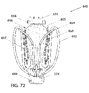
[0068] Figure 72 shows a perspective view of an example implantable device or
implant and
associated sensor(s).
[0069] Figure 73 shows a perspective view of an example implantable device or
implant and
associated sensor(s).
[0070] Figure 74 shows a perspective view of an example implantable device or
implant and
associated sensor(s).
8
CA 03224720 2023-12-18
WO 2023/003755 PCT/US2022/037176
[0071] Figure 75 shows a perspective view of an example implantable device or
implant and
associated sensor(s).
[0072] Figure 76 shows a perspective view of an example implantable device or
implant and
associated sensor(s).
[0073] Figure 77 shows a perspective view of an example implantable device or
implant and
associated sensor(s).
[0074] Figure 78 shows an example valve repair system and associated
sensor(s).
DETAILED DESCRIPTION
[0075] The following description refers to the accompanying drawings, which
illustrate example
implementations of the present disclosure. Other implementations having
different structures and
operation do not depart from the scope of the present disclosure.
[0076] Example implementations of the present disclosure are directed to
systems, devices,
methods, etc. for repairing a defective heart valve. For example, various
implementations of
implantable devices, valve repair devices, implants, and systems (including
systems for delivery
thereof) are disclosed herein, and any combination of these options can be
made unless
specifically excluded. In other words, individual components of the disclosed
devices and
systems can be combined unless mutually exclusive or otherwise physically
impossible. Further,
the techniques and methods herein can be performed on a living animal or on a
simulation, such
as on a cadaver, cadaver heart, simulator (e.g., with the body parts, heart,
tissue, etc. being
simulated), etc.
[0077] As described herein, when one or more components are described as being
connected,
joined, affixed, coupled, attached, or otherwise interconnected, such
interconnection can be
direct as between the components or can be indirect such as through the use of
one or more
intermediary components. Also as described herein, reference to a "member,"
"component," or
"portion" shall not be limited to a single structural member, component, or
element but can
include an assembly of components, members, or elements. Also as described
herein, the terms
"substantially" and "about" are defined as at least close to (and includes) a
given value or state
(preferably within 10% of, more preferably within 1% of, and most preferably
within 0.1% of).
[0078] Figures 1 and 2 are cutaway views of the human heart H in diastolic and
systolic phases,
respectively. The right ventricle RV and left ventricle LV are separated from
the right atrium RA
and left atrium LA, respectively, by the tricuspid valve TV and mitral valve
MV; i.e., the
9
CA 03224720 2023-12-18
WO 2023/003755 PCT/US2022/037176
atrioventricular valves. Additionally, the aortic valve AV separates the left
ventricle LV from the
ascending aorta AA, and the pulmonary valve PV separates the right ventricle
from the
pulmonary artery PA. Each of these valves has flexible leaflets (e.g.,
leaflets 20, 22 shown in
Figures 3-6 and leaflets 30, 32, 34 shown in Fig. 7) extending inward across
the respective
orifices that come together or "coapt" in the flow stream to form the one-way,
fluid-occluding
surfaces. The native valve repair systems of the present application are
frequently described
and/or illustrated with respect to the mitral valve MV. Therefore, anatomical
structures of the left
atrium LA and left ventricle LV will be explained in greater detail. However,
the devices
described herein can also be used in repairing other native valves, e.g., the
devices can be used in
repairing the tricuspid valve TV, the aortic valve AV, and the pulmonary valve
PV.
[0079] The left atrium LA receives oxygenated blood from the lungs. During the
diastolic phase,
or diastole, seen in Figure 1, the blood that was previously collected in the
left atrium LA (during
the systolic phase) moves through the mitral valve MV and into the left
ventricle LV by
expansion of the left ventricle LV. In the systolic phase, or systole, seen in
Figure 2, the left
ventricle LV contracts to force the blood through the aortic valve AV and
ascending aorta AA
into the body. During systole, the leaflets of the mitral valve MV close to
prevent the blood from
regurgitating from the left ventricle LV and back into the left atrium LA and
blood is collected in
the left atrium from the pulmonary vein. In some implementations, the devices
described by the
present application are used to repair the function of a defective mitral
valve MV. That is, the
devices are configured to help close the leaflets of the mitral valve to
prevent or inhibit blood
from regurgitating from the left ventricle LV and back into the left atrium
LA. Many of the
devices described in the present application are designed to easily grasp and
secure the native
leaflets around a coaptation element or spacer that beneficially acts as a
filler in the regurgitant
orifice to prevent or inhibit back flow or regurgitation during systole,
though this is not
necessary.
[0080] Referring now to Figures 1-7, the mitral valve MV includes two
leaflets, the anterior
leaflet 20 and the posterior leaflet 22. The mitral valve MV also includes an
annulus 24, which is
a variably dense fibrous ring of tissues that encircles the leaflets 20, 22.
Referring to Figures 3
and 4, the mitral valve MV is anchored to the wall of the left ventricle LV by
chordae tendineae
CT. The chordae tendineae CT are cord-like tendons that connect the papillary
muscles PM (i.e.,
the muscles located at the base of the chordae tendineae CT and within the
walls of the left
CA 03224720 2023-12-18
WO 2023/003755 PCT/US2022/037176
ventricle LV) to the leaflets 20, 22 of the mitral valve MV. The papillary
muscles PM serve to
limit the movements of leaflets 20, 22 of the mitral valve MV and prevent the
mitral valve MV
from being reverted. The mitral valve MV opens and closes in response to
pressure changes in
the left atrium LA and the left ventricle LV. The papillary muscles PM do not
open or close the
mitral valve MV. Rather, the papillary muscles PM support or brace the
leaflets 20, 22 against
the high pressure needed to circulate blood throughout the body. Together the
papillary muscles
PM and the chordae tendineae CT are known as the subvalvular apparatus, which
functions to
keep the mitral valve MV from prolapsing into the left atrium LA when the
mitral valve closes.
As seen from a Left Ventricular Outflow Tract (LVOT) view shown in Figure 3,
the anatomy of
the leaflets 20, 22 is such that the inner sides of the leaflets coapt at the
free end portions and the
leaflets 20, 22 start receding or spreading apart from each other. The
leaflets 20, 22 spread apart
in the atrial direction, until each leaflet meets with the mitral annulus.
[0081] Various disease processes can impair proper function of one or more of
the native valves
of the heart H. These disease processes include degenerative processes (e.g.,
Barlow's Disease,
fibroelastic deficiency, etc.), inflammatory processes (e.g., Rheumatic Heart
Disease), and
infectious processes (e.g., endocarditis, etc.). In addition, damage to the
left ventricle LV or the
right ventricle RV from prior heart attacks (i.e., myocardial infarction
secondary to coronary
artery disease) or other heart diseases (e.g., cardiomyopathy, etc.) can
distort a native valve's
geometry, which can cause the native valve to dysfunction. However, the
majority of patients
undergoing valve surgery, such as surgery to the mitral valve MV, suffer from
a degenerative
disease that causes a malfunction in a leaflet (e.g., leaflets 20, 22) of a
native valve (e.g., the
mitral valve MV), which results in prolapse and regurgitation.
[0082] Generally, a native valve may malfunction in different ways: including
(1) valve stenosis;
and (2) valve regurgitation. Valve stenosis occurs when a native valve does
not open completely
and thereby causes an obstruction of blood flow. Typically, valve stenosis
results from buildup of
calcified material on the leaflets of a valve, which causes the leaflets to
thicken and impairs the
ability of the valve to fully open to permit forward blood flow. Valve
regurgitation occurs when
the leaflets of the valve do not close completely thereby causing blood to
leak back into the prior
chamber (e.g., causing blood to leak from the left ventricle to the left
atrium).
[0083] There are three main mechanisms by which a native valve becomes
regurgitant¨or
incompetent¨which include Carpentier's type I, type II, and type III
malfunctions. A Carpentier
11
CA 03224720 2023-12-18
WO 2023/003755 PCT/US2022/037176
type I malfunction involves the dilation of the annulus such that normally
functioning leaflets are
distracted from each other and fail to form a tight seal (i.e., the leaflets
do not coapt properly).
Included in a type I mechanism malfunction are perforations of the leaflets,
as are present in
endocarditis. A Carpentier's type II malfunction involves prolapse of one or
more leaflets of a
native valve above a plane of coaptation. A Carpentier's type III malfunction
involves restriction
of the motion of one or more leaflets of a native valve such that the leaflets
are abnormally
constrained below the plane of the annulus. Leaflet restriction can be caused
by rheumatic
disease (Ma) or dilation of a ventricle (Tub).
[0084] Referring to Figure 5, when a healthy mitral valve MV is in a closed
position, the anterior
leaflet 20 and the posterior leaflet 22 coapt, which prevents blood from
leaking from the left
ventricle LV to the left atrium LA. Referring to Figures 3 and 6, mitral
regurgitation MR occurs
when the anterior leaflet 20 and/or the posterior leaflet 22 of the mitral
valve MV is displaced
into the left atrium LA during systole so that the edges of the leaflets 20,
22 are not in contact
with each other. This failure to coapt causes a gap 26 between the anterior
leaflet 20 and the
posterior leaflet 22, which allows blood to flow back into the left atrium LA
from the left
ventricle LV during systole, as illustrated by the mitral regurgitation MR
flow path shown in
Figure 3. Referring to Figure 6, the gap 26 can have a width W between about
2.5 mm and about
17.5 mm, between about 5 mm and about 15 mm, between about 7.5 mm and about
12.5 mm, or
about 10 mm. In some situations, the gap 26 can have a width W greater than 15
mm. As set
forth above, there are several different ways that a leaflet (e.g., leaflets
20, 22 of mitral valve
MV) may malfunction which can thereby lead to valvular regurgitation.
[0085] In any of the above-mentioned situations, a valve repair device or
implant is desired that
is capable of engaging the anterior leaflet 20 and the posterior leaflet 22 to
close the gap 26 and
prevent or inhibit regurgitation of blood through the mitral valve MV. As can
be seen in Figure 4,
an abstract representation of an implantable device, valve repair device, or
implant 10 is shown
implanted between the leaflets 20, 22 such that regurgitation does not occur
during systole
(compare Figure 3 with Figure 4). In some implementations, the coaptation
element (e.g., spacer,
coaption element, gap filler, etc.) of the device 10 has a generally tapered
or triangular shape that
naturally adapts to the native valve geometry and to its expanding leaflet
nature (toward the
annulus). In this application, the terms spacer, coaption element, coaptation
element, spacer, and
gap filler are used interchangeably and refer to an element that fills a
portion of the space
12
CA 03224720 2023-12-18
WO 2023/003755 PCT/US2022/037176
between native valve leaflets and/or that is configured such that the native
valve leaflets engage
or "coapt" against (e.g., such that the native leaflets coapt against the
coaption element,
coaptation element, spacer, etc. instead of only against one another).).
[0086] Although stenosis or regurgitation can affect any valve, stenosis is
predominantly found
to affect either the aortic valve AV or the pulmonary valve PV, and
regurgitation is
predominantly found to affect either the mitral valve MV or the tricuspid
valve TV. Both valve
stenosis and valve regurgitation increase the workload of the heart H and may
lead to very
serious conditions if left un-treated; such as endocarditis, congestive heart
failure, permanent
heart damage, cardiac arrest, and ultimately death. Because the left side of
the heart (i.e., the left
atrium LA, the left ventricle LV, the mitral valve MV, and the aortic valve
AV) are primarily
responsible for circulating the flow of blood throughout the body.
Accordingly, because of the
substantially higher pressures on the left side heart dysfunction of the
mitral valve MV or the
aortic valve AV is particularly problematic and often life threatening.
[0087] Malfunctioning native heart valves can either be repaired or replaced.
Repair typically
involves the preservation and correction of the patient's native valve.
Replacement typically
involves replacing the patient's native valve with a biological or mechanical
substitute. Typically,
the aortic valve AV and pulmonary valve PV are more prone to stenosis. Because
stenotic
damage sustained by the leaflets is irreversible, treatments for a stenotic
aortic valve or stenotic
pulmonary valve can be removal and replacement of the valve with a surgically
implanted heart
valve, or displacement of the valve with a transcatheter heart valve. The
mitral valve MV and the
tricuspid valve TV are more prone to deformation of leaflets and/or
surrounding tissue, which, as
described above, prevents the mitral valve MV or tricuspid valve TV from
closing properly and
allows for regurgitation or back flow of blood from the ventricle into the
atrium (e.g., a deformed
mitral valve MV may allow for regurgitation or back flow from the left
ventricle LV to the left
atrium LA as shown in Figure 3). The regurgitation or back flow of blood from
the ventricle to
the atrium results in valvular insufficiency. Deformations in the structure or
shape of the mitral
valve MV or the tricuspid valve TV are often repairable. In addition,
regurgitation can occur due
to the chordae tendineae CT becoming dysfunctional (e.g., the chordae
tendineae CT may stretch
or rupture), which allows the anterior leaflet 20 and the posterior leaflet 22
to be reverted such
that blood is regurgitated into the left atrium LA. The problems occurring due
to dysfunctional
chordae tendineae CT can be repaired by repairing the chordae tendineae CT or
the structure of
13
CA 03224720 2023-12-18
WO 2023/003755 PCT/US2022/037176
the mitral valve MV (e.g., by securing the leaflets 20, 22 at the affected
portion of the mitral
valve).
[0088] The devices and procedures disclosed herein often make reference to
repairing the
structure of a mitral valve. However, it should be understood that the devices
and concepts
provided herein can be used to repair any native valve, as well as any
component of a native
valve. Such devices can be used between the leaflets 20, 22 of the mitral
valve MV to prevent or
inhibit regurgitation of blood from the left ventricle into the left atrium.
With respect to the
tricuspid valve TV (Figure 7), any of the devices and concepts herein can be
used between any
two of the anterior leaflet 30, septal leaflet 32, and posterior leaflet 34 to
prevent or inhibit
regurgitation of blood from the right ventricle into the right atrium. In
addition, any of the
devices and concepts provided herein can be used on all three of the leaflets
30, 32, 34 together
to prevent or inhibit regurgitation of blood from the right ventricle to the
right atrium. That is, the
valve repair devices or implants provided herein can be centrally located
between the three
leaflets 30, 32, 34.
[0089] An example implantable device (e.g., implantable prosthetic device,
etc.) or implant can
optionally have a coaptation element (e.g., spacer, coaption element, gap
filler, etc.) and at least
one anchor (e.g., one, two, three, or more). In some implementations, an
implantable device or
implant can have any combination or sub-combination of the features disclosed
herein without a
coaptation element. When included, the coaptation element (e.g., coaption
element, spacer, etc.)
is configured to be positioned within the native heart valve orifice to help
fill the space between
the leaflets and form a more effective seal, thereby reducing or preventing
regurgitation
described above. The coaptation element can have a structure that is
impervious to blood (or that
resists blood flow therethrough) and that allows the native leaflets to close
around the coaptation
element during ventricular systole to block blood from flowing from the left
or right ventricle
back into the left or right atrium, respectively. The device or implant can be
configured to seal
against two or three native valve leaflets; that is, the device can be used in
the native mitral
(bicuspid) and tricuspid valves. The coaptation element is sometimes referred
to herein as a
spacer because the coaptation element can fill a space between improperly
functioning native
leaflets (e.g., mitral leaflets 20, 22 or tricuspid leaflets 30, 32, 34) that
do not close completely.
[0090] The optional coaptation element (e.g., spacer, coaption element, etc.)
can have various
shapes. In some implementations, the coaptation element can have an elongated
cylindrical shape
14
CA 03224720 2023-12-18
WO 2023/003755 PCT/US2022/037176
having a round cross-sectional shape. In some implementations, the coaptation
element can have
an oval cross-sectional shape, an ovoid cross-sectional shape, a crescent
cross-sectional shape, a
rectangular cross-sectional shape, or various other non-cylindrical shapes. In
some
implementations, the coaptation element can have an atrial portion positioned
in or adjacent to
the atrium, a ventricular or lower portion positioned in or adjacent to the
ventricle, and a side
surface that extends between the native leaflets. In some implementations
configured for use in
the tricuspid valve, the atrial or upper portion is positioned in or adjacent
to the right atrium, and
the ventricular or lower portion is positioned in or adjacent to the right
ventricle, and the side
surface that extends between the native tricuspid leaflets.
[0091] In some implementations, the anchor can be configured to secure the
device to one or
both of the native leaflets such that the coaptation element is positioned
between the two native
leaflets. In some implementations configured for use in the tricuspid valve,
the anchor is
configured to secure the device to one, two, or three of the tricuspid
leaflets such that the
coaptation element is positioned between the three native leaflets. In some
implementations, the
anchor can attach to the coaptation element at a location adjacent the
ventricular portion of the
coaptation element. In some implementations, the anchor can attach to an
actuation element,
such as a shaft or actuation wire, to which the coaptation element is also
attached. In some
implementations, the anchor and the coaptation element can be positioned
independently with
respect to each other by separately moving each of the anchor and the
coaptation element along
the longitudinal axis of the actuation element (e.g., actuation shaft,
actuation rod, actuation tube,
actuation wire, etc.). In some implementations, the anchor and the coaptation
element can be
positioned simultaneously by moving the anchor and the coaptation element
together along the
longitudinal axis of the actuation element (e.g., shaft, actuation wire,
etc.). The anchor can be
configured to be positioned behind a native leaflet when implanted such that
the leaflet is
grasped by the anchor.
[0092] The device or implant can be configured to be implanted via a delivery
system or other
means for delivery. The delivery system can comprise one or more of a
guide/delivery sheath, a
delivery catheter, a steerable catheter, an implant catheter, tube,
combinations of these, etc. The
coaptation element and the anchor can be compressible to a radially compressed
state and can be
self-expandable to a radially expanded state when compressive pressure is
released. The device
can be configured for the anchor to be expanded radially away from the still-
compressed
CA 03224720 2023-12-18
WO 2023/003755 PCT/US2022/037176
coaptation element initially in order to create a gap between the coaptation
element and the
anchor. A native leaflet can then be positioned in the gap. The coaptation
element can be
expanded radially, closing the gap between the coaptation element and the
anchor and capturing
the leaflet between the coaptation element and the anchor. In some
implementations, the anchor
and coaptation element are optionally configured to self-expand. The
implantation methods for
various implementations can be different and are more fully discussed below
with respect to each
implementation. Additional information regarding these and other delivery
methods can be found
in U.S. Pat. No. 8,449,599 and U.S. Patent Application Publication Nos.
2014/0222136,
2014/0067052, 2016/0331523, and PCT patent application publication Nos.
W02020/076898,
each of which is incorporated herein by reference in its entirety for all
purposes. These method(s)
can be performed on a living animal or on a simulation, such as on a cadaver,
cadaver heart,
simulator (e.g., with the body parts, heart, tissue, etc. being simulated),
etc. mutatis mutandis.
[0093] The disclosed devices or implants can be configured such that the
anchor is connected to
a leaflet, taking advantage of the tension from native chordae tendineae to
resist high systolic
pressure urging the device toward the left atrium. During diastole, the
devices can rely on the
compressive and retention forces exerted on the leaflet that is grasped by the
anchor.
[0094] Referring now to Figures 8-15, a schematically illustrated implantable
device or implant
100 (e.g., a prosthetic spacer device, valve repair device, etc.) is shown in
various stages of
deployment. The device or implant 100 and other similar devices/implants are
described in more
detail in PCT patent application publication Nos. W02018/195215,
W02020/076898, and WO
2019/139904, which are incorporated herein by reference in their entirety. The
device 100 can
include any other features for an implantable device or implant discussed in
the present
application or the applications cited above, and the device 100 can be
positioned to engage valve
tissue (e.g., leaflets 20, 22, 30, 32, 34) as part of any suitable valve
repair system (e.g., any valve
repair system disclosed in the present application or the applications cited
above).
[0095] The device or implant 100 is deployed from a delivery system or other
means for delivery
102. The delivery system 102 can comprise one or more of a catheter, a sheath,
a guide
catheter/sheath, a delivery catheter/sheath, a steerable catheter, an implant
catheter, a tube, a
channel, a pathway, combinations of these, etc. The device or implant 100
includes a coaptation
portion 104 and an anchor portion 106.
16
CA 03224720 2023-12-18
WO 2023/003755 PCT/US2022/037176
[0096] In some implementations, the coaptation portion 104 of the device or
implant 100
includes a coaptation element 110 or means for coapting (e.g., spacer, plug,
filler, foam, sheet,
membrane, coaption element, etc.) that is adapted to be implanted between
leaflets of a native
valve (e.g., a native mitral valve, native tricuspid valve, etc.) and is
slidably attached to an
actuation element 112 (e.g., actuation wire, actuation shaft, actuation tube,
etc.). The anchor
portion 106 includes one or more anchors 108 that are actuatable between open
and closed
conditions and can take a wide variety of forms, such as, for example,
paddles, gripping
elements, or the like. Actuation of the means for actuating or actuation
element 112 opens and
closes the anchor portion 106 of the device 100 to grasp the native valve
leaflets during
implantation. The means for actuating or actuation element 112 (as well as
other means for
actuating and actuation elements herein) can take a wide variety of different
forms (e.g., as a
wire, rod, shaft, tube, screw, suture, line, strip, combination of these,
etc.), be made of a variety
of different materials, and have a variety of configurations. As one example,
the actuation
element can be threaded such that rotation of the actuation element moves the
anchor portion 106
relative to the coaptation portion 104. Or, the actuation element can be
unthreaded, such that
pushing or pulling the actuation element 112 moves the anchor portion 106
relative to the
coaptation portion 104.
[0097] The anchor portion 106 and/or anchors of the device 100 include outer
paddles 120 and
inner paddles 122 that are, in some implementations, connected between a cap
114 and the
means for coapting or coaptation element 110 by portions 124, 126, 128. The
portions 124, 126,
128 can be jointed and/or flexible to move between all of the positions
described below. The
interconnection of the outer paddles 120, the inner paddles 122, the
coaptation element 110, and
the cap 114 by the portions 124, 126, and 128 can constrain the device to the
positions and
movements illustrated herein.
[0098] In some implementations, the delivery system 102 includes a steerable
catheter, implant
catheter, and means for actuating or actuation element 112 (e.g., actuation
wire, actuation shaft,
etc.). These can be configured to extend through a guide catheter/sheath
(e.g., a transseptal
sheath, etc.). In some implementations, the means for actuating or actuation
element 112 extends
through a delivery catheter and the means for coapting or coaptation element
110 to the distal
end (e.g., a cap 114 or other attachment portion at the distal connection of
the anchor portion
106). Extending and retracting the actuation element 112 increases and
decreases the spacing
17
CA 03224720 2023-12-18
WO 2023/003755 PCT/US2022/037176
between the coaptation element 110 and the distal end of the device (e.g., the
cap 114 or other
attachment portion), respectively. In some implementations, a collar or other
attachment element
removably attaches the coaptation element 110 to the delivery system 102,
either directly or
indirectly, so that the means for actuating or actuation element 112 slides
through the collar or
other attachment element and, in some implementations, through a means for
coapting or
coaptation element 110 during actuation to open and close the paddles 120, 122
of the anchor
portion 106 and/or anchors 108.
[0099] In some implementation, the anchor portion 106 and/or anchors 108 can
include
attachment portions or gripping members. The illustrated gripping members can
comprise clasps
130 that include a base or fixed arm 132, a moveable arm 134, optional barbs,
friction-enhancing
elements, or other means for securing 136 (e.g., protrusions, ridges, grooves,
textured surfaces,
adhesive, etc.), and a joint portion 138. The fixed arms 132 are attached to
the inner paddles 122.
In some implementations, the fixed arms 132 are attached to the inner paddles
122 with the joint
portion 138 disposed proximate means for coapting or coaptation element 110.
In some
implementations, the clasps (e.g., barbed clasps, etc.) have flat surfaces and
do not fit in a recess
of the inner paddle. Rather, the flat portions of the clasps are disposed
against the surface of the
inner paddle 122. The joint portion 138 provides a spring force between the
fixed and moveable
arms 132, 134 of the clasp 130. The joint portion 138 can be any suitable
joint, such as a flexible
joint, a spring joint, a pivot joint, or the like. In some implementations,
the joint portion 138 is a
flexible piece of material integrally formed with the fixed and moveable arms
132, 134. The
fixed arms 132 are attached to the inner paddles 122 and remain stationary or
substantially
stationary relative to the inner paddles 122 when the moveable arms 134 are
opened to open the
clasps 130 and expose the optional barbs, friction-enhancing elements, or
means for securing
136.
[0100] In some implementations, the clasps 130 are opened by applying tension
to actuation
lines 116 attached to the moveable arms 134, thereby causing the moveable arms
134 to
articulate, flex, or pivot on the joint portions 138. The actuation lines 116
extend through the
delivery system 102 (e.g., through a steerable catheter and/or an implant
catheter). Other
actuation mechanisms are also possible.
[0101] The actuation line 116 can take a wide variety of forms, such as, for
example, a line, a
suture, a wire, a rod, a catheter, or the like. The clasps 130 can be spring
loaded so that in the
18
CA 03224720 2023-12-18
WO 2023/003755 PCT/US2022/037176
closed position the clasps 130 continue to provide a pinching force on the
grasped native leaflet.
This pinching force remains constant regardless of the position of the inner
paddles 122.
Optional barbs, friction-enhancing elements, or other means for securing 136
of the clasps 130
can grab, pinch, and/or pierce the native leaflets to further secure the
native leaflets.
[0102] During implantation, the paddles 120, 122 can be opened and closed, for
example, to
grasp the native leaflets (e.g., native mitral valve leaflets, etc.) between
the paddles 120, 122
and/or between the paddles 120, 122 and a means for coapting or coaptation
element 110. The
clasps 130 can be used to grasp and/or further secure the native leaflets by
engaging the leaflets
with optional barbs, friction-enhancing elements, or means for securing 136
and pinching the
leaflets between the moveable and fixed arms 134, 132. The optional barbs,
friction-enhancing
elements, or other means for securing 136 (e.g., barbs, protrusions, ridges,
grooves, textured
surfaces, adhesive, etc.) of the clasps or barbed clasps 130 increase friction
with the leaflets or
can partially or completely puncture the leaflets. The actuation lines 116 can
be actuated
separately so that each clasp 130 can be opened and closed separately.
Separate operation allows
one leaflet to be grasped at a time, or for the repositioning of a clasp 130
on a leaflet that was
insufficiently grasped, without altering a successful grasp on the other
leaflet. The clasps 130 can
be opened and closed relative to the position of the inner paddle 122 (as long
as the inner paddle
is in an open or at least partially open position), thereby allowing leaflets
to be grasped in a
variety of positions as the particular situation requires.
[0103] Referring now to Figure 8, the device 100 is shown in an elongated or
fully open
condition for deployment from an implant delivery catheter of the delivery
system 102. The
device 100 is disposed at the end of the catheter of the delivery system 102
in the fully open
position, because the fully open position takes up the least space and allows
the smallest catheter
to be used (or the largest device 100 to be used for a given catheter size).
In the elongated
condition the cap 114 is spaced apart from the means for coapting or
coaptation element 110
such that the paddles 120, 122 are fully extended. In some implementations, an
angle formed
between the interior of the outer and inner paddles 120, 122 is approximately
180 degrees. The
clasps 130 are kept in a closed condition during deployment through the
delivery system 102 so
that the optional barbs, friction-enhancing elements, or other means for
securing 136 (Figure 9)
do not catch or damage the delivery system 102 or tissue in the patient's
heart.
19
CA 03224720 2023-12-18
WO 2023/003755 PCT/US2022/037176
[0104] Referring now to Figure 9, the device 100 is shown in an elongated
detangling condition,
similar to Figure 8, but with the clasps 130 in a fully open position, ranging
from about 140
degrees to about 200 degrees, from about 170 degrees to about 190 degrees, or
about 180 degrees
between fixed and moveable portions 132, 134 of the clasps 130. Fully opening
the paddles 120,
122 and the clasps 130 has been found to improve ease of detanglement or
detachment from
anatomy of the patient, such as the chordae tendineae CT, during implantation
of the device 100.
[0105] Referring now to Figure 10, the device 100 is shown in a shortened or
fully closed
condition. The compact size of the device 100 in the shortened condition
allows for easier
maneuvering and placement within the heart. To move the device 100 from the
elongated
condition to the shortened condition, the means for actuating or actuation
element 112 is
retracted to pull the cap 114 towards the means for coapting or coaptation
element 110. The
connection portion(s) 126 (e.g., joint(s), flexible connection(s), etc.)
between the outer paddle
120 and inner paddle 122 are constrained in movement such that compression
forces acting on
the outer paddle 120 from the cap 114 being retracted towards the means for
coapting or
coaptation element 110 cause the paddles or gripping elements to move radially
outward. During
movement from the open to closed position, the outer paddles 120 maintain an
acute angle with
the means for actuating or actuation element 112. The outer paddles 120 can
optionally be biased
toward a closed position. The inner paddles 122 during the same motion move
through a
considerably larger angle as they are oriented away from the means for
coapting or coaptation
element 110 in the open condition and collapse along the sides of the means
for coapting or
coaptation element 110 in the closed condition. In some implementations, the
inner paddles 122
are thinner and/or narrower than the outer paddles 120, and the connection
portions 126, 128
(e.g., joints, flexible connections, etc.) connected to the inner paddles 122
can be thinner and/or
more flexible. For example, this increased flexibility can allow more movement
than the
connection portion 124 connecting the outer paddle 120 to the cap 114. In some
implementations, the outer paddles 120 are narrower than the inner paddles
122. The connection
portions 126, 128 connected to the inner paddles 122 can be more flexible, for
example, to allow
more movement than the connection portion 124 connecting the outer paddle 120
to the cap 114.
In some implementations, the inner paddles 122 can be the same or
substantially the same width
as the outer paddles
CA 03224720 2023-12-18
WO 2023/003755 PCT/US2022/037176
[0106] Referring now to Figures 11-13, the device 100 is shown in a partially
open, grasp-ready
condition. To transition from the fully closed to the partially open
condition, the means for
actuating or actuation element (e.g., actuation wire, actuation shaft, etc.)
is extended to push the
cap 114 away from the means for coapting or coaptation element 110, thereby
pulling on the
outer paddles 120, which in turn pull on the inner paddles 122, causing the
anchors or anchor
portion 106 to partially unfold. The actuation lines 116 are also retracted to
open the clasps 130
so that the leaflets can be grasped. In some implementations, the pair of
inner and outer paddles
122, 120 are moved in unison, rather than independently, by a single means for
actuating or
single actuation element 112. Also, the positions of the clasps 130 are
dependent on the positions
of the paddles 122, 120. For example, referring to Figure 10 closing the
paddles 122, 120 also
closes the clasps. In some implementations, the paddles 120, 122 can be
independently
controllable. For example, the device 100 can have two actuation elements and
two independent
caps (or other attachment portions), such that one independent actuation
element (e.g., wire,
shaft, etc.) and cap (or other attachment portion) are used to control one
paddle, and the other
independent actuation element and cap (or other attachment portion) are used
to control the other
paddle.
[0107] Referring now to Figure 12, one of the actuation lines 116 is extended
to allow one of the
clasps 130 to close. Referring now to Figure 13, the other actuation line 116
is extended to allow
the other clasp 130 to close. Either or both of the actuation lines 116 can be
repeatedly actuated
to repeatedly open and close the clasps 130.
[0108] Referring now to Figure 14, the device 100 is shown in a fully closed
and deployed
condition. The delivery system or means for delivery 102 and means for
actuating or actuation
element 112 are retracted and the paddles 120, 122 and clasps 130 remain in a
fully closed
position. Once deployed, the device 100 can be maintained in the fully closed
position with a
mechanical latch or can be biased to remain closed through the use of spring
materials, such as
steel, other metals, plastics, composites, etc. or shape-memory alloys such as
Nitinol. For
example, the connection portions 124, 126, 128, the joint portions 138, and/or
the inner and outer
paddles 122, and/or an additional biasing component (not shown) can be formed
of metals such
as steel or shape-memory alloy, such as Nitinol¨produced in a wire, sheet,
tubing, or laser
sintered powder¨and are biased to hold the outer paddles 120 closed around the
means for
coapting or coaptation element 110 and the clasps 130 pinched around native
leaflets. Similarly,
21
CA 03224720 2023-12-18
WO 2023/003755 PCT/US2022/037176
the fixed and moveable arms 132, 134 of the clasps 130 are biased to pinch the
leaflets. In some
implementations, the attachment or connection portions 124, 126, 128, joint
portions 138, and/or
the inner and outer paddles 122, and/or an additional biasing component (not
shown) can be
formed of any other suitably elastic material, such as a metal or polymer
material, to maintain the
device 100 in the closed condition after implantation.
[0109] Figure 15 illustrates an example where the paddles 120, 122 are
independently
controllable. The device 101 illustrated by Figure 15 is similar to the device
illustrated by Figure
11, except the device 101 of Figure 15 includes an actuation element that is
configured as two
independent actuation elements 111, 113 that are coupled to two independent
caps 115, 117. To
transition a first inner paddle 122 and a first outer paddle 120 from the
fully closed to the
partially open condition, the means for actuating or actuation element 111 is
extended to push the
cap 115 away from the means for coapting or coaptation element 110, thereby
pulling on the
outer paddle 120, which in turn pulls on the inner paddle 122, causing the
first anchor 108 to
partially unfold. To transition a second inner paddle 122 and a second outer
paddle 120 from the
fully closed to the partially open condition, the means for actuating or
actuation element 113 is
extended to push the cap 115 away from the means for coapting or coaptation
element 110,
thereby pulling on the outer paddle 120, which in turn pulls on the inner
paddle 122, causing the
second anchor 108 to partially unfold. The independent paddle control
illustrated by Figure 15
can be implemented on any of the devices disclosed by the present application.
For comparison,
in the example illustrated by Figure 11, the pair of inner and outer paddles
122, 120 are moved in
unison, rather than independently, by a single means for actuating or
actuation element 112.
[0110] Referring now to Figures 16-21, the implantable device 100 of Figures 8-
14 is shown
being delivered and implanted within the native mitral valve MV of the heart
H. Referring to
Figure 16, a delivery sheath/catheter is inserted into the left atrium LA
through the septum and
the implant/device 100 is deployed from the delivery catheter/sheath in the
fully open condition
as illustrated in Figure 16. The means for actuating or actuation element 112
is then retracted to
move the implant/device into the fully closed condition shown in Figure 17.
[0111] As can be seen in Figure 18, the implant/device is moved into position
within the mitral
valve MV into the ventricle LV and partially opened so that the leaflets 20,
22 can be grasped.
For example, a steerable catheter can be advanced and steered or flexed to
position the steerable
22
CA 03224720 2023-12-18
WO 2023/003755 PCT/US2022/037176
catheter as illustrated by Figure 18. The implant catheter connected to the
implant/device can be
advanced from inside the steerable catheter to position the implant as
illustrated by Figure 18.
[0112] Referring now to Figure 19, the implant catheter can be retracted into
the steerable
catheter to position the mitral valve leaflets 20,22 in the clasps 130. An
actuation line 116 is
extended to close one of the clasps 130, capturing a leaflet 20. Figure 20
shows the other
actuation line 116 being then extended to close the other clasp 130, capturing
the remaining
leaflet 22. Lastly, as can be seen in Figure 21, the delivery system 102
(e.g., steerable catheter,
implant catheter, etc.), means for actuating or actuation element 112 and
actuation lines 116 are
then retracted and the device or implant 100 is fully closed and deployed in
the native mitral
valve MV.
[0113] Referring now to Figures 22-27, an example of an implantable device or
implant or
implant 200 is shown. The implantable device 200 is one of the many different
configurations
that the device 100 that is schematically illustrated in Figures 8-14 can
take. The device 200 can
include any other features for an implantable device or implant discussed in
the present
application, and the device 200 can be positioned to engage valve tissue 20,22
as part of any
suitable valve repair system (e.g., any valve repair system disclosed in the
present application).
The device/implant 200 can be a prosthetic spacer device, valve repair device,
or another type of
implant that attaches to leaflets of a native valve.
[0114] In some implementations, the implantable device or implant 200 includes
a coaptation
portion 204, a proximal or attachment portion 205, an anchor portion 206, and
a distal portion
207. In some implementations, the coaptation portion 204 of the device
optionally includes a
coaptation element 210 (e.g., a spacer, coaption element, plug, membrane,
sheet, etc.) for
implantation between leaflets of a native valve. In some implementations, the
anchor portion 206
includes a plurality of anchors 208. The anchors can be configured in a
variety of ways. In some
implementations, each anchor 208 includes outer paddles 220, inner paddles
222, paddle
extension members or paddle frames 224, and clasps 230. In some
implementations, the
attachment portion 205 includes a first or proximal collar 211 (or other
attachment element) for
engaging with a capture mechanism 213 (Figures 43-49) of a delivery system 202
(Figures 38-
42 and 49). Delivery system 202 can be the same as or similar to delivery
system 102 described
elsewhere and can comprise one or more of a catheter, a sheath, a guide
catheter/sheath, a
23
CA 03224720 2023-12-18
WO 2023/003755 PCT/US2022/037176
delivery catheter/sheath, a steerable catheter, an implant catheter, a tube, a
channel, a pathway,
combinations of these, etc.
[0115] In some implementations, the coaptation element 210 and paddles 220,
222 are formed
from a flexible material that can be a metal fabric, such as a mesh, woven,
braided, or formed in
any other suitable way or a laser cut or otherwise cut flexible material. The
material can be cloth,
shape-memory alloy wire¨such as Nitinol¨to provide shape-setting capability,
or any other
flexible material suitable for implantation in the human body.
[0116] An actuation element 212 (e.g., actuation shaft, actuation rod,
actuation tube, actuation
wire, actuation line, etc.) extends from the delivery system 202 to engage and
enable actuation of
the implantable device or implant 200. In some implementations, the actuation
element 212
extends through the capture mechanism 213, proximal collar 211, and coaptation
element 210 to
engage a cap 214 of the distal portion 207. The actuation element 212 can be
configured to
removably engage the cap 214 with a threaded connection, or the like, so that
the actuation
element 212 can be disengaged and removed from the device 200 after
implantation.
[0117] The coaptation element 210 extends from the proximal collar 211 (or
other attachment
element) to the inner paddles 222. In some implementations, the coaptation
element 210 has a
generally elongated and round shape, though other shapes and configurations
are possible. In
some implementations, the coaptation element 210 has an elliptical shape or
cross-section when
viewed from above (e.g., Figure 51) and has a tapered shape or cross-section
when seen from a
front view (e.g., Figure 23) and a round shape or cross-section when seen from
a side view (e.g.,
Figure 24). A blend of these three geometries can result in the three-
dimensional shape of the
illustrated coaptation element 210 that achieves the benefits described
herein. The round shape of
the coaptation element 210 can also be seen, when viewed from above, to
substantially follow or
be close to the shape of the paddle frames 224.
[0118] The size and/or shape of the coaptation element 210 can be selected to
minimize the
number of implants that a single patient will require (preferably one), while
at the same time
maintaining low transvalvular gradients. In some implementations, the anterior-
posterior distance
at the top of the coaptation element is about 5 mm, and the medial-lateral
distance of the
coaptation element at its widest is about 10 mm. In some implementations, the
overall geometry
of the device 200 can be based on these two dimensions and the overall shape
strategy described
above. It should be readily apparent that the use of other anterior-posterior
distance anterior-
24
CA 03224720 2023-12-18
WO 2023/003755 PCT/US2022/037176
posterior distance and medial-lateral distance as starting points for the
device will result in a
device having different dimensions. Further, using other dimensions and the
shape strategy
described above will also result in a device having different dimensions.
[0119] In some implementations, the outer paddles 220 are jointably attached
to the cap 214 of
the distal portion 207 by connection portions 221 and to the inner paddles 222
by connection
portions 223. The inner paddles 222 are jointably attached to the coaptation
element by
connection portions 225. In this manner, the anchors 208 are configured
similar to legs in that the
inner paddles 222 are like upper portions of the legs, the outer paddles 220
are like lower
portions of the legs, and the connection portions 223 are like knee portions
of the legs.
[0120] In some implementations, the inner paddles 222 are stiff, relatively
stiff, rigid, have rigid
portions and/or are stiffened by a stiffening member or a fixed portion 232 of
the clasps 230. The
stiffening of the inner paddle allows the device to move to the various
different positions shown
and described herein. The inner paddle 222, the outer paddle 220, the
coaptation can all be
interconnected as described herein, such that the device 200 is constrained to
the movements and
positions shown and described herein.
[0121] In some implementations, the paddle frames 224 are attached to the cap
214 at the distal
portion 207 and extend to the connection portions 223 between the inner and
outer paddles 222,
220. In some implementations, the paddle frames 224 are formed of a material
that is more rigid
and stiff than the material forming the paddles 222, 220 so that the paddle
frames 224 provide
support for the paddles 222, 220.
[0122] The paddle frames 224 provide additional pinching force between the
inner paddles 222
and the coaptation element 210 and assist in wrapping the leaflets around the
sides of the
coaptation element 210 for a better seal between the coaptation element 210
and the leaflets, as
can be seen in Figure 51. That is, the paddle frames 224 can be configured
with a round three-
dimensional shape extending from the cap 214 to the connection portions 223 of
the anchors 208.
The connections between the paddle frames 224, the outer and inner paddles
220, 222, the cap
214, and the coaptation element 210 can constrain each of these parts to the
movements and
positions described herein. In particular the connection portion 223 is
constrained by its
connection between the outer and inner paddles 220, 222 and by its connection
to the paddle
frame 224. Similarly, the paddle frame 224 is constrained by its attachment to
the connection
portion 223 (and thus the inner and outer paddles 222, 220) and to the cap
214.
CA 03224720 2023-12-18
WO 2023/003755 PCT/US2022/037176
[0123] Configuring the paddle frames 224 in this manner provides increased
surface area
compared to the outer paddles 220 alone. This can, for example, make it easier
to grasp and
secure the native leaflets. The increased surface area can also distribute the
clamping force of the
paddles 220 and paddle frames 224 against the native leaflets over a
relatively larger surface of
the native leaflets in order to further protect the native leaflet tissue.
Referring again to Figure
51, the increased surface area of the paddle frames 224 can also allow the
native leaflets to be
clamped to the implantable device or implant 200, such that the native
leaflets coapt entirely
around the coaptation member or coaptation element 210. This can, for example,
improve sealing
of the native leaflets 20, 22 and thus prevent or further reduce mitral
regurgitation.
[0124] In some implementations the clasps comprise a moveable arm coupled to
the anchors. In
some implementations, the clasps 230 include a base or fixed arm 232, a
moveable arm 234,
optional barbs 236, and a joint portion 238. The fixed arms 232 are attached
to the inner paddles
222, with the joint portion 238 disposed proximate the coaptation element 210.
The joint portion
238 is spring-loaded so that the fixed and moveable arms 232, 234 are biased
toward each other
when the clasp 230 is in a closed condition. In some implementations, the
clasps 230 include
friction-enhancing elements or means for securing, such as optional barbs,
protrusions, ridges,
grooves, textured surfaces, adhesive, etc.
[0125] In some implementations, the fixed arms 232 are attached to the inner
paddles 222
through holes or slots 231 with sutures (not shown). The fixed arms 232 can be
attached to the
inner paddles 222 with any suitable means, such as screws or other fasteners,
crimped sleeves,
mechanical latches or snaps, welding, adhesive, clamps, latches, or the like.
The fixed arms 232
remain substantially stationary relative to the inner paddles 222 when the
moveable arms 234 are
opened to open the clasps 230 and expose the optional barbs or other friction-
enhancing elements
236. The clasps 230 are opened by applying tension to actuation lines 216
(e.g., as shown in
Figures 43-48) attached to holes 235 in the moveable arms 234, thereby causing
the moveable
arms 234 to articulate, pivot, and/or flex on the joint portions 238.
[0126] Referring now to Figure 29, a close-up view of one of the leaflets 20,
22 grasped by a
clasp such as clasp 230 is shown. The leaflet 20, 22 is grasped between the
moveable and fixed
arms 234, 232 of the clasp 230. The tissue of the leaflet 20, 22 is not
pierced by the optional
barbs or friction-enhancing elements 236, though in some implementations the
optional barbs
236 can partially or fully pierce through the leaflet 20, 22. The angle and
height of the optional
26
CA 03224720 2023-12-18
WO 2023/003755 PCT/US2022/037176
barbs or friction-enhancing elements 236 relative to the moveable arm 234
helps to secure the
leaflet 20, 22 within the clasp 230. In particular, a force pulling the
implant off of the native
leaflet 20, 22 will encourage the optional barbs or friction-enhancing
elements 236 to further
engage the tissue, thereby ensuring better retention. Retention of the leaflet
20, 22 in the clasp
230 is further improved by the position of fixed arm 232 near the optional
barbs/friction-
enhancing elements 236 when the clasp 230 is closed. In this arrangement, the
tissue is formed
by the fixed arms 232 and the moveable arms 234 and the optional
barbs/friction-enhancing
elements 236 into an S-shaped torturous path. Thus, forces pulling the leaflet
20, 22 away from
the clasp 230 will encourage the tissue to further engage the optional
barbs/friction-enhancing
elements 236 before the leaflets 20, 22 can escape. For example, leaflet
tension during diastole
can encourage the optional barbs 236 to pull toward the end portion of the
leaflet 20, 22. Thus,
the S-shaped path can utilize the leaflet tension during diastole to more
tightly engage the leaflets
20, 22 with the optional barbs/friction-enhancing elements 236.
[0127] Referring to Figure 25, the prosthetic device or implant 200 can also
include a cover 240.
In some implementations, the cover 240 can be disposed on the coaptation
element 210, the outer
and inner paddles 220, 222, and/or the paddle frames 224. The cover 240 can be
configured to
prevent or reduce blood-flow through the prosthetic device or implant 200
and/or to promote
native tissue ingrowth. In some implementations, the cover 240 can be a cloth
or fabric such as
PET, velour, or other suitable fabric. In some implementations, in lieu of or
in addition to a
fabric, the cover 240 can include a coating (e.g., polymeric) that is applied
to the implantable
prosthetic device or implant 200.
[0128] During implantation, the paddles 220, 222 of the anchors 208 are opened
and closed to
grasp the native valve leaflets 20, 22 between the paddles 220, 222 and the
coaptation element
210. The anchors 208 are moved between a closed position (Figures 22-25) to
various open
positions (Figures 26-37) by extending and retracting the actuation element
212. Extending and
retracting the actuation element 212 increases and decreases the spacing
between the coaptation
element 210 and the cap 214, respectively. The proximal collar 211 (or other
attachment element)
and the coaptation element 210 slide along the actuation element 212 during
actuation so that
changing of the spacing between the coaptation element 210 and the cap 214
causes the paddles
220, 220 to move between different positions to grasp the mitral valve
leaflets 20, 22 during
implantation.
27
CA 03224720 2023-12-18
WO 2023/003755 PCT/US2022/037176
[0129] As the device 200 is opened and closed, the pair of inner and outer
paddles 222, 220 are
moved in unison, rather than independently, by a single actuation element 212.
Also, the
positions of the clasps 230 are dependent on the positions of the paddles 222,
220. For example,
the clasps 230 are arranged such that closure of the anchors 208
simultaneously closes the clasps
230. In some implementations, the device 200 can be made to have the paddles
220, 222 be
independently controllable in the same manner (e.g., the device 100
illustrated in Figure 15).
[0130] In some implementations, the clasps 230 further secure the native
leaflets 20, 22 by
engaging the leaflets 20, 22 with optional barbs and/or other friction-
enhancing elements 236 and
pinching the leaflets 20, 22 between the moveable and fixed arms 234, 232. In
some
implementations, the clasps 230 are barbed clasps that include barbs that
increase friction with
and/or can partially or completely puncture the leaflets 20, 22. The actuation
lines 216 (Figures
43-48) can be actuated separately so that each clasp 230 can be opened and
closed separately.
Separate operation allows one leaflet 20, 22 to be grasped at a time, or for
the repositioning of a
clasp 230 on a leaflet 20, 22 that was insufficiently grasped, without
altering a successful grasp
on the other leaflet 20, 22. The clasps 230 can be fully opened and closed
when the inner paddle
222 is not closed, thereby allowing leaflets 20, 22 to be grasped in a variety
of positions as the
particular situation requires.
[0131] Referring now to Figures 22-25, the device 200 is shown in a closed
position. When
closed, the inner paddles 222 are disposed between the outer paddles 220 and
the coaptation
element 210. The clasps 230 are disposed between the inner paddles 222 and the
coaptation
element 210. Upon successful capture of native leaflets 20, 22 the device 200
is moved to and
retained in the closed position so that the leaflets 20, 22 are secured within
the device 200 by the
clasps 230 and are pressed against the coaptation element 210 by the paddles
220, 222. The outer
paddles 220 can have a wide curved shape that fits around the curved shape of
the coaptation
element 210 to more securely grip the leaflets 20, 22 when the device 200 is
closed (e.g., as can
be seen in Figure 51). The curved shape and rounded edges of the outer paddle
220 also prohibits
or inhibits tearing of the leaflet tissue.
[0132] Referring now to Figures 30-37, the implantable device or implant 200
described above
is shown in various positions and configurations ranging from partially open
to fully open. The
paddles 220, 222 of the device 200 transition between each of the positions
shown in Figures 30-
28
CA 03224720 2023-12-18
WO 2023/003755 PCT/US2022/037176
37 from the closed position shown in Figures 22-25 up extension of the
actuation element 212
from a fully retracted to fully extended position.
[0133] Referring now to Figures 30-31, the device 200 is shown in a partially
open position. The
device 200 is moved into the partially open position by extending the
actuation element 212.
Extending the actuation element 212 pulls down on the bottom portions of the
outer paddles 220
and paddle frames 224. The outer paddles 220 and paddle frames 224 pull down
on the inner
paddles 222, where the inner paddles 222 are connected to the outer paddles
220 and the paddle
frames 224. Because the proximal collar (or other attachment element) and
coaptation element
210 are held in place by the capture mechanism 213, the inner paddles 222 are
caused to
articulate, pivot, and/or flex in an opening direction. The inner paddles 222,
the outer paddles
220, and the paddle frames all flex to the position shown in Figures 30-31.
Opening the paddles
222, 220 and frames 224 forms a gap between the coaptation element 210 and the
inner paddle
222 that can receive and grasp the native leaflets 20, 22. This movement also
exposes the clasps
230 that can be moved between closed (Figure 30) and open (Figure 31)
positions to form a
second gap for grasping the native leaflets 20, 22. The extent of the gap
between the fixed and
moveable arms 232, 234 of the clasp 230 is limited to the extent that the
inner paddle 222 has
spread away from the coaptation element 210.
[0134] Referring now to Figures 32-33, the device 200 is shown in a laterally
extended or open
position. The device 200 is moved into the laterally extended or open position
by continuing to
extend the actuation element 212 described above, thereby increasing the
distance between the
coaptation element 210 and the cap 214 of the distal portion 207. Continuing
to extend the
actuation element 212 pulls down on the outer paddles 220 and paddle frames
224, thereby
causing the inner paddles 222 to spread apart further from the coaptation
element 210. In the
laterally extended or open position, the inner paddles 222 extend horizontally
more than in other
positions of the device 200 and form an approximately 90-degree angle with the
coaptation
element 210. Similarly, the paddle frames 224 are at their maximum spread
position when the
device 200 is in the laterally extended or open position. The increased gap
between the
coaptation element 210 and inner paddle 222 formed in the laterally extended
or open position
allows clasps 230 to open further (Figure 33) before engaging the coaptation
element 210,
thereby increasing the size of the gap between the fixed and moveable arms
232, 234.
29
CA 03224720 2023-12-18
WO 2023/003755 PCT/US2022/037176
[0135] Referring now to Figures 34-35, the example device 200 is shown in a
three-quarters
extended position. The device 200 is moved into the three-quarters extended
position by
continuing to extend the actuation element 212 described above, thereby
increasing the distance
between the coaptation element 210 and the cap 214 of the distal portion 207.
Continuing to
extend the actuation element 212 pulls down on the outer paddles 220 and
paddle frames 224,
thereby causing the inner paddles 222 to spread apart further from the
coaptation element 210. In
the three-quarters extended position, the inner paddles 222 are open beyond 90
degrees to an
approximately 135-degree angle with the coaptation element 210. The paddle
frames 224 are less
spread than in the laterally extended or open position and begin to move
inward toward the
actuation element 212 as the actuation element 212 extends further. The outer
paddles 220 also
flex back toward the actuation element 212. As with the laterally extended or
open position, the
increased gap between the coaptation element 210 and inner paddle 222 formed
in the laterally
extended or open position allows clasps 230 to open even further (Figure 35),
thereby increasing
the size of the gap between the fixed and moveable arms 232, 234.
[0136] Referring now to Figures 36-37, the example device 200 is shown in a
fully extended
position. The device 200 is moved into the fully extended position by
continuing to extend the
actuation element 212 described above, thereby increasing the distance between
the coaptation
element 210 and the cap 214 of the distal portion 207 to a maximum distance
allowable by the
device 200. Continuing to extend the actuation element 212 pulls down on the
outer paddles 220
and paddle frames 224, thereby causing the inner paddles 222 to spread apart
further from the
coaptation element 210. The outer paddles 220 and paddle frames 224 move to a
position where
they are close to the actuation element. In the fully extended position, the
inner paddles 222 are
open to an approximately 180-degree angle with the coaptation element 210. The
inner and outer
paddles 222, 220 are stretched straight in the fully extended position to form
an approximately
180-degree angle between the paddles 222, 220. The fully extended position of
the device 200
provides the maximum size of the gap between the coaptation element 210 and
inner paddle 222,
and, in some implementations, allows clasps 230 to also open fully to
approximately 180 degrees
(Figure 37) between the fixed and moveable arms 232, 234 of the clasp 230. The
position of the
device 200 is the longest and the narrowest configuration. Thus, the fully
extended position of
the device 200 can be a desirable position for bailout of the device 200 from
an attempted
CA 03224720 2023-12-18
WO 2023/003755 PCT/US2022/037176
implantation or can be a desired position for placement of the device in a
delivery catheter, or the
like.
[0137] Configuring the prosthetic device or implant 200 such that the anchors
208 can extend to
a straight or approximately straight configuration (e.g., approximately 120-
180 degrees relative
to the coaptation element 210) can provide several advantages. For example,
this configuration
can reduce the radial crimp profile of the prosthetic device or implant 200.
It can also make it
easier to grasp the native leaflets 20,22 by providing a larger opening
between the coaptation
element 210 and the inner paddles 222 in which to grasp the native leaflets
20,22. Additionally,
the relatively narrow, straight configuration can prevent or reduce the
likelihood that the
prosthetic device or implant 200 will become entangled in native anatomy
(e.g., chordae
tendineae CT shown in Figures 3 and 4) when positioning and/or retrieving the
prosthetic device
or implant 200 into the delivery system 202.
[0138] Referring now to Figures 38-49, an example implantable device 200 is
shown being
delivered and implanted within the native mitral valve MV of the heart H. As
described above,
the device 200 shown in Figures 38-49 includes the optional covering 240
(e.g., Figure 25) over
the coaptation element 210, clasps 230, inner paddles 222 and/or the outer
paddles 220. The
device 200 is deployed from a delivery system 202 (e.g., which can comprise an
implant catheter
that is extendable from a steerable catheter and/or a guide sheath) and is
retained by a capture
mechanism 213 (see e.g., Figures 43 and 48) and is actuated by extending or
retracting the
actuation element 212. Fingers of the capture mechanism 213 removably attach
the collar 211 to
the delivery system 202. In some implementations, the capture mechanism 213 is
held closed
around the collar 211 by the actuation element 212, such that removal of the
actuation element
212 allows the fingers of the capture mechanism 213 to open and release the
collar 211 to
decouple the capture mechanism 213 from the device 200 after the device 200
has been
successfully implanted.
[0139] Referring now to Figure 38, the delivery system 202 (e.g., a delivery
catheter/sheath
thereof) is inserted into the left atrium LA through the septum and the
device/implant 200 is
deployed from the delivery system 202 (e.g., an implant catheter retaining the
device/implant can
be extended to deploy the device/implant out from a steerable catheter) in the
fully open
condition for the reasons discussed above with respect to the device 100. The
actuation element
212 is then retracted to move the device 200 through the partially closed
condition (Figure 39)
31
CA 03224720 2023-12-18
WO 2023/003755 PCT/US2022/037176
and to the fully closed condition shown in Figures 40-41. Then the delivery
system or catheter
maneuvers the device/implant 200 towards the mitral valve MV as shown in
Figure 41. Referring
now to Figure 42, when the device 200 is aligned with the mitral valve MV, the
actuation
element 212 is extended to open the paddles 220, 222 into the partially opened
position and the
actuation lines 216 (Figures 43-48) are retracted to open the clasps 230 to
prepare for leaflet
grasp. Next, as shown in Figures 43-44, the partially open device 200 is
inserted through the
native valve (e.g., by advancing an implant catheter from a steerable
catheter) until leaflets 20,
22 are properly positioned in between the inner paddles 222 and the coaptation
element 210 and
inside the open clasps 230.
[0140] Figure 45 shows the device 200 with both clasps 230 closed, though the
optional barbs
236 of one clasp 230 missed one leaflet 22. As can be seen in Figures 45-47,
the out of position
clasp 230 is opened and closed again to properly grasp the missed leaflet 22.
When both leaflets
20, 22 are grasped properly, the actuation element 212 is retracted to move
the device 200 into
the fully closed position shown in Figure 48. With the device 200 fully closed
and implanted in
the native valve, the actuation element 212 is disengaged from the cap 214 and
is withdrawn to
release the capture mechanism 213 from the proximal collar 211 (or other
attachment element)
so that the capture mechanism 213 can be withdrawn into the delivery system
202 (e.g., into a
catheter/sheath), as shown in Figure 49. Once deployed, the device 200 can be
maintained in the
fully closed position with a mechanical means such as a latch or can be biased
to remain closed
through the use of spring material, such as steel, and/or shape-memory alloys
such as Nitinol.
For example, the paddles 220, 222 can be formed of steel or Nitinol shape-
memory alloy¨
produced in a wire, sheet, tubing, or laser sintered powder¨and are biased to
hold the outer
paddles 220 closed around the inner paddles 222, coaptation element 210,
and/or the clasps 230
pinched around native leaflets 20, 22.
[0141] Referring to Figures 50-54, once the device 200 is implanted in a
native valve, the
coaptation element 210 functions as a gap filler in the valve regurgitant
orifice, such as the gap
26 in the mitral valve MV illustrated by Figure 6 or a gap in another native
valve. In some
implementations, when the device 200 has been deployed between the two
opposing valve
leaflets 20, 22, the leaflets 20, 22 no longer coapt against each other in the
area of the coaptation
element 210, but instead coapt against the coaptation element 210. This
reduces the distance the
leaflets 20, 22 need to be approximated to close the mitral valve MV during
systole, thereby
32
CA 03224720 2023-12-18
WO 2023/003755 PCT/US2022/037176
facilitating repair of functional valve disease that may be causing mitral
regurgitation. A
reduction in leaflet approximation distance can result in several other
advantages as well. For
example, the reduced approximation distance required of the leaflets 20, 22
reduces or minimizes
the stress experienced by the native valve. Shorter approximation distance of
the valve leaflets
20,22 can also require less approximation forces which can result in less
tension experienced by
the leaflets 20, 22 and less diameter reduction of the valve annulus. The
smaller reduction of the
valve annulus¨or none at all¨can result in less reduction in valve orifice
area as compared to a
device without a coaptation element or spacer. In this way, the coaptation
element 210 can
reduce the transvalvular gradients.
[0142] To adequately fill the gap 26 between the leaflets 20, 22, the device
200 and the
components thereof can have a wide variety of different shapes and sizes. For
example, the outer
paddles 220 and paddle frames 224 can be configured to conform to the shape or
geometry of the
coaptation element 210 as is shown in Figures 50-54. As a result, the outer
paddles 220 and
paddle frames 224 can mate with both the coaptation element 210 and the native
valve leaflets
20, 22. In some implementations, when the leaflets 20, 22 are coapted against
the coaptation
element 210, the leaflets 20, 22 fully surround or "hug" the coaptation
element 210 in its entirety,
thus small leaks at lateral and medial aspects 201, 203 of the coaptation
element 210 can be
prevented or inhibited. The interaction of the leaflets 20, 22 and the device
200 is made clear in
Figure 51, which shows a schematic atrial or surgeon's view that shows the
paddle frame 224
(which would not actually be visible from a true atrial view, e.g., Figure
52), conforming to the
coaptation element 210 geometry. The opposing leaflets 20, 22 (the ends of
which would also not
be visible in the true atrial view, e.g., Figure 52) being approximated by the
paddle frames 224,
to fully surround or "hug" the coaptation element 210.
[0143] This coaptation of the leaflets 20, 22 against the lateral and medial
aspects 201, 203 of
the coaptation element 210 (shown from the atrial side in Figure 52, and the
ventricular side in
Figure 53) would seem to contradict the statement above that the presence of a
coaptation
element 210 minimizes the distance the leaflets need to be approximated.
However, the distance
the leaflets 20, 22 need to be approximated is still minimized if the
coaptation element 210 is
placed precisely at a regurgitant gap 26 and the regurgitant gap 26 is less
than the width (medial¨
lateral) of the coaptation element 210.
33
CA 03224720 2023-12-18
WO 2023/003755 PCT/US2022/037176
[0144] Figure 50 illustrates the geometry of the coaptation element 210 and
the paddle frame
224 from an LVOT perspective. As can be seen in this view, the coaptation
element 210 has a
tapered shape being smaller in dimension in the area closer to where the
inside surfaces of the
leaflets 20, 22 are required to coapt and increase in dimension as the
coaptation element 210
extends toward the atrium. Thus, the depicted native valve geometry is
accommodated by a
tapered coaptation element geometry. Still referring to Figure 50, the tapered
coaptation element
geometry, in conjunction with the illustrated expanding paddle frame 224 shape
(toward the
valve annulus) can help to achieve coaptation on the lower end of the
leaflets, reduce stress, and
minimize transvalvular gradients.
[0145] Referring to Figure 54, the shape of the coaptation element 210 and the
paddle frames
224 can be defined based on an Intra-Commissural view of the native valve and
the device 200.
Two factors of these shapes are leaflet coaptation against the coaptation
element 210 and
reduction of stress on the leaflets due to the coaptation. Referring to
Figures 54 and 24, to both
coapt the valve leaflets 20, 22 against the coaptation element 210 and reduce
the stress applied to
the valve leaflets 20, 22 by the coaptation element 210 and/or the paddle
frames 224, the
coaptation element 210 can have a round or rounded shape and the paddle frames
224 can have a
full radius that spans nearly the entirety of the paddle frame 224. The round
shape of the
coaptation element 210 and/or the illustrated fully rounded shape of the
paddle frames 224
distributes the stresses on the leaflets 20, 22 across a large, curved
engagement area. For
example, in Figure 54, the force on the leaflets 20, 22 by the paddle frames
is spread along the
entire rounded length of the paddle frame 224, as the leaflets 20 try to open
during the diastole
cycle.
[0146] Referring now to Figure 55, an example of an implantable prosthetic
device or implant
300 is shown. The implantable device 300 is one of the many different
configurations that the
device 100 that is schematically illustrated in Figures 8-14 can take. The
device 300 can include
any other features for an implantable device or implant discussed in the
present application, and
the device 300 can be positioned to engage valve tissue 20, 22 as part of any
suitable valve repair
system (e.g., any valve repair system disclosed in the present application).
[0147] The implantable device or implant 300 includes a proximal or attachment
portion 305, an
anchor portion 306, and a distal portion 307. In some implementations, the
device/implant 300
includes a coaptation portion 304, and the coaptation portion 304 can
optionally include a
34
CA 03224720 2023-12-18
WO 2023/003755 PCT/US2022/037176
coaptation element 310 (e.g., spacer, plug, membrane, sheet, etc.) for
implantation between the
leaflets 20, 22 of the native valve. In some implementations, the anchor
portion 306 includes a
plurality of anchors 308. In some implementations, each anchor 308 can include
one or more
paddles, e.g., outer paddles 320, inner paddles 322, paddle extension members
or paddle frames
324. The anchors can also include and/or be coupled to clasps 330. In some
implementations,
the attachment portion 305 includes a first or proximal collar 311 (or other
attachment element)
for engaging with a capture mechanism (e.g., a capture mechanism such as the
capture
mechanism 213 shown in Figures 43-49) of a delivery system (e.g., a delivery
system such as
the system shown in Figures 38-42 and 49).
[0148] The anchors 308 can be attached to the other portions of the device
and/or to each other
in a variety of different ways (e.g., directly, indirectly, welding, sutures,
adhesive, links, latches,
integrally formed, a combination of some or all of these, etc.). In some
implementations, the
anchors 308 are attached to a coaptation member or coaptation element 310 by
connection
portions 325 and to a cap 314 by connection portions 321.
[0149] The anchors 308 can comprise first portions or outer paddles 320 and
second portions or
inner paddles 322 separated by connection portions 323. The connection
portions 323 can be
attached to paddle frames 324 that are hingeably attached to a cap 314 or
other attachment
portion. In this manner, the anchors 308 are configured similar to legs in
that the inner paddles
322 are like upper portions of the legs, the outer paddles 320 are like lower
portions of the legs,
and the connection portions 323 are like knee portions of the legs.
[0150] In implementations with a coaptation member or coaptation element 310,
the coaptation
member or coaptation element 310 and the anchors 308 can be coupled together
in various ways.
For example, as shown in the illustrated implementation, the coaptation
element 310 and the
anchors 308 can be coupled together by integrally forming the coaptation
element 310 and the
anchors 308 as a single, unitary component. This can be accomplished, for
example, by forming
the coaptation element 310 and the anchors 308 from a continuous strip 301 of
a braided or
woven material, such as braided or woven nitinol wire. In the illustrated
example, the coaptation
element 310, the outer paddle portions 320, the inner paddle portions 322, and
the connection
portions 321, 323, 325 are formed from the continuous strip of fabric 301.
[0151] Like the anchors 208 of the implantable device or implant 200 described
above, the
anchors 308 can be configured to move between various configurations by
axially moving the
CA 03224720 2023-12-18
WO 2023/003755 PCT/US2022/037176
distal end of the device (e.g., cap 314, etc.) relative to the proximal end of
the device (e.g.,
proximal collar 311 or other attachment element, etc.) and thus the anchors
308 move relative to
a midpoint of the device. This movement can be along a longitudinal axis
extending between the
distal end (e.g., cap 314, etc.) and the proximal end (e.g., collar 311 or
other attachment element,
etc.) of the device. For example, the anchors 308 can be positioned in a fully
extended or straight
configuration (e.g., similar to the configuration of device 200 shown in
Figure 36) by moving the
distal end (e.g., cap 314, etc.) away from the proximal end of the device.
[0152] In some implementations, in the straight configuration, the paddle
portions 320, 322 are
aligned or straight in the direction of the longitudinal axis of the device.
In some
implementations, the connection portions 323 of the anchors 308 are adjacent
the longitudinal
axis of the coaptation element 310 (e.g., similar to the configuration of
device 200 shown in
Figure 36). From the straight configuration, the anchors 308 can be moved to a
fully folded
configuration (e.g., Figure 55), e.g., by moving the proximal end and distal
end toward each
other and/or toward a midpoint or center of the device. Initially, as the
distal end (e.g., cap 314,
etc.) moves toward the proximal end and/or midpoint or center of the device,
the anchors 308
bend at connection portions 321, 323, 325, and the connection portions 323
move radially
outwardly relative to the longitudinal axis of the device 300 and axially
toward the midpoint
and/or toward the proximal end of the device (e.g., similar to the
configuration of device 200
shown in Figure 34). As the cap 314 continues to move toward the midpoint
and/or toward the
proximal end of the device, the connection portions 323 move radially inwardly
relative to the
longitudinal axis of the device 300 and axially toward the proximal end of the
device (e.g.,
similar to the configuration of device 200 shown in Figure 30).
[0153] In some implementations, the clasps comprise a moveable arm coupled to
an anchor. In
some implementations, the clasps 330 (as shown in detail in Figure 56) include
a base or fixed
arm 332, a moveable arm 334, optional barbs/friction-enhancing elements 336,
and a joint
portion 338. The fixed arms 332 are attached to the inner paddles 322, with
the joint portion 338
disposed proximate the coaptation element 310. The joint portion 338 is spring-
loaded so that the
fixed and moveable arms 332, 334 are biased toward each other when the clasp
330 is in a closed
condition.
[0154] The fixed arms 332 are attached to the inner paddles 322 through holes
or slots 331 with
sutures (not shown). The fixed arms 332 can be attached to the inner paddles
322 with any
36
CA 03224720 2023-12-18
WO 2023/003755 PCT/US2022/037176
suitable means, such as screws or other fasteners, crimped sleeves, mechanical
latches or snaps,
welding, adhesive, or the like. The fixed arms 332 remain substantially
stationary relative to the
inner paddles 322 when the moveable arms 334 are opened to open the clasps 330
and expose
the optional barbs 336. The clasps 330 are opened by applying tension to
actuation lines (e.g., the
actuation lines 216 shown in Figures 43-48) attached to holes 335 in the
moveable arms 334,
thereby causing the moveable arms 334 to articulate, pivot, and/or flex on the
joint portions 338.
[0155] In short, the implantable device or implant 300 is similar in
configuration and operation
to the implantable device or implant 200 described above, except that the
coaptation element
310, outer paddles 320, inner paddles 322, and connection portions 321, 323,
325 are formed
from the single strip of material 301. In some implementations, the strip of
material 301 is
attached to the proximal collar 311, cap 314, and paddle frames 324 by being
woven or inserted
through openings in the proximal collar 311, cap 314, and paddle frames 324
that are configured
to receive the continuous strip of material 301. The continuous strip 301 can
be a single layer of
material or can include two or more layers. In some implementations, portions
of the device 300
have a single layer of the strip of material 301 and other portions are formed
from multiple
overlapping or overlying layers of the strip of material 301.
[0156] For example, Figure 55 shows a coaptation element 310 and inner paddles
322 formed
from multiple overlapping layers of the strip of material 301. The single
continuous strip of
material 301 can start and end in various locations of the device 300. The
ends of the strip of
material 301 can be in the same location or different locations of the device
300. For example, in
the illustrated example of Figure 55, the strip of material 301 begins and
ends in the location of
the inner paddles 322.
[0157] As with the implantable device or implant 200 described above, the size
of the coaptation
element 310 can be selected to minimize the number of implants that a single
patient will require
(preferably one), while at the same time maintaining low transvalvular
gradients. In particular,
forming many components of the device 300 from the strip of material 301
allows the device 300
to be made smaller than the device 200. For example, in some implementations,
the anterior-
posterior distance at the top of the coaptation element 310 is less than 2 mm,
and the medial-
lateral distance of the device 300 (i.e., the width of the paddle frames 324
which are wider than
the coaptation element 310) at its widest is about 5 mm.
37
CA 03224720 2023-12-18
WO 2023/003755 PCT/US2022/037176
[0158] The concepts disclosed by the present application can be used with a
wide variety of
different valve repair devices. Figures 57-63 illustrate another example of
one of the many valve
repair systems 400 for repairing a native valve of a patient that the concepts
of the present
application can be applied to. The valve repair system 400 includes a delivery
device 401 and a
valve repair device 402.
[0159] The valve repair device 402 includes a base assembly 404, a pair of
paddles 406, and a
pair of gripping members 408. In some implementations, the paddles 406 can be
integrally
formed with the base assembly. For example, the paddles 406 can be formed as
extensions of
links of the base assembly. In the illustrated example, the base assembly 404
of the valve repair
device 402 has a shaft 403, a coupler 405 configured to move along the shaft,
and a lock 407
configured to lock the coupler in a stationary position on the shaft. The
coupler 405 is
mechanically connected to the paddles 406, such that movement of the coupler
405 along the
shaft 403 causes the paddles to move between an open position and a closed
position. In this
way, the coupler 405 serves as a means for mechanically coupling the paddles
406 to the shaft
403 and, when moving along the shaft 403, for causing the paddles 406 to move
between their
open and closed positions.
[0160] In some implementations, the gripping members 408 are pivotally
connected to the base
assembly 404 (e.g., the gripping members 408 can be pivotally connected to the
shaft 403, or any
other suitable member of the base assembly), such that the gripping members
can be moved to
adjust the width of the opening 414 between the paddles 406 and the gripping
members 408.
The gripping member 408 can include a barbed portion 409 for attaching the
gripping members
to valve tissue when the valve repair device 402 is attached to the valve
tissue. The gripping
member 408 forms a means for gripping the valve tissue (in particular tissue
of the valve leaflets)
with a sticking means or portion such as the optional barbed portion 409. When
the paddles 406
are in the closed position, the paddles engage the gripping members 408, such
that, when valve
tissue is attached to the optional barbed portion 409 of the gripping members,
the paddles act as
holding or securing means to hold the valve tissue at the gripping members and
to secure the
valve repair device 402 to the valve tissue. In some implementations, the
gripping members 408
are configured to engage the paddles 406 such that the optional barbed portion
409 engages the
valve tissue member and the paddles 406 to secure the valve repair device 402
to the valve tissue
member. For example, in certain situations, it can be advantageous to have the
paddles 406
38
CA 03224720 2023-12-18
WO 2023/003755 PCT/US2022/037176
maintain an open position and have the gripping members 408 move outward
toward the paddles
406 to engage valve tissue and the paddles 406.
[0161] While the examples shown in Figures 57-63 illustrate a pair of paddles
406 and a pair of
gripping members 408, it should be understood that the valve repair device 402
can include any
suitable number of paddles and gripping members.
[0162] In some implementations, the valve repair system 400 includes a
placement shaft 413 that
is removably attached to the shaft 403 of the base assembly 404 of the valve
repair device 402.
After the valve repair device 402 is secured to valve tissue, the placement
shaft 413 is removed
from the shaft 403 to remove the valve repair device 402 from the remainder of
the valve repair
system 400, such that the valve repair device 402 can remain attached to the
valve tissue, and the
delivery device 401 can be removed from a patient's body.
[0163] The valve repair system 400 can also include a paddle control mechanism
410, a gripper
control mechanism 411, and a lock control mechanism 412. The paddle control
mechanism 410
is mechanically attached to the coupler 405 to move the coupler along the
shaft, which causes the
paddles 406 to move between the open and closed positions. The paddle control
mechanism 410
can take any suitable form, such as, for example, a shaft or rod. For example,
the paddle control
mechanism can comprise a hollow shaft, a catheter tube or a sleeve that fits
over the placement
shaft 413 and the shaft 403 and is connected to the coupler 405.
[0164] The gripper control mechanism 411 is configured to move the gripping
members 408
such that the width of the opening 414 between the gripping members and the
paddles 406 can
be altered. The gripper control mechanism 411 can take any suitable form, such
as, for example,
a line, a suture or wire, a rod, a catheter, etc.
[0165] The lock control mechanism 412 is configured to lock and unlock the
lock. The lock 407
serves as a locking means for locking the coupler 405 in a stationary position
with respect to the
shaft 403 and can take a wide variety of different forms and the type of lock
control mechanism
412 can be dictated by the type of lock used. In one example, the lock 407
includes a pivotable
plate having a hole, in which the shaft 403 of the valve repair device 402 is
disposed within the
hole of the pivotable plate. In this example, when the pivotable plate is in
the tilted position, the
pivotable plate engages the shaft 403 to maintain a position on the shaft 403,
but, when the
pivotable plate is in a substantially non-tilted position, the pivotable plate
can be moved along
the shaft (which allows the coupler 405 to move along the shaft 403). In other
words, the
39
CA 03224720 2023-12-18
WO 2023/003755 PCT/US2022/037176
coupler 405 is prevented from moving in the direction Y (as shown in Figure
61A) along the
shaft 403 when the pivotable plate of the lock 407 is in a tilted (or locked)
position, and the
coupler is allowed to move in the direction Y along the shaft 403 when the
pivotable plate is in a
substantially non-tilted (or unlocked) position. In examples in which the lock
407 includes a
pivotable plate, the lock control mechanism 412 is configured to engage the
pivotable plate to
move the plate between the tilted and substantially non-tilted positions. The
lock control
mechanism 412 can be, for example, a rod, a suture, a wire, or any other
member that is capable
of moving a pivotable plate of the lock 407 between a tilted and substantially
non-tilted position.
In some implementations, the pivotable plate of the lock 407 is biased in the
tilted (or locked)
position, and the lock control mechanism 412 is used to move the plate from
the tilted position to
the substantially non-tilted (or unlocked) position. In some implementations,
the pivotable plate
of the lock 407 is biased in the substantially non-tilted (or unlocked)
position, and the lock
control mechanism 412 is used to move the plate from the substantially non-
tilted position to the
tilted (or locked) position.
[0166] Figures 61A-61B illustrate the valve repair device 402 moving from an
open position (as
shown in Figure 61A) to a closed position (as shown in Figure 61B). The base
assembly 404
includes a first link 1021 extending from point A to point B, a second link
1022 extending from
point A to point C, a third link 1023 extending from point B to point D, a
fourth link 1024
extending from point C to point E, and a fifth link 1025 extending from point
D to point E. The
coupler 405 is movably attached to the shaft 403, and the shaft 403 is fixed
to the fifth link 1025.
The first link 1021 and the second link 1022 are pivotally attached to the
coupler 405 at point A,
such that movement of the coupler 405 along the shaft 403 moves the location
of point A and,
consequently, moves the first link 1021 and the second link 1022. The first
link 1021 and the
third link 1023 are pivotally attached to each other at point B, and the
second link 1022 and the
fourth link 1024 are pivotally attached to each other at point C. One paddle
406a is attached to
first link 1021 such that movement of first link 1021 causes the paddle 406a
to move, and the
other paddle 406b is attached to the second link 1022 such that movement of
the second link
1022 causes the paddle 406b to move. In some implementations, the paddles
406a, 406b can be
connected to links 1023, 1024 or be extensions of links 1023, 1024.
[0167] In order to move the valve repair device from the open position (as
shown in Figure 61A)
to the closed position (as shown in Figure 61B), the coupler 405 is moved
along the shaft 403 in
CA 03224720 2023-12-18
WO 2023/003755 PCT/US2022/037176
the direction Y, which moves the pivot point A for the first links 1021 and
the second link 1022
to a new position. Movement of the coupler 405 (and pivot point A) in the
direction Y causes a
portion of the first link 1021 near point A to move in the direction H, and
the portion of the first
link 1021 near point B to move in the direction J. The paddle 406a is attached
to the first link
1021 such that movement of the coupler 405 in the direction Y causes the
paddle 406a to move
in the direction Z. In addition, the third link 1023 is pivotally attached to
the first link 1021 at
point B such that movement of the coupler 405 in the direction Y causes the
third link 1023 to
move in the direction K. Similarly, movement of the coupler 405 (and pivot
point A) in the
direction Y causes a portion of the second link 1022 near point A to move in
the direction L, and
the portion of the second link 1022 near point C to move in the direction M.
The paddle 406b is
attached to the second link 1022 such that movement of the coupler 405 in the
direction Y causes
the paddle 406b to move in the direction V. In addition, the fourth link 1024
is pivotally attached
to the second link 1022 at point C such that movement of the coupler 405 in
the direction Y
causes the fourth link 1024 to move in the direction N. Figure 61B illustrates
the final position
of the valve repair device 402 after the coupler 405 is moved as shown in
Figure 61A.
[0168] Referring to Figure 58, the valve repair device 402 is shown in the
open position (similar
to the position shown in Figure 61A), and the gripper control mechanism 411 is
shown moving
the gripping members 408 to provide a wider gap at the opening 414 between the
gripping
members and the paddles 406. In the illustrated example, the gripper control
mechanism 411
includes a line, such as a suture, a wire, etc. that is threaded through an
opening in an end of the
gripper members 408. Both ends of the line extend through the delivery opening
516 of the
delivery device 401. When the line is pulled through the delivery opening 516
in the direction Y,
the gripping members 408 move inward in the direction X, which causes the
opening 414
between the gripping members and the paddles 406 to become wider.
[0169] Referring to Figure 59, the valve repair device 402 is shown such that
valve tissue 20, 22
is disposed in the opening 414 between the gripping members 408 and the
paddles 406.
Referring to Figure 60, after the valve tissue 20, 22 is disposed between the
gripping members
408 and the paddles 406, the gripper control mechanism 411 is used to lessen
the width of the
opening 414 between the gripping members and the paddles. That is, in the
illustrated example,
the line of the gripper control mechanism 411 is released from or pushed out
of the opening 516
of the delivery member in the direction H, which allows the gripping members
408 to move in
41
CA 03224720 2023-12-18
WO 2023/003755 PCT/US2022/037176
the direction D to lessen the width of the opening 414. While the gripper
control mechanism 411
is shown moving the gripping members 408 to increase the width of the opening
414 between the
gripping members and the paddles 406 (Figure 59), it should be understood that
the gripping
members may not need to be moved in order to position valve tissue in the
opening 414. In
certain circumstances, however, the opening 414 between the paddles 406 and
the gripping
members 408 can be wider in order to receive the valve tissue.
[0170] Referring to Figure 62, the valve repair device 402 is in the closed
position and secured
to valve tissue 20, 22. The valve repair device 402 is secured to the valve
tissue 20 by the
paddles 406a, 406b and the gripping members 408a, 408b. In particular, the
valve tissue 20,22
is attached to the valve repair device 402 by the optional barbed portion 409
of the gripping
members 408a, 408b, and the paddles 406a, 406b engage the gripping members 408
to secure the
valve repair device 402 to the valve tissue 20, 22.
[0171] In order to move the valve repair device 402 from the open position to
the closed
position, the lock 407 is moved to an unlocked condition (as shown in Figure
62) by the lock
control mechanism 412. Once the lock 407 is in the unlocked condition, the
coupler 405 can be
moved along the shaft 403 by the paddle control mechanism 410. In the
illustrated example, the
paddle control mechanism 410 moves the coupler 405 in a direction Y along the
shaft, which
causes one paddle 406a to move in a direction X and the other paddle 406b to
move in a
direction Z. The movement of the paddles 406a, 406b in the direction X and the
direction Z,
causes the paddles to engage the gripping members 408a, 408b and secure the
valve repair
device 402 to the valve tissue 20, 22.
[0172] Referring to Figure 63, after the paddles 406 are moved to the closed
position to secure
the valve repair device 402 to the valve tissue 20, 22 (as shown in Figure
62), the lock 407 is
moved to the locked condition by the locking control mechanism 412 (Figure 62)
to maintain the
valve repair device 402 in the closed position. After the valve repair device
402 is maintained in
the locked condition by the lock 407, the valve repair device 402 is removed
from the delivery
device 401 by disconnecting the shaft 403 from the placement shaft 413 (Figure
62). In addition,
the valve repair device 402 is disengaged from the paddle control mechanism
410 (Figure 62),
the gripper control mechanism 411 (Figure 62), and the lock control mechanism
412. Removal
of the valve repair device 402 from the delivery device 401 allows the valve
repair device to
remain secured to valve tissue 20, 22 while the delivery device 401 is removed
from a patient.
42
CA 03224720 2023-12-18
WO 2023/003755 PCT/US2022/037176
[0173] Referring now to Figure 64, an example valve repair device 570 is
shown. The valve
repair device 570 can comprise any combination of features of the implantable
prosthetic
device(s) or implant(s) as described herein. As shown in this example, the
valve repair device
570 is deployed between an Atrium A and a Ventricle V, such as in the mitral
valve or tricuspid
valve of the heart. Valve repair device 570 is engaged with tissue, such as
native valve leaflets 20
and 22 to repair the native valve function (e.g., control one-way blood flow
from the Atrium A to
Ventricle V). As described herein. The valve repair device 570 can be secured
in place by
paddles, clasps, barbs, anchors, or the like, for example, in any of the
manners described herein.
[0174] In some implementations, the valve repair device 570 includes one or
more sensors, for
example, sensor 572 and/or sensor 574. In some implementations, sensor(s) 572
and/or 574 are
pressure sensors operable to measure pressures (e.g., blood pressures)
proximate to the sensor(s).
For example, in one example, the sensor 572 is configured to measure a
proximal pressure (i.e.,
the pressure in the atrium) and sensor 574 is configured to measure a distal
pressure (i.e.,
pressure in the ventricle). Using the measured proximal (atrial) and distal
(ventricular) pressures,
it is possible to calculate a pressure gradient which offers insight as to the
function of the valve
repair device and the status of the device within the patient. While sensor(s)
are described herein
primarily relate to pressure, in some examples the one or more sensors can be
configured to
measure, collect, interpret, and/or transmit data related and unrelated to
pressure, such as, for
example, heart rate, physical activity, blood flow, pressure gradient, etc.
Furthermore, the ability
to observe and collect the above mentioned data in real-time or near-real time
enables doctors or
other medical professionals to quickly determine the operational effectiveness
of the valve repair
device.
[0175] Some sensor(s) as described herein can be configured to measure,
collect, interpret,
and/or transmit multiple types of data within a single sensor device. It is
appreciated that
different sensors are contemplated, such as, for example, pressure plate
sensors, capacitive-based
sensors, inductive-based sensors, etc. The sensors 572, 574 can be the same
type of sensor or
can be different types of sensors. It is further appreciated that in some
implementations, the
sensor(s) 572 and 574 can be embodied in a single sensor configuration. Other
configurations,
including those with a plurality of sensors are contemplated. With regard to
location of sensor(s)
572 and 574, it is appreciated that while depicted in the various locations
described herein, the
43
CA 03224720 2023-12-18
WO 2023/003755 PCT/US2022/037176
sensor(s) 572 and 574 can, in some implementations, be disposed anywhere on or
near a valve
repair device.
[0176] The sensor(s) 572 and 574 can optionally include a transmitter for
wirelessly transmitting
data measured by the sensor(s) 572 and 574 in real-time or near real-time. As
shown in Figure
65, an example valve repair device 580 is shown with sensor(s) 572 and 574 and
a transmitter
582. The transmitter 582 can take a wide variety of different forms. The
transmitter 582 can be
an antenna. Such an antenna can take a wide variety of different forms. In the
illustrated
example, the antenna extends between the sensors 572, 574. In some
implementations, the
transmitter 582 is a radio-frequency (RF) transmitter. In some
implementations, the transmitter
582 is a wi-fi transmitter. In some implementations, the transmitter 582 is a
Bluetooth
transmitter.
[0177] As data is measured, collected, and/or interpreted by the sensor(s) 572
and 574 it can be
transmitted wirelessly outside of the body to a compatible receiver device. It
is appreciated that
the receiver device can be embodied in various devices, including but not
limited to, a cell
phone, laptop/desktop computer, tablet computer, smart watch, or the like. It
is further
appreciated that a compatible receiver device can comprise a processor and
memory operable to
perform calculations, display data, etc. based on the data received from the
sensor(s) 572 and
574. In some implementations, the transmitter 582 is configured to transmit
and receive data at
the sensor(s) 572 and 574. For example, in some implementations, the receiver
device is
operable to configure and/or calibrate the sensor(s) 572 and 574 via wireless
communication
with the transmitter 582. It is appreciated that the transmitter 582 as
described above can be
integrated within the sensor(s) 572 and 574, the valve repair device 580, or
both.
[0178] In some implementations, the sensor(s) 572 and 574 can include a
processor and a
memory. The processor and memory configuration can be associated with the
sensor(s) and
utilized to make various calculations related to the measurements at the
sensor(s) 572 and 574. In
certain configurations, the sensor(s) 572 and 574 can be further associated
with a memory
configured to store measured data which can then be used by a processor and/or
additional
memories to process calculations related to the data. It is appreciated that
the processor and
memory as described above can be integrated within the sensor(s) 572 and 574,
the valve repair
device (e.g., valve repair device 570 and/or 580), or both.
44
CA 03224720 2023-12-18
WO 2023/003755 PCT/US2022/037176
[0179] In some implementations, the sensor(s) 572 and 574 are battery powered.
In some
implementations, the sensor(s) 572 and 574 are configured to receive power
wirelessly, for
example, through a near-field RF power signal. In some implementations, the
sensor(s) 572 and
574 would be operable when in communication range with a near-field RF power
signal. In some
implementations, an example receiver device can transmit such a power signal
to the sensor(s)
572 and 574 in order to activate the sensors and facilitate transmission of
data from the sensor(s)
to the receiver device.
[0180] Figure 66 illustrates an example valve repair device 590 with a spacer
592. The valve
repair device 590 can take a wide variety of different forms. For example, the
valve repair
device 590 can be the valve repair device 100 shown in Figures 8-21 and
described herein. The
illustrated valve repair device 590 includes clasp(s) 594, and paddle(s) 596.
The spacer 592,
clasp(s) 594, and paddle(s) 596 are used to position and secure the valve
repair device 590 in the
native valve (e.g., mitral valve, tricuspid valve, etc.) to improve, repair,
and/or replace native
valve functionality. However, in some examples, the valve repair device 590
can be used in
other valves, such as the tricuspid valve, the aortic valve, or the pulmonary
valve.
[0181] In the example illustrated by Figure 66, the valve repair device 590
also includes
sensor(s) 572 and 574. The spacer 592, clasp(s) 594, and/or paddle(s) 596 can
be modified from
those of the device 100 to facilitate the inclusion of the sensor(s) 572 and
574. As shown, the
sensor 572 can be configured to determine a characteristic or property in the
atrium A, such as
the pressure in atrium A and the sensor 574 can be configured to determine a
characteristic or
property in the ventricle, such as the pressure in ventricle V.
[0182] Figure 67 illustrates an example valve repair device 600. The valve
repair device 600 can
take a wide variety of different forms. For example, the valve repair device
600 can be the valve
repair device 100 shown in Figures 8-21 and described herein. The valve repair
device 600 can
include a coaptation element or spacer 602, clasp(s) 604, and paddle(s) 606.
As described herein,
coaptation element/spacer 602, clasp(s) 604, and paddle(s) 606 can be used to
position and
secure the valve repair device 600 in the native valve (e.g., mitral valve,
tricuspid valve, etc.) to
improve, repair, and/or replace native valve functionality. Also illustrated
in Figure 67 are the
sensor(s) 572 and 574 and a transmitter 582. The coaptation element/spacer
602, clasp(s) 604,
and/or paddle(s) 606 can be modified from those of the device 100 to
facilitate the inclusion of
the sensor(s) 572 and 574 and/or the transmitter 582. As shown, the sensor 572
can be configured
CA 03224720 2023-12-18
WO 2023/003755 PCT/US2022/037176
to determine a proximal pressure in atrium A and the sensor 574 can be
configured to determine
a distal pressure in ventricle V. The proximal pressure and distal pressure
can then be transmitted
to a receiving device (not shown) via the transmitter 582.
[0183] Figure 68 illustrates an example valve repair device 610 attached to
native valve leaflets
20 and 22. The valve repair device 610 can take a wide variety of different
forms. For example,
the valve repair device 610 can be the valve repair device 402 shown in
Figures 57-63 and
described herein. The valve repair device 610 comprises clasp(s) 616, and
paddle(s) 612 that are
used to secure the valve repair device 600 in the native valve to repair
native valve functionality.
The valve repair device 610 includes a linkage 613 that moves the paddles 612.
The linkage 613
can be manipulated through movement of a coupler 611 up and down a shaft 615.
Once the
desired position of the paddles 612 is attained, the coupler can be fixed in
place by a lock 618.
Also illustrated in Figure 68 are the sensor(s) 572 and 574. The paddles 612,
clasp(s) 616,
linkage 613, coupler and/or lock 618 can be modified from those of the device
402 to facilitate
the inclusion of the sensor(s) 572 and 574. As shown, the sensor 572 can be
configured to
determine a proximal pressure in atrium A and the sensor 574 can be configured
to determine a
distal pressure in ventricle V.
[0184] Figure 69 illustrates that the atrial sensor(s) 572 of the device 610
can be arranged at a
wide variety of different positions, including, but not limited to the
positions 6916, 6917, and/or
6923. The positions 6916 illustrate that the atrial sensor(s) 572 of the
device 610 can be
positioned on one or more of the clasps, such as at an end of one or more of
the clasps 616 or
along the length of one or more of the clasps. The position 6917 illustrates
that the atrial
sensor(s) 572 of the device 610 can be positioned on the shaft 615, such as at
an end of the shafts
or along the length of the shaft. The positions 6923 illustrate that the
atrial sensor(s) 572 of the
device 610 can be positioned at or more positions on the links 623 that are
exposed to the atrial
pressure.
[0185] Figure 70 illustrates that the ventricular sensor(s) 574 of the device
610 can be arranged
at a wide variety of different positions, including, but not limited to the
positions 623 and the
positions 632. The positions 632 illustrate that the ventricle sensor(s) 574
of the device 610 can
be positioned on one or more portions of links of the linkage 613 that are
exposed to the
ventricular pressure. The positions 632 illustrate that the ventricular
sensor(s) 574 of the device
610 can be positioned on one or more portions of the paddles 612.
46
CA 03224720 2023-12-18
WO 2023/003755 PCT/US2022/037176
[0186] Figures 71 and 72 illustrate an example valve repair device 640. The
valve repair device
640 can take a wide variety of different forms. For example, the valve repair
device 640 can be
the valve repair device 200 shown in Figures 22-53 and described herein. The
valve repair
device 640 further comprises outer paddle(s) 652, inner paddle(s) 653, paddle
frame 654, a
spacer 655, moveable clasp arm(s) 656, and fixed clasp arm(s) 657. The
paddle(s) and clasp(s)
are used to position and secure the valve repair device 640 in the native
valve to repair native
valve functionality. The valve repair device 640 can further comprise a collar
658 and a cap 659.
Also illustrated in Figure 72 are the sensor(s) 572 and 574. The outer
paddle(s) 652, inner
paddle(s) 653, paddle frame 654, spacer 655, moveable clasp arm(s) 656, fixed
clasp arm(s) 657,
collar 658 and/or the cap 659 can be modified from those of the device 402 to
facilitate the
inclusion of the sensor(s) 572 and 574 and/or a transmitter 582. As shown, the
sensor 572 can be
configured to determine a proximal pressure in atrium A and the sensor 574 can
be configured to
determine a distal pressure in ventricle V.
[0187] Figure 73 illustrates that the atrial sensor(s) 572 of the device 640
can be arranged at a
wide variety of different positions, including, but not limited to the
positions 7358, 7355a,
7355b, 7356, and/or 7357. The position 7358 illustrates that the atrial
sensor(s) 572 of the
device 640 can be positioned on the collar 658. The positions 7355a, 7355b
illustrate that the
sensor(s) can be positioned on the spacer 655. The position 7355a illustrates
that the sensor(s)
can be positioned on a proximal end of the spacer 655. The position 7355b
illustrates that the
sensor(s) can be positioned on a middle portion along the length of the
coaptation element/spacer
655. The positions 7356, 7357 illustrate that the sensor(s) can be positioned
on the moveable
clasp arm(s) 656. The position 7356 illustrates that the sensor(s) can be
positioned on an end of
the moveable clasp arm(s) 656. The position 7357 illustrates that the
sensor(s) can be positioned
along the length of the moveable clasp arm(s) 656.
[0188] Figure 74 illustrates that the ventricular sensor(s) 574 of the device
640 can be arranged
at a wide variety of different positions, including, but not limited to the
positions 7452a, 7452b,
7453, and 7459. The positions 7452a, 7452b illustrate that the ventricle
sensor(s) 574 of the
device 640 can be positioned on one or more portions of the outer paddles 652
that are exposed
to the ventricular pressure. The positions 7452a illustrate that the
ventricular sensor(s) 574 of the
device 640 can be positioned on one or more proximal portions of the outer
paddles 652. The
positions 7452b illustrate that the ventricular sensor(s) 574 of the device
640 can be positioned
47
CA 03224720 2023-12-18
WO 2023/003755 PCT/US2022/037176
on one or more distal portions of the outer paddles 652. The positions 7453
illustrate that the
ventricular sensor(s) 574 of the device 640 can be positioned on one or more
portions of the
inner paddles 653 and/or one or mor portions of the fixed clasp arms 657. The
positions 7459
illustrate that the ventricular sensor(s) 574 of the device 640 can be
positioned on the cap 659.
[0189] Figure 75 illustrates an example valve repair device 680. The valve
repair device 640 can
take a wide variety of different forms. For example, the valve repair device
640 can be the valve
repair device 300 shown in Figure 55 and described herein. The valve repair
device 680
comprises outer paddle(s) 652, inner paddle(s) 653, paddle frame 654, spacer
655 (comprising
top portion 655a and middle portion 655b), moveable clasp arm(s) 656 (see
Figures 76 and 77),
and fixed clasp arm(s) 657 (see Figures 76 and 77). The spacer, paddle(s) and
clasp(s) are used
to position and secure the valve repair device 680 in the native valve to
repair native valve
functionality. The valve repair device 680 can also include a collar 658
(Figure 77) and/or a cap
659 (Figure 76). Also illustrated in Figure 75 is the sensor(s) 572 and 574.
The outer paddle(s)
652, inner paddle(s) 653, paddle frame 654, spacer 655, moveable clasp arm(s)
656, fixed clasp
arm(s) 657, collar 658 and/or the cap 659 can be modified from those of the
device 300 to
facilitate the inclusion of the sensor(s) 572 and 574 and/or a transmitter
582. As shown, the
sensor 572 can be configured to determine a proximal pressure in atrium A and
the sensor 574
can be configured to determine a distal pressure in ventricle V.
[0190] Figure 76 illustrates that the atrial sensor(s) 572 of the device 680
can be arranged at a
wide variety of different positions, including, but not limited to the
positions 7658, 7655, and/or
7456. The position 7658 illustrates that the atrial sensor(s) 572 of the
device 640 can be
positioned on the collar 658 (see Figure 77). The position 7655 illustrate
that the sensor(s) can
be positioned on the spacer 655. The positions 7656 illustrates that the
sensor(s) can be
positioned on the moveable clasp arm(s) 656.
[0191] Figure 77 illustrates that the ventricular sensor(s) 574 of the device
680 can be arranged
at a wide variety of different positions, including, but not limited to the
positions 7752a, 7752b,
7753, and 7759. The positions 7752a, 7752b illustrate that the ventricle
sensor(s) 574 of the
device 640 can be positioned on one or more portions of the outer paddles 652
that are exposed
to the ventricular pressure. The positions 7752a illustrate that the
ventricular sensor(s) 574 of the
device 640 can be positioned on one or more proximal portions of the outer
paddles 652. The
positions 7752b illustrate that the ventricular sensor(s) 574 of the device
640 can be positioned
48
CA 03224720 2023-12-18
WO 2023/003755 PCT/US2022/037176
on one or more distal portions of the outer paddles 652. The positions 7753
illustrate that the
ventricular sensor(s) 574 of the device 640 can be positioned on one or more
portions of the
inner paddles 653 and/or one or more portions of the fixed clasp arms 657. The
positions 7759
illustrate that the ventricular sensor(s) 574 of the device 640 can be
positioned on the cap 659.
[0192] Figure 78 shows an example delivery system 702 deploying a valve repair
device in a
human heart H. In the illustrated implementation, as opposed to the valve
repair device (e.g.,
valve repair device 570) including the one or more sensors, for example,
sensor 572, and/or
sensor 574, the one or more sensors are positioned on one or more components
of the delivery
system 702. However, in some implementations the sensor 572 or the sensor 574
can be
included on the valve repair device in any of the manners disclosed herein and
the other sensor
can be included on one or more components of the delivery system. The valve
repair device can
be any of the valve repair devices disclosed herein, for example valve repair
device 100.
[0193] Figure 78 illustrates the valve repair device 100 positioned at the
mitral valve MV
between the left atrium LA and the left ventricle LV and engaging valve tissue
20, 22 as part of
any suitable valve repair system (e.g., any valve repair system disclosed in
the present
application). The delivery system 702 can be configured to position the valve
repair device at the
mitral valve MV between the left atrium LA and the left ventricle LV in a wide
variety of
different ways. For example, the valve repair device can be delivered through
the atrium as
shown, transapically, transeptally, etc. In Figure 78, the delivery through
the atrium is selected
merely because it provides the simplest illustration of the system. In
addition, the valve repair
device 10 can be configured for implanting on other native heart valves, such
as the tricuspid
valve.
[0194] The device or implant 100 includes the coaptation element 110 (e.g.,
spacer, plug, filler,
foam, sheet, membrane, coaption element, etc.) that is adapted to be implanted
between the
leaflets 20, 22 of a native valve (e.g., a native mitral valve MV, native
tricuspid valve, etc.) and is
slidably attached to an actuation element 112 (e.g., actuation wire, actuation
shaft, actuation tube,
etc.). The anchor portion 106 of the device 100 includes one or more anchors
108 that are
actuatable between open and closed conditions and can take a wide variety of
forms, such as, for
example, paddles, gripping elements, or the like. The actuation of actuation
element 112 opens
and closes the anchor portion 106 of the device 100 to grasp the native valve
leaflets 20, 22
during implantation.
49
CA 03224720 2023-12-18
WO 2023/003755 PCT/US2022/037176
[0195] In some implementations, the delivery system 702 includes a steerable
catheter 704, an
implant catheter 706, and an actuation element 112. These can be configured to
extend through a
guide catheter/sheath (e.g., a transseptal sheath, etc.). In some
implementations, the actuation
element 112 extends through the implant catheter 706 and the coaptation
element 110 to a distal
end 714 of the anchor portion 106.
[0196] In some implementations, the sensors 572, 574 are pressure sensors
operable to measure
pressures proximate to the sensors. For example, in one example, the first
sensor 572 is
configured to measure a proximal pressure (i.e., the pressure in the left
atrium) and the second
sensor 574 is configured to measure a distal pressure (i.e., pressure in the
left ventricle). The first
sensor 572 and the second sensor 574 can be located on the delivery system 702
in any suitable
location to measure the proximal and distal pressure. Using the measured
proximal (atrial) and
distal (ventricular) pressures, it is possible to calculate a pressure
gradient which offers insight as
to the function of the valve repair device and the status of the device within
the patient. While
sensor(s) are described herein primarily relate to pressure, in some examples
the one or more
sensors can be configured to measure, collect, interpret, and/or transmit data
related and
unrelated to pressure, such as, for example, heart rate, physical activity,
blood flow, pressure
gradient, etc. Furthermore, the ability to observe and collect the above
mentioned data in real-
time or near-real time enables doctors or other medical professionals to
quickly determine the
operational effectiveness of the valve repair device.
[0197] In some implementations, the first sensor 572 and the second sensor 574
comprise fluid-
filled lumens where each lumen forms a continuous fluid path, allows
concurrent real-time
assessment of atrial and ventricular pressure, and thus, allows for
transvalvular gradient
assessment. The first sensor 572 and the second sensor 574 can be provided in
the delivery
system 702 in any suitable location to measure the proximal and distal
pressure. In some
implementations, the first sensor 572 can be a first lumen formed in the
steerable catheter 704
and extending from a distal portion 716 of the steerable catheter 704 to a
first outlet pressure port
718 that can be connected to a pressure transducer (not shown) or other
pressure sensing device.
The fluid (e.g., saline) in the first lumen forms a continuous fluid path that
is capable of relaying
a pressure signal along the first lumen from the distal portion 716 of the
steerable catheter to the
pressure transducer so that real-time pressure can be monitored. Since the
distal portion 716 of
CA 03224720 2023-12-18
WO 2023/003755 PCT/US2022/037176
the steerable catheter 704 is positioned in the left atrium LA during
deployment of the device or
implant 100, the first sensor 572 can measure atrial pressure.
[0198] In a similar manner, the second sensor 574 can be a second lumen formed
in one or more
of the implant catheter 706 and the means for actuating or actuation element
112. For example,
the means for actuating or actuation element 112 can be an actuation tube that
includes the
second lumen or a portion of the second lumen. The tubular actuation element
112 extends
through the implant catheter 706 from the distal end 714 of the device or
implant 100. The
tubular actuation element can be in fluid communication with a second outlet
pressure port 720
that can be connected to a pressure transducer (not shown) or other pressure
sensing device. The
fluid (e.g., saline) in the second lumen forms a continuous fluid path that is
capable of relaying a
pressure signal along the second lumen from the distal end 714 of the device
or implant 100 to
the pressure transducer so that real-time pressure can be monitored. Since the
distal end 714 of
the device or implant 100 is positioned in the left ventricle LV, the second
sensor 574 can
measure ventricular pressure which can be relayed along the implant catheter
706 and be
monitored real-time and simultaneously similarly to atrial pressure. Combining
the atrial and
ventricular pressure assessment, users can assess transvalvular gradient
before and after the
implant procedure to evaluate procedural success.
[0199] In some implementations, the first lumen and the second lumen can both
be formed in the
implant catheter 706. For example, the second sensor 574 can comprise the
actuation element
112 and a lumen in the implant catheter that is disposed around the actuation
element. An
optional seal can be provided between the actuation element 112 and the
implant catheter 706
that prevents, substantially prevents, or inhibits fluid in the atrium from
entering the lumen in the
implant catheter that is disposed around the actuation element 112, but allows
the actuation
element to slide relative to the implant catheter 706. The lumen in the
implant catheter that is
disposed around the actuation element and the actuation element 112 extend
from the distal end
714 of the device or implant 100 and are in communication with a second outlet
pressure port
720, to measure ventricular pressure. The first sensor 572' can be a first
lumen, that instead of
being formed in the steerable catheter 704, is formed in the implant catheter
706 and extends
from a distal portion 722 of the implant catheter 706 to an outlet pressure
port 718' that can be
connected to a pressure transducer (not shown) or other pressure sensing
device. The distal
portion 722 of the implant catheter 706 remains in the left atrium during
deployment of the
51
CA 03224720 2023-12-18
WO 2023/003755 PCT/US2022/037176
device or implant 100 so that the fluid (e.g., saline) in the first lumen
forms a continuous fluid
path that is capable of relaying a pressure signal along the first lumen from
the distal portion 722
of the implant catheter 706 to the pressure transducer so that real-time
pressure can be
monitored. Since the distal portion 722 of the implant catheter 706 is
positioned in the left
atrium LA, the first sensor 572' can measure atrial pressure.
[0200] Any of the various systems, devices, apparatuses, etc. in this
disclosure can be sterilized
(e.g., with heat, radiation, ethylene oxide, hydrogen peroxide, etc.) to
ensure they are safe for use
with patients, and the methods herein can comprise sterilization of the
associated system, device,
apparatus, etc. (e.g., with heat, radiation, ethylene oxide, hydrogen
peroxide, etc.).
[0201] While various inventive aspects, concepts and features of the
disclosures can be
described and illustrated herein as embodied in combination in the various
examples, these
various aspects, concepts, and features can be used in many alternative
implementations, either
individually or in various combinations and sub-combinations thereof. Unless
expressly
excluded herein all such combinations and sub-combinations are intended to be
within the scope
of the present application. Still further, while various alternative
implementations as to the
various aspects, concepts, and features of the disclosures¨such as alternative
materials,
structures, configurations, methods, devices, and components, alternatives as
to form, fit, and
function, and so on¨can be described herein, such descriptions are not
intended to be a
complete or exhaustive list of available alternative implementations, whether
presently known or
later developed. Those skilled in the art can readily adopt one or more of the
inventive aspects,
concepts, or features into additional implementations and uses within the
scope of the present
application even if such implementations are not expressly disclosed herein.
[0202] Additionally, even though some features, concepts, or aspects of the
disclosures can be
described herein as being a preferred arrangement or method, such description
is not intended to
suggest that such feature is required or necessary unless expressly so stated.
Still further,
example or representative values and ranges can be included to assist in
understanding the
present application, however, such values and ranges are not to be construed
in a limiting sense
and are intended to be critical values or ranges only if so expressly stated.
[0203] Moreover, while various aspects, features and concepts can be expressly
identified herein
as being inventive or forming part of a disclosure, such identification is not
intended to be
exclusive, but rather there can be inventive aspects, concepts, and features
that are fully
52
CA 03224720 2023-12-18
WO 2023/003755 PCT/US2022/037176
described herein without being expressly identified as such or as part of a
specific disclosure, the
disclosures instead being set forth in the appended claims. Descriptions of
example methods or
processes are not limited to inclusion of all steps as being required in all
cases, nor is the order
that the steps are presented to be construed as required or necessary unless
expressly so stated.
The words used in the claims have their full ordinary meanings and are not
limited in any way by
the description of the implementations in the specification.
53