Note: Descriptions are shown in the official language in which they were submitted.
WO 2023/019186
PCT/US2022/074785
COMPOSITIONS AND METHODS FOR TREATMENT OF CANCER
The present application claims the benefit of U.S. provisional application
63/231,694
filed August 10, 2021, which is incorporated herein by reference in its
entirety.
FIELD OF THE INVENTION
Embodiments are directed to compositions that inhibit glycosphingolipid
synthesis and
their use in the treatment of cancers, such as colorectal cancer.
STATEMENT REGARDING FEDERALLY SPONSORED RESEARCH
This invention was made with government support under grant number HL107153
awarded by the National Institutes of Health. The government has certain
rights in this invention.
BACKGROUND
Colorectal cancer (CRC) affects more than 1.4 million people, causes over
690,000
deaths world-wide (P. Favoriti, et al., Worldwide burden of colorectal cancer:
a review, Updates
Surg. 68(1) (2016) 7-11. doi.org/10.1007/s13304-016-0359-y. H. Brenner, et
al., Colorectal
cancer, Lancet. 383(9927) (2014) 1490-1502. doi .org/10.1016/S0140-
6736(13)61649-9. Ferl ay,
I. et al., Cancer incidence and mortality worldwide: sources, methods and
major patterns in
GLOBOCAN 2012, mt. J. Cancer. 136(5) (2015) E359-E386.
doi.org/10.1002/ijc.29210. M.
Arnold, et al., Global patterns and trends in colorectal cancer incidence and
mortality, Gut. 66(4)
(2017) 683-691. doi.org/10.1136/gutjn1-2015-310912), and is third in
prevalence of all cancer
types (H. Brenner, C. Stock, M. Hoffmeister, Colorectal cancer screening: the
time to act is now,
BildC Med. 13 (2015) 262. doi.org/10.1186/s12916-015-0498-x). Current CRC
early detection
methods are challenged by limited availability, poor patient compliance, and
poor test specificity
(T. Tanaka, et al., Biomarkers for colorectal cancer, Int. I Mol. Sci. 11(9)
(2010) 3209-3225.
doi.org/10.3390/ijms11093209. S. Hundt, U. Haug, H. Brenner, Blood markers for
early
detection of colorectal cancer: a systematic review, Cancer EindetnioL
Bionlarkers Prey. 16(10)
(2007) 1935-1953. doi.org/10.1158/1055-9965.EPI-06-0994. K. Simon, V. Balchen,
Colorectal
cancer development and advances in screening. Cl/n. Interv. Aging. 11(2016)
967-976.
1
CA 03225489 2024- 1- 10
WO 2023/019186
PCT/US2022/074785
doi.org/10 2147/CIA.S109285. T.F. Imperiale, etal., Multitarget stool DNA
testing for
colorectal-cancer screening, N. Engl. J. Med. 370 (2014) 1287-1297.
doi.org/10.1056/NEJMoa1311194).
SUMMARY
Embodiments of the invention are directed to compositions comprising
inhibitors of
glycosphingolipid synthesis and methods of use.
In a first aspect, a humanized antibody is provided that can that specifically
bind to a
1,4-galactosyltransferase-V (f3-1,4-GalT-V) epitope.
The present humanized antibodies are particularly useful for treating against
cancer,
particularly cancers that overexpress GalT-V, such as colorectal cancer, renal
cancer, and
neuroblastomas.
In a second aspect, a method of treating cancer, comprises administering to a
subject in
need thereof a composition comprising a therapeutically effective amount of:
an antibody,
wherein the antibody specifically binds to a 13-1,4-galactosyltransferase-V
(I3-1,4-GalT-V)
epitope, the antibody comprising: (i) a heavy chain variable region sequence
having at least an
80% amino acid sequence identity to:
EVQLEQSGAELARPGASVKLSCRTSGYTFTNYWMQW1KQRPGQGLEWIGAMEIPGRAYI
RYNQKFQGKATLTADKSSSTAYMQLNSLASEDSAVYYCARWSDYDYWGQGTTLTVSS
(SEQ ID NO: 3), and/or, (ii) a light chain variable sequence having at least a
80% amino acid
sequence identity to:
DVVMTQTPPTLSVTIGQPASISCKSSQSLLDSDGKTYLNWLLQRPGQSPKRLIYLVSKLG
SGVPDRFTGSGSGTDFTLKISRVEAEDLGVYYCWQGTHFPRTEGGGTKLEIKR (SEQ ID
NO: 4). Preferably, a therapeutically effective amount of at least one
inhibitor of
glycosphingolipid synthesis is also administered to the subject.
In a third aspect, a method of treating cancer, comprises administering to a
subject in
need thereof a composition comprising a therapeutically effective amount of:
an antibody,
wherein the antibody specifically binds to a f3-1,4-galactosyltransferase-V
(13-1,4-GalT-V)
epitope, the antibody comprising: (i) a heavy chain variable region sequence
having at least an
2
CA 03225489 2024- 1- 10
WO 2023/019186
PCT/US2022/074785
83, 84, 85, 86 or 87% amino acid sequence identity to:
EVQLEQ SGAELARPGASVKL SCRT SGYTF TNYWMQWIKQRPGQGLEWIGAMHPGRAYI
RYNQKFQGKATLTADKSSSTAYMQLNSLASEDSAVYYCARWSDYDYWGQGTTLTVSS
(SEQ ID NO: 3), and/or, (ii) a light chain variable sequence having at least a
83, 84, 85, 86 or
87% amino acid sequence identity to:
DVVMTQTPPTLSVTIGQPASISCKSSQSLLDSDGKTYLNWLLQRPGQSPKRLIYLVSKLG
SGVPDRFTGSGSGTDFTLKISRVEAEDLGVYYCWQGTHFPRTEGGGTKLEIKR (SEQ ID
NO: 4). Preferably, a therapeutically effective amount of at least one
inhibitor of
glycosphingolipid synthesis is also administered to the subject.
In a fourth aspect, a method of treating cancer, comprises administering to a
subject in
need thereof a composition comprising a therapeutically effective amount of:
(a) an antibody,
wherein the antibody specifically binds to a f3-1,4-galactosyltransferase-V
(f3-1,4-GalT-V)
epitope, the antibody comprising: (i) a heavy chain variable region sequence
having at least a
90% or 95% amino acid sequence identity to:
EVQLEQSGAELARPGASVKLSCRTSGYTFTNYWMQW1KQRPGQGLEWIGAMHPGRAYI
RYNQKFQGK A TL TADK SS ST AYMQLNSLA SEDS AVYYC ARWSDYDYWGQGTTLTVS S
(SEQ ID NO: 3), and/or, (ii) a light chain variable sequence having at least a
90% or 95% amino
acid sequence identity to:
DVVMTQTPPTLSVTIGQPASISCKSSQSLLDSDGKTYLNWLLQRPGQSPKRLIYLVSKLG
SGVPDRFTGSGSGTDFTLKISRVEAEDLGVYYCWQGTHFPRTFGGGTKLEIKR (SEQ ID
NO: 4). Preferably, a therapeutically effective amount of at least one
inhibitor of
glycosphingolipid synthesis is also administered to the subject.
In certain embodiments, the antibody comprises a heavy chain variable region
sequence
having an amino acid sequence set forth in SEQ ID NO: 3. In certain
embodiments, the antibody
comprises a light chain variable region sequence having an amino acid sequence
set forth in SEQ
ID NO: 4 In certain embodiments, the pharmaceutical composition further
comprises one or
more secondary therapeutic agents. In certain embodiments, the one or more
secondary
therapeutic agents comprise: chemotherapeutic agents, anti-inflammatory
agents, cholesterol
lowering agents, insulin, antibodies, peptides, enzymes, adjuvants or
combinations thereof. In
certain embodiments, the pharmaceutical composition further comprises
conjugating the
antibody to a detectable agent, a radiotherapeutic agent, a toxin, a
radioactive agent, a dye, a
3
CA 03225489 2024- 1- 10
WO 2023/019186
PCT/US2022/074785
peptide, a polynucleotide or a nanoliposome. In certain embodiments, the
nanoliposome
comprises a therapeutic agent(s). In certain embodiments, the pharmaceutical
composition
further comprises a peptide having at least a 90% sequence identity to
IGAQVYEQVLRSAYAKRNSSVND (SEQ ID NO: 5).
In a fifth aspect, a pharmaceutical composition comprises a therapeutically
effective
amount of: (i) an antibody comprising (a) a heavy chain variable region
sequence nucleic acid
sequence having at least an 80%, 85%, 90% or 95% sequence identity to SEQ ID
NO: 3, and (b)
a light chain variable region sequence nucleic acid sequence having at least a
90% sequence
identity to SEQ ID NO: 2; and/or, (ii) a synthetic peptide comprising an amino
acid sequence
having at least an 80%, 85%, 90% or 95% amino acid sequence to SEQ ID NO: 5.
In certain
embodiments, the pharmaceutical composition may also comprise one or more
adjuvants and/or
pone or more pharmaceutically acceptable carriers. In certain embodiments, the
antibody
comprises (a) a heavy chain variable region nucleic acid sequence comprising
SEQ ID NO: 3,
and/or (b) a light chain variable region nucleic acid sequence comprising SEQ
ID NO: 2; and/or,
the synthetic peptide amino acid sequence comprising SEQ ID NO: 5.
In a sixth aspect, a pharmaceutical composition comprises a therapeutically
effective
amount of: (a) an antibody, wherein the antibody specifically binds to a 13-
1,4-
galactosyltransferase-V (f3-1,4-Ga1T-V) epitope, the antibody comprising: (i)
a heavy chain
variable region sequence having at least an 80%, 85%, 90% or 95% amino acid
sequence identity
to:
EVQLEQSGAELARPGASVKLSCRTSGYTFTNYWMQW1KQRPGQGLEWIGAMHPGRAYI
RYNQKFQGKATLTADKSSSTAYMQLNSLASEDSAVYYCARWSDYDYWGQGTTLTVSS
(SEQ ID NO: 3), and/or (ii) a light chain variable sequence having at least an
80%, 85%, 90% or
95% amino acid sequence identity to:
DVVMTQTPPTLSVTIGQPASISCKSSQSLLDSDGKTYLNWLLQRPGQSPKRLIYLVSKLG
SGVPDRFTGSGSGTDFTLKISRVEAEDLGVYYCWQGTHFPRTFGGGTKLEIKR (SEQ ID
NO: 4). In certain preferred aspects, the pharmaceutical composition also may
comprise a
therapeutically effective amount of at least one inhibitor of
glycosphingolipid synthesis. In
certain embodiments, the antibody comprises a heavy chain variable region
sequence having an
amino acid sequence set forth in SEQ ID NO: 3. In certain embodiments, the
antibody comprises
a light chain variable region sequence having an amino acid sequence set forth
in SEQ ID NO: 4.
4
CA 03225489 2024- 1- 10
WO 2023/019186
PCT/US2022/074785
In certain embodiments, the at least one inhibitor of glycosphingolipid
synthesis comprises: D-
threo-l-pheny1-2-d ecanoylamino-3-morpholino-l-propanol (D-PDMP), (1R,2R)-
nonanoic
acid(2-(2',3-dihydro-benzo (1, 4) dioxin-6'-y1)-2-hydroxy-1-pyrrolidin-1-
ylmethyl-ethyl)-
amide-L-tartaric acid salt (Genz-123346), an imide sugar, 1-phenyl-2-
decanoylamino-3-
morpholino-l-propanol (DMP), 1-pheny1-2-palmitoyl-amino-3-morpholino-1-
propanol (PPMP),
lipids, ceramides or combinations thereof are unencapsulated or encapsulated
by a biodegradable
polymer. In certain embodiments, the inhibitor of glycosphingolipid synthesis
is D-threo-1-
pheny1-2-decanoylamino-3-morpholino-1-propanol (D-PDMP), unencapsulated or
encapsulated
in a biodegradable polymer (BPD). In certain embodiments, the biodegradable
polymer consists
of polyethylene glycol and sebacic acid. In certain embodiments,
poly(amidoamine) dendrimers
based nanoplatforms coupled to an antibody disclosed herein, D-PDMP peptide be
useful in
cancer detection as well as targeted therapy.
In a seventh aspect, the antibody which specifically binds to a 13-1,4-
galactosyltransferase-V (13-1,4-GalT-V) epitope is humanized. In certain
embodiments, the
antibody comprises: (i) a heavy chain variable region sequence having at least
an 80%, 85%,
90% or 95% amino acid sequence identity to:
EVQLEQSGAELARPGASVKLSCRTSGYTFTNYWMQW1KQRPGQGLEWIGAMIIPGRAYI
RYNQKFQGKATLTADKSSSTAYMQLNSLASEDSAVYYCARWSDYDYWGQGTTLTVSS
(SEQ ID NO: 3), and/orr, (ii) a light chain variable sequence having at least
an 80%, 85%, 90%
or 95% amino acid sequence identity to:
DVVMTQTPPTLSVTIGQPASISCKSSQSLLDSDGKTYLNWLLQRPGQSPKRLIYLVSKLG
SGVPDRFTGSGSGTDFTLKISRVEAEDLGVYYCWQGTHFPRTFGGGTKLEIKR (SEQ ID
NO: 4).
In an eighth aspect, a pharmaceutical composition comprises a therapeutically
effective
amount of a synthetic peptide comprising an amino acid sequence having at
least a 90% amino
acid sequence to SEQ ID NO: 5 and preferably at least one adjuvant or at
leastg one other
pharmaceutically acceptable carrier. In certain embodiments, the synthetic
peptide comprises
SEQ ID NO: 5.
In an ninth aspect, an expression vector comprises a heavy chain variable
region
sequence nucleic acid sequence having at least an 80%, 85%, 90% or 95%
sequence identity to
gaagttcagctggagcagtctggggctgaactggctagacctggggettcagtgaagttgtcctgtaggacttctggct
acacctttacaaac
5
CA 03225489 2024- 1- 10
WO 2023/019186
PCT/US2022/074785
tactggatgcagtggattaaacagaggcctggacagggtctggaatggattggggctatgcatcctggacgtgcgtata
ttaggtacaacca
gaagttccagggcaaggccacattgactgcagataaatcctccagcacagatacatgcaactcaacagcttggcatctg
aggactctgcg
gtctattactgtgcaagatggagtgactacgactactggggtcaaggcaccactctcacagtctectca (SEQ ID
NO: 1). In
certain embodiments, the vector comprises nucleic acid sequence set forth in
SEQ ID NO: 1.
In a tenth aspect, an expression vector comprises a light chain variable
region sequence
nucleic acid sequence having at least an 80%, 85%, 90% or 95% sequence
identity to
gatgttgtgatgacccagactccactcactttgteggttaccattggacaaccagcctccatctettgcaagtcaagtc
agagcctatagatag
tgatggaaagacatatttgaattggttgttacagaggccaggccagtctccaaagcgcctaatctatctggtgtctaaa
ctgggctctggagtc
cctgacaggttcactggcagtggatcagggacagatttcacactgaaaatcagcagagtggaggctgaggatttgggag
tttattattgctgg
caaggtacacattttecteggacgtteggtggaggcaccaagctggaaatcaaacgg (SEQ ID NO: 2). In
certain
embodiments, the vector comprises nucleic acid sequence set forth in SEQ ID
NO: 2
In an eleventh aspect, an expression vector comprises (i) a heavy chain
variable region
sequence nucleic acid sequence having at least an 80%, 85%, 90% or 95%
sequence identity to
SEQ ID NO: 3, and/or (ii) a light chain variable region sequence nucleic acid
sequence having at
least an 80%, 85%, 90% or 95% sequence identity to SEQ ID NO: 2
In a twelfth aspect, an expression vector comprises (i) a heavy chain variable
region
sequence nucleic acid sequence comprising SEQ ID NO: 3, and (ii) a light chain
variable region
sequence nucleic acid sequence comprising SEQ ID NO: 2.
In a thirteenth aspect, a synthetic peptide comprises an amino acid sequence
having at
least an 80%, 85%, 90% or 95% amino acid sequence to SEQ ID NO: 5. In certain
embodiments,
the synthetic peptide comprises SEQ ID NO: 5.
In a fourteenth aspect, a method of generating an immune response to13-1,4-
galactosyltransferase-V (f3-1,4-Ga1T-V) in a subject in need thereof,
comprises administering a
therapeutically effective amount of a synthetic peptide comprising an amino
acid sequence
having at least an 80%, 85%, 90% or 95% amino acid sequence to SEQ ID NO: 5;
and preferably
an adjuvant ore pharmaceutically acceptable carrier.
In a fifteenth aspect, a method of treating colorectal cancer, comprises
administering to a
subject a pharmaceutical composition comprising an antibody comprising (a) a
heavy chain
variable region sequence nucleic acid sequence having at least an 80%, 85%,
90% or 95%
6
CA 03225489 2024- 1- 10
WO 2023/019186
PCT/US2022/074785
sequence identity to SEQ ID NO: 3, and/or (b) a light chain variable region
sequence nucleic
acid sequence having at least an 80%, 85%, 90% or 95% sequence identity to SEQ
ID NO: 2. In
certain embodiments, the antibody comprises (a) a heavy chain variable region
nucleic acid
sequence comprising SEQ ID NO: 3, and (b) a light chain variable region
nucleic acid sequence
comprising SEQ ID NO: 2. In certain embodiments, the method further comprises
administering
a therapeutically effective amount of a synthetic peptide comprising an amino
acid sequence
having at least an 80%, 85%, 90% or 95% amino acid sequence identity to SEQ ID
NO. 5 and
preferably at least one adjuvant or pharmaceutically acceptable carrier. In
certain embodiments,
the synthetic peptide comprises SEQ ID NO: 5. In certain embodiments, the
method further
comprises administering an anti-cancer agent, such as, a chemotherapeutic
agent, radiotherapy, a
toxin or combinations thereof. In certain embodiments, the anti-cancer agent
is a
chemotherapeutic or growth inhibitory agent, a targeted therapeutic agent, a T
cell expressing a
chimeric antigen receptor, an antibody or antigen-binding fragment thereof, an
antibody-drug
conjugate, an angiogenesis inhibitor, an antineoplastic agent, a cancer
vaccine, an adjuvant, and
combinations thereof In certain embodiments, the anti-cancer agent is a
chemotherapeutic or
growth inhibitory agent. For example, a chemotherapeutic or growth inhibitory
agent may
include an alkylating agent, an anthracycline, an anti-hormonal agent, an
aromatase inhibitor, an
anti-androgen, a protein kinase inhibitor, a lipid kinase inhibitor, Lyn
kinase inhibitor, Src kinase
inhibitor, VEGF-RI R2 inhibitor,EGF-R inhibitor GSK-alpha kinase inhibitor an
antisense
oligonucleotide, a ribozyme, an antimetabolite, a topoisomerase inhibitor, a
cytotoxic agent or
antitumor antibiotic, a proteasome inhibitor, an anti-microtubule agent, an
EGFR antagonist, a
retinoid, a tyrosine kinase inhibitor, a hi stone deacetylase inhibitor, and
combinations thereof
In certain embodiments, the anti-cancer agent is an adjuvant. Any substance
that
enhances an anti-cancer immune response, such as against a cancer-related
antigen, or aids in the
presentation of a cancer antigen to a component of the immune system may be
considered an
anti-cancer adjuvant of the present disclosure. In certain embodiments, the
method further
comprises administering at least one inhibitor of glycosphingolipid synthesis
comprising: D-
threo-l-pheny1-2-decanoylamino-3-morpholino-l-propanol (D-PDMP), (1R,2R)-
nonanoic
acid(2-(2',3'-dihydro-benzo ( 1, 4) dioxin-6'-y1)-2-hydroxy-1-pyrrolidin-1-
ylmethyl-ethyl)-
amide-L-tartaric acid salt (Genz-123346), an imide sugar, 1-pheny1-2-
decanoylamino-3-
morpholino-1-propanol (DMP), 1-phenyl-2-palmitoyl-amino-3-morpholino- I -
propanol (PPMP),
7
CA 03225489 2024- 1- 10
WO 2023/019186
PCT/US2022/074785
lipids, ceramides or combinations thereof are unencapsulated or encapsulated
by a biodegradable
polymer. In certain embodiments, the inhibitor of glycosphingolipid synthesis
is D-threo-1-
pheny1-2-decanoylamino-3-morpholino-1-propanol (D-PDMP), including D-PDMP that
may be
admixed with a biodegradable polymer e.g. unencapsulated or encapsulated in a
biodegradable
polymer (BPD). In certain embodiments, the biodegradable polymer consists of
polyethylene
glycol and sebacic acid.
In a sixteenth aspect, a method of treating diabetes, atherosclerosis,
obesity, autoimmune
diseases, or diseases associated with abnormal levels of13-1,4-
galactosyltransferase-V (13-1,4-
GalT-V), for example systemic lupus erythematosus(SLE), renal cancer, lung
cancer, melanoma,
neuroblastom a, gliobalstoma, lung, cancer, liver cancer comprises
administering to a subject in
need thereof, the pharmaceutical compositions embodied herein; the expression
vectors
embodied herein; the synthetic peptide embodied herein; or combinations
thereof.
In a seventeenth aspect, a method of diagnosing and treating colorectal
cancer, comprises
measuring levels of13-1,4-galactosyltransferase-V (13-1,4-GalT-V) and/or
glycosphingolipids in a
subject's biological sample, wherein increase13-1,4-GalT-V and/or GSL levels
as compared to a
healthy subject are elevated, wherein elevated 13-1,4-GalT-V and/or GSL levels
are diagnostic of
colorectal cancer; administering to the subject diagnosed with colorectal
cancer, a
pharmaceutical composition embodied herein; the expression vectors embodied
herein; the
synthetic peptide embodied herein; or combinations thereof, thereby, treating
colorectal cancer.
In certain embodiments, the method further comprises measuring levels of
colorectal cancer
tumor markers in combination with 13-1,4-GalT-V and/or GSL levels. In certain
embodiments,
colorectal cancer tumor markers comprise: NMT-1, APC, p53, NOTCH-1, B-CATENIN
and
combinations thereof.
In an eighteenth aspect, a method of monitoring tumor progression (including
rectal or
colorectal tumors or cancer) using a tagged GalT-V antibody including a
fluorescent tagged
GalT-V antibody or radioactive isotope (e.g. [1251], [89]Zr, and other gamma
emitting isotopes,
or a CF-750-tagged GATT-V antibody.
In treatment of colorectal cancer in a subject, treatment may comprise
administering to
the subject diagnosed with colorectal cancer, a pharmaceutical composition
disclosed herein; the
expression vectors embodied herein; the synthetic peptide embodied herein; or
combinations
thereof, measuring levels of 13-1,4-galactosyltransferase-V (f3-1,4-GalT-V)
and/or
8
CA 03225489 2024- 1- 10
WO 2023/019186
PCT/US2022/074785
glycosphingolipids in a subject's biological sample, wherein a decrease in (3-
1,4-GalT-V and/or
GSL levels (e.g. using fluorescent tagged glycosphingolipid antibody) as
compared to a baseline,
are indicative of a decrease in colorectal cancer cells and treatment of the
colorectal cancer. In
certain embodiments, the dose of the compositions administered to the subject
are modulated
based on the progression of the colorectal cancer.
In certain embodiments, the cancer being treated or monitored is Dukes B
(stage II) or
Dukes C (stage !ID colorectal cancer.
In vet further aspects, methods are provided for treating a patient suffering
from or
susceptible to macular degeneration, comprising administering to the subject
an effective amount
of one or more pharmaceutical composition, peptide and/or expression vector as
disclosed
herein, including combinations thereof. In certain aspects, the subject may be
identified as
suffering from macular degeneration and the one or more pharmaceutical
composition, peptide
and/or expression vector as disclosed herein is administered to the identified
subject. In certain
preferred embodiments, the subject is a human.
In additional aspects, methods are provided for treating a patient suffering
from or
susceptible to Alzheimer's disease, comprising administering to the subject an
effective amount
of one or more pharmaceutical composition, peptide and/or expression vector as
disclosed
herein, including combinations thereof. In certain aspects, the subject may be
identified as
suffering from Alzheimer's disease and the one or more pharmaceutical
composition, peptide or
expression vector as disclosed herein is administered to the identified
subject. In certain
preferred embodiments, the subject is a human.
In further aspects, methods are provided for treating a patient suffering from
or
susceptible to migraines or migraine pain, comprising administering to the
subject an effective
amount of one or more pharmaceutical composition, peptide and/or expression
vector as
disclosed herein, including combinations thereof. In certain aspects, the
subject may be
identified as suffering from migraines or migraine pain and the one or more
pharmaceutical
composition, peptide and/or expression vector as disclosed herein is
administered to the
identified subject. In certain preferred embodiments, the subject is a human.
In vet additional aspects, methods are provided for treating a patient
suffering from or
susceptible to Metabolic syndrome, comprising administering to the subject an
effective amount
9
CA 03225489 2024- 1- 10
WO 2023/019186
PCT/US2022/074785
of one or more pharmaceutical composition, peptide and/or expression vector as
disclosed
herein, including combinations thereof. In certain aspects, the subject may be
identified as
suffering from Metabolic syndrome and the one or more pharmaceutical
composition, peptide
and/or expression vector as disclosed herein is administered to the identified
subject. In certain
preferred embodiments, the subject is a human.
Other aspects are described infra.
Definitions
The terminology used herein is for the purpose of describing particular
embodiments only
and is not intended to be limiting of the invention. As used herein, the
singular forms "a", "an"
and "the" are intended to include the plural forms as well, unless the context
clearly indicates
otherwise. Furthermore, to the extent that the terms "including", "includes",
"having", "has",
"with", or variants thereof are used in either the detailed description and/or
the claims, such
terms are intended to be inclusive in a manner similar to the term
"comprising."
The term "about" or "approximately" means within an acceptable error range for
the
particular value as determined by one of ordinary skill in the art, which will
depend in part on
how the value is measured or determined, i.e., the limitations of the
measurement system. For
example, "about" can mean within 1 or more than 1 standard deviation, per the
practice in the
art. Alternatively, "about" can mean a range of up to 20%, up to 10%, up to
5%, or up to 1% of
a given value or range. Alternatively, particularly with respect to biological
systems or
processes, the term can mean within an order of magnitude within 5-fold, and
also within 2-fold,
of a value. Where particular values are described in the application and
claims, unless otherwise
stated the term "about" meaning within an acceptable error range for the
particular value should
be assumed.
The term "adjuvant" has its usual meaning in the art of vaccine technology,
i.e. a
substance or a composition of matter which is 1) not in itself capable of
mounting a specific
immune response against the immunogen of the vaccine, but which is 2)
nevertheless capable of
enhancing the immune response against the immunogen. Or, in other words,
vaccination with the
adjuvant alone does not provide an immune response against the immunogen,
vaccination with
the immunogen may or may not give rise to an immune response against the
immunogen, but the
CA 03225489 2024- 1- 10
WO 2023/019186
PCT/US2022/074785
combined vaccination with immunogen and adjuvant induces an immune response
against the
immunogen which is stronger than that induced by the immunogen alone.
The term "administering," as used herein, refers to any mode of transferring,
delivering,
introducing, or transporting a therapeutic agent to a subject in need of
treatment with such an
agent. Such modes include, but are not limited to, oral, topical, intravenous,
intraperitoneal,
intramuscular, intradermal, intranasal, and subcutaneous administration.
As used herein, the term "agent" is meant to encompass any molecule, chemical
entity,
composition, drug, therapeutic agent, chemotherapeutic agent, or biological
agent capable of
modulating131,4-Galactosyltransferase V (B GA) expression or activity. The
term includes small
molecule compounds, antisense oligonucleotides, siRNA reagents, antibodies,
antibody
fragments bearing epitope recognition sites, such as Fab, Fab', F(ab')2
fragments, Fv fragments,
single chain antibodies, antibody mimetics (such as DARPins, affibody
molecules, affilins,
affitins, anticalins, avimers, fynomers, Kunitz domain peptides and
monobodies), peptoids,
aptamers; enzymes, peptides organic or inorganic molecules, natural or
synthetic compounds and
the like. An agent can be assayed in accordance with the methods of the
invention at any stage
during clinical trials, during pre-trial testing, or following FDA-approval.
As used herein, the term "antibody" is inclusive of all species, including
human and
humanized antibodies and the antigenic target, can be from any species. Thus,
an antibody, for
example, which binds to an antigen "X" can be mouse anti-human X, human anti-
human X;
humanized anti-human X, goat anti-human X, goat anti-mouse X; rat anti-human
X; mouse anti-
rat X and the like. The combinations of antibody generated in a certain
species against an
antigen target, e.g. "X", from another species, or in some instances the same
species (for
example, in autoimmune or inflammatory response) are limitless and all species
are embodied in
this invention. The term antibody is used in the broadest sense and includes
fully assembled
antibodies, monoclonal antibodies (including human, humanized or chimeric
antibodies),
polyclonal antibodies, multispecific antibodies (e.g., bispecific antibodies),
and antibody
fragments that can bind antigen (e.g., Fab', Fi(ab)2, Fv, single chain
antibodies, diabodies),
comprising complementarity determining regions (CDRs) of the foregoing as long
as they
exhibit the desired biological activity. Examples of a bispecific antibody
include a combination
11
CA 03225489 2024- 1- 10
WO 2023/019186
PCT/US2022/074785
of the GalT-V antibody with another antibody e.g. Lactoslceramide, Lyn kinase,
Src kinase,
VEGF-R1, R2 ,EGF-R GSK-alpha kinase.
By "antisense oligonucleotides" or "antisense compound" is meant an RNA or DNA
molecule that binds to another RNA or DNA (target RNA, DNA). For example, if
it is an RNA
oligonucleotide it binds to another RNA target by means of RNA-RNA
interactions and alters
the activity of the target RNA. An antisense oligonucleotide can upregulate or
downregul ate
expression and/or function of a particular polynucleotide. The definition is
meant to include any
foreign RNA or DNA molecule which is useful from a therapeutic, diagnostic, or
other
viewpoint. Such molecules include, for example, antisense RNA or DNA
molecules, interference
RNA (RNAi), micro RNA, decoy RNA molecules, siRNA, enzymatic RNA, short,
hairpin RNA
(shRNA), therapeutic editing RNA and agonist and antagonist RNA, antisense
oligomeric
compounds, antisense oligonucleotides, external guide sequence (EGS)
oligonucleotides,
alternate splicers, primers, probes, and other oligomeric compounds that
hybridize to at least a
portion of the target nucleic acid. As such, these compounds may be introduced
in the form of
single-stranded, double-stranded, partially single-stranded, or circular
oligomeric compounds.
As used in this specification and the appended claims, the term "or" is
generally employed
in its sense including "and/or" unless the content clearly dictates otherwise.
As used herein, the term "chemotherapeutic agent," consistent with its use in
the art,
refers to one or more agents known, or having characteristics known to, treat
or contribute to the
treatment of cancer. In particular, chemotherapeutic agents include pro-
apoptotic, cytostatic,
and/or cytotoxic agents. In some embodiments, a chemotherapeutic agent can be
or include
alkylating agents, anthracyclines, cytoskeletal disruptors (e.g., microtubule
targeting moieties
such as taxanes, maytansine, and analogs thereof, of), epothilones, histone
deacetylase inhibitors
HDACs), topoisomerase inhibitors (e.g., inhibitors of topoisomerase I and/or
topoisomerase II),
kinase inhibitors, nucleotide analogs or nucleotide precursor analogs, peptide
antibiotics,
platinum-based agents, retinoids, vinca alkaloids, and/or analogs that share a
relevant anti-
proliferative activity. In some embodiments, a chemotherapeutic agent can be
or include
Actinomycin, All-trans retinoic acid, an Auiristatin, Azacitidine,
Azathioprine, Bleomycin,
Bortezomib, Carboplatin, Capecitabine, Cisplatin, Chlorambucil,
Cyclophosphamide, Curcumin,
Cytarabine, Daunorubicin, Docetaxel, Doxifluridine, Doxorubicin, Epirubicin,
Epothilone,
12
CA 03225489 2024- 1- 10
WO 2023/019186
PCT/US2022/074785
Etoposide, Fluorouracil, Gemcitabine, Hydroxyurea, Idarubicin, Imatinib,
Irinotecan,
Maytansine and/or analogs thereof (e.g., DM1) Mechlorethamine, Mercaptopurine,
Methotrexate, Mitoxantrone, a Maytansinoid, Oxaliplatin, Paclitaxel,
Pemetrexed, Teniposide,
Tioguanine, Topotecan, Valrubicin, Vinblastine, Vincristine, Vindesine,
Vinorelbine, or a
combination thereof. In some embodiments, a chemotherapeutic agent can be
utilized in the
context of an antibody-drug conjugate. In some embodiments, a chemotherapeutic
agent is one
found in an antibody-drug conjugate selected from the group consisting of:
hLL1-doxonthicin,
hRS7-SN-38, hMN-14-SN-38, hLL2-SN-38, hA20-SN-38, hPAM4-SN-38, hLL1-SN-38,
hRS7-
Pro-2-P-Dox, hMN-14-Pro-2-P-Dox, hLL2-Pro-2-P-Dox, hA20-Pro-2-P-Dox, hPAM4-Pro-
2-P-
Dox, hLL1-Pro-2-P-Dox, P4/D10-doxorubicin, gemtuzumab ozogami cin, brentuximab
vedotin,
trastuzumab emtansine, inotuzumab ozogamicin, glembatumomab vedotin, SAR3419,
SAR566658, BIIB015, BT062, SGN-75, SGN-CD19A, AMG-172, A_MG-595, BAY-94-9343,
ASG-SME, ASG-22ME, ASG-16M8F, MDX-1203, MLN-0264, anti-PSMA ADC, RG-7450,
RG-7458, RG-7593, RG-7596, RG-7598, RG-7599, RG-7600, RG-7636, ABT-414, IMGN-
853,
IMGN-529, vorsetuzumab mafodotin, and lorvotuzumab mertansine.
As used herein, the term "combination therapy", as used herein, refers to
those situations
in which two or more different pharmaceutical agents are administered in
overlapping regimens
so that the subject is simultaneously exposed to both agents. When used in
combination therapy,
two or more different agents may be administered simultaneously or separately.
This
administration in combination can include simultaneous administration of the
two or more agents
in the same dosage form, simultaneous administration in separate dosage forms,
and separate
administration. That is, two or more agents can be formulated together in the
same dosage form
and administered simultaneously. Alternatively, two or more agents can be
simultaneously
administered, wherein the agents are present in separate formulations. In
another alternative, a
first agent can be administered just followed by one or more additional
agents. In the separate
administration protocol, two or more agents may be administered a few minutes
apart, or a few
hours apart, or a few days apart.
As used herein, the terms "comprising," "comprise" or "comprised,' and
variations
thereof, in reference to defined or described elements of an item,
composition, apparatus,
method, process, system, etc. are meant to be inclusive or open ended,
permitting additional
elements, thereby indicating that the defined or described item, composition,
apparatus, method,
13
CA 03225489 2024- 1- 10
WO 2023/019186
PCT/US2022/074785
process, system, etc. includes those specified elements--or, as appropriate,
equivalents thereof--
and that other elements can be included and still fall within the
scope/definition of the defined
item, composition, apparatus, method, process, system, etc.
As used herein, the term -diagnosis" refers to determining whether, and/or the
qualitative
of quantitative probability that, a subject has or will develop a disease,
disorder, condition, or
state. For example, in diagnosis of cancer, diagnosis can include a
determination regarding the
risk, type, stage, malignancy, or other classification of a cancer. In some
instances, e.g., as set
forth herein, a diagnosis can be or include a determination relating to
prognosis and/or likely
response to one or more general or particular therapeutic agents or regimens.
A "disease" is a state of health of an animal wherein the animal cannot
maintain
homeostasis, and wherein if the disease is not ameliorated then the animal's
health continues to
deteriorate. In contrast, a "disorder" in an animal is a state of health in
which the animal is able
to maintain homeostasis, but in which the animal's state of health is less
favorable than it would
be in the absence of the disorder. Left untreated, a disorder does not
necessarily cause a further
decrease in the animal's state of health. A disease or disorder is
"alleviated" if the severity of a
symptom of the disease or disorder, the frequency with which such a symptom is
experienced by
a patient, or both, is reduced.
A "dosing regimen" (or "therapeutic regimen"), as that term is used herein, is
a set of unit
doses (typically more than one) that are administered individually to a
subject, typically
separated by periods of time. In some embodiments, a given therapeutic agent
has a
recommended dosing regimen, which may involve one or more doses. In some
embodiments, a
dosing regimen comprises a plurality of doses each of which are separated from
one another by a
time period of the same length, in some embodiments, a dosing regimen
comprises a plurality of
doses and at least two different time periods separating individual doses. In
some embodiments,
a dosing regimen is or has been correlated with a desired therapeutic outcome,
when
administered across a population of patients. As used herein, a "controlled
release dosage
formulation" refers to a formulation of a drug that offers prolonged release
at a specific
controllable rate.
By "effective amount" is meant the amount required to ameliorate the symptoms
of a
disease relative to an untreated patient. The effective amount of active
compound(s) used to
14
CA 03225489 2024- 1- 10
WO 2023/019186
PCT/US2022/074785
practice the present invention for therapeutic treatment of a disease varies
depending upon the
manner of administration, the age, body weight, and general health of the
subject. Ultimately, the
attending physician or veterinarian will decide the appropriate amount and
dosage regimen. Such
amount is referred to as an "effective" amount. Determination of a
therapeutically effective
amount, as well as other factors related to effective administration of a
compound of the present
invention to a subject of this invention, including dosage forms, routes of
administration, and
frequency of dosing, may depend upon the particulars of the condition that is
encountered,
including the subject and condition being treated or addressed, the severity
of the condition in a
particular subject, the particular compound being employed, the particular
route of
administration being employed, the frequency of dosing, and the particular
formulation being
employed. Determination of a therapeutically effective treatment regimen for a
subject of this
invention is within the level of ordinary skill in the medical or veterinarian
arts. In clinical use,
an effective amount may be the amount that is recommended by the U.S. Food and
Drug
Administration, or an equivalent foreign agency. The amount of active
ingredient that can be
combined with the carrier materials to produce a single dosage form varies
depending upon the
subject being treated and the particular mode of administration.
The term -high affinity" for an antibody refers to an antibody having a KD of
1 x10⁻⁷ M or
less, more preferably 5 x10⁻⁸ M or less, even more preferably 1 x 10⁻⁸ M or
less, even more
preferably 5 x 10⁻⁹ M or less and even more preferably lxi O M or less for a
target antigen.
However, "high affinity" binding can vary for other antibody isotypes. For
example, "high
affinity" binding for an IgM isotype refers to an antibody having a KD of 10-
6M or less, 10-7 M
or less, or 10-8M or less.
The term "enhancement," "enhance," "enhances," or "enhancing" refers to an
increase in
the specified parameter (e.g., at least about a 1.1-fold, 1.25-fold, 1.5-fold,
2-fold, 3-fold, 4-fold,
5-fold, 6-fold, 8-fold, 10-fold, twelve-fold, or even fifteen-fold or more
increase) and/or an
increase in the specified activity of at least about 5%, 10%, 25%, 35%, 40%,
50%, 60%, 75%,
80%, 90%, 95%, 97%, 98%, 99% or 100%.
As used herein, the term "in combination" in the context of the administration
of a
therapy to a subject refers to the use of more than one therapy for
therapeutic benefit. The term
"in combination" in the context of the administration can also refer to the
prophylactic use of a
CA 03225489 2024- 1- 10
WO 2023/019186
PCT/US2022/074785
therapy to a subject when used with at least one additional therapy. The use
of the term "in
combination" does not restrict the order in which the therapies (e.g., a first
and second therapy)
are administered to a subject. A therapy can be administered prior to (e.g., 1
minute, 5 minutes,
15 minutes, 30 minutes, 45 minutes, 1 hour, 2 hours, 4 hours, 6 hours, 12
hours, 24 hours, 48
hours, 72 hours, 96 hours, 1 week, 2 weeks, 3 weeks, 4 weeks, 5 weeks, 6
weeks, 8 weeks, or 12
weeks before), concomitantly with, or subsequent to (e.g., 1 minute, 5
minutes, 15 minutes, 30
minutes, 45 minutes, 1 hour, 2 hours, 4 hours, 6 hours, 12 hours, 24 hours, 48
hours, 72 hours, 96
hours, 1 week, 2 weeks, 3 weeks, 4 weeks, 5 weeks, 6 weeks, 8 weeks, or 12
weeks after) the
administration of a second therapy to a subject which had, has, or is
susceptible to cancer. The
therapies are administered to a subject in a sequence and within a time
interval such that the
therapies can act together. In a particular embodiment, the therapies are
administered to a subject
in a sequence and within a time interval such that they provide an increased
benefit than if they
were administered otherwise. Any additional therapy can be administered in any
order with the
other additional therapy.
As used herein, an "inhibitor" of glycosphingolipid synthesis or of
glucosylceramide
synthesis inhibits the synthesis of these molecules including those associated
in the cycle of the
synthesis. The inhibition of synthesis of these molecules can be measured by
any standard assay.
See, for example, the methods in the examples section which follows.
As used herein, "inhibition" or "decrease" off31,4-Galactosyltransferase V
reduces the
amount of f31,4-Galactosyltransferase V in the cell by greater than about 20%,
40%, 60%, 80%,
85%, 90%, 95%, or 100%. The amount off31,4-Galactosyltransferase V can be
determined by
well-known methods including, but are not limited to, densitometer,
fluorometer, radiography,
luminometer, antibody-based methods and activity measurements.
The term "inhibit," "diminish," "reduce" or "suppress" refers to a decrease in
the
specified parameter (e.g., at least about a 1.1-fold, 1.25-fold, 1.5-fold, 2-
fold, 3-fold, 4-fold, 5-
fold, 6-fold, 8-fold, 10-fold, twelve-fold, or even fifteen-fold or more
increase) and/or a decrease
or reduction in the specified activity of at least about 5%, 10%, 25%, 35%,
40%, 50%, 60%,
75%, 80%, 90%, 95%, 97%, 98%, 99% or 100%. These terms are intended to be
relative to a
reference or control.
16
CA 03225489 2024- 1- 10
WO 2023/019186
PCT/US2022/074785
The term "Kassoc" or "Ka," as used herein, is intended to refer to the
association rate of a
particular antibody-antigen interaction, whereas the term "Kais" or "Ka," as
used herein, is
intended to refer to the dissociation rate of a particular antibody-antigen
interaction. The term
"Ku," as used herein, is intended to refer to the dissociation constant, which
is obtained from the
ratio of Ka to Ka (i.e., Ka/Ka) and is expressed as a molar concentration (M).
KD values for
antibodies can be determined using methods well established in the art. A
preferred method for
determining the Kb of an antibody is by using surface plasmon resonance, for
example, using a
biosensor system such as a BIACORETM system.
As used herein, "modulate," "modulates" or "modulation" refers to enhancement
(e.g., an
increase) or inhibition (e.g., diminished, reduced or suppressed) of a
specified activity or level
(e.g. amount of mRNA, amount of protein, expression of a marker, amount of GSL
etc.).
Relative to a control level, the level that is to be determined may be an
increased level. As used
herein, the term "increased" with respect to a level (e.g., protein or mRNA
level) refers to any %
increase above a control level. In various embodiments, the increased level
may be at least or
about a 5% increase, at least or about a 10% increase, at least or about a 15%
increase, at least or
about a 20% increase, at least or about a 25% increase, at least or about a
30% increase, at least
or about a 35% increase, at least or about a 40% increase, at least or about a
45% increase, at
least or about a 50% increase, at least or about a 55% increase, at least or
about a 60% increase,
at least or about a 65% increase, at least or about a 70% increase, at least
or about a 75%
increase, at least or about a 80% increase, at least or about a 85% increase,
at least or about a
90% increase, at least or about a 95% increase, relative to a control level.
Relative to a control
level, the level that is determined may a decreased level. As used herein, the
term "decreased"
with respect to level (e.g., protein or mRNA level) refers to any % decrease
below a control
level. Tn various embodiments, the decreased level may be at least or about a
5% decrease, at
least or about a 10% decrease, at least or about a 15% decrease, at least or
about a 20% decrease,
at least or about a 25% decrease, at least or about a 30% decrease, at least
or about a 35%
decrease, at least or about a 40% decrease, at least or about a 45% decrease,
at least or about a
50% decrease, at least or about a 55% decrease, at least or about a 60%
decrease, at least or
about a 65% decrease, at least or about a 70% decrease, at least or about a
75% decrease, at least
or about a 80% decrease, at least or about a 85% decrease, at least or about a
90% decrease, at
least or about a 95% decrease, relative to a control level.
17
CA 03225489 2024- 1- 10
WO 2023/019186
PCT/US2022/074785
The terms "prevent" and "prevention," as used herein in connection with the
occurrence
of a disease, disorder, or condition, refers to reducing the risk of
developing the disease, disorder,
or condition; delaying onset of the disease, disorder, or condition; delaying
onset of one or more
characteristics or symptoms of the disease, disorder, or condition; and/or to
reducing the
frequency and/or severity of one or more characteristics or symptoms of the
disease, disorder, or
condition. Prevention can refer to prevention in a particular subject or to a
statistical impact on a
population of subjects. Prevention can be considered complete when onset of a
disease, disorder,
or condition has been delayed for a predefined period of time.
As used herein, the term "prognosis" refers to determining the qualitative of
quantitative
probability of at least one possible future outcome or event. As used herein,
a prognosis can be a
determination of the likely course of a disease, disorder, or condition such
as cancer in a subject,
a determination regarding the life expectancy of a subject, or a determination
regarding response
to therapy, e.g., to a particular therapy.
As used herein, the term "prognostic information" refers to information useful
in
providing a prognosis. Prognostic information can include, without limitation,
biomarker status
information.
The term "sample" as used herein refers to a biological sample obtained for
the purpose
of evaluation in vitro. In embodiments, the sample may comprise a body fluid.
In some
embodiments, the body fluid includes, but is not limited to, whole blood,
plasma, serum, lymph,
breast milk, saliva, mucous, semen, cellular extracts, inflammatory fluids,
cerebrospinal fluid,
vitreous humor, tears, vitreous, aqueous humor, or urine obtained from the
subject. In some
aspects, the sample is a composite panel of two or more body fluids. In
exemplary aspects, the
sample comprises blood or a fraction thereof (e.g., plasma, serum, or a
fraction obtained via
leukapheresis).
As used herein, the terms "prevent," "preventing" and "prevention" in the
context of the
administration of a therapy to a subject refer to the prevention or inhibition
of the recurrence,
onset, and/or development of a disease or disorder or a symptom thereof in a
subject resulting
from the administration of a therapy (e.g., a prophylactic agent), or a
combination of therapies
(e.g., a combination of prophylactic agents).
18
CA 03225489 2024- 1- 10
WO 2023/019186
PCT/US2022/074785
By "reduces" is meant a negative alteration of at least 10%, 25%, 50%, 75%, or
100% as
compared to a reference.
By "reference" is meant a standard or control condition.
As used herein, an antibody that "specifically binds" to a polypeptide or
epitope is
intended to refer to a an antibody that binds to a polypeptide or epitope with
a KD of 1 x 10 M or
less, or 5x10⁻⁸M or less, or 3x10⁻⁸ M or less, more preferably lx10-8M or
less, or 5 x 10' M or
less. Therefore, the terms "specific binding" or "specifically binding" when
used in reference to
the interaction of a protein and an antibody or alternative protein scaffold
or peptoid or aptamers,
means that the interaction is dependent upon the presence of a particular
structure (i.e., the
antigenic determinant or epitope) on the protein; in other words the antibody
is recognizing and
binding to a specific protein structure rather than to proteins in general.
Thus, an antibody that
"specifically binds to" or is "specific for" a particular polypeptide or an
epitope on a particular
polypeptide is one that binds to that particular polypeptide or epitope on a
particular polypeptide
without substantially binding to any other polypeptide or polypeptide epitope.
As used herein, a "sustained release dosage formulation" is a formulation of a
drug
designed to release the drug at a predetermined rate in order to maintain a
constant drug
concentration for a specific period of time with minimum side effects.
Optionally, the period of
time is 30 minutes or more, e.g., 2-4 hours or more, e.g., 3-8 hours or more,
e g , 4-24 hours or
more, e.g., 1-3 days or more, e.g., 2-7 days or more, e.g., 4-14 days or more,
e.g., 7 days
or more, e.g., 14 days to a month or more.
As used herein, "treating" or "treatment" of a condition, disease or disorder
or symptoms
associated with a condition, disease or disorder refers to an approach for
obtaining beneficial or
desired results, including clinical results. Beneficial or desired clinical
results can include, but
are not limited to, alleviation or amelioration of one or more symptoms or
conditions,
diminishment of extent of condition, disorder or disease, stabilization of the
state of condition,
disorder or disease, prevention of development of condition, disorder or
disease, prevention of
spread of condition, disorder or disease, delay or slowing of condition,
disorder or disease
progression, delay or slowing of condition, disorder or disease onset,
amelioration or palliation
of the condition, disorder or disease state, and remission, whether partial or
total. "Treating" can
also mean inhibiting the progression of the condition, disorder or disease,
slowing the
19
CA 03225489 2024- 1- 10
WO 2023/019186
PCT/US2022/074785
progression of the condition, disorder or disease temporarily, although in
some instances, it
involves halting the progression of the condition, disorder or disease
permanently.
Ranges provided herein are understood to be shorthand for all of the values
within the
range. For example, a range of 1 to 50 is understood to include any number,
combination of
numbers, or sub-range from the group consisting of 1, 2, 3, 4, 5, 6, 7, 8, 9,
10, 11, 12, 13, 14, 15,
16, 17, 18, 19, 20, 21, 22, 23, 24, 25, 26, 27, 28, 29, 30, 31, 32, 33, 34,
35, 36, 37, 38, 39, 40, 41,
42, 43, 44, 45, 46, 47, 48, 49, or 50, as well as all intervening decimal
values between the
aforementioned integers such as, for example, 1.1, 1.2, 1.3, 1.4, 1.5, 1.6,
1.7, 1.8, and 1.9. With
respect to sub-ranges, "nested sub-ranges" that extend from either end point
of the range are
specifically contemplated. For example, a nested sub-range of an exemplary
range of 1 to 50
may comprise 1 to 10, 1 to 20, 1 to 30, and 1 to 40 in one direction, or 50 to
40, 50 to 30, 50 to
20, and 50 to 10 in the other direction.
Any compositions or methods provided herein can be combined with one or more
of any
of the other compositions and methods provided herein.
BRIEF DESCRIPTION OF THE DRAWINGS
The patent or application file contains at least one drawing executed in
color. Copies of
this patent or patent application publication with color drawings will be
provided by the Office
upon request and payment of the necessary fee.
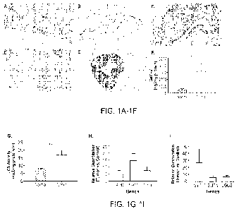
FIGS. 1A-11 are a series of immunostains and graphs demonstrating that CRC
tissue
strongly immunoreacts to an anti-13-1,4-GalT-V antibody and shows increased
LCS activity and
B4GALT5 expression. (FIG. 1A) Normal colon score 2 (20X magnification). (FIG.
1B)
Cytoplasmic staining of endothelium (20X). (FIG. 1C) Colon cancer case score 1
(20X). (FIG.
1D) Colon cancer case score 2 (20X). (FIG. 1E) Colon cancer case score 3
(20X). (FIG. 1F)
CRC tissues overexpressfI-1,4-GalT-V. Visibly normal and CRC tissues (50mg
each) were
homogenized in RIPA buffer and centrifuged at 1000 rpm. The supernatant was
used to measure
13-1,4-GalT-V mass by ELISA. This assay was then run in triplicate with N=10,
for both normal
and tumor samples. Averages L SEm values are shown, *P=0.0340. Unpaired t-test
was used to
determine statistical significance. (FIG. 1G) Increased LCS activity in
colorectal tumors.
Averages SEm, *P=0.0052. (FIG. 1H) AFC, NMTI , and TP53 genes showed increased
expression in tumors, compared to that of normal samples. (FIG. ILI) B4GAL T5
specifically
CA 03225489 2024- 1- 10
WO 2023/019186
PCT/US2022/074785
showed increased expression, while B4GALT6 and UGCG relatively did not.
Averages + SEM,
with -1\14 for both patient normal and tumor samples. Ordinary one-way ANOVA
was utilized
for statistical analysis.
FIGS. 2A-2G are a series of graphs demonstrating that LacCer mass is increased
in CRC
tissue. Visibly normal and CRC tissue (50mg each) were homogenized in
chloroform-methanol
(2:1), in the presence of internal sphingolipid standards. Lipid extracts were
then subjected to
LC-MS to investigate changes in the levels of sphingolipid species, in CRC or
normal tissues,
with (FIG. 2A) Cer, (FIG. 2B) DHCer, (FIG. 2C) GalCer and GlcCer, (FIG. 212))
dihydroGalCer/dihydroGlcCer, (FIG. 2E) LacCer, and (FIG. 2F) DHLacCer. Among
the GSL
investigated, only (FIG. 2E) LacCer levels were statistically and
significantly increased in
colorectal tumors (*P=0.0112). For Cer and dihydroGalCer/dihydroGlcCer, N=10
(normal) and
N=9 (tumor); DHCer, N=9 (normal) and N=10 (tumor); and for GalCer and GlcCer,
LacCer, and
DHLacCer N=10 (both normal and tumor) (FIG. 2G) Normal and tumor tissues were
evaluated
via LC-MS for sphingomyelin and DHSM levels. No statically significant
differences were
found in sphingomyelin values for normal vs. tumor. In contrast, tumor samples
showed
significantly elevated DHSM values compared to normal (**P=0.0059). For
sphingomyelin,
N=10 (normal) and N=6 (tumor); and for DHSM, N=10 (both normal and tumor).
Averages
SEm values, and unpaired t-tests were used to determine statistical
significance
FIGS. 3A-3H are a series of graphs and fluorescent stains demonstrating that
pharmacologic inhibition of GSL synthesis dose-dependently decreases
proliferation, and
reduces 13-1,4-GalT-V protein expression in HCT-116. D-PDMP exerted a dose-
and time-
dependent decrease at (FIG. 3A) 24 and (FIG. 3B) 96h in HCT-116 cell
proliferation, compared
to controls, with the maximal effective dose at 20 11M. *P 0.05, **P 0.01,
***P < 0.001. No
difference was found in UGCG immunofluorescence in (FIG. 3D) D-PDMP-treated
cells,
compared to that of (FIG. 3C) control at 24h. However, D-PDMP treatment at
(FIG. 3F) 24 and
(FIG. 311) 96h reduced GalT-V fluorescence compared to that of untreated
controls (E and G,
respectively).
FIGS. 4A-4H are a series of graphs demonstrating that D-PDMP treatment reduces
the
levels of several sphingolipids in HCT-116. HCT-116 cells were seeded (105)
onto sterilized
(100-mm2) plastic Petri dishes in 10mL of medium for 24h. Media was then
changed to 2%
21
CA 03225489 2024- 1- 10
WO 2023/019186
PCT/US2022/074785
serum-containing media, with and without D-PDMP (10pM). After 24h, media was
removed,
and total lipids were extracted, using hexane-isopropanol (3:2, by volume) in
the presence of
sphingolipid internal standards, and subject to MS. D-PDMP treatment
(designated D10 in the x-
axis) reduced levels of (FIG. 4A) Cer, (FIG. 4B) DHCer, (FIG. 4C)
monohexosylceramides,
(FIG. 40) dihydroGlc/galceramides, (FIG. 4E) dihexosylceramide, (FIG. 4F)
DHLacCer but
not that of (FIG. 4G) sphingomyelin nor (FIG. 411) DHSM, at lOpM, compared to
control
values (designated as C in the x-axis). Data represent averages SEm, N=3
biological replicates
for control and lOpM D-PDMP in (FIGS. 4A-4H), and the unpaired t-tested was
used for
statistical analysis. *P 0.05, **P 0.01] \
FIGS. 5A-5D are a series of immunostains demonstrating that CRC tissue
strongly
immunoreacts to an anti-f3-1,4-GalT-V antibody.
FIG. 6 shows an immunostaining of colon cancer section with GalT-V antibody.
FIG. 7 is a schematic representation showing the sphingolipid synthesis
pathway.
FIG. 8 is a graph demonstrating that treatment with GalT-V antibody against
GalT-V
dose dependently decreases proliferation ion HCT-116 cells. HCT-116 cells were
seeded (Ix104
cells/well) in a 96-well tray and grown in medium supplemented with 10% fetal
calf serum.
After 24hrs, fresh medium containing 31-1-thymidine (5 Ci/m1) plus various
dilutions of GalT-V
monoclonal antibody was added. Treatment with D-PDMP (5p.M) served as a
positive control.
Following incubation for 24 hrs, the incorporation of radioactivity into DNA
was measured by
scintillation spectrometry. GalT-V antibody dose-dependently decreased HCT-116
cell
proliferation P* 0.01, P***< 0.001 and P****< 0.0001 (N=5).
FIG. 9 is a graph demonstrating that treatment with GalT-V antibody against
GalT-V
dose-dependently decreases proliferation in mouse colorectal cancer cells.
Mouse colorectal
cancer cells were seeded (1x10⁴ cells /well) in a 96 well tray and grown in
minimum essential
medium supplemented with 10% fetal calf serum and processed as described in
FIG. 8 above. It
is noted that GalT-V antibody dose-dependently decreased MC-38 cell
proliferation (N=5).
FIGS. 10A-10I are a series of photo images and a graph demonstrating that VEGF
induced tube formation was mitigated by antibody against 13-GalT-V and
lactosylceramide
antibody. Human umbilical vein endothelial cells were incubated for one hour
with various
dilutions of13-GalT-V antibody or LacC er antibody, followed by treatment with
VEGF for 6
22
CA 03225489 2024- 1- 10
WO 2023/019186
PCT/US2022/074785
hours. Tube formation assays were then performed. The letters on the treatment
axis of the graph
(FIG 101) represent the treatments shown in FIGS. 10A-10H.
FIGS. 11A-11D are a series of photographs demonstrating that treatment with 13-
1,4GalT-V antibody or a biopolymer ¨encapsulated D-PDMP prevented tumor growth
in normal
female mice. Normal female mice (C57BL6) 32 weeks of age, were shaven. The
dorsal area was
cleaned with an alcoholic swab and100 L of colorectal cancer cells; HCT-116
(4x10⁶)
suspension was injected. On week later, 100 L of monoclonal antibody against B-
1,4 GalT-V
was injected daily into the site of the tumor cell injection (FIGS. 11A, 11B)
or a 3-1,4GalT-V
inhibitor (5mpk of a biopolymer ¨encapsulated D-PDMP (FIGS. 11C, 11D) for
three weeks.
The dorsal area was shaven again with Nair as hair had grown back and mice
photographed.
Note that no tumor growth was observed in the treated mice (FIGS. 11A-11)).
FIG. 12 is a schematic representation depicting an outline of reactions as to
how 13-
galactosyltransferase (f3-Ga1T-V) may contribute to colorectal cancer and the
novel approaches
herein to prevent it.
FIG. 13 is a schematic of the GalT-V antibody treatment model of Example 4.
FIG. 14 sets forth results showing treatment with GalT-V antibody did not
alter body
weight in NOD-SC1D mice.
FIG. 15 (includes FIGS. 15A-15B) shows treatment with GalT-V antibody dose-
dependently reduced tumor volume in NOD-SCID mice inoculated with HCT-116
cells.
FIG. 16 (includes FIGS. 16A-16C) shows optical imaging of mice bearing HCT-116
rectal orthotopic tumor.
FIG. 17 shows q-RT-PCR analysis of B4GALT-V, CEA, and NIVIT-1 gene expression
in
CRC mice.
FIG. 18 (includes FIGS. 18A-18B) show in FIG. 18A ELISA assays that
demonstrate
that treatment with GalT-V -Ab reduced the mass of GalT-V in plasma, and in
FIG. 18B HPTLC
and densitometric analysis demonstrating that treatment with GalT-V -Ab
reduced the mass of
LacCer in tumor tissue compared with placebo.
FIG. 19 shows cell surface localization of GalT-V antibody determined by
confocal
microscopy (Example 5).
23
CA 03225489 2024- 1- 10
WO 2023/019186
PCT/US2022/074785
FIG. 20 shows GalT-V antibody internalized (37 C) as determined by confocal
microscopy.
FIG. 21 shows [89Zr] GalT-V antibody binding in human coronary arterial
endothelial
cells (HCAEC) and human colorectal cancer cells.
FIG. 22 shows [89Zr] Gall-V antibody binding in human coronary arterial
endothelial
cells (HCAEC) and human colorectal cancer cells.
FIG. 23 shows D-PDMP inhibits zirconium tagged GalT-V antibody binding in
human
colorectal cancer cells.
FIG. 24 shows specificity of binding and internalization of [89Zr] GalT-V
antibody in
human colorectal cancer cells.
FIG. 25 shows time-dependent binding and internalization of [89Zr] GalT-V
antibody in
human colorectal cancer cells.
FIG. 26 and 27 show in vivo xenogen fluorescence images of human CRC tumor
bearing
mice at specified time periods.
FIG. 28 (includes FIGS. 28A-28C) shows distribution of CF-750 GalT-V antibody
fluorescence in individual tissues from a subcutaneous/xenograft tumor bearing
mice.
DETAILED DESCRIPTION
The invention is based, in part, on the finding that P-galactosyltransferase
(13-GalT-V)
plays a role in human CRC, and that its inhibition also mitigates tumor cell
proliferation.
Samples from colorectal cancer subjects were found to be immunoreactive to 13-
1,4-GalT-V
antibodies. In addition, 13-1,4-GalT-V mass, mRNA expression, enzymatic
activity, and GSL
end-product levels were assessed. The effect of a GSL glycosyltransferase
inhibitor in human
CRC cell lines was examined. These results described in detail in the examples
section provide
new insights into the pathogenesis of and reveal promising
detection/prognostic biomarkers for
CRC Applications include biomarkers useful for screening for cancer,
particularly colorectal
cancer and precursor tumors to colorectal cancers (e.g., advanced adenomas).
Compositions are
also described for use in the treatment of cancers, such as colorectal cancer
and the like.
24
CA 03225489 2024- 1- 10
WO 2023/019186
PCT/US2022/074785
Colorectal cancers include, without limitation, colon cancer, rectal cancer,
and
combinations thereof. Colorectal cancers include metastatic colorectal cancers
and non-
metastatic colorectal cancers. Colorectal cancers include cancer located in
the proximal part of
the colon cancer and cancer located the distal part of the colon. Colorectal
cancers include
colorectal cancers at any of the various possible stages known in the art,
including, e.g., Stage I,
Stage II, Stage III, and Stage IV colorectal cancers (e.g., stages 0, I, IIA,
II.B, IIC, IIIA, IIIB,
IIIC, IVA, IVB, and IVC). Colorectal cancers include all stages of the
Tumor/Node/Metastasis
(TNM) staging system. With respect to colorectal cancer, T can refer to
whether the tumor
grown into the wall of the colon or rectum, and if so by how many layers; N
can refer to whether
the tumor has spread to lymph nodes, and if so how many lymph nodes and where
they are
located; and M can refer to whether the cancer has spread to other parts of
the body, and if so
which parts and to what extent. Particular stages of T, N, and M are known in
the art. T stages
can include TX, TO, Tis, Ti, T2, T3, T4a, and T4b; N stages can include NX,
NO, Nla, Nib,
Ni c, N2a, and N2b; M stages can include MO, Ml a, and Ml b. Moreover, grades
of colorectal
cancer can include GX, Gl, G2, G3, and G4. Various means of staging cancer,
and colorectal
cancer in particular, are well known in the art summarized, e.g.,
cancer.net/cancer-
types/colorectal-cancer/stages.
In certain embodiments, the present disclosure includes screening of early
stage
colorectal cancer. Early stage colorectal cancers can include, e.g.,
colorectal cancers localized
within a subject, e.g., in that they have not yet spread to lymph nodes of the
subject, e.g., lymph
nodes near to the cancer (stage NO), and have not spread to distant sites
(stage MO). Early stage
cancers include colorectal cancers corresponding to, e.g., Stages 0 to II C.
Thus, colorectal cancers include, among other things, pre-malignant colorectal
cancer
(e.g., advanced adenomas) and malignant colorectal cancer. Methods and
compositions of the
present disclosure are useful for screening of colorectal cancer in all of its
forms and stages,
including without limitation those named herein or otherwise known in the art,
as well as all
subsets thereof. Accordingly, the person of skill in art will appreciate that
all references to
colorectal cancer provided here include, without limitation, colorectal cancer
in all of its forms
and stages, including without limitation those named herein or otherwise known
in the art, as
well as all subsets thereof.
CA 03225489 2024- 1- 10
WO 2023/019186
PCT/US2022/074785
Accordingly, in certain embodiments, pharmaceutical compositions in the
prevention and
treatment of cancer, e.g. colorectal cancer comprise administration of
inhibitors of
glycosphingolipid synthesis to subjects in need thereof.
In certain embodiments, a method of treating cancer, comprises administering
to a subject
in need thereof a composition comprising a therapeutically effective amount
of: (a) an antibody,
wherein the antibody specifically binds to a f3-1,4-galactosyltransferase-V
(f3-1,4-GalT-V)
epitope, the antibody comprising: (i) a heavy chain variable region sequence
having at least a
90% amino acid sequence identity to:
EVQLEQSGAELARPGASVKLSCRTSGYTFTNYWMQW1KQRPGQGLEWIGAMHPGRAYI
RYNQKFQGK A TL TADK SS ST AYMQLNSLA SEDS AVYYC ARWSDYDYWGQGTTLTVS S
(SEQ ID NO: 3), and, (ii) a light chain variable sequence having at least a
90% amino acid
sequence identity to:
DVVMTQTPPTLSVTIGQPASISCKSSQSLLDSDGKTYLNWLLQRPGQSPKRLIYLVSKLG
SGVPDRFTGSGSGTDFTLKISRVEAEDLGVYYCWQGTHFPRTFGGGTKLEIKR (SEQ ID
NO: 4), and, (b) a therapeutically effective amount of at least one inhibitor
of glycosphingolipid
synthesis. In certain embodiments, the antibody comprises a heavy chain
variable region
sequence having an amino acid sequence set forth in SEQ ID NO: 3. In certain
embodiments, the
antibody comprises a light chain variable region sequence having an amino acid
sequence set
forth in SEQ ID NO: 4.
In certain embodiments, a pharmaceutical composition comprises a
therapeutically
effective amount of: (i) an antibody comprising (a) a heavy chain variable
region sequence
nucleic acid sequence having at least a 90% sequence identity to SEQ ID NO: 3,
and (b) a light
chain variable region sequence nucleic acid sequence having at least a 90%
sequence identity to
SEQ Ti) NO. 2; and, (ii) a synthetic peptide comprising an amino acid sequence
having at least a
90% amino acid sequence to SEQ ID NO: 5; and, (iii) an adjuvant. In certain
embodiments, the
antibody comprises (a) a heavy chain variable region nucleic acid sequence
comprising SEQ ID
NO: 3, and (b) a light chain variable region nucleic acid sequence comprising
SEQ ID NO: 2;
and, the synthetic peptide amino acid sequence comprising SEQ ID NO: 5.
In certain embodiments, a pharmaceutical composition comprises a
therapeutically
effective amount of: (a) an antibody, wherein the antibody specifically binds
to a 13-1,4-
galactosyltransferase-V (f3-1,4-Ga1T-V) epitope, the antibody comprising: (i)
a heavy chain
26
CA 03225489 2024- 1- 10
WO 2023/019186
PCT/US2022/074785
variable region sequence having at least a 90% amino acid sequence identity
to:
EVQLEQSGAELARPGASVKLSCRTSGYTFTNYWMQW1KQRPGQGLEWIGAMHPGRAYI
RYNQKFQGKATLTADKSSSTAYMQLNSLASEDSAVYYCARWSDYDYWGQGTTLTVSS
(SEQ ID NO: 3), and, (ii) a light chain variable sequence having at least a
90% amino acid
sequence identity to:
DVVMTQTPPTLSVTIGQPASISCKSSQSLLDSDGKTYLNWLLQRPGQSPKRLIYLVSKLG
SGVPDRFTGSGSGTDFTLKISRVEAEDLGVYYCWQGTHFPRTFGGGTKLEIKR (SEQ ID
NO: 4), and, (b) a therapeutically effective amount of at least one inhibitor
of glycosphingolipid
synthesis. In certain embodiments, the antibody comprises a heavy chain
variable region
sequence having an amino acid sequence set forth in SEQ ID NO: 3. In certain
embodiments, the
antibody comprises a light chain variable region sequence having an amino acid
sequence set
forth in SEQ ID NO: 4.
In certain embodiments, the pharmaceutical compositions further comprise at
least one
inhibitor of glycosphingolipid synthesis comprises: D-threo-l-pheny1-2-d
ecanoylamino-3-
morpholino-l-propanol (D-PDMP), (1R,2R)-nonanoic acid(2-(2',3-dihydro-benzo
(1, 4) dioxin-
6'-y1)-2-hydroxy-1-pyrroli din-l-ylm ethyl-ethyl)- amide-L-tartaric acid salt
(Genz-123346), an
imide sugar, 1-pheny1-2-decanoylamino-3-morpholino-1-propanol (DMP), 1-pheny1-
2-
palmitoyl-amino-3-morpholino-1-propanol (PPMP), lipids, ceramides or
combinations thereof
are unencapsulated or encapsulated by a biodegradable polymer. In certain
embodiments, the
inhibitor of glycosphingolipid synthesis is D-threo-l-pheny1-2-decanoyl amino-
3-morpholino-1-
propanol (D-PDMP), unencapsulated or encapsulated in a biodegradable polymer
(BPD). In
certain embodiments, the biodegradable polymer consists of polyethylene glycol
and sebacic
acid.
In certain embodiments, the pharmaceutical compositions further comprise one
or more
secondary therapeutic agents. In certain embodiments, the one or more
secondary therapeutic
agents comprise: chemotherapeutic agents, anti-inflammatory agents,
cholesterol lowering
agents, insulin, antibodies, peptides, enzymes, adjuvants or combinations
thereof. In certain
embodiments, the pharmaceutical composition further comprises conjugating the
antibody to a
detectable agent, a radiotherapeutic agent, a toxin, a radioactive agent, a
dye, a peptide, a
polynucleotide or a nanoliposome. In certain embodiments, the nanoliposome
comprises a
therapeutic agent(s).
27
CA 03225489 2024- 1- 10
WO 2023/019186
PCT/US2022/074785
In certain embodiments, the pharmaceutical composition further comprises a
peptide
having at least a 90% sequence identity to IGAQVYEQVLRSAYAKRNSSVND (SEQ ID NO:
5).
In certain embodiments, the composition comprises a therapeutically effective
amount of
at least one inhibitor of glycosphingolipid synthesis and/or a therapeutically
effective amount of
the antibody which specifically binds to 131,4-Galactosyltransferase V (BGA),
isoforms or
peptides thereof.
In certain embodiments, the composition comprises a therapeutically effective
amount of
at least one inhibitor of glycosphingolipid synthesis and/or a therapeutically
effective amount of
an agent which modulates the expression or activity of131,4-
Galactosyltransferase V (BGA),
isoforms or peptides thereof. In certain embodiments, the agent inhibits the
expression or
activity of131,4-Galactosyltransferase V (BGA), isoforms or peptides thereof.
Examples of lipids include, without limitation fatty acids, free fatty acids,
cholesterol,
sterol esters, triglycerides, diglycerides, glycerides, wax esters, squalene,
ceramides, lipids,
phospholipids, glycolipids, linoleic acids or combinations thereof.
In other embodiments, a method of treating cancer, comprises administering to
a subject
in need thereof a therapeutically effective amount of an inhibitor of
glycosphingolipid synthesis,
lipids or combinations thereof In certain embodiments, the inhibitor of
glycosphingolipid
synthesis is D-threo-l-pheny1-2-decanoylamino-3-morpholino-l-propanol (D-
PDMP),
unencapsulated, unbound or encapsulated in a biodegradable polymer (BPD). In
certain
embodiments, the biodegradable polymer consists of polyethylene glycol and
sebacic acid.
Combination Therapies
In certain embodiments, the pharmaceutical compositions include an anti-cancer
agent,
such as, a chemotherapeutic agent, radiotherapy, a toxin or combinations
thereof.
In certain embodiments, In certain embodiments, the anti-cancer agent is a
chemotherapeutic or growth inhibitory agent, a targeted therapeutic agent, a T
cell expressing a
chimeric antigen receptor, an antibody or antigen-binding fragment thereof, an
antibody-drug
conjugate, an angiogenesis inhibitor, an antineoplastic agent, a cancer
vaccine, an adjuvant, and
combinations thereof In certain embodiments, the anti-cancer agent is a
chemotherapeutic or
28
CA 03225489 2024- 1- 10
WO 2023/019186
PCT/US2022/074785
growth inhibitory agent. For example, a chemotherapeutic or growth inhibitory
agent may
include an alkylating agent, an anthracycline, an anti-hormonal agent, an
aromatase inhibitor, an
anti-androgen, a protein kinase inhibitor, a lipid kinase inhibitor, an
antisense oligonucleotide, a
ribozyme, an antimetabolite, a topoisomerase inhibitor, a cytotoxic agent or
antitumor antibiotic,
a proteasome inhibitor, an anti-microtubule agent, an EGFR antagonist, a
retinoid, a tyrosine
kinase inhibitor, a hi stone deacetylase inhibitor, and combinations thereof.
In certain
embodiments, the anti-cancer agent is an adjuvant. Any substance that enhances
an anti-cancer
immune response, such as against a cancer-related antigen, or aids in the
presentation of a cancer
antigen to a component of the immune system may be considered an anti-cancer
adjuvant of the
present disclosure.
Chemotherapy: Cancer therapies in general also include a variety of
combination
therapies with both chemical and radiation based treatments. Combination
chemotherapies
include, for example, cisplatin (CDDP), carboplatin, procarbazine,
mechlorethamine,
cyclophosphamide, camptothecin, ifosfamide, melphalan, chlorambucil, busulfan,
nitrosurea,
dactinomycin, daunorubicin, doxorubicin, bleomycin, plicomycin, mitomycin,
etoposide (VP16),
tamoxifen, raloxifene, estrogen receptor binding agents, taxol, gemcitabien,
navelbine, famesyl-
protein transferase inhibitors, transplatinum, 5-fluorouracil, vincristine,
vinblastine and
methotrexate, Temazolomide (an aqueous form of DTIC), or any analog or
derivative variant of
the foregoing. The combination of chemotherapy with biological therapy is
known as
biochemotherapy. The chemotherapy may also be administered at low, continuous
doses which
is known as metronomic chemotherapy.
Yet further combination chemotherapies include, for example, alkylating agents
such as
thiotepa and cyclosphosphamide; alkyl sulfonates such as busulfan, improsulfan
and piposulfan;
aziridines such as benzodopa, carboquone, meturedopa, and uredopa;
ethylenimines and
methylamelamines including altretamine, triethylenemelamine,
trietylenephosphoramide,
triethiylenethiophosphoramide and trimethylolomelamine; acetogenins
(especially bullatacin and
bullatacinone); a camptothecin (including the synthetic analogue topotecan);
bryostatin;
callystatin; CC-1065 (including its adozelesin, carzelesin and bizelesin
synthetic analogues);
cryptophycins (particularly cryptophycin 1 and cryptophycin 8); dolastatin;
duocarmycin
(including the synthetic analogues, KW-2189 and CB1-TM1); eleutherobin,
pancratistatin; a
sarcodictyin, spongistatin; nitrogen mustards such as chlorambucil,
chlornaphazine,
29
CA 03225489 2024- 1- 10
WO 2023/019186
PCT/US2022/074785
cholophosphamide, estramustine, ifosfamide, mechlorethamine, mechlorethamine
oxide
hydrochloride, melphalan, novembichin, phenesterine, prednimustine,
trofosfamide, uracil
mustard; nitrosureas such as carmustine, chlorozotocin, fotemustine,
lomustine, nimustine, and
ranimnustine; antibiotics such as the enediyne antibiotics (e.g.,
calicheamicin, especially
calicheamicin gamma and calicheamicin omegall; dynemicin, including dynemicin
A;
bisphosphonates, such as clodronate; an esperamicin; as well as
neocarzinostatin chromophore
and related chromoprotein enediyne antiobiotic chromophores, aclacinomysins,
actinomycin,
authrarnycin, azaserine, bleomycins, cactinomycin, carabicin, carminomycin,
carzinophilin,
chromomycinis, dactinomycin, daunorubicin, detorubicin, 6-diazo-5-oxo-L-
norleucine,
doxorubicin (including morpholino-doxorubicin, cyanomorpholino-doxorubicin, 2-
pyrrolino-
doxorubicin and deoxydoxorubicin), epirubicin, esorubicin, idarubicin,
marcellomycin,
mitomycins such as mitomycin C, mycophenolic acid, nogalarnycin, olivomycins,
peplomycin,
potfiromycin, puromycin, quelamycin, rodorubicin, streptonigrin, streptozocin,
tub ercidin,
ubenimex, zinostatin, zorubicin; anti-metabolites such as methotrexate and 5-
fluorouracil (5-
FU); folic acid analogues such as denopterin, pteropterin, trimetrexate;
purine analogs such as
fludarabine, 6-mercaptopurine, thiamiprine, thioguanine; pyrimidine analogs
such as ancitabine,
azacitidine, 6-azauridine, carmofur, cytarabine, dideoxyuridine,
doxifluridine, enocitabine,
floxuridine; androgens such as calusterone, dromostanolone propionate,
epitiostanol,
mepitiostane, testolactone; anti-adrenals such as mitotane, trilostane; folic
acid replenisher such
as frolinic acid; aceglatone; aldophosphamide glycoside; aminoleyulinic acid;
eniluracil;
amsacrine; bestrabucil; bisantrene; edatraxate; defofamine; demecolcine;
diaziquone;
elformithine; elliptinium acetate; an epothilone; etoglucid; gallium nitrate;
hydroxyurea;
lentinan;lonidainine; maytansinoids such as maytansine and ansamitocins;
mitoguazone;
mitoxantrone; mopidanmol; nitraerine; pentostatin; phenamet; pirarubicin;
losoxantrone;
podophyllinic acid; 2-ethylhydrazide; procarbazine; PSK polysaccharide
complex; razoxane;
rhizoxin; sizofiran; spirogermanium; tenuazonic acid; triaziquone; 2,2',2"-
trichlorotriethylamine;
trichothecenes (especially T-2 toxin, verracurin A, roridin A and anguidine);
urethan; vindesine;
dacarbazine; mannomustine; mitobronitol; mitolactol; pipobroman; gacytosine;
arabinoside
("Ara-C"); cyclophosphamide; taxoids, e.g., paclitaxel and docetaxel
gemcitabine; 6-
thioguanine; mercaptopurine; platinum coordination complexes such as
cisplatin, oxaliplatin and
carboplatin; vinblastine; platinum; etoposide (VP-16); ifosfamide;
mitoxantrone; yincristine;
CA 03225489 2024- 1- 10
WO 2023/019186
PCT/US2022/074785
vinorelbine; novantrone; teniposide; edatrexate; daunomycin; aminopterin;
xeloda; ibandronate;
irinotecan (e.g., CPT-11); topoisomerase inhibitor RFS 2000;
difluorometlhylornithine (DMF0);
retinoids such as retinoic acid; capecitabine; carboplatin, procarbazine,
plicomycin, gemcitabien,
navelbine, farnesyl-protein transferase inhibitors, transplatinum; and
pharmaceutically
acceptable salts, acids or derivatives of any of the above. In certain
embodiments, one or more
chemotherapeutic may be used in combination with the compositions provided
herein.
Radiotherapy: Other factors that cause DNA damage and have been used
extensively in
cancer therapies include what are commonly known as gamma-rays, X-rays, and/or
the directed
delivery of radioisotopes to tumor cells. Other forms of DNA damaging factors
are also known
such as microwaves and UV-irradiation. It is most likely that all of these
factors effect a broad
range of damage on DNA, on the precursors of DNA, on the replication and
repair of DNA, and
on the assembly and maintenance of chromosomes. Dosage ranges for X-rays range
from daily
doses of 50 to 200 roentgens for prolonged periods of time (3 to 4 wk), to
single doses of 2000 to
6000 roentgens. Dosage ranges for radioisotopes vary widely, and depend on the
half-life of the
isotope, the strength and type of radiation emitted, and the uptake by the
neoplastic cells.
liniminotherapy: Immunotherapeutics, generally, rely on the use of immune
effector cells
and molecules to target and destroy cancer cells. The immune effector may be,
for example, an
antibody specific for some marker on the surface of a tumor cell. The antibody
alone may serve
as an effector of therapy or it may recruit other cells to actually effect
cell killing. The antibody
also may be conjugated to a drug or toxin (chemotherapeutic, radionuclide,
ricin A chain, cholera
toxin, pertussis toxin, etc.) and serve merely as a targeting agent.
Alternatively, the effector may
be a lymphocyte carrying a surface molecule that interacts, either directly or
indirectly, with a
tumor cell target. Various effector cells include cytotoxic T cells and NK
cells as well as
genetically engineered variants of these cell types modified to express
chimeric antigen
receptors.
The immunotherapy may be a cancer vaccine comprising one or more cancer
antigens, in
particular a protein or an immunogenic fragment thereof, DNA or RNA encoding
said cancer
antigen, in particular a protein or an immunogenic fragment thereof, cancer
cell lysates, and/or
protein preparations from tumor cells. As used herein, a cancer antigen is an
antigenic substance
present in cancer cells. In principle, any protein produced in a cancer cell
that has an abnormal
structure due to mutation can act as a cancer antigen. In principle, cancer
antigens can be
31
CA 03225489 2024- 1- 10
WO 2023/019186
PCT/US2022/074785
products of mutated oncogenes and tumor suppressor genes, products of other
mutated genes,
overexpressed or aberrantly expressed cellular proteins, cancer antigens
produced by oncogenic
viruses, oncofetal antigens, altered cell surface glycolipids and
glycoproteins, or cell type-
specific differentiation antigens. Examples of cancer antigens include the
abnormal products of
ras and p53 genes. Other examples include tissue differentiation antigens,
mutant protein
antigens, oncogenic viral antigens, cancer-testis antigens and vascular or
stromal specific
antigens. Tissue differentiation antigens are those that are specific to a
certain type of tissue.
Mutant protein antigens are likely to be much more specific to cancer cells
because normal cells
shouldn't contain these proteins. Normal cells will display the normal protein
antigen on their
MT-IC molecules, whereas cancer cells will display the mutant version. Some
viral proteins are
implicated in forming cancer, and some viral antigens are also cancer
antigens.
In certain embodiments, the method of treating cancer comprises administering
a
therapeutically effective amount of a synthetic peptide comprising an amino
acid sequence
having at least a 90% amino acid sequence to SEQ ID NO: 5 and at least one
adjuvant. In certain
embodiments, the synthetic peptide comprises SEQ ID NO: 5. Administration of a
therapeutically effective amount of SEQ ID NO: 5 generates an immune response
to13-1.4-
galactosyltransferase-V (13-1,4-GalT-V) In certain embodiments, an adjuvant is
also administered
to the subject.
In certain embodiments, the immunotherapy may be an antibody, such as part of
a
polyclonal antibody preparation, or may be a monoclonal antibody. The antibody
may be a
humanized antibody, a chimeric antibody, an antibody fragment, a bispecific
antibody or a single
chain antibody. An antibody as disclosed herein includes an antibody fragment,
such as, but not
limited to, Fab, Fab and F(ab')2, Fd, single-chain Fvs (say), single-chain
antibodies, disulfide-
linked Fvs (sdfv) and fragments including either a VL or VII domain.
In certain embodiments, the antibody comprises: (i) a heavy chain variable
region
sequence having at least a 90% amino acid sequence identity to:
EVQLEQSGAELARPGASVKLSCRTSGYTFTNYWMQWIKQRPGQGLEWIGAMHPGRAYI
RYNQKFQGKATLTADKSSSTAYMQLNSLASEDSAVYYCARWSDYDYWGQGTTLTVSS
(SEQ ID NO: 3), and, (ii) a light chain variable sequence having at least a
90% amino acid
sequence identity to:
DVVNITQTPPTLSVTIGQPASISCKSSQSLLDSDGKTYLNWLLQRPGQSPKRLIYLVSKLG
32
CA 03225489 2024- 1- 10
WO 2023/019186
PCT/US2022/074785
SGVPDRFTGSGSGTDFTLKISRVEAEDLGVYYCWQGTHFPRTEGGGTKLEIKR (SEQ ID
NO: 4).
In certain embodiments, one or more antibodies are administered to a subject
in need
thereof as a combination therapy. Examples of monoclonal antibodies that may
be used in
combination with the compositions provided herein include, without limitation,
trastuzumab
(anti-HER2/neu antibody); Pertuzumab (anti-HER2 mAb); cetuximab (chimeric
monoclonal
antibody to epidermal growth factor receptor EGFR); panitumumab (anti-EGFR
antibody);
nimotuzumab (anti-EGFR antibody); Zalutumumab (anti-EGFR mAb); Necitumumab
(anti-
EGFR mAb); MDX-210 (humanized anti-HER-2 bispecific antibody); MDX-210
(humanized
anti-HER-2 bispecific antibody); MDX-447 (humanized anti-EGF receptor
bispecific antibody);
Rituximab (chimeric murine/human anti-CD20 mAb); Obinutuzumab (anti-CD20 mAb):
Ofatumumab (anti-CD20 mAb); Tositumumab-I131 (anti-CD20 mAb); Ibritumomab
tiuxetan
(anti-CD20 mAb); Bevacizumab (anti-VEGF mAb); Ramucirumab (anti-VEGFR2 mAb);
Ranibizumab (anti-VEGF mAb); Aflibercept (extracellular domains of VEGFR1 and
VEGFR2
fused to IgG1 Fc); AMG386 (angiopoietin-1 and -2 binding peptide fused to IgG1
Fc);
Dalotuzumab (anti-IGF-1R mAb); Gemtuzumab ozogamicin (anti-CD33 mAb);
Alemtuzumab
(anti-Campath-1/CD52 mAb); Brentuximab vedotin (anti-CD30 mAb); Catumaxomab
(bispecific mAb that targets epithelial cell adhesion molecule and CD3);
Naptumomab (anti-5T4
mAb); Girentuximab (anti-Carbonic anhydrase ix); or Farletuzumab (anti-folate
receptor). Other
examples include antibodies such as Panorex TM (17-1A) (murine monoclonal
antibody); Panorex
(17-1A) (chimeric murine monoclonal antibody); BEC2 (ami-idiotypic mAb, mimics
the GD
epitope) (with BCG); Oncolym (Lym-1 monoclonal antibody); SMART M195 Ab,
humanized
13 1 LYlVI-1 (Oncolym), Ovarex (B43.13, anti-idiotypic mouse mAb); 3622W94 mAb
that
binds to EGP40 (17-1A) pancarcinoma antigen on adenocarcinomas: Zenapax (SMART
Anti-
Tac (IL-2 receptor); SMART M195 Ab, humanized Ab, humanized); NovoMAb-G2
(pancarcinoma specific Ab); TNT (chimeric mAb to histone antigens); TNT
(chimeric mAb to
histone antigens); Gliomab-H (Monoclonals-Humanized Abs); GNI-250 Mab; EMD-
72000
(chimeric-EGF antagonist); LymphoCide (humanized IL.L.2 antibody); and MDX-260
bispecific, targets GD-2, ANA Ab, SMART IDIO Ab, SMART ABL 364 Ab or ImmuRAIT-
CEA. Examples of antibodies include those disclosed in U.S. Pat. No.
5,736,167, U.S. Pat. No.
7,060,808, and U.S. Pat. No. 5,821,337.
33
CA 03225489 2024- 1- 10
WO 2023/019186
PCT/US2022/074785
Passive Ininntnotherapy: A number of different approaches for passive
immunotherapy of
cancer exist. They may be broadly categorized into the following: injection of
antibodies alone;
injection of antibodies coupled to toxins or chemotherapeutic agents;
injection of antibodies
coupled to radioactive isotopes; injection of anti-idiotype antibodies; and
finally, purging of
tumor cells in bone marrow.
Accordingly, in certain embodiments, a method of treating cancer, comprises
administering to a subject in need thereof a composition comprising a
therapeutically effective
amount of: (a) an antibody, wherein the antibody specifically binds to a13-1,4-
galactosyltransferase-V (f3-1,4-GalT-V) epitope, the antibody comprising: (i)
a heavy chain
variable region sequence having at least a 90% amino acid sequence identity
to:
EV QLEQ SGAELARPGAS VKL SCR"' SGY TFTN Y WMQW1KQRPGQGLEW IGAMHPGRAY1
RYNQKFQGKATLTADKSSSTAYMQLNSLASEDSAVYYCARWSDYDYWGQGTTLTVSS
(SEQ ID NO: 3), and, (ii) a light chain variable sequence having at least a
90% amino acid
sequence identity to:
DVVMTQTPPTLSVTIGQPASISCKSSQSLLDSDGKTYLNWLLQRPGQSPKRLIYLVSKLG
SGVPDRETGSGSGTDETLKISRVEAEDLGVYYCWQGTHFPRTFGGGTKLEIKR (SEQ ID
NO: 4), and, one or more secondary therapeutic agents. In certain embodiments,
the one or more
secondary therapeutic agents comprise: chemotherapeutic agents, anti-
inflammatory agents,
cholesterol lowering agents, insulin, antibodies, peptides, enzymes, adjuvants
or combinations
thereof. In certain embodiments, the pharmaceutical composition further
comprises conjugating
the antibody to a detectable agent, a radiotherapeutic agent, a toxin, a
radioactive agent, a dye, a
peptide, a polynucleotide or a nanoliposome. In certain embodiments, the
nanoliposome
comprises a therapeutic agent(s).
Other Agents: It is contemplated that other agents may be used in combination
with the
compositions provided herein to improve the therapeutic efficacy of treatment.
These additional
agents include immunomodulatory agents, agents that affect the upregulation of
cell surface
receptors and GAP junctions, cytostatic and differentiation agents, inhibitors
of cell adhesion, or
agents that increase the sensitivity of the hyperproliferative cells to
apoptotic inducers
Immunomodulatory agents include tumor necrosis factor; interferon alpha, beta,
and gamma; IL-
2 and other cytokines; F42K and other cytokine analogs; or MIP-1, MIP-lbeta,
MCP-1,
RANTES, and other chemokines. It is further contemplated that the upregulation
of cell surface
34
CA 03225489 2024- 1- 10
WO 2023/019186
PCT/US2022/074785
receptors or their ligands such as Fas/Fas ligand, DR4 or DRS/TRAIL would
potentiate the
apoptotic inducing abilities of the compositions provided herein by
establishment of an autocrine
or paracrine effect on hyperproliferative cells. Increases intercellular
signaling by elevating the
number of GAP junctions would increase the anti-hyperproliferative effects on
the neighboring
hyperproliferative cell population. In other embodiments, cytostatic or
differentiation agents can
be used in combination with the compositions provided herein to improve the
anti-
hyerproliferative efficacy of the treatments. Inhibitors of cell adhesion are
contemplated to
improve the efficacy of the present invention. Examples of cell adhesion
inhibitors are focal
adhesion kinase (FAKs) inhibitors and Lovastatin. It is further contemplated
that other agents
that increase the sensitivity of a hyperproliferative cell to apoptosis, such
as the antibody c225,
could be used in combination with the compositions provided herein to improve
the treatment
efficacy.
In further embodiments, the other agents may be one or more oncolytic viruses,
such as
an oncolytic viruses engineered to express a gene other than p53 and/or IL24,
such as a cytokine.
Examples of oncolytic viruses include adenoviruses, adeno-associated viruses,
retroviruses,
lentiviruses, herpes viruses, pox viruses, vaccinia viruses, vesicular
stomatitis viruses, polio
viruses, Newcastle's Disease viruses, Epstein-Barr viruses, influenza viruses
and reoviruses
In certain embodiments, hormonal therapy may also be used in conjunction with
the
present embodiments or in combination with any other cancer therapy previously
described. The
use of hormones may be employed to lower the level or block the effects of
certain hormones.
This treatment is often used in combination with at least one other cancer
therapy as a treatment
option or to reduce the risk of metastases
In some aspects, the additional anti-cancer agent is a protein kinase
inhibitor or a
monoclonal antibody that inhibits receptors involved in protein kinase or
growth factor signaling
pathways such as an EGFR, VEGFR, AKT, Erb1, Erb2, ErbB, Syk, Bcr-Abl, JAK,
Src, GSK-3,
PI3K, Ras, Raf, MAPK, MAPKK, mTOR, c-Kit, eph receptor or BRAF inhibitors.
Nonlimiting
examples of protein kinase or growth factor signaling pathways inhibitors
include Afatinib,
Axitinib, Bevacizumab, Bosutinib, Cetuximab, Crizotinib, Dasatinib, Erlotinib,
Fostamatinib,
Gefitinib, Imatinib, Lapatinib, Lenvatinib, Mubritinib, Nilotinib,
Panitumumab, Pazopanib,
Pegaptanib, Ranibizumab, Ruxolitinib, Saracatinib, Sorafenib, Sunitinib,
Trastuzumab,
Vandetanib, AP23451, Vemurafenib, MK-2206, GSK690693, A-443654, VQD-002,
CA 03225489 2024- 1- 10
WO 2023/019186
PCT/US2022/074785
Miltefosine, Perifosine, CAL101, PX-866, LY294002, rapamycin, temsirolimus,
everolimus,
ridaforolimus, Alvocidib, Genistein, Selumetinib, AZD-6244, Vatalanib, P1446A-
05, AG-
024322, ZD1839, P276-00, GW572016 or a mixture thereof.
It is contemplated that the additional cancer therapy can comprise an
antibody, peptide,
polypeptide, small molecule inhibitor, siRNA, miRNA or gene therapy which
targets, for
example, epidermal growth factor receptor (EGFR, EGFR1, ErbB-1, HERI), ErbB-2
(HER2/neu), ErbB-3/HER3, ErbB-4/HER4, EGFR ligand family; insulin-like growth
factor
receptor (IGFR) family, IGF-binding proteins (IGFBPs), IGFR ligand family (IGF-
1R); platelet
derived growth factor receptor (PDGFR) family, PDGFR ligand family; fibroblast
growth factor
receptor (FGFR) family, FGFR ligand family, vascular endothelial growth factor
receptor
(VECitR) family, VEGF family; HUE receptor family: 'IRK receptor family;
ephrin (EPH)
receptor family; AXE, receptor family; leukocyte tyrosine kinase (LTK)
receptor family; TIE
receptor family, angiopoietin 1, 2; receptor tyrosine kinase-like orphan
receptor (ROR) receptor
family; discoidin domain receptor (DDR) family; RET receptor family; KLG
receptor family;
RYK receptor family; MuSK receptor family; Transforming growth factor alpha
(TGF-a), TGF-
a receptor; Transforming growth factor-beta (TGF-13), TGF-13 receptor;
Interleukin 13 receptor
a1pha2 chain (1L13Ralpha2), Interleukin-6 (IL-6), 1L-6 receptor, Interleukin-
4, IL-4 receptor,
Cytokine receptors, Class I (hematopoietin family) and Class II (interferon/1L-
10 family)
receptors, tumor necrosis factor (TNF) family, TNF-a, tumor necrosis factor
(TNF) receptor
superfamily (TNTRSF), death receptor family, TRAIL-receptor; cancer-testis
(CT) antigens,
lineage-specific antigens, differentiation antigens, alpha-actinin-4, ARTC1,
breakpoint cluster
region-Abelson (Bcr-abl) fusion products, B-RAF, caspase-5 (CASP-5), caspase-8
(CASP-8),
beta-catenin (CTNNB1), cell division cycle 27 (CDC27), cyclin-dependent kinase
4 (CDK4),
CDKN2A, COA-1, dek-can fusion protein, EFTUD-2, Elongation factor 2 (ELF2),
Ets variant
gene 6/acute myeloid leukemia 1 gene ETS (ETC6-AML1) fusion protein,
fibronectin (FN),
GPNMB, low density lipid receptor/GDP-L fucose: beta-Dgalactose 2-alpha-
Lfucosyltraosferase
(LDLR/FUT) fusion protein, HLA-A2, arginine to isoleucine exchange at residue
170 of the
alpha-helix of the a1pha2-domain in the HLA-A2 gene (HLA-A*201-R1701), MLA-Al
1, heat
shock protein 70-2 mutated (HSP70-2M), KIAA0205, MART2, melanoma ubiquitous
mutated 1,
2, 3 (MUM-1, 2, 3), prostatic acid phosphatase (PAP), neo-PAP, Myosin class 1,
NFYC, OGT,
OS-9, pml-RARalpha fusion protein, PRDXS, PTPRK, K-ras (KRAS2), N-ras (NRAS),
HRAS,
36
CA 03225489 2024- 1- 10
WO 2023/019186
PCT/US2022/074785
RBAF600, S1RT2, SNRPD1, SYT-SSX1 or -SSX2 fusion protein, Triosephosphate
Isomerase,
BAGE, BAGE-1, BAGE-2, 3,4, 5, GAGE-1, 2, 3,4, 5, 6, 7, 8, GnT-V (aberrant N-
acetyl
giucosaminyl transferase V. MGATS), HERV-K-MEL, KK-LC, KM-I-IN-1, LAGE, LAGE-
1,
CTL-recognixed antigen on melanoma (CAMEL), MAGE-Al (MAGE-1), MAGE-A2, MAGE-
A3, MAGE-A4, MAGE-AS, MAGE-A6, MAGE-A8, MAGE-A9, MAGE-A10, MAGE-All,
MAGE-Al2, MAGE-3, MAGE-B1, MAGE-B2, MAGE-B5, MAGE-B6, MAGE-C1, MAGE-
C2, mucin 1 (1VIIUC1), MART-1/Melan-A (MLANA), gp100, gp100/Pme117 (S1LV),
tyrosinase
(TYR), TRP-1, HAGE, NA-88, NY-ESO-1, NY-ES0-1/LAGE-2, SAGE, Sp17, SSX-1, 2,
3,4,
TRP2-1NT2, carcino-embryonic antigen (CEA), Kallikfein 4, mammaglobm-A, A1,
prostate
specific antigen (PSA), prostate specific membrane antigen, TRP-1/gp75, TRP-2,
adipophilin,
interferon inducible protein absent in nielanorna 2 (AIM-2), B1NG-4, CPSF,
cyclin D1, epithelial
cell adhesion molecule (Ep-CAM), EpbA3, fibroblast growth factor-5 (FGF-5),
glycoprotein 250
(gp250intestina1 carboxyl esterase (iCE), alpha-feto protein (AFP), M-CSF, mdm-
2, MUCI, p53
(TP53), PBF, FRAME, PSMA, RAGE-1, RNF43, RU2AS, SOX10, STEAP11, survivin
(BIRCS), human telomerase reverse transcriptase (hTERT), telomerase, Wilms
tumor gene
(WT1), SYCP1, BRDT, SPANX, XAGE, ADAM2, PAGE-5, LIP1, CTAGE-1, CSAGE,
1V1AJA1, CAGE, BORIS, HOM-TES-85, AF15q14, HCA66I, LDHC, MORC, SGY-1, SP011,
TPX1, NY-SAR-35, FTHLI7, NXF2 TDRD1, TEX 15, FATE, TPTE, immunoglobulin
idiotypes, Bence-Jones protein, estrogen receptors (ER), androgen receptors
(AR), CD40, CD30,
CD20, CD19, CD33, CD4, CD25, CD3, cancer antigen 72-4 (CA 72-4), cancer
antigen 15-3 (CA
15-3), cancer antigen 27-29 (CA 27-29), cancer antigen 125 (CA 125), cancer
antigen 19-9 (CA
19-9), beta-human chorionic gonadotropin, 1-2 microglobulin, squamous cell
carcinoma antigen,
neuron-specific enoJase, heat shock protein gp96, GM2, sargramostim, CTLA-4,
707 alanine
proline (707-AP), adenocarcinoma antigen recognized by T cells 4 (ART-4),
carcinoembryogenic antigen peptide-1 (CAP-1), calcium-activated chloride
channel-2 (CLCA2),
cyclophilin B (Cyp-B), human signet ring tumor-2 (HST-2), Human papilloma
virus (EIPV)
proteins (HPV-E6, HPV-E7, major or minor capsid antigens, others), Epstein-
Barr vims (EBV)
proteins (EBV latent membrane proteins-LMP1, LMP2; others), Hepatitis B or C
virus proteins,
and HIV proteins
Glycosphingolipid Synthesis Inhibitors. In certain embodiments, the method
further
comprises administering at least one inhibitor of glycosphingolipid synthesis
comprising: D-
37
CA 03225489 2024- 1- 10
WO 2023/019186
PCT/US2022/074785
threo-l-pheny1-2-decanoylamino-3-morpholino-l-propanol (D-PDMP), (1R,2R)-
nonanoic
acid(2-(2',3'-dihydro-benzo (1, 4) dioxin-6'-y1)-2-hydroxy-1-pyrrolidin-1-
ylmethyl-ethyl)-
amide-L-tartaric acid salt (Genz-123346), an imide sugar, 1-pheny1-2-
decanoylamino-3-
morpholino-1-propanol (DMP), 1-pheny1-2-palmitoyl-amino-3-morpholino-1-
propanol (PPMP),
lipids, ceramides or combinations thereof are unencapsulated or encapsulated
by a biodegradable
polymer. In certain embodiments, the inhibitor of glycosphingolipid synthesis
is D-threo-1-
pheny1-2-decanoylamino-3-morpholino-1-propanol (D-PDMP) , including D-PDA,TP
that may be
admixed with a biodegradable polymer e.g. unencapsulated or encapsulated in a
biodegradable
polymer (BPD). In certain embodiments, the biodegradable polymer consists of
polyethylene
glycol and sebacic acid.
In certain embodiments, a composition comprises an inhibitor of
glycosphingolipid
synthesis, an inhibitor of glucosylceramide synthase or a combination thereof,
In certain
embodiments, a compound that inhibits glucosylceramide synthesis is an imino
sugar. In another
embodiment, the imide sugar is N-butyldeoxynojirimycin, N-
butyldeoxygalactonojirimycin (NB-
DGJ), or N-nonyldeoxynojirimycin. In another embodiment, the inhibitor of
glucosylceramide
synthesis is 1-pheny1-2-decanoylamino-3-morpholino-1-propanol (DMP), D-threo-l-
pheny1-2-
decanoylamino-3-morpholino-1-propanol and structurally related analogues
thereof. In another
embodiment, the inhibitor of glucosylceramide synthesis is 1-pheny1-2-
palmitoyl-amino-3-
morpholino-1-propanol (PPMP) and structurally related analogues thereof. In
certain
embodiments, the composition comprises D-threo-l-pheny1-2-decanoylamino-3-
morpholino-l-
propanol (D-PDMP), (1R,2R)-nonanoic acid(2-(2',3'-dihydro-benzo (1, 4) dioxin-
6'-y1)-2-
hydroxy-1-pyrrolidin-1-ylmethyl-ethyl)- amide-L-tartaric acid salt (Genz-
123346), an imide
sugar, 1-phenyl-2-decanoyl ami no-3-m orphol ino-l-propanol (DMP), 1 -phenyl-2-
pal mitoyl-
amino-3-morpholino-1-propanol (PPMP), lipids, ceramides or combinations
thereof are
encapsulated by a biodegradable polymer.
Pharmaceutical Formulations
In certain embodiments, the pharmaceutical compositions embodied herein are
formulated for systemic administration, e.g. oral, i.v., i.m. etc., comprises
a therapeutically
effective amount of inhibitor of glycosphingolipid synthesis, such as, a
therapeutically effective
amount of: (a) an antibody, wherein the antibody specifically binds to a 13-
1,4-
galactosyltransferase-V (f3-1,4-GalT-V) epitope, the antibody comprising: (i)
a heavy chain
38
CA 03225489 2024- 1- 10
WO 2023/019186
PCT/US2022/074785
variable region sequence having at least a 90% amino acid sequence identity
to:
EVQLEQ SGAELARPGASVKL SCRTSGYTFTNYWMQWIKQRPGQGLEWIGAMHPGRAYI
RYNQKFQGKATLTADKSSSTAYMQLNSLASEDSAVYYCARWSDYDYWGQGTTLTVSS
(SEQ ID NO: 3), and, (ii) a light chain variable sequence having at least a
90% amino acid
sequence identity to:
DVV1VITQTPPTLSVTIGQPASISCKSSQSLLDSDGKTYLNWLLQRPGQSPKRLIYLVSKLG
SGVPDRFTGSGSGTDFTLKISRVEAEDLGVYYCWQGTHFPRTFGGGTKLEIKR (SEQ ID
NO: 4), and/or a peptide comprising SEQ ID NO: 5 and/or for example, D-threo-1-
pheny1-2-
decanoylamino-3-morpholino-1-propanol (D-PDMP) including D-PDMP that may be
admixed
with a biodegradable polymer e.g. unencapsulated or encapsulated in a
biodegradable polymer
(BPD), or combinations thereof
The pharmaceutical compositions may include a pharmaceutically acceptable
carrier. The
term "pharmaceutically acceptable" means approved by a regulatory agency of
the Federal or a
state government or listed in the U.S. Pharmacopeia or other generally
recognized pharmacopeia
for use in animals, and more particularly in humans. The term "carrier" refers
to a diluent,
adjuvant, excipient, or vehicle with which the therapeutic is administered.
Such pharmaceutical
carriers can be sterile liquids, such as water and oils, including those of
petroleum, animal,
vegetable or synthetic origin, such as peanut oil, soybean oil, mineral oil,
sesame oil, olive oil,
gel (e.g., hydrogel), and the like. Saline is a preferred carrier when the
pharmaceutical
composition is administered intravenously. Saline solutions and aqueous
dextrose and glycerol
solutions can also be employed as liquid carriers, particularly for injectable
solutions.
Suitable pharmaceutical excipients include starch, glucose, lactose, sucrose,
gelatin, malt,
rice, flour, chalk, silica gel, sodium stearate, glycerol monostearate, talc,
sodium chloride, dried
skim milk, glycerol, propylene, glycol, water, ethanol, and the like. The
composition, if desired,
can also contain minor amounts of wetting or emulsifying agents, or pH
buffering agents. These
compositions can take the form of solutions, suspensions, emulsion, tablets,
pills, capsules,
powders, sustained-release formulations and the like. Oral formulation can
include standard
carriers such as pharmaceutical grades of mannitol, lactose, starch, magnesium
stearate, sodium
saccharine, cellulose, magnesium carbonate, etc. Examples of suitable
pharmaceutical carriers
are described in "Remington's Pharmaceutical Sciences" by E. W. Martin, the
contents of which
are hereby incorporated by reference in its entirety. Such compositions will
generally contain a
39
CA 03225489 2024- 1- 10
WO 2023/019186
PCT/US2022/074785
therapeutically effective amount of the pharmaceutical agents and/or
therapeutic compounds
(e.g., biopolymer encapsulated D-PDMP), in purified form, together with a
suitable amount of
carrier so as to provide the form for proper administration to the patient.
The formulation should
suit the mode of administration.
In embodiments, the pharmaceutical agents and/or therapeutic compounds are
administered locally as an immediate release or controlled release
composition, for example by
controlled dissolution and/or the diffusion of the active substance
Dissolution or diffusion
controlled release can be achieved by incorporating the active substance into
an appropriate
matrix. A controlled release matrix may include one or more of a biopolymer,
shellac, beeswax,
glycowax, castor wax, carnauba wax, stearyl alcohol, glyceryl monostearate,
glyceryl distearate,
glycerol palmitostearate, ethylcellulose, acrylic resins, dl-polylactic acid,
cellulose acetate
butyrate, polyvinyl chloride, polyvinyl acetate, vinyl pyrrolidone,
polyethylene,
polymethacrylate, methylmethacrylate, 2-hydroxymethacrylate, methacrylate
hydrogels, 1,3
butylene glycol, ethylene glycol methacrylate, and/or polyethylene glycols
and/or sebacic acid.
In a controlled release matrix formulation, the matrix material may also
include, e.g., hydrated
metylcellulose, carnauba wax and stearyl alcohol, carbopol 934, silicone,
glyceryl tristearate,
methyl acrylate-methyl methacrylate, polyvinyl chloride, polyethylene, and/or
halogenated
fluorocarbon. In certain embodiments, the controlled release composition is
achieved via a
transdermal patch.
The controlled release matrix may also be a hydrogel: a three-dimensional,
hydrophilic or
amphiphilic polymeric network capable of taking up large quantities of water.
The networks may
be composed of homopolymers or copolymers, which are insoluble due to the
presence of
covalent chemical or physical (e.g., ionic, hydrophobic interactions,
entanglements) crosslinks.
The crosslinks provide the network structure and physical integrity. Hydrogels
exhibit a
thermodynamic compatibility with water that allows them to swell in aqueous
media. The chains
of the network are connected in such a fashion that pores exist and that a
substantial fraction of
these pores are of dimensions between 1 nm and 1000 nm.
The hydrogels can be prepared by crosslinking hydrophilic biopolymers or
synthetic
polymers. Examples of the hydrogels formed from physical or chemical
crosslinking of
hydrophilic biopolymers, include but are not limited to, hyaluronans,
chitosans, alginates,
CA 03225489 2024- 1- 10
WO 2023/019186
PCT/US2022/074785
collagen, dextran, pectin, carrageenan, polylysine, gelatin, agarose,
(meth)acrylate-oligolactide-
PEO-oligolactide-(meth)acrylate, poly(ethylene glycol) (PEO), poly(propylene
glycol) (PPO),
PEO-PPO-PEO copolymers (Pluronics), poly(phosphazene), poly(methacrylates),
poly(N-
vinylpyrrolidone), PL(G)A-PEO-PL(G)A copolymers, poly(ethylene imine), and the
like. See
Hennink and van Nostrum, Adv. Drug Del. Rev. 54:13-36 (2002); Hoffman, Adv.
Drug Del. Rev.
43:3-12 (2002); Cadee et al., J Control. Release 78:1-13 (2002); Surini et
al., J. Control. Release
90:291-301 (2003); and U.S. Pat. No. 7,968,085, each of which is incorporated
by reference in
its entirety. These materials consist of high-molecular weight backbone chains
made of linear or
branched polysaccharides or polypeptides.
The amount of the pharmaceutical composition of the invention which will be
effective in
the treatment or prevention of atherosclerotic heart disease can be determined
by standard
clinical techniques. In addition, in vitro assays may optionally be employed
to help identify
optimal dosage ranges. The precise dose to be employed in the formulation may
also depend on
the route of administration, and the seriousness of the disease, and should be
decided according
to the judgment of' the practitioner and each patient's circumstances.
Effective doses may be
extrapolated from dose-response curves derived from the in vitro or animal
model test systems
described herein or known to one of skill in the art.
Dosages and Administration Regimens
The pharmaceutical agents and/or therapeutic compounds or compositions
containing
these agents/compounds may be administered in a manner compatible with the
dosage
formulation, and in such amount as may be therapeutically affective,
protective and
immunogenic.
The agents and/or compositions may be administered through different routes,
including,
but not limited to, oral, oral gavage, parenteral, buccal and sublingual,
rectal, aerosol, nasal,
intramuscular, subcutaneous, intradermal, intraosseous, dermal, and topical
The term parenteral
as used herein includes, for example, intraocular, subcutaneous,
intraperitoneal, intracutaneous,
intravenous, intramuscular, intraarticular, intraarterial, intrasynovi al,
intrastemal, intrathecal,
intralesional, and intracranial injection, or other infusion techniques.
In embodiments, the pharmaceutical agents and/or therapeutic compounds
formulated
according to the present invention are formulated and delivered in a manner to
evoke a systemic
41
CA 03225489 2024- 1- 10
WO 2023/019186
PCT/US2022/074785
response. Thus, in embodiments, the formulations are prepared by uniformly and
intimately
bringing into association the active ingredient with liquid carriers.
Formulations suitable for
administration include aqueous and non-aqueous sterile solutions, which may
contain anti-
oxidants, buffers, bacteriostats and solutes which render the formulation
isotonic with the blood
of the intended recipient, and aqueous and non-aqueous sterile suspensions
which may include
suspending agents and thickening agents. The formulations may be presented in
unit-dose or
multi-dose containers, for example, sealed ampoules and vials, and may be
stored in a freeze-
dried (lyophilized) condition requiring only the addition of the sterile
liquid carrier, for example,
water, immediately prior to use. Extemporaneous solutions and suspensions may
be prepared
from sterile powders, granules and tablets commonly used by one of ordinary
skill in the art.
The agents and/or compositions may be administered in different forms,
including, but
not limited to, solutions, emulsions and suspensions, microspheres, particles,
microparticles,
nanoparticles, liposomes, and the like.
The pharmaceutical agents and/or therapeutic compounds may be administered in
a
manner compatible with the dosage formulation, and in such amount as may be
therapeutically
effective, immunogenic and protective. The quantity to be administered depends
on the subject
to be treated, including, for example, the stage of the disease. Precise
amounts of active
ingredients required to be administered depend on the judgment of the
practitioner. However,
suitable dosage ranges are readily determinable by one skilled in the art and
may be of the order
of micrograms to milligrams of the active ingredient(s) per dose. The dosage
may also depend on
the route of administration and may vary according to the size of the host.
The pharmaceutical agents and/or therapeutic compounds should be administered
to a
subject in an amount effective to ameliorate, treat, and/or prevent the
disease. Specific dosage
and treatment regimens for any particular subject may depend upon a variety of
factors,
including the activity of the specific compound employed, the age, body
weight, general health
status, sex, diet, time of administration, rate of excretion, drug
combination, the severity and
course of the disease (including tumor size), condition or symptoms, the
subject's disposition to
the disease, condition or symptoms, method of administration, and the judgment
of the treating
physician. Actual dosages can be readily determined by one of ordinary skill
in the art.
42
CA 03225489 2024- 1- 10
WO 2023/019186
PCT/US2022/074785
Exemplary unit dosage formulations are those containing a dose or unit, or an
appropriate
fraction thereof, of the administered ingredient. It should be understood that
in addition to the
ingredients mentioned herein, the formulations of the present invention may
include other agents
commonly used by one of ordinary skill in the art.
In certain embodiments, the antibody which specifically binds toI31,4-
Galactosyltransferase V (BGA), isoforms or peptides thereof is administered
systemically or via
endoscopy or intra-anally.
In certain embodiments, the composition comprising a therapeutically effective
amount
of at least one inhibitor of glycosphingolipid synthesis and/or a
therapeutically effective amount
of an agent which modulates the expression or activity ofI31,4-
Galactosyltransferase V (BGA),
isoforms or peptides thereof is administered systemically or topically.
In certain embodiments, the composition comprising a therapeutically effective
amount
of at least one inhibitor of glyeosphingolipid synthesis and/or a
therapeutically effective amount
of an agent which modulates the expression or activity of 131,4-
Galactosyltransferase V (BGA),
isoforms or peptides thereof is co-administered to the subject The term "co-
administer" refers to
the simultaneous presence of two active agents in the blood of an individual.
Active agents that
are co-administered can be concurrently or sequentially delivered.
Typically in conventional systemically administered treatments, a
therapeutically
effective dosage should produce a serum concentration of compound of from
about 0.1 ng/ml to
about 50-100 ug/ml. The pharmaceutical compositions typically provide a dosage
of from about
0.001 mg to about 2000 mg of compound per kilogram of body weight per day. For
example,
dosages for systemic administration to a human patient can range from 1-10
lag/kg, 20-80 ug/kg,
5-50 ug/kg, 75-150 ug/kg, 100-500 ug/kg, 250-750 lag/kg, 500-1000 mg/kg, 1-10
mg/kg, 5-50
mg/kg, 25-75 mg/kg, 50-100 mg/kg, 100-250 mg/kg, 50-100 mg/kg, 250-500 mg/kg,
500-750
mg/kg, 750-1000 mg/kg, 1000-1500 mg/kg, 1500-2000 mg/kg, 5 mg/kg, 20 mg/kg, 50
mg/kg,
100 mg/kg, 500 mg/kg, 1000 mg/kg, 1500 mg/kg, or 2000 mg/kg. In an exemplary
embodiment,
an oral dosage for a human weighing 200 kg would be about 200 mg/day.
Pharmaceutical dosage
unit forms are prepared to provide from about 1 mg to about 5000 mg, for
example from about
100 to about 2500 mg of the compound or a combination of essential ingredients
per dosage unit
form.
43
CA 03225489 2024- 1- 10
WO 2023/019186
PCT/US2022/074785
In general, a therapeutically effective amount of the present compounds in
dosage form
usually ranges from slightly less than about 0.025 mg/kg/day to about 2.5
g/kg/day, preferably
about 0.1 mg/kg/day to about 100 mg/kg/day of the patient or considerably
more, depending
upon the compound used, the condition or infection treated and the route of
administration,
although exceptions to this dosage range may be contemplated by the present
invention. It is to
be understood that the present invention has application for both human and
veterinary use.
The agents and/or compositions are administered in one or more doses as
required to
achieve the desired effect. Thus, the agents and/or compositions may be
administered in 1, 2, to
3, 4, 5, or more doses. Further, the doses may be separated by any period of
time, for example
hours, days, weeks, months, and years.
The agents and/or compositions can be formulated as liquids or dry powders, or
in the
form of microspheres.
The agents and/or compositions may be stored at temperatures of from about -
100 C. to
about 25 C. depending on the duration of storage. The agents and/or
compositions may also be
stored in a lyophilized state at different temperatures including room
temperature. The agents
and/or compositions may be sterilized through conventional means known to one
of ordinary
skill in the art. Such means include, but are not limited to, filtration. The
composition may also
be combined with other anti-atherosclerotic therapeutic agents.
The amount of active ingredient that may be combined with carrier materials to
produce a
single dosage form may vary depending upon the host treated and the particular
mode of
administration. In embodiments, a preparation may contain from about 0.1% to
about 95% active
compound (w/w), from about 20% to about 80% active compound, or from any
percentage
therebetween.
In embodiments, the pH of the formulation may be adjusted with
pharmaceutically
acceptable acids, bases, or buffers to enhance the stability of the formulated
compound or its
delivery form.
In embodiments, the pharmaceutical carriers may be in the form of a sterile
liquid
preparation, for example, as a sterile aqueous or oleaginous suspension. Among
the acceptable
44
CA 03225489 2024- 1- 10
WO 2023/019186
PCT/US2022/074785
vehicles and solvents that may be employed are mannitol, water, Ringer's
solution and isotonic
sodium chloride solution.
In addition, sterile, fixed oils are conventionally employed as a solvent or
suspending
medium. For this purpose, any bland fixed oil may be employed including
synthetic mono- or to
diglycerides. Fatty acids, such as oleic acid and its glyceride derivatives
are useful in the
preparation of injectables, as are natural pharmaceutically-acceptable oils,
such as olive oil or
castor oil, especially in their polyoxyethylated versions. These oil solutions
or suspensions may
also contain a long-chain alcohol diluent or dispersant, or carboxymethyl
cellulose or similar
dispersing agents which are commonly used in the formulation of
pharmaceutically acceptable
dosage forms such as emulsions and or suspensions.
Other commonly used surfactants such as TWEENTm or SPANTM and/or other similar
emulsifying agents or bioavailability enhancers which are commonly used in the
manufacture of
pharmaceutically acceptable solid, liquid, Or other dosage forms may also be
used for the
purposes of formulation.
In embodiments, the agents and/or compositions can he delivered in an exosomal
delivery system. Exosomes are small membrane vesicles that are released into
the extracellular
environment during fusion of multivesicular bodies with plasma membrane.
Exosomes are
secreted by various cell types including hematopoietic cells, normal
epithelial cells and even
some tumor cells.
In certain embodiments, the biopolymer encapsulating the D-PDMP comprises
polyethelene glycol (PEG) and sebacic acid (SA). Both PEG and SA are FDA
approved. The
polyethylene glycol-sebacic acid (PEG-SA) copolymer can be prepared as
previously described
(Fu J, et al. Biomaterials. 2002; 23:4425-4433), Microparticles of D-PDMP
encapsulated by the
PEG-SA copolymer are prepared by modifying the single emulsion solvent
evaporation method.
For scintigraphic tracking of the biopolymer, the PEG polymer is radio-
iodinated with 45 mCi
(810 kBq) of (125I)NaI. The radiolabeled PEG was then incorporated into the
PEG-SA
biopolymer. The PEG-SA co-polymer can be prepared following the published
literature
procedure by Fu and coworkers (Id.). Briefly, sebacic acid prepolymer is made
by refluxing
sebacic acid (SA) in acetic anhydride followed by dryingunder high vacuum
(evaporation),
crystallized from dry toluene, washed with 1:1 anhydrous ethyl ether-petroleum
ether and finally
CA 03225489 2024- 1- 10
WO 2023/019186
PCT/US2022/074785
air dried PEG prepolymer is made by refluxing of polyoxyethylene dicarboxylic
acid in acetic
anhydride, volatile solvents are removed under vacuum. The solid mass is
extracted with
anhydrous ether and air dried. The poly(PEG-SA) co-block polymer is then
synthesized by the
melt polycondensation method and characterized by proton NMR. Note that this
copolymer has
been extensively characterized for the composition and structural
identity(Aich U, etal.
Glycoconjugate journal. 2010; 27: 445-459).
Encapsulation of D-PDMP in poly(PEG-SA) (to prepare polymer-encapsulated drug
subsequently referred to as BPD) followed by the melt polycondensation method
described
above for SA and PEG prepolymers but with the inclusion of D-PDMP at starting
ratios of poly
(PEG-SA) to D-PDMP of 70:30 by weight. Subsequently, microparticles are
prepared using a
single emulsion solvent evaporation method. Briefly, D-PDMP and PEG-SA are
dissolved in
chloroform (50 mg/mL) and emulsified into a 1.0% w/w poly(vinyl alcohol)
aqueous solution
under sonication condition keeping the temperature below 25 C. Particles are
hardened by
allowing chloroform to evaporate at room temperature while stirring for 12 h.
Particles are
collected and washed three times with double distilled water via
centrifugation at2,600×g
(30 min) and lyophilized for 48 h before it was ready to use.
In certain embodiments, the D-PDMP is encapsulated in a multilamellar lipid
vesicle
comprising covalent crosslinks between lipid bilayers, wherein at least two
lipid bilayers in the
multilamellar lipid vesicle are covalently crosslinked to each other by a
thiolated biopolymer. In
certain embodiments, the lipid bilayers are crosslinked via functionalized
lipids. In certain
embodiments, the one or more lipids comprise DOTAP, DOPE, DOBAQ, DOPC or
combinations thereof. In certain embodiments, the lipid is maleimide-
functionalized or modified
with dibenzocyclooctyne (DBCO). In certain embodiments, the thiolated
biopolymer is selected
from the group consisting of chitosan, polyglutamic acid, polyphosphazene,
polyethyleneimine,
polyalky acrylic acids (e.g polymethylmethacrylate, poly(ethylacrylic acid),
poly(propylacrylic
acid), or poly(butyl acrylic acid), HA, pegylated azide-modified
polyethylenimine, branched
polyethylenimine, and diazide. In certain embodiments, the thiolated
biopolymer comprises
multiple sulfhydryl moieties.
Also contemplated by the invention is delivery of the pharmaceutical agents
and/or
therapeutic compounds using nanoparticles. For example, the agents and/or
compositions
46
CA 03225489 2024- 1- 10
WO 2023/019186
PCT/US2022/074785
provided herein can contain nanoparticles having at least one or more agents
linked thereto, e.g.,
linked to the surface of the nanoparticle. A composition typically includes
many nanoparticles
with each nanoparticle having at least one or more agents linked thereto.
Nanoparticles can be
colloidal metals. A colloidal metal includes any water-insoluble metal
particle or metallic
compound dispersed in liquid water. Typically, a colloid metal is a suspension
of metal particles
in aqueous solution. Any metal that can be made in colloidal form can be used,
including gold,
silver, copper, nickel, aluminum, zinc, calcium, platinum, palladium, and
iron. In some cases,
gold nanoparticles are used, e.g., prepared from HAuC14. Nanoparticles can be
any shape and can
range in size from about 1 nm to about 10 nm in size, e.g., about 2 nm to
about 8 nm, about 4 to
about 6 nm, or about 5 nm in size. Methods for making colloidal metal
nanoparticles, including
gold colloidal nanoparticles from 1-1AuCh4, are known to those having ordinary
skill in the art.
For example, the methods described herein as well as those described elsewhere
(e.g., US Pat.
Publication Nos. 2001/005581; 2003/0118657; and 2003/0053983, which are hereby
incorporated by reference) are useful guidance to make nanoparticles.
In certain cases, a nanoparticle can have two, three, four, five, six, or more
active agents
linked to its surface. Typically, many molecules of active agents are linked
to the surface of the
nanoparticle at many locations. Accordingly, when a nanoparticle is described
as having, for
example, two active agents linked to it, the nanoparticle has two active
agents, each having its
own unique molecular structure, linked to its surface. In some cases, one
molecule of an active
agent can be linked to the nanoparticle via a single attachment site or via
multiple attachment
sites.
An active agent can be linked directly or indirectly to a nanoparticle
surface. For
example, the active agent can be linked directly to the surface of a
nanoparticle or indirectly
through an intervening linker.
Any type of molecule can be used as a linker. For example, a linker can be an
aliphatic
chain including at least two carbon atoms (e.g., 3, 4, 5, 6, 7, 8, 9, 10 or
more carbon atoms), and
can be substituted with one or more functional groups including ketone, ether,
ester, amide,
alcohol, amine, urea, thiourea, sulfoxide, sulfone, sulfonamide, and disulfide
to functionalities.
In cases where the nanoparticle includes gold, a linker can be any thiol-
containing molecule.
47
CA 03225489 2024- 1- 10
WO 2023/019186
PCT/US2022/074785
Reaction of a thiol group with the gold results in a covalent sulfide (--S--)
bond. Linker design
and synthesis are well known in the art.
In embodiments, the nanoparticle is linked to a targeting agent/moiety. A
targeting
functionality can allow nanoparticles to accumulate at the target at higher
concentrations than in
other tissues. In general, a targeting molecule can be one member of a binding
pair that exhibits
affinity and specificity for a second member of a binding pair. For example,
an antibody or
antibody fragment therapeutic agent can target a nanoparticle to a particular
region or molecule
of the body (e.g., the region or molecule for which the antibody is specific)
while also
performing a therapeutic function. In some cases, a receptor or receptor
fragment can target a
nanoparticle to a particular region of the body, e.g., the location of its
binding pair member.
Other therapeutic agents such as small molecules can similarly target a
nanoparticle to a
receptor, protein, or other binding site having affinity for the therapeutic
agent.
When the compositions of this invention comprise one or more additional
therapeutic or
prophylactic agents, the therapeutic agent and the additional agent should be
present at dosage
levels of between about 0.1 to 100%, or between about 5 to 95% of the dosage
normally
administered in a monotherapy regimen. The additional agents may be
administered separately,
as part of a multiple dose regimen, from the agents of this invention.
Alternatively, those
additional agents may be part of a single dosage form, mixed together with the
agents of this
invention in a single composition.
The administration of the pharmaceutical agents and/or therapeutic compounds
of the
invention elicits, for example, an anti-cancer response. Typically, the dose
can be adjusted within
this range based on, e.g., the subject's age, the subject's health and
physical condition, the
capacity of the subject's immune system to produce an immune response, the
subject's body
weight, the subject's sex, diet, time of administration, the degree of
protection desired, and other
clinical factors. Those in the art can also readily address parameters such as
biological half-life,
bioavailability, route of administration, and toxicity when formulating the
agents and/or
compositions of the invention.
EXAMPLES
Example 1: Laetosyleeramide synthase 13-1,4-GalT-V: a novel target for the
diagnosis and
therapy of human colorectal cancer
48
CA 03225489 2024- 1- 10
WO 2023/019186
PCT/US2022/074785
It was hypothesized that13-1,4-galactosyltransferase-V (13-1,4-GalT-V) may
well play a
role in human CRC, and that its inhibition may also mitigate tumor cell
proliferation. To test this
hypothesis, we acquired colorectal tissue samples from de-identified cancer
patients and
evaluated their immunoreactivity to 13-1,4-GalT-V antibodies. In addition, 13-
1,4-GalT-V mass,
mRNA expression, enzymatic activity, and GSL end-product levels were assessed.
Finally, the
effect of a GSL glycosyltransferase inhibitor in human CRC cell lines was also
examined. These
results provide new insights into the pathogenesis of and reveal promising
detection/prognostic
biomarkers for CRC. Furthermore, these findings demonstrate viable targets for
future CRC
therapeutics.
Materials and Methods
Immunohistochemical (IHC) localization of fl-1,4-GalT-V in human CRC tissue.
IHC was performed on archival tissue from The Johns Hopkins Pathology
Department
after approval from the Institutional Review Board on human subjects research.
Archival tissue
was sectioned from formalin fixed, paraffin embedded blocks of colon cancer
cases selected
from 2-3-year-old material Four-micron thick sections were cut and stained for
IHC analysis. f1-
1,4-GalT-V staining was performed on automated instruments using standard 11-
IC methods.
Briefly, sections were de-paraffinized, hydrated, and prepared for staining.
Sections were incubated with a13-1,4-GalT-V mouse monoclonal antibody raised
against
a GalT-V synthetic peptide, IGAQVYEQVLRSAYAKRNSSVND, SEQ ID NO: 5 (1:600
dilution) for 30 minutes. A secondary antibody anti-rabbit HRP was applied and
a brown signal
was developed using DAB chromogen detection (Cat # DS9800, Leica Biosystems).
Slides were
then counterstained with hematoxylin, washed, dehydrated, and cover slipped.
Evaluation of IHC
staining was done by a blinded pathologist. Stains were scored based on
intensity of staining and
area of tumor stained.
H-Score determination of human CRC tissue.
Tumor tissues were independently examined by two pathologists on the team, (RM
and
MA). Twenty-one cases of colon adenocarcinoma were selected from the Johns
Hopkins
Hospital archival data. Only colon cancer cases with primary origin were
included for this study.
For case selection, archival cases were reviewed and selected based on reports
from anatomic
49
CA 03225489 2024- 1- 10
WO 2023/019186
PCT/US2022/074785
pathologists. Unstained sections were obtained from the paraffin blocks and
RIC with a GalT-V
antibody was performed as described in the methods section. Results were
evaluated after
generating a scoring system of 0-3, where 0=no staining, 1=very weak staining,
2=moderate
staining, and 3=strong staining. Area of positive staining on the tumor was
also assessed, which
was used for H-score calculation.
Gene expression analysis.
Patient normal and CRC tissues were provided through collaboration with Dr.
Bert
Vogelstein (JHU). Total RNA was isolated from tissue samples using RNAqueous-
4PCR Kits
(Life Technologies), per the manufacturer's instructions TaqMan Gene
Expression Assays
(Applied Biosystems) were used to determine expression levels of adenomatous
polyposis coli
(APC, Hs01568269), N-myristoyltransferase-1
Hs00221506), tumor protein p53 (TP53,
Hs01034249), UDP-Ga1:f3GleNAcf3-1,4-galactosyltransferase, polypeptide 5 (13-
1,4-GalT-V,
Hs00191142), LTDP-Gal f3G1cNAc, f3-1,4-galactosyltransferase, polypeptide 6 (3-
1,4 GalT-VI,
Hs00191135), and UDP-glucose ceramide glucosyltransferase (UGCG, Hs00234293).
cDNA
was synthesized from isolated RNA using the High Capacity cDNA Reverse
Transcription Kit
(Life Technologies 4374966), per the manufacturer's protocol. TaqMan gene
expression assays
were performed by 7900HT Fast Real-Time PCR at The Genetic Resources Core
Facility (Johns
Hopkins Medical Institutions).
Measurement of lactosylceramide synthase (LCS) activity.
LCS activity, in visibly normal and cancer tissues, was measured according to
the
inventor's previously published method (20, 21). All assays, for both 10
normal and 10 tumor
samples, were run in triplicate, average values standard error of
measurements (SEm)
represented, and an unpaired t-test conducted to determine statistical
significance.
Measurement of 13-1,4-GalT-V mass in CRC tissue.
About 10mg of tissue was homogenized in radioimmunoprecipitation assay (RIPA)
buffer and centrifuged at 10,000 rpm. GalT-V mass was measured in the
supernatant using
ELISA, as previously published (22).
Liquid Chromatography-Mass Spectrometry (LC-MS).
CA 03225489 2024- 1- 10
WO 2023/019186
PCT/US2022/074785
Sphingolipid levels in human CRC tissue and in human cultured CRC cells (HCT-
116)
were measured by LC-MS, as described previously (23). Visibly normal and CRC
tissues (50
mg) were homogenized in chloroform-methanol (2:1), in the presence of
sphingolipid internal
standards. 105 HCT-116 cells were grown in 100 mm2 sterile, plastic Petri
dishes. Lipid extracts
were subject to LC-MS, as described (24, 25).
Determination of the effect of D-PDMP on IHC localization of
glycosyltransferases in
human CRC cells.
HCT-116 cells were seeded (104) onto sterilized glass coverslips, which were
then placed
in 6-well sterile plastic trays and grown for 24h in complete media Next, the
media was replaced
with 2mL of 2% serum-containing media, plus 1004 D-PDMF'. After incubation for
24 and 96h,
the media was removed, and cells were fixed with ethanol, washed, incubated
with antibody
against 13-1,4-GalT-V or UGCG, and photographed.
Determination of the effect of D-PDMP on cell proliferation.
HCT-116 cells were seeded (104/well) in 96-well sterile plastic plates, and
grown for 24h
in complete medium, with 10% fetal calf serum. The media was then replaced
with 2% serum-
containing media (100 L) plus 3H-thymidine (5 luCi/mL), with and without D-
PDMP. After
another 24h incubation, incorporation of3H-thymidine into DNA was measured by
scintillation
spectrometry.
Results
Human CRC tissues exhibit strongly positive immunoreactivity to a /1-1,4-GaIT-
V
antibody.
First, 13-1,4-GalT-V immunoreactivity in 24 human CRC specimens was examined,
using
a monoclonal antibody. Normal colon tissue showed cytoplasmic localization of
(3-1,4-GalT-Y
and strongly positive immunostained endothelial cells in large and small blood
vessels (FIG.
1B). In colonic adenocarcinomas (FIG. 1C), it was found that about 50% of
lesioned cells
expressed mild cytoplasmic 13-1,4-GalT-V immunoreactivity. H-score analysis
yielded a total
score of 100 in some cases but reached that of 200 in some cases (FIG. 1D) and
that of 300 in
other cases (FIG. 1E), (Table 1). Additional studies showed strong
immunoreactivity in
51
CA 03225489 2024- 1- 10
WO 2023/019186
PCT/US2022/074785
perinuclear areas, in association with antigen localization to the Golgi
apparatus, cytoplasm, and
to a lesser extent, the inner aspect of the cell surface.
Table 1: f3-1,4-Ga1T-V immunohistochemistry staining of tumor tissues.
tumor score Stealing
pattern
Case No 1; 2 3 Normal tissue H score
Nuclear/Cytoplasmic
1 50 0- 120
Cytop!asFMc
. .2. 8.0 0 50 Cytoplasmic
3 50 1 100 Cytopiasmc
4 100 2 300 Cytopasmic
20 1 40 Cytoplasmic
6 80 2 160
NucieariCyt.)plasm
7 80 160 Cy-
to.plasmc
8 90 2 180
Cvtop.asmic
9 100 1 300 Cytopasmic
60 2 120 NocLeariCytop!asrnic
11 80 2 180
t'7,ytpLasmic.
12 20 1 20 Cytopasmic
13 90 0 270 Cyte0asmic
14 90 2 180
Cytop4astrtic
. 15 30 na 30
NucleariCytoplasmic
19 30 1 60 Cyto0.asmic
. ... . . ..........................
17 60 2 240
C;:,/topasmi
19 90 0 370 Cytopasmic
19 90 1 180 NucleariCy-
tviasmic
85 0 170 CytoOasmic
H score is ceir.:ukited as % tumor intensity score X total area of tumor
5 Table 1 shows the assessments for 13-1,4-GalT-V immunohistochemistry
staining of
normal and CRC tissue sections. Review of the immuno-stains for Ga1T-V
revealed varying
degrees of positivity in the tumor cells: 1+ (15%; 3/20), 2+ (65%; 13/20), and
3+ (20%; 4/20).
Staining was mostly observed in the cytoplasm of tumors and very occasionally
in the nuclei of
tumor cells. Weak (1+) to moderate (2+) cytoplasmic staining was also observed
in adjoining
10 normal colonic mucosa in 79% cases (15/19), when available. In most
cases normal colonic
mucosa also stained at mild to moderate levels. The H score was determined as
% tumor
intensity score X total area of tumor_
CRC tissues have increased protein mass, activity (synthesis of
lactosylceramide), and
gene expression of 11-1 ,4-GalT-V.
15 EL1SA revealed a marked increase (approximately 6.5-fold) of13-1,4-
Galf -V in CRC
tissues, compared to visibly normal areas (i.e., "adjacent normals") from the
same tissue
52
CA 03225489 2024- 1- 10
WO 2023/019186
PCT/US2022/074785
specimens (FIG. 1F). A 2.25-fold increase was observed in lactosylceramide
synthase activity, in
CRC samples, compared to those of normal colon epithelia (**P=0.0052, FIG.
1G). Quantitative
RT-PCR further revealed elevated expression of several genes previously
associated with CRC,
e.g., adenomatous polyposis coli (APC) (26), N-myristoyltransferase 1 (ATMT/),
and tumor
protein p53 (TP53), compared to normal samples (FIG. 1H). Moreover, 13-1,4-
Ga1T-V
(B4GAL15) specifically showed increased expression, while f3-1,4-
galactosyltransferase,
polypeptide 6 (B4GALT6) and UDP-glucose-ceramidef3-1,4-glucosyltransferase
(LIGCG) did not
(FIG. 1I). LC-MS revealed insignificant but modestly elevated levels of
ceramide (Cer) (FIG.
2A), dihydroceramide (DHCer) (FIG. 2B), monoglycosylceramides (i.e.
galactosylceramide
(GalCer)) and glucosylceramide (GlcCer), FIG. 2C), dihydroGalCer/dihydroGlcCer
(FIG. 2D),
and dihydrolactosylceramide (DHLacCer) (FIG. 2F) in tumors, compared to normal
colon
specimens However, among the GSLs investigated, only lactosylceramide (LacCer)
levels were
statistically and significantly increased in CRC tissues (FIG. 2E, *P=0.0112).
CRC samples also
showed elevated dihydrosphingomyelin (DHSM), compared to normal tissue (FIG.
2G,
**P-0.0059), and similar levels of sphingomyelin (FIG. 2G).
Inhibition of GM. synthesis dose-dependently decreases proliferation of human
CRC
cells.
HCT-116 cells were treated with D-threo-l-phenyl-2-decanoylamino-3-morpholino-
l-
propanol (D-PDMP, FIG. 3A), a potent inhibitor of UDP-glucose-cer:
glucosyltransferase and
LCS/ 3-1,4-galactosyltransferase (GalT-V) activity (27, 28, 29). It was found
that in CRC cells,
D-PDMP exerted a dose- and time-dependent (FIGS. 3A, 3B) decrease in
proliferation,
compared to control cells, with the maximal effective dose at 2011M.
D-PDMP treatment reduces 11-1,4-GalT-V protein expression and activity (i.e.
sphingolipid synthesis) in human CRC cells.
No difference was found in UGCG immunofluorescence in D-PDMP treated cells
(FIG.
3D) compared to control (FIG. 3C) at 24h. However, D-PDMP treatment for 24
(FIG. 3F) and
96h (FIG. 3H) reduced GalT-V fluorescence compared to that of controls (FIGS.
3E and 3G,
respectively). LC-MS analysis of GSL derived from HCT-116 cells treated with
and without D-
PDMP (10RM) revealed reduced levels of Cer (FIG. 4A), DHCer (FIG. 4B),
monohexosylceramides (FIG. 4C), dihydroGalCer/dihydroGlcCer (FIG. 4D),
dihexosylceramide
53
CA 03225489 2024- 1- 10
WO 2023/019186
PCT/US2022/074785
(FIG. 4E), DHLacCer (FIG 4F) but not that of sphingomyelin (FIG. 4G) and DHSM
(FIG. 4H),
compared to control values.
Discussion
Several major findings emerge from this study. First, it was observed that 13-
1,4-GalT-V
expression, protein mass, and IHC staining were all markedly increased in
human CRC tissue, as
compared to visibly normal tissue. Second, the activity of 0-1,4-GalT-V (i.e.
synthesis of
lactosylceramide), in human CRC tumors, was statistically and significantly
higher, compared to
control tissue. Third, inhibition of glycosphingolipid synthesis decreased
immunostaining ofp-
1,4-GalT-V, consequently decreasing cell proliferation in cultured human CRC
(HCT-116) cells
Fourth, enhanced dihydrosphingolipid metabolism (FIG. 2G) was noted in tumor
tissues, leading
to significantly increased levels of dihydrosphingomyelin, compared to visibly
normal tissue.
Immunostaining using f3-1,4-GalT-V antibody also allowed pathologists to
clearly
distinguish normal epithelial cells (FIG. 1A) from cancerous epithelial cells
(FIGS. 1C-1E).
After staining, the tissues were assessed for adequacy of tumor and 20 cases
were selected where
adequate tumor tissue was available for evaluation. Review of the immunostains
for Gal T-V
revealed varying degrees of positivity in the tumor cells (Table 1): 1+ (15%;
3/20), 2+ (65%;
13/20), and 3+ (20%; 4/20). Staining was mostly observed in the cytoplasm of
tumors and very
occasionally in the nuclei of tumor cells. Weak (1+) to moderate (2+)
cytoplasmic staining was
also observed in adjoining normal colonic mucosa in 79% cases (15/19), when
available.
Moreover, computerized (Asperion program) analysis of 20 such tissues provided
a quantitative
estimate of the immunostaining index, yielding a substantial H-score of 100 to
300 (from a scale
of 0 to 300) (21). These observations provide evidence that 13-1,4-GalT-V
immunostaining could
indeed serve as a novel biomarker for CRC progression. Strong r3-1,4-GalT-V
immunostaining
of capillary endothelial cells was also observed consistent with the
inventor's previous studies
(30, 31). Within squamous epithelial cells, strong immunostaining was observed
in the
perinucl ear area, suggesting the antigen to be enriched within the Golgi
apparatus (data not
shown).
13-1,4-GalT-V immunostaining of the cytoplasm, both in control and CRC tumor
tissues,
provides evidence that 13-1,4-GalT-V must exist in a membrane-bound, as well
as soluble form.
Solubility would enable measurement of this antigen in various body fluids,
via noninvasive or
54
CA 03225489 2024- 1- 10
WO 2023/019186
PCT/US2022/074785
minimally invasive procedures. Since the brush border membrane in colonic
epithelial cells also
reacted positively to the antibody, this provides evidence that in colorectal
tissue, GalT-V may
be shed in exosomes.
Previous studies have shown that 13-1,4-galactosyltransferases share a common
stem
region. Since in f3-1,4-GalT-1 (lactosamine synthase), the stem region is
short; most of the
enzyme localizes to the Golgi, cytoplasm and relatively less with the plasma
membrane (32).
Additionally, it has been proposed that the number of hydroxylated amino
acids, constituting the
stem region, dictates the localization of a 0-1,4-galactosyltransferase,
although other factors may
determine the localization of this protein. Further studies are warranted to
examine whether
alterations in the stem region allow for plasma membrane localization off3-1,4-
GalT-V.
Moreover, enrichment of 13-1,4-GalT-V as observed by IHC, was further
substantiated by
quantitative measurement ofI3-1,4-GalT-V protein mass. In CRC tissues, 0-1,4-
GalT-V mass
was statistically and significantly increased, compared to control tissue
(FIG. IF). The activity of
lactosylceramide synthase (and thus, lactosylceramide mass) was also
significantly higher in
CRC tissues, compared to controls (FIG. 1G). In contrast, differences in the
levels of other
sphingolipids, in tumor versus control tissue, were not statistically
significant (FIGS. 2A-2G).
Another exciting finding of this study was that the dihydrosphingolipid
pathway (FIG. 7)
was significantly active in tumor tissue (FIG. 2G). For example, in tumor
tissue, it was observed
that the masses of dihydrocerami de, dihydroGlcCer/dihydroGalCer, and
dihydroLacCer were all
modestly elevated (but not significant). However, the most significant
difference between tumor
tissue and visibly normal tissue was that the level of dihydrosphingomyelin
was markedly
increased in tumor tissue (FIG. 2G). Recent studies suggest that
dihydrosphingolipids, e.g.
dihydroceramide, play an important role in autophagy (33, 34). However, the
role of
dihydrosphingomyelin, in human CRC, needs to be explored.
The quantitative RT-PCR studies herein revealed increased B4GALT5 gene
expression in
tumor tissue, but relatively not for that of its isoform B4GALT6, or a homolog
UGCG (FIG. 1I).
These observations validated previously reported specific increases in13-1,4-
GalT-V but not f3-
1,4-GalT-VI in CRC tumors, using serial analysis of gene expression technology
(30). Thus,
increased 11-1,4-GalT-V gene and protein expression is specific for CRC
tissue, and may well
serve as a biomarker to diagnose this disease and measure drug response.
Previously, several
CA 03225489 2024- 1- 10
WO 2023/019186 PCT/US2022/074785
other genes have been suggested to serve as CRC biomarkers, including MIT],
APC, and TP53
(35, 36, 37). Accordingly, the expression of these genes were analyzed and
upregulation was
observed in all three, in CRC tumor versus normal tissue (FIG. 1H). Thus, 0-
1,4-GalT-V
upregulation could feasibly be added to the three-biomarker panel, thus
enhancing the predictive
value of CRC diagnosis.
In HCT-116 cells, 13-1,4-GalT-V immunostaining was also observed within the
inner
layer of the plasma membrane and cytosol Moreover, immunoreactivity increased
as the cells
grew from 24h to 96h (FIGS. 3A-3H), correlating with increased expression of
13-1,4-GalT-V. In
contrast, inhibition of glycosphingolipid synthesis with D-PDMP, significantly
diminished GalT-
V immunoreactivity in HCT-116 cells (FIGS. 3A-3H), thus providing evidence
that treatment
may decrease p-1,4-GalT-V protein mass and dihydroglycosylceramides/LacCer
levels in these
cells, and consequently decrease cell proliferation (FIGS. 3A-3H).
Immunostaining of UGCG in HCT-116 cells was peiformed. It was similarly found
that
this antigen/enzyme localized to the perinuclear area and cytoplasm. However,
treatment with D-
PDMP did not decrease immunoreactivity to an anti-UGCG antibody. Thus,
treatment might
have reduced glucosylceramide levels in HCT-116 cells by inhibiting enzyme
activity.
It was found that in addition to human CRC tumors, HCT-116 cells also had an
active
dihydrosphingolipid pathway. Treatment with D-PDMP decreased levels of all
dihydrosphingoli pi d species in our study, except for those of
dihydrosphingomyelin. Additional
mechanistic studies are warranted to address this observation further. It was
also found that the
net result of D-PDMP treatment was a dose-dependent decrease in HCT-116 cell
proliferation.
In summary, this study showed a specific increase in the gene expression,
protein levels,
and enzymatic activity ofP-1,4-GalT-V, concurrent with increased
lactosylceramide mass, in
human CRC. These molecular and biochemical data were further substantiated by
IHC and
pathology studies. These findings demonstrate that levels of 13-1,4-GalT-V and
lactosylceramide
in human liquid biopsy specimens could complement other currently used
biomarkers (e.g.,
1V7vIT1, ARC and TP 53), thus increasing the positive predictive value for
CRC. Last, inhibition of
glycosphingolipid synthesis may be a novel approach to treat human colorectal,
and possibly
other types of cancer.
References
56
CA 03225489 2024- 1- 10
WO 2023/019186
PCT/US2022/074785
(1) P. Favoriti, G. Carbone, M. Grego, et al., Worldwide burden of
colorectal cancer. a
review, Updates Surg. 68(1) (2016) 7-11. https://doi.org/10.1007/s13304-016-
0359-y.
(2) H. Brenner, M. Kloor, C.P. Pox, Colorectal cancer, Lancet. 383(9927)
(2014) 1490-1502.
https://doi.org/10.1016/S0140-6736(13)61649-9.
(3) J. Ferlay, I. Soerjomataram, R. Dikshit, et al., Cancer incidence and
mortality worldwide:
sources, methods and major patterns in GLOBOCAN 2012, Int. J. Cancer. 136(5)
(2015) E359-
E386. https://doi.org/1O.1002/ijc.29210.
(4) M. Arnold, M. S. Sierra, M. Laversanne, et al., Global patterns and
trends in colorectal
cancer incidence and mortality, Gut. 66(4) (2017) 683-691.
https://doi.org/10.1136/gutjn1-2015-
310912.
(5) H. Brenner, C. Stock, M. Hoffmeister, Colorectal cancer screening: the
time to act is
now, BMC Med. 13 (2015) 262. https://doi.org/10.1186/s12916-015-0498-x.
(6) T. Tanaka, M. Tanaka, T. Tanaka, et al., Biomarkers for colorectal
cancer, Int. J. Mol.
Sci. 11(9) (2010) 3209-3225. https://doi.org/10.3390/ijms11093209.
(7) S. Hundt, U. Haug, H. Brenner, Blood markers for early detection of
colorectal cancer: a
systematic review, Cancer Epidemiol. Biomarkers Prey. 16(10) (2007) 1935-1953.
https://doi.org/10.1158/1055-9965.EP1-06-0994.
(8) K. Simon, V. Balchen, Colorectal cancer development and
advances in screening. Clin.
Interv. Aging. 11(2016) 967-976. https://doi.org/10.2147/CIA.S109285.
(9) T.F. Imperiale, D.F. Ransohoff, S.H. Itzkowitz, et al., Multitarget
stool DNA testing for
colorectal-cancer screening, N. Engl. J. Med. 370 (2014) 1287-1297.
https://doi.org/10.1056/NEJMoa1311194.
(10) S.I. Hakomori, W.T. Murakami, Glycolipids of hamster fibroblasts and
derived
malignant-transformed cell lines, Proc. Natl. Acad. Sci. USA. 59(1) (1968) 254-
61.
(11) J.W. Dennis, S. Laferte, Oncodevelopmental expression of ¨G1cNAc beta 1-
6Man alpha
1-6Man beta 1¨branched asparagine-linked oligosaccharides in murine tissues
and human
breast carcinomas, Cancer Res. 49(4) (1989) 945-950.
57
CA 03225489 2024- 1- 10
WO 2023/019186
PCT/US2022/074785
(12) S. Hakomori, Tumor malignancy defined by aberrant glycosylation and
sphingo(glycol)lipid metabolism, Cancer Res. 56(23) (1996) 5309-18.
(13) M. Garcia-Barros, N. Coant, J.P. Truman, et al., Sphingolipids in colon
cancer, Biochim.
Biophys. Acta. 1841(5) (2014) 773-782.
https://doi.org/10.1016/j.bbalip.2013.09.007.
(14) Y.Y. Liu, R.A. Hill, Y.T. Li, Ceramide glycosylation catalyzed by
glucosylceramide
synthase and cancer drug resistance, Adv. Cancer Res. 117 (2013) 59-89.
https://doi.org/10.1016/B978-0-12-394274-6.00003-0.
(15) B. Segui, N. Andrieu-Abadie, J.P. Jaffrezou, et al., Sphingolipids as
modulators of cancer
cell death: potential therapeutic targets, Biochim. Biophys. Acta. 1758(12)
(2006) 2104-2120.
hitps.//doi.org/10.1016/j .bbamem.2006.05.024.
(16) J. Arango, M. Pierce, Comparison of N-acetylglucosaminyltransferase V
activities in
Rous sarcoma-transformed baby hamster kidney (RS-BHK) and BHX_ cells, I Cell
Biochem.
37(2) (1988) 225-31. https://doi.org/10.1002/jcb.240370209.
(17) K. Shirane, T. Sato, K. Segawa, et al., Involvement of beta-1,4-
galactoslytransferase V in
malignant transformation-associated changes in glycosylation, Biochem.
Biophys. Res.
Commun. 265(2) (1999) 434-8. https://doi.org/10.1006/bbrc.1999.1684.
(18) S. Xu, S. Zhang, C. Chen, et al., Over-expression of beta-1,4-
galactosyltransferase V
increases the growth of astrocytoma cell line, J. Exp. Clin. Cancer Res. 21(3)
(2002) 409-414.
(19) Y. Wei, F. Zhou, Y. Ge, et al., 31,4-Galactosyltransferase V regulates
self-renewal of
glioma-initiating cell, Biochem. Biophys. Res. Commun. 396(3) (2010) 602-607.
https://doi.org/10.1016/j .bbrc.2010.04.110.
(20) S. Chatterjee, Assay of lactosylceramide synthase and comments on its
potential role in
signal transduction, Methods Enzymol. 311(2000) 73-81. https://doi .
org/10.1016/S0076-
6879(00)11068-7.
(21) Varghese, F., Bukhari, A.B., Melhotra, R., et al., 2014. IHC profiler: an
open source
plugin for the quantitative evaluation and automated scoring of
immunohistochemistry images of
human tissue samples. PLoS One 9(5), e96801.
https://doi.org/10.1371/journal.pone.0096801.
(22) D. Bedja, W. Yan, V. Lad, et al., Inhibition of glycosphingolipid
synthesis reverses skin
inflammation and hair loss in ApoE -/- mice fed western diet, Sci. Rep. 8
(2018) 11463.
58
CA 03225489 2024- 1- 10
WO 2023/019186
PCT/US2022/074785
https://doi.org/10.1038/s41598-018-28663-9.
(23) Chatterjee, S., Alsaeedi, N., Hou, J., et al., 2013. Use of a
glycolipid inhibitor to
ameliorate renal cancer in a mouse model. PLoS One. 8(5), e63726.
https://doi.org/10.1371/j ournal.pone.0063726.
(24) N.J. Haughey, R.G. Cutler, A. Tamara, et al., Perturbation of
sphingolipid metabolism
and ceramide production in HIV-dementia, Ann. Neurol. 55(2) (2004) 257-267.
https://doi.org/10.1002/ana.10828.
(25) V.V.R. Bandaru, J. Troncoso, D. Wheeler, et al., ApoE4 disrupts sterol
and sphingolipid
metabolism in Alzheimer's but not normal brain, Neurobiol. Aging. 30(4) (2009)
591-599.
https://doi .org/10.1016/j .neurobiolaging.2007.07.024.
(26) M. El Zoghbi, L.C. Cummings, New era of colorectal cancer screening,
World J.
Gastrointest. Endosc. 8(5) (2016) 252-258.
https.//doi.org/10.4253/vjge.v815.252.
(27) S. Chatterjee, Sphingolipids in atherosclerosis and vascular biology,
Arterioscler.
Thromb. Vase. Biol. 18(10) (1998) 1523-1533.
(28) S. Chatterjee, T. Cleveland, W.Y. Shi, et al., Studies of the action
of ceramide-like
substances (D- and L-PDMP) on sphingolipid glycosyltransferases and purified
lactosylceramide synthase, Glycoconj. J. 13(3) (1996) 481-486.
https://doi.org/10.1007/BF00731481.
(29) S. Chatterjee, A.K. Bhunia, A. Snowden, et al., Oxidized low density
lipoproteins
stimulate galactosyltransferase activity, ras activation, p44 mitogen
activated protein kinase and
c-fos expression in aortic smooth muscle cells, Glycobiology. 7(5) (1997) 703-
710.
(30) A. Kolmakova, M. Raj esh, D. Zang, et al., VEGF recruits lactosylceramide
to induce
endothelial cell adhesion molecule expression and angiogenesis in vitro and in
vivo, Glycoconj.
J. 26(5) (2009) 547-558. https://doi.org/10.1007/s10719-008-9206-9.
(31) M. Rajesh, A. Kolmakova, S. Chatterjee, Novel role of lactosylceramide in
vascular
endothelial growth factor-mediated angiogenesis in human endothelial cells,
Circ. Res. 97(8)
(2005) 796-804. https://doi .org/10.1161/01.RES.0000185327.45463. A8.
(32) P.K. Qasba, B. Ramakrishnan, E. Boeggeman, Structure and function of beta
-1,4-
galactosyltransferase, Curr. Drug Targets. 9(4) (2008) 292-309.
https://doi.org/10.2174/138945008783954943.
(33) W. Zheng, J. Kollmeyer, H. Symolon, et al., Ceramides and other bioactive
sphingolipid
59
CA 03225489 2024- 1- 10
WO 2023/019186
PCT/US2022/074785
backbones in health and disease: lipidomic analysis, metabolism and roles in
membrane
structure, dynamics, signaling and autophagy. Biochim. Biophys. Acta. 1758(12)
(2006) 1864-
84. https://doi.org/10.1016/j.bbamem.2006.08.009.
(34) M.M. Siddique, Y. Li, B. Chaurasia, et al., Dihydroceramides: from bit
players to lead
actors, J. Biol. Chem. 290(25) (2015) 15371-9.
https://doi.org/10.1074/jbc.R115.653204.
(35) C.E. Ducker, J.J. Upson, K.J. French, et al., Two N-myristoyltransferase
isozymes play
unique roles in protein myristylation, proliferation, and apoptosis, Mol.
Cancer Res. 3(8) (2005)
463-476. https://doi.org/10.1158/1541-7786.MCR-05-0037.
(36) T. Armaghany, J.D. Wilson, Q. Chu, et al., Genetic alterations in
colorectal cancer,
Gastrointest. Cancer Res. 5(1) (2012) 19-27.
(37) X.L. Li, J. Zhou, Z.R. Chen, etal., p53 mutations in colorectal cancer-
molecular
pathogenesis and pharmacological reactivation, World J. Gastroenterol. 21(1)
(2015) 84-93.
https://doi.org/10.3748/wjg.v21.i1.84.
(38) N. S. Radin, J.A. Shayman, J. Inokuchi, Metabolic effects of inhibiting
glucosylceramide
synthesis with PDMP and other substances, Adv. Lipid. Res. 26 (1993) 183-213.
(39) J. Inokuchi, I. Mason, N.S. Radin, Antitumor activity via inhibition of
glycosphingolipid
biosynthesis. Cancer Lett. 38(1-2) (1987) 23-30. https://doi.org/10.1016/0304-
3835(87)90196-0.
(40) J. Inokuchi, M. Jimbo, K. Momosaki, et al., Inhibition of experimental
metastasis of
murine Lewis lung carcinoma by an inhibitor of glucosylceramide synthase and
its possible
mechanism of action, Cancer Res. 50(20) (1990) 6731-6737.
Example 2: Immunotherapy using lactosylceramide synthase (11-1,4 GalT-V)
antibody to
prevent colorectal cancer (CRC) in vivo.
It was hypothesized that 13-1,4GalT-V plays an important role in human CRC,
and
manipulating this enzyme may well mitigate tumor cell proliferation and
metastasis-by way of
inhibiting angiogenesis. To test this hypothesis, mouse monoclonal antibodies
were prepared
against13-1,4GalT-V and determined its effect on proliferation and
angiogenesis in human and
mouse CRC cells, human umbilical vein endothelial cells and a mouse xenograft
model of
human CRC.
CA 03225489 2024- 1- 10
WO 2023/019186
PCT/US2022/074785
Materials and Methods
Monoclonal antibody against a f3-1,4galactosyltransferase(GalT-V) peptide
having the
amino acid sequence(IGAQVYEQVLRSAYAKRNSSVND, SEQ ID NO: 5) was prepared and
characterized in regard to its titer, and use in EL1SA, western immunoblot
assay and
immunopreccipitation of mouse and human tissues. A human colorectal cancer
cell line (HCT-
116) was a gift from late, Dr. David Huso, Dept of Comparative Medicine, from
the institution.
A mouse colorectal cancer line MC-38 and thin tissue sections from mouse
colorectal cancer
were a gift from Dr. Cindy Sears, Dept of Oncology at the institution. Human
umbilical vein
endothelial cells were purchased from Clonetics and cultured in appropriate
growth medium.
Human microvascular endothelial cells were a gift from Ms. Stephanie Brindal
in the
department. Vascular endothelial growth factor was purchased from R and D Inc.
Matrigel and
all other reagents were from Sigma- Aldrich. Biopolymer -encapsulated D-PDMP
was prepared
as described (6).
Cell Proliferation Assay
lx 104 HCT-116 cells and MC-38 cells were seeded in 96 well -sterile plastic
trays and
grown in 1004 of Dulbecco's minimum essential medium containing 10% fetal calf
serum in
5% CO2 -air humidified incubator at 37 for 24hrs. Medium was replaced with
fresh medium
supplemented with 2% serum and 5 CitmL of (3H) thymidine. Increasing dilutions
of13-1,4
GalT-V Antibody were added to the wells. Human IgG or mouse IgG served as a
negative
control and D-PDMP (10 M) or luM BPD served as a positive control in these
experiments.
Following incubation for 24hrs, the experiment was terminated and the
incorporation of (3H)
thymidine into DNA was measured by scintillation spectrometry.
Angiogenesis assay
Angiogenesis assays were performed using a commercially available kit from
Chemicon
Inc. (7).
Measurement of p-1, 4 GalT-V activity
The activity of13-1, 4 GalT-V was measured in cells treated with and without
13-1,4 GaIT-
V antibody as described previously (14).
Measurement of Glycosphingolipids
61
CA 03225489 2024- 1- 10
WO 2023/019186
PCT/US2022/074785
The mass of GSL was measured by quantitative HPTLC as described (9).
Prevention of tumor cell growth.
Normal male and female mice (C57 BL-6) were purchased from The Jackson
Laboratory
and were fed regular mice chow. Semi-confluent culture of HCT-116 cells were
harvested and
the cell pellet resuspended in medium supplemented with Matrigel in a ratio of
70:30 (by
volume). The dorsal hair of the mouse was removed with the use of Nair, a hair
remover (Church
and Dwight Co.), and the skin area devoid of hair was cleaned with an
alcoholic swab. Next,
4x10⁶ HCT cell suspension was injected subcutaneously. One week later, either
1000 of GalT-V
Antibody or 100 L of BPD (1 mg/kg. Body weight) was injected subcutaneously at
the sight of
tumor cell injection daily for 3 weeks. Since hair grew back in the shaven
area, hair was removed
using Nair to expose the skin area and mice were photographed and recorded.
Immunohistochemistry
Thin tissue sections were cut from mice colorectal cancer tumor tissue and
subject to
immunohistochemical staining with (3-1,4 GalT-V Antibody as described
previously (8). Briefly,
sections were de-paraffinized, hydrated, and incubated with a13-1,4 GalT-V
antibody (1:600
dilution) for 30 min. A second antibody, anti-rabbit ARP was applied and a
brown signal was
developed using DAB chromogen detection (Leica Biosy stems). Slides were next
counterstained
with hematoxylin, washed, dehydrated, cover slipped and photographed (8).
Results
GalT-V Antibody is taken up by endothelial cells and IICT-116 cells in a time -
dependent manner.
In human microvascular endothelial cells, fluorescent tagged GalT-V was taken
up at
4 C. As the temperature was shifted to 37 C, a time-dependent increase was
observed in the
uptake of GalT-V Antibody into the cytoplasm and in the perinucl ear area -
representing the
Golgi apparatus. Pre-incubation of cells with an excess amount of GalT-V
peptide. Similarly,
HCT-116 cells also took up fluorescent tagged 3 -1,4 GalT-V Antibody in a
similar temperature
and time dependent manner. These studies showed that f3-1,4 GalT-V Antibody is
taken up and
internalized by human endothelial cells and HCT-116 cells in a time and
temperature -dependent
manner.
62
CA 03225489 2024- 1- 10
WO 2023/019186
PCT/US2022/074785
GalT-V Antibody inhibits proliferation in human and mouse colorectal cancer
cells.
thymidine uptake studies revealed that in human colorectal cancer cells GalT-V
antibody dose-dependently decreases cell proliferation and this inhibition was
within the range of
inhibition observed using a pharmacological inhibitor of 13-1,4 GalT-V; D-
PDMP.
Similarly, 13-1,4 GalT-V Antibody also dose-dependently inhibited the
proliferation of
mouse colorectal cells (FIG. 8); MC-38 (FIG. 9). Inhibition of cell
proliferation was not
observed when cells were incubated with mouse IgG.
GalT-V Antibody inhibits VEGF-induced angiogenesis in human umbilical vein
endothelial cells.
FIGS. 10A-10H show photographs of tube formation/angiogenesis in HUVEC's. FIG.
101 shows a corresponding quantification of angiogenesis. Incubation of HUVEC'
s with
VEGF/FGF markedly increased tube formation/angiogenesis (FIG. 10B) compared
with control
(FIG. 10A). Treatment with 13-1,4 Gall-V Antibody exerted a dose-dependent
decrease in
VEGF-induced angiogenesis in HUVEC's (FIGS. 10C10-F) but not by rabbit IgG
(FIG. 10G).
Gaff-V Antibody prevents tumor growth in mice.
Seven days after mice were injected with HCT-16 cells, they were treated with
and
without 13-1, 4 GalT-V Antibody daily for three weeks. In parallel another
group- of mice were
treated with BPD (5mpk) daily for three weeks at the site of tumor cell
injection. It was observed
that treatment with either 13-1, 4 GalT-V Antibody (FIGS. 11A, 11B) or BPD
(FIGS. 11C, 11D)
completely prevented tumor growth and progression.
Discussion
This study lead to the following conclusions. (i). That treatment with 13-1,4
Gal T-V
Antibody dose-dependently decreased proliferation in cultured human colorectal
cancer cells and
mouse colorectal cancer cells. (ii). In human umbilical vein endothelial cells
treatment with p -
1,4 GalT-V Antibody dose-dependently decreased VEGF-induced angiogenesis.
(iii). In a mouse
xenograft model of colorectal cancer, treatment with 13-1,4 GalT-V Antibody
and BPD prevented
tumor growth.
13-1,4 GalT-V is a member of a large family of galactosyltransferases whose
function is to
transfer galactose from UDP- galactose to glucosylceramide to form Lactosyl
cerami de (1). It
63
CA 03225489 2024- 1- 10
WO 2023/019186
PCT/US2022/074785
also transfers galactose to GlcNAc 13-1,6 mannose group of the highly branched
N glycans,
which are characteristic of tumor cells (2, 3). Of these products, LC has been
shown to serve as
an independent mitogenic agent, angiogenesis agent as well implicated in cell
migration,
apoptosis and cell adhesion (4). Most importantly, LC serves as a surrogate to
mediate the action
of growth factors e.g. VEGF, FGF, PDGF, EGF and a pro-inflammatory cytokine
TNF leading
to the above phenotypes depending on the cell type (4). Importantly, these
growth factor and
INFia-induced phenotypes can be mitigated by the use of pharmacological agents
BPD and D-
PDMP (6, 9) and GalT-V gene manipulation in vitro (7), and in vivo (15) (FIG.
12). This is the
first report wherein (3-1, 4 GalT-V immunotherapy is shown to be effective in
reducing the
growth and proliferation of colorectal cancer. The studies herein show that in
human colorectal
cancer tissue endothelial, cells 13-1, 4 Gall-V mRNA levels are specifically
increased and
consequently the mass of f3-1, 4 GalT-V protein is also increased in a cancer
stage -dependent
manner. While in normal colonic cells 13-1, 4 GalT-V is found mostly in the
cytoplasm/Golgi, in
colorectal cancer it is present in addition in the brush border membrane in
epithelial cells and to
some extent in the cell membrane in human colorectal cancer cells (8). This
allows the 0-1, 4
GalT-V antibody to bind to the GalT-V protein. At 37 C the fluorescent tagged
13-1, 4 GalT-V
antibody wase observed to bind to the antigen largely in the cytoplasm in
normal human
endothelial cells and HCT-116 cells (8). Since large doses of unlabeled
antibody competitively
inhibited the uptake of tagged antibody, suggest that 13-1, 4 GalT-V antibody
binding and uptake
is specific in cultured colorectal cells (data not shown).
The inventor's previous studies has shown that LC generated due to growth
factor -
induced activation of 13-1,4 GalT-V, activated NAD(P)H oxidase to generate
reactive oxygen
species which served as a signaling intermediate in a mitogen- activated
protein kinase /c-fos
pathway leading to cell proliferation (4) In this study it was observed that
treatment with 13-1,4
GalT-V antibody reduces 13-1,4 GalT-V enzyme activity and LC mass and
therefore mitigated
cell proliferation in HCT-116 cells.
Monoclonal antibodies are a specific type of antibodies/proteins made for
therapeutic use.
Such antibodies can be used in a targeted therapy to block an abnormal protein
in a cancer cell.
Monoclonal antibody can be used in immunotherapy as some of them attach
specifically to a
cancer cell expressing that protein. Thus by identifying cancer cells allows
the immune system to
attack and destroy it. Another type of antibodies affect cancer growth by
releasing the brakes on
64
CA 03225489 2024- 1- 10
WO 2023/019186
PCT/US2022/074785
the immune system so it can destroy the cancer cell. Studies show that
programmed cell death
(PD1)/programmed cell death ligand (PDL-1), CTLA-4 pathways are critical to
the immune
system's ability to control cancer growth. Such pathways are termed "immune
check points".
Several types of cancer judicially use these pathways to escape the immune
system. In contrast,
immune check point inhibitors e.g. penbrolizumab (Keytruda) etc. are useful in
identifying the
blockade by the PD-Li protein which acts like a protective shield in cancer
cells. Recently,
penbrolizumab has been approved by the FDA to treat tumors metastatic cancers
which cannot
be treated by chemotherapy as well as Merkel skin cancer due to Merkel polyoma
virus
infection. Thus this check point inhibitor can target any tumor in the body
and are therefore
called tumor agnostic treatments. Nivolumab is a drug approved to treat CRC
with M91-H or
dMMR in patients after chemotherapy has failed. lnterferons and interleukins
have also been
used to fight cancer and to develop immune system to generate cells which
destroy cancer. The 2
year success rate for immunotherapy has been the highest (82%) in stage IV
lymphomas and
only 38% for patients with stage IV CRC. The 13-1,4 GalT-V monoclonal antibody
is of the IgG
type and may well serve in targeted therapy to block the excess amounts of 3-
1,4 GalT-V protein
found in human CRC tissue and decorating the inner aspect of cell membrane in
endothelial cells
and cytoplasm in cultured human CRC cells( 8). Use of humanized 13-1,4 GalT-V
monoclonal
antibody alone or in combination with BPD and/or other CRC drugs e.g.
Nivolumab may well
the be future direction of research to accelerate our therapeutic efforts to
mitigate CRC. Since 13-
1,4 GalT-V protein is also over expressed in renal cancer (9) may find
multiple uses of this
immunotherapeutic approach.
References
1. Kolmakova A, Chatterjee S. Platelet derived growth factor recruits
lactosylceramide to
induce cell proliferation in UDPGal:GlcCer:131¨AGalactosyltransferase (GalT-V)
mutant
Chinese hamster ovary cells. Glycoconj J 2005; 22: 401-7.
2. J. Arango, M. Pierce, Comparison of N-acetylglucosaminyltransferase V
activities in Rous
sarcoma-transformed baby hamster kidney (RS-BHIC) and BIM cells, J. Cell.
Biochem., 37 (2)
(1988), pp. 225-231.
CA 03225489 2024- 1- 10
WO 2023/019186
PCT/US2022/074785
3. K. Shirane, T. Sato, K. Segawa, etal. Involvement of beta-1,4-
galactoslytransferase V in
malignant transformation-associated changes in glycosylation, Biochem.
Biophys. Res.
Commun., 265 (2) (1999), pp. 434-438.
4. Chatterjee, S. and Pandey, A. The Yin and Yang of lactosylceramide
metabolism:
implications in cell function. Biochem.Biophys Acta. 1780:370-382, 2007.
5. Chatterjee, S. Kolmakova, A and Mohanraj,R. Regulation of
lactosylceramide synthase;
implications as a drug target. Curr. Drug Targets 9: 272-281, 2008.
6. D. Bedj a, W. Yan, V. Lad, et al. Inhibition of glycosphingolipid
synthesis reverses skin
inflammation and hair loss in ApoE -/- mice fed western diet, Sci. Rep., 8
(2018), p. 11463.
7. M. Raj esh, A. Kolmakova, S. Chatterjee. Novel role of lactosylceramide in
vascular
endothelial growth factor-mediated angiogenesis in human endothelial cells,
Circ. Res., 97 (8)
(2005), pp 796804.
8. S. Chatterjee, et al. Lactosylceramide synthase B-1,4GalT-V : A novel
target for the
diagnosis and therapy of human colorectal cancer. Bio Chem. Biophys. Res.
Comm. 2019; 508,
380-400.
9. S. Chatterjee, N. Alsaeedi, J. Hou, et al., Use of a glycolipid
inhibitor to ameliorate renal
cancer in a mouse model, PloS One, 8 (5) (2013), Article e63726.
10. P. Favoriti, G. Carbone, M. Grego, et al. Worldwide burden of
colorectal cancer: a review,
Updates Surg, 68(1) (2016), pp. 7-11.
11. S.I. Hakomori, W.T. Murakami, Glycolipids of hamster fibroblasts and
derived malignant-
transformed cell lines, Proc. Natl. Acad. Sci. U.S.A., 59 (1) (1968), pp. 254-
261.
12. J.W. Dennis, S. Laferte, Oncodevelopmental expression of ¨GleNAc
beta 1-6Man alpha 1-
6Man beta 1¨branched asparagine-linked oligosaccharides in murine tissues and
human breast
carcinomas, Cancer Res., 49 (4) (1989), pp. 945-950.
13. S. Hakomori, Tumor malignancy defined by aberrant glycosylation and
sphingo(glycol)lipid metabolism, Cancer Res., 56 (23) (1996), pp. 5309-5318.
14. Y.Y. Liu, R.A. Hill, Y.T. Li, Ceramide glycosylation catalyzed
by glucosylceramide
synthase and cancer drug resistance, Adv. Cancer Res., 117 (2013), pp. 59-89.
66
CA 03225489 2024- 1- 10
WO 2023/019186
PCT/US2022/074785
15. Y. Wei, F. Zhou, Y. Ge, et al., 131,4-Galactosyltransferase V
regulates self-renewal of
glioma-initiating cell, Biochem. Biophys. Res. Commun., 396 (3) (2010), pp.
602-607.
Example 3: fl-I,4GalT-V monoclonal antibody to mitigate atherosclerosis and
reduction in
body weight in type II diabetic mice(db/db).
Male Type II diabetic mice (db/db) aged 11 weeks were purchased from the
Jackson
Laboratory. They we raised on a normal mice chow and water. At the age of 30
weeks, mice
were divided into two groups. The first group of mice (Placebo)were given
saline (100 L) daily
by intra peritoneal injection for 6 weeks. The second group of mice were
treated with GalT-V
antibody (1mg/kg body weight) for the same duration. At the end of 36 weeks of
age mice ,were
weighed and various tissues were collected and frozen away until further
analysis. Next, to
extract total lipids, about 10mg of liver tissue was excised, (internal
standards of C12 ceramide
and C12 sphingomylein were added to check recovery) and homogenized in
acetonitrile and
centrifuged at 10001-pm for min The clear supernatant was saved and the
pellets was subject to
repeated extraction. The pooled supernatant were dried in N2 and reconstituted
in chloroform-
methanol (2: lv/v). A suitable aliquot of the lipid extract was loaded onto a
high performance thin
layer chromatography plate. Also a misinterpretation of standard neutral
lipids consisting of
cholesterol ester, triglyceride and cholesterol were also loaded to calibrate
the plate. The plate
was developed using Heptane:ethyl-ether and acetic acid(65:16: lv/v) as
solvent. The lipids were
identified by exposing the plate with iodine vapors and photographed
Quantification of the mass
of lipids was carried out by densitometric analysis using standard curves for
individual lipids and
using a two -tailed parametric t-test.
The results demonstrated that diabetic mice treated with GalT-V monoclonal AB
had
significantly reduced level of cholesterol as compared to placebo group of
mice. Treatment also
markedly reduced the level of triglycerides (neutral fat) as compared to
placebo group of mice.
In addition treatment reduced mice body weight by ¨20%.
Example 4:
We have shown that in cultured human CRC cells (HCT-116), treatment with GalT-
V
AB dose-dependently mitigated cell proliferation and angiogenesis [1].
Additionally, we have
shown the enrichment of CF-750 tagged GalT-V-Ab in a xenograft tumor in NOD-
SC1D mice
model of CRC.
67
CA 03225489 2024- 1- 10
WO 2023/019186
PCT/US2022/074785
We further wish to determine whether treatment with GalT-V-Ab affect tumor
growth
and metastasis in mouse orthotopic tumors in mice rectum.
Methods:
Rectal cancer was induced in NOD-SCID/immunocompromised mice by injecting live
human colorectal cancer cells (HCT-116) (1x10⁶cells in 50 uL in McCoy's
medium). The
treatment group of mice received 50 uL PBS (Placebo), 20 ug/kg Gal T-V-Ab, and
200 ug/kg
GalT-V-Ab by IV injection in the tail vein.
FIG. 13 summarizes a scheme followed to study the effects of treatment with
GalT-V
antibody in a mouse model of rectal cancer. Briefly, HCT-116 cells were grown
in tissue culture
to ¨75% confluence. Cells were harvested using trypsin, centrifuged and cell
count was
performed 1x10⁶ cells suspended in serum -free McCoy's medium were injected
into the rectum
in male NOD SC1D mice (-10-week-old). Two weeks later, when rectal tumors were
visible and
quantified, treatment began. Mice were divided into three groups: A. Placebo,
B. 20 ug GalT-V-
Ab/kg body weight, and C. 200 ug GalT-V-Ab/kg body weight. Treatments were
given by IV
into the tail vein on alternate days and body weight and tumor size recorded.
Four weeks after
treatment, mice were divided into two groups: 1. A few mice were used in
imaging the tumor
and 2. rest of the mice were euthanized, and blood was drawn to prepare plasma
and various
tissues harvested. One half of the tumor tissue was saved in a formalin
solution and used in lipid
analysis as well as immunohistochemistry studies. The other half of the tumor
tissue was flash
frozen and used in molecular studies.
Results:
Treatment with GalT-AB antibody dose-dependently reduces rectal tumor volume
in an
orfhotopic model of CRC in NOD-SCID mice.
The body weight of mice did not change irrespective of treatment over a period
of 4
weeks (FIG. 14). Treatment with GalT-V-Ab had a dose and time- dependent
reduction in tumor
volume. For example, two weeks after treatment we noted that tumor volume was
reduced ¨38-
41% in mice receiving 20 ug/kg and 200 ug/kg of the antibody (FIG. 15A). After
four weeks of
treatment tumor volume in mice treated with 20 and 200 ug/kg antibody were 32%
and 41%
lower respectively, compared to placebo mice bearing rectal tumor (FIG. 15B).
68
CA 03225489 2024- 1- 10
WO 2023/019186
PCT/US2022/074785
Molecular imaging of reduction in tumor volume:
Four weeks after treatment, mice were injected with 50 uL of CF-750 tagged
carcinoembryonic antigen (CEA), an established tumor biomarker. Two hours
later whole mice
were imaged as shown in FIG. 16A.
In FIG 16, optical imaging of mice bearing HCT-116 rectal orthotopic tumor is
hown:
liver (1), large intestine (2), blood (3), brain (4), tumor (5) indicated by
the black arrow, small
intestine (6), spleen (7), heart (8), lungs (9), cecum (10), stomach (11),
kidney (12) shown in a
clockwise order.
HCT-116 human CRC tumor cells (1x10⁶) were implanted in the rectum of NOD-SCID
male mice (10 weeks) old. Two weeks later treatment was begun with IV
injections on alternate
days for 4 weeks. The mice were imaged (FIG 16A) using a Forager imaging
machine 2 hours
after the delivery of CF-750- antibody against carcinoembryonic antigen (CEA):
control, mice
with no CEA-Ab, Ml. Mice treated with 200 ug GalT-V Ab/Kg body weight, M2,
mice given
placebo and M3. mice treated with 20 ug GalT-V Ab/Kg body weight. Twenty-four
hours later,
mice tissues were harvested, placed on a petri dish and photographed (FIG.
16B) and finally
imaged (FIG. 16C).
The imaging studies involved preparing CF-750 tagged antibody against human
CEA and
delivering it by an IV injection in the tail vein. As shown in FIG. 16A whole
mouse body
imaging given placebo (M2 in FIG. 16A) revealed extensive
localization/concentration of the tag
into the rectum(green), liver(blue) and head(blue). Relatively less tag was
found associated with
these tissues in mice given 20 ug of GalT-V Ab (M3 in FIG. 16A). However, upon
treatment
with 200 ug of GalT-V-Ab most of the tag was concentrated in the rectum
tumor(blue) (M1 in
FIG. 16A)
FIG. 16B shows photographs of individual mice tissues before imaging and after
imaging
24 hours after the injection of CF-750-CEA-Ab (FIG. 16C). Placebo tumor (FIG.
16C, M2) took
up the highest level of the tag(red). Intensity of staining was highest in
placebo liver (M2 in FIG.
16C) relative to GalT-V -Ab treated group Some tag was found in lungs, kidney
and blood(red).
We also note that the intensity of staining decreased dose- dependently into
the tumor tissue
(red) upon treatment with 20(M3 in FIG. Fig 4C) and 200ug of GalT-V-Ab/kg (M1
in FIG.
16C). Since we used intact CEA-Ab (IgG1) it entered the liver via portal
circulation as well the
kidneys and lungs. And this observation agrees with previous reports. In the
future we plan to
69
CA 03225489 2024- 1- 10
WO 2023/019186
PCT/US2022/074785
use only the CF-750- Fab fragment of CEA-Ab as it is taken up by tumor tissue
vs liver at a ratio
of >10:1.
Treatment with GalT-V antibody and the expression of tumor marker genes:
Quantitative analysis of gene expression through reverse-transcription
polymerase chain
reaction (RT-PCR) shows that mRNA level of CEA and NWIT-1 are similar in the
placebo and
treatment groups (ns) However, the expression of B4GALT-V is decreased in the
20 U/kg
group compared to the control and was not significant in the 200 U/kg group.
See FIG. 17.
Treatment with GalT-V antibody reduced the blood level of GalT-V and tumor
level of
Lactosylceramide.
Our previous study in human CRC tissue revealed that the mass of GalT-V and
the level
of LacCer both were increased compared to visibly normal tissue from the same
CRC patient.
We also showed that treatment with D-PDMP; an inhibitor of GalT-V reduced the
level of GalT-
V and mass of LacCer in HCT-116 cells, and reduced cell proliferation. And in
human
endothelial cells, treatment with D-PDMP and with GalT-V-Ab reduced
angiogenesis. Last, in a
mouse model of renal cancer treatment with D-PDMF' markedly reduced tumor
volume and
GalT-V mass [2]. Hence, we measured the mass of GalT-V in plasma and the level
of LacCer in
tumor tissue. As shown in FIG. 18A, treatment with GalT-V-Ab significantly
reduced the level
of GalT-V in plasma. And there was a trend of decreased level of LacCer in
tumor tissue in the
GalT-V treated group of mice (FIG. 18B).
Stumm-my:
1. In an orthotopic mice model of rectal cancer wherein live HCT-116 cells
were inoculated
there was a marked increase in tumor volume over a period of 6 weeks.
2. Treatment with GalT-V-Ab dose and time- dependently reduced tumor volume in
the
order of 32%- 38% 2-4 weeks in mice given 20 ug/kg of Gal T-V Ab.
3. Treatment with a higher dose of GalT-V-Ab reduced tumor volume by 41%
compared to
placebo tumor volume.
4. The Biochemical and molecular basis of this observation was explained as we
observed
that treatment targeted the antigen GalT-V by reducing its mass in blood as
well it's
CA 03225489 2024- 1- 10
WO 2023/019186
PCT/US2022/074785
product LacCer in tumor tissue by way of reducing GalT-V gene expression at
least when
mice were treated with 20ugGalT-V Ab/kg.
5. Imaging studies recapitulated the observations above. Additionally, imaging
studies
revealed that in placebo mice, 6 weeks after inoculation, tumor metastasis
occurred to
other tissues e.g. liver, kidney and lungs -the major tissues in blood
circulation. This was
significantly mitigated when mice were treated with the low or high dose of
GalT-V -Ab
and markedly diminished in mice treated with the higher dose of GalT-V-Ab
Immunotherapy using GalT-V -antibody is an effective therapy to prevent or
inhibit the
growth and metastasis of rectal tumor.
Example 5:
Localization of Gall-V and co-localization with cell-organelle specific
biomarkers of cell
surface protein and Golgi apparatus in human colorectal cancer cells was
determined.
In vitro confocal fluorescent images of HCT-116 cells were taken (see FIG.
19). Cell
surface localization of labeled mouse monoclonal antibody (mAb) raised against
human GalT-V
(red, rhodamine, Sigma Aldrich) and monoclonal anti-caveolin-1 antibody
produced in mouse
(green, Alexa Fluor 488 NHS ester, ThermoFisher Scientific) at 4 C after 1
hour of incubation
was observed. As well as co-localization of GalT-V and caveolin when the
images were merged
(yellow, note white arrows). Co-localization with monoclonal anti-Golgi 58k
protein (green,
Alexa Fluor 488 NHS ester, ThermoFisher Scientific) at 4 C was not observed
when
corresponding images were merged.
Mouse monoclonal antibody (mAb) against human GalT-V were tagged with
rhodamine.
Caveolin-1 Ab and Golgi Ab also were tagged with indocyanine green. Then,
these antibodies
were used to determine the localization of GalT-V using confocal microscopy
(followed protocol
from DOI: 10.1021/acs.molpharmaceut 0c00457). We also stained the nucleus with
DAPI stain
(ThermoFisher Scientific) which binds to DNA (blue). As shown in FIG. 19 (top
panel) at 4 C
GalT-V (red stain) and caveolin (green stain) antibodies strongly bound to the
cell surface on a
human colorectal cancer cell line, HCT-116. When these figures were merged
(top panel to the
extreme right) we observed that the GalT-V and caveolin immunostaining overlap
exhibiting a
71
CA 03225489 2024- 1- 10
WO 2023/019186
PCT/US2022/074785
change in color to yellow (indicated by white arrows). When cells were
incubated with methyl-
cyclodextrin ¨ a blocker of internalization, most of the GalT-V immunostaining
was associated
with the cell surface. In contrast, very little if any immunostaining were
observed when cells
were treated with an antibody against the Golgi apparatus (FIG. 19 bottom
panel). Confocal
microscopy images were taken on Zeiss LSM700 using 20X magnification and
processed on
Image-J.
Example 6:
In vitro confocal fluorescent images of HCT-116 cells with fluorescently
tagged
antibodies against GalT-V (red), caveolin (green) and Golgi (green). See FIG.
20. Cellular
internalization of GalT-V antibody (red) and caveolin (green) antibody after
incubation at 37 C
for 2 hours was observed. Limited co-localization of GalT-V and caveolin
antibody was
observed when the images were merged (yellow, see white arrow).
Next, cells were warmed to 37 C for 2 hours, and then confocal microscopy was
conducted. As shown in FIG. 20 top panel, GalT-V Ab (red stain) had
internalized the cells and
was associated with the cytoplasm. Since caveolin, contained within coated
pits, is known to be
internalized and return the cell surface, we observed cytoplasm and cell-
surface immunostaining
(green stain). Faint immunostaining with the Golgi antibody (green stain) was
observed in the
bottom panel.
Example 7: Binding and internalization of [89Zr] GalT-V antibody in human
coronary arterial
endothelial cells (HAFC) and human colorectal cancer cells (HCT-116)
Cell cultures were incubated with increasing concentrations of [89Zr] GalT-V
antibody,
washed and radioactivity was measured in an automated gamma counter (1282
Compu-gamma
CS, Pharmacia/LKB Nuclear, Inc.). Note that HCT-116 cells bind significantly
more amount of
GalT-V antibody relative to HCAEC cells (n = 3, p"" (5 ug/mL) = 0.000053, p***
(10 ug/mL)
= 0.000805).
FIG. 21 shows [89Zr] GalT-V antibody binding in human coronary arterial
endothelial
cells (HCA_EC) and human colorectal cancer cells (HCT-116) after 1 hour
incubation at 4 C.
72
CA 03225489 2024- 1- 10
WO 2023/019186
PCT/US2022/074785
Example 8: [89Zr] Gal T-V antibody binding in human coronary arterial
endothelial cells
(HCAEC) and human colorectal cancer cells (HCT-116)
Cell cultures were incubated with increasing concentrations of [89Zr] GalT-V
antibody,
washed and radioactivity was measured in a gamma counter. Note that HCT-116
cells bind and
internalize significantly more amount of GalT-V antibody relative to HCAEC
cells (n = 3, p"*
(10 ug/MI,) = 0.000124). Results are set forth inn FTG 22, which shows [897r]
Cral T-V
antibody binding in human coronary arterial endothelial cells (HCAEC) and
human colorectal
cancer cells (HCT-116) after 2-hour incubation at 37 C.
Example 9:
Cells were pre-incubated with and without an inhibitor of Gal T-V, D-PDMP (20
uM),
followed by the measurement of binding and internalization of [89Zr] GalT-V
antibody. As D-
PDMP reduces the mass of GalT-V, it also reduced the binding and
internalization of [89Zr]
GalT-V antibody (n = 3, p** = 0.0079). Results are depicted in FIG. 23 which
shows D-PD1V113
inhibits zirconium tagged GalT-V antibody binding in human colorectal cancer
cells (HCT-116).
Example 10:
Cells were incubated in the presence of excess unlabeled (cold, 50 ug/mL) GalT-
V
antibody as well as [89Zr] GalT-V Ab. Note that the presence of unlabeled
antibody markedly
diminished the binding and internalization of [89Zr] in HCT-116 cells treated.
(n = 3, p**** = <
0.0001). Results are shown in FIG. 24 which depicts specificity of binding and
internalization
of [89Zr] GalT-V antibody in human colorectal cancer cells (HCT-116).
Example 11:
The GalT-V antibody was radiolabelled with [89Zr] as it is a strong gamma-
emitter and
is useful in in vitro studies as well as in vivo studies in mice. As shown in
Fig. 21, [89Zr] GalT-V
Ab binding to HCAEC cells (blue) and HCT-116 cells (red) measured at 4 C was
concentration
dependent. Moreover, GalT-V Ab binding to HCT-116 cells was statistically
significantly higher
compared to HCAEC cells both at 4 C (FIG. 21) and at 37 C (FIG. 22). D-PDMI",
which is
known to inhibit GalT-V activity and mass in HCT-116 cells, also bound less
GalT-V Ab
73
CA 03225489 2024- 1- 10
WO 2023/019186
PCT/US2022/074785
compared to untreated cells (FIG 23). The specificity of [89Zr] GalT-V Ab
binding was assessed
by incubating HCT-cells with an excess of GalT-V antibody, which can compete
with the
radiolabeled Ab. As shown in FIG. 24, HCT-116 cells incubated with 50 ug/mL
cold, unlabeled
GalT-V markedly diminished (blue bar) binding compared to cells incubated with
hot,
radioactive Gall-V Ab (red bar). A study of the time course of [89Zr] GalT-V
Ab binding to
HCT-116 cells (FIG. 25) showed an almost linear binding from 30 minutes to 2
hours in HCT-
116 cells.
Example 12: In vivo xenogen fluorescence images of human CRC tumor bearing
mice
In vivo xenogen fluorescence images of human CRC tumor bearing mice were
taken. 5
hours post injection of CF-750- GalT-V antibody depicts
subcutaneous/xenotropic CRC tumor
indicated with a black arrow as shown in FIG. 26.
48 hours post injection of fluorescence-tagged Gall-V antibody depicts
xenotropic CRC
tumor indicated with a black arrow in FIG. 27. Note that the intensity of CF-
750-GalT-V
antibody has markedly increased from 5 hours post injection to 48 hours post
injection (blue
spots) in mice bearing xenotropic CRC tumors.
As shown in FIG. 26, we were able to image the xenotropic CRC tumors in mice
within 5
hours after injection of the CF-750-GalT-V antibody (indicated with a black
arrow). The bar on
the right refers to the intensity of fluorescence. Deep blue and red color
represent very high and
very low GalT-V antibody fluorescence intensity, respectively. As the fur in
these mice were
auto fluorescent, we were unable to image the other internal organs non-
invasively after
anesthesia e.g., liver, spleen etc. in intact animals. We observed a time-
dependent enrichment of
fluorescence in the xenotropic tumors as noted by the change in color from
green to blue
(compare FIG. 26 at 5 hours and FIG. 27 at 48 hours post injection with the
fluorescently tagged
GalT-V antibody).
Example 13: Tissue distribution of CF-750 fluorescent GalT-V antibody in mice
bearing
xenotropic tumor
FIGS. 28A, B and C show distribution of CF-750 GalT-V antibody fluorescence in
individual tissues from a subcutaneous/xenograft tumor bearing mice. Liver
(1), colon (2), brain
74
CA 03225489 2024- 1- 10
WO 2023/019186
PCT/US2022/074785
(3), tumor (4), sigmoid colon (5), large intestine (6), spleen (7), heart (8),
lungs (9), small
intestine (10), cecum (11), stomach (12), kidney (13), muscle (14), and blood
(15).
At 72 hours, post injection of CF-750-GalT-V antibody, three xenotropic tumor
bearing
mice (M1, M2, M3) were euthanized and individual organs were excised and
photographed
(FIG. 28A, B, C). First, the individual organs were photographed (left panel
FIG. 28A, B, C) and
then xenotropic images were taken. Note that the liver, spleen, and kidney
took up significant
fluorescence. But the tumor tissue (shown by a white arrow) accumulated the
largest amount of
the fluorescence.
In sum, our in vivo studies reveal that fluorescence tagging of Gal T-V
antibody is a
valuable reagent to non-invasively image CRC tumors in mice. Over time, the CF-
750-GaIT-V
antibody is primarily concentrated in the tumor tissue. As the liver, kidney,
and lungs are
involved in the metabolism and subsequent excretion of the antibody,
significant fluorescence
was also observed in_ these tissues. This additional information may be useful
in determining the
therapeutic efficacy of the GalT-V antibody in mitigating kidney and liver
cancer, in addition to
CRC. Also, CF-750-GalT-V antibody could be used as a diagnostic tool in human
CRC and
other cancer tissues wherein GalT-V enrichment may occur.
Statistical Analysis: Statistical analysis of the data was performed using
singular/multiple
unpaired t-tests with the GraphPad Prism 9 Software.
Conclusions:
1. Immuno-histochemical studies show that in human HCT-116 cells GaIT-V is co-
localized
on the cell-surface with caveolin.
2
As the incubation temperature is shifted from 4 C to 37 C, the cell-
surface bound GalT-
V is internalized and resides in the cytoplasm. Weak immunostaining is
associated with
the Golgi apparatus due to low abundance.
3 Binding and internalization studies using [89Zr] GalT-V antibody showed
concentration-
dependent and time-dependent binding to HCT-116 cells and HCAEC cells.
4 Binding was specific to the GalT-V antigen
CA 03225489 2024- 1- 10
WO 2023/019186
PCT/US2022/074785
5. Binding was dependent on the cellular level of GalT-V as: a.
HCT-116 cells significantly
bound more of the GalT-V antibody compared to HCAEC. b. D-PDMP-treated HCT-116
cells bound less than untreated cells. Our previous study show that D-PDMP
reduces
GalT-V mass.
In human colorectal cells GalT-V is localized on the cell-surface allowing the
biding of
corresponding [89Zr] GalT-V antibody and its subsequent internalization.
OTHER EMBODIMENTS
From the foregoing description, it will be apparent that variations and
modifications may
be made to the invention described herein to adopt it to various usages and
conditions. Such
embodiments are also within the scope of the following claims.
All citations to sequences, patents and publications in this specification are
herein
incorporated by reference to the same extent as if each independent patent and
publication was
specifically and individually indicated to be incorporated by reference.
76
CA 03225489 2024- 1- 10