Note: Descriptions are shown in the official language in which they were submitted.
WO 2023/283736
PCT/CA2022/051093
1
HOMOLOGOUS DIMERIZATION PEPTIDES AND ANTIBODIES
COMPRISING THE SAME
FIELD OF INVENTION
[0001] The present invention relates to homologous dimerization peptides,
antibodies
comprising said homologous dimerization peptides, and methods and uses
thereof.
BACKGROUND OF THE INVENTION
[0002] Antibodies have been praised as "magic bullets" to combat disease and
have
emerged as a major therapeutic tool for the treatment of chronic diseases,
such as cancer
and autoimmune disorders. Notable success stories include Herceptink in the
treatment
of breast cancer and Rituxank in the treatment of non-Hodgkin's lymphoma. A
key
advantage of antibodies in the treatment of disease lies in their ability to
target disease-
causing cells or molecules, while sparing healthy tissues and normal products
of the body.
[0003] Currently there are sixteen FDA approved monoclonal antibodies
accounting for
$62Bn in annual sales. This is expected to grow to 70 approved products
generating
$120Bn in armual sales by 2025. Moreover, the newest and most successful
cancer
therapy of the last decade, monoclonal antibodies (mAbs) directed at
suppressor
molecules on the surface of cancer cells such as PD-Li have proven to be able
to release
the brakes on a cancer patient's immune system resulting in high therapeutic
activity in
cancers previously not well treated by traditional chemotherapy. This target
alone is
expected to generate annual sales of $35Bn by 2025.
[0004] However, antibodies that exhibit desired specificities in laboratory
studies often
fail in pre-clinical and clinical evaluations because of inefficient
targeting, low biological
activity, low therapeutic efficacy, and/or unacceptable side effects. This is
in part due the
fact that antibodies represent only one arm of the immune defense, where T-
cells provide
the other strategy in immune defense.
[0005] Antibodies are ideal platforms for targeting and delivery devices.
Antibodies have
been used as delivery devices for several biologically active molecules, such
as toxins,
drugs and cytokines (ADC). In some cases fragments of antibodies, antigen
binding
fragments (Fab) or single chain variable fragments (scFv), are preferred
because of better
CA 03225902 2024- 1- 15
WO 2023/283736
PCT/CA2022/051093
2
tissue penetration. Despite the fact that there are now several approved ADC,
their
development has been very costly requiring long approval times.
[0006] A preferred therapeutic mAb form is a human (or humanized) IgG1
wherein, the
mAbs therapeutic activity is imparted by its binding (agonist, antagonistic,)
or effector
functions [Complement-mediated cytotoxicity (CMC), Antibody-dependent cell-
mediated toxicity (ADCC) or triggering of apoptosis1. mAb are already adapted
for long
survival in blood, have sites which help vascular and tissue penetration and
are
functionally linked with a number of defense mechanisms of the innate
immunity.
[0007] It is known that a major mechanism by which therapeutic antibodies are
effective
against their target cells is by inducing cell death, i.e., antibody-induced
apoptosis. Such
induced apoptosis is typically triggered by crosslinking receptors that are
part of the cell's
apoptosis signal pathway. For example, crosslinking the B-cell antigen
receptor by means
of antibodies induces apoptosis in B-cell tumors (Ghetie M., et al., 1997).
Crosslinking of
cellular receptors also increases the binding avidity of an antibody to its
target antigen,
and thus is likely to increase all cell surface-dependent therapeutic
mechanisms, such as
complement-mediated killing and complement-dependent opsonization and
phagocytosis,
antibody-dependent cellular cytotoxicity (ADCC), as well as enhanced
inhibition of cell
growth or alterations in metabolic pathways within cells through increased
binding to and
blockade of cellular receptors when using antibodies targeted to cellular
receptors.
[000g] The therapeutic properties of the antibodies can be enhanced with
respect to
affinity for its target antigen by using Fab libraries aimed at "evolving" the
native
antibody. This can result in the increase of affinity by sometimes as much as
100 to 1000
fold. However, this does not overcome the basic nature of the binding of
monoclonal
antibodies. With regard to protein epitopes, monoclonal antibodies typically
bind with
one arm to a single epitope ¨ when a mAb disassociates from its target the
next target is
typically too far away for the mAb to rebind. Such a handicap can be overcome
by using a
target system in which the mAb can span the distance between epitopes; this
requires
typically very high antigen density clustered on the membrane. An example is
cell surface
immunoglobulin on B-cells. Unfortunately there are very few therapeutic
targets of this
nature.
CA 03225902 2024- 1- 15
WO 2023/283736
PCT/CA2022/051093
3
[0009] Antigen binding can be enhanced by increasing the opportunity for a mAb
to
cross-link its target, engaging epitopes simultaneously on neighboring target
antigens.
With many targets this cross-linking is a potent means to trigger apoptosis.
The potential
for cross-linking can be enhanced by increasing valency of antibodies such as
with
pentameric 1gM antibodies. This can also be done through recombinant means
creating
multimeric immunoglobulin molecules from IgG (Xiao-Yun Liu, Laurentiu M. Pop,
Lydia Tsai, Iliodora V. Pop and Ellen S. Vitetta, Int. J. Cancer 129, 497-506
(2011)).
However with most therapeutic targets, cross-linking cannot be achieved even
with an
increase in valency and size of the immunoglobulin molecule.
[0010] Valency and avidity is increased in a rare class of self-binding or
homophilic
antibodies, variously known as "autophilic antibodies" or "autobodies", which
have been
identified in Nature (Kang, C. Y., Cheng, H. L., Rudikoff, S. and Kohler, H.
J. Exp. Med.
165:1332, (1987); Xiyun, A. N., Evans, S. V., Kaminki, M. J., Fillies, S. F.
D., Reisfeld,
R. A., Noughton, A. N. and Chapman, P. B. J. Immunol. 157: 1582-1588 (1996)).
This
originates as a result of secondary (to antigen binding) interactions and can
incorporate
multiple IgG and span any distance on the cell surface between targets. They
are capable
of forming dimers and/or polymers through noncovalent interactions with self
One
example of an autophilic antibody is TEPC-15 (T15), which targets a normally
cryptic
determinant of phosphorylcholine on apoptotic cells and atherosclerotic
lesions (Binder,
J., et al., 2003; Kang, C-Y, et al., 1988). Dimerization or multimerization
may be induced
only after the modified antibody attaches to its cell surface target, i.e.,
"differential
oligomeri zati on". In solution, an autophilic antibody can be in equilibrium
between its
monomeric and dimeric forms (Kaveri S., et al., 1990). Unfortunately, Nature
has created
very few of these types of antibodies and they are to a limited number of
targets.
[0011] A peptide in the heavy chain region of the TEPC-15 (T15) antibody was
identified
as self-binding and imparting higher therapeutic activity to the antibody
(Kang, C. Y.
Brunck, T. K., Kieber-Emmons, T., Blalock, J. E. and Kohler, H., Science; 240:
1034-
1036, 1988). Such peptides are known as "autophilic peptides" or "homologous
dimerization (HD) peptides". Elucidation of this peptide sequence and others
with similar
ability to induce self-association of antibodies offered the opportunity for
imparting the
same property of self-association to other antibodies targeting different
antigens. More
recently the nature of self-binding was explored and the preference for the
synthetic form
CA 03225902 2024- 1- 15
WO 2023/283736
PCT/CA2022/051093
4
of the peptide to form secondary and tertiary features was deduced (Bost KL,
Blalock JE.
Viral Immunol. 2(4), 229-238 (1989); Kohler, H, Immunotherapy (2013) 5(3), 235-
246).
[0012] In efforts to enhance antigen detection and/or therapeutic efficacy of
known
antibodies, hybrid molecules comprising two distinct covalently linked domains
have
been proposed. For example, U.S. Patent Pub. No. 2003/0103984 (Kohler) and
U.S.
Patent Pub. No. 2004/0185039 (Kohler) disclose a fusion proteins comprising
antibody
and peptide domains in which the peptide domain can have autophilic activity.
WO
2009/002939 discloses an immunoglobulin component having binding affinity for
a CD-
20 antigen fused to an autophilic peptide. WO 2009/108803 disclose methods and
kits for
detecting analytes in a sample using an antibody conjugated to an autophilic
peptide.
[0013] However, there is still a need for improving sensitivity and efficacy
of antibodies
for detection, prevention and/or treatment of disease.
SUMMARY OF THE INVENTION
[0014] The present invention relates to homologous dimerization (HD) peptides
having
improved biological activity of self-association or homologous dimerization.
The
invention also relates to fusion proteins (e.g. chemically conjugated or
recombinant
antibodies) comprising said HD peptides. The fusion proteins comprising an
immunogl obulin component or antibody and the HD peptide may be recombinant or
the
HD peptide may be conjugated thereto, in a manner that does not disrupt
antigen binding
and that allows preferred conformation changes in the HD peptide sequence
thereby
imparting dimerization activity. The HD peptides disclosed herein may be used
to
enhance the potency of therapeutic antibodies or to increase binding
sensitivity and/or
avidity for other application such as diagnostics.
[0015] In one aspect it is provided a homologous dimerization (HD) peptides
comprising
an amino acid sequence in reverse configuration compared to a corresponding
naturally
occurring HD peptide, or conservative variants thereof. In some embodiments,
the
corresponding naturally occurring HD peptide may be a T15 HD peptide or a R24
HD
peptide.
CA 03225902 2024- 1- 15
WO 2023/283736
PCT/CA2022/051093
[0016] In some embodiments, the HD peptide may comprise an amino acid sequence
having at least 90% sequence identity to RSVIFRGKVSASYETTYDNAKNRSA (SEQ
ID No: 4) or AYNISSGGSSIYAY (SEQ ID No. 6), i.e. the reverse amino acid
sequence
of T15 and R24 HD peptides.
5 0017I[ It
a further aspect it is provide a homologous dimerization (HD) peptides
comprising one or more than one substitutions imparting improved self-binding
properties, or conservative variants thereof The one or more than one
substitutions may
be hydrophilic substitutions. Accordingly the HD peptide as described herewith
may
comprise one or more than one amino acid substitutions wherein the one or more
than
one substitution increases the hydropathy of the HD peptide.
[0018] In some embodiments, the homologous dimerization (HD) peptide may
comprise
the amino acid sequence ASRNKANDYTTEYSASVKGRFIVSR (SEQ ID No: 1),
having one or more substitutions at nucleotide positions 4, 7, and 18. The one
or more
than one amino acid substitutions at position 4 may be to K or a conserved
substitution of
K, the one or more than one amino acid substitutions at position 7 may be to R
or a
conserved substitution of R and the one or more than one amino acid
substitutions at
position 18 may be to H or a conserved substitution of H.
[0019] In some embodiments, the one or more than one substitutions may be N4K,
N7R
and/or K1 8H.
[0020] In some embodiments, the HD peptide may comprise an amino acid sequence
having at least 90% sequence identity to SEQ ID NO: 8, 9 or 10.
[0021] In yet another aspect it is provide homologous dimerization (HD)
peptide dimer
comprising HD peptides as described above. The HD peptides of the dimer may be
joined by a linker. In some embodiments, the linker may be gly-gly.
[0022] It is further provided a homol ogous dimerization (HD) peptide dimer
comprising
a first HD peptide and a second HD peptide, wherein the first and second HD
peptide are
derived from a naturally occurring HD peptide or wherein the first and second
HD
peptide are derived from a reverse sequences of a naturally occurring HD
peptide.
CA 03225902 2024- 1- 15
WO 2023/283736
PCT/CA2022/051093
6
[0023] In some embodiments, the HD peptide dimer may comprise an amino acid
sequence having at least 90% sequence identity to SEQ ID No. 5 or 7, i.e. a
dimer of T15
or R24 HD peptides joined by a gly-gly linker.
[0024] In another embodiments. the HD peptide dimer may comprise an amino acid
sequence having at least 90% sequence identity to SEQ ID No. 11, 12, 13, 14 or
15. In
one embodiment the HD peptide dimer comprises an amino acid sequence having 80-
100% sequence identity to SEQ ID NO: 5, 7, 11, 12, 13, 14, 15, 17 or 19.
[0025] The invention further provides an antibody or antigen-binding fragment
comprising the HD peptide or HD peptide dimers described herein fused thereto.
Antibodies with the HD peptides or HD peptide dimer described herein have been
found
to impart improved binding and therapeutic properties.
[0026] In some embodiments, the antibody or antigen-binding fragment is a
humanized
IgG. For example, the antibody or antigen-binding fragment may be humanized
IgGl,
humanized IgG4 or humanized IgG3.
[0027] The HD peptide or HD peptide dimer may be located at an appropriate
site in the
antibody. In some embodiments, the HD peptide or HD peptide dimer may be fused
to a
nucleotide affinity site of the antibody or antigen-binding fragment. For
example, the HD
peptide or HD peptide dimer may be fused through lysines, cysteines or
carbohydrates.
[0028] In some embodiments, the HD peptide or HD peptide dimer is positioned
immediately following the CDR3 of the heavy chain or light chain of the
antibody. In
alternative embodiments, the HD peptide or HD peptide dimer is positioned
immediately
following the C-terminal of a heavy chain or light chain constant region of
the antibody.
In alternative embodiments, the HD peptide or HD peptide dimer is positioned
immediately following the heavy chain variable region of the antibody. In
alternative
embodiments, the HD peptide or HD peptide dimer is positioned immediately to a
C-
terminal of a Fc region of the antibody.
[0029] The antibodies described herein may be created by conjugating an HD
peptide or
HD peptide dimer thereto. Alternatively, the antibodies may be created by
recombinant
methods. The present invention also relates to methods for creating
recombinant antibody
CA 03225902 2024- 1- 15
WO 2023/283736
PCT/CA2022/051093
7
and a peptide having the ability to self associate forming lattices and cross-
linking their
target antigen. Such cross-linking can then lead to enhanced cell signaling,
enhanced
receptor blockade, internalization of antigen/antibody complex es and even
induction of
apoptosis.
[0030] In some embodiments, the attachment position of the HD peptide or HD
peptide
dimer to the antibody may be preceded by a spacer, such as gly-gly.
[0031] The antibody or antigen-binding fragment (Fab) may be a single-chain
antibody
(scFvs), hi-specific antibody (BsAbs) or antibody-like peptide.
[0032] In some embodiments, the antibody is a humanized monoclonal antibody.
The
antibody may be a Her-2neu antibody, such as Herceptin. The antibody may be a
CD-20
antibody, such as Rituxin. The antibody may be a vascular endothelial growth
factor
antibody, such as Avastin. The antibody may be a checkpoint inhibitor
antibody, such as
PD-Li.
[0033] There is further provided a composition comprising one or more antibody
or
antigen-binding fragment described herein, and a pharmaceutically acceptable
carrier.
[0034] There is further provided an expression vector comprising a first
nucleic acid
sequence encoding the HD peptide or HD peptide dimer described herein. In some
embodiments, the expression vector further comprises a second nucleic acid
sequence
encoding an antibody or antigen binding fragment, such that when the first and
second
nucleic acid sequences are expressed the HD peptide or HD peptide dimer and
the
antibody or antigen binding fragment are expressed as a fusion protein.
[0035] There is provided a method of generating a homologous dimeri zati on
(HD)
antibody, comprising expressing the expression vector described herein in a
host cell. For
example, the host cell may be an animal cell, a yeast cell or a plant cell.
[0036] There is provided an isolated host cell transformed with the expression
vector
described herein.
[0037] There is provided a method of enhancing binding and/or potency of an
antibody,
comprising: conjugating a HD peptide or HD peptide dimer described herein to
the
CA 03225902 2024- 1- 15
WO 2023/283736
PCT/CA2022/051093
8
antibody; or recombinantly expressing the antibody with the HD peptide or HD
peptide
dimer.
[0038] There is provided a method of treating a patient suffering from a
disease or
condition, comprising administering an antibody or antigen binding fragment
described
herein. The disease or condition may be one selected from the list consisting
of cancer,
auto-immune disorders, inflammatory disorders; neurodegenerative disease,
cardiovascular disease, graft or transplant rejection.
[0039] There is provided a method of detecting an analyte in a sample,
comprising:
contacting the analyte with an antibody or antigen binding fragment directed
to the
analyte, wherein the antibody or antigen binding fragment is fused to the HD
peptide or
HD peptide dimer described herein; and detecting a complex formed by the
analyte and
the antibody fused to the HD peptide or HD peptide dimer.
[0040] There is provided a kit for detecting an analyte in a sample,
comprising: an
antibody or antigen binding fragment directed to the analyte, the antibody or
antigen
binding fragment fused to the HD peptide or HD peptide dimer described herein;
and
instructions for use in detecting the analyte.
[0041] There is provided a phage display library comprising an antibody or
antigen
binding fragment linked to a HD peptide or HD peptide dimer described herein.
[0042] The antibodies described herein may be used in therapy, for example,
the
prophylaxis and/or treatment of disease. The antibodies described herein may
also be
used in diagnostics, for example, in vitro diagnostic assays.
[0043] This summary of the invention does not necessarily describe all
features of the
invention.
BRIEF DESCRIPTION OF THE DRAWINGS
[0044] These and other features of the invention will become more apparent
from the
following description in which reference is made to the appended drawings
wherein:
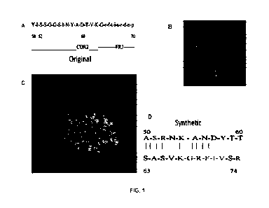
[0045] FIGURE 1 shows (from left to right): (A) the amino acid sequence of the
homophilic domain in T15 antibody (aa 50-70), indicating the CDR2 and
Framework 3;
CA 03225902 2024- 1- 15
WO 2023/283736
PCT/CA2022/051093
9
(B) carbon background tracing of the T15 sequence aa 50-70 in the fold of the
MPC 603
Fab structure; (C) ball representation of the T15 aa 50-70 MPC603 structure;
(D)
alignment of the T15 peptide showing hyrdopathic interactions;
[0046] FIGURE 2 shows the hydropathic profile of the T15 HD peptide (T15) and
its
reversed sequence (rs-T15) using the Kyte-Doolittle algorithm with a window of
7
residues;
[0047] FIGURE 3 shows the hydropathic alignments of the T15 peptide and its
reversed
sequence (rs-T15) as profiled in Figure 2;
[0048] FIGURE 4 shows the hydropathic analysis, amino acid by amino acid of
the T15
peptide in 3 germline sequences drawn from the heavy chain CDR2/framework3 of
Mope
antibodies with no, little or high HD activity. Top peptide: T15 peptide with
amino acids
contributing to self-binding illustrated in grey-scale. Hydropathic score of
individual
amino acid shown above or below. Middle peptide: Low HD binding, amino acid
substitutions from TI5 illustrated in grey-scale. Bottom peptide: No HD
binding (Mpc
167), amino acid substitutions from T15 illustrated in grey-scale;
[0049] FIGURE 5 shows the temperature dependent self-association for G11, S107
and
G9 antibodies at 4 C and 37 C under non-physiologic conditions (see Bryan,
J.A. and
Kohler, H. Physical and Biological Properties of Homophilic therapeutic
Antibodies,
Cancer Immunology Immunotherapy, 60: 507, 2010). The time required for
meniscus to
reach equilibrium after top-down to horizontal movement was measured. Error
bars
represent percent variations of two runs;
[0050] FIGURE 6 shows human lymphoma cells with binding of Rituxin and HD-
Rituxin detected by flow cytometry. Rituxin identifies 2 primary populations
of cancer
cells, one with a low antigen density (second arrow) and a second with higher
density
(third arrow);
[0051] FIGURE 7 shows the complement dependent cytotoxi city of Rituxin and
IID-
Rituxin. Rituxin can mediate C'MC against 3 different cell lines (Raji, Ramon
and JOK1)
with different antigen density (light bar). C' MC by HD-Rituxin is
significantly enhanced
demonstrating higher levels of killing at lower antibody concentrations (dark
bar).
CA 03225902 2024- 1- 15
WO 2023/283736
PCT/CA2022/051093
Antibody-dependent Cell-Mediated Cellular Cytotoxicity (ADCC) is also enhanced
with
HD-antibodies (similar results were obtained with HD-Herceptine, data not
shown).
[0052] FIGURE 8 shows the efficacy of Herceptin and HD-Herceptin in a nude
mouse
model of alow antigen expressing breast cancer (MCF-7), 7 days after injection
of tumor;
5 [0053] FIGURE 9 shows an HD peptide sequence from antibody R24. The
interaction of
the amino acids within the hairpin loop resulting in self binding is also
illustrated;
[0054] FIGURE 10 shows the hydropathic profile of the R24 HD peptide (R24)
using
the Kyte-Doolittle algorithm;
[0055] FIGURE 11 shows detection of PSA antibodies using electro-
chemiluminescence
10 (e.g. for diagnostics). The graphs shows the signal generated with
an anti-PSA antibody,
modified by HD technology versus unmodified antibody.
[0056[ DETAILED DESCRIPTION
[0057] The following description is of a preferred embodiment.
[0058] The present disclosure relates to homologous dimerization (HD)
peptides.
More specifically, the present disclosure relates to artificial or synthetic
homologous
dimerization (HD) peptide. The HD peptides of the present disclosure have
improved
biological activity of self-association or homologous di men zati on.
[0059] In one aspect the HD peptide may be an artificial or synthetic HD
peptide that
is derived from a naturally occurring HD peptide or a conservative variants
thereof
For example the HD peptide may comprise an amino acid sequence in reverse
configuration compared to a corresponding naturally occurring HD peptide, or a
conservative variants thereof In one embodiment the artificial or synthetic HD
peptide may comprise an amino acid sequence that is reverse to the sequence of
a T15
HD peptide or a R24 HD peptide.
[0060] It a further aspect it is provide a homologous dimerization (HD)
peptides
comprising one or more than one substitutions imparting improved self-binding
properties, or conservative variants thereof The one or more than one
substitutions
CA 03225902 2024- 1- 15
WO 2023/283736
PCT/CA2022/051093
11
may be hydrophilic substitutions. The homologous dimerization (HD) peptide may
comprise one or more than one amino acid substitutions wherein the one or more
than
one substitution increases the hydropathy of the HD peptide.
[0061] In some embodiments, the homologous dimerization (HD) peptide may
comprise the amino acid sequence ASRNKANDYTTEYSASVKGRFIVSR (SEQ ID
No: 1), having one or more substitutions at nucleotide positions 4, 7, and 18.
The one
or more than one amino acid substitutions at position 4 may be to K or a
conserved
substitution of K, the one or more than one amino acid substitutions at
position 7 may
be to R or a conserved substitution of R and the one or more than one amino
acid
substitutions at position 18 may be to H or a conserved substitution of H. The
HD
peptide may comprise an amino acid sequence having 80%400% sequence identity
to
SEQ ID NO: 8, 9 or 10.
[0062] It is also provided artificial or synthetic HD peptide dimer that may
comprise
dimer of a naturally occurring HD peptide, dimer of a reverse sequence of a
naturally
occurring HD peptide or a conservative variants thereof. The homologous
dimerization (HD) peptide dimer may comprise a first HD peptide and a second
HD
peptide, wherein the first and second HD peptide are derived from a naturally
occurring HD peptide or wherein the first and second HD peptide are derived
from a
reverse sequences of a naturally occurring HD peptide. The first and second HD
peptides of the dimer may be joined by a linker. The HD peptides in the dimer
may
further comprise one or more than one substitution as described herewith. The
homologous dimerization (HD) peptide dimer may comprises an amino acid
sequence
having 80-100% sequence identity to SEQ ID NO: 5,7, 11, 12, 13, 14,15, 17 or
19.
[0063] Accordingly, the artificial or synthetic HD dimer and may comprise a
first HD
peptide and a second HD peptide, wherein the first, the second or the first
and second
HD peptide are derived from a naturally occurring HD peptide or a conservative
variants thereof Furthermore, the artificial or synthetic HD may be an HD
dimer and
comprise a first HD peptide and a second HD peptide, wherein the first, the
second or
the first and second HD peptide are derived from a reverse sequence of a
naturally
occurring HD peptide, or a conservative variants thereof For example, the
first and
second HD peptide may be derived from a T15 HD peptide, a R24 HD peptide, a
CA 03225902 2024- 1- 15
WO 2023/283736
PCT/CA2022/051093
12
reverse T15 HD peptide, a reverse R24 HD peptide or conserved variants thereof
For
example, the first and second HD peptide of the dimer may comprises an amino
acid
sequence having at least 90% sequence identity to SEQ ID No. 5, 7, 11, 12 or
13.
[0064_1 The HD peptides within the dimer may be joined by a linker. In some
embodiments, the linker may be gly-gly.
[0065] The present disclosure further provides fusion proteins (e.g.
chemically
conjugated or recombinant antibodies) comprising the HD peptides and/or HD
climers
as described herewith. The fusion proteins may comprise an immunoglobulin
component or antibody and the HD peptide or HD peptide dimer may be
recombinant
or the HD peptide or HD peptide dimer may be conjugated thereto, in a manner
that
does not disrupt antigen binding and that allows preferred conformation
changes in
the HD peptide sequence thereby imparting dimerization activity. The HD
peptides
and HD peptide dimers disclosed herein may be used to enhance the potency of
therapeutic antibodies or to increase binding sensitivity and/or avidity for
other
application such as diagnostics.
[0066] The term "homologous dimerization", also referred to as "authophilic",
describes
an entity that is self-associating. For example, -homodimerizing antibodies"
or
"autophilic antibody" are antibodies that self-bind. The term "homologous
dimerization
(HD) peptide" or -autophilic peptide" is a peptide that enables self-
association or self-
binding e.g. of an antibody or other immunoglobulin.
[0067] The term "naturally occurring HD peptide" or "native HD peptide" refers
to an
HD peptide found in nature. For example, "T15" refers to an anti-
phosphorylcholine
antibody and the T15 HD peptide refers to the HD peptide found in the T15
antibody
having the amino acid sequence SRNKANDYTTEYSASVKGRFIVSR (SEQ ID No. 1).
-R24" refers to an antibody recognizing disialoganglioside GD3 (J. Biol. Chem
374:
5597-55604. 1999) and the R24 HD peptide refers to the HD peptide found in the
R24
antibody having the amino acid sequence VAYISSGGSSINYA (SEQ ID No. 3).
[0068] The term -reverse configuration" or -reverse sequence" in relation to
an HD
peptide sequence means that the amino acid sequence of the naturally occurring
HD
peptide is in reverse order, i.e. the N-terminus becomes the C-terminus and
the C-
CA 03225902 2024- 1- 15
WO 2023/283736
PCT/CA2022/051093
13
terminus becomes the N-terminus. The reverse sequence may be obtained by
reading a
peptide or protein sequence backward. The backwardly read sequence (retro
sequence) is
anew peptide (retro peptide) or protein sequence (retro protein).
[0069] The term "antibody" refers generally to a heavy or light chain
immunoglobulin
molecule or any functional combination or fragment thereof containing an
antigen
binding site, e.g. an antigen-binding fragment (Fab). The antibody is
preferably specific
for a cellular receptor, on a membrane structure such as a protein,
glycoprotein,
polysaccharide or carbohydrate, and on a normal cell or on tumor cells. The
antibody may
be a full-length immunoglobulin molecule or a variable domain fragment of an
antibody.
to The term "antibody" encompasses nanobodies, bi-specific antibodies
and diabodies, Fv
and Fab, F(ab)2, camelid and other antigen-binding scaffolds.
[0070] The term "chimeric- refers to a combination of components from
different genetic
sources or species. For example a chimeric antibody according to the present
disclosure
may comprise an immunoglobulin portion from one antibody and an HD peptide as
described herein. A chimeric antibody may also include immunoglobulin portions
derived
from two or more sources or species and an HD peptide.
[0071] A "conservative variant- with reference to an amino acid sequence
refers to a
variant of the amino acid sequence with one or more conservative
substitutions.
[0072] As used herein, the term "conserved substitution- or "conservative
substitution" and grammatical variations thereof, refers to the presence of an
amino
acid residue in the sequence of the HD peptide that is different from, but is
in the
same class of amino acid as the described substitution or described residue
(i.e., a
nonpolar residue replacing a nonpolar residue, an aromatic residue replacing
an
aromatic residue, a polar-uncharged residue replacing a polar-uncharged
residue, a
charged residue replacing a charged residue). In addition, conservative
substitutions
can encompass a residue having an interfacial hydropathy value of the same
sign and
generally of similar magnitude as the residue that is replacing the wildtype
residue.
[0073] As used herein, the term "nonpolar residue" refers to glycine (G, Gly),
alanine (A,
Ala), valine (V, Val), leucine (L, Leu), isoleucine (I, Ile), and proline (P,
Pro); the term
"aromatic residue" refers to phenylalanine (F, Phe), tyrosine (Y. Tyr), and
tryptophan (W,
CA 03225902 2024- 1- 15
WO 2023/283736
PCT/CA2022/051093
14
Trp); the term "polar uncharged residue" refers to serine (S, Ser), threonine
(T, Thr),
cysteine (C, Cys), methionine (M, Met), asparagine (N, Asn) and glutamine (Q,
Gin); the
term "charged residue" refers to the negatively charged amino acids aspartic
acid (D,
Asp) and glutamic acid (E, Glu), as well as the positively charged amino acids
lysine (K,
Lys), arginine (R, Arg), and histidine (H, His). Other classification of amino
acids may be
as follows:
amino acids with hydrophobic side chain (aliphatic): Alanine (A, Ala),
Isoleucine
(I, Ile), Leucine (L, Leu), Methionine (M, Met) and Valine (V, Val);
amino acids with hydrophobic side chain (aromatic): Phenylalanine (F, Phe),
Tryptophan (W, Trp), Tyrosine (Y, Tyr);
amino acids with polar neutral side chain: Asparagine (N, Asn), Cysteine (C,
Cys), Glutamine (Q, Gln), Serine (S, Ser) and Threonine (T, 'Thr);
amino acids with electrically charged side chains (acidic): Aspartic acid (D,
Asp),
Glutamic acid (E, Glu);
amino acids with electrically charged side chains (basic): Arginine (R, Arg);
Histi dine (H, His); Lysine (K, Lys), Glycine G, Gly) and Proline (P, Pro).
[0074] Conservative amino acid substitutions are likely to have a similar
effect on the
activity of the resultant HA protein variant or modified HA protein, as the
original
substitution or modification. Further information about conservative
substitutions can be
found, for instance, in Ben Bassat et al. (J. Bacteriol, 169:751-757, 1987),
O'Regan et al.
(Gene, 77:237-251, 1989), S ahin-Toth et al. (Protein ScL, 3:240-247, 1994),
Hochuli et al
(Bio/Technology, 6:1321-1325, 1988) and in widely used textbooks of genetics
and
molecular biology.
[0075] The Blosum matrices are commonly used for determining the relatedness
of
polypeptide sequences. The Blosum matrices were created using a large database
of
trusted alignments (the BLOCKS database), in which pairwise sequence
alignments
related by less than some threshold percentage identity were counted (Henikoff
et al.,
Proc. Natl. Acad. Sci. USA, 89:10915-10919, 1992). A threshold of 90% identity
was
used for the highly conserved target frequencies of the BLOSUM90 matrix. A
threshold
CA 03225902 2024- 1- 15
WO 2023/283736
PCT/CA2022/051093
of 65% identity was used for the BLOSUM65 matrix. Scores of zero and above in
the
Blosum matrices are considered "conservative substitutions" at the percentage
identity
selected. The following table shows exemplary conservative amino acid
substitutions:
Table 1.
5 [0076] Table 1. Exemplary conservative amino acid substitutions.
_
Original Very Highly - Highly Conserved Conserved
Substitutions
Residue Conserved Substitutions (from the (from the
Blosum65 Matrix)
Substitutions B1ostun90 Matrix)
Ala Ser _Gly, Ser, Thr Cys, Gly, Ser, Thr,
Val
Arg Lys Ciln His Lys
, Asn, Gin, Giu, His,
Lys
Asn Gin; His Asp, Gin, His, Lys, Ser, Tin Arg, Asp,
Gin. Giu, His, Lys, Ser, Thr
Asp Giu Mn, Glu , Asn, Gin, Giu, Ser
Gys Set- None Ala
Gin Asn Arg, Asn, Giu, His, Lys, Met Arg, Asn,
Asp, Glu, His, Lys, Met, Ser
Glu Asp Asp, Gin, Lys Arg, Asn, Asp, Gin,
His, Lys, Ser
Gly Pro Ala Ala, Ser
H is Asn; Gin Arg, Mn, Gin, Tyr Arg, Asn, Gin, Gin,
Tyr
Ile Leu; Val Leu, Met, Val Leu, Met, Phe, Val
Leu lie; Val Ile, Met, Phe, Val Ile, Met, Phe, Val
Lys Arg; Gin; Giu Arg, Asn, Gin, Glu
Arg, Asn, Gin, Glu, Ser,
Met Leu; Ile Gin, Ile, Len, Val Gin, lie, Leu, Phe,
Val
Phe Met; Len; Tyr Leu, Trp, Tyr Ile,
Leu, Met, Trp, Tyr
Ser Thr Ala, Mn, Thr Ala, Mn, Asp, Gin,
Gin, Gly, 1,ys, Thr
Thr Ser Ala, Mn, Ser Ala, Asn, Ser, Val
Trp Tyr Phe, Tyr Phe, Tyr
Tyr Tip; Phe His, Phe, Trp His, Phe, Trp
Val Ile; Leu lie. Leu, Met Ala, Ile, Leu, Met,
Thr
[0077] Without wishing to be bound by theory, it is believed that by
increasing
hydropathy (by using hydropathic indices for individual amino acids) the level
of binding
might be increased, whereas reduction of hydropathy may decrease self-binding.
10 [007 8] In some embodiments, the HD peptide sequences may be
modified to enhance the
crosslinking potential of the HD antibodies as described herein. In one
embodiment, such
functionally enhanced peptides are determined by producing a series of
synthetic peptides
with substitutions at each amino acid position within the template sequence
and then
testing this library of peptides for autophilic binding or for binding to the
original peptide
15 sequence. Those peptides with superior binding to the original
sequence are then
conjugated to immunoglobulins and the resultant conjugates are tested for
potency,
specificity, and the unwanted ability to induce aggregation. In one specific
embodiment,
the 115 peptide sequence is altered and modified sequences are selected for
enhanced
function. In another embodiment of the invention, the self -binding potential
of a peptide
can be enhanced by increasing complementarity of the sequence, such as
described in
CA 03225902 2024- 1- 15
WO 2023/283736
PCT/CA2022/051093
16
U.S. Patent No. 4,863,857 (issued to Blalock et al.). The self-binding
potential and/or
toleration of a peptide can also be enhanced by humanizing a self- binding
peptide
sequence derived from non-human animals. Humanizing a peptide sequence
involves
optimizing the sequence for expression or functionality in humans. Examples
and
methods of humanizing peptides and proteins have been described elsewhere
(Roque-
Navarro et al., 2003; Caldas et al., 2003; Leger et al., 1997; Isaacs and
Waldmann, 1994;
Miles et al. 1989; Veeraraghavan et al., 2004; Dean et al., 2004; Hakenberg et
al., 2003;
Gonzales et al., 2004; and H. Schellekens, 2002).
[0079] The term "expression construct" refers to a recombinant nucleic acid
sequence
including a nucleic acid sequence encoding a peptide or protein to be
expressed. The
nucleic acid encoding a peptide or protein to be expressed is operably linked
to one or
more regulatory nucleic acid sequences that facilitate expression of the
peptide or protein
to be expressed. Nucleic acid sequences are operably linked when they are in
functional
relationship. A regulatory nucleic acid sequence is illustratively a promoter,
an enhancer,
a DNA and/or RNA polymerase binding site, a ribosomal binding site, a
polyadenylation
signal, a transcription start site, a transcription termination site or an
internal ri bos ome
entry site (IRES). An expression construct can be incorporated into a vector,
such as an
expression vector and/or cloning vector. The term "vector" refers to a
recombinant
nucleic acid vehicle for transfer of a nucleic acid. Exemplary vectors are
plasmids,
cosmids, viruses and bacteriophages. Particular vectors are known in the art
and one of
skill in the art will recognize an appropriate vector for a specific purpose.
[0080] Homologous di men zation (HD) pepti des
[0081] The present application describes homologous dimerization (HD) peptides
with
improved self-binding and is exemplified with T15 and R24 HD peptides (see
Example
1). Analysis of the self-binding and hydropathic character of the T15 peptide
and other
HD peptides produced a motif useful in predicting amino acid substitutions
that may lead
to improved self-binding.
[0082] Exemplary homologous dimerization peptides described herein are
summarized in Table 2 below.
CA 03225902 2024- 1- 15
LII
LO
rs.1
[0083] Table 2¨ autophilidhomologous dimerization (HD) sequences
SEQ ID NO. HD peptide name Derivation Sequence
1 T15 T15 anti-phosphorylcholine AS
RNKANDYT TEYSASVKGRF VS
2 Anti-viral peptide CR3014 anti-
SARS RIRKAYSYTTEYAASVKGRFTISR"
R24 (short) R24 anti-disialoganglioside VAYI S S
GGS S I NYA
4 rs-T15 T15 anti-phosphorylcholine P.SVI
FRNYASSASYET rSYDNAKNRSA
rs-T15 dimer (rs-GG- T15 anti-phosphorylcholine RSVI 7RS'EVSA5YET
SYDNAKNRSAGGRSVI FRGNVSASYET TYD \TA'<NRSA
rs)
6 rs-R24 R24 anti-disialoganglioside AYNI S S
NGS. S I YAY
7 rs-R24 dimer (rs-GG- R24 anti-disialoganglioside AYNI
S S S'GS S. I YAYGGAYNI S S GGS S `LAY
rs)
8 Ti 5-van T15 anti-phosphorylcholine AS
RKKANDYT T EYSASVIKGRF VS R
9 T15-var2 T15 anti-phosphorylcholine AS
51AR5Y7 TEYSASVKGRFTVSR
T15-var3 T15 anti-phosphorylcholine AS ANNAN= T EYSASVHGRF I VS R
11 T15-varl dimer T15 anti-phosphorylcholine
AS RKKANDYT T EYSASVIKGRF VS RGNAS:
NDYTTEYSASVKGRNIN'SR
12 T15-var2 dimer T15 anti-phosphorylcholine
As RNKARDYT TEYST AA'GRF I VS RC RrATTE'"- SYKGREIN'SR
13 T15-var3 dimer T15 anti- hos horylcholine
AS RNKANNYT T EY -L'? .11GRE I VS RGSA = I .TTE !HGREIVSR
14 T15 dimer AS RNKANNYT TE
." . RF VS RGNAS.: "TTE RE'IN'SR
R24 dimer (short) VAYI S S NGS S I NYAGGN YI
SSGGSSINYIA
16 R24 (long) GAAVAYI S S.GGS
S INYA
17 R24 dimer (long) GAAVAYI S EGGS
S INYAGGAANAYI S SGGS S I NYA
18 rs-R24 (long)
ATNISSRGSSIYAVAAR -d
19 Rs-R24 dimer (long) AYN I S S GGS S
I YAVAAGGAYN I S S GGS S I YAVAAG L-t
*YTTEY (bold in SEQ ID NO: 1) denotes the potential hinge region
SEQ ID 2 is a putative natural self-binding antibodies. The sequences may be
enhanced by the motif of hydropathy as disclosed in the current disclosure
tv)
(4)
WO 2023/283736
PCT/CA2022/051093
18
[0084] A substantially identical amino acid sequence of an immunoglobulin
component
has an amino acid sequence at least 70%, 80%, 85%, 90% and more preferably
95%,
96%, 97%, 98%, 99% or greater % identical to an amino acid sequence disclosed
herein
in particular embodiments of the present invention, wherein the substantially
identical
protein retains a substantially similar or better function compared to the
reference protein
with which it is substantially identical. As will be appreciated by one of
skill in the art,
the degeneracy of the genetic code is such that more than one nucleic acid
will encode a
particular immunoglobulin component and these alternative sequences are
considered
within the scope of the present invention.
o [0085] An amino acid sequence which is substantially identical to
the 25-mers of SEQ ID
Nos. 4 and 8-10 has at least 20 contiguous amino acids, more preferably at
least 22
contiguous amino acids, having an amino acid sequence at least 70%, 80%, 85%,
90%
and more preferably 95%, 96%, 97%, 98%, 99% or 100% identical to 20 or more
contiguous amino acids of the identified autophilic amino acid sequence. An
amino acid
sequence which is substantially identical to the 14-mers of SEQ ID Nos. 3 and
6 has at
least 10 contiguous amino acids, more preferably at least 8 contiguous amino
acids,
having an amino acid sequence at least 70%, 80%, 85%, 90% and more preferably
95%,
96%, 97%, 98%, 99% or 100% identical to 10 or more contiguous amino acids of
the
identified autophilic amino acid sequence. The same applies to each amino acid
sequence
in the HD-dimers e.g. SEQ ID Nos. 5, 7 11, 12 and 13. The linker does not
necessarily
need to be gly-gly and may be any other suitable linker.
[0086] An amino acid sequence having an amino acid sequence at least 70%, 80%,
85%,
90% and more preferably 95%, 96%, 97%, 98%, 99% or 100% or any amount
therebetween identity or similarity to the sequence of SEQ ID NOs 1, 2, 3,4,
5, 6, 7, 8, 9,
10, 11, 12 , 13, 14, 15, 16, 17, 18, or 19.
[0087] The substantially identical amino acid sequences may comprise one or
more
conservative substitution and may be referred to as a conservative variant. A
peptide
which is substantially identical to a HD peptide described herein retains a
substantially
similar or better autophilic function compared to the reference autophilic
peptide with
which it is substantially identical.
CA 03225902 2024- 1- 15
WO 2023/283736
PCT/CA2022/051093
19
[0088] Homodimerizing antibodies (also referred to as autophillic, self-
binding
antibodies or HD antibodies)
[00891 The present disclosure describes the generation of an antibody-peptide
fusion
protein that enhances the biological and immunological activity of the
antibody without
changing the antibody specificity for the corresponding antigen. Specifically,
the present
disclosure provides the generation of antibody fusion proteins containing the
complete or
partial autophilic 24 mer peptide derived from T15 or the 17-mer peptide
derived from
R24. The HD peptides may be provided in reverse configuration, as dimers or
with
hydrophilic substitutions.
[0090] CDRs from any antibody may be used in the peptide/antibody complex as
described herewith. Preferred antibodies are ones binding immuno-regulatory or
checkpoint inhibitors. For example the CDR sequences may encode for antibodies
specific for PD-1/PD-1L or CTLA-4 and expressing activity for T-cell
activation. Any
restrictions on peptide length are those practical limitations associated with
peptide
synthesis and not restrictions associated with practice of the method of the
disclosure.
Other preferred CDR sequences originate from FDA approved antibodies such as
Rituxin, Herceptin and/or Avastin.
[0091] Recombinant monoclonal antibodies have been created with the genes for
CDRs
(antigen binding sequences) of non-human species (typically mouse) inserted
into a
human antibody framework for the primary purpose of decreasing immunogeni
city.
Although these recombinant antibodies have the ability to interact with
effector cells and
human complement, therapeutic activity, if any, is usually imparted from the
original
CDR sequences due to the nature of the antigen/antibody interaction.
[0092] HD peptides, attached to antibodies, allow for the formation of
lattices at the cell
surface incorporating both antibody bound to target as well as antibody bound
only by
self-binding interactions with other target bound antibodies. This not only
allows for
lattice formation and crosslinking of cell surface antigen but also allows for
higher than
the typical 1:1 ratio of antibody bound to antigen epitopes. Based on flow
cytometry
analysis this ratio, at least in some antigen-antibody systems, can be as high
as 50-100
fold. In fact, this greatly increased binding of an HD modified antibody, is
the hallmark of
CA 03225902 2024- 1- 15
WO 2023/283736
PCT/CA2022/051093
HD antibodies and is used as the initial assay for assessment of recombinant
and
chemically-conjugated antibodies.
[0093] The discovery of homologous dimerization sequences (HD) that allow
antibodies
to self-associate, form lattices and crosslink a target antigen offer the
opportunity to
5 impart therapeutic activity to a recombinant monoclonal antibody
that is independent of
the antigen binding CDR sequences and therefore applicable to many
antigen/antibody
systems. Such peptide sequences have been previously chemically conjugated to
antibodies through the nucleotide binding site and inserted on the C-terminus
of the Fc
region of a fusion protein. The former methodology allows insertion of the HD
peptide
10 into the antibody in a site specific manner but is not a scaleable
manufacturing process.
The latter approach in contrast is scaleable but does not allow for optimal
activity. For
optimal HD activity in antibodies, the peptide needs to interact at multiple
amino acid
positions with a HD peptide on an adjacent antibody. This self-binding can be
reduced by
the peptide's tendency to form a hairpin structure near the c-terminus of the
HD peptide.
15 Unexpectantly, it was found that HD peptide sequences could be
inserted into
recombinant antibodies or by site-specific conjugation without loss of antigen
binding or
reduction in effector functions by careful choice as to the position of the
peptide, e.g. by
reversing the order of the amino acids in the HD peptide. The problem of loss
of HD
activity upon insertion onto the C-terminus of the Fc region, the previous
recombinant
20 antibody approach, was overcome by reversing the normal sequence
order (N-terminus to
c-terminus) not allowing for the C-terminus of the HD peptide to form its
preferred
hairpin structure. Similar results could also be achieved by dimeri zing the I-
1D peptide
either in the forward or reverse configuration, and linking the two HD
peptides with a
linker such as gly-gly.
[0094] Incorporating HD technology into other antibody forms other than the
prototypical
human or humanized IgG1 antibody form will improve their therapeutic or
diagnostic
activity due to greatly enhanced binding. Such antibody forms include but are
not limited
by nanobodies, bi-specific antibodies and diabodies, Fv and Fab, F(ab)2,
camelid and
other antigen-binding scaffolds (Reviewed in: Hoglan Yu, Abhiskek Saxena,
Sachdev S.
Sidhu, Donghui Wu, Frontiers of Immunology 8: Article 38, 2017).
CA 03225902 2024- 1- 15
WO 2023/283736
PCT/CA2022/051093
21
[0095] A group of recombinant antibody forms, modified within the Fc region,
to
increase or decrease serum half-life, antibody-dependent cellular
cytotoxicity,
complement binding or complement-mediated killing al so represent antibody
forms for
which increased antigen binding through HD technology can enhance therapeutic
or
diagnostic activity (Reviewed in: Abhishek Saxena & Donghui Wu, Frontiers in
Immunology, 7: Article 580, 2016). The recombinant antibody forms of the
present
invention are not limited by these referenced forms.
[0096] Antibodies according to the present invention can spontaneously bind to
self only
after first binding to their target antigen. The homodimeri zing antibodies of
the present
invention preferably bond non-covalently with other such conjugated antibodies
when
bound to their target antigen(s), usually a cell-surface, trans-membrane
receptor(s).
[0097] Homodimerizing antibodies of the present invention typically comprise
antibodies
conjugated with one or more peptides having an HD peptide sequence. A
homodimerizing antibody of the invention can comprise virtually any
immunoglobulin. In
some embodiments, the antibodies bind to targets implicated in a disease or
disorder,
where binding of the target has a therapeutic effect on the disease or
disorder. The target
antigens can include cell-surface antigens, including trans-membrane
receptors. In
specific embodiments, the Ig component of the antibodies can comprise a
monoclonal
antibody.
[0098] The present invention affords antibodies having self-binding properties
that mimic
those of rare, naturally occurring, autophilic antibodies. The invention
thereby offers a
simple and attractive alternative to covalent dimerization and other
engineering
approaches directed to enhancing the therapeutic potential of antibodies.
[0099] Expression systems
00100J[ The invention
provides an isolated host cell transformed with an
expression vector encoding an immunoglobulin heavy chain having an antigen
binding
domain and an HD peptide. In particular embodiments, the isolated host cell is
also
transformed with an expression vector encoding an immunoglobulin light chain
having an
antigen binding domain and the antigen binding domain of the immunoglobulin
heavy
chain and the antigen binding domain of the immunoglobulin light chain
together form an
CA 03225902 2024- 1- 15
WO 2023/283736
PCT/CA2022/051093
22
antigen binding site. The isolated host cell for producing a recombinant
autophilic
antibody of the present invention may be in vitro. Expression systems for HD
antibody
expression illustratively include: eukaryoti c cells such as mammalian cells,
plant cells,
insect cells, yeast, and amphibian cells; and prokaryotic expression systems
such as
bacteria. One of skill in the art is able to select a particular expression
system for use in
producing a recombinant HD antibody.
[00101] Location of HD peptide
[00102] As seen in US Patent Publication No. 20030103984,
it was previously
only considered practical to insert the HD peptide sequence on the c-terminus
of the Fc
region. This was due to the concern that insertion of the HD peptide in other
regions of
the mAb might reduce or disrupt antigen binding or potential therapeutic
activities such
as ADCC or complement binding. In fact, published data indicates that the 24
mer HD
sequence derived from the T15 antibody when synthesized and assayed expresses
three-
dimensional conformation; encoding it into an antibody might not only negate
that 3D
structure of the peptide but also may impair antigen binding of nearby CDRs.
Although
self-associating activity in the Fc terminus construct (Kohler, H, Rector, K &
Amick J.,
Hybridoma 2012 (6): 395-402) was described in comparison to a nucleotide
affinity site
chemical conjugate, the activity was reduced (data not shown).
[00103] In preferred embodiments, of the invention, the HD
peptide is localized to
one of three positions within the antibody, e.g. IgG.
[00104] The first is immediately following the C D R3 of
the heavy chain or light
chain. In this form, the Ig molecule can be expressed as a single chain FIT
with the HD
peptide encoded in any of the optimized configurations described herein on the
most c-
terminal portion preceded by a gly-gly or similar spacer. Despite the
proximity to the
antigen binding portion of the antibody this location can impart maximal self-
association
without a reduction in antigen binding. The retention of antigen binding in
this location
was totally unexpected, as we, the inventors of the original technology
considered this so
improbable that we actually previously linked the sequence onto the c-terminus
of the Fc
region of the whole IgG - the furthest we could separate antigen binding from
self-
association.
CA 03225902 2024- 1- 15
WO 2023/283736
PCT/CA2022/051093
23
[00105] A second site is immediately c-terminal to the
heavy or light chain
constant region preceded by a gly-gly spacer. This is can be expressed as a
Fab. In this
form, the Fab can also be part of a naïve or immunized phage expression
library assayed
for binding to a target antigen. Using the HD peptide would allow for enhanced
binding
and identification of even low affinity antibodies.
[00106] The third site is one in which the HD sequence is
cloned onto the c-
terminus of the Fc region preceded by a gly-gly or other spacer. In
embodiments where
the HD peptide is a dimer of an HD peptide, in normal or reverse
configuration, the
separation of the two HD peptides by a gly-gly or alternative spacer is
especially relevant
to the antibody c-terminus constructs to overcome steric inhibition. In
initial testing this
form of the HD peptide has a higher avidity of interaction.
[00107] Methods of producing homodimerizing (autophilic)
antibodies
[00108] IID peptide modified antibodies created by a
variety of site-specific
chemical conjugation methods can also be used to create fully active, self
binding
antibodies. Numerous methods for attaching HD peptides to antibody molecules
will be
known by those skilled in the art. One method is to use chemical crosslinking,
such as the
affinity-crosslinking method described in US Patent Publication No.
20040185039. Such
a method was initially adopted due to the fact that it is able to link a
peptide into the
nucleotide binding site of antibodies which is localized to the end of the
heavy chain of
the Fab. This affinity site allows insertion of the peptide without reducing
affinity of
binding and retains the self-association activity of the peptide. When
compared to
conjugation of HD-peptide to carbohydrate, such site-specific conjugates near
the
antigen-binding region were more active, even though conjugation through
carbohydrate
resulted in more peptide bound to antibody (J. Immunol Methods: (2005) 304:
100-106,
Photo-activated affinity-site cross-linking of antibodies using tryptophan
containing
peptides, Mike Russ, Dingyuan Lou, Heinz Kohler). However, due to elements of
the
methodology, namely the photoactivation step, the manufacturing process for
binding for
this method is not scaleable and only useful to produce small quantities of
conjugate.
[00109] A second method of site specific conjugation has
been developed for use
with antibody-drug conjugates (ADC), namely Smartag technology (Wu, P., et
al., Site-
CA 03225902 2024- 1- 15
WO 2023/283736
PCT/CA2022/051093
24
specific chemical modification of recombinant proteins produced in mammalian
cells by
using for genetically encoded aldehyde tag. Proc Natl Acad Sci U S A, 2009.
106(9): p.
3000-5.). Use of such a technology or similar site specific technologies with
HD peptides
could overcome the issue of scaleability.
[001 101 Alternatively, recombinant methods may be used. For example, a
fusion
gene comprising a nucleic acid sequence encoding an antibody and a nucleic
acid
sequence encoding the peptide may be prepared, wherein the nucleic acid
sequence
encoding the peptide is located inside the nucleic acid sequence encoding the
antibody at
a site wherein, when the fusion is expressed, the fusion protein that is
created thereby
includes the antibody plus the peptide, and the peptide is connected to the
antibody at a
site that does not interfere with antigen binding of the antibody, and
expressing the fusion
gene to create the fusion protein. In particular, the fusion protein may be
created by
providing a gene encoding an antibody, wherein the gene is mutated to contain
a
restriction site, wherein the restriction site is located immediately C-
terminal to the CDR3
of the antibody, or immediately c-terminal to the heavy chain variable region
or onto the
c-terminus of the antibody. A humanized antibody generated by CDR swapping of
mouse
or other human or non-human sources together with a human IgG framework
encoding an
HD-peptide may be expressed by the fusion gene.
[00111] Methods of creating fusion proteins are described,
for example, in the
following U.S. patents: U.S. Pat. No. 5,563,046 to Mascarenhas et al; U.S.
Pat. No.
5,645,835 to Fell, Jr.; U.S. Pat. No. 5,668,225 to Murphy; U.S Pat. No.
5,698,679 to
Nemazee; U.S. Pat. No. 5,763,733 to Whitlow et al; U.S. Pat. No. 5,811,265 to
Quertermous et al; U.S. Pat. No. 5,908,626 to Chang et al; U.S. Pat. No.
5,969,109 to
Bona et al; U.S. Pat. No. 6,008,319 to Epstein et al; U.S. Pat. No. 6,117,656
to Seed; U.S.
Pat. No. 6,121,424 to Whitlow et al; U.S. Pat. No. 6,132,992 to Ledbetter et
al; U.S. Pat.
No. 6,207,804 to Huston et al; and U.S. Pat. No. 6,224,870 to Segal. Methods
of creating
Ig fusion proteins are described, for example, in Antibody Engineering, 2nd
Edition. ed.:
Carl A. K. Borrebaeck, Oxford University Press 1995, and in "Molecular
Cloning: A
Laboratory Manual, Second Ed., Cold Spring Harbor Press, 1989.
[001 121 In specific embodiments, a DNA sequence encoding an HD peptide
described herein, or a substantially identical HD peptide is inserted in-frame
with a DNA
CA 03225902 2024- 1- 15
WO 2023/283736
PCT/CA2022/051093
sequence encoding an immunoglobulin heavy chain and/or immunoglobulin light
chain.
The fusion protein (or HD antibody) expressed from the DNA sequence contains
an
immunogl obulin heavy chain and/or immunoglobulin light chain having said HD
peptide.
[00113_1 Nucleic acids encoding immunoglobulin heavy chains
or immunoglobulin
5 light chains are well-known and any of various nucleic acids
encoding immunoglobulin
heavy chains or immunoglobulin light chains can be used to produce a
recombinant
chimeric HD antibody of the present invention. Specific nucleic acids are
described
herein which encode human constant heavy chain and/or a human constant light
chains,
particularly human gamma constant heavy chains and human kappa constant light
chains.
10 Nucleic acids encoding human gamma constant heavy chains and/or
human kappa
constant light chains can be obtained from commercial sources, such as vector
pAc-k-
CH3, available from Progen Biotechnik GmbH. Nucleic acids encoding protein
and/or
peptides described herein, including human gamma constant heavy chains and/or
human
kappa constant light chains, can be produced using recombinant techniques such
as by
15 cloning or synthesis. Particular immunoglobulin constant heavy
chains and/or
immunoglobulin kappa constant light chains, are described, for instance, in
U.S. Patent
Nos. 5,736,137 and 6,194,551.
[00114] IgG fusion proteins
[00115_1 Ig fusion proteins have the advantage of joining
the antibody combining
20 specificity and/or antibody effector functions with molecules
contributing unique
properties. The ability to produce this family of proteins was first
demonstrated when c-
myc was substituted for the Fc of the antibody molecule,(Neuberger M S.
Williams G T
and Fox R 0. Nature 125:604, 1984) but many examples now exist. Ab fusion
proteins
can be achieved in several different ways. In one approach non-Ig sequences
are
25 substituted for the variable region; the molecule replacing the V
region provides
specificity of targeting with the antibody contributing properties such as
effector
functions and improved pharmacokinetics. Examples include IL-2 and CD4.
Alternatively, non-1g sequences can be substituted for or attached to the
constant region.
The resulting molecules retain the binding specificity of the original
antibody but gain
characteristics from the attached protein. Depending on the position of the
substitution,
different antibody-related effector functions and biologic properties will be
retained.
CA 03225902 2024- 1- 15
WO 2023/283736
PCT/CA2022/051093
26
[00116] Vectors for the Construction of IgG Fusion
Proteins
[00117] A series of vectors have been produced that
permits the fusion of proteins
at different positions within an antibody molecule, thereby facilitating the
construction of
fusion proteins with different properties. Using these vectors it is possible
to produce a
family of fusion proteins with molecules of differing molecular weight,
valence, and
having different subsets of the functional properties of the antibody
molecule.
[00118] As a specific example of how to facilitate the
construction of fused genes,
site-directed mutagenesis was used to generate unique restriction enzyme sites
in the
human IgG3 heavy chain gene. In this particular example, restriction sites
were generated
at the 3' end of the CH1 exon, immediately after the hinge at the 5' end of
the CH2 exon,
and at the 3' end of the CH3 exon. The restriction sites thus produced were
SnaB I at the
end of CH1 by replacing TtgGTg with TacGTa, Pvu II at the beginning of CH2 by
replacing CAcCTG with CAgCTG, and Ssp I at the end of CH3 replacing AATgag
with
AATatt. These manipulations provided a unique blunt-end cloning site at these
positions.
In all cases the restriction site was positioned so that after cleavage the Ig
would
contribute the first base of the codon. Human IgG3 with an extended hinge
region of 62
amino acids was chosen for use as the immunoglobulin; when present this hinge
should
provide spacing and flexibility, thereby facilitating simultaneous antigen and
receptor
binding. An EcoR1 site was also introduced at 3' of the IgG3 gene to provide a
3' cloning
site and polyA addition signal. Although initially designed for use with
growth factors,
these restrictions sites can be used to position any novel sequence at defined
positions in
the antibody. Also, using these cloning cassettes the variable region can
easily be
changed. Similar techniques may be used to generate suitable restriction sites
in other
antibody genes.
[001 191 Production ofA Fusion Gene
[00120] As a first step in the production of a fusion
protein, a blunt-end restriction
site must be introduced at the desired position into the 5' end of the gene to
be fused. In
order to maintain the correct reading frame, the site must be positioned so
that after
cleavage it will contribute two bases to the codon. If the objective is to
make a fusion
protein with the complete molecule, the restriction site is usually introduced
at the
position of any post-translational processing, such as after the leader
sequence.
CA 03225902 2024- 1- 15
WO 2023/283736
PCT/CA2022/051093
27
Alternatively, if the obj ective is to use only a portion of the protein, the
blunt-end site can
be introduced at any position within the gene, but attention must always be
paid to
maintaining the correct reading frame. Additionally, if there is carboxyl -
terminal post-
translational processing of the fused protein, it is frequently desirable to
introduce a stop-
codon at this processing site.
[00121] A major concern when producing fusion proteins is
maintaining the
biologic activities of all of the components. The production of fusion
proteins with
antibodies is facilitated by the domain structure of the antibody, and all of
the cloning
sites have been positioned immediately following an intact domain. In this
configuration
the correct folding of the immunoglobulin should be assured. The folding of
the attached
protein depends on its structure and where it is fused. Whenever structural
information is
available, it is desirable to produce the fusion at a position that will
maintain the
structural integrity of the attached protein.
[00122] To produce quantities of protein sufficient for
functional analysis, it is
desirable to have the protein secreted into the medium. While in the examples
reported to
date, assembled fusion proteins have been assembled and secreted, this remains
a concern
when designing additional fusion proteins.
[00123] The method to design a fusion gene that contains a
biologically activity
peptide as part of the heavy or light chain gene can use established antibody
engineering
protocols (Antibody Engineering, 2nd Edition. ed.: Carl A. K. Borrebaeck,
Oxford
University Press 1995. Chapter 9, pages 267-293). The peptide can fused either
to N-
terminal residues or the C-terminal residues of H or L chains. The expression
of such
fused genes is typically done in mammalian cell lines, although other
expression systems,
such as, for example, bacteria or yeast expression systems, may be used.
[00124] Pharmaceutical compositions
[00 I 251 The invention also relates to compositions
comprising a homodimen zing
antibody of the invention and a pharmaceutically acceptable carrier.
[00126] The antibodies of the invention are useful in
pharmaceutical compositions
for systemic administration to humans and animals in unit dosage forms,
sterile solutions
CA 03225902 2024- 1- 15
WO 2023/283736
PCT/CA2022/051093
28
or suspensions, sterile non-parenteral solutions or suspensions oral solutions
or
suspensions, oil in water or water in oil emulsions and the like, containing
suitable
quantities of an active ingredient. Topical application can be in the form of
ointments,
creams, lotions, jellies, sprays, douches, and the like. The compositions are
useful in
pharmaceutical compositions (wt %) of the active ingredient with a carrier or
vehicle in
the composition in about 1 to 20% and preferably about 5 to 15%.
[00127] The above parenteral solutions or suspensions may
be administered
transdermally and, if desired a more concentrated slow release form may be
administered.
The fusion proteins of the invention may be administered intravenously,
intramuscularly,
intraperitoneally or topically. Accordingly, incorporation of the active
compounds in a
slow release matrix may be implemented for administering transdennally. The
pharmaceutical carriers acceptable for the purpose of this invention are the
art known
carriers that do not adversely affect the drug, the host, or the material
comprising the drug
delivery device. The carrier may also contain preserving, stabilizing,
wetting, emulsifying
agents and the like together with the penetration enhancer of this invention.
The effective
dosage for mammals may vary due to such factors as age, weight activity level
or
condition of the subject being treated. Typically, an effective dosage of a
compound
according to the present invention is about 10 to 500 mg, when administered in
solution
at least once daily. Administration may be repeated at suitable intervals.
[00128] Conjugate autophilic antibodies can bind non-covalently with other
autophilic antibodies when bound to their target antigen(s). However,
premature
formation of dimers or multimers of the antibodies may lead to difficulties in
manufacturing, such as during purification and concentration, as well as
drawbacks in
administration, which may lead to side effects. As such, compositions
containing
autophilic antibody-peptide conjugates of the invention are formulated to
reduce this
dimerizing potential and maximize monomeric properties while in solution and
before
administration. For example, it has been found that solution dimerization can
be reduced
or mitigated by using a hypertonic composition. ht some embodiments, salt
concentrations of 0.5M or more, low levels of SDS or other various detergents
such as
those of an anionic nature (see U.S. Patent No. 5,151,266, which is
incorporated herein
by reference), or modifications of the antibody to decrease its isoelectric
point, for
CA 03225902 2024- 1- 15
WO 2023/283736
PCT/CA2022/051093
29
example through the use of succinyl anhydride (see U.S. Patent No. 5,322,678,
which is
incorporated herein by reference), an be used to formulate compositions.
[00129] Immunoassays
[00130] Immunoassays are provided according to the present
invention which
include contacting an analyte in a biological or environmental sample with an
antibody
conjugated to an autophilic peptide. A complex formed by the analyte and the
antibody
conjugated to an autophilic peptide is then detected.
[00131] In particular embodiments, assays of the present
invention are
characterized by detection of antigens expressed at low levels, such as on a
cell surface,
using antibodies containing autophilic peptides either naturally or by
conjugation of an
autophilic peptide to the antibody.
[00132] The use of highly-specific antibodies is common in
many diagnostic
applications. The binding of said antibodies may be detected directly by a
number of
means or, alternatively, secondary antibodies are required for signal
enhancement and
detection. Previously, the signal detection was directly related to the amount
of antibody
bound, either mono or divalently bound to target. In the present invention,
enhanced
signal strength results from polyvalent binding interactions of autophilic
peptide-
conjugated antibodies bound to a target analyte.
[00133] Whether directly or indirectly detected,
autophilic peptide-conjugated
antibodies greatly enhance signal detection because of lattice formation and
more
antibody surrounding the target. Thus, naturally occurring autophilic
antibodies and non-
naturally occurring autophilic peptide-conjugated antibodies can be directly
labeled with
a detectable label affording an enhanced signal in an immunoassay. Similarly,
indirect
labeling, such as labeling of a secondary antibody produces increased signal
in an
immunoassay since the secondary antibody will bind with increased numbers to
the
primary autophilic peptide- conjugated antibodies.
[00134] In embodiments of assays of the present invention,
a naturally-occurring
autophilic antibody or an antibody conjugated to a homologous dimerization
(HD)
CA 03225902 2024- 1- 15
WO 2023/283736
PCT/CA2022/051093
peptide is used to enhance signal detection of antigen immobilized on a
substrate, such as
plastic, in assays such as ELISA.
[00135] In embodiments of assays of the present invention,
a naturally-occurring
autophilic antibody or an antibody conjugated to a homologous dimerization
(HD)
5 peptide is used to enhance "off-rate" or increase in avidity of
antigen coated or bound to a
polymer chip and detected by surface plasmon resonance.
[00136] In embodiments of assays of the present invention,
a naturally-occurring
autophilic antibody or an antibody conjugated to homologous dimerization (HD)
peptide
is used to enhance binding and localization detection of the antibody bound to
a target in
10 vivo, such as in a xenograft tumor animal model, by fluorescence or
other signal
detection method.
[00137] Assays according to embodiments of the present
invention can include
virtually any immunoglobulin conjugated with one or more homologous
dimerization
(HD) peptides for enhanced detection of an analyte.
15 [00138] The term "analyte" refers to any molecule or compound
which is
specifically recognized by an antibody conjugated to an autophilic peptide,
illustratively
including a protein, a peptide, a hapten, a carbohydrate, a lipid, a
ganglioside and
combinations of these. In particular embodiments, an analyte can be a
mammalian
analyte, illustratively including a protein, peptide, hapten, carbohydrate,
lipid, ganglioside
20 or combination thereof generated, for instance, by a normal or
abnormal cell of a
mammal.
[00139] In further particular embodiments, an analyte can
be a non-mammalian
analyte, such as a protein, peptide, hapten, carbohydrate, lipid, ganglioside
or
combination thereof, generated by a microorganism, such as a bacterium or a
virus. Thus,
25 particular assays of the present invention are provided for
detection of a microorganism
and/or a product of a microorganism. Assays for detection of a microorganism
and/or a
product of a microorganism are used to assay a sample obtained from a human or
a non-
human animal to detect infection, for example. In further embodiments, assays
for
detection of a microorganism and/or a product of a microorganism are used to
assay a
CA 03225902 2024- 1- 15
WO 2023/283736
PCT/CA2022/051093
31
sample obtained from an environment or object to be tested for contamination
with a
microorganism and/or a product of a microorganism.
[00140] In embodiments of assays of the present invention,
a target analyte is a B-
cell receptor, CD20, Her2, ganglioside GM2, glycolic ganglioside GM3, GD3
ganglioside, caspases, oxidized low density lipoproteins, phosphocholine,
EGFR,
CD32B, HLADR1, CD19, EpCAM, P SA and bacterial antigens such as a
staphylococcal
antigens.
11001411 In some embodiments of the present invention,
diagnostic and prognostic
immunoassays are provided. The term "diagnostic immunoassay" refers to an
immunoassay that allows for determination of presence or amount of an analyte
indicative of a disease or pathological condition in an animal or human
subject. The term
"prognostic immunoassay" refers to an immunoassay that allows for
determination of
presence or amount of an analyte indicative of progression of a disease or
pathological
condition in an animal or human subject.
[00142] Kits
[00143] In embodiments of the present invention, kits are
provided for use in
performing an assay using an antibody-homologous dimerization (HD) peptide
conjugate.
In particular embodiments, a kit includes an antibody-homologous dimerization
(HD)
peptide conjugate and instructions for use in detecting an analyte in a
sample.
[00144] Methods of treatment
[00145] A method of enhancing apoptosis, complement
fixation, effector cell-
mediated killing of targets, or preventing the development of, or enhancement
of, a
disease state, is also contemplated, which employs a homologous dimerization
(HD)-
antibody of the invention or a composition comprising the homologous
dimerization
(HD)-antibody. In one embodiment, an autophilic conjugate of the invention, or
a
composition containing an autophilic conjugate of the invention, is
administered to a
subject. Once administered, the antibodies bind to target cells and enhance
apoptosis,
complement fixation, effector cell-mediated killing of targets, or prevent
target antigens
or cells from stimulating the development of, or further enhancing, a disease
state. In a
CA 03225902 2024- 1- 15
WO 2023/283736
PCT/CA2022/051093
32
further embodiment, allowing time for the autophilic conjugate to bind to
target cells and
enhance apoptosis, complement fixation, effector cell-mediated killing of
targets, or
prevent target antigens or cells from further enhancing a disease state, and
for the
autophilic conjugate to be cleared from normal tissues, a second anti-
autophilic peptide
antibody can be administered.
[00146] In some aspects, a patient who suffers from a
debilitating or potentially
life-threatening disease or condition is administered at least one subject
homologous
dimerization (HD) antibody in an amount effective to alleviate symptoms of the
disease
or condition. A disease or condition contemplated for treatment by an antibody
of the
to invention can be a malignancy, neoplasm, cancer, auto-immune
disorder, Alzheimer's
disease or other neuro-degenerative condition, or graft or transplantation
rejection.
[00147] In some aspects, a method of potentiating
apoptosis of targeted cells of a
patient comprises administering a first homologous dimerization (HD) antibody-
peptide
conjugate and a second antibody that recognizes the peptide domain of the
conjugate. In
this embodiment, the antibody-peptide conjugate recognizes the extracellular
region of a
transmembrane receptor of the target cell. Owing to its homodimerization
property, the
antibody-peptide conjugate can bind more avidly to the target than the
corresponding
antibody lacking the self-binding peptide domain. Moreover, whenever the
autophilic
antibodies bind to two or more receptors, with those receptors being brought
in close
proximity due to the self-binding property of the antibodies, an apoptosis
signal within
the cell can be triggered. In those instances when the peptide domain of the
conjugate
presents an exposed epitope, a second antibody, specific for the autophilic
peptide, can be
administered, bind to the modified antibody, and enhance the process of
crosslinking and
even cause temporary clearance of the target antigen. If the target antigen is
a receptor,
clearance from the cell surface, endocytosis, and degradation will
subsequently require
synthesis of new receptor protein, meaning that the biological function of the
receptor
will be more effectively inhibited for a longer period than using either a
simple blocking
antibody or small molecule inhibitor. Alternatively, the second antibody can
bear a
radiolabel or other potentially therapeutic substance, so that when
administered it can
attack the targeted cells. The key to use of this second antibody is that
antibody's
specificity. The homologous dimerization (HD) peptide, though naturally
occurring, is
CA 03225902 2024- 1- 15
WO 2023/283736
PCT/CA2022/051093
33
present on only a small number of murine immunoglobulins. Thus, antibody
specific to
this peptide will have the requisite selectivity to be used in vivo.
[00148] Diseases
[00149] A disease or condition contemplated for treatment
by an antibody of the
invention can be a malignancy, neoplasm, cancer, atherosclerosis, auto-immune
disorder,
Alzheimer's disease or other neurodegenerative condition, graft or
transplantation
rejection, or any other disease or condition responsive to antibody therapy.
[00150] Doses
[00151] The homologous dimerization (HD) antibodies
described herein can be
administered in one or more dosage amounts substantially identical to or less
than those
practicable for unmodified antibodies.
Table 3. SEQ ID NO: and Description of Sequences
SEQ ID NO: Description of Sequence
1 T15
2 CR3014 anti-SARS
3 R24 (short)
4 rs-T15
5 rs-T15 dimer
6 rs-R24
7 rs-R24 dimer
8 T15-varl
9 T15-var2
10 T15-var3
11 T15-varl dimer
12 T15-var2 dimer
13 T15-var3 dimer
14 T15 dimer
R24 dimer (short)
16 R24 (long)
CA 03225902 2024- 1- 15
WO 2023/283736
PCT/CA2022/051093
34
17 R24 dimer (long)
18 Rs-R24 (long)
19 Rs-R24 dimer (long)
20 Rituximab heavy chain
21 Rituximab light chain
22 Herceptin Light chain (1 and 2)
23 Herceptin Heavy chain (1 and 2)
24 Bevacizumab light chain
25 Bevacizumab heavy chain
26 Atezolizumab (anti-PD-Li mAb) heavy chain
27 Atezolizumab (anti-PD-Li mAb) light chain
[00152] The following examples are presented to illustrate
certain aspects of the
invention, and are not intended to limit the scope of the invention.
[00153] The present invention will be further illustrated
in the following examples.
[00154] EXAMPLES
[00155] Example 1 ¨ HD peptides with potential for
optimized self-binding
[00156] The 24 mer peptide sequence derived from T15 was
analyzed and
compared with other anti-phosphorylcholine antibodies of the same gennline
configuration that possess limited or no HD activity using hydropathic scores
of
to individual amino acids and the overall peptide hydropathic plot
(Kyte-Doolittle), a
method of protein analysis normally employed with non-immunoglobulin proteins.
[00157] As shown in Figure 1, the T15 peptide has a
preference to form a hairpin
structure at the C-terminus, which might impair its ability to self-bind (Fig.
1B). In the
original HD antibody (T15), the HD sequence was inserted into the latter
portion of the
CDR3 and the first portion of CH1, a position that does not allow for
secondary
conformation. We hypothesized that in a recombinant form of an antibody with
the HD
peptide inserted at the c-terminus of the antibody, that the HD peptide was
free to form
the hairpin structure, reducing self-binding and its ability to potentiate
therapeutic activity
CA 03225902 2024- 1- 15
WO 2023/283736
PCT/CA2022/051093
of antibodies through lattice formation. Similarly, the T15 peptide chemically
conjugated
to antibody, in the normal N to C-terminus configuration, would also have the
ability to
form a secondary conformation with reduced self-binding. In addition, the
small HD
peptide sequence, in the context of the large antibody protein, might have
steric
5 limitations to self-binding.
[00158]
The first comparison was of the known T15 peptide configuration in
normal or reverse order (see Figs.2 and 3). The examination of the original
T15 sequence
(left panel of Fig. 2) illustrates that self-binding correlates with the
sequence hydropathy
plot of the peptide. The first half of the peptide was derived from the CDR2
of the
10 antibody while the second half was derived from the framework
between CDRs. The first
half then would contain the amino acids that confer self-binding and which are
altered
from the germline sequence; as important the more hydropathic residues of the
second
half serve to bind to the less hydropathic residues in the first half to
initiate the self-
binding process. Surprisingly when we examined the 24 mer peptide in reverse
sequence
15 (right panel of Fig. 2), it was also able to assume the required
self-binding configuration
despite the presence of the hairpin structure at the c-terminus. This then
allows the
tethering of the peptide through the former c-terminus (now n-terminus) to the
antibody
with the more important first half of the peptide, which determines self
binding, tethered
further from the antibody and more able to seek out the corresponding self-
binding
20 sequences in other peptide/antibody conj ugates.
[00159]
Identifying the hydropathic contribution of individual amino acids to self-
binding in order to construct an optimizable motif, was done by comparing
amino acid
differences in 3 germline sequences drawn from the heavy chain CDR2/framework3
of
Mopc antibodies with no, little or high HD activity. Individual amino acid
hydropathic
25 scores of amino acids (available
at:
http://gcat.davidson.edu/DGPB/kd/aminoacidscores.htm) were plotted to known
positions
of amino acids involved in self binding (see Fig. 4). As is shown, the amino
acid changes
in the T15 peptide (high HD activity) enabling self-binding are associated
with distinctive
changes in the hydropathic score of the substituted amino acid i.e. typically
more
30 hydrophilic substitutions enabling self binding to the more
hydrophobic framework
region. This allows one to predict that substitution of other amino acids into
the former
CDR2 region of the T15 peptide need to conserve the hydropathic character of
the
CA 03225902 2024- 1- 15
WO 2023/283736
PCT/CA2022/051093
36
individual amino acids. It also became apparent that additional hydrophilic
substitutions
within the former CDR2 region of T15 peptide may increase the potential
binding with
the more hydrophobic former framework portion of the peptide and visa versa;
substitution of more hydrophobic amino acids into the framework3 portion would
allow
more self binding to the more hydrophilic portion of the T15 peptide. 'this
hydropathis-
hydrophillic interaction serves to initiate the self-binding process which
then results in
the particular amino acids involved in self-binding, to form other non-
covalent
interactions. The whole process confers the relative affinity of the self-
binding process.
Some illustrative single amino acid, conservative substitutions are given in
Table 2 (e.g.
T15-varl, T15-var2 and T15-var3).
[00160] Without wishing to be bound by theory, it is
believed that by increasing
hydropathy of a HD peptide (for example by substitution of amino acids) the
binding
characteristics of the HD peptide may be modulated for example the binding
characteristics of the HD peptide may be increased. It is further believed
that the affinity
for self-binding may be optimized to avoid the formation of antibody dimers
before the
binding of the antibody to a target. Therefore, the affinity of the HD peptide
may be
increased compared to a natural occurring HD peptide, but to still be less
than the binding
affinity of the antibody to its target. For example the biding affinity of the
HD peptide
may be 10-4, 10-5, 10-6, 10-7 or 10-8.
00161I[ The potential increase in self-binding can be readily measured by
synthetic
peptide synthesis of single amino acid variants, labeling hydrogen atoms with
tritium and
then measuring binding to immobilized T15 peptide. Positive changes for singly
amino
acid substituted peptides then can be incorporated as double or triple amino
acid changes.
It must be pointed out that the final outcome is a self-binding peptide with
higher self
binding affinity but not so much that it causes aggregation of antibody.
[00162] T15 is not the only naturally-occurring HD
antibody and source of HD
peptides. A well studied example is that of R24 directed to disialoganglioside
GD3. As
with T15, increased potency in the original murine antibody was associated
with the
presence of a self-binding peptide that allowed formation of lattices at the
cell surface
increasing therapeutic efficacy. As shown in Figure 9, the R24 sequence,
responsible for
lattice formation will self-bind in anti-parallel fashion. As with T15, the
R24 peptide
CA 03225902 2024- 1- 15
WO 2023/283736
PCT/CA2022/051093
37
though shorter than T15 demonstrates a corresponding hydropathic plot where
one end of
the peptide is more hydropathic than the other (see Fig. 10). The plot is
basically a
reversal of T15 where hydropathy goes from low to high, instead going from
high to low.
As such the same principles for promoting self-binding through reversal of the
sequence,
formation of dimers and making single, conservative amino acid substitutions
with
altered hydropathic character serve to increase self-binding.
[00163] Example 2¨ Identification of antibodies with
autophilic/homologous
dimerization (HD) activity
[00164] The following method uses the specific biophysical
properties of
antibodies incorporating an HD peptide to test for HD activity in an antibody
preparation
or in a phage library of scFy or Fab. The method can also be used as a quality
assurance
(QA) release assay for HD antibodies.
[00165] Antibody viscosity
[00166] Microdilution tubes (USA Scientific, Ocala, FL
containing HPC G9 or
HPC Gil at 1 mg/ml in PBS) were mounted on standard microscope slides. They
were
equilibrated at 4 , 20 , and 37 C, sealed and then positioned vertically and
then
horizontally then photographed at each temperature.
[00167] lime lapse measurement of equilibrium
[00168] Microdilution tubes containing antibodies at 1
mg/ml in PBS were
equilibrated at either 4 or 37 C. Tubes were positioned vertically for 3 s
then horizontally
and filmed. The time required for the meniscus to cease movement was measured
using a
stopwatch.
[00169] Results
[00170] Temperature-dependent equilibrium of homodimers:
HPC G9/HPC Gil
are isoforms of IgG anti-phosphocholine antibodies whereby HPC Gil shares
idiotype
and sequence with CDR2/FR3 in TEPC-15. Accordingly, HPC Gil is homophilic and
HPC G9 is not. Since temperature is known to affect the physical properties of
proteins,
including antibodies, the behavior of HPC G9 and HPC Gil was compared at 4,
20, and
CA 03225902 2024- 1- 15
WO 2023/283736
PCT/CA2022/051093
38
37 C using size- exclusion chromatography. The amount of antibody eluted
within the
exclusion volume was compared with that of the inclusion volume.
[00171] The ratio of excluded/included HPC G11 at
different temperatures. There
was no significant change in the ratio of excluded protein with HPC G9 at 4,
20, and
37 C (data not shown), but the ratio for HPC Gll increased with temperature.
These
findings support the notion that the degree of dimerization of homophilic Gil
in solution
is higher at physiological temperature than at non-physiological temperatures.
[00172_1 Viscosity dylerences
[00173] The viscosity of HPC G9 and HPC Gil was also
compared at different
temperatures. A volume of 500 ml of each antibody was placed into racked tubes
and
allowed to equilibrate in a horizontal position at 4, 20, and 37 C for 30 mm.
The tubes
were then moved into a top-up, vertical position for 1 s and then returned to
the
horizontal position and photographed. The position of the meniscus was
measured and
the ratios of these measurements was calculated with HPC G9 or HPC G11. The
ratio at
4 C was 1.45, at 20 C 0.92, and at 37 C it was 0.54. The tilting of the tubes
after
equilibration was reversed (i.e. the bottom of the tube was raised vertically
for 1 s and
then returned to a horizontal position and photographed). The ratio of HPC G9
to HPC
Gil at 4 C was 1.5, 1.41 at 20 C, and 2.06 at 37 C.
[00174] To exclude the possibility that the viscosity of
Gil is unique, S107 a
murine monoclonal IgA antibody known to be hemophilic was tested. For
comparison,
another murine antibody G9 was included in the viscosity analysis. The tubes
were
positioned first bottom-up for 3 s and then returned to a horizontal position.
The time
required for the meniscus to cease movement was measured for each antibody. In
Fig. 5,
we show the seconds recorded for the meniscus of these antibodies to cease
movement at
4 and 37 C. The more time required for movement to cease, the higher the
viscosity of
the solution. The time differences recorded at both temperatures as shown in
Fig. 5 are
smaller for Gil and S 107 than for G9. The ratio of time at 4 C divided by
time at 37 C
for G11, S 107, and 1F7 are identical, while the ratio for G9 is more than 2
times greater.
Gil and S107 are facultative homophilic polymers, while 1F7 IgM is a covalent
pentamer. It is interesting to note that the homophilic polymers and the
covalent IgM
CA 03225902 2024- 1- 15
WO 2023/283736
PCT/CA2022/051093
39
polymer have similar viscosity but different viscosity from the monomeric G9
bivalent
antibody. This unique viscosity of homophilic antibodies is independent of Ig
class and
might be part of their unique dimeri zati on potential responsible for their
superior
targeting.
O01751 Example 3 ¨ Production and characterization of a recombinant
therapeutic HD antibody (Rutuximab) with reverse confiEuration
[00176] The following example illustrates production of a
reverse sequence HD
antibody and the changes in therapeutic characteristics of a recombinant HD
form of
an antibody compared to the native antibody. To test the capacity of the HD
peptide,
molecular biological techniques were used to generate a chimeric version of
the
Rituximab antibody (chRituximab), and a chimeric HD antibody that is identical
to
chRituximab, except for the addition of the HD peptide (rs-T15) to the C-
terminus of
each variable region of the heavy chain.
[00177] Materials and Methods
00178I[ Cell lines
[00179] JOK-1 cells were a gift of Affimed Inc. JOK-1
cells were grown in RPMI-
1640 with Glutamax (Gibco), supplemented with 10% FBS-Premium-HI (Aleken
Biologicals), and 1% Penicillin/Streptomycin (Gibco). Raji, and Ramos, cells
were
obtained from the American Type Culture Collection (ATCC), numbers HB-9645,
CCL-
86, CRL-1596, and TIB- 152, respectively. Raji and Ramos cells were maintained
in
RPMI-1640 Medium with HEPES (ATCC), supplemented with 10% FBS -Premium-HI
(Aleken Biologicals), and 1% Penicillin/Streptomycin (Gibco). Rituximab cells
were
maintained in RPMI-1640 Medium with HEPES (ATCC), supplemented with 10% FBS-
low-IgG (Gibco), 1% Penicillin/Streptomycin (Gibco), and 0.5% Glutamax
(Gibco).
CHO-S cells were purchased from Invitrogen, and were grown in CD CHO medium,
supplemented with I % HT supplement (Gibco), 2% Glutamax (Gibco), and I 00
U/ml
pen/strep (Gibco). After introduction of vector DNA, CHO-S cells were grown as
above
with the addition of 1.2 mg/ml 418 (Invivogen) for selection. All cells were
maintained at
37oC and 5% CO2.
CA 03225902 2024- 1- 15
WO 2023/283736
PCT/CA2022/051093
[00180] Construction of chimeric antibody genes
[00181] Heavy and Light chain variable regions were
synthesized from published
Rituximab sequences:
Rituximab heavy chain:
5 QVQLQQPGAELVKPGASVKMSCKASGYTFTSYNMHWVKQTPGRGLEWIGAIYPGNGDTS
YNQKFKGKATLTADKSSSTAYMQLSSLTSEDSAVYYCARSTYYGGDWYFNVWGAGTTVT
VSAASTKGPSVFPLAPSSKSTSGGTAALGCLVKDYFPEPVTVSWNSGALTSGVHTFPAV
LQSSGLYSLSSVVTVPSSSLGTQTYICNVNHKPSNTKVDKKAEPKSCDKTHTCPPCPAP
ELLGGPSVFLFPPKPKDTLMISRTPEVTCVVVDVSHEDPEVKFNWYVDGVEVHNAKTKP
10 REEQYNSTYRVVSVLTVLHQDWLNGKEYKCKVSNKALPAPIEKTISKAKGQPREPQVYT
LPPSRDELTKNQVSLTCLVKGFYPSDIAVEWESNGQPENNYKTTPPVLDSDGSFFLYSK
LTVDKSRWQQGNVESCSVMHEALHNHYTQKSLSLSPGK (SEQ ID NO: 20)
Rituximab light chain:
QIVLSQSPAILSASPGEKVTMTCRASSSVSYTHWFQQKPGSSPKPWIYATSNLASGVPV
15 RFSGSGSGTSYSLTISRVEAEDAATYYCQQWTSNFPTFGGGTKLEIKRTVAARSVFIFP
PSDEQLKSGTASVVCLLNNFYPREAKVQWKVDNALQSGNSQESVTEQDSKDSTYSLSST
LTLSKADYEKHKVYACEVTHQGLSSPVTKSFNRGEC (SEQ ID NO:21)
[001 821 The Rituximab heavy chain variable regions were
amplified from the
cDNA pool by PCR using primers modVHRituximabfwd and modVHRituximabrev. All
20 oligos were purchased from Operon. The Rituximab light chain
variable regions were
amplified from the cDNA pool by PCR using primers modVLRituximabfwd and
modVLRituximabrev. The heavy chain and light chain PCR products were cloned
into
the XhoI-NheI and SacI-HindIII sites, respectively, of vector pAc-k-CH3
(Progen
Biotechnik GmbH), to form pAc-k-RituximabH and RituximabK. Clones were
verified
25 by sequencing in both directions. All restriction enzymes were
purchased from Takara or
New England Biolabs. Taq polymerase (Promega) was used for all PCR. All
enzymatic
reactions were carried out using manufacturers' protocols.
[00183] Construction of antibody expression vectors
CA 03225902 2024- 1- 15
WO 2023/283736
PCT/CA2022/051093
41
[00184] Oligos LongT15fwd, LongT15rev, and PrimerB were
used in a nested
PCR to construct a DNA sequence that encodes the reverse sequence of T15
peptide. The
resulting PCR product was cloned into the Sali-NotI sites in MC SB of pIRES
(Clontech)
to form pHD. The complete heavy and light chains of pAc-k-RituximabH and pAc-k-
RituximabK were PCR amplified using primers modVH.Xfwd and modVHXrev, or
VKXfwd and VKXrev, respectively. The light chain was cloned into the NheI-XhoI
sites
of MC SA of vector pHD, and the heavy chain was cloned into the Sall-NotI
sites of the
resulting vector to form pchRituximab -HD. Clones were verified by sequencing
in both
directions. To produce pchRituximab (anti-CD20 without the T15 peptide),
pchRituximab-HD and pIRES were digested with NotI and CU. Resulting DNA
fragments of ¨6 Kb from pchRituximab-HD, and ¨2.2 Kb from pIRES were each gel
purified from a 1% agarose gel using a Qiaquick kit (Qiagen), and ligated
together to
form Rituximab. Clones were verified by sequencing in both directions. All
vector
constructs were introduced into E. coil (XL-10 cells, from Stratagene) using
the provided
heat shock protocols. Plasmids were purified from 3 ml of overnight bacterial
culture
using a Qiagen mini-prep kit. Vectors pchRituximab and pchRituximab-HD were
electroporated into CHO-S cells using a 4 mm gap cuvette in an Eppendorf
Multiporator
set to 580 V and 40 us. Two days of recovery were allowed before the start of
selection.
[00185] Purification of recombinant antibodies
[001 861 Cell culture supematant was harvested every 3-5 days, depending on
cell
density. Cell suspensions were centrifuged at low speed (480-740 x g) for 7 to
10
minutes, and the supernatant was held at -20 C prior to additional processing.
After rapid
thawing at 37 C, supernatant was passed through a 0.2 p.m filter (Coming) by
vacuum
filtration to remove cell debris, and filtered supernatant was then passed
over HiTrap
Protein G HP column (GE Healthcare). Bound antibodies were eluted with 0.1 M
glycine
buffer pH 2.7, collected in lmL fractions, and the pH was neutralized with 50
pL 1M Tris
pH 9. Elution profile was determined by reading UV absorbance at 280 (data not
shown).
Fractions with significant protein content were then pooled and concentrated
using
Amicon Ultra centrifugal filtration device 50,000 MW cutoff (Millipore)
according to the
manufacture's instructions.
[00187] Cell Surface Binding
CA 03225902 2024- 1- 15
WO 2023/283736
PCT/CA2022/051093
42
[00188] 3x10⁵ per well of Raji, Ramos, or JOK-1 cells were
seeded in a 24 well
plate and incubated overnight at 37 C and 5% CO2. Cells were then harvested
and
washed twice with PBS. Cells were resuspended in 1 rriL PBS and were incubated
with
either chRituximab or HD chRituximab at increasing concentrations (1 pg. 5 Kg,
10 lig,
20 itg/mL) and incubated at 4 C for 30 minutes. Excess antibody was removed by
washing cells twice with PBS, and then cells were resuspended in a lmL
solution of
FITC conjugated goat anti-Human (Sigma,1: 1000) and incubated at 4 C for 30
minutes.
After washing twice, cells were resuspended in 200 KL PBS and analyzed by flow
cytometry (BD FACSCalibur Instrument, BD Bioscience). Specific mean
fluorescence
intensity was determined by using the formula: specific MFI= MFI (primary Ab +
goat
anti-Human FITC) ¨ MFI (goat anti-Human FITC).
[00189] Apoptosis Assay
[00190] 2x10⁵ per well of Raji, Ramos, or JOK-1, cells
were seeded in a 24 well
plate and incubated overnight at 37 C, and 5% CO2. Cells were then treated
with
increasing concentrations of Abs for 20 hours at 37 C. Cells were harvested,
washed once
with PBS, and resuspended with 100 FL 1X annexin binding buffer containing 3
tiL
annexin V Alexa Fluor 488 conjugate (Invitrogen) and propidium iodide (Sigma)
at a
final concentration of 4 pg/rriL to detect apoptosis and cell death,
respectively. After 20
minutes incubation at 37 C, cells were diluted with 150 pi, of lx annexin
binding buffer
and analyzed by flow cytometry (BD FACS Calibur Instrument, BD Bioscience).
Percent
apoptotic cells was determined by gating the healthy population in the
untreated control
samples and using the formula: Percent Apoptotic Cells = (1-(Live Treated
Target
Cells/Live Untreated Target Cells)) * 100.
[00191] CDC Assay
00192I[ 2x10⁵ cells were seeded into a 24 well plate and incubated
overnight at 37
C and 5% CO2. Cells were then treated with increasing concentrations of Abs
for 2 hours
at 37 C in the presence of 5% rabbit HLA-ABC complement enriched sera (Sigma).
Cells
were harvested and washed once with PBS, resuspended in 200 ML of PBS
containing 50
nM calcein-AM (Biochemica) and 4 lig/mL propidium iodide (Sigma). After
incubation
for 20 minutes at 37 C cell viability was analyzed by flow cytometry (BD
FACSCalibur
CA 03225902 2024- 1- 15
WO 2023/283736
PCT/CA2022/051093
43
Instrument, BD Bioscience). Percent killing was determined by the formula:
Percent
Dead Cells = (1-(Live Treated Target Cells/Live Untreated Target Cells)) *
100.
[00193] PBMC Separation
[00194] Peripheral blood mononuclear cells (PBMC) were
prepared from healthy
donors' huffy coat (Kentucky Blood Center, Lexington KY) by Ficoll-Hypaque
density
gradient centrifugation. PBMC were diluted to 6x10⁶cells/mL in hRPMI
(100/0FBS, low
IgG) culture media and maintained for a maximum of three days. PBMC viability
and
day- to-day cell population variation was analyzed by flow cytometry (BD FAC S
Calibur
Instrument, BD Bioscience) before experimentation.
1.001951 ADCC Assay
[00196] Target cells (Raji, Ramos, or JOK-1) were
harvested from T75 flasks and
resuspended in lmL of media containing 400 nM calcein-AM (Biochemica) and 8
[tL of
TFL2 dye (OncoImmunin), used according to manufacturer's instructions. Target
cells
were labeled for 45 minutes at 37 C, washed twice in media, and resuspended to
a
density of 6x10⁵ cells/mL. Effector cells (PBMC) were then harvested from T75
flasks
and resuspended to a density of 1.2x10⁷ cells/mL. Target cells and effector
cells were
mixed at an E:T ratio of 20:1 then 250 uL of cell mixture was aliquoted into
individual
5mL round bottom tubes and incubated with increasing concentrations of Abs for
2 hours
at 37 C. After incubation, target cell viability was analyzed by flow
cytometry (BD
FACSCalibur Instrument, BD Bioscience). Percent killing was determined by the
formula: Percent Dead Cells = (1-(Live Treated Target Cells/Live Untreated
Target
Cells)) * 100.
[00197] Results
[00198] Florescence activated cell sorting (FA CS)
1.001991 To verify that the recombinant chRituximab and chRituximab-HD
antibodies are functional, their ability to bind to cells from the human B-
cell JOK-1 line
was tested, using fluorescence activated cell sorting (FACS). In Figure 6, the
lower panel
shows the mean fluorescence intensity (MFD of staining with the chRituximab-HD
antibody, while the upper panel represents the staining using the chRituximab,
non-HD
CA 03225902 2024- 1- 15
WO 2023/283736
PCT/CA2022/051093
44
antibody. Binding of the chRituximab-HD antibody was approximately four-fold
higher
than binding of chRituximab.
[00200] Induction of Apoptosis is dependent on receptor
cross-linking
[00201] One of the proposed mechanisms of HD antibodies is
receptor crosslinked
induction of apoptosis. The induction of apoptosis of the chRituximab and HD
antibodies
were compared in three cell lines Raj i, Ramos and JOK-1. The addition of
chRituximab
induces apoptosis in approximately 30% of the cells in some cell lines
especially at the
highest doses. The HD antibody induces significantly more apoptosis than the
unmodified
chimeric. Similarly, the HD antibody is a more potent inducer of apoptosis in
Ramos cells
and other B lymphoma cells. Table 4 shows the apoptotic effects of the two
versions of
Rituximab over a range of concentrations. It is interesting to note, at lower
concentration
of Abs the enhancing effect is much more pronounced. For example, after
treatment of
Raj i cells with 5 pg/mL of either antibody, the percent of apoptotic cells is
2.5 fold higher
after HD treatment, but it is slightly less than 2-fold higher after treatment
with 20
pg/mL.
[00202] Table 4 - Comparison of Induction of Apoptosis
Cell Line Antibody dose' chRituximab2 HD-ch-
Rituximab
Raji 1 0.83(2.18) 5.06
(2.16)
5 14.90 (1.81) 36.91
(8.73)
10 26.73 (4.28) 47.40
(2.89)
30.05 (3.13) 58.37 (4.67)
Ramos 1 4.00(0.11) 19.36
(2.06)
5 20.11 (2.30) 33.06
(7.1)
10 24.61 (0.40) 42.53
(4.28)
20 31.74 (1.70) 40.79
(1.41)
Jok-1 1 7.85 (0.99) 4.39
(0.99)
5 23.77 (5.48) 27.19
(12.14)
10 59.43 (13.89) 52.13
(18.97)
29 49.44 (7.50) 56.87
(4.60)
Differing amounts of antibodies were added for 20 hours to each cell line
(iig/m1)
2Percent apoptotic cells induced by chRituximab (Number in parenthesis=
plus/minus)
20 'Percent apoptotic cells induced by HD-chRituximab (Number in
parenthesis= plus/minus
[00203] Comparison of Complement Dependent Cytotoxicity
(CDC)
CA 03225902 2024- 1- 15
WO 2023/283736
PCT/CA2022/051093
[00204] The CDC activity of the chRituximab and
chRituximab-HD was compared
(see Figures 7A-C). CDC is induced after binding of complement components to
the Fc
region of an antibody, and is potent in the IgG1 isotype, which is the isotype
of the HD
construct. An enhancing effect was observed in all cell lines. As seen in
Figure 7A, for
5 example, at 5 jig /mL there was virtually no CDC activity in Raj i
cells with the chimeric,
however, 35% of cells were killed with the HD mAb. This correlates to the
highest
improvement of effectiveness in apoptosis. It is interesting to note that the
potency of the
HD antibody plateaus at 5 jig /ml in Ramos cells (Figure 7B). The chRituximab
appears
to plateau at 10 jig /ml, but it does not reach the potency of the HD Ab at
any level tested,
10 suggesting that even higher doses would not reach the killing
capacity of 5 jig /ml HD
Ab.
[00205] Comparison of ADCC
[00206] The chimeric antibody was tested in its ability to
induce antibody-
dependent cellular cytotoxicity (ADCC). The HD antibody induces significantly
more
15 ADCC than chRituximab in Raj i and Ramos cells at 1 jig /ml and 3
jig /ml.
[00207] Inhibition of Lymphoma Growth In Vitro
[00208] To approximate the in vivo killing potential of
these anti-CD20 antibodies
on tumor cells, the anti-proliferative effects of the chRituximab and
chRituximab-HD
was tested in Raji and Ramos cell lines. The assay measures the level of
fluorescence dye
20 binding to nucleic acid (see Methods and Materials). The HD
antibody inhibited
proliferation to a greater extent in both cell lines at all concentrations
tested.
[00209] Example 4 - In Vivo Characterization of a HD anti-
Her-2 Antibody
(Trastuzumab) with Reverse Confi2uration
[00210] An HD form of Herceptin (Trastuzumab) was produced
as described in
25 Example 3. The CDR sequences were drawn from the heavy and light
chain variable
regions:
Herceptin Light chain (1 and 2)
CA 03225902 2024- 1- 15
WO 2023/283736
PCT/CA2022/051093
46
DIQMTQSPSSLSASVGDRVTITCRASQDVNTAVAWYQQKPGKAPKLLIYSASFLYSGVP
SRFSGSRSGTDFTLTISSLQPEDFATYYCQQHYTTPPTFGQGTKVEIKRTVAAPSVFIF
PPSDEQLKSGTASVVCLLNNFYPREAKVQWKVDNALQSGNSQESVTEQDSKDSTYSLSS
TLTLSKADYEKHKVYACEVTHQGLSSPVTKSFNRGEC (SEQ ID NO:22)
Herceptin Heavy chain (1 and 2)
EVQLVESGGGLVQPGGSLRLSCAASGFNIKDTYIHWVRQAPGKGLEWVARTYPTNGYTR
YADSVKGRFTISADTSKNTAYLQMNSLRAEDTAVYYCSRWGGDGFYAMDYWGQGTLVTV
SSASTKGPSVFPLAPSSKSTSGGTAALGCLVKDYFPEPVTVSWNSGALTSGVHTFPAVL
QSSGLYSLSSVVTVPSSSLGTQTYICNVNHKPSNTKVDKKVEPPKSCDKTHTCPPCPAP
ELLGGPSVFLFPPKPKOTLMISRTPEVICVVVDVSHEDPEVKFNWYVDGVEVHNAKTKP
REEQYNSTYRVVSVLTVLHQDWLNGKEYKCKVSNKALPAPIEKTISKAKGQPREPQVYT
LPPSRDELTKNQVSLTCLVKGFYPSDIAVEWESNGQPENNYKTTPPVLDSDGSFFLYSK
LTVDKSRWQQGNVFSCSVMHEALHNHYTQKSLSLSPGK (SEQ ID NO: 23)
[00211] To evaluate HD-antibodies for efficacy in a
relevant human tumor model,
therapy was initiated in a nude mouse model of a low antigen expressing breast
cancer
(MCF-7), 7 days after injection of tumor
[00212] As shown in Figure 8, Herceptin showed no
therapeutic effect, with tumor
measurements the same as the control (top line). In contrast, HD-Herceptin
suppressed
tumor growth dramatically (bottom line). The lack of activity of Herceptin in
this model
was expected as it does not recognize low antigen expressing breast cancer.
Upon
histological examination of tumors treated with HD-Herceptin, few viable
cancer cells
vvere found while inflammatory cells were in abundance.
[00213] When extrapolated to human doses, it was found
that the maximal active
dose of HD-Herceptin was 10 pg while Herceptin had no effect up to 100 ng/dose
(based
on the assumption of a 75 kg human subject).
[00214] Example 5 - Characterization of a HD anti-Vegf
(Avastin) Antibody
with Reverse Confi2uration
[00215] A biosimilar Bev acizumab antibody was converted
into its recombinant
HD form as described in Example 3 utilizing CDRs drawn from the variable heavy
and
light chain sequences:
CA 03225902 2024- 1- 15
WO 2023/283736
PCT/CA2022/051093
47
Bevacizumab light chain
DIQMTQSPSSLSASVGDRVTITCSASQDISNYLNWYQQKPGKAPKVLIYFTSSLHSGVP
SRFSGSGSGTDFTLTISSLQPEDFATYYCQQYSTVPWTFGQGTKVEIKRTVAAPSVFIF
PPSDEQLKSGTASVVCLLNNFYPREAKVQWKVDNALQSGNSQESVTEQDSKDSTYSLSS
TLILSKADYEKHKVYACEVTHQGLSSPVTKSFNRGEC (SEQ ID NO: 24)
Bevacizumab heavy chain
EVQLVESGGGLVQPGGSLRLSCAASGYTFTNYGMNWVRQAPGKGLEWVGWINTYTGEPT
YAADFKRRFTFSLDTSKSTAYLQMNSLRAEDTAVYYCAKYPHYYGSSHWYFDVWGQGIL
VIVSSASTKGPSVFPLAPSSKSTSGGTAALGCLVKDYFPEPVTVSWNSGALTSGVHTFP
AVLQSSGLYSLSSVVTVPSSSLGTQTYICNVNHKPSNTKVDKKVEPKSCDKTHTCPPCP
APELLGGPSVFLFPPKPKDILMISRTPEVTCVVVDVSHEDPEVKFNWYVDGVEVHNAKT
KPREEQYNSTYRVVSVLTVLHQDWLNGKEYKCKVSNKALPAPIEKTISKAKGQPREPQV
YTLPPSREEMTKNQVSLTCLVKGFYPSDIAVEWESNGQPENNYKTTPPVLDSDGSFFLY
SKLTVDKSRWQQGNVFSCSVMHEALHNHYTQKSLSLSPGK (SEQ ID NO: 25)
[00216] VEGF is produced by healthy and neoplastic cells. Its activities
are
mediated by two receptor tyrosine kinases. VEGF signaling is often a rate-
limiting step in
physiologic and pathologic angiogenesis. Bevacizumab has been studied as an
antiangiogenic cancer therapeutic as a single agent and in combination with
chemotherapy in patients with stage III and IV colon cancer.
[00217] Summation of in vivo tumor model evaluation of HD Bevacizumab:
a) Inhibits tumor growth (in renal cell carcinoma and in prostate cancer);
b) Regresses tumors (in colon carcinoma);
c) Prevents tumor recurrence);
d) Reduces/prevents metastases (in both renal and colon metastases);
e) Reduces growth of A498 human renal carcinoma cells.
[00218] The in vivo evaluation indicates that other types
of cancer can be treated
with the HD ant-Vegf and that it not only inhibits proliferation but regresses
cancers.
CA 03225902 2024- 1- 15
WO 2023/283736
PCT/CA2022/051093
48
[00219] Example 6 - Characterization of an HD anti-PD-Li
Antibody with
T15 dimer Configuration
[00220] CDRs from the variable heavy and light chain
sequences below of
Atezolizumab (anti-PD-Li mAb) was used to construct a humanized IgG1 (as
described
in Example 3) incorporating the reverse configuration of the T15 dimer (see
Table 2) onto
the Fc end of the antibody.
Heavy chain:
EVQLVESGGGLVQPGGSLRLSCAASGFTFSDSWIHWVRQAPGKGLEWVAWISPYGGS
TYYADSVKGRFTISADTSKNTAYLQMNSLRAEDTAVYYCARRHWPGGFDYWGQGTLV
TVSSASTKGPSVFPLAPSSKSTSGGTAALGCLVKDYFPEPVTVSWNSGALTSGVHTF
PAVLOSSGLYSLSSVVTVPSSSLGTQTYICNVNHKPSNTKVDKKVEPKSCDKTHTCP
PCPAPELLGGPSVFLEPPKPKDTLMISRTPEVICVVVDVSHEDPEVKFNWYVDGVEV
HNAKTKPREEQYASTYRVVSVLTVLHQDWLNCKEYKCKVSNKALPAPIEKTISKAKG
QPREPQVYTT,PPSRERMTKNQVST,TCLVKGFYPSDTAVFWFSNGQPFNMYKTTPPVT,
DSDGSFFLYSKLTVDKSRWQQGNVFSCSVMHEALHNHYTQKSLSLSPGK (SEQ ID
NO: 26)
Light Chain:
DIQMTQSPSSLSASVGDRVTITCRASQDVSTAVAWYQQKPGKAPKLLIYSASFLYSG
VPSRFSGSGSGTDFTLTISSLQPEDFATYYCQQYLYHPATFGQGTKVEIKRTVAAPS
VFIFPPSDEQLKSGTASVVCLLNNFYPREAKVQWKVDNALQSGNSQESVTEQDSKDS
TYSLSSTLTLSKADYEKHKVYACEVTHQGLSSPVTKSFNRGEC (SEQ ID NO:
27)
[00221] HD-Atezolizumab was tested versus the parental
antibody against
melanoma cells bearing the PD-Li antigen. Expression of human PD-Li was
assessed by
flow cytometry. In brief, melanoma cell suspensions were prepared and washed
in
fluorescence-activated cell sorter buffer consisting of phosphate-buffered
saline (PBS; pH
7.2) containing 2% fetal bovine serum. Cells were incubated with anti-PD-L1
antibody (2
lig/mL), or mouse IgG1 isotype control (2 ug/mL) for 60 minutes at 4 C, washed
three
times, and incubated with FITC-labeled secondary antibody (BD PharMingen, San
Jose,
CA) for 30 minutes at 4 C. The cells were then washed three additional times
in PBS,
fixed in 2% formalin, and assessed for fluorescence (FACScalibur flow
cytometer; BD
Bioscience, San Diego, CA).
CA 03225902 2024- 1- 15
WO 2023/283736
PCT/CA2022/051093
49
[00222] Results were expressed as the Mean Fluorescence
Index (MFI) or Fold
increase in Binding (with background subtracted) - see Table 5 below.
[00223] Table 5
FITC (background) Atezolizumab HD-Atezolizumab Fold
Increase
27 2498 113
5 [00224] The results demonstrate over a 100 fold increase in
binding to cell surface
PD-Li with the HD form of the antibody. This will necessarily result in
increased ability
to block tumor cell mediated T-cell suppression.
[00225] Example 7- HD technology for In Vitro Diagnostic
Applications
[00226] A monoclonal antibody to PSA was HD-modified by
recombinant
technology and then labeled for el ectro-ch emilumn es cen ce detection and
utilized in a
antigen detection assay (Anal. Sci. 2009, May 25(5): 587-97). The non-modified
antibody
was compared to the HD-modified one, wherein the T15 peptide was linked to the
end of
the light chain of the antibody.
[00227] The HD-modified antibody provided a much higher
signal to noise ratio
than the non-modified antibody making the assay more sensitive especially at
lower
antigen concentrations (see Fig. 11).
[00228] The results demonstrate that HD-modified
antibodies can be used in any
configuration (direct or indirect detection), and with any method of detection
including
the more standard ELISAs. HD-modified antibodies can be used to enhance signal
as well
as reduce the time to read-out of assays due to the higher signal generation.
11002291 The use of the word "a" or "an" when used herein
in conjunction with the
term -comprising" may mean -one," but it is also consistent with the meaning
of -one or
more," "at least one" and "one or more than one." Any element expressed in the
singular
form also encompasses its plural form. Any element expressed in the plural
form also
encompasses its singular form. The term "plurality- as used herein means more
than one,
for example, two or more, three or more, four or more, and the like.
CA 03225902 2024- 1- 15
WO 2023/283736
PCT/CA2022/051093
[00230] As used herein, the terms "comprising," "having,"
"including" and
"containing," and grammatical variations thereof, are inclusive or open-ended
and do not
exclude additional, un-recited elements and/or method steps. The term
"consisting
essentially of' when used herein in connection with a composition, use or
method,
5 denotes that additional elements, method steps or both additional
elements and method
steps may be present, but that these additions do not materially affect the
manner in
which the recited composition, method or use functions. The term "consisting
of' when
used herein in connection with a composition, use or method, excludes the
presence of
additional elements and/or method steps. As used herein, the term "about",
when used to
10 describe a recited value, means within 10% of the recited value.
[00231] All citations are hereby incorporated by
reference.
[00232] The present invention has been described with
regard to one or more
embodiments. However, it will be apparent to persons skilled in the art that a
number of
variations and modifications can be made without departing from the scope of
the
15 invention as defined in the claims. The scope of the claims should
not be limited by the
preferred embodiments set forth in the examples, but should be given the
broadest
interpretation consistent with the description as a whole
CA 03225902 2024- 1- 15