Note: Descriptions are shown in the official language in which they were submitted.
WO 2023/004332
PCT/US2022/073910
ADENO-ASSOCIATED VIRAL VECTOR COMPOSITIONS AND METHODS OF
PROMOTING MUSCLE REGENERATION
GOVERNMENT SUPPORT
[0001] This invention was made with government support under RO1
AR074430-01 awarded
by the National Institutes of Health. The government has certain rights in the
invention.
1. FIELD OF THE INVENTION
[0002] The present invention relates to compositions and methods of
treating conditions
associated with loss of muscle or muscle performance or promoting muscle
formation by
administration of doses of gene therapy vectors, such as AAV gene therapy
vectors, in which the
transgene encodes an AUF1.
2. BACKGROUND
[0003] Muscle wasting diseases represent a major source of human disease. They
can be genetic
in origin (primarily muscular dystrophies), related to aging (sarcopenia), or
the result of traumatic
muscle injury, among others. There are few treatment options available for
individuals with
myopathies, or those who have suffered severe muscle trauma, or the loss of
muscle mass with
aging (known as sarcopenia). The physiology of myopathies is well understood
and founded on a
common pathogenesis of relentless cycles of muscle degeneration and
regeneration, typically
leading to functional exhaustion of muscle stem (satellite) cells and their
progenitor cells that fail
to reactivate, and at times their loss as well (Carlson & Conboy, "Loss of
Stem Cell Regenerative
Capacity Within Aged Niches," Aging Cell 6(3):371-82 (2007); Shefer et al.,
"Satellite-cell Pool
Size Does Matter: Defining the Myogenic Potency of Aging Skeletal Muscle,"
Dev. Biol.
294(1):50-66 (2006); Bernet et al., "p38 MAPK Signaling Underlies a Cell-
autonomous Loss of
Stem Cell Self-renewal in Skeletal Muscle of Aged Mice," Nat. Med. 20(3):265-
71 (2014); and
Dumont et al., "Intrinsic and Extrinsic Mechanisms Regulating Satellite Cell
Function,"
Development 142(9):1572-1581 (2015)).
[0004] Age-related skeletal muscle loss and atrophy is characterized by the
progressive loss of
muscle mass, strength, and endurance with age. It can be a significant source
of frailty, increased
fractures, and mortality in the elderly population (Vermeiren et al., "Frailty
and the Prediction of
Negative Health Outcomes: A Meta-Analysis," J. Am. Med. Dir. Assoc.
17(12):1163.e1-1163.e17
1
CA 03226402 2024- 1- 19
WO 2023/004332
PCT/US2022/073910
(2016) and Buford, T. W., "Sarcopenia: Relocating the Forest among the Trees,"
Toxicol. Pathol.
45(7):957-960 (2017)). Although different strategies have been investigated to
counter muscle
loss and atrophy, regular resistance exercise is the most effective in slowing
muscle loss and
atrophy, but compliance and physical limitations are significant barriers
(Wilkinson et al., "The
Age-Related loss of Skeletal Muscle Mass and Function: Measurement and
Physiology of Muscle
Fibre Atrophy and Muscle Fibre Loss in Humans,- Ageing Res. Rev. 47:123-132
(2018)).
Consequently, with an aging global population, therapeutic strategies need to
be developed to
reverse age-related muscle decline.
[0005] Muscle regeneration is initiated by skeletal muscle stem (satellite)
cells that reside
between striated muscle fibers (myofibers), which are the contractile cellular
bundles, and the basal
lamina that surrounds them (Carlson & Conboy, "Loss of Stem Cell Regenerative
Capacity within
Aged Niches,- Aging Cell 6(3):371-382 (2007) and Schiaffino & Reggiani, -Fiber
Types in
Mammalian Skeletal Muscles," Physiol. Rev. 91(4):1447-1531 (2011)). Upon
physical injury to
muscle, the anatomical niche is disrupted, normally quiescent satellite cells
become activated and
proliferate asymmetrically. Some satellite cells reconstitute the stem cell
population while most
others differentiate and fuse to form new myofibers (Hindi et al., "Signaling
Mechanisms in
Mammalian Myoblast Fusion," Sci. Signal. 6(272):re2 (2013)). Studies have
demonstrated the
singular importance of the satellite cell/myoblast population in muscle
regeneration (Shefer et al.,
"Satellite-cell Pool Size Does Matter: Defining the Myogenic Potency of Aging
Skeletal Muscle,"
Dev. Biol. 294(1):50-66 (2006); Dumont et al., "Intrinsic and Extrinsic
Mechanisms Regulating
Satellite Cell Function," Development 142(9):1572-1581 (2015); Briggs &
Morgan, -Recent
Progress in Satellite Cell/Myoblast Engraftment -- Relevance for Therapy, FEBS
J. 280(17):4281-
93 (2013); Morgan & Zammit, "Direct Effects of the Pathogenic Mutation on
Satellite Cell
Function in Muscular Dystrophy," Exp. Cell Res. 316(18):3100-8 (2010); and
Relaix & Zammit,
"Satellite Cells are Essential for Skeletal Muscle Regeneration: The Cell on
the Edge Returns
Centre Stage," Development 139(16):2845-56 (2012)).
[0006] Myofibers are divided into two types that display different contractile
and metabolic
properties: slow-twitch (Type I) and fast-twitch (Type II). Slow- and fast-
twitch myofibers are
defined according to their contraction speed, metabolism, and type of myosin
gene expressed
(Schiaffino & Reggiani, "Fiber Types in Mammalian Skeletal Muscles," Physiol .
Rev. 91(4):1447-
2
CA 03226402 2024- 1- 19
WO 2023/004332
PCT/US2022/073910
1531 (2011) and Bassel-Duby & Olson, "Signaling Pathways in Skeletal Muscle
Remodeling,"
Annu. Rev. Biochem. 75:19-37 (2006)). Slow-twitch myofibers are rich in
mitochondria,
preferentially utilize oxidative metabolism, and provide resistance to fatigue
at the expense of
speed of contraction. Fast-twitch myofibers more readily atrophy in response
to nutrient
deprivation, traumatic damage, advanced age-related loss (sarcopenia), and
cancer-mediated
cachexia, whereas slow-twitch myofibers are more resilient (Wang & Pessin,
"Mechanisms for
Fiber-Type Specificity of Skeletal Muscle Atrophy," Curr. Opin. Clin. Nutr.
Metab. Care
16(3):243-250 (2013); Tonkin et al., "SIRT1 Signaling as Potential Modulator
of Skeletal Muscle
Diseases," Curr. Opin. Pharmacol. 12(3):372-376 (2012); and Arany, Z, "PGC-1
Coactivators and
Skeletal Muscle Adaptations in Health and Disease," Curr. Opin. Genet. Dev.
18(5):426-434
(2008)). Peroxisome proliferator-activated receptor gamma co-activator 1-alpha
(PGC la or
Ppargc 1) is a major physiological regulator of mitochondria' biogenesis and
Type I myofiber
specification (Lin et al., "Transcriptional Co-Activator PGC-1 Alpha Drives
the Formation of
Slow-Twitch Muscle Fibres," Nature 418 (6899):797-801 (2002)).
PGC la stimulates
mitochondrial biogenesis and oxidative metabolism through increased expression
of nuclear
respiratory factors (NRFs) such as NRF1 and 2 that stimulate mitochondrial
biosynthesis,
mitochondria transcription factor A (Tfam), and in addition to mitochondria'
biosynthesis, also
promote slow myofiber formation through increased expression of Mef2 proteins
(Lin et al.,
"Transcriptional Co-Activator PGC-1 Alpha Drives the Formation of Slow-Twitch
Muscle
Fibres," Nature 418 (6899):797-801 (2002); Lai et al., "Effect of Chronic
Contractile Activity on
mRNA Stability in Skeletal Muscle," Am. J. Physiol. Cell. Physiol. 299(1):C155-
163 (2010);
Ekstrand et al., "Mitochondria' Transcription Factor A Regulates mtDNA Copy
Number in
Mammals," Hum. Mol. Genet. 13(9):935-944 (2004); and Scarpulla, RC,
"Transcriptional
Paradigms in Mammalian Mitochondrial Biogenesis and Function," Physiol. Rev.
88(2): 611-638
(2008)). Importantly, PGCla protects muscle from atrophy due to disuse,
certain myopathies,
starvation, sarcopenia, cachexia, and other causes (Wiggs, M. P., -Can
Endurance Exercise
Preconditioning Prevention Disuse Muscle Atrophy?," Front. Physiol. 6:63
(2015); Wing et al.,
"Proteolysis in Illness-Associated Skeletal Muscle Atrophy: From Pathways to
Networks," Crit.
Rev. Clin. Lab. Sci. 48(2):49-70 (2011); Bost & Kaminski, "The Metabolic
Modulator PGC-
lalpha in Cancer," Am. J. Cancer Res. 9(2):198-211 (2019); and Dos Santos et
al., "The Effect of
3
CA 03226402 2024- 1- 19
WO 2023/004332
PCT/US2022/073910
Exercise on Skeletal Muscle Glucose Uptake in type 2 Diabetes: An Epigenetic
Perspective,"
Metabolism 64(12):1619-1628 (2015)).
[0007] Skeletal muscle can remodel between slow- and fast-twitch myofibers in
response to
physiological stimuli, load bearing, atrophy, disease, and injury (Bassel-Duby
& Olson, "Signaling
Pathways in Skeletal Muscle Remodeling," Annu. Rev. Biochem. 75:19-37 (2006)),
involving
transcriptional, metabolic, and post-transcriptional control mechanisms
(Schiaffino & Reggiani,
"Fiber Types in Mammalian Skeletal Muscles," Physiol. Rev. 91(4):1447-1531
(2011) and
Robinson & Dilworth, "Epigenetic Regulation of Adult Myogenesis," Curr. Top
Dev. Biol.
126:235-284 (2018)). The ability to selectively promote slow-twitch muscle has
been a long-
standing goal, because endurance slow-twitch Type I myofibers provide greater
resistance to
muscle atrophy (Talbot & Mayes, "Skeletal Muscle Fiber Type: Using Insights
from Muscle
Developmental Biology to Dissect Targets for Susceptibility and Resistance to
Muscle Disease,"
Wiley Interdiscip. Rev. Dev. Biol. 5(4):518-534 (2016)), and could be an
effective therapy for
sarcopenia, Duchenne Muscular Dystrophy, cachexia, and other muscle wasting
diseases (Selsby
et al., "Rescue of Dystrophic Skeletal Muscle By PGC-lalpha Involves A Fast To
Slow Fiber Type
Shift In The Mdx Mouse," PLoS One 7(1):e30063 (2012); von Maltzahn et al..
"Wnt7a Treatment
Ameliorates Muscular Dystrophy," Proc. Natl. Acad. Sci. USA 109(50):20614-
20619 (2012); and
Ljubicic et al., "The Therapeutic Potential Of Skeletal Muscle Plasticity In
Duchenne Muscular
Dystrophy: Phenotypic Modifiers As Pharmacologic Targets," FASEB J. 28(2):548-
568 (2014)).
[0008] Duchenne Muscular Dystrophy ("DMD") is one of the most severe disorders
of muscle
degeneration known as myopathies. Inherited in an X-linked recessive manner,
the disorder is
caused by mutations in the dystrophin gene, resulting in a near-absence of
expression of the
protein, which plays a key role in stabilization of muscle cell membranes
(Bonilla et al., "Duchenne
Muscular Dystrophy: Deficiency of Dystrophin at the Muscle Cell Surface," Cell
54(4):447-452
(1988) and Hoffman et al., "Dystrophin: The Protein Product of the Duchenne
Muscular Dystrophy
Locus," Cell 51(6):919-928 (1987)). Consequently, only males with the mutation
are afflicted
with DMD, which affects 1 in 3500 live births. There are no cures for DMD, and
currently
approved approaches involve limited use of corticosteroids to dampen
inflammatory immune
responses, a secondary exacerbating effect of muscle atrophy. While the
inflammatory response
is generally beneficial in normal muscle wound repair and regeneration, in DMD
the response is
4
CA 03226402 2024- 1- 19
WO 2023/004332
PCT/US2022/073910
no longer self-limiting due to the chronic nature of muscle damage. This
results in exacerbation
of necrosis of existing muscle and depletion of muscle fibers (rnyofibers)
with replacement by
connective and adipose tissue (Carnwath & Shotton, -Muscular Dystrophy in the
mdx Mouse:
Histopathology of the Soleus and Extensor Digitorum Longus Muscles," J.
Neurol. Sci. 80(1):39-
54 (1987); Tanabe et al., "Skeletal Muscle Pathology in X Chromosome-Linked
Muscular
Dystrophy (mdx) Mouse,- Acta Neuropathol. 69(1-2):91-95 (1986); and Fairclough
et al.,
"Pharmacologically Targeting the Primary Defect and Downstream Pathology in
Duchenne
Muscular Dystrophy," Curt. Gene Ther. 12(3):206-244 (2012)). While steroids
can provide short-
term increased muscle strength, long-term treatment is ultimately ineffective
and can exacerbate
disease. Steroids do not target the underlying cause of disease. There is
therefore an urgent need
for pharmacologic approaches that address the primary underlying cause of DMD:
loss of muscle
fiber strength, loss of muscle stem cells, loss of muscle regenerative
capacity, and attenuation of
the exacerbating destructive effects of the pathological immune response on
muscle health and
integrity (Fairclough et al., "Pharmacologically Targeting the Primary Defect
and Downstream
Pathology in Duchenne Muscular Dystrophy," Curr. Gene Ther. 12(3):206-244
(2012)).
[0009] Dystrophin functions to assemble the dystroglycan complex at the
sarcolemma, which
connects the extracellular matrix to the cytoplasmic intermediate filaments of
the muscle cell,
providing physical strength and structural integrity to muscle fibers which
are readily damaged in
the absence of dystrophin (Yin & Kornberg, "Duchenne Muscular Dystrophy,"
Neurol. India
56(3):236-247 (2008)). Dystrophin-defective myofibers are very easily damaged
by minor
stresses and micro-tears in DMD. This triggers continuous cycles of muscle
repair and
regeneration, depletes the muscle stem cell population, and provokes a
destructive immune
response that increases with age (Yiu & Kornberg, "Duchenne Muscular
Dystrophy," Neurol.
India 56(3):236-247 (2008); Smythe et al., "Age Influences The Early Events of
Skeletal Muscle
Regeneration: Studies of Whole Muscle Grafts Transplanted Between Young (8
Weeks) and Old
(13-21 Months) Mice," Exp. Gerontol. 43(6):550-562 (2008); Heslop et al.,
"Evidence for a
Myogenic Stem Cell that is Exhausted in Dystrophic Muscle." J. Cell Sci.
113(Pt 12):2299-32208
(2000); Cros et al., -Muscle Hypertrophy in Duchenne Muscular Dystrophy. A
Pathological and
Morphometric Study," J. Neurol. 236(1):43-47 (1989); and Abdel-Salam et al.,
"Markers of
Degeneration and Regeneration in Duchenne Muscular Dystrophy," Acta Myol.
28(3):94-100
(2009)). In this regard, it has been shown that the progressive loss of muscle
and its regenerative
CA 03226402 2024- 1- 19
WO 2023/004332
PCT/US2022/073910
capacity in DMD results from exhaustion (inability to activate) and depletion
of the muscle stem
cell population (i.e., satellite cells) (Carlson & Conboy, "Loss of Stem Cell
Regenerative Capacity
Within Aged Niches," Aging Cell 6(3):371-82 (2007); Kudryashova et al.,
"Satellite Cell
Senescence Underlies Myopathy in a Mouse Model of Limb-girdle Muscular
Dystrophy 2H," J.
Clin. Invest. 122(5):1764-76 (2012); Gopinath & Rando, "Stem Cell Review
Series: Aging of the
Skeletal Muscle Stem Cell Niche,- Aging Cell 7(4):590-8 (2008); Morgan &
Zammit, "Direct
Effects of the Pathogenic Mutation on Satellite Cell Function in Muscular
Dystrophy," Exp. Cell
Res. 316(18):3100-8 (2010); and Collins et al., "Stem Cell Function, Self-
renewal, and Behavioral
Heterogeneity of Cells From the Adult Muscle Satellite Cell Niche," Cell
122(2):289-301 (2005)).
Typically, in normal muscle, the small pool of satellite cells that do not
differentiate following
injury repopulate muscle and re-enter the quiescent state in their niche, only
to be activated again
upon muscle damage to differentiate and fuse into myofibers. The niche is
defined both
structurally and morphologically as sites where satellite cells reside
adjacent to muscle fibers, in
which quiescence is maintained by the structural integrity of the micro-
environment, identified by
laminin and other structural proteins (Carlson Sc. Conboy, "Loss of Stem Cell
Regenerative
Capacity Within Aged Niches," Aging Cell 6(3):371-82 (2007); Dumont et al., -
Intrinsic and
Extrinsic Mechanisms Regulating Satellite Cell Function," Development
142(9):1572-1581
(2015); Gopinath & Rando, "Stem Cell Review Series: Aging of the Skeletal
Muscle Stem Cell
Niche," Aging Cell 7(4):590-8 (2008); Seale & Rudnicki, "A New Look at the
Origin, Function,
and "Stem-cell" Status of Muscle Satellite Cells," Dev. Biol. 218(2):115-24
(2000); Briggs &
Morgan, -Recent Progress in Satellite Cell/Myoblast Engraftment -- Relevance
for Therapy, FEBS
J 280(17):4281-93 (2013); Collins et al., "Stem Cell Function, Self-renewal,
and Behavioral
Heterogeneity of Cells From the Adult Muscle Satellite Cell Niche," Cell
122(2):289-301 (2005);
and Murphy et al.. "Satellite Cells, Connective Tissue Fibroblasts and Their
Interactions are
Crucial for Muscle Regeneration," Development 138(17):3625-37 (2011)). Studies
suggest that it
is the continuous damage to muscle in DMD that destroys this satellite cell
niche, preventing these
stem cells from renewing and ultimately leading to their functional exhaustion
and cessation of
muscle repair.
[0010] The myogenesis program is controlled by genes that encode myogenic
regulatory factors
(MRFs) (Mok & Sweetman, "Many Routes to the Same Destination: Lessons From
Skeletal
Muscle Development," Reproduction 141(3):301-12 (2011)), which orchestrate
differentiation of
6
CA 03226402 2024- 1- 19
WO 2023/004332
PCT/US2022/073910
the activated satellite cell to become myoblasts, arrest their proliferation,
cause them to
differentiate, and fuse with multi-nucleated myofibers (Mok & Sweetman, "Many
Routes to the
Same Destination: Lessons From Skeletal Muscle Development," Reproduction
141(3):301-12
(2011)). Unique expression markers identify and stage skeletal muscle
regeneration. PAX7 is a
transcription factor expressed by quiescent and early activated satellite
cells (Brack, A.S., "Pax7
is Back,- Skelet. Muscle 4(1):24 (2014) and Gunther, S., et al., "Myf5-
positive Satellite Cells
Contribute to Pax7-dependent Long-term Maintenance of Adult Muscle Stem
Cells," Cell Stem
Cell 13(5):590-601 (2013)).
[0011] As satellite cells age, they lose their ability to maintain a quiescent
population (Dumont
et al., "Intrinsic and Extrinsic Mechanisms Regulating Satellite Cell
Function," Development
142(9):1572-1581 (2015)), and become depleted or functionally exhausted, a
primary cause of
sarcopenia (muscle loss) with aging and in myopathic diseases (Bernet et al.,
"p38 MAPK
Signaling Underlies a Cell-autonomous Loss of Stem Cell Self-renewal in
Skeletal Muscle of Aged
Mice," Nat. Med. 20(3):265-71 (2014); Dumont et al., "Intrinsic and Extrinsic
Mechanisms
Regulating Satellite Cell Function," Development 142(9):1572-1581 (2015);
Kudryashova et al.,
"Satellite Cell Senescence Underlies Myopathy in a Mouse Model of Limb-girdle
Muscular
Dystrophy 2H," J. Clin. Invest. 122(5):1764-76 (2012); and Silva et al.,
"Inhibition of Stat3
Activation Suppresses Caspase-3 and the Ubiquitin-proteasome System, Leading
to Preservation
of Muscle Mass in Cancer Cachexia," J. Biol. Chem. 290(17):11177-87 (2015)).
[0012] Thus, there remains an urgent need for effective therapeutic options
that address the
primary underlying cause myopathic diseases (e.g., sarcopenia, Duchenne
muscular dystrophy,
traumatic muscle injury), which include, e.g., loss of muscle fiber strength,
loss of muscle stem
cells, loss of muscle regenerative capacity, and attenuation of the
exacerbating destructive effects
of the pathological immune response on muscle health and integrity.
[0013] Cultured or laboratory-based meat production provides an alternative to
slaughtered
animals, particularly chicken, beef or pork, as a source of meat. Technologies
currently used for
cultured meat production suffer from the inability to increase the presence of
slow-twitch (dark)
muscle fibers in cultured meat (myotubes or myofibers), and contain instead a
large proportion or
are entirely composed of fast-twitch myotubes or myofibers. Slow twitch muscle
is generally
7
CA 03226402 2024- 1- 19
WO 2023/004332
PCT/US2022/073910
considered more flavorful and desirable, but methods have not been developed
to reliably enhance
the slow twitch muscle composition in cultured muscle.
[0014] The present application is directed to overcoming these and other
deficiencies in the art.
3. SUMMARY OF THE INVENTION
[00151 As shown in the Examples herein, supplementing AUF1 protein in muscle
cells, for
example, by viral vector gene transfer increases skeletal muscle mass and
fiber formation in AUF1
knock out mice, and mouse models of muscular dystrophy and muscle injury,
promotes increase
in slow-twitch muscle fiber, enhances exercise endurance, reduces biomarkers
of muscle atrophy
and inflammation in age-related muscle loss, and stabilizes the sarcolemma by
increasing
expression of components of the dystrophin glycoprotein complex (DGC) (also
known as the
Dystrophin Associated Protein Complex (DAPC)) and/or participation of those
components in the
DGC.
[0016] Accordingly, provided are methods and pharmaceutical compositions for
promoting
muscle regeneration, restoring or increasing muscle mass, muscle function or
performance, and/or
reducing or reversing muscle atrophy by increasing the levels of AUF1 in
muscle cells in a subject
in need thereof. Methods and compositions are provided for administering AUF1
protein or
nucleic acid that encodes and expresses AUF1 protein in muscle cells, such as
DNA, mRNA,
plasmid DNA or viral vectors encoding AUF1. Provided are therapeutic
compositions comprising,
and methods of administering, gene therapy vectors, particularly recombinant
AAV vectors,
comprising genomes with transgenes encoding an AUF1 protein (mouse or human
p37AUF1,
p40AUF1, p42UAUF1, or p45AUF1) operably linked to regulatory elements that
promote AUF1
expression in muscle cells for restoring or increasing muscle mass, muscle
function or
performance, and/or reducing or reversing muscle atrophy. In embodiments, the
gne therapy
vectors are delivered to the subject in need such that the AUF1 protein is
expressed in muscle cells
of the subject.
[0017] In an embodiment, provided is a method of and pharmaceutical
compositions for use in
stabilizing sarcolemma in a subject comprising administering to the subject a
pharmaceutical
composition comprising a therapeutically effective amount of AUF1 or a nucleic
acid encoding
AUF1 and a pharmaceutically acceptable carrier. In an embodiment, the method
of stabilizing the
8
CA 03226402 2024- 1- 19
WO 2023/004332
PCT/US2022/073910
sarcolemma comprises administering to the subject a vector comprising a
nucleic acid molecule
encoding an AUF1 protein or a functional fragment thereof, operatively coupled
to the muscle
cell-specific promoter such that AUF1 is expressed in muscle cells of the
patient. In embodiments
methods are provided for increasing the expression of one or more components
of the DGC and/or
participation in the DGC, including one or more of a-sarcoglycan,f3-
sarcoglycan, 6-sarcoglycan,
y-sarcoglycan, E- sarco gl ycan , c-sarcoglycan, oc-dystroglycan, f3-
dystroglycan, sarcospan, oc-
syntrophin,13-syntrophin, oc-dystrobrevin,13-dystrobrevin, Caveolin-3, or nNOS
[0018] In embodiments, stabilization of the sarcolemma is compared (at, for
example, 1 month,
2 months, 3 months. 4 months, 5 months or 6 months after administration) to
normal muscle (or
reference normal or diseased muscle) or muscle of the subject prior (e.g. 2
weeks, 1 month or 2
months prior) to administration of the therapeutic (including "pre-treatment
levels- being
measured within 1 day, 1 week, 2 weeks or 1 month prior to therapeutic
administration or other
appropriate time period for assessing a baseline value), wherein the
stabiliziation provides for 20%,
30%, 40%, 50%, 60%, 70%, 80%, 90% or 100% or greater (2 fold, 3 fold or more)
reduction in
markers of sarcolemma integrity, including, for example, serum creatine kinase
levels, 20%, 30%,
40%, 50%, 60%, 70%, 80%. 90% or 100% or greater (2 fold, 3 fold or more)
reduction in markers
of muscle atrophy (for example, biomarkers as disclosed herein), 20%. 30%,
40%, 50%, 60%,
70%, 80%, 90% or 100% or greater (2 fold, 3 fold or more) increase in utrophin
levels or a member
of the dystrophin sarcoglycan complex, 20%, 30%, 40%, 50%, 60%, 70%, 80%, 90%
or 100% or
greater (2 fold, 3 fold or more) increase compared to normal muscle or muscle
of the subject prior
to administration of the therapeutic of muscle mass, or muscle function, or
performance using
methods known in the art for assessing muscle mass, muscle function or muscle
performance.
[0019] In certain embodiments, provided are methods of increasing utrophin
(including at least
20%, 30%, 40%, 50%, 60%, 70%, 80%, 90% or 100% or more) in a dystrophin
glycoprotein
complex (DGC) in a subject comprising administering AUF1 or a nucleic acid
encoding AUF1 to
the subject, including as a vector comprising a muscle cell-specific promoter
and a nucleic acid
molecule encoding an AU-rich mRNA binding factor 1 (AUF1) protein or a
functional fragment
thereof, wherein the nucleic acid molecule is operatively coupled to the
muscle cell-specific
promoter. In certain methods, the subject has a mutated dystrophin. And, in
further embodiments,
the method promotes replacement of the mutated dystrophin with utrophin in the
DGC.
9
CA 03226402 2024- 1- 19
WO 2023/004332
PCT/US2022/073910
[0020] Further provided are methods of and pharmaceutical compositions for use
in increasing
muscle mass or treating sarcopenia in a subject having age-related muscle loss
comprising
administering to the subject a therapeutically effective amount of an AUF1
protein or a nucleic
acid encoding an AUF1 protein, including a vector comprising a nucleic acid
molecule encoding
an AUF1 protein or a functional fragment thereof operatively coupled to the
muscle cell-specific
promoter. In certain embodiments, the subject is over 65 years old, over 75
years old, over 85
years old or over 90 years old. . In embodiments, increasing muscle mass is
compared to normal
muscle or muscle of the subject prior (e.g. 2 weeks, 1 month or 2 months
prior) to administration
of the therapeutic, wherein the muscle mass increases for 20%, 30%, 40%, 50%,
60%, 70%, 80%,
90% or 100% or greater (2 fold, 3 fold or more) within 1 month, 2 months, 3
months, 4 months, 5
months or at least 6 months of administration of the therapeutic.
[0021] In an embodiment, provided are methods of and pharmaceutical
compositions for use in
treating a dystrophinopathy in a subject comprising administering to the
subject a therapeutically
effective amount of an AUF1 protein or a nucleic acid encoding an AUF1
protein, including a
nucleic acid molecule encoding an AUF1 protein or a functional fragment
thereof operatively
coupled to the muscle cell-specific promoter. The dystrophinopathy may be
Duchenne muscular
dystrophy (DMD), Becker muscular dystrophy (B MD), X-linked dilated
cardiomyopathy or limb-
girdle muscular dystrophy.
[0022] In embodiments, provided are methods of and pharmaceutical compositions
for use in
increasing healing of traumatic muscle injury in a subject in need thereof
comprising administering
to the subject an AUF1 protein or nucleic acid encoding an AUF1 protein,
including a vector
comprising a nucleic acid encoding an AUF1 protein or a functional fragment
thereof operatively
coupled to the muscle cell-specific promoter. Healing of traumatic muscle
injury can be assessed
at within 1 month, 2 months, 3 months, 4 months, 5 months or at least 6 months
of administration
of the therapeutic using methods known in the art for assessing increased
muscle mass, healing,
strength and performance as well as monitoring of creatine kinase levels. In
embodiments, healing
of traumatic muscle injury is assessed at, for example, 1 month, 2 months, 3
months. 4 months, 5
months or 6 months after administration wherein the healing provides for 20%,
30%, 40%, 50%,
60%, 70%, 80%, 90% or 100% or greater (2 fold, 3 fold or more) reduction in
markers of muscle
leakiness, including, for example, serum creatine kinase levels, 20%, 30%,
40%, 50%, 60%, 70%,
CA 03226402 2024- 1- 19
WO 2023/004332
PCT/US2022/073910
80%, 90% or 100% or greater (2 fold, 3 fold or more) increase compared to
reference injured
muscle or muscle of the subject prior to administration of the therapeutic of
muscle mass, or muscle
function, or performance using methods known in the art for assessing muscle
mass, muscle
function or muscle performance.
[0023] In the embodiments, the administration of AUF1 or nucleic acid encoding
AUF1
increases muscle mass, increase muscle strength, reduce expression of
biomarkers of muscle
atrophy, enhance muscle performance, increase muscle stamina, increase muscle
resistance to
fatigue and/or increase proportion of slow twitch fibers to fast twitch
fibers.
[0024] The AUF1 administered may be one or more of p37AUF1, p40AUF1, p42AUF1,
or
p45AUF1. The muscle cell-specific promoter may be a muscle creatine kinase
(MCK) promoter,
a C5-12 promoter, a CK6-CK9 promoter, a dMCK promoter, a tMCK promoter, a
smooth muscle
22 (SM22) promoter, a myo-3 promoter, a SPc5-12 promoter, a creatine kinase
(CK) 8 promoter,
a creatine kinase (CK) 8e promoter, a U6 promoter, a H1 promoter, a desmin
promoter, a Pitx3
promoter, a skeletal alpha-actin promoter. a MHCK7 promoter, and a Sp-301
promoter. In
embodiments, the nucleic acid encoding the AUF1 is delivered in a viral
vector, including an
rAAV viral particle, which, in a particular embodiment is an AAV8 serotype.
The rAAV may be
administered intravenously or intramuscularly, and may be administered at a
dose of 1x10'3 to
ix Q4 genome copies/kg. In embodiments, the administration is a single dose of
the gene therapy
therapeutic.
[0025] Also provided are methods of producing synthetic meat or cultured or
synthetic muscle
tubes, fibers or tissue comprising contacting cultured muscle cells with AUF1
or a nucleic acid
encoding and expressing AUF1, wherein the AUF1 is present in an amount
sufficient to induce
slow twitch muscle fibers in the cultured muscle cells; and growing the muscle
cells under
conditions and for a time sufficient to produce synthetic meat or cultured or
synthetic muscle tubes,
fibers or tissue, wherein the synthetic meat or cultured or synthetic muscle
tubes, fibers or tissue
comprises a greater proportion of slow twitch muscle fibers than synthetic
meat produced in the
absence of AUF1. The method may be used to increase slow twitch muscle fibers
in synthetic
meat or cultured or synthetic muscle tubes, fibers or tissue comprising
contacting cultured muscle
cells with AUF1 or a nucleic acid encoding and expressing AUF1, wherein the
AUF1 is present in
11
CA 03226402 2024- 1- 19
WO 2023/004332
PCT/US2022/073910
an amount sufficient to induce slow twitch muscle fibers in the cultured
muscle cells; and growing
the muscle cells under conditions and for a time sufficient to produce
synthetic meat or cultured
or synthetic muscle tubes, fibers or tissue having a greater proportion of
slow twitch muscle fibers
than synthetic meat or cultured or synthetic muscle tubes, fibers or tissue
produced in the absence
of AUF1. In embodiments, the muscle cells are sheep, goat, pig, cow, buffalo,
chicken, duck, or
goose muscle cells. In additional embodiments, the muscle cells are cultured
in two-dimensional
monolayer systems or three-dimensional complex muscle structure systems, which
may be a
consumable matrix.
4. BRIEF DESCRIPTION OF THE FIGURES
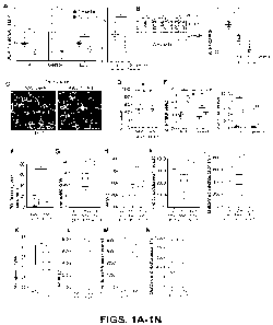
[0026] FIGs. 1A-1N show AUF1 supplementation in skeletal muscle improves
exercise
endurance in 12 and 28 month old mice. FIG. 1A. Relative expression of auf]
mRNA in the TA,
gastrocnemius, EDL and soleus muscles normalized to invariant thp mRNA at 3
and 12 months of
age in wild type (WT) mice. FIG. 1B. Representative immunoblot and
quantification of AUF1
protein levels in the TA muscle of WT mice at 3, 12 and 18 months. GAPDH is a
loading control.
n=2 mice chosen at random per group. FIG. 1C. Representative staining of AAV
GFP control and
AAV AUF1/GFP positive myofibers in TA muscle 40 d post-administration. FIG.
1D.
Quantification of GFP positive myofibers in TA muscle 40 d post-AAV
administration. n=5 mice.
FIG. 1E. Relative fold increased expression of aufl mRNA in gastrocnemius, TA,
EDL and soleus
muscles 40 d post-AAV administration. n=8-9 mice. FIGS. 1F-1J Strength and
exercise endurance
in 3 and 12 month old mice and 40 d post-AAV administration: (FIG. 1F) Grid
hanging time, (FIG.
1G) maximum speed, (FIG. 1H) work performance, (FIG. 11) time to exhaustion,
(FIG. 1J)
distance to exhaustion. n=5-9 mice. FIGS. 1K-1N Strength and exercise
endurance 6 months post-
AAV administration in 18 month old mice: (FIG. 1K) maximum speed, (FIG. 1L)
work
performance, (FIG. 1M) time to exhaustion, (FIG. 1N) distance to exhaustion.
n=4 mice. Mean
SEM from 5 or more independent studies. *P<0.05, **P<0.01 by unpaired Mann-
Whitney U test.
[0027] FIGs. 2A-20 show AUF1 gene therapy induces muscle mass along with an
increase in
myofibcr capacity. FIGs. 2A-B arc graphs showing muscle weight relative to
total body weight
40 d post-AAV administration for gastrocnemius and TA muscles, respectively.
n=8-9 mice.
FIGs. 2C-D are graphs showing frequency distribution of gastrocnemius myofiber
CSA and mean
area at 40 d post-AAV administration. n=6 mice/group. FIGs. 2E-F are graphs
showing frequency
12
CA 03226402 2024- 1- 19
WO 2023/004332
PCT/US2022/073910
distribution of TA muscle CSA and mean area at 40 d post-AAV administration.
n=5 mice. FIG.
2G show TA muscle CSA at 40 d post-AAV administration for GFP- myofibers. n=5
mice. FIG.
2H presents photographic images showing representative immunostain of slow
myofiber (red) and
nuclei (DAPI blue) in gastrocnemius muscle at 40 d post-therapy. Scale bar:
200 pm. FIGs. 21-
2K are graphs showing slow myofibers per field and mean CSA, respectively of
slow and fast
myofibers in gastrocnemius muscle at 40 d post-AAV administration. FIG. 2L
shows
representative immunostain of slow myofiber (red) and nuclei (blue) in soleus
muscle 40 d after
AAV AUF1-GFP or AAV GFP administration. FIG. 2M shows mean cross surface area
(CSA)
of slow-twitch soleus muscle myofiber 40 d after AAV AUF1 or AAV GFP
administration. n=3
mice per group. FIG. 2N shows mean soleus weight in GFP control and AUF1-GFP
AAV8 treated
animals 40 d post-gene transfer. n=4 mice per group. FIG. 20 shows
representative
immunostaining and quantification of different myofibers in the soleus muscle,
6 months post-
AUF1 gene transfer in 12 month old mice. Scale bar, 100 mm.
[0028] FIGs. 3A-3L show molecular markers of skeletal muscle myogenesis in
AAV8 AUF1-
GFP gene transferred mice. FIGs. 3A-B are graphs showing relative myh7 mRNA
levels in
gastrocnemius (FIG. 3A) and soleus (FIG. 3B) muscles normalized to invariant
nuclear TATA-
box binding protein (thp) mRNA at 40 d post-gene transfer. FIGs. 3C-D are
graphs showing
relative fast myosin mRNA levels in gastrocnemius (FIG. 3C) and soleus (FIG.
3D) muscles
normalized to >bp mRNA at 40 d gene transfer. FIG. 3E is a graph showing
expression levels of
non-mitochondrial mRNAs (pparg, six]) and mitochondrial mRNA in gastrocnemius
muscle at 40
d post-gene transfer. FIG. 3F is a graph showing the level of mitochondrial
mRNA for acadvl and
tfam in gastrocnemius and EDL muscles at 40 d post-gene transfer. FIGs. 3G and
H are graphs
showing nif/ and ntf2 mRNA levels in gastrocnemius muscle and soleus muscle,
respectively, 40
d after gene transfer. FIG. 31 is a pair of graphs showing mitochondrial DNA
content in the
gastrocnemius muscle 40 d and 6 months after gene transfer. FIG. 3J is a graph
showing
mitochondrial DNA content in the soleus muscle 40 d after gene transfer. Red
histogram, AAV
AUF1-GFP. Black histogram, AAV GFP. FIG. 3K provides representative images of
succinate
dehydrogenase (SDH) enzyme activity in TA, EDL and gastrocnemius muscles from
mice 40 d
post-administration of AAV8 GFP or AAV8 AUF1-GFP. FIG.3L shows quantitation of
SDH
positive myofibers per field for TA, EDL and gastrocnemius muscles
corresponding to (K). n=3
13
CA 03226402 2024- 1- 19
WO 2023/004332
PCT/US2022/073910
mice per muscle, 5 fields chosen at random. Mean SEM from 3 or more
independent studies.
*P<0.05; **P<0.01, ***P<0.001 by unpaired Mann-Whitney U test.
[0029] FIGs. 4A-4K show AUF1 promotes slow-twitch fiber myogenesis by
stabilizing pgcla
mRNA. FIG. 4A is a pair of graphs showing relative aufl mRNA expression in 3
and 12 month
old WT mice in TA, gastrocnemius, EDL, and soleus muscles. n=5-7 mice. FIG. 4B
shows
representative immunofluorescence staining of AUF1 expression in slow
myofibers in 3 month
old mice. FIG. 4C shows representative immunoblot of AUF1 protein level and
quantification in
TA, gastrocnemius, EDL and soleus muscle in 3 month old mice. FIG. 4D is a
graph showing
relative myh7 mRNA expression in 3 month old mouse TA, gastrocnemius, EDL, and
soleus
muscles. FIG. 4E shows relative pgc]a mRNA expression and protein levels in WT
C2C12
myoblasts and AUF1 KO myoblasts. FIG. 4F is a pair of graphs showing relative
pgcla mRNA
expression in TA, gastrocnemius, and EDL muscles 40 d post-treatment, and in
gastrocnemius at
6 months post gene transfer in 12 month old mice. FIG. 4G is a representative
immunoblot of two
AAV8-GFP control and AAV8-AUF1 GFP animals (left) and quantification of AUF1
and PGC in
in three animals per group (right) at 6 months after treatment. FIG. 4H shows
Pgcht mRNA
immunoprecipitation with endogenous AUF1 protein in myoblasts 48 h after
myotube induction
of differentiation in WT C2C12 cells. n=5. (I) pgcl a mRNA decay rate in WT
and AUF1 KO
C2C12 cells. FIG. 41 is a graph showing Pgcla mRNA immunoprecipitation with
endogenous
AUF1 protein in myoblasts 48 h after myotube induction of differentiation in
WT C2C12 cells.
n=3. FIGS. 4J and K show results with C2C12 cells overexpressing AUF1
transfected with
plasmids expressing luciferase reporters without (pIS1) and with (pIS1 pgcla
3'UTR) the pgcla
3'UTR AREs. Cells were harvested at 36 h, equal protein amounts analyzed by
immunoblot,
luciferase activity determined and luciferase mRNA levels quantified. Mean
SEM from 3 or
more independent studies. FIGS. 4 A and B: ****P<0.001 by Kruskall-Wallis
test. All other panels
by unpaired Mann¨Whitney U test *P<0.05, **P40.01. ***P<0.001. a, TA 3 vs. 12
month, b,
gastrocnemius 3 vs. 12 month, **; c, EDL 3 vs. 12 month, **; d, soleus 3 vs.
12 month, **.
[0030] FIGs. 5A-5H show loss of AUF1 expression induces atrophy of slow-twitch
myofibers.
FIG. 5A is a graph showing body weight of WT and AUF1 KO mice at 3 months.
FIG. 5B shows
TA, gastrocnemius, EDL, and soleus muscle mass in 3 month old WT and AUF1 KO
mice.
Representative image of WT and AUF1 KO soleus muscles shown. FIG. 5C shows
photographic
14
CA 03226402 2024- 1- 19
WO 2023/004332
PCT/US2022/073910
images of a representative immunostain of slow (top) or fast (bottom) myosin
(red) and laminin
(green) in the soleus muscle from 3 month old WT and AUF1 KO mice. Scale bar:
200 um. FIGs.
5D and 5E are graphs showing slow-twitch myofibers per field of percentage and
number,
respectively, in 3 month old WT and AUF1 KO mice. FIGs. 5F and 5G are graphs
showing fast-
twitch myofibers per field of percentage and number, respectively, in 3 month
old WT and AUF1
KO mice. FIG. 5H is a graph showing mean soleus slow- and fast-twitch myofiber
CSA in 3
month old WT and AUF1 KO mice, n=6-7 mice.
[0031] FIGs. 6A-6I show AUF1 deletion induces slow- and fast-twitch muscle
atrophy at 6
months of age. FIG. 6A is a graph showing body weight of WT and AUF1 KO mice
at 6 months,
n=5-6 mice. FIG. 6B shows TA, EDL, gastrocnemius, and soleus muscle weight in
6 month old
WT and AUF1 KO mice. FIG. 6C shows representative photographic images of
excised muscles
from 6 month old WT and AUF1 KO mice. FIG. 6D are photographic images showing
representative immunostain of slow myosin (red) and laminin (green) in soleus
muscle from 6
month old WT and AUF1 KO mice. Scale bar: 500 urn. FIG. 6E is a graph showing
mean CSA
of slow- and fast-twitch myofibers in soleus muscle of 6 month old WT and AUF1
KO mice. FIG.
6F is a graph showing percentage of slow-twitch myofibers in 6 month old WT
and AUF1 KO
mice in soleus muscle. FIG. 6G is a pair of photographic images showing
representative staining
of slow myosin (red) and laminin (green) in 6 month old WT and AUF1 KO
gastrocnemius muscle.
Nuclei were stained by DAPI (blue), scale bar, 200 um. FIG. 6H is a graph
showing the number
of slow-twitch myofibers per field in gastrocnemius muscle of 6 month old WT
and AUF1 KO
mice. n=4 mice per group. FIG. 61 is a graph showing mean gastrocnemius
myofiber CSA of
slow- and fast-twitch myofibers in 6 month old WT and AUF1 KO mice. n=4 mice
per group.
Mean SEM from 4 or more independent studies. *P<0.05, **P<0.01 by unpaired
Mann-Whitney
U test.
[00321 FIGs. 7A-7I show AUF1 supplementation in skeletal muscle increased Pax7
expression
in muscle and reduces markers of muscle atrophy improves exercise endurance in
12-month old
(middle-aged) and 18 month old mice. FIG. 7A presents graphs showing TA,
gastrocnemius, EDL
muscle mass, and soleus in 3, 12, and 18 month old WT mice normalized to total
body weight.
FIG. 7B is an immunoblot of AUF1 and 13-tubulin in TA muscle 40 d after AAV8
administration.
FIG. 7C shows representative immunofluorescence staining of TA muscle at 40
day post-
CA 03226402 2024- 1- 19
WO 2023/004332
PCT/US2022/073910
administration of AAV8 GFP control or AAV8 AUF1 GFP vectors, shown is DAPI
staining,
AUF1, laminin a2 to highlight myofibers and merged images. White arrows point
to nuclear
AUF1; yellow arrows point to sarcoplasmic AUF1. FIG. 7D is a graph showing
calf] mRNA
expression normalized to invariant gapdh mRNA in various organs of 12 month
old mice. 40 d
after AAV8 AUF1-GFP or AAV8 GFP control administration. FIG. 7F shows
representative
Pax7, GFP and DAPI staining in TA muscle in 12 month old mice 40 d after AAV8
AUF1-GFP
or AAV8 GFP control vector administration. Scale bar, 100 lam. Quantification
of Pax7 mRNA
expression normalized to invariant TBP mRNA. in TA muscle of 12 month old mice
40 d after
AAV8 AUF1-GFP or AAV8 GFP control vector administration. n=8-9 per mice group.
FIG. 7G
is a graph showing relative expression of Trim63 and Fbxo32 mRNAs in TA muscle
normalized
to TBP mRNA 40 d after AAV administration. FIG. 7H is a graph showing relative
expression of
Trim63 and Fbxo32 mRNAs in gastrocnemius muscle normalized to TBP mRNA 40 d
after AAV
administration. FIG. 71 shows representative co-immunostaining of Pax7 (red)
and Myf5 (purple)
showing activated satellite cells in 12 month old TA muscle of mice at 40 days
following AUF1
gene transfer. Mean SEM from 3 or more independent studies. FIG. 7A-B:
*P<0.05, *P<0.01
by Kruskall-Wallis test. All other panels *P<0.05. **P<0.01 by unpaired Mann-
Whitney U test.
[0033] FIG. 8A-8E show AUF1 controls myosin and MEF2C expression. The graphs
of FIG.
8A show relative expression of fast and slow myosin mRNAs normalized to gapdh
mRNA in
differentiating (48 h) wild type myotubes and AUF1 knock out C2C12 cells. n=5
mice per group.
FIG. 8B is a graph showing mef2c mRNA expression normalized to TBP mRNA in
gastrocnemius
muscle 40 days after AAV AUF1-GFP or AAV GFP injection. n=5 mice per group.
FIG. 8C is a
graph showing mef2c mRNA expression normalized to TBP mRNA in gastrocnemius
muscle 6
months after AAV AUF1-GFP or AAV GFP injection. n=5 mice per group. FIG. 8D
shows
representative protein levels in the gastrocnemius muscle from two mice chosen
at random at 6
months after AAV GFP (GFP) or AAV AUF-1GFP (AUF1) administration. Mean SEM
from 5
or more independent studies. *P<0.05 by unpaired Mann-Whitney U test. Ns, not
significant. FIG.
8E shows a schematic of the Renilla luciferase (RLuc) reporter construct in
plasmid pIS1
containing either the plasmid 3'UTR without ARE sequences, or as shown, the AU-
rich 3'UTR of
the pcg1 o mRNA. Red, UA-rich elements, blue, U-rich elements. Insertion sites
are indicated.
16
CA 03226402 2024- 1- 19
WO 2023/004332
PCT/US2022/073910
[0034] FIGs. 9A-9G show AUF1 deletion induces slow-twitch muscle atrophy at a
young age.
FIG. 9A shows representative photographic images of TA, EDL, and gastrocnemius
muscles in 3
month old WT and AUF1 KO mice. FIG. 9B shows representative immunostain images
of slow
and fast myosin (red) myofibers in the soleus of WT and AUF1 KO mice. DAPI
stain (blue) of
nuclei, laminin (green) stain of extracellular matrix. Scale bar: 500 pm. FIG.
9C shows
photographic images of representative stains of slow myosin (red) and laminin
(green) in 3 month
old WT and AUF1 KO gastrocnemius muscle (scale bar, 200 p.m). FIGs. 9D-E are
graphs showing
percentage and number, respectively, of slow-twitch myofibers per field in
gastrocnemius muscle
of 3 month old WT and AUF1 KO mice. FIG. 9F is a graph showing mean
gastrocnemius muscle
area of slow- and fast-twitch myofibers in 3 month old WT and AUF1 KO mice.
n=4 mice per
group. FIG. 9G shows levels of PGC la, AUF1, and control GAPDH protein in
gastrocnemius
and soleus muscles of 3 month old WT and AUF1 KO mice. Each lane corresponds
to one mouse.
Lower band in AUF1 gastrocnemius muscle lanes is a non-specific protein. Mean
SEM from 3
or more independent studies. *P<0.05 by unpaired Mann-Whitney U test. ns, (not
significant).
[0035] FIGs. 10A-10C illustrate the development of AAV8 expression vectors.
FIG. 10A is a
schematic illustration of the development of AAV8 expression vectors. The cDNA
of the murine
p40"F1 cDNA was cloned into an AAV8 vector under the tMCK promoter (AAV8-tMCK-
AUF1-
TRES-eGFP) (Vector Biolabs). The tMCK promoter was generated by the addition
of a triple
tandem of 2RS5 enhancer sequences (3-Ebox) ligated to the truncated regulation
region of the
MCK (muscle creatine kinase) promoter, which induces high muscle specificity
(Blankinship et
al., "Efficient Transduction of Skeletal Muscle Using Vectors Based on Adeno-
associated Virus
Serotype 6," Mol. Ther. 10(4):671-8 (2004), which is hereby incorporated by
reference in its
entirety). AAV8 vectors express AUF1 and GFP (AUF1-GFP, with GFP translated
from the same
mRNA by the HCV IRES), or as a control only GFP. Expression of both genes is
controlled by
the creatine kinase tMCK promoter that is selectively active in skeletal
muscle cells. The AAV8-
tMCK-lRES-eGFP construct was used as a control vector. FIG. 10B shows the
amino acid
sequence of the encoded p40AuF1 isoform (SEQ ID NO:6) expressed in transduced
cells by the
AAV8 vector in FIG. 10A. FIG. IOC shows the nucleotide sequence (SEQ ID NO:32)
of the
coding region of the p40AuF1 isoform.
17
CA 03226402 2024- 1- 19
WO 2023/004332
PCT/US2022/073910
[0036] FIGs. 11A-11B show AAV8 transduction frequency in mdx mice. AAV8 AUF1-
GFP
and AAV8 GFP control vector-treated mdx mice displayed similar vector
transduction and
retention rates, shown by tibialis anterior (TA) muscle GFP staining. FIG. 11A
shows
representative photographic images of GFP immunofluorescence staining of TA
muscle (green) to
highlight AAV8 transduction efficiency and laminin-a2 staining (red) to
highlight muscle fiber
architecture and integrity. FIG. 11B is a graph showing quantification of 3
animals per condition
for AAV8 GFP transduction in TA muscle. There is no statistical difference
(ns) in transduction
efficiency between control AAV8 GFP and treatment AAV8 AUF1 GFP groups.
[0037] FIGs. 12A-12F show AUF1 gene therapy enhances muscle mass and endurance
in !mix
mice. One month old C57BL/10ScSn male DMD mice (herein mdx mice, JACS) were
administered 2x10" genome copies of AAV8 AUF1-GFP or control AAV8 GFP as a
single retro-
orbital injection of 50 [11 containing 2.5x10" AAV particles. Two months
following AAV8
administration, mdx mice transduced with AAV8 AUF1-GFP or AAV8 GFP as a
control were
tested by standard procedures for exercise performance (see Examples, infra).
FIG. 12A is a
graph showing mdx control mice receiving only AAV8 GFP at three months old had
an average
body weight of 29 gm compared to 30 gm for wild type (WT) C57BL mice. In
contrast, when
compared to control AAV8 GFP treated mdx mice, AAV8 AUF1-GFP supplemented mdx
mice
had an average body weight of 31 gm, a significant increase compared to
control mdx mice.
FIG. 12B is a graph showing when normalized to body weight and at 2 months
post-gene
therapy transduction, AAV8 AUF1-GFP treated mdx mice demonstrated a 10%
increase in
tibialis anterior (TA) muscle mass, an 11% increase in extensor digitorum
longus (EDL) muscle
mass, and an 8.5% increase in gastrocnemius muscle mass. There was no
difference in soleus
muscle mass. Compared to control AAV8 GFP treated mdx mice. AUF1 supplemented
mdx mice
showed a ¨40% improvement in grid hanging time (FIG. 12C), a measure of limb-
girdle skeletal
muscle strength and endurance. When tested by treadmill, AAV AUF1-GFP mdx mice
displayed
16% higher maximum speed (FIG. 12D), a 35% greater time to exhaustion (FIG.
12E), and a 37%
increased distance to exhaustion (FIG. 12F). These data demonstrate a
substantial and statistically
significant increase in exercise performance and endurance in mdx mice as a
result of AUF1 gene
transfer. All results are expressed as the mean SEM. Two group comparisons
were analyzed by
the unpaired Mann-Whitney test. *, P<0.05.
18
CA 03226402 2024- 1- 19
WO 2023/004332
PCT/US2022/073910
[0038] FIGs. 13A-13D show AUF1 gene therapy does not increase WT muscle mass
or
endurance. Normal WT C57BL mice, the same background as mdx mice, were
administered at
1 month of age AAV8 GFP control or AAV8 AUF1-GFP at 2x10" genome copies by
retro-
orbital injection as described in FIGs. 12A-12F. Mice were analyzed at 3
months post-gene
transfer. These data are in contrast to the significant increase in muscle
mass and exercise
endurance found in inch mice. Rather, WT mice administered with AAV8 AUF1-GFP
compared
to control AAV8 GFP mice of the same genetic background, show no statistically
significant
increase in body weight (FIG. 13A), treadmill time to exhaustion (FIG. 13B),
maximum speed
(FIG. 13C), and distance to exhaustion (FIG. 13D). All results are expressed
as the mean SEM.
Two group comparisons were analyzed by the unpaired Mann-Whitney test. No
results were found
to be significantly different at P<0.05.
[0039] FIG. 14 shows AAV8 AUF1 gene therapy reduces serum creatine kinase
levels in mdx
mice. mdx mice at 1 month old were administered AAV8 AUF1-GFP or control AAV8
GFP as
described in FIGs. 12A-12F. At 3 months, mice were tested for levels of serum
creatine kinase
(CK) activity, a measure of sarcolemma leakiness and muscle atrophy. Top: Raw
data showing
serum CD activity results for WT control, mdx mice treated with AAV8 GFP
vector alone, and
mdx mice treated with AAV8 AUF1 GFP. Bottom: Quantification of three replicate
studies of
3 mice each. Control AAV8 GFP mdx mice displayed high levels of serum CK
activity, mdx
mice that received AAV8 AUF1-GFP gene therapy were reduced in serum CK
activity by more
than 4-fold, a highly significant reduction. WT C57BL mice had no detectable
level of serum
CK activity. ND, not detected. All results are expressed as the mean SEM.
Two group
comparisons were analyzed by the unpaired Mann-Whitney test. **, P<0.01; ***
P<0.001.
[0040] FIGs. 15A-15B show AAV8 AUF1 gene therapy reduces muscle necrosis and
fibrosis
in mdx mouse diaphragm. mdx mice at 1 month old were administered AAV8 AUF1-
GFP or
control AAV8 GFP as described in FIGs. 12A-12F. At 3 months, diaphragms were
reduced
from AAV8 GFP control and AAV8 AUF1-GFP mice, embedded FFPE and stained with
H&E
(FIG. 15A). The percent degenerative diaphragm muscle was scored and found to
be reduced
by 74% by AUF1 gene transfer. WT C57BL mouse diaphragm served as a control.
Diaphragm
muscle from mdx mice was stained with Masson Trichome to quantify muscle
fibrosis (FIG.
15B). Shown are representative muscle sections. AUF1 gene transfer reduced
fibrosis by 2-fold
19
CA 03226402 2024- 1- 19
WO 2023/004332
PCT/US2022/073910
compared to control AAV8 GFP treated animals. All results are expressed as the
mean SEM.
Two group comparisons were analyzed by the unpaired Mann-Whitney test. **,
P<0.01.
Otherwise analyzed by Fisher Exact test as indicated.
[0041] FIGs. 16A-16B show AAV8 AUF1 gene therapy reduces muscle immune cell
invasion.
mdx mice at 1 month old were administered AAV8 AUF1-GFP or control AAV8 GFP as
described in FIGs. 12A-12F. At 3 months, diaphragms were resected from AAV8
GFP control
and AAV8 AUF1-GFP treated mice, embedded in FFPE, and stained with an antibody
to the
macrophage biomarker CD68 coupled with the red fluorescence marker Alexa Fluor
555.
Representative images show strong reduction in macrophage CD68 staining in
AAV8 AUF1-
GFP treated animals compared to AAV8 GFP controls (FIG. 16A). Quantification
of 5 fields
per specimen from 3 mice per group for CD68 staining (FIG. 16B). All results
are expressed as
the mean SEM. Two group comparisons were analyzed by the unpaired Mann-
Whitney test. *,
P<0.05.
[0042] FIGs. 17A-17E show AAV8 AUF1 gene therapy suppresses expression of
embryonic
myosin heavy chain (eMHC) in mdx mice. eMHC is a clinical marker of muscle
degeneration
in DMD. mdx mice at 1 month old were administered AAV8 AUF1-GFP or control
AAV8 GFP
as described in FIGs. 12A-12F. At 3 months, diaphragm muscle was removed,
fixed in FFPE,
and stained with antibodies to eMHC (green), nuclei (DAPI, blue), and laminin
(red).
Immunofluorescence was carried out and representative images shown compared to
WT
C57BL6 mice (FIG. 17A). AAV8 AUF1-GFP gene transfer strongly reduced eHMC
expression
in diaphragm. High magnification of diaphragm stained as in FIG. 17A showing
strong
reduction in eMHC expression by AUF1 gene transfer (FIG. 17B). Quantification
of eMHC
staining in myofibers, showing a 75% reduction in eMHC expression by AUF1 gene
transfer
(FIG. 17C). The percent of centro-nuclei per myofiber/field was quantified, a
measure of
normal muscle fiber maturation (FIG. 17D). AUF1 gene transfer reduced the
percentage of
centro-nuclei by 52% compared to AAV8 GFP controls. Myofiber cross sectional
area (CSA)
was quantified (FIG. 17E). AUF1 gene transfer strongly increased the CSA of
the larger
myofibers, indicative of mature regenerative muscle. All results arc expressed
as the
mean SEM. Two group comparisons were analyzed by the unpaired Mann-Whitney
test.
Multiple group comparisons were performed using one-way analysis of variance
(ANOVA). The
CA 03226402 2024- 1- 19
WO 2023/004332
PCT/US2022/073910
non-parametric Kruskal¨Wallis test followed by the Dunn's comparison of pairs
was used to
analyze groups when suitable. *, P<0.05; *** P<0.001.
[00431 FIGs. 18A-18C show AAV8 AUF1 gene transfer increases expression of
endogenous
utrophin-A in mdx mice. mdx mice at 1 month old were administered AAV8 AUF1-
GFP or
control AAV8 GFP as described in FIGs. 12A-12F. The gastrocnemius muscle was
removed at
3 months, fixed in FFPE, and stained with DAPI (blue for nuclei, antibodies to
utrophin (red)
and laminin (green) (FIG. 18A). Representative images from 3 mice for each
group are shown.
AUF1 gene therapy strongly increased expression of utrophin and showed
evidence for
normalization of myofiber integrity (laminin staining). Immunoblot analysis
for utrophin,
AUF1, and GAPDH (invariant control) proteins was conducted on the
gastrocnemius muscle of
3 AAV8 GFP and 3 AAV8 AUF1-GFP mdx mice at 3 months (FIG. 18B). Gastrocnemius
utrophin protein levels were increased by an average of 20-fold in animals
receiving AUF1
gene therapy. AUF1 protein levels were increased an average of 3-4 fold.
Utrophin mRNA
levels were quantified by qRT-PCR and normalized to invariant TBP mRNA (FIG.
18C). There
was no statistically significant difference between samples. n=3 animals for
each condition.
[0044] FIGs. 19A-19C show AAV8 AUF1 gene transfer increases expression of
satellite cell
activation gene Pax7, key muscle regeneration genes pgcl a and mef2c, slow
twitch
determination genes and mitochondrial DNA content in mdx mice. mdx mice at 1
month old
were administered AAV8 AUF1-GFP or control AAV8 GFP as described in FIGs. 12A-
12F.
The gastrocnemius muscle was removed at 3 months, mRNA extracted and
quantified by qRT-
PCR relative to invariant tbp mRNA. AUF1 gene therapy increased expression of
pgcl a,
mef2c, and Pax7 mRNAs in the gastrocnemius of mdx mice relative to controls
receiving vector
alone (FIG. 19A). Wild type non-mdx animals (WT) served as a control for
normal muscle
levels in age-matched animals. AAV8 AUF1 gene therapy restored near WT levels
or exceeded
WT levels of gene expression. AUF1 gene therapy increased expression of slow-
twitch lineage
determination myosin mRNAs in the gastrocnemius muscle in mdx animals relative
to controls
receiving vector alone (FIG. 19B). AAV8 AUF1 gene therapy restored near WT
levels or
exceeded WT levels of gene expression. AUF1 gene therapy increased expression
of
mitochondrial DNA in the gastrocnemius muscle of mdx mice, consistent with
increased slow-
twitch muscle mass (FIG. 19C). All results are expressed as the mean SEM.
Two group
21
CA 03226402 2024- 1- 19
WO 2023/004332
PCT/US2022/073910
comparisons were analyzed by the unpaired Mann-Whitney test. *, P<0.05; **,
P<0.01; ***
P<0.001.
[0045] FIG. 20 shows genome-wide transcriptomic and translatomic studies
demonstrate AUF1
activation of C2C12 myoblast muscle fiber development. Proliferating C2C12
mouse cardiac
myoblasts were transduced with lentivirus control vectors or lentivirus
vectors expressing p45
AUF1, and induced to differentiate into myotubes by culturing in
differentiation medium as
described in the Examples infra. Proliferating myoblasts were used because
they are activated in
p38 MAPK and other signaling pathways that promote myogenesis, which is
representative of the
activated state and population of muscle cells following muscle damage from
wounding, or the
state of muscle in myogenic diseases, such as chronic regenerative attempts
that occur in Duchene
Muscular Dystrophy (DMD). Overview of the experimental approach. At 48 h, when
myotubes
begin to form, polyribosomes were separated by sucrose sedimentation
corresponding to poorly
translated (2 & 3 ribosome) fraction and well translated (>4 polysome)
fractions, total mRNA and
mRNA in polyribosome fractions were independently purified (polyA+ fraction
devoid of rRNA),
bacterial libraries were generated and subjected to deep sequencing using
RNAseq, in two
independent studies. Genome-wide mRNA abundance used log, ratios of
translated/total mRNA.
Procedures and bioinformatic pipeline used for analysis arc described in the
Examples infra.
[0046] FIGs. 21A-21B show AUF1 supplementation stimulates expression of major
muscle
development pathways and decreases expression of inflammatory cytokine,
inflammation, cell
proliferation, cell death, and anti-muscle regeneration pathways. Data from
FIG. 20 genome-wide
mRNA expression and translation analysis. Major upregulated pathways at the
levels of
transcription, translation, or both with AUF1 supplementation in C2C12
myoblasts (FIG. 21A).
Analyzed by KEGG. Major downregulated pathways at the levels of transcription,
translation, or
both with AUF1 supplementation in C2C12 myoblasts (FIG. 21B). Analyzed by
KEGG.
[0047] FIGs. 22A-22B show AUF1 supplementation of C2C12 myoblasts upregulates
pathways
for major biological processes and molecular functions in muscle development
and regeneration.
Data from FIG. 20 genome-wide mRNA expression and translation analysis. Major
upregulated
biological processes at the levels of transcription, translation, or both with
AUF1 supplementation
in C2C12 myoblasts (FIG. 22A). Analyzed by KEGG. Major upregulated molecular
functions at
22
CA 03226402 2024- 1- 19
WO 2023/004332
PCT/US2022/073910
the levels of transcription, translation, or both with AUF1 supplementation in
C2C12 myoblasts
(FIG. 22B). Analyzed by KEGG.
[0048] FIGs. 23A-23B show AUF1 supplementation of C2C12 myoblasts decreases
muscle
inflammation, inflammatory cytokine, and signaling pathways that oppose muscle
regeneration.
Data from FIG. 20 genome-wide mRNA expression and translation analysis. Major
downregulated biological processes at the levels of transcription,
translation, or both with AUF1
supplementation in C2C12 myoblasts (FIG. 23A). Analyzed by KEGG. Major
downregulated
molecular functions at the levels of transcription, translation, or both with
AUF1 supplementation
in C2C12 myoblasts (FIG. 23B). Analyzed by KEGG.
[0049] FIG. 24 shows AUF1 supplementation of C2C12 myoblasts decreases
expression of
muscle genes associated with development of fibrosis. Data from FIG. 20 genome-
wide mRNA
expression and translation analysis. Major downregulated pathways and
functions at the levels of
transcription, translation, or both with AUF1 supplementation in C2C12
myoblasts. Analyzed by
KEGG.
[00501 FIGs. 25A-25D show lentivirus transduction of injured TA muscle with
p45 AUF1 in
mice activates satellite cells and reduces biomarkers of muscle atrophy. A
lentivirus vector was
developed expressing cDNA for p45 AUF1 under control of the CMV promoter
(Abbadi et al.,
"Muscle Development and Regeneration Controlled by AUF1-mediated Stage-
specific
Degradation of Fate-determining Checkpoint mRNAs," Proc. Nat'l. Acad. Sci. USA
116:11285-
90 (2019), which is hereby incorporated by reference in its entirety). Three
month old male mice
were administered an intramuscular injection of 50 d of filtered 1.2% BaC12 in
sterile saline with
control lentivirus vector or with lentivirus AUF1 vector (1x10⁸ genome copies)
(total volume 100
IA) into the left Tibialis Anterior (TA) muscle (FIG. 25A). The right TA
muscle remained
uninjured as a control. Mice were sacrificed at 7 days post-injection. TA
muscles were excised,
weighed, and normalized to mouse body weight in grams. TA injury reduced TA
weight by 27%
which was restored to near-uninjured levels by concurrent AUF1 gene therapy.
In FIG. 25B,
immunoblot analysis of AUF1 normalized to invariant GAPDH protein for TA
muscle at 7 days
post-lentivirus p45 AUF1 administration as in FIG. 25A. Shown is a
representative uninjured, two
injured, and injured TA muscles with concurrent p4-5 AUF1 gene therapy from
independent
23
CA 03226402 2024- 1- 19
WO 2023/004332
PCT/US2022/073910
animals. Lentivims p45 AUF1 gene transfer strongly increased levels of the p45
AUFI isoform but
not p42 "Fl and p40 AUF1 that were not encoded (p37 "Fl is undetectable). In
FIG. 25C, TA
muscles as in FIG. 25A were probed by qRT-PCR for Pax7 mRNA levels, a
biomarker of muscle
satellite (stem) cell activation, and normalized to invariant TATA-box binding
protein (TBP)
mRNA. AUF1 gene therapy increased Pax7 expression by >3-fold. In FIG. 25D, TA
muscles as
in FIG. 25A were probed by qRT-PCR for expression of muscle atrophy biomarker
genes TRIM63
and Fbxo32, normalized to TBP mRNA. TA muscle injury strongly induced
expression of
TRIM63 and Fbxo32 mRNA, which were downregulated to uninjured TA muscle levels
by p45
AUF1 gene therapy, indicating strong cessation of muscle injury due to AUF1
intramuscular
administration. No statistical difference (ns). All results are expressed as
the mean SEM with
at least three independent trials of 3 or more animals per condition. Two
group comparisons were
analyzed by the unpaired Mann-Whitney test. *, P<0.05; **, P<0.01; ***,
P<0.001.
[0051] FIGs. 26A-26D show p45 AUF1 lentivirus transduction enhances expression
of muscle
regeneration factors (MRFs) following TA muscle injury. Three month old male
mice were
injured in the TA muscle with BaCt, and administered with an intramuscular
injection of control
lentivirus vector or lentivirus AUF1 vector (see FIGs. 25A-D). Mice were
sacrificed at 7 days
post-injection. TA muscles were probed by qRT-PCR for identified niRNAs
normalized to
invariant TBP mRNA. In FIG. 26A, myogenin and MyoD mRNA levels, biomarkers of
myoblast
activation, differentiation, and muscle regeneration (Zammit, "Function of the
Myogenic
Regulatory Factors Myf5, MyoD, Myogenin and MRF4 in Skeletal Muscle, Satellite
Cells and
Regenerative Myogenesis," Semin. Cell. Dev. Biol. 72:19-32 (2017), which is
hereby incorporated
by reference in its entirety), were increased ¨2-fold by AUF1 gene therapy
relative to injured
control vector specimens. In FIG. 25B, myh8 mRNA, an embryonic myosin only
expressed in
adult muscle during muscle regeneration and a marker of co-expression of
utrophin (Guiraud et
al., "Embryonic Myosin is a Regeneration Marker to Monitor Utrophin-based
Therapies for
DMD," Hunt. Mol. Genet. 28:307-19 (2019), which is hereby incorporated by
reference in its
entirety), was increased in expression by 5-fold in injured muscle with AUF1
gene therapy relative
to injured control vector specimens. In FIG. 26C, myh7 mRNA, a myosin that
specifies slow-
twitch muscle (Zammit, "Function of the Myogenic Regulatory Factors Myf5,
MyoD, Myogenin
and MRF4 in Skeletal Muscle, Satellite Cells and Regenerative Myogenesis,"
Semin. Cell. Dev.
24
CA 03226402 2024- 1- 19
WO 2023/004332
PCT/US2022/073910
Biol. 72:19-32 (2017), which is hereby incorporated by reference in its
entirety), was increased in
expression by -2-fold in injured muscle with AUF1 gene therapy relative to
injured control vector
specimens. In FIG. 26D, myh4 mRNA, a myosin that specifies fast-twitch muscle
(Zammit,
"Function of the Myogenic Regulatory Factors Myf5, MyoD, Myogenin and MRF4 in
Skeletal
Muscle, Satellite Cells and Regenerative Myogenesis," Semin. Cell. Dev. Biol.
72:19-32 (2017),
which is hereby incorporated by reference in its entirety), was increased in
expression by -2-fold
in injured muscle with AUF1 gene therapy relative to injured control vector
specimens. All results
are expressed as the mean SEM with at least three independent trials of 3 or
more animals per
condition. Two group comparisons were analyzed by the unpaired Mann-Whitney
test. *, P<0.05;
**, P<0.01.
[0052] FIGs. 27A-27D show p4-5 AUF1 lentivirus gene therapy promotes rapid
regeneration of
injured muscle. Three month old male mice were injured in the TA muscle with
BaC11, and
administered with an intramuscular injection of control lentivirus vector or
lentivirus AUF1 vector,
as in FIGS. 25A-25D. Mice were sacrificed at 3 days and 7 days post-injury.
FIG. 27A shows
photographic images of muscle fibers provide evidence for accelerated but
normal muscle
regeneration of myofibers in animals administered lentiviral AUF1 gene
therapy. TA muscle in
OCT was sectioned and stained for immunofluorescence microscopy analysis for
Laminin alpha
2 (red), Nuclei are stained with DAPI (blue). Note the disrupted myofiber
architecture and high
level of central nuclei in the injured TA muscle treated with vector alone
compared to the injured
TA muscle administered lentiviral AUF1 gene therapy, consistent with
accelerated muscle
regeneration and mature myofibers. Scale bar, 200 pm. FIG. 27B is a graph
showing the percent
muscle loss (atrophy) or gain (increase in mass) determined for the injured TA
muscle compared
to uninjured control or injured muscle receiving control lentivirus vector or
lentivirus p45 AUF1,
measured at sacrifice at 3 days and 7 days post-injury. Injured TA muscle
receiving sham gene
therapy sustained a 20% loss in mass by day 3 following injury, which only
very slightly improved
by day 7. In contrast, injured TA muscle receiving AUF1 gene therapy showed a
trend to less
atrophy by day 3, which was almost fully recovered by day 7, demonstrating
near normal mass.
FIG. 27C is a graph showing high levels of myotube central nuclei are a marker
of immature
myofiber development (Yin et al., "Satellite Cells and the Muscle Stem Cell
Niche," Physiol. Rev.
93:23-67 (2013) and Schiaffino & Reggiani, "Fiber Types in Mammalian Skeletal
Muscles,"
CA 03226402 2024- 1- 19
WO 2023/004332
PCT/US2022/073910
Physiol. Rev. 91:1447-531 (2011), which are hereby incorporated by reference
in their entirety).
TA muscle analyzed at day 7 post-injury administered p45 AUF1 gene therapy
were reduced by
half in the percent of myofibers with central nuclei compared to vector only
control injured muscle.
This is consistent with accelerated muscle regeneration provided by AUF1 gene
transfer. FIG.
27D is a graph showing a wider cross-sectional area of myofibers (cross-
sectional area, CS A) with
low numbers of central nuclei are indicative of mature myofiber development
(Yin et al., "Satellite
Cells and the Muscle Stem Cell Niche," Physiol. Rev. 93:23-67 (2013) and
Schiaffino & Reggiani,
"Fiber Types in Mammalian Skeletal Muscles," Physiol. Rev. 91:1447-531(2011),
which are
hereby incorporated by reference in their entirety). AUF1 gene transfer in
injured TA muscle
produced a striking increase in CSA with reduced central nuclei per myofiber,
consistent with
generation of mature myofibers. All results are expressed as the mean SEM
with at least three
independent trials of 3 or more animals per condition. Two group comparisons
were analyzed by
the unpaired Mann-Whitney test. *, P<0.05; **, P<0.01, ***, P<0.001.
[0053] FIGs. 28A-28F show AUF1 is essential to promote repair of injured
muscle, and can
provide injury protection benefit when delivered by AAV8 gene transfer. FIG.
28A is a schematic
illustration of an AUF1 conditional knockout mouse developed as an aspect of
the technology
described herein. Shown is a schematic of the exon 3 LoxP site insertions in
the AUF1 gene. Lox
sites were cloned to flank exon 3 of AUF1, which is maintained in all 4 AUF1
isoforms and
contains the RNA binding domain. AuFiFlox/Flox mice were derived, siblings
mated to homogeneic
me
purity generated, then mated with a Pax7cre ERT2 (B6;129_pax72 1(cr /ERT2
)Fan/J mouse) (Jackson
Labs). This provides cre recombinase induction by tamoxifen administration
only in PAX7+
expressing muscle satellite and myoblast cells. FIG. 28B is a graph showing
results of three month
old mice induced for cre expression with 5 daily i.p. injections of tamoxifen
(3 mg/kg). There was
no change in body weight of cre-induced mice. FIG. 28C is a graph showing
weight of non-injured
skeletal muscles in mice were not significantly different in uninduced and
tamoxifen induced cre
mice. FIG. 28D shows tamoxifen induction of cre for 3 months specifically
deletes the aufl gene
in skeletal muscle and abolishes skeletal muscle AUF1 protein expression. A
representative
immunoblot is shown for AUF1 levels in TA skeletal muscle and kidney,
normalized to invariant
GAPDH in control AUF1F10/"' and AUF1 1F ox/Flox x pAx-cre
ERT2 mice after 5 days of cre
induction and analyzed at day 7. There is no evidence for expression of AUF1
after Pax7-specific
cre induction in muscle, whereas abundant AUF1 is present in kidney. FIG. 28E
is a graph
26
CA 03226402 2024- 1- 19
WO 2023/004332
PCT/US2022/073910
showing one month old AUF1Fl0x/Flox x PAX7"e ERT2 mice were either sham
injected or injected
with tamoxifen for 5 days as above, then maintained on a diet that included
oral tamoxifen for 5
months daily at 500 mg/kg (Envigo). Wild type (WT) BL6 mice and AUF1Flo'dFlox
x PAX7"'ERT2
mice were either not induced for cre-expression (labeled AUF1flifliP"7) or
induced for 5 months
and deleted in the AUF1 gene (labeled AAUF111"Pax7). One set of AAUF1fiffi/P"7
mice induced for
cre expression for 5 months were also administered at 1 month of age with
2.0x10" AAV8 AUF1
particles (2x10" genome copies) by single retro-orbital injection of 50 lid.
All mice were then
injured by 1.2% BaC12 injection in the TA muscle, as described in FIGs. 25A-D.
AUF1 is
controlled by the creatine kinase tMCK promoter that is selectively active in
skeletal muscle cells.
TA muscle was excised at 7 days post-BaC12 injection and the percent of muscle
atrophy
determined by weight. TA muscle of AUF1Fl0x/Fl0x x pAx-,cre
ERT2 mice expressing AUF1 and
WT mice expressing AUF1 (not induced for cre) showed 16-18% atrophy that was
not statistically
different. In contrast, deletion of the AUF1 gene caused strongly increased
atrophy of the TA
muscle, doubling atrophy levels to 35%. However, animals deleted for the AUF1
gene but
prophylactically administered AAV8 AUF1 gene therapy demonstrated dramatically
reduced
levels of TA muscle atrophy, averaging -3%. FIG. 28F is a graph showing AUF1
control and cre-
induced skeletal muscle AUF1 deleted mice were tested at 5 months for grip
strength, a measure
of limb-girdle skeletal muscle strength and endurance. AUF1 deleted mice
showed a -50%
reduction in grip strength. Collectively, these data demonstrate that AUF1 is
essential for
maintenance of muscle strength and muscle regeneration following injury, and
that AUF1 gene
therapy provides a remarkable ability to promote muscle regeneration and
protect muscle from
extensive damage despite traumatic injury. All results are expressed as the
mean SEM with at
least three independent trials of 3 or more animals per condition. Two group
comparisons were
analyzed by the unpaired Mann-Whitney test. *, P<0.05.
[0054] FIGS 29A and 29B. FIG. 29A shows that supplemental expression of AUF1
(p40)
accelerates the formation of myotubes from wild type C2C12 myoblast cells in
culture. FIG. 29B
provides RNA sequence analysis of the wild type C2C12 myoblast cultures after
differentiation
into myotubes as compared to vector control samples. Results show AUF1
supplementation
stimulates expression of slow muscle specification genes.
27
CA 03226402 2024- 1- 19
WO 2023/004332
PCT/US2022/073910
[0055] FIGS 30A-E. FIG. 30A shows that prophylactic administration of AAV8-
mAUF1
significantly decreases the percent of muscle atrophy compared to WT control
mice measured at
7 d and 14 d post-BaC12 induction of muscle necrosis. FIG. 30B is a histogram
that quantifies
centrally located nuclei compared to mean cross-sectional area (csa) of muscle
fibers. Results show
that the greatest central nuclei with the greatest csa is in muscle of mAUF1-
treated animals at 14
d post-injury. FIGs. 30C-E are raw data plots used to derive the summary
histogram shown in FIG.
30B.
5. DETAILED DESCRIPTION
[0056] Provided are methods and compositions for promoting muscle
regeneration, restoring or
increasing muscle mass, muscle function or performance, and/or reducing or
reversing muscle
atrophy by increasing the levels of AUF1 in muscle cells in a subject in need
thereof. Methods
and compositions are provided for administering AUF1 protein or nucleic acid
that encodes and
expresses AUF1 protein in muscle cells, such as DNA, naRNA, plasmid DNA or
viral vectors
encoding AUF1. Provided are compositions comprising, and methods of
administering, gene
therapy vectors, particularly recombinant AAV vectors, comprising genomes with
transgenes
encoding AUF1 proteins operably linked to regulatory elements that promote
AUF1 expression in
muscle cells for restoring or increasing muscle mass, muscle function or
performance, and/or
reducing or reversing muscle atrophy. Such methods include stabilizing the
sarcolemma of the
muscle cell by reducing leakiness (for example, as measured by creatine kinase
levels), increasing
expression of 13-sarcoglycan or utrophin or other components of the dystrophin-
glycoprotein
complex (including a-dystroglycan,13-dystroglycan, a-
sarcoglycan,13¨sarcoglycan, 6-sarcoglycan,
y-sarcoglycan, c-Sarcoglycan, c-sarcoglycan, a-dystroglycan, 13-dystroglycan,
sarcospan, a-
syntrophin, 13- syntrophin, a-dystrobrevin, 13-dystrobrevin, caveolin-3, or
nNOS) and/or their
presence in the dystrophin-glycoprotein complex of muscle cells by providing
AUF1 protein,
including by gene therapy methods, such as A AV gene therapy. Other methods
provided include
treatment, prevention or amelioration of the symptoms of muscle wasting
including sarcopenia,
including in the elderly, traumatic muscle injury, and diseases or disorders
associated with a lack
or loss of muscle mass, function or performance, such as, but not limited to
dystrophinopathies
and other related muscle diseases or disorders. Such methods include promoting
an increase in
muscle cell mass, number of muscle fibers, size of muscle fibers, muscle cell
regeneration,
28
CA 03226402 2024- 1- 19
WO 2023/004332
PCT/US2022/073910
reduction in or reverse of muscle cell atrophy, satellite cell activation and
differentiation,
improvement in muscle cell function (for example, by increasing mitochondrial
oxidative
capacity), and increasing proportion of slow twitch fiber in muscle (including
by conversion of
fast to slow twitch muscle fibers). Other methods disclosed herein include
methods of producing
synthetic or cultured meat by promoting muscle cell formation, for example, in
vitro in cell culture
and particularly producing slow twitch muscle fibers, including converting
fast twitch to slow
twitch muscle fibers in the cultured muscle cells, thereby providing an
improved cultured meat
product. In addition, other methods disclosed herein include methods of
producing synthetic
muscle tubes, muscle fiber and muscle by promoting muscle cell formation, for
example, in vitro
in cell culture and particularly producing slow twitch muscle fibers,
including converting fast
twitch to slow twitch muscle fibers in the cultured muscle cells, thereby
providing an improved
cultured muscle composition and uses thereof.
[0057] Such methods may be carried out by administration of AUF1 (including
mouse or human
p37AUF1, p40AUF1, p42AUF1 or p45AUF1), including administering a gene therapy
vector,
such as a lentiviral vector or a recombinant AAV gene therapy vector
comprising a nucleic acid
encoding the mouse or human AUF1, or a functional fragment thereof, operably
linked to
regulatory elements promoting AUF1 expression in muscle cells. Compositions
comprising rAAV
comprising a genome comprising a transgene encoding human AUF1, or a
functional fragment
thereof, operably linked to regulatory elements that promote expression of the
AUF1 encoding
nucleic acid in muscle cells are further provided.
5.1.Definitions
[0058] The term -vector" is used interchangeably with -expression vector." The
term -vector"
may refer to viral or non-viral, prokaryotic or eukaryotic, DNA or RNA
sequences that are capable
of being transfected into a cell, referred to as "host cell," so that all or a
part of the sequences are
transcribed. It is not necessary for the transcript to be expressed. It is
also not necessary for a
vector to comprise a transgene having a coding sequence. Vectors are
frequently assembled as
composites of elements derived from different viral, bacterial, or mammalian
genes. Vectors
contain various coding and non-coding sequences, such as sequences coding for
selectable
markers, sequences that facilitate their propagation in bacteria, or one or
more transcription units
that are expressed only in certain cell types. For example, mammalian
expression vectors often
29
CA 03226402 2024- 1- 19
WO 2023/004332
PCT/US2022/073910
contain both prokaryotic sequences that facilitate the propagation of the
vector in bacteria and one
or more cukaryotic transcription units that are expressed only in cukaryotic
cells. It will be
appreciated by those skilled in the art that the design of the expression
vector can depend on such
factors as the choice of the host cell to be transformed, the level of
expression of protein desired,
etc.
[0059] The term "promoter" is used interchangeably with "promoter element" and
"promoter
sequence." Likewise, the term "enhancer" is used interchangeably with
"enhancer element" and
"enhancer sequence." The term "promoter" refers to a minimal sequence of a
transgene that is
sufficient to initiate transcription of a coding sequence of the transgene.
Promoters may be
constitutive or inducible. A constitutive promoter is considered to be a
strong promoter if it drives
expression of a transgene at a level comparable to that of the cytomegalovirus
promoter (CMV)
(Boshart et al., "A Very Strong Enhancer is Located Upstream of an Immediate
Early Gene of
Human Cytomegalovirus," Cell 41:521 (1985), which is hereby incorporated by
reference in its
entirety). Promoters may be synthetic, modified, or hybrid promoters.
Promoters may be coupled
with other regulatory sequences/elements which, when bound to appropriate
intracellular
regulatory factors. enhance ("enhancers") or repress ("repressors") promoter-
dependent
transcription. A promoter, enhancer, or repressor, is said to be "operably
linked" to a transgene
when such element(s) control(s) or affect(s) transgene transcription rate or
efficiency. For
example, a promoter sequence located proximally to the 5' end of a transgene
coding sequence is
usually operably linked with the transgene. As used herein, the term
"regulatory elements" is used
interchangeably with "regulatory sequences" and refers to promoters,
enhancers, and other
expression control elements, or any combination of such elements.
[0060] Promoters are positioned 5' (upstream) to the genes that they control.
Many eukaryotic
promoters contain two types of recognition sequences: TATA box and the
upstream promoter
elements. The TATA box. located 25-30 bp upstream of the transcription
initiation site, is thought
to be involved in directing RNA polymerase II to begin RNA synthesis at the
correct site. In
contrast, the upstream promoter elements determine the rate at which
transcription is initiated.
These elements can act regardless of their orientation, but they must be
located within 100 to 200
bp upstream of the TATA box.
CA 03226402 2024- 1- 19
WO 2023/004332
PCT/US2022/073910
[0061] Enhancer elements can stimulate transcription up to 1000-fold from
linked homologous
or heterologous promoters. Enhancer elements often remain active even if their
orientation is
reversed (Li et al., "High Level Desmin Expression Depends on a Muscle-
Specific Enhancer," J.
Bio. Chem. 266(10):6562-6570 (1991), which is hereby incorporated by reference
in its entirety).
Furthermore, unlike promoter elements, enhancers can be active when placed
downstream from
the transcription initiation site, e.g., within an intron, or even at a
considerable distance from the
promoter (Yutzey et al., "An Internal Regulatory Element Controls Troponin I
Gene Expression,"
Mol. Cell. Bio. 9(4):1397-1405 (1989), which is hereby incorporated by
reference in its entirety).
[0062] The term "muscle cell-specific" refers to the capability of regulatory
elements, such as
promoters and enhancers, to drive expression of an operatively linked nucleic
acid molecule (e.g.,
a nucleic acid molecule encoding an AU-rich mRNA binding factor 1 (AUF1)
protein or a
functional fragment thereof) exclusively or preferentially in muscle cells or
muscle tissue.
[0063] The term "AAV" or "adeno-associated virus" refers to a
Dependoparvovirus within the
Parvoviridae genus of viruses. The AAV can be an AAV derived from a naturally
occurring "wild-
type" virus, an AAV derived from a rAAV genome packaged into a capsid
comprising capsid
proteins encoded by a naturally occurring cap gene and/or from a rAAV genome
packaged into a
capsid comprising capsid proteins encoded by a non-naturally occurring capsid
cap gene. An
example of the latter includes a rAAV having a capsid protein having a
modified sequence and/or
a peptide insertion into the amino acid sequence of the naturally-occurring
capsid.
[0064] The term "rAAV" refers to a "recombinant AAV." In some embodiments, a
recombinant
AAV has an AAV genome in which part or all of the rep and cap genes have been
replaced with
heterologous sequences, including a transgene, such as nucleotide sequence
encoding AUF1, and
regulatory elements for expression of the transgene.
[0065] The term "rep-cap helper plasmid" refers to a plasmid that provides the
viral rep and cap
gene function and aids the production of AAVs from rAAV genomes lacking
functional rep and/or
the cap gene sequences.
31
CA 03226402 2024- 1- 19
WO 2023/004332
PCT/US2022/073910
[0066] The term "cap gene" refers to the nucleic acid sequences that encode
capsid proteins that
form or help form the capsid coat of the virus. For AAV, the capsid protein
may be VP1, VP2, or
VP3.
[0067] The term -rep gene" refers to the nucleic acid sequences that encode
the non-structural
protein needed for replication and production of virus.
[0068] The terms "nucleic acids" and "nucleotide sequences" include DNA
molecules (e.g.,
cDNA or genomic DNA), RNA molecules (e.g., mRNA), combinations of DNA and RNA
molecules or hybrid DNA/RNA molecules, and analogs of DNA or RNA molecules.
Such analogs
can be generated using, for example, nucleotide analogs, which include, but
are not limited to,
inosine or tritylated bases. Such analogs can also comprise DNA or RNA
molecules comprising
modified backbones that lend beneficial attributes to the molecules such as,
for example, nuclease
resistance or an increased ability to cross cellular membranes. The nucleic
acids or nucleotide
sequences can be single-stranded, double-stranded, may contain both single-
stranded and double-
stranded portions, and may contain triple-stranded portions, but preferably is
double-stranded
DNA.
[0069] Amino acid residues as disclosed herein can be modified by conservative
substitutions
to maintain, or substantially maintain, overall polypeptide structure and/or
function. As used
herein, "conservative amino acid substitution" indicates that: hydrophobic
amino acids (i.e., Ala,
Cys, Gly, Pro, Met, Val, lie, and Leu) can be substituted with other
hydrophobic amino acids;
hydrophobic amino acids with bulky side chains (i.e., Phe, Tyr, and Trp) can
be substituted with
other hydrophobic amino acids with bulky side chains; amino acids with
positively charged side
chains (i.e., Arg, His. and Lys) can be substituted with other amino acids
with positively charged
side chains; amino acids with negatively charged side chains (i.e., Asp and
Glu) can be substituted
with other amino acids with negatively charged side chains; and amino acids
with polar uncharged
side chains (i.e., Ser, Thr, Asn, and Gln) can be substituted with other amino
acids with polar
uncharged side chains.
[0070] The terms "subject", "host", and "patient" are used interchangeably. A
subject may be a
mammal such as a non-primate (e.g., cows, pigs, horses, cats. dogs, rats etc.)
or a primate (e.g.,
monkey and human), and includes a human.
32
CA 03226402 2024- 1- 19
WO 2023/004332
PCT/US2022/073910
[0071] The terms "therapeutic agent" refers to any agent which can be used in
treating,
managing, or ameliorating symptoms associated with a disease or disorder,
where the disease or
disorder is associated with a function to be provided by a transgene. A
"therapeutically effective
amount" refers to the amount of agent, (e.g., an amount of product expressed
by the transgene)
that provides at least one therapeutic benefit in the treatment or management
of the target disease
or disorder, when administered to a subject suffering therefrom. Further, a
therapeutically
effective amount with respect to an agent of the invention means that amount
of agent alone, or
when in combination with other therapies, that provides at least one
therapeutic benefit in the
treatment or management of the disease or disorder.
[0072] The term "prophylactic agent" refers to any agent which can be used in
the prevention,
reducing the likelihood of, delay, or slowing down of the progression of a
disease or disorder,
where the disease or disorder is associated with a function to be provided by
a transgene. A
"prophylactically effective amount" refers to the amount of the prophylactic
agent (e.g., an amount
of product expressed by the transgene) that provides at least one prophylactic
benefit in the
prevention or delay of the target disease or disorder, when administered to a
subject predisposed
thereto. A prophylactically effective amount also may refer to the amount of
agent sufficient to
prevent, reduce the likelihood of, or delay the occurrence of the target
disease or disorder; or slow
the progression of the target disease or disorder; the amount sufficient to
delay or minimize the
onset of the target disease or disorder; or the amount sufficient to prevent
or delay the recurrence
or spread thereof. A prophylactically effective amount also may refer to the
amount of agent
sufficient to prevent or delay the exacerbation of symptoms of a target
disease or disorder. Further,
a prophylactically effective amount with respect to a prophylactic agent of
the invention means
that amount of prophylactic agent alone, or when in combination with other
agents, that provides
at least one prophylactic benefit in the prevention or delay of the disease or
disorder.
[0073] A prophylactic agent of the invention can be administered to a subject
"pre-disposed" to
a target disease or disorder. A subject that is "pre-disposed" to a disease or
disorder is one that
shows symptoms associated with the development of the disease or disorder, or
that has a genetic
makeup, environmental exposure, or other risk factor for such a disease or
disorder, but where the
symptoms are not yet at the level to be diagnosed as the disease or disorder.
For example, a patient
with a family history of a disease associated with a missing gene (to be
provided by a transgene)
33
CA 03226402 2024- 1- 19
WO 2023/004332
PCT/US2022/073910
may qualify as one predisposed thereto. Further, a patient with a dormant
tumor that persists after
removal of a primary tumor may qualify as one predisposed to recurrence of a
tumor.
[0074] As used herein, the terms "promote," "promotion," and "promoting" refer
to an increase
in an activity, response, condition, or other biological parameter, including
the production,
presence, expression, or function of cells, biomolecules or bioactive
molecules. The terms
"promote," -promotion," and "promoting include, but are not limited to,
initiation of an activity,
response, or condition, as well as initiation of the production, presence, or
expression of cells,
biomolecules, or bioactive molecules. The terms "promote," "promotion," and
"promoting" may
also include measurably increasing an activity, response, or condition, or
measurably increasing
the production, presence, expression, or function of cells, biomolecules, or
bioactive molecules, as
compared to a native or control level.
5.2. AU-rich mRNA binding factor 1 Transgenes
[0075] Provided are nucleic acids, including transgenes, encoding AUF1s,
including the p37,
p40, p42 and p4-5 isoforms of human and mouse AUF1, or therapeutically
functional fragments
thereof, and vectors and viral particles, including rAAVs, containing same and
methods of using
same in methods of treatment, prevention or amelioration of symptoms of
conditions associated
with loss of muscle mass or performance or where an increase in muscle mass or
performance is
desired or useful, as well as methods of producing synthetic meat.
[0076] Genes involved in rapid response to cell stimuli are highly regulated
and typically encode
mRNAs that are selectively and rapidly degraded to quickly terminate protein
expression and
reprogram the cell (Moore et al., "Physiological Networks and Disease
Functions of RNA-binding
Protein AUF1," Wiley Interdiscip. Rev. RNA 5(4):549-64 (2014), which is hereby
incorporated by
reference in its entirety). These include growth factors, inflammatory
cytokines (Moore et al.,
"Physiological Networks and Disease Functions of RNA-binding Protein AUF1,
Wiley
Interdiscip Rev RNA 5(4):549-64 (2014) and Zhang et al., "Purification,
Characterization, and
cDNA Cloning of an AU-rich Element RNA-binding Protein, AUF1," Mol. Cell.
Biol.
13(12):7652-65 (1993), which are hereby incorporated by reference in their
entirety), and tissue
stem cell fate-determining mRNAs (Chenette et al., "Targeted naRNA Decay by
RNA Binding
Protein AUF1 Regulates Adult Muscle Stem Cell Fate, Promoting Skeletal Muscle
Integrity," Cell
34
CA 03226402 2024- 1- 19
WO 2023/004332
PCT/US2022/073910
Rep. 16(5):1379-90 (2016), which is hereby incorporated by reference in its
entirety) that have
very short half-lives of 5-30 minutes.
[0077] Short-lived mRNAs typically contain an AU-rich element ("ARE") in the
3' untranslated
region ("3'UTR") of the mRNA, having the repeated sequence AUUUA (Moore et
al.,
-Physiological Networks and Disease Functions of RNA-binding Protein AUF1,"
Wiley
Interdiscip Rev. RNA 5(4):549-64 (2014), which is hereby incorporated by
reference in its
entirety), which confers rapid decay. The ARE serves as a binding site for
regulatory proteins
known as AU-rich binding proteins (AUBPs) that control the stability and in
some cases the
translation of the mRNA (Moore et al., "Physiological Networks and Disease
Functions of RNA-
binding Protein AUF1." Wiley Interdiscip. Rev. RNA 5(4):549-64 (2014); Zhang
et al.,
"Purification, Characterization, and cDNA Cloning of an AU-rich Element RNA-
binding Protein,
AUF1," Mol. Cell. Biol. 13(12):7652-65 (1993); and Halees et al., "ARED
Organism: Expansion
of ARED Reveals AU-rich Element Cluster Variations Between Human And Mouse,"
Nucleic
Acids Res 36(Database issue):D137-40 (2008), which are hereby incorporated by
reference in their
entirety).
[0078] AU-rich mRNA binding factor 1 (AUF1; HNRNPD) binds with high affinity
to repeated
AU-rich elements ("AREs") located in the 3 untranslated region ("3' UTR")
found in
approximately 5% of mRNAs. Although AUF1 typically targets ARE-mRNAs for rapid
degradation, while not as well understood, it can oppositely stabilize and
increase the translation
of some ARE-mRNAs (Moore et al., "Physiological Networks and Disease Functions
of RNA-
Binding Protein AUF1," Wiley Interdiscip. Rev. RNA 5(4):549-564 (2014), which
is hereby
incorporated by reference in its entirety). It was previously reported that
mice with AUF1
deficiency undergo an accelerated loss of muscle mass due to an inability to
carry out the
myogenesis program (Chenette et al., "Targeted mRNA Decay by RNA Binding
Protein AUF1
Regulates Adult Muscle Stem Cell Fate, Promoting Skeletal Muscle Integrity,"
Cell Rep.
16(5):1379-90 (2016), which is hereby incorporated by reference in its
entirety). It was also found
that AUF1 expression is severely reduced with age in skeletal muscle, and this
significantly
contributes to loss and atrophy of muscle, loss of muscle mass, and reduced
strength (Abbadi et
al., "Muscle Development and Regeneration Controlled by AUF1-mediated Stage-
specific
Degradation of Fate-determining Checkpoint mRNAs," Proc. Natl. Acad. Sci. USA
CA 03226402 2024- 1- 19
WO 2023/004332
PCT/US2022/073910
116(23):11285-11290 (2019), which is hereby incorporated by reference in its
entirety). It was
also found that AUF1 controls all major stages of skeletal muscle development,
starting with
satellite cell activation and lineage commitment, by selectively targeting for
rapid degradation the
major differentiation checkpoint mRNAs that block entry into each next step of
muscle
development.
[0079] AUF1 has four related protein isoforms identified by their molecular
weight (p37Aun,
p 40 AUF1, p42 AUF1, p45 AUF1) derived by differential splicing of a single
pre-mRNA (Moore et al.,
"Physiological Networks and Disease Functions of RNA-Binding Protein AUF1,"
Wiley
Interdiscip. Rev. RNA 5 (4) :549-564 (2014); Chen & Shyu, "AU-Rich Elements:
Characterization
and Importance in mRNA Degradation," Trends Biochem. Sci. 20(11):465-470
(1995); and Kim
et al., "Emerging Roles of RNA and RNA-Binding Protein Network in Cancer
Cells, BMB Rep.
42(3):125-130 (2009), which are hereby incorporated by reference in their
entirety). Each of these
four isoforms include two centrally-positioned, tandemly arranged RNA
recognition motifs
("RRMs") which mediate RNA binding (DeMaria et al., "Structural Determinants
in AUF 1
Required for High Affinity Binding to A+U-rich Elements," J. Biol. Chem.
272:27635-27643
(1997), which is hereby incorporated by reference in its entirety).
[0080] The general organization of an RRM is a 13-a-I3-13-a-13 RNA binding
platform of anti-
parallel 13-sheets backed by the a-helices (Zucconi & Wilson, "Modulation of
Neoplastic Gene
Regulatory Pathways by the RNA-binding Factor AUF1," Front. Biosci. 16:2307-
2325 (2013);
Nagai et al., "The RNP Domain: A Sequence-specific RNA-binding Domain Involved
in
Processing and Transport of RNA," Trends Biochem. Sci. 20:235-240 (1995),
which are hereby
incorporated by reference in their entirety). Structures of individual AUF1
RRM domains resolved
by NMR are largely consistent with this overall tertiary fold (Zucconi &
Wilson, "Modulation of
Neoplastic Gene Regulatory Pathways by the RNA-binding Factor AUF1," Front.
Biosci.
16:2307-2325 (2013); Nagata et al., "Structure and Interactions with RNA of
the N-terminal
UUAG-specific RNA-binding Domain of hnRNP DO," J. Mol. Biol. 287:221-237
(1999); and
Katahira et al., -Structure of the C-terminal RNA-binding Domain of hnRNP DO
(AUF1), its
Interactions with RNA and DNA, and Change in Backbone Dynamics Upon Complex
Formation
with DNA," J. Mol. Biol. 311:973-988 (2001), which are hereby incorporated by
reference in their
entirety).
36
CA 03226402 2024- 1- 19
WO 2023/004332
PCT/US2022/073910
[0081] Mutations and/or polymorphisms in AUF1 are linked to human limb girdle
muscular
dystrophy (LGMD) type 1G (Chenctte et al., "Targeted mRNA Decay by RNA Binding
Protein
AUF1 Regulates Adult Muscle Stem Cell Fate, Promoting Skeletal Muscle
Integrity," Cell Rep.
16(5):1379-1390 (2016), which is hereby incroproated by reference in its
entirety), suggesting a
critical requirement for AUF1 in post-natal skeletal muscle regeneration and
maintenance.
[0082] The term "fragment" or "portion" when used herein with respect to a
given polypeptide
sequence (e.g., AUF1), refers to a contiguous stretch of amino acids of the
given polypeptide's
sequence that is shorter than the given polypeptide' s full-length sequence. A
fragment of a
polypeptide may be defined by its first position and its final position, in
which the first and final
positions each correspond to a position in the sequence of the given full-
length polypeptide. The
sequence position corresponding to the first position is situated N-terminal
to the sequence position
corresponding to the final position. The sequence of the fragment or portion
is the contiguous
amino acid sequence or stretch of amino acids in the given polypeptide that
begins at the sequence
position corresponding to the first position and ends at the sequence position
corresponding to the
final position. Functional or active fragments are fragments that retain
functional characteristics,
e.g., of the native sequence or other reference sequence. Typically, active
fragments are fragments
that retain substantially the same activity as the wild-type protein. A
fragment may, for example,
contain a functionally important domain, such as a domain that is important
for receptor or ligand
binding. Functional fragments are at least 10, 15, 20, 50, 75, 100, 150, 200,
250 or 300 contiguous
amino acids of a full length AUF1 (including the p37, p40, p42 or p45 isoforms
thereof) and retain
one or more AUF1 functions.
[0083] Accordingly, in certain embodiments, functional fragments of AUF1 as
described herein
include at least one RNA recognition domain ("RRM") domain. In certain
embodiments,
functional fragments of AUF1 as described herein include two RRM domains.
[0084] AUF1 or functional fragments thereof as described herein may be derived
from a
mammalian AUF1. In one embodiment, the AUF1 or functional fragment thereof is
a human
AUF1 or functional fragment thereof. In another embodiment, the AUF1 or
functional fragment
thereof is a murine AUF1 or a functional fragment thereof. The AUF1 protein
according to
,
,
embodiments described herein may include one or more of the AUF1 isoforms
p37AuFi p40AuFt
37
CA 03226402 2024- 1- 19
WO 2023/004332
PCT/US2022/073910
p42AuFt, and p45Au". The GenBank accession numbers corresponding to the
nucleotide and
amino acid sequences of each human and mouse isoform is found in Table 1
below, each of which
is hereby incorporated by reference in its entirety.
Table 1: Summary of GenBank Accession Numbers of AUF1 Sequences
Isoform Human Mouse
Nucleotide Amino Acid Nucleotide Amino Acid
p37AuFt NM 001003810.2 NP_001003810.1 NM 001077267.2 NP 001070735.1
(SEQ ID NO:1) (SEQ ID NO:2) (SEQ ID NO:3) (SEQ ID
NO:4)
p40Au" NM 002138.3 NP 002129.2 NM 007516.3
NP 031542.2
(SEQ ID NO:5) (SEQ ID NO:6) (SEQ ID NO:7) (SEQ ID
NO:8)
p 4 2 AuF NM 031369.2 NP 112737.1 NM 001077266.2 NP
001070734.1
(SEQ ID NO:9) (SEQ ID NO:10) (SEQ ID NO:11) (SEQ ID
NO:12)
p 4 5 AuF t NM 031370.2 NP_112738.1 NM 001077265.2 NP
001070733.1
(SEQ ID NO:13) (SEQ ID NO:14) (SEQ ID NO:15) (SEQ ID
NO:16)
[0085] The sequences referred to in Table 1 are reproduced below.
[0086] The human p37AuF1 nucleotide sequence of GenBank Accession No.
NM_001003810.1
(SEQ ID NO:1) is as follows:
CITCCGTCGG CCATITIAGG IGGICCGCGG CGGCGCCATT AAAGCGAGGA GGAGGCGAGA 60
GCGGCCGCCG CTGGTGCTTA TICTTITTIA GTGCAGCGGG AGAGAGCGGG AGTGTGCGCC
120
GCGCGAGAGT GGGAGGCGAA GGGGGCAGGC CAGGGAGAGG CGCAGGAGCC TTTGCAGCCA
180
CGCGCGCGCC TTCCCTGTC? IGIGTCCTIC GCGAGGTAGA GCGGGCGCGC GGCAGCGGCG
240
GGGATTACIT IGGTGCTAU: ITGOGTICGC GGCAGCGGCG GGIGTAGTCT CGGCGGCAGC
300
CCCCCACACA CTACCACTA7 CTCCCACCAC CACTTCCCCC CCCACCCCCC CCCCCCACCC
360
GCAACGGCGG CGGTAGGCGG CTCGGCGGGC GAGCAGGAGG GAGCCAIGGT GGCGGCGACA
420
CAGGGGGCAG CGGCGGCGGC GGGAAGCGGA GCCGGGACCG GGGGCGGAAC CGCGTCIGGA
480
GGCACCGAAG GGGGCAGCGC CGAGTCGGAG GGGGCGAAGA TTGACGCCAG TAAGAACGAG
540
GAGGATGAAG CGAAAAIGIT TATAGGAGGC CTTAGCTGGG ACACTACAAA GAAAGAICIG
600
AAGGACTACT TTICCAAATI IGGTGAAGII GIAGACIGCA CICIGAAGII AGAICCIAIC
660
ACAGGGCGAT CAAGGCGTT? TGGCTITGIG GIATITAAAC AAIGGGAGAG IGIAGATAAG
720
GICATGGATC AAAAAGAACA TAAATIGAA? GGGAAGGTGA ITGATCCTAA AAGGGCCAAA
780
GCCATGAAAA CAAAAGAGCC GGITAAAAAA ATITTIGTIG GIGGCCITIC TCCAGATACA
840
38
CA 03226402 2024- 1- 19
WO 2023/004332
PCT/US2022/073910
CCIGAAGAGA AAATAAGGGA GIACTTTGS? GGTITTGGIG AGGIGGAATC CATAGACCTC
900
CCCAIGGACA ACAAGACCAA TAAGAGGCGI GGGTTCTGCT TTATTACCIT TAAGGAAGAA
960
GAACCAGIGA AGAAGATAA: GGAAAAGAAA TAGCACAATU IIGUICIIAG IAAAIGIGAA
1020
ATAAAACTAG CCATCTCCAA CCAACAATAI CACCAACAGC AACACTCCCC ATCTACACCA
1080
GGATTTGCAG GAAGAGCTCG TGGAAGAGS? GGTGACCAGC AGAGTGGTTA TGGGAAGGTA
1140
TCCAGGCGAG GTGGTCATCA AAATAGCTAC AAACCATACT AAATTATTCC ATTTGCAACT
1200
TATCCCCAAC AGCTCGTGAA CCAGTATITC CCAATTTCAA CATICATTTC AACCTGCCIC
1260
CIGCCACCTG CIAATACCAC TICAAACTAA AITTITTCIA ICAAGICCCT CAAIGCAAGT
1320
AIGACGTICC GICCCICICA AGTITAATIC TGACTTCTCA TTAAAACAAA ITTCCTITCA
1380
TICIITTATI ICTIAATICC TATCCTTCAG AATCAATTIG TCTTTTATCC CCTTTCCCCC
1440
AGTATTGTAG AGCAAGTCT? GIGITAAAAG CCGAGTOTGA CAGIGTCAIG ATGTAGTAGT
1500
GICITACIGG IITITIAATA AATCCTTTIG TATAAAAAIG TATTGGCTCT ITTAICATCA
1560
GAAIAGGAAA AATIGICAIG GATICAAGIC AITAAAAGCA IAAGIIIGGA AGACAGGCII
1620
GCCGAAAIIG AGGACAIGA: IAAAATIGCA GIGAAGIIIG AAAIGIIIII AGCAAAAICT
1680
AATTTTTGCC ATAATGTGTC CTCCCTGTCC AAATTGGGAA TGACTTAATG TCAATTTCTT
1740
TCTTCCTTCT TTTAATAATA CTTCCTTATC TACCCATTAA CATTTATATC AATATTTTCC
1800
CAAATGCCCA GTTTTTGCT? AATATGTAT? GTGCTTTTTA CAACAAATCT CGATAAATGT
1860
CCAAAACTAC CCCTITCCAC AGATAGTTAA IGTTTTATSC TTCCATTAAA TAAAAACGAC
1920
TTAAAATCTG TTAATTATAA TAGAAATGCG GCTAGTTCAG AGAGATITIT AGAGCTCTGG
1980
IGGACITCAI AGAICAATIC AAGIGITCAG GGAGGATIAA AGAAAIAIAT ACCGIGIIIA
2040
IGIGIGTGIG CIT
[0087] The human p37AuF1 amino acid sequence of GenBank Accession No.
NP_001003810.1
(SEQ ID NO:2) is as follows:
MSEEQFGGDG AAAAATAAVG GSAGEQEGAM VAAIQGAAAA AGSGAGIGGG TASGGTEGGS 60
AESEGAKIDA SKNEEDEGKM FIGGLSWDI? KKDLKDYFSK FGEVVDCTLK LDPITGRSRG
120
FGFVLFKESE SVDKVMDQKE HKLNGKVIDP KRAKAMKTKE PVKKIFVGGL SPDTPEEKIR
180
EYFGGEGEVE SIELPMDNKI NKRKGFCEII FKEEEPVKKI MEKKYHNVGL SKGEIKVAMS
240
KEQYQQQQQW GSRGGFAGRA RGRGGDQQSG YOKVSRRGGH QNSYKPY
[0088] The human p40AuF1 nucleotide sequence of GenBank Accession No. NM
002138.3
(SEQ ID NO:5) is as follows:
CTTCCGTCGG CCATTTTAGG TGGTCCCCGG CGGCGCCATT AAAGCGAGGA GGAGGCGAGA 60
GCGGCCGCCG CIGGIGCITA TICITTTTIA GTCCAGCGGG AGAGAGCGGG AGTGTGCGCC
120
GCGCGAGAGI GGGAGGCGAA GGGGGCAGGC CAGGGAGAGG CGCAGGAGCC IIIGCAGCCA
180
CGCGCGCGCC TICCCIGTCC TGTGTGCTIC GCGAGGIAGA GCGGGCGCGC GGCAGCGGCG
240
39
CA 03226402 2024- 1- 19
VIVO 2021(004332
PCT/US2022/073910
GGGATTACTT TGCTGCTAGT TTCCGTTCGC GGCAGCGGCG GGTGTACTCT CGGCGGCAGC
300
GGCGGAGACA CIAGCACTAT GTCGGAGGAG CAGTTCGGCG GGGACGGGGC GGCGGCAGCG
360
GCAACGGCGG CGGTAGGCGG CICCGGCGGGC GAGCAGGAGG GAGCCAIGGI GGCGGCGACA
420
CACCCCCCAC CCCCCCCCCC CCCAACCCCA CCCCCCACCC CCCCCCCAAC CCCCTCTCCA
480
GGCACCGAAG GGGGCAGCGC CGAGTCGGAG GGGGCGAACA TTGACGCCAG TAAGAACGAG
540
GAGGATGAAG GCCATTCAAA CTCCTCCCCA CGACACTCTG AAGCAGCGAC GGCACAGCGG
600
GAAGAATCGA AAATCTTTAT AGGAGGCCTT AGCTGGGACA CTACAAAGAA ACATCTGAAG
660
GACTACTTTT CCAAATTTCC TCAACTTCIA CACTGCACTC TCAACTTACA TCCTATCACA
720
CCCCCATCAA GGGGITTTCG CTTTCTGCIA TTTAAAGAAT CGCACACTOT AGATAACCTC
780
ATCCATCAAA AACAACATAA ATTCAATCCG AACCTGATTG ATCCTAAAAG CCCCAAACCC
840
ATGAAAACAA AAGAGCCCGT TAAAAAAAT? ITTGTTGGIG GCCTTTCTCC AGATACACCT
900
GAAGAGAAAA TAAGGGAGTA CTTTGGTGGI ITTGGTGAGG TGGAATCCAT AGAGCTCCCC
960
AIGGACAACA AGACCAATAA GAGGCGIGGG ITCIGCTITA ITACCIITAA GGAAGAAGAA
1020
CCAGIGAAGA AGATAAIGGA AAAGAAATAC CACAATCITC GICITAGIAA AIGIGAAAIA
1080
AAAGTAGCCA TGTCGAAGGA ACAATATCAG CAACAGCAAC AGTGGGCATC TAGAGCAGGA
1140
TTTCCACCAA CACCICCIGC AACAGCTGC: CACCACCACA GTCGTTATCC CAACCTATCC
1200
AGGCGAGGTG GTCATCAAAA TAGCTACAAA CCATACTAAA TTATTCCATT TGCAACTTAT
1260
CCCCAACAGG TGCTCAACCA CTATITTCCA ATTTGAACAT TCATTTCAAC GTCGCTCCTC
1320
CCACCTCCTA ATAGOACTTC AAACIAAATT ITTIGTAICA AGICCCIGAA TGGAAGIATG
1380
ACCITCGCIC CCICICAAC': ITAAITCICA CTICICAITA AAAGAAAITI CCIIICAITG
1440
TTTTATTTCT TAATTCCTAT CCTTCAGAA? CAATTTGIGT TTTATGCCCT TTCCCCCAGT
1500
ATTGTAGAGC AAGTCTTGTG TTAAAAGCCC AGTCTGACAG TGTCATCATG TAGTAGTGTC
1560
TIACIGGTIT TITAATAAAT CCTTITGIAT AAAAATGIAT TGGCTCITIT AICATCACAA
1620
TAGGAAAAAT TGTCATGGAT TCAAGTTATT AAAAGCATAA GTTTGGAAGA CAGGCTTGCC
1680
GAAATTGAGG ACATGATTAA AATTGCAGTG AAGTTTGAAA TGTTTTTAGC AAAATCTAAT
1740
TITTCCCATA ATCTCTCCTC CCTCTCCAAA ITCCGAATCA CTTAATCTCA ATTTCTTTCT
1800
TGGTTGTTTT AATAATACT? CCTTATGTAG CCATTAAGAT TTATATCAAT ATTTTCCCAA
1860
ATGCCCAGTT TTTGGTTAAT ATGTATTGTG CTTTTTAGAA CAAATCTGGA TAAATGTGCA
1920
AAAGTACCCC TTTGCACAGA TAGTTAATS? TTTATGCTTC CATTAAATAA AAAGGACTTA
1980
AAATCTGTTA ATTAIAATAG AAATGCGGCI AGTICAGAGA GATITTIAGA GCTGTGCTGG
2040
ACTICAIACA ICAAITCAAG IGIICAGGGA CGAIIAAACA AAIATAIACC GIGITTAIGI
2100
CICICICCIT
[0089] The human p40An" amino acid sequence of GenBank Accession No. NP
002129.2
(SEQ ID NO:6) is as follows:
MSEEQFGGDG AAAAATAAVG GSAGEQEGAM VAATQGAAAA AGSGAGTGGG TASGGTEGGS 60
AESEGAKIDA SKNEEDEGHS NSSPRHSEAA TAQREEWKMF IGGLSWDTIK KDLKDYFSKF
120
CA 03226402 2024- 1- 19
WO 2023/004332
PCT/US2022/073910
GEVVDCTLKL DPITGRSRGF GFVLFKESES VDKVMDQKEH KLNGKVIDPK RAKAMKTKEP
180
VKKIFVGGLS PDTPEEKIRE YFGGFGEVES IELPMDNKIN KRRGFCFITF KEEEPVKKIM
240
EKKYHNVULS KGEIKVAMSK EQYQWWWG SRGUAGRAR SRUSDQQSGY GKVSRRGUHQ
300
NSYKPY
[0090] The human p42AuF1 nucleotide sequence of GenBank Accession No. NM
031369.2
(SEQ ID NO:9) is as follows:
CTTCCGTCGG CCATTTTAGG TGGTCCGCGG CGGCGCCATT AAAGCGAGGA GGAGGCGAGA 60
GCGGCCGCCG CTCGIGCTIA TICITTITIA GTGCAGCGGG AGAGAGCGGG AGIGIGCGCC
120
GCGCGAGAGT GGGAGGCGAA GGGGGCAGGC CAGGGAGAGG CGCAGGAGCC ITICCAGCCA
180
CGCGCGCGCC TICCCYGTCY TGTGTGCTIC GCGAGGIAGA GCGGGCGCGC GGCAGCGGCG
240
GGGATTACTT TGCTOCTAGY TICGGITCGC GGCAGCGGCG GGTGTAGTCT CGGCGGCAGC
300
GGCGGAGACA CTAGCACTAY GTGGGAGGAG CAGTTGGGCG GGGACGGGGC GGCGGCAGCG
360
GCAACGGCGG CGGTAGGCGG CTCGGCGGGC GAGCAGGAGG GAGCCATGGT GGCGGCGACA
420
CAGGGGGCAG CGGCGGCGGC GGGAAGCGGA GCCGGGACCG GGGGCGGAAC CGCGTCTGGA
480
GGCACCGAAG GGGGCAGCGC CCAGTCGGAG CGGGCGAAGA TTGACGCCAG TAAGAACCAG
540
GAGCATGAAS GSAAAATGTY TATAGGAGGC CTTAGCTGSG ACACTACAAA GAAAGATCTG
600
AACCACIACT TTTCCAAATY TGCTCAACTY CTACACTCCA CTCTCAACIT ACATCCTATC
660
ACAGGGCGAT CAAGGGGTTY TGGCTITGIG CIATTIAAAG AATCGGAGAG TGTAGATAAG
720
GICATGGATC AAAAAGAACA TAAATIGAAY GGGAAGGTGA TIGATCCIAA AAGGGCCAAA
780
GCCATGAAAA CAAAAGAGCC GGTTAAAAAA ATTTTTGTTG GTGGCCTTTC TCCAGATACA
840
CCTCAACACA AAATAACCCA CTACTITCCY CCTTYTCCTC ACCTCCAATC CATACACCTC
900
CCCATCGACA ACAACACCAA TAACAGGCC7 CGCTTCTCCT TIATTACCTT TAACGAACAA
960
GAACCAGTGA AGAAGATAAY GGAAAAGAAA TACCACAATG TTGGTCTTAG TAAATGTGAA
1020
ATAAAAGTAG CCATGTCGAA GGAAGAATAY CAGCAACAGC AACAGTGGGG ATCTAGAGGA
1080
GGATTTGCAG GAAGAGCTCG TGGAAGAGGY GGTGGCCCCA GTCAAAACIG GAACCAGGGA
1140
TATAGTAACT ATTGGAATCA AGGCTATGSC AAGTATOCAT ATAACAGCCA AGGITACGGT
1200
CSITAIGGAG GATATCACTA CACIGGIIAC AACAACIACI AIGGAIATGG IGAITAYAGC
1260
AACCAGCAGA GTGGITATGG GAAGGTATCC AGGCGAGCTG GICATCAAAA TAGCTACAAA
1320
CCATACTAAA TTATTCCATY TGCAACTTAY CCCCAACAGG TGGTGAACGA GIATTITCCA
1380
ATTTGAAGAI TCATTIGAAG GIGGCICCTG CCACCTGCTA A1AGCAGTTC AAACTAAAIT
1440
TTTTGTATCA AGTCCCTCAA TGGAAGTAIG ACGTTGGGIC CCTCIGAAGT TTAATTCTGA
1500
GTTCTCATTA AAACAAATTY GCTTTCATTG TTTTATTTCT TAATTCCTAT GCTYCAGAAT
1560
CAATTTCTCT TTTATSCCC7 TTCCCCCAS7 ATTCTACASC AACTCTTCTG TTAAAAGCCC
1620
AGTGTGACAG TGTCATGATG TAGIAGTGTC TTACTGGTTT TTTAATAAAT CCTTTTGTAT
1680
AAAAATGTAT TGGCTCTTTY ATCATCAGAA TAGGAAAAAT TGTCATGGAT TCAAGTTATT
1740
AAAAGCATAA GTTTGGAAGA CAGGCTTGCC GAAATTGAGG ACATGATTAA AATTGCAGTG
1800
41
CA 03226402 2024- 1- 19
WO 2023/004332
PCT/US2022/073910
AAGTTTGAAA TGTTTTTAGC AAAATCTAAS TTTTGCCATA ATGIGTCCTC CCTGTCCAAA
1860
TTGGGAATGA CTTAATGTCA ATTTGTTTSS TGGTTGTTTT AATAATACTT CCTTATCTAG
1920
GUATTAAGAT ITATATUAA: ATITICCCAA ATGCCCAGTI ITTUCT1AAT ATCTATIGIC
1980
CTTTTTAGAA CAAATCTGGA TAAATCTCCA AAACTACCCC TTTGCACACA TAGTTAATCT
2040
TTTATGCTTC CATTAAATAA AAAGSACTTA AAATCTGTTA ATTATAATAG AAATGCGGCT
2100
ASTTCAGAGA GATTTTTASA SCTGTGGTSG ACTTCATAGA TGAATTCAAG TGTTGAGGGA
2160
GGATTAAAGA AATATATACC GTGTTTATC7 GTGTGTGCTT
[0091] The human p42AuF1 amino acid sequence of GenBank Accession No. NP
112737.1
(SEQ ID NO:10) is as follows:
MSEEQFGGDG AAAAATAAVG GSAGEQESAM VAATQGAAAA AGSGAGTGGG TASSGTESGS
61
AESEGAKIDA SKNEEDEGKM FIGGLSWDTS KKDLKDYFSK FGEVVDCTLK LDPITGRSRC
121
FGFVLFKESE SVDKVMDQKE HKLNGKVIDP KRAKAMKTKE PVKKIFVGGL SPDTPEEKIR
181
EYEGGFGEVE SIELPMDNKS NKRRGFCFIS FKEEEPVKKI MEKKYHNVGL SKCEIKVAMS
241
KEQYQQQQQW GSRGGFAGRA RGRGGGPSON WNQGYSNYWN QGYGNYGYNS QGYGGYGGYD
301
YTGYNNYYGY GDYSNQQSGY GKVSRRGGHQ NSYKPY
[0092] The human p45"" nucleotide sequence of GenBank Accession No. NM
031370.2
(SEQ ID NO:13) is as follows:
CTTCCGTCGG CCATTTTAGC TCGTCOCGCG CGGCGCCATT AAAGCGAGGA GGAGGCGAGA
60
GCGGCCGCCG CTGGTGCTTA TTCTTTTTTA GTGCAGCGGG AGAGAGCGGG AGTGTGCGCC
120
GCGCGAGAGT GGGAGGCGAA GGGGGCAGSC CAGGGAGAGG CGCAGGAGCC TTTGCAGCCA
180
CGCGCGCGCC TTCCCTOTCS TGTGTGCTTC GCGAGGTAGA GCGGGCCCGC GGCACCCGCG
240
GGGATTACTT TGCTCCTAGS TTCGGTTCGC GGGAGCGGCG GGTGTACTCT CGGCGGCAGC
300
GGCGGAGACA CTAGCACTAS GTCGGAGGAG CAGTTCGGCG GGGACGCGGC GGCGGCAGCG
360
GCAACGGCGG CGGTAGGCGG CIGGGCGGGC GAGCAGGAGG GAGCCATGGT GGCGGCCACA
420
CAGGGGGCAG CGGCGGCGGC GGGAAGCGGA GCCGGGACCG GGGGCGGAAC CGCGTCTGGA
480
CCCACCCAAC CCCCCACCCC CCACTCCCAC CCCCCCAAGA TTGACCCCAC TAACAACCAC
540
GAGGATCAAG GCCATTCAAA CTCCTCCCCA CGACACTCTG AAGCAGCGAC GGCACAGCGG
600
GAAGAATGGA AAATGTTTAT AGGAGGCCT7 AGCTGGGACA CTACAAAGAA AGATCTGAAG
660
GACIAGTTIT CCAAATTTGG TGAAGTTGTA GACTGCACTC TGAAGTTAGA TCCTATCACA
720
CCCCCATCAA CCCCTTTTGG CTTTCTCCTA TTTAAAGAAT CGCACACTCT AGATAACCTC
780
ATGGATCAAA AAGAAGATAA ATTGAATGSG AAGGTGATTG ATCCTAAAAG GGCCAAAGCC
840
ATGAAAACAA AAGAGCCGGS TAAAAAAATS TTTGTTCGTG GCCTTTCTCC ACATACACCT
900
GAAGAGAAAA TAAGGGAGTA CTITGGIGGI- TTICGIGAGG TGGAATCCAT AGAGCTCCCC
960
ATGGACAACA AGACCAATAA GAGGCGTGSG TTCTGCTTTA TTACCTTTAA GGAAGAAGAA
1020
42
CA 03226402 2024- 1- 19
WO 2023/004332
PCT/US2022/073910
CCAGTGAAGA AGATAATGCA AAAGAAATAC CACAATCTIC CTCTTACTAA ATGTGAAATA
1080
AAAGTAGCCA TGTCGAAGGA ACAATATCAG CAACAGCAAC AGTGGGCATC TAGAGGAGGA
1140
ITIGUAGCAA GAGCIGGIGG AAGAGGIGGI UGCCCCAGTC AAAACTGGAA CCAGGGATAT
1200
ACTAACTATT CCAATCAACC CTATCCCAAC TATCCATATA ACACCCAACC TTACCCICCT
1260
TATGGAGGAT ATGACTACAC TGGTTACAAC AACTACTATG GATATGGTGA TTATAGCAAC
1320
CAGCAGAGTG GTTATGGCAA GGTATCCAGG CGAGGTGGTC ATCAAAATAG CTACAAACCA
1380
TACTAAATTA TTCCATTTGC AACTTATCCC CAACAGGIGG TGAAGCAGTA TTTTCCAATT
1440
IGAAGATICA ITTGAAGGIG GCTCCTGCCA CCTGCIAATA GCAGTTGAAA CTAAATITTT
1500
TCTATCAAGT CCCTGAATGG AAGTATGACC TT=TCCGT CTCAAGITTA ATTCTGAGTT
1560
CTCATTAAAA CAAATTTCCI TTCATTCTTI TATTTCTTAA TTCCTATCCT TCACAATCAA
1620
TTTGTGTTTT ATGCCCTTTC CCCOAGTATI CTACACCAAG TCTIGTCTIA AAACCCCAGT
1680
GTGACAGTCT CATGATGTAG TAGTCTCTTA CTGGTTTTTT AATAAATCCT TTTCTATAAA
1740
AAIGIATIGG CICIITIAIC AICAGAATAG GAAAAAIIGI CAIGGAIICA AGITAIIAAA
1800
AGCATAAGIT IGCAAGACAG GCTIGCCGAA ATIGAGGAGA ICAITAAAAT IGCAGIGAAG
1860
TTTGAAATCT TTTTACCAAA ATCTAATTTI TGCCATAATG TGTCCTCCCT CTCCAAATTG
1920
CCAATCACTT AATCTCAATI TCTTTCTTCC TTCTTTTAAT AATACTTCCT TATCTACCCA
1980
TTAAGATTTA TATGAATATI TTCCCAAATG CCCACTTTTT CCTTAATATG TATTGTCCTT
2040
TTTAGAACAA ATCTGGATAA ATGTCCAAAA GTACCCCTTT GCACAGATAG TTAATGTTTT
2100
ATGCTTCCAT TAAATAAAAA GGACTTAAAA TCTGTTAATT ATAATACAAA TGCGGCTAGT
2160
ICAGAGAGAT TITTAGAGG': CIGGIGGAC ICATAGAISA ATIOAACIGT TGAGGGAGGA
2220
TTAAAGAAAT ATATACCGIG ITTAIGTOTG IGTOCTT
[0093] The human p45AuF1 amino acid sequence of GenBank Accession No. NP
112738.1
(SEQ ID NO:14) is as follows:
MSEEQFCGDO AAAAATAAVC CSAGEQEGAM VAATQOAAAA ACSCACTGOS TASGOTEGGS 60
AESEGAKIDA SKNEEDEGHS NSSPRHSEAA TAQREEWKMF IGGLSWDTTK KDLKDYFSKF
120
GEVVDCTLKL DPITCRSROF CFVLFKESES VDKVMDQKEH KLNCKVIDPK RAKAMKTKEP
180
VKKIFVGGLS PDTPEEKIRE YEGGEGEVES IELPMDNKTN KRRGECEITE KEEEPVKKIM
240
EKKYHNVGLS KCEIKVAMSK EQYQQQQQWG SRGGFAGRAR GRGCGPSQNW NQGYSNYWNQ
300
CYCNYCYNSQ CYCCYCCYDY TCYNNYYCYC DYSNQQSCYC KVSRRCCHQN SYKPY
[0094] The mouse p37AuF1 nucleotide sequence of GenBank Accession No.
NM_001077267.2
(SEQ ID NO:3) is as follows:
CCATTTTACG TGCTCCCOCC CCCCGCCATI AAAGCGAGGA GGAGGCGAGA GTGGCCGCCG 60
CTGCTACTTC ATTCITTTTI TTTTCAGICC AGCCGGGGAG AGCGAGAGAG CGCGCTGCGC
120
GAGAGTGGGA GGCCACGOGC GCAGGCCGCG CACAGGCGCA CGAGCCCTTG CAGCCACGCG
180
43
CA 03226402 2024- 1- 19
VIVO 2021(004332
PCT/US2022/073910
CCCCCCTTGT CTAGCGTGCC TCGCGAGGIA GAGCGGGCAT CGCGCGCCGG CGCCCGCGAT
240
TACTTTGCTG CIAGTTTCOG TICGCGGCGG CGGCGGCGIC GGCGGGIGIC GTCTICGGCG
300
GCGGCAGTAG CACIATUTCG GAGGAGCAGI TCGGAGGGGA CGGGGCGGCG GCGGCGUCAA
360
CCGCCGCCGT ACCCCCCICC CCCCCCCACC ACCACCCACC CATCCTCCCC CCGCCCCCCC
420
AGGGGCCGGC GGCGGCGGCG GGAAGCGGGA GCGGCGGCGG CGGCTCTGCG GCCGGAGGCA
480
CCGAAGGAGG CAGCGCCGAG GCAGAGGGAG CCAAGATCGA CGCCAGTAAG AACGAGGAGG
540
ATCAACCCAA AATGTTTATA CGAGGCCITA GCTCCGACAC CACAAACAAA CATCTGAAGG
600
ACTACITTIC CAAATTTGGG CAACTTGTAG ACTCCACTCT GAACTTACAT CCTATCACAG
660
GCCGATCAAG GGGTTTTGGC TTTGTGCTAT TTAAAGAGTC GGAGAGTGIA CATAAGGTCA
720
T=ATCACAA AGAACATAAA TTCAATCCCA AACTCATTCA TCCTAAAAGG CCCAAACCCA
780
TGAAAACAAA AGAGCCTGTC AAAAAAATTI TTGITGGIGG CCTITCICCA GACACACCTG
840
AAGAAAAAAT AAGAGAGTAC ITTGGTGCTI TTGCTGAGGT TGAATCCATA GAGCTCCCTA
900
TGGACAACAA GACCAATAAG AGGCGIGGGI ICIGITITAT TACCTITAAG GAAGAGGAGC
960
CAGICAAGAA GATAAIGGAA AAGAAATACC ACAAIGITSG IGITAGIAAA TGTGAAATAA
1020
AACTAGCCAT CTCAAAGGAA CACTATCAGC AGCAGCAGCA GTGGGGATCT AGAGGACGGT
1080
TTCCACCCAC AGCTCCCCCA AGACCTCGAC ATCACCACAC TCCTTATCCC AAACTATCCA
1140
GGCGAGCTGC ACATCAAAAT AGCTACAAAC CATACTAAAT TATTCCATTT GCAACTTATC
1200
CCCAACACGT GGTGAAGCAG TATTTTCCAA TTTGAAGATT CATTTGAAGG TGGCTCCTGC
1260
CACCTCCTAA TAGCAGTTCA AACTAAATIT ITTCIATCAA GTTCCTGAAT GGAAGTATGA
1320
CCIIGGGICC CTCTGAAGIT IAAIICIGAG ITCICATIAA AAGAAIIIGC IIICAIIGII
1380
TTATTTCTTA ATTGCTATGC TTCAGTATCA ATTTGTGTTT TATOCCCCCC CTCCCCCCCA
1440
GTATTGTAGA GCAAGTCTTG TGTTAAAAAA AGCCCAGIST GACAGTGTCA TGATGTAGTA
1500
GTGTCTTACT GGTTTTTTAA TAAATCCITT IGTATAAAAA TGTATTCGCT CTTTTATCAT
1560
CAGAATAGGA GGAAGTGAAA IACTACAAAT GTTTGTCTTG GATTCAAGTC ACTAGAAGCA
1620
TAAATTTGAG GGGATAAAAA CAACGGTAAA CTTTGTCTGA AAGAGGGCAT GGTTAAAAAT
16E30
CIACTCAATT TTAAATCTTI TTACCAAAAT TTCATTTTCC CCAACAATCC CTCTCTCAAT
1740
TGCAAATCAC TTAATCTAGI CAATGTGCTI GTTGGTTGTC TTAATATTAC TTCTGTAGCC
1800
ATTAAGTTTT ATGAGTAAC7 TCCCAAATAC CCACGTTTTT CTTTATATCT ATTGTGCTTT
1860
TTAAAAACAA ATCTGGAAAA ATGCGCAAGA ACATTTGCAG ACAATTCTTT TTAACCITCC
1920
ATTAAATAAA AAAAATGTGG ACTTAAGCAA ATCTATTAAT TTAAATAGAA CIGCAGCTAG
1980
ITTAGAGAGT ATIIIIIICT IAAAGCIIIG GTGIAAIIAG GGAAGAIIII AAAAAAIGCA
2040
TAGIGITTAT TIGTAIGIGT GCICITIIIT IAAGICAATI IIIGGGCGGI IGGICIGIIA
2100
ACTGAGTCTA GGATTTAAAG GIAAGATGIT CCTAGAAATC TTGTCATCCC AAAGGGGCGG
2160
GCGCTAAGGT GAAACTTCAG GGTTCAGTCA GGGTCACTGC TTTATGTGTG AAAICACTCA
2220
AATTGGTAAG TCTCTTATG7 TAGCATTCAG GACATTGATT TCAACTTGGA TGGACAATTT
2280
ATAGTTACTA CIGAATTGTG TGTTAATGTG TTCAGTCCIG GTAAGTTTTC AGTTTGATCA
2340
CITACTTCCA AGCACACTTG AAGACCTCTI AGTCACGTGA GCCATGCCIG CACTCGATCT
2400
CTCCTCACAT CCCTCACTC7 CTCATACTCA ATTC,TCTCTA AACACATTTT AATCATAAAA
2460
44
CA 03226402 2024- 1- 19
VIVO 2021(004332
PCT/US2022/073910
GICAGTGCTG TAAAGTTGAA AGTTCATGAG AGACATACAA TGAGGGCTGC AGCCCATTTT
2520
TAAAAACATT ATAATACAAA AGTATGCACA ITTGITTACA TATCCCIGCC TTTGTATTAC
2580
AGIGGCAUGT IIGIUTAC= AAACIUCGAA AGCCICAGAI CIAIGAITAC CIGGCCIATC
2640
ATACAAACTC TCTAAATAAA ICACICTGIC AATTCAATAC ATTACTATTA CCTACCATAC
2700
TICATTATGC CTGTTTTCCA TAAATACCAC ACCAAAAACT TGCTTGCGGC AGTTTGAGCC
2760
TAGTTCATGA GCTGCTATCA GATTGGTCT? GATCCTATAT AATAGGCCAA ATGTCTCTAA
2820
ACAGCTGTCC TGGTGGAATG TAGAAAGTCA CTGCACTCAG ATTCAACTTC CTGATTGGAA
2880
GTCATCACAG TCTGATTAAA CATTTTCACA AACAATACIA CATAAATAAC TTGCTTITTA
2940
AIGTTAACTT TGTTTCCAT? AAGTCACAT? TAAAAACTTA TCCTCACCCC TACCTGAGTT
3000
AATTATCTCT TGACCTACA? ATCTTTCTGC CCACTCACTC ACTTATTTCT TCAACTTTTC
3060
CCATTTGCAT AAATCTTGTC AGCTTTGTIC TTGATTATGC ATTGTCCAGG CTGAGCTAGT
3120
TGTCTTTCCA GGAATCCCT? TGTCTCTGAA TTAGGTCCTT TGTTTCCTAA ATCATCCTGC
3180
TIGITTGGCA CAAGIGITCC CAGGCCAGIG AGACCTCGGI GICCICTCAG CACCATAGGG
3240
GIAGGIAACC CIGGITAGGC TGGACAGGGG ITIGUI:GAGG GAGIIIGTIC ATITGAATCI
3300
AGGTCTTACA TGACGTCTT? CAAATAGGC? TTTTACCTTG ACACTAAACT GTCCAGTCTA
3360
ACCACTTCTC CAAAATCTCA CCCAATTATC AACTTCTTCC TCCACTCCCT TTTTATCCTT
3420
TIGGTTTGTT TTTTOTTGT? TTGGTTCTI? GTTGAGCCCT GGACAAAAAC TTCCCTAGTT
3480
CIGGTTTCTA CAATTTAAAT TAAAAACASA ATTCATCTTA GAATTTTTCA CCCTCTTCCC
3540
CAACTATTCT AATCAATCTI AAGTATGCCC ITCATCTITT ITCCTTCCIA AGGCTTITAC
3600
IGAIAGIGIA AIICCGTAU: GITCAACCa_' GGGAACGCTO AAGIGCATIC TIGAGGICAI
3660
TICAAGGCTG ACCTOGGTOG TGGCAAGAAC CCAGCTTAGA ACAAACACAT GCAAGGCCAT
3720
CTTACCTTAC ATCCTGTTGC TTGGACTTC? TCCTGCTCAA AGTTTTTAGT GGATGCTAAG
3780
TGATCTTTGC TTCCACTGAG GAGTGGAACA CTTTAGAATG AACCTCTAGA TAGATATTIT
3840
TATTGTCTGG TGAGCGTTAC TGGAGTTTCC CACCCTGCCT GAAGGGTGAA TCTGGCTTAC
3900
AGTGTTCTCA TCTCAAAGGG AAGAAGGCAG ATCGCTGIGT CCACAGAGAG CCATCACAGT
3960
TICCTTCACA CACACTACAA TCCCCTCCAA CATCTACTCC TCTTAATCAC ACTTCAAACC
4020
TGGCCTTTCT TCATTACCCA TATGTCTACC AGTACTTG3G CTAACACTTA AGCCATTAGG
4080
GCCTTTGTAG GGGTGTTTTG AGACCCCCTC CATGCTAACA AATATACAGG TTTCTTAACA
4140
TITGCTCATA AACTTGTAAA GCTTACTTTC TCTTAATCCA CCCCACATTT AACAAGCCCT
4200
GGTACTTAGA ATTTCAGAAG AGTAATGGCA GGTAGGTGIG IGTGTGIGIG TGTGTGIGIG
4260
IGIGIGIGIG IGIGICIGAG AGAGAGAGAG AGAGAGAGAG AGAGAGAGAG AGAGAGAGAG
4320
AGAGAGAGAG AGAAGITTU: GGAAAATCAG GTAATGACAG CTCATCCTII TAGAATIGIA
4380
CITCAGAATA GAAACATTTG GIGGGCTGI? AGGTAGCTIT GATTACTIGT GGGTAGACCT
4440
GCTAGTATTG CCAGTCCTCA AGCAATGAGC TTTCTGTATC TTGTTTACTA GATATATACT
4500
ACCAGGTGAG TCATTTCCTG GGGTICTGT7 TTCTTTTAAA ATCTTTCCCT AAACTTAATA
4560
TGTATTAAAA ACTCTGGCT? TICACTCCAT TCTITGTCCA CTGCGATCCC AATTCCITCA
4620
TIATATGACA ATTCCTCTTC CCAACTCACA ATTCACTCTC CTCATTTCAC ATCACTTCGT
4680
CCCCAATAAC TTCCTCTTAC CACCATTTAC ATTCACCACA TTAGAAACTT GTTGCTCTGC
4740
CA 03226402 2024- 1- 19
WO 2023/004332
PCT/US2022/073910
TTTTATTCTT GGAGCATTIT CCTTAGACTA CCTICCACTT TGAGTGCTCT GTTTAGGATG
4800
TTGAGGTGTT AGGATTCTTG ACAGCCAGAA AGACTGAACC CACTATCTGG GCACAGIGIT
4860
CUTUTIGGIG TATAAAIGIA IUCTITIIII GAITIGUGGI IGIITIACCI ACATTGICAA
4920
ACIACATCCA ICCTTAACAC TCATAATCAA CCCTTTTTCT TTCTTTTCTT TCTCCCTCCT
4980
CCCCCCCCCC CCAAGACAGG GTTTCTCTST AGGCTGTCCT AGAACTTGTT CTTTTTTAAC
5040
CAAAATTTGG CAAGGCTGAA AATGGAATCC TATAATCAAT GCTGGCCACA TTAAAGTTAA
5100
TAGTTGAGAA GTCTIGTCTG AATTICCITG CGCAAAAAGA TTCTAGCCAG TTCAATACCC
5160
TGTTGTGCAA ATTCAATTTG CTGTTATAAT TTGCTCTCAG TTATCAGTTG GAAGGACGTT
5220
AATTCTAATG TACTTGGAAG AGGCCTGTAG ACCATCTATA ACTCCAICAG TTGTACAGCG
5280
TTCTTCCCTC CCATTCTCTA CTTCACATAA ACTCCCAAST CTTACCCCIC CTCATGCCTA
5340
CAGTGTGGAA GATGGTGAGC ATTCTAGTSA GTATCGCGAT GACGGCAGTA AAGAGCACCA
5400
GGCAGCCGTG GCTGGGCTCA CTGACCGTCG CTGTAAGTTA CGGAGGCAGC ACACACTTCT
5460
GIACACACCT CICATCAGTI- ACCGGAGICA TTGCATIGCG GACTAACTGG CTGACICAAG
5520
TIGICTIGCT ACTGAAGICI IGAGITGGIC ICAIGGAITT ACCCTGilGA CITGAGCACC
5580
TTAAAGTCGA AAGGATGTCO CGTTCTGCCO TTATTGTAAA CAGCCTTAGG TAAAGAGGGG
5640
ACTATATCOC TTACCAACCO CAAAAATCAO ACTTCCAACT TCACTCCCAA ACCCTCCCTT
5700
TATCCCCCAG CTTAAGAAAG AATGCCTAAC AATGTTTCAG AATTAGATTC TGTGGAAGGT
5760
GAGGGTGTTA GAACAGTCCA AATTIGTTAT TGTAGACTTG CAGTGGGAGG AATTTTTAAA
5820
TATACAGATC AGTCGACAC? CATTAACTTC ACTGATAAAG GTGGAAACGG ATOTGGCAAC
5880
ACTICIAACI ICATIIGTAI AIGTITGIAA =GAIT= IGIATICIGI TOCACTCIAG
5940
AATTTGAAGG CAAGOTTACC TCTGCTTITT AATITTTITT TTTTTAAAGA AAGAAAAAAC
6000
ACTGAAAGAA ACTTCAAAAG ATCTGTTAAI GCTAATACCT GAATG=CA TTTAACATGT
6060
CATGGAAACT GCTTTGAATA AATACTTGAG AAAAGGAATG AAATAATTGC CGTTTTTGTT
6120
GTTGAGTGAA TGGGTGTGGT TTAATGAGCG TAATCATTTT TATAAAACAG CTGTGAGACT
6180
GAAGTOGAAT CCTTATTAAA IGTGGAAAAT GGCCTTTGAG GATTACACTA GAGATTCAAC
6240
TAACACACTA AATAAACCTT CAAACTAATT CCTTCTAAAT TCCTTCTACA ATCATTCCTC
6300
TATATAGCAT GCTATTGCCA ATCAGTTTTA TGTATTAAGA CCTATCAGCA TGTCTTTTTT
6360
AGGTTGACCT CATTITAAAT TATAAGATSC TCTCTGTACC GTTTTAACAT TTCCAGGATT
6420
TATTCTTTCT AGGCAAATTC CACTGGACTG ITTCCATIGT AGAAGCTTCC TTATAGATTC
6480
TTCAAATGAA GCTTACAGTG TGCTTTCTTG GGGTTTTGAT TTGCACTAAA TTTTATTTTC
6540
TGAAAGAICA CITATOTITA TAATOTAGIG OTTIGICITA ACAATTAAAC ITIOCAGCAC
6600
ICAIGCA
[0095] The mouse p37Adj" amino acid sequence of GenBank Accession No.
NP_001070735.1
(SEQ ID NO:4) is as follows:
MSEEQFGGDG AAAAATAAVG GSAGEQEGAM VAAAAQGPAA AAGSGSGCGG SAAGGTEGGS 60
AEAEGAKIDA SKNEEDEGKM FIGGLSWDIT KKDLKDYFSK FGEVVDCTLK LDPITGRSRG
120
46
CA 03226402 2024- 1- 19
WO 2023/004332
PCT/US2022/073910
FGFVLFKESE SVDKVMDQKE HKLNGKVIDP KRAKAMKTKE PVKKIFVGGL SPDTPEEKIR
180
EYEGGFGEVE SIELPMDNKY NKRRGFCFIY FKEEEPVKKI MEKKYHNVGL SKCEIKVAMS
240
KEQYQQQQQW USRUGEAUKA KURGGDQQSG YGKVSKRUGH QNSYKYY
[9096] The mouse p40' nucleotide sequence of GenBank Accession No. NM 007516.3
(SEQ ID NO:7) is as follows:
CCATTTTACC TCCTCCCCCC CCCCCCCATT AAACCCACCA CCACCCCACA CICCCCCCCC 60
CIGCTACTTC ATTCITTTIC ITTTCAGIGC AGCCGGGGAG AGCGAGAGAG CGCGCTGCGC
120
GAGAGIGGGA GGCGAGGGGG GCAGGCCGGG GAGAGGCGCA GGAGCCCIIG CAGCCACGCG
180
CGCGCCTIGT CIAGGGIGCC TCGCGAGGIA GAGCGGGCAT CGCGCGGCGG CGGCGGGGAT
240
TACTTTGCTG CIAGITTCGG TICGCGGCGG CGGCGGCGIC GGCGGGTGTC GTCTTCGGCG
300
GCGGCAGTAG CACTATGTCG GAGGAGCAGT TCGGAGGGGA CGGGGCGGCG GCGGCGGCAA
360
CGGCGGCGGT AGGCGGCTCG GCGGGCGAGC AGGAGGGAGC CATGGTCGCG GCGGCGGCGC
420
AGGGGCCGGC GGCGGCGGCG GGAAGCGGGA GCGGCGGCGG CGGCTCTGCG GCCGGAGGCA
480
CCGAAGGAGG CAGCGCCGAG GCAGAGGGAG CCAAGATCGA CGCCAGTAAG AACGAGGAGG
540
ATCAAGGCCA TICAAACTCC TCCCCACGAC ACACIGAAGC AGCGGCGGCA CAGCGGGAAC
600
AATGGAAAAT GTTTATAGGA GGCCTTAGC7 CGGACACCAC AAACAAAGAT CTGAAGGACT
660
ACTTTTCCAA ATTTCCTCAA CTTCTACAGT CCACTCTCAA CTTACATCCT ATCACACCCC
720
GATCAAGGGG TITTGGCTIG GIGCTATTIA AAGAGTCGGA GAGIGTAGAI AAGGTCATGG
780
AICAGAAAGA ACATAAATTG AATGGGAAAG ICATIGATCC TAAAAGGGCC AAAGCCATGA
840
AAACAAAAGA GCCTGTCAAA AAAATTTTTG TTGGTGGCCT TTCTCCAGAC ACACCTGAAG
900
AAAAAATAAC ACACTACTIC CCTCCTTTIC CTCACCTTCA ATCCATACAC CICCCIATCC
960
ACAACAAGAC CAATAAGAGG CGTCGCYTCY GYYTTATTAC CTTTAACCAA GAGGAGCCAG
1020
TGAAGAAGAT AATGGAAAAG AAATACCACA ATGTTGGTCT TAGTAAATGT CAAATAAAAC
1080
TAGCCATGTC AAAGGAACAG TATCAGCAGC AGCAGCACTG GGGATCTAGA GGAGGGITTG
1140
CAGGCAGAGC TCGCGGAACA GGTGGAGATC ASCACAGIGG TTATGGCAAA GTATCCAGGC
1200
GAGGTGGACA TCAAAATAGC TACAAACCAG ACTAAATTAT ICCATTIGCA ACTIATCCCC
1260
AACAGGIGGT GAAGGAGIA: ITICCAAITY GAAGATIGAT ITGAAGGIGG CIGCTGCCAC
1320
CIGCTAAIAG CAGTICAALC TAAATTTTIG CTATCAAGIT CCTGAATGGA AGTATGACCT
1380
TGGGTCCCTG TGAAGTTTAA TTCTGAGTIC TCATTAAAAG AATTTGCTIT CATTGTTTTA
1440
TITCTTAATT GCTATGCTTC AGTATCAATG TGTGTTTTAT GCCCCCCCIC CCCCCCAGTA
1500
TTGTAGAGCA AGTCTTGTG7 TAAAAAAAGC CCAGTGTGAC AGTGTCATGA TGTAGTAGTG
1560
TCTTASTGOT TITTTAATAA ATCCITTISC ATAAAAATST ATTCGCTCIT TTATCATCAG
1620
AATAGGAGGA AGTGAAATAC TACAAATGT7 IGTCTIGGAT TCAAGICACT AGAAGCATAA
1680
ATTTGAGGCG ATAAAAACAA CGGTAAACIT TGTCTGAAAG AGGGCATGGT TAAAAATGTA
1740
GTGAATTTTA AATGTTTTTA CCAAAATTIG ATTTTGCCCA AGAATCCCIG TCTGAATTGG
1800
AAATGACTTA ATGTAGTCAA IGTGCTTGIT GGTIGTCTIA ATATTACTIC TGTAGCCATT
1860
47
CA 03226402 2024- 1- 19
NAITO 2021(004332
PCT/US2022/073910
AACTTTTAIG AGTAACTTCC CAAATACCCA CGTTTTTCTT TATATGIATT CTGCTTITTA
1920
AAAACAAATC TGGAAAAATG GGCAAGAACA TITGCAGACA AITGTITTIA AGCTTCCAIT
1980
AAAIAAAAAA AATUTUGACT TAAGUAAATC TATTAATITA AATAGAACIG CAGCIAGITI
2040
ACAGACTATT TTTTTCTTAA ACCTTICCIC TAATTACCGA ACATTITAAA AAATGCATAC
2100
TGTTTATTTG TATGTGTGCG CTTTTTTTAA GTCAAITTTT GGGGGGTTGG TCTGTTAACT
2160
GAGTCTAGGA TTTAAAGGTA AGATGTTCCG AGAAAICTTG TCATCCCAAA GGGGCGGGCG
2220
CTAAGGTGAA ACTTCACGG7 TCAGTCAGSG TCACTGCTTT ATGTGTGAAA TCACTCAAAT
2280
TOCTAACTCT CITATOTTAC CATTCAGGAC ATTCATTTCA ACTTCGATGC ACAATTTATA
2340
GITACTACIG AATTGTGTGG TAATGTGITC AGTCCTGGTA AGTITTCAGT TTCATCAGTT
2400
ACTTCCAACC ACACTTCAAC ACCTCTTASG CACCTCACCC ATCCCTCCAC TCCATCTCTC
2460
CICACATCCC TGACTCTCTG ATACIGAAIG CTGICTAAAG ACAITTIAAT GATAAAAGTC
2520
AGTGCTGTAA AGTTCAAACI ICATCAGACA CATACAAIGA GGGCTGCAGC CCATTTITAA
2580
AAACATIATA ATACAAAAGG AIGCACATIT GTITACATAT CCCTGCCITT GIATIACAGT
2640
GGCACCITIG IGTACTIAAA CIGGGAAAGC CrCAGATCTA IGATTACCIG GCCIATCATA
2700
CAAAGTGTCT AAATAAATCA CTCTGTCAAG TGAATACATT AGTATTAGCT ACCATACTIC
2760
ATTATCCCTC TTTTCCATAA ATACCACACC AAAAACTTSC TICCGCCACT TICACCCTAC
2820
TTCATGAGCT GCTATCAGAG TGGTCTTGAG CCTATATAAT AGGCCAAATG TCTGTAAACA
2880
GCTGTGCTCG TGGAATGTAG AAAGTCACTG CACTCAGATT CAACTTCCTG ATTGGAAGTC
2940
ATCACAGTGT GATTAAACAG TTTCACAAAG AATAGIAGAT AAATAACTIG GITITIAAIC
3000
ITAACITIGT TICCATTAAG ICACATITAA AAACTIATCC ICACCCCIAC CICAGITAAT
3060
TATCTGTTGA CCTAGATATC ITTCIGGCCA CTCACTGACT TATITCITGA ACTTTTCCCA
3120
TITCCATAAA TCTTGTCAGC TTTGTTCTTG ATTATGCATT GTCCAGGCTG AGCTAGTTGT
3180
CTTTCCAGGA ATCCCTTTGG CTCTGAAITA GGTCCITIGT TICCTAAATC AICCIGCTIG
3240
TTTGGCACAA GTCTTCCCAG GCCAGTGACA COTCCGTGTC CTCTCAGCAC CATAGGGGTA
3300
GGTAACCCTG GTTAGGCTGG ACAGGGGTI? GCTGAGGGAG TTTGTTCATT TGAATCTAGG
3360
TCTTACATCA CCTCTTTCAA ATACCCTTTG TACCTICACA CIAAACTCTC CACTCIAACC
3420
AGTTCTGCAA AATGTGAGGG AATTATGAAC TTCTTCCTSC AGTGGGTTTT TATGGITTIC
3480
GTTTGTTTTT TGTTGTTTTG GTTCITTGT7 GAGCCCTCSA CAAAAACTTC CCTAGTTCTG
3540
GITTCTACAA TTTAAATTAA AAACACAAIG CATCTTACAA TTTTTCACCC TCTTCCCCAA
3600
CTATTCTAAT CAATCTTAAG TATGCCCTTC AICTTITTIC CTTCCTAAGG CITITACTGA
3660
IAGIGTAAII CCGIACIC= CAACCCTGGG AAGGCTGAAG IGGATIGTIG AGCICAITIC
3720
AAGGCIGACC TCGGICTIGG CAAGAACCCA GCITAGAACA AACACAIGCA AGGCCAICTI
3780
ACCTTACATC CTGTIGCTIG GACTICTTCC IGCTCAAAST TITIAGIGGA TGCTAAGTGA
3840
TCTTTGCTTC CACTGAGGAG TGGAACACTG TAGAAIGAAC CICTAGATAG ATATTITTAT
3900
TGTCTGGTGA GGGTTACTGG AGTTTCCCAC CCTGCCTGAA GGGTGAATCT GGCTTACAGT
3960
CITCTCATCT CAAAGGGAAG AAGGCAGATG CCTCTOTCCA CAGAGAGCCA TCACACITTC
4020
CITCAGACAC ACTACAATCC CCTCCAACAG CTACTOCTCT TAATCACACT TCAAACCIGC
4080
CCTTTCTTCA TTACCCATA7 CTCTACCAC7 ACTTCCCCTA ACACTTAACC CATTACCCCC
4140
48
CA 03226402 2024- 1- 19
VIVO 2021(004332
PCT/US2022/073910
TTTGTAGGGG TGTTTTGAGA CCCCCTCCAS GCTAACAAAT ATACAGGTIT CTTAACATTT
4200
GCTCATAAAC TTGTAAAGCI TACTTTCTCI TAATCCACCC CACATTTAAC AAGCCCTGGT
4260
ACIIAGAAIT ICAGAAGAGT AAIGGCAGGI AGGIGIGIGI GIGIGIGIUT GIGIGIGIUT
4320
CICTCTCTCT CTCTCACACA CAGACACACA CACAGAGACA CACACACACA CACACACACA
4380
GAGAGAGAGA AGTTTGTGGA AAATCACGTA ATGACACCTC ATCCTTTTAG AATTGTACTT
4440
CAGAATAGAA ACATTTGGTG GGCTGTTAGG TAGCTTTGAT TACTTGTGGG TAGACCIGCT
4500
AGTATTGCCA GTSCTCAAGC AATGACCITC CTCTATCITC TTTACTAGAT ATATACTACC
4560
AGCTCACTCA TTTCCTCSCC TTCTCTTTTC TTTTAAAATC TTTCCCTAAA CTTAATATCT
4620
ATTAAAAACT CTGCCTTTTC ACTCCATTCS TT=CCACTG CGATCGCAAT TGCTTCATTA
4680
TATCACAATT CCTCTTCCCA ACTCACAATS CAGTCTCCTC ATTTCACATC ACTTCCTCCC
4740
GAATAAGTTC CTGTTACCAG GATTTACATS CAGCACATTA GAAACTTGTT GGTGTGCTTT
4800
TATTCTTGGA GCATITTCCS TAGACTACCS TCCAGTTTGA GTGCTCTGTT TAGGATGTTG
4860
AGGIGITAGG ATICTIGACA GCCACAAAGA CTGAACCCAC TAICIGCGCA CAGIGTICGT
4920
GIIGSTCIAT AAAIGTAIGC IIIIITIGAT ITGGGGIIST TITACCIACA IIGICAAACT
4980
ACATCCATGC TTAACAGTGA TAATGAAGGC TTTTTGTTTG TTTTGTTTGT GGGTCCTCCC
5040
CCCCCCCCCA ACACACCCTI TCTCTCTACC CTCTCCTASA ACTTCTTCTT TTTTAACCAA
5100
AATTTGGCAA GGCTGAAAAT GGAATCCTAT AATCAATGCT GGCCACATTA AAGTTAATAG
5160
TTGAGAAGTC TTGTCTCAA7 TTCCITCCGC AAAAAGATTC TAGCCACTTC AATACCCTGT
5220
TGTGCAAATT CAATTTGCTG TTATAATTTG CTCTCAGTTA TCACTTGGAA GGAGGTTAAT
5280
ICIAAIGIAC IICGAAGAGG CCIGIAGACC AICIAIAACT GCATCAGIIC IACAGCGIIG
5340
TTCCCTGCCA TTCTCTACTS CACATAAACC CCCAAGTCTT AGCCCTCCIG ATCCCTACAG
5400
TGTGGAAGAT GGTGAGCATS CTAGTGAGTA TCGCGATGAC GGCAGTAAAG AGCAGCAGGC
5460
AGCCGTGGCT GGGCTCACTG ACCGIGGCTG TAAGTTACGG AGGCAGCACA CACTTCTGTA
5520
CACACCTCTC ATCAGTTACC GGAGTCATTG CATTGCGGAC TAACTCGCTC ACTCAAGTTG
5580
TCTTGCTACT GAAGTCTTGA GTTGGTCTCA TGCATTTACC CTGTTGACTT GAGCACCTTA
5640
AACTCCAAAC CATCTCTCCI TCTCCCTTTA TTCTAAACAC CCTTACCTAA ACACCCCACT
5700
ATATCGGTTA GGAAGGTGAA AAATGATACT TCCAAGTTCA GTGGGAAACC CTCGGTTTAT
5760
CCCCCAGCTT AAGAAAGAAT GCCTAACAA7 GTTTCAGAAT TAGATTCTGT GGAAGGTGAG
5820
GCTGTTAGAA CAGTCCAAAS TTOTTATTCS AGACTTGCAG TGGGAGGAAT TTTTAAATAT
5880
ACAGATCAGT CGACACTCAI TAACTTCACI GATAAAGGTG GAAACGCATG TGGCAACACT
5940
ISTAAGTICA IIIGIAIAIG ITIGIAAIIT GATIGCTIST AIICTCITGC ACTCTAGAAT
6000
IIGAAGGCAA GGIIACCTCT GSTIITTAAT MIMI= IIAAAGAAAG AAAAAACACI
6060
GAAAGAAACT TCAAAACATC TGTTAATGCT AATACCTGAA TGTGGCATTT AACATGTCAT
6120
GGAAACTGCT TTGAATAAAT ACTTGAGAAA AGGAATGAAA TAATTGCCGT TTTTGTTGTT
6180
GAGTGAATGG GTGTGGTTTA ATGAGCGTAA TCATTTTTAT AAAACAGCTG TGAGACTGAA
6240
GICCAATCCT TATTAAATCT CGAAAATCGC CTTICACCAT TACASTASAG ATTCAACTAA
6300
CACACTAAAT AAACCTTCAA ACTAATTCST TGTAAATTSC TTCTACAATC ATTCCTCTAT
6360
ATACCATGCT ATTGCCAATC AGTTTTATST ATTAAGACCT ATCACCATCT CTTTTTTAGG
6420
49
CA 03226402 2024- 1- 19
WO 2023/004332
PCT/US2022/073910
TTGACCTCAT TTTAAATTAG AACATGCTOG CTOTACCGTT TTAACATTIC CAGGATTTAT
6480
TCTTTCTAGG CAAATTCCAC TGGACTGTIG CCATTGTASA AGCTTCCTTA TAGATTCTTC
6540
AAATUAAUCT TACAUTUTUC ITICITUGOG ITITUATITU CACTAAATIT GAMIC:ICA
6600
AAGATCACTT AIGTTTATAA TGTAGTGCTG TGTCTTAACA ATTAAACTTT CCAGCACTCA
6660
TGCA
[0097] The mouse p40AuFI amino acid sequence of GenBank Accession No. NP
031542.2 (SEQ
ID NO:8) is as follows:
MSEEQFGGDG AAAAATAAVG GSAGEQEGAM VAAAAQGPAA AAGSGSGGGG SAAGGGEGGS 60
AEAEGAKIDA SKNEEDEGHS NSSPRHTEAA AAQREEWKME IGGLSWETTK KDLKDYESKE
120
GEVVDCTLKL DPITGRSRSF GFVLFKESES VDKVMDQKEH KLNGKVIDPK RAKAMKTKEP
180
VKKIEVGOLS PDTPEEKIRE YFGOEGEVES TELPMDNKTN KRROECEITE KEEEPVKKIM
240
EKKYHNVGLS KCEIKVAMSK EQYQQQQQWG SRGGFACRAR GRGGDQQSGY GKVSRRGGHQ
300
NSYKPY
[0098] The mouse p42AuF1 nucleotide sequence of GenBank Accession No.
NM_001077266.2
(SEQ ID NO:11) is as follows:
CCATTTTAGG TGGTCCGC7G C7GCGCCAT7 AAAGCGAGGA GGAGGCCAGA GTGGCCCCCG 60
CTGCTACTIC ATICITITIT ITTICAGTGC AGCCGGGGAG AGCGAGAGAG CGCGCTGCGC
120
GAGAGTGGGA GGCGAGGGGG GCAGGCCGGG GAGAGGCGCA GGAGCCCIIG CAGCCACGCG
180
CCCCCCTTGT CTACCCTCCC TCCCCACCTA CACCCCCCAT CCCCCCCCCC CCCCCCCCAT
240
TACTTTGCTG CTAGTTTCGG TTCGCGGCGG CGGCGGCGIC GGCGGGTGTC GTCTTCGGCG
300
GCGGCAGTAG CACTATGTCG GAGGAGCAGT TCGGAGGGGA CGGGGCGGCG GCGGCGGCAA
360
CGGCGGCGGT AGGCGGCICG GCGGGCGASC AGGAGGGAGC CATGGTGGCG GCGGCGGCGC
420
AGGGGCCGGC GGCGGCGGCG GGAAGCGGGA GCGGCGGCGG CGGCTCIGCG GCCGGAGGCA
480
CCGAAGGAGG CAGCGCCGAG GCAGAGGGAG CCAACATCGA CGCCAGTAAG AACGAGGAGG
540
ATGAAGGGAA AATGTTTATA GGAGGCCTTA GCTGGGACAC CACAAAGAAA GATCTGAAGG
600
ACTAGGITIC CAAATTIGGT GAAGITGIAG AGIGGACICI GAAGTTAGAT CGTATCACAG
660
GGCCATCAAG GGGTTTTGGC TTTGTGCTAG TTAAAGAGTC GGAGAGTGTA GATAAGGTCA
720
TGGATCAGAA AGAACATAAA TTGAATGGGA AAGTOATTGA TCCTAAAAGG GCCAAAGCCA
780
TGAAAACAAA AGAGCCIGIC AAAAAAATIG ITGTIGGIGG CCTITGICCA GACACACCTG
840
AAGAAAAAAT AAGAGAGTAC TTTGOTGGIG TTGGTGAGST TGAATCCATA GAGCTCCC=
900
TGGACAACAA GACCAATAAG AGGCGTGGSG TCTOTTTTAT TACCTTTAAG GAAGAGGAGC
960
GAGTGAAGAA GATAATGGAA AAGAAATACC ACAATCTTGC TCTTAGTAAA TGTGAAATAA
1020
AAGTAGCCAT GTCAAAGGAA CAGTATCASC AGGAGGAGCA GTGGGGATCT AGAGGAGGGT
1080
TIGCAGGCAG AGCTCGCGGA AGAGGTGGAG GCCCCAGTCA AAACTGGAAC CAGGGATATA
1140
CA 03226402 2024- 1- 19
W02021(004332
PCT/US2022/073910
GTAACTATTG GAATCAAGGC TATGCCAACT ATCGATATAA CAGCGAAGGT TACGGACGTT
1200
ATGGAGGATA TGACTACACT GGTTACAACA ACTACTATSG ATATGGTGAT TATAGCAATC
1260
AGCAGAGIUG ITATUGUAAA GIATCCAGGC GAGGIGGACA ICAAAAGAGG TACAAACCAG
1320
ACTAAATTAT TCCATTTCCA ACTTATCCCC AACACCIGCT GAACCACTAT TTTCCAATTT
1380
GAAGATTCAT TTGAAGGTGG CTCCTGCCAC CTGCTAATAG CAGTTCAAAC TAAATTTTTT
1440
CTATCAAGTT CCTGAATGGA AGTATGACGG TGGGTCCCTC TGAAGTTTAA TTCTGAGTTG
1500
TCATTAAAAG AATTTGCTT7 CATTGTTTTA TTTCTTAATT GCTATGCTTC AGTATCAATT
1560
TCTCTTTTAT GCCCOCCCIC CCCCCCAGIA TTGTAGACCA ACTCTTCTCT TAAAAAAACC
1620
CCAGTGTGAC AGTGTCATGA IGTAGTAGIG TCTTACTGST TTTTTAATAA ATCCTTTTCT
1680
ATAAAAATCT ATTCCCTCTG TTATCATCAC AATACCACOA ACTCAAATAC TACAAATCTT
1740
TGTCTTGGAT TCAAGTCACG AGAAGCATAA ATTTGACCGG ATAAAAACAA CGGTAAACTT
1800
TGTCTGAAAG AGGGCATGGG TAAAAATGIA GTCAATTTIA AATGTTTTIA GCAAAATTTG
1860
AITTIGCCCA AGAAICCCIG ICIGAATIGG AAATGACTIA AIGIAGICAA IGTGCTIGIT
1920
GGITGICTIA AIATIACTIC IGTAGCCA= AAGIITTAl'G ACIAACIICC CAAAIACCCA
1980
CGTTTTTCTT TATATGTATG GTGCTTTTIA AAAACAAATC TGGAAAAATG GCCAAGAACA
2040
TITCCAGACA ATTCTTTTTA ACCTTCCATG AAATAAAAAA AATGTGCACT TAACCAAATC
2100
TATTAATTTA AATAGAACTG CAGCTAGTTG AGAGAGTATT TTTTTCTTAA AGCTTTGGTG
2160
TAATTAGGGA AGATTTTAAA AAATGCATAG TGTTTATTTG TATGTGTGCT CTTTTTTTAA
2220
GTCAATTTTT GGGGGGTTGG TCTGTTAACG GAGTCTAGGA TTTAAAGGTA AGATGTTCCT
2280
AGAAAIGTIG TGATCCCAAA CGGGCGGGCG GTAAGGIGAA ACTICAGCCI ICACICAGGG
2340
TCACTCCTTT ATGTGTGAAA TCACTCAAAT TGOTAAGTCT CTTATGTTAG CATTCAGGAC
2400
ATTGATTTCA ACTTGGATGG ACAATTTAIA GTTACTACIG AATTGTCTGT TAATGTCTTC
2460
AGICCTGGTA AGTTTTCAGT TIGATCAGIT AGTTGGAAGC AGACTTGAAG AGCTGTTAGT
2520
CACGTGAGCC ATGGGTGCAG TCGATCTGTG GTCAGATCCC TGAGTCTGTG ATAGTGAATT
2580
CIGTCTAAAG ACATTTTAAG GATAAAAGTC AGIGCTGTAA AGTTGAAAGT TCATCAGAGA
2640
CATACAATCA CCCCICCACC CCATTTTTAA AAACATTATA ATACAAAACT ATCCACATTT
2700
GITTACATAT CCCTCCCTTG GTATTACAGG GGCAGGTTTG TGTACTTAAA CTGGCAAAGC
2760
CTCAGATCTA TGATTACCTG GCCTATCATA CAAAGTGTCT AAATAAATCA CTCTGTCAAT
2820
TGAATACATT AGTATTAGCG ACCAIACTIC ATTATCCCIC TTTTCCATAA ATACCACACC
2880
AAAAACTTGC TIGOGGCAGT TTGAGCCIAG TTCATGAGCT GCTATCAGAT TGGTCTTGAT
2940
CCIATAIAAT AGGCCAAAIG ICIGTAAACA GCIGIGCIGG IGGAAIGIAG AAAGICACIG
3000
CACICAGATT CAACITCCIG ATIGCAAGIC ATCACAGIST GATTAAACAI TITCACAAAG
3060
AATAGTAGAT AAATAACTTG GITTTTAAIG TTAACTTTGT TTCCATTAAG TCACATITAA
3120
AAACTTATCC TCACCCCTAC CTGAGTTAAG TATCTGTTGA CCTAGATATC TTTCTGCCCA
3180
CTCACTGACT TATTTCTTGA ACTTTTGCCA TTTGCATAAA TCTTGTCAGC TTTGTTCTTG
3240
ATTATCCATT CICCAGGCTG ACCTAGTTSG CTTTCCAGGA ATCCCTTTCT CTCTCAATTA
3300
CCTCCTTTCT TTCCTAAAIC ATCCICCTIC TTTCGCACAA CTCTTCCCAG GCCAGTCACA
3360
CCTCCCTCTC CTCTCACCAC CATACCCCTA CGTAACCCTC CTTACCCTCC ACACCCCTTT
3420
51
CA 03226402 2024- 1- 19
NAITO 2021(004332
PCT/US2022/073910
GCTGAGGGAG TTTGITCATC TGAATCTAGG TCTTACATGA CGTCTTTCAA ATAGGGTTTT
3480
TACCTTGACA CTAAACTGTC CAGTCTAAGC AGTTCTGCAA AATGTGAGGG AATTATCAAC
3540
TICTICCTGC AGIGGUITIC IAIGUITITU GITIGITITT TUTIGITTIG GIICTITGIT
3600
CACCCCTCCA CAAAAACTIC CCTACTTCTC CTTTCTACAA TTTAAATTAA AAACACAATT
3660
CATCTTAGAA TTTTTCACCC TCTTCCCCAA CTATTCTAAT CAATCTTAAG TATGCCCTTC
3720
ATCTTTTTTC CTTCCTAAGG CTTTTACTSA TAGTGTAATT CCGTACTCIT CAACCCIGGG
3780
AAGGCTGAAG TGGATTCTTG AGCTCATTTC AAGGCTGACC TGGGTGTTGG CAAGAACCCA
3840
CCITACAACA AACACATCCA AGGCCATCIC ACCTTACATC CTCTTOCTIC GACTTCITCC
3900
TCCTCAAAGT TTTTAGTGGA TCCTAAGICA TCTTTGCTTC CACTGAGGAG TGGAACACTT
3960
TACAATCAAC CTCTACATAC ATATITTTAT TCTCTCCTCA CCCTTACTCC ACTTTCCCAC
4020
CCTGCCTGAA GGGTGAATCG GGCTTACAS1 GTTCTCATCT CAAAGGCAAG AAGGCACATG
4080
GCTGTGTCCA GAGAGAGCCA TCACAGTTIG CTTCAGAGAC ACTAGAATGG GCTGGAAGAT
4140
CIAGIGGICT TAATCACACI IGAAACCIGG CCITICTICA ITACCCATAT GICIACCAGT
4200
ACTIGGGCIA ACACTIAAGC CATTAGGGCC IIIGIAGGCG IGTITIGAGA CCCCCICCAT
4260
GCTAACAAAT ATACAGGTTC CTTAACATIT GCTCATAAAC TTGTAAAGCT TACTTTCTCT
4320
TAATCCACCC CACATTTAAC AACCCCTGCT ACTTACAATT TCACAACACT AATOCCACCT
4380
AGGTGTGTGT GTGTGTGTGT GTGTGTGTST GTGTGTGT3T GTGTGACAGA GAGAGAGAGA
4440
GAGAGAGAGA GAGAGAGAGA GAGAGAGAGA GAGAGAGAGA AGTTTGTGGA AAATCACGTA
4500
AICACAGCTC ATCCITTTAG AATTGTACIT CAGAATAGAA ACAITTCCIG GGCIGTIAGG
4560
TACCITIGAI TACTICIGGG IAGACCIGGC ACIATICCCA CICCICAAGC AATGAGCTIT
4620
CTGTATCTTG TTTACTAGAC ATATACTACC ACGTGAGTCA TTTCCTCGCG TTCTGTTTTC
4680
TTTTAAAATC TTTCCCTAAA CTTAATATCT ATTAAAAAST CTGGCTITIC AGTCCATTCT
4740
TTGTGCACTG GGATGGCAAT TGCTTCATTA TATGACAAIT GCTGTTGCCA AGTCAGAATT
4800
CAGTGTGCTG ATTTGACATC AGTTCGTCCC GAATAAGTTC CTGTTACCAG GATTTACATT
4860
CAGCACATTA GAAACTTGIC GGTGIGCTIC TATICTTGCA GCATTTTCCT TAGACIACCI
4920
TCCACTTTCA CTCCTCTCTT TACCATCTIC ACCTCTTACC ATTCTTCACA CCCACAAACA
4980
CTGAACCCAC TATCTGGGCA CAGTGTTCST GTTGCTCTAT AAATGTATGC TTTTTTTGAT
5040
TTGGGGTTGT TTTACCTACA TTGTCAAACC AGATCCAIGC TTAACACTGA TAATGAAGGC
5100
TTTTTGTTTG TTTTGTTTGI GGCTCCTCCC CCCCCCCCCA AGACAGCGIT TCTCTGTAGG
5160
CIGTCCTAGA ACTTGTTCTT TTTTAACCAA AATTTGGCAA GGCTGAAAAT GGAATCCTAT
5220
AAICAATGCT GGCCAGAIIA AAGIIAATAG ITGAGAAGTC IICICIGAAT TICCTIGGGC
5280
AAAAACATIC TAGCCAGIIC AATACCCICT IGICCAAATT CAAITICCTG TIATAATTIG
5340
CTCTCAGTTA TCAGTTGGAA GGAGGTTAAT TCTAATGTAC TTGGAAGAGG CCTGTAGACC
5400
ATCTATAACT GCATCAGTTG TACAGCGTIG TTGCCICGGA TTCTCTAGIT CACATAAACT
5460
CCCAAGTCTI AGCCGIGGIG AIGGCTACAG TGTGGAAGAT GGTGAGCATT CTAGTGAGTA
5520
TCGCCAICAC GGCAGIAAAG AGCACCAGGC AGCCGTOCCI CGCCTCACIG ACCCICCCIC
5580
TAACTTACGG AGGCAGCACA CACTTCTGIA CACACCTCTC ATCAGTTACC GCAGTCATTG
5640
CATTGCCCAC TAACTCCCTC ACTCAACTTC TCTTCCTACT CAACTCTTCA CTTCCTCTCA
5700
52
CA 03226402 2024- 1- 19
WO 2023/004332
PCT/US2022/073910
TCCATTTACC CTGTTGACTG GAGCACCTTA AAGTCGAAAG GATGTCTGGT TGTGGCTTTA
5760
TTGTAAACAG CCTTAGGIAA ACAGGGGAGG ATAICGGITA GGAAGGIGAA AAATGAIACT
5820
TUCAAUTICA GIGUGAAACC CIGGUTTIAT CCCCCAUCTI AAGAAAGAAT GCCIAACAAI
5880
CITTCACAAT TACATTCTCG CCAACCTCAC CCTCTTACAA CACTCCAAAT TTCTTAITCT
5940
AGACTTGCAG TGGGAGGAAG TTTTAAATAG ACAGATCACT CCACACTCAT TAACTTCACT
6000
GATAAAGGTG GAAACGGATG TGGCAACACG TCTAAGTTCA TTTGTATATG TTTGTAATTT
6060
GATTGGTTGT ATTCTGTTGC ACTCTAGAAG TTGAAGGCAA GGTTACCTCT GCTTTTTAAT
6120
TITTTTTTTT TIAAAGAAAG AAAAAACACT CAAAGAAACT TCAAAAGAIC TOTTAAICCT
6180
AATACCTCAA TGTCCCATTG AACATCTCAG CCAAACTCCT TTCAATAAAT ACTTCACAAA
6240
ACCAATCAAA TAATTCCCCG ITTTCTTCIG CACTCAATCC CTCTCCTTIA ATCACCCTAA
6300
TCATTTTTAT AAAACAGCTG TGAGACTGAA GTGGAATCCT TATTAAATGT GGAAAATGGC
6360
CTTTGAGGAT TACAGTAGAG ATTCAACTAA GAGAGTAAAT AAAGCTTGAA ACTAATTCGT
6420
IGIAAATIGC TICIACAAIC ATIGCTCIAT ATAGCAIGCT ATIGCCAATC AGITITAIGI
6480
ATTAAGACCI ATCAGCAIGT CITTITTAGG ITGACCICAI ITTAAAITAT AAGAIGCTCT
6540
CIGTACCGTT TTAACATTTC CAGGATTTAG TCTTTCTAGG CAAATTCCAC TGGACTCTTT
6600
CCATTCTACA ACCTTCCTTA TACATTCTTC AAATCAACCT TACACTCTCC TTTCTTCCCC
6660
TITTGATTTG CACTAAATTG TATTTTCTSA AAGATCACTT ATGTTTATAA TGTAGTCCTT
6720
TGTCTTAACA ATTAAACTT7 CCAGCACTCA TGCA
[0099] The mouse p42AuF1 amino acid sequence of GenBank Accession No. NP
001070734.1
(SEQ ID NO:12) is as follows:
MSEEQFCCDC AAAAATAAVC CSACEQECAM VAAAAQCPAA AACSCSCCCC SAACCTECCS 60
AEAECAKIDA SKNEEDECKM FICCLSWDTG KKDLKDYFSK FCEVVDCTLK LDPITCRSRG
120
FGFVLFKESE SVDKVMDQKE HKLNCKVIDP KRAKAMKTKE DVKKIFVCCL SPDTPEEKIR
180
EYEGGFGEVE SIELPMDNKG NKRRGFCFIG FKEEEPVKKI MEKKYHNVGL SKCEIKVAMS
240
KEQYQQQQQW GSRGGFAGRA RGROGGPSQN WNQGYSNYWN QGYGNYCYNS QGYGGYGGYD
300
YICYNNYYGY CDYSNQQSGY GKVSRRGGHQ NSYKPY
[00100] The mouse p45Aun nucleotide sequence of GenBank Accession No.
NM_001077265.2
(SEQ ID NO:15) is as follows:
CCATITTACC TGCTCCCCGC CCCCGCCATT AAAGCGAGGA GGAGGCGAGA GTGGCCGCCG 60
CTCCTACTTC ATTCTTTTTT TTTTCACTCC ACCCCCCCAC ACCCACACAG CCCCC:CCCC
120
GAGAGTGGGA GGCGAGGGSG CCAGGCCGCG GAGAGGCGCA GGAGCCCTTG CAGCCACGCG
180
CGCGCCTTGG CTAGGGTGCC TCGCGAGGIA GAGCGGGCAT CGCGCGGCGG CGGCGGGGAT
240
TACTITGCIG CIAGITICGG ITCGCGGCGG CGGCGGCGTC GGCGGGIGIC GICTICGGCG
300
GCGGCAGTAG CAGTATGTCG GAGGAGCAGT TCGGAGGGGA CGGGGCGGCG GCGGCGGCAA
360
53
CA 03226402 2024- 1- 19
W02021(004332
PCT/US2022/073910
CGGCGGCGGI AGGCGGCTCG GCGGGCGACC AGGAGGCAGC CATCGTCGCG GCGGCGGCGC
420
AGGGGCCGGC GGCGGCGGCG GGAAGCGGGA GCGGCGGCGG CGGCTCIGCG GCCGGAGGCA
480
CCGAAGGAGG CAGCGCCGAG GCAGAGGGAG CCAAGAICGA CGCCAGIAAG AACGAGGAGG
540
AICAACCCCA TTCAAACTCC TCCCCACCAC ACACTGAACC ACCCCCCCCA CACCCCCAAC
600
AATGGAAAAT GTTTATAGGA GGCCTTAGCT GGGACACCAC AAAGAAAGAT CTGAAGGACT
660
ACTTTTCCAA ATTTGGTGAA GTTGTAGACT GCACTCTGAA GTTAGATCCT ATCACAGGGC
720
GATCAAGGGC TTTTGGCTTT GTGCTATTTA AAGAGTCGGA GAGIGTAGAT AAGGTCATGG
780
ATCAGAAAGA ACATAAATTG AAIGCGAAAG TCATTGATCC TAAAAGGGCC AAAGCCATGA
840
AAACAAAACA GCCTCTCAAA AAAATTTIIG ITGGIGGCCT TTCTCCAGAC ACACCI-CAAG
900
AAAAAATAAC ACACIACITT CCTCCTTTTC CICACCTICA ATCCATACAC CTCCCMTCG
960
ACAACAAGAC CAATAAGAGG CGTGGGTICT GITITATIAC CTTIAACCAA GAGGAGCCAG
1020
TGAACAAGA? AATGCAAAAG AAATACCACA ATGIIGGICT TAGIAAAIGT GAAATAAAAG
1080
IAGCCAIGIC AAAGGAACAG TATCAGCAGC AGGAGCAGIG GGGAICIAGA GGAGGGIIIG
1140
CAGGCAGAGC TCGCGGAAGA GGTGGAGGCC CCAGTCAAAA CTGGAACCAG GGATATAGIA
1200
ACTATTGGAA TCAAGGCTAT GGCAACTATG GATATAACAG CCAAGGTTAC GGAGGI-TATG
1260
CACCATATCA CTACACTCCT TACAACAACT ACTATCCATA TCCTCATTAT ACCAATCACC
1320
AGAGTGGTTA TGGGAAAGTA TCCAGGCGAG GTGGACATCA AAATAGCTAC AAACCATACT
1380
AAATTATTCC ATTTGCAACT TATCCCCAAC AGGTGGTGAA 7CACTAITTT CCAATTIGAA
1440
GATTCATTTG AAGGIGGCTC CIGCCACCTG CTAAIAGCAG TTCAAACTAA ATTTTITCTA
1500
ICAAGITCCT CAAIGGAACI ATGACGIIGG GTCCCICICA AGIITAATIC TGAGTTCTCA
1560
TIAAAAGAAC ITGCITTCAT IGTITTATIT CITAATTOCT ATGCTTCAGT ATCA2\1-TTGT
1620
GITTIATGCC CCCCCTCCCC CCCAGTATTG TAGAGCAAGT CTTGTGTIAA AAAAAGCCCA
1680
GTGTGACAGT GTCATGATGT AGTAGTGTCT TACIGGITIT TTAATAAATC CTTITGIATA
1740
AAAATGTATT GGCTCTTTTA ICATCAGAAT AGGAGGAAGT GAAAIACTAC AAATGI-TTGT
1800
CTTGGAITCA AGICACTAGA AGCATAAATI IGAGGGGAIA AAAACAACGG TAAACTITGI
1860
CTCAAAGACC CCATCCTTAA AAATCTACTC AATTTTAAAT CTTTTTACCA AAATTI-CATT
1920
TTGCCCAAGA ATCCCTGTCT GAATTGGAAA TGACTTAATG TAGTCAATGT GCTTGTTGGT
1980
TGTCTTAATA TTACTTCTST AGCCATTAAG ITTTATGAST AACTTCCCAA ATACCCACGT
2040
TITICTTTAC ATGTATTGTG CTTTTTAAAA ACAAATCIGC AAAAATCGGC AAGAACATTT
2100
GCAGACAATT GTTTITAAGC TTCCATTAAA TAAAAAAAAT GTGGACITAA GGAAAICTAT
2160
TAATITAAAT AGAACIGCAG CIAGIIIAGA GAGIAIIITI TICITAAAGC TIIGGTGTAA
2220
ITAGCGAAGA ITITAAAAAA ICCAIAGIGI ITAITIGIAI GIGIGCICII TIITTAAGIC
2280
AATTTTTGGG GGGTTGGTCT GTTAACTGAG ICTAGGATIT AAAGGTAAGA TGTICCIAGA
2340
AATCTTGTCA TCCCAAAGGG GCGGGCGCTA AGGTGAAACT TCAGGGTTCA GTCAGGGTCA
2400
CTGCTTTATG TGTGAAATCA CTCAAATTGG TAAGTCTCTT ATGTTAGCAT TCAGGACATT
2460
GATTTCAACT TGGAIGGACA ATITATACTI ACIACICAAT TOICTGITAA TOICTTCAGI
2520
CCTGCTAAGT TTTCAGTTTG ATCAGTTAGT TGGAAGCAGA CTTGAACAGC TGTTAGTCAC
2580
CTCACCCATC CGTCCACTCC ATCTCTCCTC AGATCCCTCA CTCTCTCATA CTGAACTCTC
2640
54
CA 03226402 2024- 1- 19
VIVO 2021(004332
PCT/US2022/073910
TCTAAAGACA TTTTAATGAT AAAAGTCAGT GCTGTAAAGT TGAAAGTTCA TGAGAGACAT
2700
ACAATGAGGG CTGCAGCCCA ITTTTAAAAA GATIATAATA CAAAAGIATG CACATTIGTT
2760
IACATATCCC IGCCITIGIA ITACACTUGC ACUTITUTUT ACTIAAACTU GUAAAGGCIC
2820
ACATCTATCA TTACCTCGCC IATCATACAA ACTCICTAAA TAAATCACTC TCTCAAITCA
2880
ATACATTAG? ATTAGCTACC ATACTTCATT ATGCCTGTTT TCCATAAATA CCACACCAAA
2940
AACTTGCTTG GGGCAGTTTG AGCCTAGTTC ATGAGCTGCT ATCAGATTGG TCTTGATCCT
3000
ATATAATAGG CCAAATGTCT GTAAACAGCT GTGCTGGTGG AATGTAGAAA GTCACTGCAC
3060
TCAGATTCAA GTTCCTGATT GGAACTCATC ACAGTGTGAT TAAACATTTT CACAAACAAT
3120
AGTAGATAAA TAACTTGGTT TTTAATGTTA ACTTTGTTTC CATTAAGTCA CATTTAAAAA
3180
CTTATCCTCA CCCCTACCTC ACTTAATTAT CTCTTCACCT ACATATCTTT CTCCCCACTC
3240
ACTGACTTAT TTCTIGAACT TTTGCCATTT GCATAAATCT TGTCAGCTTT GTTCTTGATT
3300
ATGCATTGTC CAGGCTGAGC TACTTGTCTT TCCAGGAATC CCTTTGTCTC TCAATIAGGT
3360
CCITIGITIC CIAAATCATC CICCTIGIII GGCACAAGIC TTCCCAGGCC AGIGAGACCI
3420
CCGICICCIC ICAGCACCAT AGGGGIAGGI AACCCIGCTI AGGCTGGACA GCGGIIIGCT
3480
GAGGGAGTTT GTTCATTTGA ATCTAGGTCT TACATGACGT CTTTCAAATA GGCTTCTTAC
3540
CTTCACACTA AACTCTCCAC TCTAACCACT TCTCCAAAAT CTCACCCAAT TATCAACTTC
3600
TTCCTGCAGT GGGTTTTTAT GGTTTTGGTT TGTTTTTTGT TGTTTTGGTT CTTTGCTGAG
3660
CCCTGGACAA AAACTTCCCT AGTTCTGGTT TCTACAATTT AAATTAAAAA CAGAACTCAT
3720
CTTAGAATIT TTCACCCTOT TCCCCAACIA ITCIAATCAA ICTIAAGIAT GCCCITCATC
3780
ITITTICC= CCIAAGGCTI ITACTGATAG IGIAATICCG TACICTICAA CCCIGGGAAC
3840
GCTGAAGTGG ATTCTTGAGC TCATTTCAAG GCTGACCICG GTOTTGGCAA GAACCCAGCT
3900
TAGAACAAAC ACATGCAAGG CCATCTTACC TTACATCCIG TTGCTTCGAC TTCTTCCTGC
3960
TCAAAGTTIT TAGTGGATCC TAAGTGAICT ITGCTTCCAC TGAGGAGIGG AACACIITAG
4020
AATCAACCIC TAGATAGATA TTTTTATTGT CTGGTGAGGG TTACTGGAGT TTCCCACCCT
4080
GCCTGAAGGG TGAATCTGGC TTACAGTGTT CICATCTCAA AGGGAAGAAG GCAGATCOCT
4140
CTCTCCACAC ACACCCATCA CACTTTCCTT CACACACACT AGAATCCCCT CCAACATCTA
4200
GTGGTCTTAA TCAGACTTCA AACCTGGCCT TTCTTCATTA CCCATATGTC TACCAGTACT
4260
TGGGCTAACA CTTAAGCCAT TAGGGCCTTT GTAGGGGIST TTTGAGACCC CCTCCATGCT
4320
AACAAATATA CAGGTTTCTT AACATTTGCT CATAAACTTG TAAAGCTTAC TTTCTCTTAA
4380
TCCACCCCAC ATTTAACAAG CCCTGGTACT IAGAATTICA GAAGAGIAAT GGCAGGIAGG
4440
IGIGIGIGIG IGIGIGICIG TGIGIGICTG IGIGIGIGIG IGAGAGAGAG AGAGAGAGAG
4500
AGAGAGAGAG AGAGAGAGAG AGAGAGAGAG AGAGAGAAGT TIGIGGAAAA TCAGG'_AATC
4560
ACACCTCATC CTTTIAGAAT IGTACTTCAG AATAGAAACA ITTGGTGGGC TGTTAGGTAG
4620
CTTTGATTAC TTGTGGGTAG ACCTGCTAGT ATTGCCACTC CTCAAGCAAT GAGCTTTCTG
4680
TATCTTGTT7 ACTAGATATA TACTACCAGG TGAGTCATTT CCTGGGGTTC TGTTTCCTTT
4740
TAAAATCTT? CCCTAAACIT AATAIGTATT AAAAAGTCIG GCTITTCAGT CCATTCITTG
4800
TOCACT=A TGGCAATTSC TTCATTATAT GACAATTGCT GTTCCCAAGT CAGAATTCAG
4860
TCTCCTCATT TCACATCACT TCCTCCCCAA TAACTTCCTC TTACCACCAT TTACACTCAC
4920
CA 03226402 2024- 1- 19
WO 2023/004332
PCT/US2022/073910
CACATTAGAA ACTTCTTCCT GTOCTTTTAT TCTICCACCA TTTTCCTTAG ACTACCTTCC
4980
ACTTTGAGTG CTCTGTTTAG GATGTTGAGG IGTIAGGAIT CTTGACAGCC AGAAAGACTG
5040
AACCCACTAT CTGGGCACAG TCTICGTUIT UC'TUTATAAA TGIATGCTIT TITTGAITIG
5i00
CCGT=TTIT ACCTACATTC TCAAACIACA ICCATCCITA ACAGTCATAA TCAAGCCTTT
5160
TTGTTTGTTT TGTTTGTGCG TCCTCCCCCC CCCCCCAAGA CAGGGTTTCT CTGTAGGCTG
5220
TCCTAGAACT TGTTCTTTTT TAACCAAAAT TTGGCAAGGC TGAAAATGGA ATCCTATAAT
5280
CAATGCTCGC CACATTAAAG TTAATAGTTG AGAAGTCTTG TCTGAATTTC CTTGGGCAAA
5340
AAGATTCTAG CCAGTTCAAT ACCCTICTIGT CCAAATTCAA TTTGCTGTTA TAATT1-GCTC
5400
TCACTTATCA CTTCCAACCA CCTTAATTCT AATCTACTTG CAACAGCCCT CTACACCATC
5460
TATAACTCCA TCACTTCTAC ACCCTTCTTC C=CATIC TCTACTTCAC ATAAACTCCC
5520
AAGTCTTAGC CGTGGTGATG GCTACAGICT GGAAGATGGT GAGCATTCTA GTGAGI-ATCG
5580
CGATGACGGC AGTAAAGAGC AGCAGGCAGC CGTGGCTGGG CTCACTGACC GTGGCI-GTAA
5640
GIIACGGAGG CAGCACACAC TICIGTACAC ACCICICATC AGITACCGGA GICATTGCAT
5700
IGCGGACIAA CIGCCTGACT CAAGTIGICT IGCTACTGAA GICTTGAGIT GGICICATGC
5760
ATTTACCCTG TTGACTTGAG CACCTTAAAG TCGAAACGAT GTCTGGTTGT GGCTTTATTG
5820
TAAACACCCT TACGTAAACA CCCCACTATA TCCGTTAGCA ACCTGAAAAA TCATACTTCC
5880
AAGTTCAGTG GGAAACCCTG GGTTTATCCC CCAGCTTAAC AAAGAATGCC TAACAATGTT
5940
TCAGAATTAG ATTCTGTCCA AGGTGAGGGT GTTAGAACAG TCCAAATTTG TTATTGTAGA
6000
CTTGCAGTGG GAGGAATTTT TAAATATACA GATCAGTCGA CACTCATTAA CTTCACTGAT
6060
AAAGGTGGAA ACGGATGICG CAACACTICT AAGITCATTI GIATATCTIT GIAAT=GAT
6120
TCCTICTAT? CTGTICCACT CTAGAATTIG AAGGCAACGT TACCTCTCCT TTTTAATTTT
6180
TTTTTTTTTA AAGAAAGAAA AAACACTGAA AGAAACTTCA AAAGATCTGT TAATGCTAAT
6240
ACCTCAATGT CCCAITTAAC ATGTCATGGA AACTGCTTIG AATAAATACT TGAGAAAAGG
6300
AATGAAATAA TTGCCGTTTT TGTTGTTGAG TGAATGGGTG TGGTTTAATG AGCGTAATCA
6360
TTTTTATAAA ACAGCTGTCA GACTGAAGIG GAATCCTTAT TAAATGTGGA AAATGGCCTT
6420
TCACCATTAC ACTACACATT CAACTAACAC ACTAAATAAA CCTTCAAACT AATTCCTTCT
6480
AAATTGCTTC TACAATCATT GCTCTATATA GCATGCTATT GCCAATCAGT TTTATGTATT
6540
AAGACCTATC AGCATGTCTT TTTTAGGTTG ACCTCATTTT AAATTATAAG ATGCTCTCTG
6600
TACCGTTTTA ACATTTCCAG GATTTATTCT TTCTAGCCAA ATTCCACTGC ACTCTTTCCA
6660
TTGTAGAAGC TTCCTTATAG ATTCTTCAAA IGAAGCTIAC AGTGTGCITT CTTGGGGTTT
6720
ICATTIGCAC TAAATTITAT ITICTGAAAG ATCACTiATG TITATAATGI AGICCTITGI
6780
CITAACAATT AAACITICCA GCACTCATGC A
[001011 The mouse p45 AI jF1 amino acid sequence of GenBank Accession No.
NP_001070733.1
(SEQ ID NO:16) is as follows:
MSEEQFGGDG AAAAATAAVG GSAGEQEGAM VAAAAQGPAA AAGSGSCGGG SAAGGTEGGS 60
AEAEGAKIDA SKNEEDEGHS NSSPRHTEAA AAQREEWKMF IGCLSWDTTK KDLKDYFSKF
120
56
CA 03226402 2024- 1- 19
WO 2023/004332
PCT/US2022/073910
GEVVDCTLKL DPITGRSRGF GFVLFKESES VDKVMDQKEH KLNGKVIDPK RAKAMKTKEP
180
VKKIFVGGLS PDTPEEKIRE YFGGFGEVES IELPMDNKTN KRRGFCFITF KEEEPVKKIM
240
EKKYHNVULS RGETKVAMSK EQYQQQQQWG SRUG.bAGRAR GRGGGPSQNW NQGYSNYWNQ
300
GYCNYCYNSQ CYCCYCCYDY TCYNNYYCYC DYSNQQSCYC KVSRRCCHQN SYKPY
[00102] It is noted that the sequences described herein may be described with
reference to
accession numbers, for example, as provided in Table 1, that include, e.g., a
coding sequence or
protein sequence with or without additional sequence elements or portions
(e.g., leader sequences,
tags, immature portions, regulatory regions, etc.). Thus, reference to such
sequence accession
numbers or corresponding sequence identification numbers refers to either the
sequence fully
described therein or some portion thereof (e.g., that portion encoding a
protein or polypeptide of
interest to the technology described herein (e.g., AUF1 or a functional
fragment thereof); the mature
protein sequence that is described within a longer amino acid sequence; a
regulatory region of
interest (e.g., promoter sequence or regulatory element) disclosed within a
longer sequence
described herein; etc.). Likewise, variants and isoforms of accession numbers
and corresponding
sequence identification numbers described herein are also contemplated.
[00103] Accordingly, in certain embodiments, the AUF1 protein referred to
herein has an amino
acid sequence as set forth in Table 1 and the sequences disclosed herein, or
is a functional fragment
thereof. In certain embodiments, the AUF1 is a p37, p40, p42 or p45 form of
human AUF1 and
has an amino acid sequence of SEQ ID NO: 2, 6, 10 or 14, respectively. In
other embodiments, the
AUF1 is a p37, p40, p42 or p45 form of mouse AUF1 and has an amino acid
sequence of SEQ TD
NO: 4, 8, 12 or 16, respectively. In certain embodiments, the AUF1 has 90%,
95% or 99% sequence
identity to the amino acid sequence of SEQ ID NO: 2, 6, 10 or 14 and has AUF1
functional activity.
In certain embodiments, the AUF1 has 90%, 95% or 99% sequence identity to the
amino acid
sequence of SEQ ID NO: 4, 8, 12 or 16 and has AUF1 functional activity. In one
embodiment, the
functional fragment as referred to herein includes an amino acid sequence that
has at least 80%, at
least 85%, at least 90%, at least 95%, at least 97%, or at least 99% amino
acid sequence identity to
amino acid sequence of SEQ ID NO: 2, 6, 10 or 14 for human AUF1 or in other
embodiments to
the amino acid sequence of SEQ ID NO: 4, 8, 12 or 16 for mouse AUF1.
[00104] Also provided are nucleic acids comprising nucleotide sequences
encoding a human
AUF1 protein, or functional fragment thereof, for example, the nucleotide
sequences of SEQ ID
57
CA 03226402 2024- 1- 19
WO 2023/004332
PCT/US2022/073910
NO: 1, 5, 9, or 13. Also provided are nucleic acids comprising nucleotide
sequences having 80%,
85%, 90%, 95%, or 99% sequence identity to one of the nucleotide sequences of
SEQ ID NO: 1, 5,
9, or 13 and encoding a human AUF1 protein having an amino acid sequence of
SEQ ID NO: 2, 6,
or 14, or a functional fragment thereof. Also provided are nucleic acids
comprising nucleotide
sequences having 80%, 85%, 90%, 95%, or 99% sequence identity to one of the
nucleotide
sequences of SEQ ID NO: 3, 7, 11. or 15 and encoding a mouse AUF1 protein
having an amino
acid sequence of SEQ ID NO: 4, 8, 12 or 16, or a functional fragment thereof.
[00105] In some embodiments, the AAV vectors and viral particles described
herein comprise a
nucleic acid molecule comprising a nucleotide sequence set forth in Table 1
(or described herein),
or portions thereof that encode a functional fragment of an AUF1 protein as
described supra,
particularly in an expression cassette as described herein for expression in
the cells of a subject,
particularly, muscle cells of a subject.
[00106] As described in more detail below, provided are compositions
comprising vectors
encoding an AUF1 protein that may be useful in gene therapy, which includes
both ex vivo and in
vivo techniques. Thus, host cells can be genetically engineered ex vivo with a
nucleic acid
molecule that encodes an AUF1 or functional fragment thereof that is expressed
in the host cell,
with the engineered cells then being provided to a patient to be treated.
Delivery of the active
agent of a composition described herein in vivo may involve a process that
effectively introduces
a molecule of interest (e.g., AUF1 protein or a functional fragment thereof)
into the cells or tissue
being treated. In the case of polypeptide-based active agents, this can be
carried out directly or,
alternatively, by transfecting transcriptionally active DNA into living cells
such that the active
polypeptide coding sequence is expressed and the polypeptide is produced by
cellular machinery.
Transcriptionally active DNA may be delivered into the cells or tissue, e.g.,
muscle, being treated
using transfection methods including, but not limited to, electroporation,
microinjection, calcium
phosphate coprecipitation, DEAE dextran facilitated transfection, cationic
liposomes, retroviruses
or gene therapy viral vectors such as AAV or adenoviral vectors. In certain
embodiments, the
DNA to be transfected is cloned into a vector and, in certain embodiments, a
gene therapy vector,
such as an rAAV vector, and is operably linked to regulatory sequences which
promote expression
of the AUF1 in muscle cells.
58
CA 03226402 2024- 1- 19
WO 2023/004332
PCT/US2022/073910
[00107] Alternatively, cells can be engineered in vivo by administration of
the polynucleotide
using techniques known in the art. For example, by direct injection of a
"naked" polynucleotide
(Feigner et al., "Gene Therapeutics," Nature 349:351-352 (1991); U.S. Patent
No. 5,679,647;
Wolff et al.. "The Mechanism of Naked DNA Uptake and Expression," Adv. Genet.
54:3-20
(2005), which are hereby incorporated by reference in their entirety) or a
polynucleotide
formulated in a composition with one or more other targeting elements which
facilitate uptake of
the polynucleotide by a cell. Alternatively, nucleic acids encoding an AUF1
protein, or functional
fragment thereof, operably linked to a regulatory element, particularly for
expression in muscle
cells, may be introduced into cells in vivo using gene therapy methods
described in further detail
herein, for example, by a viral vector, such as a recombinant AAV viral
particle.
[00108] Host cells that can be used with the vectors described herein include,
without limitation,
myocytes. The term "myocyte," as used herein, refers a cell that has been
differentiated from a
progenitor myoblast such that it is capable of expressing muscle-specific
phenotype under
appropriate conditions. Terminally differentiated myocytes fuse with one
another to form
myotubes, a major constituent of muscle fibers. The term "myocyte" also refers
to myocytes that
are de-differentiated. The term includes cells in vivo and cells cultured ex
vivo regardless of
whether such cells are primary or passaged. Myocytes are found in all muscle
types, e.g., skeletal
muscle, cardiac muscle, smooth muscle, etc. Myocytes are found and can be
isolated from any
vertebrate species, including, without limitation, human, orangutan, monkey,
chimpanzee, dog,
cat, rat, rabbit, mouse, horse, cow, pig, elephant, etc. Alternatively, the
host cell can be a
prokaryotic cell, e.g., a bacterial cell such as E. coli, that is used, for
example, to propagate the
vectors.
[00109] It may be desirable in certain circumstances to utilize myocyte
progenitor cells such as
mesenchymal precursor cells or myoblasts rather than fully differentiated
myoblasts. Examples
of tissue from which such cells can be isolated include placenta, umbilical
cord, bone marrow,
skin, muscle, periosteum, or perichondrium. Myocytes can be derived from such
cells, for
example, by inducing their differentiation in tissue culture. The present
application encompasses
not only myocyte precursor/progenitor cells, but also cells that can be trans-
differentiated into
myocytes, e.g., adipocytes and fibroblasts.
59
CA 03226402 2024- 1- 19
WO 2023/004332
PCT/US2022/073910
[00110] Also encompassed are expression systems comprising nucleic acid
molecules described
herein. Generally, the use of recombinant expression systems involves
inserting a nucleic acid
molecule encoding the amino acid sequence of a desired peptide into an
expression system to
which the molecule is heterologous (i.e., not native or not normally present).
One or more desired
nucleic acid molecules encoding a polypeptide described herein (e.g., AUF1)
may be inserted into
the vector. When multiple nucleic acid molecules are inserted, the multiple
nucleic acid molecules
may encode the same or different peptides. The heterologous nucleic acid
molecule is inserted
into the expression system or vector in proper sense (5' to 3') orientation
relative to the promoter
and any other 5' regulatory molecules, and correct reading frame.
[00111] The preparation of the nucleic acid constructs can be carried out
using standard cloning
procedures well known in the art, for example, as described by Joseph Sambrook
et al.,
MOLECULAR CLONING: A LABORATORY MANUAL (Cold Springs Harbor 2012), which is
hereby incorporated by reference in its entirety.
[00112] A nucleic acid molecule encoding an AUF1 protein or functional
fragment thereof may
be operably linked to a promoter, for example, a constitutive promoter or a
muscle specific
promoter (e.g., human muscle creatine kinase (MCK) promoter and others
described herein, for
example in Table 2). The vector may further comprise one or more additional
regulatory elements
including, without limitation, a leader sequence, a suitable 3' regulatory
region to allow
transcription in the host or a certain medium, intron sequences, enhancers and
polyA signal
sequences, and/or any additional desired component, such as reporter or marker
genes. Such
additional elements may be cloned into the vector of choice using standard
cloning procedures in
the art.
5.3.Gene Cassettes and Regulatory Elements
[00113] Another aspect provided herein relates to nucleic acid expression
cassettes comprising a
nucleic acid encoding an AUF1(including p37, p40, p42 or p45 AUF1) or a
functional fragment
thereof operably linked to regulatory elements, including promoter elements,
and optionally
enhancer elements and/or introns, to enhance or facilitate expression of the
nucleic acid encoding
the AUF1 or functional fragment thereof. The expression cassettes or
transgenes provided herein
may comprise nucleotide sequences encoding a human AUF1 protein having an
amino acid
CA 03226402 2024- 1- 19
WO 2023/004332
PCT/US2022/073910
sequence of SEQ ID NO: 2, 6, 10, or 14, or a functional fragment thereof (or,
alternatively, for
example, for mouse model studies, the expression cassette comprises a
nucleotide sequence
encoding a mouse AUF1 protein having an amino acid sequence of SEQ ID NO: 4,
8, 12 or 16, or
a functional fragment thereof). In embodiments, the nucleotide sequence
encoding the human
AUF1 is SEQ ID NO: 1, 5, 9, or 13 (or the nucleotide sequence encoding mouse
AUF1 is SEQ ID
NO: 3,7, 11 or 15). In certain embodiments, the AUF1 protein has no more than
1,2, 3,4, 5, 10,
15 amino acid substitutions, including conservative substitutions, with
respect to the amino acid
sequence of SEQ ID NO: 2, 6, 10, or 14, or a functional fragment thereof (or,
alternatively, for
example, for mouse model studies, with respect to the amino acid sequence of
SEQ ID NO: 4, 8,
12 or 16), where the AUF1 protein has one or more AUF1 functions. In
embodiments, the
regulatory control elements include promoters and may be either constitutive
or may be tissue-
specific, that is, active (or substantially more active or significantly more
active) only in the target
cell/tissue. In particular, provided are promoter and other regulatory
elements that promote muscle
specific expression, such as those in Table 2 infra. In embodiments, including
for use as a
transgene in a recombinant AAV particle, the expression cassette or transgene
is flanked by
inverted terminal repeats (ITRs) (for example AAV2 ITRs), including forms of
ITRs for single-
stranded AAV genomes or self-complementary AAV genomes.
5.3.1 Promoters
5.3.1.1 Tissue-specific promoters
[00114] In specific embodiments, an expression cassette, including of of an
AAV vector,
comprises a regulatory sequence, such as a promoter, operably linked to the
transgene that allows
for expression in target tissues. The promoter may be a muscle promoter. In
certain embodiments,
the promoter is a muscle-specific promoter. The phrase "muscle-specific",
"muscle-selective" or
"muscle-directed" refers to nucleic acid elements that have adapted their
activity in muscle cells
or tissue due to the interaction of such elements with the intracellular
environment of the muscle
cells. Such muscle cells may include myocytes, myotubes, cardiomyocytes, and
the like.
Specialized forms of myocytes with distinct properties such as cardiac,
skeletal, and smooth
muscle cells are included. Various therapeutics may benefit from muscle-
specific expression of a
transgene. In particular, gene therapies that treat various forms of muscular
dystrophy or other
indications associated with muscle wasting or reduced muscle performance
delivered to and
61
CA 03226402 2024- 1- 19
WO 2023/004332
PCT/US2022/073910
enabling high transduction efficiency in muscle cells have the added benefit
of directing expression
of the transgene in the cells where the transgene is most needed. Cardiac
tissue will also benefit
from muscle-directed expression of the transgene. Muscle-specific promoters
may be operably
linked to the transgenes of the invention.
[00115] Adeno-associated viral (AAV) vectors disclosed herein comprise a
muscle cell-specific
promoter. In some embodiments, the muscle cell-specific promoter mediates cell-
specific and/or
tissue-specific expression of an AUF1 protein or fragment thereof. The
promoter may be a
mammalian promoter. For example, the promoter may be a human promoter, a
murine promoter,
a porcine promoter, a feline promoter, a canine promoter, an ovine promoter, a
non-human primate
promoter, an equine promoter, a bovine promoter, or the like.
[00116] In some embodiments, the muscle cell-specific promoter a
muscle creatine kinase
(MCK) promoter, a C5-12 promoter, a CK6-CK9 promoter, a dMCK promoter, a tMCK
promoter
(SEQ ID NO: 33), a smooth muscle 22 (SM22) promoter, a myo-3 promoter, a
Spc512 promoter,
a creatine kinase (CK) 8 promoter, a creatine kinase (CK) 8e promoter, a 1J6
promoter, a HI
promoter, a desmin promoter, a Pitx3 promoter, a skeletal alpha-actin
promoter, a MHCK7
promoter, or a Sp-301 promoter. Suitable muscle cell-specific promoter
sequences are well known
in the art and are provided in Table 2 below (Malerba et al., "PABPN1 Gene
Therapy for
Oculopharyngeal Muscular Dystrophy," Nat. Commun. 8:14848 (2017); Wang et al.,
"Construction and Analysis of Compact Muscle-Specific Promoters for A AV
Vectors," Gene.
Ther. 15:1489-1499 (2008); Piekarowicz et al., "A Muscle Hybrid Promoter as a
Novel Tool for
Gene Therapy," Mol. Ther. Methods Clin. Dev. 15:157-169(2019); Salva et al.,
"Design of Tissue-
Specific Regulatory Cassettes for High-Level rAAV-Mediated Expression in
Skeletal and Cardiac
Muscle," Mol. Ther. 15(2):320-329 (2007); Lui et al., "Synthetic Promoter for
Efficient and
Muscle-Specific Expression of Exogenous Genes," Plasmid 106:102441 (2019),
which are hereby
incorporated by reference in their entirety.).
62
CA 03226402 2024- 1- 19
WO 2023/004332
PCT/US2022/073910
Table 2: Muscle Specific-Promoter Sequences
Promoter Sequence*
SEQ ID
NO:
Human AGCCAGCCTCAGTTTCCCCTCCACTCAGTCCCTAGGAGGAAGGGGCGCCCA
17
muscle AGCGCGGGTTICTGGGGTTAGACTGCCCTCCATTGCAATTGGTCCTTCTCC
creatine CGGCCICTGCTTCCTCCAGCTCACAGGGTATCTGCTCCTCCTCGACCCACA
kinasc CCTTGGTTCCCCGAGGTGCCGCTGGGACTCGGGTAGGGGTGAGGGCCCAGG
(MCK) GGGCACAGCCGGAGCCGACCGCCACAGGAAGGGCTCGTCGCTGAAGGAGAC
TCACGCGCCACCCCACCCTCCCTTCTACCTCC=CCACCTTCCCACCCAC
CGTCCCATUITCCCGGCGGGGGGCCAGCTGTCCCCACCGCCAGCCCAACTC
AGCACTTGGTCAGGGTATCAGCTTGGTGGGGGGGCGTGAGCCCAGCCCCTG
GGGCGGCTCAGCCCATACAAGGCCATGGGGCTGGGCGCAAAGCATGCCTGG
GTTCAGGGTGGGTATGGTGCGGGAGCAGGGAGGTGAGAGGCTCAGCTGCCC
TCCAGAACTCCTCCCTGGGGACAACCCCTCCCAGCCAATAGCACAGCCTAG
GTCCCCCTATATAAGGCCACGGCTGCTGGCCCTTCCTTT
(NCB' sequence ID No. 1158)
Human CTGAGGCTCAGGGCTAGCTCGCCCATAGACATACATGGCAGGCAGGCTITG
18
desmin GCCAGGATCCCTCCGCCTGCCAGGCGTCTCCCTGCCCTCCCTTCCTGCCTA
CAGACCCCCACCCTCAACCCTGGCT=CTTTGCCTGAGACCCAAACCICT
TCGACTTCAAGAGAATATTTAGGAACAAGCTGUITTAUGGCCITTCCIGGG
AACAGGCCTTGACCCTTTAAGAAATGACCCAAAGICTCTCCTTGACCAAAA
ACCGGACCCTCAAACTAAACGCAACCCTCTCTTCTCCT=CTCCCCTCACC
CCACTCCCCCCCACCCCAGGACGAGGAGATAACCAGGGCTGAAAGAGGCCC
GCCTGGGGGCTGCAGACATGCTTGCTGCCTGCCCTGGCGAAGGATTGGCAG
GCTTGCCCGTCACAGGACCCCCGCTGGCTGACTCAGGGGCGCAGGCCTCTT
GCGGGGGAGCTGGCCTCCCCGCCCCCACGGCCACGGGCCGCCCTTTCCTGG
CAGGACAGCGGGATCTTGCAGCTGICAGGGGAGGGGAGGCGGGGGCTGATG
TCAGGAGGGATACAAATAGTGCCGACGGCTGGGGGCCCT
(NCB' sequence ID No. 1674)
Human GGAGTTCCAGGGGCGTAAAGGAGAGGGAGTTCGCCTTCCTTCCCTTCCTGA
19
skeletal GACTCAGGAGTGACTGCTTCTCCAATCCTCCCAAGCCCACCACTCCACACG
muscle ACTCCCTCTICCCGGTAGTCGCAAGTGGGAGTTIGGGGATCTGAGCAAAGA
alpha actin ACCCGAAGAGGAGTTGAAATATTGGAAGTCAGGAGTCAGGCACCTTCCCGA
GCGCCCAGGGCGCTCAGAGTCGACATGGITCGGGAGGCCTTTCGGACAGGT
actal
GCGGTICCCGGAGCGCAGGCGCACACATGCACCCACCGGCGAACGCGGTGA
CCCTCCCCCCACCCCATCCCCTC=C=CAACTCGCTCCGCTCAGCACC
GGCAAACCCGCTAGGGAGACACTCCATATACGGCCCGGCCCGCGTTACCTG
GGACCGGGCCAACCCGCTCCTTCTITGGTCAACGCAGGGGACCCGGGCGGG
GGCCCAGGCCGCGAACCGGCCGAGGGAGGGGGCTCTAGTGCCCAACACCCA
AATATGGCTCGAGAAGGGCAGCGACATTCCTGCGGGGTGGCGCGGAGGGAA
TGCCCGCGGGCTATATAAAACCTGAGCAGAGGGACAAGC
(NCB' sequence ID No. 58)
63
CA 03226402 2024- 1- 19
WO 2023/004332
PCT/US2022/073910
Promoter Sequence*
SEQ ID
NO:
Mouse AGAAACCTGTGGTCTAGAGGCGGGGCGGGGCCGAIGGAGGCAACGCACGC c
20
muscle CCCGCAGGCGCCCAGGCCACGCCCICTGCCGCAGCATTCGGTGAAACCIGC
creatine GTTCCGAGAACTTCTGAAAACTTTATCTGGGGGCCTTCGAGAAGGCTCAGA
kinase CAGTAAGGGIGCATGCTGCCAATCCTGAGGAGCTGAGTICGATCCCTGAGA
(MCK) CCTTCAGGGTGGACAGAGACGGACTCCCACATGTIGTTITCTGACTTCTAC
ATGTGICCAGTCATACATACACAAATATGGAATAAACAGATGGCTCATCAG
GTAAGAGTGCTGGCTGCTTTTGCAGAGGACCCAGGTTCGATTTCCAGAACC
CACATGTCGGCTCAAAATCATCTGTAATTCCAGITCCAGGGAGATCCAGCA
CTTTCITCCAGGGCCTCCACAGACACACATAAAATAAAGATAAAAATCTCC
AAAAAATATIGTITTAATAATTACAACCTGAAGACCITGCACAACTATICC
IGGCTGAGAAGAIGGTAAGGCCGCIAGCTGCCAAGCTIGACAGCCIGAGII
ICATCTCCAAGAACCATUAAAACTUACTCCTUGUAATTA
(NCBI sequence ID No. 12715)
Mouse GGAAGCAGAAGGCCAACATTCCTCCCAAGGGAAACTGAGGCTCAGAGTTAA
21
desmin AACCCAGGTATCAGTGATATGCATGTGCCCCGGCCAGGGTCACTCTCTGAC
TAACCGGTACCTACCCTACAGGCCTACCTAGAGACTCTITTGAAAGGAIGG
TAGAGACCIGICCGGGCTITGCCCACAGICCIIGGAAACCICAGCATITIC
TAGGCAACTTGTGCGAATAAAACACTTCGGGGGICCTTCTTGTICATTCCA
ATAACCTAAAACCTCTCCTCGGAGAAAATAGGGGGCCTCAAACAAACGAAA
TTCTCTAGCCCGCTTTCCCCAGGATAAGGCAGGCATCCAAATGGAAAAAAA
GGGGCCGGCCGGGGGTCTCCTGTCAGCTCCTIGCCCIGTGAAACCCAGCAG
GCCTGCCTGICTTCTGTCCICTTGGGGCTGTCCAGGGGCGCAGGCCTCTTG
CGGGGGAGCTGGCCTCCCCGCCCCCTCGCCTGTGGCCGCCCTTTTCCTGGC
AGGACAGAGGGATCCTGCAGCTGICAGGGGAGGGGCGCCGGGGGGTGAIGT
CAGGAGGGCIACAAATAGIGCAGACAGGIAAGGGGCICC
(NCBI sequence ID No. 13346)
Mouse GGGGIGATGIGTGICAGATCTCTGGATTGGGGGAGCTTCAAAGIGGGAAAG
22
skeletal AAAATGGAGTTCAAATGTGGGGCTTATTTICCATCCCTACCTGGAGCCCAT
muscle GACTCCTCCCGGCTCACCTGACCACAGGGCTACCICCCCTGAGCTTAAGCA
alpha actin TCAAGGCTTAGTAGTCTGAGTTAAGdAACCCATAAATGGGGTGCATTGIGG
CAGGTCAGCAATCGTGTGTCCAGGIGGGCAGAACTGGGGAGACCTTTCAAA
actal
CAGGTAAATCTTGGGAAGTACAGACCAGCAGTCTGCAAAGCAGTGACCTTT
GGCCCAGCACAGCCCTTCCGTGAGCCTTGGAGCCAGTTGGGAGGGGCAGAC
AGCTGGGGATACTCTCCATATACGGCCIGGTCCGGTCCIAGCTACCIGGGC
CAGGGCCAGICCTCTCCTICTTTGGTCAGTGCAGGAGACCCGGGCGGGGAC
CCAGGCTGAGAACCAGCCGAAGGAAGGGACTCTAGTGCCCGACACCCAAAT
ATGGCITGGGAAGGGCAGCAACATTCCTTCGGGGCGGTGTGGGGAGAGCTC
CCGGGACTATATAAAAACCIGTGCAAGGGGACAGGCGGIC
(NCBI sequence ID No. 11459)
64
CA 03226402 2024- 1- 19
WO 2023/004332
PCT/US2022/073910
Promoter Sequence*
SEQ ID
NO:
MCK7 CTAGAAGCTGCATGTCTAAGCTAGACCCTICAGATTAAAAATAACTGAGGT
23
AAGGGCCTGGGTAGGGGAGGTGGIGTGAGACGCTCCTUICTCTCCTCTATC
TGCCCATCGGCCCTTTGGGGAGGAGGAATGTGCCCAAGGACTAAAAAAAGG
CCATGGAGCCAGAGGGGCGAGGGCAACAGACCTITCATGGGCAAACCTIGG
GGCCCTGCTGTCTAGCATGCCCCACTACGGGTCTAGGCTGCCCATGTAAGG
AGGCAAGGCCTGGGGACACCCGAGATGCCTGGTTATAATTAACCCAGACAT
GTGGCTGCCCCCCCCCCCCCAACACCTGCTGCCTCTAAAAATAACCCTGTC
CCTGGIGGATCCCCTGCATGCGAAGATCTTCGAACAAGGCTGTGGGGGACT
GAGGGCAGGCTGTAACAGGCTTGGGGGCCAGGGCTTATACGTGCCTGGGAC
TCCCAAAGTATTACTGTTCCATUITCCCGGCGAAGGGCCAGCTGTCCCCCG
CCAGCTAGACTCAGCACTTAGTTTACGAACCAGTGAGCAAGTCAGCCCTTG
UGGCAGCCCATACAAGGCCATGUGGCTUGGCAAGCTGCACGCCIGGCTCCG
GGGTGGGCACGGTGCCCGGGCAACGAGCTGAAAGCTCATCTGCTCTCAGGG
GCCCCTCCCTGGGGACAGCCCCTCCTGGCTAGTCACACCCTGTAGGCTCCT
CTATATAACCCAGGGGCACAGGGGCTGCCCTCATTCTACCACCACCTCCAC
AGCAC
Spc5-12 CGAGCTCCACCGCGGTGGCGGCCGTCCGCCCTCGGCACCATCCTCACGACA 24
CCCAAATATGGCCACGCCTGACCAATCGTGGCGAGTTATTTTTAGAGCGGT
GAGGAAGGTGGGCAGGCAGCAGGTGTTGGCGCTCTAAAAATAACTCCCGGG
AGTTATTTTTAGAGCGGAGGAATGGTGGACACCCAAATATGGCGACGGITC
CTCACCCGTCGCCATATTIGGGTGICCGCCCTCGGCCGGGGCCGCATTCCT
GGCGGCCCGGCGCTGCTCCCGCCCGCCTCGATAAAACGCTCCGGGCCCGGC
GGCGGCCCACGAGCTACCCGGAGGAGCGGGAGGCGCCAAGCTCTAGAACTA
GTCGATCCCCCGCCCTCCAGGAATTC
Truncated CCACTACGGG TCTAGGCTGC CCATGTAAGG AGGCAAGGCC
33
MCK TCCCCACACC CCACATCCCT CCTTATAATT AACCCCAACA
(tMCK) CCTGCTGCCC CCCCCCCCCC AACACCTGCT GCCTGAGCCT
GAGCGGTTAC CCCACCCCGG TGCCTGGGTC TTAGGCTCTG
TACACCATGG AGGAGAAGCT CGCTCTAAAA ATAACCCTGT
CCCTGGTGGA TCCACTACGG GTCTATGCTG CCCATGTAAG
GAGGCAAGGC CTGGGGACAC CCGAGATGCC TGGTTATAAT
TAACCCCAAC ACCTGCTGCC CCCCCCCCCC CAACACCTGC
TGCCTGAGCC TGAGCGGTTA CCCCACCCCG GTGCCTGGGT
CTTAGGCTCT GTACACCATG GAGGAGAAGC TCGCTCTAAA
AATAACCCTG TCCCTGGTGG ACCACTACGG GTCTAGGCTG
CCCATGTAAG GAGGCAAGGC CTGGGGACAC CCGAGATGCC
TGGTTATAAT TAACCCCAAC ACCTGCTGCC CCCCCCCCCC
AACACCTGCT GCCTGAGCCT GAGCGGTTAC CCCACCCCGG
TGCCTGGGTC TTAGGCTCTG TACACCATGG AGGAGAAGCT
CGCTCTAAAA ATAACCCTGT CCCTGGTCCT CCCTGGGGAC
ACCCCCTCCT GCCTAGTCAC ACCCTGTAGG CTCCTCTATA
TAACCCACCC CCACACCCCC TCCCCCCCCC TCAC
[00117] In some embodiments, the muscle cell-specific promoter is a muscle
creatine-kinase
("MCK") promoter or a truncated MCK promoter. The muscle creatine kinase (MCK)
gene is
highly active in all striated muscles. Creatine kinase plays an important role
in the regeneration
of ATP within contractile and ion transport systems. It allows for muscle
contraction when neither
CA 03226402 2024- 1- 19
WO 2023/004332
PCT/US2022/073910
glycolysis nor respiration is present by transferring a phosphate group from
phosphocreatine to
ADP to form ATP. There are four known isoforms of creatinc kinasc: brain
creatine kinase (CKB),
muscle creatine kinase (MCK), and two mitochondrial forms (CKMi). MCK is the
most abundant
non-mitochondrial mRNA that is expressed in all skeletal muscle fiber types
and is also highly
active in cardiac muscle. The MCK gene is not expressed in myoblasts, but
becomes
transcriptionally active when myoblasts commit to terminal differentiation
into myocytes. MCK
gene regulatory regions display striated muscle-specific activity and have
been extensively
characterized in vivo and in vitro. The major known regulatory regions in the
MCK gene include
a muscle-specific enhancer located approximately 1.1 kb 5' of the
transcriptional start site in mouse
and a 358-bp proximal promoter. Additional sequences that modulate MCK
expression are
distributed over 3.3 kb region 5' of the transcriptional start site and in the
3.3-kb first intron.
Mammalian MCK regulatory elements, including human and mouse promoter and
enhancer
elements, are described in Hauser et al., -Analysis of Muscle Creatine Kinase
Regulatory Elements
in Recombinant Adenoviral Vectors," Mol. Therapy 2:16-25 (2000), which is
hereby incorporated
by reference in its entirety. Suitable muscle creatine kinase (MCK) promoters
include, without
limitation, a wild type MCK promoter, a dMCK promoter, and a tMCK promoter
(Wang et al.,
"Construction and Analysis of Compact Muscle-Specific Promoters for AAV
Vectors," Gene
Ther. 15(22):1489-1499 (2008), which is hereby incorporated by reference in
its entirety).
[00118] Alternatively, the promoter may be a constitutive promoter, for
example, the CB7
promoter. Additional promoters include: cytomegalovirus (CMV) promoter, Rous
sarcoma virus
(RSV) promoter, MMT promoter, EF-1 alpha promoter, UB6 promoter, chicken beta-
actin
promoter, or CAG promoter. In some embodiments, particularly where it may be
desirable to turn
off transgene expression, an inducible promoter is used, e.g., hypoxia-
inducible or rapamycin-
inducible promoter.
5.3.2 Introns
[00119] Certain gene expression cassettes further include an intron, for
example, 5' of the AUF1
coding sequence which may enhances proper splicing and, thus, AUF1 expression.
Accordingly,
in some embodiments, an intron is coupled to the 5' end of a sequence encoding
an AUF1 protein.
In particular, the intron nucleotide sequence can be linked to the nucleotide
sequence attached to
66
CA 03226402 2024- 1- 19
WO 2023/004332
PCT/US2022/073910
the actin-binding domain. In other embodiments, the intron is less than 100
nucleotides in length.
In embodiments, the intron is a VH4 intron. The VH4 intron nucleic acid can
comprise SEQ ID
NO: 25 as shown in Table 3 below.
Table 3: Nucleotide sequences for different introns
Structure SEQ ID Sequence
VH4 intron 25
GTGAGTATCTCAGGG'ATCCAGACATGGGGATATGG'GAGGTGCCTCTGATC
CCACCCCTCACTCTCCCTCTCTCTGITCACAG
Chimeric 26
GTAAGTATCAAGGTTACAAGACAGGITTAAGGAGACCAATAGAAACTGGG
intron
CTTGTCGAGACAGAGAAGACTCTTGCGTTTCTGATAGGCACCTATTGGTC
TTACTGACATCCACTITGCCTITCICTCCACAG
SV40 intron 27
GTAAGTTTAGTCTTTTIGTCTITTATTTCAGGTCCCGGATCCGGTGGTGG
TGCAAATCAAAGAACTGCTCCICAGIGGATGTTGCCTTTACTTCTAG
5.3.3 Other regulatory elements
5.3.3.1 polyA
[00120] Another aspect of the present disclosure relates to expression
cassettes comprising a
polyadenylation (polyA) site downstream of the coding region of the AUF1
transgene. Any polyA
site that signals termination of transcription and directs the synthesis of a
polyA tail is suitable for
use in AAV vectors of the present disclosure. Exemplary polyA signals are
derived from, but not
limited to, the following: the SV40 late gene, the rabbit 0-globin gene, the
bovine growth hormone
(BPH) gene, the human growth hormone (hGH) gene, and the synthetic polyA (SPA)
site.
5.3.4 Reporter genes
[00121] In some embodiments, the disclosed gene cassettes, and thus the adeno-
associated viral
vectors, comprise a nucleic acid molecule encoding a reporter protein. The
reporter protein may
be, e.g., 13-galactosidase, chloramphenicol acetyl transferase, luciferase, or
fluorescent proteins. In
embodiments, the reporter gene sequence is linked to a transgene (such as an
AUF1 coding
sequence) through a linker, such as an IRES element, such that both the
transgene and the reporter
sequences are co-expressed from the viral vector.
[00122] In certain embodiments, the reporter protein is a fluorescent protein.
Suitable fluorescent
proteins include, without limitation, green fluorescent proteins (e.g., GFP,
GFP-2, tagGFP,
turboGFP, EGFP, Emerald, Azami Green, Monomeric Azami Green, CopGFP, AceGFP,
67
CA 03226402 2024- 1- 19
WO 2023/004332
PCT/US2022/073910
ZsGreen1), yellow fluorescent proteins (e.g., YFP, EYFP, Citrine, Venus, YPet,
PhiYFP,
ZsYellowl), blue fluorescent proteins (e.g., EBFP, EBFP2, Azurite, mKalamal,
GFPuv, Sapphire,
T-sapphire), cyan fluorescent proteins (e.g., ECFP, Cerulean, CyPet, AmCyanl,
Midoriishi-Cyan),
red fluorescent proteins (mKate, mKate2, mPlum, DsRed monomer, mCherry, mRFP1,
DsRed-
Express, DsRed2, DsRed-Monomer, HcRed-Tandem, HcRedl, AsRed2, mRasberry,
mStrawben-y,
Jred), and orange fluorescent proteins (mOrange, mKO, Kusabira-Orange,
Monomeric Kusabira-
Orange, mTangerine, tdTomato), or any other suitable fluorescent protein.
In certain
embodiments, the reporter protein is a fluorescent protein, including green
fluorescent protein
(GFP), enhanced green fluorescent protein (EGFP), and yellow fluorescent
protein (YFP).
[00123] In some embodiments, the reporter protein is luciferase. As used
herein, the term
"luciferase" refers to members of a class of enzymes that catalyze reactions
that result in
production of light. Luciferases have been identified in and cloned from a
variety of organisms
including fireflies, click beetles, sea pansy (Renilla), marine copepods, and
bacteria among others.
Examples of luciferases that may be used as reporter proteins include, e.g.,
Renilla (e.g., Renilla
reniformis) luciferase, Gaussia (e.g., Gaussia princeps) luciferase), Metridia
luciferase, firefly
(e.g., Photinus pyralis luciferase), click beetle (e.g., Pyrearinus
termitilluminans) luciferase, deep
sea shrimp (e.g., Oplophorus gracilirostris) luciferase). Luciferase reporter
proteins include both
naturally occurring proteins and engineered variants designed to have one or
more altered
properties relative to the naturally occurring protein, such as increased
photostability, increased
pH stability, increased fluorescence or light output, reduced tendency to
dimerize, oligomerize,
aggregate or be toxic to cells, an altered emission spectrum, and/or altered
substrate utilization.
5.3.5 Viral vectors
[00124] Adeno-associated viral vectors and recombinant adeno-associated virus
(AAV) vectors
are well known delivery vehicles that can be constructed and used to deliver a
nucleic acid
molecule to cells, as described in Shi et al., -Therapeutic Expression of an
Anti-Death Receptor-5
Single-Chain Fixed Variable Region Prevents Tumor Growth in Mice," Cancer Res.
66:11946-53
(2006); Fukuchi et al., "Anti-A13 Single-Chain Antibody Delivery via Adeno-
Associated Virus for
Treatment of Alzheimer' s Disease," Neurobiol. Dis. 23:502-511(2006);
Chatterjee et al., "Dual-
Target Inhibition of HIV-1 In Vitro by Means of an Adeno-Associated Virus Anti
sense Vector,"
Science 258:1485-1488 (1992); Ponnazhagan et al., "Suppression of Human Alpha-
globin Gene
68
CA 03226402 2024- 1- 19
WO 2023/004332
PCT/US2022/073910
Expression Mediated by the Recombinant Adeno-associated Virus 2-based
Antisense Vectors," J.
Exp. Med. 179:733-738 (1994), which are hereby incorporated by reference in
their entirety. In
vivo use of these vehicles is described in Flotte et al., "Stable In Vivo
Expression of the Cystic
Fibrosis Transmembrane Conductance Regulator With an Adeno-Associated Virus
Vector," Proc.
Nat'l. Acad. Sci. 90:10613-10617 (1993), which is hereby incorporated by
reference in its entirety.
[00125] Recombinant adeno-associated virus (AAV) vectors provide the ability
to stably
transduce and express genes with very long-term (many years) duration in
skeletal muscle, and
depending on the AAV vector serotype and its modification, to do so with high
muscle-tropism
and selectivity whether using local intramuscular injection or systemic routes
of delivery (Phillips
et al., "Systemic Gene Transfer to Skeletal Muscle Using Reengineered AAV
Vectors," Methods
Mol. Biol. 709:141-51(2011) and Muraine et al., "Transduction Efficiency of
Adeno-Associated
Virus Serotypes After Local Injection in Mouse and Human Skeletal Muscle,"
Hum. Gene Ther.
31(3-4):233-240 (2020), which are hereby incorporated by reference in their
entirety). Moreover,
for certain AAV serotypes and engineered variants, particularly AAV8 and its
engineered variants,
studies in mice have been shown to be predictive of human skeletal muscle
transduction and gene
expression, as found in clinical trials for skeletal muscle transmission and
expression (Phillips et
al., "Systemic Gene Transfer to Skeletal Muscle Using Reengineered AAV
Vectors," Methods
Moir. Biol. 709:141-51 (2011) and Muraine et al., "Transduction Efficiency of
Adeno-Associated
Virus Serotypes After Local Injection in Mouse and Human Skeletal Muscle,"
Hum. Gene Ther.
31(3-4):233-240 (2020), which are hereby incorporated by reference in their
entirety).
[00126] Accordingly, the transgenes or expression cassettes encoding AUF1 as
described herein
can be included in an AAV vector for gene therapy administration to a human
subject. In some
embodiments, recombinant AAV (rAAV) vectors can comprise an AAV viral capsid
and a viral
or artificial genome comprising an expression cassette flanked by AAV inverted
terminal repeats
(ITRs) wherein the expression cassette comprises an AUF1 transgene, operably
linked to one or
more regulatory sequences that control expression of the transgene in muscle
cells to express and
deliver the AUF1 protein. Provided are compositions comprising any isolated
recombinant AAV
particles encoding an AUF1 protein, and methods for treating a disease or
disorder amenable for
treatment with AUF1 in a subject in need thereof comprising the administration
of any isolated
recombinant AAV particles encoding AUF1 as described herein. As such, the rAAV
can be of
69
CA 03226402 2024- 1- 19
WO 2023/004332
PCT/US2022/073910
any serotype, variant, modification, hybrid, or derivative thereof, known in
the art, or any
combination thereof (collectively referred to as "serotype"), including for
delivery and expression
of the AUF1 transgene in muscle cells, including skeletal and/or cardiac
muscle cells.
Accordingly, in particular embodiments, the AAV serotype has a tropism for
muscle tissue. The
rAAV vector described herein may comprise a cap sid of serotype 1 (AAV1), 2
(AAV2), 3 (AAV3),
4 (AAV4), 5 (AAV5), 6 (AAV6). 7 (AAV7), 8 (AAV8), 9 (AAV9), 10 (AAV10), 11
(AAV11) or
any combination thereof.
[00127] In some embodiments, the adeno-associated viral (AAV) vector is a
recombinant vector.
[00128] In one particular embodiment, the AAV vector is AAV8 serotype. AAV8
derived from
macaques is very poorly immunogenic, resulting in long-term expression of the
encoded transgene
(for many years), and efficiently transduce skeletal muscle with high tropism
and selectivity in
both human and mouse (Phillips et al., "Systemic Gene Transfer to Skeletal
Muscle Using
Reengineered AAV Vectors," Methods Mol. Biol. 709:141-51 (2011); Muraine et
al.,
"Transduction Efficiency of Adeno-Associated Virus Serotypes After Local
Injection in Mouse
and Human Skeletal Muscle," Hum. Gene Ther. 31(3-4):233-240 (2020);
Blankinship et al.,
"Efficient Transduction of Skeletal Muscle Using Vectors Based on Adeno-
associated Virus
Serotype 6," Mol. Ther. 10(4):671-8 (2004); and Gregorevic et al., "Viral
Vectors for Gene
Transfer to Striated Muscle," Cuff. Opin. Mol. Ther. 6(5):491-8 (2004), which
are hereby
incorporated by reference in their entirety). AAV8 shows essentially no liver
tropism, is largely
specific for skeletal fibers and satellite cells, and has been shown to
transduce skeletal muscles
throughout the body (Wang et al., "Construction and Analysis of Compact Muscle-
specific
Promoters for AAV Vectors," Gene Ther. 15(22):1489-99 (2008), which is hereby
incorporated
by reference in its entirety).
[00129] According to one embodiment, the adeno-associated viral (AAV) vector
is an AAV8
vector which has a capsid encoded by the nucleotide sequence of SEQ ID NO:28.
AAV8 Capsid Nucleotide Sequence (SEQ ID NO:28)
CCTGCAGGCA GCTGCGCGCT CGCTCGCTCA CTGAGGCCGC CCGGGCAAAG CCCGGGCGIC 60
CCCCCACCTT TCCTCCCCCC CCCTCACTCA CCCACCCACC CCCCACACAC CCACTCCCCA
120
ACTCCATCAC _AGGGC1fCC _GCGGCC1AA GGCAA1ZGGC CACTACGGC1 CTAGGCTGCC
180
CA 03226402 2024- 1- 19
W02021(004332
PCT/US2022/073910
CATGTAAGGA GGCAAGGCCT GGGGACACCC GAGATGCCTG OTTATAATTA ACCCCAACAC
240
CTGCTGCCCC CCCCCCCCAA CACCTGCTGC CIGAGCCTGA GCGGTTACCC CACCCOGGIG
300
CCIUGGICII AGGCICTUIA CACCAIGGAG GAGAAGCICG CTCIAAAAAI AACCCIGICC
360
CICCTCCATC CCCACTACCC CTCTAGGCIC CCCATCTAAC CACCCAACCC CTCCCCACAC
420
CCGAGATGCC TGGTTATAAT GAACCCCAAC ACCTGCTGCC CCCCCCCCCC AACACCTGCT
480
GCCTGAGCCT GAGCGGTTAC CCCACCCCGG TGCCTGGGTC TTAGGCTCTG TACACCAGGG
540
AGGAGAAGCT CGCTCTAAAA ATAACCCTCT CCCTGGTCCA TCGCCACTAC GCCTCTAGGC
600
TGCCCATGTA AGGAGGCAAG GCCTGGGGAC ACCCGAGATG CCTOGITATA ATTAACCCCA
660
ACACCTGCTG CCCCCCCCCC CCAACACCTC CTGCCTGAOC CICACCGGIT ACCCCACCCC
720
CCTCCCTCCC GCTTAGCCTC GCTACACCAT OCACCACAAC CTCCCTCTAA AAATAACCCT
780
GICCCTGGTG GATCCCTCCC GGGGGACAGC CCCTCCTGGC TAGTCACACC CIGTAGCCTC
840
CTCTATATAA CCCAGGGGCA CAGGGGCTGC CCCCGGGTCA CCGCTAGCCA AAGCTTCGCG
900
AGCCIGGCIA GITAAGCTAT CAACAAGITT GTACAGAAAA GCAGGCTITA AAGGAACCAA
960
IICAGICGAC GCIAGCAAGC GTGGIACCGG AICCGAATIC CACCAIGICG GAGGAGGAGT
1020
TCGGAGGGGA CGGGGCGGCG GCGGCGGCAA CGGCGGCGGT AGGCGGCTCG GCGGGCGAGC
1080
ACCACCCACC CATCCTCCCC CCCCCCCCCC ACCCCCCCCC CCCCCCCCCC CCAACCCCCA
1140
GCGGCGGCGG CGGCTCTGCG GCCGGAGGCA CCGAAGGAGG CAGCGCCGAG GCAGAGGGAG
1200
CCAACATCGA CGCCAGTAAG AACGAGGAGG ATGAAGGCCA TTCAAACTCC TCCCCACGAC
1260
ACACTGAAGC AGCGGCGGCA CAGCGGGAAG AATGGAAAAT GTTTATAGGA GGCCTTACCT
1320
GGGACACCAC AAAGAAAGAT CTGAAGGACI ACTIIICCAA AITIGGIGAA GITGIAGACT
1380
GCACTCTGAA CITAGATCCT ATCACAGGGC GATCAAGGGC TITTGGCTIT GIGCTATCTA
1440
AAGAGTCGGA GAGTGTAGAT AAGGTCATGG ATCAGAAAGA ACATAAATTG AATGGGAAAG
1500
TCATTGATCC GAAAAGGGCC AAAGCCATGA AAACAAAAGA GCCTGTCAAA AAAATTTITG
1560
TTGGTGGCCT GICTCCAGAC ACACCTGAAG AAAAAATAAG AGAGTACTTT GGTGGITI-TG
1620
GTGAGGTTGA ATCCATAGAG CTCCCTATGG ACAACAAGAC CAATAAGAGG CGTGGGT1-CT
1680
CTTTTATTAC CTTTAACCAA CACCACCCAC TCAACAACAT AATCCAAAAC AAATACCACA
1740
ATGTTGGTCT TAGTAAATOT OAAATAAAAC TAGCCATGTC AAAGGAACAC TATCAGCAGC
1800
AGCAGCAGTG GGGATCTAGA GGAGGGTTTG CAGGCAGAGC TCGCGGAAGA GGTGGAGATC
1860
ACCAGAGTOG GTATGOGAAA GTATCCAGGC GAGGTGGACA TCAAAATACC TACAAACCAT
1920
ACTAAGATAT CGCGGCCGCC GCGAGGACTA CAAGGATGAC GATGACAAGG ATTACAAAGA
1980
CGACGAIGAI AAGGACTAIA AGGAIGAIGA CGACAAAIAA IAGCAAITCC ICGACGACIG
2040
CATAGGGTIA CCCCCCICIC CCTCCCCCCC CCCTAACGII ACIGGCCGAA GCCGCTIGGA
2100
ATAAGGCCGG OCTGCGTTIG ICTATATGIT ATTTTCCACC ATATTGCCGT CTTTTGGCAA
2160
TGTGAGGGCC CGGAAACCTG GCCCTGTCTT CTTGACGAGC ATTCCTAGGC GTCTTTCCCC
2220
TCTCGCCAAA GGAATGCAAG CTCTGTTGAA TGTCGTGAAG GAAGCAGTTC CTCTGGAAGC
2280
TTCITCAAGA CAAACAACGT CTGTAGCGAC CCITTGCAGG CAGCGGAACC CCCCACC=GG
2340
CGACACGTGC CICTCCCGCC AAAAGCCACC TCTATAACAT ACACCTCCAA AGGCGGCACA
2400
ACCCCACTCC CACCTTCTCA CTICCATAGI TCTCCAAACA CTCAAATCCC TCTCCTCAAC
2460
71
CA 03226402 2024- 1- 19
W02023/004332
PCT/US2022/073910
CGTATTCAAC AAGGGGCTGA AGGATGCCCA GAAGGTACCC CATTGTATGG CATCTGACCT
2520
GGGGCCTCGG ICCACATGCT ITACATGTGT TTAGICGAGC TTAAAAAACG TCTAGGCCCC
2580
CCGAACCACG GGGACGIGUI =ICCITTUA AAAACACGAI GAIAATGGCC ACAACIAGIG
2640
CCACCATCCI CACCAACCGC GACCACCTCT TCACCCCCCT CGIGCCCATC CTCCTCCACC
2700
TCGACGGCGA CGTAAACGGC CACAAGTTCA GCGTGTCCGG CGAGGGCGAG GGCGATGCCA
2760
CCTACGGCAA GCTGACCCTG AAGTTCATCT GCACCACCGG CAAGCTGCCC GIGCCCIGGC
2820
CCACCCTCGT GACCACCCTG ACCTACGGCG TGCAGTGCTT CAGCCGCTAC CCCGACCACA
2880
IGAACCAGCA CGACTICITC AAGTCCGCCA TGCCCGAAGG CTACGTCCAG GAGCGCACCA
2940
TCTTCTTCAA GGACGACGGC AACTACAAGA CCCGCGCCGA GCTGAAGTIG GAGGGCGACA
3000
CCCICGTGAA CCCCATCCAC CTCAACCCCA TCCACTTCAA CCACGACGCC AACATC=CC
3060
GGCACAAGCT GGAGTACAAC -2ACAACAGCC ACAACGTCTA TATCATGGCC GACAAGCAGA
3120
AGAACGGCAT CAAGGTGAAC -2TCAAGATCC GCCACAACAT CGAGGACGCC AGCGTGCAGC
3180
ICGCCGACCA CTACCAGCAG AACACCCCCA TCGGCGACGG CCCCGTGCTG CTGCCCGACA
3240
ACCACIACCI GAGCACCCAG ICCGCCCIGA GCAAAGACCC CAACGAGAAG CGCGAICACA
3300
TGGTOCTGCT GGAGTTCGTG ACCGCCGCCO GGATCACTCT CGGCATGGAC GAGCTGTACA
3360
ACTAACTTTA AACTCTACAC CCACCTTTCT TCTACAAAGT CCTTCATCTA CACCCCCCCT
3420
AACTAGTTGA GCGGCCGCAA CTCGAGACTC TAGAGGTTAA TCGATAATCA ACCTCTGGAT
3480
TACAAAATTT GTGAAAGATT GACTGGTATT CTTAACTATG TTGCTCCTTT TACCCTA7CT
3540
GGATACGCTG CITTAAIGCC ITIGTAICAT GCTATIGCIT CCCGTATGGC TITCATTITC
3600
ICCICCITGI AIAAAICCTG GITGCICICT GIITAIGAGG AGTIGIGCCC CGIICICAOG
3660
CAACGTGGCG TGGTGTOCAO ICIGTITGCT GACGCAACCC CCACTOGTTO GGGCATTGCC
3720
ACCACCTGTC AGCTCCTTTC CGGGACTTTC GCTTTCCCCC TCCCTATTGC CACGGCGGAA
3780
CTCATCGCCG CCTGCCITGC CCGCTGCTGG ACAGGGGCTC GGCTGTTGCG CACTCACAAT
3840
TCCGTGGTGT IGTCGGGGAA ATCATCGTCC TTTCCTTGGC TGCTCGCCIG TGTTGCCACC
3900
TGGATTCTGC GCGGGACGTC CTTCTGCTAC GTCCCTTCGG CCCTCAATCC AGCGGACCTT
3960
CCTTCCCCCC CCCTCCTCCC CCCTCTGCCC CCTCTTCCCC CTCTTCCCCT TCCCCCTCAC
4020
ACGAGTCGGA ICTCCCTTTG GGCCGCCTCC CCGCAICGAA ACCCGCTGAC TAGACGACTG
4080
TCCCTTCTAG 7TGCCAGCCA CCIGTIGTTT GCCCCTCCCC CCTGCCTTCC TTGACCC7GG
4140
AAGGTGCCAC -2CCCACTCTC CTITCCTAAT AAAATGAGGA AATTGGATCG CATTCTCCGA
4200
GTAGGTGTCA IICTATICTG GGGGGTGGGG TGGGGCAGGA CAGCAAGGGG GAGGATTGGG
4260
AAGACAAIAG CAGGCAIGCT GGGGATGCGG IGGGCTCIAT GGCCGCGGGC CGCAGGAACC
4320
CCTAGICAIG GAGTIGGCCA CTCCCTCICI GCGCGCTCGC TCGCTCACIG AGGCCGGGCG
4380
ACCAAAGGTC GCCCGACGCC CGGGCTTTGC CCGGGCGGCC TCAGTGAGCG AGCGAGCGCG
4440
CAGCTGCCTG CAGGGGCGCC TGATGCGGTA TTTTCTCCTT ACGCATCTGT GOGGTATI-TC
4500
ACACCGCATA CGTCAAAGCA ACCATAGTAC GCGCCCTGTA GCGGCGCATT AAGCGCGGCG
4560
CCTOTGCTGG ITACGCGCAO CCTCACCGCT ACACTTGCCA OCGCCCIACC GCCCGCTCCI
4620
TTCCCTTTCT -2CCCTICCTT CCTCCCCACC TTCGCCGGCT TTCCCCGTCA AGCTCTAAAT
4680
CCCCCCCTCC CTTTAGGCTT CCCATTTACT CCTTTACCCC ACCTCCACCC CAAAAAACTT
4740
72
CA 03226402 2024- 1- 19
VIVO 2021(004332
PCT/US2022/073910
GATTTGGGTG ATGGTTCACG IACTGGCCCA TCCCCCTGAT AGACGCTTIT TCGCCCTITG
4800
ACGITGGAGI CCACGTTCTT IAATAGTGGA CTCTTGTTCC AAACTGGAAC AACACTCAAC
4860
CCIAICICGG GCIATICIII IGAIIIAIAA OGGAIIIICC CGAIIICCOC CIATIGUITA
4920
AAAAATCACC TCATTTAACA AAAATTTAAC CCCAATTTTA ACAAAATAIT AACCTTTACA
4980
ATTTTATGGT GCACICTCAG IACAATCTGC TCTGATGCCG CATAGTTAAG CCAGCCCCGA
5040
CACCCGCCAA CACCCGCTGA CGCGCCCTGA CGGGCTTGTC TGCTCCCGGC ATCCGCTTAC
5100
AGACAAGCTC TGACCGTCTC CGGGAGCTGC ATCTGICACA GCTTTTCACC GTCATCACCG
5160
AAACGCGCCA CACGAAACCG CCTCCTCATA CGCCTATTTI TATACCTTAA TCTCATCATA
5220
ATAATGCTTT CTTAGACGTC AGC=CCACT TTTCCGCGAA ATCTGCGCCC AACCCCTATT
5280
TCTTTATTTT OCTAAATACA ITCAAATATC TATCCCCTCA TCACACAATA ACCCTCATAA
5340
ATGCTTCAAT AATATTCAAA AACGAACAGT ATGAGTATTC AACATTTCCG TCTCGCCCTT
5400
ATTCCCTTTT TIGCGGCATT ITCCCITCCI GTTTTTCCTC ACCCAGAAAC GCTGOTGAAA
5460
GTAAAAGAIG CIGAACAICA GTTGGGIGCA CGAGIGGGII ACATCGAACT GGAICTCAAC
5520
AGCGGIAACA TCCIIGAGAG IIIICGCCCC GAAGAACGIT IICCAAIGAT CAGCAC=li
5580
AAAGTTCTOC TATGTCCCGC GGTATTATCC CGTATTGACG CCGGGCAACA GCAACTCGCT
5640
CCCCCCATAC ACTATTCICA CAATCACTTC CTTCACTACT CACCACTCAC ACAAAACCAT
5700
CTTACGGATG GCATGACAGT AAGACAATTA TCCAGIGCTG CCATAACCAT GAGTGATAAC
5760
ACIGCGGCCA ACTTACTTCT GACAACCATC GGAGGACCGA AGGAGCTAAC CGCTTTTOTC
5820
CACAACATGG GGGAICATGT AACTCGCCTT GATCGTTGGG AACCGGAGCT CAATCAAGCC
5880
ATACCAAACG ACGAGCGTGA CACCACGATC CCTGIAGCAA TOCCAACAAC CITCGGCAAA
5940
CTATTAACTG GCGAACTACT TACTCTAGCT TCCCGGCAAC AATTAATAGA CIGGATGGAG
6000
GCGGATAAAG TTGCAGGACC ACTTCTGCGC TCGGCCCTTC CGGCTGGCTG GTTTATTGCT
6060
GATAAATCTG GAGCCGGTGA GCGTGGGTCT CGCGGTATCA TTGCAGCACT GGGGCCACAT
6120
GGIAAGCCCT CCCGTATCGT AGTTATCTAC ACGACGGGGA GICAGGCAAC TATGGATGAA
6180
CGAAATAGAC AGATCGCTGA GATAGGTGCC TCACTGATTA AGCATIGGIA ACTGTCAGAC
6240
CAACTTTACT CATATATACT ITACATTCAT TTAAAACTTC ATTTTTAATT TAAAACCATC
6300
TAGGTGAAGA TCCTTTTTGA IAATCTCATG ACCAAAATCC CTTAACGTCA GTTTTCGITC
6360
CACTGAGCGT CAGACCCCGT AGAAAAGATC AAAGGATCTT CTTGAGATCC TITTTTTCTG
6420
CGCGTAATCT GCTGCTTGCA AACAAAAAAA CCACCGCTAC CAGCGGTGGT TTGTTTGCCG
6480
GATCAAGAGC IACCAACTCT ITTTCCGAAC GTAACTGGCT TCAGCAGAGC GCAGAIACCA
6540
AAIACIGICC IICIACICIA GCCCIAGIIA GGCCACCACT ICAAGAACIC TOTAGCACCG
6600
CCIACATACC ':CGCTCIGCT AATCCIGITA CCAGTGGCTG CTGCCAGTGG CGATAAGTCG
6660
TOTCTTACCG GOTTGGACIC AAGACGATAG TTACCGCATA AGGCGCAGCG CTCGGGCTGA
6720
ACGGGGGGTT CGTGCACACA GCCCAGCTTG GAGCGAACGA CCTACACCGA ACIGAGAIAC
6780
CTACAGCCTG AGCTATGAGA AAGCGCCACG CTTCCCGAAG GGAGAAAGGC GGACAGG7AT
6840
CCGCTAACCO GCAGCGICOG AACACCACAG CGCACGAGGG ACCTTCCAGG CCCAAACGCC
6900
TCCTATCTTT ATACICCTGT CGCCTTTCGC CACCTCTGAC TTCAGCGTCC ATTTTTGICA
6960
TGCTCGTCAC GCGCCOCCAG CCTATC,CAAA AACCCCACCA ACCCCCCCTT TTTACC,C7TC
7020
73
CA 03226402 2024- 1- 19
WO 2023/004332
PCT/US2022/073910
CTGGCCTTTT GCTGGCCTTT 1-GCTCACATG T
7051
[00130] In certain embodiments, the rAAV particles have an AAV8 serotype
capsid and AAV2
5' and 3' ITRs. The amino acid sequence of the AAV8 capsid and the nucleotide
sequences of the
AAV2 ITRs arc provided in Table 4.
Table 4:
Structure SEQ Sequence
ID
AAV8 29 MAADGYLPDW LEDNLSEG]IR EWWALKPGAP KPKANQQKQD
DGRGLVLPGY
Capsid KYLGPFNGLD KGEPVNAADA AALEHDKAYD QQLQAGDNPY LRYNHADAEF
QERLQEDTSF GGNLGRAVFQ AKKRVLEPLG LVEEGAKTAP GKKRPVEPSP
QRSPDSSTGI GKKGQQPARK RLNFGQTGDS ESVPDPQPLG EPPAAPSGVG
PNTMAAGGGA PMADNNEGAD GVGSSSGNWH CDSTWLGDRV ITTSTRTWAL
PTYNNHLYKQ ISNGTSGGAT NDNTYFGYST PWGYFDENRF HCHFSPRDWQ
RLINNNWGFR PKRLSFKLFN IQVKEVTQNE GTKTIANNLT STIQVFTDSE
YQLPYVLGSA HQGCLPPFPA DVFMIPQYGY LTLNNGSQAV GRSSFYCLEY
FPSQMLRTGN NFQFTYTFED VPFHSSYAHS QSLDRLMNPL IDQYLYYLSR
TQTTGGTANT QTLGFSQGGP NTMANQAKNW LPGPCYRQQR VSTTTGQNNN
SNFAWTAGTK YHLNGRNSLA NPGIAMATHK DDEERFFPSN GILIFGKQNA
ARDNADYSDV MLTSEEEIKT TNPVATEEYG IVADNLQQQN TAPQIGTVNS
QCALPGMVWQ NRDVYLQCPI WAKIPHTDCN FHPSPLMCCF CLKHPPPQIL
IKNTEWPADP PTTFNQSKLN SFITQYSTGQ VSVEIEWELQ KENSKRWNPE
IQYTSNYYKS TSVDFAVNTE GVYSEPRPIG TRYLTRNL
AAV2 30 cgcgcgctcg ctcgctcact gaggccgccc gggcaaagcc cgggcgtcgg
5'ITR gcgacctttg gtcgcccggc ctcagtgagc gagcgagcgc gcagagaggg
agtggccaac tccatcacta ggggttcct
AAV2 31 aggaacccct agtgatggag ttggccactc cctctctgcg cgctcgctcg
31TR ctcactgagg ccgggcgacc aaaggtcgcc cgacgcccgg gctttgcccg
ggcggcctca gtgagcgagc gagcgcgcag
[00131] In some embodiments, rAAV particles comprise a capsid protein that has
an AAV8
capsid protein at least 80% or more identical, e.g., 85%, 85%, 87%, 88%, 89%,
90%, 91%, 92%,
93%, 94%, 95%, 96%, 97%, 98%, 99%, 99.5%, etc., i.e. up to 100% identical, to
the VP1, VP2
and/or VP3 sequence of the AAV8 capsid protein (SEQ ID NO: 29). In some
embodiments, the
rAAV particles have an AAV capsid serotype of AAV8 or a derivative,
modification, or
pseudotype thereof. In other embodiments, the rAAV is an AAV2i8 or AAV2.5
serotype or
alternatively may be an AAVrh.8, AAVrh.10, AAVrh.43, or AAVrh.74 serotype.
[00132] In additional embodiments, rAAV particles comprise a pseudotyped AAV
capsid. In
some embodiments, the pseudotyped AAV capsids are an rAAV2/8 pseudotyped AAV
capsids.
Methods for producing and using pseudotyped rAAV particles are known in the
art (see, e.g., Duan
74
CA 03226402 2024- 1- 19
WO 2023/004332
PCT/US2022/073910
etal., J. Virol., 75:7662-7671 (2001); Halbert et al., J. Virol., 74:1524-1532
(2000); Zolotukhin et
al., Methods 28:158-167 (2002); and Auricchio et al., Hum. Molec. Genet.
10:3075-3081, (2001).
[00133] In certain embodiments, a single-stranded AAV (ssAAV) can be used. In
certain
embodiments, a self-complementary vector, e.g., scAAV, can be used (see, e.g.,
Wu. 2007, Human
Gene Therapy, 18(2):171-82, McCarty et al, 2001, Gene Therapy, Vol. 8, Number
16, Pages 1248-
1254; and U.S. Patent Nos. 6,596,535; 7,125,717; and 7,456,683, each of which
is incorporated
herein by reference in its entirety).
[00134] Another aspect of the present application relates to a composition
comprising an adeno-
associated viral (AAV) vector as described herein.
[00135] In some embodiments, the rAAV genome comprises a vector comprising the
following
components: (1) AAV inverted terminal repeats that flank an expression
cassette; (2) regulatory
control elements, such as a) promoter/enhancers, b) a poly A signal, and c)
optionally an intron;
and (3) nucleic acid sequences coding for AUF1. In a specific embodiment, the
constructs
described herein comprise the following components: (1) AAV2 or AAV8 inverted
terminal
repeats (ITRs) that flank the expression cassette; (2) control elements, which
include a muscle-
specific promoter and a poly A signal; and (3) transgene providing (e.g.,
coding for) a nucleic acid
encoding human AUF1 as described herein (including a human p37AUF1, p40AUF1,
p42AUF1
or p45AUF1). In a specific embodiment, the constructs described herein
comprise the following
components: (1) AAV2 or AAV8 ITRs that flank the expression cassette; (2)
control elements,
which include a) the muscle-specific MCK promoter, b) a poly A signal, and c)
optionally, an
intron sequence; and (3) AUF1 coding sequence
[00136] In a specific embodiment, the constructs described herein comprise the
following
components: (1) AAV2 ITRs that flank the expression cassette; (2) control
elements, which
include a promoter, such as the muscle-specific MCK promoter, and b) a poly A
signal; and (3)
the nucleic acid encoding an AUF1 (including a human p37AUF1, p40AUF1, p42AUF1
or
p45AUF1). In some embodiments, constructs described herein comprising AAV ITRs
flanking
an AUF1 expression cassette, which includes one or more of the AUF1 sequences
disclosed herein.
CA 03226402 2024- 1- 19
WO 2023/004332
PCT/US2022/073910
5.4. Methods of Making rAAV Particles
[00137] Provided are methods of making the rAAV particles comprising a genome
with a
transgene encoding an AUF1 protein. In embodiments, the rAAV particles are
made by providing
a nucleic acid encoding a capsid protein, such as an AAV8 capsid protein; and
using a packaging
cell system to prepare corresponding rAAV particles with capsid coats made up
of the capsid
protein. The capsid protein, coat, and rAAV particles may be produced by
techniques known in
the art. In some embodiments, the viral genome comprises at least one inverted
terminal repeat to
allow packaging into a vector. In some embodiments, the viral genome further
comprises a cap
gene and/or a rep gene for expression and splicing of the cap gene. In
embodiments, the cap and
rep genes are provided by a packaging cell and not present in the viral
genome.
[00138] In some embodiments, the nucleic acid encoding the engineered capsid
protein is cloned
into an AAV Rep-Cap plasmid in place of the existing capsid gene. When
introduced together into
host cells, this plasmid helps package an rAAV genome into the engineered
capsid protein as the
capsid coat. Packaging cells can be any cell type possessing the genes
necessary to promote AAV
genome replication, capsid assembly, and packaging.
[00139] Numerous cell culture-based systems are known in the art for
production of rAAV
particles, any of which can be used to practice a method disclosed herein. The
cell culture-based
systems include transfection, stable cell line production, and infectious
hybrid virus production
systems which include, but are not limited to, adenovirus-AAV hybrids,
herpesvirus-AAV hybrids
and baculovirus-AAV hybrids. rAAV production cultures for the production of
rAAV virus
particles require: (1) suitable host cells, including, for example, human-
derived cell lines,
mammalian cell lines, or insect-derived cell lines; (2) suitable helper virus
function, provided by
wild type or mutant adenovirus (such as temperature-sensitive adenovirus),
herpes virus,
baculovirus, or a plasmid construct providing helper functions; (3) AAV rep
and cap genes and
gene products; (4) a transgene (such as a therapeutic transgene) flanked by
AAV ITR sequences
and optionally regulatory elements; and (5) suitable media and media
components (nutrients) to
support cell growth/survival and rAAV production.
[00140] Nonlimiting examples of host cells include: A549, WEHI, 10T1/2, BHK,
MDCK, COSI,
COS7, BSC 1, BSC 40, BMT 10, VERO, W138, HeLa, HEK293 and their derivatives
(HEK293T
76
CA 03226402 2024- 1- 19
WO 2023/004332
PCT/US2022/073910
cells, HEK293F cells), Saos, C2C12, L, HT1080, HepG2, primary fibroblast,
hepatocyte, myoblast
cells, CHO cells or CHO-derived cells, or insect-derived cell lines such as SF-
9 (e.g. in the case
of baculovirus production systems). For a review, see Aponte-Ubillus et al.,
2018, Appl.
Microbiol. Biotechnol. 102:1045-1054, which is incorporated by reference
herein in its entirety
for manufacturing techniques.
[00141] A skilled artisan is aware of the numerous methods by which AAV rep
and cap genes,
AAV helper genes (e.g., adenovirus El a gene, El b gene, E4 gene, E2a gene,
and VA gene), and
rAAV genomes (comprising one or more genes of interest flanked by inverted
terminal repeats
(ITRs)) can be introduced into cells to produce or package rAAV. The phrase
"adenovirus helper
functions" refers to a number of viral helper genes expressed in a cell (as
RNA or protein) such
that the AAV grows efficiently in the cell. The skilled artisan understands
that helper viruses,
including adenovirus and herpes simplex virus (HSV), promote AAV replication
and certain genes
have been identified that provide the essential functions, e.g. the helper may
induce changes to the
cellular environment that facilitate such AAV gene expression and replication.
In some
embodiments of a method disclosed herein, AAV rep and cap genes. helper genes,
and rAAV
genomes are introduced into cells by transfection of one or more plasmid
vectors encoding the
AAV rep and cap genes, helper genes, and rAAV genome.
[00142] Any combination of vectors can be used to introduce AAV rep and cap
genes, AAV
helper genes, and rAAV genome to a cell in which rAAV particles are to be
produced or packaged.
In some embodiments of a method disclosed herein, a first plasmid vector
encoding an rAAV
genome comprising a gene of interest flanked by AAV inverted terminal repeats
(ITRs), a second
vector encoding AAV rep and cap genes, and a third vector encoding helper
genes can be used. In
some embodiments, a mixture of the three vectors is co-transfected into a
cell. In some
embodiments, a combination of transfection and infection is used by using both
plasmid vectors
as well as viral vectors.
[00143] In some embodiments, one or more of rep and cap genes, and AAV helper
genes are
constitutively expressed by the cells and does not need to be transfected or
transduced into the
cells. In some embodiments, the cell constitutively expresses rep and/or cap
genes. In some
embodiments, the cell constitutively expresses one or more AAV helper genes.
In some
77
CA 03226402 2024- 1- 19
WO 2023/004332
PCT/US2022/073910
embodiments, the cell constitutively expresses E la. In some embodiments, the
cell comprises a
stable transgene encoding the rAAV genome.
5.5.Compositions
[00144] Disclosed herein are compositions comprising one or more of the
nucleic acid sequences,
proteins, vectors, viral particles or cells described herein.
[00145] In some embodiments, the composition of the present application
further comprises a
buffer solution.
[00146] The composition of the present application may further comprise one or
more targeting
elements. Suitable targeting elements include, without limitation, agents such
as saponins or
cationic polyamides (see, e.g., U.S. Patent Nos. 5,739,118 and 5,837,533,
which are hereby
incorporated by reference in their entirety); microparticles, microcapsules,
liposomes, or other
vesicles; lipids; cell-surface receptors; transfecting agents; peptides (e.g.,
one known to enter the
nucleus); or ligands (such as one subject to receptor-mediated endocytosis).
Suitable means for
using such targeting elements include, without limitation: microparticle
bombardment; coating the
polynucleotide with lipids, cell-surface receptors, or transfecting agents;
encapsulation of the
polynucleotide in liposomes, microparticles, or microcapsules; administration
of the
polynucleotide linked to a peptide which is known to enter the nucleus; or
administration of the
polynucleotide linked to a ligand subject to receptor-mediated endocytosis
(see, e.g., Wu et al.,
"Receptor-Mediated in vitro Gene Transformation by a Soluble DNA Carrier
System," J. Biol.
Chem. 262:4429-4432 (1987), which is hereby incorporated by reference in its
entirety), which
can be used to target cell types specifically expressing the receptors.
Alternatively, a
polynucleotide-ligand complex can be formed allowing the polynucleotide to be
targeted for cell
specific uptake and expression in vivo by targeting a specific receptor (see,
e.g., PCT Application
Publication Nos. WO 92/06180, WO 92/22635, WO 92/203167, WO 93/14188, and WO
93/20221, which are hereby incorporated by reference in their entirety).
[00147] Thus, in some embodiments, the composition further includes a
transfection reagent. The
transfection reagent may be a positively charged transfection reagent.
Suitable transfection
reagents arc well known in the art and include, e.g., Lipofectamine0 RNAiMAX
(InvitrogenTM),
Lipofectaminee 2000 (InvitrogenTM), Lipofectamine 3000 (InvitrogenTM),
78
CA 03226402 2024- 1- 19
WO 2023/004332
PCT/US2022/073910
InvivofectamineTM 3.0 (InvitrogenTM), LipofectanaineTM MessengerMAXTM
(InvitrogenTM),
LipofectinTM (InvitrogenTM), siLentFetTM (Bio-Rad), DharmaFECTTM (Dharmacon),
HiPerFect (Qiagen), TransIT-X20 (Mirus), jetMES SENGERCD (Polyplus), Trans-
HiTM, JetPEICD
(Polyplus), and ViaFectTM (Promega).
[00148] In some embodiments, the composition is an aqueous composition.
Aqueous
compositions of the present application comprise an effective amount of the
vector, dissolved or
dispersed in a pharmaceutically acceptable carrier or aqueous medium.
[00149] A further aspect of the present application relates to a
pharmaceutical composition
comprising an adeno-associated viral (AAV) vector described herein and a
pharmaceutically-
acceptable carrier.
[00150] The term "pharmaceutically acceptable carrier" refers to a carrier
that does not cause an
allergic reaction or other untoward effect in patients to whom it is
administered and are compatible
with the other ingredients in the formulation. Pharmaceutically acceptable
carriers include, for
example, pharmaceutical diluents, excipients, or carriers suitably selected
with respect to the
intended form of administration, and consistent with conventional
pharmaceutical practices. For
example, solid carriers/diluents include, but are not limited to, a gum, a
starch (e.g., corn starch,
pregelatinized starch), a sugar (e.g., lactose, mannitol, sucrose, dextrose),
a cellulosic material
(e.g., microcrystalline cellulose), an acrylate (e.g., polymethylacrylate),
calcium carbonate,
magnesium oxide, talc, or mixtures thereof. Pharmaceutically acceptable
carriers may further
comprise minor amounts of auxiliary substances such as wetting or emulsifying
agents,
preservatives or buffers, which enhance the shelf life or effectiveness of the
nucleic acid molecule
described herein.
[00151] The vector(s) (i.e., adeno-associated viral (AAV) vector and/or
lentiviral vectors
disclosed herein) and/or pharmaceutical composition(s) disclosed herein can be
formulated
according to any available conventional method. Examples of preferred dosage
forms include a
tablet, a powder, a subtle granule, a granule, a coated tablet, a capsule, a
syrup, a troche, an
inhalant, a suppository, an injectable, an ointment, an ophthalmic ointment,
an eye drop, a nasal
drop, an ear drop, a cataplasm, a lotion and the like. In the formulation,
generally used additives
such as a diluent, a binder, a disintegrant, a lubricant, a colorant, a
flavoring agent, and if necessary,
79
CA 03226402 2024- 1- 19
WO 2023/004332
PCT/US2022/073910
a stabilizer, an emulsifier, an absorption enhancer, a surfactant, a pH
adjuster, an antiseptic, an
antioxidant, and the like can be used.
[00152] In addition, formulating a pharmaceutical composition can be carried
out by combining
compositions that are generally used as a raw material for pharmaceutical
formulation, according
to conventional methods. Examples of these compositions include, for example.
(1) an oil such as
a soybean oil, a beef tallow and synthetic glyceride; (2) hydrocarbon such as
liquid paraffin,
squalene, and solid paraffin; (3) ester oil such as octyldodecyl myristic acid
and isopropyl myristic
acid; (4) higher alcohol such as cetostearyl alcohol and behenyl alcohol; (5)
a silicon resin; (6) a
silicon oil; (7) a surfactant such as polyoxyethylene fatty acid ester,
sorbitan fatty acid ester,
glycerin fatty acid ester, polyoxyethylene sorbitan fatty acid ester, a solid
polyoxyethylene castor
oil and polyoxyethylene polyoxypropylene block co-polymer; (8) water soluble
macromolecule
such as hydroxyethyl cellulose, polyacrylic acid, carboxyvinyl polymer,
polyethyleneglycol,
polyvinylpyrrolidone and methylcellulose; (9) lower alcohol such as ethanol
and isopropanol; (10)
multivalent alcohol such as glycerin, propyleneglycol, dipropyleneglycol and
sorbitol; (11) a sugar
such as glucose and cane sugar; (12) an inorganic powder such as anhydrous
silicic acid, aluminum
magnesium silicicate, and aluminum silicate; (13) purified water, and the
like.
[00153] Additives for use in the above formulations may include, for example,
(1) lactose, corn
starch, sucrose, glucose, mannitol, sorbitol, crystalline cellulose, and
silicon dioxide as the diluent;
(2) polyvinyl alcohol, polyvinyl ether, methyl cellulose, ethyl cellulose, gum
arabic, tragacanth,
gelatine, shellac, hydroxypropyl cellulose, hydroxypropylmethyl cellulose,
polyvinylpyrrolidone,
polypropylene glycol-poly oxyethylene-block co-polymer, meglumine, calcium
citrate, dextrin,
pectin, and the like as the binder; (3) starch, agar, gelatine powder,
crystalline cellulose, calcium
carbonate, sodium bicarbonate, calcium citrate, dextrin, pectic,
carboxymethylcellulose/calcium,
and the like as the disintegrant; (4) magnesium stearate, talc,
polyethyleneglycol, silica, condensed
plant oil, and the like as the lubricant; (5) any colorant whose addition is
pharmaceutically
acceptable is adequate as the colorant; (6) cocoa powder, menthol, aromatizer,
peppermint oil,
cinnamon powder as the flavoring agent; (7) antioxidants whose addition is
pharmaceutically
accepted such as ascorbic acid or alpha-tophenol.
CA 03226402 2024- 1- 19
WO 2023/004332
PCT/US2022/073910
[00154] For parenteral administration in an aqueous solution, for example, the
solution generally
is suitably buffered and the liquid diluent first rendered isotonic for
example with sufficient saline
or glucose. Such aqueous solutions may be used, for example, for intravenous,
intramuscular,
subcutaneous and intraperitoneal administration. Preferably, sterile aqueous
media are employed
as is known to those of skill in the art, particularly in light of the present
application. By way of
illustration, a single dose may be dissolved in 1 ml of isotonic NaCl solution
and either added to
1000 ml of hypodermoclysis fluid or injected at the proposed site of infusion,
(see, for example,
"Remington's Pharmaceutical Sciences" 15th Edition, pages 1035-1038 and 1570-
1580, which is
hereby incorporated by reference in its entirety). Some variation in dosage
will necessarily occur
depending on the condition of the subject being treated. The person
responsible for administration
will, in any event, determine the appropriate dose for the individual subject.
Moreover, for human
administration, preparations should meet sterility, pyrogenicity, general
safety, and purity
standards as required by FDA Office of Biologics standards.
5.6.Methods of Treatment
[00155] As described herein, advancing age and sedentary life-style promotes
significant muscle
loss that becomes largely irreversible with advancing age, including very
severe muscle loss and
atrophy with age (sarcopenia). Sarcopenia and age-related muscle loss is a
significant source of
morbidity and mortality in the aging and the elderly population. Only physical
exercise is
considered an effective strategy to improve muscle maintenance and function,
but it must begin
well before the onset of disease. In addition, muscle injury in traumatic
wounds can be a
significant obstacle to healing and full recovery. There are few effective
therapeutic options. Thus,
an aspect of the present application relates to a method of promoting muscle
regeneration.
I001561 The Examples of the present application demonstrate AUF1 skeletal
muscle gene
transfer: (1) strongly enhances exercise endurance in middle-aged (12 month;
equivalent to
approximately 38 to 47 year old humans) and old mice (18 months; equivalent to
about 56 years
of age humans) to even older mice (24 months, equivalent to approximately 70
year or older) to
levels of performance displayed by young mice (3 months old; equivalent to
late teens. early 20's
in humans) (see, e.g., Flurkey, Currer, and Harrison, 2007. 'The mouse in
biomedical research.' in
James G. Fox (ed.), American College of Laboratory Animal Medicine series
(Elsevier, AP:
Amsterdam; Boston, which is incorporated by reference herein in its entirety)
(2) stimulates both
81
CA 03226402 2024- 1- 19
WO 2023/004332
PCT/US2022/073910
fast and slow muscle, but specifically specifies slow muscle lineage by
increasing levels of
expression of the gene pgcla (Peroxisome proliferator-activated receptor gamma
co-activator 1-
alpha), a major activator of mitochondrial biogenesis and slow-twitch myofiber
specification; (3)
significantly increases skeletal muscle mass and normal muscle fiber formation
in middle age and
old mice in age-related muscle loss; and (4) reduces expression of established
biomarkers of
muscle atrophy and muscle inflammation in age-related muscle loss.
[00157] Accordingly, provided are methods of promoting muscle regeneration or
reducing or
slowing the degeneration or atrophy of muscle by administering AUF1 to muscle
cells in a subject
in need thereof to increase muscle cell mass, increase muscle cell endurance,
and/or reduce serum
markers of muscle atrophy. Such administering may be systemic or direct local
administration to
muscles in need of treatment of the AUF1 protein or nucleic acid encoding
AUF1, for example as
DNA, mRNA, plasmid, or viral vector, including lentiviral vector or AAV
vector. In a specific
embodiments, provided are methods of contacting muscle cells with an adeno-
associated viral
(AAV) vector or a composition described herein under conditions effective to
express exogenous
AUF1 in the muscle cells to increase muscle cell mass, increase muscle cell
endurance, and/or
reduce serum markers of muscle atrophy.
[00158] Accordingly, provided are methods of treating sarcopenia in a subject
in need thereof by
administering AUF1 (including human AUF1 p37, AUF1 p40, AUF1 p42, AUF1 p45) to
the
muscles of the subject. The AUF1 may be administered as protein, or as nucleic
acid, for example,
as DNA, plasmid DNA, mRNA, or viral vector, including lentiviral vectors or
AAV vectors. In
specific embodiments, methods are provided of administering an rAAV particle
comprising a
genome comprising a nucleotide sequence encoding AUF1, operably linked to a
promoter for
expression of the AUF1 in muscle cells (such as, for example, a muscle
creatine kinase promoter
or other muscle specific promoter). The regulatory element, in embodiments,
promotes expression
in one or a combination of skeletal muscle, cardiac muscle, or diaphragm
muscle The subject is
human and may be a middle aged (from 40 to 50, from 45 to 55, from 50 to 60,
from 55 to 65 years
of age) or alternatively, the subject may be elderly, including subjects from
65 to 75 years of age,
70 to 80 years of age, 75 to 85 years of age, 80 to 90 years of age or even
older than 90 years of
age and the administration of the AUF1-encoding gene therapy results (within 2
weeks, 1 month,
2 months, 3 months, 4 months or 6 months) in increased muscle mass, muscle
performance, muscle
82
CA 03226402 2024- 1- 19
WO 2023/004332
PCT/US2022/073910
stamina and slowing or even reversal of muscle atrophy, for example, as
assessed by biomarkers
for muscle mass, muscle performance, muscle stamina or muscle atrophy. In
alternative
embodiments, the subject is a non-human mammal, including dogs, cats, horses,
cows, pigs, sheep,
etc. and is middle aged or elderly.
[00159] Suitable cells for use according to the methods of the present
application include, without
limitation, mammalian cells such as rodent (mouse or rat) cells, cat cells,
dog cells, rabbit cells,
horse cells, sheep cells, pig cells, cow cells, and non-human primate cells.
In some embodiments
the cells are human cells.
[00160] In some embodiments, the muscle cells are a myocyte, a myoblast, a
skeletal muscle cell,
a cardiac muscle cell, a smooth muscle cell, or a muscle stem cells (e.g., a
satellite cell).
[00161] The method may be carried out in vitro or ex vivo.
[00162] In some embodiments, the method further involves culturing the muscle
cells ex vivo
under conditions effective to express exogenous AUF1.
[00163] In some embodiments, the method is carried out in vivo.
[00164] In some embodiments, the method further involves contacting the muscle
cells with a
purine-rich element binding protein f3 (Purf3) inhibitor. The Pur3 inhibitor
may be a nucleic acid
molecule, a polypeptide, or a small molecule. In some embodiments, the nucleic
acid molecule is
an siRNA, shRNA, or miRNA. Suitable nucleic acid molecules are described in
detail supra.
[00165] The dystrophin glycoprotein complex (DGC) is a specialization of
cardiac and skeletal
muscle membrane. This large multicomponent complex has both mechanical
stabilizing and
signaling roles in mediating interactions between the cytoskeleton, membrane,
and extracellular
matrix. The DGC links the actin cytoskeleton to the basement membrane and is
thought to provide
mechanical stability to the sarcolemma (see, e.g., Petrof B J (2002) Am J Phys
Med Rehabil 81,
S162-S174). AUF1 increases expression or stability of one or more of the
components in the DGC
or that interact with the DGC, which provides stability to the sarcolemma and
thereby increases or
improves muscle strength and/or function.
83
CA 03226402 2024- 1- 19
WO 2023/004332
PCT/US2022/073910
[00166] Accordingly, disclosed are methods of stabilizing sarcolemma in a
subject, including a
human subject, in need thereof, said method comprising administering to the
subject a
pharmaceutical composition comprising a therapeutically effective amount of
AUF1 (including
human p37AUF1, p40AUF1, p42AUF1, or p45AUF1), including by administering a
vector, such
as an rAAV, and a pharmaceutically acceptable carrier, wherein the vector
comprises a muscle
cell-specific promoter and a nucleic acid molecule encoding an AUF1 protein or
a functional
fragment thereof, wherein the nucleic acid molecule is operatively coupled to
the muscle cell-
specific promoter. Provided are methods of stabilizing the sarcolemma in a
subject in need thereof,
including a human subject, said _method comprising administering to the
subject a pharmaceutical
composition comprising a therapeutically effective amount of an rAAV viral
particle comprising
a transgene encoding AUF1. These methods may be useful in the treatment of
muscle degenerative
diseases and disorders, such as dystrophinopathies, as described below.
[00167] P-dystroglycan, present in the DGC, forms a complex in skeletal muscle
fibers and plays
a role in linking dystrophin to the laminin in the extracellular matrix. The
presence of the DGC
helps strengthen muscle fibers and protect them from injury. Disclosed are
methods of increasing
13-dystroglycan in a DGC comprising administering to the subject a vector,
including an rAAV
vector, comprising a muscle cell-specific promoter and a nucleic acid molecule
encoding an AUF1
protein (including human p37AUF1, p40AUF1, p42AUF1, or p45AUF1), or a
functional fragment
thereof, wherein the nucleic acid molecule is operatively coupled to the
muscle cell-specific
promoter.
[00168] f3-sarcoglycan can also form a complex with the DGC to help stabilize
and strengthen
muscle. Disclosed are methods of increasing p-sarcoglycan in a DGC comprising
administering
to the subject a vector, including an rAAV vector, comprising a muscle cell-
specific promoter and
a nucleic acid molecule encoding an AUF1 protein or a functional fragment
thereof, wherein the
nucleic acid molecule is operatively coupled to the muscle cell-specific
promoter. Further
provided are methods of increasing utrophin participation in DGCs in a subject
in need thereof by
administering to the subject a vector, including an rAAV vector, comprising a
muscle cell-specific
promoter and a nucleic acid molecule encoding an AUF1 protein (including human
p37AUF1,
p40AUF1, p42AUF1, or p45AUF1), or a functional fragment thereof, wherein the
nucleic acid
molecule is operatively coupled to the muscle cell-specific promoter. Further
methods are provided
84
CA 03226402 2024- 1- 19
WO 2023/004332
PCT/US2022/073910
of increasing levels of and/or participation in the DGC of one or more of a-
sarcoglycan, 13-
sarcoglycan, 6-sarcoglycan, y-sarcoglycan, c-sarcoglycan, c-sarcoglycan, oc-
dystroglycan. 13-
dy s tro glyc an, s arc o sp an, a- s yntrophin, 13- s yntrophin, a-
dystrobrevin, 13-dystrobrevin, Caveolin-3,
or nNOS by administering AUF1, including by administering an rAAV vector,
comprising a
muscle cell-specific promoter and a nucleic acid molecule encoding an AUF1
protein (including
human p37AUF1, p40AUF1, p42AUF1, or p45AUF1), or a functional fragment
thereof, wherein
the nucleic acid molecule is operatively coupled to the muscle cell-specific
promoter.
[00169] A further aspect of the present application relates to a method of
treating degenerative
skeletal muscle loss in a subject. This method involves selecting a subject in
need of treatment for
skeletal muscle loss and administering to the subject a pharmaceutical
composition comprising a
therapeutically effective amount of AUF1 (including human p37AUF1, p40AUF1,
p42AUF1, or
p45AUF1), including by administering AUF1 protein or a nucleic acid encoding
AUF1, such as
DNA, a plasmid, mRNA, and includes administering a vector, such as an rAAV,
and a
pharmaceutically acceptable carrier, wherein the vector comprises a muscle
cell-specific promoter
and a nucleic acid molecule encoding an AUF1 protein or a functional fragment
thereof, wherein
the nucleic acid molecule is operatively coupled to the muscle cell-specific
promoter under
conditions effective to cause skeletal muscle regeneration in the selected
subject. For example,
the administering may be effective to activate muscle stem cells, accelerate
the regeneration of
mature muscle fibers (myofibers), enhance expression of muscle regeneration
factors, accelerate
the regeneration of injured skeletal muscle, increase regeneration of slow-
twitch (Type I) and/or
fast-twitch (Type II) fibers), and/or restore muscle mass, muscle strength,
and create normal
muscle and/or improve mitochondrial oxidative capacity, muscle exercise
capacity, muscle
performance, stamina and resistance to fatigue in the selected subject.
[00170] In some embodiments, the subject has a degenerative muscle condition.
As used herein,
the tem' -degenerative muscle condition" refers to conditions, disorders,
diseases and injuries
characterized by one or more of muscle loss, muscle degeneration or wasting,
muscle weakness,
and defects or deficiencies in proteins associated with normal muscle
function, growth or
maintenance. In certain embodiments, a degenerative muscle condition is
sarcopenia or cachexia.
In other embodiments, a degenerative muscle condition is one or more of
muscular dystrophy,
muscle injury, including acute muscle injury, resulting in loss of muscle
tissue, muscle atrophy,
CA 03226402 2024- 1- 19
WO 2023/004332
PCT/US2022/073910
wasting or degeneration, muscle overuse, muscle disuse atrophy, muscle disuse
atrophy,
denervation muscle atrophy, dysferlinopathy, AIDS/HIV, diabetes, chronic
obstructive pulmonary
disease, kidney disease, cancer, aging, autoimmune disease, polymyositis, and
dermatomyositis.
Thus, in some embodiments, the subject has a degenerative muscle condition,
including sarcopenia
or myopathy.
[00171] The compositions and methods described herein may be used in
combination with other
known treatments or standards of care for given diseases, injury, or
conditions. For example, in
the context of muscular dystrophy, a composition of the invention for
promoting muscle satellite
cell expansion can be administered in conjunction with such compounds as CT-1,
pregnisone, or
myostatin. The treatments (and any combination treatments provided herein) may
be administered
together, separately or sequentially.
[00172] The subject may have a muscle disorder mediated by functional AUF1
deficiency or a
muscle disorder not mediated by functional AUF deficiency.
[00173] In some embodiments, the subject has an adult-onset myopathy or muscle
disorder.
[00174] As used herein, the term "muscular dystrophy" includes, for example,
Duchenne, Becker,
Limb-girdle muscular dystrophy, Congenital, Facioscapulohumeral, Myotonic,
Oculopharyngeal,
Distal, and Emery-Dreifuss muscular dystrophies. In particular embodiments,
the muscular
dystrophy is characterized, at least in part, by a deficiency or dysfunction
of the protein dystrophin.
Such muscular dystrophies may include Duchenne muscular dystrophy (DMD) and
Becker
muscular dystrophy (DMD). In other embodiments, the muscular dystrophy is
associated with
degenerative muscle conditions such as muscle disuse atrophy, denervation
muscle atrophy,
dysferlinopathy, AIDS/HIV, diabetes, chronic obstructive pulmonary disease,
kidney disease,
cancer, aging, autoimmune disease, polymyositis, and dermatomyositis.
[00175] In some embodiments of the methods disclosed herein, the subject has
Duchenne
Muscular Dystrophy (DMD). As described above, DMD is an X-linked muscle
wasting disease
that is quite common (1/3500 live births), generally but not exclusively found
in males, caused by
mutations in the dystrophin gene that impair its expression for which there
are few therapeutic
options that have been shown to be effective. Muscle satellite cells are
unresponsive in DMD and
86
CA 03226402 2024- 1- 19
WO 2023/004332
PCT/US2022/073910
are said to be functionally exhausted, thereby limiting or preventing new
muscle development and
regeneration. DMD typically presents in the second year after birth and
progresses over the next
two to three decades to death in young men.
[00176] In some embodiments of the methods disclosed herein, the subject has
Becker muscular
dystrophy. As described above, Becker muscular dystrophy is a less severe form
of the disease
that also involves mutations that impair dystrophin function or expression but
less severely. There
are few therapeutic options that have been shown to be effective for Becker
muscular dystrophy.
There are no cures for DMD or Becker disease.
[00177] Accordingly, provided are methods of treating or ameliorating the
symptoms of a
dystrophinopathy, including DMD, Becker disease, or limb girdle muscular
dystrophy, by
administering an rAAV vector comprising a genome encoding AUF1 operably linked
to a
regulatory element that promotes expression of the AUF1 in muscle cells.
[001781 In embodiments, the effectiveness of the gene therapy administration
to stabilize the
sarcolemma, increases muscle mass, function and/or performance, to reduce
muscle atrophy and
to treat muscle degeneration can be assessed at, for example, 1 month, 2
months, 3 months. 4
months, 5 months or 6 months after administration relative to normal muscle
(or reference normal
or diseased muscle) or muscle of the subject prior (e.g. 2 weeks, 1 month or 2
months prior) to
administration of the therapeutic. In embodiments, the methods disclosed
herein provide for
stabilization of othe sarcolemma and/or reduction in muscle leakiness as
reflected in 20%, 30%,
40%, 50%, 60%, 70%, 80%, 90% or 100% or greater (2 fold, 3 fold or more)
reduction in markers
of sarcolemma integrity, including, for example, serum creatine kinase levels,
20%, 30%, 40%,
50%, 60%, 70%, 80%, 90% or 100% or greater (2 fold, 3 fold or more), reduction
in markers of
muscle atrophy (for example, biomarkers as disclosed herein), 20%, 30%, 40%,
50%, 60%, 70%,
80%, 90% or 100% or greater (2 fold, 3 fold or more) increase in utrophin
levels or a member of
the dystrophin sarcoglycan complex, 20%, 30%, 40%, 50%, 60%, 70%, 80%, 90% or
100% or
greater (2 fold, 3 fold or more) increase compared to normal muscle or muscle
of the subject prior
to administration of the therapeutic of muscle mass, or muscle function, or
performance using
methods known in the art for assessing muscle mass, muscle function or muscle
performance.
87
CA 03226402 2024- 1- 19
WO 2023/004332
PCT/US2022/073910
[00179] In some embodiments, the administering is effective to transduce
muscle cells, including
skeletal muscle cells, cardiac muscle cells, and/or diaphragm muscle cells
and/or provide long-
term (e.g., lasting at least 1 month, 2 months, 3 months, 4 months, 5 months.
6 months, 7 months,
8 months, 9 months, 10 months. 11 months, or more) muscle cell-specific AUF1
expression in the
selected subject.
[00180] In other embodiments, the administering of the rAAV encoding AUF1
(including human
p37AUF1, p40AUF1, p42AUF1, or p45AUF1) operably linked to a regulatory element
to promote
muscle cell expression is effective to (i) activate high levels of satellite
cells and myoblasts; (ii)
significantly increase skeletal muscle mass and normal muscle fiber formation;
and/or (iii)
significantly enhanced exercise endurance in the selected subject as compared
to when the
administering is not carried out.
[00181] In further embodiments, the administering of the rAAV encoding AUF1
(including
human p37AUF1, p40AUF1, p42AUF1, or p45AUF1) operably linked to a regulatory
element to
promote muscle cell expression is effective to reduce (i) biomarkers of muscle
atrophy and muscle
cell death; (ii) inflammatory immune cell invasion in skeletal muscle
(including diaphragm);
and/or (iii) muscle fibrosis and necrosis in skeletal muscle (including
diaphragm) in the selected
subject, as compared to when the administering is not carried out.
[00182] In certain embodiments, the administering of an rAAV encoding AUF1
(including
human p37AUF1, p40AUF1, p42AUF1, or p45AUF1) operably linked to a regulatory
element to
promote muscle cell expression is effective to (i) increase expression of
endogenous utrophin in
DMD muscle cells and/or (ii) suppress expression of embryonic dystrophin, a
marker of muscle
degeneration in DMD in the selected subject, as compared to when the
administering is not carried
out. In some embodiments of the methods disclosed herein, said administering
of an rAAV
encoding AUF1 is effective to upregulate endogenous utrophin protein
expression in the selected
subject, as compared to when the administering is not carried out. In some
embodiments of the
methods disclosed herein, said administering and rAAV encoding AUF1 is
effective to upregulate
endogenous utrophin protein expression in said muscle cells, as compared to
when the
administering is not carried out.
88
CA 03226402 2024- 1- 19
WO 2023/004332
PCT/US2022/073910
[00183] In some embodiments, the administering of rAAV encoding AUF1
(including human
p37AUF1, p40AUF1, p42AUF1, or p45AUF1) is effective to (i) increase normal
expression of
genes involved in muscle development and regeneration and/or (ii) suppress
genes involved in
muscle cell fibrosis, death, atrophy and muscle-expressed inflammatory
cytokines in the selected
subject, as compared to when the administering is not carried out.
[00184] In further embodiments, the administering does not increase muscle
mass, endurance, or
activate satellite cells in normal skeletal muscle (i.e., healthy skeletal
muscle that does not express
markers of atrophy, degeneration or loss of weight or stamina).
[001851 In some embodiments, the administering is effective to accelerate
muscle gain in the
selected subject, as compared to when said administering is not canied out.
[00186] In certain embodiments, the administering is effective to reduce
expression of established
biomarkers of muscle atrophy (for example, by 20%, 30%, 40%, 50%, 60%, 70%,
80%, 90% or
100% or more) in a subject having degenerative skeletal muscle loss relative
to pre-treatment levels
(for example, within 1 day, 1 weeks, 2 weeks or one month prior to therapeutic
administration or
an appropriate time period for assessing a baseline valueof these markers).
Suitable biomarkers
of muscle atrophy include, without limitation, TREV163 and Fbxo32 mRNA. In
some
embodiments, the administering is effective to enhance expression of
established biomarkers of
muscle myoblast activation, differentiation, and muscle regeneration in the
selected subject.
Suitable biomarkers of muscle atrophy include, without limitation, myogenin
and MyoD mRNA
levels, biomarkers of myoblast activation, differentiation and muscle
regeneration (Zammit,
"Function of the Myogenic Regulatory Factors Myf5, MyoD, Myogenin and MRF4 in
Skeletal
Muscle, Satellite Cells and Regenerative Myogenesis, Semin. Cell. Dev. Biol.
72:19-32 (2017),
which is hereby incorporated by reference in its entirety).
[00187] In some embodiments, the method further involves administering a
purine-rich element
binding protein 13 (Purf3) inhibitor. The Purf3 inhibitor may be a nucleic
acid molecule, a
polypeptide, or a small molecule. In some embodiments, the nucleic acid
molecule is an siRNA,
shRNA, and miRNA. Suitable nucleic acid molecules are describe in detail
supra.
89
CA 03226402 2024- 1- 19
WO 2023/004332
PCT/US2022/073910
Traumatic Muscle Injury
[00188] A further aspect of the present application relates to a method of
preventing traumatic
muscle injury in a subject. This method involves selecting a subject at risk
of traumatic muscle
injury and administering to the selected subject an AUF1 protein, or a nucleic
acid encoding AUF1,
such as DNA, mRNA, plasmid or viral vector such as an AAV vector described
herein, a
composition described herein, or a lentiviral vector comprising a muscle cell
specific promoter
and a nucleic acid molecule encoding an AU-rich mRNA binding factor 1 (AUF1)
protein
(including human p37AUF1, p40AUF1, p42AUF1, or p45AUF1) or a functional
fragment thereof,
where the nucleic acid molecule is heterologous to and operatively coupled to
the muscle cell-
specific promoter. The administration may be systemic or local to the muscle
or muscles at risk.
[00189] Still another aspect of the present application relates to a method of
treating traumatic
muscle injury in a subject. This method involves selecting a subject having
traumatic muscle
injury and administering to the selected subject AUF1, either as an AUF1
protein or nucleic acid
encoding AUF1, such as DNA, mRNA, plasmid or viral vector, such as an AAV)
vector described
herein that encodes AUF1, operably linked to regulatory sequences that promote
expression in
muscle cells, a composition described herein, or a lentiviral vector
comprising a muscle cell
specific promoter and a nucleic acid molecule encoding an AU-rich mRNA binding
factor 1
(AUF1) protein (including human p37AUF1, p40AUF1, p42AUF1, or p45AUF1) or a
functional
fragment thereof, where the nucleic acid molecule is operatively coupled to a
muscle cell-specific
promoter. The AUF1, or nucleic acid encoding AUF1 may be administered
systemically, such as
IV or IM, or may be administered locally to the affected muscle tissue.
[00190] In some embodiments of the methods disclosed herein, the subject has
traumatic muscle
injury. As used herein, the term "traumatic muscle injury" refers to a
condition resulting from a
wide variety of incidents, ranging from, e.g., everyday accidents, falls,
sporting accidents,
automobile accidents, to surgical resections to injuries on the battlefield,
and many more. Non-
limiting examples of traumatic muscle injuries include battlefield muscle
injuries, auto accident-
related muscle injuries, and sports-related muscle injuries.
[00191] Suitable subjects for treatment according to the methods of the
present application
include, without limitation, domesticated and undomesticated animals such as
rodents (mouse or
CA 03226402 2024- 1- 19
WO 2023/004332
PCT/US2022/073910
rat), cats, dogs, rabbits, horses, sheep, pigs, and non-human primates. In
some embodiments the
subject is a human subject. Exemplary human subjects include, without
limitation, infants,
children, adults, and elderly subjects.
[00192] In some embodiments, the subject is at risk of developing or is in
need of treatment for a
traumatic muscle injury, including a laceration, a blunt force contusion, a
shrapnel wound, a
muscle pull, a muscle tear, a burn, an acute strain, a chronic strain, a
weight or force stress injury,
a repetitive stress injury, an avulsion muscle injury, and compartment
syndrome.
[00193] In some embodiments, the subject is at risk of developing or is in
need of treatment for a
traumatic muscle injury that involves volumetric muscle loss ("VML"). The
terms "volumetric
muscle loss" or "VML" refer to skeletal muscle injuries in which endogenous
mechanisms of
repair and regeneration are unable to fully restore muscle function in a
subject. The consequences
of VML are substantial functional deficits in joint range of motion and
skeletal muscle strength,
resulting in life-long dysfunction and disability.
[00194] In some embodiments, the administering is carried out to treat a
subject having traumatic
muscle injury and said administering is carried out immediately after the
traumatic muscle injury
(for example, within one minute, 2 minutes, 3 minutes, 4 minutes, 5 minutes, 6
minutes, 7 minutes,
8 minutes, 9 minutes, 10 minutes, 11 minutes, 12 minutes, 13 minutes, 14
minutes, 15 minutes, 60
minutes, or any amount of time there between) of the traumatic muscle injury.
In certain
embodiments, said administering is carried out out within 1 hour, 2 hours, 3
hours, 4 hours, 5
hours, 6 hours, 7 hours, 8 hours, 9 hours, 10 hours, 11 hours, 12 hours, 13
hours, 14 hours, 15
hours, 16 hours, 17 hours, 18 hours, 19 hours, 20 hours, 21 hours, 22 hours,
23 hours, or 24 hours
of the traumatic muscle injury. In other embodiments, said administering is
carried out within 1
day, 2 days, 3 days, 4 days, 5 days, 6 days, 7 days, 8 days, 9 days, 10 days,
11 days, 12 days, 13
days, or 14 days of the traumatic muscle injury. In further embodiments, said
administering may
be carried out within 1 week, 2 weeks, 3 weeks, 4 weeks, 5 weeks, 6 weeks, 7
weeks, 8 weeks. 9
weeks, 10 weeks, 11 weeks, 12 weeks, 13 weeks, 14 weeks, 15 weeks. 52 weeks,
or any amount
of time after the traumatic muscle injury.
[00195] The adeno-associated viral (AAV) vector and/or the lentiviral vector
for use in the
methods disclosed herein may encode AUF1 isoform p37AUF1, p40AUF1, p42AUF1,
or p45AUF1.
91
CA 03226402 2024- 1- 19
WO 2023/004332
PCT/US2022/073910
Suitable AUF isoform nucleic acid and amino acid sequences are identified
supra. In certain
embodiments, the adeno-associated viral (AAV) vector and/or the lentiviral
vector for use in the
methods disclosed herein encodes AUF isoform p40AuFl.
[00196] In some embodiments, the rAAV is AAV8-tMCK-AUF1 or another AAV
serotype
including but not limited to AAV1, AAV2, AAV5, AAV6, or AAV9 vector encoding
AUF1 (e.g.,
AUF1 isoforms p37AuFi, p40AuFi, p42AUF1,
Or p45AUF1µ.
) In other embodiments, the AAV is a
human novel AAV capsid variant engineered for enhanced muscle-specific tropism
including but
not limited to AAV2i8 or AAV2.5. In yet other embodiments, the AAV vector is a
non-human
primate AAV vector including but not limited to AAVrh.8, AAVrh.10, AAVrh.43,
or AAVrh.74.
[00197] In some embodiments, the lentiviral vector is a lentivirus p45 AUF1
vector, or a lentivirus
expressing another AUF1 isoform (e.g., p37 AUF1, p40 AUF1, or p42 AuFI) or
combinations thereof
(Abbadi et al., "Muscle Development and Regeneration Controlled by AUF1-
mediated Stage-
specific Degradation of Fate-determining Checkpoint mRNAs," Proc. Nat'l. Acad.
Sci. USA
116:11285-90 (2019), which is hereby incorporated by reference in its
entirety). Other
p , p , p45 ,
embodiments include expression of p37 1-'6st 40 AUF! 42 AUF1
AUF1 or combinations thereof
from non-human lentivirus vectors including but not limited to simian, feline,
and other
mammalian lentivirus gene transfer vectors. In some embodiments, the
administering is effective
to prevent muscle atrophy and/or muscle loss following traumatic muscle injury
to the selected
subject. In other embodiments, the administering is effective to activate
muscle stem cells
following traumatic muscle injury to the selected subject. In further
embodiments, the
administering is effective to accelerate the regeneration of mature muscle
fibers (myofibers),
enhance expression of muscle regeneration factors, accelerate the regeneration
of injured muscle,
increased regeneration of slow-twitch (Type I) and/or fast-twitch (Type II)
fibers), and/or restore
muscle mass, muscle, strength and create not
________________________________________ -nal muscle following traumatic
muscle injury in the
selected subject.
[00198] In some embodiments, the administering is effective to accelerate
muscle gain following
traumatic muscle injury in the selected subject, as compared to when said
administering is not
carried out.
92
CA 03226402 2024- 1- 19
WO 2023/004332
PCT/US2022/073910
[00199] In certain embodiments, the administering is effective to reduce (for
example, by 20%,
30%, 40%, 50%, 60%, 70%, 80%, 90% or 100% or more relative to pre-treatment
levels of the
markers in the subject) expression of established biomarkers of muscle atrophy
following
traumatic muscle injury to the selected subject. Suitable biomarkers of muscle
atrophy include,
without limitation, TRIM63 and Fbxo32 mRNA. In some embodiments, the
administering is
effective to enhance expression of established biomarkers of muscle myoblast
activation,
differentiation and muscle regeneration following traumatic muscle injury to
the selected subject.
Suitable biomarkers of muscle atrophy include, without limitation, myogenin
and MyoD mRNA
levels, bioniarkers of inyoblast activation, differentiation and muscle
regeneration (Zammit,
"Function of the Myogenic Regulatory Factors Myf5, MyoD, Myogenin and MRF4 in
Skeletal
Muscle, Satellite Cells and Regenerative Myogenesis," Semin. Cell. Dev. Biol.
72:19-32 (2017),
which is hereby incorporated by reference in its entirety).
[00200] In some embodiments, the administering is effective to deliver the
vector or
pharmaceutical composition (including the transgene protein product) described
herein to a
specific tissue in the subject. The tissue may be muscle tissue. For example,
the muscle tissue
may be all types of skeletal muscle, smooth muscle, or cardiac muscle.
[00201] Administering, according to the methods of the present application,
may be carried out
orally, topically, transdermally, parenterally, subcutaneously, intravenously,
intramuscularly,
intraperitoneally, by intranasal instillation, by intracavitary or
intravesical
intraocularly, intraarterially, intralesionally, or by application to mucous
membranes. Thus, in
some embodiments, the administering is carried out intramuscularly,
intravenously,
subcutaneously, orally, or intraperitoneally. In specific embodiments, the
administering is carried
out by intramuscular injection. In some embodiments, an rAAV vector encoding
AUF1 is
administered peripherally, including intramuscularly, intravenously or any
other systemic
administration method or any method that results in delivery of the rAAV to
muscle cells. The
dosage of the rAAV administered may be 1E13 vg/kg to 1E14 vg/kg, including
2E13 vg/kg, and
may also include 3E13 vg/kg, 4E13 vg/kg, 5E13 vg/kg, 6E13 vg/kg, 7E13 vg/kg.
8E13 vg/kg, or
9E13 vg/kg. (Note that vector genomes (vg) and genome copy (gc) arc used
interchangeably
herein as are EX and X10x).
93
CA 03226402 2024- 1- 19
WO 2023/004332
PCT/US2022/073910
[00202] Formulations for injection may be presented in unit dosage form, e.g.,
in ampoules or in
multi-dose containers, with an added preservative. The compositions may take
such forms as
suspensions, solutions or emulsions in oily or aqueous vehicles, and may
contain formulatory
agents such as suspending, stabilizing and/or dispersing agents.
[00203] Suitable regimens for initial contacting and further doses or for
sequential contacting
steps may all be the same or may be variable. Appropriate regimens can be
ascertained by the
skilled artisan, from the disclosure of the present application, the documents
cited herein, and the
knowledge in the art.
[00204] A dosage unit to be administered in methods of the present application
will vary
depending on the vector used, the route of administration, the type of tissue
and cell being targeted,
and the purpose of treatment, among other parameters. Dosage for treatment can
be determined
by a skilled person who would know how to determine dose using methods
standard in the art. A
dosage unit, corresponding to genome copy number, for example, could range
from about, lx101
to lx1011, 1x10² to lx1011, 1x10³ to lx1011, 1x10⁴ to 1x10¹¹, 1x10⁵ to lx1011,
1x10⁶ to lx1011,
1x10⁷ to lx1011, 1x10⁸ to lx1011, 1x10⁹ to lx1011, lx101 to lx1011, lx101 to
lx101 , 1x10² to
lx101 , 1x10³ to lx101 , 1x10⁴ to lx101 , 1x10⁵ to lx101 , 1x10⁶ to 1x10¹⁰,
1x10⁷ to lx101 , 1x10⁸
to lx101 , 1x10⁹ to lx101 , lx101 to 1x10⁹, 1x10² to 1x10⁹, 1x10³ to 1x10⁹,
1x10⁴ to 1x10⁹, 1x10⁵
to 1x10⁹, 1x10⁶ to 1x10⁹, 1x10⁷ to 1x10⁹, 1x10⁸ to 1x10⁹, lx101 to 1x10⁸,
1x10² to 1x10⁸, 1x10³
to 1 x10⁸, 1 x10⁴ to 1 x10⁸, 1 x10⁵ to 1 x10⁸, 1 x10⁶ to 1 x10⁸, or 1 x10⁷ to
1 x10⁸ genome copies of a
vector disclosed herein. In some embodiments, a dosage unit, corresponding to
genome copy
number, for example, is administered in the range of lx101 to lx1012, 1x10² to
lx1012, 1x10³ to
lx1012, 1x10⁴ to lx1012, 1X10⁵ to 1x10¹², 1X10⁶ to 1x10¹², 1X10⁷ to 1x10¹²,
1x10⁸ to 1x10¹², 1x10⁹
to 1x10¹², 1X101 to lx1012, or lx1011 to lx1012 genome copies; lx101 to
lx1013, 1x10² to lx1013,
1x10³ to lx1013, 1X10⁴ to 1X10¹³, 1X10⁵ to 1X10¹³, 1X10⁶ to 1X10¹³, 1x10⁷ to
lx1013, 1x10⁸ to
lx1013, 1x10⁹ to lx1013, 1X10¹⁶ to 1x10¹³, 1x10" to 1x10¹³, or lx1012 to
lx1013 genome copies;
lx101 to lx1014, 1x10² to lx1014, 1x10³ to 1x10¹⁴, 1x10⁴ to lx1014, 1x10⁵ to
lx1014, 1x10⁶ to
lx1014, 1x10⁷ to lx1014, 1x10⁸ to lx1014, 1X10⁹ to 1X10¹⁴, 1X10¹⁶ to 1X10¹⁴,
1X10¹¹ to 1X10¹⁴,
1X10¹² to 1X10¹⁴, or lx1013 to lx1014 genome copies; lx101 to lx1015, 1x10² to
1x10¹⁵, 1x10³ to
lx i0'5, lx iO4 to lx1015, 1X10⁵ to lx1015, 1X10⁶ to lx 1015, 1x10⁷ to 1x10¹⁵,
1x10⁸ to lx1015, lx109
to 1x1 1x101 to 1x10'5, 1X10¹¹ to 1x10¹⁵, 1 x1 012 to 1)(1015,
1x10¹³ to -IX 1015, OT X1014 to
94
CA 03226402 2024- 1- 19
WO 2023/004332
PCT/US2022/073910
1x10¹⁵ genome copies; lx101 to lx1016, 1x10² to lx1016, 1x10³ to 1x10¹⁶, 1x10⁴
to lx1016, 1x10⁵
to 1x10¹⁶, 1x10⁶ to 1x10¹⁶, 1x10⁷ to 1x10¹⁶, 1 X 10⁸ to 1x10¹⁶, 1x10⁹ to
1x10¹⁶, 1x101 to 1X10¹⁶,
1x10¹¹ to 1x10¹⁶, 1X 10¹² to 1x10¹⁶, 1x10¹³ to 1x10¹⁶, 1X10¹⁴ to 1x10¹⁶, or
1x10¹⁵ to 1x10¹⁶ genome
copies; and any amount there between. Dosage will depend on route of
administration, type of
tissue and cells to receive the vector, timing of administration to human
subjects, whether dosage
is determined based on total genome copies to be delivered, and whether
administration is
determined by genome copies per kilogram body weight.
5.7.Methods of Use for Producing Cultured or Synthetic Muscle Tubes, Fibers
and Muscle
[00205] Example 9 herein discloses that expression of p40 AUF1 in cultured
muscle cells, such
as C2C12 cells, accelerated development of mature myofibers and increased
expression of
transcriptional markers of slow twitch muscle 2 to 10 fold. Accordingly,
disclosed are methods
for producing synthetic meat products that may be used for consumption. The
present disclosure
describes methods for enhancing cultured meat production, such as animal-free
meat production.
Technologies currently used for cultured meat production suffer from the
inability to increase the
presence of slow-twitch (dark) muscle fibers in cultured meat (myotubes or
myofibers), and
contain instead a large proportion or are entirely composed of fast-twitch
myotubes or myofibers.
Slow twitch muscle is generally considered more flavorful and desirable, but
methods have not
been developed to reliably enhance the slow twitch muscle composition in
cultured muscle.
[00206] Provided are methods of culturing muscle cells in the presence of AUF1
or expressing
AUF1 for the production of cultured meat, promoting the development of slow
twitch muscle over
that of fast twitch muscle in the culturing of muscle for animal-free meat
production. The presence
or expression of AUF1 in the muscle cell culture increases the proportion of
slow twitch muscle
by 2 fold, 5 fold, 10 fold or 100 fold. Thus, provided is a method for
increasing the production of
slow twitch muscle in culture by contacting the cultured muscle cells with
AUF1 or expressing
AUF1 in the cells in an amount sufficient to promote production of slow twitch
muscle that is
sustainable, scalable and can be integrated into existing platforms for
cultured meat production,
whether cultured in two-dimensional monolaycr systems, three-dimensional
complex muscle
structure systems with or without consumable matrices, or bioreactors. In
embodiments, the
muscle cells expressing or in contact with AUF1 are cultured with cell types
other than muscle
such as adipocytes to increase the natural texture and composition of cultured
muscle as a food
CA 03226402 2024- 1- 19
WO 2023/004332
PCT/US2022/073910
product. Provided are methods to increase the composition of slow twitch
muscle cells cultured
muscle from practically all animal species except human, including but not
restricted to sheep,
goat, pig, deer, rabbit, hare, whale, kangaroo; birds such as chicken, goose,
pheasant, duck, ostrich
and partridge; reptiles such as frog, turtle, crocodile; fish such as tuna,
eel, cod, sole, shark and
herring; and shellfish such as oyster, crab, langoustine and shrimp. Mixtures
or combinations of
the above can also be made.
[00207] Disclosed are methods of producing synthetic meat comprising
administering AUF1 to
or expressing AUF1 in cultured muscle cells; and growing the muscle cells to
produce synthetic
meat, wherein the synthetic meat comprises an increased proportion (2 fold, 5
fold, 10 fold, 20
fold or 100 fold increase) of slow twitch muscle fibers compared to synthetic
meat from cultured
muscle cells not contacted with AUF1 or expressing AUF1.
[00208] Disclosed are methods of increasing slow twitch muscle fibers in
synthetic meat
comprising administering AUF1 to cultured muscle cells; and growing the muscle
cells to produce
synthetic meat comprising increased slow twitch muscle fibers.
[00209] In some aspects, AUF1 can be administered as a protein, functional
protein fragment,
nucleic acid encoding AUF1, or in an expression vector encoding AUF1 or in a
viral particle, such
as an rAAV, encoding AUF1.
[00210] In some aspects, the muscle cells can be derived from any non-human
animals consumed
by humans such as mammals (e.g. cattle, buffalo, pigs, sheep, deer, etc.),
birds (e.g. chicken, ducks,
ostrich, turkey, pheasant, etc.), fish (e.g. swordfish, salmon, tuna, sea
bass, trout, catfish, etc.),
invertebrates (e.g. lobster, crab, shrimp, clams, oysters, mussels, sea
urchin, etc.), reptiles (e.g.
snake, alligator, turtle, etc.), and amphibians (e.g. frog lees). In certain
embodiments, muscle cells
are derived from pluripotent embryonic mesenchymal stem cells that give rise
to muscle cells, fat
cells, bone cells, and cartilage cells. The muscle cells may also be derived
from totipotent
embryonic stem cells such as cells from the blastocyst stage, fertilized eggs,
placenta, or umbilical
cords of these animals.
[00211] Thus, in some aspects, the disclosed methods comprise administering
AUF1 to, including
expressing AUF1 in, stem cells that can be or have been differentiated into
muscle cells.
96
CA 03226402 2024- 1- 19
WO 2023/004332
PCT/US2022/073910
Therefore, any of the disclosed methods can involve a first step of
differentiating stem cells to
muscle cells. The differentiation can occur before administering AUF1 or
expressing AUF1 in the
cells or simultaneously with administering AUF1 or expressing AUF1 in the
cells.
[00212] In one embodiment, muscle cells can be grown on, around. or inside a
three-dimensional
support structure. The support structure can be sculpted into different sizes,
shapes, and forms, as
desired, to provide the shape and form for the muscle cells to grow and
resemble different types
of muscle tissues such as steak, tenderloin, shank, chicken breast, drumstick,
lamb chops, fish
fillet, lobster tail, etc. The support structure can be made from natural or
synthetic biomaterials
that are preferably non-toxic so that they may not be harmful if ingested.
Natural biomaterials may
include, for example, collagen, fibronectin, laminin, or other extracellular
matrices. Synthetic
biomaterials may include, for example, hydroxyapatite, alginate, polyglycolic
acid, polylactic acid,
or their copolymers. The support structure can be formed as a solid or
semisolid support
[00213] In another embodiment of the invention, regulatory factors, growth
factors, or other gene
products can also be introduced into the muscle cells along with the AUF1.
These factors, known
as myogenic regulatory factors ("MRFs"), can stimulate and regulate the growth
of muscles in
vivo, but may not normally be produced by muscle cells in vivo or in vitro.
Thus, expressing
myogenic regulatory factors in cultured muscle cells can increase the
production of muscle cells
in vitro.
[00214] In another embodiment of the invention, the meat products derived from
muscle cells in
vitro can include different derivatives of meat products. These derivatives
can be prepared, for
example, by grounding or shredding the muscle tissues grown in vitro and mixed
with appropriate
seasoning to make meatballs, fishballs, hamburger patties, etc. The
derivatives can also be
prepared from layers of muscle cells cut and spiced into, for example, beef
jerky, ham, bologna,
salami, etc. Thus, the meat products produced by the methods disclosed herein
can be used to
generate any kind of food product originating from the meat of an animal.
[00215] Also provided are methods of producing cultured or synthetic muscle
tubes, fibers and/or
muscle by methods disclosed herein for producing muscle tissue that can be
used for
transplantation, to repair or supplement muscle tissue and may increase the
proportion of slow
twitch muscle at a locus in the body of a subject.
97
CA 03226402 2024- 1- 19
WO 2023/004332
PCT/US2022/073910
[00216] Disclosed are methods of producing cultured or synthetic muscle tubes,
fibers and/or
tissue comprising administering AUF1 to or expressing AUF1 in cultured muscle
cells; and
growing the muscle cells to produce the cultured or synthetic muscle tubes,
fibers or tissue,
wherein the synthetic or cultured muscle tubes, fibers or tissue comprises an
increased proportion
(2 fold, 5 fold, 10 fold, 20 fold or 100 fold increase) of slow twitch muscle
fibers compared to
synthetic muscle tubes, fibers or tissue from cultured muscle cells not
contacted with AUF1 or
expressing AUF1. Such cultured or synthetic muscle tubes, fibers or tissue may
be used as muscle
transplant to increase or induce production of slow twitch muscle fiber
content in muscle tissue..
[00217] In some aspects, AUF1 can be administered as a protein, functional
protein fragment,
nucleic acid encoding AUF1, or in an expression vector encoding AUF1 or in a
viral particle, such
as an rAAV, encoding AUF1. Thus, in some aspects, the disclosed methods
comprise
administering AUF1 to, including expressing AUF1 in, stem cells that can be or
have been
differentiated into muscle cells. Therefore, any of the disclosed methods can
involve a first step
of differentiating stem cells to muscle cells. The differentiation can occur
before administering
AUF1 or expressing AUF I in the cells or simultaneously with administering
AUF1 or expressing
AUF1 in the cells.
[00218] In another embodiment of the invention, regulatory factors, growth
factors, or other gene
products can also be introduced into the muscle cells along with the AUF1.
These factors, known
as myogenic regulatory factors ("MRFs"), can stimulate and regulate the growth
of muscles in
vivo, but may not normally be produced by muscle cells in vivo or in vitro.
Thus, expressing
myogenic regulatory factors in cultured muscle cells can increase the
production of muscle cells
in vitro.
6. EXAMPLES
6.1 Example 1 ¨
[00219] The examples below are intended to exemplify the practice of
embodiments of the
present application but are by no means intended to limit the scope thereof.
98
CA 03226402 2024- 1- 19
WO 2023/004332
PCT/US2022/073910
Materials and Methods for Examples 1 ¨8
Mice
[00220] All animal studies were approved by the NYU School of Medicine
Institutional Animal
Care and Use Committee (IACUC) and conducted in accordance with IACUC
guidelines. All aut.
/- KO mice and WT mice are of the 129/B6-background, bred at the F3 and F4
generations from
ate- heterozygous mice (Pont et al., "mRNA Decay Factor AUF1 Maintains Normal
Aging,
Telomere Maintenance, and Suppression of Senescence by Activation of
Telomerase
Transcription," Molecular Cell 47(1):5-15 (2012) and Lu et al., "Endotoxic
Shock in AUF1
Knockout Mice Mediated by Failure to Degrade Proinflammatory Cytokine mRNAs,"
Genes Dev.
20(22):3174-3184 (2006), which are hereby incorproated by reference in their
entirety). 12 month
old C57BL6 mice (Jackson) for AUF1 supplementation during AAV experiments. One
month old
C57BL10 and C57BL/10ScSn-Dincrdx/J mice (Jackson) were used for A AV
experiments in
Example 8.
Cells
[00221] C2C12 cells were obtained from the American Type Culture Collection
(ATCC),
authenticated by STR profiling and routinely checked for mycoplasma
contamination. C2C12 cells
were maintained in DMEM (Coming), 10% FBS (Gibco), and 1% penicillin
streptomycin (Life
Technologies). To differentiate cells, media was switched to DMEM (Corning),
2% Horse Serum
(Gibco), and 1% penicillin streptomycin (Life Technologies) during 96 hours
(Panda et al., -RNA-
Binding Protein AUF1 Promotes Myogenesis by Regulating MEF2C Expression
Levels," Mol.
Cell Biol. 34(16): 3106-3119 (2014), which is hereby incorporated by reference
in its entirety).
anti KO C2C12 cells were created with Crispr-Cas9 methods (Abbadi et al.,
"Muscle
Development and Regeneration Controlled by AUF1-Mediated Stage-Specific
Degradation of
Fate-Determining Checkpoint mRNAs," Proc. Natl. Acad. Sci. USA 116(23):11285-
11290 (2019),
which is hereby incorporated by refernce in its entirety). For assays
performed in the presence of
actinomycin D to determine mRNA stability, C2C12 myoblasts cells were treated
with 0.2 p,g/m1
of actinomycin D (Sigma). RNA immune-precipitation experiments were done in WT
C2C12
before and 48 hours of differentiation using a normal IgG rabbit control or a
rabbit-anti AUF1
antibody (07-260, Millipore).
99
CA 03226402 2024- 1- 19
WO 2023/004332
PCT/US2022/073910
Immunofluorescence
[00222] Mice had skeletal muscles removed as indicated in the text, put in
OCT, frozen in dry
ice-cooled isopentane (Tissue-Tek), fixed in 4% paraformaldehyde, and blocked
in 3% BSA in
TBS. C2C12 cells were fixed in 4% paraformaldehyde and blocked in 3% BSA in
PBS. Samples
were immunostained overnight with antibodies: AUF1 (07-260, Millipore), Slow
myosin
(N0Q7.5.4D, Sigma), Fast myosin (MY-32, Sigma), Laminin alpha 2 (4H8-2,
Sigma), and GFP
(2956, Cell signaling). Slow and fast myosin staining were done using MOM kit
(Vector biolabs).
Alexa Fluor donkey 488 and 555 secondary antibodies were used at 1:300 and
incubated for 1 hour
at room temperature. Slides were sealed with Vectashield with DAPI (Vector).
Images were
processed using ImageJ.
Cloning and reporter assays
[00223] The 3'UTR ARE region of mouse PGCla was cloned into the vector pIS1
downstream
of the Renilla luciferase cDNA using an EcoRV site. The pIS1-PGC1 a-31UTR or
pI51 control
plasmids were transfected using TransIT-LT1 (Mims) into WT and WT AUF1
overexpressing
C2C12 myoblasts. Cells were lysed after 24 h and luciferase activity measured
using a dual-
luciferase assay kit (Promega). All studies were performed in in triplicate.
Succinate dehydrogenase activity staining
[00224] Histochemical SDH staining was used as an index of muscle fiber
oxidative capacity as
described. Briefly, tissue sections were incubated in SDH incubation solution
(sodium succinate;
50 mM, nitroblue tetrazolium, 0.5 mg/ml and phosphate buffer, 50 mM) for 1 h
at 37 C. Tissue
sections were washed in distilled water and mounted with glycerol based
mounting medium. Five
fields chosen at random were quantified using ImageJ software.
Microscopy, Image Processing, and Analysis
[00225] Images were acquired using a Zeiss LSM 700 confocal microscope,
primarily with the
20X lens. Images were processed using ImageJ. If needed, color balance was
adjusted linearly
for the entire image and all images in experimental sets.
100
CA 03226402 2024- 1- 19
WO 2023/004332
PCT/US2022/073910
immunoblot Studies
[00226] C2C12 cells or muscle tissues were lysed using lysis buffer (50 mmol/L
Tris-HC1, pH
7.5, 150 mmol/L NaCl, 1 mmol/L EDTA, 1 mmol/L EGTA, 1% TritonX100)
supplemented with
complete protease inhibitor cocktail (Complete mini, ROCHE). Equal amounts of
total protein
were loaded on a polyacrylamide gel, resolved and transferred to PVDF
membrane. Membrane
was blocked with 5% nonfat milk in TBS-Tween 20 (0.1%) for 1 hour and probed
with Antibody
against AUF1 (07-260, Millipore) or against PGClalpha (Novus biologicals NBP1-
04676). Bands
were detected by peroxidase conjugated secondary antibodies (GE healthcare)
and visualized with
the ECL chemiluminescence system. The immunoblots were also probed with a
rabbit antibody
to I3-tubulin (Cell Signaling 2146S) or GAPDH (Cell Signaling 2118S) as a
control for loading.
Quantification was performed by Image.'.
Real-Time PCR Analysis
[00227] RNA was extracted using Trizol (Invitrogen) according to the
manufacturer's
instructions. DNase treatment was systematically performed. Quantification of
extracted RNA
was assessed using Nanodrop. The cDNA was synthesized using High Capacity cDNA
Reverse
Transcription Kit (Applied Biosystems). mRNA was analyzed by real-time PCR
using the iTaq
Universal SYBR Green Supermix (Bio Rad) probe. Relative quantification was
detet ____ mined using
the comparative CT method with data normalized to housekeeping gene and
calibrated to the
average of control groups.
AAV-AUF1 Expression/AAV VA Gene Transfer
[00228] AUF1 was integrated into an AAV8 vector under the tMCK promoter (AAV8-
tMCK-
AUF1-1RES-eGFP) (Vector Biolabs) (FIG. 10). AAV8-tMCK-IRES-eGFP was used as a
control
vector. This promoter was generated by the addition of a triple tandem of 2RS5
enhancer
sequences (3-Ebox) ligated to the truncated regulation region of the MCK
(muscle creatine kinase)
promoter, which induced high muscle specificity (Wang et al., "Construction
and Analysis of
Compact Muscle-Specific Promoters for AAV Vectors," Gene Ther. 15(22):1489-
1499 (2008),
which is hereby incorporated by reference in its entirety). C57B16 mice were
injected with a single
retro-orbital injection of 50 pl (final concentration: 2.5x10¹¹ particles).
Muscle function tests
(grid hanging time, time and distance to exhaustion and maximum speed on a
treadmill) were
101
CA 03226402 2024- 1- 19
WO 2023/004332
PCT/US2022/073910
performed 40 days or 6 months post injection. Mice were then euthanized and
tissues were
collected.
Muscle Function Tests
[00229] Grid hanging time. Mice were placed in the center of a grid, 30 cm
above soft bedding
to prevent injury should they fall. The grid was then inverted. Grid hanging
time was measured
as the amount of time mice held on before dropping off the grid. Each mouse
was analyzed
twice with 5 repetitions per mouse.
[00230] Time, distance to exhaustion, and maximum speed. After 1 week of
acclimation, mice
were placed on a treadmill and the speed was increased by 1 m/min every 3
minutes and the slope
was increased every 9 minutes by 5 cm to a maximum of 15 cm. Mice were
considered to be
exhausted when they stayed on the electric grid more than 10 seconds. Based on
their weight
and running performance, work performance was calculated in Joules (J). Each
mouse was
analyzed twice with 5 repetitions per mouse.
[00231] Strength by grip test (Examples 8 and 9): In this test, mice grasp a
horizon tall grid
connected to a dynamometer and are pulled backwards five times by tugging on
the tail. The force
applied to the grid each time before the animal loses its grip is recorded in
Newtons. The average
of the five tests is then normalized to the whole-body weight of each mouse.
Mice are typically
analyzed twice with 5 repetitions per mouse.
Dexa Muscle Mass Non-Invasive Quantitative Analysis (Example 7)
[00232] Dual energy X-ray absorptiometry (DEXA) was used to record lean muscle
mass and
changes in muscle mass upon injury or age previously published (Chenette et
al., "Targeted mRNA
Decay by RNA Binding Protein AUF1 Regulates Adult Muscle Stem Cell Fate,
Promoting
Skeletal Muscle Integrity," Cell Rep. 16(5):1379-1390 (2016), which is hereby
incorporated by
reference in its entirety).
Quantification of satellite cells (Example 7)
[00233] Muscles are excised and digested in collagenase type I. Cell numbers
are quantified by
flow cytometry gating for Sdc4+ CD45- CD31- Scal- satellite cell populations
(Shefer et al., "
Satellite-Cell Pool Size Does Matter: Defining the Myogenic Potency of Aging
Skeletal Muscle,"
102
CA 03226402 2024- 1- 19
WO 2023/004332
PCT/US2022/073910
Dev. Biol. 294(1):50-66 (2006) and Brack et al.. "Pax7 is Back," Skelet.
Muscle 4(1):24 (2014),
which are hereby incorporated by reference in their entirety).
Muscle Fiber Type Analysis (Example 7)
[00234] Skeletal muscles were removed, put in OCT compound, fixed in 4%
paraformaldehyde,
and immunostained with antibodies to AUF1 (07-260, Millipore), slow myosin
(N0Q7.5.4D,
Sigma), fast myosin (MY-32, Sigma), and laminin alpha 2 membrane component
(4H8-2, Sigma).
Histological Studies and Biochemical Analysis of Muscle Tissues (Examples 7
and 8)
[00235] Muscles were removed and frozen in OCT compound, fixed in 4%
paraformaldehyde,
and blocked in 3% BSA in TBS. Immunofluorescence or immunochemistry
(Hematoxylin and
Eosin, Masson Trichome) was performed. Fibrosis was assessed by staining of
muscle sections
with Masson trichrome to visualize areas of collagen deposition and quantified
using ImageJ
software. Immunofluorescence images were acquired using a Zeiss LSM 700
confocal
microscope. Images and morphometric analysis (Feret diameter, Cross sectional
area) were
processed using ImageJ as recently described (Abbadi et al., "Muscle
Development and
Regeneration Controlled by AUF1-Mediated Stage-Specific Degradation of Fate-
Determining
Checkpoint mRNAs," Proc. Nail. Acad. Sci. USA 116(23):11285-11290 (2019),
which is hereby
incorporated by reference in its entirety). Muscles were harvested for
biochemical analysis
including immunoblot, RNAseq, and RT-PCR analysis.
Evan Blue Dye Analysis (Example 7)
[00236] Evan Blue dye was used as an in vivo marker of muscle damage. It
identifies permeable
skeletal myofibers that have become damaged (Wooddell et al., "Myofiber Damage
Evaluation by
Evans Blue Dye Injection," Curr. Protoc. Mouse Biol. 1(4):463-488 (2011),
which is hereby
incorporated by reference in its entirety).
Serum Creatine Kinase (CK) Activity (Example 7)
[00237] Serum CK was evaluated at 37 C by standard spectrophotometric analysis
using a
creatine kinase activity assay kit (abeam). The results are expressed in
mU/mL.
103
CA 03226402 2024- 1- 19
WO 2023/004332
PCT/US2022/073910
Blood Harvesting (Example 7)
[00238] Peripheral blood was harvested to quantify creatine kinase levels, and
levels of cytokines,
cells and inflammatory markers.
Quantification and Statistical Analysis
[00239] All results are expressed as the mean SEM. Two group comparisons
were analyzed by
the unpaired Mann-Whitney test. Multiple group comparisons were performed
using one-way
analysis of variance (ANOVA). The non-parametric Kruskal-Wallis test followed
by the Dunn' s
comparison of pairs was used to analyze groups when suitable. P-values of
<0.05 were considered
significant. All statistical analyses were performed using GraphPad Prism
(version 7) software.
Genome- Wide Transcriptomic and Translatomic Studies and Bioinformatic Data
Analysis (Example 7)
[00240] Polysorne .fractionation and mRNA isolation. Polysome isolation was
performed by
separation of ribosome-bound mRNAs by sucrose gradient centrifugation using
cytoplasmic
extracts as previously described (de la Parra et al., "A Widespread Alternate
form of Cap-
Dependent mRNA Translation Initiation," Nat. Commun. 9(1):3068 (2018) and
Badura et al.,
"DNA Damage and eIF4G1 in Breast Cancer Cells Reprogram Translation for
Survival and DNA
Repair mRNAs," Proc. Natl. Acad. Sci. USA 109(46):18767-72 (2012), which are
hereby
incorporated by reference in their entirety). Post-fractionation samples were
pooled based on
enriched for mRNAs bound to 2-3 ribosomes and >4 ribosomes corresponding to
poorly translated
and well translated fractions respectively, and used for RNA sequencing
(RNAseq). RNA quality
was measured by a Bioanalyzer (Agilent Technologies).
[00241] RNA sequencing and data analysis. Paired-end RNA-seq was carried out
by the New
York University School of Medicine Genome Technology Core using the Illumina
HiSeq 4000
single read. The low-quality reads (less than 20) were trimmed with
Trimmomatic (Bolger et al.,
"Trimmomatic: A Flexible Trimmer for Illumina Sequence Data," Bioinformatics,
30(15):2114-
20 (2014), which is hereby incorporated by reference in its entirety) (version
0.36) with the reads
lower than 35 nt being excluded. The resulted sequences were aligned with STAR
(Dobin et al.,
-STAR: Ultrafast Universal RNA-Scq Aligner." Bioinformatics 29(1):15-21
(2013), which is
hereby incorporated by reference in its entirety) (version 2.6.0a) to the hg38
reference genome in
104
CA 03226402 2024- 1- 19
WO 2023/004332
PCT/US2022/073910
the single-end mode. The alignment results were sorted with SAMtools (Li et
al., "The Sequence
Alignment/Map format and SAMtools,"Bioinformatics 25(16):2078-2079 (2009),
which is hereby
incorporated by reference in its entirety) (version 1.9), after which supplied
to HTSeq (Anders et
al., "HTSeq--A Python Framework to Work with High-Throughput Sequencing Data,"
Bioinformatics 31(2):166-9 (2015), which is hereby incorporated by reference
in its entirety)
(version 0.10.0) to obtain the feature counts. The feature counts tables from
different samples
were concatenated with a custom R script. To examine differences in
transcription and translation,
total mRNA and polysome mRNA were quantile-normalized separately. Regulation
by
transcription and translation and accompanying statistical analysis was
performed using RIVET
(Emlund et al., "RIVET: Comprehensive Graphic User Interface for Analysis and
Exploration of
Genome-Wide Translatomics Data," BMC Genornics 19(1):809 (2018), which is
hereby
incorporated by reference in its entirety), where significant genes were
identified as P<0.05 and
>1 log fold change. Reactome pathway analysis was performed on genes that were
up- and down-
regulated by transcription and translation using Metascape (Zhou et al.,
"Metascape Provides a
Biologist-Oriented Resource for the Analysis of Systems-Level Datasets," Nat.
Commun.
10(1):1523 (2019), which is hereby incorporated by reference in its entirety).
Pathway analysis
and enrichment plots of the top 100 genes that were the most regulated by
transcription and/or
translation were generated using DAVID (Huang da et al., "Systematic and
Integrative Analysis
of Large Gene Lists Using DAVID Bioinformatics Resources," Nat. Protoc.
4(1):44-57 (2009),
which is hereby incorporated by reference in its entirety) and Metascape.
Prediction of
transcription factors of the same list of 100 genes was performed using
Enrichr (Chen et al.,
"Enrichr: Interactive and Collaborative HTML5 Gene List Enrichment Analysis
Tool," BMC
Bioinformatics 14:128 (2013), which is hereby incorporated by reference in its
entirety)
(TRANSFAC and JASPER PWM program) and PASTAA (Roider et al., -Predicting
Transcription
Factor Affinities to DNA from a Biophysical Model," Bioinformatics 23(2):134-
41 (2007), which
is hereby incorporated by reference in its entirety) online tool. Genes
enriched in TFH cells was
determined from GSE16697 (Johnston et al., -Bcl6 and Blimp-1 are Reciprocal
and Antagonistic
Regulators of T Follicular Helper Cell Differentiation," Science
325(5943):1006-1010 (2009),
which is hereby incorporated by reference in its entirety) and similar genes
between datasets were
determined using Venny.
105
CA 03226402 2024- 1- 19
WO 2023/004332
PCT/US2022/073910
Traumatic Injury Animal Model (Example 8)
[00242] Three month old male mice, unless otherwise noted, were administered
an intramuscular
injection of 50 pl of filtered 1.2% BaC12 in sterile saline with control or
with lentivirus AUF1
vector (1x10⁸ genome copy number/ml) (total volume 100 pi) into the left
tibialis anterior (TA)
muscle. The right TA muscle remained uninjured as a control. Mice were
sacrificed at 3 or 7 days
post-injection. Muscles were weighed and frozen in OCT for immunofluorescence
staining or put
in Trizol for mRNA extraction.
Example 1 - Skeletal Muscle AUF1 Expression is Downregulated with Age
[00243] Because mice deleted in the aufl gene undergo an accelerated loss of
muscle mass
(Chenette et al., -Targeted mRNA Decay by RNA Binding Protein AUF1 Regulates
Adult Muscle
Stem Cell Fate, Promoting Skeletal Muscle Integrity," Cell Rep. 16(5):1379-
1390 (2016); Abbadi
et al., "Muscle Development and Regeneration Controlled by AUF1-Mediated Stage-
Specific
Degradation of Fate-Determining Checkpoint mRNAs," Proc. Natl. Acad. Sci. USA
116(23):11285-11290 (2019); and Pont et al., "mRNA Decay Factor AUF1 Maintains
Normal
Aging, Telomere Maintenance, and Suppression of Senescence by Activation of
Telomerase
Transcription," Molecular Cell 47(1):5-15 (2012), which are hereby
incorporated by reference in
their entreity), whether reduced expression of AUF1 with age occurs in wild
type animals and is
involved in age-related muscle atrophy was investigated. The expression of
AUF1 in limb skeletal
muscles of young (3 month), middle-aged (12 month) and older mice (18 month)
was analyzed.
Compared to 3 month young mice, aufl mRNA expression was strongly
downregulated by 12
months of age in non-exercised animals, shown in the tibialis anterior (TA),
gastrocnemius,
extensor digitorum longus (EDL) and soleus muscles (FIG. I A). In all studies
test mRNAs were
normalized to gapdh or thp mRNAs which were unchanged in abundance regardless
of AUF1
expression. As shown in the TA muscle, AUF1 protein levels tracked mRNA
levels, which were
reduced -3-fold at 12 months and 4-fold at 18 months, normalized to muscle
total protein and
invariant GAPDH (FIG. 1B). Reduced skeletal muscle AUF1 expression with age in
non-
exercised animals was associated with a significant loss of muscle mass in
limb muscles, shown
in the TA, EDL, gastrocnemius and soleus muscles in 12 and 18 month old mice
compared to 3
month old animals (FIG. 7A). Importantly, by 18 months of age, loss of muscle
mass began to
plateau from 12 month values. The TA muscle was reduced in relative mass by
almost 50%, the
106
CA 03226402 2024- 1- 19
WO 2023/004332
PCT/US2022/073910
EDL by 30%, the soleus by almost 50% and the gastrocnemius by 25%. It should
be noted that
there was also evident reduced absolute muscle mass at 12 and 18 months that
cannot be accounted
for by an increase in overall body weight in adults compared to young mice
(Table 5). In 12 and
18 month old mice, the gastrocnemius was reduced 11% and 14%, and the TA
muscle by 18% and
24%, respectively, which has been observed by others as well.
Table 5
Age Muscle Mean weight (mg)
P value
3 month TA 81.4
EDL 18.5
Solcus 13.7
Gastroc. 234.5
12 month TA 66.5
0.0002***
EDL 17.8
0.65 ns
Soleus 13.7
0.94 ns
Gastroc. 202.4
0.004 **
18 month TA 61.8 0.05
*
EDL 17.1
0.44 ns
Soleus 10.9
0.22 ns
Gastroc. 208.2
0.25 ns
[00244] *, P<0.05, **; P<0.01, ***; P<0.001; ns, not significant by unpaired
Mann-Whitney U
test
Example 2¨ AUF1 Skeletal Muscle Gene Transfer Enhances Exercise Endurance in
Middle-
Aged and Old Mice
[00245] Whether loss of skeletal muscle mass with age in mice is a result of
reduced expression
of AUF1 in skeletal muscle was investigated. An AAV8 (adeno-associated virus
type 8) vector
was developed to deliver and selectively express AUF1 in skeletal muscle. AAV
vectors express
AUF1 and GFP (AUF1-GFP, with GFP translated from the same mRNA as AUF1 by the
HCV
107
CA 03226402 2024- 1- 19
WO 2023/004332
PCT/US2022/073910
IRES), or as a control only GFP. Expression of both genes is controlled by the
creatine kinase
tMCK promoter that is selectively active in skeletal muscle cells Wang et al.,
"Construction and
Analysis of Compact Muscle-Specific Promoters for AAV Vectors," Gene Ther.
15(22):1489-
1499 (2008), which is hereby incorporated by reference in its entirety). Mice
ages 3 and 12 months
were administered a single retro-orbital injection of either AAV AUF1-GFP or
control AAV GFP
vectors (2.0 x 10¹¹ genome copies). When analyzed starting at 40 days post-
administration of
AAV vectors, as shown in 12 month old mice, both AAV AUF1-GFP and AAV GFP
control
vector-treated animals displayed similar vector transduction and retention
rates, shown by TA
muscle GFP staining (FIGs. 1C-1D). aufl mRNA expression in skeletal muscle was
increased by
AAV8 AUF1-GFP administration, on average 2.5-fold in EDL, 6-fold in TA, 2.5-
fold in
gastrocnemius and slightly in soleus muscle (FIG. 1E). AUF1 protein levels in
gene transferred
animals in skeletal muscle, as shown in the TA muscle, demonstrated 4-6 fold
increased expression
over endogenous levels, corresponding to aufl mRNA levels (FIGS. lE and 7B).
Representative
immunofluorescence staining also demonstrated strong uptake and expression of
AUF1 localized
in nuclei (white arrows) and sarcoplasm (yellow arrows) as expected in AAV
AUF1-GFP infected
TA muscle fibers that is not seen for control AAV GFP (FIG. 7C). There was no
evidence for
increased expression of AUF1 in non-muscle tissues compared to control mice
receiving either
vector administered either vector (kidney, lung, spleen, liver) (FIG. 7D),
demonstrating strong
tissue specificity for skeletal muscle expression controlled by the tMCK
promoter. Importantly,
Pax7 expression, a key marker for activation of muscle satellite cells and
proliferating myoblasts,
was also increased 3-4 fold with AAV AUF1-GFP administration (FIG. 7E).
Moreover, increased
expression of Pax7 was limited to cells expressing AUF1-GFP, which was not
evident in cells
expressing only GFP in the absence of AUF1 gene delivery (FIG. 7F).
Correspondingly, markers
of muscle atrophy such as trini63 and fbxo32 (Nilvvik et al., "The Decline in
Skeletal Muscle Mass
with Aging is Mainly Attributed to a Reduction in type II muscle Fiber Size,"
Exp. Gerontol.
48(5):492-498 (2013), which is hereby incorporated by reference in its
entirety), were
dovvnregulated 2-3-fold in the TA muscle, and 0.5 to 4-fold (respectively) in
gastrocnemius muscle
in animals administered AAV AUF1-GFP but AAV GFP (FIGs. 7G and 7 H). These
data indicate
that AUF1 gene transfer into skeletal muscle is sufficient to reduce markers
of muscle atrophy
coincident with activation of satellite cells and myoblasts.
108
CA 03226402 2024- 1- 19
WO 2023/004332
PCT/US2022/073910
[00246] It was therefore investigated whether AUF1 gene transfer can increase
physical
endurance in middle aged and older sedentary mice (12 and 18 month old mice),
using a number
of well-established criteria. Twelve month old sedentary mice were
administered AAV8 AUF1-
GFP or control AAV8 GFP, then tested at 40 days post-administration. AUF1
supplemented mice
showed a -50% improvement in grid hanging time (FIG. 1F), a measure of limb-
girdle skeletal
muscle strength and endurance. When tested by treadmill, AAV AUF1-GFP mice
displayed 25%
higher maximum speed (FIG. 1G) and 50% increase in work performance (FIG. 1H)
compared to
AAV GFP control mice, as well as 25% greater time to exhaustion and 30%
increased distance to
exhaustion (FIGs. 11 and 1J). When compared to 3 month old mice receiving
control AAV GFP,
12 month old mice gained equivalent physical endurance capacity to the level
of young mice.
Physical endurance was also tested 6 months post AAV-AUF1 injection of 12
month old mice that
were 18 months at the time and kept non-exercised until the time of testing.
Maximum speed
(FIG. 1K), work performed (FIG. 1L), as well as time and distance to
exhaustion (FIGs. 1M and
1N) were all significantly higher in AUF1-AAV treated animals, similar to 12
month old mice at
40 days post-treatment. These results demonstrate that the enhancement of
exercise endurance in
older mice with muscle loss and atrophy by supplementation with AUF1 is
durable at 6 months
post-treatment, with no evidence for diminution. It was therefore next
investigated whether the
biological and molecular characteristics of AUF1 restored skeletal muscle.
Example 3 - AUF1 Gene Therapy Increases Muscle Mass and Greater Slow-Twitch
than
Fast-Twitch Myofibers
[00247] Skeletal muscles vary in slow- and fast-twitch myofiber composition
(Type I or II,
respectively). EDL, and gastrocnemius muscles are composed mostly of Type II
fast-twitch
myofibers (nearly 99% fast, 1% slow), the TA is -20% Type land 80% Type II,
whereas the soleus
muscle is highly enriched in Type I slow-twitch myofibers (nearly 40% slow,
60% fast) (Augusto
et al., "Skeletal Muscle Fiber Types in C57BL6J mice," J. Morphol. Sci.
21(2):89-94 (2004),
which is hereby incorporated by reference in its entirety). Analysis of the
gastrocnemius and TA
muscles showed that 12 month sedentary old mice gained an average total
increase of -20% in
muscle mass relative to body weight in animals administered AAV AUF1-GFP
compared to AAV
GFP controls (FIGs. 2A and 2B). Increased muscle fiber size (myofiber cross-
sectional area, CSA)
and number are established hallmarks of muscle regeneration (Schiaffino &
Reggiani, -Fiber
109
CA 03226402 2024- 1- 19
WO 2023/004332
PCT/US2022/073910
Types in Mammalian Skeletal Muscles," Physiol. Rev. 91(4):1447-1531 (2011) and
Yin et al.,
"Satellite Cells and the Muscle Stem Cell Niche," Physiol. Rev. 93(1):23-67
(2013), which are
hereby incorporated by reference in their entirety). In 12 month old mice,
AUF1 supplemented
gastrocnemius and TA muscles increased in muscle fiber size (myofiber cross-
sectional area, CSA)
particularly in the percentage of larger myofibers (>3200 11na2), as well as
number, which largely
represents increased fast twitch Type II fibers mice, (Figure 2C-F). Increased
myofiber size can
be indicative of vigorous and mature muscle regeneration. In contrast to AUF1-
transduced
myofibers, non-transduced (GFP-) myofibers saw no increase in CSA, as shown
for the TA muscle
in animals administered either AAV GFP or AAV AUF1-GFP (FIG. 2G). Non-
transduced
myofibers tended to have a greater CSA than vector transduced fibers
expressing GFP. It is
possible that the largest fibers are not efficiently infected.
[002481 The gastrocnemius muscle in animals administered either control GFP or
AUF1-GFP
AAV8 was analyzed by co-staining for GFP and slow myosin to determine AAV8
infection levels
in slow and fast-twitch myofibers (FIG. 2H). The AAV8 vector efficiently
infected both slow
myofibers (red and GFP stained) and fast myofibers (GFP stained only). Since
Type I myofibers
comprise a small percentage of most muscles, the effect of supplemental AUF1
expression
specifically on slow-myofibers was investigated by co-staining with slow
myosin and GFP. In the
gastrocnemius muscle, AUF1 supplementation increased by more than 50% the
number of Type T
myofibers per field, the percentage per field, and the CSA (FIGs. 2H-2K). In
the soleus muscle,
which is composed primarily of ¨40% Type I fibers, the CSA was similarly
increased with AUF1
supplementation, as was muscle weight normalized to body weight (FIGs. 2L-2N).
Next, we
immunostaining of the different myofibers in the soleus muscle was carried out
at 40 d post-AUF1
gene transfer in 12 month old mice (FIG. 20). These data show that AUF1 gene
transfer results in
a small increase in Type I fibers and a small reduction in Type IIa myofibers
in the soleus muscle,
without altering levels of Ilb and IIx myofibers. These data correlate with
increased endurance in
mice receiving AUF1 gene transfer and that AUF1 promotes formation of Type I
myofibers, either
by myofiber conversion, regeneration, or both. Therefore, immunostaing of Pax7
and Myf5 in the
TA muscle of 12 month old mice was carried out 40 days after AUF1 gene
transfer to determine
levels of satellite cell activation indicative of muscle hypertrophy and
regeneration. As shown in
representative images (FIG. 71), Myf5 staining correlated with Pax7 co-
staining, supporting the
110
CA 03226402 2024- 1- 19
WO 2023/004332
PCT/US2022/073910
conclusion that AUF1 gene therapy promotes muscle hypertrophy, regeneration
and fiber
conversion.
[00249] Expression levels of different myosin type mRNAs also support that
AUF1 gene transfer
resulted in real gain in skeletal muscle mass. The major slow-twitch myosin
mRNA, myh7, was
increased 6-fold in gastrocnemius and 2-fold in soleus muscle with AUF1 gene
transfer (FIG. 3A,
FIG. 3B), whereas fast myosin mRNAs such as myhl , myh2 and myh4 were not
statistically
changed (FIG. 3C, FIG. 3D).
[00250] Further evidence was obtained for increased muscle generation by AUF1
is supported by
measuring the mRNA levels of several genes whose expression are hallmarks of
increased
myofiber regeneration, oxidative processes and mitochondria' biogenesis. Slow-
twitch myofibers
in particular are enriched in oxidative mitochondria (Schiaffino & Reggiani, -
Fiber Types in
Mammalian Skeletal Muscles," Physiol. Rev. 91(4):1447-1531 (2011), which is
hereby
incorporated by reference in its entirety). The focus of these studies was on
the gastrocnemius
muscle because it demonstrated a median response to AUF1 gene therapy and it
is not biased
toward enrichment of slow-twitch myofibers. While AUF1 gene transfer had no
effect on
gastrocnemius mRNA levels of non-mitochondrial genes such as ppara (peroxisome
proliferator-
activated receptor alpha) or six] (Sineoculis homeobox homolog 1), it
increased levels of
mitochondrial mRNAs for tfam (mitochondria transcription factor A) by 4-fold,
acadvl (acyl-CoA
dehydrogenase very long chain) by 6-fold, nrfl and nrf2 by 2-3-fold (nuclear
respiratory factor)
(FIGs. 3E-3H). The ratio of mitochondrial to nuclear DNA was also increased in
the
gastrocnemius with AUF1 gene transfer, indicative of increased mitochondria'
content at both 40
days and 6 months post-gene transfer (FIG. 31, FIG. 3J). Finally, the myofiber
succinate
dehydrogenase (SDH) activity, a mitochondria' membrane protein complex in
muscle that is an
established indicator of mitochondrial and oxidative potential, was
quantified. ezawork-Geleta, A.,
Rohlena, J., Dong, L., Pacak, K. & Neuzil, J. (2017). Mitochondrial Complex
II: At the Crossroads.
Trends Biochem Sci 42, 312-325. Immunohistochemical determination of SDH
activity showed
that it was strongly increased in TA, gastrocnemius and EDL muscle fibers, and
only in animals
receiving AAV AUF1 supplementation (FIG. 2K), averaging 30% or more in all
three muscles
(FIG 3L). Collectively, these results show that AUF1 promotes transition from
fast to slow twitch
111
CA 03226402 2024- 1- 19
WO 2023/004332
PCT/US2022/073910
myofiber. Collectively, these results show that AUF1 promotes transition from
fast to slow twitch
myofiber.
Example 4 ¨ AUF1 Stimulates Slow-Twitch Muscle Development in Part by
Increasing
PGC1 a Expression
[00251] Increased levels slow-twitch Type I muscle fibers are particularly
sought for combating
muscle loss with age because it is associated with increased muscle endurance.
A key feature of
slow muscle is that it confers exercise endurance because slow-twitch
myofibers have much higher
oxidative capacity than fast-twitch fibers (Cartee et al., "Exercise Promotes
Healthy Aging of
Skeletal Muscle," Cell Metab. 23(6):1034-1047 (2016) and Yoo et al., "Role of
Exercise in Age-
Related Sarcopenia," J. Exerc. Rehabil. 14(4):551-558 (2018), which are hereby
incorporated by
reference in their entirety). Therefore, the level of AUF1 expression in
different muscles with
varying proportions of slow- and fast myofibers was characterized. There was a
notable 2-4 fold
higher level of expression of aufl mRNA and AUF1 protein levels in the soleus
muscle of 3 month
and sedentary 12 month old untreated mice compared to other muscle types with
fewer slow-twitch
myofibers (FIGs. 4A-4C). Accordingly, of the lower limb skeletal muscles, the
soleus muscle is
the most endurant, the most enriched in slow-twitch myofibers (Schiaffino &
Reggiani, "Fiber
Types in Mammalian Skeletal Muscles," Physiol. Rev. 91(4):1447-1531 (2011) and
Augusto et al.,
"Skeletal Muscle Fiber Types in C57BL6J mice," J. Morphol. Sci. 21(2):89-94
(2004), which are
hereby incorporated by reference in their entirety), and expresses much higher
levels of tnyh7 (FIG.
4D), the main slow-twitch myofiber myosin. Therefore, the role of AUF1 in
expression of
different levels of myosin mRNAs was assessed by deletion of AUF1 in C2C12
mouse myoblasts.
Deletion of AUF1 increased the expression of fast-twitch myh2 mRNA levels,
while slow myosin
mRNAs, such as myh7 or my12, were decreased (FIG. 8A), consistent with ALTF1
greater
specification of slow-twitch myofiber development. MEF2c can activate or
repress different
myogenic transcriptional programs and its increased expression is also
consistent with increased
generation of Type I slow-twitch muscle (Lin et al., "Transcriptional Co-
Activator PGC-1 Alpha
Drives the Formation of Slow-Twitch Muscle Fibres," Nature 418 (6899):797-801
(2002), which
is hereby incorproated by reference in its entirety), suggesting involvement
in AUF1-mediated
specification of slow-twitch muscle. MEF2C levels were assessed because AUF1
was previously
shown to promote megc ARE-mRNA translation without altering its mRNA
stability. Lin, J. et
112
CA 03226402 2024- 1- 19
WO 2023/004332
PCT/US2022/073910
al. (2002). Transcriptional co-activator PGC-1 alpha drives the formation of
slow-twitch muscle
fibres. Nature 418, 797-801; and Panda, A. C. et al. (2014). RNA-binding
protein AUF1 promotes
myogenesis by regulating MEF2C expression levels. Mol Cell Biol 34, 3106-3119.
mef2c mRNA
levels were not increased at 40 days post-AUF1 supplementation, and showed
only a slight
increase at 6 months (FIGs. 8B and 8C). MEF2C protein levels were moderately
increased at 40 d
post-supplementation (FIG. 8D), whereas PGC 1a protein levels were increased
strongly at 40 d
post-supplementation. As shown later (FIG. 4G), increased PGC la protein
levels were sustained
at 6 months post-AUF1 supplementation.
[00252] The MEF2c protein stimulates expression of PGC la (Peroxisome
proliferator-activated
receptor gamma coactivator 1 alpha) which drives the specification and
development of slow-
twitch myofibers (Lin et al., "Transcriptional Co-Activator PGC-1 Alpha Drives
the Formation of
Slow-Twitch Muscle Fibres," Nature 418 (6899):797-801 (2002), which is hereby
incorproated
by reference in its entirety). Deletion of the cuff/ gene in C2C12 myoblasts
induced to differentiate
to myotubes decreased pgcla mRNA levels by half and protein levels by 4-fold
(FIG. 4E),
suggesting that AUF1 acts to increase PGC la protein and mRNA expression.
Accordingly,
AAV8-AUF1 gene transfer in mice showed that pgcla mRNA levels were increased 2-
3 fold in
the gastrocnemius and EDL muscles, and trended toward upregulation in the TA
muscle in 12
month old mice (FIG. 4F). AUF1 gene transfer in 18 month old sedentary mice
also strongly
increased pgcla mRNA levels ¨2.5-fold, as shown in the gastrocnemius muscle
(FIG. 4F), which
corresponded to an average 5-fold increase in PGC la protein levels (FIG. 4G).
[00253] The pgcl ci.. mRNA contains a 3' UTR with multiple ARE motifs that
could be potential
AUF1-binding sites (Lai et al., "Effect of Chronic Contractile Activity on
mRNA Stability in
Skeletal Muscle," Am. J. Physiol. Cell. Physiol. 299(1):C155-163 (2010), which
is hereby
incorporated by reference in its entirety). Therefore, AUF1 was
immunoprecipitated from WT
C2C12 myoblasts 48 hours after differentiation when AUF1 is expressed, with
control IgG or anti-
AUF1 antibodies, followed by qRT-PCR to quantify the levels of bound pgcla
mRNA (FIG. 4G).
AUF1 bound strongly to the pgcla mRNA in differentiating C2C12 cells. The
effect of AUF1
expression on the pgcla mRNA half-life was then determined using WT and AUF1
KO C2C12
cells by addition of actinomycin D to block new transcription (FIG. 4H).
Surprisingly, in the
absence of AUF1, pgcla mRNA displayed an almost 3-fold reduced stability.
Studies next
113
CA 03226402 2024- 1- 19
WO 2023/004332
PCT/US2022/073910
determined whether AUF1 acts on the pgcl a 3'UTR AU-rich elements. The pgcl a
3'UTR AU-
rich region was inserted into the 3'UTR of a luciferase reporter (FIG. 8E) and
compared control
luciferase activity and mRNA levels to luciferase with the pgcl a ARE in
transfected C2C12
myoblasts, or myoblasts stably transfected with p40 AUF1 to increase AUF1
expression. Three-
fold increased expression of AUF1 increased activity (expression) of
luciferase by ¨3-fold from
the mRNA containing the pgcl aAREs, and luc-ARE mRNA levels by 6-fold (FIGs.
4J and 4K).
The pgcl z mRNA therefore belongs to the class of ARE-mRNAs that are
stabilized rather than
destabilized by AUF1, accounting in part for increased levels of PGC 1 a
protein and increased
specification of slow-twitch fiber formation by AUF1 The effect of AUFI
expression was
investigated specifically on slow-twitch muscle loss and atrophy.
Example 5 ¨ Loss of AUF1 Expression Selectively Accelerates Atrophy of Slow-
Twitch
Muscle in Young Mice
[00254] To better understand the role of AUF1 gene therapy in the formation
and maintenance of
slow-twitch myofibers, slow-twitch myofibers in WT and AUF1 KO mice were
investigated at 3
months of age, before the onset of dystrophy (Chenette et al., "Targeted mRNA
Decay by RNA
Binding Protein AUF1 Regulates Adult Muscle Stem Cell Fate. Promoting Skeletal
Muscle
Integrity," Cell Rep. 16(5):1379-1390 (2016), which is hereby incorproated by
reference in its
entirety). At 3 months, WT and aut.] KO mice have similar body weights (FIG.
5A). While
deletion of aufl did not change the size, color (mitochondria] density,
myoglobin content) or
weight of the TA, EDL or gastrocnemius muscles, it did reduce the size and
weight of the soleus
muscle by half at 3 months, which was much paler, indicative of loss of
mitochondrial and
myoglobin-rich Type I myofibers (FIG. 5B; FIG. 9A). The proportion and number
per field of
slow myosin myofibers in the AUF1 KO mouse soleus muscle was reduced 40-50%
(FIGs. 5C-
5E; FIG. 9B). In contrast, both the proportion and number of fast-myosin-
expressing myofibers
was increased by 25% or more in the absence of AUF1 expression (FIG. 5C-5E and
FIG. 9B).
Reduced expression of slow myosin was also seen in the gastrocnemius muscle
with aufl deletion
in aufl KO mice (FIGs. 9C-9E). In addition, the mean CSA was reduced by 2-fold
in slow-twitch
myofibers, as shown in the soleus and gastrocnemius muscles, but was unchanged
in fast-twitch
myofibers (FIG. 5H; FIG. 9F). Consistent with these data, AUF1 KO mice at 3
months expressed
3-4 fold lower levels of PGCloc protein than WT mice, as shown in the
gastrocnemius and soleus
114
CA 03226402 2024- 1- 19
WO 2023/004332
PCT/US2022/073910
muscles (FIG. 9G). AUF1 therefore specifies regeneration and maintenance of
slow-twitch
muscle.
Example 6 -- Reduced expression of AUF1 in adult mice accelerates atrophy and
decline of
both slow- and fast-twitch muscle
[00255] At 6 months of age, aufl KO mice show a 20% loss of body weight, which
is largely a
result of loss of skeletal muscle mass (FIGs. 6A and 6B). Unlike 3 month old
mice where the
slow-twitch rich soleus muscle was the only muscle showing significant atrophy
in the absence of
AUF1 expression, in 6 month old mice both fast-twitch rich and slow-twitch
rich muscles
demonstrate significant atrophy. The size and weight of the TA, EDL and
gastrocnemius muscles
were reduced by ¨25% in aufl KO compared to WT animals, and the soleus muscle
was reduced
by almost 50% (FIG. 6B). In addition, aufl KO mouse skeletal muscles were
paler than control
WT mice, consistent with greater loss of mitochondrial-dense, slow-twitch
myofibers (FIG. 6C).
Accordingly, the mean CSA of both slow- and fast-twitch myofibers, as shown in
the soleus and
gastrocnemius muscles, showed a striking reduction at 6 months in aufi KO mice
compared to
WT, indicative of overall myofiber atrophy (FIG. 6D, FIG. 6E). As seen in 3
month old mice,
AUF1 deficiency reduced by half the percentage and number of slow-twitch
myofibers per field
in the soleus and gastrocnemius muscles (FIGs. 6F-61). Thus, while AUF1
specifies development
of slow-twitch muscle, its additional activities are essential for maintenance
and regeneration of
both slow- and fast-twitch muscle, consistent with the ability of AUF1 gene
transfer to promote
increased overall muscle mass and function in sedentary animals that have
undergone muscle loss
and atrophy during aging.
Discussion of Examples 1 ¨ 6
[00256] These examples report four important sets of findings: (1) AUF1
expression in skeletal
muscle is diminished in adult compared to young mice, which contributes to a
reduction in muscle
mass and function; (2) AUF1 gene transfer might provide a therapeutic
intervention to delay or
possibly reverse the loss of muscle mass and strength with age; and (3) AUF1
is required for the
maintenance of both slow and fast myofibers; and (4) AUF1 promotes a
transition from fast to
slow muscle phenotype by increasing PGC1 ri levels through stabilization of
its mRNA. AUF1
generally promotes rapid decay of ARE-containing mRNAs but can stabilize a
subset of other
115
CA 03226402 2024- 1- 19
WO 2023/004332
PCT/US2022/073910
ARE-mRNAs (Moore et al., "Physiological Networks and Disease Functions of RNA-
Binding
Protein AUF1," Wiley Interdiscip. Rev. RNA 5(4):549-564 (2014), which is
hereby incorporated
by reference in its entirety). During muscle regeneration, AUF1 therefore
regulates satellite cell
maintenance and differentiation in part by programming each stage of
myogenesis through
selective degradation of short-lived myogenic checkpoint ARE-mRNAs (Chenette
et al.,
"Targeted mRNA Decay by RNA Binding Protein AUF1 Regulates Adult Muscle Stem
Cell Fate,
Promoting Skeletal Muscle Integrity," Cell Rep. 16(5):1379-1390 (2016) and
Abbadi et al.,
"Muscle Development and Regeneration Controlled by AUF1-Mediated Stage-
Specific
Degradation of Fate-Determining Checkpoint naRNAs," Proc. Nail. Acad. Sci. USA
116(23):11285-11290 (2019). which are hereby incorporated by reference in
their entirety). In
addition, as shown here, by increasing AUF1 expression levels in skeletal
muscles in mice using
AAV gene transfer, AUF1 increases the expression of slow myosins and oxidative
mitochondrial
genes which mediate slow myofiber formation and oxidative phenotype. There is
also evidence
for reduced AUF1 expression in human skeletal muscle with aging (Masuda et
al., "Tissue- and
Age-Dependent Expression of RNA-Binding Proteins that Influence mRNA Turnover
and
Translation," Aging (Albany NY) 1:681-698 (2009), which is hereby incorporated
by reference in
its entirety), although the general inability to obtain serial age-related but
otherwise normal muscle
specimens limits the ability to expand this finding.
[00257] Gene therapy of skeletal muscle with AUF1 by AAV8-AUF1 significantly
promoted new
muscle mass and exercise endurance in 12 and 18 month old non-exercised mice
that had
significant muscle loss and atrophy and increased muscle decline and atrophy
compared to 3 month
old young mice. Notably, in a rat model designed to characterize skeletal
muscle markers of
increased physical exercise endurance, two major factors that were found to be
increased in
expression were AUF1 and PGCla (Lai et al., "Effect of Chronic Contractile
Activity on mRNA
Stability in Skeletal Muscle," Am. J. Physiol. Cell. Physiol. 299(1):C155-163
(2010), which is
hereby incorporated by reference in its entirety). Moreover, an exercise study
in mice found that
while one week of exercise induced increased levels of PGCloc, after four
weeks of exercise AUF1
increased as much as 50% without changes in other ARE-binding proteins
(Matravadia et al.,
"Exercise Training Increases the Expression and Nuclear Localization of mRNA
Destabilizing
116
CA 03226402 2024- 1- 19
WO 2023/004332
PCT/US2022/073910
Proteins in Skeletal Muscle," Am. J. Physiol. Regul. Integr. Comp. Physiol.
305(7):R822-831
(2013), which is hereby incorporated by refernece by its entirety).
[00258] Interestingly, pgc 1 a, Yam and nrf2 mRNAs all contain AREs in their
3'UTRs, which
may be subject to regulation by ARE-binding proteins, including AUF1 (D'Souza
et al., "mRNA
Stability as a Function of Striated Muscle Oxidative Capacity," Am. J.
Physiol. Regul. Integr.
Comp. Physiol_ 303(4):R408-417 (2012), which is hereby incorporated by
reference in its entirety).
While perplexing at the time, AUF1 was then only known to cause ARE-naRNA
decay, not
stabilization. These findings, when combined with the results disclosed
herein, suggests that
AUF1 programs a feed-forward mechanism to promote muscle regeneration through
stabilization
of pgc/ocmRNA and, through other AUF1 activities as well (Chenette et al.,
"Targeted mRNA
Decay by RNA Binding Protein AUF1 Regulates Adult Muscle Stem Cell Fate,
Promoting
Skeletal Muscle Integrity," Cell Rep. 16(5):1379-1390 (2016) and Abbadi et
al., "Muscle
Development and Regeneration Controlled by AUF1-Mediated Stage-Specific
Degradation of
Fate-Determining Checkpoint rriRNA s," Proc. Natl. Acad. Sci. USA
116(23):11285-11290(2019),
which are hereby incorporated by reference in their entirety). Consistent with
this conclusion, the
AUF1 KO mice used herein present at a young age a reduction of slow twitch
myofiber size and a
decreased level of PGCla expression.
[00259] That AUF1 muscle supplementation increases PGC1 a protein levels
suggesting an
important additional level of AUF1 activity in promoting myogenesis. PGCla
activates expression
of downstream factors such as NRFs and Tfam that promote mitochondrial
biogenesis. which are
essential for the formation of slow-twitch muscle fibers, reduced fatigability
of muscle and greater
oxidative metabolism (Lin et al., "Transcriptional Co-Activator PGC-1 Alpha
Drives the
Formation of Slow-Twitch Muscle Fibres," Nature 418(6899):797-801 (2002),
which is hereby
incorproated by reference in its entirety). These findings, along with
enhanced mitochondrial
DNA content observed with AUF1 supplementation, suggest that AUF1 is
responsible for key
activities in slow-twitch myofiber maintenance and increased exercise
endurance in mice.
Previous studies have shown the benefit of increased PGC la expression in
muscle damage repair
and angiogenesis (Wiggs, M. P., "Can Endurance Exercise Preconditioning
Prevention Disuse
Muscle Atrophy?," Front. Physiol. 6:63 (2015); Wing et al., "Proteolysis in
Illness-Associated
Skeletal Muscle Atrophy: From Pathways to Networks," Crit. Rev. Clin. Lab.
Sci. 48(2):49-70
117
CA 03226402 2024- 1- 19
WO 2023/004332
PCT/US2022/073910
(2011); Bost & Kaminski, "The Metabolic Modulator PGC-lalpha in Cancer," Am.
J. Cancer Res.
9(2):198-211 (2019); Dos Santos et al., "The Effect of Exercise on Skeletal
Muscle Glucose
Uptake in type 2 Diabetes: An Epigenetic Perspective," Metabolism 64(12):1619-
1628 (2015);
Haralampieva et al., "Human Muscle Precursor Cells Overexpressing PGC-lalpha
Enhance Early
Skeletal Muscle Tissue Formation," Cell Transplant 26(6):1103-1114 (2017); and
Janice Sanchez
et al., "Depletion of HuR in Murine Skeletal Muscle Enhances Exercise
Endurance and Prevents
Cancer-Induced Muscle Atrophy," Nat. Commun. 10(1):4171 (2019), which are
hereby
incorporated by reference in their entriety).
[00260] AUF1 skeletal muscle gene transfer is therefore beneficial in
countering muscle loss and
atrophy because it is required to enable multiple key steps in myogenesis.
AUF1 stimulates greater
muscle development and physical exercise capacity in aging sedentary muscle,
which in turn likely
further stimulates AUF1 expression as a result of exercise itself. Moreover,
the effects of AUF1
gene transfer appear to be long-lasting. Improved exercise endurance in the
studies disclosed
herein was found to be sustained for at least 6 months beyond the time of gene
transfer (the last
time point tested) with no evidence for reduction in AUF1 expression or
efficacy. In this regard,
AUF1 supplementation also increased levels of Pax7 activated satellite cells
and myoblasts,
suggesting gene transfer into muscle stem cells and an active myogenesis
process.
[00261] Apart from AUF1, other ARE RNA-binding proteins have also been shown
to be
involved in the myogenesis process. Of particular relevance to the studies
disclosed herein. HuR
was recently found to destabilize pgcla mRNA, leading to the formation of type
II myofibers
(Janice Sanchez et al., "Depletion of HuR in Murine Skeletal Muscle Enhances
Exercise
Endurance and Prevents Cancer-Induced Muscle Atrophy," Nat. Commun. 10(1):4171
(2019),
which is hereby incorporated by reference in its entriety). It is noteworthy
that AUF1 and HuR
often have opposite effects on ARE-mRNA stability, in accord with the findings
disclosed herein,
and both are essential for the maintenance of myofiber specification. AUF1 can
also interact with
HuR although the potential functional consequence is unknown, and AUF1 can
also compete for
binding to AREs with TIA-1, which blocks AUF1-mediated mRNA decay ARE-mRNA
translation (Pullmann et al., -Analysis of Turnover and Translation Regulatory
RNA-Binding
Protein Expression Through Binding to Cognate mRNAs," Mol. Cell Biol.
27(18):6265-6278
(2007), which is hereby incorporated by reference in its entirety). Clearly,
the role of ARE-binding
118
CA 03226402 2024- 1- 19
WO 2023/004332
PCT/US2022/073910
proteins in myogenesis is complex and further investigation into their
combined activities is
needed to better understand this complexity. How muscle homeostasis is
regulated by AUF1 with
the other ARE-binding proteins remains to be discovered.
[00262] Finally, it is important to note that while AUF1 specifies Type I slow-
twitch myofiber
development, it also promotes and reprograms the overall myogenesis
regeneration program
(Abbadi et al., "Muscle Development and Regeneration Controlled by AUF1-
Mediated Stage-
Specific Degradation of Fate-Determining Checkpoint mRNAs," Proc. Natl. Acad.
Sci. USA
116(23):11285-11290 (2019), which is hereby incorporated by reference in its
entirety), evidenced
by the fact that AUF1 skeletal muscle gene transfer did not result in abnormal
muscle development,
abnormal balance of muscle fiber types or muscle overgrowth.
Example 7 ¨ AUF1 Restores Skeletal Muscle Mass and Function in Duchenne
Muscular
Dystrophy (DMD) Mice
[00263] To examine the effect of AUF1 gene therapy on skeletal muscle mass and
function in a
mouse model of Duchenne muscular dystrophy (DMD), the cDNA for full-length p45
AUF1
isoform, which carries out all AUF1 functions, was cloned into an AAV8 vector
under the control
of the tMCK promoter (AAV8-tMCK-AUF1-IRES-eGFP) (Vector Biolabs), with the
AAV8-
tMCK-1RES-eGFP 2RS5 enhancer sequences (3-Ebox) ligated to the truncated
regulation region
of the MCK (muscle creatine kinase) promoter, which provides high skeletal
muscle specificity.
[00264] The transduction frequency of AAV8 AUF1-GFP and AAV8 GFP control
vectors was
evaluated in mdx mice by tibialis and muscle GFP staining (FIG. 11A). No
statistical differences
in transduction efficiency was observed between control AAV8 GFP and treatment
AAV8 AUF1
GFP groups (FIG. 11B).
[00265] To determine whether AUF1 supplementation enhances muscle mass and/or
endurance
in mdx mice, one month old C57B110 and mdx mice were administered AAV8-AUF1-
GFP or
control AAV8-GFP vectors at 2x10¹¹ genome copies by retro-orbital injection
(FIGs. 12A-12F
and FIGs. 13A-13D). Mice were weighted and monitored for 2 months. AAV8 AUF1-
GFP
supplemented mdx mice had a significant increase in average body weight, as
compared to
control mdx mice (FIG. 12A). Moreover, AAV8 AUF1-GFP treated mdx mice
demonstrated a
119
CA 03226402 2024- 1- 19
WO 2023/004332
PCT/US2022/073910
10% increase in TA muscle mass and an 11% increase in extensor digitorum
longus (EDL)
muscle mass (FIG. 12B), as compared to control AAV8 GFP treated mdx mice.
Compared to
control AAV8 GFP treated mdx mice, AUF1 supplemented mdx mice showed a -40%
improvement in grid hanging time, a measure of limb-girdle skeletal muscle
strength and
endurance (FIG. 12C). When tested by treadmill, AAV AUF1-GFP mdx mice
displayed 16%
higher maximum speed (FIG. 12D), a 35% greater time to exhaustion (FIG. 12E),
and a 37%
increased distance to exhaustion (FIG. 12F). These data demonstrate a
substantial and statistically
significant increase in exercise performance and endurance in mdx mice as a
result of AUF1 gene
transfer. In contrast to the mdx mice, there was no significant increase in
body weight (FIG. 13A),
treadmill time to exhaustion (FIG. 13B), maximum speed (FIG. 13C), or distance
to exhaustion
(FIG. 13D) in AAV8 AUF1-GFP treated WT mice as compared to control AAV8 GFP
treated
mice of the same genetic background.
[00266] AUF1 overexpression in mdx mice also ameliorated the diaphragm
dystrophic phenotype
(FIGs. 15A-15B). The percent degenerative diaphragm muscle was reduced by 74%
in AAV8
AUF I -GFP treated mdx mice as compared to control AAV8 GFP treated mdx mice
(FIG. 15A).
AUF1 gene transfer also significantly reduced diaphragm fibrosis (FIG. 15B)
and macrophage
infiltration (FIGs. 16A-16B) in AAV8 AUF1-GFP treated mdx mice, as compared to
control
AAV8 GFP treated mdx mice.
[00267] Histological signs of muscular dystrophy, including myofiber centro-
nucleation and
embryonic myosin heavy chain (eMHC) expression were tested. The percent of
centro-nuclei and
eMHC positive fibers found increased in mdx mice were highly downregulated
upon AUF1
supplementation (FIGs. 17A-17D). The size of centro-nuclei myofibers was also
increased upon
AUF1 supplementation (FIG. 17E).
[00268] Serological level of creatine kinase (CK) activity, a measure of
sarcolemma leakiness
used to aid diagnosis of DMD is found increased in control mdx mice, however
CK activity was
highly decreased upon AUF1 supplementation in mdx mice (FIG. 14).
[00269] Utrophin expression was also assessed in vitro and in vivo. In vitro,
only WT C2C12
myoblasts differentiated into myotubes present an increase of utrophin mRNA
and protein. AUF1
gene therapy strongly increased expression of utrophin and showed evidence for
normalization
120
CA 03226402 2024- 1- 19
WO 2023/004332
PCT/US2022/073910
of myofiber integrity in mdx mice, relative to control melx mice receiving
vector alone (FIGs.
18A-18C). AAV8 AUF1 gene transfer increased expression of satellite cell
activation gene
Pax7 (FIG. 19A), key muscle regeneration genes pgcl a and rneac (FIG. 19A),
slow twitch
determination genes (FIG. 19B), and mitochondrial DNA content (FIG. 19C) in
rndx mice,
relative to control indx mice receiving vector alone.
[00270] Genome-wide transcriptomic and translatomic studies were carried out
to evaluate
whether AUF1 activation of C2C12 activates myoblast muscle fiber development
(FIG. 20). These
studies demonstrate that AUF1 supplementation (i) stimulates expression of
major muscle
development pathways and decreases expression of inflammatory cytokine,
inflammation, cell
proliferation, cell death, and anti-muscle regeneration pathways (FIGs. 21A-
21B); (ii) upregulates
pathways for major biological processes and molecular functions in muscle
development and
regeneration (FIGs. 22A-22B); (iii) decreases muscle inflammation,
inflammatory cytokine, and
signaling pathways that oppose muscle regeneration (FIGs. 23A-23B); and (iv)
decreases
expression of muscle genes associated with development of fibrosis (FIG. 24).
Discussion of Example 7
Dystrophin Gene Therapy
[00271] As described above, DMD is caused by mutations in the dystrophin gene,
resulting in a
near-absence of expression of the protein, which plays a key role in
stabilization of muscle cell
membranes (Bonilla et al., -Duchcnne Muscular Dystrophy: Deficiency of
Dystrophin at the
Muscle Cell Surface," Cell 54(4):447-452 (1988) and Hoffman et al.,
"Dystrophin: The Protein
Product of the Duchenne Muscular Dystrophy Locus," Cell 51(6):919-928 (1987),
which is hereby
incorporated by reference in its entirety). Since the dystrophin gene is very
large, it is impossible
to reintroduce the entire gene by gene therapy. Thus, current gene therapy
attempts involve
introducing by gene transfer "mini" and "micro" dystrophin genes, i.e., small
pieces of the
dystrophin gene packaged in AAV vectors. Since dystrophin is mutated in DMD,
there is currently
intense interest in finding ways to increase expression of the dystrophin
homolog known as
utrophin that has overlapping function. To date, this has not been achieved at
therapeutic levels
that can be shown to be effective.
121
CA 03226402 2024- 1- 19
WO 2023/004332
PCT/US2022/073910
DMD mdx Mouse Model
[00272] The most widely used DMD mdx mouse (C57BL/10 background) has a
spontaneous
genetic mutation resulting in a nonsense mutation (premature stop codon) in
exon 23 of the very
large dystrophin mRNA, similar to the occurrence in roughly 13% of DMD males
(Bulfield et al.,
"X Chromosome-Linked Muscular Dystrophy (mdx) in the Mouse," Proc. Natl. Acad.
Sci. USA
81(4):1189-1192 (1984), which is hereby incorporated by refernce in its
entirety). This mdx mouse
model has been used extensively for DMD investigations and therapeutics
research, and is
considered the "gold standard" animal model for study of DMD. The C57BL/10 mdx
mice are as
susceptible to physical muscle damage as are humans and reflects human disease
in certain tissues
(diaphragm, cardiac muscles), although they are less susceptible to damage in
skeletal muscle
(Moens et al., "Increased Susceptibility of EDL Muscles from mdx Mice to
Damage Induced by
Contractions with Stretch," J. Muscle Res. Cell. Motil. 14(4):446-451 (1993),
which is hereby
incorporated by refemce in its entirety). As in humans, the disease progresses
in skeletal muscle
with age in mdx mice (Moens et al., "Increased Susceptibility of EDL Muscles
from mdx Mice to
Damage Induced by Contractions with Stretch," J. Muscle Res. Cell. Motil.
14(4):446-451 (1993),
which is hereby incorporated by reference in its entirety). Equally important,
the diaphragm as a
target for myo-pathogenesis in Indy mice has been shown to very precisely
reproduce the level and
rate of damage seen in humans and is an excellent readout for effectiveness of
therapeutic
intervention (Stedman et al.. "The mdx Mouse Diaphragm Reproduces the
Degenerative Changes
of Duchenne Muscular Dystrophy," Nature 352(6335):536-539 (1991), which is
hereby
incorporated by reference in its entirety), and will be studied here.
[002731 Importantly, both mdx mice and DMD patients deplete their satellite
cells after cycles of
necrosis and regeneration of myofibers which promotes disease progression
(Manning 84
O'Malley, "What has the mdx Mouse Model of Duchenne Muscular Dystrophy
Contributed to our
Understanding of this Disease?" J. Muscle Res. Cell Motil. 36(2):155-167
(2015) and Coley et al.,
"Effect of Genetic Background on the Dystrophic Phenotype in mdx Mice," Hum.
Mol. Genet.
25(1):130-145 (2016), which are hereby incorporated by reference in their
entirety). Moreover,
nulx mice and DMD patients both develop an inflammatory response that
increases with disease
progression (Manning & O'Malley, "What has the mdx Mouse Model of Duchenne
Muscular
Dystrophy Contributed to our Understanding of this Disease?" J. Muscle Res.
Cell Motil.
122
CA 03226402 2024- 1- 19
WO 2023/004332
PCT/US2022/073910
36(2):155-167 (2015) and Coley et al., -Effect of Genetic Background on the
Dystrophic
Phenotype in mdx Mice," Hum. Mol. Genet. 25(1):130-145 (2016), which are
hereby incorporated
by reference in their entirety).
[00274] Despite the fact that skeletal muscle dystrophic disease is generally
milder in the mdx
mouse than in humans, it still provides a predictive model for pharmacologic
response, particularly
when coupled with progression of disease in diaphragm. Thus, the mdx mouse
provides a reliable,
well-established and predictive model in which to follow disease progression
and treatment
response in animals that has been proven to be useful in development of
strategies for
interventional agents for DMD clinical trial (Fairclough et al., "Davies,
Pharmacologically
Targeting the Primary Defect and Downstream Pathology in Duchenne Muscular
Dystrophy,"
Curr. Gene Ther. 12(3):206-244 (2012) and Stedman et al., "The mdx Mouse
Diaphragm
Reproduces the Degenerative Changes of Duchenne Muscular Dystrophy." Nature
352(6335):536-539 (1991), which are hereby incorporated by reference in their
entirety).
Moreover, studies have also shown that allowing mdx mice to participate in
voluntary exercise
(wheel running, treadmill) increases skeletal muscle disease due to the
introduction of micro-tears
from physical stress, similar to human (Smythe et al., "Voluntary Wheel
Running in Dystrophin-
Deficient (mdx) Mice: Relationships Between Exercise Parameters and
Exacerbation of the
Dystrophic Phenotype," PLoS Curr. 3:RRN1295 (2011); Nakae et al.,
"Quantitative Evaluation of
the Beneficial Effects in the mdx Mouse of Epigallocatechin Gallate. an
Antioxidant Polyphenol
from Green Tea," Histochem. Cell Biol. 137(6):811-27 (2012); and Archer et
al., "Persistent and
Improved Functional Gain in mdx Dystrophic Mice after Treatment with L-
Arginine and
Deflazacort," FASEB J. 20(6):738-740 (2006), which are hereby incorporated by
reference in their
entirety). Thus, there are readily available methods for producing a
representative human skeletal
muscle form of disease in mdx mice that constitute a model for therapeutic
assessment and clinical
development.
AUF1 Gene Therapy
[00275] The results of Example 7 demonstrate that muscle cell-specific AUF1
gene therapy
restores skeletal muscle mass and function in a mouse model of Duchenne
muscular dystrophy.
In particular, evaluation of muscle cell-specific gene therapy in the DMD mdx
mdoel provided
evidenced that AAV8 vectored AUF1 gene therapy: (1) efficiently transduced
skeletal muscle
123
CA 03226402 2024- 1- 19
WO 2023/004332
PCT/US2022/073910
including cardiac diaphragm and to provide long-duration AUF1 expression
without evidence of
loss of expression over 6 months (the longest time point tested); (2)
activated high levels of satellite
cells and myoblasts; (3) significantly increased skeletal muscle mass and
normal muscle fiber
formation; (4) significantly enhanced exercise endurance; (5) strongly reduced
biomarkers or
muscle atrophy and muscle cell death in DMD mice; (6) strongly reduced
inflammatory immune
cell invasion in skeletal muscle including diaphragm; (7) strongly reduced
muscle fibrosis and
necrosis in skeletal muscle including diaphragm; (8) strongly increased
expression of endogenous
utrophin in DMD muscle cells while suppressing expression of embryonic
dystrophin, a marker
of muscle degeneration in DMD; (9) increased normal expression of a large
group of genes all of
which are involved in muscle development and regeneration, and to suppress
genes involved in
muscle cell fibrosis, death and muscle-expressed inflammatory cytokines; and
(10) did not increase
muscle mass, endurance or activate satellite cells in normal skeletal muscle.
No aberrant effects
of AUF1 skeletal muscle specific gene therapy were observed.
Example 8 ¨ AUF1 Gene Therapy Accelerates Skeletal Muscle Regeneration In
Muscle-
Injured Mice
[00276] A mouse model of Baal-, induced necrosis (Garry et al.. "Cardiotoxin
Induced Injury and
Skeletal Muscle Regeneration," Methods Mol. Biol. 1460:61-71 (2016) and
Tierney et al.,
"Inducing and Evaluating Skeletal Muscle Injury by Notexin and Barium
Chloride," Methods Mol.
Biol. 1460:53-60 (2016), which are hereby incorporated by reference in their
entirety) was used to
examine whether AUF1 gene therapy accelerates skeletal muscle regeneration.
[00277] In this study, three month old male mice were administered an
intramuscular injection of
50 pl of filtered 1.2% BaC12 in sterile saline with control lentivirus vector
or with lentivirus p45
AUF1 vector (Abbadi et al., "Muscle Development and Regeneration Controlled by
AUF1-
mediated Stage-specific Degradation of Fate-determining Checkpoint niRNAs,"
Proc. Nat'l.
Acad. Sci. USA 116:11285-90 (2019), which is hereby incorporated by reference
in its entirety)
into the left tibialis anterior (TA) muscle. The right TA muscle remained
uninjured as a control.
[00278] Muscle atrophy was determined by weight of excised TA muscle. In mice
sacrificed at
7 days post-injection, TA injury reduced TA weight by 27% which was restored
to near-uninjured
levels by concurrent AUF1 gene therapy (FIG. 25A). p45 AUF1 gene transfer
increased AUF1
124
CA 03226402 2024- 1- 19
WO 2023/004332
PCT/US2022/073910
expression by several fold in lentivirus transduced muscle (FIG. 25B), which
was associated with
reduced expression of TRIN463 and Fbxo32, two established biomarkers of muscle
atrophy, that
were strongly increased following muscle injury but reduced to near non-
injured levels with AUF1
gene transfer (FIG. 25D). Strong muscle regeneration correlated with strong
activation of the
PAX7, gene consistent with satellite cell activation in the TA muscle (FIG.
25C). p45 AUF1 gene
transfer also significantly enhanced expression of muscle regeneration factors
(MRFs) such as
MyoD and myogenin (FIG. 26A), myh8 (FIG. 26B), myh7 (FIG. 26C), and myh4 (FIG.
26D).
[00279] Images of muscle fibers provide further evidence for accelerated but
normal muscle
regeneration of myofibers in animals administered lentiviral AUF1 that was not
seen in control
vector mice. A disrupted myofiber architecture and high level of central
nuclei in the vector alone
TA muscle was observed compared to lenti-AUF1 supplementation (FIG. 27A).
Likewise, injured
TA muscle receiving sham gene therapy sustained a 20% loss in mass by day 3
following injury,
which only very slightly improved by day 7 (FIG. 27B). In contrast, injured TA
muscle receiving
AUF1 gene therapy showed a trend to less atrophy by day 3, which was almost
fully recovered by
day 7, demonstrating near normal mass (FIG. 27B). Accelerated muscle
regeneration produced
mature myofibers, as shown by the striking increase in CSA and reduced central
nuclei per
myofibcr (FIGs. 27C-27D).
[00280] Finally, using an inducible AUF1 conditional knockout mouse (FIGs. 28A-
28D)
developed as party of the technology described herein, selective AUF1 deletion
only in skeletal
muscle demonstrated the essential requirement for AUF1 expression to promote
regeneration of
muscle following traumatic injury (FIG. 28E), and the ability to protect
muscle from extensive
injury when delivered as AAV8 AUF1 gene therapy (FIG. 28E). In particular, TA
muscle from
mice injured by 1.2% BaC12 injection were evaluated for muscle atrophy at 7
days injection. TA
muscle of AUF1FtwdR" x PAX7'ERT2 mice expressing AUF1 and WT mice expressing
AUF1
(not induced for cre) showed 16-18% atrophy that was not statistically
different (FIG. 28E). In
contrast, deletion of the AUF1 gene caused strongly increased atrophy of the
TA muscle, doubling
atrophy levels to 35% (FIG. 28E). However, animals deleted for the AUF1 gene
but
prophylactically administered AAV8 AUF1 gene therapy demonstrated dramatically
reduced
levels of TA muscle atrophy, averaging ¨3% (FIG. 28E). AUF1 deleted mice were
tested at 5
125
CA 03226402 2024- 1- 19
WO 2023/004332
PCT/US2022/073910
months for grip strength, a measure of limb-girdle skeletal muscle strength
and endurance. AUF1
deleted mice showed a -50% reduction in grip strength (FIG. 28F).
[00281] Collectively, these data demonstrate that AUF1 is essential for
maintenance of muscle
strength and muscle regeneration following injury, and that AUF1 gene therapy
provides a
remarkable ability to promote muscle regeneration and protect muscle from
extensive damage
despite traumatic injury.
Discussion of Example 8
[00282] Large, severe, or traumatic muscle injuries can result in volumetric
muscle loss (VML)
in which the conventional muscle repair mechanisms of the body that innately
repair and
regenerate muscle are overwhelmed, resulting in permanent muscle injury, poor
ability to repair
muscle, muscle loss, and functional impairment (Grogan et al., "Volumetric
Muscle Loss," J. Am.
Acad. Orthop. Surg. 19(Suppl 1):S35-7 (2011); Sicherer et al., "Recent Trends
in Injury Models
to Study Skeletal Muscle Regeneration and Repair," Bioengineering (Basel) 7
(2020); Qazi et al.,
-Cell Therapy to Improve Regeneration of Skeletal Muscle Injuries," J.
Cachexia Sarcopenia
Muscle 10:501-16 (2019); and Garg et al.. "Volumetric Muscle Loss: Persistent
Functional
Deficits Beyond Frank Loss of Tissue," .1. Orthop. Res. 33:40-6 (2015), which
are hereby
incorporated by reference in their entirety). Traumatic skeletal muscle
injuries are the most
common injuries whether in military service, sports or just accidents in
everyday life (Copland et
al., "Evidence-Based Treatment of Hamstring Tears," Curr. Sports Med. Rep.
8:308-14 (2009),
which is hereby incorporated by reference in its entirety). Traumatic injuries
typically result in
muscle necrosis and chronic inflammation, and if they proceed to VML, they can
irreparably
deplete muscle by 20% or more, which is replaced by fibrotic scar tissue and
sets in and persistently
long-term disability (Copland et al., "Evidence-Based Treatment of Hamstring
Tears," Curr.
Sports Med. Rep. 8:308-14 (2009) and Jarvinen et al., "Muscle Injuries:
Biology and Treatment,"
Am. J. Sports Med. 33:745-64(2005), which arc hereby incorporated by reference
in their entirety).
In fact, open bone fractures resulting from accidents or military injuries, of
which there are more
than 150,000 a year in the civilian population alone in the United States, arc
responsible for the
majority (65%) of severe and poorly healing muscle injuries, in many cases
resulting in permanent
functional disabilities in as much as 8% of the population (Owens et al.,
"Characterization of
Extremity Wounds in Operation Iraqi Freedom and Operation Enduring Freedom,"
J. Orthop.
126
CA 03226402 2024- 1- 19
WO 2023/004332
PCT/US2022/073910
Trauma 21:254-7 (2007); Corona et al., "Volumetric Muscle Loss Leads to
Permanent Disability
Following Extremity Trauma," J. Rehabil. Res. Dev. 52:785-92 (2015); and Court-
Brown et al.,
"The Epidemiology of Tibial Fractures," J. Bone Joint Surg. Br. 77:417-21
(1995), which are
hereby incorporated by reference in their entirety).
[00283] With skeletal muscle injury, normally quiescent muscle satellite cells
are released from
their niche in the basal lamina, become activated and begin proliferating
(Dumont et al., "Intrinsic
and Extrinsic Mechanisms Regulating Satellite Cell Function," Development
142:1572-81 (2015),
which is hereby incorporated by reference in its entirely). Typically,
activation of quiescent
satellite cells results from micro-damage to muscle fibers (Murphy et al.,
"Satellite Cells,
Connective Tissue Fibroblasts and their Interactions are Crucial for Muscle
Regeneration,"
Development 138:3625-37 (2011); Carlson et al., "Loss of Stem Cell
Regenerative Capacity within
Aged Niches," Aging Cell 6:371-82 (2007); Collins et al., "Stem Cell Function,
Self-Renewal, and
Behavioral Heterogeneity of Cells from the Adult Muscle Satellite Cell Niche,"
Cell 122:289-301
(2005); Gopinath et al., "Stem Cell Review Series: Aging of the Skeletal
Muscle Stem Cell Niche,"
Aging Cell 7:590-8 (2008); Seale et al., "A New Look at the Origin, Function,
and "Stem-Cell"
Status of Muscle Satellite Cells," Dev Biol 218:115-24 (2000); and Dumont et
al., "Intrinsic and
Extrinsic Mechanisms Regulating Satellite Cell Function," Development 142:1572-
81 (2015),
which are hereby incorporated by reference in their entirety) but with
extensive damage there is
chronic release and activation of satellite cells which can become
functionally exhausted and even
depleted in such circumstances.
[00284] Satellite cells are a small population of muscle cells comprising ¨2-
4% of adult skeletal
muscle cells. Only a small number of satellite cells self-renew and return to
quiescence, while the
rest differentiate into muscle progenitor cells called myoblasts. Myoblasts
undergo myogenesis
(muscle development), a program that includes fusing with existing damaged
muscle fibers
(myofibers), thereby repairing and regenerating new muscle (Gunther et al.,
"Myf5-Positive
Satellite Cells Contribute to Pax7-Dependent Long-Term Maintenance of Adult
Muscle Stem
Cells," Cell Stem Cell 13:590-601 (2013), which is hereby incorporated by
reference in its
entirety). However, traumatic muscle injury can easily exceed the ability of
the myogenesis
program to repair injured muscle fibers.
127
CA 03226402 2024- 1- 19
WO 2023/004332
PCT/US2022/073910
[00285] The newly generated myofibers fall into one of two categories: slow-
twitch (Type I) or
fast-twitch (Type II) fibers, defined according to their speed of movement,
type of metabolism,
and myosin gene expression. Type II myofibers are the first to atrophy in
response to traumatic
damage, whereas slow-twitch myofibers are more resilient (Arany, Z. "PGC-1
Coactivators and
Skeletal Muscle Adaptations in Health and Disease," Curr. Opin. Genet. Dev.
18:426-34 (2008)
and Wang et al., "Mechanisms for Fiber-Type Specificity of Skeletal Muscle
Atrophy," Curr.
Opin. Clin. Nutr. Metab. Care 16:243-50 (2013), which are hereby incorporated
by reference in
their entirety). The ability to stimulate skeletal muscle regeneration in
general, and to selectively
promote more resilient slow-twitch muscle in particular, has been a long-
standing goal of
regenerative muscle biology and clinical practice, as it could potentially be
an effective therapy
for traumatic muscle injury and various forms of muscular dystrophies
(Ljubicic et al., "The
Therapeutic Potential of Skeletal Muscle Plasticity in Duchenne Muscular
Dystrophy: Phenotypic
Modifiers as Pharmacologic Targets," FASEB J. 28:548-68 (2014), which is
hereby incorporated
by reference in its entirety). As satellite cells age, or with traumatic
muscle injuries that result in
chronic cycles of necro-regeneration, satellite cells lose their regenerative
capacity and are difficult
to reactivate (Bernet et al., -p38 MAPK Signaling Underlies a Cell-Autonomous
Loss of Stem
Cell Self-Renewal in Skeletal Muscle of Aged Mice," Nat. Med. 20:265-71
(2014); Dumont et al.,
"Intrinsic and Extrinsic Mechanisms Regulating Satellite Cell Function,"
Development 142:1572-
81(2015); Kudryashova et al., "Satellite Cell Senescence Underlies Myopathy in
a Mouse Model
of Limb-Girdle Muscular Dystrophy 2H," J. Clin. Invest. 122:1764-76 (2012);
and Silva et al.,
"Inhibition of Stat3 Activation Suppresses Caspase-3 and the Ubiquitin-
Proteasome System,
Leading to Preservation of Muscle Mass in Cancer Cachexia," J. Biol. Chem.
290: 1177-87 (2015),
which are hereby incorporated by reference in their entirety).
[00286] The cycles of muscle degeneration and regeneration in large or
traumatic injuries can
lead to functional exhaustion and even loss of muscle stem cells that are
essential for muscle
regeneration and repair (Carlson et al., "Loss of Stem Cell Regenerative
Capacity within Aged
Niches," Aging Cell 6:371-82 (2007); Shefer et al., "Satellite-Cell Pool Size
does Matter: Defining
the Myogenic Potency of Aging Skeletal Muscle," Dev. Biol. 294:50-66 (2006);
Bernet et al.. -p38
MAPK Signaling Underlies a Cell-Autonomous Loss of Stem Cell Self-Renewal in
Skeletal
Muscle of Aged Mice," Nat. Med. 20:265-71(2014); and Dumont et al., "Intrinsic
and Extrinsic
Mechanisms Regulating Satellite Cell Function," Development 142:1572-81(2015),
which are
128
CA 03226402 2024- 1- 19
WO 2023/004332
PCT/US2022/073910
hereby incorporated by reference in their entirety), resulting in severe loss
of muscle regenerative
capacity, permanent muscle loss and chronic disability (Brack, A. S., "Pax7 is
Back," Skelet
Muscle 4:24 (2014), which is hereby incorporated by reference in its
entirety). Consequently,
there are few therapeutic options to increase de nova muscle regeneration,
mass and strength
available for individuals with severe skeletal muscle injuries, and little
evidence that any
approaches are very particularly effective (Corona et al., "Pathophysiology of
Volumetric Muscle
Loss Injury," Cells Tissues Organs 202:180-88 (2016), which is hereby
incorporated by reference
in its entirety).
[00287] Physical rehabilitation approaches have not been found to be effective
in increasing
existing muscle mass, muscle regeneration or strength in individuals who have
VML injuries (Garg
et al., "Volumetric Muscle Loss: Persistent Functional Deficits Beyond Frank
Loss of Tissue," J.
Orthop. Res. 33:40-6 (2015) and Mase et al., "Clinical Application of an
Acellular Biologic
Scaffold for Surgical Repair of a Large, Traumatic Quadriceps Femoris Muscle
Defect,"
Orthopedics 33:511(2010), which are hereby incorporated by reference in their
entirety). Muscle
regeneration approaches that are focused on attenuating the underlying
inflammatory response
resulting from injury fail to promote effective regeneration of new muscle
mass or strength
(Corona et al., "Pathophysiology of Volumetric Muscle Loss Injury," Cells
Tissues Organs
202:180-88 (2016) and Qazi et al., -Cell Therapy to Improve Regeneration of
Skeletal Muscle
Injuries," J. Cachexia Sarcopenia Muscle 10:501-16 (2019), which are hereby
incorporated by
reference in their entirety).
[00288] Surgical treatments for individuals with chronic muscle injury are
also not very effective
and have significant limitations. Surgical intervention normally involves
surgical reconstruction
of injured muscle using autologous muscle transplant and engraftment from
healthy muscle
elsewhere in the body, which has a high rate of graft degeneration and
failure, re-injury, and itself
can cause traumatic injury of the resident healthy donor muscle and loss of
function (Whiteside,
L. A., "Surgical Technique: Gluteus Maximus and Tensor Fascia Lata Transfer
for Primary
Deficiency of the Abductors of the Hip,- Cl/n. Orthop. Relat. Res. 472:645-53
(2014); Dziki et al.,
Acellular Biologic Scaffold Treatment for Volumetric Muscle Loss: Results of a
13-Patient
Cohort Study," NPJ Regen. Med. 1:16008 (2016); Sicari et al., "An Acellular
Biologic Scaffold
Promotes Skeletal Muscle Formation in Mice and Humans with Volumetric Muscle
Loss," Sci.
129
CA 03226402 2024- 1- 19
WO 2023/004332
PCT/US2022/073910
Transl. Med. 6:234ra58 (2014); Hurtgen et al., "Autologous Minced Muscle
Grafts Improve
Endogenous Fracture Healing and Muscle Strength after Musculoskeletal Trauma,"
Physiol. Rep.
(2017); and Qazi et al., "Cell Therapy to Improve Regeneration of Skeletal
Muscle Injuries," J.
Cachexia Sarcopenia Muscle 10:501-16 (2019), which are hereby incorporated by
reference in its
entirety). Other surgical approaches that use experimental scaffolds and
muscle organoids to
promote increased muscle regeneration are technically complex and have also
not shown
consistent efficacy in model systems (Gholobova et al., "Vascularization of
Tissue-Engineered
Skeletal Muscle Constructs," Biornaterials 235:119708 (2020) and Sicherer et
al., "Recent Trends
in Injury Models to Study Skeletal Muscle Regeneration and Repair,"
Bioengineering (Basel) 7
(2020), which are hereby incorporated by reference in their entirety).
[00289] Molecular approaches to treat skeletal traumatic injuries generally
consist of growth
factor therapies, including intramuscular administration or release from
implanted biomaterials of
hepatocyte growth factor (HGF), insulin-like growth factor (IGF), vascular
endothelial growth
factor (VEGF) and fibroblast growth factor (FGF) among others (Syverud et al.,
"Growth Factors
for Skeletal Muscle Tissue Engineering." Cells Tissues Organs 202:169-79
(2016); Pawlikowski
et al., "Regulation of Skeletal Muscle Stem Cells by Fibroblast Growth
Factors," Dev. Dyn.
246:359-67 (2017); Menetrey et al., "Growth Factors Improve Muscle Healing in
vivo," J. Bone
Joint Surg. Br. 82:131-7 (2000); Rodgers eta]., "mTORC1 Controls the Adaptive
Transition of
Quiescent Stem Cells from GO to G(Alert)," Nature 510:393-6 (2014); Allen et
al., "Hepatocyte
Growth Factor Activates Quiescent Skeletal Muscle Satellite Cells in vitro,"
J. Cell Physiol.
165:307-12 (1995); Miller et al., "Hepatocyte Growth Factor Affects Satellite
Cell Activation and
Differentiation in Regenerating Skeletal Muscle,- Ant J. Physiol. Cell
Physiol. 278:C174-81
(2000); Grasman et al., "Biomimetic Scaffolds for Regeneration of Volumetric
Muscle Loss in
Skeletal Muscle Injuries," Ada Biomater. 25:2-15 (2015); and Cezar et al.,
"Timed Delivery of
Therapy Enhances Functional Muscle Regeneration," Adv. Healthc. Mater. 6
(2017), which are
hereby incorporated by reference in their entirety). These approaches suffer
from the limitation of
administration of a single muscle growth promoting factor, and that these
factors are short-lived,
whereas muscle regeneration is complex and requires many factors that must act
in concert with
each other in a precise spatial and temporal manner over time to effect muscle
repair and
regeneration. It is therefore not surprising that administration of growth
factors, even in
combinations, have not shown significant muscle regenerative effects even in
experimental models
130
CA 03226402 2024- 1- 19
WO 2023/004332
PCT/US2022/073910
of traumatic muscle injury (Pumberger et al., "Synthetic Niche to Modulate
Regenerative Potential
of MSCs and Enhance Skeletal Muscle Regeneration," Biomaterials 99:95-108
(2016), which is
hereby incorporated by reference in its entirety).
[00290] Most cellular therapies attempt to repopulate muscle regenerative stem
(satellite) cells,
and reduce necro-inflammation by using transplanted muscle satellite cells or
other cells of
myogenic origin. However, there are significant impediments to this approach.
First, the cells
employed must be freshly isolated allogeneic, which means harvesting them from
existing
surgically removed healthy muscle, in the case of individuals with traumatic
and VML injuries.
Second, the stem and myogenic cells need to be cultured and expanded, which is
technically
difficult and not scalable given the magnitude of unmet need. Thus, autologous
muscle cell
therapies are not clinically feasible for treatment of the majority of
patients in need (Qazi et al.,
"Cell Therapy to Improve Regeneration of Skeletal Muscle Injuries," J.
Cachexia Sarcopenia
Muscle 10:501-16 (2019), which is hereby incorporated by reference in its
entirety).
[00291] The therapeutic options currently available for the treatment of large
and/or traumatic
muscle injury (e.g., cell therapies, surgical therapies, growth factor and
hormonal therapies,
molecular therapies, and gene therapies) aim to increase muscle regeneration,
muscle mass, and
muscle strength for severe skeletal muscle injuries. However, most of the
available treatment
options work only very poorly, if at all. The results of Example 8 demonstrate
that AUF1 gene
therapy (e.g., by lentivirus vector delivery directly to muscle or systemic
delivery of AUF1 by
AAV8 vector) is effective to: (1) activate muscle stem (satellite) cells; (2)
reduce expression of
established biomarkers of muscle atrophy; (3) accelerated the regeneration of
mature muscle fibers
(myofibers); (4) enhanced expression of muscle regeneration factors; (5)
strongly accelerate the
regeneration of injured muscle; (6) increase regeneration of both major types
of muscle (i.e., slow-
twitch (Type I) or fast-twitch (Type II) fibers); and restore muscle mass,
muscle strength, and
create normal muscle.
Example 9: AUF1 Supplementation Increases Development of Slow Muscle Myotubes
in
Cultured Cells
[00292] Without being bound by theory or limited by any specific
representative example, certain
aspects of the invention employ immortalized pluripotent stem cells that can
be differentiated to
131
CA 03226402 2024- 1- 19
WO 2023/004332
PCT/US2022/073910
muscle cells using standard techniques in the literature, muscle cells, muscle
stem cells or muscle
myoblasts, as shown by example immortalized murine C2C12 myoblast cells.
[00293] Transcriptionally active DNA may be delivered into cells or tissue in
culture, e.g., C2C12
cells or other immortalized muscle progenitor or myoblast cells, immortalized
muscle cells, being
treated using transfection methods including, but not limited to,
electroporation, microinjection,
calcium phosphate coprecipitation, DEAE dextran facilitated transfection,
cationic liposomes, and
retroviruses. AUF1 can be delivered into cells in culture expressed from a
lentivirus vector, an
AAV vector, other viral vectors, from a plasmid vector, with or without
selection. In certain
embodiments, the DNA to be transfected is cloned into a vector. The AUF1
transgene can be
constitutively expressed or expressed from a regulated promoter for inducible
expression.
[00294] In the present example, C2C12 cells were maintained in DMEM (Corning),
20% FBS
(Gibco), and 1% penicillin streptomycin (Life Technologies). To differentiate
cells, media was
switched to DMEM (Corning), 2% Horse Serum (Gibco), and 1% penicillin
streptomycin (Life
Technologies) during 96 h. Proliferating wild type C2C12 mouse cardiac
myoblasts were stably
infected with lentivirus control vector or a lentivirus vector expressing the
p40 isoform of AUF1
under the control of the CMV promoter at lx108 transforming units (TU) per ml.
While wild type
C2C12 cells express endogenous AUF1 (all four isoforms), supplementation of
C2C12 cells with
exogenous expressed p40 AUF1 from the lentivirus vector accelerated
development of mature
myofibers as shown at 48 hours in the phase contrast images, whereas normal
maturation typically
requires up to 96 hours (Abbadi, D., Yang, M., Chenette, D.M., Andrews, J.J.
and Schneider, R.J.
(2019). Muscle development and regeneration controlled by AUF1-mediated stage-
specific
degradation of fate-determining checkpoint mRNAs. Proc. Nat'l. Acad. Sci. USA
116:11285-
11290). RNAseq gene expression analysis of vector control and p40 AUF1
lentivirus vector
expressing C2C12 myotubes demonstrates that supplementation with AUF1 strongly
increased
expression of slow myosin mRNAs ranging from 5 to 10 fold (10g2 data shown),
providing
compelling evidence for development of increased levels of slow muscle
myotubes compared to
vector control myotubes by additional expression of AUF1.
132
CA 03226402 2024- 1- 19
WO 2023/004332
PCT/US2022/073910
Example 10: Prophylactic Administration of AUF1 Gene Therapy Significantly
Decreases
the Percent of Muscle Atrophy After Injury
[00295] 1.2% BaC12 was injected into the tibialis anterior (TA) muscle of WT
mice at 2 months
post-administration of 2E13 vg/kg AAV8-mAUF1. TA muscle was analyzed for
percent atrophy
(FIG. 30A). Results shows that prophylactic administration of mAUF1
significantly decreases the
percent of muscle atrophy compared to WT control mice measured at 7 d and 14 d
post-Ban-)
induction of muscle necrosis. In fact, at 14 days, AAV8-mAUF1-admiistered WT
mice
demonstrated strong muscle regeneration not seen in WT control mice.
[00296] FIG. 30B is a graph plotting centrally located nuclei mean csa.
Increased central nuclei
and larger muscle fiber are a measure of mature muscle fiber. Results show
greatest central nuclei
with greatest csa muscle in AAV8-mAUF-administered animals at 14 d. FIGs. 30C-
E are raw data
plots used to derive the summary histogram in FIG. 30B. It shows centrally
nuclei mean CSA at 5
days (C), 7 days (D) and 14 days (E) post-injury. *, P<0.05; **, P<0.01 by
ANOVA. If not marked,
not significant. Control = WT mice, no mAUF1 administration.
[00297] 1.2% BaC12 was injected into the tibialis anterior (TA) muscle of WT
mice 1 month post-
administration of 2E13 vg/kg AAV8-mAUF1. TA muscles were harvested and stained
with H&E
at 7 and 14 d post-injury from injured WT mice and injured WT mice that had
received AAV8-
mAUF1. Inspection of results from two mice shows that prophylactic
administration of AAV8-
mAUF1 significantly decreased muscle degeneration (detected by darker
staining) compared to
WT mice that did not receive mAUF1 (data not shown). After staining for
laminin to highlight
music morphology, embryonic myosin heavy chain (eMHC) indicative of muscle
regeneration,
and DAPI for nucleic, results showed a strong reduction of eMHC (successful
muscle
regeneration) and improved muscle fiber morphology at 7 d in the AAV8-mAUF1
prophylaxed
animals (data not shown). Staining with Pax7 (marker of satellite cells and
active myoblasts, DAPI
and laminin also showed significant improvement in muscle fiber morphology at
14 d only in the
AAV8-mAUF1 prophylaxed TA muscle following injury (data not shown).
[00298] Although the invention is described in detail with reference to
specific embodiments
thereof, it will be understood that variations which are functionally
equivalent are within the scope
of this invention. Indeed, various modifications of the invention in addition
to those shown and
133
CA 03226402 2024- 1- 19
WO 2023/004332
PCT/US2022/073910
described herein will become apparent to those skilled in the art from the
foregoing description
and accompanying drawings. Such modifications are intended to fall within the
scope of the
appended claims. Those skilled in the art will recognize or be able to
ascertain using no more than
routine experimentation, many equivalents to the specific embodiments of the
invention described
herein. Such equivalents are intended to be encompassed by the following
claims.
[00299] All publications, patents and patent applications mentioned in this
specification are
herein incorporated by reference into the specification to the same extent as
if each individual
publication, patent or patent application was specifically and individually
indicated to be
incorporated herein by reference in their entireties.
[00300] The discussion herein provides a better understanding of the nature of
the problems
confronting the art and should not be construed in any way as an admission as
to prior art nor
should the citation of any reference herein be construed as an admission that
such reference
constitutes -prior art" to the instant application.
134
CA 03226402 2024- 1- 19