Note: Descriptions are shown in the official language in which they were submitted.
CA 03227990 2024-01-29
WO 2023/015298
PCT/US2022/074612
ISOLATION OF THERAPEUTIC PROTEIN
CROSS-REFERENCE TO RELATED APPLICATIONS
This application claims the benefit of U.S. Provisional Patent Application No.
63/230,483,
filed August 6,2021, the disclosure of which is incorporated by reference
herein in its entirety.
REFERENCE TO SEQUENCE LISTING
The present application is being filed along with a Sequence Listing in
electronic format. The
Sequence Listing is provided as a file entitled A-2883-W001-SEC.xml created on
August 5, 2022,
which is 4000 bytes in size. The information in the electronic format of the
Sequence Listing is
incorporated herein by reference in its entirety.
FIELD
Embodiments herein relate to methods and kits for isolation of therapeutic
protein from
samples, such as ex vivo samples.
BACKGROUND
Therapeutic monoclonal antibodies possess a wide variety of modifications
(also referred to
as attributes) associated with cell expression, manufacturing, and storage.[1]
To assure
pharmaceutical safety and efficacy, understanding how the critical quality
attributes (CQAs) affect
therapeutic protein quality is developed from the start of drug design.[2, 3]
CQAs are controlled and
monitored within defined intervals during manufacturing for consistent product
quality.[4] CQAs
evaluation was traditionally performed with in vitro studies. However, the
acquired attribute
knowledge may not represent the in vivo case due to lack of comparable
physiological conditions. As
a result, understanding the effect of attributes on drug metabolism with
preclinical and clinical
samples has gained growing interest.[5] Because serum contains proteins with
various sizes and
highly dynamic ranges of concentration which interfere with purification and
downstream analysis of
therapeutic proteins, in vivo study of attributes will involve purification of
target therapeutic
proteins from the serum matrix.
lmmunoaffinity purification is generally applied to therapeutic antibody
purification. Protein
A-based chromatography has been utilized in majority of purification processes
with the advantages
of achieving high purity and recovery of therapeutic in one purification
unit.[6, 7]
1
CA 03227990 2024-01-29
WO 2023/015298
PCT/US2022/074612
SUMMARY
In some embodiments, a method of isolating a therapeutic protein from a sample
is
described. The therapeutic protein may comprise an IgG1 constant region
comprising one or more
of the following mutations numbered according to the EU system and selected
from the group
consisting of: L242C, A287C, R292C, N297G, V302C, L306C, and K334C. The method
may comprise
incubating the sample comprising the therapeutic protein with an antibody
immobilized on a
substrate. The antibody may bind selectively, compared to wild-type IgG1, to
the IgG1 constant
region comprising the one or more mutations. The immobilized antibody may thus
bind to the IgG1
constant region of the therapeutic protein. The method may further comprise
washing the
immobilized antibody bound to the IgG1 constant region of the therapeutic
protein. The method
may further comprise eluting the therapeutic protein, thus isolating the
therapeutic protein.
In any of the methods of isolating a therapeutic protein described herein, the
sample may be
an ex vivo sample of a human. In any of the methods of isolating a therapeutic
protein described
herein, the sample may comprise serum and/or serum proteins. In some methods,
the ex vivo
sample may comprise albumin bound to the therapeutic protein. In some methods,
the ex vivo
sample comprises immunoglobulins, in which the said immunoglobulins are
different from the
therapeutic protein.
In any of the methods of isolating a therapeutic protein described herein, the
incubating is
for about 15 minutes or less, such as 10 minutes or less.
In any of the methods of isolating a therapeutic protein described herein, the
eluting may be
at pH 3 -3.5. In some methods, the elution is in a solution comprising acetic
acid, optionally at 0.02%
to 0.09% acetic acid, 0.02% to 0.07% acetic acid, 0.02% to 0.05% acetic acid,
0.05% to 0.09% acetic
acid, or about 0.05% acetic acid.
In any of the methods of isolating a therapeutic protein described herein, the
therapeutic
protein may be an antigen binding protein and is bound to its antigen in the
sample, in which the
therapeutic protein remains bound to its antigen after the eluting.
In any of the methods of isolating a therapeutic protein described herein, the
method may
further comprise applying at least one analytical technique to the therapeutic
protein.
In any of the methods of isolating a therapeutic protein described herein, the
method may
further comprise applying the eluted therapeutic protein to a chromatography
column. In some
methods, the chromatography may be size exclusion chromatography. In some
methods, the
therapeutic protein is an antigen binding protein bound to its antigen in the
sample, wherein the
therapeutic protein remains bound to its antigen after the elution, and the
size exclusion
2
CA 03227990 2024-01-29
WO 2023/015298
PCT/US2022/074612
chromatography comprises detecting a complex of the therapeutic protein bound
to its antigen. By
way of the example, the antigen binding protein may be a monoclonal antibody
or an antibody
protein product as described herein.
In any of the methods of isolating a therapeutic protein described herein, the
method may
further comprise performing mass spectrometry on the isolated antibody, such
as LC-MS/MS.
In any of the methods of isolating a therapeutic protein described herein, the
antibody
(which may also be referred to as the "capture antibody") may be a monoclonal
antibody that binds
specifically to the amino acid sequence CEEQYGSTYRC (SEQ ID NO: 1). In any of
the methods of
isolating a therapeutic protein described herein, the antibody (which may also
be referred to as the
"capture antibody") may be a monoclonal antibody that was raised against a
protein comprising the
amino acid sequence
ASTKGPSVFPLAPSSKSTSGGTAALGCLVKDYFPEPVTVSWNSGALTSGVHTFPAVLQSSG LYS LSSVVTVPSSS
LGT
QTYICNVNHKPSNTKVDKKVEPKSCDKTHTCPPCPAPELLGGPSVFLFPPKPKDTLMISRTPEVTCVVVDVSHEDPE
VKFNWYVDGVEVHNAKTKPCEEQYGSTYRCVSVLTVLHQDWLNGKEYKCKVSNKALPAPIEKTISKAKGQPREPQ
VYTLPPSREEMTKNQVSLTCLVKGFYPSDIAVEWESNGQPENNYKTTPPVLDSDGSFFLYSKLTVDKSRWQQGNVF
SCSVMHEALHNHYTQKSLSLSPGK (SEQ ID NO: 2). In any of the methods of isolating a
therapeutic
protein described herein, the antibody (which may also be referred to as the
"capture antibody")
may be a mouse monoclonal antibody.
In any of the methods of isolating a therapeutic protein described herein, the
substrate may
comprise a bead. In some methods, the bead comprises a non-porous monodisperse
superparamagnetic bead, optionally wherein the bead is of a plurality of beads
having an average
diameter of about 2-4 M, about 2-3 M, about 2.5-3.5 M, about 3 M, or 2.8
M.
In any of the methods of isolating a therapeutic protein described herein, the
IgG1 constant
region of the therapeutic protein comprises the mutations N297G and at least
one of R292C and
V302C. In any of the methods of isolating a therapeutic protein described
herein, the IgG1 constant
region of the therapeutic protein comprises the amino acid sequence:
CEEQYGSTYRC (SEQ ID NO: 1)
or
ASTKGPSVFPLAPSSKSTSGGTAALGCLVKDYFPEPVTVSWNSGALTSGVHTFPAVLQSSG LYS LSSVVTVPSSS
LGT
QTYICNVNH KPSNTKVDKKVEPKSCDKTHTCP PCPAP ELLGGPSVFLFPP KPKDTLM ISRTP
EVTCVVVDVSH EDP E
VKFNWYVDGVEVHNAKTKPCEEQYGSTYRCVSVLTVLHQDWLNGKEYKCKVSNKALPAPIEKTISKAKGQPREPQ
VYTLPPSREEMTKNQVSLTCLVKGFYPSDIAVEWESNGQPENNYKTTPPVLDSDGSFFLYSKLTVDKSRWQQGNVF
SCSVMHEALHNHYTQKSLSLSPGK (SEQ ID NO: 2).
In any of the methods of isolating a therapeutic protein described herein, the
therapeutic
protein is selected from the group consisting of an antibody such as a
monoclonal antibody, an
3
CA 03227990 2024-01-29
WO 2023/015298
PCT/US2022/074612
antigen-binding antibody fragment, an antibody protein product, a Bispecific T
cell engager (BiTE )
molecule, optionally wherein the bi-specific T cell engager molecule comprises
a half-life extension
moiety, a bispecific antibody, a trispecific antibody, an Fc fusion protein, a
recombinant protein, a
recombinant virus, a recombinant T cell, a synthetic peptide, and an active
fragment of a
recombinant protein.
In some embodiments, a kit is described. The kit may comprise an antibody that
binds
selectively to an IgG1 constant region comprising the one or more mutations
numbered according to
the EU system and selected from the group consisting of: L242C, A287C, R292C,
N297G, V302C,
L306C, and K334C, as defined in any one of the methods of isolating a
therapeutic protein described
.. herein. The kit may further comprise a substrate. Additionally, regarding
the kit, (i) the substrate
may be configured for immobilization of the antibody thereon; or (ii) the
antibody may be
immobilized on the substrate.
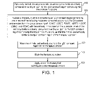
BRIEF DESCRIPTION OF THE DRAWINGS
FIG. 1 is a flow diagram illustrating a method for purifying a therapeutic
protein according to
some embodiments.
FIG. 2 is a SEC profile of mAb 1 standard and supernatant from mAb 1
incubation with
capture antibody immobilized on DYNABEAD beads.
FIG. 3 is a SEC profile of mAb 1 after incubation with capture antibody
immobilized on
DYNABEAD beads for 10 min (dashed line), 1 hour (bold line), and overnight
(unbolded line).
FIG. 4 is an SEC profile of SEFL2 immunoaffinity purified samples including
mAb 1 spiked in
PBS (unbolded line), mAb 1 spiked in human serum (bold line), and serum blank
sample (flat line).
FIG. 5 is a SEC profile of mAb 1, mixture among mAb 1, antigen 1, and antigen
2 (2:1:1, bold
line), and mixture between mAb 1 and antigen 2 (1:1, unbolded line).
Consistent with drug-target
.. complex model study in PBS buffer, slight excess of antigen 1 and antigen 2
led to a broad peak
eluted ¨4.3 min, indicating a large complex among mAb 1, antigen 1, and
antigen 2. And slight excess
of antigen 2 led to peaks indicating antigen 2 bound mAb 1 with 1:1, 1:2, and
1:3 ratio.
FIG. 6 is a SE profile of SEFL2 immunoaffinity-purified antigen binding
protein 1 spiked in PBS
(bold line) and antigen binding protein 1 spiked in human serum (unbolded
line).
4
CA 03227990 2024-01-29
WO 2023/015298
PCT/US2022/074612
DETAILED DESCRIPTION
Described herein are methods useful for isolating a therapeutic protein from a
sample, such
as an ex vivo sample of a human. The therapeutic protein may comprise an IgG1
constant region
comprising one or more mutations (e.g., one or more of L242C, A287C, R292C,
N297G, V302C,
L306C, and/or K334C, numbered according to the EU system). The sample
comprising the
therapeutic protein may be incubated with an antibody, immobilized on a
substrate such as a bead.
The antibody can bind selectively to the IgG1 constant region comprising the
one or more mutations,
compared to wild-type IgG1. The antibody bound to the therapeutic protein can
be washed, and the
therapeutic protein can then be eluted, so as to isolate the therapeutic
protein. It is noted that such
methods can be useful for isolating therapeutic proteins from ex vivo samples
in order to identify
molecular attributes of the therapeutic protein after it has been in an in
vivo environment. It is
contemplated that the methods may also be useful for isolating therapeutic
proteins from other
sample types, for example, in vitro samples or in process samples from
manufacturing.
Advantageously, such methods can isolate a therapeutic protein (such as an
antibody) comprising an
IgG1 constant region comprising the listed one or more mutations, but are
agnostic to any binding
target or targets of the therapeutic protein. Accordingly, generating critical
reagents and the
development of molecule specific affinity methods can be avoided. The methods
described herein
may be used for a number of different types of therapeutic proteins, for
example monoclonal
antibodies such as IgG monoclonal antibodies, bispecific T cell engager (BiTE
) molecules comprising
a half-life extension (HLE) moiety (which may be referred to herein as an HLE-
BiTE molecule),
hetero-IgGs, and IgGsc-Fv. Product quality attribute monitoring of individual
molecules and further
attribute understanding and characterization can be readily achieved without
the development or
use of molecule-specific affinity columns. This may include monitoring,
understanding, and/or
characterization of molecular attributes of therapeutic proteins in vivo
(after administration to a
subject), and recovered from ex vivo samples.
The methods described herein can separate a therapeutic protein from serum
matrix and
can be useful for understanding and monitoring molecular attributes (such as
critical quality
attributes) after therapeutic protein administration. Conventionally, there
have been analytical
challenges to isolating a therapeutic protein of interest from serum,
including the low concentration
of therapeutic proteins in serum and interference from high levels of
endogenous proteins. As
discussed in further detail herein, conventional affinity-based methods, such
as protein A
chromatography by targeting Fc regions of therapeutic antibodies, are
generally not applicable due
to a lack of selectivity and co-isolation with endogenous antibodies. Molecule-
specific affinity
chromatography methods have also been reported. However, protein-specific
reagents or
5
CA 03227990 2024-01-29
WO 2023/015298
PCT/US2022/074612
antibodies, which typically bind to complementarity-determining region (CDRs)
of therapeutic
proteins, were generated to achieve the desired selectivity in serum matrix.
Development of such a
molecule-specific method for each therapeutic protein is not only laborious
and time-consuming,
but also greatly limited by the availability of molecule specific reagents or
antibody. By way of
example, the methods described herein can isolate therapeutic proteins such as
antibodies without
a need to generate a capture antibody specific to their variable regions, and
can isolate antibodies
that comprise molecular attributes in their CDRs. Attributes in the CDRs,
thought potentially
relevant to efficacy and/or stability, may interfere with recovery of the
antibody through
conventional molecule-specific methods.
Immunoaffinity purification has conventionally been applied to therapeutic
antibody
purification. While Protein A-based chromatography has been utilized in
majority of purification
processes, human serum includes highly abundant polyclonal antibodies which
are also captured by
and coeluted with therapeutic antibodies from a Protein A column. Therefore,
isolating a therapeutic
protein (such as an antibody) of interest from other antibodies using Protein
A chromatography can
be challenging, as other antibodies may be co-purified along with an antibody
of interest. Moreover,
the abundant enzymatic peptides from these other antibodies' constant regions
can suppress the
signals of variable region peptides with lower abundance from therapeutic
monoclonal antibodies
using LC-MS/MS based approach after enzymatic digestion to monitor attributes
in CDR/variable
regions. In addition, the resulting complex peptide mixture can confound
proteome analysis. For
example, the origin of peptides cannot be assigned if more than one protein
yielding identical
enzymatic peptides are present.[8] As a result, the Protein A immunoaffinity
purification method
itself or in combination with LC-MS/MS is associated with challenges, and is
typically not practicable.
As an alternative to Protein A chromatography, target ligand or customized
anti-drug
antibodies, binding the CDR region of target therapeutic protein, may be
utilized to selectively pull
the target therapeutic proteins from serum matrix before further attribute
characterization.[9-13]
This method generally features high sensitivity and high specificity, but
involves the generation and
use of customized anti-drug antibodies for each target therapeutic protein,
which can require
extensive development work. Moreover, attributes of interest in CDR regions
(which may be
particularly relevant to the efficacy of a therapeutic protein) could impact
binding affinity to resins
and lead to bias enrichment of certain populations. Furthermore, target ligand
or customized anti-
drug antibodies approaches may only purify the free form of therapeutic
proteins in serum.[14]
Currently, many therapeutic proteins are engineered with mutations in an IgG
constant
region, which can eliminate undesired effector function. Therefore, an
immunoaffinity approach
6
CA 03227990 2024-01-29
WO 2023/015298
PCT/US2022/074612
targeting IgG1 constant regions comprising mutations (as described herein) of
therapeutic proteins
offers advantages of high selectivity (compared to Protein A column) and high
versatility (applicable
for all therapeutic proteins with an IgG1 constant region comprising these
mutations). Methods as
described herein have been used to isolate a hetero-IgG therapeutic protein
and bispecific T cell
engager (BiTE ) molecule comprising a half-life extension moiety, and followed
by SEC
characterizations. In certain embodiments, monoclonal antibody 1A3 is used to
capture therapeutic
proteins comprising an IgG1 comprising mutations R292C, N297G, and V302C (EU
numbering). In
certain embodiments, the antibody is immobilized on non-porous monodisperse
superparamagnetic
beads as described herein, for example DYNABEAD beads. It has been observed
that, compared to
other beads such as SEPHAROSE beads, DYNABEAD beads exhibited higher
reproducibility. It is
noted that DYNABEAD beads are generally uniform, and do not have an inner
surface[15].
Without being limited by theory, it is contemplated that DYNABEAD beads may
be less prone to
nonspecific binding. Accordingly, it is contemplated that methods described
herein are useful for
high selectivity and high versatility isolation of therapeutic proteins from
samples such as ex vivo
samples, and can facilitate the analysis of molecular attributes of the
therapeutic proteins.
Samples
The term "sample" and variations of this root term has its ordinary and
customary meaning
as would be understood by a person of ordinary skill in view of this
disclosure. It refers to a
composition that may contain a therapeutic protein a described herein, such as
an ex vivo
composition from a subject to whom the therapeutic protein has been
administered. For example, a
sample may comprise, consist essentially of, or consist of whole blood,
plasma, serum, tissue
biopsies, cerebrospinal fluid, peripheral blood mononuclear cells with in
vitro stimulation, peripheral
blood mononuclear cells, and lymphoid tissues. The sample may comprise, or may
be expected to
comprise the therapeutic protein. A "biological sample" will be understood
herein to refer to a type
of sample. In vitro or synthetic samples are also suitable for some
embodiments, for example in
process samples from the manufacturing of a therapeutic protein. The sample
may be used in
accordance with methods and/or kits as described herein. In some embodiments,
the sample is an
ex vivo sample of a human. In some embodiments, the sample comprises serum
and/or serum
proteins such as albumin. In various embodiments, the sample comprises
albumin. The sample
(e.g., an ex vivo sample of a human) may comprises albumin bound to the
therapeutic protein. In
some embodiments, the sample comprises immunoglobulins different from the
therapeutic protein,
for example immunoglobulins of a subject from whom the sample was obtained.
Methods and kits
described herein may be used to isolate the therapeutic protein from such
immunoglobulins
different from the therapeutic protein.
7
CA 03227990 2024-01-29
WO 2023/015298
PCT/US2022/074612
In some embodiments, the therapeutic protein is an antigen binding protein,
antibody, Bi-
specific T cell engager (BiTE ) molecule, bispecific antibody, trispecific
antibody, or Fc fusion protein
and the sample further comprises an antigen for the therapeutic protein. The
therapeutic protein
may be bound to its antigen in the sample.
In some embodiments, the sample comprises or consists of the therapeutic
protein in a
formulation. The formulation may be a pharmaceutically acceptable formulation.
The formulation
may comprise the biological therapy together with a pharmaceutically
acceptable diluent, carrier,
solubilizer, emulsifier, preservative, and/or adjuvant. Optionally, the sample
may further comprise
serum or serum protein.
Acceptable formulation materials for therapeutic proteins as described herein
preferably are
nontoxic to recipients at the dosages and concentrations employed. In certain
embodiments, the
pharmaceutical composition may contain formulation materials for modifying,
maintaining or
preserving, for example, the pH, osmolality, viscosity, clarity, color,
isotonicity, odor, sterility,
stability, rate of dissolution or release, adsorption or penetration of the
composition. In such
embodiments, suitable formulation materials include, but are not limited to,
amino acids (such as
glycine, glutamine, asparagine, arginine or lysine); antimicrobials;
antioxidants (such as ascorbic acid,
sodium sulfite or sodium hydrogen-sulfite); buffers (such as borate,
bicarbonate, Tris-HCI, citrates,
phosphates or other organic acids); bulking agents (such as mannitol or
glycine); chelating agents
(such as ethylenediamine tetraacetic acid (EDTA)); complexing agents (such as
caffeine,
polyvinylpyrrolidone, beta-cyclodextrin or hydroxypropyl-beta-cyclodextrin);
fillers;
monosaccharides; disaccharides; and other carbohydrates (such as glucose,
sucrose, mannose or
dextrins); proteins (such as serum albumin, gelatin or immunoglobulins);
coloring, flavoring and
diluting agents; emulsifying agents; hydrophilic polymers (such as
polyvinylpyrrolidone); low
molecular weight polypeptides; salt-forming counterions (such as sodium);
preservatives (such as
benzalkonium chloride, benzoic acid, salicylic acid, thimerosal, phenethyl
alcohol, methylparaben,
propylparaben, chlorhexidine, sorbic acid or hydrogen peroxide); solvents
(such as glycerin,
propylene glycol or polyethylene glycol); sugar alcohols (such as mannitol or
sorbitol); suspending
agents; surfactants or wetting agents (such as pluronics, PEG, sorbitan
esters, polysorbates such as
polysorbate 20, polysorbate, triton, tromethamine, lecithin, cholesterol,
tyloxapal); stability
enhancing agents (such as sucrose or sorbitol); tonicity enhancing agents
(such as alkali metal
halides, preferably sodium or potassium chloride, mannitol sorbitol); delivery
vehicles; diluents;
excipients and/or pharmaceutical adjuvants. See, e.g., REMINGTON'S
PHARMACEUTICAL SCIENCES,
18" Edition, (A. R. Genrmo, ed.), 1990, Mack Publishing Company.
8
CA 03227990 2024-01-29
WO 2023/015298
PCT/US2022/074612
A suitable vehicle or carrier for the formulation may be water for injection,
physiological
saline solution or artificial cerebrospinal fluid, possibly supplemented with
other materials common
in compositions for parenteral administration. Neutral buffered saline or
saline mixed with serum
albumin are further exemplary vehicles. In specific embodiments,
pharmaceutical compositions
comprise Tris buffer of about pH 7.0-8.5, or acetate buffer of about pH 4.0-
5.5, and may further
include sorbitol or a suitable substitute therefor.
The formulation components are present preferably in concentrations that are
acceptable to
the site of administration. In certain embodiments, buffers are used to
maintain the composition at
physiological pH or at a slightly lower pH, typically within a pH range of
from about 5 to about 8. For
example, the pH of the formulation may be about 5.1, about 5.2, about 5.3,
about 5.4, about 5.5,
about 5.6, about 5.7, about 5.8, about 5.9, about 6.0, about 6.1, about 6.2,
about 6.3, about 6.4,
about 6.5, about 6.6, about 6.7, about 6.8, about 6.9, about 7.0, about 7.1,
about 7.2, about 7.3,
about 7.4, about 7.5, about 7.6, about 7.7, about 7.8, about 7.9, or about
8Ø
Therapeutic proteins
As used herein "therapeutic protein," and variations of this root term, has
its ordinary and
customary meaning as would be understood by one of ordinary skill in the art
in view of this
disclosure. It refers to a polypeptide for medical use in a subject, typically
a human subject.
The therapeutic protein may comprise an IgG1 constant region comprising one or
more of
the following mutations numbered according to the EU system and selected from
the group
consisting of: L242C, A287C, R292C, N297G, V302C, L306C, and K334C. Unless
stated otherwise,
positions in (including positions of mutations in) constant regions will be
referred to herein using EU
numbering. By way of example, the mutation may comprise N297G and at least one
of R292C
and/or V302C. By way of example, the mutation may comprise R292C, N297G, and
V302C. In some
embodiments, the IgG1 constant region of the therapeutic protein comprises the
amino acid
sequence: CEEQYGSTYRC (SEQ ID NO: 1) or
ASTKGPSVFPLAPSSKSTSGGTAALGCLVKDYFPEPVTVSWNSGALTSGVHTFPAVLQSS
GLYSLSSVVTVPSSSLGTQTYICNVNHKPSNTKVDKKVEPKSCDKTHTCPPCPAPELLGGPSVFLFPPKPKDTLMISRT
PEVTCVVVDVSHEDPEVKFNWYVDGVEVHNAKTKPCEEQYGSTYRCVSVLTVLHQDWLNGKEYKCKVSNKALPA
PIEKTISKAKGQPREPQVYTLPPSREEMTKNQVSLTCLVKGFYPSDIAVEWESNGQPENNYKTTPPVLDSDGSFFLYS
KLTVDKSRWQQGNVFSCSVMHEALHNHYTQKSLSLSPGK (SEQ ID NO: 2).
9
CA 03227990 2024-01-29
WO 2023/015298
PCT/US2022/074612
In methods described herein, the therapeutic protein may be selected from the
group
consisting of: an antibody (such as a monoclonal antibody, for example an IgG1
monoclonal
antibody), an antigen binding protein, an antibody protein product, a Bi-
specific T cell engager
(BiTE ) molecule, a bispecific antibody, a trispecific antibody, an Fc fusion
protein, a recombinant
protein, a synthetic peptide, and an active fragment of a recombinant protein.
An "antibody" has its customary and ordinary meaning as understood by one of
ordinary
skill in the art in view of this disclosure. It refers to an immunoglobulin of
with specific binding to
the target antigen, and includes, for instance, chimeric, humanized, and fully
human antibodies. By
way of example, the antibody may be a monoclonal antibody. By way of example,
human antibodies
can be of a specified isotype, including IgG (including IgG1, IgG2, IgG3 and
IgG4 subtypes), IgA
(including IgA1 and IgA2 subtypes), IgM and IgE. A human IgG antibody
generally comprises two
full-length heavy chains and two full-length light chains. Antibodies may be
derived solely from a
single source, or may be "chimeric," that is, different portions of the
antibody may be derived from
two or more different antibodies from the same or different species. It will
be understood that once
an antibody is obtained from a source, it may undergo further engineering, for
example to enhance
stability and folding. Accordingly, it will be understood that a "human"
antibody may be obtained
from a source, and may undergo further engineering, for example in the Fc
region. The engineered
antibody may still be referred to as a type of human antibody. Similarly,
variants of a human
antibody, for example those that have undergone affinity maturation, will also
be understood to be
"human antibodies" unless stated otherwise. In some embodiments, an antibody
comprises,
consists essentially of, or consists of a human, humanized, or chimeric
monoclonal antibody. In
various embodiments, the therapeutic protein comprises or consists of a
chimeric, human, or
humanized antibody comprising an IgG1 constant region comprising one or more
of the following
mutations numbered according to the EU system and selected from the group
consisting of: L242C,
A287C, R292C, N297G, V302C, L306C, and K334C. In various embodiments, the
therapeutic protein
is human or humanized antibody comprising an IgG1 constant region comprising
one or more of the
following mutations numbered according to the EU system and selected from the
group consisting
of: L242C, A287C, R292C, N297G, V302C, L306C, and K334C. In various
embodiments, the
therapeutic protein is a human antibody comprising an IgG1 constant region
comprising one or more
of the following mutations numbered according to the EU system and selected
from the group
consisting of: L242C, A287C, R292C, N297G, V302C, L306C, and K334C. By way of
example, the
mutation may comprise N297G and at least one of R292C and/or V302C according
to EU numbering.
By way of example, the mutation may comprise N297G, R292C, and V302C according
to EU
numbering.
CA 03227990 2024-01-29
WO 2023/015298
PCT/US2022/074612
A "heavy chain" of an antibody, antigen binding protein, antibody protein
product, Bi-
specific T cell engager molecule, bispecific antibody, or trispecific antibody
includes a variable region
("VH"), and three constant regions: CH1, CH2, and CH3. A "light chain" of an
antibody, antigen
binding protein, antibody protein product, Bi-specific T cell engager
molecule, bispecific antibody, or
trispecific antibody includes a variable region ("VL"), and a constant region
("CL"). Human light
chains include kappa chains and lambda chains. Example light chain constant
regions suitable for
antigen binding proteins include human lambda and human kappa constant
regions.
In various aspects, the therapeutic protein is an antibody protein product. As
used herein,
the term "antibody protein product" refers to any one of several antibody
alternatives which in
various instances is based on the architecture of an antibody but is not found
in nature. In some
aspects, the antibody protein product has a molecular-weight within the range
of at least about 12-
150 kDa. In certain aspects, the antibody protein product has a valency (n)
range from monomeric
(n = 1), to dimeric (n = 2), to trimeric (n = 3), to tetrameric (n = 4), if
not higher order valency.
Antibody protein products in some aspects are those based on the full antibody
structure and/or
those that mimic antibody fragments which retain full antigen-binding
capacity, e.g., scFvs, Fabs and
VHH/VH (discussed below). The smallest antigen binding antibody fragment that
retains its
complete antigen binding site is the Fv fragment, which consists entirely of
variable (V) regions. A
soluble, flexible amino acid peptide linker is used to connect the V regions
to a scFy (single chain
fragment variable) fragment for stabilization of the molecule, or the constant
(C) domains are added
to the V regions to generate a Fab fragment [fragment, antigen-binding]. Both
scFy and Fab
fragments can be easily produced in host cells, e.g., prokaryotic host cells.
Other antibody protein
products include disulfide-bond stabilized scFy (ds-scFv), single chain Fab
(scFab), as well as di- and
multimeric antibody formats like dia-, tria- and tetra-bodies, or minibodies
(miniAbs) that comprise
different formats consisting of scFvs linked to oligomerization domains. The
smallest fragments are
VHH/VH of camelid heavy chain Abs as well as single domain Abs (sdAb). The
building block that is
most frequently used to create novel antibody formats is the single-chain
variable (V)-domain
antibody fragment (scFv), which comprises V domains from the heavy and light
chain (VH and VL
domain) linked by a peptide linker of ¨15 amino acid residues. A peptibody or
peptide-Fc fusion is
yet another antibody protein product. The structure of a peptibody consists of
a biologically active
peptide grafted onto an Fc domain. Peptibodies are well-described in the art.
See, e.g., Shimamoto
et al., mAbs 4(5): 586-591 (2012). Bispecific T-cell engage molecules, for
example those comprising
a half-life extension moiety are also examples of antibody protein products.
Therapeutic proteins suitable for the methods described herein can include
polypeptides,
including those that bind to one or more of the following: CD proteins,
including CD3, CD4, CD8,
11
CA 03227990 2024-01-29
WO 2023/015298
PCT/US2022/074612
CD19, CD20, CD22, CD30, and CD34; including those that interfere with receptor
binding. HER
receptor family proteins, including HER2, HER3, HER4, and the EGF receptor.
Cell adhesion
molecules, for example, LFA-I, Mol, p150, 95, VLA-4, ICAM-I, VCAM, and alpha
v/beta 3 integrin.
Growth factors, such as vascular endothelial growth factor ("VEGF"), growth
hormone, thyroid
stimulating hormone, follicle stimulating hormone, luteinizing hormone, growth
hormone releasing
factor, parathyroid hormone, Mullerian-inhibiting substance, human macrophage
inflammatory
protein (MIP-1alpha), erythropoietin (EPO), nerve growth factor, such as NGF-
beta, platelet-derived
growth factor (PDGF), fibroblast growth factors, including, for instance, aFGF
and bFGF, epidermal
growth factor (EGF), transforming growth factors (TGF), including, among
others, TGF-a and TGF-13,
including TGF-131, TGF-132, TGF-133, TGF-134, or TGF-135, insulin-like growth
factors-I and -II (IGF-I and
IGF-II), des(1-3)-IGF-I (brain IGF-I), and osteoinductive factors. Insulins
and insulin-related proteins,
including insulin, insulin A-chain, insulin B-chain, proinsulin, and insulin-
like growth factor binding
proteins. Coagulation and coagulation-related proteins, such as, among others,
factor VIII, tissue
factor, von Willebrand factor, protein C, alpha-1-antitrypsin, plasminogen
activators, such as
urokinase and tissue plasminogen activator ("t-PA"), bombazine, thrombin, and
thrombopoietin; (vii)
other blood and serum proteins, including but not limited to albumin, IgE, and
blood group antigens.
Colony stimulating factors and receptors thereof, including the following,
among others, M-CSF, GM-
CSF, and G-CSF, and receptors thereof, such as CSF-1 receptor (c-fms).
Receptors and receptor-
associated proteins, including, for example, f1k2/f1t3 receptor, obesity (0B)
receptor, LDL receptor,
growth hormone receptors, thrombopoietin receptors ("TPO-R," "c-mpl"),
glucagon receptors,
interleukin receptors, interferon receptors, T-cell receptors, stem cell
factor receptors, such as c-Kit,
and other receptors. Receptor ligands, including, for example, OX4OL, the
ligand for the 0X40
receptor. Neurotrophic factors, including bone-derived neurotrophic factor
(BDNF) and
neurotrophin-3, -4, -5, or -6 (NT-3, NT-4, NT-5, or NT-6). Relaxin A-chain,
relaxin B-chain, and
prorelaxin; interferons and interferon receptors, including for example,
interferon-a, -13, and -y, and
their receptors. Interleukins and interleukin receptors, including IL-1 to IL-
33 and IL-1 to IL-33
receptors, such as the IL-8 receptor, among others. Viral antigens, including
an AIDS envelope viral
antigen. Lipoproteins, calcitonin, glucagon, atrial natriuretic factor, lung
surfactant, tumor necrosis
factor-alpha and -beta, enkephalinase, RANTES (regulated on activation
normally T-cell expressed
and secreted), mouse gonadotropin-associated peptide, DNAse, inhibin, and
activin. Integrin, protein
A or D, rheumatoid factors, immunotoxins, bone morphogenetic protein (BMP),
superoxide
dismutase, surface membrane proteins, decay accelerating factor (DAF), HIV
envelope, transport
proteins, homing receptors, addressins, regulatory proteins, immunoadhesins,
antibodies.
Myostatins, TALL proteins, including TALL-I, amyloid proteins, including but
not limited to amyloid-
12
CA 03227990 2024-01-29
WO 2023/015298
PCT/US2022/074612
beta proteins, thymic stromal lymphopoietins ("TSLP"), RANK ligand ("RANKL" or
"OPGL"), c-kit, TNF
receptors, including TNF Receptor Type 1, TRAIL-R2, angiopoietins, and
biologically active fragments
or analogs or variants of any of the foregoing.
Examples of therapeutic proteins suitable for the methods described herein
include
antibodies or variants thereof comprising an IgG1 constant region comprising
one or more of the
following mutations numbered according to the EU system and selected from the
group consisting
of: L242C, A287C, R292C, N297G, V302C, L306C, and K334C, such as infliximab,
bevacizumab,
cetuximab, ranibizumab, palivizumab, abagovomab, abciximab, actoxumab,
adalimumab,
afelimomab, afutuzumab, alacizumab, alacizumab pegol, a1d518, alemtuzumab,
alirocumab,
.. altumomab, amatuximab, anatumomab mafenatox, anrukinzumab, apolizumab,
arcitumomab,
aselizumab, altinumab, atlizumab, atorolimiumab, tocilizumab, bapineuzumab,
basiliximab,
bavituximab, bectumomab, belimumab, benralizumab, bertilimumab, besilesomab,
bevacizumab,
bezlotoxumab, biciromab, bivatuzumab, bivatuzumab mertansine, blinatumomab,
blosozumab,
brentuximab vedotin, briakinumab, brodalumab, canakinumab, cantuzumab
mertansine,
cantuzumab mertansine, caplacizumab, capromab pendetide, carlumab,
catumaxomab, cc49,
cedelizumab, certolizumab pegol, cetuximab, citatuzumab bogatox, cixutumumab,
clazakizumab,
clenoliximab, clivatuzumab tetraxetan, conatumumab, crenezumab, cr6261,
dacetuzumab,
daclizumab, dalotuzumab, daratumumab, demcizumab, denosumab, detumomab,
dorlimomab
aritox, drozitumab, duligotumab, dupilumab, ecromeximab, eculizumab,
edobacomab,
edrecolomab, efalizumab, efungumab, elotuzumab, elsilimomab, enavatuzumab,
enlimomab pegol,
enokizumab, enoticumab, ensituximab, epitumomab cituxetan, epratuzumab,
erenumab, erlizumab,
ertumaxomab, etaracizumab, etrolizumab, evolocumab, exbivirumab, fanolesomab,
faralimomab,
farletuzumab, fasinumab, fbta05, felvizumab, fezakinumab, ficlatuzumab,
figitumumab,
flanvotumab, fontolizumab, foralumab, foravirumab, fresolimumab, fulranumab,
futuximab,
.. galiximab, ganitumab, gantenerumab, gavilimomab, gemtuzumab ozogamicin,
gevokizumab,
girentuximab, glembatumumab vedotin, golimumab, gomiliximab, gs6624,
ibalizumab, ibritumomab
tiuxetan, icrucumab, igovomab, imciromab, imgatuzumab, inclacumab, indatuximab
ravtansine,
infliximab, intetumumab, inolimomab, inotuzumab ozogamicin, ipilimumab,
iratumumab,
itolizumab, ixekizumab, keliximab, labetuzumab, lebrikizumab, lemalesomab,
lerdelimumab,
.. lexatumumab, libivirumab, ligelizumab, lintuzumab, lirilumab, lorvotuzumab
mertansine,
lucatumumab, lumiliximab, mapatumumab, maslimomab, mavrilimumab, matuzumab,
mepolizumab, metelimumab, milatuzumab, minretumomab, mitumomab, mogamulizumab,
morolimumab, motavizumab, moxetumomab pasudotox, muromonab-cd3, nacolomab
tafenatox,
namilumab, naptumomab estafenatox, narnatumab, natalizumab, nebacumab,
necitumumab,
13
CA 03227990 2024-01-29
WO 2023/015298
PCT/US2022/074612
nerelimomab, nesvacumab, nimotuzumab, nivolumab, nofetumomab merpentan,
ocaratuzumab,
ocrelizumab, odulimomab, ofatumumab, olaratumab, olokizumab, omalizumab,
onartuzumab,
oportuzumab monatox, oregovomab, orticumab, otelixizumab, oxelumab,
ozanezumab,
ozoralizumab, pagibaximab, palivizumab, panitumumab, panobacumab,
parsatuzumab,
pascolizumab, pateclizumab, patritumab, pemtumomab, perakizumab, pertuzumab,
pexelizumab,
pidilizumab, pintumomab, placulumab, ponezumab, priliximab, pritumumab, PRO
140, quilizumab,
racotumomab, radretumab, rafivirumab, ramucirumab, ranibizumab, raxibacumab,
regavirumab,
reslizumab, rilotumumab, rituximab, robatumumab, roledumab, romosozumab,
rontalizumab,
rovelizumab, ruplizumab, samalizumab, sarilumab, satumomab pendetide,
secukinumab, sevirumab,
sibrotuzumab, sifalimumab, siltuximab, simtuzumab, siplizumab, sirukumab,
solanezumab,
solitomab, sonepcizumab, sontuzumab, stamulumab, sulesomab, suvizumab,
tabalumab,
tacatuzumab tetraxetan, tadocizumab, talizumab, tanezumab, taplitumomab
paptox, tefibazumab,
telimomab aritox, tenatumomab, tefibazumab, teneliximab, teplizumab,
teprotumumab,
tezepelumab, TGN1412, tremelimumab, ticilimumab, tildrakizumab, tigatuzumab,
TNX-650,
tocilizumab, toralizumab, tositumomab, tralokinumab, trastuzumab, TRBS07,
tregalizumab,
tucotuzumab celmoleukin, tuvirumab, ublituximab, urelumab, urtoxazumab,
ustekinumab,
vapaliximab, vatelizumab, vedolizumab, veltuzumab, vepalimomab, vesencumab,
visilizumab,
volociximab, vorsetuzumab mafodotin, votumumab, zalutumumab, zanolimumab,
zatuximab,
ziralimumab, or zolimomab aritox.
In some embodiments, the therapeutic protein is a BiTE molecule. BiTE
molecules are
engineered bispecific antigen binding constructs which direct the cytotoxic
activity of T cells against
cancer cells. They are the fusion of two single-chain variable fragments
(scFvs) of different
antibodies, or amino acid sequences from four different genes, on a single
peptide chain of about 55
kilodaltons. One of the scFvs binds to T cells via the CD3 receptor, and the
other to a tumor cell via a
tumor specific molecule. Blinatumomab (BLINCYTO product) is an example of a
BiTE molecule,
specific for CD19. BiTE molecules that are modified, such as those modified
to extend their half-
lives, can also be used in the disclosed methods. In various aspects, the
polypeptide is an antigen
binding protein, e.g., a BiTE molecule. In some embodiments, an antibody
protein product
comprises a BiTE molecule.
Antibodies that bind to IgG1 constant regions comprising mutations
Antibodies that bind to IgG1 constant regions comprising mutations as
described herein may
be used in methods and kits described herein. The antibody can bind
specifically to an IgG1
14
CA 03227990 2024-01-29
WO 2023/015298
PCT/US2022/074612
comprising one or more mutations numbered according to the EU system and
selected from the
group consisting of: L242C, A287C, R292C, N297G, V302C, L306C, and K334C. For
example, the IgG1
may comprise N297G and at least one of R292C and/or V302C according to EU
numbering. The
antibody may be a monoclonal antibody. By way of example, the antibody may be
from a
mammalian host organism, such as a mouse, hamster, rat, rabbit, goat, or
donkey. For example, the
antibody may be a mouse monoclonal antibody. The antibody may be provided
immobilized on a
substrate as described herein. The antibody immobilized on the substrate may
also be referred to
herein as a "capture antibody." In some embodiments, the antibody binds to the
amino acid
sequence CEEQYGSTYRC (SEQ ID NO: 1). In some embodiments, the antibody
comprises, consists
essentially of, or consists of monoclonal antibody 1A3.
Suitable antibodies that bind to IgG1 constant regions comprising mutations as
described
herein may be prepared by techniques that are established in the art. For
example, antibodies may
be prepared by immunizing an animal (e.g., a mammal as described herein such
as a mouse or rat or
rabbit) with a protein comprising or consisting of the IgG1 constant region
comprising mutation, and
then by immortalizing spleen cells harvested from the animal after completion
of the immunization
schedule. The spleen cells can be immortalized using any technique known in
the art, e.g., by fusing
them with myeloma cells to produce hybridomas. See, for example, Antibodies;
Harlow and Lane,
Cold Spring Harbor Laboratory Press, 1' Edition, e.g. from 1988, or 2nd
Edition, e.g. from 2014). By
way of example, the antibody may be raised against a protein comprising or
consisting of the amino
acid sequence
ASTKGPSVFPLAPSSKSTSGGTAALGCLVKDYFPEPVTVSWNSGALTSGVHTFPAVLQSSGLYSLSSVVTVPSSSLGT
QTYICNVNHKPSNTKVDKKVEPKSCDKTHTCPPCPAPELLGGPSVFLFPPKPKDTLMISRTPEVTCVVVDVSHEDPE
VKFNWYVDGVEVHNAKTKPCEEQYGSTYRCVSVLTVLHQDWLNGKEYKCKVSNKALPAPIEKTISKAKGQPREPQ
VYTLPPSREEMTKNQVSLTCLVKGFYPSDIAVEWESNGQPENNYKTTPPVLDSDGSFFLYSKLTVDKSRWQQGNVF
SCSVMHEALHNHYTQKSLSLSPGK (SEQ ID NO: 2). In some embodiments, the method
comprising
raising an antibody against a protein comprising or consisting of the amino
acid sequence
ASTKGPSVFPLAPSSKSTSGGTAALGCLVKDYFPEPVIVSWNSGALTSGVHTFPAVLQSSGLYSLSSVVTVPSSSLGT
QTYICNVNHKPSNTKVDKKVEPKSCDKTHTCPPCPAPELLGGPSVFLFPPKPKDTLMISRTPEVTCVVVDVSHEDPE
VKFNWYVDGVEVHNAKTKPCEEQYGSTYRCVSVLTVLHQDWLNGKEYKCKVSNKALPAPIEKTISKAKGQPREPQ
VYTLPPSREEMTKNQVSLTCLVKGFYPSDIAVEWESNGQPENNYKTTPPVLDSDGSFFLYSKLTVDKSRWQQGNVF
SCSVMHEALHNHYTQKSLSLSPGK (SEQ ID NO: 2). The antibody may be used to bind to
IgG1 regions of
therapeutic proteins in methods as described herein.
In certain embodiments, a B-cell that is producing a desired antibody is
selected and the
light chain and heavy chain variable regions are cloned from the B-cell
according to established
CA 03227990 2024-01-29
WO 2023/015298
PCT/US2022/074612
molecular biology techniques (WO 92/02551; U.S. patent 5,627,052; Babcook et
al., Proc. Natl. Acad.
Sci. USA 93:7843 48 (1996)) and described herein. B-cells from an immunized
animal may be
isolated from the spleen, lymph node, or peripheral blood sample by selecting
a cell that is
producing a desired antibody. B-cells may also be isolated from humans, for
example, from a
peripheral blood sample. Methods for detecting single B-cells that are
producing an antibody with
the desired specificity are well known in the art, for example, by plaque
formation, fluorescence
activated cell sorting, in vitro stimulation followed by detection of specific
antibody, and the like.
Methods for selection of specific antibody producing B-cells include, for
example, preparing a single
cell suspension of B-cells in soft agar that contains antigen. Binding of the
specific antibody
produced by the B-cell to the antigen results in the formation of a complex,
which may be visible as
an immunoprecipitate. After the B-cells producing the desired antibody are
selected, the specific
antibody genes may be cloned by isolating and amplifying DNA or mRNA according
to methods
known in the art.
An additional method for obtaining antibodies of the present disclosure is by
phage display.
See, e.g., Winter et al., 1994 Annu. Rev. Immunol. 12:433 55; Burton et al.,
1994 Adv. Immunol.
57:191 280. Human or murine immunoglobulin variable region gene combinatorial
libraries may be
created in phage vectors that can be screened to select Ig fragments (Fab, Fv,
sFy, or multimers
thereof) that bind specifically to PCSK9 or variant or fragment thereof. See,
e.g., U.S. Patent No.
5,223,409; Huse et al., 1989 Science 246:1275-81; Sastry et al., Proc. Natl.
Acad. Sci. USA 86:5728-32
(1989); Alting Mees et al., Strategies in Molecular Biology 3:1-9 (1990); Kang
et al., 1991 Proc. Natl.
Acad. Sci. USA 88:4363-66; Hoogenboom et al., 1992 J. Molec. Biol. 227:381-
388; Schlebusch et al.,
1997 Hybridoma 16:47-52; and references cited therein. For example, a library
containing a plurality
of polynucleotide sequences encoding Ig variable region fragments may be
inserted into the genome
of a filamentous bacteriophage, such as M13 or a variant thereof, in frame
with the sequence
encoding a phage coat protein. A fusion protein may be a fusion of the coat
protein with the light
chain variable region domain and/or with the heavy chain variable region
domain. According to
certain embodiments, immunoglobulin Fab fragments may also be displayed on a
phage particle
(see, e.g., U.S. Patent No. 5,698,426).
Heavy and light chain immunoglobulin cDNA expression libraries may also be
prepared in
.. lambda phage, for example, using AlmmunoZapTM(H) and AlmmunoZapTM(L)
vectors (Stratagene,
La Jolla, California). Briefly, mRNA is isolated from a B-cell population, and
used to create heavy and
light chain immunoglobulin cDNA expression libraries in the AlmmunoZap(H) and
AlmmunoZap(L)
vectors. These vectors may be screened individually or co expressed to form
Fab fragments or
antibodies (see Huse et al., supra; see also Sastry et al., supra). Positive
plaques may subsequently
16
CA 03227990 2024-01-29
WO 2023/015298
PCT/US2022/074612
be converted to a non-lytic plasmid that allows high level expression of
monoclonal antibody
fragments from a microbial organism such as E. co/i.
Once cells producing antibodies that bind to IgG1 comprising mutations
according to the
present disclosure have been obtained using any of the above-described
immunization and other
techniques, the specific antibody genes may be cloned by isolating and
amplifying DNA or mRNA
therefrom according to standard procedures as described herein. The antibodies
produced
therefrom may be sequenced and the CDRs identified and the DNA coding for the
CDRs may be
manipulated as described previously to generate other suitable antibodies that
bind to IgG1
comprising mutations according to the present disclosure.
Molecular evolution of the complementarity determining regions (CDRs) in the
center of the
antibody binding site also has been used to isolate antibodies with increased
affinity, as described
by, for example, Schier et al., 1996, J. Mol. Biol. 263:551. Accordingly, such
techniques are useful in
preparing antibodies of the present disclosure.
Substrates
In methods and kits described herein, an antibody that binds to an IgG1
comprising a
mutation as described herein may be immobilized on a substrate, such as a
bead. For example, the
substrate may be a bead comprising or consisting of a non-porous monodisperse
superparamagnetic
bead (commercially available, for example, as DYNABEADS beads). The beads may
have an average
diameter of about 2-4 M, about 2-3 M, about 2.5-3.5 M, about 3 M, or 2.8
M. The antibody
may be immobilized covalently on the bead via, for example, p-toluene-sulfonyl
(tosylactivation)
chemistry, or avidin-biotin chemistry. Optionally, the bead may comprise or
consist of a Sepahrose
bead. However, it has been observed that non-porous monodisperse
superparamagnetic beads such
as M-280 DYNABEADS beads yield higher reproducibility than SEPHAROSE beads.
Methods of isolating a therapeutic protein
In accordance with various embodiments described herein, methods of isolating
a
therapeutic protein from a sample are described. The therapeutic protein may
comprise an IgG1
constant region comprising one or more mutations as described herein. The
therapeutic protein
may be incubated with an antibody that binds selectively to the IgG1 constant
region of the
therapeutic protein relative to wild-type IgG1. The antibody may be
immobilized on a substrate as
17
CA 03227990 2024-01-29
WO 2023/015298
PCT/US2022/074612
described herein. Thus, the immobilized antibody may bind the therapeutic
protein. The
immobilized antibody bound to the IgG1 constant region of the therapeutic
protein may be washed.
The therapeutic protein may be eluted, thus isolating the therapeutic protein.
Optionally, at least
one analytical technique is applied to the eluted (and isolated) therapeutic
protein, for example to
detect a presence and/or level of one or more molecular attributes of the
therapeutic protein after
it has been in an in vivo environment. For example, the analytical technique
may comprise
chromatography and/or mass spectrometry. In some embodiments, the IgG1
constant region of the
therapeutic protein comprises one or more of the following mutations numbered
according to the
EU system and selected from the group consisting of: L242C, A287C, R292C,
N297G, V302C, L306C,
and K334C.
An exemplary method of isolating a therapeutic protein from a sample is
illustrated in FIG. 1.
Optionally, the method can comprise immobilizing on a substrate an antibody
that binds selectively
(compared to wild-type IgG1) to the IgG1 constant region comprising the one or
more mutations as
described herein 110. The method can further comprise incubating a therapeutic
protein with the
antibody immobilized on the substrate. The antibody can bind selectively
(compared to wild-type
IgG1) to the IgG1 constant region comprising the one or more mutations, so
that the immobilized
antibody binds to the IgG1 constant region of the therapeutic protein 120. As
described herein, the
therapeutic protein may comprise an IgG1 constant region comprising one or
more of the following
mutations numbered according to the EU system and selected from the group
consisting of: L242C,
A287C, R292C, N297G, V302C, L306C, and K334C. The method can comprise washing
the
immobilized antibody bound to the IgG1 constant region of the therapeutic
protein 130. Optionally,
the wash may be repeated one or more times. The therapeutic protein can be
eluted 140. The
therapeutic protein may be eluted in an acidic eluant. Acidic eluants, such as
acetic acid, are
described further herein. Thus, the therapeutic may be isolated. In some
embodiments, one or
more analytical techniques may be applied to the eluted therapeutic protein
150.
In some embodiments, the method further comprises immobilizing, on the
substrate, the
antibody that binds to the IgG1 constant region of the therapeutic protein.
The immobilizing may
comprise coupling the antibody to the substrate. For example, for a substrate
that is a non-porous
monodisperse superparamagnetic bead, the method can comprise covalently
binding the antibody
using tosylactivation chemistry, or biotin-avidin chemistry.
Incubation times of overnight, one hour, and 10 minutes were compared, and it
was
observed that the longer incubation times of overnight and one hour led to
greater aggregation
(Example 2). Accordingly, it is contemplated that incubation times of 20
minutes or less, such as
18
CA 03227990 2024-01-29
WO 2023/015298
PCT/US2022/074612
about 10 minutes (or no more than 10 minutes) provide superior isolation of
therapeutic proteins,
while minimizing aggregation. In the method of some embodiments, the
incubating is for about 20
minutes or less, for example, no more than 20, 15, 10, 8, 5, or 3 minutes. By
way of example, the
incubation may be performed on a roller.
After incubating the therapeutic protein with the antibody, the immobilized
antibody bound
to the IgG1 constant region of the therapeutic protein may be washed. The wash
may be performed
in a buffer, such as PBS. The wash may be at a pH of 6-8 or 7-8. Optionally,
more than one wash
may be performed, for example at least 1, 2, 3, 4, or 5 washes, including
ranges between any two of
the listed values, for example, 1-5 washes.
The incubated therapeutic protein may then be eluted. Different elution
conditions for the
therapeutic protein have been compared herein. Among elution solutions and
pH's tested were 100
mM acetate buffer (pH 3.6, pH 4.6, and pH 5.6), acetic acid (0.005%, 0.01%,
0.05%, and 0.1%), 100
mM glycine (pH 3.0, pH 3.5, and pH 4.0), and Thermo gentle elution buffer. It
was observed that
0.05% Acetic acid yielded less HMW peak area than the other elutions (Example
2). As the 0.05%
acetic acid had a pH of 3.4 (compared to pH 3.9 for 0.005% acetic acid; pH 3.8
for 0.01% acetic acid;
and pH 3.3 for 0.05% acetic acid) it was contemplated that an elution pH of
about 3.4 to about 3.7
provides superior isolation while minimizing HWM species. As such, it will be
appreciated that the
elution may be in an acidic eluant such as acetic acid. In some embodiments,
the elution is at a pH
of 3.4 to 3.7, 3.4 to 3.6, 3.4 to 3.5, or about 3.4. In some embodiments, the
elution is in a solution
comprising acetic acid, for example 0.02% to 0.09% acetic acid, 0.02% to 0.07%
acetic acid, 0.02% to
0.05% acetic acid, or 0.05% to 0.09% acetic acid. The elution in acetic acid
may have a pH of 3.4 to
3.7, 3.4 to 3.6, 3.4 to 3.5, or about 3.4.
It may be desirable to isolate therapeutic protein that remains bound to its
antigen from the
sample. This may permit analyses informative of molecular attributes of the
therapeutic protein
that correlate with antigen binding. Accordingly, in some embodiments, the
therapeutic protein is
an antigen binding protein (e.g., antibody, Bi-specific T cell engager (BiTE )
molecule, bispecific
antibody, or trispecific antibody) and is bound to its antigen in the sample.
The therapeutic protein
may remain bound to its antigen after the elution.
Following elution of the therapeutic protein, the method may further comprise
applying one
or more analytical techniques to the therapeutic protein. For example, the
presence and/or levels
of molecular attributes may be identified. The eluted therapeutic protein may
be analyzed by
chromatography, such as size exclusion chromatography (SEC). SEC may identify
relative amounts of
19
CA 03227990 2024-01-29
WO 2023/015298
PCT/US2022/074612
therapeutic protein unbound to any target, and therapeutic protein bound to
target. By way of
example, the fraction of therapeutic protein bound to target may be
determined. By way of
example, molecular attributes that correlate with the therapeutic protein
being bound (or unbound)
to target may be determined (See, e.g, PCT Pub. No. WO 2020/247790, which
describes relationships
between therapeutic protein complexes identified by SEC and molecular
attributes, and which is
incorporated by reference in its entirety herein). Also, stoichiometry of
numbers of therapeutic
protein and/or target in complex may be determined (See, e.g., FIG. 5).
Accordingly, in some
embodiments, the method comprises applying the eluted therapeutic protein to a
chromatography
column (e.g., an SEC column). For therapeutic proteins that are an antigen
binding protein (such as
an antibody or antibody protein product as described herein), the therapeutic
protein may be bound
to its antigen in the sample, the therapeutic protein may remain bound to its
antigen after the
elution, and the size exclusion chromatography may comprise detecting a
complex of the
therapeutic protein bound to its antigen
In some embodiments, at least one analytical technique is applied to the
eluted therapeutic
protein. Examples of suitable analytical techniques include mass spectrometry,
chromatography,
electrophoresis, spectroscopy, light obscuration, a particle method (such as
nanoparticle/visible/micron-sized resonant mass or Brownian motion),
analytical centrifugation,
imaging or imaging characterization, or immunoassay. In some embodiments, the
method
comprises performing mass spectrometry on the isolated antibody. The mass
spectrometry may be
part of a peptide mapping analysis to identify the presence and/or levels of
one or more molecular
attributes. By way of example, peptide mapping by LC-MS/MS may be performed on
the eluted
therapeutic protein.
Examples of molecular attributes include acidic species, basic species, high
molecular weight
species, subvisible particle number, low molecular weight, middle molecular
weight, glycosylation
(such as non-glycosylated heavy chain or high mannose), non-heavy chain and
light chain,
deamidation, deamination, cyclization, oxidation, isomerization,
fragmentation/clipping, N-terminal
and C-terminal variants, reduced and partial species, folded structure,
surface hydrophobicity,
chemical modification, covalent bond, a C-terminal amino acid motif PARG, or a
C-terminal amino
acid motif PAR-Amide.
Upon isolation, the therapeutic protein may be free or substantially free of
other proteins of
the sample. For example, the isolated therapeutic protein may be at least one:
(1) free of at least
some other proteins with which it would normally be found, (2) essentially
free of other proteins
from the species of the subject from whom the sample was derived, and/or (3)
separated from at
CA 03227990 2024-01-29
WO 2023/015298
PCT/US2022/074612
least about 50 percent of polynucleotides, lipids, carbohydrates, or other
materials of the sample.
By way of example, serum protein produced by the subject from whom the sample
was derived may
comprise no more than 3%, 2%, 1%, or 0.1% of proteins that reside in the
composition of the
isolated therapeutic protein. By way of example, immunoglobulins produced by
the subject from
whom the sample was derived may comprise no more than 3%, 2%, 1%, or 0.1% of
proteins that
reside in the composition of the isolated therapeutic protein. In some
embodiments, the isolated
therapeutic protein constitutes at least about 5%, at least about 10%, at
least about 25%, or at least
about 50% of the composition in which it resides.
Kits
In accordance with various embodiments described herein, kits for isolating a
therapeutic
protein from a sample are described. The kit can comprise an antibody that
binds selectively to an
IgG1 constant region comprising one or more mutations as described herein. For
example, the IgG1
constant region may comprise one or more mutations selected from the group
consisting of: L242C,
A287C, R292C, N297G, V302C, L306C, and K334C. For example, the IgG1 may
comprise N297G and
at least one of R292C and/or V302C. In some embodiments, the kit further
comprises a substrate.
The substrate may be configured for immobilization of the antibody on the
substrate, or the
antibody of the kit may be immobilized on the substrate. For example, the
substrate may comprise
a non-porous monodisperse superparamagnetic bead as described herein.
Optionally, the kit may further comprise one or more of wash buffer and/or
elution buffer.
For example, the elution buffer may have a pH of about 3.4 to about 3.7, about
3.4, 3.4 to 3.7, 3.4 to
3.6, or 3.4 to 3.5. The elution buffer may comprise acetic acid, for example
0.02% to 0.09% acetic acid,
0.02% to 0.07% acetic acid, 0.02% to 0.05% acetic acid, or 0.05% to 0.09%
acetic acid.
EXAMPLES
EXAMPLE 1: Materials
Human serum was purchased from EMD Millipore (Billerica, MA, USA). All
therapeutic
proteins (mAb 1 and antigen binding protein 1) and antigen 1 were obtained
from Amgen Inc.
DYNABEADS M-280 tosylactivated, PBS buffer, acetic acid (99.99%), monobasic
sodium
phosphate, dibasic sodium phosphate, sodium chloride, Tris solution, and
recombinant antigen 2
protein were acquired from Thermo Fisher Scientific (Waltham, MA, USA).
Ammonium sulphate and
bovine serum albumin (BSA) were obtained from Sigma Aldrich (St. Louis, MO,
USA). Therapeutic
proteins tested were obtained from Amgen Inc., and included mAb 1 (a hetero
IgG bispecific
monoclonal antibody that binds to antigen 1 and antigen 2 simultaneously), and
antigen binding
21
CA 03227990 2024-01-29
WO 2023/015298
PCT/US2022/074612
protein 1 (a bispecific T cell engager (BiTE ) molecule comprising a half-life
extension (HLE) moiety).
Both therapeutic proteins comprise an IgG1 constant region comprising
mutations R292C, N297G,
and V302C (EU numbering), and in particular comprised the amino acid sequence
CEEQYGSTYRC
(SEQ ID NO: 1) comprising these mutations.
EXAMPLE 2: Isolation of therapeutic protein mAb 1
Preparation of DYNABEAD beads coupled with 1A3 mAb
Applicant-generated anti-SEFL2 mAb (1A3 mAb) was coupled to the M-280
tosylactivated
DYNABEADS according to the manufacturer's protocol. Briefly, 50 mg beads were
washed with PBS
buffer (pH 7.4), and 1 mg 1A3 mAb in 3 M ammonium sulphate were added to the
washed beads for
incubation on a roller overnight. After removal of supernatant, PBS (pH 7.4)
with 0.5% (w/v) BSA was
then applied to the beads for one hour to prevent non-specific binding. After
washing, the beads
were reconstituted in PBS Buffer (-20 mg/mL) (FIG. 1).
Purification of Therapeutic Protein
100 pl beads (20 lig maximum binding capacity for target protein) were added
to
therapeutic protein containing solution, and incubation was performed on a
roller. The tube was
then put on a magnet device for ¨1 min before carefully removing the
supernatant. The collected
beads were washed with PBS three times before target protein elution (FIG. 1).
Size-exclusion Chromatography
A Waters Acquity UPLC System (Milford, MA, USA) with Acquity UPLC BEH SEC
column
(200A, 1.7 p.m, 4.6 mm x 300 mm) was used for separation prior to detection.
Mobile phase A, B, C,
and D consisted of 500 mM monobasic sodium phosphate, 500 mM dibasic sodium
phosphate, 1000
mM sodium chloride, and water, respectively. Samples (6 rig) were loaded onto
the SEC column with
mobile phase (7% A: 13% B: 25% C: 55% D) at a flow rate of 0.4 mL/min, and
isocratic elution was
then performed with the same mobile phase composition and flow rate over 12
min. The eluate was
monitored by TUV detector at 220 nm and 280 nm with sampling rate of 20
points/second. Data
were manually interpreted by use of Empower 3 software (Waters).
Discussion of Immunoaffinity Purification Conditions
mAb 1 was used for immunoaffinity purification condition optimization. After
mAb1 was
incubated with the customized DYNABEAD beads with 1A3, the supernatant was
collected for SEC
22
CA 03227990 2024-01-29
WO 2023/015298
PCT/US2022/074612
analysis. As shown in the SEC profile in FIG. 2, the supernatant did not
generate UV signal, which
indicates all mAb 1 were bound to the DYNABEAD beads.
Next, incubation time was optimized by mixing mAb 1 with DYNABEADS for 10
min, one
hour, and overnight followed by DYNABEADS wash and elution. Besides the main
peak
(corresponding to mAb 1 monomer) eluting at ¨5.8 min, an additional peak
eluted at ¨4.5 min,
corresponding to high molecular weight (HMW) species, was also observed in the
SEC profile (FIG.
3). The HMW peak area was 5% for 10 min incubation time. However, incubation
for one hour and
overnight generated 29% and 30% HMW peak area, which indicates longer
incubation time leads to
high degree of protein aggregation. Therefore, incubation time was optimized
to be 10 min.
The effect of elution solution on purified therapeutic protein was also
studied. Applicant
investigated different eluant with various pH including 100 mM acetate buffer
(pH 3.6, pH 4.6, and
pH 5.6), acetic acid (0.005%, 0.01%, 0.05%, and 0.1%), 100 mM glycine (pH 3.0,
pH 3.5, and pH 4.0),
and Thermo gentle elution buffer. 0.05% Acetic acid yielded minimum HMW peak
area and was
selected as the elution solution.
Purification of Therapeutic Protein in Serum Matrix
The optimized therapeutic protein purification procedure was applied to mAb 1
spiked in
human serum (FIG. 4). The 1A3 mAb (coupled to the DYNABEADS ) specifically
binds antibodies with
SEFL2 mutation. As a result, blank serum sample yielded no interference peaks.
mAb 1 was
successfully purified from PBS and serum (shown as main peak). In addition,
significant higher
abundance of HMW region was detected in the sample of mAb 1 spiked in human
serum. The HMW
peak area percentage in serum matrix ranged from 55% to 36% (corresponding to
mAb 1
concentration from 0.015 g/L to 1.2 g/L) and is much higher compared to that
in PBS buffer matrix.
Therefore, the high HMW peak area was potentially attributed to serum proteins
binding to mAb 1
(or mAb 1 HMW species). The observed HMWs will be further characterized.
Purification of Therapeutic Protein¨Target Complex in Serum Matrix
To extend the application to immune complex purification, the developed
therapeutic
protein purification method was applied to human serum spiked with mAb 1 and
its antigens (which
may also be referred to as binding targets) (FIG. 5). mAb 1 is a bispecific
hetero-IgG targeting both
trimeric antigen 2 and antigen 1 simultaneously.
For human serum containing mAb 1 and antigen 2 spiked in with 1:1 ratio, two
peaks at
HMW elution window (unbolded line) were resolved, which indicates antigen 2
potentially binds to
one mAb 1 and two or more mAb 1. For human serum containing mAb 1, antigen 1,
and antigen 2
23
CA 03227990 2024-01-29
WO 2023/015298
PCT/US2022/074612
with 2: 1: 1 ratio, a broad HMW peak eluted early which indicates formation of
a large complex
between mAB 1 and its targets. All the peak assignments are solely based on
retention time and
have not been confirmed with other techniques.
EXAMPLE 3: Isolation of therapeutic protein "antigen binding protein 1"
The approach described in Example 2 (and using materials as described in
Example 1) was
also applied to antigen binding protein 1, a bispecific T-cell engager (BiTE )
molecule with a half-life
extension moiety as an example. As shown in FIG. 6, antigen binding protein 1
was successfully
purified from PBS or serum matrix. The HMW peak is potentially caused by high
density capturing
antibody on the DYNABEADS , and switching to Agarose beads with larger surface
area may yield
fewer HMW species.
Conclusions
An immunoaffinity platform method useful for extracting therapeutic proteins
from serum
matrix was developed. In the method, the therapeutic proteins comprise
specified mutations in the
IgG1 region. IgG1 targeting antibody was covalently coupled to the magnetic
beads for isolation and
enrichment of target proteins. This platform approach has been successfully
used to isolate mAb 1
(and its complex from serum) and antigen binding protein 1. The developed
method is expected to
be extended to other mAbs and modalities with the engineered mutations in
IgG1. This method may
be used to purify therapeutic proteins after in vivo exposure to aid CQA
analysis.
References
Each of the following documents is incorporated by reference in its entirety
herein:
1. Jefferis, R.: Posttranslational Modifications and the Immunogenicity of
Biotherapeutics. J
Immunol Res. 2016, 5358272 (2016)
2. Yu, L.X.: Pharmaceutical quality by design: product and process
development,
understanding, and control. Pharm Res. 25, 781-791 (2008)
3. Alt, N., Zhang, T.Y., Motchnik, P., Taticek, R., Quarmby, V.,
Schlothauer, T., Beck, H., Emrich,
T., Harris, R.J.: Determination of critical quality attributes for monoclonal
antibodies using
quality by design principles. Biologicals. 44, 291-305 (2016)
24
CA 03227990 2024-01-29
WO 2023/015298
PCT/US2022/074612
4. Goetze, A.M., Schenauer, M.R., Flynn, G.C.: Assessing monoclonal
antibody product quality
attribute criticality through clinical studies. MAbs. 2, 500-507 (2010)
5. Li, Y., Huang, Y., Ferrant, J., Lyubarskaya, Y., Zhang, Y.E., Li, S.P.,
Wu, S.L.: Assessing in vivo
dynamics of multiple quality attributes from a therapeutic IgG4 monoclonal
antibody
circulating in cynomolgus monkey. MAbs. 8, 961-968 (2016)
6. Moser, A.C., Hage, D.S.: Immunoaffinity chromatography: an introduction
to applications
and recent developments. Bioanalysis. 2, 769-790 (2010)
7. Liu, H.F., Ma, J., Winter, C., Bayer, R.: Recovery and purification
process development for
monoclonal antibody production. MAbs. 2, 480-499 (2010)
8. Mann, M., Kelleher, N.L.: Precision proteomics: the case for high
resolution and high mass
accuracy. Proc Natl Acad Sci U S A. 105, 18132-18138 (2008)
9. Goetze, A.M., Liu, Y.D., Zhang, Z., Shah, B., Lee, E., Bondarenko,
P.V., Flynn, G.C.: High-
mannose glycans on the Fc region of therapeutic IgG antibodies increase serum
clearance in
humans. Glycobiology. 21, 949-959 (2011)
10. Zhang, Q., Schenauer, M.R., McCarter, J.D., Flynn, G.C.: IgG1 thioether
bond formation in
vivo. J Biol Chem. 288, 16371-16382 (2013)
11. Liu, Y.D., Chen, X., Enk, J.Z., Plant, M., Dillon, T.M., Flynn, G.C.:
Human IgG2 antibody
disulfide rearrangement in vivo. J Biol Chem. 283, 29266-29272 (2008)
12. Liu, Y.D., van Enk, J.Z., Flynn, G.C.: Human antibody Fc deamidation in
vivo. Biologicals. 37,
313-322 (2009)
13. Geist, B.J., Davis, D., McIntosh, T., Yang, T.Y., Goldberg, K., Han,
C., Pendley, C., Davis, H.M.:
A novel approach for the simultaneous quantification of a therapeutic
monoclonal antibody
in serum produced from two distinct host cell lines. MAbs. 5, 150-161 (2013)
14. Neubert, H., Shuford, C.M., Olah, T.V., Garofolo, F., Schultz, G.A.,
Jones, B.R., Amaravadi, L.,
Laterza, 0.F., Xu, K., Ackermann, B.L.: Protein Biomarker Quantification by
Immunoaffinity
Liquid Chromatography-Tandem Mass Spectrometry: Current State and Future
Vision. Clin
Chem. 66, 282-301 (2020)
15. Perform reproducible immunoprecipitation in less than 40 minutes.
Accessible on the world
wide web at assets.thermofisher.com/TFS-Assets/LSG/brochures/reproducible-
immunoprecipitation-brochure.pdf.