Note: Descriptions are shown in the official language in which they were submitted.
WO 2023/039410
PCT/US2022/076028
1
LYMPHOCYTE POTENCY ASSAY
RELATED APPLICATIONS
This application claims the benefit under 35 U.S.C. 119(e) of U.S.
provisional
application number 63/241,768, filed September 8, 2021, U.S. provisional
application
number 63/291,655, filed December 20, 2021, and U.S. provisional application
number
63/391.118, filed July 21, 2021, each of which is incorporated by reference
herein in its
entirety.
REFERENCE TO AN ELECTRONIC SEQUENCE LISTING
The contents of the electronic sequence listing (K071370018W000-SEQ-HJD.xml;
Size: 21,568 bytes; and Date of Creation: September 7, 2022) is herein
incorporated by
reference in its entirety.
FIELD
The present disclosure relates to methods and compositions useful for
assessing the
antitumor potency of lymphocytes, such as tumor infiltrating lymphocytes.
BACKGROUND
Lymphocytes are white blood cells essential to the immune system. Tumor-
infiltrating
lymphocytes (TILs) are white blood cells, including T cells and B cells, that
have left the
bloodstream and migrated toward a tumor. The presence of lymphocytes in tumors
is often
associated with better clinical outcomes, and indeed, lymphocytes such as TILs
have been
implicated in killing tumor cells. Lymphocytes are routinely used as an
adoptive cell therapy
(ACT) to treat certain types of cancer. The adoptive transfer of TILs, for
example, is a
powerful approach to the treatment of bulky, refractory cancers, especially in
patients with
poor prognoses. In ACT, the cells are expanded ex vivo and must be
characterized for
potency prior to being infused back into patients.
TIL potency assays, which measure direct or indirect biological activity
specifically
relevant to TILs, are especially important for ACT using TILs given the broad
heterogeneity
of TIL/tumor specificity among patients. Additionally. TIL potency assays are
required by
CA 03228969 2024-2- 14
WO 2023/039410
PCT/US2022/076028
2
the U.S. Food and Drug Administration (FDA) to ensure quality of individual
TIL products
that may be used in ACT.
Importantly, not all TIL potency assays are considered biologically relevant,
either by
the biological context of the assay, or the assay endpoint. Increasing
regulatory guidelines
require that TIL potency assay read-outs must correlate with in vivo function
(e.g., tumor
recognition and cell death) in order to translate to clinical efficacy.
Although assays may use
non-cell based TIL stimulation approaches and be based on T cell properties
that are
surrogates for cytotoxicity (e.g., the use of interferon-gamma (IFN-y) release
assay for an
assessment of TIL potency), in vitro TIL potency assays based on tumor cell-
mediated
activation of TILs yield a more accurate representation of TIL potency and,
when
cytotoxicity or surrogate endpoints including IFN-y release are used, are
considered in the
field to be most evident of correlation with clinical efficacy (see, e.g., de
Wolf et al.
Cytotherapy 2018 May;20(5):601-622). Such biologically relevant TIL potency
assays are
currently limited in the field.
SUMMARY
The present disclosure provides lymphocyte potency assays, such as TIL potency
assays (also referred to herein as TIL antitumor potency assays), that may be
used to assess
the ability of lymphocytes to generate a clinically-relevant antitumor
response. In some
embodiments, the present disclosure provides immortalized cells (e.g., tumor
cells) that
comprise a molecule that activates lymphocytes, such as a molecule that
activates T cells
(e.g., a molecule that binds to a T cell antigen). This interaction between
the immortalized
cells, such as tumor cells, and the lymphocytes can be used to assess the
potency of the
lymphocytes as an antitumor therapy for cancer, for example. Prior to the
assays described
herein, the majority of lymphocyte (e.g.. TIL) potency assays were based on
non-cell-based
lymphocyte activation and cytokine (e.g., IFN-)') release assays as a measure
of lymphocyte
activity and could only be regarded as a physiologically-irrelevant
stimulation for
interrogation of cytolytic function.
The present disclosure provides data showing that tumor cells, such as human
melanoma A375 cells, engineered to express membrane associated anti-CD3 (OKT3)
antibody, for example, can be used to activate TIL antitumor function. OKT3 is
an activating
antibody used to activate lymphocytes, either in its soluble form or bound to
beads and
CA 03228969 2024-2- 14
WO 2023/039410 PCT/US2022/076028
3
tethered to magnetic beads. Unexpectedly, the process of decreasing the
affinity of A375-
pKSQ367 cells for TILs by incorporating mutations into the OKT3 antibody that
decreases
the affinity of the membrane-associated binding domain for TILs serves to
improve the
usefulness of the assay for evaluating TIL potency, for example, in three-
dimensional in vitro
tumor spheroid functional assays.
The present disclosure also provides data showing that, in some embodiments,
two-
dimensional monolayer cell cultures are better suited for assessing antitumor
activities of
lymphocytes, while three-dimensional multilayer spheroid cultures, in some
embodiments,
are better suited for assessing functional properties of different populations
of lymphocytes,
such as edited TILs versus unedited TILs. The spheroid setting described
herein enabled the
identification and assessment of multiple aspects of TIL function/biology,
including
cytotoxicity, cytokine production, proliferation, and phenotypic changes. On
the target side,
the spheroid dimension offers a higher level of cell-cell contact and
interactions that is closer
to that occurring in vivo. Without being bound by theory, the potency assays
provided herein
offer elegant yet complex cellular microenvironments with which to compare and
contrast
changes in morphological as well as other cellular properties.
Aspects of the present disclosure provide methods for assessing potency of
lymphocytes (e.g., T cells), comprising: coculturing lymphocytes (e.g., T
cells) and
immortalized cells, wherein the immortalized cells comprise a molecule that
activates a
lymphocyte (e.g., T cell), and assessing potency of the lymphocytes. In some
embodiments,
the lymphocytes are non-engineered (e.g., do not include non-naturally-
occurring genomic
modification).
Other aspects of the present disclosure provide methods for assessing potency
of
TILs, comprising: coculturing TILs and engineered tumor cells, wherein the
engineered
tumor cells comprise a molecule that activates a T cell, and assessing potency
of the TILs.
Yet other aspects of the present disclosure a method for assessing potency of
polyclonal T cells, comprising coculturing polyclonal T cells and immortalized
cells, wherein
the immortalized cells comprise a molecule that activates a T cell, and
assessing potency of
the polyclonal T cells.
In some embodiments, the molecule binds to a T cell antigen. In some
embodiments,
the TILs express the T cell antigen. In some embodiments, the polyclonal T
cells express the
CA 03228969 2024-2- 14
WO 2023/039410
PCT/US2022/076028
4
T cell antigen. In some embodiments, the T cell antigen is a CD3 antigen. In
some
embodiments, the immortalized cells express the molecule.
In some embodiments, the molecule is an antibody or an antibody fragment. For
example, the antibody fragment may be selected from a single-chain variable
fragment
(scFv), a F(ab')2 fragment, a Fab fragment, a Fab' fragment, and an Fv
fragment. In some
embodiments, the antibody fragment is an scFv. In some embodiments, the
antibody or
antibody fragment is respectively an OKT3 antibody or OKT3 antibody fragment.
In some
embodiments, the OKT3 antibody fragment is a membrane-bound OKT3 (mOKT3) scFv.
For
example, the mOKT3 scFv may be a low-affinity mOKT3 scFv variant.
In some embodiments, the molecule binds to CD3 with a dissociation constant
(KD)
that is lower than the KD of mOKT3 scFv, wherein the KD of mOKT3 scFv is about
5 x
10-1 M. In some embodiments, the low-affinity mOKT3 scFv variant comprises an
amino
acid sequence having R55 and Y57 mutations, relative to the amino acid
sequence of SEQ ID
NO: 2. In some embodiments, the low-affinity mOKT3 scFv variant comprises an
amino acid
sequence having R55M and Y57A mutations, relative to the amino acid sequence
of SEQ ID
NO: 2. In some embodiments, the low-affinity mOKT3 scFv variant binds to CD3
with a
dissociation constant KD that is least 250-fold lower than the KD of mOKT3
scFv. In some
embodiments, the low-affinity mOKT3 scFv variant comprises an amino acid
sequence
having R55L and Y57T mutations, relative to the amino acid sequence of SEQ ID
NO: 2. In
some embodiments, the low-affinity mOKT3 scFv variant binds to CD3 with a
dissociation
constant KD that is least 1000-fold lower than the KD of mOKT3 scFv.
In some embodiments, the molecule is selected from phytohaemagglutinin (PHA)
and
concanavalin A (ConA). In some embodiments, the molecule is a bacterial
superantigen, for
example, staphylococcal enterotoxin B (SEB). In some embodiments, the molecule
is a
membrane-tethered molecule.
In some embodiments, the TILs are engineered TILs (eTILs). In some
embodiments,
the eTILs are edited eTILs. In some embodiments, the edited eTILs comprise a
genomic
modification.
In some embodiments, the polyclonal T cells comprise neoantigen-specific T
cells.
In some embodiments, the polyclonal T cells are from peripheral blood.
In some embodiments, the polyclonal T cells are from bone marrow.
CA 03228969 2024-2- 14
WO 2023/039410 PCT/US2022/076028
In some embodiments, the immortalized cells comprise a clonal population of
immortalized cells. In some embodiments, the immortalized cells are
immortalized human
cells. In some embodiments, the immortalized cells are engineered (e.g.,
human) cancer cells.
In some embodiments, the engineered cancer cells are selected from engineered
melanoma
cells, engineered colorectal cancer cells, engineered bile duct cancer cells,
and engineered
breast cancer cells.
In some embodiments, the coculturing is for at least 4 hours (e.g., about 4-6,
about 4-
8, about 4-12 hours, about 4-18 hours, or about 4-24 hours). In some
embodiments, the
coculturing is for at least 12 hours. In some embodiments, the coculturing is
for at least 24
hours, at least 48 hours, or at least 72 hours. In some embodiments, the
coculturing is for
about 1-days, about 1-3 days, about 1-4 days, about 1-5 days, about 1-6 days,
or about 1-7
days.
In some embodiments, the assessing potency comprises measuring surrogate
markers
of TIL reactivity to tumor, including release of effector cytokines such as
IFN-7 or expression
of CD107a. In some embodiments, the assessing potency comprises measuring
growth of the
immortalized cells. In some embodiments, the assessing potency comprises
measuring cell
death and/or viability of the immortalized cells.
In some embodiments, the measuring comprises performing a cell viability
assay. In
some embodiments, the measuring comprises performing a cell cytotoxicity
assay. In some
embodiments, the measuring comprises performing an assay selected from real-
time cell
viability assays, ATP cell viability assays, live cell protease viability
assays, tetrazolium
reduction cell viability assays, resazurin reduction cell viability assays,
dead-cell protease
release cytotoxicity assays, lactate dehydrogenase release cytotoxicity
assays, and DNA dye
cytotoxicity assays.
Further aspects of the present disclosure provide methods for assessing
potency of
tumor infiltrating lymphocytes (TILs), comprising: coculturing TILs and a
clonal population
of engineered cancer cells, wherein the engineered cancer cells express an
anti-CD3 antibody
or anti-CD3 antibody fragment; and assessing death and/or viability of the
engineered cancer
cells.
In some embodiments, the TILs express a CD3 antigen.
Still other aspects of the present disclosure provide methods for assessing
potency of
polyclonal T cells, comprising: coculturing polyclonal T cells and a clonal
population of
CA 03228969 2024-2- 14
WO 2023/039410
PCT/US2022/076028
6
engineered cancer cells, wherein the engineered cancer cells express an anti-
CD3 antibody or
anti-CD3 antibody fragment; and as death and/or viability of the
engineered cancer
cells.
In some embodiments, the polyclonal T cells express a CD3 antigen.
In some embodiments, the engineered cancer cells express an anti-CD3 antibody
fragment. For example, the anti-CD3 antibody fragment may be an anti-CD3
single-chain
variable fragment (scFv). In some embodiments, the anti-CD3 scFv is mOKT3
scFv. In some
embodiments, the mOKT3 scFv is a low-affinity mOKT3 scFv variant.
In some embodiments, the low-affinity mOKT3 scFv variant binds to CD3 with a
dissociation constant (KD) that is lower than the KD of mOKT3 scFv, wherein
the KD of
mOKT3 scFv is about 5 x 10⁻¹ M. In some embodiments, the low-affinity mOKT3
scFv
variant binds to CD3 with a dissociation constant KD that is least 1000-fold
lower than the
KD of mOKT3 scFv. In some embodiments, the low-affinity mOKT3 scFv variant
comprises
an amino acid sequence having R55L and Y57T mutations, relative to the amino
acid
sequence of SEQ ID NO: 2.
In some embodiments, the anti-CD3 antibody or anti-CD3 antibody fragment is
respectively a membrane-tethered anti-CD3 antibody or membrane-tethered anti-
CD3
antibody fragment.
In some embodiments, the TILs are engineered TILs (eTILs). In some
embodiments,
the eTILs are edited eTILs. In some embodiments, the edited eTILs comprise a
genomic
modification.
In some embodiments, the engineered cancer cells are selected from engineered
melanoma cells, engineered colorectal cancer cells, engineered bile duct
cancer cells, and
engineered breast cancer cells. In some embodiments, the engineered cancer
cells are
engineered melanoma cells.
In some embodiments, the coculturing is for at least 24 hours, for example,
about 24
to 72 hours.
In some embodiments, the measuring comprises performing an assay selected from
real-time cell viability assays, ATP cell viability assays, live cell protease
viability assays,
tetrazolium reduction cell viability assays, resazurin reduction cell
viability assays, dead-cell
protease release cytotoxicity assays, lactate dehydrogenase release
cytotoxicity assays, and
DNA dye cytotoxicity assays.
CA 03228969 2024-2- 14
WO 2023/039410 PCT/US2022/076028
7
Further aspects of the present disclosure provide methods for assessing
potency of
TILs, comprising: coculturing TILs, immortalized cells, and a bispecific
molecule that
activates a T cell and binds to the immortalized cells, and assessing potency
of the TILs. In
some embodiments, the TILs express a CD3 antigen. In some embodiments, the
bispecific
molecule comprises a molecule that binds CD3.
In some embodiments, the TILs are engineered TILs (cTILs). In some
embodiments,
the eTILs are edited eTILs. In some embodiments, the edited eTILs comprise a
genomic
modification.
Other aspects of the present disclosure provide methods for assessing potency
of
polyclonal T cells, comprising: coculturing polyclonal T cells, immortalized
cells, and a
bispecific molecule that activates a T cell and binds to the immortalized
cells, and assessing
potency of the polyclonal T cells. In some embodiments, the polyclonal T cells
express a
CD3 antigen. In some embodiments, the bispecific molecule comprises a molecule
that binds
CD3.
In some embodiments, the immortalized cells comprise a clonal population of
immortalized cells. In some embodiments, the immortalized cells are human
cells. In some
embodiments, the immortalized cells are (e.g., human) cancer cells. In some
embodiments,
the cancer cells are selected from melanoma cells, colorectal cancer cells,
bile duct cancer
cells, and breast cancer cells.
In some embodiments, the bispecific molecule comprises a molecule that binds
CD19.
In some embodiments, the bispecific molecule comprises a molecule that binds
CD3. In some
embodiments, the bispecific molecule comprises a molecule that binds CD19 and
CD3.
In some embodiments, the bispecific molecule is a CD19-CD3 BiTEO.
In some embodiments, the coculturing is for at least 24 hours, for example,
about 24
to 72 hours.
In some embodiments, the measuring comprises performing a cell viability
assay. In
some embodiments, the measuring comprises performing a cell toxicity assay. In
some
embodiments, the measuring comprises performing an assay selected from real-
time cell
viability assays, ATP cell viability assays, live cell protease viability
assays, tetrazolium
reduction cell viability assays, resazurin reduction cell viability assays,
dead-cell protease
release cytotoxicity assays, lactate dehydrogenase release cytotoxicity
assays, and DNA dye
cytotoxicity assays.
CA 03228969 2024-2- 14
WO 2023/039410 PCT/US2022/076028
8
BRIEF DESCRIPTION OF THE DRAWINGS
The foregoing and other features and advantages of the present disclosure will
be
more fully understood from the following detailed description of illustrative
embodiments
taken in conjunction with the accompanying drawings.
FIG. 1 shows that A375 cells transduced with 0 L, 250., 501..11., 100 !IL,
200 I-,
400 tL, or 800 iuL of pKSQ366 or pKSQ367 lentivirus encoding mOKT3 were
cocultured
with Human Pan-CD3+ T cells overnight. The following day, CD69 activation was
assessed
via flow cytometry. A375 cells transduced with varying amounts of pKSQ366
virus were
found to lead to similar levels of CD69 activation, apart from 25 tiL of
virus. A375 cells
transduced with pKSQ367 lead to slightly higher levels of CD69 activation and
similar levels
of activation were found with the varying amounts of virus used.
FIG. 2 shows that A375-pKSQ367 and non-transduced cells were stained with a
goat-
anti-mouse antibody to confirm mOKT3 surface expression. 99.2% of A375-pKSQ367
were
found to be expressing mOKT3.
FIGs. 3A-3C show pre-REP TIL cocultured with A375-pKSQ367 for 3 days at
varying effector cell to target cell ratios (E:T). No killing of A375 by pre-
REP TIL is
observed except for at 10:1 with D2777. Increased TIL recognition and
subsequent killing of
A375-pKSQ367 is observed at all E:T.
FIG. 4 shows that constructs with varying membrane anchors, signal peptides,
and
scFv linkers all produce robust activation of T cells. K562 cells with the
indicated construct
were cocultured with pan T cells. After 24 hours, T cells were stained for CD8
and CD69
surface expression and measured by flow cytometry. CD69 expression was high
after
coculture with K562 cells expressing mOKT3, regardless of the anchor used to
tether it to the
plasma membrane of the K562 cells.
FIG. 5 shows that high affinity A375 pKSQ367 shows high expression of mOKT3
marker in comparison to the A375 parental cell line from which it was derived.
FIG. 6 shows that low affinity line A375 pKSQ397 shows high mOKT3 marker
expression. Increasing the volume of virus used in the transduction does not
increase the
expression of mOKT3 on the surface of these cells.
CA 03228969 2024-2- 14
WO 2023/039410 PCT/US2022/076028
9
FIG. 7 shows that low affinity line A375 pKSQ398 shows medium levels of mOKT3
marker expression. Increasing the volume of virus used in the transduction
does not increase
the expression of mOKT3 on the surface of these cells.
FIG. 8 shows that after coculture with A375 pKSQ367 target cells, pan T cells
show
increased expression of CD69 on the cell surface. This indicates recognition
of target cells
(A375 pSKQ367) and activation of the pan T cells. In contrast, pan T cells
show very little
CD69 expression after coculture with A375 parental cells. This demonstrates a
lack of
recognition and therefore no increase in activation of the pan T cells. The
pan T cells cultured
alone as a control indicate the lack of basal CD69 expression.
FIG. 9 shows that after coculture with A375 pKSQ396 target cells, pan T cells
show
little expression of CD69 on the cell surface. This level of expression is
comparable to pan T
cells cocultured with A375 parental cells (FIG. 8). This demonstrates a lack
of target cell
recognition by the pan T cells and therefore no increase in their activation.
FIG. 10 shows that after coculture with A375 pKSQ397 target cells, pan T cells
show
expression of CD69 on the cell surface. This level of expression indicates
recognition of
target cells (A375 pSKQ397) and activation of the pan T cells. Target cells
transduced with
higher volumes of virus appear to have the ability slightly increase the
activation of the pan T
cells.
FIG. 11 shows that after coculture with A375 pKSQ398 target cells, pan T cells
show
expression of CD69 on the cell surface. This level of expression indicates
recognition of
target cells (A375 pSKQ398) and activation of the pan T cells. Target cells
transduced with
higher volumes of virus appear to have the ability slightly increase the
activation of the pan T
cells.
FIG. 12 shows that A375 parental cells were cocultured as a monolayer with
gene
target-edited TIL (No EP, OLFR, SOCSI , CBLB) from TIL donor 3239. Due to the
lack of
mOKT3 surface expression, no reduction, or differences in growth of target
A375 cells was
observed.
FIG. 13 shows that A375 pKSQ367 cells were cocultured as a monolayer with gene
target-edited TIL (No EP, OLFR, SOCSI , CBLB) from TIL donor 3239. Due to the
high
levels of mOKT3 surface expression observed previously, a large reduction in
growth of the
target A375 pKSQ367 cells was demonstrated.
CA 03228969 2024-2- 14
WO 2023/039410 PCT/US2022/076028
FIG. 14 shows that A375 pKSQ398 cells were cocultured as a monolayer with gene
target-edited TIL (No EP, OLFR, SOCS1, CBLB) from TIL donor 3239. Because
pKSQ398
has reduced affinity towards CD3 compared with pKSQ367, a reduction in the
kinetics of
TIL-killing of target A375 pKSQ398 cells was demonstrated. All of the groups
have similar
activities against the cell line, suggesting that the lower OKT3 affinity
renders the target
difficult for TIL to kill.
FIG. 15 shows that 10,000 A375-pKSQ367or A375-pKSQ398 were plated in ultra-
low-attachment plates and imaged over the course of 10 days to observe
spheroid
morphology.
FIG. 16 shows that 10,000 A375-pKSQ367 or A375-pKSQ398 were plated in ultra-
low-attachment plates. Non-electroporated (noEP) and SOCS1-edited TIL were
added after 4
days at various E:T ratios. Imaging was continuous throughout spheroid
formation and
coculture period. These images were collected after 36 hours for A375-pKSQ367
and 72
hours for A375-pKSQ398.
FIG. 17 shows that 10,000 A375-pKSQ367 or A375-pKSQ398 were plated in ultra-
low-attachment plates. Non-electroporated (noEP) and SOCS1-edited TIL were
added after 4
days at various E:T ratios. Imaging was continuous throughout spheroid
formation and
coculture period. These images were collected after 72 hours of coculture.
FIG. 18 shows that 10,000 A375-pKSQ367 were plated in ultra-low-attachment
plates. Non-electroporated (noEP) and SOCS1-edited TIL were added after 4 days
at various
E:T ratios. Imaging was continuous throughout spheroid formation and coculture
period.
These images were collected after 5 days of coculture.
FIG. 19 shows that 5,000 or 10,000 A375-pKSQ398 were plated in ultra-low-
attachment plates. Non-electroporated (noEP) and SOCS1-edited TIL were added
after 4 days
at various E:T ratios. Imaging was continuous throughout spheroid formation
and coculture
period. These images were collected after 5 days of coculture.
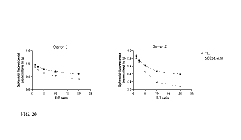
FIG. 20 shows cytotoxicity of SOCS1-edited TIL and control unedited TIL
against
A375-0KT3lt spheroids over 72 hours at different Effector:Target (E:T) ratios.
In a
monolayer culture, SOCS1-edited cells did not differ from unedited cells in
the killing of both
A375-pKSQA367 and A375-pKSQ368 cells (FIG. 13 and FIG. 14). In the 3D spheroid
setting, SOCS/-edited cells showed enhanced cytotoxicity in comparison to
control. This
suggests that in a spheroid setting the assay has different sensitivity.
CA 03228969 2024-2- 14
WO 2023/039410 PCT/US2022/076028
11
FIG. 21 shows IFNy produced by SOCS1-edited TIL and control unedited TIL
against A375-0KT3lt spheroids at 24 hours and different Effector:Target (E:T)
ratios.
FIG. 22 shows IL-6 produced by SOCS1-edited TIL and control unedited TIL
against
A375-0KT3It spheroids at 24 hours and different Effector:Target (E:T) ratios.
FIG. 23 shows luminescence level detected in supernatant after 24hr TIL-
spheroid
co-culture.
FIG. 24 shows calculated LDH level in supernatant after 24hr TIL-spheroid co-
culture.
DETAILED DESCRIPTION
The present disclosure provides, in some aspects, methods and compositions
useful
for measuring the antitumor potency of tumor infiltrating lymphocytes (TILs).
In some
embodiments, these methods include for example, coculturing TILs and
immortalized cells,
and assessing TIL potency using approaches that comprise coculturing TILs,
immortalized
cells, and bispecific molecules.
Many TIL potency assays currently being used for the monitoring of T cell
function
have focused on the use of cytokine (e.g., IFN-y or IL-2) release assays. For
example, T cell
activation can be determined by measuring IFN-y secretion following a short
coculture period
with non-cell-based T cell activation reagents, including anti-CD3 /anti-CD28
antibody-
coated beads. Cytokine secretion has been correlated with cytolytic activity
of CDS+ T cells
because cytokines, such as IFN-y, enhance MHC I and Fas expression on target
cells. For
example, TILs may be considered potent if, for instance, interferon gamma (IFN-
y) release is
greater than 50 pg/ml, greater than 100 pg/ml, greater than 150 pg/ml, or
greater than 200
pg/ml upon TCR stimulation.
However, activation by and direct killing of target tumor cells, which
requires that the
TILs are capable at least of interacting with the target cells and
producing/releasing mediators
for death induction, such as degranulation of cytolytic granules containing
granzyme B and
perforin, is an equally important indicator of clinical efficacy that is not
measured in
presently available cytokine release assays, which are driven by non-cell-
based TIL
activation methods. The TIL potency assays provided herein improve upon the
existing TIL
potency assays (e.g., cytokine release assays) by activating TIL through
relevant tumor cell-
based interactions that afford the opportunity to directly measure cell death
and/or viability of
CA 03228969 2024-2- 14
WO 2023/039410 PCT/US2022/076028
12
target cells (e.g., immortalized cells, such as cancer cells), or measure of
surrogate markers of
degranulation such as CD107a, or the production of effector
cytokines/chemokines (e.g.,
proinflammatory cytokines/chemokines), such as 1FN-y, 1L-6, 1L_2, and TNFct,
in a more
physiologically relevant TIL / tumor-cell co-culture setting.
A "TIL potency assay" refers to an assay used to characterize, for example,
quantify,
antitumor activity (e.g., cytokine production, TIL degranulation, tumor growth
inhibition) of
TILs. TIL potency assays may be used for assessing TIL antitumor activity
before and/or
after rapid expansion of the TILs and prior to clinical use applications, such
as adoptive cell
therapy (ACT).
Tumor Infiltrating Lymphocytes
Tumor infiltrating lymphocytes (TILs), including engineered TILs and/or edited
TILs,
may be characterized based on the potency of their antitumor activity (e.g.,
inhibition of
tumor cell growth).
The phrase -tumor infiltrating lymphocytes" or -TILs" refers to a population
of
lymphocytes that have left the bloodstream of a subject and migrated into a
tumor. TILs
include, but are not limited to, CDS+ cytotoxic T cells, CD4+ T cells
including Thl and Th17
CD4+ T cells, natural killer T cells, and natural killer (NK) cells. TTT,s
include both primary
and secondary TILs. "Primary TILs" are those that are obtained from patient
tissue samples
as outlined herein (sometimes referred to as "freshly harvested"), and
"secondary TILs" are
any TIL cell populations that have been expanded or proliferated, including,
but not limited
to bulk TILs and expanded TILs ("REP TILs" or "post-REP TILs"). In some
embodiments,
primary TILs include tumor reactive T cells that are obtained from peripheral
blood of a
patient. TIL cell populations can include genetically modified or otherwise
engineered TILs.
"TILs" also refers to a population of lymphocytes that have left the blood
stream of a subject,
have migrated into a tumor and then have departed to again enter the
bloodstream.
As generally outlined herein, TILs are generally taken from a patient sample
and
manipulated to expand their number prior to transplant into a patient. In some
embodiments,
the TILs may be genetically manipulated as discussed below. In general, TILs
are initially
obtained from a patient tumor sample ("primary TILs") and then expanded into a
larger
population for further manipulation as described herein, optionally
cryopreserved and re-
CA 03228969 2024-2- 14
WO 2023/039410 PCT/US2022/076028
13
stimulated, and optionally evaluated for phenotype and metabolic parameters as
an indication
of TIL health.
The terms "subject" and "patient" refer to a human being. In some embodiments,
this
human being may be a patient in need of immunotherapy involving an expanded
population
of the patient's own TILs. In other embodiments, this human being may be a
patient in need
of immunotherapy involving an expanded population of another patient's own
TILs.
TILs can generally be defined either biochemically, using cell surface
markers, or
functionally, by their ability to infiltrate tumors and effect treatment. TILs
can be generally
categorized as expressing one or more of the following biomarkers: CD4, CD8,
TCR ap,
TCRgd, CD27, CD28, CD56, CCR7, CD45RA, CD45RO, CD95, PD-1, and CD25.
Additionally, and alternatively, TILs can be functionally defined by their
ability to infiltrate
solid tumors upon reintroduction into a patient.
Adoptive cell therapy utilizing TILs cultured ex vivo by conventional TIL
manufacturing processes involves at least two steps, namely at least one rapid
expansion
protocol (REP) step subsequent to a pre-REP step. Adoptive cell therapy has
resulted in
successful therapy following host immunosuppression in patients with melanoma.
Current
infusion acceptance parameters rely on readouts of the composition of TILs
(e.g., CD28.
CD8, or CD4 positivity) and on the numerical folds of expansion and viability
of the REP
product.
The phrase "population of cells" or "population of TILs" refers to a number of
cells or
TILs that share common traits. In general, populations generally range from
lx106 to lx101
in number, with different TM, populations comprising different numbers. For
example, initial
growth of primary TILs in the presence of IL-2 can result in a population of
bulk TILs of
roughly lx 107 cells. REP expansion is generally done to provide populations
of 1.5x10⁹ to
1.5x10¹⁰ cells for infusion. In sonic embodiments, the population of cells is
monoclonal. In
other embodiments, the population of cells is polyclonal. In some embodiments,
when the
population of cells is polyclonal, the cells still share one or more common
traits. A
monoclonal T cell population will result in the predominance of a single TCR-
gene
rearrangement pattern. In contrast, polyclonal T cell populations have diverse
TCR-gene
rearrangement pattern, which can make them more effective in certain
situations.
In some embodiments, the TILs are genetically engineered to include additional
functions including, but not limited to, a high-affinity T cell receptor
(TCR), e.g., a TCR
CA 03228969 2024-2- 14
WO 2023/039410
PCT/US2022/076028
14
targeted at a tumor-associated antigen such as MAGE-1, HER2, or NY-ESO-1, or a
chimeric
antigen receptor (CAR) which binds to a tumor-associated cell surface molecule
(e.g.,
mesothelin) or lineage-restricted cell surface molecule (e.g., EGFR, CD19 or
HER2).
The term "engineered TIL" or "eTIL" encompasses TILs comprising one or more
genomic modifications, effected through non-natural means, resulting in the
reduced
expression and/or function of one or more endogenous target genes as well as
TILs
comprising a non-naturally occurring gene-regulating system capable of
reducing the
expression and/or function of one or more endogenous target genes. An
"unmodified TIL" or
"control TIL" refers to a TIL or population of TILs wherein the genomes have
not been
modified through non-naturally occurring means and that does not comprise a
non-naturally
occurring gene-regulating system or comprises a control gene-regulating system
(e.g., an
empty vector control, a non-targeting gRNA, a scrambled siRNA, etc.). TILs
that occur
naturally that have reduced expression and/or function of one or more
endogenous genes are
included under the terms unmodified or control TILs.
In some embodiments, the engineered TILs manufactured by the methods described
herein comprise one or more modifications (e.g., insertions, deletions, or
mutations of one or
more nucleic acids) in the genomic DNA sequence of an endogenous target gene
resulting in
the reduced expression and/or function the endogenous gene. In some
embodiments, the
modifications in the genomic DNA sequence reduce or inhibit mRNA
transcription, thereby
reducing the expression level of the encoded mRNA transcript and protein. In
some
embodiments, the modifications in the genomic DNA sequence reduce or inhibit
mRNA
translation, thereby reducing the expression level of the encoded protein. In
some
embodiments, the modifications in the genomic DNA sequence encode a modified
endogenous protein with reduced or altered function compared to the unmodified
(i.e., wild-
type) version of the endogenous protein (e_g_, a dominant-negative mutant,
described infra).
In some embodiments, the modified TILs further comprise an engineered antigen-
specific receptor recognizing a protein target expressed by a target cell,
such as a tumor cell
or an antigen presenting cell (APC). The term "engineered antigen receptor"
refers to a non-
naturally occurring antigen-specific receptor such as a chimeric antigen
receptor (CAR) or a
recombinant T cell receptor (TCR). In some embodiments, the engineered antigen
receptor is
a CAR comprising an extracellular antigen binding domain fused via hinge and
transmembrane domains to a cytoplasmic domain comprising a signaling domain.
In some
CA 03228969 2024-2- 14
WO 2023/039410 PCT/US2022/076028
embodiments, the CAR extracellular domain binds to an antigen expressed by a
target cell in
an MHC-independent manner leading to activation and proliferation of the RE
cell. In some
embodiments, the extracellular domain of a CAR recognizes a tag fused to an
antibody or
antigen binding fragment thereof. In such embodiments, the antigen-specificity
of the CAR is
dependent on the antigen-specificity of the labeled antibody, such that a
single CAR construct
can be used to target multiple different antigens by substituting one antibody
for another. In
some embodiments, the extracellular domain of a CAR may comprise an antigen
binding
fragment derived from an antibody. Antigen binding domains that are useful in
the present
disclosure include, for example, scFvs, antibodies, antigen binding regions of
antibodies,
variable regions of the heavy/light chains, and single chain antibodies.
In some embodiments, the intracellular signaling domain of a CAR may be
derived
from the TCR complex zeta chain (such as CD3 signaling domains), FcyRIIL
FcERI, or the
Tlymphocyte activation domain. In some embodiments, the intracellular
signaling domain of
a CAR further comprises a costimulatory domain, for example a 4-1B B, CD28,
CD40,
MyD88, or CD70 domain. In some embodiments, the intracellular signaling domain
of a
CAR comprises two costimulatory domains, for example any two of 4-1BB, CD28,
CD40,
MyD88, or CD70 domains. Exemplary CAR structures and intracellular signaling
domains
are known in the art (See e.g., WO 2009/091826; US 20130287748; WO
2015/142675; WO
2014/055657; and WO 2015/090229, incorporated herein by reference).
CARs specific for a variety of tumor antigens are known in the art, for
example
CD171-specific CARs (Park et al., Mol Ther (2007) 15(4):825-833), EGFRvIll-
specific
CARs (Morgan etal., Hum Gene Ther (2012) 23(10):1043-1053). EGF-R-specific
CARs
(Kobold etal., J Natl Cancer Inst (2014) 107(1):364), carbonic anhydrase K-
specific CARs
(Lamers etal., Biochem Soc Trans (2016) 44(3):951-959), FR-a-specific CARs
(Kershaw et
al., Clin Cancer Res (2006) 12(20):6106-6015), HER2-specific CARs (Ahmed et
al., J Clin
Oncol (2015) 33(15)1688- 1696;Nakazawa etal., Mol Ther (2011) 19(12):2133-
2143;
Ahmed etal., Mol Ther (2009) 17(10):1779-1787; Luo etal., Cell Res (2016)
26(7):850-853;
Morgan et al., Mol Ther (2010) 18(4):843-851; Grada etal., Mol Ther Nucleic
Acids (2013)
9(2):32), CEA-specific CARs (Katz etal., Clin Cancer Res (2015) 21(14):3149-
3159),
IL13Ra2-specific CARs (Brown etal., Clin Cancer Res (2015) 21(18):4062-4072),
GD2-
specific CARs (Louis etal., Blood (2011) 118(23):6050-6056; Caruana etal., Nat
Med
(2015) 21(5):524-529), ErbB2-specific CARs (Wilkie etal., J Clin Immunol
(2012)
CA 03228969 2024-2- 14
WO 2023/039410 PCT/US2022/076028
16
32(5):1059-1070), VEGF-R-specific CARs (Chinnasamy etal., Cancer Res (2016)
22(2):436-447), FAP-specific CARs (Wang et al., Cancer Immunol Res (2014)
2(2):154-
166), MSLN-specific CARs (Moon eta!, Clin Cancer Res (2011) 17(14):4719- 30),
NKG2D-
specific CARs (VanSeggelen et al., Mol Ther (2015) 23(10):1600-1610), CD19-
specific
CARs (Axicabtagene ciloleucel (Yescartae) and Tisagenlecleucel (Kymriah0). See
also,
82/337 Li et al., J Hematol and Oncol (2018) 11(22), reviewing clinical trials
of tumor-
specific CARs.
As generally outlined herein, TILs are generally taken from a patient sample
and
manipulated to expand their number prior to transplant into a patient. In some
embodiments,
the TILs may be genetically manipulated as discussed below. In general, TILs
are initially
obtained from a patient tumor sample ("primary TILs") and then expanded into a
larger
population for further manipulation, optionally cryopreserved and re-
stimulated, and
optionally evaluated for phenotype and metabolic parameters as an indication
of TIL health.
A patient tumor sample may be obtained using methods known in the art,
generally
via surgical resection, needle biopsy, or other means for obtaining a sample
that contains a
mixture of tumor and TIL cells. In general, the tumor sample may be from any
solid tumor,
including primary tumors, invasive tumors or metastases. The solid tumor may
be of any
cancer type, including, but not limited to, bladder cancer, brain cancer,
breast cancer
(including triple negative breast cancer), cervical cancer, colon and rectal
cancer, stomach
cancer, endometrial cancer, renal cancer, lip and oral cancer, head and neck
cancer
(including, for example, head and neck squamous cell carcinoma (HNSCC))
glioblastoma,
glioblastoma multiforme, neuroblastoma, liver cancer, mesothelioma, lung
cancer (including
non-small cell lung cancer (NSCLC) and small cell lung cancer), skin cancer
(including but
not limited to squamous cell carcinoma, basal cell carcinoma, nonmelanoma skin
cancer and
melanoma), ovarian cancer, uveal cancer, uterine cancer, pancreatic cancer,
prostate cancer,
sarcoma, and thyroid cancer. In some embodiments, useful TILs are obtained
from malignant
melanoma tumors, as these have been reported to have particularly high levels
of TILs.
Primary lung, (including non-small cell lung cancer (NSCLC)), bladder,
cervical and
melanoma tumors or metastases thereof can be used to obtain TILs.
Once obtained, the tumor sample is generally fragmented using sharp dissection
into
small pieces of from about 1 to about 8 mm3, or from about 0.5 to about 4 mm3
with from
about 2-3 mm3 being particularly useful. The TILs are cultured from these
fragments using
CA 03228969 2024-2- 14
WO 2023/039410
PCT/US2022/076028
17
enzymatic tumor digests. Such tumor digests may be produced by incubation in
enzymatic
media (e.g., Roswell Park Memorial Institute (RPMI) 1640 buffer, 2 mM
glutamate, 10
gg/mlgentamicin, 30 units/ml of DNase and 1.0 nag/m1 of collagenase), followed
by
mechanical dissociation (e.g., using a tissue dissociator). Tumor digests may
be produced by
placing the tumor in enzymatic media and mechanically dissociating the tumor
for
approximately 1 minute, followed by incubation for 30 minutes at 370 C in 5%
CO?, followed
by repeated cycles of mechanical dissociation and incubation under the
foregoing conditions
until only small tissue pieces are present. At the end of this process, if the
cell suspension
contains a large number of red blood cells or dead cells, a density gradient
separation using
FICOLL branched hydrophilic polysaccharide may be performed to remove these
cells.
Alternative methods known in the art may be used, such as those described in
U.S. Patent
Application Publication No. 2012/0244133 Al, the disclosure of which is
incorporated herein
by reference in its entirety. Any of the foregoing methods may be used in any
of the
embodiments described herein for methods of expanding TILs or methods treating
a cancer.
In general, the harvested cell suspension is called a "primary cell
population" or a
"freshly harvested" cell population. In some embodiments, fragmentation
includes physical
fragmentation, including for example, dissection as well as digestion. In some
embodiments,
the fragmentation is physical fragmentation. In some embodiments, the
fragmentation is
dissection. In some embodiments, the fragmentation is by digestion. In some
embodiments,
TILs can be initially cultured from enzymatic tumor digests and tumor
fragments obtained
from patients.
In some embodiments, the TILs are obtained from tumor digests. In some
embodiments, tumor digests are generated by incubation of mechanically
dissociated tumor
in enzyme media, for example, but not limited to RPMI 1640, 2 mM GlutaMAX, 10
mg/ml
gentamicin, 30 U/ml DNase, and 1.0 mg/ml collagenase, followed by mechanical
dissociation
(GentleMACS, Miltenyi Biotec, Auburn, Calif.). In some embodiments, the
mechanically
dissociated tumor would be broken up into approximately 1 mm3 pieces. After
placing the
tumor in enzyme media, the tumor can be mechanically dissociated for
approximately 1
minute. The solution can then be incubated for 30 minutes at 37 C in 5% CO?
and can then
be mechanically disrupted again for approximately 1 minute. After being
incubated again for
30 minutes at 37 C in 5% CO2, the tumor can be mechanically disrupted a third
time for
approximately 1 minute. In some embodiments, after the third mechanical
disruption if large
CA 03228969 2024-2- 14
WO 2023/039410 PCT/US2022/076028
18
pieces of tissue are present, one or two additional mechanical dissociations
can be applied to
the sample, with or without 30 additional minutes of incubation at 37 C in 5%
CO2. In some
embodiments, at the end of the final incubation if the cell suspension
contains a large number
of red blood cells or dead cells, a density gradient separation using FICOLL
can be
performed to remove these cells.
In some embodiments, cells can be optionally frozen or cryopreserved after
sample
harvest and stored frozen prior to entry into the expansion phase.
In some embodiments, the TILs are expanded for up to a total of 9, 10, 11, 12,
13, 14,
15, 16, 17, 18, 19, 20, 21, 22, 23, 24, 25, 26,27 or 28 days from the initial
tumor
fragmentation or disaggregation. In some embodiments, the TILs are expanded
for a total of
9-25 days, 9-21 days, or 9-14 days. In some embodiments, the TILs are expanded
for up to a
total of 9 days. In some embodiments, the TILs are expanded for up to a total
of 10 days. In
some embodiments, the TILs are expanded for up to a total of 11 days. In some
embodiments, the TILs are expanded for up to a total of 12 days. In some
embodiments, the
TILs are expanded for up to a total of 13 days. In some embodiments, the TILs
are expanded
for up to a total of 14 days. In some embodiments, the TILs are expanded for
up to a total of
15 days. In some embodiments, the TILs are expanded for up to a total of 16
days. In some
embodiments, the TILs are expanded for up to a total of 17 days. In some
embodiments, the
TILs are expanded for up to a total of 18 days. In some embodiments, the TILs
are expanded
for up to a total of 19 days. In some embodiments, the TILs are expanded for
up to a total of
20 days. In some embodiments, the TILs are expanded for up to a total of 21
days. In some
embodiments, the TILs are expanded for up to a total of 22 days. In some
embodiments, the
TILs are expanded for up to a total of 23 days. In some embodiments, the TILs
are expanded
for up to a total of 24 days. In some embodiments, the TILs are expanded for
up to a total of
25 days. In some embodiments, the TILs are expanded for up to a total of 26
days. In some
embodiments, the TILs are expanded for up to a total of 27 days. In some
embodiments, the
TILs are expanded for up to a total of 28 days.
In some embodiments, the expanded TILs are analyzed for expression of numerous
phenotype markers, including those described herein. In some embodiments, the
marker is
selected from: TCRot/13, CD57, CD28, CD4, CD27, CD56, CD8a, CD45RA, CD45RO,
CD8a, CCR7, CD4, CD3, CD38, and HLA-DR. In some embodiments, expression of one
or
CA 03228969 2024-2- 14
WO 2023/039410 PCT/US2022/076028
19
more regulatory markers is measured, namely from the group: CD137, CD8a, Lag3,
CD4,
CD3, PD-1, TIM-3, CD69, CD8a, TIGIT, CD4, CD3, KLRG1, and CD154.
In some embodiments, the memory marker is CCR7 or CD62L. In embodiments.
restimulated TILs are evaluated for cytokine release, using cytokine release
assays. In some
embodiments, TILs are evaluated for interferon-gamma (IFN-y) secretion in
response to
stimulation either with OKT3 or coculture with autologous tumor digest. In
some
embodiments, TILs are evaluated for IL-6 secretion in response to stimulation
either with
OKT3 or coculture with autologous tumor digest. Additional effector cytokines
that could be
measured include, but are not limited to, 1L-1, IL-2, IL-12, IL-17, IL-18,
granulocyte-
macrophage colony stimulating factor (GM-CSF), and tumor necrosis factor-a
(TNFa).
Chemokines such as CXCL10, CXCL13, CCL1, CCL3, CCL4, CCL5, CCL9/10, CCL17,
CCL22, CCL23, and XCL1 can also be evaluated.
TILs are evaluated for various regulatory markers, such as TCRa/13, CD56,
CD27,
CD28, CD57, CD45RA, CD45RO, CD25, CD127, CD95, IL-2R, CCR7, CD62L, KLRG1,
and CD122
Immortalized Cells
Immortalized cells are cells that have been manipulated to proliferate
indefinitely and
can thus be cultured for long periods of time. Immortalized cell lines are
typically derived
from a variety of sources that have chromosomal abnormalities or mutations
that permit them
to continually divide, such as tumors. Immortalized cells are thus considered
to be
"engineered." A population of immortalized cells may be a heterogenous
population or may
be derived from a single immortalized clone (to form a clonal population). In
some
embodiments, immortalized cells comprise a heterogenous population of
immortalized cells.
In some embodiments, immortalized cells comprise a clonal population of
immortalized cells.
Immortalized cells can be derived from a variety of species and/or origins.
For
example, immortalized cells can be immortalized animal cells or immortalized
human cells,
or a combination thereof. In some embodiments, immortalized cells are
immortalized human
cells.
In some embodiments, immortalized cells are cancer cells. For example, cancer
cells
may include, but are not limited to, melanoma cells, colorectal cancer cells,
bile duct cancer
CA 03228969 2024-2- 14
WO 2023/039410 PCT/US2022/076028
cells, and breast cancer cells. In some embodiments, the cancer cells are
selected from
melanoma cells, colorectal cancer cells, bile duct cancer cells, and breast
cancer cells.
Immortalized cells that may be engineered for use in the TIL potency assays
described herein may include, but are not limited to, A375 melanoma cells
(e.g., ATCCO
CRL-16191-m), K562 multipotential, hematopoietic malignant cells, primary
cells (e.g.,
ATCCO CCL-2431m), human embryonic kidney (HEK) 293T cells (e.g., ATCCO CRL-
1573), and Chinese hamster ovary (CHO) cells (e.g., ATCCO CCL-61'M). In some
embodiments, the immortalized cells are selected from A375 cells, K562 cells,
primary cells,
HEK293T cells. and CHO cells. It should be understood that the immortalized
cells useful for
the assays and methods described herein arc not limited to the foregoing
examples. Other
immortalized cell lines are known in the field and may be used in accordance
with the present
disclosure.
The use of a cell line expressing immune suppressive markers to suppress T
cell
responses, such as PD-L1, PD-L2, to test whether TILs are resistant to
suppressive signals are
also contemplated herein. In some embodiments, immortalized cells that may be
engineered
for use in TIL potency assays comprise a cell line expressing immune
suppressive markers to
suppress T cell responses, such as PD-Li.
Cell lines that express co-stimulatory markers to enhance T cell responses,
such as
CD80/86, OX4OL, and/or 41BBL may also be engineered for use in a TIL potency
assay. In
some embodiments, immortalized cells that may be engineered for in TIL potency
assays
comprise a cell line may express co-stimulatory markers to enhance T cell
responses, such as
CD80/86.
The phrase "tumor cells" or "cancer cells" refers to cells that divide in an
uncontrolled
manner, forming solid tumors or flooding the blood with abnormal cells.
Healthy cells stop
dividing when there is no longer a need for more daughter cells, but tumor
cells or cancer
cells continue to produce copies. They are also able to spread from one part
of the body to
another in a process known as metastasis. Tumor cells can be isolated from a
number of
cancer types including bladder cancer, brain cancer, breast cancer (including
triple negative
breast cancer), cervical cancer, colon and rectal cancer, stomach cancer,
endometrial cancer,
renal cancer, lip and oral cancer, head and neck cancer (including, for
example, head and
neck squamous cell carcinoma (HNSCC)) glioblastoma, glioblastoma multiforme,
neuroblastoma, liver cancer, mesothelioma, lung cancer (including non-small
cell lung cancer
CA 03228969 2024-2- 14
WO 2023/039410 PCT/US2022/076028
21
(NSCLC) and small cell lung cancer), skin cancer (including but not limited to
squamous cell
carcinoma, basal cell carcinoma, nonmelanoma skin cancer and melanoma),
ovarian cancer,
uveal cancer, uterine cancer, pancreatic cancer, prostate cancer, sarcoma, and
thyroid cancer.
In some embodiments, cancer cells are also isolated from lymphoma. Tumor cells
can be
isolated from primary tumors and metastases.
While immortalized cells are described throughout in the connect of
coculturing with
TILs, it should be understood that the immortalized cells may be replaced with
any tumor
cells, such as cancer cells, that express or have been engineered to express a
molecule that
activates a T cell, e.g., binds to a T cell antigen.
Molecules that Activate Lymphocytes
Engineered immortalized cells of the present disclosure may comprise or
express a
molecule that activates a T cell.
A "molecule that activates a lymphocyte- and a "molecule that activates a T
refers to a nonendogenous stimulus that causes the cell to become activated.
In the
endogenous process, T cells, for example, become activated when they are
presented with
peptide antigens by MHC class II molecules, which are expressed on the surface
of antigen-
presenting cells (APCs). Once activated, the T cells divide rapidly and
secrete cytokines that
regulate or assist the immune response. The endogenous T cell activation
process involves at
least (a) activation of the TCR complex, which involves CD3, and (b) co-
stimulation of
CD28 or 4-1BB by proteins on the APC surface. It is known in the art that the
endogenous
activation of T cells can be simulated by stimulation of T cells by CD3, CD28
or 4-1BB
agonists (e.g., antibodies). Thus, CD3, CD28 and/or 4-1BB can together
activate T cells.
Activated T cells increase in number or proliferate and begin producing
cytokines
(activated TILs) to boost the immune response.
Immortalized cells may comprise and/or express a molecule that activates a T
cell by
binding (e.g., directly binding) to a T cell antigen. In some embodiments,
TILs (e.g.,
engineered TILs and/or edited TILs) express a T cell antigen. Non-limiting
examples of T
cell antigens include CD3. CD28, CD2, 41BB, 0X40, GITR, ICOS, CD4, CD8. In
some
embodiments, the T cell antigen is CD3. In some embodiments, the T cell
antigen is CD28. In
some embodiments, the T cell antigen is CD2.
CA 03228969 2024-2- 14
WO 2023/039410
PCT/US2022/076028
22
The term "CD3" refers to the CD3 (cluster of differentiation 3) T cell co-
receptor that
helps to activate both the cytotoxic T cell (CD8+ naive T cells) and also T
helper cells (CD4+
naive T cells). CD3 is a protein complex composed of six distinct polypeptide
chains (2 CD3
zeta chains, 2 CD3 epsilon chains, 1 CD3e gamma chain, and 1 CD3 delta chain).
These
chains associate with the T cell receptor (TCR) alpha and beta chains (or
gamma and delta
chains) to generate an activation signal in T lymphocytes. The TCR alpha and
beta chains (or
gamma and delta chains), and CD3 molecules together constitute the TCR
complex. The
human CD3E gene is identified by National Center for Biotechnology Information
(NCBI)
Gene ID 916. An exemplary nucleotide sequence for a human CD3E gene is the
NCBI
Reference Sequence: NG_007383.1.
The term "CD28" refers to cluster of differentiation 28, which is one of the
proteins
expressed on T cells that provides co-stimulatory signals required for T cell
activation and
survival. T cell stimulation through CD28 in addition to the T cell receptor
(TCR) can
provide a potent signal for the production of various cytokines, such as
interleukins. CD28 is
the receptor for CD80 and CD86 proteins. When activated by Toll-like receptor
ligands,
CD80 expression is upregulated in antigen-presenting cells (APCs). The human
CD28 gene is
identified by NCBI Gene ID 940. An exemplary nucleotide sequence for a human
CD28 gene
is the NCB' Reference Sequence: NG 029618.1. An exemplary amino acid sequence
of a
human CD28 polypeptide is:
MLRLLLALNLFPSIQVTGNKILVKQSPMLVAYDNAVNLSCKYSYNLFSREFR
ASLHKGLDSAVEVCVVYGNYSQQLQV YSKTGFNCDGKLGNESVTFYLQNLYVNQT
DIYFCKIEVMYPPPYLDNEKSNGTIIHVKGKHLCPSPLFPGPSKPFWVLVVVGGVLAC
YSLLVTVAFTIFWVRSKRSRLLHSDYMNMTPRRPGPTRKHYQPYAPPRDFAAYRS
(SEQ ID NO: 19).
The term "CD2" refers to cluster of differentiation 2, which is a cell
adhesion
molecule found on the surface of T cells and natural killer (NK) cells. CD2
interacts with
other adhesion molecules and acts as a co-stimulatory molecule on T and NK
cells. The
human CD2 gene is identified by NCBI Gene ID 914. An exemplary nucleotide
sequence for
a human CD2 gene is the NCBI Reference Sequence: NG_050908.1. An exemplary
amino
acid sequence of a human CD2 polypeptide is:
MSFPCKFVASFLLIFNVSSKGAVSKE I TNALETWGALGQDINLD IP SFQMSDD IDD I
KWEKTSDKKKIAQFRKEKETFKEKDTYKLFKNGTLKIKHLKTDDQD I YKVS IYDTKGKNVLE
CA 03228969 2024-2- 14
WO 2023/039410
PCT/US2022/076028
23
KIFDLKIQERVSKPKISWTOINTTLTCEVMNGTDPELNLYQDGKHLKLSQRVITHKWTTSLS
AKFKOTAGNKVSKESSVEPVSCPEKGLDIYLIIGICGGGSLLMVFVALLVFYITKRKKQRSR
RNDEELETRAHRVATEERGRKPHQIPASTPQNPATSQHPPPPPGHRSQAPSHRPPPPGHRVQ
HQPQKRPPAPSGTQVHQQKGPPLPRPRVQPKPPHGAAENSLSPSSN (SEQ ID NO:
2 0 ) .
In some embodiments, immortalized cells comprise a molecule that activates a T
cell
by binding, e.g., specifically binding, to a T cell antigen. In some
embodiments, immortalized
cells comprise a molecule that activates a T cell by binding to a CD3 antigen.
In some
embodiments, immortalized cells comprise a molecule that activates a T cell by
binding to a
CD28 antigen. In some embodiments, immortalized cells comprise a molecule that
activates a
T cell by binding to a CD2 antigen.
The phrase "specifically binding" refers to a molecule (e.g., antibody)
interacting with
high specificity with a particular antigen (e.g., T cell antigen), as compared
with other
antigens for which the complex has a lower affinity to associate. The specific
binding
interaction can be mediated through ionic bonds, hydrogen bonds, or other
types of chemical
or physical associations. In some embodiments, the molecule specifically binds
a particular
antigen when it recognizes its target antigen in a complex mixture of proteins
and/or
macromolecules. In some embodiments, the molecule that activates a T cell
binds to a T cell
antigen with an affinity (KD) of approximately less than 10-5 M, such as
approximately less
than 10-6 M, 10-7 M, 10-8 m, 10-9 M or 10-1 M or even lower.
In some embodiments, a molecule that activates a T cell is a T cell agonist.
The term
"agonist" refers to a chemical, a molecule, a macromolecule, a complex of
molecules, or a
complex of macromolecules that binds to a target, either on the surface of a
cell or in soluble
form. In certain embodiments, when an agonist binds to a target on the surface
of a cell, the
agonist activates the target to produce a biological response. Agonists
include hormones,
neurotransmitters, antibodies, and fragments of antibodies.
Non-limiting examples of molecules that activate T cells include, but are not
limited
to, antibodies, such as whole antibodies and/or antibody fragments, NANOBODY
binders,
AFFIMERO binders, and other molecular binders, such as ligands and receptors.
The term "antibody" refers to an immunoglobulin (Ig) molecule, which is
generally
comprised of four polypeptide chains, two heavy (H) chains and two light (L)
chains, or a
functional fragment, mutant, variant, or derivative thereof, that retains the
epitope binding
CA 03228969 2024-2- 14
WO 2023/039410 PCT/US2022/076028
24
features of an Ig molecule. Such fragment, mutant, variant, or derivative
antibody formats are
known in the art. In an embodiment of a full-length antibody, each heavy chain
is comprised
of a heavy chain variable region (VH) and a heavy chain constant region (CH).
The heavy
chain variable region (domain) is also designated as VDH in this disclosure.
The CH is
comprised of three domains, CH1, CH2 and CH3. Each light chain is comprised of
a light
chain variable region (VL) and a light chain constant region (CL). The CL is
comprised of a
single CL domain. The light chain variable region (domain) is also designated
as VDL in this
disclosure. The VH and VL can be further subdivided into regions of
hypervariability, termed
complementarity determining regions (CDRs), interspersed with regions that are
more
conserved, termed framework regions (FRs). Generally, each VH and VL is
composed of
three CDRs and four FRs, arranged from amino-terminus to carboxy-terminus in
the
following order: FR1, CDR1, FR2, CDR2, FR3, CDR3, and FR4. Immunoglobulin
molecules
can be of any type (e.g., IgG, IgE, IgM, IgD, IgA and IgY), class (e.g., IgGl,
IgG2, IgG3,
IgG4, IgAl and IgA2), or subclass.
In some embodiments, immortalized cells express a molecule that is an antibody
fragment. In some embodiments, an antibody fragment is selected from a single-
chain
variable fragment (scFv), a F(ab')2 fragment, a Fab fragment, a Fab fragment,
and an Fv
fragment.
The term "fragment" used in association with agonist or antibody, refers to a
fragment
of the agonist or antibody that retains the ability to specifically bind to an
antigen. Examples
of fragments of antibodies include (i) an Fab fragment, a monovalent fragment
consisting of
the VL, VH, CL and CH1 domains; (ii) an F(ab')2 fragment, a bivalent fragment
comprising
two Fab fragments linked by a disulfide bridge at the hinge region; (iii) an
Fd fragment
consisting of the VH and CH1 domains; (iv) an Fv fragment consisting of the VL
and VH
domains of a single arm of an antibody; (v) a dAb fragment, which comprises a
single
variable domain; and (vi) an isolated complementarity determining region
(CDR).
Furthermore, although the two domains of the Fv fragment, VL and VH, are
encoded by
separate genes, they can be joined, using recombinant methods, by a synthetic
linker that
enables them to be made as a single protein chain in which the VL and VH
regions pair to
form monovalent molecules (known as single chain Fv (scFv)). Such single chain
antibodies
are also intended to be encompassed within the term "antigen-binding portion"
of an
antibody. Other forms of single chain antibodies, such as diabodies are also
encompassed. In
CA 03228969 2024-2- 14
WO 2023/039410 PCT/US2022/076028
addition, single chain antibodies also include "linear antibodies" comprising
a pair of tandem
Fv segments (VH-CH1-VH-CH1), which, together with complementary light chain
polypeptides, form a pair of antigen binding regions.
The term "KD" refers to the dissociation equilibrium constant of a particular
agonist-
antigen interaction. Typically, the agonists described herein bind to a target
with a
dissociation equilibrium constant (KD) that is higher than the KD of mOKT3
scFv, which is
about 1x10⁻⁹ M or 1x10⁻¹ M, for example, as determined using surface plasmon
resonance
(SPR) technology in a Biacore instrument using the agonist as the ligand and
the target as the
analyte. In some embodiments, the agonists described herein (e.g., a low-
affinity mOKT3
scFv variant) bind to a target protein (e.g., CD3) with an affinity
corresponding to a KD that
is higher than 5x10⁻¹ M.
The term "koff' (sec-1) refers to the dissociation rate constant of a
particular agonist-
antigen interaction. Said value is also referred to as the kd value.
The term "kon- (M¨lxsec-1) refers to the association rate constant of a
particular
agonist-antigen interaction.
The term "KD" (M) refers to the dissociation equilibrium constant of a
particular
agonist-antigen interaction
The term "KA" (M-1) refers to the association equilibrium constant of a
particular
agonist-antigen interaction and is obtained by dividing the kon by the koff.
The phrase "anti-CD28 antibody" refers to an antibody or variant thereof,
e.g., a
monoclonal antibody, and includes human, humanized, chimeric or murine
antibodies which
are directed against the CD28 receptor in the T cell antigen receptor of
mature T cells.
The phrase "anti-CD2 antibody" refers to an antibody or variant thereof, e.g.,
a
monoclonal antibody, and includes human, humanized, chimeric or murine
antibodies which
are directed against the CD2 receptor in the T cell antigen receptor of mature
T cells.
The phrase "anti-CD3 antibody" refers to an antibody or variant thereof, e.g.,
a
monoclonal antibody, and includes human, humanized, chimeric or murine
antibodies which
are directed against the CD3 receptor in the T cell antigen receptor of mature
T cells (see,
e.g., International Publication No. W02013186613A 1, incorporated herein by
reference).
Anti-CD3 antibodies include OKT3, also known as muromonab. Anti-CD3 antibodies
also
include the UCHT1 clone, also known as T3 and CD3c. Other anti-CD3 antibodies
include,
for example, otelixizumab, teplizumab, and visilizumab.
CA 03228969 2024-2- 14
WO 2023/039410 PCT/US2022/076028
26
Muromonab-CD3 light chain
QIVLTQSPAIMSASPGEKVTMTCSASSSVSYMNWYQQKSGTSPKRWIYDTSKLASGV
PAHFRGSGSGTSYSLTISGMEAEDAATYYCQQWSSNPFTFGSGTKLEINRADTAPTVSIFPP
SSEQLTSGGASVVCFLNNFYPKDINVKWKIDGSERQNGVLNSWTDQDSKDSTYSMSSTLTLT
KDEYERHNSYTCEATHKTSTSPIVKSFNRNEC (SEQ ID NO: 17)
Muromonab-CD3 heavy chain
QVQLQQSGAELARPGASVKMSCKASGYTFTRYTMHWVKQRPGQGLEWIGYINPSRGY
TNYNQKFKDKATLTTDKSSSTAYMQLSSLTSEDSAVYYCARYYDDHYCLDYWGQGTTLTVSS
AKTTAPSVYPLAPVCGGTTGSSVTLGCLVKGYFPEPVTLTWNSGSLSSGVHTFPAVLQSDLY
TLSSSVTVTSSTWPSQSITCNVAHPASSTKVDKKIEPRPKSCDKTHTCPPCPAPELLGGPSV
FLFPPKPKDTLMISRTPEVTCVVVDVSHEDPEVKFNWYVDGVEVHNAKTKPREEQYNSTYRV
VSVLTVLHQDWLNGKEYKCKVSNKALPAPIEKTISKAKGQPREPQVYTLPPSRDELTKNQVS
LTCLVKGFYPSDIAVEWESNGQPENNYKTTPPVLDSDGSFFLYSKLTVDKSRWQQGNVFSCS
VMHEALHNHYTQKSLSLSPGK (SEQ ID NO:18)
The term "OKT3" refers to the anti-CD3 antibody produced by Miltenyi Biotech,
Inc., San Diego, Calif., USA) and or biosimilar or variant thereof (e.g., a
humanized,
chimeric, or affinity matured variant). A hybridoma capable of producing OKT3
is available
in the American Type Culture Collection and assigned the ATCC accession number
CRL
8001. A hybridoma capable of producing OKT3 is available in the European
Collection of
Authenticated Cell Cultures (ECACC) and assigned Catalogue No. 86022706.
In some embodiments, the antibody fragment is an OKT3 antibody, such as an
OKT3
antibody fragment. In some embodiments, an OKT3 antibody fragment is a
membrane-bound
OKT3 (mOKT3) scFv fragment.
Transmembrane domains may be utilized to anchor mOKT3 scFv to the cell surface
of immortalized cells. For example, the human CD8 transmembrane domain can be
utilized
to anchor mOKT3 scFv to the cell surface of immortalized cells. The use of CD8
transmembrane domains from other species, such as mouse (pKSQ366), or other
transmembrane proteins, such as CD14 or CD28 to anchor mOKT3 scFv to the cell
surface of
immortalized cells is also contemplated herein. In some embodiments, mOKT3
scFv is
expressed tethered to the cell surface of the immortalized cells. In some
embodiments, human
CD8 transmembrane domain is utilized to anchor mOKT3 scFv to the cell surface
of
immortalized cells.
CA 03228969 2024-2- 14
WO 2023/039410
PCT/US2022/076028
27
Many T cells will respond to the very strong stimulation of mOKT3. The use of
lower
affinity T cell receptor binding may allow for the separation of subtle
differences in T cell
receptor signaling thresholds due to cell-to-cell variation and may allow more
sensitive
quality determinations between T cell therapy products prior to infusion into
patients. Low-
affinity variants of mOKT3 fragments can be made via site mutagenesis to model
more
closely the affinities of natural T cell receptors with their recognized
antigens. Published
affinities of natural T cell receptors are in the p.M KD range.
In some embodiments, a low-affinity mOKT3 scFv variant is used. In some
embodiments, a molecule binds to CD3 with a dissociation constant (KD) that is
lower than
the KD of mOKT3 scFv, wherein the KD of mOKT scFv is about 1 x 10⁻⁹ to about 1
x 10-11,
for example, about 5 x 10⁻¹ M. In some embodiments, the low-affinity mOKT3
scFv variant
comprises an amino acid sequence having a R55 mutation, relative to the amino
acid
sequence of SEQ ID NO: 2. In some embodiments, the low-affinity mOKT3 scFv
variant
comprises an amino acid sequence having a Y57 mutation, relative to the amino
acid
sequence of SEQ ID NO: 2. In some embodiments, the low-affinity mOKT3 scFv
variant
comprises an amino acid sequence having R55 and Y57 mutations, relative to the
amino acid
sequence of SEQ ID NO: 2. In some embodiments, the low-affinity mOKT3 scFv
variant
comprises an amino acid sequence having R55M and Y57A mutations, relative to
the amino
acid sequence of SEQ ID NO: 2. In some embodiments, the low-affinity mOKT3
scFv
variant comprises an amino acid sequence having R55L and Y57T mutations,
relative to the
amino acid sequence of SEQ ID NO: 2.
In some embodiments, the low-affinity mOKT3 scFv variant binds to CD3 with a
dissociation constant (KD) of about 1 x 10⁻⁶ to about 5 x 10⁻⁸. For example,
the low-affinity
mOKT3 scFv variant may bind to CD3 with a KD of about 1 x 10⁻⁶, 1 x RY7, or 1
x 10-8. In
some embodiments, the low-affinity mOKT3 scFv variant binds to CD3 with a KD
of about 5
x 10-7.
In some embodiments, the low-affinity mOKT3 scFv variant binds to CD3 with a
KD
that is least 10-fold lower, at least 20-fold lower, at least 30-fold lower,
at least 40-fold lower,
at least 50-fold lower, at least 60-fold lower, at least 70-fold lower, at
least 80-fold lower, at
least 90-fold lower, at least 100-fold lower, at least 110-fold lower, at
least 120-fold lower, at
least 130-fold lower, at least 140-fold lower, at least 150-fold lower, at
least 160-fold lower,
at least 170-fold lower, at least 180-fold lower, at least 190-fold lower, at
least 200-fold
CA 03228969 2024-2- 14
WO 2023/039410 PCT/US2022/076028
28
lower, at least 210-fold lower, at least 220-fold lower, at least 230-fold
lower, at least 240-
fold lower, at least 250-fold lower, at least 275-fold lower, at least 300-
fold, at least 400-fold,
at least 500-fold, at least 600-fold, at least 700-fold, at least 800-fold, at
least 900-fold, at
least 1000-fold, at least 1100-fold, at least 1200-fold lower than the KD of
mOKT3 scFv. In
some embodiments, the low-affinity mOKT3 scFv variant binds to CD3 with a
dissociation
constant KD that is least 250-fold lower than the KD of mOKT3 scFv. In some
embodiments,
the low-affinity mOKT3 scFv variant binds to CD3 with a dissociation constant
KD that is
least 1000-fold lower than the KD of OKT3 scFv.
In some embodiments, a molecule that activates a T cell is a membrane-tethered
molecule. Other molecules, such as bacterial superantigens (e.g., SEB).
phytohacmagglutinin
(PHA) or concanavalin A (ConA), that bind and cluster the T cell receptor CD3
to activate
the T cell are contemplated to work in a similar manner to the mOKT3 scFv
tethered to the
cell surface of immortalized cells. In some embodiments, a molecule that
activates a T cell is
phytohaemagglutinin (PHA). In some embodiments, a molecule that activates a T
cell is
concanavalin A (ConA). Other monoclonal antibody scFv fragments that bind a T
cell
receptor/CD3 component, such as anti-CD3 antibody clone BC3, are also
contemplated to
work in a similar manner to the mOKT3 scFv tethered to the cell surface of
immortalized
cells. In some embodiments, a molecule that activates a T cell is an anti-CD3
antibody clone
BC3.
In some embodiments, the immortalized cells express an Fe receptor. In such
embodiments, an antibody, such as a monoclonal anti-CD3 antibody, that binds
both the Fe
receptor and a T cell antigen, such as CD3, may be used to assess the potency
of the TILs.
In other embodiments, the molecule that activates a T cell is not expressed by
the
immortalized cell. Rather, the molecule may be a bispecific molecule that can
bind to both
the immortalized cell and to the T cell. Bispecific T cell engagers, such as
blinatumomab, is
one non-limiting example of a such a molecule that binds to CD19 expressed by
the
immortalized cells and CD3 expressed by T cells.
CocuIture Conditions
The methods provided herein, in some aspects, comprise coculturing lymphocytes
(e.g., TIL s) and immortalized cells.
CA 03228969 2024-2- 14
WO 2023/039410 PCT/US2022/076028
29
The immortalized cells may be cultured as a two-dimensional monolayer of cells
or as
multilayer, three-dimensional spheroids. Spheroids are self-assembling
multicellular
aggregates that form in an environment that prevents attachment to a flat
surface.
A two-dimensional culture is an adherent culture in which cells grow as a
monolayer,
for example, in a culture flask, dish, or multiwell plate having an adherent
surface. By
comparison, a three-dimensional spheroid culture is a nonadherent or low-
adherent culture
system in which cells grow as multilayer spheroids, for example, in suspension
culture on
non-adherent plates, in concentrated medium or in a gel-like substance, or on
a scaffold.
In some embodiments, the immortalized cells of a three-dimensional system are
cultured on an ultra-low attachment (ULA) surface. An ultra-low attachment
surface is a
surface that includes a substance that inhibits specific and nonspecific
immobilization,
forcing cells into a suspended state, enabling three-dimensional spheroid
formation. In some
embodiments, an ultra-low attachment surface comprises a hydrophilic,
neutrally charged
coating. In some embodiments, the hydrophilic, neutrally charged hydrogel
coating is
covalently bound to the surface. In some embodiments, the surface comprises
polystyrene.
Thus, in some embodiments, an ultra-low attachment surface is a polystyrene
surface to
which a hydrophilic, neutrally charged coating is covalently bound. As is
known in the art,
ultra-low attachment surfaces are generally stable, noncytotoxic, biologically
inert, and non-
degradable.
In some embodiments, the immortalized cells of a three-dimensional system are
cultured in an ultra-low attachment multiwell plate (e.g., Coming ). In some
embodiments,
the ultra-low attachment surface of the multiwell plate (e.g., multiwell
polystyrene plate) is a
covalently bound hydrogel layer that is hydrophilic and neutrally charged.
Various multiwell
formats are available, for example, 6-, 24-, or 96-well formats may be used.
The culture conditions provided below may be used for either two-dimensional
or
three-dimensional cocultures.
In some embodiments, a coculture of immortalized cells includes 25,000 to
200,000
immortalized cells. For example, a coculture of immortalized cells may include
50,000 to
100,000 immortalized cells. In some embodiments, a coculture of immortalized
cells includes
25000, 30000, 40000, 50000, 60000, 70000, 80000, 90000, or 10000 immortalized
cells.
CA 03228969 2024-2- 14
WO 2023/039410 PCT/US2022/076028
In some embodiments, the immortalized cells are plated, for example, in a
multiwell
plate, such as a 96-well plate, in media (e.g., Dulbecco's Modified Eagle
Medium (DMEM))
with serum (e.g., fetal bovine serum (FBS)) and an antibiotic (e.g.,
Pen/Strep).
In some embodiments, the immortalized cells are cultured (e.g., in a multiwell
plates
with a ULA surface or in adherent multiwell plates) in media for about 48
hours to about 96
hours (e.g., about 48 hours, about 60 hours, about 72 hours, about 84 hours,
or about 96
hours) to form three-dimensional spheroids prior to the addition of TILs.
To generate TILs, for example, such as pre-REP TILs, prior to coculture, in
some
embodiments, tumor digest samples (e.g., melanoma tumor digest samples) are
thawed and
plated (e.g., 1.5e6 cells/mL) in pre-REP media in the presence of IL-2. Re-
plating and
feeding of IL-2, in some embodiments, is repeated every 1-3 days until growth
slows to less
than 1.5x growth after two days, for example.
The day before coculture initiation, in some embodiments, pre-REP TILs are
thawed
and rested overnight in REP media (e.g., RPMI, AIM-V, 5% Human AB Serum and IL-
2).
In some embodiments, pan T cells are isolated from peripheral blood
mononuclear
cells (PBMCs) and cocultured with the immortalized cells.
In some embodiments, cocultures are performed at a 2:1 effector (T cell) to
target
(immortalized cell) ratio (abbreviated as E:T). In some embodiments,
cocultures are
performed at a 1:1 E:T ratio. In some embodiments, cocultures are performed at
a 1:2 E:T
ratio. In some embodiments, cocultures are performed at a 3:1. 2:1, or 1:1 E:T
ratio. In some
embodiments, cocultures are performed at a 5:1 E:T ratio. In some embodiments,
cocultures
are performed at a 10:1 E:T ratio. In some embodiments, cocultures are
performed at a 20:1
E:T ratio.
The period of coculture may vary. In some embodiments, TILs and immortalized
cells
are cocultured for at least 2, at least 3, or at least 4 hours. For example,
TILs and
immortalized cells may be cocultured for 2 to 72 hours, 2 to 48 hours, 2 to 36
hours, 2 to 24
hours, 2 to 12 hours, 4 to 72 hours, 4 to 48 hours, 4 to 36 hours, 4 to 24
hours, 4 to 12 hours,
12 to 72 hours, 8 to 72 hours, 8 to 48 hours, 8 to 36 hours, 8 to 24 hours, 8
to 12 hours, 12 to
72 hours, 12 to 48 hours, 12 to 36 hours, 12 to 24 hours, 24 to 72 hours, 24
to 48 hours, or 24
to 36 hours. In some embodiments, TILs and immortalized cells are cocultured
for about 2
hours, 4 hours, 6 hours, 8 hours, 12 hours. 18 hours, 24 hours, 36 hours, 48
hours, or 72
CA 03228969 2024-2- 14
WO 2023/039410 PCT/US2022/076028
31
hours. In some embodiments, TILs and immortalized cells are cocultured for at
least 12
hours, at least 24 hours, at least 48 hours, at least 36 hours, or for at
least 72 hours.
In some embodiments, a coculture of TILs and immortalized cells includes
interleukin-2 (IL-2), IL-15, or a combination thereof, for example, at a final
concentration of
about 5,000 U/m1 to about 8,000 Um' (e.g., 5,000 U/ml, 5,500 U/ml, 6,000 U/ml,
6,500
U/ml, 7,000 U/ml, 7,500 U/ml, or 8,000 U/ml).
1L-2 is an interleukin, a type of cytokine signaling molecule in the immune
system. It
is a 15.5 - 16 kDa protein that regulates the activities of white blood cells
(leukocytes, often
lymphocytes) that are responsible for immunity. IL-2 is part of the body's
natural response to
microbial infection. IL-2 mediates its effects by binding to IL-2 receptors,
which arc
expressed by lymphocytes. The major sources of IL-2 are activated CD4+ T cells
and
activated CD8+ T cells.
IL-2 has essential roles in key functions of the immune system, tolerance and
immunity, primarily via its direct effects on T cells. In the thymus, where T
cells mature, it
prevents autoimmune diseases by promoting the differentiation of certain
immature T cells
into regulatory T cells, which suppress other T cells that are otherwise
primed to attack
normal healthy cells in the body. IL-2 enhances activation-induced cell death
(AICD). IL-2
also promotes the differentiation of T cells into effector T cells and into
memory T cells when
the initial T cell is also stimulated by an antigen, thus helping the body
fight off infections.
Together with other polarizing cytokines, IL-2 stimulates naive CD4+ T cell
differentiation
into Thl and Th2 lymphocytes while it impedes differentiation into Th17 and
follicular Th
lymphocytes. Its expression and secretion are tightly regulated and functions
as part of both
transient positive and negative feedback loops in mounting and dampening
immune
responses. Through its role in the development of T cell immunologic memory,
which
depends upon the expansion of the number and function of antigen-selected T
cell clones, it
plays a role in enduring cell-mediated immunity.
IL-15 is 14-15 kDa glycoprotein encoded by the 34 kb region of chromosome 4q31
in
humans. IL-15 is a cytokine with structural similarity to IL-2. Like IL-2, IL-
15 binds to and
signals through a complex composed of IL-2/IL-15 receptor beta chain (CD122)
and the
common gamma chain (gamma-C, CD132). IL-15 is constitutively expressed by a
large
number of cell types and tissues, including monocytes, macrophages, dendritic
cells (DC),
keratinocytes, fibroblasts, myocyte and nerve cells. As a pleiotropic
cytokine, it plays an
CA 03228969 2024-2- 14
WO 2023/039410 PCT/US2022/076028
32
important role in innate and adaptive immunity. IL-15 regulates the activation
and
proliferation of T and natural killer (NK) cells. Survival signals that
maintain memory T cells
in the absence of antigen are provided by IL-15. This cytokine is also
implicated in NK cell
development.
Assessment of Lymphocyte Antitumor Potency
Following coculturing of lymphocytes (e.g., TILs) and immortalized cells,
lymphocyte antitumor potency may be assessed, for example, by measuring
degranulation
(e.g., CD107a expression), cytokine production (e.g., IFN-y) and/or cell death
and/or viability
of immortalized cells over a period of time. For example, a period of time for
assessing
potency may be 12 to 72 hours. In some embodiments, lymphocyte (e.g., TIL)
antitumor
potency is assessed for 12 to 48 hours, 12 to 36 hours, 12 to 24 hours, 24 to
72 hours, 24 to
48 hours, or 24 to 36 hours. In some embodiments, lymphocyte (e.g., TIL)
antitumor potency
is assessed for 12 hours, 24 hours, 36 hours, 48 hours, or 72 hours. In some
embodiments,
lymphocyte (e.g., TIL) antitumor potency is assessed for at least 12 hours, at
least 24 hours,
at least 48 hours, at least 36 hours, or for at least 72 hours.
Methods for measuring degranulation include, but are not limited to, cell
surface
staining of lysosomal-associated membrane glycoproteins (LAMPs), such as
CD107a or
CD107b, using flow cytometry readout. LAMPs are found on the lipid bilayer of
cytolytic
granules, which, upon release for mediation of killing by TIL, are fused to
the TIL's surface,
serving as a marker for degranulation and direct cytotoxicity.
Methods for measuring cytokine product include, but are not limited to, ELISA,
Luminex or MSD assay of cell culture supernatants following co-culture of the
mOKT3-
A375 cell line with TIL, or qRT-PCR analysis of cytokine transcripts in TIL
following
mOTK3-A375 co-culture.
Methods for measuring cell death and/or viability to assess potency include,
but are
not limited to, real-time cell viability assays, ATP cell viability assays,
live cell protease
viability assays, tetrazolium reduction cell viability assays, resazurin
reduction cell viability
assays, dead-cell protease release cytotoxicity assays, lactate dehydrogenase
release
cytotoxicity assays, and DNA dye cytotoxicity assays. Other assays known in
the art for
measuring cell health, cell death and/or cell viability are also contemplated
herein. Non-
limiting examples of such assays follow.
CA 03228969 2024-2- 14
WO 2023/039410 PCT/US2022/076028
33
Cell viability assays use a variety of markers as indicators of metabolically
active
(living) cells. Examples of markers commonly used include measuring ATP
levels,
measuring the ability to reduce a substrate, and detecting enzymatic/protease
activities unique
to living cells.
The RcalTime-GloTm MT Cell Viability Assay (Promega, Cat.# G9711) measures
cell
viability in real-time. In this assay, an engineered luciferase and a
prosubstrate (which is not a
substrate of luciferase) are added directly to the culture medium. The
prosubstrate can
penetrate cell membranes and enter cells. However, only viable cells with
active metabolism
can reduce the prosubstrate into a substrate for luciferase. The substrate
then exits the cell
where it is used by luciferase in the detection reagent to generate a
luminescent signal. The
same wells can be measured repeatedly for 3 days. The main advantages of this
method are
that it allows simple kinetic monitoring to determine dose response using
fewer plates and
cells. Also, because the method does not require cell lysis, the same cells
can be used in
additional cell-based assays or downstream applications.
ATP can be used to measure cell viability since only viable cells can
synthesize ATP.
ATP can be measured using the CellTiter-Glo Luminescent Cell Viability Assay
(Promega,
Cat.# G7570) with reagents containing detergent, stabilized luciferase and
luciferin substrate.
The detergent lyses viable cells, releasing ATP into the medium. In the
presence of ATP,
luciferase uses luciferin to generate luminescence, which can be detected
within 10 minutes
using a luminometer. The CellTiter-Glo 2.0 Assay (Promega, Cat.# G9241) is
provided as a
single solution that reduces reagent preparation time and provides the
convenience of room
temperature storage for easy implementation. These ATP assays do not require
long
incubation times to convert a substrate into a colored product. They also have
excellent
sensitivity and broad linearity, making them highly compatible with high-
throughput
applications where low cell numbers are used. They are also less prone to
artifacts than other
methods.
Live-cell protease activity disappears rapidly after cell death, so it is a
useful marker
of viable cells. Using the CellTiter-FluorTm Cell Viability Assay (Promega,
Cat.# G6080),
live-cell protease activity can be measured using a cell-permeable fluorogenic
protease
substrate (GF-AFC). The substrate enters live cells where it is cleaved by
live-cell protease to
generate a fluorescent signal proportional to the number of viable cells. The
incubation time
for this method is 0.5-1 hour, which is shorter than tetrazolium assays (1-4
hours). Because
CA 03228969 2024-2- 14
WO 2023/039410 PCT/US2022/076028
34
this method does not lyse cells, it allows for multiplexing with many other
assays in the same
sample wells, including bioluminescent reporter cell-based assays.
Tetrazolium compounds used to detect viable cells fall into two basic
categories:
Positively charged compounds (MTT) that readily penetrate viable cells:
Viable cells with active metabolism arc able to convert MTT into a purple-
colored
formazan product. Thus, color formation can be a useful marker of viable
cells. The CellTiter
96 Non-Radioactive Cell Proliferation Assay (MTT) (Promega, Cat.# G4000) uses
this
chemistry. However, the incubation time for this method is long (usually 4
hours). Also, the
formazan product is insoluble, so a solubilizing reagent must be added prior
to recording
absorbance readings.
Negatively charged compounds (MTS, XTT, WST-1) that do not penetrate cells:
When using the CellTiter 96 AQueous One Solution Cell Proliferation Assay
(MTS) (Promega, Cat.# G3582), negatively charged compounds must be combined
with
intermediate electron coupling reagents, which can enter cells, be reduced and
then exit the
cell to convert tetrazolium to the soluble formazan product. The incubation
time for this
method is 1-4 hours. There is no need to add a solubilizing reagent since the
resulting
formazan is soluble, making it more convenient.
Resazurin is a cell-permeable indicator dye that is dark blue in color with
little
intrinsic fluorescence. The CellTiter-Blue Cell Viability Assay (Promega,
Cat.# G8080)
uses resazurin to measure cell viability. Only viable cells with active
metabolism can reduce
resazurin into resorufin, which is pink and fluorescent. After 1-4 hours of
incubation, the
signal is quantified using a microplate spectrophotometer or fluorometer. This
method is
relatively inexpensive and more sensitive than tetrazolium assays. However,
fluorescence
from compounds being tested may interfere with resorufin readings.
A disadvantage of all tetrazolium or resazurin reduction assays is that they
depend on
the accumulation of colored or fluorescent products over time. Since the
signal gradually
increases over time, a decrease in cell viability during this long incubation
cannot be
detected.
When cells die and lose membrane integrity, dead-cell proteases are released.
A
luminogenic substrate (CytoTox-GloTm Cytotoxicity Assay, Promega, Cat.# G9290)
or
fluorogenic substrate (CytoTox-FluorTm Cytotoxicity Assay, Promega, Cat.#
G9260) can then
be used to measure dead-cell protease activity. Because the substrate is not
cell-permeable,
CA 03228969 2024-2- 14
WO 2023/039410 PCT/US2022/076028
essentially no signal from this substrate is generated by intact, viable
cells. Furthermore,
since the assays are non-lytic, they can be multiplexed with other compatible
assay
chemistries.
Dead cells that have lost membrane integrity release lactate dehydrogenase
(LDH),
which catalyzes the conversion of lactate to pyruvate with the concomitant
production of
NADH. Released LDH activity can be measured by providing excess substrates
(lactate and
NAD+) to produce NADH. This NADH can be measured using different assay
chemistries:
1. LDH-Glom Cytotoxicity Assay: In the LDHG1oTM Cytotoxicity Assay (Cat.#
J2380), reductase uses NADH and reductase substrate (proluciferin) to generate
luciferin.
The luciferin is measured using a proprietary luciferase and the light signal
is proportional to
the amount of LDH, measured by a luminometer.
2. CytoTox-ONETm Homogenous Membrane Integrity Assay: The CytoTox-ONETm
Homogeneous Membrane Integrity Assay (Promega, Cat.# G7890): Conversion of
resazurin
to a fluorescent resorufin product, measured using a fluorometer.
3. CytoTox 96 Non-Radioactive Cytotoxicity Assay: The CytoTox 96 Non-
Radioactive Cytotoxicity Assay (Promega, Cat.# G1780) detects conversion of a
tetrazolium
salt (INT) into a red formazan product, measured by color absorbance
Some DNA-binding dyes are excluded from live cells but can enter and stain the
DNA of permeable dead cells. Conventional dyes, like trypan blue, often
require manual
counting of stained cells using a hemocytometer, which is labor-intensive and
not easily
scalable. Another disadvantage of conventional dyes is they may be toxic to
cells and can
only be used for endpoint measurement.
Newer dyes, such as the CellToxTm Green Dye, produce a fluorescent signal when
bound to DNA, which is easily measured using a fluorometer. It can be diluted
in culture
medium and delivered directly to cells at seeding or when treating with a test
compound,
allowing real-time kinetic measurement. The CellToxTm Green Cytotoxicity Assay
(Promega.
Cat.# G8741) is nontoxic, highly photo-stable and easily scalable.
All references, patents and patent applications disclosed herein are
incorporated by
reference with respect to the subject matter for which each is cited, which in
some cases may
encompass the entirety of the document.
The indefinite articles "a" and "an," as used herein in the specification and
in the
claims, unless clearly indicated to the contrary, should be understood to mean
"at least one."
CA 03228969 2024-2- 14
WO 2023/039410 PCT/US2022/076028
36
It should also be understood that, unless clearly indicated to the contrary,
in any
methods claimed herein that include more than one step or act, the order of
the steps or acts
of the method is not necessarily limited to the order in which the steps or
acts of the method
are recited.
In the claims, as well as in the specification above, all transitional phrases
such as
"comprising," "including," "carrying," "having," -containing," "involving,"
"holding,"
"composed of." and the like are to be understood to be open-ended, i.e., to
mean including
but not limited to. Only the transitional phrases "consisting of' and
"consisting essentially of'
shall be closed or semi-closed transitional phrases, respectively, as set
forth in the United
States Patent Office Manual of Patent Examining Procedures, Section 2111.03.
The terms "about" and "substantially" preceding a numerical value mean 10% of
the
recited numerical value.
Where a range of values is provided, each value between and including the
upper and
lower ends of the range are specifically contemplated and described herein.
EXAMPLES
EXAMPLE 1: GENERATION OF UNIVERSAL T CELL AGONIST PROTEIN MOKT3 AND LOWER
AFFINITY VARIANTS THEREOF
A universal human T cell agonist (referred to herein as membrane-OKT3 (mOKT3)
or
pKSQ367) was generated by fusion of the following amino acid sequences:
a signal peptide from mouse IgG Heavy chain: MERHWIFLLLLSVTAGVHS (SEQ
ID NO: 1);
a scFv from the mouse monoclonal anti-CD3e antibody clone OKT3:
QVQLQQSGAELARPGASVKMSCKASGYTFTRYTMHWVKQRPGQGLEWIGYINPSR
GYTNYNQKFKDKATLTTDKSSSTAYMQLSSLTSEDSAVYYCARYYDDHYCLDYWG
QGTTLTVSSGGGGSGGGGSGGGGSQIVLTQSPAIMSASPGEKVTMTCS ASS SVSYMN
WYQQKSGTSPKRWIYDTSKLASGVPAHFRGSGSGTSYSLTISGMEAEDAATYYCQQ
WSSNPFTFGSGTKLEIN (SEQ ID NO: 2); and
the transmembrane domain from human CD8:
SHFVPVFLPAKPTTTPAPRPPTPAPTIASQPLSLRPEACRPAAGGAVHTRGLDFACDIY
TWAPLAGTCGVLLLSLVITLYCNHRNRRRVCKCPRPVVKSGDKPSLSARYV (SEQ ID
NO: 12).
CA 03228969 2024-2- 14
WO 2023/039410
PCT/US2022/076028
37
A codon optimized cDNA encoding the mOKT3 protein was cloned in a lentiviral
vector plasmid containing an EF1A promoter and followed by a T2A self-cleaving
peptide
(EGRGSLLTCGDVEENPGP (SEQ ID NO: 15)) and a blasticidin resistance gene
(MAKPLSQEESTLIERATATINSIPISEDYSVASAALSSDGRIFTGVNVYHFTGGPCAEL
VVLGTAAAAAAGNLTCIVAICiNENRGILSPCGRCRQVLLDLHPGIKAIVKDSDGQPT
AVG1RELLPSGYVWEG* (SEQ ID NO: 16)). The final construct with the OKT3-CD8
protein expressed by EF1A promoter is also referred to herein as pKSQ367.
Lower affinity variants of the mOKT3 protein were constructed by site directed
mutagenesis of the pKSQ367 lentiviral construct. Positions R55 and Y57 of the
scFv were
mutated to R55M and Y57A, respectively (known as mOKT3ina/pKSQ397), or R55L
and
Y57T, respectively (known as mOKT31t/pKSQ398), as described in Chen et al.
doi.org/10.3389/fimmu.2017.00793. The published affinities of these scFv
variants are 250-
fold lower (OKT3ma) or 1000-fold lower (OKT31t) than the parental OKT3 clone
(published
affinity of KD of 5 x 10⁻¹ M Law et al, Reinhertz et al.). The lower affinity
variants of
mOKT3 are to model more closely the affinities of natural T cell receptors
(TCRs) with their
recognized antigens (published affinities in the laM KD range). Many T cells
will respond to
the very strong stimulation of mOKT3, while lower affinity TCR binding allows
the
separation of subtle differences in TCR signaling thresholds from cell to cell
and may allow
more sensitive quality determinations between T cell therapy products prior to
infusion into
patients.
Lentiviral constructs were packaged by co-transfection of 293T cells with pCMV-
VSV-G and psPax2 lentiviral packaging plasmids. Viral supernatant was
transferred onto
A375 cells and 48 hours after transduction the media was switched to
blasticidin containing
media to select for successfully transduced cells bearing the mOKT3 construct.
The
successfully transduced and selected A375 cells will be referred to as A375-
pKSQ367 (also
referred to as A375-mOKT3).
EXAMPLE 2: GENERATION OF MOKT3-A375 CELLS AND DEMONSTRATION THAT THEY
ACTIVATE T CELLS.
50,000 or 100,000 A375-pKSQ367 cells were plated per well of a 96-well plate
in
Dulbecco's Modified Eagle Medium (DMEM), fetal bovine serum (FBS), and an
antibiotic.
Pan T cells were isolated from peripheral blood mononuclear cells (PBMCs) and
cocultured
CA 03228969 2024-2- 14
WO 2023/039410 PCT/US2022/076028
38
with the A375-pKSQ367 plated previously. Cocultures were performed at a 2:1
effector to
target ratio (abbreviated as E:T). Media was removed from the A375-pKSQ367
plated
previously, and 100,000 or 200,000 T cells in hematopoietic serum-free culture
media with
IL-2 were added to the wells containing 50,000 or 100,000 A375-pKSQ367,
respectively.
Supernatant was removed from the plate and cells were stained with anti-CD3
and anti-CD69
antibodies. Flow cytometry was performed, and CD69 expression was measured.
CD69
activation was observed in all transduced samples (FIG. 1).
To verify surface expression of mOKT3, A375-pKSQ367 cells were incubated with
goat serum in cell staining buffer before the cells were washed with cell
staining buffer and
then incubated with 1:100 goat-anti-mouse antibody. Cells were washed and flow
cytometry
was performed to assess expression. OKT3 expression was observed on 99.2% of
transduced
cells (FIG. 2).
Killing of mOKT3-A375 cells by tumor infiltrating lymphocytes (TILs)
To generate pre-REP TILs, melanoma tumor digest samples were thawed and plated
at 1.5e6 cells/mL in pre-REP media with heat-inactivated Human AB serum,
Pen/Step,
HEPES buffer, Glutamax, beta-mercaptoethanol, gentamicin, IL-2, and DNase
(added only to
D277 and D3291 cultures). IL-2 was later added to the cells. The cells were
counted and re-
plated at 1e6 cells/mL in pre-REP media with 1L-2 (D277 and D3291) or 1:1 pre-
REP T1L
media: hematopoietic serum-free culture media and IL-2 (D5746). Re-plating and
feeding of
IL-2 was repeated until growth slowed down to less than 1.5x growth after two
days. D277,
D3291, and D5746 were frozen down after the culture period.
The day before coculture initiation, pre-REP TILs were thawed from three
donors and
rested overnight in REP media with IL-2. 6,000 A375-pKSQ367 were plated in a
96-well
plate the day before coculture initiation in REP media with IL-2 and Caspase
3/7 dye. The
following day media was removed from the 96-well plate and TILs were added to
the A375-
pKSQ367 that were plated the previous day at a 10:1, 3:1, 1:1, or 0.3:1 E:T).
The plate was
imaged every 2 hours for 3 days by the IncuCyte. TIL recognition and killing
of A375-
pKSQ367 cells was observed across all E:T tested (FIGs. 3A-3C).
Example of alternative OKT3 membrane anchors:
The objective of this experiment was to show that various signal peptides and
membrane anchors may be used to express mOTK3 on the cell surface, and that
mOKT3 can
CA 03228969 2024-2- 14
WO 2023/039410 PCT/US2022/076028
39
be expressed by other cell types in addition to A375 cells. 2 million K562
cells were
transduced with lentivirus encoding for pKSQ328, pKSQ377, pKSQ378, pKSQ368,
and
pKSQ369 constructs containing OKT3 scFv fused to a variety of signal peptides
such as
mouse IgG, human CSF2R and human CD5, and membrane anchor proteins including
mouse
CD8a, human CD8a, human CD28 and human CD14. 48 hours after transduction the
transduced K562 cells were selected for blasticidin resistance marker if
present. On day 8
post transduction, K562 stable cell lines were plated at 100,00 cells/well in
the presence of
100,000 pan T cells isolated from donor PBMCs in IL-2 containing medium. 24
hours after
plating, cells were stained with anti CD8 and anti-CD69 antibodies. Flow
cytometry was
performed and CD8+ T cells were quantified for surface expression of CD69 (a
marker of T
cell activation) relative to unstimulated controls. We observed that multiple
combinations of
signal peptides and membrane anchors used to construct mOKT3 constructs drove
activation
of T cells (FIG. 4).
EXAMPLE 3: CHARACTERIZATION: EXPRESSION OF MOKT3, COCULTURE WITH PAN T
CELLS AND CD69 INDUCTION
A375 parental cells (containing NucLightRed, a red fluorescence nuclear
reporter)
were transduced with pKSQ367, pKSQ396, pKSQ397 or pKSQ398. The slow affinity'
constructs, pKSQ396, pKSQ397 and pKSQ398, were transduced with three different
volumes of virus to ensure transduction.
To assess efficacy of transduction and constructs, several assays were
performed.
1. Expression of mOKT3 on the cell surface.
2. Activation of pan T cells though their expression of CD69 after
coculture with
A375 transduced target cells.
1. Reduction in growth (an indicator of recognition by TIL)
of A375 transduced
cells after coculture with patient derived melanoma TIL.
A375 cells (both A375 parental line and the transduced lines) were resuspended
at
1.5e6/mL cell culture media then plated at 0.75e5/50 L/well in a 96 well V
bottom plate and
incubated overnight. The next day, plated cells were blocked by Goat Serum in
Cell Staining
Buffer and incubated in the dark. Subsequently, the cells were stained by
(1:100) goat anti-
mouse IgG2a polyclonal, Alexa647 in Cell Staining Buffer and incubated. After
washing, the
cells were resuspended in Cell Staining Buffer and acquired on BD Fortessa.
(FIGs. 5-7)
CA 03228969 2024-2- 14
WO 2023/039410 PCT/US2022/076028
A375 cells (both A375 parental line and the transduced lines) were resuspended
at
1.5e6/mL in cell culture media then plated at 0.75e5/5011L/well in a 96 well V
bottom plate
and incubated overnight. Following day; PBMCs (Donor#148192) was thawed, pan T
cells
were isolated following the manufacturer protocol. Isolated Pan T cells were
counted and
resuspended at 1.5e6/naL. 0.75c5 isolated pan T cells were added (50[1.1_, per
well) to the
plated A375 target cells and incubated overnight. The plated cells were washed
by Cell
Staining Buffer, stained for anti-CD69 Ab B V785 in Cell Staining Buffer and
incubated.
After washing, the cells were resuspended in Cell Staining Buffer and acquired
on BD
Fortessa. (FIGs. 8-11)
EXAMPLE 4: TWO-DIMENSIONAL A375-PKSQ367 COCULTURE MODEL
A375 cells (both A375 parental line and the transduced lines) were resuspended
at
1.2e5/mL in cell culture media then plated at 6e3/50 L/well in a 96-Well flat
bottom plate
and incubated overnight. Following day; 6e3/50 L/well of each TIL condition in
REP media
(AIMV, RPMI, Human AB Serum) was added on top of the plated A375 target cells.
1 REP
media containing IL-2 was added to each coculture well. The coculture plate
was incubated at
room temperature before moving into the IncuCyte. The coculture plate was
incubated in the
IncuCyte for an additional period of time before beginning image collection.
An image was
acquired every 2 hours for 72 hours total (Read Phase & Red at 10x objective).
IncuCyte
images were quantified on total area of red fluorescence detected in each
well, then
normalized back to Ohr to identify changes in target cell expansion/reduction
over time.
(FIGs. 12-14)
In FIGs. 13 and 14, A375 pKSQ367 and A375 pKSQ398 cells were each cultured as
a monolayer. Unedited and gene edited TILs were added into the culture and TIL
cytotoxicity was measured by changes in tumor cell area through imaging. In
the case of
A375 pKSQ367 as tumor target, neither SOCS1 nor CBLB gene edited TILs showed
better
control of tumor than unedited TILs (FIG. 13); in the case of A375 pKSQ398 as
tumor
target, all of the TILs, whether edited or unedited, were unable to control
tumor growth (FIG.
14). Thus, in the setting in which the tumor targets are cultured as
monolayers, the efficacy
of unedited vs edited TILs cannot be differentiated.
CA 03228969 2024-2- 14
WO 2023/039410 PCT/US2022/076028
41
EXAMPLE 5: THREE-DIMENSIONAL A375-PKSQ367 SPHEROID COCULTURE MODEL
Using A375-pKSQ367 spheroids as target cells for coculture assays was
explored, as
the 3D tumor spheroid may provide a more challenging target and complex
microenvironment than a monolayer of target cells. This could enable us to see
subtle
potency differences between edited and non-edited TILs.
Both high-affinity (pKSQ367) and low-affinity (pKSQ398) A375 lines generated
from single clones were used for spheroid-based cocultures.
A375-pKSQ367 high-and low-affinity clones were maintained in DMEM
supplemented with heat-inactivated FBS and an antibiotic. A375-pKSQ367 high
affinity
single cell clone 20 was used for high-affinity spheroid coculturc assays,
A375-pKSQ367
low affinity single cell clone 6 was used for low-affinity spheroid coculture
assays.
For A375-pKSQ367 spheroid cocultures, a common protocol was followed:
= Day -4: A375-pKSQ367 were passaged into RPMI supplemented with FBS
and antibiotic and plated at a density of 5,000-10,000 live cells/well. Plates
were transferred
to the Incucyte imaging system inside a 37 C 5%CO2 incubator and incubated to
facilitate
spheroid formation. Images were taken at 4X magnification in the phase and red
channels
once every few hours.
= Day -1: cryopreserved TIL from 4 donors (Donor 1: D4267, NSCLC, Donor 2:
D4397, NSCLC, Donor 3: D6164, Melanoma, Donor 4: D6481, Melanoma) was thawed,
washed, counted, and resuspended at 3e^6 live cells per mL in TIL media (RPMI
1640, AIM
V. supplemented with human AB serum). IL-2 was added . Cells were rested in
appropriately
sized flasks overnight in a 37 C 5%CO2 incubator.
= Day 0: TIL were filtered over a cell strainer into an appropriately sized
conical
tube, centrifuged, and counted. Plate containing A375-pKSQ367 spheroids was
removed
from the Incucyte. TIL were added to A375-pKSQ367 spheroids at effector cell
to target cell
(E:T) ratios from 10:1 to 0.625:1. E:T ratios were calculated based on live
T1L (effector)
counts on Day 0 and A375-pKSQ367 (target) initial seeding density. Some wells
did not have
TIL added and served as growth controls. Plates were transferred to the
Incucyte imaging
system inside a 37 C 5%CO2 incubator and imaging was continued as on Day -4.
= Day 6: imaging was stopped and analysis was conducted by visual
assessment
of images collected by the Incucyte.
CA 03228969 2024-2- 14
WO 2023/039410 PCT/US2022/076028
42
In this case, analysis was conducted by converting the images to black and
white and
comparing the area of the spheroid (black) remaining in a well at a given time
point. This
data was also analyzed by assessing the intensity of the spheroid's
fluorescence. A reduction
in fluorescence level was used as an indicator of spheroid death. This assay
could also be
designed to assess target cell apoptosis by reading out by chromium release or
caspase 3
expression.
The morphology of A375-pKSQ367 spheroids was assessed in wells where no T1L
was added. Differences in high- and low-affinity spheroid morphology were
observed.
Specifically, at the same plating density (10,000 cells/well), the high-
affinity line (labeled as
"A375-pKSQ367" in the figure) formed a much "larger"/"looser" spheroid than
the low-
affinity line (labeled as "A375-0KT3lt" in the figure). Morphological
differences were
observable throughout the duration of the spheroid coculture (days 4-10 after
A375-0KT3
seeding). It is possible that differences in morphology also exist between
clones of the same
lineage, and that these differences in morphology could offer another
opportunity to tease out
subtle differences between T cells. (FIG. 15)
The cytotoxicity of TILs against A375-pKSQ367 spheroids was assessed by
comparing the effect of increased E:T ratio at the same time point. When TIL
and A375-
pKSQ367 spheroids were cocultured, dose-dependent killing by both noEP and
SOCS1-
edited TIL was observed against both high-affinity (labeled as "A375-0KT3" in
the figure)
(Donors 1 and 2) and low affinity (labeled as "A375-0KT3lt" in the figure)
(Donors 1, 2, 3,
and 4) spheroids. (FIG. 16)
The difference in cytotoxicity dynamics of TIL against A375-pKSQ367 high-
(labeled
as "A375-pKSQ367" in the figure) and low-affinity (labeled as "A375-
pKSQ367A375-
pKSQ398" in the figure) spheroids was assessed by comparing the size of high-
and low-
affinity spheroids remaining at the same time point. We observed that the high
affinity
spheroid was killed more quickly than the low affinity spheroid by donors 1
and 2. (FIG. 17)
In FIG 17, in which A375 pKSQ367 (A375-0KT3) tumor cells were growth as a 3D
tumor spheroid for 4 days prior to TIL addition, SOCS1 edited TIL demonstrated
greater
ability to kill the tumor than the unedited control (No EP), with complete
clearance of the
tumor by SOCS1 edited TILs at 1.25:1 ratio while residual tumors are seen in
the well with
unedited control. Enhanced efficacy of SOCS1 edited TILs was also observed
when A375
CA 03228969 2024-2- 14
WO 2023/039410 PCT/US2022/076028
43
pKSQ398 (A375-0KT310 cells were grown as 3D tumor spheroids, although the
difference
is observed at the higher E:T ratios (5:1).
Whether SOCS1-edited TIL drove increased killing of high-affinity A375-pKSQ367
spheroids at the same E:T was assessed. Increased cytotoxicity against high
affinity A375-
pKSQ367 spheroids by SOCS1-edited TIL was observed for Donor 2. These images
were
captured after 5 days of coculture, demonstrating that A375-pKS Q367 can
distinguish
between T1L of different anti-tumor potencies. (FIG. 18)
Whether SOCS1-edited TIL drove increased killing of low-affinity A375-pKSQ367
spheroids was assessed. For Donors 2, 3, and 4, we observed increased potency
against
A375-pKSQ367 low-affinity spheroids from SOCS1-edited TIL. These results
demonstrate
that A375-pKSQ367 low-affinity spheroids increase the sensitivity of
distinguishing between
TIL populations of different potencies (FIG. 19)
Whether SOCS/ -edited TIL drove increased killing of low-affinity A375-pKSQ367
spheroids was assessed by spheroid fluorescence. A375-0KT3lt cells were plated
in 96-well
ultra-low attachment plates and cultured for 4 days to allow for spheroid
formation. On day
4, SOCS/ -edited TIL or control (e.g., unedited) TIL were thawed and added to
the spheroid
cultures at various Effector:Target (E:T) ratios. Cy totoxicity of spheroids
by SOCS/-edited
T1L or control (e.g., unedited) T1L was monitored by 1nCucyte as a function of
changes in
spheroid fluorescence level. SOCS1 -edited TIL exhibited increasing killing of
low-affinity
A375-pKSQ367 spheroids with increasing E:T ratios (FIG. 20)
EXAMPLE 6: USE THE A375-PKSQ367 MODEL TO EVALUATE TIL POTENCY BASED ON IFN-
AND/OR IL-6 CYTOKINE RELEASE
TIL potency can also be measured from supernatants based on cytotoxicity
and/or
cytokine release. Cytotoxicity and effector cytokine release were evaluated
using low-affinity
A375-pKSQ367 cells three-dimensional (3D) spheroids as target cells for
coculture assays.
High- (pKSQ367) lines generated from single clones could also be used as
described below.
A375-pKSQ367 high-and low-affinity clones were maintained in DMEM
supplemented with heat-inactivated FBS and antibiotic for single-cell
suspension and 3D
spheroid coculture assays.
For A375-pKSQ367 3D spheroid cocultures, a similar protocol was followed:
= Day -4 (for 3D spheroid co-cultures only): A375-pKSQ367 were passaged
into RPMI supplemented with FBS and antibiotic and plated at a density of
5,000-10,000 live
CA 03228969 2024-2- 14
WO 2023/039410 PCT/US2022/076028
44
cells/well. Plates were transferred to the Incucyte imaging system inside a 37
C 5%CO2
incubator and incubated to facilitate spheroid formation. Images are taken at
4X
magnification in the phase and red channels once every few hours.
= Day -1: cryopreserved TIL were thawed, washed once, counted, and
resuspended at 3e6 live cells per mL in TIL media (a 1:1 mixture of RPM- 1640
and AIM V,
supplemented with human AB serum). 1L-2 is added. Cells were rested in
appropriately sized
flasks overnight in a 37 C 5%C0/ incubator. For single-cell suspension
assays, A375-
pKSQ367 cells were plated at a density of 5,000-10,000 live cells/well. Plates
were
transferred inside a 37 C 5%C09 incubator overnight prior to addition of TIL.
= Day 0: TIL were filtered over a cell strainer into an appropriately sized
conical
tube, centrifuged, and counted. Plate containing A375-pKSQ367 spheroids and/or
A375-
pKSQ367 single-cell suspensions are removed from the incubator. TIL were added
at effector
cell to target cell (E:T) ratios from 20:1 to 0.625:1. E:T ratios were
calculated based on live
TIL (effector) counts on Day 0 and A375-pKSQ367 (target) initial seeding
density. Some
wells did not have T1L added and served as growth controls. Plates were
transferred to a 37
C 5%CO2 incubator or to an Incucyte imaging system inside a 37 C 5%CO2
incubator as
appropriate for the assay, and imaging continued.
= 3D Spheroid Assay: Between Day 1 ¨ Day 2 following addition of TIL,
imaging was stopped and analysis conducted by visual assessment of images
collected by the
Incucyte. The cytotoxicity of TILs against A375-pKSQ367 spheroids was assessed
by
comparing the effect of increased E:T ratio at this time point, with
supernatants harvested in
parallel. The presence of IFNy and/or IL-6 production and other cytokincs was
detected
directly in the supernatants. As an alternative approach to assessing TIL
cytotoxicity. the
level of lactate dehydrogenase, which is released rapidly into the supernatant
upon target cell
lysis, was also measured in the supernatant using the LDHGloTM Cytotoxicity
Assay
(Promega).
TILs with increased potency were predicted to produce higher amounts of IFN
and
IL-6 cytokine production, as evaluated by ELISA, MSD or Luminex assay, in
either a single-
cell suspension or spheroid co-culture assay, and/or show greater cytotoxicity
as reflected in
increased measurement of lactate dehydrogenase in the supernatant.
Whether SOCS1-edited TIL grown in coculture with low-affinity A375-pKSQ367
sphcroids had increased potency of IFNy and/or IL-6 was assessed. Increasing
E:T ratios of
CA 03228969 2024-2- 14
WO 2023/039410
PCT/US2022/076028
S OC S 1-edited TIL cocultured with low-affinity A375-pKSQ367 spheroids
exhibited
increasing secretion of both TENT (FIG. 21) and IL-6 (FIG. 22). SOCS1 edited
TIL
produced higher levels of both cytokines across all E:T ratios.
Supernatants from co-cultures of SOCS1-edited TIL and low affinity A375-
pKSQ367
co-cultures also showed higher levels of lactate dehydrogenase in comparison
to co-cultures
with control TIL, consistent with enhanced cytotoxicity leading to greater
amount of A375-
KSQ367 cell death and lactate dehydrogenase release (FIG. 23 and FIG. 24)
Table 1: Sequences relevant to the disclosure
Resistance
Promoter signal peptide CO3 binder Membrane anchor
marker
PKSQ328: EF1A Mouse IGG heavy OKT3 scFv: Mouse CD8a amino
acids GFP
chain: QVQLQQ S GAELARP GAS 100-207:
MERHWIFELLLS VKMS CKAS GYT F TRY TM S SVVPVLQKVNS TT TKPV
Mouse IgG signal VTAGVHS (SEQ HWVKQRP GQGLEWI GY I LRTP SPVHPTGT
SQPQRP
peptide and mouse cd8 ID NO: 1) NPSRGYTNYNQKFKDKA
EDCRPRGSVKGTGLDFAC
transmembrane domain TLTTDKSSSTAYMQLSS
DIYIWAPLAGICVALLLS
LTSEDSAVYYCARYYDD LIITLICYHRSRKRVCKC
HYCLDYWGQGTTLTVSS PRPLVRQEGKPRPSEKTV
GGGGSGGGGSGGGGS Q (SEQ ID NO: 3)
VLTQSPAIMSASPGEKV
TMTC SASS SVSYMNWYQ
QKSGT SPKRW I YDTSKL
ASGVPAHFRGSGSGT SY
SLT I SGMEAEDAATYYC
QQWS SNPFTFGSGTKLE
IN (SEQ ID NO: 2)
PKSQ377 EF1A CSF2RA: OKT3 scFv: human CD8a aa138-
235: Blasticidin
MLLLVTSLLLCE QVQLLE SGAELARP GAS T TTPAPRP P TPAP T IASQ
LPHPAELLTP VKMS CKAS GYT F TRY TM PLS LRP
EACRPAAGGAVH
Human CSF2R signal (SEQ Ill NO: 4) HWVKQRP GQGLEWI GY I TRGLDFACD
I Y I WAPLAG
peptide, OKT3 scFv NPSRGYTNYNQKFKDKA TCGVLLLSLVI
TLYCNHR
with glycine-serine TLTTDKSSSTAYMQLSS
NRRRVCKCPRPVVKSGDK
linker, and human cd8 LTSEDSAVYYCAGYYDD P SLSARYVN (SEQ
ID
transmembrane domain HYCLDYWGQGTLVTVSS NO: 6)
GGGG SGGGGS GGGGS D
VMTQSPAIMSASPGEKV
TMTC SASS SVSYMNWYQ
QKSGTSPKRWI YDTSKL
ASGVPAHFRGSGSGT SY
SLT I SGMEAEDAATYYC
QQWS SNPFTFGSGTKLE
IKRT TT (SEQ ID NO:
5)
CA 03228969 2024-2- 14
WO 2023/039410 PCT/US2022/076028
46
Resistance
Promoter signal peptide CD3 binder Membrane anchor
marker
PKSQ378: EF I A CSF2RA: OKT3 scFv: human CD8a aa138-
235: Blasticidin
MLLLVTSLLLCE QVQLQQSGAELARPGAS TTTPAPRPPTPAPTIASQ
LPHPAFLLIP VKMSCKASGYTFTRYTM
PLSLRPEACRPAAGGAVH
human CSE2RA signal
(SEQ ID NO: 4) HWVKQRPGQGLEWIGYI
TRGLDFACDIYIWAPLAG
peptide, non-glycine- NPSRGYTNYNQKFKDKA
TCGVLLLSLVITLYCNHR
serine linker for OKT3 TLTTDKS S STAYMQL SS
NRRRVCKCPRPVVKSGDK
scFv fused to human LTSEDSAVYYCARYYDD PSLSARYVN (SEQ ID
cd8 transmembrane HYCLDYWGQGTTLTVSS NO: 6)
domain AKTTPKLEEGEFSEARV
DIVLTQSPAIMSASPGE
KVTMTCSASSSVSYMNW
YQQKSGTSPKRWIYDTS
KLASGVPAHFRGSGSGT
SYSLT I SGMEAEDAATY
YCQQWSSNPFTFGSGTK
LEINR (SEQ Ill NO: 7)
PKSQ668: SFFV CD5: OKT3 scFv: human CD28
GFP
MPMGSLQPLATL VLAQVQLQQSGAELARP transmembrane:
YLLGMLVAS GASVKMSCKASGYTF TR
PSPLFPGPSKPFWVLVV
Human CD5 signal (SEQ ID NO: 8) YTMHWVKQRPGQGLEWI
VGGVLACYSLLVTVAF
peptide,, glyeine,-serine GYINPSRGYTNYNQKFK IIFWVRSKRSRLLHSDY
linker OKT3 scFv DKATLTTDKSSSTAYMQ MNMTPRRPGPTRKHY
fused to human CD28 LSSLTSEDSAVYYCARY QPYAPPRDFAAYRS
transmembrane domain YDDHYCLDYWGQGTTVT (SEQ ID NO: 10)
VSAGGGGSGGGGSGGGS
SQIVLTQSPAIMSASPG
EKVTMTCSASSSVSYMN
WYQQKSGTSPKRWIYDT
SKLASGVPAHFRGSGSG
TSYSLTISGMEAEDAAT
YYCQQWSSNPFTFGSGT
KLEINRGGGDP(SEQ
ID NO: 9)
PKSQ369: SFFV CD5: OKT3 scFv: human CD14:
GFP
MPMGSEQPLATL VLAQVQLQQSGAELARP TTPEPCELDDEDFRCVCN
Human CD5 signal
YLLGMLVAS GASVKMSCKASGYTF TR
FSEPQPDWSEAFQCVSAV
peptide, glycine-serine
(SEQ ID NO: 8) YTMHWVKQRPGQGLEWI EVE
IHAGGLNLEPFLKRV
linker 0K13 scFv, GYINPSRGYTNYNQKFK
DADADPRQYADTVKALRV
fused to human CD14 DKATLTTDKS S STAYMQ
RRLTVGAAQVPAQLLVGA
protein LSSLTSEDSAVYYCARY
LRVLAYSRLKELTLEDLK
YDDHYCLDYWGQGTTVT ITGTMPPLPLEATGLALS
VSAGGGGSGGGGSGGGS SLRLRNVSWATGRSWLAE
SQIVLTQSPAIMSASPG LQQWLKPGLKVLSIAQAH
EKVTMTCSASSSVSYMN SPAFSCEQVRAFPALTSL
WYOOKSGTSPKRWTYDT DLSDNPqLGERGLMAALC
SKLASGVPAHFRGSGSG PHKFPAIQNLALRNTGME
TSYSLTISGMEAEDAAT TPTGVCAALAAAGVQPHS
YYCQQWSSNPFTFGSGT LDLSHNSLRATVNPSAPR
KLEINRGGGDP(SEQ CMWSSALNSLNLSFAGLE
ID NO: 9)
QVPKGLPAKLRVLDLSCN
RLNRAPQPDELPEVDNLT
LDGNPFLVPGTALPHEGS
MNSGVVPACARSTLSVGV
CA 03228969 2024-2- 14
WO 2023/039410
PCT/US2022/076028
47
Resistance
Promoter signal peptide CD3 binder Membrane anchor
marker
SGTLVLLQGARGFA
(SEQ ID NO: 11)
PKSQ366 EF1A Mouse IGG heavy OKT3 scFv: Mouse CD8a amino
acids Blasticidin
chain: QVQLQQ S GAELARP GAS 100-207:
Mouse IgG heavy
MERHWIFLELLS VKMS CKAS GYT F TRY TM S SVVPVLQKVNS TT TKPV
chain signal peptide,
VTAGVI-IS (SEQ HWVKQRP GQGLEWI GY I LRTP SPVHPTGT SQPQRP
OKT3 scFv with
ID NO: 1) NPSRGYTNYNQKFKDKA
EDCRPRGSVKGTGLDFAC
glycine-serine linker TLTTDKS S STAYMQL SS D TY IWAP LAGI
CVALLL S
fused to mouse CD8 LT SED SAVYYCARYYDD LI I TL I
CYHRSRKRVCKC
transmembrane domain HYCLDYWGQGT TLTVSS PRP LVRQEGKP RP
SEK IV
GGGGSGGGGSGGGGS Q I (SEQ ID NO: 3)
VLTQSPAIMSASPGEKV
TMTC SAS S SVSYMNWYQ
QKSGTSPKRWIYDTSKL
ASGVPAHFRGSGSGT SY
SLT I SGMEAEDAATYYC
QQWS SNP F TFGSGTKLE
IN (SEQ ID NO: 2)
PKSQ367: EF1A Mouse IGG heavy OKT3 scFv: human CD8a aa 1-
235: Blasticidin
chain: QVQLQQSGAELARP GAS SHFVPVFLPAKP TT
TPAE'
Mouse IGG signal
MERHWIFLELLS VKMSCKASGYT F TRY TM RPP TPAP T IASQPLSLRP
peptide, OKT3 scFv
VTAGVIIS (SEQ HWVKQRP GQGLEWI GY I EACRPAAGGAVHTRGLDF
with glycine-serine
ID NO: 1) NPSRGYTNYNQKFKDKA ACD TY
IWAPLAGTCGVLL
linker fused to entire TLTTDKS S STAYMQL SS
LSLVITLYCNHRNRRRVC
human CD8 sequence LTSEDSAVYYCARYYDD
KCPRPVVKSGDKPSLSAR
HYCLDYWGQGT TLTVSS yV (SEQ ID NO: 12)
GGGG SGGGGS GGGGS Q I
VLTQSPAIMSASPGEKV
TMTC SASS SVSYMNWYQ
QKSGTSPKRWIYDTSKL
ASGVPAHFRGSGSGT SY
SLT I SGMEAEDAATYYC
QQWS SNP F TFGSGTKLE
IN (SEQ ID NO: 2)
CA 03228969 2024-2- 14
WO 2023/039410 PCT/US2022/076028
48
Resistance
Promoter signal peptide CD3 binder Membrane anchor
marker
PKSQ397: EF1A Mouse IGG heavy OKT3ma scFv: human CD8a aa 1-
235: Blasticidin
chain: MERHW IFLLLL SVTAGV SHFVPVFLPAKP TT
TPAP
Mouse IgG signal
MERHWIFLLLLS HSQVQEQQSGAELARPG RPP TPAP T IASQPLSLRP
peptide, OKTma lower
VTAGVHS (SEQ ASVKMSCKASGYTF TRY EACRPAAGGAVHTRGLDF
affinity scFv with
ID NO: 1) TMHWVKQRPGQGLEWIG ACD TY
IWAPLAGTCGVLL
glycine-serine linker YINP SMGATNYNQKFKD
LSLVITLYCNHRNRRRVC
fused to human CD8 KATLTTDKSS S TAYMQL
KCPRPVVKSGDKPSLSAR
SSLT SEDSAVYYCARYY IV (SEQ ID NO: 12)
DDHYCLDYWGQGT TL TV
SSGGGGSGGGGSGGGGS
QIVL TQSPAIMSASP GE
KVTMTC SAS S SVSYMNW
YQQK SGT SPKRW I YD T S
KLASGVPAHFRGSGS GT
SYSLT I SGMEAEDAATY
YCQQWS SNPFTFGSGTK
LEIN (SEQ ID NO: 13)
PKSQ397: EF1A Mouse IGG heavy OKT3ma scFv: human CD8a aa 1-
235: Blasticidin
chain: MERHW IFLLLL SVTAGV SHFVPVFLPAKP TT
TPAP
Mouse IgG signal
MERHWIFLLLLS HS QVQEQQ SGAELARPG RPP TPAP T IASQPLSLRP
peptide, OKTIt lower
VTAGVHS (SEQ ASVKMS CKAS GYTF TRY EACRPAAGGAVHTRGLDF
affinity scFv with
ID NO: 1) TMHWVKQRPGQGLEWIG ACD TY
IWAPLAGTCGVLL
glycine-serine linker YINP SLGTTNYNQKFKD
LSLVITLYCNHRNRRRVC
fused to human CDS KATI, T TDKS S S TAYMQL
KCPRPVVKSGDKPSLSAR
SSLT SEDSAVYYCARYY IV (SEQ ID NO: 12)
DDHYCLDYWGQGT TL TV
SSGGGGSGGGGSGGGGS
QIVL TQSPAIMSASP GE
KVTMTC SAS S SVSYMNW
YQQK SGT SPKRW I YD T S
KLASGVPAHFRGSGS GT
SYSLT I SGMEAEDAATY
YCQQWS SNPFTFGSGTK
LEIN (SEQ ID NO: 14)
CA 03228969 2024-2- 14