Note: Descriptions are shown in the official language in which they were submitted.
WO 2023/039499
PCT/US2022/076153
INTRAORAL PHOTOTHERAPY DEVICE
Related Applications
This application claims the benefit of 63/242,166 filed on September 9, 2021.
Which is herein incorporated by reference in its entirety.
TECHNICAL FIELD
This present disclosure relates generally to phototherapy and more particular
to
intraoral phototherapy devices.
BACKGROUND
Phototherapy can be utilized for treating and providing pain relief for
various
conditions, including a condition called Oral Mucositis (OM). Phototherapy can
be
delivered in several ways, e.g., directly to the tissue via Low Level Laser
Therapy
(LLLT) or via a light emitting diode (LED) array that propagates light through
the skin
into the affected region.
Currently there are two known methods for administering phototherapy for the
treatment of various phototherapy treatment conditions of the mouth including,
but not
limited to Oral Mucositis (OM), low level laser therapy and light emitting
diode (LED)
arrays. Oral Mucositis is one of the most common and highly significant
toxicities of
cancer therapy.
Barriers to the acceptance of low-level laser therapy include the cost of
laser
equipment and the labor intensiveness. Additionally, there are problems with
interoperator variability and the need for specialized training. Also,
patients receiving
this form of treatment are required to hold their mouths open for long periods
of time
which is uncomfortable and becomes extremely painful as the Mucositis
progresses.
LED arrays utilize a plurality of LEDs to irradiate larger areas of tissue
externally.
The light from these arrays penetrates the skin to stimulate the mucosal
membrane. LED
arrays have the advantage of irradiating a large surface area, are simpler to
implement
than spot laser systems, and are more comfortable to the patient. The main
disadvantages
of using LED arrays for administering phototherapy treatment is that they lack
dose
control because they must transilluminate cheek tissue and have difficulty
reaching all
regions of the oral cavity, including the tonsillar and palatal regions which
are highly
susceptible to OM. Also, variability in tissue thickness between different
buccal regions
1
CA 03229815 2024- 2- 22
WO 2023/039499
PCT/US2022/076153
and different patients makes it impossible to accurately monitor and control
the dose of
light administered to the mucosa.
SUMMARY
Many patients experience painful lesions at the back of the throat and
tonsillar
region of the oral cavity caused by oral mucositis. However, it is difficult
to treat these
effected areas because it is difficult to reach these areas with
phototherapeutic light.
The present disclosure provides an intraoral phototherapy device for
illuminating
a tonsillar region of the oral cavity by using a breathing tube to improve
patient breathing
and, consequently, open up the tonsillar region for improve illumination of
tissues located
at the back of the throat.
The present disclosure also provides an intraoral phototherapy device for
illuminating a tonsillar region of the oral cavity by using a dorsal
projection to interact
with the roof of the oral cavity, reducing gagging and opening up the
tonsillar region for
improve illumination of tissues located at the back of the throat.
The present disclosure also provides a trifurcated sleeve for receiving an
intraoral
phototherapy device for providing a barrier between the intraoral phototherapy
device and
the oral cavity, while maintaining the sleeve near the intraoral phototherapy
device to
improve patient breathing and, consequently, open up the tonsillar region for
improved
illumination of tissues located at the back of the throat.
While several features are described herein with respect to embodiments of the
invention; features described with respect to a given embodiment also may be
employed
in connection with other embodiments. The following description and the
annexed
drawings set forth certain illustrative embodiments of the invention. These
embodiments
are indicative, however, of but a few of the many ways in which the principles
of the
invention may be employed. Other objects, advantages, and novel features
according to
aspects of the invention will become apparent from the following detailed
description
when considered in conjunction with the drawings.
BRIEF DESCRIPTION OF THE DRAWINGS
The annexed drawings, which are not necessarily to scale, show various aspects
of
the invention in which similar reference numerals are used to indicate the
same or similar
parts in the various views.
2
CA 03229815 2024- 2- 22
WO 2023/039499
PCT/US2022/076153
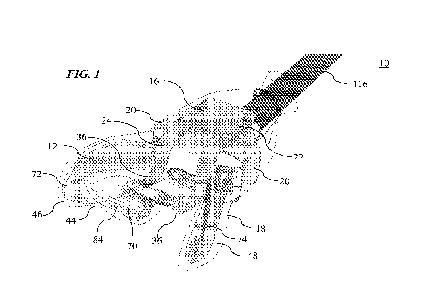
FIG. 1 is a front perspective view of an embodiment of an intraoral
phototherapy
system including an intra-oral phototherapy device and an intra-oral
phototherapy device
protection system having a breathing apparatus and a protective sleeve.
FIG. 2 is a front perspective view of the intra-oral phototherapy device and
an
exploded view of the breathing apparatus of FIG. 1.
FIG. 3 is a front perspective view of the intra-oral phototherapy device
separated
from the breathing apparatus of FIGS. 1 and 2.
FIG. 4 is a front perspective view of the breathing apparatus and the
protective
sleeve of FIG. 1.
FIG. 5 is a front perspective view of an alternative embodiment of the
breathing
apparatus.
FIG. 6 is a side perspective view of the breathing apparatus of FIG. 5.
FIG. 7 is a rear perspective view of the breathing apparatus of FIGS. 5 and 6.
FIG. 8 is a bottom perspective view of the breathing apparatus of FIGS. 5-7.
FIG. 9 is a front perspective view of a bottom piece of the breathing
apparatus of
FIGS. 5-8.
FIG. 10 is a bottom perspective view of the bottom piece of FIG. 9.
FIG. 11 is a side perspective view of the bottom piece of FIGS. 9 and 10.
FIG. 12 is a front perspective view of a top piece of the breathing apparatus
of
FIGS. 5-8.
FIG. 13 is a bottom perspective view of the top piece of FIG. 12.
FIG. 14 is a side perspective view of the top piece of FIGS. 12 and 13.
FIG. 15 is a front perspective view of an embodiment of the intra-oral
phototherapy device.
FIG. 16 is a top perspective view of the intra-oral phototherapy device of
FIG. 15.
FIG. 17 is a bottom perspective view of the intra-oral phototherapy device of
FIGS. 15 and 16.
FIG. 18 is a side perspective view of the intra-oral phototherapy device of
FIGS.
15-17.
FIG. 19 is a front perspective view of the intra-oral phototherapy device of
FIG.
15 and the breathing apparatus of FIG. 5.
FIG. 20 is a side perspective view of the intra-oral phototherapy device of
FIG. 15
and the breathing apparatus of FIG. 5.
3
CA 03229815 2024- 2- 22
WO 2023/039499
PCT/US2022/076153
FIG. 21 is a top view of the intra-oral phototherapy device of FIG. 15 and the
breathing apparatus of FIG. 5.
FIG. 22 is a top front view of the intra-oral phototherapy device of FIG. 15
and
the breathing apparatus of FIG. 5.
FIG. 23 is a bottom front view of the intra-oral phototherapy device of FIG.
15
and the breathing apparatus of FIG. 5.
FIG. 24 is a top view of the protective sleeve being manufactured.
FIG. 25 is a view of an unprocessed sleeve showing a position of cuts and
connecting of the cuts to form a protective sleeve
FIG. 26 shows a protective sleeve and a breathing apparatus including
breathing
tubes.
FIG. 27 shows the breathing apparatus of FIG 26 being attached to the
protective
sleeve.
FIG. 28 is a top view of the protective sleeve and breathing apparatus of
FIGS. 26
and 27.
FIG. 29 shows the intra-oral phototherapy device being inserted into the
protective
sleeve of FIG. 28.
FIG. 30 shows an alternative embodiment of a breathing apparatus attached to
the
protective sleeve.
FIG. 31 shows an embodiment of a breathing apparatus attached to the intra-
oral
phototherapy device.
FIG. 32 is a side perspective view of a light guide of the intra-oral
phototherapy
device.
FIG. 33 is a side view of the light guide of FIG. 32.
FIG. 34 is a top view of the light guide of FIG. 32.
FIG. 35 is a bottom view of the light guide of FIG. 32.
FIG. 36 is a top view of the light guide of FIG. 32 inserted into the oral
cavity.
FIG. 37 is a side view of the light guide of FIG. 32 inserted into the oral
cavity.
The present invention is described below in detail with reference to the
drawings.
In the drawings, each element with a reference number is similar to other
elements with
the same reference number independent of any letter designation following the
reference
number. In the text, a reference number with a specific letter designation
following the
reference number refers to the specific element with the number and letter
designation
and a reference number without a specific letter designation refers to all
elements with the
4
CA 03229815 2024- 2- 22
WO 2023/039499
PCT/US2022/076153
same reference number independent of any letter designation following the
reference
number in the drawings.
DETAILED DESCRIPTION
According to an exemplary embodiment, an intraoral phototherapy device is
provided that improves illumination of a tonsillar region of the oral cavity.
Oral tissue
illumination is particularly difficult in the back of the throat. The
intraoral phototherapy
device improves illumination of these tissues by using a breathing tube to
improve patient
breathing. The breathing tube allows a patient to breathe through their mouth,
which
opens up the tonsillar region of the oral cavity. This opening of the
tonsillar region
allows light emitted from the intra-oral phototherapy device to illuminate
tissues located
at the back of the throat.
Additionally, objects contacting near the back of the throat often induce a
gagging
reflex. This gagging reflex shortens the time available to perform
phototherapy, making
it difficult for phototherapy to be delivered to the back of the throat. The
intraoral
phototherapy device may reduce the gagging reflex by contacting the hard
palate of the
oral cavity with a dorsal fin, such that the intraoral phototherapy device
does not make
contact further back in the oral cavity. That is, the intraoral phototherapy
device may
include a dorsal fin configured to contact the oral cavity further from the
throat, such that
portions of the intraoral phototherapy device nearer the throat do not contact
the oral
cavity and induce a gagging reflex.
Turning to FIGS. 1-4, an exemplary embodiment of a phototherapy system 10 for
illuminating a tonsillar region of an oral cavity is shown. The phototherapy
system 10
includes an intra-oral phototherapy device 12 and an intra-oral phototherapy
device
protection system 14. The protection system 14 includes a breathing apparatus
16 and a
sleeve 18 (also referred to as a protective sleeve).
The breathing apparatus 16 improves illumination of the tonsillar region of
the
oral cavity of a patient during phototherapy by improving patient breathing
while
receiving the phototherapy with the intra-oral phototherapy device 12. Without
the
breathing apparatus 16, the patient may need to breath through their nose,
which
constricts the back of the throat, making it difficult to illuminate the
tonsillar region.
Conversely, by using the breathing apparatus 16, the patient may breathe
through their
mouth, opening the back of the throat and improving illumination of the
tonsillar region.
5
CA 03229815 2024- 2- 22
WO 2023/039499
PCT/US2022/076153
As shown in FIGS. 5-14, the breathing apparatus 16 includes a fixation
structure
20, a main body 22, and a bifurcated protrusion 24. The fixation structure 20
mechanically engages with a mounting structure 26 of the intra-oral
phototherapy device
12, such that a position of the breathing apparatus 16 is maintained relative
to the intra-
oral phototherapy device 12. The main body 22 has a central lumen 28 and a
distal
opening 30 to the central lumen 28. The bifurcated protrusion 24 includes a
first lateral
lumen 32, a second lateral lumen 34, and a proximal opening 36. The proximal
opening
includes a first proximal opening 38 to the first lateral lumen 32 and a
second proximal
opening 40 to the second lateral lumen 34. The first lateral lumen 32 and the
second
lateral lumen 34 are fluidly coupled to the central lumen 28.
The proximal opening 36 is configured (e.g., shaped) to be received within the
oral cavity when the intra-oral phototherapy device 12 is located within the
oral cavity
and the fixation structure 20 is mechanically engaged with the intra-oral
phototherapy
device 12. Similarly, the distal opening 30 is configured (e.g., shaped) to be
located
outside of the oral cavity when the intra-oral phototherapy device is located
within the
oral cavity and the fixation structure is mechanically engaged with the intra-
oral
phototherapy device, such that the oral cavity is fluidly coupled with an
external
environment via the central lumen, the first lateral lumen, and the second
lateral lumen.
That is, because the proximal opening 36 and distal opening 30 are fluidly
connected and
because the proximal opening 36 is located within the oral cavity and the
distal opening
is located outside the oral cavity, the breathing apparatus 16 provides a
passage (i.e.,
via the central lumen 28) for the patient to breath through their mouth.
Turning to FIGS. 24 and 25, the protection system 14 includes the sleeve 18.
Making the interior volume of the sleeve 18 more form fitting to the intra-
oral
25 phototherapy device 12 makes it more difficult to insert the intra-oral
phototherapy device
12 into the interior of the sleeve 18. However, making the interior volume
larger to
improve insertion of the intra-oral phototherapy device 12 into the sleeve 18
also makes it
more difficult for the patient to breathe through their mouth with the sleeved
intra-oral
phototherapy device 12 inside their mouth. By making multiple connected
volumes
30 inside the sleeve 18 (e.g., one for each projection of the light guide),
it is possible to
reduce the bagginess of the sleeve 18 and to improve the patient's ability to
breathe
through their mouth.
The sleeve 18 is formed from an optically transparent sheet material and has a
trifurcated interior volume 42 including a central volume 44, a first lateral
volume 46, and
6
CA 03229815 2024- 2- 22
WO 2023/039499
PCT/US2022/076153
a second lateral volume 48. The trifurcated interior volume 42 is formed by
folding over
the sheet material to form a top sheet 50 and a bottom sheet 52 connected by a
closed
distal edge 54. The top sheet 50 and the bottom sheet 52 overlap to form an
open first
lateral edge 56, an open second lateral edge 58, and an open proximal edge 60.
The top
sheet 50 and the bottom sheet 52 are connected along the first lateral edge 56
to form a
closed first lateral edge 62. Similarly, the top sheet 50 and the bottom sheet
52 are
connected along the second lateral edge 58 to form a closed second lateral
edge 64.
Sheet material is removed from along the closed distal edge 54 to form the
central
volume 44, the first lateral volume 46, and the second lateral volume 48. That
is, a first
area 66 of material is removed between the central volume 44 and the first
lateral volume
46 and connecting the top sheet 50 and the bottom sheet 52 along a border of
the first area
66. Similarly, a second area 68 of material is removed between the central
volume 44 and
the second lateral volume 48 and connecting the top sheet 50 and the bottom
sheet 52
along a border of the second area 68.
The connecting of the top sheet 50 and the bottom sheet 52 forms a seam along
the first lateral edge 62 and the second lateral edge 64. This seam may
distort light
emitted by the intra-oral phototherapy device 12. Conversely, the folding over
of the
sheet material to form the top sheet 50 and the bottom sheet 52 connected by
the closed
distal edge 54 does not form a seam along the closed distal edge 54, such that
light
transmitted through the closed distal edge is not interfered with by a seam.
The top sheet
50 and the bottom sheet 52 may be connected using any suitable method (such as
heat
welding, adhesives, etc.).
The sleeve 18 may be formed in any suitable manner, such that the sleeve 18
does
not include a seam along the distal edge 54. For example, the sleeve 18 may be
a dip
mold or vacuum formed (e.g., similar to a latex glove).
As shown in FIG. 1, the central volume 44 is shaped to receive a light guide
70 of
the intra-oral phototherapy device 12. Similarly, the first lateral volume 46
is shaped to
receive a first lateral wing 72 and the second lateral volume 48 is shaped to
receive a
second lateral wing 74 of the intra-oral phototherapy device 12. The fixation
structure 20
of the breathing apparatus 16 maintains a position of the sleeve 18 relative
to the intra-
oral phototherapy device 12 when the fixation structure 20 is mechanically
engaged with
the intra-oral phototherapy device 12 with the sleeve 18 located between the
breathing
apparatus 16 and the intra-oral phototherapy device 12. That is, the breathing
apparatus
7
CA 03229815 2024- 2- 22
WO 2023/039499
PCT/US2022/076153
16 may be used to fix the sleeve 18 in position relative to the intra-oral
phototherapy
device 12.
The sleeve 18 may be made of any suitable material. For example, the sleeve 18
may be disposable and made of a biocompatible plastic sufficient to act as a
microbial
barrier between the oral cavity and the light guide 70.
As shown in FIGS. 26-31, the breathing apparatus 16 may take different forms.
For example, the breathing apparatus 16 may lack a main body and instead
include at
least one breathing tube 122. In the depicted embodiment, the breathing
apparatus 16
includes two breathing tubes 122a, 122b each having a proximal opening that is
fluidly
connected with a distal opening. The breathing tube(s) 122 may not be fluidly
coupled to
one another (e.g., the breathing tube(s) 122 may be physically separated).
The breathing tube(s) 122 may be mechanically attached to the sleeve 18 or may
be free floating. The breathing tube(s) 122 may also be bendable to enable
repositioning
of the distal opening(s) (located outside of the oral cavity) relative to the
proximal
opening(s) (located inside the oral cavity). Alternatively, the breathing
tube(s) 122 may
have a fixed shape.
The breathing tube(s) 122 may be fixed to the sleeve 18 using tape, by heat
welding a piece of the sleeve 18 material to the sleeve 18, or by using any
suitable
method. For example, the breathing apparatus 16 may be integrated into the
sleeve 18
such that the breathing apparatus 16 is mechanically fixed to the sleeve 18.
The sleeve 18
may also be sterilized.
In the embodiments shown in FIGS. 30 and 31, an alternative embodiment of the
breathing tubes is shown. In both embodiments, the breathing tubes 122 are
bent and
attached via a clip 124 to the surface of the sleeve. As shown, the breathing
tube(s) 122
may be placed in different positions and/or orientations (e.g., vertically or
horizontally)
relative to the intraoral phototherapy device 12.
In one embodiment, the breathing apparatus 16 may include protrusion(s) that
engage with depression(s) in the intra-oral phototherapy device 12 to maintain
a position
of the breathing apparatus 16 relative to the intraoral phototherapy device
12.
The breathing apparatus 16 may be configured not to interfere with patient
breathing. In one embodiment, the proximal opening(s) of the breathing
apparatus 16
may have a cross-sectional area allowing for a tidal volume (Vt) greater than
300m1.
Tidal volume is defined as the amount of air that moves in and out of the
lungs with each
respiratory cycle (inhalation / exhalation). The breathing apparatus 16 may be
configured
8
CA 03229815 2024- 2- 22
WO 2023/039499
PCT/US2022/076153
to allow at least 300m1 of air to be passed through the breathing apparatus 16
without
increasing the work of breathing for the patient during a five (5) minute
therapy.
An exemplary embodiment of the intraoral phototherapy device 10 for
illuminating targeted regions of the oral cavity with light emitted by a light
source 75 is
shown in FIGS. 15-23. The intra-oral phototherapy device 10 includes a light
guide 70
having a proximal end 76, a distal end 78, a dorsal surface 80, a ventral
surface 82, and a
main body 84 extending between the proximal end 76, the distal end 78, the
dorsal
surface 80, and the ventral surface 82.
The light guide 70 receives light from the light source 75 at the proximal end
76
and propagates the received light from the proximal end 76 to the distal 78
end via the
main body 84 The main body 84 includes light extracting features 86 that cause
light to
be emitted from the dorsal surface 80 and the ventral surface 82. The light
guide 70 also
projects light from the distal end 78. The dorsal surface 80 may have a convex
shape and
the ventral surface 82 may have a concave shape, such that the main body 84
conforms to
contours of the oral cavity when inserted therein to direct light to targeted
regions of the
oral cavity.
The light guide 70 may also include a dorsal projection 88 extending from the
dorsal surface 80 that pushes against a roof of the oral cavity when the intra-
oral
phototherapy device 12 is inserted into the oral cavity. By interacting with
the roof of the
oral cavity, the dorsal projection 80 maintains space between the roof of the
mouth and
the dorsal surface 80 of the intraoral phototherapy device 12 as shown in FIG.
37.
As shown, the dorsal projection 88 is configured to interact with the hard
palate
and mitigate the leading edge (i.e., the distal end 78) of the light guide 70
from pushing
against the upper back of the throat. This improves illumination of the soft
palate (also
referred to as tonsillar tissues) for phototherapy.
The dorsal projection 88 may take any suitable shape for interacting with the
hard
palate while mitigating the distal end 78 from pushing against the upper back
of the
throat. For example, in the depicted figures the dorsal projection 88 has a
fin shape. The
dorsal projection 88 is not limited to a fin shape but may take any suitable
shape (e.g.,
spherical, elliptical, oblong, etc.).
As described above, the intra-oral phototherapy device 12 may also include
lateral
wings 72, 74. The lateral wings 72, 74 are optically coupled to the light
guide 70 and
receive and propagate the light from the light source 75. The first lateral
wing 72 (also
referred to as a left wing) and the second later wing 74 (also referred to as
a right wing)
9
CA 03229815 2024- 2- 22
WO 2023/039499
PCT/US2022/076153
are vertically spaced apart with the light guide 70 is positioned vertically
between the first
lateral wing 72 and the second lateral wing 74. As shown in FIG. 26, the
lateral wings
72, 74 are sized and shaped to be received between buccal tissues and gums of
the patient
in the oral cavity. That is, the lateral wings 72, 74 include an inner surface
90 facing
towards the gums and an outer surface 92 opposite the inner surface 90 facing
towards the
buccal tissues when inserted into the oral cavity. The lateral wings 72, 74
emit the
received light from the inner surface 90 and the outer surface 92.
Turning to FIGS. 19-23, the bifurcated protrusion 24 may include a first
lateral
protrusion 94 enclosing the first lateral lumen 32 and a second lateral
protrusion 96
enclosing the second lateral lumen 34. The first lateral protrusion 94 may be
separated
from the second lateral protrusion 96, such that a central protrusion of the
intra-oral
phototherapy device is positioned between the first lateral protrusion and the
second
lateral protrusion when the breathing apparatus is mechanically engaged with
the intra-
oral phototherapy device.
Turning to FIGS. 9-14, the fixation structure 20, the main body 22, and the
bifurcated protrusion 24 may be monolithically formed by a housing 98 having a
top
piece 100 and a bottom piece 102. The bottom piece 102 and the top piece 100
may be
shaped to mechanically engage to form the central lumen 28, the first lateral
lumen 32,
and the second lateral lumen 34. For example, the top piece 100 and bottom
piece 102
may snap together. In the depicted example, the bottom piece 102 includes tabs
that
engage with slots located in the top piece 100.
In one embodiment, the bifurcated protrusion 24 may include a first peripheral
opening 104 to the first lateral lumen 32 and a second peripheral opening 106
to the
second lateral lumen 34. The first peripheral opening 104 may be located in a
different
plane than the first proximal opening 38. Similarly, the second peripheral
opening 106
may be located in a different plane than the second proximal opening 40 For
example, in
FIG. 5, the first peripheral opening 104 is located in a plane that is
perpendicular to the
first proximal opening 38. The peripheral openings 104, 106 may be utilized to
improve
airflow through breathing apparatus 16.
Turning to FIGS. 32-37, the light-extracting features 86 may include dorsal
surface features 108 at a distal region 110 of the dorsal surface 80 adjacent
the distal end
78. The dorsal surface features 108 may include at least one of projections or
depressions
in the dorsal surface 80. At least a portion of the dorsal projection 88 may
be located in
the distal region 110 of the dorsal surface 80. For example, the distal region
110 may
CA 03229815 2024- 2- 22
WO 2023/039499
PCT/US2022/076153
comprise one third, one quarter, one fifth, or any suitable portion of the
dorsal surface 80
of the light guide 70. The light-extracting features 86 may include ventral
surface
features on the ventral surface 82 and the ventral surface features 112. The
ventral
surface features 112 may include at least one of projections or depressions in
the ventral
surface 82.
The distal end 78 may include projecting optical features configured to direct
light
from the distal end 78 to the tonsillar tissues. For example, the distal end
78 of the light
guide 70 may be contoured to at least partially focus light exiting the distal
end 78. That
is, the distal end may be configured to emit more focused light from the light
guide (e.g.,
as shown in FIGS. 32-35). The projecting optical features may include any
suitable
optical structures such as surface shaping lensing, surface aberations, etc.
In a particular embodiment, the ventral surface of the light guide has a
curvature
that conforms to contours of the dorsal surface of the tongue, such that light
emitted from
the ventral surface illuminates the dorsal surface of the tongue. The light
guide 70 may
be contoured to improve patient comfort by following the curve of the roof of
the mouth
and/or the tongue.
The light-extracting features 86 may be any suitable structure for extracting
light
from the light guide (e.g., to target a specific light output distribution).
For example, the
light-extracting 86 features may include at least one of surface aberrations,
micro-lenses,
reflective spots, partial reflective planes, or diffraction gratings.
Alternatively or
additionally, a diffuser sheet or a 2-D lensing sheet may be (1) placed on an
emission
surface of the light guide. In one embodiment, the surface aberrations include
at least one
of a contour of the surface, surface depositions, or surface etchings.
As shown in FIG. 2, the light source 75 may be external to and not physically
supported by the light guide 70 or a casing 114 that the light guide 70 is
mechanically
attached to. For example, the light source 75 may be a light box supported by
an external
structure (e.g., table) that is optically connected to the light guide 70 via
a light cable 116
(e.g., fiber optics). Alternatively, the light source 75 may be physically
supported by the
casing 114. For example, the light source 75 may be housed in the casing 114
and
powered by a power source electrically connected to the light source 75 (e.g.,
a battery).
Alternatively or additionally, the light source 75 may be mechanically
supported by the
light guide 70 (e.g., light emitting diodes (LEDs) embedded and/or affixed to
the light
guide 70). In another example, the light source may be physically mounted to a
tab (e.g.,
a protrusion) on an external surface of the light guide.
11
CA 03229815 2024- 2- 22
WO 2023/039499
PCT/US2022/076153
In one embodiment, the casing 114 of the intra-oral phototherapy device 12
includes an interface 126 for receiving the main body 22 of the breathing
apparatus 16.
As shown in FIGS. 15, 16, and 18, the interface 126 may be an indentation or
depression
shaped to receive a portion of the main body 22.
In one embodiment, the light source is a remote light source that is optically
coupled to rearwardly protruding ends of the light guide via a fiber optic
cable. In an
embodiment, the remote light source includes one or more LEDs or a laser.
The light source 75 may be any suitable structure for emitting light. For
example,
the light source 75 may include one or more light emitting diodes (LEDs),
organic LEDs
(OLEDs), microLEDs, laser diodes, mini-LED, quantum dot (QD)-conversion,
phosphor
conversion, excimer lamps, multi-photon combination, or SLM wavefront
manipulation.
The light source 75 may emit light (also referred to as electromagnetic
radiation) having a
wavelength from 600 nm to 1000nm. For example, the light source 75 may emit
light
having a wavelength approximately equal to at least one of 630 nm, 660 nm, 670
nm, 810
nm, or 880 nm.
The intra-oral phototherapy device 12 may be configured to illuminated
targeted
regions 118 of the oral cavity including tissues in addition to the tonsillar
tissues. For
example, the targeted regions 118 of the oral cavity may include the tonsillar
tissues and
at least one of the tongue, mandibular and maxillary buccal surfaces of the
oral cavity, the
floor and roof of the oral cavity, and tonsillar tissues. In one embodiment,
the targeted
regions 118 of the oral cavity include the tongue, mandibular and maxillary
buccal
surfaces of the oral cavity, the floor and roof of the oral cavity, and
tonsillar tissues.
The light guide 70 may be made of any suitable material that is at least
partially
transparent to light 120 emitted by the light source 75. For example, the
light guide may
be made of an optically transparent soft flexible biocompatible polymeric
material such as
silicone. As an example, the light guide may be made of different formulations
of
polycarbonate, polymethyl methacrylate, polystyrene, nylon, acrylonitrile
butadiene
styrene, polyolefin, or other biocompatible thermoplastic elastomer
formulations.
The intraoral phototherapy device may be used in several applications, several
examples of which include oral mucositis, acute necrotizing ulcerative
gingivitis
(ANUG), periodontal diseases, trismus, decreasing recovery time from oral
surgery, light
delivery for orthodontics, and photodynamic light therapy, e.g., to activate a
chemical
mouthwash.
12
CA 03229815 2024- 2- 22
WO 2023/039499
PCT/US2022/076153
In one embodiment of the phototherapy system 10, the intra-oral phototherapy
device 12 includes a light guide 70 having a proximal end 76, a distal end 78,
a dorsal
surface 80, a ventral surface 82, and a main body 84 extending between the
proximal end
76, the distal end 78, the dorsal surface 80, and the ventral surface 82. The
light guide 70
receives light 120 from the light source 75 at the proximal end 76 and
propagates the
received light 120 from the proximal end 76 to the distal end 78 via the main
body 84.
The main body 84 includes light extracting features 86 for causing light to be
emitted
from the dorsal surface 80 and the ventral surface 82. The light guide 70
projects light
from the distal end 78. The light guide 70 may take any suitable shape for
projecting
light into the oral cavity, such as spherical, elliptical, S shaped, etc.
This embodiment of the phototherapy system 10 also includes a breathing
apparatus 16 having a fixation structure 20 for mechanically engage with the
mounting
structure of the intra-oral phototherapy device 12, and a main body 22 having
a central
lumen 28, a distal opening 30 to the central lumen 28, and a proximal opening
36 to the
central lumen 28. For example, in this embodiment, the breathing apparatus 16
may
comprise a single breathing tube 122. As an example, the breathing apparatus
16 may be
a single breathing located above, below, to the side of the light guide 70. As
another
example, the light guide 70 may have a central opening and the breathing tube
122 may
be formed by or located in this central opening.
All ranges and ratio limits disclosed in the specification and claims may be
combined in any manner. Unless specifically stated otherwise, references to
"a," "an,"
and/or -the" may include one or more than one, and that reference to an item
in the
singular may also include the item in the plural.
Although the invention has been shown and described with respect to a certain
embodiment or embodiments, equivalent alterations and modifications will occur
to
others skilled in the art upon the reading and understanding of this
specification and the
annexed drawings. In particular regard to the various functions performed by
the above
described elements (components, assemblies, devices, compositions, etc.), the
terms
(including a reference to a "means") used to describe such elements are
intended to
correspond, unless otherwise indicated, to any element which performs the
specified
function of the described element (i.e., that is functionally equivalent),
even though not
structurally equivalent to the disclosed structure which performs the function
in the herein
illustrated exemplary embodiment or embodiments of the invention. In addition,
while a
particular feature of the invention may have been described above with respect
to only
13
CA 03229815 2024- 2- 22
WO 2023/039499
PCT/US2022/076153
one or more of several illustrated embodiments, such feature may be combined
with one
or more other features of the other embodiments, as may be desired and
advantageous for
any given or particular application
14
CA 03229815 2024- 2- 22