Note: Descriptions are shown in the official language in which they were submitted.
WO 2023/034595
PCT/US2022/042509
PLATFORM FOR ANTIMICROBIAL SUSCEPTIBILITY TESTING AND METHODS
OF USE THEREOF
SUMMARY
In accordance with one aspect, there is provided a system for determining a
susceptibility
of a microbial species in the presence of an antimicrobial agent. The system
may include an
image collection subsystem constructed and arranged to generate a plurality of
images of a
microbial sample. The system further may include an image analysis subsystem
including a non-
transitory computer-readable medium storing thereon sequences of computer-
executable
instructions for determining the susceptibility of the microbial species from
the plurality of
images of the microbial sample. When executed by one or more processors, the
sequences of
computer-executable instructions stored on the non-transitory computer-
readable medium cause
the one or more processors to perform operations including i) receiving, from
the image
collection subsystem, one or more of the plurality of images of the microbial
sample; ii)
extracting data corresponding to a pixel intensity of one or more regions of
the one or more of
the plurality of images; iii) reducing intensity variations in the per pixel
intensity of the one or
more regions of the one or more of the plurality of images; and iv)
calculating the susceptibility
of the microbial species by determining one or both of microbial species
replication and
microbial species stasis in the presence of the antimicrobial agent from the
manipulation of the
changing pixel intensities.
In some embodiments, the image collection subsystem may include a light
source, a
photosensitive element constructed and arranged to collect light from the
light source that has
transmitted through the microbial sample. and a memory for storing the
plurality of images
representative of the collected transmitted light from the microbial sample.
In some embodiments, determining the susceptibility of the microbial species
may
include one or more of: a) reducing noise in the pixel intensity of one or
more regions of the one
or more of the plurality of images; b) removing statistical outliers from the
pixel intensity of the
one or more regions of the one or more of the plurality of images; and/or c)
fitting the pixel
intensity of the one or more regions of the one or more of the plurality of
images to a model
representative of a growth dynamic of the microbial species to determine the
susceptibility.
In further embodiments, the image analysis subsystem may be configured to
display the
results of the image analysis to a user. For example, the displayed results
may be used to
1
CA 03230784 2024-3- 1
WO 2023/034595
PCT/US2022/042509
determine a treatment course for a patient. In particular embodiments, the
displayed results may
be used for epidemiological purposes, e.g., determining which antibiotic or
antimicrobials to
keep in stock for clinical use.
In some embodiments, the microbial species may include at least one species
from the
genus Acinetobacter, Escherichia, Klebsiella, Pseudomonas, Enterococcus,
Streptococcus, and
Siaphylocoecus. In certain embodiments, the microbial species may be selected
from A.
baumannii, E. con, K. pneumoniae, P. aeruginosa, and S. aureus. The system
disclosed herein is
not limited to the analysis of growth of these exemplary genera or species.
In some embodiments, the microbial species may be grown for less than or about
12
hours during collection of the plurality of images. In some embodiments, the
microbial species
may be grown for less than or about 9 hours during collection of the plurality
of images. the
microbial species may be grown for less than or about 6 hours during
collection of the plurality
of images. For example, the microbial species may be grown for less than or
about three 3
hours, e.g., less than about 3 hours, less than about 2.5 hours, less than
about 2 hours, less than
about 1.5 hours, or less than about 1 hour during image acquisition.
In specific embodiments, the microbial sample includes a well plate having a
plurality of
wells each separated by at least one surrounding interwell region, the
microbial sample including
microbial growth in a portion of the plurality of wells. In certain
embodiments, the one or more
regions of the at least one of the plurality of images correspond to the
plurality of wells and the
associated at least one surrounding interwell region.
In some embodiments, reducing intensity variations may include correcting the
pixel
intensity of the pixels in each of the plurality of wells using the pixel
intensities of the associated
at least one surrounding interwell region. In some embodiments, reducing noise
may include
performing independent component analysis (ICA) on the variation reduced pixel
intensity data
of the pixels in each of the plurality of wells to generate at least one
signal corresponding to
microbial growth and at least one signal corresponding to growth inhibition
from the
antimicrobial agent. In some embodiments, removing statistical outliers may
include performing
one or both of a mean absolute deviation calculation and a k-means clustering
calculation on the
noise reduced pixel intensity data.
10 In some embodiments, wherein fitting the pixel intensity comprises
fitting the outlier
reduced pixel intensity data to a growth dynamic model comprising one or more
2
CA 03230784 2024-3- 1
WO 2023/034595
PCT/US2022/042509
phenomenological models. In particular embodiments, the growth dynamic model
comprises a
combined multi-dimensional growth dynamic model comprising the Gompertz model
and the
Hill model.
In further embodiments, the image analysis subsystem may be configured to
calculate the
minimum inhibitory concentration (MIC) of the antimicrobial agent.
In accordance with an aspect, there is provided a method of determining a
susceptibility
of a microbial species in the presence of an antimicrobial agent. The method
may include
acquiring a plurality of images of a microbial sample using an image
collection system. The
method may include sending or transmitting one or more of the plurality of
images to an image
analysis system comprising a non-transitory computer-readable medium storing
thereon
sequences of computer-executable instructions for determining the
susceptibility of the microbial
species from the plurality of images of the microbial sample by manipulating
data corresponding
to a pixel intensity of one or more regions of one or more of the plurality of
images to a hybrid
model representative of a growth dynamic of the microbial species. The method
further may
include calculating a minimum inhibitory concentration (MIC) of the
antimicrobial agent from
the one or more of the plurality of images by determining one or both of
microbial species
replication and microbial species stasis in the presence of the antimicrobial
agent from the
determined growth dynamic. The method additionally may include storing or
providing the
calculated MIC to a user.
In further embodiments, determining the susceptibility of the microbial
species from the
plurality of images of the microbial sample may include extracting data
corresponding to a pixel
intensity of one or more regions of the one or more of the plurality of
images. In further
embodiments, determining the susceptibility of the microbial species from the
plurality of
images of the microbial sample may include reducing intensity variations in
the pixel intensity of
the one or more regions of the one or more of the plurality of images. In
further embodiments,
determining the susceptibility of the microbial species from the plurality of
images of the
microbial sample may include reducing noise in the pixel intensity of one or
more regions of the
one or more of the plurality of images. In further embodiments, determining
the susceptibility of
the microbial species from the plurality of images of the microbial sample may
include removing
statistical outliers from the pixel intensity of the one or more regions of
the one or more of the
plurality of images.
3
CA 03230784 2024-3- 1
WO 2023/034595
PCT/US2022/042509
In some embodiments, the hybrid model comprises a combined multi-dimensional
growth dynamic model comprising the Gompertz model and the Hill model.
In accordance with an aspect, there is provided a non-transitory computer-
readable
medium storing instruction which, when executed by a computer, cause the
computer to perform
a method. The method may include acquiring a plurality of images of a
microbial sample using
an image collection system. The method further may include determining from
analysis of one
or more of the plurality of images of the microbial sample a growth dynamic
including one or
both of microbial species replication and microbial species stasis in the
presence of an
antimicrobial agent. The method additionally may include calculating a minimum
inhibitory
concentration (MIC) of the antimicrobial agent from the determined microbial
growth dynamic
in the one or more of the plurality of images of the microbial sample.
In certain embodiments, the step of determining the growth dynamic comprises
determining the growth dynamic using a combined multi-dimensional growth
dynamic model
comprising the Gompertz model and the Hill model.
BRIEF DESCRIPTION OF THE DRAWINGS
The accompanying drawings are not intended to be drawn to scale. In the
drawings, each
identical or nearly identical component that is illustrated in various figures
is represented by a
like numeral. For purposes of clarity, not every component may be labeled in
every drawing. In
the drawings:
FIG. 1 illustrates a schematic of a system for determining a susceptibility of
a microbial
species in the presence of an antimicrobial agent, according to an embodiment
of this disclosure;
FIG. 2 illustrates a portion of a 384 well microwell plate simulation with
landmarks
defined;
FIG. 3 illustrates a 384 well microvvell plate where pixel intensities of each
well are
equalized;
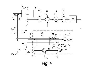
FIG. 4 illustrates an equalized interwell signal over time using a model
according to an
embodiment of this disclosure;
FIGS. 5A and 5B illustrate independent component analysis (ICA) modified
signals.
FIG. 5A illustrates ICA-decumposed latent sources of the imaging data using
two components.
4
CA 03230784 2024-3- 1
WO 2023/034595
PCT/US2022/042509
FIG. 5B illustrates the reconstruction of the de-noised imaging data based on
the two latent
sources with the solid lines showing the original data;
FIGS. 6A-6F illustrate replicate filtering and statistical analysis of the
filtered data.
FIGS. 6A and 6B illustrate the median (FIG. 6A) and mean (FIG. 6B) of
replicate intensity
versus time for select wells from a 384 well microplate. FIGS. 6C and 6D
illustrate the
corresponding mean absolute deviation (MAD) value for the data in FIGS. 6A and
FIG. 6B,
respectively. FIG. 6E illustrates a comparison in the original unfiltered mean
(blue) and mean of
the filtered data after k-means clustering (black). FIG. 6F illustrates the
MAD value for the data
in FIG. 6E;
FIGS. 7A-7B illustrate a comparison of the fit to a prior growth model using a
prior
image analysis scheme (FIG. 7A) and the image analysis scheme according to an
embodiment of
this disclosure (FIG. 7B);
FIGS. 8A-8B illustrate a comparison of the fit to a prior growth model using a
prior
image analysis scheme (FIG. 8A) and an image analysis scheme according to an
embodiment of
this disclosure (FIG. 8B);
FIG. 9 illustrates a comparison between the modeled growth of a prior growth
model
(blue line), the Gompertz model (yellow line), and a model according to an
embodiment of this
disclosure (red line); and
FIGS. 10A-10D illustrate 3D surface plots of processed image data (FIGS. 10A
and 10C)
and the model fit according to an embodiment of this disclosure (FIGS. 10B and
10D) as a
function of time and log(concentration).
DETAILED DESCRIPTION
The invention relates to the fields of cell growth and detection. In many
industries,
particularly the food, beverage, healthcare, electronic, livestock/animal
husbandry,
biotechnology, and pharmaceutical industries, it is essential to rapidly
analyze samples for the
degree of contamination by microorganisms, such as bacteria, yeasts, or molds.
Conventional methods used in clinical laboratories worldwide require isolation
of
bacteria on culture plates as single bacterial colonies. The colonies are then
used to set up one of
several culturing methods, e.g., the broth microdilution reference method,
agar dilution, disk
diffusion, gradient diffusion, or several commercial methods that are either
modified versions of
5
CA 03230784 2024-3- 1
WO 2023/034595
PCT/US2022/042509
the broth microdilution method or extrapolate the results from the broth
microdilution method
based on growth kinetics of organisms in culture. Available testing is limited
to first-line drugs.
In practice, these methods provide antimicrobial susceptibility testing (AST)
results in a
minimum of two days after specimen receipt in the clinical lab. This testing
generally requires at
least one day to isolate pure bacterial colonies, and one additional day to
obtain the AST results
from these colonies. With emerging antimicrobial resistance, this two-or-more
day delay may
lead to adverse clinical outcomes.
Systems that provide faster AST results from pure bacterial colonies are
notably
expensive and thus cost prohibitive. Some AST instruments can cost over
$100,000 and still can
take seven or more hours to run an AST analysis after isolating a pure
bacterial colony. The cost
of testing a single sample can be in excess of $200. As a result of their high
cost, rapid AST
systems would not likely be widely available throughout the world, including
many parts of the
United States, in rural areas, and in developing countries.
Definitions
Terms used in the claims and specification are defined as set forth below
unless otherwise
specified.
It must be noted that, as used in the specification and the appended claims,
the singular
forms "a," "an" and "the" include plural referents unless the context clearly
dictates otherwise.
"About" and "approximately" shall generally mean an acceptable degree of error
for the
quantity measured given the nature or precision of the measurements. Exemplary
degrees of
error are within 20 percent (%), typically within 15%, more typically within
10%, and even more
typically, within 5% of a given value or range of values.
As used interchangeably herein, the terms "subject," "patient," and
"individual" refer to
any organism to which a therapeutic agent in accordance with the invention may
be
administered, e.g., for experimental, diagnostic, prophylactic, and/or
therapeutic purposes.
Typical subjects include any animal (e.g., mammals, such as mice, rats, cats,
dogs, pigs, horses,
rabbits, non-human primates, and humans). A subject may seek or be in need of
treatment,
require treatment, be receiving treatment, be receiving treatment in the
future, or be a human or
animal who is under care by a trained professional for a particular disease or
condition.
6
CA 03230784 2024-3- 1
WO 2023/034595
PCT/US2022/042509
"Microbial species" as used herein, encompasses lifeforms including bacteria,
e.g.,
mycobacteri a, enterobacteria, non-fermenting bacteria, and Gram-positive or
Gram-negative
cocci or rods, archaca, fungi, i.e., yeasts and molds, algae, protozoa, and
viruses.
"Microbial sample" as used herein, can refer to any suitable apparatus capable
of holding
a microbial sample such that the sample can be grown for analysis and imaged.
For example, a
microbial sample may include a Petri dish, a well plate, e.g., a 6, 12, 24,
48, 96, 384 or 1536 well
microplate, a microscope slide, a glass plate with isolated droplets, or any
other suitable holder
for a microbial species.
"Images" as used herein, can refer to still photographic images collected
using any
suitable apparatus configured for such purpose or a video collected over a
period of time using
any suitable apparatus configured for such purpose. Still images may be
isolated from a video
accordingly.
"Light source" as used herein, refer to any suitable source of electromagnetic
radiation
for collecting an image of a microbial sample. Example sources can include
visible light,
infrared light, and ultraviolet light. Light from the light source may be of
any suitable plane
polarization, e.g., p-polarization or s-polarization, or unpolarized, i.e.,
random direction, light.
"Pixel intensity" as used herein refers to the brightness and/or color of an
identified pixel.
Low brightness is considered to have low to zero intensity and high brightness
is high intensity.
Darker colors are considered to have low to zero intensity and brighter colors
are considered to
have high intensity.
In accordance with an aspect, there is provided a system for the determination
of a
susceptibility of a microbial species in the presence of an antimicrobial
agent. The system may
comprise an image collection subsystem constructed and arranged to generate a
plurality of
images of a microbial sample. The system further may comprise an image
analysis subsystem
comprising a non-transitory computer-readable medium storing thereon sequences
of computer-
executable instructions for determining the susceptibility of the microbial
species from the
plurality of images of the microbial sample. When executed by the computer,
the sequences of
computer-executable instructions stored on non-transitory computer-readable
medium causes the
computer to perform operations including, for example receiving from the image
collection
subsystem one or more of the plurality of images of the microbial sample. The
operations
performed by the computer when executing the instructions stored on the non-
transitory
7
CA 03230784 2024-3- 1
WO 2023/034595
PCT/US2022/042509
computer-readable medium further may include extracting data corresponding to
a pixel intensity
of one or more regions of the one or more of the plurality of images. The one
or more regions of
the at least one of the plurality of images may correspond to the plurality of
wells and the
associated at least one surrounding interwell region surrounding each of the
plurality of wells.
The operations performed by the computer when executing the instructions
stored on the non-
transitory computer-readable medium additionally may include reducing
intensity variations in
the per pixel intensity of the one or more regions of the one or more of the
plurality of images.
The operations performed by the computer when executing the instructions
stored on the non-
transitory computer-readable medium additionally may include calculating the
susceptibility of
the microbial species by determining one or both of microbial species
replication and microbial
species stasis in the presence of the antimicrobial agent from the
manipulation of the changing
pixel intensities.
Systems and methods of this disclosure provide for a rapid determination of
microbial
species growth and the determination of a minimum inhibitory concentration
(MIC) of an
antibiotic or antimicrobial compound for a microbial species. This rapid
determination of the
MIC associated with a particular species provides benefits for treatment
determination in a
clinical setting. For example, when a patient in a clinical setting is
suspected of having an
infection, they are generally given a pharmaceutical, such as an antibiotic,
that represents the
best guess as to what will effectively treat the infecting pathogen.
Generally, for bacterial
infections, broad-spectrum antibiotics are given with the hope of covering any
potential
pathogen. Once the AST results are available, directed therapy, tailored to
the susceptibility
profile of the pathogen, can be given. This treatment path fails to account
for microbial life that
has developed resistance to pharmaceutical treatments, and further fails to
account for the overall
impacts of broad-spectrum antibiotics or antimicrobial compounds on beneficial
microbial life.
Having specific information on the MIC and any observed resistance to
pharmaceutical
treatments such as provided by the systems and methods of this disclosure can
allow for the
precise tailoring of treatment regimens on a timescale that prevents
additional delay and any
adverse outcomes associated with said delays.
Information. i.e., MIC associated with specific microbial species, may also be
used for
epidemiological purposes. For example, in a clinical setting, e.g., a
hospital, when
microorganisms are isolated from two individual patients have the same pattern
in their MIC in
8
CA 03230784 2024-3- 1
WO 2023/034595
PCT/US2022/042509
response to the administration of an antibiotic or other antimicrobial, this
raises the suspicion
that the isolated microorganisms originated from the same source. Further,
hospitals and public-
health laboratories routinely produce summary statistics for each microbial
species encountered
frequently among its patients. In so doing, these institutions generally
record how often the MIC
was, e.g., 0.5, vs. 1, vs. 2, vs. 4, and so on, producing histograms that
track changes in MIC.
Following these histograms over time may be useful for quantifying the spread
of antibiotic or
antimicrobial resistance, which may provide useful guidance for aiding in the
procurement of
one or more proper antibiotics or other antimicrobial agents to keep on hand
to treat the
microorganism, when its presence is observed.
As illustrated in FIG. 1, a system 100 for the determination of a
susceptibility of a
microbial species in the presence of an antimicrobial agent comprises an image
collection
subsystem 101, and image analysis subsystem 102, and an output 110 to display
the results of the
image analysis to a user or operator. The image collection subsystem 101 may
include a light
source 105, a camera comprising a photosensitive element 107 constructed and
arranged to
collect light from the light source 105 that has transmitted through a
microbial sample 103, and a
memory 109 for storing an image representative of the collected transmitted
light from the
microbial sample 103. The microbial sample 103 may be any suitable sample
containing
microorganisms, such as a well plate, Petri dish, or the like. Alternatively,
the microbial sample
103 may include aerosolized or nebulized droplets of a fluid including the
microbial species
deposited onto a suitably transparent or translucent substrate, e.g., a glass
plate or the like,
sufficiently spaced apart to minimize droplet coalescence. An image collection
subsystem 101
may be any suitable image collection subsystem having any combination of light
sources,
photodetection elements, and connections to other system components, and this
disclosure is in
no way limited to the example image collection subsystem shown and described.
With continued reference to FIG. 1, a system 100 for the determination of a
susceptibility
of a microbial species in the presence of an antimicrobial agent comprises an
image analysis
subsystem 102. The image analysis subsystem 102 may include a non-transitory
computer-
readable medium 104 having computer-executable instructions stored thereon for
determining
the susceptibility of the microbial species from the plurality of images of
the microbial sample.
The non-transitory computer-readable medium 104 may include, for example, a
disk, e.g., a hard
disk drive (HDD), solid state drive (SSD), or flash memory. Typically, in
operation, the CPU of
9
CA 03230784 2024-3- 1
WO 2023/034595
PCT/US2022/042509
computer 108 causes data to be read from the non-transitory computer-readable
medium 104 into
another memory 106 that allows for faster access to the information by the
computer 108 than
does the non-transitory computer-readable medium 104. This memory 106 is
typically a volatile,
random access memory such as DRAM or SRAM as described herein. The computer
108
generally manipulates the data within its internal memory and then copies the
data to the non-
transitory computer-readable medium 104 after processing is completed. A
variety of
mechanisms are known for managing data movement between the non-transitory
computer-
readable medium 104 and the internal memory 106 of the computer 108, and
embodiments
disclosed herein are not limited to any particular data movement mechanism.
With continued reference to FIG. 1, a system 100 further may comprise a
display 110 for
the output of the operations performed from the execution of the instructions
for determining the
susceptibility of the microbial species in the presence of the antimicrobial
agent, The display
110 may by any type of display, such as a visual output or a file containing
the resultant analysis,
or both, and embodiments disclosed herein are in no way limited to any
particular data output or
data display mechanism.
Systems and methods disclosed herein are generally performed using computers
to
acquire the plurality of images of the microbial samples, transmit the
acquired images, e.g., still
photographic images of the microbial sample collected over a timeseries or a
video of the
microbial sample, to the image analysis subsystem, and produce the desired
output, e.g., a MIC
or other related output. In some embodiments, bootstrapping a single synthetic
data set may be
used to generate sufficient data to establish the central tendency, i.e., a
central or typical value
for a probability distribution such as a mean, median or mode, corresponding
to the growth of the
microbial species. In some embodiments, for the analysis of images of
microbial samples that
include motion blur, e.g., blurred still photographs or videos, one or more
deconvolution
techniques, such as Richardson-Lucy deconvolution or Wiener deconvolution, may
be applied.
The addition of deconvolution as part of the image analysis subsystem may
allow for images to
be acquired at a faster rate, i.e., an increased temporal resolution. In
further embodiments,
artifacts present in the plurality of images of the microbial samples may be
removed using one or
more classical digital signal processing techniques. As a non-limiting
example, high frequency
artifacts present in the plurality of images of the microbial samples may be
removed by the
application of a low-pass filter.
CA 03230784 2024-3- 1
WO 2023/034595
PCT/US2022/042509
One or more parts of systems and methods disclosed can be achieved by using
artificial
intelligence for automation, such as unsupervised learning approaches. For
example, the
artificial intelligence that acquires and analyzes images may include a neural
network. Neural
networks are patterned mathematically to acquire, process, and interpret
incoming information in
a manner similar to the human brain, e.g., by taking input information and
passing it along to at
least one "neuron," further propagating information until terminating at an
output. By passing
information along to multiple "neurons" the neural network is able to improve
the way in which
it interprets an input signal, i.e., it learns from previous input signals,
thereby improving the
accuracy of the end result. The "neurons" are typically organized in layers.
Different layers may
perform different kinds of transformations on their inputs. Another non-
limiting example of
artificial intelligence for one or more of the systems and methods disclosed
herein is cluster
analysis, where sets of data are iteratively grouped based around one or more
specific properties,
such as a density or a centroid of a set of values. An exemplary clustering
model for use with
data that varies in time is k-means clustering, where a mixed set of data can
be grouped into k
clusters, with k being a natural number, and each data point in the set
belonging to the nearest
mean. Other types of unsupervised iterative models for analyzing data
corresponding to the
plurality of images of microbial samples collected by image collection
subsystems disclosed
herein and the specific types recited herein are in no way limiting.
In some embodiments, determining the susceptibility of the microbial species
comprises
may include a step of reducing intensity variations in the pixel intensity of
the one or more
regions of the one or more of the plurality of images. This is also known as
image equalization,
and in some embodiments may include correcting the pixel intensity of the
pixels in each of the
plurality of wells using the pixel intensities of the associated at least one
surrounding interwell
region. The general framework underlying the reduction of pixel intensity
variations is the a
priori expectation that areas of a microbial sample that do not have any
microbial activity, e.g.,
the interwell regions if a 384 well plate, should all have equal mean pixel
intensity when imaged
minus some variance, such as from manufacturing defects in the material used
to manufacture
the sample carrier. There are a number of approaches that may be used for
reducing intensity
variations in the pixel intensity of the one or more regions of the one or
more of the plurality of
images. In some cases, a two-step image equalization process may be used. A
global image
equalization may be performed by calculating the global average intensity over
all non-sample
11
CA 03230784 2024-3- 1
WO 2023/034595
PCT/US2022/042509
regions, e.g., the interwell regions in a well plate-based sample, and
applying the difference in
intensity from the global average as a correction factor. The correction
factor, i.e., the delta (A)
intensity, can then be used to calculate a region-specific correction, e.g., a
per-well correction in
a 384 well plate. As a non-limiting example, a global interwell intensity
average may be
calculated across all timepoints, i.e., in a plurality of images taken in
time. This type of
correction can then be used to correct the intensity of each individual pixel
in an image, with the
amount of correction being a function of the distance between a surrounding
non-sample
containing, e.g., interwell, region and the specific sample area pixel, e.g.,
well pixel, being
corrected. For example, a pixel near the top of one of the plurality of images
of a microbial
sample (in a well) will be less influenced by a correction factor from an
interwell region near the
bottom of the same image of the microbial sample. With reference to FIGS. 2
and 3, i.e., a 384
well plate, pixel corrections may be made in one or both of the row-wise
dimensions or the
column-wise directions. The image equalization schemes described in this
disclosure are in no
way limited to those described and other available schemes are within the
scope of this
disclosure.
In some embodiments, determining the susceptibility of the microbial species
comprises
may include a step of reducing noise in the pixel intensity of one or more
regions of the one or
more of the plurality of images. As described herein, reducing noise in the
pixel intensity of one
or more regions of the one or more of the plurality of images may be reducing
timepoint-to-
timepoint variance of said pixel intensities.
In general, microbial growth can he considered a multicomponent process,
including a
lag phase before microbial growth begins, a log phase of exponential growth
classically
represented as a logarithmic function, and a stationary phase once the
carrying capacity of the
environment is reached. Rather than be bound by the classical approach, this
disclosure
considers the dynamics of microbial growth as statistically separable and
weighted independent
biological processes, for example growth/cell division and stasis, without the
need to fit to any
classical model. The reduction in noise, i.e., timepoint-to-timepoint
variance, of said pixel
intensities thus can be considered a separation of the growth and stasis
processes from each
region of a microbial sample, such as each well in a 384 well plate, and
determination of the
statistical weights for these processes, with noise reduction occurring as it
cannot be part of
either growth or stasis. There are any number of different approaches for
separating a signal of
12
CA 03230784 2024-3- 1
WO 2023/034595
PCT/US2022/042509
interest from a collection of mixed signals. The separation of signals may be
achieved by using
independent component analysis (ICA), an unsupervised statistical technique
that extracts
individual source signals from the measured mixture signal. For example, in
some embodiments,
reducing noise may include performing ICA on the variation reduced pixel
intensity data of the
pixels in each of the of the one or more regions of the one or more of the
plurality of images to
generate at least one signal corresponding to microbial growth and at least
one signal
corresponding to growth inhibition from the antimicrobial agent. There are
other similar
techniques that can separate one or more specific signals from a mixed source,
including, but not
limited to, principal component analysis (PCA), singular value decomposition,
dependent
component analysis, non-negative matrix factorization, and stationary subspace
analysis, among
others. In some embodiments, one or more specific approaches for noise
reduction may be used.
For example, the noise reduction may first utilize a technique for reducing
the dimensionality of
the source signal, such as PCA, then separation of signals of interest from
the reduced
dimensionality source signal using a different noise reduction technique, such
as ICA. The noise
reduction schemes described in this disclosure are in no way limited to those
described and other
available schemes are within the scope of this disclosure.
In particular embodiments, the dimensionality of the source signal is reduced
using PCA,
for example, by determining the number of components that explained the
substantial majority of
any variance in the data. The dimensionality reduced data can be separated
into individual
components using ICA, of which the two resulting signals can be considered to
track the
processes of microbial species replication and of the inhibition of microbial
species growth, e.g.,
by an antibiotic or other antimicrobial compound. Without wishing to be bound
by any
particular theory, it is believed that dimensionality reduction using PCA may
be able to remove a
portion, e.g., a majority, of the noise from the data. Thus, in some cases,
with the majority of the
noise removed by reducing dimensionality, application of ICA to separate out
signals of interest
provide for the original signal per region of the microbial sample, such as
the per well signal of a
384 well plate.
In some embodiments, determining the susceptibility of the microbial species
may
include a step of removing statistical outliers from the pixel intensity of
the one or more regions
of the one or more of the plurality of images. In a typical microbial growth
experiment, there
may be particular regions, such as individual wells in a 384 well plate for
example, where
13
CA 03230784 2024-3- 1
WO 2023/034595
PCT/US2022/042509
microbial life fails to grow and produce a detectable result. The inclusion of
failed microbial
growth in the calculation of a microbial growth rate may artificially lower
the predicted growth
rate relative to the true growth rate, an important consideration for the
determining the MIC of an
antibiotic or other antimicrobial compound. The present disclosure
contemplates the filtering of
the pixel intensities of each of the plurality of images using a statistical
filter, such as by
calculating a mean and mean absolute deviation (MAD) of the original data. The
MAD is the
average distance between each set of data points and the mean. The resulting
MAD can be
evaluated against a threshold value to determine whether a specific replicate
should be excluded
from the dataset. As used herein, a "replicate" is a well of a well plate with
the same contents,
i.e., the same organism, and the same antibiotic at the same concentration.
The threshold value
may be determined experimentally or from previous compiled information on
similar microbial
growth. Once outliers are removed, replicates in the remaining pixel intensity
data from each of
the plurality of images can be calculated using one or more filtering steps.
As noted herein,
calculation steps such as replicate filtering may be performed using
artificial intelligence, such as
an unsupervised learning process. In this context, the results of replicate
filtration can be used to
train the image analysis subsystem to improve performance over time. As the
goal is to remove
replicates, one approach is to use one or more analysis techniques to group
similar datapoints
together, such as by cluster analysis. There are a number of suitable cluster
techniques which
may be used for replicate removal including, but not limited to, connectivity
clustering, centroid
clustering, statistical distribution clustering, and density clustering, among
others. An exemplary
clustering technique to filter replicate datapoints in each timeseries, i.e.,
the plurality of images,
is k-means clustering as described herein. A k-means algorithm often assigns
each point to a
cluster for which the center (also referred to as a centroid) is nearest. The
center often is the
average of all the points in the cluster, that is, its coordinates often are
the arithmetic mean for
each dimension separately over all the points in the cluster. A number of
clusters can be selected
as appropriate. An appropriate number of dimensions used in determining
clusters can be
selected as appropriate. The replicate filtering schemes described in this
disclosure are in no way
limited to those described and other available schemes are within the scope of
this disclosure.
In some embodiments, determining the susceptibility of the microbial species
comprises may include a step of fitting the pixel intensity of the one or more
regions of the one
or more of the plurality of images to a model representative of a growth
dynamic of the
14
CA 03230784 2024-3- 1
WO 2023/034595
PCT/US2022/042509
microbial species to determine the susceptibility. As described herein,
microbial species growth
is generally modeled on a sigmoidal curve, known as a Gompertz model,
representing the lag
phase, exponential growth, and a reduction in growth once carrying capacity is
reached. In some
prior treatments, microbial growth following this type of curve is
approximated by the classic
"hockey stick" fit based loosely on experimental evidence of bacterial growth
in a closed system.
Traditional models suffer from overfitting data and ignoring information that
can be derived
from the changes in growth across varying concentrations of antimicrobial or
antibiotic
compounds. One approach to improve the prediction of microbial species growth
is to extend a
classical model to be hybrid growth dynamic that, in addition to considering
growth at given
antimicrobial or antibiotic concentrations, also considers the dose response
of microbial growth
to an antimicrobial or antibiotic at a given time. An exemplary concentration-
based model for
microbial growth is called the Hill model, which is a modified logistic
function whose inflection
point corresponds to the minimum inhibitory concentration (MIC) of the
antimicrobial or
antibiotic. The hybrid growth dynamic model incorporating both the
concentration dependence
and time dependence of exposure to antimicrobial or antibiotic compounds on
microbial species
growth provides for an improved model for determining the MIC compared to
classical
approaches for modeling microbial species growth while being less susceptible
to overfitting and
non-biological dependencies in modeled growth processes.
As described herein, traditional AST in clinical and non-clinical settings is
generally a
slow process, requiring hours to days in order to culture and observe
sufficient colony formation
to enable appropriate determinations on susceptibility and other
epidemiological considerations.
Using an image analysis subsystem as described herein allows for the rapid
determination of
microbial species growth that is on a scale of a factor of two or more lower
than that of
traditional AST methods. In general, images of the microbial sample are
acquired about once
per minute during the growth of the microbial sample. In some embodiments, the
images of the
microbial sample are acquired about once per every 45 seconds, about once per
every 40
seconds, about once per every 35 seconds, about once per every 30 seconds,
about once per
every 25 seconds, about once per every 20 seconds, about once per every 15
seconds, about once
per every 10 seconds, about once per every 9 seconds, about once per every 8
seconds, about
once per every 7 seconds, about once per every 6 seconds, about once per every
5 seconds, about
once per every 4 seconds, about once per every 3 seconds, about once per every
2 seconds, or
CA 03230784 2024-3- 1
WO 2023/034595
PCT/US2022/042509
about once per every second. In some embodiments, the microbial species may be
grown during
image acquisition for less than or about 12 hours, e.g., less than about 12
hours, less than about
11.5 hours, less than about 11 hours, less than about 10.5 hours, less than
about 10 hours, less
than about 9.5 hours, less than about 9 hours, less than about 8.5 hours, less
than about 8 hours,
less than about 7.5 hours, less than about 7 hours, less than about 6.5 hours,
less than about 6
hours, less than about 5.5 hours, less than about 5 hours, less than about 4.5
hours, less than
about 4 hours, less than about 3.5 hours, less than about 3 hours, less than
about 2.5 hours, less
than about 2 hours, less than about 1.5 hours, or less than about 1 hour
during image acquisition.
Under these conditions, detectable changes in microbial species growth may be
observed in less
than about 10 minutes, and statistical confidence in the detection and
quantification of microbial
species growth may be achieved in less than about 30 minutes. The rapidity of
which the
disclosed systems and methods can detect and model microbial species growth
provides for a
determination of the time and concentration dependence on said microbial
growth even in the
absence of more refined modeling, additional signal inputs, or further
experimental steps.
Further, as one or more analysis techniques incorporated into the image
analysis subsystem may
include artificial intelligence components, such as unsupervised learning,
e.g., neural networks,
clustering algorithms, and the like, the resulting growth dynamic and MIC
determinations
generally will increase in accuracy and precision the more image analysis that
occurs, thus
decreasing the duration necessary to determine microbial species growth and
MIC in a specific
sample.
In some embodiments, the microbial species may include one or more species
including
bacteria, e.g., mycobacteria, enterobacteria, non-fermenting bacteria, and
Gram-positive or
Gram-negative cocci or rods, archaea, fungi, i.e., yeasts and molds, algae,
protozoa, and viruses.
For example, the microbial species may include at least one species from the
genus
Acinetobacter, Enterococcus, Escherichia, Klebsiella, Pseuclomonas, and
Staphylococcus. In
specific embodiments, the microbial species is selected from A. baumantzii, E.
coli, K.
pneumoniae, P. aeruginosa, and S. aureus . The recited microbial species are
exemplary, and this
disclosure is in no way limited by the specific microbial species under study
and analysis.
In accordance with an aspect, there is provided a method of determining a
susceptibility
of a microbial species in the presence of an antimicrobial agent. The method
may comprise
acquiring a plurality of images of a microbial sample using an image
collection system. The
16
CA 03230784 2024-3- 1
WO 2023/034595
PCT/US2022/042509
method may comprise sending or transmitting one or more of the plurality of
images to an image
analysis system comprising a non-transitory computer-readable medium storing
thereon
sequences of computer-executable instructions for determining the
susceptibility of the microbial
species from the image of the microbial sample. The non-transitory computer-
readable medium
storing thereon sequences of computer-executable instructions may include
instruction for
manipulating data corresponding to a pixel intensity of one or more regions of
one or more of the
plurality of images to a hybrid model representative of a growth dynamic of
the microbial
species. The method further may comprise calculating a minimum inhibitory
concentration
(MIC) of the antimicrobial agent from the one or more of the plurality of
images by determining
one or both of microbial species replication and microbial species stasis in
the presence of the
antimicrobial agent from the determined growth dynamic. The method
additionally may
comprise storing or providing the calculated WC to a user.
In some embodiments, determining the susceptibility of the microbial species
further
comprises extracting data corresponding to a pixel intensity of one or more
regions of the one or
more of the plurality of images. In some embodiments, determining the
susceptibility of the
microbial species further comprises reducing intensity variations in the pixel
intensity of the one
or more regions of the one or more of the plurality of images. In some
embodiments,
determining the susceptibility of the microbial species further comprises
reducing random noise
in the pixel intensity of one or more regions of the one or more of the
plurality of images. In
some embodiments, determining the susceptibility of the microbial species
further comprises
removing statistical outliers from the pixel intensity of the one or more
regions of the one or
more of the plurality of images. In particular embodiments, the hybrid model
used to determine
the susceptibility of the microbial species comprises a combined multi-
dimensional growth
dynamic model comprising the Gompertz model and the Hill model.
In accordance with an aspect, there is provided a non-transitory computer-
readable
medium having a computer-readable algorithm stored thereon that defines
instructions that, as a
result of being executed by a computer, causes the computer to perform a
method determining a
susceptibility of a microbial species in the presence of an antimicrobial
agent. The method to be
performed upon execution of the instructions stored on the non-transitory
computer-readable
medium may include acquiring a plurality of images of a microbial sample using
an image
collection system. The method to be performed further may include determining
from analysis
17
CA 03230784 2024-3- 1
WO 2023/034595
PCT/US2022/042509
of one or more of the plurality of images of the microbial sample a growth
dynamic in the
presence of an antimicrobial agent. The method to be performed additionally
may include
calculating a minimum inhibitory concentration (MIC) of the antimicrobial
agent from the
determined microbial growth dynamic in the one or more of the plurality of
images of the
microbial sample.
In some embodiments, the step of determining the growth dynamic comprises
determining the growth dynamic using a hybrid multi-dimensional growth dynamic
model
comprising the Gompertz model and the Hill model.
EXAMPLES
The function and advantages of these and other embodiments can be better
understood
from the following examples. These examples are intended to be illustrative in
nature and are
not considered to be in any way limiting the scope of the invention.
In the following Example, it is demonstrated that the image analysis as
described herein
provides for more rapid detection of microbial growth in the presence of
antimicrobial agents
compared to existing model. As described herein, the microbial sample may
include a standard
laboratory microwell plate, such as a 384 well plate illustrated in FIGS. 2
and 3. The image
analysis method described in this Example is not solely limited to the
analysis of a 384 well
plate, and any sample holder, such as a smaller or larger well plate, a Petri
dish, a microscope
slide, or a glass plate, or the like may be imaged. A first step in performing
the image analysis is
in defining the well and interwell regions of a standard microwell plate.
As illustrated in FIG. 2, in a 384-well plate used as a microbial sample, each
well is
labeled using an alphanumeric key, with rows labeled A-P and columns labeled 1-
24. An
arbitrary well is referred to as W, and specific wells arc notated by their
position on the grid of
well, i.e., the upper leftmost well is WA'. The interwell regions, i.e., the
area surrounding the
four sides a well, is referred to as S, (for "surrounding"). Each S, is
subscripted by its location
around a specific W. For example, as illustrated in FIG. 2, each W has a ST,
Ss, SL, and SR, for
top, bottom, left, and right, respectively. In the present disclosure, a
baseline image correction is
performed on each well and interwell rcgion using the following procedure:
1. A mask for the interwell region (Is) is generated. Thresholding or a
similar
edge/object detection algorithm is used to generate the masked image.
18
CA 03230784 2024-3- 1
WO 2023/034595
PCT/US2022/042509
2. The masked image is used to generate a global interwell average (Sg) across
all
timepoints. This is a scalar quantity that we assume is the true mean
intensity of all
the interwell regions on the plate. The interwell region is replaced with this
value.
3. A delta interwell image is generated: IA = I ¨ Sg
4. The mean delta for the Ssubregion of each Src for every timepoint is
calculated along its
larger matrix dimension:
AT= mean(ST, axis = rows)
AR= meart(SB, axis = columns)
AB= mean(SB, axis = rows)
AL= mean(SL, axis = columns)
Each pixel (p) in each well W is then corrected according to:
r \ r c\
Pc = Prc ¨ 0.5 * [AT * (1 7
AB * AL * (1n) + AR * (¨C)1
72\
where dim(W) = (m,n).
This equation states that the true value of a pixel p' in a given well W can
be estimated as
subtracting the Ax from the pixel p. However, the influence of a given sub-
interwell region will
be a function of the distance from the pixel, i.e., a pixel zero rows below AT
will have a
maximum influence from that correction whereas a pixel at the bottom row of W
will have
minimal influence from AT. This relationship is inverted for corrections
stemming from AB. The
equation also has a weight of 0.5 to account for correction being made in both
row-wise and
column-wise directions. The results of the correction algorithm can be seen in
FIGS. 3 and 4.
The next step in processing an image of a well plate is minimizing timepoint-
to-timepoint
variance using independent component analysis. As described herein, microbial
growth has
often been modeled as a generalized logistic function with three phases: a log
phase of
exponential growth, and a stationary phase once the carrying capacity of the
environment is
reached. In this disclosure, microbial growth is not assigned to a specific
function; rather, the
dynamics of growth as noted herein are seen as statistically separable
independent biological
processes including growth/cell division and stasis. This separation of
independent biological
processes from the extracted pixel data is performed using Independent
Component Analysis
(ICA). As it pertains to the pixel intensities in each well from an image of a
microwell plate, the
mixed intensity signals from each well can be represented mathematically as:
19
CA 03230784 2024-3- 1
WO 2023/034595
PCT/US2022/042509
(t) = aiisi(t) + a12s2(t) . . .+ ainsn(t)
x2(t) = ansi(t) + a22s2(t) . . . + a2õsn(t)
xm(t) = aniis,(t) + arii2s2(t) . . . +
where xin(t) is the mixed signals observed, s(t) are latent source signals,
and all . . . anm are
unknown coefficients that when linearly combined with the latent sources, lead
to the mixed
signal. The above system of equations is often re-written in matrix form:
X = AS
If the coefficients of A were known this would be a linear system of equations
that could be
solved conventionally. However, the goal is to estimate both the unknown S and
A. ICA
enables estimation of both S and A if the signals are statistically
independent and non-Gaussian
in nature. Here, the noisy image data yield the observed time series, i.e.,
one per well (X), S is
the latent source signal comprising the dynamical processes, and A is the
"mixing matrix" that
holds the weights, which when multiplied by the latent sources generates the
time series, modulo
noise. xi(t) . . . xin(t) are the well On = 384) intensities as a function of
time (typically t < 75). S
is of size n * i where n is the number of latent signal sources that is solved
for (here, 2). The
maximum number of sources is in. This systematic process further includes the
option to use
principal component analysis (PCA) to reduce the dimensions of signal sources
and then apply
ICA to separate each signal source into independent components. After multiple
trials with
dimensionality reduction, it was found that n = 2 components explained > 90%
of the variance in
the data, and once ICA was applied, the two source signals could be
interpreted as the processes
of bacterial replication and of inhibition of bacterial growth (e.g., by an
antibiotic or other
antimicrobial). Gaussian or random noise was removed because it cannot be a
part of either
process, per the framework of ICA. The results of an ICA decomposition of
mixed-signal input
data into two separate components is illustrated in FIGS. 5A and 5B, where
FIG. 5A illustrates
ICA for one mixed signal and FIG. 5B illustrates a comparison between ICA
fitted and
experimental data for five individual wells on a well plate.
The next step in processing an image of a well plate is handling growth
failures, which
are to be expected in any microbial culture. As noted herein, prior models
often incorporate
those wells which fail to show any observable growth, resulting in a lower
predicted growth rate
CA 03230784 2024-3- 1
WO 2023/034595
PCT/US2022/042509
compared to the true growth rate. Here, a two-step procedure is performed on
the original set of
replicate data to exclude such extreme outliers to generate a more
physiologically relevant
estimate of replicates' central tendency.
The first step uses the mean absolute deviation (MAD) statistic to filter the
original set of
replicates. The mean and the MAD of the replicate timeseries are calculated as
follows:
- 1 i
i=1
1
MAD= ¨n IT:¨ 71
i=1
If for a given timeseries Ti the ratio of the absolute deviation to the MAD is
greater than a
defined threshold (experimentally determined), it is excluded from the
dataset:
Tt e T, if ¨Iri-71 > 2.4
MAD
The second step was further filtering the filtered set of replicates via k-
means clustering.
The k-means algorithm minimizes the sum of the within-cluster sum-of-squares
using the
expectation-maximization (EM) algorithm:
min/ Ix ¨ /2112
i=1xESi
where x {xi. x2, X3. . .} is a set of observations, S k clusters, and ,u, is
the mean of points in
cluster S. The filtered n timeseries are partitioned into either two or three
clusters, where one of
the clusters must have at most two timeseries. This assumption is based on the
principle that if
the clusters contain a similar number of timeseries the data variance is high,
and not that there
are one or two outliers in the filtered data. The MAD of each cluster is
calculated. If the ratio of
the MAD of a cluster to the MAD of the filtered T is less than 0.8, or the
Silhouette score is >
0.6, the sub-cluster is selected. The Silhouette score is defined as:
b ¨ a
S ¨
max (a, b)
where a is the mean distance between a sampled point and all the points in the
same cluster, and
b is the mean distance between a sampled point and all the points in the
nearest cluster.
The benefits of performing replicate filtering are illustrated in FIGS. 6A-6F.
FIGS. 6A
and 6B show replicate intensity (median and mean, respectively) versus time in
different wells of
21
CA 03230784 2024-3- 1
WO 2023/034595
PCT/US2022/042509
the well plate, with failed growth clearly shows as having zero intensity.
FIGS. 6C and 6D show
the MAD of the intensity, median and mean, respectively, versus time in
different wells of the
well plate, with failed growth clearly shows as having a substantial deviation
away from the
wells with observed microbial growth. FIGS. 6E and 6F illustrate the effects
of filtering the
data, with the black line in FIG. 6E being the mean of the filtered data
following k-means
clustering and FIG. 6F illustrating the new MAD for the mean filtered data.
The further effects of filtering out the two minimally growing replicates and
the resultant
mean measurement, and thus providing a more accurate representation of the
aggregate growth
and better estimate of the true growth, are illustrated in FIGS. 7A, 7B, 8A
and 8B. For example,
FIGS. 7A and 7B illustrate an example of a prior growth model in modeling the
growth of a
microbial species in the presence of 32 ug/mL cefepime using different image
analysis schemes.
In each of FIGS. 7A and 7B, the dark fit line represents a growth control,
i.e., no cefepime, and
the light fit line represents growth in the presence of cefepime. As is clear
from FIGS. 7A and
7B, compared to a prior image analysis scheme, the image analysis scheme
disclosed herein
improves the mean absolute error of the prior growth model, from 0.362 (FIG.
7A) to 0.022
(FIG. 7B), respectively. The results illustrated in FTCiS. 7A and 7B also
suggest that the
estimated growth using the image analysis scheme disclosed herein in the prior
growth model is
slower than predicted by the prior image analysis scheme as applied to the
prior growth model.
Similarly, FIGS. 8A-8B illustrate an example of a prior growth model in
modeling the
growth of a microbial species in the presence of 16 vg/mL ciprofloxacin using
different image
analysis schemes. In each of FIGS. 8A and 8B, the light fit line represents a
growth control, i.e.,
no ciprofloxacin, and the dark fit line represents growth in the presence of
ciprofloxacin. As is
clear from FIGS. 8A-8B, the prior image analysis scheme used in the prior
model overpredicts
growth and has a higher mean absolute error of 0.321 (FIG. 8A) compared to the
mean absolute
error of 0.122 for the image analysis scheme of the present disclosure (FIG.
8B).
As is clearly seen in FIGS. 7A, 7B, 8A, and 8B, the image analysis scheme
disclosed
herein increased the confidence to deteunine growth from sample images
collected on shorter
timeseales. Thus, instead of the average of about 60-70 minutes to claim the
observed growth is
statistically different than no observed growth under prior image analysis
methods, the
confidence in the growth determination, using image collection time as a
metric, decreased to
about 25-35 minutes using the image analysis scheme disclosed herein.
22
CA 03230784 2024-3- 1
WO 2023/034595
PCT/US2022/042509
The third step was fitting the de-noised, filtered-mean time series to a novel
integrated
model based on two standard models in the microbial susceptibility field. As
described herein,
the Gompertz model is a sigmoidal model of microbial growth that is
represented by the
following basic equation:
= A * exp (¨ exp(b ¨ cx))
It can be re-written such that the parameters better reflect biological
phenomena, where X.
is the lag time, and !_lin is the maximum growth rate:
itnte
y = A * exp ¨expr¨A (A ¨ t) + 11}
where /1m = ¨AC and A = ¨5c1
FIG. 9 illustrates a comparison between the modeled growth of a prior growth
model, the
Gompertz model, and a model according to an embodiment of this disclosure. In
each model fit
in FIG. 9, the models were fit following the background correction, ICA
separation, and replicate
filtering steps as described herein in this example. As illustrated in FIG. 9,
the Gompertz fit has
a lower squared error than the prior model, and the parameters have a better
motivated biological
interpretation. Another advantage is that the Gompertz model requires three
parameters (A, k,
and !um) instead of the prior model's four factors (ma, bo, mi, and bi), and
thus the model of this
disclosure has less risk of overfitting.
However, overfitting becomes more of a concern when the Gompertz model is fit
to each
median time series of the several antibiotic concentrations typically of
interest in AST. For
example, for n concentrations tested, the total number of parameters are 3n,
increasing the
opportunities for overfitting the data, with the issue of overfitting becoming
problematic in
models having a 4/z parameter space. In additional to risk of overfitting, the
Gompertz model
may ignore information that can be derived from the changes in growth across
concentrations to
generate a robust model, which, biologically, should be smooth and thereby
statistically
interdependent.
Whereas the Gompertz equation models bacterial growth at a given
concentration, the
Hill equation models the dose response of bacterial growth to antibiotics at a
given time. The
Hill equation can be represented as:
Ymax
y = A + n
-h
ex
23
CA 03230784 2024-3- 1
WO 2023/034595
PCT/US2022/042509
The Hill equation is a modified logistic equation, where the inflection point
k corresponds to the
minimum inhibitory concentration (M1C) of the antibiotic. In practice, the
concentration (x) of
the antibiotic is exponentiated due to the typical range of antibiotic
concentrations (7 to 14-fold
dilutions) as the data is fitted to 10g2(concentrations). The same caveats
regarding overfitting,
and overlooking dependency information, apply here as for the Gompertz
equation, except across
timepoints instead of across concentrations.
To minimize overfitting while taking advantage of dependencies across
timepoints
and concentrations, the denoised, clean-median time data series was fit to a
combined time-
concentration-effect model based on the combination of the Gompertz model and
the Hill model:
7
A+ Ymaxp
y = Yrnaxii \
1+ (41)nA I
e 1, exp ¨exp ii
AA *e
(AA +
1+ (4e ) I
Y,,iaxA
1+ HicAl" ((AA+ 1+ ______________________________________________________ t)
+ 1}
ex )
Qualitatively, this equation modeled microbial growth at a given timepoint via
the
Gompertz curve, but parameterizes the Gompertzian variables (A, k, ja.) as
functions of three
independent Hill functions. As such, the microbial growth data for a given
concentration-time
domain were simultaneously fit to 12 total parameters, down from the typical
21-42, i.e., 3n,
parameters of independent fitting which also ignored important dependencies in
the data.
Results of fitting to this hybrid model are illustrated in FIGS. 10A-10D. Note
how the
concentration-time surface fit did not overfit the data at concentrations
where microbial growth
is expected to be lower, e.g., log[(concentration) = 0] < [log(concentration)
= -1] in the raw data,
but the model suggests that growth should be equal or less at the higher
antibiotic concentration.
As illustrated by the red curve in FIG. 9, the time-concentration surface
provides a better fit than
prior models, but a poorer fit than the 1D Gompertz model, which as described
herein predicts
the non-biological dependency on concentration as a symptom of overfitting, i
e_, its fit is not
influenced by the growth patterns of adjacent concentrations.
24
CA 03230784 2024-3- 1
WO 2023/034595
PCT/US2022/042509
The phraseology and terminology used herein is for the purpose of description
and should
not be regarded as limiting. As used herein, the term "plurality" refers to
two or more items or
components. The terms "comprising," "including," -carrying," "having."
"containing," and
"involving," whether in the written description or the claims and the like,
are open-ended terms,
i.e., to mean "including but not limited to." Thus, the use of such terms is
meant to encompass
the items listed thereafter, and equivalents thereof, as well as additional
items. Only the
transitional phrases "consisting of' and "consisting essentially of," are
closed or semi-closed
transitional phrases, respectively, with respect to the claims. Use of ordinal
terms such as "first,"
"second," -third," and the like in the claims to modify a claim element does
not by itself connote
any priority, precedence, or order of one claim element over another or the
temporal order in
which acts of a method are performed, but are used merely as labels to
distinguish one claim
element having a certain name from another element having a same name (but for
use of the
ordinal term) to distinguish the claim elements.
Having thus described several aspects of at least one embodiment, it is to be
appreciated
various alterations, modifications, and improvements will readily occur to
those skilled in the art.
Any feature described in any embodiment may be included in or substituted for
any feature of
any other embodiment. Such alterations, modifications, and improvements are
intended to be
part of this disclosure and are intended to be within the scope of the
invention. Accordingly, the
foregoing description and drawings are by way of example only. Those skilled
in the art should
appreciate that the parameters and configurations described herein are
exemplary and that actual
parameters and/or configurations will depend on the specific application in
which the disclosed
methods and materials are used. Those skilled in the art should also recognize
or be able to
ascertain, using no more than routine experimentation, equivalents to the
specific embodiments
disclosed.
What is claimed is:
CA 03230784 2024-3- 1