Note: Descriptions are shown in the official language in which they were submitted.
BISPECIFIC ANTIBODY COMPRISING A HETERODIMER BASED ON
MHC PROTEINS
Field of the invention
The present invention relates to the field of biotechnology, specifically to
bivalent bispecific chimeric antibodies that include a heterodimer based on
the
membrane-proximal domains of MHC (major histocompatibility complex) or MHC-
like proteins (CD1 (cluster of differentiation 1) or HFE (hemochromatosis
protein)),
as well as to a technique for producing said bispecific antibodies. The
invention
further relates to a nucleic acid encoding said bispecific antibody, an
expression
vector, a host cell for producing said bivalent chimeric bispecific antibody
and to a
method for producing said cell.
Background of the invention
Monoclonal antibodies in the form of chimeric, humanized or fully human
molecules have proven to be useful as effective medicine for treating a number
of
disorders and diseases.
Naturally occurring human antibody molecules consist of two heavy chain
homodimers, each of which forms a heterodimer in partnership with two
identical
light chain molecules. Conventional monoclonal antibodies in the form of whole
molecules consist of bivalent ("two-armed") heterodimers of heavy and light
chains.
Diseases are often caused as a result of several pathologies and are
accompanied by many concomitant diseases. Bispecific antibodies are capable of
binding and thereby neutralizing two or more different antigens per antibody
molecule. The potential for a significant improvement in the therapeutic
properties
(and value) of medicinal products as compared to monoclonal antibodies has
made
bispecific antibodies an active area of research. Over the past twenty years,
the
1
CA 03231335 2024- 3- 8
literature has described many solutions regarding engineered versions of
bispecific
antibodies, as described in Brinkmann, U and RE Kontermann, 2017, The Making
of Bispecific Antibodies, MAbs; 209 Feb/Mar; 9(2):182-212, doi:
10.1080/19420862.2016.1268307.
As mentioned above, there are many approaches to create molecules with
combined antigen-binding domains, that is, with antigen-binding domains that
differ
from each other. However each of these methods has its disadvantages.
Cross-linking by chemical methods is a time-consuming process, since
homodimers and other undesirable by-products should be removed from the
corresponding portions. In addition, the steps of chemical modification may
alter the
integrity of proteins, thus impairing stability thereof. Thus, the above
method is
typically ineffective and may lead to the loss of antibody activity.
A method based on cell fusion (for example, production of hybridomas) is an
arbitrary assembly of two heavy and two light chains, resulting in 10
combinations
of antibodies. Target heteromultimeric antibodies are only a small part of the
antibodies produced in this fashion. Isolation of target heteromultimeric
proteins
significantly reduces the yield of product and increases production costs.
Recombinant DNA techniques are employed to create various
heteromultimeric antibodies, for example, single-chain Fv fragments,
diabodies, etc.
that are free of an Fc fragment. The main disadvantage of this type of an
antibody
molecule is an absent Fc domain, which results in antibody failure to trigger
an
effector function (such as, for example, complement activation, binding to an
Fc
receptor, etc.). Thus, there is a need for a bispecific antibody comprising a
functional
Fc domain.
Recombinant DNA techniques are further employed to design bispecific
antibodies using the Knob-into-Holes technology. See international
applications
W09627011 and W09850431, as well as Merchant AM ET ALL., An efficient route
2
CA 03231335 2024- 3- 8
to human bispecific IgG, Nat Biotechnol. 1998 Jul;16(7):677-81. One factor
limiting
the use of the above method is the fact that the light chains of the two
initial
antibodies should be identical to prevent mispairing and formation of
undesirable
and/or inactive molecules when expressed in a single cell.
The purity of bispecific antibody product depends on two factors as follows:
a) heterodimeric assembly of two distinct heavy chains co-expressed in the
cell, and
b) correct pairing of two distinct light chains to the corresponding heavy
chains.
The "Knob-into-Holes" technology to design bispecific antibodies solves the
problem of correct heterodimeric assembly of two distinct heavy chains co-
expressed in the cell. However, the use of the Knob-into-Holes technology to
design
bispecific antibodies makes it possible to achieve only about 25% yield of a
properly
assembled bispecific product, as the problem of correct pairing of two
distinct light
chains to the corresponding heavy chains is still unresolved.
The problem of correct pairing of two distinct light chains to the
corresponding heavy chains is solved in various fashions:
1. Use of an (identical) light chain in the first and second antigen-binding
portions of antibody (Van Blarcom T ET AL., Productive common light chain
libraries yield diverse panels of high affinity bispecific antibodies, MAbs.
2018
Feb/Mar;10(2):256-268. doi: 10.1080/19420862.2017.1406570).
The disadvantage of the above solution is non-universality thereof, since it
may be problematic to select a light chain suitable for the both valences.
Furthermore, amino acid substitutions in the light chain to optimize the
properties of
the antigen-binding fragment may affect the both valences. Further, the
binding of
the antibody to the second antigen may be disrupted.
3
CA 03231335 2024- 3- 8
2. Use of single-chain formats, i.e. the formats wherein the light and heavy
chains of the antigen-binding fragment specific for the first antigen are
interconnected via a linker of several amino acids.
This format has technological disadvantages, since it uses linkers either to
fuse the antibody core (IgA, IgD, IgE, IgG or IgM) to a further binding
protein (for
example, scFv or scFab), or to fuse, for example, light and heavy variable
domains
(VII and VL) within scFv or a light chain (VL-CK(or CL)) to VH-CH1 within
scFab.
Linkers may cause problems in therapeutic settings. In fact, these foreign
peptides
can elicit an immune response against the linker itself or the junction region
between
the protein and the linker. Furthermore, the flexible nature of these peptides
and the
mobility thereof make them more prone to proteolytic cleavage, potentially
leading
to poor antibody stability, aggregation and increased immunogenicity.
3. Modification of CH1-CK domains in a bispecific antibody to allow altering
the interaction interface in bispecific antibody expression techniques so as
to exclude
the incorrect association of light chains. For example, the international
application
W02017059551 provides various amino acid substitutions in CH1 and/or CK that
facilitate the preferred pairing between the desired heavy chain and the
desired light
chain.
Despite the above various bispecific antibody expression technologies, there
is still a need in the art for improved purity of the bispecific antibody
product, as
well as for a scalable production solution for producing correctly assembled
bispecific antibodies.
Brief description of the invention
The developed, by the authors of the present invention, novel format of
bispecific chimeric antibodies that include a heterodimer based on the
membrane-
proximal domains of MHC or MHC-like proteins, for example, CD1 (cluster of
4
CA 03231335 2024- 3- 8
differentiation 1) or HFE (Human homeostatic iron regulator protein), as well
as the
technology for producing said bispecific chimeric antibodies surprizinlgy make
it
possible to produce a high yield of product with the correct heterodimeric
assembly
of two distinct heavy chains co-expressed in the cell and the correct pairing
between
two distinct light chains and the corresponding heavy chains.
The developed, by the authors of the present invention, novel format of
bispecific chimeric antibodies that include a heterodimer based on membrane-
proximal domains of MHC or MHC-like proteins, as well as the technology for
producing said bispecific antibodies surprizingly make it possible to produce
a
correctly assembled bispecific chimeric antibody product with high purity.
Consequently, the above results reduce production costs and lead to a scalable
production solution for producing correctly assembled bispecific antibodies.
In one aspect, the present invention relates to a bivalent bispecific chimeric
antibody, wherein said antibody comprises:
a) a first light chain and a first heavy chain of antibody specifically
binding to
a first antigen;
wherein the first light chain comprises a light chain variable domain and a
light chain constant domain;
wherein the first heavy chain comprises a heavy chain variable domain and
heavy chain constant domains of antibody that include a first (CH1) heavy
chain
constant domain and an Fc fragment monomer comprising second (CH2) and third
(CH3) heavy chain constant domains;
b) a second light chain and a second heavy chain of antibody specifically
binding to a second antigen,
wherein the second light chain comprises a light chain variable domain and a
constant domain that is selected from the group:
CA 03231335 2024- 3- 8
a first membrane-proximal domain of MHC (major histocompatibility
complex) or
a first membrane-proximal domain of MHC-like protein;
wherein the second heavy chain comprises a heavy chain variable domain, a
constant domain that is selected from the group:
a second membrane-proximal domain of MHC (major histocompatibility
complex) or
a second membrane-proximal domain of MHC-like protein;
and an Fc fragment monomer comprising second (CH2) and third (CH3)
heavy chain constant domains;
wherein the first membrane-proximal domain of MHC or MHC-like protein
and the second membrane-proximal domain of MHC or MHC-like protein form a
heterodimer therebetween, which is stabilized by a disulfide bond;
wherein the CH3 domain of one heavy chain and the CH3 domain of another
heavy chain contact each other with surfaces thereof, which are modified to
form a
bivalent bispecific chimeric antibody, said modifications in the heavy chain
CH3
domains being substitutions to facilitate heterodimerization.
In some embodiments of the invention, the bivalent bispecific chimeric
antibody includes a first membrane-proximal domain of MHC or MHC-like protein
and a second membrane-proximal domain of MHC or MHC-like protein that form a
heterodimer therebetween, which is stabilized by a disulfide bond by means of
a
mutation or mutations in the first and/or second constant domain to form an S-
S
bond (disulfide cysteine bridge) between the first and second membrane-
proximal
domains of MHC or MHC-like protein.
In some embodiments of the invention, the bivalent bispecific chimeric
antibody includes a first membrane-proximal domain of MHC or MHC-like protein
and a second membrane-proximal domain of MHC or MHC-like protein that form a
6
CA 03231335 2024- 3- 8
heterodimer therebetween, which is stabilized by a disulfide bond by means of
elongation of the first membrane-proximal domain of MHC or MHC-like protein by
one or more (1 to 10) amino acids at the C-terminus and with terminal Cys at
the C-
terminus to form an S-S bond (disulfide bond, cysteine bridge) between the
first
membrane-proximal domain of MHC or MHC-like protein and the hinge.
In some embodiments of the invention, the bivalent bispecific chimeric
antibody includes an elongation of the first membrane-proximal domain of MHC
or
MHC-like protein, the elongation is a sequence of three amino acids GSC.
In some embodiments of the invention, the bivalent bispecific chimeric
antibody comprises:
a first light chain that comprises a light chain variable domain (VL1) and a
light chain constant domain;
a first heavy chain that comprises a heavy chain variable domain (VH1) and
heavy chain constant domains of antibody that include a first (CH1) heavy
chain
constant domain and an Fc fragment monomer comprising second (CH2) and third
(CH3) heavy chain constant domains;
a second light chain that comprises a light chain variable domain (VL2) and a
first membrane-proximal domain of MHC or a first membrane-proximal domain of
MHC-like protein;
a second heavy chain that comprises a heavy chain variable domain (VH2), a
second membrane-proximal domain of MHC or a second membrane-proximal
domain of MHC-like protein and an Fc fragment monomer comprising second
(CH2) and third (CH3) heavy chain constant domains;
wherein the first membrane-proximal domain of MHC or MHC-like protein
and the second membrane-proximal domain of MHC or MHC-like protein form a
heterodimer therebetween, which is stabilized by a disulfide bond by means of
a
mutation or mutations in the first and/or second constant domain to form an S-
S
7
CA 03231335 2024- 3- 8
bond (disulfide cysteine bridge) between the first and second membrane-
proximal
domains of MHC or MHC-like protein;
wherein between the first Fc variant and the second Fc variant in the CH3
domain there is formed a knob-into-hole structure.
In some embodiments of the invention, the bivalent bispecific chimeric
antibody comprises:
a first light chain that comprises a light chain variable domain (VL1) and a
light chain constant domain;
a first heavy chain that comprises a heavy chain variable domain (VH1) and
heavy chain constant domains of antibody that include a first (CH1) heavy
chain
constant domain and an Fc fragment monomer comprising second (CH2) and third
(CH3) heavy chain constant domains;
a second light chain that comprises a light chain variable domain (VL2) and a
first membrane-proximal domain of MHC or a first membrane-proximal domain of
MHC-like protein;
a second heavy chain that comprises a heavy chain variable domain (VH2), a
second membrane-proximal domain of MHC or a second membrane-proximal
domain of MHC-like protein and an Fc fragment monomer comprising second
(CH2) and third (CH3) heavy chain constant domains;
wherein between the first Fc variant and the second Fc variant in the CH3
domain there is formed a knob-into-hole structure
wherein the first membrane-proximal domain of MHC or the first membrane-
proximal domain of MHC-like protein further comprises an elongation of the
first
membrane-proximal domain of MHC or MHC-like protein by one or more (from 1
to 10) amino acids at the C-terminus with terminal Cys at the C-terminus to
form an
S-S bond (disulfide bond, cysteine bridge) between the first membrane-proximal
domain of MHC or MHC-like protein and the hinge.
8
CA 03231335 2024- 3- 8
In some embodiments of the invention, the bivalent bispecific chimeric
antibody comprises:
a first light chain that comprises a light chain variable domain (VL1) and a
light chain constant domain;
a first heavy chain that comprises a heavy chain variable domain (VH1) and
heavy chain constant domains of antibody that include a first (CH1) heavy
chain
constant domain and an Fc fragment monomer comprising second (CH2) and third
(CH3) heavy chain constant domains;
a second light chain that comprises a light chain variable domain (VL2) and a
first membrane-proximal domain of MHC or a first membrane-proximal domain of
MHC-like protein;
a second heavy chain that comprises a heavy chain variable domain (VH2), a
second membrane-proximal domain of MHC or a second membrane-proximal
domain of MHC-like protein and an Fc fragment monomer comprising second
(CH2) and third (CH3) heavy chain constant domains;
wherein between the first Fc variant and the second Fc variant in the CH3
domain there is formed a knob-into-hole structure
wherein the first membrane-proximal domain of MHC or the first membrane-
proximal domain of MHC-like protein further comprises an elongation of the
first
membrane-proximal domain of MHC or MHC-like protein by the amino acid
sequence GSC to form an S-S bond (disulfide bond, cysteine bridge) between the
first membrane-proximal domain of MHC or MHC-like protein and the hinge.
In some embodiments of the invention, the bivalent bispecific chimeric
antibody comprises a first membrane-proximal domain of MHC, which can be
selected from a group comprising:
a first membrane-proximal domain of MHC class I (major histocompatibility
complex class I),
9
CA 03231335 2024- 3- 8
a first membrane-proximal domain of MHC class II (major histocompatibility
complex class II),
a modified variant of a first membrane-proximal domain of MHC class I or
a modified variant of a first membrane-proximal domain of MHC class II.
In some embodiments of the invention, the bivalent bispecific chimeric
antibody comprises a second membrane-proximal domain of MHC, which can be
selected from a group comprising:
a second membrane-proximal domain of MHC class I (major
histocompatibility complex class I),
a second membrane-proximal domain of MHC class II (major
histocompatibility complex class II),
a modified variant of a second membrane-proximal domain of MHC class I or
a modified variant of a second membrane-proximal domain of MHC class II.
In some embodiments of the invention, the bivalent bispecific chimeric
antibody comprises a membrane-proximal domain of human MHC class I.
In some embodiments of the invention, the bivalent bispecific chimeric
antibody comprises a membrane-proximal domain of human MHC class II.
In some embodiments of the invention, the bivalent bispecific chimeric
antibody comprises a membrane-proximal domain of MHC class II, which is
selected from the group: HLA-DM, HLA-DO, HLA-DP, HLA-DQ or HLA-DR.
In some embodiments of the invention, the bivalent bispecific chimeric
antibody comprises a membrane-proximal domain of MHC class I, which is
selected
from the group: HLA-A, HLA-B, HLA-C, HLA-E, HLA-F or HLA-G.
In some embodiments of the invention, the bivalent bispecific chimeric
antibody comprises a membrane-proximal domain of a human MHC-like protein.
CA 03231335 2024- 3- 8
In some embodiments of the invention, the bivalent bispecific chimeric
antibody comprises a first membrane-proximal domain of MHC-like protein, which
may be selected from a group comprising:
a first membrane-proximal domain of CD1 (cluster of differentiation 1),
a first membrane-proximal domain of HFE (hemochromatosis protein),
a modified variant of a first membrane-proximal domain of CD1 or
a modified variant of a first membrane-proximal domain of HFE.
In some embodiments of the invention, the bivalent bispecific chimeric
antibody comprises a second membrane-proximal domain of MHC-like protein,
which may be selected from a group comprising:
a second membrane-proximal domain of CD1 (cluster of differentiation 1),
a second membrane-proximal domain of HFE (hemochromatosis protein),
a modified variant of a second membrane-proximal domain of CD1 or
a modified variant of a second membrane-proximal domain of HFE.
In some embodiments of the invention, the bivalent bispecific chimeric
antibody comprises a membrane-proximal domain of CD1, which is selected from
the group: CD1a, CD1b, CD1c, CD1d or CD1e.
In some embodiments of the invention, the bivalent bispecific chimeric
antibody includes a variable fragment of the second light chain (VL), which is
separated from a first membrane-proximal domain of MHC or MHC-like protein by
a linker of 1 to 25 amino acids long, and/or a variable fragment of the second
heavy
chain (VH), which is separated from a second membrane-proximal domain of MHC
or MHC-like protein by a linker of 1 to 25 amino acids long.
In some embodiments of the invention, the bivalent bispecific chimeric
antibody comprises:
a) the CH3 domain of one heavy chain, which is modified so that on the
surface of the CH3 domain of one heavy chain contacting the surface of the CH3
11
CA 03231335 2024- 3- 8
domain of another heavy chain in the bivalent bispecific antibody, the amino
acid
residue is substituted for an amino acid residue that has a larger side chain
volume,
leading to formation of a knob on the surface of the CH3 domain of one heavy
chain
that can fit into a hole on the surface of the CH3 domain of another heavy
chain,
and
b) the CH3 domain of another heavy chain, which is modified so that on the
surface of the CH3 domain of the second heavy chain contacting the surface of
the
CH3 domain of the first heavy chain in the bivalent bispecific antibody, the
amino
acid residue is substituted for an amino acid residue that has a smaller side
chain
volume, leading to formation of a hole on the surface of the CH3 domain of the
second heavy chain that can accept a knob on the interface of the CH3 domain
of the
first heavy chain;
wherein said amino acid residue that has a larger side chain volume is
selected
from a group including arginine (R), phenylalanine (F), tyrosine (Y),
tryptophan
(W),
and wherein said amino acid residue that has a smaller side chain volume is
selected from a group including alanine (A), serine (S), threonine (T), valine
(V).
In some embodiments of the invention, the bivalent bispecific chimeric
antibody comprises a constant domain of the first light chain of antibody,
which is
selected from CK or CL.
In some embodiments of the invention, the bivalent bispecific chimeric
antibody comprises CH3 domains of antibody that are further modified by
introduction of cysteine (C) as an amino acid into the corresponding positions
of
each CH3 domain so that a disulfide bridge may form between the CH3 domains.
In some embodiments of the invention, the bivalent bispecific antibody
comprises a CH3 domain of one heavy chain, which is modified to form Knob, and
a CH3 domain of another heavy chain, which is modified to form Hole, or vice
versa.
12
CA 03231335 2024- 3- 8
In some embodiments of the invention, the bivalent bispecific chimeric
antibody comprises a CH3 domain of one heavy chain, which has amino acid
substitutions S354C/T366W, and a CH3 domain of another heavy chain, which has
amino acid substitutions Y349C/T366S/L368A/Y407V.
In some embodiments of the invention, the bivalent bispecific chimeric
antibody includes a CH3 domain of one heavy chain, which has amino acid
substitutions Y349C/T366S/L368A/Y407, and a CH3 domain of another heavy
chain, which has amino acid substitutions S354C/T366W.
In some embodiments of the invention, the bivalent bispecific chimeric
antibody includes a first membrane-proximal domain of MHC and a second
membrane-proximal domain of MHC that are the a2 domain of MHC I and the f32
domain of MHC II, respectively, and form a heterodimer therebetween.
In some embodiments of the invention, the bivalent bispecific chimeric
antibody includes a first membrane-proximal domain of MHC and a second
membrane-proximal domain of MHC that are the (32 domain of MHC II and the a2
domain of MHC II, respectively, and form a heterodimer therebetween.
In some embodiments of the invention, the bivalent bispecific chimeric
antibody comprises the a2 domain of WIC II that has an amino acid sequence
that
is selected from the group: SEQ ID NO: 30, SEQ ID NO: 32, SEQ ID NO: 34, SEQ
ID NO: 36 or SEQ ID NO: 38, and the (32 domain of MHC II that has the amino
acid
sequence that is selected from the group: SEQ ID NO: 31, SEQ ID NO: 33, SEQ ID
NO: 35, SEQ ID NO: 37 or SEQ ID NO: 39.
In some embodiments of the invention, the bivalent bispecific chimeric
antibody includes a first membrane-proximal domain of MHC and a second
membrane-proximal domain of MHC that are a modified variant of the a2 domain
of MHC II and a modified variant of the 132 domain of MHC II, respectively,
and
form a heterodimer therebetween.
13
CA 03231335 2024- 3- 8
In some embodiments of the invention, the bivalent bispecific chimeric
antibody includes a first membrane-proximal domain of MHC and a second
membrane-proximal domain of MHC that are a modified variant of the (32 domain
of MHC II and a modified variant of the a2 domain of MHC II, respectively, and
form a heterodimer therebetween.
In some embodiments of the invention, the bivalent bispecific chimeric
antibody includes a first membrane-proximal domain of MHC and a second
membrane-proximal domain of MHC that are the a3 domain of MHC I and (32
microglobulin (132m), respectively, and form a heterodimer therebetween.
In some embodiments of the invention, the bivalent bispecific chimeric
antibody includes a first membrane-proximal domain of MHC and a second
membrane-proximal domain of MHC that are (32 microglobulin (P2M) and the a3
domain of MHC I, respectively, and form a heterodimer therebetween.
In some embodiments of the invention, the bivalent bispecific chimeric
antibody comprises the a3 domain of MHC I that has an amino acid sequence
selected from the group comprising SEQ ID NO: 1 to 29, and (32 microglobulin
(132M) that has the amino acid sequence of SEQ ID NO: 46.
In some embodiments of the invention, the bivalent bispecific chimeric
antibody includes a first membrane-proximal domain of MHC and a second
membrane-proximal domain of MHC that are a modified variant of the a3 domain
of MHC I and a modified variant of (32 microglobulin (f32M), respectively, and
form
a heterodimer therebetween.
In some embodiments of the invention, the bivalent bispecific chimeric
antibody includes a first membrane-proximal domain of MHC and a second
membrane-proximal domain of MHC that are a modified variant of (32
microglobulin
(132M) and a modified variant of the a3 domain of MHC I, respectively, and
form a
heterodimer therebetween.
14
CA 03231335 2024- 3- 8
In some embodiments of the invention, the bivalent bispecific chimeric
antibody comprises a modified variant of (32 microglobulin (132M) that has an
amino
acid sequence that is selected from SEQ ID NO: 47 or SEQ ID NO: 48.
In some embodiments of the invention, the bivalent bispecific chimeric
antibody includes a first membrane-proximal domain of MHC-like protein and a
second membrane-proximal domain of MHC-like protein that are the a3 domain of
CD1 and (32 microglobulin ((32M), respectively, and form a heterodimer
therebetween.
In some embodiments of the invention, the bivalent bispecific chimeric
antibody includes a first membrane-proximal domain of MHC-like protein and a
second membrane-proximal domain of MHC-like protein that are 132 microglobulin
(P2M) and the a3 domain of CD1, respectively, and form a heterodimer
therebetween.
In some embodiments of the invention, the bivalent bispecific chimeric
antibody comprises the a3 domain of CD1 that has an amino acid sequence
selected
from the group comprising SEQ ID NO: 40 to 44, and 132 microglobulin (32M)
that
has the amino acid sequence of SEQ ID NO: 46.
In some embodiments of the invention, the bivalent bispecific chimeric
antibody includes a first membrane-proximal domain of MHC-like protein and a
second membrane-proximal domain of MHC-like protein that are a modified
variant
of the a3 domain of CD1 and a modified variant of (32 microglobulin (f32M),
respectively, and form a heterodimer therebetween.
In some embodiments of the invention, the bivalent bispecific chimeric
antibody includes a first membrane-proximal domain of MHC-like protein and a
second membrane-proximal domain of MHC-like protein that are a modified
variant
of 132 microglobulin (f32M) and a modified variant of the a3 domain of CD1,
respectively, and form a heterodimer therebetween.
CA 03231335 2024- 3- 8
In some embodiments of the invention, the bivalent bispecific chimeric
antibody comprises a modified variant of the a3 domain of CD1 that has an
amino
acid sequence selected from the group comprising SEQ ID NO: 49 to 56 and/or
SEQ
1113 NO: 109, and a modified variant of (32 microglobulin (pm) that has an
amino
acid sequence selected from SEQ ID NO: 47 or SEQ ID NO: 48.
In some embodiments of the invention, the bivalent bispecific chimeric
antibody includes a first membrane-proximal domain of MHC-like protein and a
second membrane-proximal domain of MHC-like protein that are the a3 domain of
HFE and 02 microglobulin (02m), respectively, and form a heterodimer
therebetween.
In some embodiments of the invention, the bivalent bispecific chimeric
antibody includes a first membrane-proximal domain of MHC-like protein and a
second membrane-proximal domain of MHC-like protein that are (32 microglobulin
((32M) and the a3 domain of HFE, respectively, and form a heterodimer
therebetween.
In some embodiments of the invention, the bivalent bispecific chimeric
antibody comprises the a3 domain of HFE that has an amino acid sequence of SEQ
ID NO: 45, and 132 microglobulin (32M) that has the amino acid sequence of SEQ
ID NO: 46.
In some embodiments of the invention, the bivalent bispecific chimeric
antibody includes a first membrane-proximal domain of MHC-like protein and a
second membrane-proximal domain of MHC-like protein that are a modified
variant
of the a3 domain of HFE and a modified variant of 132 microglobulin (132M),
respectively, and form a heterodimer therebetween.
In some embodiments of the invention, the bivalent bispecific chimeric
antibody includes a first membrane-proximal domain of MHC-like protein and a
second membrane-proximal domain of MHC-like protein that are a modified
variant
16
CA 03231335 2024- 3- 8
of 132 microglobulin (132M) and a modified variant of the a3 domain of HFE,
respectively, and form a heterodimer therebetween.
In some embodiments of the invention, the bivalent bispecific chimeric
antibody comprises a modified variant of (32 microglobulin (132M) that has an
amino
acid sequence that is selected from SEQ ID NO: 47 or SEQ ID NO: 48.
In some embodiments of the invention, the bivalent bispecific chimeric
antibody comprises a modified variant of MHC or MHC-like protein, wherein the
modified variant refers to a variant comprising substitutions for cysteine (C)
to form
a disulfide bridge between the chains of heterodimer produced from the first
and
second membrane-proximal domains of MHC or MHC-like protein.
In some embodiments of the invention, the bivalent bispecific chimeric
antibody comprises a modified variant of MHC or MHC-like protein, wherein the
modified variant refers to a variant including one or more substitutions at
various
positions of the membrane-proximal domains of MHC or MHC-like protein, leading
to increased thermodynamic stability Tm by more than 1 degree Celsius as
compared
to wild type membrane-proximal domains of MHC or MHC-like protein,
respectively.
In some embodiments of the invention, the bivalent bispecific chimeric
antibody comprises a modified variant of MHC or MHC-like protein, wherein the
modified variant refers to a variant including one or more substitutions at
various
positions of the membrane-proximal domains of MHC or MHC-like protein, leading
to decreased amount of aggregates by more than 5% at concentration above 10
mg/ml as compared to wild type membrane-proximal domains of MHC or MHC-
like protein, respectively.
In some embodiments of the invention, the bivalent bispecific chimeric
antibody comprises a modified variant of MHC or MHC-like protein, wherein the
modified variant refers to a variant including one or more substitutions at
various
17
CA 03231335 2024- 3- 8
positions of the membrane-proximal domains of MHC or MHC-like protein, leading
to removed glycosylation sites as compared to wild type membrane-proximal
domains of MHC or MHC-like protein, respectively.
In some embodiments of the invention, the bivalent bispecific chimeric
antibody comprises the variable domain of a first light chain and the variable
domain
of a second light chain, which are identical.
In some embodiments of the invention, the bivalent bispecific chimeric
antibody comprises an Fc fragment that belongs to IgG.
In some embodiments of the invention, the bivalent bispecific chimeric
antibody comprises an Fc fragment selected from the group comprising: human
IgGl, IgG2, or IgG4.
In some embodiments of the invention, the bivalent bispecific chimeric
antibody includes an Fc fragment monomer, wherein substitutions are further
introduced, leading to absent ADCC, CDC and/or ADCP properties in the bivalent
bispecific antibody.
In some embodiments of the invention, the bivalent bispecific chimeric
antibody includes an Fc fragment monomer, wherein LALA substitutions (L234A
and L235A) are further introduced.
In some embodiments of the invention, the bivalent bispecific chimeric
antibody includes an Fc fragment monomer, wherein substitutions are further
introduced, leading to prolonged action of the antibody.
In some embodiments of the invention, the bivalent bispecific chimeric
antibody includes an Fc fragment monomer, wherein YTE substitutions (M252Y,
S254T and T256E) are further introduced.
In some embodiments of the invention, the bivalent bispecific chimeric
antibody includes an Fc fragment monomer, wherein substitutions are further
18
CA 03231335 2024- 3- 8
introduced, leading to enhanced ADCC, CDC and/or ADCP properties in the
bivalent bispecific antibody.
In some embodiments of the invention, the bivalent bispecific chimeric
antibody includes an Fc fragment monomer, wherein the substitution E345R is
further introduced.
In some embodiments of the invention, the bivalent bispecific chimeric
antibody
specifically binds to CD20 and CD3.
In some embodiments of the invention, the bivalent bispecific chimeric
antibody specifically binds to BCMA and CD3.
In some embodiments of the invention, the bivalent bispecific chimeric
antibody specifically binds to PD-Li and CD47.
In some embodiments of the invention, the bivalent bispecific chimeric
antibody specifically binds to coagulation factor 9 (FIX) and coagulation
factor 10
(FX).
In some embodiments of the invention, the bivalent bispecific chimeric
antibody specifically binds to GD2 and CD3.
In some embodiments of the invention, the bivalent bispecific chimeric
antibody specifically binds to AXL and CD3.
In some embodiments of the invention, the bivalent bispecific chimeric
antibody specifically binds to PD-Li and TGF beta.
In one aspect, the present invention relates to an isolated nucleic acid that
encodes any of the above bivalent bispecific chimeric antibodies.
In some embodiments of the invention, the nucleic acid is DNA.
In one aspect, the present invention relates to an expression vector
comprising
any of the above nucleic acids.
19
CA 03231335 2024- 3- 8
In one aspect, the present invention relates to a method for producing a host
cell for producing any of the above bivalent bispecific chimeric antibodies
and
includes transformation of a cell with the above expression vector.
In one aspect, the present invention relates to a host cell for producing any
of
the above bivalent bispecific chimeric antibodies that comprises any of the
above
nucleic acids.
In one aspect, the present invention relates to a method for producing any of
the above bivalent bispecific chimeric antibodies that includes the steps of:
a) transforming a host cell
- with expression vectors comprising nucleic acid molecules encoding the first
light chain and the first heavy chain of the bispecific chimeric antibody,
- with expression vectors comprising nucleic acid molecules encoding the
second light chain and the second heavy chain of the bispecific chimeric
antibody,
b) culturing the host cell under conditions suitable for synthesis of said
bivalent bispecific chimeric antibody; and
c) isolating said bivalent bispecific antibody from cell culture.
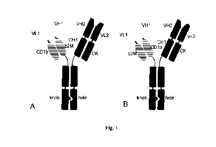
Brief description of drawings
Figure 1 is a schematic representation of the format of the bivalent
bispecific
chimeric antibody.
A is the "forward" orientation of the dimerization unit, B is the "reverse"
orientation of the dimerization unit
VH1, VL1 are heavy and light chain variable domains, respectively,
responsible for binding to antigen 1;
VH2, VL2 are variable domains responsible for binding to antigen 2.
CH1, CK are heavy and light chain constant domains, respectively;
b2M is (32 microglobulin,
CA 03231335 2024- 3- 8
CD lb is a3 domain of CD lb protein.
Knob, hole are mutations in CH3 domain of antibody, that enable
heterodimerization of heavy chains.
Figure 2 is a schematic representation of the heavy and light chain complex
of the chimeric antibody where the CH1 and CK domains were substituted for (32
microglobulin and the membrane-proximal domain a3 of CD lb protein.
A is "direct" orientation of domains of the dimerization unit;
B is "reverse" orientation of the dimerization units.
VH, VL are variable domains of the antibody,
b2M is P2 microglobulin,
CD1b is the membrane-proximal domain a3 of CD lb protein,
hinge is the hinge region of antibody, the first 5 amino acids are shown
explicitly,
Fc is Fc fragment of the antibody;
amino acids from the C-terminus of the b2M, a3 domains of CD lb and from
the N-terminus of the hinge region are shown explicitly;
SS bond is shown as a dotted line.
Figure 3 is an electrophoregram of chimeric antibodies comprising and free
of additional disulfide bonds between heavy and light chains, 7.5%
polyacrylamide
gel, non-reducing conditions.
Lanes:
M is a standard marker of Precision Plus ProteinTM Dual Color Standards
(BIO-RAD), the molecular weights of the corresponding lanes are indicated as
kDa
in the column to the left of the gel;
1- Control Prolgolimab ¨ IgGl;
21
CA 03231335 2024- 3- 8
2 ¨ Monospecific chimeric antibody based on variable domains of
Prolgolimab and domains of MHC-like proteins substituting CH1-CK, with a
further
disulfide bridge;
3 ¨ Monospecific chimeric antibody based on variable domains of
Prolgolimab and domains of MHC-like proteins substituting CH1-CK, without a
further disulfide bridge;
4 ¨ Monospecific chimeric antibody based on variable domains of
Prolgolimab and domains of MHC-like proteins substituting CH1-CK, in the
"reverse' orientation with a further disulfide bridge.
Figure 4 is a diagram of an experiment to test the effect of the MHC
dimerization unit on the incorrect pairing between heavy and light chains.
Ovals
indicate domains substituting CH1 and CK from MHC-like proteins.
Group 1: samples 01-001 ¨ 01-003 are antibodies with a "correct"
combination of VH and VL and an "incorrect" pair of constant domains.
Group 2: samples 01-004 ¨ 01-007 are antibodies with an "incorrect"
combination of VH and VL and a "correct" pair of constant domains.
Group 3: samples 01-008 ¨ 01-010 are antibodies with an "incorrect"
combination of VH and VL and an "incorrect" pair of constant domains.
Group 4: samples 01-011 and 01-012 are control antibodies that are a chimeric
antibody wherein constant domains are substituted for MHC dimerization units
and
a classical antibody of the IgG1 format, respectively.
The "correct" combination of VH and VL means a pair of VH and VL from a
single antibody. An "incorrect" combination of VH and VL means VH and VL from
distinct antibodies.
The "correct" pair of constant domains means either a pair of CH1-CK or a
pair of domains from MHC-like proteins (in this case, f32 microglobulin and
the a3
22
CA 03231335 2024- 3- 8
domain of the CD1b protein). The "incorrect" pair of constant domains means
the
CH1-CD lb or 02 microglobulin-CK pairs.
Figure 5 is an electrophoregram of samples produced in an experiment
involving incorrect pairing of chains. 7.5% polyacrylamide gel, non-reducing
conditions.
A - Lanes:
M is a standard marker of Precision Plus ProteinTM Dual Color Standards
(BIO-RAD), the molecular weights of the corresponding lanes are indicated as
kDa
in the column to the left of the gel;
1 ¨ 01-001;
2 ¨ 01-002;
3 ¨ 01-007;
4 ¨ 01-008;
¨ 01-009;
6¨ 01-012;
B - Lanes:
M ¨ Precision Plus ProteinTM Dual Color Standards (BIO-RAD) standard
marker;
1 ¨ 01-003;
2 ¨ 01-004;
3 ¨ 01-005;
4 ¨ 01-006;
5 ¨ 01-007,
6¨ 01-012;
C - Lanes:
M ¨ Precision Plus ProteinTM Dual Color Standards (BIO-RAD) standard
marker;
23
CA 03231335 2024- 3- 8
1 ¨ 01-010;
2 ¨ 01-011.
Figure 6 is an electrophoregram of bispecific chimeric antibodies.
A ¨7.5% polyacrylamide gel, non-reducing conditions,
B ¨ 12.5% polyacrylamide gel, reducing conditions.
Lanes:
M is a standard marker of Precision Plus ProteinTM Dual Color Standards
(BIO-RAD), the molecular weights of the corresponding lanes are indicated as
kDa
in the column to the left of the gel;
1 ¨ 02-004,
2 ¨ 02-005,
3 ¨ 02-006,
4 ¨ 02-007,
¨ 02-008,
6 ¨ 02-009.
Figure 7 is a sensogram of an experiment involving simultaneous interaction
of antibodies 02-006 and 02-007 with two distinct antigens (hPDlex-H6F and
hCSF1R His). The stages (steps of the experiment) are numbered at the top of
the
image and separated by vertical lines. Given are 2 sensograms for each
antibody that
show interaction with hCSF1R His at step 8 (increased signal level is
observed) and
a reference signal in a lxICB buffer free of hCSF1R_His at step 8 (no increase
in
signal level).
Figure 8 is the deconvoluted mass spectrum of the total ion current
chromatogram for sample 02-004.
A¨ peak 4,
B - peak 5,
C¨ peak 6.
24
CA 03231335 2024- 3- 8
Figure 9 is an electrophoregram of SDS gel electrophoresis of bispecific
chimeric antibodies following proteolysis by the GingisKHAN protease. 7.5%
polyacrylamide gel, non-reducing conditions.
A - lanes:
M is a standard marker of Precision Plus ProteinTM Dual Color Standards
(BIO-RAD), the molecular weights of the corresponding lanes are indicated as
kDa
in the column to the left of the gel;
1 ¨ 02-004 not processed by GingisKHAN,
2 - 02-009 not processed by GingisKHAN,
3 ¨ Ocrelizumab not processed by GingisKHAN,
4 ¨ 01-011 not processed by GingisKHAN,
¨ Prolgolimab not processed by GingisKHAN,
6- 02-004 not processed by GingisKHAN in 100 mM ammonium bicarbonate
buffer pH 7.2;
B ¨ lanes:
M ¨ Precision Plus ProteinTM Dual Color Standards (BIO-RAD) standard
marker;
1 - 02-004 following proteolysis by GingisKHAN,
2 - 02-009 following proteolysis by GingisKHAN,
3 ¨ Ocrelizumab following proteolysis by GingisKHAN,
4 ¨ 01-011 following proteolysis by GingisKHAN,
5 ¨ Prolgolimab following proteolysis by GingisKHAN,
6 - 02-004 following proteolysis by GingisKHAN in 100 mM ammonium
bicarbonate buffer pH 7.2,
C - lanes:
M ¨ Precision Plus ProteinTM Dual Color Standards (BIO-RAD) standard
marker;
CA 03231335 2024- 3- 8
1 ¨ 02-005 not processed by GingisKHAN,
2 - 02-005 following proteolysis by GingisKHAN,
3 ¨ 02-006 not processed by GingisKHAN,
4 - 02-006 following proteolysis by GingisKHAN,
¨ 02-007 not processed by GingisKHAN,
6 - 02-007 following proteolysis by GingisKHAN,
D ¨ lanes:
M ¨ Precision Plus ProteinTM Dual Color Standards (BIO-RAD) standard
marker,
1 ¨ 02-008 not processed by GingisKHAN,
2 - 02-008 following proteolysis by GingisKHAN,
3 ¨ 02-004 not processed by GingisKHAN,
4 - 02-004 following proteolysis by GingisKHAN.
Figure 10 is an electrophoregram of samples with modifications introduced
into the dimerization unit based on the membrane-proximal domains of MHC-like
protein. 7.5% polyacrylamide gel, non-reducing conditions.
A ¨ Lanes:
M is a standard marker of Precision Plus ProteinTM Dual Color Standards
(BIO-RAD), the molecular weights of the corresponding lanes are indicated as
kDa
in the column to the left of the gel;
1 ¨ Prolgolimab;
2 ¨ 01-011;
3 ¨ 03-003;
4 ¨ 03-004;
5 ¨ 03-005;
6 ¨ 03-006;
7 ¨ 03-007;
26
CA 03231335 2024- 3- 8
8 ¨ 03-008.
B - Lanes:
M ¨ Precision Plus ProteinTM Dual Color Standards (BIO-RAD) standard
marker;
1 ¨ Prolgolimab;
2 ¨ 01-011;
3 ¨ 03-001;
4¨ 03-002.
Description of the invention
General definitions and general methods
Unless defined otherwise herein, all technical and scientific terms used in
connection with the present invention will have the same meaning as is
commonly
understood by those skilled in the art.
Furthermore, unless otherwise required by context, singular terms shall
include plural terms, and the plural terms shall include the singular terms.
Typically,
the present classification and methods of cell culture, molecular biology,
immunology, microbiology, genetics, analytical chemistry, organic synthesis
chemistry, medical and pharmaceutical chemistry, as well as hybridization and
chemistry of protein and nucleic acids described herein are well known by
those
skilled and widely used in the art. Enzyme reactions and purification methods
are
performed according to the manufacturer's guidelines, as is common in the art,
or as
described herein.
The term "KD" in this description refers to the affinity constant (or
equilibrium constant), which is calculated from the ratio of Kd to Ka (i.e.
KdiKa),
and it is expressed as a molar concentration (M).
27
CA 03231335 2024- 3- 8
"Binding affinity" generally refers to the strength of the sum total of
noncovalent interactions between a single binding site of a molecule (e.g. an
antibody) and its binding partner (e.g. an antigen). Unless indicated
otherwise,
"binding affinity" refers to intrinsic (characteristic, true) binding affinity
which
reflects a 1:1 interaction between members of a binding pair (e.g. antibody
and
antigen). The affinity of a molecule X for its binding partner Y can generally
be
represented by the affinity constant (1(D). The preferred Kd value is about
200 nM,
150 nM, 100 nM, 60 nM, 50 nM, 40 nM, 30 nM, 20 nM, 10 nM, 8 nM, 6 nM, 4 nM,
2 nM, 1 nM, or less. Affinity can be measured by common methods known in the
art, including those described in the present description. Low-affinity
antibodies
generally bind an antigen slowly and tend to dissociate readily, whereas high-
affinity
antibodies generally bind an antigen faster and tend to remain bound longer. A
variety of methods of measuring binding affinity are known in the art, any of
which
can be used for the purposes of the present invention.
The term "Kd", "koff' or "kdis" refers to the off rate constant of a
particular
interaction between a binding molecule and antigen. The off rate constant koff
can
be measured using bio-layer interferometry, for example, using OctetTM system.
The term "Ka", "kon" or "on-rate" refers to the association rate constant.
The term "R2" refers to the coefficient of determination.
The term "Response" refers to the antibody-antigen binding signal.
As used in the present description and claims that follow, unless otherwise
dictated by the context, the words "include" and "comprise", or variations
thereof
such as "includes", "including", "comprises", or "comprising", will be
understood to
imply the inclusion of a stated integer or group of integers but not the
exclusion of
any other integer or group of integers.
28
CA 03231335 2024- 3- 8
Detailed description of the invention
Bivalent bispecific chimeric antibody
The present invention relates to a bivalent bispecific chimeric antibody.
The antibody according to the invention is a monoclonal antibody.
The term "monoclonal antibody" or "mAb" refers to an antibody that is
synthesized and isolated by a separate clonal population of cells.
The antibody of the invention is a recombinant antibody.
The term "recombinant antibody" refers to an antibody that is expressed in a
cell or cell line comprising nucleotide sequence(s) encoding antibodies,
wherein said
nucleotide sequence(s) is (are) not associated with the cell in nature.
The bivalent bispecific chimeric antibody according to the invention is an
isolated antibody.
The term "isolated" used to describe various antibodies according to this
description refers to an antibody which has been identified and separated
and/or
regenerated from a cell or cell culture, in which the antibody is expressed.
Impurities
(contaminant components) from natural environment are materials which
typically
interfere with diagnostic or therapeutic uses of the polypeptide, and may
include
enzymes, hormones, and other proteinaceous or nonproteinaceous solutes. The
isolated polypeptide is typically prepared by at least one purification step.
In one aspect, the present invention relates to a bivalent bispecific chimeric
antibody, wherein said antibody comprises:
a) a first light chain and a first heavy chain of antibody specifically
binding to
a first antigen;
wherein the first light chain comprises a light chain variable domain and a
light chain constant domain;
wherein the first heavy chain comprises a heavy chain variable domain and
heavy chain constant domains of antibody that include a first (CH1) heavy
chain
29
CA 03231335 2024- 3- 8
constant domain and an Fc fragment monomer comprising second (CH2) and third
(CH3) heavy chain constant domains;
b) a second light chain and a second heavy chain of antibody specifically
binding to a second antigen,
wherein the second light chain comprises a light chain variable domain and a
constant domain that is selected from the group:
a first membrane-proximal domain of MHC (major histocompatibility
complex) or
a first membrane-proximal domain of MHC-like protein;
wherein the second heavy chain comprises a heavy chain variable domain, a
constant domain that is selected from the group:
a second membrane-proximal domain of MHC (major histocompatibility
complex) or
a second membrane-proximal domain of MHC-like protein;
and an Fc fragment monomer comprising second (CH2) and third (CH3)
heavy chain constant domains;
wherein the first membrane-proximal domain of MHC or MHC-like protein
and the second membrane-proximal domain of MHC or MHC-like protein form a
heterodimer therebetween, which is stabilized by a disulfide bond;
wherein the C113 domain of one heavy chain and the C113 domain of another
heavy chain contact each other with surfaces thereof, which are modified to
form a
bivalent bispecific chimeric antibody, said modifications in the heavy chain
CH3
domains being substitutions to facilitate heterodimerization.
The heterodimer that is formed by the first membrane-proximal domain of
MHC or MHC-like protein and the second membrane-proximal domain of MHC or
MHC-like protein and stabilized by a disulfide bond means:
CA 03231335 2024- 3- 8
1) a heterodimer that includes a mutation or mutations in the first and/or
second membrane-proximal domain of MHC or MHC-like protein to form an S-S
bond (disulfide bond, cysteine bridge) between the first and second membrane-
proximal domain of MHC or MHC-like protein;
or
2) an elongation of the first membrane-proximal domain of MHC or MHC-
like protein by one or more (from 1 to 10) amino acids at the C-terminus and
with
terminal Cys at the C-terminus to form an S-S bond (disulfide bond, cysteine
bridge)
between the first membrane-proximal domain of MHC or MHC-like protein and the
hinge.
In some embodiments of the invention, the bivalent bispecific chimeric
antibody includes a first membrane-proximal domain of MHC or MHC-like protein
and a second membrane-proximal domain of MHC or MHC-like protein that form a
heterodimer therebetween, which is stabilized by a disulfide bond by means of
a
mutation or mutations in the first and/or second membrane-proximal domain of
MHC or WIC-like protein to form an S-S bond (disulfide cysteine bridge)
between
the first and second membrane-proximal domain of MHC or MHC-like protein.
In some embodiments of the invention, the bivalent bispecific chimeric
antibody includes a first membrane-proximal domain of MHC or MHC-like protein
and a second membrane-proximal domain of MHC or MHC-like protein that form a
heterodimer therebetween, which is stabilized by a disulfide bond by means of
elongation of the first membrane-proximal domain of MHC or MHC-like protein by
one or more (1 to 10) amino acids at the C-terminus and with terminal Cys at
the C-
terminus to form an S-S bond (disulfide bond, cysteine bridge) between the
first
membrane-proximal domain of MHC or MHC-like protein and the hinge.
31
CA 03231335 2024- 3- 8
In some embodiments of the invention, the bivalent bispecific chimeric
antibody includes an elongation of the first membrane-proximal domain of MHC
or
MHC-like protein, which elongation is a sequence of three amino acids GSC.
In some embodiments of the invention, the bivalent bispecific chimeric
antibody comprises:
a first light chain that comprises a light chain variable domain (VL1) and a
light chain constant domain;
a first heavy chain that comprises a heavy chain variable domain (VH1) and
heavy chain constant domains of antibody that include a first (CH1) heavy
chain
constant domain and an Fc fragment monomer comprising second (CH2) and third
(CH3) heavy chain constant domains;
a second light chain that comprises a light chain variable domain (VL2) and a
first membrane-proximal domain of MHC or a first membrane-proximal domain of
MHC-like protein;
a second heavy chain that comprises a heavy chain variable domain (VH2), a
second membrane-proximal domain of MHC or a second membrane-proximal
domain of MHC-like protein and an Fc fragment monomer comprising second
(CH2) and third (CH3) heavy chain constant domains;
wherein the first membrane-proximal domain of MHC or MHC-like protein
and the second membrane-proximal domain of MHC or MHC-like protein form a
heterodimer therebetween, which is stabilized by a disulfide bond by means of
a
mutation or mutations in the first and/or second membrane-proximal domain of
MHC or MHC-like protein to form an S-S bond (disulfide cysteine bridge)
between
the first and second membrane-proximal domain of MHC or MHC-like protein;
wherein between the first Fc variant and the second Fc variant in the CH3
domain there is formed a knob-into-hole structure.
32
CA 03231335 2024- 3- 8
In some embodiments of the invention, the bivalent bispecific chimeric
antibody comprises:
a first light chain that comprises a light chain variable domain (VL1) and a
light chain constant domain;
a first heavy chain that comprises a heavy chain variable domain (VH1) and
heavy chain constant domains of antibody that include a first (CH1) heavy
chain
constant domain and an Fc fragment monomer comprising second (CH2) and third
(CH3) heavy chain constant domains;
a second light chain that comprises a light chain variable domain (VL2) and a
first membrane-proximal domain of MHC or a first membrane-proximal domain of
MHC-like protein;
a second heavy chain that comprises a heavy chain variable domain (VH2), a
second membrane-proximal domain of MHC or a second membrane-proximal
domain of MHC-like protein and an Fc fragment monomer comprising second
(CH2) and third (CH3) heavy chain constant domains;
wherein between the first Fc variant and the second Fc variant in the CH3
domain there is formed a knob-into-hole structure,
wherein the first membrane-proximal domain of MHC or the first membrane-
proximal domain of MHC-like protein further comprises an elongation of the
first
membrane-proximal domain of MHC or MHC-like protein by one or more (from 1
to 10) amino acids at the C-terminus and with terminal Cys at the C-terminus
to form
an S-S bond (disulfide bond, cysteine bridge) between the first membrane-
proximal
domain of MHC or MHC-like protein and the hinge.
In some embodiments of the invention, the bivalent bispecific chimeric
antibody comprises:
a first light chain that comprises a light chain variable domain (VL1) and a
light chain constant domain;
33
CA 03231335 2024- 3- 8
a first heavy chain that comprises a heavy chain variable domain (VH1) and
heavy chain constant domains of antibody that include a first (CH1) heavy
chain
constant domain and an Fc fragment monomer comprising second (CH2) and third
(CH3) heavy chain constant domains;
a second light chain that comprises a light chain variable domain (VL2) and a
first membrane-proximal domain of MHC or a first membrane-proximal domain of
MHC-like protein;
a second heavy chain that comprises a heavy chain variable domain (VH2), a
second membrane-proximal domain of MHC or a second membrane-proximal
domain of MHC-like protein and an Fc fragment monomer comprising second
(CH2) and third (CH3) heavy chain constant domains;
wherein between the first Fc variant and the second Fc variant in the CH3
domain there is formed a knob-into-hole structure
wherein the first membrane-proximal domain of MHC or the first membrane-
proximal domain of MHC-like protein further comprises an elongation of the
first
membrane-proximal domain of MHC or MHC-like protein by GSC to form an S-S
bond (disulfide bond, cysteine bridge) between the first membrane-proximal
domain
of MHC or MHC-like protein and the hinge.
In the above bivalent bispecific chimeric antibody, the variable and constant
domains are arranged in the heavy and light chains in the following order:
the first light chain:
1) a light chain variable domain,
2) a light chain constant domain;
the first heavy chain of antibody:
1) a heavy chain variable domain,
2) a first (CH1) heavy chain constant domain,
3) a second (CH2) heavy chain constant domain and
34
CA 03231335 2024- 3- 8
4) a third (CH3) heavy chain constant domain;
the second light chain:
1) a light chain variable domain,
2) a first membrane-proximal domain of MHC or a first membrane-proximal
domain of MHC-like protein;
the second heavy chain of antibody:
1) a heavy chain variable domain,
2) a second membrane-proximal domain of MHC or a second membrane-
proximal domain of MHC-like protein,
3) a second (CH2) heavy chain constant domain and
4) a third (CH3) heavy chain constant domain.
Figure 1 shows a few variants of the format of the above bivalent bispecific
chimeric antibody.
The term "antibody" or "immunoglobulin" (Ig), as used in the present
description, includes whole antibodies. The term "antibody" refers to a
glycoprotein
comprising at least two heavy (H) chains and two light (L) chains
interconnected by
disulfide bonds, or an antigen-binding portion. Each heavy chain comprises a
heavy
chain variable region (abbreviated referred to in the present description as
VH) and
a heavy chain constant region. Known are five types of mammalian antibody
heavy
chains denoted by Greek letters: a, 8, c, y and p,. (Janeway C.A., Jr. et al,
Immunobiology, 5th ed., publ. by Garland Publishing, 2001). The type of a
heavy
chain present defines the class of an antibody; these chains are found in IgA,
IgD,
IgE, IgG, and IgM antibodies, respectively. (Rhoades R.A., Pflanzer R.G.,
Human
Physiology, 4th ed., publ. by Thomson Learning, 2002). Distinct heavy chains
differ
in size and composition; a and y contain approximately 450 amino acids, while
p,
and E have approximately 550 amino acids. The constant region is identical in
all
antibodies of the same isotype, but differs in antibodies of different
isotypes. Heavy
CA 03231335 2024- 3- 8
chains 7, a and 6 have a constant region composed of three constant domains
CH1,
CH2 and CH3 (in a line), and a hinge region for added flexibility (Woof J.,
Burton
D., Nat Rev Immunol 4,2004, cc. 89-99); heavy chains pt and E have a constant
region
composed of four constant domains CH1, CH2, CH3 and CH4 (Janeway C.A., Jr. et
al, Immunobiology, 5th ed., pub!. by Garland Publishing, 2001). In mammals,
known are only two types of light chains denoted by lambda (X) and kappa (K).
Each
light chain consists of a light chain variable region (abbreviated referred to
in the
present description as VL) and light chain constant region. The approximate
length
of a light chain is 211 to 217 amino acids. Preferably the light chain is a
kappa (lc)
light chain, and the constant domain CL is preferably C kappa 00.
"Antibodies" according to the invention can be of any class (e.g., IgA, IgD,
IgE, IgG, and IgM, preferably IgG), or subclass (e.g., IgGl, IgG2, IgG3, IgG4,
IgAl
and IgA2, preferably IgG1).
VL and VII regions may be further subdivided into hyper-variability regions
called complementarity determining regions (CDRs), located between regions
that
are more conserved, termed framework regions (FRs). Each VH and VL is composed
of three CDRs and four FRs, arranged from amino-terminus to carboxy-terminus
in
the following order: FR1, CDR1, FR2, CDR2, FR3, CDR3, FR4. The variable
regions of heavy and light chains contain a binding domain that interacts with
an
antigen. The constant regions of antibodies may mediate the binding of
immunoglobulin to host tissues or factors, including various cells of the
immune
system (e.g. effector cells) and the first component (C 1q) of the classical
complement system.
The term "antigen-binding portion" of an antibody or "antigen-binding
fragment" (or simply "antibody portion" or "antibody fragment"), as used in
this
description, refers to one or more fragments of an antibody that retain the
capability
of specific binding to an antigen. An example of a binding fragment
incorporated
36
CA 03231335 2024- 3- 8
into the term "antigen-binding portion" of antibody includes a Fab fragment,
i.e. a
monovalent fragment consisting of VL, VH, CL and CH1 domains or a Fab-like
fragment, i.e. a monovalent fragment consisting of VL, VII domains, a first
membrane-proximal domain of MHC or MHC-like protein and a second membrane-
proximal domain of MHC or MHC-like protein.
Preferably, the CDR of an antigen-binding portion, or the whole antigen
binding portion of antibodies of the invention is derived from a mouse, lama
or
human donor library or substantially of human origin with certain amino acid
residues altered, e.g. substituted with different amino acid residues so as to
optimize
specific properties of the antibody, e.g. KD, koff, IC50, EC50, EDS .
Preferably,
the framework regions of the antibody of the invention are of human origin or
substantially of human origin (at least 80, 85, 90, 95, 96, 97, 98 or 99% of
human
origin).
In other embodiments, the antigen binding portion of the invention may be
derived from other non-human species including mouse, lama, rabbit, rat or
hamster,
but not limited to. Alternatively, the antigen-binding region can be derived
from the
human species.
The term "variable" refers to the fact that certain portions of the variable
domains greatly differ in sequence among antibodies. The V domain mediates
antigen binding and determines specificity of each particular antibody for its
particular antigen. However, the variability is not evenly distributed across
the 110-
amino acid span of the variable domains. Instead, the V regions consist of
invariant
fragments termed framework regions (FRs) of 15-30 amino acids separated by
shorter regions of extreme variability termed "hypervariable regions" or CDRs.
The
variable domains of native heavy and light chains each comprise four FRs,
largely
adopting a beta-sheet configuration, connected by three hypervariable regions,
which form loops connecting, and in some cases forming part of, the beta-sheet
37
CA 03231335 2024- 3- 8
structure. The hypervariable regions in each chain are held together in close
proximity by FRs and, with the hypervariable regions from the other chain,
contribute to the formation of the antigen-binding site of antibodies (see
Kabat et al.,
Sequences of Proteins of Immunological Interest. 5 th Ed. Public Health
Service,
National Institutes of Health, Bethesda, MD. (1991)). The constant domains are
not
involved directly in binding of antibody to antigen, but exhibit various
effector
functions, such as participation of antibody in antibody-dependent cellular
cytotoxicity (AD CC).
The term "hypervariable region" according to the present description refers to
the amino acid residues of antibody which are responsible for antigen binding.
The
hypervariable region typically comprises amino acid residues from a
"complementarity determining region" or "CDR" and/or those residues from a
"hypervariable loop".
"Kabat numbering scheme" or "numbering according to Kabat" as used in this
application refers to the system for numbering of amino acid residues that are
more
variable (i.e. hypervariable) than other amino acid residues in variable
regions of
heavy and light chains of the antibody (Kabat et al. Ann. N.Y. Acad. Sci.,
190:382-
93 (1971); Kabat et al. Sequences of Proteins of Immunological Interest, Fifth
Edition, U.S. Department of Health and Human Services, NIH Publication No. 91-
3242 (1991)).
The antibody of the present invention "which binds" a target antigen refers to
an antibody that binds the antigen with sufficient affinity such that the
antibody can
be used as a diagnostic and/or therapeutic agent targeting a protein or cell
or tissue
expressing the antigen, and slightly cross-reacts with other proteins.
According to
analytical methods: fluorescence-activated cell sorting (FACS),
radioimmunoassay (RIA) or ELISA, in such embodiments, the degree of antibody
binding to a non-target protein is less than 10 % of antibody binding to a
specific
38
CA 03231335 2024- 3- 8
target protein. With regard to the binding of antibody to a target molecule,
the term
"specific binding" or "specifically binds to" or "is specific for" a
particular
polypeptide or an epitope on a particular target polypeptide means binding
that is
significantly (measurably) different from a non-specific interaction.
Specific binding may be measured, for example, by determining binding of a
molecule as compared to binding of a control molecule. For example, specific
binding may be determined by competition with another molecule that is similar
to
the target, for example, an excess of non-labeled target. In this case,
specific binding
is indicated if the binding of the labeled target to a probe is competitively
inhibited
by the excess of unlabeled target. As used in the present description, the
term
"specific binding" or phrases "specifically binds to" or "is specific for" a
particular
polypeptide or an epitope on a particular target polypeptide may be described
by
example of a molecule having a Kd for the target of at least about 200 nM, or
at least
about 150 nM, or at least about 100 nM, or at least about 60 nM, or at least
about 50
nM, or at least about 40 nM, or at least about 30 nM, or at least about 20 nM,
or at
least about 10 nM, or at least about 8 nM, or at least about 6 nM, or at least
about 4
nM, or at least about 2 nM, or at least about 1 nM, or greater. In one
embodiment,
the term "specific binding" refers to binding where a molecule binds to a
particular
polypeptide or epitope on a particular polypeptide without substantially
binding to
any other polypeptide or epitope on a polypeptide.
The term "bispecific antibody" refers to an antibody having antigen-binding
domains that are capable of specific binding to two distinct epitopes on a
single
biological molecule or capable of specific binding to epitopes on two distinct
biological molecules. The bispecific antibody is also referred to herein as
having
"dual specificity" or as being a "dual specificity" antibody.
The fragment crystallizable region ("Fe region, Fe") of an immunoglobulin is
the "tail" region of an immunoglobulin molecule that interacts with cell
surface Fc-
39
CA 03231335 2024- 3- 8
receptor, as well as some proteins of the complement system. This property
allows
antibodies to activate the immune system. In IgG, IgA and IgD isotypes, the Fc
region is composed of two identical protein fragments, respectively, from the
second
and third constant domains of the two heavy chains; in IgM and IgE isotypes,
the Fc
contains three heavy chain constant domains (CH2, CH3, and CH4 domains) in
each
polypeptide chain.
"Fc fragment monomer" is understood to mean an Fc region from the
second and third constant domains of either one of the two heavy chains (for
IgG, IgA and IgD isotypes); for IgM and IgE isotypes, the Fc monomer
comprises three constant domains of one of the two heavy chains (CH2, CH3
and CH4 domains).
In some embodiments of the invention, the bivalent bispecific chimeric
antibody comprises:
a first light chain that comprises a light chain variable domain (VL1) and a
light chain constant domain;
a first heavy chain that comprises a heavy chain variable domain (VH1) and
heavy chain constant domains of antibody that include a first (CH1) heavy
chain
constant domain and an Fc fragment monomer comprising second (CH2) and third
(CH3) heavy chain constant domains;
a second light chain that comprises a light chain variable domain (VL2) and a
first membrane-proximal domain of MHC or a first membrane-proximal domain of
MHC-like protein;
a second heavy chain that comprises a heavy chain variable domain (VH2), a
second membrane-proximal domain of MHC or a second membrane-proximal
domain of MHC-like protein and an Fc fragment monomer comprising second
(CH2) and third (CH3) heavy chain constant domains;
CA 03231335 2024- 3- 8
and wherein between the first Fe variant and the second Fe variant in the CH3
domain there is formed a knob-into-hole structure
and between the second membrane-proximal domain of MHC and/or the
second membrane-proximal domain of MHC-like protein and the hinge there is
inserted a further peptide linker.
The term "peptide linker" as used herein is intended to mean any peptide
having the ability to combine domains, with a length which depends on the
domains which it binds to each other, and comprising any amino acid sequence.
Preferably, the peptide linker has a length of 4 amino acids or more and
consists
of any set of amino acids selected from G, A, S. P. E, T, D, K.
MHC (major histocompatibility complex) refers to a major histocompatibility
complex molecule that is the central component of the immune system of
vertebrates
present on the surface of all nuclear cells. There are two main forms of MHC,
in
particular MHC Class I and II.
MHC-like proteins refer to proteins that have a structural similarity to the
extracellular portion of MHC class I molecules or MHC class II molecules. In
particular, MHC-like proteins mean, for example, CD1 (cluster of
differentiation 1)
protein or HFE (hereditary hemochromatosis protein) protein.
The general structural similarity between MHC I, II, CD1 and HFE molecules
is obvious. This includes the uniformity of the spatial organization of the
whole
molecule, the number of domains (four), the structural similarity of the
membrane-
proximal domains of MHC and MHC-like proteins, as well as the antigen-binding
site, similar mol. wts.
In some embodiments of the invention, the bivalent bispecific chimeric
antibody comprises a first membrane-proximal domain of MHC, which can be
selected from a group comprising:
41
CA 03231335 2024- 3- 8
a first membrane-proximal domain of MHC class I (major histocompatibility
complex class I),
a first membrane-proximal domain of MHC class II (major histocompatibility
complex class II),
a modified variant of a first membrane-proximal domain of MHC class I or
a modified variant of a first membrane-proximal domain of MHC class II.
In humans, the classical MHC I genes are referred to as HLA-A, HLA-B,
HLA-C, HLA-E, HLA-F or HLA-G. Class I molecules consist of two polypeptide
chains: a polymorphic a-chain (sometimes referred to as heavy chain) and a
smaller
chain called 132 microglobulin (also known as light chain), which is generally
not
polymorphic. These two chains form a non-covalent heterodimer on the cell
surface.
The a-chain contains three domains (al, a2 and a3). Exon 1 of the a-chain gene
encodes the leader sequence, exons 2 and 3 encode the al and a2 domains, exon
4
encodes the a3 domain, exon 5 encodes the transmembrane domain, and exons 6
and
7 encode the cytoplasmic tail. The a-chain forms a peptide-binding region
involving
the al and a2 domains.
The a2 domain is followed by the a3 domain located at the C-terminus of the
extracellular portion of the a chain of MHC I and forms together with 132
microglobulin a heterodimeric non-covalent complex. Said heterodimeric non-
covalent complex composed of the a3 domain of MHC I and 132 microglobulin is
referred to in the description of the present invention as a heterodimer based
on the
membrane-proximal domains of MHC I.
MHC class I molecules are expressed on all nucleated cells, including tumor
cells. They are specifically expressed on, inter alia, T- and B-lymphocytes,
macrophages, dendritic cells and neutrophils and function to present peptide
fragments (typically 8-10 amino acids in length) on the surface of CD8+ to
cytotoxic
T-lymphocytes (CTL).
42
CA 03231335 2024- 3- 8
In humans, the classical MHC II genes are referred to as HLA-DM, HLA-DO,
HLA-DP, HLA-DQ or HLA-DR. MHC Class II molecules are heterodimers
composed of non-covalently linked alpha and beta chains. The extracellular
portion
of each of the chains consists of two domains (al (alphal), a2 (a1pha2) and 01
(betal), (32 (beta2), respectively) and is connected by a short peptide with a
transmembrane segment (approximately 30 amino acid residues long). The
transmembrane segment is followed by the cytoplasmic domain containing
approximately 10-15 residues.
The antigen-binding region of MHC class II molecules is formed by alpha-
helical sections of interacting chains similarly to class I molecules except
for: the
antigen-binding groove of MHC class II molecules is formed not by two domains
of
a single alpha chain but by two domains of distinct chains, the al and 01
domains.
The al domain is followed by the a2 domain, which forms a heterodimeric
non-covalent complex with the (32 domain. Said heterodimeric non-covalent
complex composed of the a2 domain of MHC II and the 02 domain of MHC II is
referred to in the description of the present invention as a heterodimer based
on the
membrane-proximal domains of MHC II.
In the structure of MHC class II molecules, the antigen-binding groove is more
open than that of MHC class I molecules and thus receptive of longer peptides.
In some embodiments of the invention, the bivalent bispecific chimeric
antibody comprises a second membrane-proximal domain of MHC, which can be
selected from a group comprising:
a second membrane-proximal domain of MHC class I (major
histocompatibility complex class I),
a second membrane-proximal domain of MHC class II (major
histocompatibility complex class II),
a modified variant of a second membrane-proximal domain of MHC class I or
43
CA 03231335 2024- 3- 8
a modified variant of a second membrane-proximal domain of MHC class II.
In some embodiments of the invention, the bivalent bispecific chimeric
antibody comprises a membrane-proximal domain of human MHC class I.
In some embodiments of the invention, the bivalent bispecific chimeric
antibody comprises a membrane-proximal domain of human MHC class II.
In some embodiments of the invention, the bivalent bispecific chimeric
antibody comprises a first membrane-proximal domain of MHC, which can be
selected from a group comprising:
a first membrane-proximal domain of MHC class I (major histocompatibility
complex class I),
a first membrane-proximal domain of MHC class II (major histocompatibility
complex class II),
a modified variant of a first membrane-proximal domain of MHC class I or
a modified variant of a first membrane-proximal domain of MHC class II,
and
a second membrane-proximal domain of MHC that may be selected from a
group including:
a second membrane-proximal domain of MHC class I (major
histocompatibility complex class I),
a second membrane-proximal domain of MHC class II (major
histocompatibility complex class II),
a modified variant of a second membrane-proximal domain of MHC class I or
a modified variant of a second membrane-proximal domain of MHC class II.
In some embodiments of the invention, the bivalent bispecific chimeric
antibody comprises:
a first membrane-proximal domain of MHC that is a first membrane-proximal
domain of MHC class I (major histocompatibility complex class I),
44
CA 03231335 2024- 3- 8
and
a second membrane-proximal domain of MHC that is a second membrane-
proximal domain of MHC class I (major histocompatibility complex class I).
In some embodiments of the invention, the bivalent bispecific chimeric
antibody comprises:
a first membrane-proximal domain of MHC that is a first membrane-proximal
domain of MHC class II (major histocompatibility complex class II),
and
a second membrane-proximal domain of MHC that is a second membrane-
proximal domain of MHC class II (major histocompatibility complex class II).
In some embodiments of the invention, the bivalent bispecific chimeric
antibody comprises:
a first membrane-proximal domain of MHC that is a modified variant of a first
membrane-proximal domain of MHC class I,
and
a second membrane-proximal domain of MHC that is a modified variant of a
second membrane-proximal domain of MHC class I.
In some embodiments of the invention, the bivalent bispecific chimeric
antibody comprises:
a first membrane-proximal domain of MHC that is a modified variant of a first
membrane-proximal domain of MHC class II,
and
a second membrane-proximal domain of MHC that is a modified variant of a
second membrane-proximal domain of MHC class II.
In some embodiments of the invention, the bivalent bispecific chimeric
antibody comprises:
CA 03231335 2024- 3- 8
a first membrane-proximal domain of MHC that is a first membrane-proximal
domain of MHC class I (major histocompatibility complex class I),
and
a second membrane-proximal domain of MHC that is a modified variant of a
second membrane-proximal domain of MHC class I.
In some embodiments of the invention, the bivalent bispecific chimeric
antibody comprises:
a first membrane-proximal domain of MHC that is a modified variant of a first
membrane-proximal domain of MHC class I,
and
a second membrane-proximal domain of MHC that is a second membrane-
proximal domain of MHC class I (major histocompatibility complex class I).
In some embodiments of the invention, the bivalent bispecific chimeric
antibody comprises:
a first membrane-proximal domain of MHC that is a first membrane-proximal
domain of MEC class II (major histocompatibility complex class II),
and
a second membrane-proximal domain of MHC that is a modified variant of a
second membrane-proximal domain of MHC class II.
In some embodiments of the invention, the bivalent bispecific chimeric
antibody comprises:
a first membrane-proximal domain of MHC that is a modified variant of a first
membrane-proximal domain of MHC class II,
and
a second membrane-proximal domain of MHC that is a second membrane-
proximal domain of MHC class II (major histocompatibility complex class II).
46
CA 03231335 2024- 3- 8
In some embodiments of the invention, the bivalent bispecific chimeric
antibody comprises a membrane-proximal domain of MHC class II, which is
selected from the group: HLA-DM, HLA-DO, HLA-DP, HLA-DQ or HLA-DR.
In some embodiments of the invention, the bivalent bispecific chimeric
antibody comprises a membrane-proximal domain of MHC class I, which is
selected
from the group: HLA-A, HLA-B, HLA-C, HLA-E, HLA-F or HLA-G.
In some embodiments of the invention, the bivalent bispecific chimeric
antibody comprises a membrane-proximal domain of a human MHC-like protein.
In some embodiments of the invention, the bivalent bispecific chimeric
antibody comprises a first membrane-proximal domain of MHC-like protein, which
may be selected from a group comprising:
a first membrane-proximal domain of CD1 (cluster of differentiation 1),
a first membrane-proximal domain of HFE (hemochromatosis protein),
a modified variant of a first membrane-proximal domain of CD1 or
a modified variant of a first membrane-proximal domain of HFE.
CD1 (cluster of differentiation 1) refers to a cluster of differentiation 1
molecule that is a component of the immune system located on the surface of
various
antigen-presenting cells, such as dendritic cells, macrophages and other
cells. In the
same fashion as MHC classes I, II, CD1 present antigens for recognition by T
cells
through interaction with the T cell receptor. Unlike MHC classes I, II, CD1
proteins
present lipids and derivatives thereof rather than peptides.
The plurality of CD1 variants found in humans are divided into 5 groups:
CD1a, CD1b, CD1c, CD1d, CD1e, differring in the structure of the antigen-
binding
fragment and, consequently, specificity for lipids of different structures.
CDle group
proteins, unlike proteins of other groups, are not expressed on the cell
surface, but
are soluble and are responsible for lipid transport (Kaczmarek, R., Pasciak,
M.,
Szymczak-Kulus, K., & Czerwinski, M. (2017). CD1: A Singed Cat of the Three
47
CA 03231335 2024- 3- 8
Antigen Presentation Systems. Arch ivum Immunologiae et Therapiae
Experimentalis, 65(3), 201-214).
CD1 molecules are similar in structure to MHC class I. In a similar fashion,
one CD1 molecule is a non-covalent complex consisting of two polypeptide
chains:
a polymorphic a-chain (sometimes referred to as heavy chain) and a smaller
chain
called (32 microglobulin (also known as light chain), which is generally not
polymorphic.
The a chain forms an antigen-binding region comprising the al and a2
domains. The a2 domain is followed by the a3 domain located at the C-terminus
of
the extracellular portion of the a chain of CD1 and forms together with (32
microglobulin a heterodimeric non-covalent complex. Said heterodimeric non-
covalent complex composed of the a3 domain of CD1 and (32 microglobulin is
referred to in the description of the present invention as a heterodimer based
on the
membrane-proximal domains of MHC-like proteins.
HFE (Hereditary hemochromatosis protein) refers to a hemochromatosis
protein molecule. The human HFE gene locus is located on chromosome 6 and
consists of 5 exons and 4 introns. Hemochromatosis protein is a membrane
protein
associated with 132 microglobulin, it is responsible for the regulation of
iron
absorption via the regulation of transferrin-transferrin receptor interaction.
Mutations in the HFE gene lead to hemochromatosis, an iron metabolism-related
recessive pathology associated with iron accumulation in various organs. The
HFE
molecule is identical in structure to MHC I. One HFE molecule is a non-
covalent
complex consisting of two polypeptide chains: a polymorphic a-chain (sometimes
referred to as heavy chain) and a smaller chain called 02 microglobulin (also
known
as light chain), which is generally not polymorphic.
The a chain includes al, a2 and a3 domains. The a3 domain follows the a2
domain and is located at the C-terminus of the extracellular portion of the a
chain of
48
CA 03231335 2024- 3- 8
HFE and forms with 132 microglobulin a heterodimeric non-covalent complex.
Said
heterodimeric non-covalent complex composed of the a3 domain of HFE and 132
microglobulin is referred to in the description of the present invention as a
heterodimer based on the membrane-proximal domains of MHC-like proteins.
In some embodiments of the invention, the bivalent bispecific chimeric
antibody comprises a second membrane-proximal domain of MHC-like protein,
which may be selected from a group comprising:
a second membrane-proximal domain of CD1 (cluster of differentiation 1),
a second membrane-proximal domain of HFE (hemochromatosis protein),
a modified variant of a second membrane-proximal domain of CD1 or
a modified variant of a second membrane-proximal domain of HFE.
In some embodiments of the invention, the bivalent bispecific chimeric
antibody comprises:
a first membrane-proximal domain of MHC-like protein that may be selected
from a group comprising:
a first membrane-proximal domain of CD1 (cluster of differentiation 1),
a first membrane-proximal domain of HFE (hemochromatosis protein),
a modified variant of a first membrane-proximal domain of CD1 or
a modified variant of a first membrane-proximal domain of HFE,
and
a second membrane-proximal domain of MHC-like protein that may be
selected from a group comprising:
a second membrane-proximal domain of CD1 (cluster of differentiation 1),
a second membrane-proximal domain of HFE (hemochromatosis protein),
a modified variant of a second membrane-proximal domain of CD1 or
a modified variant of a second membrane-proximal domain of HFE.
49
CA 03231335 2024- 3- 8
In some embodiments of the invention, the bivalent bispecific chimeric
antibody comprises:
a first membrane-proximal domain of MHC-like protein, which is a first
membrane-proximal domain of CD1,
and
a second membrane-proximal domain of MHC-like protein, which is a second
membrane-proximal domain of CD1.
In some embodiments of the invention, the bivalent bispecific chimeric
antibody comprises:
a first membrane-proximal domain of MHC-like protein, which is a first
membrane-proximal domain of HFE,
and
a second membrane-proximal domain of MHC-like protein, which is a second
membrane-proximal domain of HFE,
In some embodiments of the invention, the bivalent bispecific chimeric
antibody comprises:
a first membrane-proximal domain of MHC-like protein, which is a modified
variant of a first membrane-proximal domain of CD1,
and
a second membrane-proximal domain of MHC-like protein, which is a
modified variant of a second membrane-proximal domain of CD1.
In some embodiments of the invention, the bivalent bispecific chimeric
antibody comprises:
a first membrane-proximal domain of MHC-like protein, which is a modified
variant of a first membrane-proximal domain of HFE,
and
CA 03231335 2024- 3- 8
a second membrane-proximal domain of MHC-like protein, which is a
modified variant of a second membrane-proximal domain of HFE.
In some embodiments of the invention, the bivalent bispecific chimeric
antibody comprises:
a first membrane-proximal domain of MHC-like protein, which is a first
membrane-proximal domain of CD1,
and
a second membrane-proximal domain of MHC-like protein, which is a
modified variant of a second membrane-proximal domain of CD!.
In some embodiments of the invention, the bivalent bispecific chimeric
antibody comprises:
a first membrane-proximal domain of MHC-like protein, which is a modified
variant of a first membrane-proximal domain of CD1,
and
a second membrane-proximal domain of MHC-like protein, which is a second
membrane-proximal domain of CD1.
In some embodiments of the invention, the bivalent bispecific chimeric
antibody comprises:
a first membrane-proximal domain of MHC-like protein, which is a first
membrane-proximal domain of HFE,
and
a second membrane-proximal domain of MHC-like protein, which is a
modified variant of a second membrane-proximal domain of HFE.
In some embodiments of the invention, the bivalent bispecific chimeric
antibody comprises:
a first membrane-proximal domain of MHC-like protein, which is a modified
variant of a first membrane-proximal domain of HFE,
51
CA 03231335 2024- 3- 8
and
a second membrane-proximal domain of MHC-like protein, which is a second
membrane-proximal domain of HFE.
In some embodiments of the invention, the bivalent bispecific chimeric
antibody comprises a membrane-proximal domain of CD1, which is selected from
the group: CD1a, CD1b, CD1c, CD1d or CD1e.
In some embodiments of the invention, the bivalent bispecific chimeric
antibody includes a variable fragment of the second light chain (VL), which is
separated from a first membrane-proximal domain of MHC or MHC-like protein by
a linker of 1 to 25 amino acids long, and/or a variable fragment of the second
heavy
chain (VH), which is separated from a second membrane-proximal domain of MHC
or MHC-like protein by a linker of 1 to 25 amino acids long.
In some embodiments of the invention, the bivalent bispecific chimeric
antibody comprises:
a) the CH3 domain of one heavy chain, which is modified so that on the
surface of the CH3 domain of one heavy chain contacting the surface of the CH3
domain of another heavy chain in the bivalent bispecific antibody, the amino
acid
residue is substituted for an amino acid residue that has a larger side chain
volume,
leading to formation of a knob on the surface of the CH3 domain of one heavy
chain
that can fit into a hole on the surface of the CH3 domain of another heavy
chain,
and
b) the CH3 domain of another heavy chain, which is modified so that on the
surface of the CH3 domain of the second heavy chain contacting the surface of
the
CH3 domain of the first heavy chain in the bivalent bispecific antibody, the
amino
acid residue is substituted for an amino acid residue that has a smaller side
chain
volume, leading to formation of a hole on the surface of the CH3 domain of the
52
CA 03231335 2024- 3- 8
second heavy chain that can accept a knob on the interface of the CH3 domain
of the
first heavy chain;
wherein said amino acid residue that has a larger side chain volume is
selected
from a group including arginine (R), phenylalanine (F), tyrosine (Y),
tryptophan
(W),
and wherein said amino acid residue that has a smaller side chain volume is
selected from a group including alanine (A), serine (S), threonine (T), valine
(V).
In some embodiments of the invention, the bivalent bispecific chimeric
antibody comprises a constant domain of the first light chain of antibody,
which is
selected from CK or CL.
In mammals, known are only two types of light chains denoted by lambda (X)
and kappa (lc). The constant domain of the lambda light chain is designated
CL, and
that of the kappa light chain is designated CK.
In some embodiments of the invention, the bivalent bispecific chimeric
antibody comprises CH3 domains of antibody that are further modified by
introduction of cysteine (C) as an amino acid into the corresponding positions
of
each CH3 domain so that a disulfide bridge may form between the both CH3
domains.
In some embodiments of the invention, the bivalent bispecific antibody
comprises a CH3 domain of one heavy chain, which is modified to form Knob, and
a CH3 domain of another heavy chain, which is modified to form Hole, or vice
versa.
"knobs-into-holes" (interactions of the "knobs -into-holes" type) is an
approach that enables to circumvent the problem associated with mispaired
byproducts. This approach aims at forcing the pairing of two different
antibody
heavy chains by introducing mutations into the CH3 domains to modify the
contact
interfaces. On one chain, bulky amino acids were replaced by amino acids with
short
side chains to create a "hole". Conversely, amino acids with large side chains
were
53
CA 03231335 2024- 3- 8
introduced into the other CH3 domain to create a "knob". Co-expression of
these
two heavy chains produced a high yield of the heterodimer formation ("knob-
hole")
relative to the homodimer formation ("hole-hole" or "knob-knob") (W09627011
and
W09850431, as well as Merchant AM ET ALL., An efficient route to human
bispecific IgG, Nat Biotechnol. 1998 Jul;16(7):677-81).
In some embodiments of the invention, the bivalent bispecific chimeric
antibody comprises a CH3 domain of one heavy chain, which has amino acid
substitutions S354C/T366W, and a CH3 domain of another heavy chain, which has
amino acid substitutions Y349C/T366S/L368A/Y407V.
In some embodiments of the invention, the bivalent bispecific chimeric
antibody includes a CH3 domain of one heavy chain, which has amino acid
substitutions Y349C/T366S/L368A/Y407, and a CH3 domain of another heavy
chain, which has amino acid substitutions S354C/T366W.
In some embodiments of the invention, the bivalent bispecific chimeric
antibody includes a first membrane-proximal domain of MHC and a second
membrane-proximal domain of MHC that are the a2 domain of MHC I and the 132
domain of MHC II, respectively, and form a heterodimer therebetween.
In some embodiments of the invention, the bivalent bispecific chimeric
antibody includes a first membrane-proximal domain of MHC and a second
membrane-proximal domain of MHC that are the f32 domain of MHC II and the a2
domain of MHC II, respectively, and form a heterodimer therebetween.
In some embodiments of the invention, the bivalent bispecific chimeric
antibody comprises the a2 domain of MHC II that has an amino acid sequence
that
is selected from the group: SEQ ID NO: 30, SEQ ID NO: 32, SEQ ID NO: 34, SEQ
ID NO: 36 or SEQ ID NO: 38, and the (32 domain of MHC II that has the amino
acid
sequence that is selected from the group: SEQ ID NO: 31, SEQ ID NO: 33, SEQ ID
NO: 35, SEQ ID NO: 37 or SEQ ID NO: 39.
54
CA 03231335 2024- 3- 8
SEQ ID NO: 30 is an amino acid sequence of the a2 membrane-proximal
domain of MHC class II HLA-DM (HLA-DMA*01:01:01:01).
SEQ ID NO: 31 is an amino acid sequence of the (32 membrane-proximal
domain of MHC class II HLA-DM (HLA-DMB*01:01:01:01).
SEQ ID NO: 32 is an amino acid sequence of the a2 membrane-proximal
domain of MHC class II HLA-DO (HLA-DOA*01:01:01).
SEQ ID NO: 33 is an amino acid sequence of the (32 membrane-proximal
domain of MHC class II HLA-DO (HLA-DOB*01:01:01:01).
SEQ ID NO: 34 is an amino acid sequence of the a2 membrane-proximal
domain of MHC class II HLA-DP (HLA-DPA1*01:03:01:01).
SEQ ID NO: 35 is an amino acid sequence of the 02 membrane-proximal
domain of MHC class II HLA-DP (HLA-DPB1*01:01 :01 :01).
SEQ ID NO: 36 is an amino acid sequence of the a2 membrane-proximal
domain of MHC class II HLA-DQ (HLA-DQA1*01:01:01:01).
SEQ ID NO: 37 is an amino acid sequence of the (32 membrane-proximal
domain of MEC class II HLA-DQ (HLA-DQB1*05:01:01:01).
SEQ ID NO: 38 is an amino acid sequence of the a2 membrane-proximal
domain of MHC 2 class II HLA-DR (HLA-DRA*01:01:01:01).
SEQ ID NO: 39 is an amino acid sequence of the 02 membrane-proximal
domain of MHC class HLA-DR (HLA-DRB1*01 : 01 :01 :01).
In some embodiments of the invention, the bivalent bispecific chimeric
antibody includes a first membrane-proximal domain of MHC and a second
membrane-proximal domain of MHC that are a modified variant of the a2 domain
of MHC II and a modified variant of the 02 domain of MHC II, respectively, and
form a heterodimer therebetween.
In some embodiments of the invention, the bivalent bispecific chimeric
antibody includes a first membrane-proximal domain of MHC and a second
CA 03231335 2024- 3- 8
membrane-proximal domain of MHC that are a modified variant of the 132 domain
of MHC II and a modified variant of the a2 domain of MHC II, respectively, and
form a heterodimer therebetween.
In some embodiments of the invention, the bivalent bispecific chimeric
antibody includes a first membrane-proximal domain of MHC and a second
membrane-proximal domain of MHC that are the a3 domain of MHC I and (32
microglobulin ((32M), respectively, and form a heterodimer therebetween.
In some embodiments of the invention, the bivalent bispecific chimeric
antibody includes a first membrane-proximal domain of MHC and a second
membrane-proximal domain of MHC that are (32 microglobulin (132M) and the a3
domain of MHC I, respectively, and form a heterodimer therebetween.
(32 microglobulin is a non-glycosylated protein of 12 kDa; one of functions
thereof is to stabilize the a chain of MHC class I, as well as CD1 and HFE
proteins.
The human (32 microglobulin within the above proteins has a virtually
identical
structure and, therefore, in the description of the present invention, the
human (32
microglobulin is mentioned without indicating the protein that it is part of.
Unlike the a chain, (32 microglobulin does not pass through the membrane.
The locus of the human (32 microglobulin is located on chromosome 15. The (32
microglobulin gene consists of 4 exons and 3 introns.
In some embodiments of the invention, the bivalent bispecific chimeric
antibody comprises the a3 domain of MHC I that has an amino acid sequence
selected from the group comprising SEQ ID NO: 1 to 29, and (32 microglobulin
((32M) that has the amino acid sequence of SEQ ID NO: 46.
SEQ ID NO: 1 is an amino acid sequence of the a3 membrane-proximal
domain of MHC class I HLA-A (allele A*01:01:01:01).
SEQ ID NO: 2 is an amino acid sequence of the a3 membrane-proximal
domain of MHC class I HLA-A (A*02:01:01:01).
56
CA 03231335 2024- 3- 8
SEQ ID NO: 3 is an amino acid sequence of the a3 membrane-proximal
domain of MHC class I HLA-A (A*03:01:01:01).
SEQ ID NO: 4 is an amino acid sequence of the a3 membrane-proximal
domain of MHC class I HLA-A (A*11:01:01:01).
SEQ ID NO: 5 is an amino acid sequence of the a3 membrane-proximal
domain of MHC class I HLA-A (A*23:01:01:01).
SEQ ID NO: 6 is an amino acid sequence of the a3 membrane-proximal
domain of MHC class I HLA-B (B*07:02:01:01).
SEQ ID NO: 7 is an amino acid sequence of the a3 membrane-proximal
domain of MHC class I HLA-B (B *08:01:01:01).
SEQ ID NO: 8 is an amino acid sequence of the a3 membrane-proximal
domain of MHC class I HLA-B (B*13:01:01:01).
SEQ ID NO: 9 is an amino acid sequence of the a3 membrane-proximal
domain of MHC class I HLA-B (B*14:01:01:01).
SEQ ID NO: 10 is an amino acid sequence of the a3 membrane-proximal
domain of MHC class I HLA-B (B*15:01:01:01).
SEQ ID NO: 11 is an amino acid sequence of the a3 membrane-proximal
domain of MHC class I HLA-C (C*01:02:01:01).
SEQ ID NO: 12 is an amino acid sequence of the a3 membrane-proximal
domain of MHC class I HLA-C (C*03:02:01).
SEQ ID NO: 13 is an amino acid sequence of the a3 membrane-proximal
domain of MHC class I HLA-C (C*04:01:01:01).
SEQ ID NO: 14 is an amino acid sequence of the a3 membrane-proximal
domain of MHC class I HLA-C (C*05:01:01:01).
SEQ ID NO: 15 is an amino acid sequence of the a3 membrane-proximal
domain of MHC class I HLA-C (C*06:02:01:01).
57
CA 03231335 2024- 3- 8
SEQ ID NO: 16 is an amino acid sequence of the a3 membrane-proximal
domain of MHC class I HLA-E (E*01:01:01:01).
SEQ ID NO: 17 is an amino acid sequence of the a3 membrane-proximal
domain of MHC class I HLA-E (E*01:03:01:01).
SEQ ID NO: 18 is an amino acid sequence of the a3 membrane-proximal
domain of MHC class I HLA-E (E*01:05).
SEQ ID NO: 19 is an amino acid sequence of the a3 membrane-proximal
domain of MHC class I HLA-E (E*01:06).
SEQ ID NO: 20 is an amino acid sequence of the a3 membrane-proximal
domain of MHC class I HLA-E (E*01:09).
SEQ ID NO: 21 is an amino acid sequence of the a3 membrane-proximal
domain of MHC class I HLA-F (F*01:01:01:01).
SEQ ID NO: 22 is an amino acid sequence of the a3 membrane-proximal
domain of MHC class I HLA-F (F*01:02).
SEQ ID NO: 23 is an amino acid sequence of the a3 membrane-proximal
domain of MHC class I HLA-F (F*01:03:01:01).
SEQ ID NO: 24 is an amino acid sequence of the a3 membrane-proximal
domain of MHC class I HLA-F (F*01:04:01:01).
SEQ ID NO: 25 is an amino acid sequence of the a3 membrane-proximal
domain of MHC class I HLA-F (F*01:05).
SEQ ID NO: 26 is an amino acid sequence of the a3 membrane-proximal
domain of MHC class I HLA-G (G*01:01:01:01).
SEQ ID NO: 27 is an amino acid sequence of the a3 membrane-proximal
domain of MHC class I HLA-G (G*01:03:01:01).
SEQ ID NO: 28 is an amino acid sequence of the a3 membrane-proximal
domain of MHC class I HLA-G (G*01:04:01:01).
58
CA 03231335 2024- 3- 8
SEQ ID NO: 29 is an amino acid sequence of the a3 membrane-proximal
domain of MHC class I HLA-G (G*01:06).
SEQ ID NO: 46 is an amino acid sequence of the universal 132 microglobulin
(132M) of MHC class I or MHC-like proteins (CD1, HFE).
In some embodiments of the invention, the bivalent bispecific chimeric
antibody includes a first membrane-proximal domain of MHC and a second
membrane-proximal domain of MHC that are a modified variant of the a3 domain
of MHC I and a modified variant of 132 microglobulin (132M), respectively, and
form
a heterodimer therebetween.
In some embodiments of the invention, the bivalent bispecific chimeric
antibody includes a first membrane-proximal domain of MHC and a second
membrane-proximal domain of MHC that are a modified variant of (32
microglobulin
(P2M) and a modified variant of the a3 domain of MHC I, respectively, and form
a
heterodimer therebetween.
In some embodiments of the invention, the bivalent bispecific chimeric
antibody comprises a modified variant of 132 microglobulin (132M) that has an
amino
acid sequence that is selected from SEQ ID NO: 47 or SEQ ID NO: 48.
SEQ ID NO:47 is an amino acid sequence of the universal 132 microglobulin
(132M) of MHC class I or MHC-like proteins (CD1, HFE) with the mutation R12C
(SS bridge is inside the unit).
SEQ ID NO: 48 is an amino acid sequence of the universal (32 microglobulin
((32M) of MHC class I or MHC-like proteins (CD1, HFE) with the mutation R12C
(SS bridge is inside the unit) + hinge with the mutation C220A (SS bridge has
been
removed from the C-terminus of the unit and transferred inside the unit).
In some embodiments of the invention, the bivalent bispecific chimeric
antibody includes a first membrane-proximal domain of MHC-like protein and a
second membrane-proximal domain of MHC-like protein that are the a3 domain of
59
CA 03231335 2024- 3- 8
CD1 and 132 microglobulin (I32M), respectively, and form a heterodimer
therebetween.
In some embodiments of the invention, the bivalent bispecific chimeric
antibody includes a first membrane-proximal domain of MHC-like protein and a
second membrane-proximal domain of MHC-like protein that are In microglobulin
(f32M) and the a3 domain of CD1, respectively, and form a heterodimer
therebetween.
In some embodiments of the invention, the bivalent bispecific chimeric
antibody comprises the a3 domain of CD1 that has an amino acid sequence
selected
from the group comprising SEQ ID NO: 40 to 44, and In microglobulin (I32M)
that
has the amino acid sequence of SEQ ID NO: 46.
SEQ ID NO: 40 is an amino acid sequence of the a3 membrane-proximal
domain of CD 1 a.
SEQ ID NO: 41 is an amino acid sequence of the a3 membrane-proximal
domain of CD lb.
SEQ ID NO: 42 is an amino acid sequence of the a3 membrane-proximal
domain of CD 1 c.
SEQ ID NO: 43 is an amino acid sequence of the a3 membrane-proximal
domain of CD 1 d.
SEQ ID NO: 44 is an amino acid sequence of the a3 membrane-proximal
domain of CD 1 e.
In some embodiments of the invention, the bivalent bispecific chimeric
antibody includes a first membrane-proximal domain of MHC-like protein and a
second membrane-proximal domain of MHC-like protein that are a modified
variant
of the a3 domain of CD1 and a modified variant of 132 microglobulin (32M),
respectively, and form a heterodimer therebetween.
CA 03231335 2024- 3- 8
In some embodiments of the invention, the bivalent bispecific chimeric
antibody includes a first membrane-proximal domain of MHC-like protein and a
second membrane-proximal domain of MHC-like protein that are a modified
variant
of 132 microglobulin (f32M) and a modified variant of the a3 domain of CD1,
respectively, and form a heterodimer therebetween.
In some embodiments of the invention, the bivalent bispecific chimeric
antibody comprises a modified variant of the a3 domain of CD1 that has an
amino
acid sequence selected from the group comprising SEQ ID NO: 49 to 56 or SEQ ID
NO: 109, and a modified variant of 132 microglobulin (32M) that has an amino
acid
sequence selected from SEQ ID NO: 47 or SEQ ID NO: 48.
SEQ ID NO: 49 is an amino acid sequence of the a3 membrane-proximal
domain of CD1b with the mutation N57C (an additional SS bridge inside the
unit)
and an elongation by 3 amino acids GSC at the C terminus.
SEQ ID NO: 50 is an amino acid sequence of the a3 membrane-proximal
domain of CD1b with the mutation N57C (the SS bridge is inside the unit) and
an
elongation by 2 amino acids GS at the C terminus (the SS bridge has been
removed
from the C terminus of the unit and transferred inside the unit).
SEQ ID NO: 51 is an amino acid sequence of the a3 membrane-proximal
domain of CD1b with the mutation N59A (substitution at the predicted N-
glycosylation site) and an elongation by 3 amino acids GSC at the C terminus.
SEQ ID NO: 52 is an amino acid sequence of the a3 membrane-proximal
domain of CD1b with the mutation N59D (substitution at the predicted N-
glycosylation site) and an elongation by 3 amino acids GSC at the C terminus.
SEQ ID NO: 53 is an amino acid sequence of the a3 membrane-proximal
domain of CD1b with the mutations N57C, N59A and an elongation by 3 amino
acids GSC at the C terminus (the additional SS-bridge is inside the unit + the
predicted N-glycosylation site has been removed).
61
CA 03231335 2024- 3- 8
SEQ ID NO: 54 is an amino acid sequence of the a3 membrane-proximal
domain of CD lb with the mutations N57C, N59D and an elongation by 3 amino
acids GSC at the C terminus (the additional SS-bridge is inside the unit + the
predicted N-glycosylation site has been removed).
SEQ ID NO: 55 is an amino acid sequence of the a3 membrane-proximal
domain of CD lb with the mutations N57C, N59A and an elongation by 2 amino
acids GS at the C terminus (the SS-bridge has been transferred inside the unit
+ the
predicted N-glycosylation site has been removed).
SEQ ID NO: 56 is an amino acid sequence of the a3 membrane-proximal
domain of CD1b with the mutations N57C, N59D and an elongation by 2 amino
acids GS at the C terminus (the SS-bridge has been transferred inside the unit
+ the
predicted N-glycosylation site has been removed).
SEQ ID NO: 109 is an amino acid sequence of the a3 membrane-proximal
domain of CD1b with an elongation by 3 amino acids GSC at the C terminus.
In some embodiments of the invention, the bivalent bispecific chimeric
antibody includes a first membrane-proximal domain of MHC-like protein and a
second membrane-proximal domain of MHC-like protein that are the a3 domain of
HFE and 132 microglobulin (PM), respectively, and form a heterodimer
therebetween.
In some embodiments of the invention, the bivalent bispecific chimeric
antibody includes a first membrane-proximal domain of MHC-like protein and a
second membrane-proximal domain of MHC-like protein that are 132 microglobulin
(I32M) and the a3 domain of HFE, respectively, and form a heterodimer
therebetween.
In some embodiments of the invention, the bivalent bispecific chimeric
antibody comprises the a3 domain of HFE that has an amino acid sequence of SEQ
62
CA 03231335 2024- 3- 8
ID NO: 45, and 132 microglobulin (I32M) that has the amino acid sequence of
SEQ
ID NO: 46.
SEQ ID NO: 45 is an amino acid sequence of the a3 membrane-proximal
domain of HFE.
In some embodiments of the invention, the bivalent bispecific chimeric
antibody includes a first membrane-proximal domain of MHC-like protein and a
second membrane-proximal domain of MHC-like protein that are a modified
variant
of the a3 domain of HFE and a modified variant of 132 microglobulin 0(32M),
respectively, and form a heterodimer therebetween.
In some embodiments of the invention, the bivalent bispecific chimeric
antibody includes a first membrane-proximal domain of MHC-like protein and a
second membrane-proximal domain of MHC-like protein that are a modified
variant
of (32 microglobulin (f32M) and a modified variant of the a3 domain of HFE,
respectively, and form a heterodimer therebetween.
In some embodiments of the invention, the bivalent bispecific chimeric
antibody comprises a modified variant of 132 microglobulin ((32M) that has an
amino
acid sequence that is selected from SEQ ID NO: 47 or SEQ ID NO: 48.
Known are more than 1,000 characterized structures of MHC, CD1 and HFE.
Any of the above structures may be used in the bispecific antibody according
to the
invention.
In some embodiments of the invention, the bivalent bispecific chimeric
antibody comprises a modified variant of MHC or MHC-like protein, wherein the
modified variant refers to a variant comprising substitutions for cysteine (C)
to form
a disulfide bridge between the chains of heterodimer produced from the first
and
second membrane-proximal domains of MHC or MHC-like protein.
In some embodiments of the invention, the bivalent bispecific chimeric
antibody comprises a modified variant of MHC or MHC-like protein, wherein the
63
CA 03231335 2024- 3- 8
modified variant refers to a variant including one or more substitutions at
various
positions of the membrane-proximal domains of MHC or MHC-like protein, leading
to increased thermodynamic stability Tm by more than 1 degree Celsius as
compared
to wild type membrane-proximal domains of MHC or MHC-like protein,
respectively.
In some embodiments of the invention, the bivalent bispecific chimeric
antibody comprises a modified variant of MHC or MHC-like protein, wherein the
modified variant refers to a variant including one or more substitutions at
various
positions of the membrane-proximal domains of MHC or MHC-like protein, leading
to decreased amount of aggregates by more than 5% at concentration above 10
mg/ml as compared to wild type membrane-proximal domains of MHC or MHC-
like protein, respectively.
In some embodiments of the invention, the bivalent bispecific chimeric
antibody comprises a modified variant of MHC or MHC-like protein, wherein the
modified variant refers to a variant including one or more substitutions at
various
positions of the membrane-proximal domains of MHC or MHC-like protein, leading
to removed glycosylation sites as compared to wild type membrane-proximal
domains of MHC or MHC-like protein, respectively.
In some embodiments of the invention, the bivalent bispecific chimeric
antibody comprises the variable domain of a first light chain and the variable
domain
of a second light chain, which are identical.
In some embodiments of the invention, the bivalent bispecific chimeric
antibody comprises an Fc fragment that belongs to IgG.
In some embodiments of the invention, the bivalent bispecific chimeric
antibody comprises an Fc fragment selected from the group comprising: human
IgGl, IgG2, or IgG4.
64
CA 03231335 2024- 3- 8
In some embodiments of the invention, the bivalent bispecific chimeric
antibody includes an Fe fragment monomer, wherein substitutions are further
introduced, leading to absent ADCC, CDC and/or ADCP properties in the bivalent
bispecific antibody.
In some embodiments of the invention, the bivalent bispecific chimeric
antibody includes an Fe fragment monomer, wherein LALA substitutions (L234A
and L235A) are further introduced.
In some embodiments of the invention, the bivalent bispecific chimeric
antibody includes an Fe fragment monomer, wherein substitutions are further
introduced, leading to prolonged action of the antibody.
In some embodiments of the invention, the bivalent bispecific chimeric
antibody includes an Fe fragment monomer, wherein YTE substitutions (M252Y,
S254T and T256E) are further introduced.
In some embodiments of the invention, the bivalent bispecific chimeric
antibody includes an Fe fragment monomer, wherein substitutions are further
introduced, leading to enhanced ADCC, CDC and/or ADCP properties in the
bivalent bispecific antibody.
In some embodiments of the invention, the bivalent bispecific chimeric
antibody includes an Fe fragment monomer, wherein the substitution E345R is
further introduced.
The above mutations in the Fe fragment are numbered according to EU
numbering for amino acid chains of antibodies (Edelman, G.M., et al., Proc.
Natl.
Acad. Sci. USA 63 (1969), pp. 78-85; Kabat, E.A., et al., Sequences of
Proteins of
Immunological Interest, 5th ed., Public Health Service, National Institutes of
Health,
Bethesda, MD, (1991).
Mutations in Fe fragment are understood to mean modification(s) of the
amino acid sequences of antibodies described in the present application. Amino
CA 03231335 2024- 3- 8
acid sequence variants of antibody are prepared by introducing appropriate
nucleotide changes into the antibody nucleic acid, or by peptide synthesis.
Such
modifications include, for example, deletions, and/or insertions and/or
substitutions of residues within the amino acid sequences of antibody. Any
combination of deletion, insertion, and substitution is made to arrive at the
final
construct, provided that the final construct possesses the desired
characteristics.
A variant of modification of the amino acid sequences of antibodies using
amino acid substitutions is the substitution of at least one amino acid
residue in
the antibody molecule with another residue.
Conservative substitutions are shown in Table A under "preferred
substitutions".
Table A
Initial residue Exemplary substitutions Preferred
substitutions
Ala (A) Val; Leu; Ile Val
Arg(R) Lys; Gln; Asn Lys
Asn(N) Gln; His; Asp, Lys; Arg Gln
Asp (D) Glu; Asn Glu
Cys (C) Ser; Ala Ser
Gln(Q) Asn; Glu Asn
Glu (E) Asp; Gln Asp
Gly(G) Ala Ala
His (H) Asn; Gln; Lys; Arg Arg
Ile (I) Leu; Val; Met; Ala; Phe; Norleucine Leu
Leu (L) Norleucine; Ile; Val; Met; Ala; Phe Ile
Lys (K) Arg; Gln; Asn Arg
Met (M) Leu; Phe; Ile Leu
66
CA 03231335 2024- 3- 8
Phe(F) Trp; Leu; Val; Ile; Ala; Tyr Tyr
Pro (P) Ala Ala
Ser(S) Thr Thr
Thr (T) Val; Ser Ser
Trp(W) Tyr; Phe Tyr
Tyr(Y) Trp; Phe; Thr; Ser Phe
Val (V) Ile; Leu; Met; Phe; Ala; Norleucine .. Leu
The term antibody "effector function" refers to biological activities
attributable to the Fc-region (native Fc-region sequence or Fc-region amino
acid
variants) of an antibody, which vary with the antibody isotype. Examples of
antibody
effector functions include: Clq binding and complement dependent cytotoxicity;
Fc
receptor binding; antibody-dependent cell-mediated cytotoxicity (ADCC);
phagocytosis; down regulation of cell surface receptors (e.g., B-cell
receptor, BCR),
and B-cell activation.
"Antibody-dependent cellular cytotoxicity" or "ADCC" refers to a cell-
mediated response, in which nonspecific cytotoxic cells that express Fc
receptors
(FcR) (for example, natural killer (NK) cells, neutrophils, and macrophages)
recognize bound antibody on a target cell and subsequently cause lysis or
phagocytosis of the target cell. The primary cells for mediating ADCC, NK
cells,
express FcyRJII only, whereas monocytes express FcyRI, FcyRII and FcyRIII. FcR
expression on hematopoietic cells is summarized in Table 3 on page 464 of
Ravetch
and Kinet, Annu. Rev. Immunol 9: 457-92 (1991). To assess ADCC activity of a
molecule of interest, an in vitro ADCC assay, such as that described in U.S.
Patent
Nos. 5,500,362 or 5,821,337 may be performed. Useful effector cells for such
assays
include peripheral blood mononuclear cells (PBMC) and Natural Killer (NK)
cells.
Alternatively, or additionally, ADCC activity of the molecule of interest may
be
67
CA 03231335 2024- 3- 8
assessed in vivo, e.g., in an animal model such as that disclosed in Clynes et
al.
PNAS (USA) 95: 652-656 (1998).
"Human effector cells" are leukocytes which express one or more FcRs and
perform effector functions. Preferably, the cells express at least FcyRIII and
perform
ADCC effector function. Examples of human leukocytes which mediate ADCC
include peripheral blood mononuclear cells (PBMC), natural killer (NK) cells,
monocytes, cytotoxic T cells and neutrophils; with PBMCs and NK cells being
preferred. The effector cells may be isolated from a native source thereof,
e.g., from
blood or PBMCs as described herein.
"Complement dependent cytotoxicity" and "CDC" refer to the ability of a
molecule to lyse a target in the presence of complement. The complement
activation
pathway is initiated by the binding of the first component of the complement
system
(Clq) to a molecule {e.g., an antibody) complexed with a cognate antigen. To
assess
complement activation, a CDC assay, e.g., as described in Gazzano-Santoro et
al., J.
Immunol. Methods 202: 163 (1996).
In some embodiments of the invention, the bivalent bispecific chimeric
antibody specifically binds to CD20 and CD3.
In some embodiments of the invention, the bivalent bispecific chimeric
antibody specifically binds to BCMA and CD3.
In some embodiments of the invention, the bivalent bispecific chimeric
antibody specifically binds to PD-Li and CD47.
In some embodiments of the invention, the bivalent bispecific chimeric
antibody specifically binds to coagulation factor 9 (FIX) and coagulation
factor 10
(FX).
In some embodiments of the invention, the bivalent bispecific chimeric
antibody specifically binds to GD2 and CD3.
68
CA 03231335 2024- 3- 8
In some embodiments of the invention, the bivalent bispecific chimeric
antibody specifically binds to AXL and CD3.
In some embodiments of the invention, the bivalent bispecific chimeric
antibody specifically binds to PD-Li and TGF beta.
The application materials provide the following antibodies and antibody-like
molecules: 01-001, 01-002, 01-003, 01-004, 01-005, 01-006, 01-007, 01-008, 01-
009, 01-010, 01-011, 01-012, 02-004, 02-005, 02-006, 02-007, 02-008, 02-009,
03-
001, 03-002, 03-003, 03-004, 03-005, 03-006, 03-007, 03-008.
01-001 is a model monospecific antibody-like molecule that includes a heavy
chain based on VH and CH 1, as well as a light chain based on VL and CD lb,
wherein
VH is the amino acid sequence of the heavy chain variable domain of antibody
Prolgolimab (Prolgolimab_VH) with SEQ ID NO: 103, VL is the amino acid
sequence of the light chain variable domain of the antibody Prolgolimab
(Prolgolimab_VL) with SEQ ID NO: 104, and CD lb is the a3 domain of CD1b that
has the amino acid sequence of SEQ ID NO: 109.
01-002 is a model monospecific antibody-like molecule that includes a heavy
chain based on VH and b2M and a light chain based on VL and CK, wherein VH is
the amino acid sequence of the heavy chain variable domain of antibody
Prolgolimab
(Prolgolimab VH) with SEQ ID NO: 103, b2M is 132 microglobulin that has the
amino acid sequence of SEQ ID NO: 46, and VL is the amino acid sequence of the
light chain variable domain of antibody Prolgolimab (Prolgolimab_VL) with SEQ
ID NO: 104.
01-003 is a model monospecific antibody-like molecule that includes a heavy
chain based on VH and CH1, as well as a light chain based on VL and CD1b,
wherein
VH is the amino acid sequence of the heavy chain variable domain of antibody
Ocrelizumab (Ocrelizumab VH) with SEQ ID NO: 105, VL is the amino acid
sequence of the light chain variable domain of antibody Ocrelizumab
69
CA 03231335 2024- 3- 8
(Ocrelizumab VH) with SEQ ID NO: 106, and CD1b is the a3 domain of CD1b that
has the amino acid sequence of SEQ ID NO: 109.
01-004 is a model monospecific antibody-like molecule that includes a heavy
chain based on VH and CH1 and a light chain based on VL and CK, wherein VH is
the amino acid sequence of the heavy chain variable domain of antibody
Prolgolimab
(Prolgolimab_VH) with SEQ ID NO: 103, and VL is the amino acid sequence of the
light chain variable domain of antibody Ocrelizumab (Ocrelizumab_VL) with SEQ
ID NO: 106.
01-005 is a model monospecific antibody-like molecule that includes a heavy
chain based on VH and CH1, as well as a light chain based on VL and b2M,
wherein
VH is the amino acid sequence of the heavy chain variable domain of antibody
Prolgolimab (Prolgolimab_VH) with SEQ ID NO: 103, b2M is In microglobulin
that has the amino acid sequence of SEQ ID NO: 46, VL is the amino acid
sequence
of the light chain variable domain of antibody Ocrelizumab (Ocrelizumab_VL)
with
SEQ ID NO: 106, and CD1b is the a3 domain of CD1b that has the amino acid
sequence of SEQ ID NO: 109.
01-006 is a model monospecific antibody-like molecule that includes a heavy
chain based on VH and CH1 and a light chain based on VL and CK, wherein VH is
the amino acid sequence of the heavy chain variable domain of antibody
Ocrelizumab (Ocrelizumab VH) with SEQ ID NO: 105, and VL is the amino acid
sequence of the light chain variable domain of antibody Prolgolimab
(Prolgolimab_VL) with SEQ ID NO: 104.
01-007 is a model monospecific antibody-like molecule that includes a heavy
chain based on VH and CH1, as well as a light chain based on VL and b2M,
wherein
VH is the amino acid sequence of the heavy chain variable domain of antibody
Ocrelizumab (Ocrelizumab VH) with SEQ ID NO: 105, b2M is 02 microglobulin
that has the amino acid sequence of SEQ ID NO: 46, VL is the amino acid
sequence
CA 03231335 2024- 3- 8
of the light chain variable domain of antibody Prolgolimab (Prolgolimab_VL)
with
SEQ ID NO: 104, and CD1b is the a3 domain of CD1b that has the amino acid
sequence of SEQ ID NO: 109.
01-008 is a model monospecific antibody-like molecule that includes a heavy
chain based on VH and CH1, as well as a light chain based on VL and CD1b,
wherein
VH is the amino acid sequence of the heavy chain variable domain of antibody
Prolgolimab (Prolgolimab_VH) with SEQ ID NO: 103, VL is the amino acid
sequence of the light chain variable domain of antibody Ocrelizumab
(Ocrelizumab VH) with SEQ ID NO: 106, and CD1b is the a3 domain of CD1b that
has the amino acid sequence of SEQ ID NO: 109.
01-009 is a model monospecific antibody-like molecule that includes a heavy
chain based on VH and b2M and a light chain based on VL and CK, wherein VH is
the amino acid sequence of the heavy chain variable domain of antibody
Prolgolimab
(Prolgolimab_VH) with SEQ ID NO: 103, b2M is (32 microglobulin that has the
amino acid sequence of SEQ ID NO: 46, and VL is the amino acid sequence of the
light chain variable domain of antibody Ocrelizumab (Ocrelizumab_VL) with SEQ
ID NO: 106.
01-010 is a model monospecific antibody-like molecule that includes a heavy
chain based on VH and CH1, as well as a light chain based on VL and CD1b,
wherein
VH is the amino acid sequence of the heavy chain variable domain of antibody
Ocrelizumab (Ocrelizumab VH) with SEQ ID NO: 105, VL is the amino acid
sequence of the light chain variable domain of the antibody Prolgolimab
(Prolgolimab_VL) with SEQ ID NO: 104, and CD1b is the a3 domain of CD1b that
has the amino acid sequence of SEQ ID NO: 109.
01-011 is a model monospecific antibody-like molecule that includes a heavy
chain based on VH and CH1, as well as a light chain based on VL and b2M,
wherein
VH is the amino acid sequence of the heavy chain variable domain of antibody
71
CA 03231335 2024- 3- 8
Prolgolimab (Prolgolimab_VH) with SEQ ID NO: 103, b2M is 132 microglobulin
that has the amino acid sequence of SEQ ID NO: 46, VL is the amino acid
sequence
of the light chain variable domain of antibody Prolgolimab (Prolgolimab_VL)
with
SEQ ID NO: 104, and CD1b is the a3 domain of CD1b that has the amino acid
sequence of SEQ ID NO: 109.
01-012 is a model monospecific antibody-like molecule that includes a heavy
chain based on VII and CH1 and a light chain based on VL and CK, wherein VH is
the amino acid sequence of the heavy chain variable domain of antibody
Prolgolimab
(Prolgolimab_VH) with SEQ ID NO: 103, and VL is the amino acid sequence of the
light chain variable domain of antibody Prolgolimab (Prolgolimab_VL) with SEQ
ID NO: 104.
02-004 is a bivalent bispecific chimeric antibody that specifically binds to
PD1 and CD20 and comprises:
a) a first light chain and a first heavy chain of antibody specifically
binding to
PD1;
wherein the first light chain comprises the light chain variable domain of
antibody Prolgolimab (Prolgolimab_VL) with SEQ ID NO: 104 and a light chain
constant domain;
wherein the first heavy chain comprises the heavy chain variable domain of
antibody Prolgolimab (Prolgolimab_VH) with SEQ ID NO: 103 and the heavy chain
constant domains of the antibody that include a first (CH1) heavy chain
constant
domain and an Fc fragment monomer comprising second (CH2) and third (CH3)
heavy chain constant domains, wherein CH3 includes amino acid substitutions to
form Hole;
b) the second light chain and the second heavy chain of antibody specifically
binding to CD20,
72
CA 03231335 2024- 3- 8
wherein the second light chain comprises the light chain variable domain of
antibody Ocrelizumab (Ocrelizumab_VL) with SEQ ID NO: 106 and the a3
membrane-proximal domain of CD1b that has the amino acid sequence of SEQ ID
NO: 109;
wherein the second heavy chain comprises the heavy chain variable domain
of antibody Ocrelizumab (Ocrelizumab_VH) with SEQ ID NO: 105, a membrane-
proximal domain of (32 microglobulin that has the amino acid sequence of SEQ
ID
NO: 46, and an Fc fragment monomer comprising second (CH2) and third (CH3)
heavy chain constant domains, wherein CH3 includes amino acid substitutions to
form Knob.
02-004 is a bivalent bispecific chimeric antibody that specifically binds to
PD1 and CD20 and comprises:
a) a first light chain and a first heavy chain of antibody specifically
binding to
PD1;
wherein the first light chain comprises the amino acid sequence of SEQ ID
NO: 58 (Prolgolimab_VL_CK);
wherein the first heavy chain comprises the amino acid sequence of SEQ ID
NO: 57 (Prolgolimab_VH_HC_hole);
b) the second light chain and the second heavy chain of antibody specifically
binding to CD20,
wherein the second light chain comprises the amino acid sequence of SEQ ID
NO: 60 (Ocrelizumab VL CD lb);
wherein the second heavy chain comprises the amino acid sequence of SEQ
ID NO: 59 (Ocrelizumab VH b2m Fc knob).
02-005 is a bivalent bispecific chimeric antibody that specifically binds to
PD1 and CD20 and comprises:
73
CA 03231335 2024- 3- 8
a) a first light chain and a first heavy chain of antibody specifically
binding to
CD20;
wherein the first light chain comprises the light chain variable domain of
antibody Ocrelizumab (Ocrelizumab VL) with SEQ ID NO: 106 and a light chain
constant domain;
wherein the first heavy chain comprises the heavy chain variable domain of
antibody Ocrelizumab (Ocrelizumab_VH) with SEQ ID NO: 105 and the heavy
chain constant domains of the antibody that include a first (CH1) heavy chain
constant domain and an Fc fragment monomer comprising second (CH2) and third
(CH3) heavy chain constant domains, wherein CH3 includes amino acid
substitutions to form Hole;
b) a second light chain and a second heavy chain of antibody specifically
binding to PD1,
wherein the second light chain comprises the light chain variable domain of
antibody Prolgolimab (Prolgolimab_VL) with SEQ ID NO: 104 and the a3
membrane-proximal domain of CD1b that has the amino acid sequence of SEQ ID
NO: 109;
wherein the second heavy chain comprises the heavy chain variable domain
of antibody Prolgolimab (Prolgolimab VH) with SEQ ID NO: 103, a membrane-
proximal domain of (32 microglobulin that has the amino acid sequence of SEQ
ID
NO: 46, and an Fe fragment monomer comprising second (CH2) and third (CH3)
heavy chain constant domains, wherein CH3 includes amino acid substitutions to
form Knob.
02-005 is a bivalent bispecific chimeric antibody that specifically binds to
PD1 and CD20 and comprises:
a) a first light chain and a first heavy chain of antibody specifically
binding to
CD20;
74
CA 03231335 2024- 3- 8
wherein the first light chain comprises the amino acid sequence of SEQ ID
NO: 62 (Ocrelizumab VL CK);
wherein the first heavy chain comprises the amino acid sequence of SEQ ID
NO: 61 (Ocrelizumab VH HC hole);
b) a second light chain and a second heavy chain of antibody specifically
binding to PD1,
wherein the second light chain comprises the amino acid sequence of SEQ ID
NO: 64 (Prolgolimab_VL_CD lb);
wherein the second heavy chain comprises the amino acid sequence of SEQ
ID NO: 63 (Prolgolimab_VH_b2m_Fc_knob).
02-006 is a bivalent bispecific chimeric antibody that specifically binds to
PD1 and CSF1R and comprises:
a) a first light chain and a first heavy chain of antibody specifically
binding to
PD1;
wherein the first light chain comprises the light chain variable domain of
antibody Prolgolimab (Prolgolimab_VL) with SEQ ID NO: 104 and a light chain
constant domain;
wherein the first heavy chain comprises the heavy chain variable domain of
antibody Prolgolimab (Prolgolimab VH) with SEQ ID NO: 103 and the heavy chain
constant domains of the antibody that include a first (CH1) heavy chain
constant
domain and an Fc fragment monomer comprising second (CH2) and third (CH3)
heavy chain constant domains, wherein CH3 includes amino acid substitutions to
form Hole;
b) a second light chain and a second heavy chain of antibody specifically
binding to CSF1R,
CA 03231335 2024- 3- 8
wherein the second light chain comprises the light chain variable domain of
antibody to CSF1R with SEQ ID NO: 108 and the a3 membrane-proximal domain
of CD lb that has the amino acid sequence of SEQ ID NO: 109;
wherein the second heavy chain comprises the heavy chain variable domain
of antibody to CSF1R with SEQ ID NO: 107, a membrane-proximal domain of (32
microglobulin that has the amino acid sequence of SEQ ID NO: 46, and an Fc
fragment monomer comprising second (CH2) and third (CH3) heavy chain constant
domains, wherein CH3 includes amino acid substitutions to form Knob.
02-006 is a bivalent bispecific chimeric antibody that specifically binds to
PD1 and CSF1R and comprises:
a) a first light chain and a first heavy chain of antibody specifically
binding to
PD1;
wherein the first light chain comprises the amino acid sequence of SEQ ID
NO: 58 (Prolgolimab_VL_CK);
wherein the first heavy chain comprises the amino acid sequence of SEQ ID
NO: 57 (Prolgolimab_VH_HC_hole);
b) a second light chain and a second heavy chain of antibody specifically
binding to CSF1R,
wherein the second light chain comprises the amino acid sequence of SEQ ID
NO: 66 (Anti-CSF1R VL CD1b);
wherein the second heavy chain comprises the amino acid sequence of SEQ
ID NO: 65 (Anti-CSF1R VH b2m Fc knob).
02-007 is a bivalent bispecific chimeric antibody that specifically binds to
PD1 and CSF1R and comprises:
a) a first light chain and a first heavy chain of antibody specifically
binding to
CSF1R;
76
CA 03231335 2024- 3- 8
wherein the first light chain comprises the light chain variable domain of
antibody to CSF1R with SEQ ID NO: 108 and a light chain constant domain;
wherein the first heavy chain comprises the heavy chain variable domain of
antibody to CSF1R with SEQ ID NO: 107 and the heavy chain constant domains of
the antibody that include a first (CH1) heavy chain constant domain and an Fc
fragment monomer comprising second (CH2) and third (CH3) heavy chain constant
domains, wherein CH3 includes amino acid substitutions to form Hole;
b) a second light chain and a second heavy chain of antibody specifically
binding to PD1,
wherein the second light chain comprises the light chain variable domain of
antibody Prolgolimab (Prolgolimab_VL) with SEQ ID NO: 104 and the ot3
membrane-proximal domain of CD1b that has the amino acid sequence of SEQ ID
NO: 109;
wherein the second heavy chain comprises the heavy chain variable domain
of antibody Prolgolimab (Prolgolimab_VH) with SEQ ID NO: 103, a membrane-
proximal domain of 02 microglobulin that has the amino acid sequence of SEQ ID
NO: 46, and an Fc fragment monomer comprising second (CH2) and third (CH3)
heavy chain constant domains, wherein CH3 includes amino acid substitutions to
form Knob.
02-007 is a bivalent bispecific chimeric antibody that specifically binds to
PD1 and CSF1R and comprises:
a) a first light chain and a first heavy chain of antibody specifically
binding to
CSF1R;
wherein the first light chain comprises the amino acid sequence of SEQ ID
NO: 68 (Anti-CSF1R VL CK);
wherein the first heavy chain comprises the amino acid sequence of SEQ ID
NO: 67 (Anti-CSF1R VH HC hole);
77
CA 03231335 2024- 3- 8
b) a second light chain and a second heavy chain of antibody specifically
binding to PD1,
wherein the second light chain comprises the amino acid sequence of SEQ ID
NO: 64 (Prolgolimab VL CD lb);
wherein the second heavy chain comprises the amino acid sequence of SEQ
ID NO: 63 (Prolgolimab_VH_b2m_Fc_knob).
02-008 is a bivalent bispecific chimeric antibody that specifically binds to
CD20 and CSF1R and comprises:
a) a first light chain and a first heavy chain of antibody specifically
binding to
CD20;
wherein the first light chain comprises the light chain variable domain of
antibody Ocrelizumab (Ocrelizumab_VL) with SEQ ID NO: 106 and a light chain
constant domain;
wherein the first heavy chain comprises the heavy chain variable domain of
antibody Ocrelizumab (Ocrelizumab_VH) with SEQ ID NO: 105 and the heavy
chain constant domains of the antibody that include a first (CH1) heavy chain
constant domain and an Fc fragment monomer comprising second (CH2) and third
(CH3) heavy chain constant domains, wherein CH3 includes amino acid
substitutions to form Hole;
b) a second light chain and a second heavy chain of antibody specifically
binding to CSF1R,
wherein the second light chain comprises the light chain variable domain of
antibody to CSF1R with SEQ ID NO: 108 and the a3 membrane-proximal domain
of CD lb that has the amino acid sequence of SEQ ID NO: 109;
wherein the second heavy chain comprises the heavy chain variable domain
of antibody to CSF1R with SEQ ID NO: 107, a membrane-proximal domain of f32
microglobulin that has the amino acid sequence of SEQ ID NO: 46, and an Fc
78
CA 03231335 2024- 3- 8
fragment monomer comprising second (CH2) and third (CH3) heavy chain constant
domains, wherein CH3 includes amino acid substitutions to form Knob.
02-008 is a bivalent bispecific chimeric antibody that specifically binds to
CD20 and CSF1R and comprises:
a) a first light chain and a first heavy chain of antibody specifically
binding to
CD20;
wherein the first light chain comprises the amino acid sequence of SEQ ID
NO: 62 (Ocrelizumab VL CK);
wherein the first heavy chain comprises the amino acid sequence of SEQ ID
NO: 61 (Ocrelizumab VH HC hole);
b) a second light chain and a second heavy chain of antibody specifically
binding to CSF1R,
wherein the second light chain comprises the amino acid sequence of SEQ ID
NO: 66 (Anti-CSF1R VL CD1b);
wherein the second heavy chain comprises the amino acid sequence of SEQ
ID NO: 65 (Anti-CSF1R VH b2m Fc knob).
02-009 is a bivalent bispecific chimeric antibody that specifically binds to
CD20 and CSF1R and comprises:
a) a first light chain and a first heavy chain of antibody specifically
binding to
CSF1R;
wherein the first light chain comprises the light chain variable domain of
antibody to CSF1R with SEQ ID NO: 108 and a light chain constant domain;
wherein the first heavy chain comprises the heavy chain variable domain of
antibody to CSF1R with SEQ ID NO: 107 and the heavy chain constant domains of
the antibody that include a first (CH1) heavy chain constant domain and an Fe
fragment monomer comprising second (CH2) and third (CH3) heavy chain constant
domains, wherein CH3 includes amino acid substitutions to form Hole;
79
CA 03231335 2024- 3- 8
b) the second light chain and the second heavy chain of antibody specifically
binding to CD20,
wherein the second light chain comprises the light chain variable domain of
antibody Ocrelizumab (Ocrelizumab VL) with SEQ ID NO: 106 and the a3
membrane-proximal domain of CD1b that has the amino acid sequence of SEQ ID
NO: 109;
wherein the second heavy chain comprises the heavy chain variable domain
of antibody Ocrelizumab (Ocrelizumab_VH) with SEQ ID NO: 105, a membrane-
proximal domain of 02 microglobulin that has the amino acid sequence of SEQ ID
NO: 46, and an Fc fragment monomer comprising second (CH2) and third (CH3)
heavy chain constant domains, wherein CH3 includes amino acid substitutions to
form Knob.
02-009 is a bivalent bispecific chimeric antibody that specifically binds to
CD20 and CSF1R and comprises:
a) a first light chain and a first heavy chain of antibody specifically
binding to
CSF1R;
wherein the first light chain comprises the amino acid sequence of SEQ ID
NO: 68 (Anti-CSF1R VL CK);
wherein the first heavy chain comprises the amino acid sequence of SEQ ID
NO: 67 (Anti-CSF1R VH HC hole);
b) the second light chain and the second heavy chain of antibody specifically
binding to CD20,
wherein the second light chain comprises the amino acid sequence of SEQ ID
NO: 60 (Ocrelizumab VL CD lb);
wherein the second heavy chain comprises the amino acid sequence of SEQ
ID NO: 59 (Ocrelizumab VH b2m Fc knob).
CA 03231335 2024- 3- 8
03-001 is a bivalent monospecific chimeric antibody that specifically binds to
PD1 and comprises:
a light chain that comprises the light chain variable domain of antibody
Prolgolimab (Prolgolimab VL) with SEQ ID NO: 104 and the a3 membrane-
proximal domain of CD lb with the mutation N57C (additional SS bridge inside
the
unit) and elongation by 3 amino acids GSC at the C terminus, which has the
amino
acid sequence of SEQ ID NO: 49;
a heavy chain that comprises the heavy chain variable domain of antibody
Prolgolimab (Prolgolimab_VH) with SEQ ID NO: 103, a membrane-proximal
domain of 132 microglobulin with the mutation R12C, which has the amino acid
sequence of SEQ ID NO: 47, and an Fe fragment monomer comprising second
(CH2) and third (CH3) heavy chain constant domains.
03-001 is a bivalent monospecific chimeric antibody that specifically binds to
PD1 and comprises:
a light chain that comprises the amino acid sequence of SEQ ID NO: 72 and
a heavy chain that comprises the amino acid sequence of SEQ ID NO: 69.
03-002 is a bivalent monospecific chimeric antibody that specifically binds to
PD1 and comprises:
a light chain that comprises the light chain variable domain of antibody
Prolgolimab (Prolgolimab_VL) with SEQ ID NO: 104 and the a3 membrane-
proximal domain of CD lb with the mutation N57C (the SS bridge is inside the
unit)
and elongation by 2 GS amino acids at the C terminus (the SS bridge has been
removed from the C-terminus of the unit and transferred inside the unit),
which has
the amino acid sequence of SEQ ID NO: 50;
a heavy chain that comprises the heavy chain variable domain of antibody
Prolgolimab (Prolgolimab VH) with SEQ ID NO: 103, a membrane-proximal
domain of 132 microglobulin with the mutation R12C and a hinge with the
mutation
81
CA 03231335 2024- 3- 8
C220A, which have the amino acid sequence of SEQ ID NO: 48 and an Fc fragment
monomer comprising second (CH2) and third (CH3) heavy chain constant domains.
03-002 is a bivalent monospecific chimeric antibody that specifically binds to
PD1 and comprises:
a light chain that comprises the amino acid sequence of SEQ ID NO: 73 and
a heavy chain that comprises the amino acid sequence of SEQ ID NO: 71.
03-003 is a bivalent monospecific chimeric antibody that specifically binds to
PD1 and comprises:
a light chain that comprises the light chain variable domain of antibody
Prolgolimab (Prolgolimab_VL) with SEQ ID NO: 104 and the a3 membrane-
proximal domain of CD lb with the mutation N59A (Substitution in the predicted
N-
glycosylation site on the light chain) and elongation by 3 amino acids GSC at
the C
terminus, which has the amino acid sequence of SEQ ID NO: 51;
a heavy chain that comprises the heavy chain variable domain of antibody
Prolgolimab (Prolgolimab_VH) with SEQ ID NO: 103, a membrane-proximal
domain of f32 microglobulin that has the amino acid sequence of SEQ ID NO: 46,
and an Fe fragment monomer comprising second (CH2) and third (CH3) heavy chain
constant domains.
03-003 is a bivalent monospecific chimeric antibody that specifically binds to
PD1 and comprises:
a light chain that comprises the amino acid sequence of SEQ ID NO: 74 and
a heavy chain that comprises the amino acid sequence of SEQ ID NO: 70.
03-004 is a bivalent monospecific chimeric antibody that specifically binds to
PD1 and comprises:
a light chain that comprises the light chain variable domain of antibody
Prolgolimab (Prolgolimab VL) with SEQ ID NO: 104 and the a3 membrane-
proximal domain of CD lb with the mutation N59D (Substitution in the predicted
N-
82
CA 03231335 2024- 3- 8
glycosylation site on the light chain) and elongation by 3 amino acids GSC at
the C
terminus, which has the amino acid sequence of SEQ ID NO: 52;
a heavy chain that comprises the heavy chain variable domain of antibody
Prolgolimab (Prolgolimab VH) with SEQ ID NO: 103, a membrane-proximal
domain of (32 microglobulin that has the amino acid sequence of SEQ ID NO: 46,
and an Fe fragment monomer comprising second (CH2) and third (CH3) heavy chain
constant domains.
03-004 is a bivalent monospecific chimeric antibody that specifically binds to
PD1 and comprises:
a light chain that comprises the amino acid sequence of SEQ ID NO: 75 and
a heavy chain that comprises the amino acid sequence of SEQ ID NO: 70.
03-005 is a bivalent monospecific chimeric antibody that specifically binds to
PD1 and comprises:
a light chain that comprises the light chain variable domain of antibody
Prolgolimab (Prolgolimab_VL) with SEQ ID NO: 104 and the a3 membrane-
proximal domain of CD lb with the mutations N57C, N59A and an elongation by 3
amino acids GSC at the C-terminus (a further SS bridge is inside the unit +
the
predicted N-glycosylation site has been removed) that has the amino acid
sequence
of SEQ ID NO: 53;
a heavy chain that comprises the heavy chain variable domain of antibody
Prolgolimab (Prolgolimab_VH) with SEQ ID NO: 103, a membrane-proximal
domain of f32 microglobulin with the mutation R12C, which has the amino acid
sequence of SEQ ID NO: 47, and an Fe fragment monomer comprising second
(CH2) and third (CH3) heavy chain constant domains.
03-005 is a bivalent monospecific chimeric antibody that specifically binds to
PD1 and comprises:
a light chain that comprises the amino acid sequence of SEQ ID NO: 76 and
83
CA 03231335 2024- 3- 8
a heavy chain that comprises the amino acid sequence of SEQ ID NO: 69.
03-006 is a bivalent monospecific chimeric antibody that specifically binds to
PD1 and comprises:
a light chain that comprises the light chain variable domain of antibody
Prolgolimab (Prolgolimab_VL) with SEQ ID NO: 104 and the a3 membrane-
proximal domain of CD lb with the mutations N57C, N59D and an elongation by 3
amino acids GSC at the C-terminus (a further SS bridge is inside the unit +
the
predicted N-glycosylation site has been removed) that has the amino acid
sequence
of SEQ ID NO: 54;
a heavy chain that comprises the heavy chain variable domain of antibody
Prolgolimab (Prolgolimab_VH) with SEQ ID NO: 103, a membrane-proximal
domain of (32 microglobulin with the mutation R12C, which has the amino acid
sequence of SEQ ID NO: 47, and an Fc fragment monomer comprising second
(CH2) and third (CH3) heavy chain constant domains.
03-006 is a bivalent monospecific chimeric antibody that specifically binds to
PD1 and comprises:
a light chain that comprises the amino acid sequence of SEQ ID NO: 77 and
a heavy chain that comprises the amino acid sequence of SEQ ID NO: 69.
03-007 is a bivalent monospecific chimeric antibody that specifically binds to
PD1 and comprises:
a light chain that comprises the light chain variable domain of antibody
Prolgolimab (Prolgolimab_VL) with SEQ ID NO: 104 and the a3 membrane-
proximal domain of CD lb with the mutations N57C, N59A and an elongation by 2
GS amino acids at the C terminus (the SS bridge has been transferred inside
the unit
+ the predicted N-glycosylation site has been removed) that has the amino acid
sequence of SEQ ID NO: 55;
84
CA 03231335 2024- 3- 8
a heavy chain that comprises the heavy chain variable domain of antibody
Prolgolimab (Prolgolimab_VH) with SEQ ID NO: 103, a membrane-proximal
domain of (32 microglobulin with the mutation R12C and a hinge with the
mutation
C220A, which have the amino acid sequence of SEQ ID NO: 48 and an Fc fragment
monomer comprising second (CH2) and third (CH3) heavy chain constant domains.
03-007 is a bivalent monospecific chimeric antibody that specifically binds to
PD1 and comprises:
a light chain that comprises the amino acid sequence of SEQ ID NO: 78 and
a heavy chain that comprises the amino acid sequence of SEQ ID NO: 71.
03-008 is a bivalent monospecific chimeric antibody that specifically binds to
PD1 and comprises:
a light chain that comprises the light chain variable domain of antibody
Prolgolimab (Prolgolimab_VL) with SEQ ID NO: 104 and the a3 membrane-
proximal domain of CD lb with the mutations N57C, N59D and an elongation by 2
GS amino acids at the C terminus (the SS bridge has been transferred inside
the unit
+ the predicted N-glycosylation site has been removed) that has the amino acid
sequence of SEQ ID NO: 56;
a heavy chain that comprises the heavy chain variable domain of antibody
Prolgolimab (Prolgolimab VH) with SEQ ID NO: 103, a membrane-proximal
domain of (32 microglobulin with the mutation R12C and a hinge with the
mutation
C220A, which have the amino acid sequence of SEQ ID NO: 48 and an Fc fragment
monomer comprising second (CH2) and third (CH3) heavy chain constant domains.
03-008 is a bivalent monospecific chimeric antibody that specifically binds to
PD1 and comprises:
a light chain that comprises the amino acid sequence of SEQ ID NO: 79 and
a heavy chain that comprises the amino acid sequence of SEQ ID NO: 71.
CA 03231335 2024- 3- 8
These antibodies are given for illustrative purposes to confirm the
operability
of the bivalent bispecific chimeric antibody format according to the
invention, as
well as to confirm surprising properties thereof. These antibodies should not
be
construed as somehow limiting the bivalent bispecific chimeric antibody
according
to the invention.
The yield parameters of the product with the correct heterodimeric assembly
of two distinct heavy chains and the correct pairing between two distinct
light chains
and the corresponding heavy chains do not depend on the heavy and light chain
variable fragments of the bispecific chimeric antibody and specificity thereof
for
antigens.
The bivalent bispecific chimeric antibodies according to the invention may be
used to treat various diseases, in particular, oncological diseases,
autoimmune
diseases or diseases that are associated with blood clotting (coagulation)
disorder.
Nucleic acid
In one aspect, the present invention relates to an isolated nucleic acid that
encodes any of the above bivalent bispecific chimeric antibodies or fragments
thereof
The terms "nucleic acid", "nucleic sequence", "nucleic acid sequence",
"polynucleotide", "oligonucleotide", "polynucleotide sequence" and "nucleotide
sequence", used interchangeably in the present description, mean a precise
sequence
of nucleotides, modified or not, determining a fragment or a region of a
nucleic acid,
containing unnatural nucleotides or not, and being either a double-strand DNA
or
RNA, a single-strand DNA or RNA, or transcription products of said DNAs.
It should also be included here that the present invention does not relate to
nucleotide sequences in their natural chromosomal environment, i.e. in a
natural
state. The sequences of the present invention have been isolated and/or
purified, i.e.,
86
CA 03231335 2024- 3- 8
they were sampled directly or indirectly, for example by copying, their
environment
having been at least partially modified. Thus, isolated nucleic acids obtained
by
recombinant genetics, by means, for example, of host cells, or obtained by
chemical
synthesis should also be mentioned here.
A reference to a nucleotide sequence encompasses the complement thereof
unless otherwise specified. Thus, a reference to a nucleic acid having a
particular
sequence should be understood as one which encompasses the complementary
strand
thereof with the complementary sequence thereof.
In any of the above embodiments, the nucleic acid molecules may be isolated.
An "isolated" nucleic acid molecule is one which is identified and separated
from at least one nucleic acid molecule-impurity, which the former is bound to
in
the natural source of antibody nucleic acid. An isolated nucleic acid molecule
is
different from the form or set in which it is found under natural conditions.
Thus, an
isolated nucleic acid molecule is different from a nucleic acid molecule that
exists
in cells under natural conditions.
In one aspect, the present invention relates to a nucleic acid molecule
comprising a nucleotide sequence encoding an amino acid sequence selected from
SEQ ID NO: 1-79 or SEQ ID NO: 109. A nucleic acid molecule can also comprise
any combination of said nucleotide sequences.
In some embodiments, a nucleic acid is DNA.
In one variant, the present invention relates to a nucleic acid molecule
comprising a nucleotide sequence that encodes the light or heavy chain amino
acid
sequence of the above bispecific antibody according to the invention, selected
from:
a first light chain amino acid sequence that comprises a light chain variable
domain and a light chain constant domain;
a first heavy chain amino acid sequence that comprises a heavy chain variable
domain and heavy chain constant domains of antibody that include a first (CH1)
87
CA 03231335 2024- 3- 8
heavy chain constant domain and an Fc fragment monomer comprising second
(CH2) and third (CH3) heavy chain constant domains;
a second light chain amino acid sequence that comprises a light chain variable
domain and a constant domain that is selected from the group:
a first membrane-proximal domain of MHC (major histocompatibility
complex) or
a first membrane-proximal domain of MHC-like protein;
a second heavy chain amino acid sequence that comprises a heavy chain
variable domain and a constant domain that is selected from the group:
a second membrane-proximal domain of MHC (major histocompatibility
complex) or
a second membrane-proximal domain of MHC-like protein,
and an Fc fragment monomer comprising second (CH2) and third (CH3)
heavy chain constant domains.
A nucleic acid molecule can also comprise any combination of the above
nucleotide sequences.
This skilled in the art will appreciate that a peptide comprising an amino
acid sequence of the light or heavy chain of the above bispecific antibody
according to the invention, selected from:
a first light chain amino acid sequence that comprises a light chain variable
domain and a light chain constant domain;
a first heavy chain amino acid sequence that comprises a heavy chain variable
domain and heavy chain constant domains of antibody that include a first (CH1)
heavy chain constant domain and an Fc fragment monomer comprising second
(CH2) and third (CH3) heavy chain constant domains;
a second light chain amino acid sequence that comprises a light chain variable
domain and a constant domain that is selected from the group:
88
CA 03231335 2024- 3- 8
a first membrane-proximal domain of MHC (major histocompatibility
complex) or
a first membrane-proximal domain of MHC-like protein;
a second heavy chain amino acid sequence that comprises a heavy chain
variable domain and a constant domain that is selected from the group:
a second membrane-proximal domain of MHC (major histocompatibility
complex) or
a second membrane-proximal domain of MHC-like protein,
and an Fc fragment monomer comprising second (CH2) and third (CH3)
heavy chain constant domains,
may be encoded by a wide range of different DNA sequences, due to the
degeneracy of genetic code. It is well within the skill of a person trained in
the art to
create these alternative DNA sequences encoding the same amino acid sequences.
Such variant DNA sequences are within the scope of the present invention.
The nucleic acid molecule according to the present invention may be isolated
from any source that produces the bispecific antibody according to the
invention. In
certain embodiments of the invention, the nucleic acid molecule of the
invention
may be synthesized, rather than isolated.
In some embodiments of the invention, the nucleic acid is a nucleic acid that
encodes the amino acid sequence of the first heavy chain of candidate 02-004
and
02-006 (Prolgolimab_VH_HC_hole), and includes a nucleotide sequence with SEQ
ID NO: 80.
In some embodiments of the invention, the nucleic acid is a nucleic acid that
encodes the amino acid sequence of the first light chain of candidate 02-004
and 02-
006 (Prolgolimab_VL_CK), and includes a nucleotide sequence with SEQ ID NO:
81.
89
CA 03231335 2024- 3- 8
In some embodiments of the invention, the nucleic acid is a nucleic acid that
encodes the amino acid sequence of the second heavy chain of candidate 02-004
and
02-009 (Ocrelizumab VH b2m Fc knob), and includes a nucleotide sequence with
SEQ ID NO: 82.
In some embodiments of the invention, the nucleic acid is a nucleic acid that
encodes the amino acid sequence of the second light chain of candidate 02-004
and
02-009 (Ocrelizumab VL CD lb), and includes a nucleotide sequence with SEQ ID
NO: 83.
In some embodiments of the invention, the nucleic acid is a nucleic acid that
encodes the amino acid sequence of the first heavy chain of candidate 02-005
and
02-008 (Ocrelizumab VH HC hole), and includes a nucleotide sequence with SEQ
ID NO: 84.
In some embodiments of the invention, the nucleic acid is a nucleic acid that
encodes the amino acid sequence of the first light chain of 02-005 and 02-008
candidates (Ocrelizumab VL CK), and includes a nucleotide sequence with SEQ
ID NO: 85.
In some embodiments of the invention, the nucleic acid is a nucleic acid that
encodes the amino acid sequence of the second heavy chain of 02-005 and 02-007
candidates (Prolgolimab VH b2m Fc knob), and includes a nucleotide sequence
with SEQ ID NO: 86.
In some embodiments of the invention, the nucleic acid is a nucleic acid that
encodes the amino acid sequence of the second light chain of 02-005 and 02-007
candidates (Prolgolimab_VL_CD lb), and includes a nucleotide sequence with SEQ
ID NO: 87.
In some embodiments of the invention, the nucleic acid is a nucleic acid that
encodes the amino acid sequence of the second heavy chain of 02-006 and 02-008
CA 03231335 2024- 3- 8
candidates (Anti-CSF1R VH b2m Fc knob), and includes a nucleotide sequence
with SEQ ID NO: 88.
In some embodiments of the invention, the nucleic acid is a nucleic acid that
encodes the amino acid sequence of the second light chain of 02-006 and 02-008
candidates (Anti-CSF1R VL CD lb), and includes a nucleotide sequence with SEQ
ID NO: 89.
In some embodiments of the invention, the nucleic acid is a nucleic acid that
encodes the amino acid sequence of the first heavy chain of 02-007 and 02-009
candidates (Anti-CSF1R VH HC hole), and includes a nucleotide sequence with
SEQ ID NO: 90.
In some embodiments of the invention, the nucleic acid is a nucleic acid that
encodes the amino acid sequence of the first light chain of 02-007 and 02-009
candidates (Anti-CSF1R VL CK), and includes a nucleotide sequence with SEQ
ID NO: 91.
In some embodiments of the invention, the nucleic acid is a nucleic acid that
encodes the amino acid sequence of the heavy chain of 03-001 and 03-005
candidates, which comprises a universal 132 microglobulin (PM) with the
mutation
R12C and includes a nucleotide sequence with SEQ ID NO: 92.
In some embodiments of the invention, the nucleic acid is a nucleic acid that
encodes the amino acid sequence of the heavy chain of 03-003 and 03-004
candidates, which comprises a universal 132 microglobulin (P2M) and includes a
nucleotide sequence with SEQ ID NO: 93.
In some embodiments of the invention, the nucleic acid is a nucleic acid that
encodes the amino acid sequence of the heavy chain of 03-002, 03-007 and 03-
008
candidates, which comprises a universal 132 microglobulin (PM) with the
mutation
R12C and a hinge with the mutation C220A and includes a nucleotide sequence
with
SEQ ID NO: 94.
91
CA 03231335 2024- 3- 8
In some embodiments of the invention, the nucleic acid is a nucleic acid that
encodes the amino acid sequence of the light chain of 03-001 candidate, which
comprises the a3 membrane-proximal domain of CD1b with the mutation N57C and
an elongation by 3 amino acids GSC at the C-terminus, and includes a
nucleotide
sequence with SEQ ID NO: 95.
In some embodiments of the invention, the nucleic acid is a nucleic acid that
encodes the amino acid sequence of the light chain of 03-002 candidate, which
comprises the a3 membrane-proximal domain of CD1b with the mutation N57C and
an elongation by 2 amino acids GS at the C-terminus, and includes a nucleotide
sequence with SEQ ID NO: 96.
In some embodiments of the invention, the nucleic acid is a nucleic acid that
encodes the amino acid sequence of the light chain of 03-003 candidate, which
comprises the a3 membrane-proximal domain of CD1b with the mutation N59A and
an elongation by 3 amino acids GSC at the C-terminus, and includes a
nucleotide
sequence with SEQ ID NO: 97.
In some embodiments of the invention, the nucleic acid is a nucleic acid that
encodes the amino acid sequence of the light chain of 03-004 candidate, which
comprises the a3 membrane-proximal domain of CD1b with the mutation N59D and
an elongation by 3 amino acids GSC at the C-terminus, and includes a
nucleotide
sequence with SEQ ID NO: 98.
In some embodiments of the invention, the nucleic acid is a nucleic acid that
encodes the amino acid sequence of the light chain of 03-005 candidate, which
comprises the a3 membrane-proximal domain of CD1b with the mutations N57C,
N59A and an elongation by 3 amino acids GSC at the C-terminus, and includes a
nucleotide sequence with SEQ ID NO: 99.
In some embodiments of the invention, the nucleic acid is a nucleic acid that
encodes the amino acid sequence of the light chain of 03-006 candidate, which
92
CA 03231335 2024- 3- 8
comprises the a3 membrane-proximal domain of CD1b with the mutations N57C,
N59D and an elongation by 3 amino acids GSC at the C-terminus, and includes a
nucleotide sequence with SEQ ID NO: 100.
In some embodiments of the invention, the nucleic acid is a nucleic acid that
encodes the amino acid sequence of the light chain of 03-007 candidate, which
comprises the a3 membrane-proximal domain of CD1b with the mutations N57C,
N59A and an elongation by 2 amino acids GS at the C-terminus, and includes a
nucleotide sequence with SEQ ID NO: 101.
In some embodiments of the invention, the nucleic acid is a nucleic acid that
encodes the amino acid sequence of the light chain of 03-008 candidate, which
comprises the a3 membrane-proximal domain of CD1b with the mutations N57C,
N59D and an elongation by 2 amino acids GS at the C-terminus, and includes a
nucleotide sequence with SEQ ID NO: 102.
The nucleic acid molecules may be used to express the bivalent bispecific
chimeric antibodies according to the invention.
Expression vector
In one aspect, the present invention relates to an expression vector
comprising
the above isolated nucleic acid. The present invention relates to a vector
suitable for
the expression of any of nucleotide sequences described herein.
The term "vector" as used herein means a nucleic acid molecule capable of
transporting another nucleic acid to which it has been linked. In some
embodiments
of the invention, the vector is a plasmid, i.e. a circular double stranded
piece of DNA
into which additional DNA segments may be ligated. In some embodiments of the
invention, the vector is a viral vector, wherein additional DNA segments may
be
ligated into the viral genome. In some embodiments of the invention, vectors
are
capable of autonomous replication in a host cell into which they are
introduced (e.g.
93
CA 03231335 2024- 3- 8
bacterial vectors having a site of replication origin and episomal mammalian
vectors). In further embodiments of the invention, vectors (e.g. non-episomal
mammalian vectors) may be integrated into the genome of a host cell upon
introduction into a host cell, and thereby are replicated along with the host
gene.
Moreover, certain vectors are capable of directing the expression of genes to
which
they are operably linked. Such vectors are referred to herein as "recombinant
expression vectors" (or simply, "expression vectors").
The present invention relates to vectors comprising the above nucleic acid
molecules that encode any of the above bivalent bispecific antibody or
structural
portions thereof selected from:
a first light chain amino acid sequence that comprises a light chain variable
domain and a light chain constant domain;
a first heavy chain amino acid sequence that comprises a heavy chain variable
domain and heavy chain constant domains of antibody that include a first (CH1)
heavy chain constant domain and an Fc fragment monomer comprising second
(CH2) and third (CH3) heavy chain constant domains;
a second light chain amino acid sequence that comprises a light chain variable
domain and a constant domain that is selected from the group:
a first membrane-proximal domain of MHC (major histocompatibility
complex) or
a first membrane-proximal domain of MHC-like protein;
a second heavy chain amino acid sequence that comprises a heavy chain
variable domain and a constant domain that is selected from the group:
a second membrane-proximal domain of MHC (major histocompatibility
complex) or
a second membrane-proximal domain of MHC-like protein,
94
CA 03231335 2024- 3- 8
and an Fe fragment monomer comprising second (CH2) and third (CH3)
heavy chain constant domains,
as described herein.
Expression vectors include plasmids, retroviruses, adenoviruses, adeno-
associated viruses (AAVs), plant viruses, such as cauliflower mosaic virus,
tobacco
mosaic virus, cosmids, YACs, EBV derived episomes, and the like. DNA molecules
may be ligated into a vector such that transcriptional and translational
control
sequences within the vector serve their intended function of regulating the
transcription and translation of the DNA. An expression vector and expression
control sequences may be chosen to be compatible with the expression host cell
used.
DNA molecules partially or fully encoding the sequences of first and second
binding
domains (for example, heavy and light chain sequences where a binding domain
comprises a heavy and light chain sequence) can be introduced into individual
vectors. In one embodiment, any combination of said DNA molecules is
introduced
into the same expression vector. DNA molecules may be introduced into an
expression vector by standard methods (e.g. ligation of complementary
restriction
sites on an antibody gene fragment and vector, or blunt end ligation if no
restriction
sites are present).
In some embodiments of the invention, a suitable vector is one that includes
restriction sites such that any VH or VL sequence can easily be inserted and
expressed, as described above. Polyadenylation and transcription termination
may
occur at a native chromosomal site downstream of coding regions. A recombinant
expression vector can also encode a signal peptide that facilitates secretion
of an
antibody chain from a host cell. An antibody chain gene may be cloned into a
vector
such that the signal peptide is linked in-frame to the amino terminus of an
immunoglobulin chain. The signal peptide may be an immunoglobulin signal
CA 03231335 2024- 3- 8
peptide or a heterologous signal peptide (i.e. a signal peptide from a non-
immunoglobulin protein).
In some embodiments of the invention, in addition to antibody chain genes,
the recombinant vector expression of the invention can carry regulatory
sequences
that control the expression of antibody chain genes in a host cell. It will be
understood by those skilled in the art that the design of an expression
vector,
including the selection of regulatory sequences, may depend on such factors as
the
choice of a host cell to be transformed, the level of expression of a desired
protein,
and so forth. Preferred control sequences for an expression host cell in
mammals
include viral elements that ensure high levels of protein expression in
mammalian
cells, such as promoters and/or enhancers derived from a retroviral LTR,
cytomegalovirus (CMV) (such as a CMV promoter/enhancer), simian virus 40
(SV40) (such as a SV40 promoter/enhancer), adenovirus, (e.g. the major late
promoter adenovirus (AdMLP)), polyomavirus and strong mammalian promoters
such as native immunoglobulin promoter or actin promoter. Methods for
expressing
polypeptides in bacterial cells or fungal cells, e.g. yeast cells, are also
well known in
the art.
In some embodiments of the invention, in addition to antibody chain genes
and regulatory sequences, the recombinant expression vectors of the invention
may
carry additional sequences, such as sequences that regulate replication of a
vector in
host cells (e.g. origins of replication) and selectable marker genes. The
selectable
marker gene facilitates the selection of host cells into which a vector has
been
introduced.
In some embodiments of the invention, the vector may include an expression
control sequence. The term "expression control sequence" as used in the
present
description refers to polynucleotide sequences that are necessary to effect
the
expression and processing of coding sequences to which they are ligated.
Expression
96
CA 03231335 2024- 3- 8
control sequences include appropriate transcription initiation, termination,
promoter
and enhancer sequences; efficient RNA processing signals such as splicing and
polyadenylation signals; sequences that stabilize cytoplasmic mRNA; sequences
that enhance translation efficiency (i.e., Kozak consensus sequence);
sequences that
enhance protein stability; and when desired, sequences that enhance protein
secretion. The nature of such control sequences differs depending upon the
host
organism; in prokaryotes, such control sequences generally include the
promoter of
ribosome binding site, and transcription termination sequences; in eukaryotes,
typically, such control sequences include promoters and transcription
termination
sequences. The term "control sequences" includes at least all components, the
presence of which is essential for expression and processing, and can also
include
additional components, the presence of which is advantageous, for example,
leader
sequences and fusion partner sequences.
Host cells and method for production thereof
In one aspect, the present invention relates to a method for producing a host
cell for producing any of the above bivalent bispecific chimeric antibodies
and
includes transformation of a cell with the above expression vector.
In one aspect, the present invention relates to a host cell for producing any
of
the above bivalent bispecific chimeric antibodies that comprises any of the
above
nucleic acids.
The term "recombinant host cell" (or simply "host cell") as used herein refers
to a cell into which a recombinant expression vector has been introduced. The
present invention relates to host cells, which may include, for example, a
vector
according to the invention described above.
The present invention further relates to host cells that include, for example,
a
nucleotide sequence encoding the first heavy chain of the bivalent bispecific
97
CA 03231335 2024- 3- 8
chimeric antibody according to the invention, a nucleotide sequence encoding
the
first light chain of the bivalent bispecific chimeric antibody according to
the
invention, a nucleotide sequence encoding the second heavy chain of the
bivalent
bispecific chimeric antibody according to the invention, or a nucleotide
sequence
encoding the second light chain of the bivalent bispecific chimeric antibody
according to the invention or all of the above four sequences. It should be
understood
that "recombinant host cell" and "host cell" refer not only to a particular
subject cell
but to the progeny of such a cell as well. Since modifications may occur in
succeeding generations due to either mutation or environmental influences,
such
progeny may not, in fact, be identical to a parental cell; however, such cells
are still
included within the scope of the term "host cell" as used herein.
Nucleic acid molecules comprising a nucleotide sequence that encodes the
amino acid sequence of the light chain or heavy chain of the above bispecific
antibody, selected from:
a first light chain amino acid sequence that comprises a light chain variable
domain and a light chain constant domain;
a first heavy chain amino acid sequence that comprises a heavy chain variable
domain and heavy chain constant domains of antibody that include a first (CH1)
heavy chain constant domain and an Fc fragment monomer comprising second
(CH2) and third (CH3) heavy chain constant domains;
a second light chain amino acid sequence that comprises a light chain variable
domain and a constant domain that is selected from the group:
a first membrane-proximal domain of MHC (major histocompatibility
complex) or
a first membrane-proximal domain of MHC-like protein;
a second heavy chain amino acid sequence that comprises a heavy chain
variable domain and a constant domain that is selected from the group:
98
CA 03231335 2024- 3- 8
a second membrane-proximal domain of MHC (major histocompatibility
complex) or
a second membrane-proximal domain of MHC-like protein;
and an Fe fragment monomer comprising second (CH2) and third (CH3)
heavy chain constant domains,
and vectors comprising these nucleic acid molecules may be used for
transfecting a suitable mammalian or cell thereof, plant or cell thereof,
bacterial or
yeast host cell. Transformation may be carried out by any known technique of
introducing polynucleotides into a host cell. Methods for introduction of
heterologous polynucleotides into mammalian cells are well known in the art
and
include dextran--mediated transfection, cationic polymer-nucleic acid complex
transfection, calcium phosphate precipitation, polybrene-mediated
transfection,
protoplast fusion, encapsulation of the polynucleotide(s) in liposomes, and
direct
microinjection of DNA into nuclei. In addition, nucleic acid molecules may be
introduced into mammalian cells by viral vectors.
Mammalian cell lines used as hosts for transformation are well known in the
art and include a plurality of immortalized cell lines available. These
include, e.g.,
Chinese hamster ovary (CHO) cells, NSO cells, SP2 cells, HEK-293T cells,
FreeStyle 293 cells (Invitrogen), NIH-3T3 cells, HeLa cells, baby hamster
kidney
(BHK) cells, African green monkey kidney cells (COS), human hepatocellular
carcinoma cells (e.g., Hep G2), A549 cells, and a number of other cell lines.
Cell
lines are selected by determining which cell lines have high expression levels
and
provide for necessary characteristics of the protein produced. Other cell
lines that
may be used are insect cell lines, such as Sf9 or Sf21 cells. When the
recombinant
expression vectors encoding the above bispecific antibody or a portion thereof
are
introduced into mammalian host cells, the above bispecific antibody is
produced by
culturing the host cells for a period of time sufficient to express the above
bispecific
99
CA 03231335 2024- 3- 8
antibody or a portion thereof according to the invention in the host cells,
or, more
preferably, secrete the above bispecific antibody into the culture medium in
which
the host cells are cultured. The above bispecific antibody may be isolated
from
culture medium using standard protein purification techniques. Plant host
cells
include e.g. Nicotiana, Arabidopsis, duckweed, corn, wheat, potato, etc.
Bacterial
host cells include Escherichia and Streptomyces species. Yeast host cells
include
Schizosaccharomyces pombe, Saccharomyces cerevisiae and Pichia pastoris.
Furthermore, level of production of the bispecific antibody of the invention
from a producing cell line may be enhanced using a number of known techniques.
For example, the glutamine synthetase gene expression system (the GS system)
is a
common approach for enhancing expression under certain conditions. The GS
system is discussed in whole or part in connection with EP Nos. 0216846,
0256055,
0323997 and 0338841.
It is likely that the bispecific chimeric antibody of the invention in
different
cell lines or host cells will have different glycosylation patterns from each
other.
However, the bispecific antibody disclosed herein is part of this invention,
regardless
of the state of glycosylation of the binding molecules and, in general,
regardless of
the presence or absence of post-translational modifications.
The above host cell does not refer to a host cell produced using human
embryos.
The above host cell does not refer to a host cell produced by modifying the
genetic integrity of human germline cells.
Method of producing the antibody
In one aspect, the present invention relates to a method for producing any of
the above bivalent bispecific chimeric antibodies that includes the steps of:
a) transforming a host cell
100
CA 03231335 2024- 3- 8
- with expression vectors comprising nucleic acid molecules encoding the first
light chain and the first heavy chain of the bivalent bispecific chimeric
antibody
according to the invention,
- with expression vectors comprising nucleic acid molecules encoding the
second light chain and the second heavy chain of the bivalent bispecific
chimeric
antibody according to the invention,
b) culturing the host cell under conditions suitable for synthesis of said
bivalent bispecific antibody; and
c) isolating said bivalent bispecific antibody from cell culture.
The present invention relates to methods for producing the bivalent bispecific
chimeric antibodies according to the present invention. One embodiment of the
invention relates to a method for producing bivalent bispecific chimeric
antibodies
as defined herein, comprising producing a recombinant host cell capable of
expressing the bivalent bispecific chimeric antibody, culturing said host
cells under
conditions suitable for expression of the bivalent bispecific chimeric
antibodies, and
isolating the resulting bivalent bispecific chimeric antibodies. The bivalent
bispecific chimeric antibody produced by such expression in such recombinant
host
cells is referred to herein as "bivalent bispecific chimeric antibody".
Examples
The following examples are provided for better understanding of the
invention. These examples are for purposes of illustration only and are not to
be
construed as limiting the scope of the invention in any manner.
All publications, patents, and patent applications cited in this specification
are
incorporated herein by reference. Although the foregoing invention has been
described in some detail by way of illustration and example for purposes of
clarity
of understanding, it will be readily apparent to those of ordinary skill in
the art in
101
CA 03231335 2024- 3- 8
light of the teachings of this invention that certain changes and
modifications may
be made thereto without departing from the spirit or scope of the appended
embodiments.
Materials and general methods
Recombinant DNA techniques
Standard methods were used to manipulate DNA as described in Sambrook,
J. et al, Molecular cloning: A laboratory manual; Cold Spring Harbor
Laboratory
Press, Cold Spring Harbor, New York, 1989. The molecular biological reagents
were
used according to the manufacturer protocols.
Gene synthesis
Desired gene segments were prepared from oligonucleotides made by
chemical synthesis. The gene segments of 300-4,000 bp long, which were flanked
by singular restriction sites, were assembled by annealing and ligation of
oligonucleotides including PCR amplification and subsequently cloned via the
specified restriction sites. The DNA sequences of the subcloned gene fragments
were confirmed by DNA sequencing.
DNA sequence determination
DNA sequences were determined by Sanger sequencing.
DNA and protein sequence analysis and sequence data management
Ylab2 (Biocad) software package was used for sequence creation, mapping,
analysis, annotation and illustration.
Expression vectors
102
CA 03231335 2024- 3- 8
Expression plasmid variants were applied for transient expression of the
subject antibodies, antibody-like proteins and antigens in eukaryotic cells
(e.g. CHO
cells). Beside the expression cassette for a target protein, the (plasmids)
vectors
contained: an origin of replication which allows replication of said plasmid
in E.
coli, genes which confer resistance in E. coli to various antibiotics (e.g.,
to ampicillin
and kanamycin).
The fusion genes comprising the described antibody chains as described above
were generated by PCR and/or gene synthesis and assembled with known methods
and techniques by connection of the according nucleic acid segments, e.g.
using
unique restriction sites in the corresponding vectors. The subcloned nucleic
acid
sequences were verified by DNA sequencing. The necessary amounts of plasmids
for transient transfection were generated in E. coli cell cultures and
isolated using
known techniques.
Generation and purification of recombinant antigens in suspension
culture of mammalian cells
Recombinant proteins were produced in established cell line cells obtained
from Chinese hamster ovary cells (CHO line). Suspension culture was conducted
in
flasks on orbital incubator shaker using serum-free media supplemented with 8
mM
L-glutamine and 1 g/1 pluronic 68. For transient expression, cells (2-2.2 x10⁶
cells/nil) were transfected using linear polyethyleneimine. 9 days following
transfection, culture liquid was separated from cells by filtration through a
0.5/0.22
gm deep-bed filter.
Histidine-tagged proteins were purified by metal chelate chromatography.
The purified proteins were filtered through 0.22 gm and stored at -70 C.
103
CA 03231335 2024- 3- 8
The purity of the resulting protein solution was evaluated using SDS gel
electrophoresis, electrophoresis was performed in denaturing 12%
polyacrylamide
gel supplemented with mercaptoethanol and in native 8% polyacrylamide gel.
Production of antibodies and antibody-like proteins in suspension culture
of mammalian cells
Control antibodies, bispecific chimeric antibodies according to the invention
and antibody-like proteins were produced in cells of a permanent cell line
obtained
from Chinese hamster ovary cells (CHO line). Suspension culture was conducted
in
flasks on orbital incubator shaker using serum-free media supplemented with 8
mM
L-glutamine and 1 g/1 pluronic 68. For transient expression, cells (2-2.2 x10⁶
cells/ml) were transfected using linear polyethyleneimine. DNA/PEI ratio was
1:3/1:10. 9 days following transfection, the culture fluid was separated from
the cells
by filtration through a 0.5/0.22 gm deep-bed filter, and the protein titer was
then
measured on ForteBio using the standard methodology. The clarified culture
liquid
was passed through a Protein A affinity sorbent column at 10-20 mg per ml of
the
sorbent, the column was equilibrated with phosphate-buffered saline (PBS, pH
7.4).
The column was then washed with 5 column volumes of PBS to remove non-
specifically binding components. Bound protein was eluted using 0.1 M glycine
buffer (pH 3). The principal protein elution peak was collected and brought to
pH
6.8 ¨ 7.0 using 1 M Tris-HC1 buffer (pH 8). All stages were conducted under
110
cm/h flow rate. Protein was then dialyzed into PBS (pH 7.4) by means of
dialysis,
filtered (0.22 gm), transferred into tubes and stored at -70 C.
The purity of the resulting protein solution was evaluated using SDS gel
electrophoresis under reducing and non-reducing conditions, as well as using
size-
exclusion high-performance liquid chromatography (SE HPLC).
104
CA 03231335 2024- 3- 8
SE HPLC was performed on a TSK-Gel G3000SWXL column, 7.8x300 mm,
particle size: 5 gm, pore size: 250A, and a TSK-Gel Guard SWx1 pre-column.
Example 1. Selection of CH1/CK domain substitution in bispecific
antibody
For the correct heterodimerization of heavy and light chains in the bispecific
antibody, one of the two pairs of CH1/CK domains was substituted for
structurally
identical domains from other proteins (Fig. 1). Suitable substitution domains
were
selected in three steps:
1) pre-generation of a three-dimensional model of CH1/CK domains;
2) structural alignment of the three-dimensional models of CH1/CK domains
to all objects of the Protein Data Bank (www.rcsb.org) except for antibodies
and T-
cell receptors;
3) selection of a suitable substitution following the results of structural
alignment based on the RMSD metric and the number of amino acids that form
identical secondary structure elements
The first step, the pre-generation of a three-dimensional model of CH1/CK
domains, is the addition of the missing atoms in the CH1/CK domains from the
PDBid 4ny1 structure using the Prepwizard utility from Schrodinger Suite. At
the
second step, the resulting model was alternately aligned to all structures
from the
Protein Data Bank (except for antibodies and T-cell receptors) using the
Protein
Structure Alignment utility from Schrodinger Suite. The degree of similarity
between an arbitrary structure and the CH1/CK domain model was evaluated using
two metrics as follows: 1) RMSD that reflects a root mean square deviation for
atoms between one structure and another structure (polypeptide chain backbone
atoms were used for calculation), 2) the number of amino acids that form
identical
105
CA 03231335 2024- 3- 8
secondary protein structure elements following the results of alignment. Table
1
shows the best results of alignment using the RMSD metric.
Table 1. PDB structures that demonstrated the best quality of alignment to the
CH1/CK domains of human antibody according to the RMSD metric. PdbID is a
structure identifier in the PDB database; RMSD is the root mean square
deviation of
the atoms of the structure from those of the CH1/CK domain model; structure
name
is a header from the PDB database
pdbID Amino acids RMSD, A Structure name Protein
type
from identical
secondary
structures
lktd 101 3.253 CRYSTAL STRUCTURE MHC Class
II
OF CLASS II MHC
MOLECULE IEK
BOUND TO PIGEON
CYTOCHROME C
PEPTIDE
4mdj 101 3.269 Immune Receptor MHC
Class II
3qxd 101 3.526 F54C HLA-DR1 bound MHC Class
II
with CLIP peptide
1d5x 101 3.551 X-RAY CRYSTAL MHC Class
II
STRUCTURE OF HLA-
DR4 COMPLEXED
WITH DIPEPTIDE
MIMETIC AND SEB
5w11 103 3.585 Crystal Structure of MHC-like
protein
Human CD lb in Complex CD lb
lgzq 102 3.594 CD1b in complex with MHC-like
protein
Phophatidylinositol CD lb
106
CA 03231335 2024- 3- 8
6c15 103 3.607 CD1c in complex with MHC-like
protein
phosphatidylcholine CD lc
5jla 108 3.639 Antigen presenting MHC-
like protein
molecule CD1a
4mx7 106 3.647 Structure of mouse CD1d MHC-
like protein
in complex with dioleoyl- of CD1d
phosphatidic acid
4i0p 101 3.689 HLA-DO in complex with MHC
Class II
HLA-DM
lbx2 105 3.690 CRYSTAL STRUCTURE MHC Class
II
OF HLA-DR2
(DRA*0101,DRB1*1501)
COMPLEXED WITH A
PEPTIDE FROM
HUMAN MYELIN
BASIC PROTEIN
4e0r 108 3.739 Structure of the chicken MHC
class I
MHC class I molecule
BF2*0401
5tw5 101 3.794 Structure of mouse CD1d MHC-
like protein
with bound of CD1d
glycosphingolipid JJ112
3gmo 102 3.814 Structure of mouse CD1d MHC-
like protein
in complex with C8PhF of CD1d
4f7c 107 3.847 Crystal structure of bovine
MHC-like protein
CD1d with bound C12-di- of CD1d
sulfatide
lgzp 101 3.858 CD1b in complex with MHC-like
protein
GM2 ganglioside CD1b
1 a6z 109 4.041 HFE (HUMAN) MHC-like
protein
HEMOCHROMATOSIS of HFE
PROTEIN
107
CA 03231335 2024- 3- 8
1s7q 112 4.069 Crystal structures of the MHC
class I
murine class I major
histocompatibility
complex H-2Kb in
complex with LCMV-
derived gp33 index
peptide and three of its
escape variants
The structures having the maximum structural similarity to the CH1 /CK
domains according to the RMSD metric belong to major histocompatibility
complex
class I and II (MHC) proteins, as well as to structural analogues thereof, CD1
and
HFE. Based on the comparable values of the degree of structural similarity
(RMSD
and the number of amino acids from identical secondary structures), any of the
above
proteins may be selected for substitution. For further work involving CH1/CK
domain substitutions and for illustrative purposes, we chose CD lb, in
particular the
membrane-proximal domains thereof.
Example 2. Selection of location of disulfide bond between heavy and light
chains in antibody molecule with substitution of CH1/CK for membrane-
proximal domains of MHC or MHC-like proteins.
A human IgG isotype antibody molecule consists of two light and two heavy
chains. Each of the two light chains of the antibody is connected to the heavy
chain
via an SS-bridge, a covalent bond between sulfur atoms in cysteine residues.
This
bond is formed (in the case of the IgG1 isotype) by the C-terminal cysteine
residue
of the light chain and the hinge region N-terminus-proximal cysteine residue
of the
heavy chain.
The membrane-proximal domains of MHC and MHC-like proteins are
connected only by non-covalent bonds and do not form SS bridges therebetween;
108
CA 03231335 2024- 3- 8
therefore, the substitution of CH1/CK domains for the membrane-proximal
domains
of MHC can potentially reduce the thermal stability of the antibody molecule
and
the yield of the target product during generation. To avoid this, a point
cysteine
substitution was required in the light chain of the chimeric antibody
molecule, at a
position suitable for the formation of an SS bridge with a cysteine residue
proximal
to the hinge region N-terminus.
The position for the introduction of a cysteine residue into the membrane-
proximal domain of MHC or MHC-like protein was selected based on the
structural
alignment of the MHC or MHC-like protein to a full-length antibody molecule.
As
a result of alignment, the cysteine residue was introduced into the membrane-
proximal domain of MHC or MHC-like protein located at the site of the initial
CL
(CK) domain of the antibody, to the position proximal to the hinge region
cysteine
residue forming an SS-bridge with a light chain in the native IgG1 isotype
antibody
molecule. By analogy with the light chain of the native antibody, the
introduced
cysteine residue is terminal. In order for the distance between the cysteine
residues
to be sufficient to establish a disulfide bond, the light chain was extended
by two
amino acids GS. Fig. 2 schematically shows the location of the SS-bridge
between
the heavy and light chains in the chimeric antibody with CH1-CK domains
substituted for the membrane-proximal domains of the MHC-like protein CD lb.
Example 3
Production of genetic constructs to generate bivalent bispecific chimeric
antibodies
To produce constructs encoding sequences of the first light and first heavy
chains of the antibody specifically binding to the first antigen, PCR products
comprising the genes of heavy and light chain variable domains of the
antibodies
were generated using primers comprising restriction sites. The heavy chain
variable
109
CA 03231335 2024- 3- 8
domain was cloned into the vector pSXn-HChole-NR_VH1 using SalI/XbaI
restriction sites. The light chain variable domain was cloned into the vector
pSXn-
CL-BR VL1 using SalI/XbaI restriction sites.
To produce constructs encoding the sequences of second light and second
heavy chains of the antibody specifically binding to the second antigen, we
synthesized constructs comprising the genes of heavy and light chain variable
domains of the antibodies and the membrane-proximal domains of the human CD1
sequence (https://www.rcsb.org/structure/5w11) including or free of various
modifications.
The sequence was synthesized from oligonucleotides by PCR using primers
comprising restriction sites. The heavy chain variable domain with the first
membrane-proximal domain of the human CD1 sequence including or free of
modifications was cloned into the vector pSX-FCknob-PR using SalI/XbaI
restriction sites. The light chain variable domain with the second membrane-
proximal domain of the human CD1 sequence including or free of modifications
was
cloned into the vector pSX-HR using Sall/Xbal restriction sites.
The above four vectors were combined at the transfection step to produce the
bivalent bispecific chimeric antibodies according to the invention.
The required quantities of all of the above plasmids were generated in E.Coli
cells and purified using Maxiprep Qiagen kit.
Example 4. Effect of introduced disulfide bond on assembly of full-length
chimeric antibody.
To test the operability of the introduced disulfide bond, we generated model
monospecific chimeric antibodies. This format is an antibody consisting of two
heavy and two light chains, wherein the heavy chain CH1 and the light chain CK
110
CA 03231335 2024- 3- 8
domains have been substituted for 132 microglobulin and the a3 domain of the
CD lb
protein, respectively.
The sequences of VII (SEQ ID NO: 103) and VL (SEQ ID NO: 104) variable
domains were obtained from the antibody Prolgolimab (CJSC Biocad). There was
no disulfide bond between the heavy and light chains in the first variant of
the
sample. In the second variant there was added cysteine to the light chain C-
terminus
(as described in Example 2); the cysteine is considered to form a disulfide
bond with
the cysteine of the upper hinge region, thereby stabilizing the heavy and
light chain
heterodimer.
Fig. 3 shows the result of non-reducing SDS gel electrophoresis of the
generated samples. Prolgolimab sample (classical IgG1) was applied on lane 3
as a
control. The results show that the full-length molecule assembled only in the
presence of a further disulfide bond (lane 2), whereas in the absence thereof
we
observed only a fragment which mobility corresponds to the heavy-chain dimer
(lane
3).
We also generated a chimeric antibody with reverse orientation of
dimerization units relative to heavy and light chains (the heavy chain CH1
domain
and the light chain CK domain were substituted for the a3 domain of the CD1b
protein and (32 microglobulin, respectively, see Fig. 1B). Fig. 3 (lane 4)
shows the
result of SDS gel electrophoresis for the sample under non-reducing
conditions; the
results confirm that this orientation also makes it possible to assemble the
chimeric
antibodies.
Example 5. Verification of effect of dimerization unit based on MHC or
MHC-like protein on correct pairing between heavy and light chains.
To prove the functioning of the dimerization unit based on MHC or MHC-like
protein, in particular preventing incorrect pairing between light chains and
111
CA 03231335 2024- 3- 8
inappropriate heavy chains, we conducted the following experiment involving
monospecific molecules. We generated 4 groups of molecules as follows:
1. Molecules having a "correct" combination of VH and VL and an "incorrect"
pair of constant domains.
2. Molecules having an "incorrect" combination of VH and VL and a "correct"
pair of constant domains.
3. Molecules having an "incorrect" combination of VII and VL and an
"incorrect" pair of constant domains.
4. Control molecules including a classical IgG1 format antibody and a
chimeric antibody in which the constant domains have been substituted for the
membrane-proximal domains of MHC or MHC-like proteins (in this case, 02
microglobulin and the a3 domain of the CD1b protein).
The "correct" combination of VII and VL means a pair of VII and VL obtained
from a single antibody. The "incorrect" combination of VII and VL means VII
and
VL obtained from distinct antibodies.
The "correct" pair of constant domains means either a pair of CH1-CK or a
pair of interacting domains from MHC-like proteins (in this case, 02
microglobulin
and the a3 domain of the CD1b protein). The "incorrect" pair of constant
domains
means the CH1-CD lb or 02 microglobulin-CK pairs.
Fig. 4 shows the general scheme of the experiment. Variable domain
sequences of antibody Prolgolimab were used as variable domains VH1 and VL1,
the corresponding variable domain sequences of antibody Ocrelizumab
(Genentech)
were used as VH2 and VL2 variable domains.
Table 2 shows protein productivity data. The results show that candidates
having the CH1 constant domain in the heavy chain thereof and the membrane-
proximal domain of the CD1b protein in the light chain thereof demonstrated
low
productivity (antibodies 01-001, 01-003, 01-008, 01-010). This is consistent
with the
112
CA 03231335 2024- 3- 8
known fact that the correct folding and export from the endoplasmic reticulum
(ER)
requires the interaction between the CH1 domain and the CL domain (Feige MJ,
Groscurth S. Marcinowski M, Shimizu Y, Kessler H, Hendershot LM, Buchner J.
An unfolded CH1 domain controls the assembly and secretion of IgG antibodies.
Mol Cell. 2009 Jun 12;34(5):569-79. doi: 10.1016/j.molce1.2009.04.028. PMID:
19524537; PMCID: PMC2908990). Antibodies 01-002 and 01-009 did not pass such
quality control in the ER and demonstrated productivity above 200 mg/1;
however,
despite the high productivity levels, the mobility thereof during PAGE did not
correspond to that of a full-length molecule (Fig. 5A, lanes 2, 5).
Table 2. Productivity of model monospecific chimeric antibodies. VH is the
heavy chain variable domain of the antibody, VL is the light chain variable
domain
of the antibody, CH1 is the first heavy chain constant domain of the antibody,
CK is
the light chain constant domain of the antibody, b2M is in microglobulin, CD
lb is
the a3 domain of the CD lb protein.
Antibody Experimental Heavy
chain Light chain (VL_CK) Productivity on
group (VH_CH1) day 9,
mg/1
01-001 1 Prolgolimab_CH1 Prolgolimab_CD1b 13.6
01-002 1 Prolgolimab_b2M Prolgolimab_CK
230.83
01-003 1 Ocrelizumab_CH1 Ocrelizumab_CD1b 12.97
01-004 2 Prolgolimab_CH1 Ocrelizumab_CK
221.7
01-005 2 Prolgolimab_b2M Ocrelizumab_CD lb
214.8
01-006 2 Ocrelizumab_CH1 Prolgolimab_CK 328.87
01-007 2 Ocrelizumab_b2M Prolgolimab_CD lb
201.7
01-008 3 Prolgolimab_CH1 Ocrelizumab_CD1b 7.31
01-009 3 Prolgolimab_b2M Ocrelizumab_CK
200.47
01-010 3 Ocrelizumab_CH1 Prolgolimab_CD1b 33.53
01-011 Control Prolgolimab_b2M Prolgolimab_CD1b 293.63
01-012 Control Prolgolimab_CH1 Prolgolimab_CK
284.03
113
CA 03231335 2024- 3- 8
Fig. 5 shows the results of SDS gel electrophoresis under non-reducing
conditions. According to the results, the electrophoretic mobility of
antibodies from
group 1 (Fig. 5A lanes 1, 2, Fig. 5B lane 1) and group 3 (Fig. 5A lanes 4.5,
Fig. 5B
lane 1) was higher than that of a full-length molecule (Fig. 5B lane 2),
indicating
that the molecules failed to assemble. Despite the VH and VL sequences were
obtained from distinct antibodies, the electrophoretic mobility of antibodies
from
group 2 (Fig. 5B lanes 2, 3, 4, 5 (the same as Fig. 5A lane 3)) corresponds to
the
mobility of a full-length molecule (Fig. 5B lane 2). As expected, antibodies
from the
control group assembled (Fig. 5B lane 2, Fig. 5A lane 6 (the same as Fig. 5B
lane 6)
The resulting data indicate that the dimerization unit based on the membrane-
proximal domains of MHC or MHC-like proteins prevents the formation of
incorrect
pairs between the heavy and light chains when the chains include constant
domains
of distinct nature.
Example 6. Generation of bivalent bispecific chimeric antibodies
comprising MHC-like dimerization unit.
To verify the universality of the subject approach as a platform solution for
assembling bispecific molecules using any pair of antigen-binding fragments
(hereinafter light and heavy chain variable fragments), we chose three random
pairs
of light and heavy chain variable fragments from known antibodies (see Table
3)
and used them to generate 6 bispecific chimeric antibodies. Table 3 shows the
results
of productivity for the produced proteins. Fig. 6 shows the results of SDS gel
electrophoresis of the generated samples under non-reducing and reducing
conditions. For all six samples there is present a major band at about 150
kDa,
corresponding to a full-length molecule. Table 3 also shows the results of the
sample
purity according to SE HPLC.
114
CA 03231335 2024- 3- 8
Table 3. Productivity and purity of bispecific chimeric antibodies produced
using dimerization units based on domains from the membrane-proximal domains
of MHC-like proteins.
Sample First pair of Second pair of Productivity, Purity by SE HPLC
light and light and day 9, mg/1 Aggregates, Monomer,
Fragments,
heavy chain heavy chain % % %
variable variable
fragments fragments and
within_Fab MHC-like
dimerization
unit
02-004 Prolgolimab Ocrelizumab 188.6 14.22 72.94 12.84
02-005 Ocrelizumab Prolgolimab 224.3 7.1 90.4 2.6
02-006 Prolgolimab Anti-C SF 1R 83.2 14.7
78.2 7.1
02-007 Anti-C SF 1R Prolgolimab 241.9 7.0 81.7
11.4
02-008 Ocrelizumab Anti-C SF 1R 141.0 9.0 87.1 3.9
02-009 Anti -CSF 1R Ocrelizumab 205.2 5.37 85.74
8.89
Example 7 Determination of affinity of full-length bispecific chimeric
antibodies on Forte Bio Octert RED 384
To verify that the generated bispecific chimeric antibodies have not lost the
antigen-binding ability thereof, we conducted an affinity analysis on Forte
Bio
Octert RED 384. For antibodies 02-004 and 02-005, we measured affinity to the
extracellular domain of the human PD-1 protein (hPDlex-H6F) and to the
biotinylated peptide [NH2]CEPANPSEKNSPSTQYCYSIQS[CH2CH2]biotin
comprising in the amino acid sequence a fragment of the human CD20 sequence
(hereinafter referred to as CD20 peptide); for antibodies 02-006 and 02-007,
we
measured affinity to the extracellular domain of the human PD-1 protein (hPD 1
ex-
H6F) and to the extracellular domain of the human CSF1R protein (hCSF1R_His);
115
CA 03231335 2024- 3- 8
for antibodies 02-008 and 02-009, we measured affinity to the extracellular
domain
of the human CSF1R protein (hCSF1R_His) and to CD20 peptide.
The sequence of aa 20-512 of the human CSF1R protein with C-terminal His
and FLAG tags, molecular weight 57.4 kDa, was used as the hCSF1R antigen.The
sequence of aa 21-170 of the human PD-1 protein with C-terminal His and FLAG
tags, molecular weight 20.6 kDa, was used as the hPD- 1 ex-H6F antigen (see
Table
6).
Genetic sequences encoding antigens were synthesized de novo, cloned into
an expression vector, generated in CHO cells and purified using affinity
chromatography, as described in general examples.
The experiments were carried out using a kinetic buffer solution (hereinafter
referred to as 1 xl(B) having the following formulation: 4.3 mM Na2HPO4; 136.9
mM NaCl, 1.5 mM KH2PO4; 2.7 mM KC1; the volume fraction of the added Tween
20 was 0.1%; the mass fraction of the added BSA (bovine serum albumin) was
0.1%;
pH 7.4. Before the measurement, the ProA sensors were regenerated with a
solution
of 50 mM glycine and hydrochloric acid, pH 1.8 (5 seconds in the regenerating
solution, 5 seconds in lxKB, three repetitions).
In the case of the hPDlex-H6F and hCSF1R His antigens, the Protein A
(ProA) biosensors (ForteBio) were immersed in a solution with antibodies at a
concentration of 10 pg/ml for 60 seconds to immobilize same. The baseline was
recorded in lxKB for 60 seconds. The sensors loaded with the antibody were
then
immersed into wells containing a solution of the target antigen (analyte) in a
kinetic
buffer for 90 seconds. Measurements were performed for solutions with hPD 1 ex-
H6F analyte at concentrations of 2.50 pg/ml (121.4 nM), 1.25 pg/ml (60.7 nM),
0.625 pg/ml (30.35 nM). Measurements were performed for solutions with
hCSF1R His analyte at concentrations of 2.50 pg/ml (43.6 nM), 1.25 pg/ml (21.8
116
CA 03231335 2024- 3- 8
nM), 0.625 lag/m1 (10.9 nM). Then we detected complex dissociation in 1 xKB
for
210 s for hPDlex-H6F, 300 s for hCSF1R His.
The reference sensors went through all the steps as the sensors used to record
analyte sensograms did, with the exception of the association step - at the
association
step, the sensors were immersed in 1 xICB solution without analyte (the
reference
sensor signals were measured in parallel with the recording of the main
sensograms).
The reference signal was subtracted from the signal received on sensors
interacting
with the analyte during the processing of sensograms.
To check the nonspecific interaction between the analyte and the sensors, we
used an antibody-free sensor (at the loading step, the sensor was immersed in
1 xKB
solution; all other steps are identical to those used for the sensor loaded
with
antibody).
In the case of the biotinylated CD20 peptide, the peptide at a concentration
of
jig/ml was immobilized on the surface of SAX sensors (High Precision
Streptavidin (SAX) biosensors, ForteBio) for 120 seconds. The baseline was
recorded in a 1 xKB solution for 120 seconds. The sensors containing the
loaded
peptide were immersed into wells containing the antibody solution (analyte) in
1 xKB for 15 seconds. Measurements were carried out for solutions with analyte
(antibodies in the case of CD20 peptide) at concentrations of 150 pg/ml, 75
pg/ml,
37.5 p,g/ml. Complex dissociation was recorded in 1 xKB for 120 seconds.
All measurements were carried out at 30 C, orbital mixing speed was 1,000
revolutions per minute.
To obtain numerical values of kinetic constants (lc is the on/association rate
constant, kdis is the dissociation rate constant, KD is the equilibrium
dissociation
constant or affinity constant), the resulting sensograms were processed
according to
the 1:1 interaction model using Global Fit (selection of one set of kon, kdis,
KD
117
CA 03231335 2024- 3- 8
constants to analyze several sensograms of different concentrations) of the
ForteBio
Octet Data Analysis 9.0 software. The results are shown in Table 4.
Table 4. Kinetic constants kon, kdis, KD for interactions between antibodies
02-
004 - 02-009 and the target antigens.
Antibody Antigen KD (M) lcon (M-lc-1) lcdis
(c-1) RA2
hPD1ex-H6F 3,49E-09 2.16E+05 7.53E-
04 0.9916
02-004
CD20 peptide 1.27E-06 1.88E+05 2.38E-
01 0.9781
hPD1ex-H6F 5.69E-09 1.78E+05 1.01E-
03 0.9894
02-005
CD20 peptide 1.12E-06 3.05E+05 3,42E-
01 0.9789
hPD1ex-H6F 2.51E-09 2.75E+05 6.91E-
04 0.9867
02-006
hCSF1R_His 7.97E-10 3.59E+05 2.86E-
04 0.9985
hPD1ex-H6F 4,49E-09 2,42E+05 1.09E-
03 0.9873
02-007
hCSF1R_His 8.31E-10 3.78E+05 3.14E-
04 0.9986
CD20 peptide 8.92E-07 3.69E+05 3.29E-
01 0.9696
02-008
hCSF1R_His 5,49E-10 4.29E+05 2.36E-
04 0.9982
hCSF1R_His 4.20E-10 5.00E+05 2.10E-
04 0.9984
02-009
CD20 peptide 8.20E-07 2.16E+05 1.77E-
01 0.9780
Table 5 shows verification results for nonspecific interaction between the
analyte and non-loaded sensors. There was no nonspecific interaction between
the
analyte and non-loaded sensors.
Table 5. Results of verification of nonspecific interaction between the
analyte
and non-loaded sensors
Sensor type Analyte Concentration, g/ml
Signal, nm
ProA hPD1ex-H6F 2.5 -0.023
ProA hCSF1R_His 2.5 -0.0038
SAX 02-004 150 0.017
SAX 02-005 150 0.0048
118
CA 03231335 2024- 3- 8
SAX 02-008 150 0.0173
SAX 02-009 150 0.0112
Following the results, all bispecific chimeric antibodies 02-004 - 02-009
demonstrate specific binding to target antigens, and the numerical KD values
have
the same order of magnitude regardless of the position of the corresponding
antigen-
binding fragments (either within the Fab fragment or within the Fab-like
fragment
comprising 132 microglobulin and the a3 domain of CD1b protein, substituting
the
CK and CH1 domains).
Example 8. Analysis of nonspecific binding of bispecific chimeric
antibodies to non-target antigens
Experimental study of nonspecific binding of bispecific chimeric antibodies
to non-target antigens was performed on Forte Bio Octert RED 384.
Protein A (ProA) biosensors (ForteBio) were immersed ino a solution
containing antibodies at a concentration of 10 pg/m1 for 150 seconds to
immobilize
same. The baseline was recorded in a 1 xKB solution for 30 seconds. The
antibody-
loaded sensors were then immersed into wells containing non-target antigens at
a
concentration of 30 pg/ml for 150 seconds. Complex dissociation was then
recorded
for 30 seconds.
The reference sensors went through all the steps as the sensors used to record
analyte sensograms did, with the exception of the association step - at the
association
step, the sensors were immersed in a kinetic buffer solution without analyte
(the
reference sensor signals were measured in parallel with the recording of the
main
sensograms). The reference signal was subtracted from the signal received on
sensors interacting with the analyte during the processing of sensograms.
119
CA 03231335 2024- 3- 8
The antigen panel included: hAng2_H6F, hPD 1 ex-H6F, hCSF1R_His, IL5R-
His, hIL4R-His, hCD38-Avi, C-Avi-His-TEVs-HSA_hBCMA, Her3-H6F,
his0X40, Macaca CSF1R C-His (see Table 6). Each studied antibody in the
antigen
panel has a specific (target) antigen, the results of interaction with the
target antigen
are given as a positive control.
Sensograms were processed using ForteBio Octet Data Analysisn 9.0
software. To report no non-specific interaction, the recorded signal level was
checked at the end of the association step (the response parameter). Table 7
shows
the results of verification of the binding of the bispecific chimeric
antibodies. The
experiment has revealed no interaction between antibodies 02-004 - 02-009 and
non-
target antigens.
Table 6. Composition of antigens
Antigen Protein name UNIPROT
Amino acids
ID
PDlex-H6F Programmed cell death protein 1 (Homo sapiens) Q15116
21-170
hCD38-Avi ADP-ribosyl cyclase/cyclic ADP-ribose hydrolase 1 (Homo
P28907 43-300
sapiens)
Her3-H6F Receptor tyrosine-protein kinase erbB-3 (Homo sapiens)
P21860 21-643
IL4R-His Interleukin-4 receptor subunit alpha (Homo sapiens)
P24394 27-227
hAng-H6F Angiopoietin-2 (Homo sapiens) 015123
19-496
His-0X40 Tumor necrosis factor receptor superfamily member 4
(Homo P43489 26-210
sapiens)
IL5R-His Interleukin-5 receptor subunit alpha (Homo sapiens)
Q01344 22-342
hCSF1R_His Macrophage colony-stimulating factor 1 receptor (Homo P07333
20-512
sapiens)
Macaca Macrophage colony-stimulating factor 1 receptor (Macaca
F7BTH9 20-513
CSF1R_C-His mulatta)
120
CA 03231335 2024- 3- 8
C-Avi-His- Tumor necrosis factor receptor superfamily member 17
(Homo Q02223 .. 1-54
TEVs- sapiens)
HSA_hBCMA
Table 7. Results of verification of nonspecific binding of the bispecific
chimeric antibodies according to the invention. Each test sample in this
antigen panel
has one or more specific (target) antigens, the results of interaction with
the target
antigen are given as a positive control.
Analyte (antigen Signal level
Antibody Concentration (nM) Notes
from the panel) (nm)
hAng_H6F 510.2 0.0212
hPD1 ex-H6F 1456 0.3503 Specific
antigen
hCSF1R_His 522.5 0.0103
IL5R-His 757.6 0.0249
hIL4R-His 1154 0.0102
hCD38-Avi 854.7 0.004
02-004
C-Avi-His-TEVs-
HSA_hBCMA 391.2 0.0108
Her3-H6F 420.8 0.0217
his0X40 1310 0.0103
Macaca CSF1R C-
- 534.8 0.0236
His
hAng_H6F 510.2 -0.0194
hPD1 ex-H6F 1456 0.1386 Specific
antigen
hCSF1R_His 522.5 0.0034
IL5R-His 757.6 0.0225
02-005 hIL4R-His 1154 -0.0057
hCD38-Avi 854.7 0.0072
C-Avi-His-TEVs-
391.2 0.0021
HSA_hBCMA
Her3-116F 420.8 -0.0128
121
CA 03231335 2024- 3- 8
his0X40 1310 -0.0169
Macaca CSF1R C-
- 534.8 -0.0084
His
hAng_H6F 510.2 -0.0239
hPD1ex-H6F 1456 0.1443
Specific antigen
hCSF1R_His 522.5 0.573
Specific antigen
IL5R-His 757.6 0.0153
hIL4R-His 1154 -0.0075
hCD38-Avi 854.7 0.0158
02-006
C-Avi-His-TEVs-
391.2 0.0009
HSA_hBCMA
Her3-H6F 420.8 -0.0115
his0X40 1310 -0.0094
Macaca CSF1R C-
- 534.8
0.5868 Specific antigen
His
hAng_H6F 510.2 -0.0079
hPD1ex-H6F 1456 0.1716
Specific antigen
hCSF1R_His 522.5 0.5375
Specific antigen
IL5R-His 757.6 0.0221
hIL4R-His 1154 0.0051
hCD38-Avi 854.7 0.0088
02-007
C-Avi-His-TEVs-
391.2 0.0061
HSA_hBCMA
Her3-H6F 420.8 -0.0063
hi s0X40 1310 -0.0056
Macaca CSF1R C-
- 534.8
0.5347 Specific antigen
His
hAng_H6F 510.2 -0.0124
hPD1ex-H6F 1456 -0.0033
hCSF1R_His 522.5 0.5477
Specific antigen
02-008
IL5R-His 757.6 0.0098
hIL4R-His 1154 -0.002
hCD38-Avi 854.7 0.0028
122
CA 03231335 2024- 3- 8
C-Avi-His-TEVs-
391.2 -0.0023
HSA_hBCMA
Her3-H6F 420.8 -0.0108
his0X40 1310 -0.012
Macaca CSF1R C-
- 534.8 0.549 Specific
antigen
His
hAng_H6F 510.2 -0.0114
hPD1ex-H6F 1456 -0.0059
hCSF1R_His 522.5 0.5641
Specific antigen
IL5R-His 757.6 -0.0064
hIL4R-His 1154 -0.0036
hCD38-Avi 854.7 -0.0089
02-009
C-Avi-His-TEVs-
HSA_hBCMA 391.2 -0.0154
Her3-H6F 420.8 -0.0161
his0X40 1310 -0.0158
Macaca CSF1R_C-
Specific antigen
His 534.8 0.5307
hAng_H6F 510.2 0.0076
hPD1ex-H6F 1456 -0.0037
hCSF1RHis 522.5 0.0102
_
Verification of
IL5R-His 757.6 0.0558
signal
of
hIL4R-His 1154 0.0105
interaction
hCD38-Avi 854.7 0.002
Negative control between
C-Avi-Hi s-TEVs-
391.2 -0.008
antibody-free
HSAhBCMA
_ sensors
and
Her3-H6F 420.8 -0.0146
antigens
his0X40 1310 -0.0087
Macaca CSF1R C-
- 534.8 0.0024
His
Example 9 Analysis of simultaneous binding of bispecific chimeric
antibodies according to the invention to two antigens.
123
CA 03231335 2024- 3- 8
Antibodies 02-006 and 02-007 were analysed for simultaneous binding to two
distinct target antigens (hPD 1 ex-H6F and hCSF1R_His) on Forte Bio Octet RED
384. The experiment was carried out on AR2G sensors (Amine Reactive Second-
Generation (AR2G) biosensors, ForteBio). The steps of the experiment are shown
in Table 8.
Table 8. Steps of experiment to verify simultaneous binding of bispecific
chimeric antibodies according to the invention to two distinct antigens
Step duration
No. Experiment step Notes
(s)
Auxiliary step, equilibration in water, verification of the
1 Baseline 60
signal recorded on sensors
Aqueous solution of activators 20 mM EDC (1-(3-
Dimethylaminopropy1)-3-ethylcarbodiimide
2 Activation 300
hydrochloride) and 10 mM sNHS (N-
hydroxysulfosuccinimide)
Loading of hPD1 ex-H6F protein (15 ig/m1) in 10 mM
3 Loading 900
sodium acetate buffer solution pH 5.0
1 M ethanolamine pH 8.5. Quenching of unreacted active
4 Quenching 300
groups on sensor surface
Baseline 2 300 Equilibration of sensors in kinetic buffer solution
Loading of antibody onto sensors is due to interaction
6 Loading 2 300
with sensor-immobilized hPDlex-H6F
Equilibration of sensors in kinetic buffer solution,
7 Baseline 3 15
verification of dissociation rate of antibody from sensors
8 Association 60 Interaction with hCSF1R_His
Auxiliary step, confirms the absence of fast dissociation
9 Dissociation 30
following binding to hCSF1R_His
The sensors were activated in an aqueous solution containing 20 mM EDC (1-
(3-dimethylaminopropy1)-3-ethylcarbodiimide hydrochloride) and 10 mM sNHS
124
CA 03231335 2024- 3- 8
(N-hydroxysulfosuccinimide) for 300 s. The antigen (hPD1ex-H6F) was loaded
onto
the surface of biosensors in a 10 mM sodium-acetate buffer with pH 5.0 for 300
s.
The concentration of protein to be loaded was 15 pg/ml. Unreacted active
centers on
the sensor surfaces were quenched in 1M aqueous solution of ethanolamine with
pH
8.5 for 300 s. The baseline at the fifth step of the experiment and all
subsequent steps
were carried out in a kinetic buffer solution (1x1(13) having the following
formulation: 4.3 mM Na2HPO4; 136.9 mM NaCl, 1.5 mM KH2PO4; 2.7 mM KC1;
the volume fraction of added Tween 20 was 0.1%; the mass fraction of added BSA
was 0.1%; pH 7.4. After recording the baseline (step 5), antibodies were
loaded onto
sensors; the concentration of the antibody to be loaded was 30 pig/ml (step
6). The
third baseline was then recorded (step 7). This step demonstrates the absence
of fast
signal decay, indicating a specific interaction between the loaded antibody
and
hPD 1 ex-H6F immobilized previously onto the sensors. At the next step of
association (step 8), the sensors containing immobilized hPD1 ex-H6F and bound
antibodies were immersed into an hCSF1R_ His antigen solution at a
concentration
of 100 fig/ml. The signal amplification at this step is due to the presence of
antibodies
loaded onto he sensors, a FAB fragment to the hCSF1R_His antigen.
Sensograms were analysed using ForteBio Octet Data Analysisn 9.0 software.
The main conclusions are shown in Table 9. Sensograms showing the steps
(stages)
of the experiment are shown in Fig. 7. Following the results, samples 02-006
and
02-007 demonstrate simultaneous binding to the hPD 1 ex-H6F and hCSF1R_His
antigens.
Table 9. Results of experiment involving simultaneous interaction of
antibodies 02-006 and 02-007 with two distinct antigens (hPD 1 ex-H6F and
hCSF1R His).
125
CA 03231335 2024- 3- 8
Signal
Test antibody Sensor-loaded proteins Association level
Notes
(nm)
hCSF1RHis Demonstrates
_ 0.1375
Immobilized hPDlex- (100 g/m1) simultaneous
interaction
02-006 H6F loaded with with two
distinct antigens
antibody 02-006 (hPDlex-H6F
and
1 xKB -0.0194
hCSF1R_His)
hCSF1R_His Demonstrates
Immobilized hPDlex- (100 gimp 0.0757simultaneous
interaction
02-007 H6F loaded with with two
distinct antigens
antibody 02-007 (hPDlex-H6F
and
1 xKB -0.0125
hCSF1R_His)
Verification of
This control demonstrates
non-specific
that sensors containing
interaction Immobilized hPDlex-
hCSF1R_His immobilized
hPDlex-
between analyte H6F not loaded with -0.0023
(100 gimp H6F do not
interact with
(hCSF1R_His) and antibody
hCSF1R_His when not
sensors loaded
loaded with antibodies
with hPDlex-H6F
Following the results, antibodies 02-006 and 02-007 demonstrate
simultaneous binding to the hPD1ex-H6F and hCSF1R_His antigens.
Example 10. Analysis of molecular weights of bispecific chimeric
antibodies according to the invention using reverse-phase ultra-high-
performance liquid chromatography (RP UHPLC) with mass spectrometric
detection
126
CA 03231335 2024- 3- 8
To confirm the correct assembly of molecules, we carried out an analysis of
molecular weights of the full-length bispecific chimeric antibodies according
to the
invention. This method makes it possible to identify full-length bispecific
chimeric
antibodies according to the invention consisting of 4 distinct chains and to
distinguish same from by-products formed from another set of chains. These
data,
combined with the data obtained in Example 5, suggest that the technological
solution involving substituting a pair of constant CH1-CK domains for a pair
of
membrane-proximal domains from MHC or MHC-like proteins provides for the
correct assembly of the bispecific chimeric antibodies according to the
invention.
The analysis was carried out using the Agilent 1290 Infinity II UPLC-Agilent
6530 Q-Tof chromatography-mass spectrometric complex on the BioResolve
Polyphenyl RP column (2.1 x 50 mm, particle diameter 2.7 pm; the BioResolve
Polyphenyl RP precolumn, 2.1 x 5 mm, particle diameter 2.7 pin, was also
used).
Prior to the analysis, all molecules were treated with the PNGase F (Promega)
enzyme to remove N-glycans. To this end, the enzyme was diluted with water to
a
concentration of 1 unit of activity in pl. A sample with the protein content
of 120 lig
was mixed with a buffer solution (100 mM ammonium bicarbonate pH 7.2), a
solution of PNGase F was added at the enzyme:protein ratio of 1 unit: 50 pg,
the
mixture was incubated in a thermostat for 18 hours at (37.0 0.1) C.
For analysis, 10 pg of the test solution were selected in accordance with the
measured protein concentrations and the concentration was adjusted with mobile
phase A to 0.2 mg/ml. The sample input volume was 7 pl.
Prior to the analysis, the chromatographic system was equilibrated with a
mobile phase at the initial ratio of mobile phases A: 95%, B: 5% (phase A
contains
0.1% formic acid solution, 0.02% trifluoroacetic acid solution in water, phase
B
contains 0.1% formic acid solution, 30% acetonitrile in isopropyl alcohol) for
at least
127
CA 03231335 2024- 3- 8
30 min until stable pressure was achieved. The mass spectrometer was
calibrated
using a calibration standard in accordance with the manufacturer's guidelines.
The sample was separated at a flow rate of 0.5 ml/min in mobile phase B
concentration gradient (from 5% to 27% from 5 to 6 minutes following sample
introduction, thereafter from 27% to 37% from 6 to 30 minutes) at a column
temperature of (60 1 1) C, a chromatogram was obtained at a wavelength of 280
MT1.
Data was processed in the PMi Intact software.
The names of the chains included into the bispecific chimeric antibodies are
shown in Table 10.
Table 10. Names of chains included into bispecific chimeric antibodies 02-
004 ¨ 02-009
Antibody Name of chains included into antibody-like
protein
02-004 Prolgolimab_VH_HC_hole
Prolgolimab_VL_CK
Ocrelizumab_VH_b2m_Fc_knob
Ocrelizumab_VL_CD lb
02-005 Ocrelizumab_VH_ HC_hole
Ocrelizumab_VL_CK
Prolgolimab_VH_b2m_Fc_knob
Prolgolimab_VL_CD lb
02-006 Prolgolimab_VH_ HC_hole
Prolgolimab_VL_CK
Anti-CSF1R_VH_b2m_Fc_knob
Anti-CSF1R_VL_CD lb
02-007 Anti-CSF1R_VH_HC_hole
Anti-CSF1R_VL_CK
Prolgolimab_VH_b2m_Fc_knob
Prolgolimab_VL_CD lb
02-008 Ocrelizumab_VH_HC_hole
128
CA 03231335 2024- 3- 8
Ocrelizumab_VL_CK
Anti-CSF1R_VH_b2m_Fc_knob
Anti-CSF1R_VL_CD lb
02-009 Anti-CSF1R_VH_HC_hole
Anti-CSF1R_VL_CK
Ocrelizumabyx_b2m_Fc_knob
Ocrelizumab_YL_CD lb
The results of the mass analysis for antibodies 02-004, 02-006, 02-009 (as the
most representative ones) are shown in Table 11. Also, Fig. 8 shows images of
the
deconvoluted mass spectrum of peaks 4 to 6 of the total ion current
chromatogram
for antibody 02-004, the peaks for the rest of the antibodies looked similar.
For each antibody, the table shows the mass of the substance at the
corresponding chromatogram peaks; the peaks were further annotated, resulting
in
identification of chains included into the fragments of a certain mass. The
mass
distribution was similar in all the samples: the major peak was at 146 kDa,
and minor
mass peaks of about 23 kDa (corresponding to the mass of a single light chain)
and
of about 74 kDa (corresponding to the heavy and light chain heterodimer). The
major
peaks were annotated based on the theoretical values of the average masses.
Peak 4 of antibody 02-004 was not annotated by software because the settings
included the assumption that all N-terminal glutamines are in the pyro form
(accordinly, the mass of pyroglutamine was 18 Da less than that of glutamine).
Thus,
the masses obtained in peak 4 correspond to the masses of molecules annotated
in
peak 5, where one of the N-terminal glutamines is in the non-pyro form.
Peaks 5 and 6 of the 02-004 antibody have 3 masses corresponding to a full-
length molecule and two lysine-free forms of a full-length molecule (missing
one C-
terminal cysteine and two C-terminal lysines). In antibodies 02-006 and 02-
009, the
mass distribution was in a similar fashion.
129
CA 03231335 2024- 3- 8
Table 11. Annotation of total ion current chromatogram peaks. The
correspondence between antibodies and chain names is shown in Table 10. "-
Lys(1)"
indicates missing one of C-terminal lysines of heavy chains, "-Lys(2)"
indicates
missing both of C-terminal lysines of heavy chains.
Relative
Yield
Relative
Peak, intensity of Peak annotation (chains that form
Mass,
Antibody time,
peak area,
no. peak mass, a molecule) Da
min %
%
1 8.12793 100 23263 23262.5
0.452
34.9407 23432 23431.6
2 9.00068
1.061
100 23245 23245.1
89.472 74260 74259.7
Ocrelizumab_VH_b2m_Fc_knob,
91.3607 73961.5
3 14.1315 Ocrelizumab_VL_CD lb
12.933
95.6128 74138 74138.2
100 74088 74088.5
37.5684 146957 146957
4 17.711 69.086 146828 146828
26.771
100 146701 146701
02-004 Ocrelizumab_VL_CD lb,
Ocrelizumab_VH_b2m_Fc_knob,
39.3818 146936
Prolgolimab_VH_HC_hole,
Prolgolimab_VL_CK
Ocrelizumab_VL_CD lb,
OcrelizumabVHb2mFcknob,
_ _ _ _ 5 18.0457 69.116 146812 55.956
Prolgolimab_VH_HC_hole-
Lys(1), Prolgolimab_VL_CK
Ocrelizumab_VL_CD lb,
OcrelizumabVHb2mFcknob,
_ _ _ _ 100 146684
Prolgolimab_VH_HC_hole-
Lys(2), Prolgolimab_VL_CK
130
CA 03231335 2024- 3- 8
Ocrelizumab_VL_CD lb,
Ocrelizumab_VH_b2m_Fc_knob,
48.9742 146935
Prolgolimab_VH_HC_hole,
Prolgolimab_VL_CK
Ocrelizumab_VL_CD lb,
OcrelizumabVHb2mFcknob,
_ _ _ _ 6 19.5708 78.6579 146826 2.828
Prolgolimab_VH_HC_hole-
Lys(1), Prolgolimab_VL_CK
Ocrelizumab_VL_CD lb,
Ocrelizumab_VH_b2m_Fc_knob,
100 146696
Prolgolimab_VH_HC_hole-
Lys(2), Prolgolimab_VL_CK
42.2628 23432 23431.9
1 8.9566 0.757
100 23245 23245.5
Anti-CSF1R_VL_CD lb, Anti-
76.2204 73805.3
CSF1R_VH_b2m_Fc_knob
2 15.5067 96.3739 74117 74117.2 9.69
98.217 73931 73931.2
100 73986 73985.7
Anti-CSF1R_VH_b2m_Fc_knob,
Anti-CSF1RVLCD1b,
_ _ 47.5686 146786
02-006 Prolgolimab_VH_HC_hole,
Prolgolimab_VL_CK,
Anti-CSF1R_VH_b2m_Fc_knob,
Anti-CSF1RVLCD1b,
_ _ 3 18.7862 76.1565 146661 89.553
Prolgolimab_VH_HC_hole-
Lys(1), Prolgolimab_VL_CK
Anti-CSF1R_VH_b2m_Fc_knob,
Anti-CSF1RVLCD1b,
_ _ 100 146534
Prolgolimab_VH_HC_hole-
Lys(2), Prolgolimab_VL_CK
45.7919 Anti-CSF1R_VL_CK, Reference 23035.6
02-009 1 8.08387
2.969
78.2384 23341 23341
131
CA 03231335 2024- 3- 8
100 23155 23154.7
Ocrelizumab_VH_b2m_Fc_knob,
60.3569 73962.3
Ocrelizumab_VL_CD1b(1)
2 14.1756 69.7348 74091
74091 14.71
97.9381 74134 74134.2
100 74259 74259.5
Anti-CSF1R_VH_HC_hole, Anti-
CSF1RVLCK,
_ _ 32.2696 146524
Ocrelizumab_VH_b2m_Fc_knob,
Ocrelizumab_VL_CD lb
Anti-CSF1R_VH_HC_hole, Anti-
C SF1RVLCK,
_ _ 3 17.2699 63.7103 146399 74.95
Ocre1izumab_VH_b2m_Fc_knob-
Lys(1), Ocrelizumab_VL_CD lb
Anti-CSF1R_VH_HC_hole, Anti-
C SF1RVLCK,
_ _ 100 146271
Ocre1izumab_VH_b2m_Fc_knob-
Lys(2), Ocrelizumab_VL_CD lb
Anti -CSF1R_VH_HC_hole, Anti -
CSF1RVLCK,
_ _ 39.1159 146526
Ocrelizumab_VH_b2m_Fc_knob,
Ocrelizumab_VL_CD lb
Anti-CSF1R_VH_HC_hole, Anti-
C SF1RVLCK,
_ _ 4 18.892 69.8928 146404 7.371
Ocre1izumab_VH_b2m_Fc_knob-
Lys(1), Ocrelizumab_VL_CD lb
Anti-CSF1R_VH_HC_hole, Anti-
C SF1RVLCK,
_ _ 100 146275
Ocre1izumab_VH_b2m_Fc_knob-
Lys(2), Ocrelizumab_VL_CD lb
To sum up so far, the results demonstrate that the mass of full-length
bispecific
chimeric antibodies corresponds to that of a molecule consisting of four
distinct
132
CA 03231335 2024- 3- 8
chains. Also, the preparations include low molecular weight fragments but they
are
minor.
Example 11. Confirmation of correct assembly of bispecific chimeric
antibodies according to the invention using proteolysis and mass analysis of
the
resulting fragments
To directly verify the correct pairing between light and heavy chains, the
bispecific chimeric antibodies according to the invention were cleaved by the
GingisKHAN protease (Genovis) whose the recognition site is located in the
antibody hinge region (...KSCDK/THTCPPCP...). Such proteolysis results in the
breaking down of the full-length antibody into an Fc fragment, a Fab fragment,
and
a Fab-like fragment comprising f32-microglobulin and the a3 domain of the CD1b
protein substituting the CK and CH1 domains. Fragments of the bispecific
molecule
were analyzed following proteolysis using vertical electrophoresis under non-
reducing conditions; also, the resulting fragments were subjected to mass
spectrometry.
The proteolysis reaction mixture contained 40 pg of the bispecific antibody
and 40 units of the GingisKHAN enzyme (at the ratio of 1 enzyme unit: 1 i.tg
of
protein) in a buffer composed of 100 mM Tris-HC1, pH 8.0, 1 mM cysteine in a
volume of 60 1. The reaction was carried out in a thermostat at (37.0 0.1)
C for
1 hour and stopped by adding iodoacetamide to a concentration of 10 mM. For
electrophoresis, a buffer containing SDS (up to a concentration of 1% SDS) was
added to the resulting samples, and SDS gel electrophoresis was performed
under
non-reducing conditions.
Fig. 9 shows the results of SDS gel electrophoresis. Following treatment of
the bispecific chimeric antibodies (02-004 ¨ 02-009) with the GingisKHAN
protease, 3 major fragments were formed, which were observed in
electrophoregram
133
CA 03231335 2024- 3- 8
in 40-50 kDa region and showed distinct mobilities in polyacrylamide gel.
Monospecific molecules containing (01-011) and free of a dimerization unit
based
on the membrane-proximal domains of the MHC-like protein CD1b (Prolgolimab,
Ocrelizumab) were also treated with the GingisKHAN protease (Fig. 9B, lanes 3,
4,
5). As a result of comparing the electrophoretic mobilities formed as a result
of
proteolysis of fragments, it was concluded that the fragment showing the
lowest
mobility level corresponds to the Fc fragment, with medium mobility to a Fab
fragment, and the fragment showing the highest mobility level to a Fab-like
fragment
comprising 02 microglobulin and the a3 domain of the CD1b protein substituting
the CK and CH1 domains. In the case of antibodies 02-006 ¨ 02-009, the
electrophoregram showed a further fragment formed as a result of nonspecific
cleavage of the variable domain of the anti-CSF1R fragment. In order to
determine
the exact masses of the fragments formed as a result of proteolysis, we
conducted
mass spectrometry analysis.
Prior to the mass spectrometry analysis, the samples, immediately following
adding iodoacetamide, were transferred to 50 mM ammonium bicarbonate, pH (7.6
0.2), on Zeba (MWCO 7 kDa) columns, according to the manufacturer's
guidelines; thereafter, PNGase F at the ratio of 1 enzyme unit: 50 pg of
protein was
added to the samples and the samples were incubated in a thermostat for 18
hours at
(37.0 0.1) C.
Mass analysis of the resulting fragments was carried out according to the
methodology described in Example 10.
Results of RP UHPLC with mass spectrometric detection are shown in Table
12.
Antibodies 02-006 and 02-008 showed fragments of 11,424 Da and 37,035
Da, in total 48,459 Da corresponding to the mass of a Fab-like fragment
comprising
(32 microglobulin and the a3 domain of the CD1b protein substituting the CK
and
134
CA 03231335 2024- 3- 8
CH1 domains and variable fragments of light and heavy chains to CSF1R.
Antibodies 02-007 and 02-009 showed fragments of 11,424 Da and 36,062 Da, in
total 47,486 Da corresponding to the mass of a Fab fragment with variable
fragments
of light and heavy chains to CSF1R, which is consistent with the data obtained
following SDS gel electrophoresis of these samples and confirms the presence
of a
putative further proteolysis site in the variable domain to CSF1R (Fig. 9).
Table 12. Annotation of total ion current chromatogram peaks. Table 10
shows the correspondence between samples and chain names. Npart is the N-
terminal section of the corresponding chain formed following cleavage of a
full-
length antibody by the GingisKHAN protease, Cpart is the C-terminal section of
the
corresponding chain formed following cleavage of a full-length bispecific
chimeric
antibody by the GingisKHAN protease.
Rela
Peak
tive
Yiel
Pe mas
peak
Antibod d Mas
ak, s Peak annotation
area,
Y time, s,
Da
ano. . fr et UV
min
ion
280n
m%
7.72 Prolgolimab_VH_HC_hole-Cpart, 5024
16.4
1 1.00
388 Ocrelizumab_VH_b2m_Fe_knob-Cpart (Fe
fragment) 2.5 8
3712
0.04 37128
8.3
02-004 4758
0.10 47583
GingisK 13.1 3.1
2
2.47
HAN 124 3602
0.13 36023
2.6
Ocrelizumab_VL_CD lb, 4861
0.74
Ocrelizumab_VH_b2m_Fe_knob-Npart 6.5
135
CA 03231335 2024- 3- 8
(Fab-like fragment comprising [32 microglobulin and the
a3 domain of CD lb protein substituting CK and CH1
domains (hereinafter Fab-like fragment))
13.3 Ocrelizumab_VH_b2m_Fc_knob-Npart, 4859
3 1.00
3.44
272 Ocrelizumab_VL_CD lb(Fab-like fragment)
9.5
OcrelizumabVLCD1b,
_ _ 13.5 4861
4 1.00 Ocrelizumab_VH_b2m_Fc_knob-Npart(Fab-like
9.04
261 5
fragment)
13.8 Ocrelizumab_VH_b2m_Fc_knob-Npart, 4859
36,4
1.00
08 Ocrelizumab_VL_CD1b(Fab-like fragment) 8.5
1
14.2 Ocrelizumab_VH_b2m_Fc_knob-Npart, 4847
6 1.00
7.95
39 Ocrelizumab_VL_CD lb(Fab-like fragment)
0,4
4791
0.22 47918.1 (non-pyro form of Fab fragment)
8.1
16.2
7 OcrelizumabVLCD1b,
1.71
_ _ 781 4860
0.78 Ocrelizumab_VH_b2m_Fc_knob-Npart(Fab-like
0.4
fragment)
17.2 Prolgolimab_VH_HC_hole-Npart, 4790
8 1.00
8.55
727 Prolgolimab_VL_CK(Fab fragment) 0.5
Prolgolimab_VH_HC_hole-Npart,
17.5 4788
9 1.00 Prolgolimab_VL_CK
12.6
877 3.1
(Fab fragment)
18,4 4786
1.00 47865 1.36
497 5.5
Ocrelizumab_VH_HC_hole_C-part,
7.72 5024
15.2
1 1.00 Prolgolimab_VH_b2m_Fc_knob_C-part
805 2.5 4
(Fc fragment)
02-005 13.0 Ocrelizumab_VH_HC_hole_N-part, 4762
2 1.00
4.52
GingisK 666 Ocrelizumab_VL_CK (Fab fragment) 5.9
HAN 13.6 Ocrelizumab_VH_HC_hole_N-part, 4762
32.1
3 1.00
131 Ocrelizumab_VL_CK (Fab fragment) 5
2
14.6 Ocrelizumab_VH_HC_hole_N-part, 4762
4 0,46
5.98
078 Ocrelizumab_VL_CK (Fab fragment) 5,4
136
CA 03231335 2024- 3- 8
4760
0.54 47607
6.8
Ocrelizumab_VH_HC_hole_N-part, 4762
0.12
16.0 Ocrelizumab_VL_CK (Fab fragment) 5.5
5 9.65
17 4887
0.88 48874
4.2
Ocrelizumab_VH_HC_hole_N-part, 4874
0.10
Ocrelizumab_VL_CK (Fab fragment) 6.5
16,4
27.7
6 Prolgolimab_VL_CD1b,
977 4885 4
0.90 Prolgolimab_VH_b2m_Fc_knob_N-part
6.7
(Fab-like fragment)
Prolgolimab_VL_CD1b,
4872
0,46 Prolgolimab_VH_b2m_Fc_knob_N-part
16.9 8.5
7 (Fab-like fragment) 4.75
122
Ocrelizumab_VH_HC_hole_N-part, 4762
0.54
Ocrelizumab_VL_CK (Fab fragment) 6,4
7.67 Prolgolimab_VH_HC_hole_C-part, Anti-
5024 15.1
1 1.00
755 CSF1R_VH_b2m_Fc_knob_C-part(Fc fragment)
2.5 1
Prolgolimab_VH_HC_hole_C-part, Anti-
5024
0.13
7.92 CSF1R_VH_b2m_Fc_knob_C-part(Fc fragment)
2.5
2 7.1
622
1142
0.87 11424
4.5
11.5 3703
02-006 3 1.00 37035 4.02
402 5
GingisK
15.2 4845
HAN 4 1.00 48459 5.16
867 8.9
15.4 Anti-CSF1R_VH_b2m_Fc_knob_N-part, Anti-
4844 24.0
5 1.00
857 CSF1R_VL_CD1b(Fab-like fragment) 2.1
4
17.0 Prolgolimab_VH_HC_hole_N-part, 4790
10.9
6 1.00
937 Prolgolimab_VL_CK(Fab fragment) 0.5
7
17.5 Prolgolimab_VH_HC_hole_N-part, 4788
27.7
7 1.00
579 Prolgolimab_VL_CK(Fab fragment) 3
6
137
CA 03231335 2024- 3- 8
18.4 4786
8 1.00 47865.2
5.84
697 5.2
Anti-CSF1R_VH_HC_hole_C-part,
7.70 5024
1 1.00 Prolgolimab_VH_b2m_Fc_knob_C-part
9.48
328 2
(Fc fragment)
7.93
1142
2 1.00 11425
6.03
537 4.5
11.0 3606
3 1.00 36062
3.63
188 1.5
4748
0.38 47486
15.6 5.9
4
6.14
825 4889
0.62 48891
0.5
4887
0.27 48873.9
02-007 16.1 3.9
33.2
GingisK 911 Anti-CSF1R_VH_HC_hole_N-part, Anti-
4746 7
0.73
HAN CSF1R_VL_CK(Fab fragment) 8.6
Anti-CSF1R_VH_HC_hole_N-part, Anti-
4746
0.10
CSF1R_VL_CK(Fab fragment) 9.1
16.5
27.7
6 Prolgolimab_VL_CD1b,
06 4885 1
0.90 Prolgolimab_VH_b2m_Fc_knob_N-part(Fab-like
6.5
fragment)
4759
0.12 47595.5
5.5
Prolgolimab_VL_CD1b,
16.9 4885
13.7
7 0.38 Prolgolimab_VH_b2m_Fc_knob_N-par(Fab-like
205 7.2 4
fragment)
4745
0.50 47451
0.7
7.66 Ocrelizumab_VH_HC_hole_C-part,
Anti- 5024 20.1
02-008 1 1.00
267 CSF1R_VH_b2m_Fc_knob_C-part(Fc fragment)
2.1 8
GingisK
7.92 Ocrelizumab_VH_HC_hole_C-part , Anti-
5024
HAN 2 0.32
4.85
792 CSF1R_VH_b2m_Fc_knob_C-part(Fc fragment)
2.6
138
CA 03231335 2024- 3- 8
1142
0.68 11425
4.5
11.6 3703
3 1.00 37035
1.25
082 5
13.1 Ocrelizumab_VH_HC_hole_N-part, 4762
4 1.00
5.25
002 Ocrelizumab_VL_CK(Fab fragment) 6
13.6 Ocrelizumab_VH_HC_hole_N-part, 4762
1.00 35.7
472 Ocrelizumab_VL_CK(Fab fragment) 5
14.5 4760
6 1.00 47607
4.54
756 7
Ocrelizumab_VH_HC_hole_N-part, 4762
0.13
15.2 Ocrelizumab_VL_CK(Fab fragment) 5.6
7
4.6
719 4845
0.87 48458.9 (non-pyro form from mass 48442)
8.9
15,4 Anti-CSF1R_VH_b2m_Fc_knob_N-part,
Anti- 4844 22.2
8 1.00
873 CSF1R_VL_CD1b(Fab-like fragment) 2.1
7
Anti-CSF1R_VH_b2m_Fc_knob_N-part, Anti-
4844
0.09
16.9 CSF1R_VL_CD1b(Fab-like fragment) 3
9
1.37
462 Ocrelizumab_VH_HC_hole_N-part, 4762
0.91
Ocrelizumab_VL_CK(Fab fragment) 6.6
1 7.90 Anti-CSF1R_VH_HC_hole-Cpart, 5024
16.7
917 Ocrelizumab_VH_b2m_Fc_knob-Cpart(Fc fragment)
2 5
8.37
1142 10.7
2 11425
167 4.6
9
11.5 3607
02-009 3 36079
1.12
2 8.6
GingisK
11.9 3606
han 4 36062
7.69
383 1.6
13.2 3604
5 36043
0.9
667 3.5
14.2 4861
6 non-pyro form from mass 48600 6.1
1.52
3
139
CA 03231335 2024- 3- 8
4865
48655
5.1
14.5 Ocrelizumab_VH_b2m_Fc_knob-Npart, 4860
7
3.73
133 Ocrelizumab_VL_CD1b(Fab-like fragment) 0
14.8 4861
8 non-pyro form from mass 48600
4.26
117 5.9
15.0 Ocrelizumab_VH_b2m_Fc_knob-Npart, 4859
9
31.1
45 Ocrelizumab_VL_CD lb(Fab-like fragment)
8.4
Ocrelizumab_VH_b2m_Fc_knob-Npart, 4859
5.94
15.5 Ocrelizumab_VL_CD1b(Fab-like fragment) 9.1
35 4847
48470
0
Anti-CSF1R_VL_CK,Anti-CSF1R_VH_HC_hole-
Npart,
Anti-CSF1R_VH_HC_hole-Cpart, 9781
4.23
Ocrelizumab_VH_b2m_Fc_knob-Cpart(antibody with a 1.8
single Fab fragment, ineffective proteolysis)
16.2 3606
11 36062
95 2
9769
97693
2.9
Ocrelizumab_VH_b2m_Fc_knob-Npart, 4859
Ocrelizumab_VL_CD1b(Fab-like fragment) 8.9
17.5 Anti-CSF1R_VH_HC_hole-Npart,
Anti- 4746
12
9.99
6 CSF1R_VL_CK(Fab fragment) 8.9
18.5 4745
13 47451
1.96
283 1
The masses 97811.8 Da and 97693 Da in the case of sample 02-009
correspond to a molecule with one truncated Fab-like fragment.
The results show that all the bispecific chimeric antibodies according to the
invention broke down following proteolysis into fragments corresponding in
mass
to an Fe fragment, Fab fragment and Fab-like fragment comprising 02
microglobulin
140
CA 03231335 2024- 3- 8
and the a3 domain of the CD1b protein substituting the CK and CH1 domains,
indicating that the technological solution based on the substitution of the
pair of
constant domains of CH1-CK for the pair of membrane-proximal domains from
MHC or MHC-like proteins provides for the correct assembly of the bispecific
chimeric antibodies according to the invention.
Example 12. Verification of effect of amino acid substitutions in
dimerization unit based on the membrane-proximal domains of MHC-like
protein on the thermal stability of chimeric antibodies.
We further tested various substitutions in the dimerization unit and the
effect
thereof on the purity and stability of the resulting chimeric antibodies. The
testing
was conducted for the monospecific format is an antibody consisting of two
heavy
and two light chains, wherein the heavy chain CH1 and the light chain CK
domains
have been substituted for (32 microglobulin and the a3 domain of the CD lb
protein,
respectively (forward orientation of the dimerization unit). The sequences of
the VII
and VL variable domains were obtained from the antibody Prolgolimab (CJSC
Biocad). Mutations indicated in Table 13 were introduced into the sequences of
132
microglobulin and the a3 domain of the CD1b protein. The sequences of the a3
domain of the CD1b protein further comprises C-terminal elongations indicated
in
Table 13. Table 13 shows the numbering of the positions where mutations were
introduced from the beginning of the dimerization unit, and further shows the
correspondence of the positions numbered according to the EU numbering of
antibody chain amino acids (Edelman G.M. et al., Proc. Natl. Acad. Sci. USA 63
(1969), pp. 78-85; Kabat, E.A., et al., Sequences of Proteins of Immunological
Interest, 5th ed., Public Health Service, National Institutes of Health,
Bethesda, MD,
(1991). Below shown is the numbering of the positions where substitutions were
141
CA 03231335 2024- 3- 8
introduced, from the beginning of the dimerization unit; the position of
substitutions
in the hinge region is according to the EU numbering.
Table 13. Description of mutant variants of dimerization unit based on (32
microglobulin and the a3 domain of CD lb protein.
Antibody Heavy chain mutations Heavy chain Mutations and Light chain
Notes
(in mutations C-terminal mutations
microglobulin).Numbering EU elongation in EU numbering
from unit beginning numbering light chain
(CD lb)
03-001 R12C R129C N57C
and N164C and molecule with 2
GSC GSC
S-S bridges in
elongation at elongation at dimerization unit
C-terminus C-terminus
03-002 R12C R129C, N57C
and N164C and molecule with 1
C220A elongation GS elongation GS
S-S bridge within
at the C- at the
C- dimerization unit
terminus terminus
03-003 N59A
and N166A and Molecule with
elongation elongation
removed
GSC at the C- GSC at the C- potential
terminus terminus
glycosylation site
in light chain
03-004 N59D
and NI 66D and Molecule with
elongation elongation
removed
GSC at the C- GSC at the C- potential
terminus terminus
glycosylation site
in light chain
03-005 R12C R129C N57C, N59A N164C,
a molecule with 2
and elongation N166A
and S-S bridges in
GSC at the C- elongation
dimerization unit
terminus GSC at the C-
and removed
terminus
potential
142
CA 03231335 2024- 3- 8
glycosylation site
in light chain
03-006 R12C R129C N57C, N59D N164C,
a molecule with 2
and elongation N166D
and S-S bridges in
GSC at the C- elongation
dimerization unit
terminus GSC at the C-
and removed
terminus
potential
glycosylation site
in light chain
03-007 R12C R129C, N57C, N59A N164C,
a molecule with 1
C220A and elongation N166A and S-S bridge inside
GS at the C- elongation GS dimerization unit
terminus at the C- and
removed
terminus
potential
glycosylation site
in light chain
03-008 R12C R129C, N57C, N59D N164C,
a molecule with 1
C220A and elongation N166D
and S-S bridge inside
GS at the C- elongation GS dimerization unit
terminus at the C- and
removed
terminus
potential
glycosylation site
in light chain
Fig. 10 shows the result of non-reducing SDS gel electrophoresis of the
generated antibodies. Samples of Prolgolimab (classical IgG1) and 01-011
(sample
with an initial dimerization unit based on the membrane-proximal domains of
MHC-
like protein, see Example 5) were applied as controls onto lanes 1 and 2 (Fig.
10A
and 10B), respectively. The results show that the mobility of the major
fragment of
all the modified samples corresponds to a full-length molecule (see Fig. 10 A
lanes
3 ¨ 8, Fig. 10 B lanes 3 ¨ 4), thus indicating that the modifications that
have been
143
CA 03231335 2024- 3- 8
introduced do not effect the assembly of a full-length molecule. SE HPLC
(Table
14) shows that some modifications lead to decreased monomer content and
increased
number of low-molecular fragments (antibodies 03-001, 03-003, 03-005, 03-006,
03-007); some of them however demonstrated increased monomer percentage (03-
002, 03-004, 03-008). Further, we analyzed the aggregation temperature by
dynamic
light scattering of the resulting antibodies; the results are also shown in
Table 14.
Table 14. Productivity, purity and aggregation temperature of monospecific
chimeric antibodies comprising modified dimerization units based on membrane-
proximal domains of MHC-like proteins.
Purity by SE HPLC
Productivity, day 9,
Aggregation
Antibody Aggregates,
mg/1 Monomer, % Fragments, A temperature, C
%
Prolgolimab 252.4 3.5 94.7 1.8 66.34
01-011 302.6 1.7 80.6 17.7 55.23
03-001 192.7 4.2 61.5 34.3 59.09
03-002 310.8 3.9 82.9 13.1 58.16
03-003 229.6 3.3 62.2 34.4 42.14
03-004 189.9 5.0 84.3 10.8 61.13
03-005 175.0 2.7 69.7 27.6 58.66
03-006 182.2 2.0 71.7 26.3 58.91
03-007 297.5 5.8 78.4 15.9 52.07
03-008 199.8 3.4 88.6 8.0 58.07
The results show that the antibody variants that demonstrated increased
relative monomer content according to SE HPLC further demonstrated an
increased
aggregation temperature as compared to a non-modified variant of the
dimerization
unit based on MHC-like proteins.
144
CA 03231335 2024- 3- 8