Note: Descriptions are shown in the official language in which they were submitted.
WO 2023/059941
PCT/US2022/048304
DEVICES, SYSTEMS, AND METHODS FOR A VALVE REPLACEMENT
CROSS-REFERENCE TO RELATED APPLICATION(S)
100011
This application claims priority and benefit to: U.S. Provisional
Application No.
63/407,624, filed on September 16, 2022, entitled "Devices, Systems, and
Methods for a Valve
Replacement"; U.S. Application No. 17/240914, filed on April 26, 2021,
entitled -Devices,
Systems, and Methods for a Collapsible Replacement Heart Valve"; International
Application No.
PCT/US21/51828, filed on September 23, 2021, entitled "Devices, Systems, and
Methods for an
Implantable Heart-Valve Adapter"; International Application No.
PCT/US21/32817, filed on May
17, 2021, entitled "Devices, Systems, and Methods for a Collapsible and
Expandable Replacement
Heart Valve"; International Application No. PCT/US21/38886, filed on June 24,
2021, entitled
"Devices, Systems, and Methods for a Collapsible Replacement Heart Valve"; and
PCT/US22/15360, filed on February 4, 2022, entitled "Devices, Systems, and
Methods for a Self-
Adapting Valve Attachment"
__________________________________________________________ the contents all of
which are incorporated herein by this reference
as though set forth in their entirety.
FIELD OF USE
[0002]
The present disclosure relates generally to replacement heart-valve
technology, and
more specifically to devices, systems, and methods for delivering a Valve
Replacement or
replacing a Valve Replacement comprising a One-Piece system and a Two-Piece
system. Aspects
of the disclosure also relate to unique features of the innovative replacement
heart valve
technology, including a helical braided wire design of the replacement heart
valve frame and a
multipoint anchoring system that utilizes a combination of supra-annular
anchoring that anchors
to the top of the annulus of the native heart valve, sub-annular anchoring
that anchors to the bottom
of the annulus of the native heart valve, and selectable and customizable
radial force within the
replacement heart valve that anchors within the annulus of the native heart
valve.
BACKGROUND
[0003]
Heart valve intervention, such as full open-heart surgery, is often
required to treat
diseases of one or more of the four heart valves (which work together to keep
blood properly
flowing through the heart). Replacement and/or repair of a heart valve is
often required when a
valve is "leaky" (e.g., there is valve regurgitation) or when a valve is
narrowed and does not open
properly (e.g., valve stenosis). Heart Valve Replacement, such as mitral valve
or tricuspid Valve
1
CA 03232729 2024- 3- 21
WO 2023/059941
PCT/US2022/048304
Replacement, typically involves replacement of the heart's original (native)
valve with a
replacement mechanical and/or tissue (bioprosthetic) valve. Common problems
with the
replacement of valves and/or the frames carrying them include degradation of
the leaflets (valve-
like structure); breaking or failing frames, particularly with laser-cut
nitinol frames; and
undesirable changing in size of the native valve annulus. Replacement heart
valves pose additional
problems after they are implanted. For example, the replacement valve may move
or migrate after
it is placed in a desired location in the heart, or its location may not
permit proper directional flow
of blood through other parts of the organ, such as the outflow tract of the
left ventricle.
100041 Replacement valves are also not readily retrievable, most
often because such removal
can damage the surrounding heart tissue. This can be particularly problematic,
for example, if the
replacement valve is not properly and accurately placed into position when it
is implanted in the
native heart, as well as when the replacement valve starts failing, which may
occur soon or years
after initial implantation. An additional problem is that typical replacement
valves, especially
laser-cut valve frames, are relatively stiff and inflexible, resulting in a
valve that does not flex with
the dynamic movements of the pumping heart. Such inflexible valves do not
conform to such
dynamic movements, which can cause trauma to the heart surfaces, cause breaks
in the frame itself,
and otherwise cause or exacerbate problems during or after implantation. Thus,
what is needed are
treatment solutions for structural heart disease (e.g., mitral valve disease)
that allow for ongoing
treatment options and improving the long-term health of patients..,Relatedly,
there is a need for an
effective Transcatheter Mitral Valve Replacement (TMVR) that can be simply and
securely
delivered while providing a platform for future intervention.
100051 Also needed are devices, systems, and methods for a Valve
Replacement that enables
compact and secure delivery into the heart and convenient control of both the
Valve Replacement
during implantation as well as the expansion and retraction of the Valve
Replacement when being
implanted or removed/replaced, preferably entirely via a catheter. Also needed
are devices,
systems, and methods for ensuring proper directional flow of blood through the
heart during and
after a Valve Replacement procedure. Also needed are devices, systems, and
methods for ensuring
that the replacement valve is placed into the proper position when being
implanted in the native
heart and prior to removing the current/prior valve.
100061 Such devices, systems, and methods should provide the
functionality of a One-Piece
system comprising both an adapter body with engaging mechanisms that secure to
the heart and a
2
CA 03232729 2024- 3- 21
WO 2023/059941
PCT/US2022/048304
Valve Assembly with leaflets that is positioned within the adapter body. Such
devices, systems,
and methods should also provide the functionality of a Two-Piece system
comprising an adapter
body and Valve Assembly that are compatible with each other yet wherein the
Valve Assembly
may be removable from the adapter body such that both can be delivered
together or separately
and such that the adapter body may remain implanted while the Valve Assembly
may be removed
and replaced. Some such devices, systems, and methods should also relate to
delivering
transcatheter therapies.
SUMMARY OF THE DISCLOSURE
100071
The following presents a simplified overview of the example embodiments
in order to
provide a basic understanding of some embodiments of the present disclosure.
This overview is
not an extensive overview of the example embodiments. It is intended to
neither identify key or
critical elements of the example embodiments nor delineate the scope of the
appended claims. Its
sole purpose is to present some concepts of the example embodiments in a
simplified form as a
prelude to the more detailed description that is presented herein below. It is
to be understood that
both the following general description and the following detailed description
are exemplary and
explanatory only and are not restrictive.
100081
The present disclosure is directed to devices, systems, and methods for
a Valve
Replacement that serves the purpose of anchoring, sealing, and controlling the
position of the
leaflets and sub-valvular structure. The Valve Replacement may be highly
flexible, resilient,
fatigue resistant, and securable to the native valve tissue. And it is self-
adapting, meaning it adapts
to ________ and, in addition, supports
______________________________________________ the natural movement of the
heart. In a preferred embodiment, the
Valve Replacement comprises a collapsible adapter body that attaches to the
native valve tissue
and provides a sealing portion. The Valve Replacement comprises a frame
optimized for effective
sealing and fixation to the valve, wherein the design of the adapting frame is
anatomically inspired
and designed to maximize ventricular filling and minimize outflow tract
obstruction.
100091
In some examples, the Valve Replacement may be a device for assisting
the functioning
of a heart valve. A device embodiment may include a tubular frame with an
inflow end and an
outflow end. In some examples, the tubular frame may include at least one
braided wire wound in
a helical spiral direction. The helical spiral direction may begin at the
inflow end and end at the
outflow end. The tubular frame may be configured to lengthen and compress in
relation to a heart
contraction.
3
CA 03232729 2024- 3- 21
WO 2023/059941
PCT/US2022/048304
100101 The Valve Replacement¨whether as a one- or Two-Piece system¨may further
comprise a Valve Assembly, wherein the Valve Assembly comprises leaflets and
is compatible to
reside within the adapting frame. In some examples, a Valve Assembly may have
a tubular braided
frame and have an inflow end and an outflow end, as well as at least one
commissure post at the
outflow end. In some examples, the Valve Assembly may also include a leaflet
assembly
connected to the at least one commissure post. The leaflet assembly may also
be configured to
provide a seal between the inflow end and the outflow end.
100111 The present disclosure also provides for a one- or Two-Piece
Valve Replacement system
that¨due to its braided-wire frame design¨is compressible to a smaller profile
when compared
to the prior art, wherein the smaller compressed profile allows for delivery
via not only transapical
approaches but also transfemoral and transseptal approaches. In embodiments,
the Valve
Replacement is constructed using a braided wire that is wrapped in an over-
under fashion
permitting the apices and crossing points of the braided wire structure to
have a cylindrical helical
movement, wherein the structure is free to move within a helical spiral form.
In embodiments,
shape set fabric and sewn nodes using sutures to sew the fabric to the frame
provide upper and
lower constraints within which the braided wire frame structure is still able
to move with the helical
movement of the heart. In embodiments, anchor features of the Valve
Replacement can be braided
with a cylindrical longitudinal flexing symmetry on a helical axis. In other
embodiments, the
anchor features or commissure posts of the Valve Replacement can be welded
onto the braided
frame separately using different types of welding techniques, such as
hypotubes or wire to wire
welding. In other embodiment, the anchors can be welded onto the replacement
valve by replacing
sections of the braided wire frame. Embodiments can also utilize techniques to
optimize the Valve
Replacement by, for example, optimizing the profile of the Valve Replacement
by electropolishing
the anchor features or braided wire frame structure.
100121 The Two-Piece system disclosed herein allows for a further
lower profile because the
adapting frame and the Valve Assembly may be delivered as two separate
devices. In one
embodiment, a device for assisting the functioning of a heart valve may
include a tubular braided
frame with an inflow end and an outflow end, and a flange structure at the
inflow end of the tubular
braided frame. The device embodiment may also have at least one feature at the
outflow end of
the tubular braided frame configured to anchor to a native leaflet, and at
least one feature at the
outflow end of the tubular braided frame configured to anchor to an area
adjacent to the native
4
CA 03232729 2024- 3- 21
WO 2023/059941
PCT/US2022/048304
annulus. The device embodiment may also have at least one commissure post at
the outflow end
of the tubular braided frame (and the commissure post may extend out from the
tubular braided
frame). The device embodiment may also have a leaflet assembly connected to
the at least one
commissure post. In some examples, the connection between the leaflet assembly
and the
commissure post may extend out from the tubular braided frame. In some
examples, the leaflet
assembly may be configured to provide a seal between the inflow end and the
outflow end of the
tubular braided frame.
100131 In another embodiment, a device for assisting the functioning
of a heart valve may
include an adapter having a tubular braided adapter frame with an inflow end
and an outflow end,
a flange structure at the inflow end of the tubular braided adapter frame, and
at least one anchor at
the outflow end of the tubular braided adapter frame. In some examples, the
device embodiment
may further include a Valve Assembly. In some examples, the Valve Assembly may
have a tubular
assembly frame comprising a second inflow end and a second outflow end.
100141 In some examples, the Valve Assembly may further include a
leaflet assembly. In some
examples, the leaflet assembly may be configured to provide a seal between the
second inflow end
and the second outflow end. In some examples, the tubular assembly frame of
the device
embodiment may be braided. In some examples, the Valve Assembly may include at
least one
commissure post at the second outflow end, and the leaflet assembly may be
connected to the at
least one commissure post. The Valve Assembly of the device embodiment may be
configured to
removably engage with the adapter, In some examples, the inflow end of the
adapter may be
proximal in location to the second inflow end and the outflow end of the
adapter may be proximal
in location to the second outflow end.
100151 Relatedly, devices, systems, and methods for delivering a
Valve Replacement are also
described herein. One method embodiment of delivering a heart valve may
include the step of
advancing a catheter device for carrying a heart valve toward a mitral
annulus. The method
embodiment may also include the step of pushing the catheter device through
the mitral annulus.
100161 The method embodiment may also include the step of deploying
at least one engagement
attachment from the catheter device embodiment in a ventricle. The method
embodiment may also
include the step of securing, in the ventricle, the at least one engagement
attachment clip to at least
one native leaflet. The method embodiment may also include the step of
deploying at least one
anchor from the catheter device in the ventricle. The method embodiment may
also include the
CA 03232729 2024- 3- 21
WO 2023/059941
PCT/US2022/048304
step of securing, in the ventricle, the at least one anchor to native heart
tissue. The method
embodiment may also include the step of releasing, in the atrium, a flange to
fit over the mitral
annulus. In some examples, the at least one engagement attachment may include
or be at least one
clip.
[0017] An embodiment of a delivery catheter device may include a
steerable distal end
comprising a nose cone, which may be configured to approach a mitral valve
upon transeptal entry
into the atrium. The device embodiment may also include a proximal end
separated a length from
and connected to the distal end. The device embodiment may also include a
deployable adapter
and a sheath spanning at least some of the length between the distal end and
the proximal end. The
device embodiment may also include at least one protactable anchor over at
least part of the
adapter. The device embodiment may also include a protractable flange located
toward the
proximal end from the adapter.
[0018] The present disclosure also provides for a "re-valvable"
system, method, and device,
where the leaflet structure of the replacement heart valve can be removed and
replaced by another
leaflet structure. One method embodiment may include the step of replacing a
heart valve may
include the step of in the atrium, transeptally advancing a first catheter
device towards the mitral
annulus. In some examples, the first catheter device may have a separable new
minimum leaflet
structure (MILS).
[0019] The method embodiment may also include the step of positioning
the first catheter
device so that the new MLS is aligned with and proximate to the mitral
annulus. In some examples,
the method embodiment may also include the step of, in the ventricle,
transapically pushing a
second catheter device towards the mitral annulus. In some examples, the
second catheter device
may be configured to remove an old MLS. In some examples, the method
embodiment may also
include the step of positioning at least part of the second catheter device to
transapically grasp the
old MLS from the mitral annulus. In some examples, the method embodiment may
also include
the step of securing and pulling, using at least part of the second catheter
device, the old MLS
away from the mitral annulus for transapical removal.
100201 In some examples, the method embodiment may also include the
step of transeptally
inserting, using the first catheter device, the new MLS into the mitral
annulus. In some examples
of the method embodiment, the transeptally inserting further comprises
inserting the new MLS
into the adapter. Also, in some examples of the method embodiment, the old MLS
may be within
6
CA 03232729 2024- 3- 21
WO 2023/059941
PCT/US2022/048304
an adapter inside the mitral annulus, and the second catheter device may be
configured to remove
an old MLS from within the adapter.
[0021] Also described herein is a device for assisting the
functioning of a heart valve. The
device may include a flange structure for placement at an inflow end of a
heart valve adapter frame.
In some embodiments, the flange structure may include a top plate having a D-
shaped perimeter
ledge having a first underside surface configured for placement over at least
some native tissue.
The flange structure may also include, interior to and topographically below
the top plate, a first
contoured ring having a second underside surface configured for placement over
at least some of
the native tissue. The flange structure may further include interior to and
topographically below
the first contoured ring, a second contoured ring having a third underside
surface configured for
placement over at least some of the native tissue.
[0022] Still other advantages, embodiments, and features of the
subject disclosure will become
readily apparent to those of ordinary skill in the art from the following
description wherein there
is shown and described a preferred embodiment of the present disclosure,
simply by way of
illustration of one of the best modes best suited to carry out the subject
disclosure. As will be
realized, the present disclosure is capable of other different embodiments and
its several details
are capable of modifications in various obvious embodiments all without
departing from, or
limiting, the scope herein. Accordingly, the drawings and descriptions will be
regarded as
illustrative in nature and not as restrictive.
BRIEF DESCRIPTION OF THE DRAWINGS
[0023] The accompanying drawings, which are incorporated in and
constitute a part of this
specification, illustrate embodiments of the disclosure and together with the
general description of
the disclosure given above and the detailed description of the drawings given
below, serve to
explain the principles of the disclosure. In certain instances, details that
are not necessary for an
understanding of the disclosure or that render other details difficult to
perceive may have been
omitted.
[0024] Figure 1 generally illustrates an embodiment of a Valve
Replacement as disclosed
herein.
[0025] Figures 2A-2C generally illustrate an embodiment of a Valve
Replacement as
disclosed herein.
7
CA 03232729 2024- 3- 21
WO 2023/059941
PCT/US2022/048304
[0026] Figures 2D-2E generally illustrate an embodiment of a Valve
Replacement as disclosed
herein.
[0027] Figure 3 generally illustrates an embodiment of a Valve
Replacement as disclosed
herein
[0028] Figure 4 generally illustrates an embodiment of a Valve
Replacement as disclosed
herein.
[0029] Figure 5 generally illustrates an embodiment of a Valve
Replacement as disclosed
herein.
[0030] Figures 6A and 6B generally illustrate embodiments of a Valve
Replacement as
disclosed herein
[0031] Figures 7A-7D generally illustrate embodiments of a Valve
Replacement as disclosed
herein.
[0032] Figure 8A generally illustrates the helical functionality of
the human heart.
[0033] Figure 8B generally illustrates an embodiment of a Valve
Replacement as disclosed
herein.
100341 Figures 8C¨F generally illustrate embodiments of a Valve
Replacement as disclosed
herein.
[0035] Figure 8G generally illustrates areas of a heart as disclosed
herein.
[0036] Figures 9A and 9B generally illustrate an embodiment of a
Valve Replacement as
disclosed herein
[0037] Figures 9C and 9D generally illustrate an embodiment of a
Valve Replacement as
disclosed herein.
[0038] Figures 10A and 10B generally illustrate embodiments of a
Valve Replacement as
disclosed herein
[0039] Figures 11A and 11B generally illustrate embodiments of a
Valve Replacement as
disclosed herein.
[0040] Figures 12A-12E generally illustrate embodiments of a Valve
Replacement as
disclosed herein.
[0041] Figures 13A-13D generally illustrate embodiments of a Valve
Replacement as
disclosed herein.
8
CA 03232729 2024- 3- 21
WO 2023/059941
PCT/US2022/048304
[0042] Figures 13E-13H generally illustrate embodiments of a Valve
Replacement as
disclosed herein.
[0043] Figures 14A-14E generally illustrate embodiments of a Valve
Replacement as
disclosed herein
[0044] Figures 15A and 15B generally illustrate embodiments of a
Valve Replacement as
disclosed herein.
[0045] Figures 16A and 16D generally illustrate embodiments of a
Valve Replacement as
disclosed herein.
[0046] Figures 17A-17D generally illustrate embodiments of a Valve
Replacement as
disclosed herein
[0047] Figures 18A and 18B generally illustrate embodiments of a
Valve Replacement as
disclosed herein.
[0048] Figures 19A and 19B generally illustrate embodiments of a
Valve Replacement as
disclosed herein.
100491 Figure 20 generally illustrates an embodiment of a Valve
Replacement as disclosed
herein.
[0050] Figures 21A¨I generally illustrate an embodiment of a Valve
Replacement as disclosed
herein.
[0051] Figures 21J¨M generally illustrate an embodiment of a Valve
Replacement as
disclosed herein
[0052] Figures 21N¨R generally illustrate an embodiment of a Valve
Replacement as
disclosed herein.
[0053] Figures 22A and 22B generally illustrate an embodiment of a
Valve Replacement as
disclosed herein
[0054] Figures 23A and 23B generally illustrate an embodiment of a
Valve Replacement as
disclosed herein.
[0055] Figure 24A generally illustrates an embodiment of a Valve
Replacement as disclosed
herein.
[0056] Figure 24B generally illustrates an embodiment of a Valve
Replacement as disclosed
herein.
9
CA 03232729 2024- 3- 21
WO 2023/059941
PCT/US2022/048304
[0057] Figures 25A and 25B generally illustrate an embodiment of a
Valve Replacement as
disclosed herein.
[0058] Figures 25C-25E generally illustrate embodiments of the Valve
Replacements as
disclosed herein
[0059] Figures 25F-25H generally illustrate an embodiment of a wire
frame for Valve
Replacements as disclosed herein.
[0060] Figure 26 generally illustrates an embodiment of a Valve
Replacement as disclosed
herein.
[0061] Figure 27 generally illustrates an embodiment of a Valve
Replacement as disclosed
herein
[0062] Figures 28A¨F generally illustrate an embodiment of a Valve
Replacement as disclosed
herein.
[0063] Figures 29A and 29B generally illustrate an embodiment of a
Valve Replacement as
disclosed herein.
100641 Figures 30A¨D generally illustrate an embodiment of a Valve
Replacement as
disclosed herein.
[0065] Figures 31A¨F generally illustrate an embodiment of a Valve
Replacement as disclosed
herein.
[0066] Figures 32A¨F generally illustrate an embodiment of a Valve
Replacement as disclosed
herein
[0067] Figures 33A¨F generally illustrate an embodiment of a Valve
Replacement as disclosed
herein.
[0068] Figures 34A¨C generally illustrate an embodiment of a Valve
Replacement as
disclosed herein
[0069] Figure 35 generally illustrates an embodiment of a delivery
system as disclosed herein.
[0070] Figure 36 generally illustrates an embodiment of a delivery
system as disclosed herein.
[0071] Figure 37 generally illustrates an embodiment of a delivery
system as disclosed herein.
[0072] Figure 38 generally illustrates an embodiment of a delivery
system as disclosed herein.
[0073] Figure 39 generally illustrates an embodiment of a delivery
system as disclosed herein.
100741 Figure 40 generally illustrates an embodiment of a delivery
system as disclosed herein.
[0075] Figure 41A generally illustrates a step for an assembly
embodiment as disclosed herein.
CA 03232729 2024- 3- 21
WO 2023/059941
PCT/US2022/048304
[001] Figure 41B generally illustrates a step for an assembly
embodiment as disclosed herein.
[0076] Figure 42 generally illustrates a step for an assembly
embodiment as disclosed herein.
[0077] Figure 43 generally illustrates a step for an assembly
embodiment as disclosed herein.
[0078] Figure 44 generally illustrates a step for an assembly
embodiment as disclosed herein.
[0079] Figure 45 generally illustrates a step for an assembly
embodiment as disclosed herein.
[0080] Figure 46 generally illustrates a step for an assembly
embodiment as disclosed herein.
[0081] Figure 47 generally illustrates a step for an assembly
embodiment as disclosed herein.
[0082] Figure 48 generally illustrates a step for an assembly
embodiment as disclosed herein.
[0083] Figure 49 generally illustrates a step for an assembly
embodiment as disclosed herein.
[0084] Figure 50 is a flow diagram generally illustrating a method of
delivering a heart valve
as disclosed herein.
[0085] Figure 51 is a flow diagram generally illustrating a method of
replacing a heart valve
as disclosed herein.
DETAILED DESCRIPTION OF EMBODIMENTS
100861 Before the present systems and methods are disclosed and
described, it is to be
understood that the systems and methods are not limited to specific methods,
specific components,
or to particular implementations. It is also to be understood that the
terminology used herein is for
the purpose of describing particular embodiments only and is not intended to
be limiting. Various
embodiments are described with reference to the drawings. In the following
description, for
purposes of explanation, numerous specific details are set forth in order to
provide a thorough
understanding of one or more embodiments. It may be evident, however, that the
various
embodiments may be practiced without these specific details. In other
instances, well-known
structures and devices are shown in block diagram form to facilitate
describing these embodiments.
[0087] Figure 1 generally illustrates an embodiment of a Valve
Replacement as disclosed
herein. Figure 1 discloses an embodiment of a Valve Replacement ("Valve
Replacement") 100
implanted in a malfunctioning mitral valve 105. The Valve Replacement 100,
however, is not
limited to compatibility with only the mitral valve 105 and may be also
implanted in the tricuspid,
aortic, or pulmonary valves (not shown). In a preferred embodiment, the Valve
Replacement 100
comprises a braided, collapsible frame and a braided valve-and-leaflet
assembly that together serve
to provide a sealing portion. The Valve Replacement may have an inflow end 110
(shown as facing
11
CA 03232729 2024- 3- 21
WO 2023/059941
PCT/US2022/048304
the top side of the mitral valve 105) and an outflow end 115 (shown as facing
the bottom side of
the mitral valve 105).
[0088] As set forth herein, the compatibility of the collapsible
frame and leaflet assembly may
be performed in various embodiments. In one embodiment, the Valve Replacement
100 may
comprise the frame and Valve Assembly as a Two-Piece apparatus (referred to
herein for ease of
reference as the "Two-Piece System"). In another embodiment, the Valve
Replacement 100 may
comprise the frame and Valve Assembly as a One-Piece apparatus (referred to
herein for ease of
reference as the "One-Piece System"). Regardless of the embodiments, the Valve
Replacement
100 may further comprise attachments and additional features for catheter
delivery, positioning,
partial deployment, and retrieval.
[0089] Two-Piece Valve Replacement Overview
[0090] Figures 2A-2C generally illustrate an embodiment of the Valve
Replacement 100a as
disclosed herein. Figures 2A-2C disclose embodiments of the Two-Piece System
100a. As shown
in Figure 2A, the Two-Piece System 100a comprises a heart valve frame 200
(referred to herein
for ease of reference as the "Adapter") and a heart Valve Assembly 250
(referred to herein for ease
of reference as the "Valve Assembly"). In one embodiment, the Adapter 200
comprises an opening
205 that is compatible with the Valve Assembly 250. The Adapter 200 further
comprises a sealing
skirt 210 at the top, a body portion 215, and one or more anchors 220
extending out from the
bottom of the body portion 215. In an embodiment, the Valve Assembly 250
comprises a leaflet-
structure component 255 that enables blood flow through the Valve Assembly
250.
[0091] Figure 2C discloses the Valve Replacement as a Two-Piece
System 100a wherein the
Adapter 200 and the Valve Assembly 250 are cooperatively sized and configured
together. The
Adapter 200 and the Valve Assembly 250 may fit as a single unit and be
compressed to be inserted
into a heart catheter for delivery to a target valve, i.e., in an as-connected
form where the two
portions are mechanically linked together. This configuration advantageously
allows the delivery
and control of both portions of the Valve Replacement 100a.
[0092] The Adapter 200 and the Valve Assembly 250 may also be carried
in a delivery catheter
in an unconnected form where the two portions are not mechanically linked
together. This
configuration advantageously allows the delivery catheter to independently
control each of the
portions and can also increase the flexibility and torsion characteristics of
the delivery catheter
containing the two portions, which can be advantageous both while conveying
the delivery catheter
12
CA 03232729 2024- 3- 21
WO 2023/059941
PCT/US2022/048304
to through the patient's body, the vasculature, the desired target, and while
delivering the
replacement valve at/to the target. In such embodiments, the Adapter 200 and
the Valve Assembly
250, as separately delivered portions, may both be further compressed,
enabling a low profile that
is conducive to delivery via blood vessels that may not be sufficiently
healthy or wide in size so
as to allow delivery of both portions as a single unit.
[0093] Figures 2D-2E generally illustrate an embodiment of the Valve
Replacement as
disclosed herein. Figure 2D shows another embodiment of the Valve Replacement
as a Two-Piece
System that includes two independent devices deliverable as one system, such
as the Adapter 200a
and the MLS") 255. The Adapter 200a may be configured to conform and fix to a
native valve (as
explained in more detail below). In some embodiments, the adapter 200a may be
designed to be
conformable to the human anatomy and provide fixation to the native valve and
interact with native
heart tissue. In some embodiments, the MLS may house the leaflets of the
replacement heart valve.
In some examples, the leaflets 260 of the MLS 255 may be made from materials
that include
bovine pericardium. The MLS 255, in some embodiments, may be configured for
secure placement
within the Adapter 200a. In some embodiments, the Adapter 200a and the MLS 255
are
cooperatively sized and may be configured together for proper placement, and
delivered as one
system to the patient through a delivery systems and methods described herein.
[0094] Figure 2E shows how the Adapter 200a and the MLS 255 may fit as a
single unit 265
and be compressed. Such compression may facilitate insertion into a heart
catheter for delivery to
a target valve, i.e., in an as-connected form where the two portions are
mechanically linked
together.
[0095] Once installed and delivered (as explained in more detail
below), the two separate
devices of the Adapter 200a and the MLS 255 may also work together as a single
unit 265 or
system.
[0096] As with the Adapter 200a and the Valve Assembly 250 shown in Figure 2D,
the
Adapter 200a and the MLS 255 of Figure 2E, may also be carried in a delivery
catheter in an
unconnected form where the two portions are not mechanically linked together,
and both portions
may also be delivered as a single unit 265.
[0097] One-Piece Valve Replacement Overview
100981 Figure 3 generally illustrates an embodiment of the Valve
Replacement as disclosed
herein. Figure 3 discloses an embodiment of the One-Piece System 300,
comprising an opening
13
CA 03232729 2024- 3- 21
WO 2023/059941
PCT/US2022/048304
305 for blood flow, a sealing skirt 310, and a leaflet structure 315. Though
not shown in Figure 3,
the One-Piece System 300 further comprises a body below the sealing skirt 310
that is similar to
the body of the Adapter 200 in Figures 2A-2C and may further comprise anchors
similar to anchors
of the Adapter 200 in Figures 2A-2C In one embodiment, the One-Piece System
300 may function
as a permanent implant.
[0099] Whether as a One- or Two-Piece System, the Valve Replacement
allows for valve-in-
valve placement, wherein embodiments of the valve-in-valve placement comprise
replacing
existing leaflets and valve assemblies without a reduction in area (such as by
placing new material
over existing material), and without compromising the functionality of the
implanted Valve
Replacement.
101001 Braided Structures
101011 The braided structures disclosed herein are applicable to the
One-Piece System and to
the Adapter and the Valve Assembly of the Two-Piece System. Thus, though
various embodiments
of braided structures may be shown in relation to the Adapter and the Valve
Assembly, it should
be understood that such embodiments are also in relation to the One-Piece
System.
101021 Figure 4 generally illustrates an embodiment of a Valve
Replacement as disclosed
herein. Figure 4 discloses the braided wire frame of an Adapter 400 and the
braided wire frame of
a Valve Assembly 450. The braided wire frame allows the Adapter 400 and the
Valve Assembly
450 to be compressed, which when released may expand in size. Similarly, the
One-Piece System
may also be compressed and expanded. The braided wire frame design thus
enables the Valve
Replacement to be compressed to a small diameter __ such as 4mm to 6mm
______________ such that it may be
delivered in a catheter. The braiding of the wire and overlapping with other
wires also reduces or
eliminates fracturing of the wire because of the decreased stress on the
frame. The braiding also
enables various-sized wires to be used.
[0103] The braided wire frame of the One-Piece System, the Adapter
400, and Valve Assembly
450 may comprise various wire embodiments, such as a single wire, two or more
wires (for
example, grafted or welded together), and a wire spliced of multiple wires.
The wire(s) making up
the One-Piece System and the Two-Piece System may be constructed of varying
material, such as
nitinol, which has shape-memory characteristics and varies in dimensions, such
as in diameter size.
101041 By integrating diverse wire thicknesses and braiding designs,
the Valve Replacement
conforms with various densities and characteristics (i.e., radial force and
expansion) of the heart's
14
CA 03232729 2024- 3- 21
WO 2023/059941
PCT/US2022/048304
anatomy. In this, the braided frame enables the Valve Replacement to have a
flexible and
conformable performance, wherein the Valve Replacement self-adapts and moves
with the heart
while being forgiving to anatomical anomalies¨similar to the heart's helical
structure, as will be
disclosed herein. The braided frame also facilitates placement of the Valve
Replacement,
maximizes its seal, and prevents migration with an integrated and optimized
anchoring system.
The braided frame geometry of the Valve Replacement allows for diverse
application, such as
being customizable to mitral and tricuspid anatomies; allows for fewer sizes
to be needed to treat
most disease states; promotes rapid prototyping; allows incorporation of
various design features;
promotes quicker design advancement with rapid evaluation and optimization of
features; and is
scalable using conventional processes. The braiding structure also allows for
more degrees of
freedom and opportunities for the wires to be in various positions.
101051 An embodiment of fabricating the braided wire frame comprises
oversizing the braided
wire frame in relation to heart valve, which allows for more radial force for
the same amount of
material and geometry, thus allowing the frame to open up more fully and
function better.
Furthermore, it decreases the manufacturing tolerances involved in
manufacturing the Valve
Replacement. Oversizing the braided frame biases the wire frame structure so
that there is less
motion between the wires as they are predisposed with elastic strain energy to
conform and adapt
with greater radial force. As a result, the valves have higher degrees of
consistency and the
manufacturing tolerances associated with attaching the leaflets, for example,
are greatly improved.
101061 In one embodiment, the braided frame is wrapped and shape-set
such that it has enough
radial force to self-expand and be opened up to desired radial capacity while
still being configured
to fit within a catheter.
101071 Embodiments of the Valve Replacement may range in diameter from 25.0mm
to more
than 55pm. In another embodiment, the wire frame is oversized, which comprises
braiding the
wire frame on a mandrel that is 25.4mm in diameter (or 28.0mm or 32.0mm,
depending on the
desired valve size) and shape-setting it by treating it in 505 C salt/sand
bath. The frame is then
removed from the initial mandrel and stretched over a 29.0mm mandrel (or
31.0mm or 33.0mm,
e.g., for larger valves) and shape-set again. Temporary strings (or other
similar methods known to
one skilled in the art) are then run through the loops and tied using a 25.4mm
mandrel as a reference
diameter for the valve frame. This compresses the frame by spring loading the
loops (though other
embodiments may comprise other structures beyond loops, such as simple
apices). The braided
CA 03232729 2024- 3- 21
WO 2023/059941
PCT/US2022/048304
Valve Replacement may thus be shape-set at a larger diameter and then
constrained to a smaller
diameter and held with string until fabric is sewn onto the frame. In another
manufacturing
embodiment, the wire frame repeats a braid pattern over its length three times
while wrapping five
times around a circle.
[0108] The braided wire architecture of embodiments of the Valve
Replacement provides
significant advantages over valve architectures that rely on laser cut frames
or that have cell
structures with fixed nodes along the replacement valve frame instead of a
helical over-under braid
pattern that permits the replacement valve frame to move with the natural
helical movement of the
native heart. Braided structures, such as those described herein in certain
embodiments, provide
collapsible scaffolding with a greater range and ability to contour to the
native heart structure
because the "nodes," where wires are wrapped in an over-under braiding style,
may in some
examples not be fixed and may be slid across each other to accommodate
anatomical contouring.
Such unfixed, sliding nodes having an over-under braiding style may allow
greater flexibility and
mobility than a pattern of fixed immovable nodes at intersection points of
wires. Relatedly, in
manufacturing, the flange embodiments deliberately position the most outer
ring of braided nodes
outward to minimize leakage between the braided wires and enhance the
stiffness of the "D"
perimeter.
[0109] Embodiments of the Valve Replacement may comprise
compatibility with various-size
catheters, such as 26F, 28F, 30F, 32F, and 34F.
[0110] Figure 5 generally illustrates an embodiment of a Valve
Replacement as disclosed
herein. As shown in Figure 5, an Adapter 510 may be compatible with a human
heart 505, wherein
embodiments of achieving coaptation comprise sealing and anchoring by
adjustment of the over-
and-under pattern of the braid to realize separable sections of the braid that
can behave
independently. The Adapter 510 may be constructed of varying material and vary
in dimensions.
In one embodiment, the Adapter 510 may be made up of a nitinol wire braid of
one or more wires
with different diameters. When released the Adapter may expand in size (e.g.,
the body expanding
to 25mm or greater in diameter and the sealing skirt expanding anywhere from
40mm to 70mm in
diameter).
[0111] The Valve Replacement may comprise other types of wire, such
as stainless steel, cobalt
chrome, and other types of implant metals. In other embodiments, the Valve
Replacement may
comprise polymer materials, such as biocompatible plastics and fiber-
reinforced polymer. Some
16
CA 03232729 2024- 3- 21
WO 2023/059941
PCT/US2022/048304
embodiments may comprise drawn-filled tubing (outside material NiTi and inside
material some
higher radiopaque material) for the Valve Replacement or portions of the Valve
Replacement (e.g.,
anchors, or features desired to be seen under fluoroscopy). The Valve
Replacement or portions of
it may be made of hollow tubing. Additionally, flat wire or other cross-
sections of wire may be
chosen for portions of the Valve Replacement, such as to provide
tailored/increased stiffness for
anchors.
[0112] Flanges and Anchors of the Braided Wire Frame
[0113] The Adapter is designed to preserve native ventricular filling
by orienting flow into the
ventricle in such a way as to limit turbulence and maximize efficient flow,
such as towards the
ventricle wall, between the papillary muscles, or otherwise oriented towards
the apex of the
ventricle ("virtual apex").
[0114] The Adapter is also designed to be anatomically customized
with patient and disease
state-specific sizing. Sizing may be based on anatomical data; for example,
using a sizing tool to
determine Adapter diameter and flange length, while also optimizing valve
orientation for both
ventricle outflow consideration and ventricular efficiency. In the example,
parameters of the sizing
tool are fed to the parametric device model, which automatically creates the
pattern for the shape-
set tooling.
[0115] Figure 6A generally illustrates an embodiment of the Valve
Replacement as disclosed
herein. As shown in Figure 6A, the Adapter may comprise an Adapter body 605
and one or more
atrial flanges 610. In one embodiment wherein the Adapter is applied to a
valve, such as a mitral
valve, the circumference of the atrial flange 610 is separated into one-third
613 and two-thirds 616.
The one-third portion 613 of the atrial flange 620 engages with the fibrous
aorta-mitral curtain and
is formed at an angle that prevents the valve being pulled into the LVOT. This
feature also
maximizes sealing during systole. The two-thirds portion 616 of the atrial
flange 620 engages the
muscular wall and is formed at an angle that pulls the valve away from the
LVOT and directs flow
towards the apex of the ventricle, between the papillary muscles, or towards
the ventricle wall. In
other embodiments, the Adapter body and atrial flange function similarly or
identically when
applied to the tricuspid valve.
[0116] Figure 6B generally illustrates an embodiment of the Valve
Replacement as disclosed
herein. As shown in Figure 6B, the Adapter may comprise valve and retainers
615 within the inner
frame of the Adapter body. The Adapter may also comprise sub-valvular anchors
620 for leaflet
17
CA 03232729 2024- 3- 21
WO 2023/059941
PCT/US2022/048304
management. In one embodiment, the sub-valvular anchors 620 are made up of one
or more of the
following: anterior leaflet anchor 625, anchor strut 630, and posterior
leaflet anchors 645. For
example, the Adapter may comprise a single anterior leaflet anchor clip 625,
two anchor struts
630, and three posterior leaflet anchor clips 645. The anchors may be
configured to be biased in
an upward direction so as to be radially overlapping in relation to the
Adapter body. In
embodiments, the anchor struts are configured to land, or anchor next to,
fibrous landing zones
within the native heart tissue and near the native heart valve being replaced.
For example, in
embodiments, the anchor struts are configured to be positioned posterior to
the trigones in fibrous
landing zones of the native heart. In embodiments, the anchor struts are also
configured to be
positioned posterior to fibrous or muscular landing zones near the anterior or
posterior leaflets of
the native heart.
101171 Figure 7A generally illustrates an embodiment of the Valve
Replacement as disclosed
herein. Figure 7A shows an embodiment of a wire braid frame that the Adapter
is comprised of.
The wire braid frame may comprise a 24-point braid pattern, with double
posterior leaflet anchors
705, wherein the double posterior leaflet anchors 705 are used to maintain
symmetry and
additionally provide twice the structural anchoring. The wire braid frame may
also comprise dual
stabilization anchors 710. Also shown is that the wire braid frame may have
the anchor locations
available in 15-degree increments.
101181 The anchors may be, in some embodiments, an extension of the
tubular braided frame
and extend out from the outflow end to function as an engagement attachment.
In other
embodiments, the wire braid frame of an Adapter may have anchors that are
grafted, welded, or
fused on. For example, Figure 7A shows the combination of a larger gage wire
(0.0175--0.02-)
(represented by the stabilization anchors 710) and smaller gage wire (0.012-
0.0175") (represented
by the posterior leaflet anchors 705 and further represented by additional
wires 715) by means of
a joining operation at the interface between the varying-size wires. The
connection interface may
be a weld or a weld with a support tube.
101191 Embodiments of welding used may be in relation to the material
that the Valve
Replacement is comprised of. In an embodiment of the anchors comprising a
hollow tubing
(hypotube) material, the inside diameter of the hypotube mates perfectly with
the diameter of the
wire so that a simple weld or other helical weld pattern may be used to join
the anchor to the frame.
There, the ends of the hypotube may be chamfered so as to present a smooth
transition with the
18
CA 03232729 2024- 3- 21
WO 2023/059941
PCT/US2022/048304
attached wire. Radiopaque wire may be inserted inside the hypotube and
positioned to be at the
peaks of the anchors (such embodiment provides optimal fluoroscopic
visualization) or anywhere
along the hypotube for clinical visualization. In embodiments, hypotube
anchors are also
preferable because the hypotube material can be chosen to have a greater
stiffness or strength than
the other wires used for the helical braided architecture of the replacement
valve. Moreover, the
hypotube material can be shaped to provide a longer surface area along a
distal tip of the hypotube
anchor that presses against the native heart anatomy to prevent migration of
the replacement heart
valve. A combination of greater stiffness and a longer surface area along a
distal tip of the hypotube
anchor distributes the anchor force of the replacement heart valve along a
wider or greater area of
the native heart anatomy, thereby decreasing the chances of damage to the
native heart anatomy.
Moreover, hypotube anchors provide opportunities for greater customization of
the anchoring
system because the hypotube anchor material can be sized and selected based on
desired stiffness
and contact area at the distal end of the anchor that anchors to the native
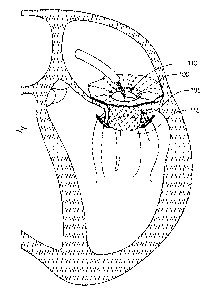
heart anatomy.
101201 Figures 7B and 7C generally illustrate embodiments of the
Valve Replacement as
disclosed herein. The Valve Replacement embodiments of Figures 7B and 7C may
include one or
more anchor features. In one embodiment, as shown in Figure 7B, the Valve
Replacement may
comprise an atrial sealing skirt 705, a frame body 710, and an anchor feature
715 that are covered
in a fabric for the purpose of flow sealing and/or encouraging (e.g.,
influencing either promoting
or inhibiting) tissue growth after implantation. The anchor feature 715 may be
strut or a
stabilization anchor 715, which may assist in preventing migration of the
Valve Replacement into
the atrium. In some examples, the stabilization anchor 715 or a portion
thereof may be configured
or designed to rest on a specific area of the annulus (e.g., the trigone
area). The embodiment may
further comprise an anchor feature 720 that in some embodiments may not be
covered in a fabric.
In some embodiments, that anchor feature 720 may be or include a clip 720 or a
hanger designed
or configured to hold native leaflets. Figure 7C shows a Valve Replacement
comprising anchor
features that may be posterior leaflet anchors (or struts) 725 and clips 720.
In other embodiments,
the clips and anchors are covered in fabric.
101211 Figure 7D generally illustrates an embodiment of the Valve
Replacement as disclosed
herein. Figure 7D shows an embodiment of the Valve Replacement comprising a
clip component
735 for the purpose of improving delivery control, via secure attachment of
the Valve Replacement
to a delivery catheter, and for the purpose of improving the efficiency and
efficacy of leaflet
19
CA 03232729 2024- 3- 21
WO 2023/059941
PCT/US2022/048304
attachment. Figure 7D shows a flat-pattern schematic of a wire frame with a
clip 735, wherein the
clip 735 may be a looped portion of the wire frame extending out from the main
body of the wire
frame. In some embodiments, a clip 735 may be positioned at two or more
separate locations
around the circumference of the Valve Replacement. In other embodiments, clips
735 may be
shape-set 180 such that they can provide for a hook shape to clip onto the
native valve leaflets.
For example, once the Valve Replacement is released from a delivery system,
the clips 735 may
attach onto the native valve leaflets, providing securement of the Valve
Replacement. In
embodiments, the clips envelope the native anterior and/or posterior leaflets
of the native heart
valve, mitral, or tricuspid for example. In embodiments, the clips wrap around
the native leaflets
to prevent the replacement heart valve from migrating into the atrium of the
native heart. In
embodiments, the clips are made of hypotube material comprising a hollow
tubing (hypotube)
material that has an inside diameter that mates with a wire of the helical
wrapped wire of the frame
of the Valve Replacement (including the frame body of the One-Piece, or of the
Adapter of the
Two-Piece systems).
101221 Various embodiments of the Valve Replacement may comprise
various quantities of
anchors at various angles and orientations. For example, one embodiment may
comprise six
anchors whereas another may comprise three anchors. In an embodiment
comprising three
anchors, applicable to the mitral valve, the Valve Replacement comprises an
approximately 150
angle between the medial and lateral anchor struts with the A2 anchor (or A2
clip) anchoring to
the anterior leaflet at or near the A2 region (as explained further with
regard to Fig. 8G) of the
anterior leaflet and the A2 anchor (or A2 clip) being symmetric between the
two anchor struts. In
another embodiment comprising three anchors, applicable to the tricuspid
valve, the Valve
Replacement comprises a uniform 1200/1200/1200 spacing of the anchors (or
clips). In another
embodiment comprising three anchors, the Valve Replacement comprises an
approximately 150
angle between the medial and lateral anchors with the P2 anchor (or P2 clip)
anchoring to the
posterior leaflet at or near the P2 region of the posterior leaflet and the P2
anchor (or P2 clip) being
symmetric between the two anchor struts. Other angles and geometries are
possible and within the
scope of this disclosure.
[0123] In other embodiments, for smaller hearts, the Valve
Replacement comprises an
approximate 150 angle between an upper medial and lateral anchor struts with
the A2 anchor (or
A2 clip) being approximately symmetric between the two upper anchor struts,
and an approximate
CA 03232729 2024- 3- 21
WO 2023/059941
PCT/US2022/048304
180 angle between a lower medial and lateral anchor struts with the P2 anchor
(or P2 clip) being
approximately symmetric between the two lower anchor struts. In other
embodiments, for larger
hearts, the Valve Replacement comprises an approximate 150 angle between an
upper medial and
lateral anchor struts with the A2 anchor (or A2 clip) being approximately
symmetric between the
two upper anchor struts, and an approximate 210 angle between a lower medial
and lateral anchor
struts with the P2 anchor (or P2 clip) being approximately symmetric between
the two lower
anchor struts.
101241 The anchors (which include clips) may be made of the same wire
as the braided frame
or different wire¨whether it be different in material and size. This provides
a novel aspect: The
ability to have thicker and/or more durable wire for the anchors allows for
the anchors¨which are
required to attach to the valve tissue and maintain the Valve Replacement in
place¨to be stronger
and/or firmer, without comprising the flexibility of the body frame. This
enables the Valve
Replacement to remain firmly and securely positioned within the heart valve
while still allowing
the Valve Replacement to move and function in accordance with the heart's
natural movements.
101251 Another novel aspect is the synchronization between the
flanges and the anchors. Once
implanted, the flanges provide a downward force on the heart tissue as the
anchors provide an
upward force. These two forces exerted by the Valve Replacement further secure
it in place without
compromising the fluidity of the braided body frame or the functionality of
the leaflets.
101261 Helical Braided Design
101271 The novel helical-braided designs of embodiments of the Valve
Replacement
purposefully leverage the natural helical movements of a beating human heart
so as to balance
both flexibility and strength. Studies of the human heart reveal that the
mechanisms of ejection
and suction are from a helical design of muscles in a "coil within a coil"
formation, which are
responsible for clockwise and counterclockwise rotation and functional
activity. More specifically,
the underlying anatomy of the human heart comprises a helical braid having a
transverse basal
loop of muscle for contraction that overlies an oblique helix that is
responsible for ejection and
suction within the heart.
101281 The disclosed braided helical design is configured to put less
stress on the individual
components of the Valve Replacement because the Valve Replacement moves with
the heart, i.e.,
the leaflets and anchors and other components have less stress and the Valve
Replacement migrates
less because its natural helical movement with the heart keeps it in place.
21
CA 03232729 2024- 3- 21
WO 2023/059941
PCT/US2022/048304
[0129] Figure 8A generally illustrates the helical functionality of
the human heart. As shown
in Figure 8A, the twisting and untwisting motions within the heart are created
by inner helical
spirals within the descending and ascending apical loop muscle segments, with
the heart having a
natural clockwise torsi on/contraction for ejection and a natural
counterclockwise
loosening/lengthening for suction. In heart disease, the natural helix of the
heart becomes
architecturally altered in shape.
[0130] Figure 8B generally illustrates an embodiment of the Valve
Replacement as disclosed
herein. As shown in Figure 8B, an embodiment of the Valve Replacement
comprises a helical
braided design that mimics and reinforces the normal helical and elliptical
formation of the heart
and its twisting/turning motions. In some embodiments, the helical braided
design may form a
wire frame. In some examples, the helical braided design may be braided so as
to allow the frame
to move in several (e.g., three) directions. Relatedly, the braided design may
in some examples
allow the frame to accommodate movement (e.g., from simultaneous compression
and twisting)
along the longitudinal axis and axis of rotation. In one embodiment, the
helical braided design
comprises a design wherein the braided wires resemble a frame that may move
and/or flex (e.g.,
symmetrically to a helical axis) as it is compressed and/or elongated around
an open center. In
some embodiments, the helical braided design may implement tensegrity and/or
floating
compression principles by, e.g., shape setting the wires and frame into
predetermined formations
(e.g., to allow wires to slide across each other in a non-rigid manner). For
example, since principles
may assist in decoupling movement in the axial and rotation directions such
that the device can
move in three dimensions to accommodate movement of longitudinal axis and
rotation while heart
is beating. By way of further example, such movement may be free in a
constrained range, which
range may be defined by the shape setting of the nitinol and the fabric sewn
onto the frame, to
permit movement of braided frame wires along one another at the over-under
braids within a
predetermined range of movement in one or more (or any) directions.
[0131] Both the One-Piece System and the Adapter and Valve Assembly
of the Two-Piece
System may comprise the helical braided design. A normal heart develops
ejection and suction as
a functional consequence of the contraction integrity of the apical ellipse.
The braided helical
design of the Valve Replacement maximizes shortening and lengthening of the
heart muscles,
thereby reinforcing the desired apical ellipse of a healthy heart movement.
22
CA 03232729 2024- 3- 21
WO 2023/059941
PCT/US2022/048304
101321 For example, as the human heart muscles compress and descend,
the braided helical
wires of the Valve Replacement¨rather than be stiff¨also compress and descend
with the heart
muscles, thereby reinforcing a natural spiral compression and descension of
the heart muscle
surrounding the braided wires. With the braided helical design, the Valve
Replacement conforms
to and reinforces the natural movement of the heart. The braided helical
design of the Valve
Replacement produces a twisting spiral coil that develops torsion in a
clockwise direction. And as
the human-heart muscles lengthen and fill, the braided helical design
reinforces a natural spiral
lengthening and filling of the braided wires with the surrounding heart
muscle, resulting in an
untwisting spiral coil within the adapter or valve that develops an ejection
force.
101331 The novel braided helical design is significant for treating
heart valves. By comprising
a braided helical design, embodiments of the Valve Replacement reinforce the
natural helical
movement of the heart, and more naturally adapts and sits within the desired
valve area. For
example, embodiments of the Valve Replacement will tend to remain in the
desired mitral or
tricuspid valve area because the braided helical design will move (contract,
twist and shorten, and
untwist and lengthen) with the natural movements of the heart. This allows for
the Valve
Replacement to self-correct and seat within the valve area in a natural state,
thus conforming to
the heart's natural movements and encouraging central vortex flow.
101341 The novel braided helical design thus facilitates a natural
heart movement. In one
embodiment, the Valve Replacement is held in place by the combined efforts of
the flange and
anchors, with the helical braided portion being in between the flange and
anchors. The helical
braided portion twists back and forth with the heart's natural movement,
enabling a pumping-and-
squeezing motion. The twisting motion, when the heart pumps, encourages flow
of liquid through
the Valve Replacement, thus allowing for better flow dynamics.
101351 Figures 8C¨F generally illustrate an embodiment of a Valve
Replacement 805 as
disclosed herein. The frame 810 of the Valve Replacement embodiment 805 may
incorporate
helical architecture using braided wire technology and fabrication. The frame
810 may utilize
overlapping helical strands that conform to the heart's natural movements and
encourage central
vortex flow, as described above with regard to Figure 8B. For example, the
Valve Replacement
805 may not only facilitate contraction-like movements but also twisting,
radial expansion, and
other movements replicating movement of the heart.
23
CA 03232729 2024- 3- 21
WO 2023/059941
PCT/US2022/048304
[0136] The frame 810 may be made from braided wire. The properties of
the frame, including
densities and characteristics of the heart's anatomy, braiding design, wire
thickness, etc., may
facilitate not only the movements described above, but also accurize
placement, maximize seal,
and prevent migration, especially in coordination with an integrated and
optimized anchoring
system (and described in further detail below). In some embodiments, the frame
810 may be made
from materials that include wires with determined thickness and geometry to
designed to increase
strength.
[0137] Figure 8G shows different regions of heart tissue. For
example, Figure 8G shows a
diagram of an aortic mitral curtain 815, an anterior leaflet 820, and a
posterior leaflet 825. The
anterior leaflet 820 may be slightly larger than the posterior leaflet 825, as
is typical for most
human hearts. The anterior and posterior leaflets 820, 825 may also be divided
into separate areas
labeled, e.g., A2 and P2.
[0138] Figures 9A and 9B generally illustrate an embodiment of a
Valve Replacement as
disclosed herein, with Figure 9B showing a vertical view of the embodiment
shown in Figure 9A.
The Valve Replacement embodiment of Figures 9A and 9B may include a flange
with a flatter
surface. The flatter surface may be configured to rest on an annulus in an
atrium area. In some
examples, the exterior surface area of the Valve Replacement embodiment where
the flange may
meet a tubular adapter frame may be referred to for present purposes as a
transition point, and may
be similar to a right angle. In some cases, such a right angle may not conform
to the surrounding
native tissue such that it may leave empty space sub between the tissue and
the Valve Replacement
embodiment surfaces.
[0139] Figures 9C and 9D generally illustrate an embodiment of a
Valve Replacement as
disclosed herein, with Figure 9C showing a vertical view of the embodiment
shown in Figure 9D.
The Valve Replacement embodiment of Figures 9C and 9D may include a flange
with a more
curved surface. For example, and more specifically, the transition point of
the Valve Replacement
embodiment of Figures 9C and 9D may also be more curved (in contrast to a
right angle), which
design may assist in the surfaces of the Valve Replacement embodiment to
expand into spaces
between the surfaces and the surrounding native tissue.
[0140] Material Covering
24
CA 03232729 2024- 3- 21
WO 2023/059941
PCT/US2022/048304
101411 In some embodiments, different materials are prepared prior to
assembling into a
continuous covering. In other embodiments, material may be added and receive a
modification
treatment post-assembly that is applied to only specific locations on the
Valve Replacement.
101421 In some Valve Replacement embodiments disclosed herein, tissue
attachment and
ingrowth may be promoted in an area that is desired to become anchored to the
tissue, while
cellular interaction can be limited to simple endothelialization or no
response at all, to allow
disturbance of part of the device at a later date without risk of tissue or
thrombus embolization.
Put simply, the varying material used may be either conducive or non-conducive
to chemical
bonding. For example, in a preferred embodiment, the materials in contact
between the inner
portion of the Adapter and the outer portion of the Valve Assembly do not
bond, so as to allow for
movement of both portions; whereas the material on the outside of the Adapter
bonds with human
tissue. Thus, depending on the location, materials may be used such that
cellular growth is inhibited
or promoted.
101431 Some Valve Replacement embodiments may be encased, either
completely or partially,
in a continuous material covering to elicit the type of physiological response
that is desired as well
as the mechanical behavior. Though the covering is continuous¨as in there are
no material gaps
at the transitions of physical features¨the materials may be modified locally
in areas of the device
to behave differently. For example, the material covering one side of the
flange may be deliberately
nonporous to facilitate sealing, while the material on the other side of the
flange may be a knit that
facilitates tissue ingrowth for anchoring. Alternatively, the flange could be
alternating rings of
nonporous and ingrowth material on both sides of the flange. These techniques
can be applied to
any surface of the device.
101441 Material differences range from being entirely different
materials¨natural tissue or
synthetic fabric¨to physical and chemical surface modification, to obtain the
desired mechanical
and biocompatible properties. These modifications can include but are not
limited to coating,
etching, mechanically biasing, ion infusion, various deposition techniques,
and
oxidizing/nitriding/carbiding. Modifications may be used in any combination to
achieve the
desired result.
[0145] Figures 10A and 10B generally illustrate embodiments of a
Valve Replacement as
disclosed herein. As shown in Figures 10A and 10B, an embodiment of the Valve
Replacement
may comprise a continuous piece of material around the outside of the frame. A
continuous seal
CA 03232729 2024- 3- 21
WO 2023/059941
PCT/US2022/048304
may be configured from the material (such as fabric) extending from an inflow
edge 1005 of the
Valve Replacement to the extrados 1010 of the body of the Valve Replacement. A
strip of ingrowth
fabric may be sewn around the inflow edge of the Valve Replacement, with a non-
porous coating
forming a continuous seal extending into the ventricle. Material can also be
configured to fill
spaces between the inner and outer fabric. For example, in an embodiment,
filler fabric is
selectively placed within the Valve Replacement to fill in spaces and gaps
between the materials
or areas where the frame and fabric have gaps.
101461 The continuous surface of the fabric may be locally influenced
and characterized for
modulating or even contradicting properties, such as coating with medical
polymer in locations
where no tissue attachment is desired, hydrogels where space-filling or latent
actions are desired,
or a hydrophilic tissue adhesive. The continuous material structure of the
fabric may be
voluminous in nature, filling space and adapting the round heart valve to the
asymmetrical shape
of the valve annulus. Combined with other attachment methods, an embodiment of
the mitral-
valve adapter fabricated with this method aids in engagement and attachment of
the leaflet tissue
and other sub-valvular structures. The partially porous fabric provides an
improved seal for a
replacement valve, enabling accommodation to irregular shaped anatomy through
the compliance
of the fabric. In embodiments, fabric is selectively treated by partial
dipping in a coating that
provides additional properties to the fabric, such as improved sealing.
101471 In other embodiments, the Valve Replacement may be fabricated
using a constraint to
hold the Valve Replacement at a specific dimension while attaching material to
influence device
performance. A fabrication technique is disclosed, which acts to influence the
disposition of a
braided wire frame¨removing the inherent freedom of movement and
unpredictability that is
present between relative members of the frame structure when in a load-free
state. This technique
involves restraining the radial expansion of the frame with a constraint, such
as feeding some
number of sutures through or around the structure to hold it at a specific
dimension other than its
unrestrained, -free" dimension. In subsequent fabrication steps, the structure
is incorporated into
an assembly that adopts this new configuration and considers this to be the
final dimension. When
the constraints are removed from the braided frame, this braided frame tries
to recover to its
original "free- dimension¨applying additional radial force to the surrounding
structure while
being constrained to the desired dimension.
26
CA 03232729 2024- 3- 21
WO 2023/059941
PCT/US2022/048304
[0148] The degree of radial force transmitted to the fabric material
from the frame can be
adjusted as required to achieve the optimal combination or performance
properties. In particular,
the strain energy density of the structure can be more uniform. A greater
stiffness is achieved
(resulting in a better seal) with less material, resulting in a more low-
profile structure. The suture
finally provides a biasing of the structure toward a desirable diameter and
height for the valve
structure.
[0149] To expand the concept further, structures that possess
features described herein may
be co-deployed singularly or with a connected design, so as to engage both the
mitral and the aortic
valve apparatus and/or annulus. The intent is to influence the leaflets of
both valves, as well as the
angulation of the valves relative to one another, to ensure the most effective
management of flow
through the ventricle and maximizing the efficiency of the outflow tract.
[0150] In some embodiments, the Valve Replacement is covered in a
material that wraps
around the frame in a continuous manner. Embodiments of the material are
fabric and animal
tissue. By using materials that can be locally modified to change
characteristics such as porosity
and surface roughness, a certain level of control over cellular interaction on
the various parts of
the device can be achieved. In other embodiments, the Adapter body and atrial
flange may be
covered in fabric for the purpose of flow sealing and/or influencing (e.g.,
either promoting or
inhibiting) tissue growth after implantation.
[0151] The material used further assists with the loading and
deployment of the Valve
Replacement. For example, the material may promote the Valve Replacement to
function as a re-
valve system, wherein a tubular braided fabric tube (coated with a polymer to
decrease porosity to
blood) surrounds the frame and constrains the diameter. This tube is sewn onto
the frame,
sometimes in conjunction with a leaflet panel, so that the strings can be
removed and what remains
is a pretensi on e d frame constrained by the fabric. In embodiments, an el
astom er-coated, tubular
knit, shape-set fabric is attached to the braided frame. In embodiments, dip-
coated braided frames
are utilized, such as a frame dipped in urethane, either as a whole device or
partial. In other
embodiments, treated panels are sewn onto the braded frames. In other
embodiments, sections of
treated fabric are cut into panels that are configured to be able to be sewn
onto a at least partially
circular surface, such as a flange.
101521 Sewing Methods and Belt Loops
27
CA 03232729 2024- 3- 21
WO 2023/059941
PCT/US2022/048304
101531 Figures 11A and 11B generally illustrate embodiments of the
Valve Replacement as
disclosed herein. More specifically, Figures 11A and 11B show various
embodiments of the
fabrication of material for the Valve Replacement, focused on the Two-Piece
System, and is also
consistent with a One-Piece System as well. In embodiments, material for the
various parts may
be made of the same or different materials, respectively. Such materials may
include, for example,
a textured or hard-knitted style fabric or textiles, or bioresorbable mesh
(e.g., made from
polymers), elastomeric scaffolds, or other materials. Other materials with
similar or different
properties may be used, such as permeable or impermeable materials, with
various degrees of
elasticity, and stretchable in predetermined directions or any direction, and
biocompatible
materials.
101541 Figure 11A shows a flange outer material 1105, a flange rim
material 1110, an outer
adapter cuff material 1115, and an inner adapter cuff material 1120 of the
Valve Replacement. The
flange outer material 1105 may form a circular top piece, wherein it may
further have a coating to
reduce fluid permeability. The flange rim material 1110 may form a circular
bottom piece that
stretches to allow expansion of the Valve Replacement when deployed. The outer
adapter cuff
material 1115 may constitute a cross-stich that attaches the ends to create
the tube-like shape. The
inner adapter cuff material 1120 may comprise a running or cross-stitch that
attaches the ends to
create a tube shape, wherein the fabric of the inner adapter cuff material
1120 may have limited
elasticity and either stitch maintains the integrity of the inner adapter cuff
material 1120.
101551 In one embodiment, the flange rim material 1110 and the outer
adapter_suff material
1115 may be combined, either by cross-stitching or other method know to one
skilled in the art, to
form a secant bottom piece. The flange may be stitched furthest away from any
anchor slots. The
formed secant bottom piece may be positioned onto the bottom of the Adapter or
the One-Piece
System, wherein the shape-set thread around the body of the frame is cut and
wrap-stitch is used
to close or secure the anchor slots.
101561 In another embodiment, the inner adapter cuff material 1120
may be attached to the
inner opening of the flange outer material 1105 to form a top piece of
material. Thetop piece or
material may be slid over the top of the Adapter or the One-Piece System after
which the fabric is
wrap-stitched closest to the frame.
101571 In another embodiment, the bottom piece of material and the
top piece of material may
be connected, such as by wrap-stitching at the bottom of the frame to connect
the top piece of
28
CA 03232729 2024- 3- 21
WO 2023/059941
PCT/US2022/048304
material to the bottom piece of material. For this, the fabric of the flange
outer material 1105 and
the flange rim material 1110 may be smoothed and held in place (such as with
sewing clips) and
connected around the wire flange (such as with a running stitch), wherein the
border of the flange
may be circular and not rigid. The excess fabric may be trimmed, and
additional stitching may be
added around the flange and each wire flange tip. Additional stitching may be
performed along the
wires to secure the fabrics together and keep flush against the wire flange.
The stitching may be
done to the second crossing of wires, followed to the next wire, and then up
towards the tip of the
wire flange. This process may be continued around the flange and repeated on
the next set of wires.
101581 One embodiment of the outer adapter cuff material 1115 may be
folded in half and
secured together (such as with a sewing clip), after which a blanket stitch
may be performed around
the edges. The blanket stitch allows the outer adapter cuff material 1115 to
retain its shape without
sinching the fabric. The outer adapter cuff material 1115 may be turned inside
out and placed over
the anchor wire, after which the outer adapter cuff material 1115 may be
secured to the front and
back of the anchor wire (such as with a running stitch). In this, the seam of
the stitch (used to close
the anchor slots) may be caught between the front and back fabric of the outer
adapter cuff material
1115 to secure it to the Adapter or One-Piece System. The running stitch may
encompass the front
of the outer adapter cuff material 1115, the seam, and the back of the outer
adapter cuff material
1115 along the base of the anchor wire. Once the outer adapter cuff material
1115 is secured at the
base, a stitch may be continued along the anchor wire to keep the outer
adapter cuff material 1115
from slipping or sliding on the anchor. Fabric may be slightly caught, wherein
it is not loose
enough to leave excess fabric but not tight enough to affect the shape of the
wire.
101591 In embodiments, stitching of the fabric to the Replacement
Valve helical braided wire
architecture performs several functions, including the following: sewing of
fabric to the braided
wire architecture helps constrain the braided wire of the Replacement Valve
from migrating into
the ventricle or atrium and helps prevent paravalvular leaks, while at the
same time permitting
slight movements along the unfixed nodes at the over-under braids to allow the
Replacement Valve
to move with the natural helical movements of the heart.
101601 Figure 11B shows an inner valve cuff material 1125 and an
outer adapter cuff material
1130. In one embodiment, leaflets may be connected to the inner valve cuff
material 1125, such
as along a strip of fabric with a double running stitch along the belly of the
leaflet, wherein the
stitches are uniform across the leaflets to allow for proper valve opening and
closing. The leaflet
29
CA 03232729 2024- 3- 21
WO 2023/059941
PCT/US2022/048304
or commissure tabs may be exposed, such as by laser-cutting with slots at the
top of the inner valve
cuff material 1125 (wherein a strip of the inner valve cuff material 1125 may
be folded in half,
making sure that leaflets are aligned on top of each other; and wherein the
ends of the inner valve
cuff material 1125 may be attached to the junction of the belly and tabs with
a double running
stitch). Following the exposure of the leaflet tabs, the inner valve cuff
material 1125 may be placed
inside the Valve Assembly frame, the tabs may be pulled through commissure
wires and laid flat
between the leaflets and the inner valve cuff material 1125.
[0161] In one embodiment, the ends of the outer valve cuff material
1130 may be connected
and the outer valve cuff material 1130 slid over the outside of the Valve
Assembly frame.
[0162] In another embodiment, the inner valve cuff material 1125 and
the outer valve cuff
material 1130 may be connected together. After placing the inner valve cuff
material 1125 on the
inside of the Valve Assembly frame and the outer valve cuff material 1130 on
the outside of the
Valve Assembly frame, the two parts may be connected to the bottom of the
frame (such as by
tacking down both parts with a square knot) directly below commissure wires,
and the parts may
be stitched along the bottom of the frame. Following a commissure attachment,
which is set forth
in the following paragraph, both portions may be combined by sewing through
the frame and the
top of the Valve Assembly frame may be stitched. Additional steps may
comprise, along the upper
perimeter of the valve, stitching around the wires travelling from the
commissures downwards and
away from the peaks so as to create a z-shaped pattern. In this, the stitches
may connect the inner
fabric behind the leaflet and the outer valve cuff material 1130.
[0163] In an embodiment of a commissure attachment, leaflet tabs are
fed through the
commissures, wherein each tab folds towards its own leaflet and is wrapped
around the
commissure wires. The ends of the tabs may be held together against the entry
of the tabs and
secured together, such as with running stitches vertically and on the inside
of the Valve Assembly.
Stitching may continue in front and around the commissure, such as for 3-4
times, and entering
and exiting at the location of the running stitch. Stitches may be
perpendicular, comprising of
embodiments such as a running stitch along the y-axis and a wrap-stich along
the x-axis.
101641 In other embodiments of the fabrication of material for the
Valve Replacement wherein
the focus is on the One-Piece System, a fabric for the various portions may
comprise a stretchy
and semi-transparent fabric; wherein the outer adapter cuff material 1115 and
flange rim material
1110 may be sewn together to create the outer piece. Cross-stitch may be used
to connect the edges
CA 03232729 2024- 3- 21
WO 2023/059941
PCT/US2022/048304
of the outer adapter cuff material 1115 and to connect the outer adapter cuff
material 1115 to the
flange rim material 1110.
[0165] In a separate embodiment, a fabric for the portions comprises
an inflexible and opaquer
fabric where the coating is visible; wherein the inner adapter cuff material
1120 and flange outer
material 1105 may be sewn together to create the inner piece. A running stitch
may be used to
connect the edges of the tubular inner adapter cuff material 1120 and a cross-
stitch is used to
connect the inner adapter cuff material 1120 to the flange outer material
1105.
[0166] For these embodiments focused on the One-Piece System, an
inner valve cuff material
may be created, wherein leaflets are attached to the inner adapter cuff
material 1120 and wherein
leaflet tabs are placed through slots of the inner adapter cuff material 1120
where the junction of
the inner adapter cuff material 1120 and tabs meet, with the leaflets held in
place, such as with
sewing clips. Using a double running stitch, the belly's edge of each leaflet
is sewed to the inner
adapter cuff material 1120. The tabs and top edge of the leaflet(s) are
flushed and level with one
another and the running stitch on each belly of the leaflet(s) is level and
uniform. (Inconsistent
stitches can lead to a defective valve.) After the leaflets are attached to
the inner adapter cuff
material 1120, the inner adapter cuff material 1120 is folded in half, keeping
leaflets level and held
in place. A double running stitch may then be sewn directly down from the
junction of the leaflet
tabs, continuing away from the leaflets with a running stitch back up towards
the junction of the
leaflet tabs. (The running stitch should be away from the leaflet belly.)
Following these steps, the
inner valve cuff material should create a tube.
[0167] A first set of materials may be created by connecting the
inner valve cuff material from
above to the flange outer material 1105, such as with a cross-stitch, wherein
leaflets are away from
the seam.
[0168] Once the first set of materials is created, it may be
connected to a second set (e.g., the
flange rim material 1110 and outer adapter cuff 1115 previously sewn together)
by using a running
stitch through the frame (between the double running stitch of the leaflet
belly) and following the
belly stitch of the leaflet(s) to secure both sets together. The tabs are then
secured through the
commissures and wrap-stich is used to connect the second set to the first set
at the base of the
frame. The deployment apertures are created, by cutting the fabric, before
finishing the wrap-stich.
Once connected, a beta-stitch is incorporated on the flange and anchors' cuff
placements.
31
CA 03232729 2024- 3- 21
WO 2023/059941
PCT/US2022/048304
101691 Figures 12A-12E generally illustrate embodiments of the Valve
Replacement as
disclosed herein. More specifically, Figures 12A-12E illustrate deployment
belt loops commonly
used for the Two-Piece System. The deployment belt loops may comprise one or
more sets of
loops.
101701 In a preferred embodiment, the Adapter comprises seven belt
loops at the base of the
body of the Adapter, wherein there are belt loops on either side of three
anchors and one belt loop
at a vertical seam. The Adapter further comprises five belt loops at the
horizontal seam between
the body and the flange, wherein the five belt loops are located at crosswires
on the same plane as
one another. Hidden belt loops may be found behind the long anchors (P1 /P3
anchors). The
Adapter also comprises four belt loops along the flange, located at the
crossed wires to the left and
right of the long anchors. Additionally, the Adapter comprises two belt loops
at the tips of the long
anchors. Though in a preferred embodiment the anchor-tip belt loops comprise
three loops and all
other belt loops comprise two loops, it should be known that the belt loops
are not limited to a
specific number of loops.
101711 Figure 12A shows the belt loops in relation to the P1 and P3
anchors. The Adapter
comprises one or more belt loops 1.205 behind (i.e., on the body and hidden
from view unless the
anchor is lifted up) the P1 and/or P3 anchor 1220 and along the horizontal
seam 1210. And the
Adapter comprises one or more belt loops 1215 to the left and right of the P1
and/or P3 anchor
1220.
101721 Figure 12B shows the belt loops in between the P1 1235 and P2
anchors 1240. Belt
loops 1225 along the horizontal seam 1210 are at the same plane. The Adapter
further comprises
belt loops 1230 to the side of the P1 anchor 1235 and the P2 anchor 1240,
wherein the belt loops
1230 are also approximately at the same plane.
101731 Figure 12C shows the belt loops in between the P1 1235 and P3
anchors 1260. One
belt loop 1245 is at the junction of the horizontal 1210 and vertical seam
1250 and one belt loop
1255 is at the bottom of the vertical seam 1250.
101741 Figure 12D shows the belt loops in between the P2 1240 and P3
anchors 1260. The
belt loops 1265 along the horizontal seam 1210 are at the same plane. Because
the P3 anchor 1260
is slightly higher, the belt loop 1270 near the P3 anchor 1260 is adjusted to
a height approximate
to the P3 anchor 1260. The belt loop 1230 is in a position in relation to the
location of the P2
32
CA 03232729 2024- 3- 21
WO 2023/059941
PCT/US2022/048304
anchor 1240. In a preferred embodiment, the anchors are biased towards the
flange. In other
embodiments, the anchors may be perpendicular to the body.
[0175] Figure 12E shows anchor loops in relation to the P1 and/or P3
anchors. In one
embodiment, the anchor loops comprise double loops 1275 positioned at the
crossed wires on the
flange to the left and right of the P1 and/or P3 anchor 1220, and the anchor
loops further comprise
a triple loop 1280 at the tip of the P1 and/or P3 anchor 1220.
[0176] Figures 13A-13D generally illustrate embodiments of a Valve
Replacement as
disclosed herein. Figures 13A-13D illustrate deployment belt loops commonly
used for the One-
Piece System. The deployment belt loops may comprise one or more sets of
loops.
[0177] In a preferred embodiment, the One-Piece System comprises
seven belt loops, wherein
the belt loops on the adapter body of the One-Piece System are double loops
and the belt loops
located on a horizontal seam are at the crossed wires along the same plane.
[0178] Figure 13A shows belt loops in between the P1 1305 and P3
anchors 1310. Three belt
loops 1315 are located along the horizontal seam 1320 with one belt loop
located at the junction
of where the horizontal 1320 and vertical seams 1325 meet, and located in line
with a commissure
wire. The two other belt loops are located just outside of the P1 1305 and P3
anchors 1310.
[0179] Figure 13B shows belt loops in between the P2 anchor 1330 and
either the P1 1305 or
P3 anchor 1310. Two loops 1335 are located along the horizontal seam and
secured at the same
plane of the crossed wires outside the P2 anchor 1330 and the long anchor
(P1/P3 anchor).
[0180] Figure 13C shows the belt loops 1340 of the long anchor,
wherein the belt loops 1340
are located on either side of the long anchor.
[0181] Figure 13D shows the belt loops 1345 of the P2 anchor 1330,
wherein the belt loops
1345 are located on either side of the P2 anchor 1330.
[0182] Figures 13E and 13F show an embodiment of the location of
deployment apertures,
wherein the deployment apertures 1350 are located directly below each long
anchor and are located
on either side 1355 of the P2 anchor 1330.
[0183] Figures 13G and 13H show another embodiment of the location of
deployment
apertures 1375. In this embodiment, fabric is removed (e.g., in a triangular
shape) two peaks away
1360 from the commissures (or commissure posts) 1365 so as to expose the wire
peak. A wrap-
stich 1370 is placed around the perimeter of the frame so as to catch wires
around the exposed
peaks.
33
CA 03232729 2024- 3- 21
WO 2023/059941
PCT/US2022/048304
[0184] Engagement Structures
[0185] Figure 14A generally illustrates an embodiment of the Valve
Replacement as
disclosed herein. In an embodiment, as shown in Figure 14A, the Adapter or the
One-Piece System
comprises a body 1405 and an atrial sealing skirt 1410 (which in some aspects
may be a flange).
The Adapter body 1405 and sealing skirt 1410 may be constructed of varying
material and vary in
dimensions. For example, the Adapter body 1405 and sealing skirt 1410 may be
made up of a wire
braid of one or more wires with different diameters. The wire may be made of
material such as
nitinol and be designed to be compressed to a small diameter¨such as 4mm to
6mm¨to be
delivered in a catheter. When released, the Adapter body 1405 and sealing
skirt 1410 may expand
in size (i.e., the body expanding to 25mm or greater in diameter and the
sealing skirt expanding
anywhere from 40mm to 70mm in diameter).
[0186] The exterior surface of the Adapter body 1405 may also be
covered with a multitude of
small, short barbs 1415. The barbs 1415 may be used to engage the leaflet or
annulus of a
malfunctioning cardiac valve, such as a mitral valve. The barbs 1415 may be
made up of basic,
short wires and/or may also have an extra barb-component, like a fishhook
barb, to fixedly retain
the annular tissue.
[0187] The Adapter body 1405 may also have one or more hooks 1420 or
1425 (more or less
in number than the barbs 1415) varying in size, that can hook under the native
valve tissue. These
larger hooks may or may not have fishhook barbs. The larger hooks may have a
spring-like
function that engages with the native valve tissue and prevents it from
moving.
[0188] In a preferred embodiment, the sealing skirt 1410 may be
connected to a catheter,
wherein the Adapter Attachment is sequentially released from the catheter once
the Adapter body
1405 is released and engaged with annulus tissue. The sealing skirt 1410 may
be designed to flex
downward, toward, or even past the plane defining the joint between the
Adapter body 1405 and
the sealing skirt 1410¨so as to be radially overlapping with the Adapter body
1405. The multitude
of barbs 1415 on the Adapter body 1405 would work together to ensure the
Adapter body 1405 is
strongly engaged in the native annulus and resists the downward pressure of
the sealing skirt 1410,
such that the sealing skirt 1410 would create a strong seal against the atrial
tissue surrounding the
native valve annulus.
101891 Figures 14B-14E generally illustrate embodiments of the Valve
Replacement as
disclosed herein. In these figures, the sealing skirt is not shown for ease of
illustration. As shown
34
CA 03232729 2024- 3- 21
WO 2023/059941
PCT/US2022/048304
in Figure 14B, the Adapter body 1405 is designed with a braid of varying weave
densities and/or
wire diameters, and/or combined with releasable mechanisms such that the
Adapter body initially
has a round cross-section. The Adapter body 1405 has barbs 1415 designed to
engage a native
valve leaflet 1450. Once the barbs 1410 are engaged, the anchoring/attaching
functi onaliti es of the
Adapter Attachment cause it to conform to a "D-shape" or other asymmetrical
shape ___ keeping the
Adapter body 1405 cylindrical or otherwise specifically shaped to receive the
valve structure. This
accommodation and conformity is achieved via the different weave, wire
diameters, or mechanism
enabling such. As shown in Figure 14C, the change in shape creates a sharper
curve radius to make
the D-shape. The change from a circular cross-section to a D-shape cross-
section may pull the
leaflet, which can be useful, for example, in a mitral valve where an implant
such as the Adapter
body may cause outflow tract obstruction. Figures 14D and 14E disclose an
oblique view of the
structure and mechanism corresponding with Figures 14B and 14C. Embodiments
disclosed in
Figures 14B-14E may also comprise the sealing skirt and other features
described in previous
drawings. Figures 15A and 15B generally illustrate embodiments of the Valve
Replacement as
disclosed herein. Figure 15A discloses an embodiment of the Valve Replacement
implanted in a
malfunctioning mitral valve, with the body 1505 deployed in the mitral valve
and the sealing skirt
1510 deployed against the floor of the left atrium. In this embodiment, the
Adapter body 1505 is
oriented at a slight angle (i.e., from 10-30 degrees relative to the plane of
the skirt), such that when
deployed, the Adapter body 1505 is biased towards the posterior leaflet 1515.
101901
Deployment as disclosed in Figure 15A ensures good engagement of barbs
into the
posterior leaflet but not necessarily the anterior leaflet. The system may be
designed to normally
be in this geometric condition but be mechanically expandable by design so
that it can expand to
engage the anterior leaflet, then released back to the normal position after
the barbs and/or hooks
engage the anterior leaflet. This forces the anterior leaflet towards the
posterior leaflet and away
from the left ventricular outflow tract obstruction (LVOT) ensuring it is not
obstructed post-
procedure. Also shown are the delivery catheter 1520 and guidewire 1525.
101911
In another embodiment, the body of the Valve Replacement may be used to
engage the
leaflets with the barbs, wherein the body expands to a diameter larger than
the diameter at
deployment to ensure engagement with the leaflets. As the device is further
deployed, the diameter
of the engaged portion reduces to a final configuration¨symmetrical or
asymmetrical¨thereby
pulling the leaflets towards the device and away from the LVOT.
CA 03232729 2024- 3- 21
WO 2023/059941
PCT/US2022/048304
[0192] Figure 15B generally illustrates an embodiment of an Adapter
attachment as disclosed
herein. Figure 15B discloses a final configuration of an Adapter in its
original position after
release, wherein the anterior leaflet is drawn and held towards the posterior
leaflet, ensuring no
obstruction of the LVO T.
[0193] Figures 16A and 16B generally illustrate embodiments of the
Valve Replacement as
disclosed herein. Figure 16A shows a top view of a Valve Replacement, and
Figure 16B shows a
bottom view of the Valve Replacement. As shown in Figures 16A and 16B, the
inflow end of the
Valve Replacement (shown in Figure 16A, while an outflow end is the end
opposite the inflow
end) may comprise anchor-retracting chords coming through the fl ow portion
and anchoring to the
underside of a flange. These sutures permit control of the anchors by pulling
and releasing the
chords. Alternatively, the sutures may be releasably attached to a delivery
system to provide
similar manipulation of the anchors. Figure 16B further discloses the chords
attached to the
anchors. Figures 16C-16D show attachment configurations for a collapsible
flange.
[0194] Figures 17A-17D generally illustrate embodiments of the Valve
Replacement as
disclosed herein. Figures 17A-17D show the attachment configurations for
collapsible anchors
and clips, and further disclose a close-up view of a suture pattern that is
used to collapse and
control the anchors from all angles of the Valve Replacement. In these
embodiments, a delivery
component, such as one comprising one or more suture lines, is connected on a
first end to the
engagement attachment, wherein the one or more suture lines connects on a
second end to a
controlling mechanism.
[0195] Valve Assembly
[0196] Figures 18A and 18B generally illustrate embodiments of the
Valve Replacement as
disclosed herein. As shown in Figures 18A and 18B, an embodiment of the Valve
Replacement
may be fabricated using a constraint to hold an Adapter frame at a specific
dimension while
attaching material to influence device performance. A fabrication technique is
disclosed, which
acts to influence the disposition of a braided wire frame¨removing the
inherent freedom of
movement and unpredictability that is present between relative members of the
frame structure
when in a load-free state. This technique involves restraining the radial
expansion of the frame
with a constraint, such as feeding some number of sutures through or around
the structure to hold
it at a specific dimension other than its unrestrained, "free" dimension. In
subsequent fabrication
steps, the structure is incorporated into an assembly that adopts this new
configuration and
36
CA 03232729 2024- 3- 21
WO 2023/059941
PCT/US2022/048304
considers this to be the final dimension. When the constraints are removed
from the braided frame,
this braided frame tries to recover to its original "free" dimension¨applying
additional radial force
to the surrounding structure while being constrained to the desired dimension.
[0197] The degree of radial force transmitted to the fabric material
from the frame can be
adjusted as required to achieve the optimal combination or performance
properties. In particular,
the strain energy density of the structure can be more uniform. A greater
stiffness is achieved
(resulting in a better seal) with less material, resulting in a more low-
profile structure. The suture
finally provides a biasing of the structure toward a desirable diameter and
height for the valve
structure.
101981 Figures 19A and 19B generally illustrate embodiments of the
Valve Replacement as
disclosed herein. Figure 19A generally illustrates an embodiment of the Valve
Assembly wherein
leaflets 1905 are assembled to each other and/or to the frame by sewing. The
leaflets are joined at
commissure seams 1910 and then sewn, welded, or otherwise attached to the
commissure posts
1915 (located at an outflow end, while the inflow end is opposite the outflow
end) as well as to
other points on the frame, such as wires or wire intersections, or to
materials attached to the frame,
for example the leaflets being attached to a cuff, which cuff is then attached
to the frame. Once the
assembly is complete, the leaflets work in concert to close on the outflow
(distal side when being
implanted into a mitral valve) when fluid pressure is increased distally, so
that the leaflets close or
coapt in a Y-pattern 1920.
101991 As shown in Figure 19B, a Valve Assembly 1930 comprises a
braided frame having a
cuff covering 1935 on the outside. This cuff over the complete outer frame may
serve as an
extended sealing zone. A belly stitch 1940 may be sewn to the frame whereas a
bellows stitch 1945
is not sewn to the frame. In this embodiment, the distal leaflet ends 1950 are
shown coapting so as
to close the valve in a loose Y-shape. In some embodiments, the valve coapt
area may comprise
some "looseness" so as to ensure sufficient and effective contact among all
three leaflets and
ensure complete closing of the valve. Leaflets may be constructed of tissue
such as porcine
pericardium or other materials known to the art. In some cases, valves or
parts of valves excised
from animals may be sewn into the disclosed frame structure.
[02001 Figure 20 generally illustrates an embodiment of the Valve
Replacement as disclosed
herein. Figure 20 shows a bottom perspective view of the One-Piece System
comprising a braided
wire frame making up a flange 2005, an adapter body 2010, commissure posts
2015 compatible
37
CA 03232729 2024- 3- 21
WO 2023/059941
PCT/US2022/048304
with leaflets. In some embodiments, commissure posts 2015 may extend out from
the Valve
Replacement and the braided wire frame thereof, in some examples, the
commissure posts 2015
may be configured to connect to the non-native leaflets (not shown in Fig. 20,
but shown without
reference numbers in Figs. 21A¨R). For example, the non-native leaflets may be
cut and folded
and stitched to the commissure posts 2015 and may in some embodiments extend
or jut out
somewhat beyond the plane of the Valve Replacement and/or the braided wire
frame thereof.
[0201] Figures 21A¨I generally illustrate an embodiment of the Valve
Replacement 2100 as
disclosed herein. The Valve Replacement embodiment 2100 of Figures 21A-1 may
show a system
embodiment of and be used to perform method embodiments relating to TMVR.
[0202] Some solutions to TMVR described herein may utilize the Valve
Replacement
embodiment 2100 and involve several types of securement. For example, the
Valve Replacement
embodiment 2100 may feature one or more types of securement, and in some
embodiments more
than two securement mechanisms For examples, some Valve Replacement 2100
embodiments
may utilize four-point securement. In some embodiments, such multipoint
securement may be
configured to distribute an implant's workload across an entire device by
utilizing a combination
of securement mechanisms. In contrast, some prior art devices may only utilize
(or primarily use)
the radial force (as described in more detail below)--essentially relying on
"brut radial force"--for
their positioning, securement, and sealing.
[0203] For example, one type is supra-annular securement. This may be
accomplished in some
embodiments through a flange being placed, clamped, or cinched onto the
annulus or to ledges
(e.g., mitral ledges) above the annulus. In embodiments, the flange may
prevent migration into the
ventricle and may be configured and/or designed to eliminate paravalvular
leaks. Another type is
sub-annular securement. Utilizing anchor features, which may be struts 2255,
2260 in some
embodiments (and as described in more detail below), such securement may
provide stability in
the medial and lateral positions directly under the anatomy of the native
heart valve in the sub-
annular region. For example, in embodiments, the sub-annular anchors perform
as struts or braces
that permit slight movements of the Valve Replacement with the natural helical
movement of the
native heart, but constrains movement of the Valve Replacement, for example in
medial and lateral
positions or directions or in anterior and posterior directions, so that the
Valve Replacement does
not migrate into the atrium of the native heart or have paravalvular leaks.
38
CA 03232729 2024- 3- 21
WO 2023/059941
PCT/US2022/048304
102041 Such sub-annular securement may also involve, or relate to,
other anchor features,
which may be clips for securing to native tissue, including leaflet
securement. Some embodiments
may thus utilize leaflet clips 2105, 2110. In some embodiments, the struts
2255, 2260 may be more
elongated than the clips 2105, 2110, and jut outward farther from the device
than the clips, with
strut tips designed and/or configured to press against and jut into native
anatomy (e.g., the trigone
area). On the other hand, in some embodiments the clips might have a more
curved shape, e.g.,
curving upward and bending inward toward the native leaflets, or otherwise
similarly configured
hold leaflets in place.
102051 In some embodiments, the anchor features for securing native
leaflets may be hangers,
which may hang onto or capture the leaflets, rather than clips 2105, 2110 to
clip onto the native
leaflets. Such securement may prevent migration into the atrium and control
movement of native
leaflets. In some embodiments, and as described elsewhere herein, the
aforementioned leaflet clips
2105, 2110 may be deployed before deployment of the struts 2255, 2260. Such
sub-annular
securement, for example, may be sufficiently restrictive enough to prevent the
device embodiment
2100 from migrating into mitral area or creating leaks, yet loose enough to
move with natural
movement of the heart. Such movement, and in accordance with other aspects of
this disclosure,
may be facilitated by overlapping wire structure without many rigid fixed
points. In embodiments,
native leaflets may be captured by leaflet anchors or engagement attachment
(e.g., clips), which in
embodiments may hang on the native leaflets (rather than pinch the native
leaflets) to prevent
migration towards the native atrium, and anchor struts, which in embodiments
transfer
compressive loads to the native annulus or native anatomy near the native
annulus in the inter-
commissural zones below the annulus in the native ventricle area, to prevent
migration into the
native atrium.
102061 With regard to leaflet securement, in some examples, different
points in the native
tissues may be associated with securement features. For instance, in some
examples, the Valve
Replacement embodiment 2100 may integrate four points for anchoring, sealing,
and fixation. In
the embodiment shown, two general points or regions of native leaflet tissue
may be associated
with two leaflet anchors or engagement attachment (e.g., clips) 2105, 2110 and
two general points
or regions of native tissue may be associated with two anchors or struts 2115,
2120. These multiple
points of securement may provide stability while preserving the LVOT,
preventing paravalvular
leaks, and trauma to tissue associated with the native heart wall.
39
CA 03232729 2024- 3- 21
WO 2023/059941
PCT/US2022/048304
102071 Such multi-point securement using the Valve Replacement
embodiment 2100 may
result in a highly flexible and conformable valving system. The system may
encourage structural
stability by preserving central flow through the valve.
102081 Another type of securement is selective radial force
securement. In some embodiments,
directional radial force securement may be controlled through a receiver or
adapter, which may
assist in preventing migration while preserving the LVOT. Such radial force,
in some examples,
may be generated by oversizing a valve frame 2125 (e.g., a wrapped or braided
nitinol wire frame
around a mandrel) with respect to the annulus hole, pushing against the
annulus walls. In some
embodiments, the valve frame 2125 may be shape-set to about a 10% oversize in
relation to the
annulus hole, while permitting some limited movement. Such radial force
securement may also
prevent migration into the atrium and ventricle while preserving LVOT.
102091 In addition, as explained elsewhere, the natural helical
structures of features associated
with valve frame 2125 embodiment may be based on the more holistic
understanding of the nature
of the heart and its motions. For example, such features may be configured to
mimic the movement
of a healthy heart using natural helical structures (as described above), by
contracting and twisting
with each beat of the heart.
102101 Such features may include braided wire designs (as explained
and shown elsewhere in
more detail), which in some embodiments may offer increased flexibility and
conformability. In
addition to such braided wire design embodiments adapting to and moving with
the heart, they
may be configured to (by, e.g., integrating diverse and various wire
thicknesses and braiding
designs) forgive anatomical anomalies, and conform with various densities and
characteristics (i.e.,
radial force and expansion) of the heart's anatomy.
102111 Further, braided wire design embodiments may leverage nitinol
strength through
geometry and a unique braided wire architecture. Such architecture in some
embodiments may
purposely omit fixed nodes at crossing points of wires and permit the braided
wires to move across
one another in a controlled fashion. Such features may assist the replacement
valve in moving with
the natural helical movement of the heart. Some braided wire design
embodiments may also
facilitate placement in the native heart, maximize seal in the human anatomy,
and prevent
unwanted migration with an integrated and optimized novel securement system.
Thus, such
features may be designed based on recognition that the mitral valve is more
than simply a structure
to be "stented."
CA 03232729 2024- 3- 21
WO 2023/059941
PCT/US2022/048304
[0212] In contrast, some earlier valve frame designs by others (e.g.,
centering on laser cut
nitinol on a lattice) may not feature such helical architecture or otherwise
be configured for
allowing similar dynamic movement. Rather, such prior art designs may often be
limited to fixed
nodes across a lattice design, or may not permit use of various wire
thicknesses, thereby impeding
the requisite flexibility and conformability for the human heart.
[0213] These different ways of securement (as mentioned above) may
assist in distributing the
Valve Replacement embodiment's 2100 (e.g., the implant's) workload across the
entire device
2100, and may provide for maximum stability while preserving the LVOT and
preventing
paravalvular leaks and trauma to the native heart wall. In addition, such
multiple ways of
securement and multi-point anchoring systems may enable methods (as also
described herein)
allowing a simpler and more secure approach to Transcatheter Mitral Valve
replacement and
resulting in a safer overall procedure for patients.
[0214] Figures 21J¨M generally illustrate an embodiment of the Valve
Replacement 2100a as
disclosed herein. Similar to the Valve Replacement embodiment 2100 shown in
Figures 21A-I the
Valve Replacement 2100a may have two leaflet clips (such as leaflet clips
2105, 2110) and two
anchors (such as anchors 2115, 2120) for securement to native heart tissue.
Unlike the flange
embodiment shown in Figures 21A¨I, which may feature more of a flatter surface
and more of a
right angle at its transition point or zone, flange embodiments shown in
Figures 21J¨S may have
more of a curved or funnel shape, where the transition point or zone
corresponding to the annulus
may also continue further down into mitral valve, where it may flex and push
outward to fill in
gaps between the valve surfaces and the native anatomy. In some examples Valve
Replacement
embodiments, the transition zone may be between the flat flange portion (which
may be configured
to rest on top of annulus in the atrium) together with and including the
funnel-like contoured ring
of the flange and the adapter portion that radially pushes out into the
annulus. Such a contoured
transition portion may assist in promoting central vortex flow and provides
improved sealing and
anchoring. The funnel-like portion may have shape-set braided wire and
material covering that is
contoured and configured to fill in the gaps between the native anatomy and
the transition zone
between the atrium and annulus heading towards the ventricle area.
[0215] Figures 21N¨R generally illustrate an embodiment of a Valve
Replacement 2100b as
disclosed herein. Similar to the Valve Replacement embodiment 2100 shown in
Figures 21A¨J,
and to Valve Replacement 2100a shown in Figures 21K¨M, Valve Replacement 2100b
may also
41
CA 03232729 2024- 3- 21
WO 2023/059941
PCT/US2022/048304
have two leaflet clips (such as leaflet clips 2105, 2110) and two anchors
(such as anchors 2115,
2120), for securement to native heart tissue. However, Valve Replacement 2100b
may have a
different layer of material than Valve Replacement 2100a, with different
design and covering less
than all surface area of the Valve Replacement 2100b.
[0216] Delivery Method Embodiments
[0217] Figures 22A-26B may show a method and/or system of delivering a Valve
Replacement, incorporating Valve Replacement embodiments such as Valve
Replacement
embodiment 2100. In some examples, the method and/or system may involve
placing the Two-
Piece adapter and MLS together as one piece and loading them together into a
delivery system, in
accordance with aspects of this disclosure, and as described below. Some
method embodiments
may apply as well to a One-Piece device consisting of both an adapter and an
MILS as a single
device.
[0218] As an initial step of a method embodiment described herein, a
sheathed valve/implant
2215 may be positioned over a mitral valve through a transeptal procedure. For
example, Figure
22A shows a transeptal puncture 2200 (in e.g., the transfemoral area) through
which a guidewire
2205 may enter the left atrium 2210. The guidewire 2205 may be used by the
method and/or system
to assist in delivering a Valve Replacement, such as Valve Replacement
embodiment 2100.
[0219] The method and/or system described herein may allow a broad
plane of movement,
allowing flection/deflection of the guidewire (and/or other delivery system
components) in several
directions, such as in the medial and lateral and anterior and posterior
directions. Utilizing such
directionality, the guidewire 2205 may also enter the mitral valve 2220.
[0220] Figure 22B shows next a sheathed valve/implant 2215 entering
the atrium 2210 along
the guidewire 2205.
[0221] Following the guidewire 2205, the sheathed valve/implant 2215
may be centered over
the mitral valve 2220, in order to enter the ventricle 2225 over the wire
2205. Some embodiments
of the delivery method described herein may also include verifying that there
is clearance over the
mitral valve 2220 and that there is proper flection for movement before
further advancing
downward. In some examples, the method may include tracking the sheathed
valve/implant 2215
as it moves down through the native heart structure.
[0222] As shown in Figure 23A, the sheathed valve/implant 2215 may then be
moved down
into the ventricle 2230 from the septal puncture. Some method embodiments may
include a step
42
CA 03232729 2024- 3- 21
WO 2023/059941
PCT/US2022/048304
of testing a path or runway before moving further into the ventricle 2230,
retracting, and then
returning to move further down into the ventricle 2230, to ensure that the
path is not obstructed
and that a correct path has been chosen.
[0223] Figure 23B shows, from the ventricle perspective, the sheathed
valve/implant 2215
moving further down into the ventricle 2225/2230 for deployment of sub-annular
anchors. In some
examples, the anterior clip 2235 may be released or extended prior to moving
down in order to
determine the location of the anterior clip 2235 in relation to the 3600 of
the device 2215. Then,
as the device 2215 (including the implant) is advanced into the ventricle, and
based on the
determined position of the anterior clip 2235, the anterior clip 2235 may be
oriented or lined up
with the location of the anterior leaflet 2240.
[0224] In some embodiments, once in the ventricle 2225, the anterior
clip 2235 (which may
also be referred to as the A2 clip 2235) may be unsheathed so that it is
aligned with the anterior
leaflet 2240. Then, after finishing entering the ventricle, the anterior clip
2235 may gently slide
across the surface of the anterior leaflet 2240 into a predetermined position
(e.g., the A2 region)
for securing the anterior leaflet 2240. In some embodiments, the position may
include the anterior
leaflet 2240 being behind the anterior clip 2235. The anterior clip 2235 may
be used to envelope
the anterior leaflet 2240, and in some embodiments, the anterior leaflet 2240
may also be secured,
which may include the anterior clip 2235 grabbing the anterior leaflet 2240
and/or a particular area
thereof (e.g., the A2 region), to provide sub-annular securement and prevent
migration of the Valve
Replacement into the atrium.
[0225] Next, once the anterior leaflet 2240 is secured within the
anterior clip 2235, the posterior
clip 2245 which may be referred to as the P2 clip 2245) may be unsheathed or
released so that it
is proximate to the posterior leaflet 2250. In some embodiments, the posterior
leaflet 2250 may be
behind the posterior clip 2245 In some embodiments, the posterior clip 2245
may be used to
envelope the posterior leaflet 2250, and in some embodiments, the posterior
leaflet 2250 may also
be secured, which may include the posterior clip 2245 grabbing the posterior
leaflet 2250 and/or a
particular area thereof (e.g., the P2 region), to provide sub-annular
securement and prevent
migration of the Valve Replacement into the atrium.
[0226] Securing both the anterior and posterior leaflets 2240, 2250
may prevent those native
leaflets 2240, 2250 from interfering with the functioning of new leaflets
2265, which may be
artificial or bovine leaflets. Once the clips 2235, 2245 are both in proper
positions (in the inter-
43
CA 03232729 2024- 3- 21
WO 2023/059941
PCT/US2022/048304
commissural or commissure-to-commissure space) and secured, the device 2215
may be slightly
retracted towards the atrium 2230. In embodiments, the clips 2235, 2245
provide securement by a
distal end of the clips (the free end opposite where it is attached to the
Valve Replacement) pressing
up against an underside of the annulus to prevent dislodgement or migration of
the Valve
Replacement.
[0227] In some embodiments, the clips 2235, 2245 may include two
vertical, smaller loop
structures configured to open and close and connect with the leaflets 2240,
2250, thereby
enveloping (but not necessarily "pinching") the leaflets 2240, 2250. In this
manner, for example,
the clips 2235, 2245 may envelope the anterior leaflet 2240 (which may be
closer to aortic valve
2405) and the posterior leaflet 2250, and/or parts thereof. In some
embodiments, an anterior clip
2235 may be an A2 clip for securing the A2 anterior region of the anterior
leaflet 2240, while the
posterior clip 2245 may be a P2 clip for securing the posterior region or a
particular part thereof.
In some embodiments, enveloping native leaflets 2240, 2250 at these regions
may provide a
particular desirable form of securement (but other forms are contemplated).
102281 Figure 24A generally illustrates an embodiment of a Valve
Replacement as disclosed
herein. Figure 24B also generally illustrates an embodiment of a Valve
Replacement as disclosed
herein. Relatedly, embodiments of the method described herein may further
include releasing
medial and lateral strut anchors 2255, 2260, as shown in Figures 24A and 24B.
Some method
embodiments may include first pulling up the device 2215 towards the atrium,
and determining or
verifying that the strut anchors 2255, 2260 are below the annulus 2265 and not
in the atrium 2230.
Some ways of so-determining include, e.g., using echo technology, and using 3D
and 2D imaging
to identify locations of the tips of the anchors, and/or to otherwise identify
locations of the anchors
(and to ledges, which are further explained below).
[0229] In some embodiments, the clips 2235, 2245 may be released or
extended based on some
trigger mechanism to be controlled by an operator, which may involve, e.g.,
pulling a type of (e.g.,
a first) string.
[0230] Figure 24A shows unsheathed medial anchor 2260 and lateral
anchor 2255. In some
embodiments, the extended or released medial 2260 and lateral anchors 2255 may
anchor to, or
rest in, the trigone area (which may be proximal to the anterior region of the
sub-annular portion
of the native heart) of the native tissues, and potentially in between
chordae. In some embodiments,
there may be desired target areas of native tissue for the anchors. For
example, the medial anchor
44
CA 03232729 2024- 3- 21
WO 2023/059941
PCT/US2022/048304
2260 may rest on/in the medial area 2275 of native heard tissue while the
lateral anchor 2255 may
rest on/in the lateral area 2280 of native heart tissue.
102311 In some embodiments, the distance or diameter from the medial
and lateral anchors
2255, 2260 to the anterior and posterior clips 2235, 2245 may be in a range of
65 to 115 , and not
necessarily 90 , and in some embodiments about 75 , from each other. In some
examples, the
design of the device 2215 may be such that the medial and lateral anchors
2255, 2260 and the
anterior and posterior clips 2235, 2245 are spaced so that appropriately
grabbing the anterior and
posterior leaflets 2240, 2250 using the clips 2235, 2245 may result in the
medial and lateral anchors
2260, 2255 aligning with predesignated and proper positions for anchoring
Thus, unsheathing the
anchors 2255, 2260 may assist in achieving full securement on the ventricular
side.
102321 In some embodiments, operators may control not only the
movement and directionality
of the guidewire 2205 and device 2215, but also the releasing and unsheathing
(and sequence
thereof) of the clips 2235, 2245 and anchors 2255, 2260, and many aspects of
delivery. It is
anticipated that the relative simplicity associated with such steps, and with
regard to placement,
will enable a broad group of qualified operators. In some embodiments, the
anchors 2255, 2260
may also be released or extended based on some trigger mechanism to be
controlled by an operator,
which may involve, e.g., pulling a type of (e.g., a second) string.
102331 Embodiments of the delivery method described herein may also
include unsheathing the
flange 2500 in the annulus 2505 on the atrial side 2510, as shown in Figure
25A (from the dorsal
perspective). In some embodiments, deploying the flange 2500 may be triggered
by or occur in
relation to the pulling back or removing the sheathe 2515.
102341 As shown in Figure 25B, releasing and deploying the flange
2500 may cinch it onto the
annulus 2505, and/or to ledges thereof or just above the annulus 2505. In some
respects, this may
assist in sandwiching or trapping the annulus 2505 between the flange 2500 and
the anchors 2255,
2260 and clips 2235, 2245 (as shown, among other figures, in Figure 24),
thereby creating a greater
level of securement while still permitting an acceptable range of movement.
(For example, as
explained above, the device may move with the contractions of the heart, not
only with the
squeezing of the heart, but also with the twist of the heart.) Such securement
may assist in
preventing the device 2215 and components thereof from inadvertently being
injected or migrating
into the atrium 2230/ventricle 2225 (shown in Fig. 24).
CA 03232729 2024- 3- 21
WO 2023/059941
PCT/US2022/048304
[0235] Then, once the device 2215 is in place as described above, the
tube may retract and the
guidewire may be removed, as shown in Figure 26.
[0236] Figures 25C-25E generally illustrate embodiments of the Valve
Replacements as
disclosed herein. Figures 25F-251-1 generally illustrate an embodiment of a
wire frame for Valve
Replacements as disclosed herein.
[0237] In particular, Figures 25C-25E illustrate an embodiment of a
flange 2515 of the Valve
Replacement. The flange 2515 may have a contoured "funnel-like" shape fitted
onto the annulus
(as opposed to a -top hat"-shaped flange with a right-angle transition).
[0238] The frame of the flange 2515 may include one or more wires.
For example, one
embodiment may feature a flange 2515 with several (e.g., three) wires and have
a braided structure,
as shown in Figures 25F-25H, aspects of which may be similar to braided
structures described
elsewhere in this disclosure. In some examples the wires of the flange may
form looping petals,
terminating at extrados 2525. In some embodiments, the pedals may be uniform
and shaped for
predetermined placement corresponding to anatomy.
102391 The angle between the intersection points 2520 of extrados
2525 may vary. In examples
with a high angle (and potentially less wires), the braid may involve
increased wire contact with
the annulus, which may also involve a longer overall compressed length. Some
such embodiments
may have straighter, stiffer, and/or stronger contact with the annulus and a
shorter overall
compressed length.
[0240] Some flange 2515 examples and other related features may be
created to have a specific
desirable shape using a tooling or shape-setting process. This shape-set
tooling may be derived
from a geometric surface that has been carefully constructed to interface
optimally with a diseased
mitral annulus. In one such technique flange tips may be forced inward (toward
the center of the
implant). This may cause the wires to buckle in a controlled manner,
potentially minimizing
triangular gaps between wire extrados.
[0241] In some examples, the flange 2515 may be fabricated using a
layer of material 2530
(e.g., "One-Piece sock"), a contoured ring 2535 (with or without grooves), a
contoured nesting
ring 2540 (with or without grooves) and a top plate 2545 with a D-shaped
perimeter ledge 2550.
Such components may be assembled with locator pins onto a cylindrical mandrel
2555 and
together control the buckling of the wires. The D-shape perimeter 2550 may be
constructed to
(optimally) seal the annulus, providing more coverage specifically at the
native commissures and
46
CA 03232729 2024- 3- 21
WO 2023/059941
PCT/US2022/048304
medial to the native commissures. In some embodiments, the braid itself and D-
shape perimeter
2550 may provide a transition zone for anatomical features, for example having
localized softness
to accommodate the aorto-mitral curtain or LVOT. Further, in some embodiments,
the straight
side of the "D" of the D-shape perimeter 2550 may be configured to not block
or press into native
anatomy, and to prevent cutting off blood flow circulation in the aortic
track. In embodiments,
tooling to construct the flange 2515 can include top plates and nested
contoured rings for producing
the transition zone. In embodiments, before shape setting of the flange, the
petals (or end loops of
the flange) of the flange can be braided to be shorter at the D-shape
perimeter than the other petals
or ends of the flange, for example, to accommodate the LVOT or aorto-mitral
curtain.
102421 In some embodiments, the layer of material 2530 may cover at
least a portion or all of
the flange 2515. In some embodiments, the top plate 2545, the first contoured
ring 2535, and the
second contoured ring 2540 may each have an underside surface, which in some
examples may be
covered by the layer of material 2530. In some embodiments, at least one of
the underside surface
of the top plate 2545, the underside surface of the first contoured ring 2535,
and the underside
surface of the second contoured ring 2540, and due in part to the braiding
structure and orientation
described herein, may be configured to contact at least a portion of native
tissue so as to reduce
gaps between the native tissue and the Valve Replacement incorporating the
flange 2515.
102431 In some embodiments, the first contoured ring 2535 may have a
particular pattern or
contours and the second contoured ring 2540 may have a pattern or contours,
which may be distinct
from each other. In addition, in some embodiments, the second contoured ring
2540 may have an
outer edge and an inner edge. In some examples, the outer edge of the second
contoured ring 2540
may be contiguous to the first contoured ring 2535 (and, e.g., an inner edge
thereof). In some
examples, the inner edge of the second contoured ring 2540 may be contiguous
to the cylindrical
mandrel 2555 or heart valve adapter frame.
102441 As shown in Figures 25D and 25G, the flange 2515 may also
include one or more (e.g.,
three, as shown) tabs 260 to assist in holding the MLS in place.
102451 ReValving Method Embodiment
102461 Described herein is also a method of replacing a valve, which
may be referred to as
"ReValving.- ReValving method embodiment may present advantages over existing
Valve
Replacement methods. For instance, the common "valve-on-valve" procedure
basically crushes
and destroys an old valve in order to install a new one. While such a method
is better than no
47
CA 03232729 2024- 3- 21
WO 2023/059941
PCT/US2022/048304
option for patients in need of a Valve Replacement, it entails certain
disadvantages. Specifically,
space is extremely limited across heart leaflets and annular regions. Thus,
stacking multiple
devices or structures across the leaflets or in the annulus of a native heart
valve results in a loss of
effective area of the heart for treatment and natural heart function.
102471 The limited area of native tissue in and surrounding the
mitral valve area may be
preserved by methods described herein. In addition, in accordance with aspects
of this disclosure
explained below, ReValving is more likely to be safely performed for a younger
population group,
thereby expanding the current projected number of patients that may currently
be treated with
TMVR.
102481 One ReValving method embodiment for replacing a valve (e.g.,
2215) may involve both
transapical access and transeptal access. A replacement valve or replacement
MLS may be
delivered transeptally to the heart valve that will be replaced. For instance,
as shown in Figures
22A¨B, and in Figure 26, a guidewire 2605 may be inserted and enter from
transeptal puncture
2610 and may be used to advance the catheter 2615 and new MLS (not shown).
102491 Unlike the delivery method embodiment described above that
includes sending an entire
device embodiment 2215, only a new MLS/replacement valve itself may require
sending inside
the sheathe or catheter 2615 and through the transeptal puncture 2610.
102501 Some ReValving method embodiments may include maintaining
vigilance while
advancing to ensure the existing leaflet structure 2625 has been cleared
and/or that the guidewire
2605 has not inadvertently been placed down a wrong path within the native
heart. In some
methods embodiments, that sheathe or catheter 2615 may then be positioned over
the existing
implant within the native heart valve.
102511 As shown in Figure 27, the guidewire 2605 may then be
exteriorized (transapically)
from the transapical sheath 2730, which may create a line or "rail" from the
transeptal side to the
transapical side. This guidewire/rail 2605 may permit removal of the old MLS
2215 and delivery
of the new MLS (not shown) off the same wire. Using the same rail for both
removal and delivery
ensures that both actions occur in the same plane, entailing advantages that
will be readily apparent
to the ordinarily skilled artisan. For example, replacement using that rail
2605 permits operations
from both opposite ends of the rail 2605. It also entails a less invasive
procedure with less abrasions
and cuts into native tissue, while eliminating the danger of crossed wires.
48
CA 03232729 2024- 3- 21
WO 2023/059941
PCT/US2022/048304
102521 Thus, in addition to transeptal access to the implant, some
ReValving embodiments may
also include transapical access up to the implant. Some embodiments may
involve determining
that the ReValving devices (such as MLS remover 2805 shown in Figure 28A) has
not or does not
become intertwined in chordae 2735, by for example advancing the transapical
sheath 2730 up
towards the valve opening near the annulus.
[0253] Some ReValving embodiments may also include orienting markers
on the catheter 2715
with tabs 2740 of the old valve/MLS 2215. Such orienting or lining up may be
performed using,
e.g., laroscopy/fluoroscopy, or through -snaring" methods that may involve -
loop-and-lassoing,"
and other methods known to those of ordinary skill in the pertinent arts.
[0254] Figure 28A shows grabbing of tabs 2740 on the old MLS 2215. In some
exemplary
(and not exhaustive or limiting) ways of removing the old MLS 2215, markers
may be placed on
the tabs 2740, and also features of the valve 2215, and on hooks or graspers
2805 of a grabber
2810 (as shown in Fig. 28G). One non-limited embodiment (shown) may feature
three tabs 2740
for removing the old MLS 2215. Other non-limited embodiments (not shown) may
feature one tab
configured to be grabbed. Other embodiments may also be implemented in the
ReValving method
embodiments described herein for transapical removal of the old MLS 2215, and
nothing in this
disclosure is intended to limit method embodiments to a single way of
transapical removal.
[0255] Once in proper position, the graspers 2805 may be released and
inserted into spots of
the tabs 2740 to snare or grab them. Some method embodiments may also include
turning on
suction to catch any unwanted and potentially harmful debris, to prevent
cerebral embolizati on
and/or a stroke from occurring. Accordingly, the graspers 2805 may be ready to
pull the tabs 2740
to remove the old MLS 2215. In some embodiments, such steps may be performed
by a first
operator.
[0256] At the same time that the old MLS/valve 2215 is being prepared
for removal, on the
transeptal or atrium side, and as shown in Figures 28B-C, new leaflets as part
of a new MLS 2720
may be placed into position for delivery/installation. In some embodiments,
such steps on the
transeptal side may be performed by a second operator.
102571 Next, the graspers 2805 may pull the tabs 2740 to remove the
old MILS 2215, as shown
in Figure 28D, at essentially the same time as the new MLS 2720 is advanced
into place, as shown
in Figure 28B, and then the sheathe 2615 may be released, as shown in Figure
28C and Figure
28E, allowing the new MILS 2720 and flange 2815 to expand radially, as shown
in Figure 28F. In
49
CA 03232729 2024- 3- 21
WO 2023/059941
PCT/US2022/048304
some examples, the replacement of the old MLS 2215 with the new MLS 2720 may
occur rapidly
to reduce complications such as regurgitation. Thus, the old valve MLS 2215
may be pulled
out/down from the transapical side as the new valve MLS 2720 is advanced into
place from the
transeptal side, thereby "ReValving."
102581 Some such ReValving embodiments may also involve pacing (e.g.,
rapid pacing and
stilling the heart) or non-pacing and slowing down the heart (as may be used
for valve-on-valve
procedures). Some method embodiments may include using backstop tabs (to
backstop the new
MLS 2720 from entering too far into the atrium) and utilizing positive
pressure.
102591 Specifically, after the old MLS 2215 and potentially related-
structure is removed
transapically as shown in Figure 29B, while catheter 2615 and guidewire 2605
are also removed
transeptally, as shown in Figures 30A¨D. One benefit of ReValving using the
Two-Piece System
is that replacing the old MLS/interior valve 2215 may not require pulling off
native tissue as
collateral damage. While it may entail pulling off fabric of the adapter, the
missing fabric may
immediately or promptly be sealed back with the new MLS 2720. Thus, the
adapter may remain
in place as in some aspects of an anchoring system (which may functionally
serve as a new
annulus) while just old and new MLS 2215, 2720 (e.g.) are exchanged.
102601 In some embodiments, the new MLS 2720 may have a different design from
the old
MLS 2215 in one or more aspects. For example, the predetermined oversizing of
the new MLS
2720 may be different, based on different needs, objectives, or adapter
dimensions that have
changed over time. Thus, embodiments of methods described herein may feature
customizable
radial force.
102611 In further embodiments, the ReValving may occur via only a
transseptal approach on
the atrial side of the heart with the old MLS being removed by and new MLS
being inserted
through the same transeptal catheter system in the atrium of the native heart,
without need for a
transapical catheter approach from the ventricle side. In other embodiments,
instead of removing
the old MLS, a new MLS is inserted into the old MLS through a -valve-in-valve"
procedure that
can be done either transapically or transeptally. In these embodiments, the
new MLS is designed
with an oversized, shape-set, helical braid pattern with sufficient outward
radial force when
deployed within the existing MLS, so that the new MLS frame will push the old
MLS out of the
way and create space within the annulus for the new MLS to function properly
within the annulus.
102621 Anchoring Embodiments
CA 03232729 2024- 3- 21
WO 2023/059941
PCT/US2022/048304
102631 The following describes several embodiments, which may still
feature one more of: (1)
supra-annular securement; (2) sub-annular securement; (3) leaflet securement;
and/or (4) radial
force securement. Each of these embodiments may either be embodied as a One-
or Two-Piece
System.
102641 Figures 31A¨F generally illustrate an embodiment of a Valve
Replacement as disclosed
herein. Figures 31A¨F show a device embodiment 3100 using two anchor features
or clips 3105,
3110. In some examples, the device embodiment 3100 may function similarly with
regard to
delivery and operation as device 2215 shown and described above, but without
anchors and the
steps described above associated with such anchors. For example, two leaflet
clips 3105, 3110 may
be configured to attach and/or secure to different regions of native tissue,
such as leaflets.
102651 Figures 32A¨F generally illustrate an embodiment of a Valve
Replacement as disclosed
herein. Figures 32A¨F show a device embodiment 3200 using two clips 3205, 3210
as described
above without anchor struts, but where the two leaflet clips 3205, 3210 are
each substantially wider
(and in some examples, substantially twice as wide) as the two leaflet clips
3105, 3110 shown with
regard to device 3100. For example, two leaflet clips 3205, 3210 may be
configured to attach
and/or secure to different regions of native tissue, such as leaflets. For
instance, in some
embodiments the two wider leaflet clips 3205, 3210 may grab or secure to a
much larger region
(or larger regions) of the native leaflets, such as the A2 anterior region or
the P2 posterior region.
102661 Figures 33A¨F generally illustrate an embodiment of a Valve
Replacement as disclosed
herein. Figures 33 A¨F show a device embodiment 3300 having four clips 3305,
3310, 3315, 3320
but without any anchors. In some examples the four clips 3305, 3310, 3315,
3320 may each be
configured to attach and/or secure to a different predetermined area of the
native tissue, such as
leaflets and areas thereof. In some examples, at least two of the four leaflet
clips 3305, 3310, 3315,
3320 may be configured to secure the anterior leaflet region (for example,
3320 and 3315 may
attach to either side of the A2 anterior leaflet region) and the posterior
leaflet region (for example,
3305 and 3310 may attach to either side of the P2 posterior leaflet region).
102671 Figures 34A¨C generally illustrate an embodiment of a Valve
Replacement as
disclosed herein. Figures 34A¨C show a device (i.e., "Dragonfly") embodiment
3400 having two
leaflet clips 3410, 3425 and four anchor struts¨two struts on the medial side
3405, 3430 and two
on the lateral side 3415, 3420.
51
CA 03232729 2024- 3- 21
WO 2023/059941
PCT/US2022/048304
102681 In embodiments, each of the clips and anchors may each be
configured to anchor to a
different predetermined area of the native tissue, such as leaflets, inter-
commissural areas, and
underneath chordae. In some examples, the clips 3410, 3425 may be configured
to clip to particular
regions of the native leaflets, e.g., may envelope and attach to the native
anterior and posterior
leaflets in the A2 and P2 regions, respectively, and the anchor struts may
rest near and under the
anterior leaflets (anchors 3405 and 3415) and posterior leaflets (anchors 3430
and 3420). In
embodiments, each of the clips and anchors may each be configured to anchor to
a different
predetermined area of the native tissue, such as leaflets, inter-commissural
areas, and underneath
chordae. For instance, the clips may be configured to clip to particular
regions of the native leaflets,
e.g., the anterior 2 region or the posterior 2 region, etc., respectively. By
way of further example,
the anchors may be configured to rest in and anchor against the native heart
trigone areas, inter-
commissural areas, underneath leaflets, and/or underneath chordae near the
annulus of the native
heart valve being treated (e.g., the mitral valve, tricuspid valve, or aortic
valve).
102691 Implant Delivery Devices
102701 Figure 35 shows an implant delivery catheter embodiment 3500,
in accordance with
aspects of this disclosure. The catheter 3500 may have a proximal end 3505 and
a distal end 3510.
In some embodiments, the distal end 3510 may include a nose cone 3515. Below
the nose cone
3515, in some embodiment, may be a steerable end of the catheter system.
102711 In between the distal end 3510 and the proximal end 3505, and
connected to the
steerable end 3520, may be a liner 3525. In some embodiments, the liner 3525
may be connected
on the proximal end 3505 side to an implant steering knob 3530. In some
embodiments, the
proximal end 3505 may also include a pull-wire slider 3535. And in some
embodiments, between
the pull-wire slider 3535 and the implant steering knob 3530 may be an implant
shaft depth
indication 3540.
102721 Figure 36 shows a tethered implant catheter embodiment 3600 in
accordance with
aspects of this disclosure. The tethered implant catheter embodiment 3600 may
be similar in some
respects to the implant catheter embodiment 3500 of Figure 35. For example,
the catheter 3600
may also have a proximal end 3505a and a distal end 3510a. The distal end
3510a may include a
nose cone 3515a, which may be connected (towards the proximal end 3505a) to a
steerable catheter
3520a. The steerable catheter 3520a may be connected (towards the proximal end
3505a) to a liner
3525a. In some embodiments, the liner 3525a may be connected (towards the
proximal end 3505a)
52
CA 03232729 2024- 3- 21
WO 2023/059941
PCT/US2022/048304
to a guide insertion lock 3605. In the direction of the proximal end 3505a
from the guide insertion
lock 3605 may be a pull-wire lock 3610 in some embodiments. And towards the
proximal end
3505a from the pull-wire lock 3610 may be a suture-locking solution 3615.
[0273] Figure 37 shows a sheathed implant catheter embodiment 3700 in
accordance with
aspects of this disclosure. In some embodiments, the sheathed implant catheter
embodiment 3700
may have a proximal end 3735 and a distal end 3740. Close to the proximal end
3735 may be
sheath steering knob 3705. In some embodiments, the sheath steering knob 3705
may facilitate
steering in two directions for a single plane. Towards the distal end 3740
from the sheath steering
3705 may be sheath retrieval handle 3710, which may include a knob.
[0274] The sheathed implant catheter embodiment 3700 may also have,
close to the distal end
3740, a nose cone retrieval handle 3730 which may include color-coded knobs.
Towards the
proximal end 3735 from the nose cone retrieval handle 3730 may be a steering
knob 3725, which
in some embodiments may also be configured for steering in two directions on a
single plane.
Towards the proximal end 3735 from the steering knob 3725 may be the retrieval
handle 3720,
which may include a knob.
102751 In between the distal end 3740 (along with the nose cone
retrieval features 3730 and/or
the chock steering features 3725) and the proximal end 3735 (along with the
sheath steering
features 3705) may be a cradle clamp 3715. In some embodiments the cradle
clamp 3715 may
include essentially a single horizontal point or single-point cylindrical loop
along the outside of
the sheathed implant catheter embodiment 3700.
[0276] Figure 38 shows a sheathed implant embodiment 3800 in
accordance with aspects of
this disclosure. More specifically, Figure 38 shows, in some respects, how the
sheathed implant
embodiment 3800 performs movement related to placement and delivery of the
Valve
Replacement. The sheathed implant embodiment 3800 may have a nose cone
retrieval 3805 on
one end.
[0277] In some embodiments, the sheathed implant embodiment 3800 may
have features for
steering in two planes. Additional features may permit unsheathing and/or re-
sheathing.
[0278] Figure 39 generally illustrates an embodiment of a Two-Piece
assembly 3900, in
accordance with aspects of this disclosure. The Two-Piece preparation assembly
embodiment 3900
may have a proximal end 3910 and a distal end 3905. At the distal end 3905 may
be a nose cone
3915, which in some embodiments may be connected to a distal suture 3920.
53
CA 03232729 2024- 3- 21
WO 2023/059941
PCT/US2022/048304
102791 The Two-Piece preparation assembly embodiment 3900, in some
examples, may have
a generally cylindrical shape, and entail several layers and/or internal
components. In some
embodiments, the layers may vary from one side (e.g., the proximal end 3910)
of the Two-Piece
preparation assembly embodiment 3900 to the other (e.g., the distal end 3905),
and according to
operation and deployments sequence. That is, the cross-section of layers and
components of the
assembly embodiments 3900 at particular lateral points may differ at different
times. In some
embodiments, the assembly embodiment 3900 may include a hypotube, through
which the distal
suture 3920 may run at or near the distal end 3905. Also near the distal end
3905, an outer layer
may include an adapter 3925, inside which may be a valve 3930, and over which
may be placed a
valve sheath 3935.
102801 The assembly embodiment 3900 may also include one or more
lumen in between layers,
such as an inner multi-lumen 3940 inside the valve 3930, and an outer multi-
lumen 3945. Outside
the outer multi-lumen 3945 and more likely on the proximal end 3910 may be
retractable guide
sheath 3950. Within the outer multi-lumen 3945 may be stored a flange suture
3955. The flange
suture 3955 may assist in releasing a flange. Within the inner multi-lumen
3940 may be a mid-
suture 3960. Over the inner multi-lumen may be a steering catheter 3965.
Inside the adapter 3925
may also be a pull-wire 3970 which, in some examples, may assist in performing
or triggering
operations in accordance with various aspects of this disclosure.
102811 Figure 40 generally illustrates in more detail some aspects of
the Two-Piece assembly
embodiment 3900a of Figure 39, in accordance with aspects of this disclosure.
Figure 40 shows a
Two-Piece valve tether layout.
102821 In some examples, the outer multi-lumen 3945a may control a
flange (and the release
or extension thereof) using three sutures 4000 (which may also be referred to
as a flange suture
4000). In some examples, the inner multi-lumen 3940a may control the mid-
suture using three
sutures 4005 (which may also be referred to as a mid-suture 4005). At the
distal end 3905a, there
may also be a pull-wire 4010. Also at the distal end 3905a may be a distal
suture set 3920a (also
referred to as a distal suture 3920a), which may pass through the hypotube.
102831 The Two-Piece assembly embodiment 3900a may connect various
sutures 4000, 4005
at an anchor suture loop 4015, which may be located towards the distal end
3905a.
102841 Also described herein is a sequence of use of deploying, e.g.,
Two-Piece assembly
embodiments 3900, 3900a. The sequence may include: (1) a starting
configuration; (2) a
54
CA 03232729 2024- 3- 21
WO 2023/059941
PCT/US2022/048304
prevalving procedure step of advancing an adapter (e.g., adapter 3925); (3)
another prevalving
procedure step of advancing a valve (e.g., valve 3930); (4) another prevalving
procedure step of
releasing the valve (e.g., valve 3930); (5) another prevalving procedure step
of sizing the valve;
(6) deployment procedure, including positioning; (7) a deployment procedure
step of extending
and deploying anchors; (8) a deployment procedure step of final positioning;
(9) and removing
deployment of the delivery system and removing the delivery system itself.
Some aspects of the
sequence may be similar to the valve delivery methods described in the
disclosure above. As with
other methods described in this disclosure, some sequences of steps and/or
details thereof,
described below, may be changed in order of operation, may be unnecessary in
some embodiments,
and/or may be more combined in some aspects.
102851 Figure 41A shows a starting configuration, in accordance with
various aspects of this
disclosure. As shown in Figure 41A, the distal end 4105 of the guide
catheter/sheath 4110 should
cross the septum 4120 to breach the space of the atrium 4125. In some
examples, the distal end
4105 of the guide catheter/sheath 4110 may be deflected between 00 and 30
upon entry.
102861 Figure 41B and Figure 42 each show a prevalving procedure step
of advancing an
adapter 4225 (e.g., adapter 3925). As shown in Figure 41B, the shaft 4115
(which in some
embodiments may relate to an adapter) of the delivery system 4120 may also be
advanced to the
distal end 4105 of the guide catheter/sheath 4110.
102871 Also, as shown in Figure 42, an adapter 4225 may advance until
it is fully within the
atrium 4120 (shown in Figure 41B). In some embodiments, the adapter 4225 may
advance
approximately in a range of 3-7cm, and in some examples about 5cm. In some
embodiment, this
distance may roughly correspond to the length L of the adapter 4225. In some
examples, the length
L of the adapter 4225 may be associated with the portion extending from the
front of the distal
suture 4205 area stretching to behind (e.g., away from the distal suture 4205)
a section associated
with the mid-suture 4210. During the prevalving/delivery procedure, an MILS or
replacement MLS
can be loaded on/within the catheter/sheath 4110 on either side of (in front
of or behind) the adapter
4225.
102881 Similar in some respects to distal sutures 3920, 3920a of Two-
Piece assembly
embodiments 3900, 3900a, adapter 4225 may have a distal suture 4205 that
entails a three-suture
set. Similar in some respects to mid-sutures 4005, 4005a, adapter 4225 may
also have a mid-suture
4210 that has a three-suture set. Similar in some respects to flange suture
3955, adapter 4200 and
CA 03232729 2024- 3- 21
WO 2023/059941
PCT/US2022/048304
or guide catheter 4230 may also have a flange (or flange-restraining) suture
4215 that has a three-
suture set.
[0289] In some embodiments, appropriate tension of the flange-
restraining sutures 4215 may
assist in permitting a proximal edge of a flange to flare out to a larger
diameter than the guide
catheter 4230 (which may be similar in some respects to guide catheter 3925).
[0290] The flange sutures 4215 may also be tightened to secure the
adapter 4225 to the distal
end 4220 of the guide catheter 4230. Tension in the flange retaining sutures
4215 may also be used
to activate and/or hold in place other features (e.g., tabs or anchor
features) and to prevent some
features from obstructing a passage for a valve.
[0291] Figure 43 shows a prevalving procedure step of advancing a
Valve Replacement 4300.
The valve 4300 may be coverable by a valve sheath 4305. In some examples, the
valve sheath
4305 and sheathed Valve Replacement may be advanced over the inner multi-lumen
4315 after
being pushed by the steerable catheter sheath 4310. In a prevalving
embodiment, the steerable
catheter sheath 4310 is advanced under a collapsed flange portion of the Valve
Replacement,
thereby shortening the effective length of the delivery system and maximizing
the space needed in
the atrium for effective delivery of the Replacement Valve. In addition, the
midline suture 4210a
(from the inner multi-lumen) may be pushed distally as valve 4300 advances.
Also, tension may
be maintained in the flange-retaining suture 4215a to keep the adapter 4225a
firmly positioned on
the end of the guide catheter 4230a. The midline suture 4210a may have an
appropriate amount of
slack, but the distal retaining sutures 4205a may need to be slackened to
allow the distal end of
the valve 4300 to protrude beyond the distal edge of the receiver 4320. The
valve 4300 may be
fully inserted into the receiver 4320 portion of the adapter 4225a once the
distal end of the valve
4300 is at or beyond the tabs (not shown).
[0292] Figure 44 shows a prevalving procedure that may include
releasing the valve 4300a.
For example, the sheath 4305a over the valve 4300a may be retracted. In some
examples, the
sheath 4305a may retract in a range of 3-5cm, and in some examples about 4cm.
This may allow
the valve 4300a to expand within the receiver 4320a portion of the adapter
4225b. The profile
4400 of the valve 4300a may expand outward upon release.
[0293] Figure 45 shows a prevalving procedure step of sizing the
valve 4300b. After or as the
valve profile 4400 expands as shown in Figure 44, the midline suture 4210b and
the distal suture
4205b may then be tightened to reduce the valve profile 4400a. Based in part
on such tightening,
56
CA 03232729 2024- 3- 21
WO 2023/059941
PCT/US2022/048304
the portion of the hypotube between the inner multi lumen 4315a and the nose
cone 4505 may be
reduced to less and/or a minimum exposed length. In some embodiments, this may
involve
retracting in a range of 1-4cm and most commonly in a range of 2-3 cm. The
steerable catheter
shaft 4310a and inner multi lumen 4315a may be at the proximal edge of the
receiver 4320a.
102941 The anchors 4500 may be and preferably remain tethered. In
some examples, the
anchor-retaining sutures 4015a may need to be tightened. The outer multi lumen
3945b may be
advanced and positioned to be flush with the distal end of the guide catheter
4230b. In some
examples, such advancing may be in a range of 4-6cm, and in some examples
about 5cm. The
flange retaining sutures 3955a may remain under tension.
102951 Figure 46 shows a positioning step of a deployment procedure.
For example, the guide
catheter 4230c may be deflected to orient the implant 4610 toward the mitral
valve annulus 4605.
The steerable catheter 4310b may be advanced relative to the guide catheter
4230c and used to
provide additional (dual-plane) deflection. In addition, the rotation and
axial position of the guide
catheter 4230c and steerable catheter 4310b may be adjusted (e.g., by an
operator such as a
physician) to orient the implant 4610 with the mitral valve annulus 4605.
102961 Figure 47 shows part of the deployment procedure that includes
the extension and
deployment of anchors 4500a. Once the desired trajectory is achieved, the
hypotube and inner and
outer multi lumens 4315b, 3945c may extend and be advanced while keeping the
steerable catheter
shaft 4310c and the guide sheath catheter 4230d stationary to place or locate
the implant 4610a at
the correct depth within the mitral valve annulus 4605a. Once the desired
trajectory and depth of
the implant 4610a are confirmed, the anchors 4500a may be released and the
alignment to the
mitral valve 4605a reconfirmed.
102971 Figure 48 shows a final positioning step of a deployment
procedure. Final positioning
details may be determined (by e.g., an operator such as a physician), such as
rotation, deflection,
and axial translation of the guide catheter 4230e, steerable catheter 4310d,
inner and outer multi-
lumens 4315c, 3945d and hypotube. Such final positioning may also include
releasing suture
tension.
102981 Figure 49 shows a sequence step of deployment. The implant
4610b is ready for release
from the delivery system and for deployment to the native mitral valve annulus
4605b. Such
release, in some embodiments, may be triggered by retraction of the pull-wire
and suture. After
57
CA 03232729 2024- 3- 21
WO 2023/059941
PCT/US2022/048304
release and installation of the implant 4610b, the rest of the assembly
embodiment may be
withdrawn in accordance with other aspects of this disclosure.
[0299] Figure 50 is a flow diagram generally illustrating a method
5000 of delivering a heart
valve, in accordance with aspects of the disclosure. In some embodiments, the
method 5000 may
include the step 5005 of advancing a catheter device embodiment for carrying
heart valve toward
the mitral annulus. The method 5000 may include the step 5010 of pushing the
catheter device
embodiment through the mitral annulus.
[0300] The method 5000 may further include the step 5015 of deploying
at least one clip from
the catheter device embodiment in the ventricle The method 5000 may further
include the step
5020 of, in the ventricle, securing the at least one clip to at least one
native leaflet. The method
5000 may further include the step 5025 of, in the ventricle, deploying at
least one anchor from the
catheter device embodiment.
[0301] The method 5000 may further include the step 5030 of, in the
ventricle, securing the at
least one anchor to native heart tissue. The method 5000 may further include
the step 5035 of, in
the atrium, releasing a flange to fit over the mitral annulus.
103021 Figure 51 is a flow diagram generally illustrating a method
5100 of replacing a heart
valve that is part of a Two-Piece assembly, in accordance with aspects of the
disclosure. In some
embodiments, the method 5100 may include the step 5105 of transeptally
advancing a first catheter
device embodiment carrying a new MILS in the atrium towards the mitral
annulus. The method
5100 may further include the step 5110 of positioning the first catheter
device embodiment
carrying the new MLS over the mitral annulus.
[0303] The method 5100 may further include the step 5115 of
transapically pushing a second
catheter device embodiment for removing an old MLS in the ventricle towards
the mitral annulus.
The method 5100 may further include the step 5120 of positioning the second
catheter device
embodiment (or components thereof) to transapically grab the old MILS from the
mitral annulus
[0304] The method 5100 may further include the step 5125 of using the
second catheter device
embodiment (or components thereof) to secure to and pull the old MLS down away
from the mitral
annulus for transapical removal. The method 5100 may further include the step
5130 of promptly
transeptally inserting, using the first catheter device embodiment, the new
MLS into the mitral
annulus.
58
CA 03232729 2024- 3- 21
WO 2023/059941
PCT/US2022/048304
103051 One or more of the aforementioned steps (including steps
involving both the transapical
operations and transeptal operations) may be performed with the assistance of
a guidewire,
including in some embodiments the same guidewire.
103061 Other embodiments may include combinations and sub-
combinations of features
described or shown in the several figures, including for example, embodiments
that are equivalent
to providing or applying a feature in a different order than in a described
embodiment, extracting
an individual feature from one embodiment and inserting such feature into
another embodiment;
removing one or more features from an embodiment; or both removing one or more
features from
an embodiment and adding one or more features extracted from one or more other
embodiments,
while providing the advantages of the features incorporated in such
combinations and sub-
combinations. As used in this paragraph, "feature" or "features" can refer to
structures and/or
functions of an apparatus, article of manufacture or system, and/or the steps,
acts, or modalities of
a method.
103071 References throughout this specification to one embodiment,"
"an embodiment," "an
example embodiment," etc., indicate that the embodiment described may include
a particular
feature, structure, or characteristic, but every embodiment may not
necessarily include that
particular feature, structure, or characteristic. Moreover, such phrases are
not necessarily referring
to the same embodiment. Further, when a particular feature, structure, or
characteristic is described
in connection with one embodiment, it will be within the knowledge of one
skilled in the art to
affect such feature, structure, or characteristic in connection with other
embodiments whether or
not explicitly described.
103081 Unless the context clearly indicates otherwise (1) the word
"and" indicates the
conjunctive; (2) the word "or" indicates the disjunctive; (3) when the article
is phrased in the
disjunctive, followed by the words "or both," both the conjunctive and
disjunctive are intended;
and (4) the word "and" or "or" between the last two items in a series applies
to the entire series.
103091 Where a group is expressed using the term one or more followed
by a plural noun,
any further use of that noun to refer to one or more members of the group
shall indicate both the
singular and the plural form of the noun. For example, a group expressed as
having one or more
members" followed by a reference to "the members" of the group shall mean "the
member" if there
is only one member of the group.
59
CA 03232729 2024- 3- 21
WO 2023/059941
PCT/US2022/048304
103101 The term "a" or "an" entity refers to one or more of that
entity. As such, the terms "a"
(or "an"), "one or more" and "at least one" can be used interchangeably
herein. It is also to be noted
that the terms "comprising", "including", and "having" can be used
interchangeably.
CA 03232729 2024- 3- 21