Note: Descriptions are shown in the official language in which they were submitted.
ANTI-LAG3 BISPECIFIC ANTIBODY, PHARMACEUTICAL
COMPOSITION AND USE
TECHNICAL FIELD
The present invention belongs to the field of biomedicine, and relates to an
anti-LAG3
bispecific antibody, and a pharmaceutical composition thereof and use thereof.
Specifically,
the bispecific antibody is an anti-LAG3/anti-PD-1 bispecific antibody.
BACKGROUND
Tumor, especially a malignant tumor, is a serious health-threatening disease
in the world
today, and it is the second leading cause of death among various diseases. In
recent years,
the incidence of the disease has been increasing remarkably. The malignant
tumor is
characterized by poor treatment response, high late metastasis rate, and poor
prognosis.
Although conventional treatment methods (such as radiotherapy, chemotherapy,
and
surgical treatment) adopted clinically at present alleviate the pain to a
great extent and
prolong the survival time, the methods have great limitations, and it is
difficult to further
improve their efficacy.
Lymphocyte-activation gene 3 (LAG3), namely CD223, is a type I transmembrane
protein
composed of 498 amino acids and is a member of the immunoglobulin superfamily
(IgSF).
LAG3 is mainly expressed in activated CD4 + T cells and CD8+ T cells.
Additionally, in cells
such as natural killer (NK) cells, B cells, regulatory T cells (Tregs), and
plasmacytoid
dendritic cells (pDCs), LAG3 is also expressed. (Ruffo Elisa, Wu Richard C,
Bruno Tullia C
et al., Lymphocyte-activation gene 3 (LAG3): The next immune checkpoint
receptor.
[J].Semin Immunol, 2019, 42: 101305.).
The LAG3 molecule gene is located on human chromosome 12 (20p13.3), adjacent
to the
CD4 molecule gene, and both have the same exons and introns. The LAG3 molecule
and the
CD4 molecule have a high degree of structural similarity, although the amino
acid sequence
homology between the two is only about 20%. Major histocompatibility complex
class II
(MHC II) molecules, liver sinusoidal endothelial cell lectin (LSECtin)
molecules, and
1
CA 03233192 2024 3 26
IEC210281PCT
galectin-3 molecules are related ligands for the LAG3 molecule. The MHC class
II molecules
are the main ligands for LAG3. The affinity (Kd: 60 nmol=Lt) of LAG3 molecules
for the
MHC class II molecules is 100 times that of CD4 molecules, indicating that the
LAG3
molecules can effectively compete with the CD4 molecules for binding to the
MHC class II
molecules and inhibit T cell activation.
In the tumor microenvironment, the expression of the immunosuppressive
molecule LAG3
can be detected 24 hours after T cell activation, which then leads to T cell
dysfunction or
apoptosis. The LAG3 molecule, through its D1 domain (which contains one
proline-rich loop
structure), forms a dimer molecule to specifically bind to the MHC class II
molecule in the
first signaling axis of CD4 + T cell activation, "CD3-TCR-MHCI I", so that on
one hand, a
signal transduction pathway for T cell activation is blocked, and on the other
hand, an
intracellular segment of the LAG3 molecule (KIEELE motif) generates an
immunosuppressive signal to down-regulate the activity of CD4+ T cells. The
LAG3 molecule
can promote the differentiation of Treg cells, participate in downstream
signaling of signal
transducer and activator of transcription 5, and thus enhance the inhibitory
effect of Treg
cells, which is one of the mechanisms by which tumors escape from killing by
the immune
system (Andrews Lawrence P, Marciscano Ariel E, Drake Charles G, et al., LAG3
(CD223)
as a cancer immunotherapy target. (J ]. I mmunol Rev, 2017, 276: 80-96.).
Multiple studies have shown that LAG3 is overexpressed in tumor-infiltrating
CD8+ T cells
of various malignant tumors. For example, in ovarian cancer, tumor-
infiltrating New York
esophageal squamous cell carcinoma 1 (NY-ESO-1) antigen-specific CD8+ T cells
express
high levels of PD-1 and LAG3, and have a reduced ability to produce IFN-y and
TNF-a,
thereby leading to lymphocyte inactivation. Galectin-3 and LSECtin interact
primarily with
LAG3 to regulate the activation and function of CD8+ T cells. In addition,
melanoma antigen-
specific T cells isolated from patients with metastatic melanoma exhibit a
significant up-
regulation in the expression of LAG3 and other immune checkpoint molecules
CTLA-4 and
TIM-3. (Liu Hao, Li Xinying, Luo Longlong, et al., Research advances in
biological function
of lymphocyte activation gene-3 (LAG-3) molecule and clinical application of
antibody drugs
targeting LAG-3 (J ]. Chinese Journal of Pharmacology and Toxicology, 2019,
33(01): 70-78.).
2
CA 03233192 2024 3 26
IEC210281PCT
Currently, a plurality of LAG3 antibody medicaments have entered the clinical
research
stages, among which Bristol Myers Squibb's Relatlimab has the fastest
progress, with 10
clinical studies underway. The vast majority of these studies involve the
combination therapy
of Relatlimab with nivolumab, used for the treatment of tumors such as
hematological
malignancies, melanoma, glioblastoma, renal cell carcinoma, non-small cell
lung cancer, and
the like.
The transmembrane receptor PD-1 (programmed cell death protein 1) is a member
of the
CD28 family, and is expressed in activated T cells, B cells and myeloid cells.
The receptors
of PD-1, PDL1 and PDL2, are members of the B7 superfamily. PDL1 is expressed
in a variety
of cells including T cells, B cells, endothelial cells and epithelial cells,
and PDL2 is expressed
only in antigen-presenting cells such as dendritic cells and macrophages.
PD-1 plays a very important role in down-regulating the activation of T cells,
and the PD-1-
mediated down-regulation of T cells is one of the important mechanisms for
tumor immune
escape. PD-L1 expressed on the surface of tumors can bind to PD-1 on the
surface of immune
cells, thereby inhibiting the killing of tumor tissues by the immune cells
through the PD-
1/PD-L1 signaling pathway, and tumors with high expression of PD-L1 are
associated with
cancers that are difficult to detect (Hamanishi et al., Proc. Natl. Acad. Sc!.
USA, 2007; 104:
3360-5). An effective way to antagonize PD-1 and thus inhibit the PD-1/PD-L1
signaling
pathway is the in-vivo injection of anti-PD-1 antibody.
Due to the broad anti-tumor prospects and surprising efficacy of PD-1
antibodies, antibodies
targeting the PD-1 pathway will bring about breakthroughs in the treatment of
a variety of
tumors: non-small cell lung cancer, renal cell carcinoma, ovarian cancer, and
melanoma
(Hornet M. B., Parisi G., et al., Anti-PD-1 therapy in melanoma. Semin Oncol.,
2015 j un;
42(3): 466-473), and hematological tumor and anemia (Held SA, Heine A, et al.,
Advances in
immunotherapy of chronic myeloid leukemia CML. Curr Cancer Drug Targets, 2013
Sep;
13(7): 768-74).
Bifunctional antibodies, also known as bispecific antibodies, are specific
antibody
medicaments that target two different antigens simultaneously, and can be
produced by
immunosorting and purification, or can be obtained by genetic engineering. The
genetic
3
CA 03233192 2024 3 26
IEC210281PCT
engineering has flexibility in aspects of binding site optimization, synthetic
form, yield, and
the like, thus having certain advantages. Currently, the bispecific antibody
has been
demonstrated to exist in over 45 forms (Muller D, Kontermann RE. Bispecific
antibodies for
cancer immunotherapy: Current perspectives. BioDrugs 2010; 24: 89-98). The IgG-
ScFv
form, namely the Morrison form (Coloma MJ , Morrison SL. Design and production
of novel
tetravalent bispecific antibodies. Nat Biotechnol. Nature Biotechnology, 1997;
15: 159-163),
has been demonstrated to be an ideal form of the bifunctional antibody due to
its similarity
to the naturally existing IgG form and advantages in antibody engineering,
expression and
purification (Miller BR, Demarest SJ , et al., Stability engineering of scFvs
for the
development of bispecific and multivalent antibodies. Protein Eng Des Sel,
2010; 23: 549-57;
Fitzgerald J, Lugovskoy A. Rational engineering of antibody therapeutics
targeting multiple
oncogene pathways. MAbs, 2011; 3: 299-309).
There is currently a need to develop a novel anti-LAG3 antibody and a
bifunctional antibody
medicament that targets PD-1 and LAG3 simultaneously.
SUMMARY
Through intensive studies and creative efforts, the inventors have obtained an
anti-LAG3
antibody, and based on this, have developed an anti-LAG3/anti-PD-1 bispecific
antibody.
The inventors have surprisingly found that the anti-LAG3 antibody of the
present invention
(also referred to as the antibody or the antibody of the present invention for
short) and the
anti-LAG3/anti-PD-1 bispecific antibody of the present invention (also
referred to as the
bispecific antibody or the bispecific antibody of the present invention for
short) have
superior affinity and/or specificity, and are even superior in one or more
aspects compared
to positive control antibodies (e.g., Nivolumab, Pembrolizumab, Relatlimab,
and the like).
The present invention is detailed below.
One aspect of the present invention relates to an anti-LAG3 antibody or an
antigen-binding
fragment thereof, comprising a heavy chain variable region and a light chain
variable region,
wherein
4
CA 03233192 2024 3 26
IEC210281PCT
the heavy chain variable region comprises HCDR1-HCDR3 having amino acid
sequences set
forth in SEQ ID NOs: 5-7, respectively, and the light chain variable region
comprises
LCDR1-LCDR3 having amino acid sequences set forth in SEQ ID NOs: 8-10,
respectively;
the heavy chain variable region comprises HCDR1-HCDR3 having amino acid
sequences set
forth in SEQ ID NOs: 5-7, respectively, and the light chain variable region
comprises
LCDR1-LCDR3 having amino acid sequences set forth in SEQ ID NO: 8, SEQ ID NO:
46
and SEQ ID NO: 47, respectively;
or
the heavy chain variable region comprises HCDR1-HCDR3 having amino acid
sequences set
forth in SEQ ID NOs: 5-7, respectively, and the light chain variable region
comprises
LCDR1-LCDR3 having amino acid sequences set forth in SEQ ID NO: 48, SEQ ID NO:
46
and SEQ ID NO: 10, respectively.
In some embodiments of the present invention, the antibody or the antigen-
binding fragment
thereof is provided, wherein
the heavy chain variable region of the antibody has an amino acid sequence set
forth in SEQ
ID NO: 2, and the light chain variable region of the antibody has an amino
acid sequence set
forth in SEQ ID NO: 4;
the heavy chain variable region of the antibody has an amino acid sequence set
forth in SEQ
ID NO: 2, and the light chain variable region of the antibody has an amino
acid sequence set
forth in SEQ ID NO: 42;
or
the heavy chain variable region of the antibody has an amino acid sequence set
forth in SEQ
ID NO: 2, and the light chain variable region of the antibody has an amino
acid sequence set
forth in SEQ ID NO: 44.
In some embodiments of the present invention, the antibody or the antigen-
binding fragment
thereof is provided, wherein the antibody or the antigen-binding fragment
thereof is selected
from a Fab, a Fab', an F(a131)2, an Fd, an Fv, a dAb, a complementarity
determining region
fragment, a single chain fragment variable, a humanized antibody, a chimeric
antibody, and
a diabody.
5
CA 03233192 2024 3 26
IEC210281PCT
In some embodiments of the present invention, the antibody or the antigen-
binding fragment
thereof is provided, wherein the antibody binds to human LAG3-mFc with an EC50
of less
than 0.2 nM, such as less than 0.15 nM, less than 0.1 nM, less than 0.08 nM,
0.06 nM, or less
than 0.05 nM, or less; preferably, the EC50 value is determined by indirect EL
ISA.
In some embodiments of the present invention, the antibody or the antigen-
binding fragment
thereof is provided, wherein
the antibody comprises a non-CDR region derived from a species other than
murine, such
as from a human antibody.
In some embodiments of the present invention, the antibody or the antigen-
binding fragment
thereof is provided, wherein
the antibody comprises a constant region derived from a human antibody;
preferably, the constant region of the antibody is selected from constant
regions of human
IgG1, IgG2, IgG3 or IgG4.
In some embodiments of the present invention, the antibody or the antigen-
binding fragment
thereof is provided, wherein
a heavy chain constant region of the anti-LAG3 antibody is Ig gamma-1 chain C
region (e.g.,
as set forth in SEQ ID NO: 39) or Ig gamma-4 chain C region (e.g., as set
forth in SEQ ID
NO: 45), and a light chain constant region of the anti-LAG3 antibody is Ig
kappa chain C
region (e.g., as set forth in SEQ ID NO: 40).
In some embodiments of the present invention, the antibody or the antigen-
binding fragment
thereof is provided, wherein
the antibody is of human IgG1 subtype,
wherein according to the EU numbering system, the heavy chain constant region
of the
antibody has the following mutations:
L234A and L235A;
L234A and G237A;
L235A and G237A;
or
6
CA 03233192 2024 3 26
IEC210281PCT
L234A, L235A and G237A;
preferably, the antibody comprises a heavy chain having an amino acid sequence
set forth
in SEQ ID NO: 11, and a light chain having an amino acid sequence set forth in
SEQ ID NO:
12.
In some embodiments of the present invention, the antibody or the antigen-
binding fragment
thereof is provided, wherein
the antibody is of human IgG4 subtype,
wherein according to the EU numbering system, the heavy chain constant region
of the
antibody has the following mutations:
F234A and L235A;
F234A and G237A;
L235A and G237A;
or
F234A, L235A and G237A;
preferably, the antibody comprises a heavy chain having an amino acid sequence
set forth
in SEQ ID NO: 13, and a light chain having an amino acid sequence set forth in
SEQ ID NO:
12.
In some embodiments of the present invention, the anti-LAG3 antibody is a
monoclonal
antibody.
In some embodiments of the present invention, the anti-LAG3 antibody is in an
immunoglobulin form.
In some embodiments of the present invention, the anti-LAG3 antibody is a
single chain
fragment variable.
Another aspect of the present invention relates to an antibody-drug conjugate
(ADC),
comprising an antibody or an antigen-binding fragment thereof and a small
molecule drug,
wherein the antibody or the antigen-binding fragment thereof is the anti-LAG3
antibody or
the antigen-binding fragment thereof according to any embodiment of the
present invention;
preferably, the small molecule drug is a small molecule cytotoxic drug; and
more preferably,
7
CA 03233192 2024 3 26
IEC210281PCT
the small molecule drug is an anti-tumor chemotherapeutic drug.
The chemotherapeutic drug may be a conventional anti-tumor chemotherapeutic
drug, such
as an alkylating agent, an antimetabolite, an anti-tumor antibiotic, a plant-
based anticancer
agent, a hormone, and an immunological agent.
In one or more embodiments of the present invention, the antibody-drug
conjugate is
provided, wherein the antibody or the antigen-binding fragment thereof is
linked to the small
molecule drug via a linker; the linker may be one known to those skilled in
the art, for
example, a hydrazone bond, a disulfide bond, or a peptide bond.
In one or more embodiments of the present invention, the antibody-drug
conjugate is
provided, wherein the molar ratio of the antibody or the antigen-binding
fragment thereof
to the small molecule drug is 1:(2-4), e.g., 1:2, 1:3, or 1:4.
Yet another aspect of the present invention relates to a bispecific antibody
comprising a first
protein functional region and a second protein functional region, wherein
the first protein functional region targets LAG 3, and
the second protein functional region targets a target other than LAG3 (e.g.,
PD-1),
wherein the first protein functional region is the antibody or the antigen-
binding fragment
thereof according to any embodiment of the present invention;
preferably, the bispecific antibody is in an IgG-scFv form;
preferably, the first protein functional region is the antibody according to
any embodiment
of the present invention, and the second protein functional region is a single
chain fragment
variable; or
preferably, the first protein functional region is a single chain fragment
variable, and the
second protein functional region is the antibody according to any embodiment
of the present
invention.
The bispecific antibody of the present invention is an anti-LAG3/anti-PD-1
bispecific
antibody.
In some embodiments of the present invention, the bispecific antibody is
provided, wherein
the first protein functional region and the second protein functional region
are linked
8
CA 03233192 2024 3 26
IEC210281PCT
directly or via a linker fragment;
preferably, the linker fragment is (GGGGS)m, m being a positive integer such
as 1, 2, 3, 4,
5, or 6; or
preferably, the linker fragment is (GGGGS)nG, n being a positive integer such
as 1, 2, 3, 4,
5, or 6.
In some embodiments of the present invention, the bispecific antibody is
provided, wherein
the numbers of the first protein functional region and the second protein
functional region
are each independently 1, 2, or more.
In some embodiments of the present invention, the bispecific antibody is
provided, wherein
the single chain fragment variable is linked to the C-terminus of the heavy
chain of the
antibody.
In some embodiments of the present invention, the bispecific antibody is
provided,
comprising:
a first protein functional region targeting LAG3, and
a second protein functional region targeting PD-1,
wherein
the first protein functional region is the anti-LAG3 antibody according to any
embodiment
of the present invention, and the anti-LAG3 antibody is in an immunoglobulin
form;
the second protein functional region is an anti-PD-1 single chain fragment
variable.
In some embodiments of the present invention, the bispecific antibody is
provided, wherein
the anti-PD-1 single chain fragment variable comprises a heavy chain variable
region and a
light chain variable region, wherein
the heavy chain variable region comprises HCDR1-HCDR3 having amino acid
sequences set
forth in SEQ ID NOs: 26-28, respectively; and
the light chain variable region comprises LCDR1-LCDR3 having amino acid
sequences set
forth in SEQ ID NOs: 29-31, respectively.
In some embodiments of the present invention, the bispecific antibody is
provided, wherein
in the anti-PD-1 single chain fragment variable,
the heavy chain variable region has an amino acid sequence set forth in SEQ ID
NO: 15, and
9
CA 03233192 2024 3 26
IEC210281PCT
the light chain variable region has an amino acid sequence set forth in SEQ ID
NO: 17; or
the heavy chain variable region has an amino acid sequence set forth in SEQ ID
NO: 19, and
the light chain variable region has an amino acid sequence set forth in SEQ ID
NO: 21 or
SEQ ID NO: 38.
In some embodiments of the present invention, the bispecific antibody is
provided, wherein
the heavy chain variable region and the light chain variable region in the
anti-PD-1 single
chain fragment variable are linked directly or via a linker fragment;
preferably, the linker fragment is (GGGGS)m, m being a positive integer such
as 1, 2, 3, 4,
5, or 6; or
preferably, the linker fragment is (GGGGS)nG, n being a positive integer such
as 1, 2, 3, 4,
5, or 6.
In some embodiments of the present invention, the bispecific antibody is
provided, wherein
the bispecific antibody comprises:
a first protein functional region targeting LAG3, and
a second protein functional region targeting PD-1;
the number of the first protein functional region is 1, and the number of the
second protein
functional region is 2;
wherein the first protein functional region is an immunoglobulin, and the
second protein
functional region is a single chain fragment variable;
the immunoglobulin comprises a heavy chain having an amino acid sequence set
forth in
SEQ ID NO: 11 or SEQ ID NO: 13, and a light chain having an amino acid
sequence set forth
in SEQ ID NO: 12;
the single chain fragment variable comprises a heavy chain variable region
having an amino
acid sequence set forth in SEQ ID NO: 19, and a light chain variable region
having an amino
acid sequence set forth in SEQ ID NO: 21 or SEQ ID NO: 38;
the single chain fragment variable is linked to the C termini of two heavy
chains of the
immunoglobulin;
the first protein functional region is linked to the second protein functional
region via a first
CA 03233192 2024 3 26
IEC210281PCT
linker fragment; the heavy chain variable region of the single chain fragment
variable is
linked to the light chain variable region of the single chain fragment
variable via a second
linker fragment; the first linker fragment and the second linker fragment are
identical or
different;
preferably, the first linker fragment and the second linker fragment each have
an amino acid
sequence independently selected from SEQ ID NOs: 35-37;
preferably, amino acid sequences of the first linker fragment and the second
linker fragment
are set forth in SEQ ID NO: 36.
In some embodiments of the present invention, the bispecific antibody is
provided,
comprising:
a first protein functional region targeting LAG3, and
a second protein functional region targeting PD-1,
wherein the first protein functional region is an anti-LAG3 single chain
fragment variable,
the second protein functional region is an anti-PD-1 antibody, and the anti-PD-
1 antibody is
in an immunoglobulin form,
wherein the anti-LAG3 single chain fragment variable comprises a heavy chain
variable
region and a light chain variable region, wherein
the heavy chain variable region comprises HCDR1-HCDR3 having amino acid
sequences set
forth in SEQ ID NOs: 5-7, respectively; and
the light chain variable region comprises LCDR1-LCDR3 having amino acid
sequences set
forth in SEQ ID NOs: 8-10, respectively.
In some embodiments of the present invention, the bispecific antibody is
provided, wherein
in the anti-LAG3 single chain fragment variable,
the heavy chain variable region has an amino acid sequence set forth in SEQ ID
NO: 2, and
the light chain variable region has an amino acid sequence set forth in SEQ ID
NO: 4;
the heavy chain variable region has an amino acid sequence set forth in SEQ ID
NO: 2, and
the light chain variable region has an amino acid sequence set forth in SEQ ID
NO: 42; or
the heavy chain variable region has an amino acid sequence set forth in SEQ ID
NO: 2, and
11
CA 03233192 2024 3 26
IEC210281PCT
the light chain variable region has an amino acid sequence set forth in SEQ ID
NO: 44.
In some embodiments of the present invention, the bispecific antibody is
provided, wherein
the heavy chain variable region and the light chain variable region in the
anti-LAG3 single
chain fragment variable are linked directly or via a linker fragment;
preferably, the linker fragment is (GGGGS)m, m being a positive integer such
as 1, 2, 3, 4,
5, or 6; or
preferably, the linker fragment is (GGGGS)nG, n being a positive integer such
as 1, 2, 3, 4,
5, or 6.
In some embodiments of the present invention, the bispecific antibody is
provided, wherein
the anti-PD-1 antibody comprises a heavy chain variable region and a light
chain variable
region, wherein
the heavy chain variable region comprises HCDR1-HCDR3 having amino acid
sequences set
forth in SEQ ID NOs: 26-28, respectively; and
the light chain variable region comprises LCDR1-LCDR3 having amino acid
sequences set
forth in SEQ ID NOs: 29-31, respectively.
In some embodiments of the present invention, the bispecific antibody is
provided, wherein
in the anti-PD-1 antibody,
the heavy chain variable region has an amino acid sequence set forth in SEQ ID
NO: 15, and
the light chain variable region has an amino acid sequence set forth in SEQ ID
NO: 17; or
the heavy chain variable region has an amino acid sequence set forth in SEQ ID
NO: 19, and
the light chain variable region has an amino acid sequence set forth in SEQ ID
NO: 21 or
SEQ ID NO: 38.
In some embodiments of the present invention, the bispecific antibody is
provided, wherein
a heavy chain constant region of the anti-PD-1 antibody is Ig gamma-1 chain C
region (e.g.,
as set forth in SEQ ID NO: 39) or Ig gamma-4 chain C region (e.g., as set
forth in SEQ ID
NO: 45), and a light chain constant region of the anti-PD-1 antibody is Ig
kappa chain C
region (e.g., as set forth in SEQ ID NO: 40).
In some embodiments of the present invention, the bispecific antibody is
provided, wherein
the anti-PD-1 antibody is of human IgG1 subtype,
12
CA 03233192 2024 3 26
IEC210281PCT
wherein according to the EU numbering system, the anti-PD-1 antibody has the
following
mutations:
L234A and L235A;
L234A and G237A;
L235A and G237A;
or
L234A, L235A and G237A;
preferably, the anti-PD-1 antibody comprises a heavy chain having an amino
acid sequence
set forth in SEQ ID NO: 34, and a light chain having an amino acid sequence
set forth in
SEQ ID NO: 25.
In some embodiments of the present invention, the bispecific antibody is
provided, wherein
the anti-PD-1 antibody is of human IgG4 subtype,
wherein according to the EU numbering system, the anti-PD-1 antibody has the
following
mutations:
F234A and L235A;
F234A and G237A;
L235A and G237A;
or
F234A, L235A and G237A;
preferably, the anti-PD-1 antibody comprises a heavy chain having an amino
acid sequence
set forth in SEQ ID NO: 32, and a light chain having an amino acid sequence
set forth in
SEQ ID NO: 25.
In some embodiments of the present invention, the bispecific antibody is
provided, wherein
the bispecific antibody comprises:
a first protein functional region targeting LAG3, and
a second protein functional region targeting PD-1;
the number of the first protein functional region is 1, and the number of the
second protein
functional region is 2;
13
CA 03233192 2024 3 26
IEC210281PCT
wherein the first protein functional region is a single chain fragment
variable, and the second
protein functional region is an immunoglobulin;
the single chain fragment variable comprises a heavy chain variable region
having an amino
acid sequence set forth in SEQ ID NO: 2, and a light chain variable region
having an amino
acid sequence set forth in SEQ ID NO: 4;
the immunoglobulin comprises a heavy chain having an amino acid sequence set
forth in
SEQ ID NO: 34 or SEQ ID NO: 32, and a light chain having an amino acid
sequence set forth
in SEQ ID NO: 25;
the single chain fragment variable is linked to the C termini of two heavy
chains of the
immunoglobulin;
the first protein functional region is linked to the second protein functional
region via a first
linker fragment; the heavy chain variable region of the single chain fragment
variable is
linked to the light chain variable region of the single chain fragment
variable via a second
linker fragment; the first linker fragment and the second linker fragment are
identical or
different;
preferably, the first linker fragment and the second linker fragment each have
an amino acid
sequence independently selected from SEQ ID NOs: 35-37;
preferably, amino acid sequences of the first linker fragment and the second
linker fragment
are set forth in SEQ ID NO: 36.
In some embodiments of the present invention, the bispecific antibody is
provided, wherein
one immunoglobulin molecule is linked to two single chain fragment variable
molecules;
preferably, the two single chain fragment variable molecules are identical.
Yet another aspect of the present invention relates to an isolated nucleic
acid molecule
encoding the anti-LAG3 antibody according to any embodiment of the present
invention, or
encoding the bispecific antibody according to any embodiment of the present
invention.
Yet another aspect of the present invention relates to a recombinant vector
comprising the
isolated nucleic acid molecule of the present invention.
Yet another aspect of the present invention relates to a host cell comprising
the isolated
14
CA 03233192 2024 3 26
IEC210281PCT
nucleic acid molecule of the present invention or the recombinant vector of
the present
invention.
Yet another aspect of the present invention relates to a method for preparing
the antibody
or the antigen-binding fragment thereof according to any embodiment of the
present
invention or the bispecific antibody according to any embodiment of the
present invention,
comprising: culturing the host cell of the present invention in a suitable
condition, and
isolating the antibody or the antigen-binding fragment thereof or the
bispecific antibody
from the cell cultures.
Yet another aspect of the present invention relates to a pharmaceutical
composition
comprising the antibody or the antigen-binding fragment thereof according to
any
embodiment of the present invention, the antibody-drug conjugate according to
any
embodiment of the present invention, or the bispecific antibody according to
any
embodiment of the present invention, wherein optionally, the pharmaceutical
composition
further comprises a pharmaceutically acceptable auxiliary material.
Yet another aspect of the present invention relates to use of the antibody or
the antigen-
binding fragment thereof according to any embodiment of the present invention,
the
antibody-drug conjugate according to any embodiment of the present invention,
or the
bispecific antibody according to any embodiment of the present invention in
the preparation
of a medicament for treating and/or preventing a tumor or anemia, wherein
preferably, the tumor is selected from one or more of ovarian cancer,
esophageal cancer,
melanoma, a hematological malignancy, glioblastoma, renal cell carcinoma, lung
cancer,
prostate cancer, bladder cancer, colon cancer, rectal cancer, liver cancer,
gastrointestinal
cancer, breast cancer, brain cancer, pancreatic cancer, thyroid cancer, head
and neck
cancer, and kidney cancer;
preferably, the lung cancer is non-small cell lung cancer;
preferably, the hematological malignancy is leukemia;
preferably, the esophageal cancer is esophageal squamous cancer.
CA 03233192 2024 3 26
IEC210281PCT
The antibody or the antigen-binding fragment thereof according to any
embodiment of the
present invention, the antibody-drug conjugate according to any embodiment of
the present
invention, or the bispecific antibody according to any embodiment of the
present invention
is for use in treating and/or preventing a tumor or anemia, wherein
preferably, the tumor is selected from one or more of ovarian cancer,
esophageal cancer,
melanoma, a hematological malignancy, glioblastoma, renal cell carcinoma, lung
cancer,
prostate cancer, bladder cancer, colon cancer, rectal cancer, liver cancer,
gastrointestinal
cancer, breast cancer, brain cancer, pancreatic cancer, thyroid cancer, head
and neck
cancer, and kidney cancer;
preferably, the lung cancer is non-small cell lung cancer;
preferably, the hematological malignancy is leukemia;
preferably, the esophageal cancer is esophageal squamous cancer.
Yet another aspect of the present invention relates to a method for treating
and/or preventing
a tumor or anemia, comprising a step of administering to a subject in need an
effective
amount of the antibody or the antigen-binding fragment thereof according to
any
embodiment of the present invention, the antibody-drug conjugate according to
any
embodiment of the present invention, or the bispecific antibody according to
any
embodiment of the present invention, wherein
preferably, the tumor is selected from one or more of ovarian cancer,
esophageal cancer,
melanoma, a hematological malignancy, glioblastoma, renal cell carcinoma, lung
cancer,
prostate cancer, bladder cancer, colon cancer, rectal cancer, liver cancer,
gastrointestinal
cancer, breast cancer, brain cancer, pancreatic cancer, thyroid cancer, head
and neck
cancer, and kidney cancer;
preferably, the lung cancer is non-small cell lung cancer;
preferably, the hematological malignancy is leukemia;
preferably, the esophageal cancer is esophageal squamous cancer.
In the present invention, unless otherwise defined, the scientific and
technical terms used
herein have the meanings generally understood by those skilled in the art. In
addition, the
16
CA 03233192 2024 3 26
IEC210281PCT
laboratory operations of cell culture, molecular genetics, nucleic acid
chemistry and
immunology used herein are the routine procedures widely used in the
corresponding fields.
Meanwhile, in order to better understand the present invention, the
definitions and
explanations of the relevant terms are provided below.
As used herein, the term EC50 refers to the concentration for 50% of maximal
effect, i.e., the
concentration that can cause 50% of the maximal effect.
As used herein, the term "antibody" refers to an immunoglobulin molecule that
generally
consists of two pairs of polypeptide chains (each pair with one "light" (L)
chain and one
"heavy" (H) chain). Antibody light chains are classified into lc and X light
chains. Heavy
chains are classified into p, 8, y, a, or E. Isotypes of antibodies are
defined as IgM, IgD, IgG,
IgA, and IgE. In light chains and heavy chains, the variable region and
constant region are
linked by a "J" region of about 12 or more amino acids, and the heavy chain
further
comprises a "D" region of about 3 or more amino acids. Each heavy chain
consists of a heavy
chain variable region (VH) and a heavy chain constant region (CH). The heavy
chain
constant region consists of 3 domains (CH1, CH2, and CH3). Each light chain
consists of a
light chain variable region (VL) and a light chain constant region (CL). The
light chain
constant region consists of one domain CL. The constant region of the antibody
can mediate
the binding of immunoglobulins to host tissues or factors, including the
binding of various
cells of the immune system (e.g., effector cells) to the first component (C1q)
of the classical
complement system. The VH and VL regions can be further subdivided into
hypervariable
regions (called complementarity determining regions (CDRs)), between which
conservative
regions called framework regions (FRs) are distributed. Each VH and VL
consists of 3 CDRs
and 4 FRs arranged from amino terminus to carboxyl terminus in the following
order: FR1,
CDR1, FR2, CDR2, FR3, CDR3, and FR4. The variable regions (VH and VL) of each
heavy
chain/light chain pair form an antibody-binding site. The assignment of amino
acids to the
regions or domains is based on Bethesda M.d., Kabat Sequences of Proteins of
Immunological Interest (National Institutes of Health, (1987 and 1991)), or
Chothia & Lesk
J. Mol. Biol., 1987; 196: 901-917; Chothia et al., Nature, 1989; 342: 878-883,
or the definition
of the I MGT numbering system, see the definition in Ehrenmann F, Kaas Q,
Lefranc M P.,
17
CA 03233192 2024 3 26
IEC210281PCT
IMGT/3Dstructure-DB and I MGT/DomainGapAlign: a database and a tool for
immunoglobulins or antibodies, T cell receptors, MHC, IgSF and MhcSFU 1.,
Nucleic acids
research, 2009; 38(suppl_1): D301-D307.
In particular, the heavy chain may further comprise more than 3 CDRs, such as
6, 9, or 12.
For example, in the bispecific antibody of the present invention, the heavy
chain may be a
heavy chain of an IgG antibody with the C-terminus linked to one ScFv, and in
this case, the
heavy chain comprises 9 CDRs.
The term "antibody" is not limited by any specific method for producing the
antibody. For
example, the antibody includes a recombinant antibody, a monoclonal antibody,
and a
polyclonal antibody. The antibody may be antibodies of different isotypes,
such as IgG (e.g.,
subtype IgG1, IgG2, IgG3, or IgG4), IgA1, IgA2, IgD, IgE, or IgM.
As used herein, the terms "mAb" and "monoclonal antibody" refer to an antibody
or a
fragment of an antibody that is derived from a group of highly homologous
antibodies, i.e.,
from a group of identical antibody molecules, except for natural mutations
that may occur
spontaneously. The monoclonal antibody is highly specific for a single epitope
on an antigen.
The polyclonal antibody, relative to the monoclonal antibody, generally
comprises at least 2
or more different antibodies which generally recognize different epitopes on
an antigen.
Monoclonal antibodies can generally be obtained using hybridoma technology
first reported
by Kohler et al. (Kohler G, Milstein C. Continuous cultures of fused cells
secreting antibody
of predefined specificity [J]. Nature, 1975; 256(5517): 495), but can also be
obtained using
recombinant DNA technology (see, e.g., U.S. Patent 4,816,567).
As used herein, the term "humanized antibody" refers to an antibody or
antibody fragment
obtained when all or a part of CDRs of a human immunoglobulin (receptor
antibody) is
replaced by the CDRs of a non-human antibody (donor antibody), wherein the
donor
antibody may be a non-human (e.g., mouse, rat or rabbit) antibody having
expected
specificity, affinity or reactivity. In addition, some amino acid residues in
the framework
regions (FRs) of the receptor antibody can also be replaced by the amino acid
residues of
corresponding non-human antibodies or by the amino acid residues of other
antibodies to
further improve or optimize the performance of the antibody. For more details
on
18
CA 03233192 2024 3 26
IEC210281PCT
humanized antibodies, see, e.g., Jones et al., Nature, 1986; 321: 522-525;
Reichmann et al.,
Nature, 1988; 332: 323-329; Presta, Curr. Op. Struct. Biol., 1992; 2: 593-596;
and Clark,
immunoL Today, 2000; 21: 397-402. In some cases, the antigen-binding fragment
of the
antibody is a diabody, in which the VH and VL domains are expressed on a
single polypeptide
chain. However, the linker used is too short to allow the pairing of the two
domains on the
same chain. Thus the domains are forced to pair with the complementary domains
on the
other chain and two antigen-binding sites are generated (see, e.g., Holliger
P. et al., Proc.
Natl. Acad. Sc!. USA, 1993; 90: 6444-6448 and Poljak R. J . et al., Structure,
1994; 2:
1121-1123).
As used herein, the term "single chain fragment variable (ScFv)" refers to a
molecule in
which the antibody heavy chain variable region (VH) and the antibody light
chain variable
region (VL) are linked by a linker. The VL and VH domains are paired to form a
monovalent
molecule by a linker that enables them to produce a single polypeptide chain
(see, e.g., Bird
et al., Science, 1988; 242:423-426 and Huston et al., Proc. Natl. Acad. Sc!.
USA, 1988;
85:5879-5883). Such scFv molecules may have the general structure: NH2-VL-
linker
fragment-VH-COOH or NH2-VH-linker fragment-VL-COOH. An appropriate linker in
the
prior art consists of GGGGS amino acid sequence repeats or a variant thereof.
For example,
a linker having the amino acid sequence (GGGGS)4 may be used, but variants
thereof may
also be used (Holliger et al., Proc. Natl. Acad. Sc!. USA, 1993; 90: 6444-
6448). Other linkers
that can be used in the present invention are described by Alfthan et al.,
Protein Eng., 1995;
8:725-731, Choi et al., Eur. J. immunol., 2001; 31:94-106, Hu et al., Cancer
Res., 1996; 56:
3055-3061, Kipriyanov et al., J. MoL BioL, 1999; 293: 41-56 and Roovers et
al., Cancer
Immunology, immunotherapy, 2001, 50(1): 51-59.
As used herein, the term "isolated" refers to obtaining by artificial means
from a natural
state. If a certain "isolated" substance or component is present in nature, it
may be the case
that a change occurs in its natural environment, or that it is isolated from
the natural
environment, or both. For example, a certain non-isolated polynucleotide or
polypeptide
naturally occurs in a certain living animal, and the same polynucleotide or
polypeptide with
high purity isolated from such a natural state is referred to as an isolated
polynucleotide or
19
CA 03233192 2024 3 26
IEC210281PCT
polypeptide. The term "isolated" does not exclude the existence of artificial
or synthetic
substances or other impurities that do not affect the activity of the
substance.
As used herein, the term "vector" refers to a nucleic acid vehicle into which
a polynucleotide
can be inserted. When a vector allows the expression of the protein encoded by
the inserted
polynucleotide, the vector is referred to as an expression vector. The vector
can be
introduced into a host cell by transformation, transduction or transfection,
such that the
genetic substance elements carried by the vector can be expressed in the host
cell. Vectors
are well known to those skilled in the art, including but not limited to:
plasmids; phagemids;
cosmids; artificial chromosomes, such as yeast artificial chromosome (YAC),
bacterial
artificial chromosome (BAC), or P1-derived artificial chromosome (PAC); phages
such as
lambda phages or M13 phages; and animal viruses. Animal viruses that can be
used as
vectors include, but are not limited to retroviruses (including lentiviruses),
adenoviruses,
adeno-associated viruses, herpes viruses (such as herpes simplex virus),
poxviruses,
baculoviruses, papillomaviruses, and papovaviruses (such as SV40). A vector
may comprise
a variety of elements that control expression, including, but not limited to,
promoter
sequences, transcription initiation sequences, enhancer sequences, selection
elements, and
reporter genes. In addition, the vector may further comprise a replication
initiation site.
As used herein, the term "host cell" refers to cells to which vectors can be
introduced,
including, but not limited to, prokaryotic cells such as E. coil or bacillus
subtilis, fungal cells
such as yeast cells or aspergillus, insect cells such as S2 drosophila cells
or Sf9, or animal cells
such as fibroblasts, CHO cells, GS cells, COS cells, NSO cells, HeLa cells,
BHK cells, HEK
293 cells, or human cells.
As used herein, the term "specific binding" refers to a non-random binding
reaction between
two molecules, such as a reaction between an antibody and an antigen it
targets. In some
embodiments, an antibody specifically binding to an antigen (or an antibody
specific to an
antigen) means that the antibody binds to the antigen with an affinity (KD) of
less than about
10-5 M, such as less than about 10-8 M, 10-7 M, 10-8 M, 10-9 M, or 10-10 M, or
less.
As used herein, the term "KD" refers to a dissociation equilibrium constant
for a specific
antibody-antigen interaction, which is used to describe the binding affinity
between the
CA 03233192 2024 3 26
IEC210281PCT
antibody and the antigen. A smaller dissociation equilibrium constant
indicates a stronger
antibody-antigen binding and a higher affinity between the antibody and the
antigen.
Generally, antibodies bind to antigens (e.g., PD-1 protein) with a
dissociation equilibrium
constant (KD) of less than about 10-5 M, such as less than about 10-6 M, 10-7
M, 104 M, 10-9
M, or 10-1 M, or less. KD can be determined using methods known to those
skilled in the art,
e.g., using a Fortebio molecular interaction instrument.
As used herein, the terms "monoclonal antibody" and "mAb" have the same
meaning and
can be used interchangeably; the terms "polyclonal antibody" and "pAb" have
the same
meaning and can be used interchangeably. Besides, as used herein, amino acids
are generally
represented by single-letter and three-letter abbreviations known in the art.
For example,
alanine can be represented by A or Ala.
As used herein, the term "pharmaceutically acceptable carrier and/or
excipient" refers to a
carrier and/or excipient that is pharmacologically and/or physiologically
compatible with
the subject and the active ingredient. Such carriers and/or excipients are
well known in the
art (see, e.g., Remington's Pharmaceutical Sciences, edited by Gennaro AR,
19th Ed.,
Pennsylvania, Mack Publishing Company, 1995), including but not limited to: pH
regulators,
surfactants, adjuvants and ionic strength enhancers. For example, the pH
regulators include,
but are not limited to, phosphate buffer; the surfactants include, but are not
limited to,
cationic, anionic or non-ionic surfactants, such as Tween-80; the ionic
strength enhancers
include, but are not limited to, sodium chloride.
As used herein, the term "effective amount" refers to an amount sufficient to
obtain or at
least partially obtain a desired effect. For example, a prophylactically
effective amount
against a disease (e.g., a tumor) refers to an amount sufficient to prevent,
stop, or delay the
onset of the disease (e.g., a tumor); a therapeutically effective amount
refers to an amount
sufficient to cure or at least partially stop diseases and complications
thereof in patients
suffering from the disease. It is undoubtedly within the ability of those
skilled in the art to
determine such an effective amount. For example, the amount effective for
therapeutic
purpose will depend on the severity of the disease to be treated, the overall
state of the
patient's own immune system, the general condition of the patient such as age,
body weight
21
CA 03233192 2024 3 26
IEC210281PCT
and gender, the route of administration, and other treatments given
concurrently, etc.
As used herein, when referring to the amino acid sequence of PD-1 protein
(NCB! GenBank:
NM 005018), it includes the full length of PD-1 protein, or the extracellular
fragment PD-1
ECD of PD-1, or a fragment comprising PD-1 ECD, and it also includes a fusion
protein of
the full length of PD-1 protein or a fusion protein of PD-1 ECD, such as a
fragment fused to
an Fc protein fragment of mouse or human IgG (mFc or hFc). However, those
skilled in the
art will appreciate that in the amino acid sequence of the PD-1 protein,
mutations or
variations (including but not limited to, substitutions, deletions, and/or
additions) can be
naturally produced or artificially introduced without affecting biological
functions thereof.
Therefore, in the present invention, the term "PD-1 protein" should include
all such
sequences, including their natural or artificial variants. In addition, when
describing a
sequence fragment of the PD-1 protein, it also includes the corresponding
sequence
fragments in their natural or artificial variants.
As used herein, when referring to the amino acid sequence of lymphocyte-
activation gene 3
(LAG3), it includes the full length of LAG3 protein, or the extracellular
fragment LAG3
ECD of LAG3, or a fragment comprising LAG3 ECD, and it also includes a fusion
protein
of the full length of LAG3 protein or a fusion protein of LAG3 ECD, such as a
fragment
fused to an Fc protein fragment of mouse or human IgG (mFc or hFc). However,
those skilled
in the art will appreciate that in the amino acid sequence of the LAG3
protein, mutations or
variations (including but not limited to, substitutions, deletions, and/or
additions) can be
naturally produced or artificially introduced without affecting biological
functions thereof.
Therefore, in the present invention, the term "LAG3 protein" should include
all such
sequences, including their natural or artificial variants. In addition, when
describing a
sequence fragment of the LAG3 protein, it also includes the corresponding
sequence
fragments in their natural or artificial variants.
In the present invention, the terms "first" (e.g., first protein functional
region) and "second"
(e.g., second protein functional region) are used for distinguishing or
clarity in expression
and do not carry typical sequential meanings, unless otherwise specified.
22
CA 03233192 2024 3 26
IEC210281PCT
Beneficial effects of the present invention
The present invention achieves one or more of the following effects:
(1) the anti-LAG3 antibody of the present invention has superior affinity and
specificity;
(2) the bispecific antibodies of the present invention (e.g., BS-PL021A, BS-
PL022B, or BS-
PL023C) can specifically bind to LAG3, and can effectively block the binding
of LAG3 to
MHC II, and specifically relieve the immunosuppression of LAG3 on an organism;
(3) the bispecific antibodies of the present invention (e.g., BS-PL021A, BS-
PL022B, or BS-
PL023C) can specifically bind to PD-1, and can effectively block the binding
of PD-1 to
PDL1, and specifically relieve the immunosuppression of PD-1 on an organism
and activate
immune responses;
(4) the first protein functional region and the second protein functional
region in the
bispecific antibody of the present invention have a synergistic effect;
(5) the bispecific antibodies of the present invention, particularly BS-
PL022B, completely
eliminate the binding activity thereof to Fc receptors Fc7RI, Fcyrinb,
Fc7rina_H131,
Fc7RIIIa_V158, and/or Fc7RIIIa_F158, and further completely eliminate the ADCC
activity
or ADCP activity thereof; and
(6) the bispecific antibodies of the present invention, particularly BS-
PL022B, completely
eliminate the binding activity thereof to a complement C1q, and further
eliminate the CDC
activity thereof.
BRIEF DESCRIPTION OF THE DRAWINGS
FIG. 1: Results of assays for the binding activity of BS-PL021A, BS-PL022B, BS-
PL023C,
Bs-PLV02, and 14C12H1L1(hG1TM) to antigen PD-1-mFc by indirect ELISA.
FIG. 2: Results of assays for the binding activity of BS-PL021A, BS-PL022B, BS-
PL023C,
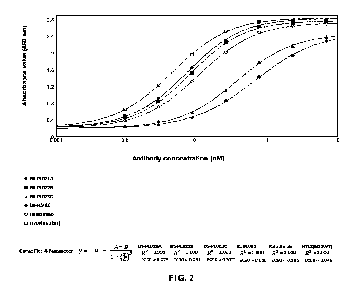
BS-PLV02, Relatlimab, and H7L8(hG1WT) to antigen LAG3-mFc by indirect ELISA.
FIG. 3: Results of assays for the activity of anti-LAG3/anti-PD-1 bispecific
antibodies in
competing with human PDL1-mFc for binding to human PD-1-mFc-Biotin by
competitive
ELISA.
FIG. 4: Results of assays for the binding activity of anti-LAG3/anti-PD-1
bispecific
23
CA 03233192 2024 3 26
IEC210281PCT
antibodies to PD-1 on 293T-PD1 membrane surface by FACS.
FIG. 5: Results of an assay for the binding activity of an anti-LAG3/anti-PD-1
bispecific
antibody to LAG3 on 293T-LAG3 membrane surface by FACS.
FIG. 6: Results of an assay for the activity of an anti-LAG3/anti-PD-1
bispecific antibody in
competing with PDL1 for binding to antigen PD-1 on cell membrane surface by
competitive
flow cytometry.
FIG. 7: Results of an assay for the activity of an anti-LAG3/anti-PD-1
bispecific antibody in
competing with LAG3 for binding to antigen MHC II on cell membrane surface by
competitive flow cytometry.
FIG. 8A: Results of assays for anti-LAG3/anti-PD-1 bispecific antibodies
blocking the
binding of LAG3 to MHCII.
FIG. 8B: Results of assays for anti-LAG3/anti-PD-1 bispecific antibodies
blocking the
binding of LAG3 to MHCII.
FIG. 9A: Results of an assay for an anti-LAG3/anti-PD-1 bispecific antibody
blocking the
binding of PD-1 to PD-Li.
FIG. 9B: Results of an assay for an anti-LAG3/anti-PD-1 bispecific antibody
blocking the
binding of PD-1 to PD-Li.
FIG. 10A: Results of an assay for an anti-LAG3/anti-PD-1 bispecific antibody
simultaneously blocking the binding of LAG3 to MHCII and PD-1 to PD-L1.
FIG. 10B: Results of an assay for an anti-LAG3/anti-PD-1 bispecific antibody
simultaneously blocking the binding of LAG3 to MHCII and PD-1 to PD-L1.
FIG. 11: Results of a bridging assay for an anti-LAG3/anti-PD-1 bispecific
antibody.
FIG. 12A. Results of an assay for the biological activity of an anti-LAG3/anti-
PD-1 bispecific
antibody in promoting IFN-y secretion by mixed lymphocyte reaction (MLR).
FIG. 12B. Results of an assay for the biological activity of an anti-LAG3/anti-
PD-1 bispecific
antibody in promoting IL-2 secretion by mixed lymphocyte reaction (MLR).
FIG. 13: Results of an assay for affinity constants of BS-PL022B to FcyRI.
FIG. 14: Results of an assay for affinity constants of H7L8(hG1WT) to FcyRI.
FIG. 15: Results of an assay for affinity constants of BS-PL022B to
Fc7RIIIa_V158.
24
CA 03233192 2024 3 26
IEC210281PCT
FIG. 16: Results of an assay for affinity constants of H7L8(hG1WT) to
Fc7RIIIa_V158.
FIG. 17: Results of an assay for affinity constants of BS-PL022B to
Fc7RIIIa_F158.
FIG. 18: Results of an assay for affinity constants of H7L8(hG1WT) to
FcyRIIIa_F158.
FIG. 19: Results of an assay for affinity constants of BS-PL022B to
Fc7RIIa_H131.
FIG. 20: Results of an assay for affinity constants of H7L8(hG1WT) to
Fc7RIIa_H131.
FIG. 21: Results of an assay for affinity constants of BS-PL022B to Fc7RIIb.
FIG. 22: Results of an assay for affinity constants of H7L8(hG1WT) to Fc7RIIb.
FIG. 23: Results of an assay for affinity constants of BS-PL022B to Clq.
FIG. 24: Results of an assay for affinity constants of H7L8(hG1WT) to C1q.
FIG. 25: Results of an ADCP effect assay for BS-PL022B.
FIG. 26: Efficacy of anti-LAG3/anti-PD-1 bispecific antibodies in a BALB/c-
hPD1/hLAG3
mouse model grafted with CT26 tumor. * P < 0.05, ** P < 0.01, *** P < 0.001,
VS isotype
control group (two-way ANOVA)
FIG. 27: Effects of anti-LAG3/anti-PD-1 bispecific antibodies on body weight
in a BALB/c-
hPD1/hLAG3 mouse model grafted with CT26 tumor.
DETAILED DESCRIPTION
The embodiments of the present invention will be described in detail below
with reference to
the examples. Those skilled in the art will appreciate that the following
examples are only
for illustrating the present invention, and should not be construed as
limitations to the scope
of the present invention. Examples where the specific technologies or
conditions are not
specified are performed according to the technologies or conditions described
in the
publications of the art (e.g., see, Molecular Cloning: A Laboratory Manual,
authored by J.
Sambrook et al., and translated by Huang Peitang et al., third edition,
Science Press) or
according to the package insert. Reagents or instruments used are commercially
available
conventional products if the manufacturers thereof are not specified. For
example, MDA-
MB-231 cells and U87-MG cells could be purchased from ATCC.
BALB/c mice were purchased from Guangdong Medical Laboratory Animal Center.
Nivolumab was purchased from BMS, with Batch No. ABA0330. Nivolumab is an anti-
PD-1
CA 03233192 2024 3 26
IEC210281PCT
antibody.
Pembrolizumab was purchased from MSD Ireland (Carlow), with Cat. No. S023942.
Pembrolizumab is an anti-PD-1 antibody.
The positive control antibody, Relatlimab, has sequences referenced to the
U.S. Patent
Publication No. US20160326248A1, wherein the heavy chain amino acid sequence
is
referenced to SEQ ID NO: 1 of this patent publication and the light chain
amino acid
sequence is referenced to SEQ ID NO: 2 of this patent publication. Relatlimab
is an anti-
LAG-3 antibody.
The cell line 293T-PD1 was constructed by Akeso Biopharma Inc. The cell line
293T-PD1
was produced by viral infection of HEK293T cells using 3rd Generation
Lentiviral Systems
(see, e.g., A Third Generation Lentivirus Vector with a Conditional Packaging
System. Dull
T, Zufferey R, Kelly M, Mandel Rj , Nguyen M, Trono D, and Naldini L., J
ViroL, 1998.
72(11): 8463-8471), wherein the lentivirus expression vector used was
p1enti6.3/V5-PD1FL-
BSD (PD1, Genebank ID: NM_005018; vector p1enti6.3/V5-BSD, purchased from
Invitrogen, Cat. No. K5315-20).
The cell line 293T-LAG3 was constructed by Akeso Biopharma Inc. The cell line
293T-LAG3
was produced by viral infection of HEK293T cells using 3rd Generation
Lentiviral Systems
(see, e.g., A Third Generation Lentivirus Vector with a Conditional Packaging
System. Dull
T, Zufferey R, Kelly M, Mandel Rj , Nguyen M, Trono D, and Naldini L., J
ViroL, 1998.
72(11): 8463-8471), wherein the lentivirus expression vector used was
p1enti6.3/V5-
huLAG3FL-BSD (LAG3, Genebank ID: NM_002277.4; vector p1enti6.3/V5-BSD,
purchased
from Invitrogen, Cat. No. K5315-20).
The cell line Raji-PDL1 was constructed by Akeso Biopharma Inc. The cell line
Raji-PDL1
was produced by viral infection of Raji cells using 3rd Generation Lentiviral
Systems (see,
e.g., A Third Generation Lentivirus Vector with a Conditional Packaging
System. Dull T,
Zufferey R, Kelly M, Mandel Rj , Nguyen M, Trono D, and Naldini L., J
ViroL,1998. 72(11):
8463-8471), wherein the lentivirus expression vector used was p1enti6.3/V5-
PDL1 (PDL1,
Genebank ID: NP 054862.1; vector p1enti6.3/V5, purchased from Invitrogen, Cat.
No.
K5315-20).
26
CA 03233192 2024 3 26
IEC210281PCT
The cell line J urkat-NFAT-PD1-LAG3 was constructed by Akeso Biopharma Inc.
The cell
line J urkat-NFAT-PD1-LAG3 was prepared by viral infection of PD-1 effector
cells (CPM,
manufacturer: Promega, Cat. No. J 112A) using 3rd Generation Lentiviral
Systems (see, e.g.,
A Third Generation Lentivirus Vector with a Conditional Packaging System. Dull
T,
Zufferey R, Kelly M, Mandel RJ , Nguyen M, Trono D, and Naldini L., J
ViroL,1998. 72(11):
8463-8471), wherein the lentivirus expression vector used was pCDH-huLAG3FL-
RFP-NE0
(LAG3, Genebank ID: NM_002277.4; vector pCDH-CMV-MCS-EF1-RFP+Neo, purchased
from Youbio, Cat. No. VT9005).
The cell line CHO-K1-PD1 was constructed by Akeso Biopharma Inc. The cell line
CHO-
K1-PD1 was prepared by viral infection of CHO-K1 cells using 3rd Generation
Lentiviral
Systems (see, e.g., A Third Generation Lentivirus Vector with a Conditional
Packaging
System. Dull T, Zufferey R, Kelly M, Mandel RJ , Nguyen M, Trono D, and
Naldini L., J
ViroL, 1998. 72(11): 8463-8471), wherein the lentivirus expression vector used
was pCDH-
CMV-PD-1F L-Puro (PD1, Genebank ID: NM_005018; vector pCDH-CMV-Puro, purchased
from Youbio, Cat. No. VT1480).
The cell line CHO-K1-LAG3 was constructed by Akeso Biopharma Inc. The cell
line CHO-
K1-LAG3 was produced by viral infection of CHO-K1 cells using 3rd Generation
Lentiviral
Systems (see, e.g., A Third Generation Lentivirus Vector with a Conditional
Packaging
System. Dull T, Zufferey R, Kelly M, Mandel RJ , Nguyen M, Trono D, and
Naldini L., J
ViroL, 1998. 72(11): 8463-8471), wherein the lentivirus expression vector used
was
p1enti6.3/V5-huLAG3FL-BSD (LAG3, Genebank ID: NM_002277.4; vector p1enti6.3/V5-
BSD, purchased from Invitrogen, Cat. No. K5315-20).
The cell line J urkat-NFAT-CD64-CD32R was constructed by Akeso Biopharma Inc.
The cell
line J urkat-NFAT-CD64-CD32R was prepared by viral infection of J urkat cells
using 3rd
Generation Lentiviral Systems (see, e.g., A Third Generation Lentivirus Vector
with a
Conditional Packaging System. Dull T, Zufferey R, Kelly M, Mandel RJ , Nguyen
M, Trono
D, and Naldini L., J ViroL, 1998. 72(11): 8463-8471), wherein the lentivirus
expression
vectors used were pCDH-NFAT-Hygro (vector pCDH-Hygro, obtained by modifying
based
on pCDH-CMV-MCS-EF1-Puro (purchased from Youbio, Cat. No. VT1480) in our
27
CA 03233192 2024 3 26
IEC210281PCT
laboratory), pcDH-hFCGR1AFL-Neo (vector pCDH-Neo, obtained by modifying based
on
pCDH-CMV-MCS-EF1-Puro (purchased from Youbio, Cat. No. VT1480) in our
laboratory), and pCDH-hFCGR2A(H167)-puro (hFCGR2A(H167), Genebank ID: P12318;
vector pCDH-CMV-MCS-EF1-Puro, purchased from Youbio, Cat. No. VT1480).
The cell line CHO-K1-PD1-LAG3 was constructed by Akeso Biopharma Inc. The cell
line
CHO-K1-PD1-LAG3 was prepared by viral infection of CHO-K1 cells using 3rd
Generation
Lentiviral Systems (see, e.g., A Third Generation Lentivirus Vector with a
Conditional
Packaging System. Dull T, Zufferey R, Kelly M, Mandel RJ , Nguyen M, Trono D,
and
Naldini L., J Virol., 1998. 72(11): 8463-8471), wherein the lentivirus
expression vectors used
were pCDH-hPD1-FL-puro (PD-1, Genebank ID: NM_005018; vector pCDH-CMV-MCS-
EF1-Puro, purchased from Youbio, Cat. No. VT1480) and p1enti6.3/V5-huLAG3FL-
BSD
(LAG3, Genebank ID: NM_002277.4; vector p1enti6.3/V5-BSD, purchased from
Invitrogen,
Cat. No. K5315-20).
Preparation Example 1: Design and Preparation of Anti-LAG3 Antibodies
1. Design of antibodies
The inventors creatively designed a series of antibody sequences based on the
known LAG3
protein sequence (NCB! Reference Sequence: NP_002277.4) and the three-
dimensional
crystal structure thereof, etc. Through extensive screening and testing,
humanized
monoclonal antibodies specifically binding to LAG3 were finally obtained,
named H7L8,
H7L9 and H7L10, respectively. The amino acid sequences of the heavy and light
chain
variable regions of the monoclonal antibodies and the encoding sequences
thereof are as
follows.
Nucleotide sequence of the heavy chain variable region H7v of H7L8 (360 bp):
CAGGTGCAGCTGCAGCAGTGGGGAGCTGGACTGCTGAAACCTAGCGAGACACT
GAGCCTGACCTGTGCTGTGTACGGCGGATCTATCAGCGATTACTACTGGAACT
GGATCAGGCAGCCCCCTGGAAAGGGACTGGAATGGATCGGAGAGATCAACCAC
AGGGGCACCACCAACTCCAATCCCTCTCTGAAGAGCAGGGTGACACTGAGCCT
CGACACAAGCAAGAATCAGTTCAGCCTGAAGCTGAGGTCCGTGACCGCTGCTG
28
CA 03233192 2024 3 26
IEC210281PCT
ATACAGCTGTGTACTACTGTGCCTTCGGCTACAGCGATTACGAGTACGATTGGT
TCGACCCTTGGGGCCAGGGAACACTGGTTACAGTGAGCTCC (SEQ ID NO: 1)
Amino acid sequence of the heavy chain variable region H7v of H7L8 (120 aa):
QVQLQQWGAG L L KPS ETLS LTCAVYGG S I SDYYWNW I RQPPG KG LEWIG El NHRG
TTNSN PS L KS RVTLS L DTS KNQF S L KLRSVTAADTAVYYCAFGYSDYEYDWF DPWG
QGTLVTVSS (SEQ ID NO: 2)
Nucleotide sequence of the light chain variable region L8v of H7L8 (321 bp):
GAGATCGTTCTGACCCAGAGCCCAGCTACACTGAGCCTGTCTCCTGGAGAGAG
GGCTACACTGTCCTGCAGAGCTAGCCAGACCATCAGCAGCTACCTGGCTTGGT
ACCAGCAGAAGCCTGGCCAAGCTCCAAGGCTGCTGATCTACGACGCCTCTAAT
AGGGCCACCGGCATCCCTGCTAGATTCTCTGGAAGCGGCAGCGGAACCGACTT
TACACTGACAATCAGCTCCCTGGAGCCCGAGGATTTCGCTGTTTACTACTGTCA
GCAGCGCAGCAACTGGCCCATCACATTCGGACAGGGCACAAATCTGGAGATCA
AG (SEQ ID NO: 3)
Amino acid sequence of the light chain variable region L8v of H7L8 (107 aa):
EIVLTQSPATLSLSPG ERATLSC RASQTISSYLAWYQQKPGQAPRLL IYDASNRATG I
PARFSGSGSGTDFTLTISSLEPEDFAVYYCQQRSNWPITFGQGTNLEIK (SEQID NO:
4)
The nucleotide sequence of the heavy chain variable region H7v of H7L9 is
identical to the
nucleotide sequence of the heavy chain variable region H7v of H7L8, as set
forth in SEQ ID
NO: 1.
The amino acid sequence of the heavy chain variable region H7v of H7L9 is
identical to the
amino acid sequence of the heavy chain variable region H7v of H7L8, as set
forth in SEQ ID
NO: 2.
Nucleotide sequence of the light chain variable region L9v of H7L9 (321 bp):
GAGATCGTTCTGACCCAGAGCCCAGCTACACTGAGCCTGTCTCCTGGAGAGAG
GGCTACACTGTCCTGCAGAGCTAGCCAGACCATCAGCAGCTACCTGGCTTGGT
ACCAGCAGAAGCCTGGCCAAGCTCCAAGGCTGCTGATCTACGACGGCTCTAAT
29
CA 03233192 2024 3 26
IEC210281PCT
AGGGCCACCGGCATCCCTGCTAGATTCTCTGGAAGCGGCAGCGGAACCGACTT
TACACTGACAATCAGCTCCCTGGAGCCCGAGGATTTCGCTGTTTACTACTGTCA
GCAGCGCAGCAACTGGCCCCTCACATTCGGACAGGGCACAAATCTGGAGATCA
AG (SEQ ID NO: 41)
Amino acid sequence of the light chain variable region L9v of H7L9 (107 bp):
EIVLTQSPATLSLSPG ERATLSCRASQTISSYLAWYQQKPGQAPRLL IYDGSNRATG I
PARFSGSGSGTDFTLTISSLEPEDFAVYYCQQRSNWPLTFGQGTN LEI K (SEQ ID NO:
42)
The nucleotide sequence of the heavy chain variable region H7v of H7L10 is
identical to the
nucleotide sequence of the heavy chain variable region H7v of H7L8, as set
forth in SEQ ID
NO: 1.
The amino acid sequence of the heavy chain variable region H7v of H7L10 is
identical to the
amino acid sequence of the heavy chain variable region H7v of H7L8, as set
forth in SEQ ID
NO: 2.
Nucleotide sequence of the light chain variable region L10v of H7L10 (321 bp):
GAGATCGTTCTGACCCAGAGCCCAGCTACACTGAGCCTGTCTCCTGGAGAGAG
GGCTACACTGTCCTGCAGAGCTAGCCAGTCCATCAGCAGCTACCTGGCTTGGT
ACCAGCAGAAGCCTGGCCAAGCTCCAAGGCTGCTGATCTACGACGGCTCTAAT
AGGGCCACCGGCATCCCTGCTAGATTCTCTGGAAGCGGCAGCGGAACCGACTT
TACACTGACAATCAGCTCCCTGGAGCCCGAGGATTTCGCTGTTTACTACTGTCA
GCAGCGCAGCAACTGGCCCATCACATTCGGACAGGGCACAAATCTGGAGATCA
AG (SEQ ID NO: 43)
Amino acid sequence of the light chain variable region L10v of H7L10 (107 bp):
EIVLTQSPATLSLSPG E RATLSC RASQS I SSYLAWYQQ KPGQAPRL LIYDGSNRATG I
PARFSGSGSGTDFTLTISSLEPEDFAVYYCQQRSNWPITFGQGTNLEI K (SEQ ID NO:
44)
The amino acid sequences of the CDRs of the antibody H7L8 are as follows
(according to the
CA 03233192 2024 3 26
IEC210281PCT
IMGT numbering system):
HCDR1: GGSISDYY (SEQ ID NO: 5);
HCDR2: INHRGTT (SEQ ID NO: 6);
HCDR3: AFGYSDYEYDWFDP (SEQ ID NO: 7);
LCDR1: QTISSY (SEQ ID NO: 8);
LCDR2: DAS (SEQ ID NO: 9); and
LCDR3: QQRSNWPIT (SEQ ID NO: 10).
The amino acid sequences of the CDRs of the antibody H7L9 are as follows
(according to the
IMGT numbering system):
HCDR1: GGSISDYY (SEQ ID NO: 5);
HCDR2: INHRGTT (SEQ ID NO: 6);
HCDR3: AFGYSDYEYDWFDP (SEQ ID NO: 7);
LCDR1: QTISSY (SEQ ID NO: 8);
LCDR2: DGS (SEQ ID NO: 46); and
LCDR3: QQRSNWPLT (SEQ ID NO: 47).
The amino acid sequences of the CDRs of the antibody H7L10 are as follows
(according to
the IMGT numbering system):
HCDR1: GGSISDYY (SEQ ID NO: 5);
HCDR2: INHRGTT (SEQ ID NO: 6);
HCDR3: AFGYSDYEYDWFDP (SEQ ID NO: 7);
LCDR1: QSISSY (SEQ ID NO: 48);
LCDR2: DGS (SEQ ID NO: 46); and
LCDR3: QQRSNWPIT (SEQ ID NO: 10).
2. Expression and purification of humanized antibody H7L8(hG1WT)
The heavy chain cDNA sequence (the encoding sequence of the variable region
was set forth
in SEQ ID NO: 1; the constant region was Ig gamma-1 chain C region, SEQ ID NO:
39) and
31
CA 03233192 2024 3 26
IEC210281PCT
the light chain cDNA sequence (the encoding sequence of the variable region
was set forth in
SEQ ID NO: 3; the constant region was P01834.1 (human Ig kappa chain C region,
SEQ ID
NO: 40) of H7L8(hG1WT) were separately cloned into pUC57simple vectors
(supplied by
GenScript), and plasmids pUC57simple-H7 and pUC57simple-L8 were obtained,
respectively. The plasmids pUC57simple-H7 and pUC57simple-L8 were each
digested
(Hind III&EcoRl). The heavy and light chains isolated by electrophoresis were
separately
subcloned into pcDNA3.1 vectors, and recombinant plasmids were extracted to co-
transfect
293F cells. After 7 days of cell culture, the culture medium was separated by
high-speed
centrifugation, and the supernatant was concentrated and loaded onto a HiTrap
MabSelect
SuRe column. The protein was eluted in one step with an elution buffer. The
target sample
was isolated, and the buffer was exchanged into PBS.
Amino acid sequence of the heavy chain constant region of H7L8(hG1WT)
ASTKG PSVF PLAPSSKSTSGGTAALGCLVKDYF PE PVTVSW N SGALTSGVHTF PAVL
QSSG LYSLSSVVTVPSSSLGTQTYI C NVN H KPSNTKVDKKVEPKSC D KTHTC PPC PA
PEL LGGPSVF LF PP KPKDTL MIS RTP EVTCVVVDVSH E D PEVKF NWYVDGVEVH NA
KTKPRE EQYNSTYRVVSVLTVL HQDW LNG KEYKCKVSNKALPAPI EKTISKAKGQ
PREPQVYTLPPSRDELTKNQVSLTC LVKGFYPSDIAVEW ESNGQPENNYKTTPPVL
DSDGSFFLYSKLTVDKSRWQQGNVFSCSVMHEALHNHYTQKSLSLSPGK (SEQ ID
NO: 39)
Amino acid sequence of the light chain constant region of H7L8(hG1WT)
RTVAAPSVFIF PPSDEQL KSGTASVVC LLNNFYPREAKVQWKVDNALQSGNSQESV
TEQDSKDSTYSLSSTLTLSKADYEKHKVYACEVTHQGLSSPVTKSFNRGEC (SEQ ID
NO: 40)
3. Design of humanized antibody H7L8(hG1TM)
On the basis of H7L8(hG1WT), the inventors obtained a humanized antibody
H7L8(hG1TM) with constant region mutations by introducing a leucine-to-alanine
point
mutation at position 234 (according to the EU numbering system, the same
below) (L234A),
32
CA 03233192 2024 3 26
IEC210281PCT
a leucine-to-alanine point mutation at position 235 (L235A), and a glycine-to-
alanine point
mutation at position 237 (G237A) in the heavy chain. The amino acid sequence
of the heavy
chain H7(hG1TM) of H7L8(hG1TM) is set forth in SEQ ID NO: 11, and the amino
acid
sequence of the light chain L8 thereof is set forth in SEQ ID NO: 12.
The humanized antibody H7L8(hG1TM) was prepared by the method described above
in
step 2.
Amino acid sequence of the heavy chain H7(hG1TM) of H7L8(hG1TM)
QVQLQQWGAG L L KPSETLSLTCAVYGGSI SDYYWNW I RQPPG KG LEWIG El NHRG
TTNSN PS L KS RVTLS L DTS KNQF S L KLRSVTAADTAVYYCAFGYSDYEYDW F DPWG
QGTLVTVSSASTKGPSVFPLAPSSKSTSGGTAALGCLVKDYFPEPVTVSWNSGALTS
GVHTFPAVLQSSG LYS LSSVVTVPSSS LGTQTYICNVNH KPSNTKVD KKVEP KSC D K
THTCPPCPAPEAAGAPSVF LF PP KPKDTL M I SRTPEVTCVVVDVSH EDP EVKF NWY
VDGVEVH NAKTKPRE EQYNSTYRVVSVLTVLHQDW L NG KEYKC KVSNKALPAPI E
KTISKAKGQPREPQVYTLPPSRDELTKNQVSLTCLVKGFYPSDIAVEWESNGQPEN
NYKTTPPVL DSDG SF F LYS KLTVD KSRWQQG NVF SCSVM H EALHNHYTQKSLSLSP
GK (SEQ ID NO: 11)
Amino acid sequence of the light chain L8 of H7L8(hG1TM)
EIVLTQSPATLSLSPG ERATLSCRASQTISSYLAWYQQKPGQAPRLL IYDASNRATG I
PARFSGSGSGTDFTLTISSLEPEDFAVYYCQQRSNWPITFGQGTNL E I KRTVAAPSVF
I FPPSDEQLKSGTASVVCLLNNFYPREAKVQWKVDNALQSGNSQESVTEQDSKDST
YSLSSTLTLSKADYEKHKVYACEVTHQGLSSPVTKSFNRG EC (SEQ ID NO: 12)
4. Design of humanized antibody H7L8(hG4DM)
On the basis of H7L8(hG1WT), the inventors obtained a humanized antibody
H7L8(hG4DM) with constant region mutations by using Ig gamma-4 chain C region
as a
heavy chain constant region and introducing a phenylalanine-to-alanine point
mutation at
position 234 (F234A) and a leucine-to-alanine point mutation at position 235
(L235A) in the
heavy chain constant region while keeping the antibody variable region
unchanged. The
amino acid sequence of the heavy chain of H7L8(hG4DM) is set forth in SEQ ID
NO: 13, and
33
CA 03233192 2024 3 26
IEC210281PCT
the amino acid sequence of the light chain thereof is set forth in SEQ ID NO:
12.
Amino acid sequence of the heavy chain H7(hG4DM) of H7L8(hG4DM):
QVQLQQWGAG L L KPSETLSLTCAVYGGSI SDYYWNW I RQPPG KG LEWIG El NHRG
TTNSN PS L KS RVTLS L DTS KNQF S L KLRSVTAADTAVYYCAFGYSDYEYDWF DPWG
QGTLVTVSSASTKGPSVF PLAPCSRSTSESTAALGC LVKDYF PE PVTVSW N SGALTS
GVHTFPAVLQSSG LYS LSSVVTVPSSS LGTKTYTC NVDH KPS NTKVD KRVES KYG PP
CPPCPAPEAAGG PSVF LF PPKPKDTLM I SRTPEVTCVVVDVSQ E D PEVQF NWYVDG
VEVH NAKTKPRE EQF NSTYRVVSVLTVL HQDW L NG KEYKC KVSN KG LPSSI EKTIS
KAKGQPREPQVYTLPPSQEEMTKNQVSLTCLVKGFYPSDIAVEWESNGQPENNYK
TTPPVLDSDGSF F LYSRLTVD KSRWQ EG NVF SCSVMH EALHNHYTQ KS LS LSLG K
(SEQ ID NO: 13)
The amino acid sequence of the light chain L8 of H7L8(hG4DM) is identical to
the amino
acid sequence of the light chain of H7L8(hG1TM), as set forth in SEQ ID NO:
12.
5. Expression and purification of humanized antibodies H7L8(hG4WT),
H7L9(hG4WT) and
H7L10(hG4WT)
The heavy chain cDNA sequences (the encoding sequences of the variable regions
were set
forth in SEQ ID NO: 1; the constant regions were Ig gamma-4 chain C regions,
set forth in
SEQ ID NO: 45) of H7L8(hG4WT), H7L9(hG4WT) and H7L10(hG4WT), the light chain
cDNA sequence (the encoding sequence of the variable region was set forth in
SEQ ID NO:
3; the constant region was human Ig kappa chain C region, set forth in SEQ ID
NO: 40) of
H7L8(hG4WT), the light chain cDNA sequence (the encoding sequence of the
variable region
was set forth in SEQ ID NO: 42; the constant region was human Ig kappa chain C
region,
set forth in SEQ ID NO: 40) of H7L9(hG4WT), and the light chain cDNA sequence
(the
encoding sequence of the variable region was set forth in SEQ ID NO: 44; the
constant region
was human Ig kappa chain C region, set forth in SEQ ID NO: 40) of H7L10(hG4WT)
were
separately cloned into pUC57simple vectors (supplied by GenScript), and
plasmids
pUC57simple-H7, pUC57simple-L8, pUC57simple-L9 and pUC57simple-L10 were
34
CA 03233192 2024 3 26
IEC210281PCT
obtained, respectively. The plasmids pUC57simple-H7, pUC57simple-L8,
pUC57simple-L9
and pUC57simple-L10 were each digested (Hindi! I&EcoRI). The heavy and light
chains
isolated by electrophoresis were separately subcloned into pcDNA3.1 vectors,
and
recombinant plasmids were extracted to co-transfect 293F cells. After 7 days
of cell culture,
the culture medium was separated by high-speed centrifugation, and the
supernatant was
concentrated and loaded onto a HiTrap MabSelect SuRe column. The protein was
eluted in
one step with an elution buffer. The target sample was isolated, and the
buffer was exchanged
into PBS.
Amino acid sequence of the heavy chain constant region of H7L8(hG4WT),
H7L9(hG4WT),
or H7L10(hG4WT):
ASTKG PSVF PLAPCSRSTSESTAALGCLVKDYF PE PVTVSW N SGALTSGVHTF PAVL
QSSG LYSLSSVVTVPSSSLGTKTYTCNVDH KPSNTKVDKRVESKYG PPCPPCPAPEF
LGGPSVF LF PP KPKDTL MISRTPEVTCVVVDVSQ E D PEVQ F NWYVDGVEVHNAKT
KPREEQF NSTYRVVSVLTVL HQ DW LNG KEYKC KVSN KG L PSS 1 E KTI SKAKGQ PRE
PQVYTL PPSQ E EMTKNQVS LTC LVKGFYPSDIAVEW ESNGQP EN NYKTTPPVLDSD
GSFFLYSRLTVDKSRWQEGNVFSCSVMHEALHNHYTQKSLSLSLGK (SEQ ID NO:
45)
Amino acid sequence of the light chain constant region of H7L8(hG4WT),
H7L9(hG4WT),
or H7 L10(hG4WT):
RTVAAPSVFIF PPSDEQL KSGTASVVC LLNNFYPREAKVQWKVDNALQSGNSQESV
TEQDSKDSTYSLSSTLTLSKADYEKHKVYACEVTHQGLSSPVTKSFNRGEC (SEQ ID
NO: 40)
Preparation Example 2: Design and Preparation of Anti-PD-1 Antibody 14C12 and
Its
Humanized Antibody 14C12H1L1
The amino acid sequences and encoding nucleotide sequences of the heavy and
light chains
of the anti-PD-1 antibody 14C12 and its humanized antibody 14C12H1L1 are
identical to
those of 14C12 and 14C12H1L1 in Chinese Patent Publication No. CN 106967172A
(or No.
CA 03233192 2024 3 26
IEC210281PCT
CN 106977602A), respectively.
(1) Heavy and light chain variable region sequences of 14C12
Nucleotide sequence of the heavy chain variable region of 14C12: (354 bp)
GAGGTCAAACTGGTGGAGAGCGGCGGCGGGCTGGTGAAGCCCGGCGGGTCAC
TGAAACTGAGCTGCGCCGCTTCCGGCTTCGCCTTTAGCTCCTACGACATGTCAT
GGGTGAGGCAGACCCCTGAGAAGCGCCTGGAATGGGTCGCTACTATCAGCGGA
GGCGGGCGATACACCTACTATCCTGACTCTGTCAAAGGGAGATTCACAATTAG
TCGGGATAACGCCAGAAATACTCTGTATCTGCAGATGTCTAGTCTGCGGTCCG
AGGATACAGCTCTGTACTATTGTGCAAACCGGTACGGCGAAGCATGGTTTGCC
TATTGGGGACAGGGCACCCTGGTGACAGTCTCTGCC (SEQ ID NO: 14)
Amino acid sequence of the heavy chain variable region of 14C12: (118 aa)
EVKLVESGGG LVKPGGSLKLSCAASGFAFSSYDMSWVRQTPEKRLEWVATI SG G G
RYTYYPDSVKG RFT! S RD NARNTLYLQ MSS L RS E DTALYYCAN RYG EAWFAYWGQ
GTLVTVSA (SEQ ID NO: 15)
Nucleotide sequence of the light chain variable region of 14C12: (321 bp)
GACATTAAGATGACACAGTCCCCTTCCTCAATGTACGCTAGCCTGGGCGAGCG
AGTGACCTTCACATGCAAAGCATCCCAGGACATCAACACATACCTGTCTTGGTT
TCAGCAGAAGCCAGGCAAAAGCCCCAAGACCCTGATCTACCGGGCCAATAGAC
TGGTGGACGGGGTCCCCAGCAGATTCTCCGGATCTGGCAGTGGGCAGGATTAC
TCCCTGACCATCAGCTCCCTGGAGTATGAAGACATGGGCATCTACTATTGCCTG
CAGTATGATGAGTTCCCTCTGACCTTTGGAGCAGGCACAAAACTGGAACTGAA
G (SEQ ID NO: 16)
Amino acid sequence of the light chain variable region of 14C12: (107 aa)
DI KMTQSPSSMYASLG ERVTFTC KASQ D I NTYLSWFQQKPGKSPKTLIYRANRLVD
GVPSRFSGSGSGQDYSLTISSLEYEDMGIYYCLQYDEFPLTFGAGTKLELK (SEQ ID
NO: 17)
(2) Heavy and light chain variable region sequences and heavy and light chain
sequences of
36
CA 03233192 2024 3 26
IEC210281PCT
humanized monoclonal antibody 14C12H1L1
Nucleotide sequence of the heavy chain variable region 14C12H1v of 14C12H1L1:
(354 bp)
GAAGTGCAGCTGGTCGAGTCTGGGGGAGGGCTGGTGCAGCCCGGCGGGTCAC
TGCGACTGAGCTGCGCAGCTTCCGGATTCGCCTTTAGCTCCTACGACATGTCCT
GGGTGCGACAGGCACCAGGAAAGGGACTGGATTGGGTCGCTACTATCTCAGGA
GGCGGGAGATACACCTACTATCCTGACAGCGTCAAGGGCCGGTTCACAATCTC
TAGAGATAACAGTAAGAACAATCTGTATCTGCAGATGAACAGCCTGAGGGCTG
AGGACACCGCACTGTACTATTGTGCCAACCGCTACGGGGAAGCATGGTTTGCC
TATTGGGGGCAGGGAACCCTGGTGACAGTCTCTAGT (SEQ ID NO: 18)
Amino acid sequence of the heavy chain variable region 14C12H1v of 14C12H1L1:
(118 aa)
EVQLVESGGG LVQPGGSLRLSCAASGFAFSSYDMSWVRQAPG KG LDWVATISGGG
RYTYYPDSVKG RFT! SRDNSKNN LYLQ M NS LRAEDTALYYCAN RYG EAWFAYWG
QGTLVTVSS (SEQ ID NO: 19)
Nucleotide sequence of the light chain variable region 14C12L1v of 14C12H1L1:
(321 bp)
GACATTCAGATGACTCAGAGCCCCTCCTCCATGTCCGCCTCTGTGGGCGACAG
GGTCACCTTCACATGCCGCGCTAGTCAGGATATCAACACCTACCTGAGCTGGTT
TCAGCAGAAGCCAGGGAAAAGCCCCAAGACACTGATCTACCGGGCTAATAGAC
TGGTGTCTGGAGTCCCAAGTCGGTTCAGTGGCTCAGGGAGCGGACAGGACTAC
ACTCTGACCATCAGCTCCCTGCAGCCTGAGGACATGGCAACCTACTATTGCCTG
CAGTATGATGAGTTCCCACTGACCTTTGGCGCCGGGACAAAACTGGAGCTGAA
G (SEQ ID NO: 20)
Amino acid sequence of the light chain variable region 14C12L1v of 14C12H1L1:
(107 aa)
DI Q MTQSPSS MSASVG DRVTFTC RASQ D I NTYLSWFQQKPG KSPKTL IYRANRLVS
GVPSRFSGSGSGQDYTLTISSLQPEDMATYYCLQYDEFPLTFGAGTKLELK (SEQ ID
NO: 21)
Nucleotide sequence of the heavy chain 14C12H1 of 14C12H1L1: (1344 bp)
GAAGTGCAGCTGGTCGAGTCTGGGGGAGGGCTGGTGCAGCCCGGCGGGTCAC
37
CA 03233192 2024 3 26
IEC210281PCT
TGCGACTGAGCTGCGCAGCTTCCGGATTCGCCTTTAGCTCCTACGACATGTCCT
G G GTG C GACAG G CACCAG GAAAG G GACTG GATTG G GTCG CTACTATCTCAG GA
GGCGGGAGATACACCTACTATCCTGACAGCGTCAAGGGCCGGTTCACAATCTC
TAGAGATAACAGTAAGAACAATCTGTATCTGCAGATGAACAGCCTGAGGGCTG
AG GACACCG CACTGTACTATTGTG CCAACCG CTAC G G G GAAG CATG GTTTG CC
TATTGGGGGCAGGGAACCCTGGTGACAGTCTCTAGTGCCAGCACCAAAGGACC
TAGCGTGTTTCCTCTCGCCCCCTCCTCCAAAAGCACCAGCGGAGGAACCGCTG
CTCTCGGATGTCTGGTGAAGGACTACTTCCCTGAACCCGTCACCGTGAGCTGG
AATAGCGGCGCTCTGACAAGCGGAGTCCATACATTCCCTGCTGTGCTGCAAAG
CAGCGGACTCTATTCCCTGTCCAGCGTCGTCACAGTGCCCAGCAGCAGCCTGG
GCACCCAGACCTACATCTGTAACGTCAACCACAAGCCCTCCAACACCAAGGTG
GACAAGAAAGTG GAG C C CAAATCCTG C GACAAGACACACACCTGTCCCCCCTG
TCCTG CTCCCGAACTC CTCG GAG G CCCTAG C GTCTTCCTCTTTC CTCCCAAAC C
CAAGGACACCCTCATGATCAGCAGAACCCCTGAAGTCACCTGTGTCGTCGTGG
ATGTCAG CCATGAG GACCC C GAG GTGAAATTCAACTG GTATGTCGATG G C GTC
GAG GTG CACAAC G C CAAAACCAAG CC CAG G GAG GAACAGTACAACTCCAC CTA
CAGGGTGGTGTCCGTGCTGACAGTCCTCCACCAGGACTGGCTGAACGGCAAGG
AGTACAAGTGCAAGGTGTCCAACAAGGCTCTCCCTGCCCCCATTGAGAAGACC
ATCAG CAAG G CCAAAG G CCAACCCAG G GAG C C C CAG GTCTATACACTG C CTCC
CTCCAGGGACGAACTCACCAAGAACCAGGTGTCCCTGACCTGCCTGGTCAAGG
GCTTTTATCCCAGCGACATCGCCGTCGAGTGGGAGTCCAACGGACAGCCCGAG
AATAACTACAAGACCACCCCTCCTGTCCTCGACTCCGACGGCTCCTTCTTCCTG
TACAGCAAGCTGACCGTGGACAAAAGCAGGTGGCAGCAGGGAAACGTGTTCTC
CTG CAG CGTGATG CACGAAG CC CTCCACAACCACTACACCCAGAAAAG C CTGT
CCCTGAGCCCCGGCAAA (SEQ ID NO: 22)
Amino acid sequence of the heavy chain 14C12H1 of 14C12H1L1: (448 aa)
EVQLVESGGG LVQPGGSLRLSCAASG FAF SSYDMSWVRQAPG KG L DWVAT I SGGG
RYTYYPDSVKG RFT! S RD N S KN N LYLQ MNSL RAE DTALYYCAN RYG EAWFAYWG
QGT LVTVSSAST KG PSVF P LAPSS KSTSGGTAALGC LVKDYF P EPVTVSWNSGALTS
38
CA 03233192 2024- 3- 26
IEC210281PCT
GVHTFPAVLQSSG LYS LSSVVTVPSSS LGTQTYIC NVNH KPSNTKVD KKVEP KSC D K
THTC PPC PAPE L LGG PS VF LF PPKP KDTL M I SRTPEVTCVVVDVSHEDPEVKF NWY
VDGVEVH NAKTKPRE EQYNSTYRVVSVLTVLHQDW L NG KEYKC KVSNKALPAPI E
KTISKAKGQPREPQVYTLPPSRDELTKNQVSLTCLVKGFYPSDIAVEWESNGQPEN
NYKTTPPVL DSDG SF F LYS KLTVD KSRWQQG NVF SC SVM H EALHNHYTQKSLSLSP
GK (SEQ ID NO: 23)
Nucleotide sequence of the light chain 14C12L1 of 14C12H1L1: (642 bp)
GACATTCAGATGACTCAGAGCCCCTCCTCCATGTCCGCCTCTGTGGGCGACAG
GGTCACCTTCACATGCCGCGCTAGTCAGGATATCAACACCTACCTGAGCTGGTT
TCAGCAGAAGCCAGGGAAAAGCCCCAAGACACTGATCTACCGGGCTAATAGAC
TGGTGTCTGGAGTCCCAAGTCGGTTCAGTGGCTCAGGGAGCGGACAGGACTAC
ACTCTGACCATCAGCTCCCTGCAGCCTGAGGACATGGCAACCTACTATTGCCTG
CAGTATGATGAGTTCCCACTGACCTTTGGCGCCGGGACAAAACTGGAGCTGAA
GCGAACTGTGGCCGCTCCCTCCGTCTTCATTTTTCCCCCTTCTGACGAACAGCT
GAAATCAGGCACAGCCAGCGTGGTCTGTCTGCTGAACAATTTCTACCCTAGAG
AGGCAAAAGTGCAGTGGAAGGTCGATAACGCCCTGCAGTCCGGCAACAGCCAG
GAGAGTGTGACTGAACAGGACTCAAAAGATAGCACCTATTCCCTGTCTAGTAC
ACTGACTCTGTCCAAGGCTGATTACGAGAAGCACAAAGTGTATGCATGCGAAG
TGACACATCAGGGACTGTCAAGCCCCGTGACTAAGTCTTTTAACCGGGGCGAA
TGT (SEQ ID NO: 24)
Amino acid sequence of the light chain 14C12L1 of 14C12H1L1: (214 aa)
DI Q MTQSPSSMSASVG DRVTFTC RASQ D I NTYLSWFQQKPG KSPKTL IYRANRLVS
GVPSRFSGSGSGQDYTLTISSLQPEDMATYYCLQYDEFPLTFGAGTKLELKRTVAA
PSVF I FPPSDEQ LKSGTASVVC LLNNFYPREAKVQWKVDNALQSG NSQ ESVTEQ DS
KDSTYSLSSTLTLSKADYEKHKVYACEVTHQG LSSPVTKSFNRG EC (SEQ ID NO: 25)
The CDRs of the antibodies 14C12 and 14C12H1L1 are identical as follows
(according to the
I MGT numbering system):
HCDR1: GFAFSSYD (SEQ ID NO: 26)
39
CA 03233192 2024 3 26
IEC210281PCT
HCDR2: ISGGGRYT (SEQ ID NO: 27)
HCDR3: ANRYGEAWFAY (SEQ ID NO: 28)
LCDR1: QDINTY (SEQ ID NO: 29)
LCDR2: RAN (SEQ ID NO: 30)
LCDR3: LQYDEFPLT (SEQ ID NO: 31)
Heavy and light chain variable region sequences of 14C12H1L1(M)
14C12H1L1(M) was obtained by mutating certain amino acids in the framework
region
(light chain) on the basis of 14C12H1L1.
The heavy chain variable region 14C12H1(M) of 14C12H1L1(M)
is identical to the heavy chain variable region 14C12H1 of 14C12H1L1, that is,
both have an
amino acid sequence set forth in SEQ ID NO: 19.
Light chain variable region 14C12L1(M) of 14C12H1L1(M):
DI Q MTQSPSSMSASVG DRVTFTC RASQD I NTYLSWFQQ KPG KSPKTL IYRANRLVS
GVPSRFSGSGSGQDYTLTISSLQPEDMATYYCLQYDEFPLTFGAGTKLELKR (SEQ
ID NO: 38)
Preparation Example 3: Design of Humanized Antibody 14C12H1L1(hG4DM)
On the basis of 14C12H1L1, the inventors obtained a humanized antibody
14C12H1L1(hG4DM) with constant region mutations by using Ig gamma-4 chain C
region
as a heavy chain constant region and introducing a phenylalanine-to-alanine
point mutation
at position 234 (F234A) and a leucine-to-alanine point mutation at position
235 (L235A) in
the heavy chain constant region while keeping the antibody variable region
unchanged. The
amino acid sequence of the heavy chain 14C12H1(hG4DM) of 14C12H1L1(hG4DM) is
set
forth in SEQ ID NO: 32, and the amino acid sequence of the light chain thereof
is set forth
in SEQ ID NO: 25.
Amino acid sequence of the heavy chain of 14C12H1L1(hG4DM)
EVQLVESGGG LVQPGGSLRLSCAASGFAFSSYDMSWVRQAPG KG LDWVATISGGG
CA 03233192 2024 3 26
IEC210281PCT
RYTYYPDSVKGRFTISRDNSKNNLYLQMNSLRAEDTALYYCANRYG EAWFAYWG
QGTLVTVSSASTKGPSVFPLAPCSRSTSESTAALGCLVKDYFPEPVTVSWNSGALTS
GVHTFPAVLQSSGLYSLSSVVTVPSSSLGTKTYTCNVDHKPSNTKVDKRVESKYGPP
CPPCPAPEAAGGPSVF LFPPKPKDTLMISRTPEVTCVVVDVSQEDPEVQFNWYVDG
VEVHNAKTKPREEQFNSTYRVVSVLTVLHQDWLNG KEYKCKVSN KG LPSSI EKTIS
KAKGQPREPQVYTLPPSQEEMTKNQVSLTCLVKGFYPSDIAVEWESNGQPENNYK
TTPPVLDSDGSFFLYSRLTVDKSRWQEGNVFSCSVMHEALHNHYTQKSLSLSLGK
(SEQ ID NO: 32)
The amino acid sequence of the light chain of 14C12H1L1(hG4DM) is identical to
the amino
acid sequence of the light chain 14C12L1 of 14C12H1L1, as set forth in SEQ ID
NO: 25.
Preparation Example 4: Sequence Design of Humanized Antibody 14C12H1L1(hG1TM)
On the basis of the humanized antibody 14C12H1L1, the inventors obtained a
humanized
mutant 14C12H1L1(hG1TM) by introducing a leucine-to-alanine point mutation at
position
234 (L234A), a leucine-to-alanine point mutation at position 235 (L235A), and
a glycine-to-
alanine point mutation at position 237 (G237A) in the hinge region of the
heavy chain
according to the EU numbering system.
Nucleotide sequence of the heavy chain 14C12H1(hG1TM) of 14C12H1L1(hG1TM):
(1344
bp)
GAAGTGCAGCTGGTCGAGTCTGGGGGAGGGCTGGTGCAGCCCGGCGGGTCAC
TGCGACTGAGCTGCGCAGCTTCCGGATTCGCCTTTAGCTCCTACGACATGTCCT
GGGTGCGACAGGCACCAGGAAAGGGACTGGATTGGGTCGCTACTATCTCAGGA
GGCGGGAGATACACCTACTATCCTGACAGCGTCAAGGGCCGGTTCACAATCTC
TAGAGATAACAGTAAGAACAATCTGTATCTGCAGATGAACAGCCTGAGGGCTG
AGGACACCGCACTGTACTATTGTGCCAACCGCTACGGGGAAGCATGGTTTGCC
TATTGGGGGCAGGGAACCCTGGTGACAGTCTCTAGTGCCAGCACCAAAGGGCC
CAGCGTGTTTCCTCTCGCCCCCTCCTCCAAAAGCACCAGCGGAGGAACCGCTG
CTCTCGGATGTCTGGTGAAGGACTACTTCCCTGAACCCGTCACCGTGAGCTGG
AATAGCGGCGCTCTGACAAGCGGAGTCCATACATTCCCTGCTGTGCTGCAAAG
41
CA 03233192 2024 3 26
IEC210281PCT
CAGCGGACTCTATTCCCTGTCCAGCGTCGTCACAGTGCCCAGCAGCAGCCTGG
GCACCCAGACCTACATCTGTAACGTCAACCACAAGCCCTCCAACACCAAGGTG
GACAAGAAAGTGGAGCCCAAATCCTGCGACAAGACACACACCTGTCCCCCCTG
TCCTGCTCCCGAAGCTGCTGGAGCCCCTAGCGTCTTCCTCTTTCCTCCCAAACC
CAAGGACACCCTCATGATCAGCAGAACCCCTGAAGTCACCTGTGTCGTCGTGG
ATGTCAGCCATGAGGACCCCGAGGTGAAATTCAACTGGTATGTCGATGGCGTC
GAGGTGCACAACGCCAAAACCAAGCCCAGGGAGGAACAGTACAACTCCACCTA
CAGGGTGGTGTCCGTGCTGACAGTCCTCCACCAGGACTGGCTGAACGGCAAGG
AGTACAAGTGCAAGGTGTCCAACAAGGCTCTCCCTGCCCCCATTGAGAAGACC
ATCAGCAAGGCCAAAGGCCAACCCAGGGAGCCCCAGGTCTATACACTGCCTCC
CTCCAGGGACGAACTCACCAAGAACCAGGTGTCCCTGACCTGCCTGGTCAAGG
GCTTTTATCCCAGCGACATCGCCGTCGAGTGGGAGTCCAACGGACAGCCCGAG
AATAACTACAAGACCACCCCTCCTGTCCTCGACTCCGACGGCTCCTTCTTCCTG
TACAGCAAGCTGACCGTGGACAAAAGCAGGTGGCAGCAGGGAAACGTGTTCTC
CTGCAGCGTGATGCACGAAGCCCTCCACAACCACTACACCCAGAAAAGCCTGT
CCCTGAGCCCCGGCAAA (SEQ ID NO: 33)
Amino acid sequence of the heavy chain 14C12H1(hG1TM) of 14C12H1L1(hG1TM):
(448
aa)
EVQLVESGGG LVQPGGSLRLSCAASGFAFSSYDMSWVRQAPG KG LDWVATISGGG
RYTYYPDSVKG RFT! SRDNSKNN LYLQ M NS LRAEDTALYYCAN RYG EAWFAYWG
QGTLVTVSSASTKGPSVF PLAPSSKSTSGGTAALGCLVKDYF PEPVTVSWNSGALTS
GVHTFPAVLQSSG LYS LSSVVTVPSSS LGTQTYIC NVNH KPSNTKVD KKVEP KSC D K
THTCPPCPAPEAAGAPSVF LF PP KPKDTL M I SRTPEVTCVVVDVSH EDP EVKF NWY
VDGVEVH NAKTKPRE EQYNSTYRVVSVLTVLHQDW L NG KEYKC KVSN KALPAPI E
KTISKAKGQPREPQVYTLPPSRDELTKNQVSLTCLVKGFYPSDIAVEWESNGQPEN
NYKTTPPVL DSDG SF F LYS KLTVD KSRWQQG NVF SC SVM H EALHNHYTQKSLSLSP
GK (SEQ ID NO: 34)
The nucleotide sequence of the light chain of 14C12H1L1(hG1TM) is set forth in
SEQ ID
42
CA 03233192 2024 3 26
IEC210281PCT
NO: 24.
The amino acid sequence of the light chain of 14C12H1L1(hG1TM) is identical to
the amino
acid sequence of the light chain 14C12L1 of 14C12H1L1, as set forth in SEQ ID
NO: 25.
Preparation Example 5: Design and Preparation of Anti-LAG3/PD-1 Bifunctional
Antibodies
1. Sequence design
The structures of the bifunctional antibodies BS-PL021A, Bs-PL022B, BS-PL023C
and Bs-
PLV02 of the present invention are in the Morrison form (IgG-scFv), i.e., C-
termini of two
heavy chains of an IgG antibody are each linked to an scFv fragment of another
antibody,
and the main composition design of the heavy and light chains is as shown in
Table 1 below.
43
CA 03233192 2024 3 26
IEC210281PCT
Table 1: Composition design of heavy and light chains of bifunctional
antibodies
Bispecific Immunoglobulin moiety Linker scFv moiety
antibody Heavy chain Light chain fragment Heavy chain
variable Linker Light chain variable
No. region
fragment region
BS-PL021A H7(hG1TM) L8 (GGGGS)3 14C12H1v
(GGGGS)4 14C12L1(M)v
(SEQ ID NO: 11) (SEQ ID NO: 12) (SEQ ID NO: 19)
(SEQ ID NO: 38)
H7(hG1TM) L8 (GGGGS)4 14C12H1v
(GGGGS)4 14C12L1(M)v
BS-PL022B
(SEQ ID NO: 11) (SEQ ID NO: 12) (SEQ ID NO: 19)
(SEQ ID NO: 38)
BS-PL023C 14C12H1(hG1TM) 14C12L1 (GGGGS)4 H7v
(GGGGS)4 L8v
(SEQ ID NO: 34) (SEQ ID NO: 25) (SEQ ID NO: 2)
(SEQ ID NO: 4)
Bs-PLV02 14C12H1(hG4DM) 14C12L1 (GGGGS)4 H7v
(GGGGS)4 L8v
(SEQ ID NO: 32) (SEQ ID NO: 25) G (SEQ ID NO: 2)
(SEQ ID NO: 4)
Bi-PGV02 H7(hG4DM) L8 (GGGGS)4 14C12H1v
(GGGGS)4 14C12L1v
(SEQ ID NO: 13) (SEQ ID NO: 12) (SEQ ID NO: 19)
(SEQ ID NO: 21)
44
I EC210281PCT
In the Table 1 above:
(1) Amino acid sequence of linker fragment (GGGGS)3:
GGGGSGGGGSGGGGS (SEQ ID NO: 35)
Amino acid sequence of linker fragment (GGGGS)4:
GGGGSGGGGSGGGGSGGGGS (SEQ ID NO: 36)
Amino acid sequence of linker fragment (GGGGS)4G:
GGGGSGGGGSGGGGSGGGGSG (SEQ ID NO: 37)
(2) Those with "v" label at the lower right corner refer to the variable
region of the
corresponding heavy chain or the variable region of the corresponding light
chain. For those
without "v" label, the corresponding heavy or light chain is the full length
comprising the
constant region. The corresponding sequences described in the above
preparation examples
are referenced to the amino acid sequences of these variable regions or the
full lengths and
the nucleotide sequences encoding them.
2. Expression and purification of antibodies
The heavy chain cDNA sequences and the light chain cDNA sequences of Bs-
PL021A, Bs-
PL022B, Bs-PL023C, and Bs-PLV02 were separately cloned into pUC57simple
vectors
(supplied by GenScript), and plasmids pUC57simple-Bs-PL021AH/pUC57simple-Bs-
PL021AL, pUC57simple-Bs-PL022BH/pUC57simple-Bs-PL022BL,
pUC57simple-Bs-
PL023CH/pUC57simple-Bs-PL023CL,
pUC57simple-Bs-PLVO2H/pUC57simple-Bs-
PLVO2L and
pUC57simple-Bi-PGV02/pUC57simple-Bi-PGV02 were obtained, respectively.
The plasmids pUC57simple-Bs-PL021AH/pUC57simple-Bs-PL021AL, pUC57simple-Bs-
PL022BH/pUC57simple-Bs-PL022BL,
pUC57simple-Bs-PL023CH/pUC57simple-Bs-
PL023CL, pUC57simple-Bs-PLVO2H/pUC57simple-Bs-PLVO2L and
pUC57simple-Bi-PGV02/pUC57simple-Bi-PGV02 were each digested (HindIll&EcoR1).
The heavy and light chains isolated by electrophoresis were separately
subcloned into
pcDNA3.1 vectors, and recombinant plasmids were extracted to co-transfect 293F
cells.
After 7 days of cell culture, the culture medium was separated by high-speed
centrifugation,
and the supernatant was concentrated and loaded onto a HiTrap MabSelect SuRe
column.
CA 03233192 2024 3 26
IEC210281PCT
The protein was eluted in one step with an elution buffer. The target sample
was isolated,
and the buffer was exchanged into PBS.
Preparation Example 6: Preparation of Fusion Proteins PD-1-mFc, PD-1-hFc and
PDL1-
h Fc
The preparation of the fusion proteins PD-1-mFc, PD-1-hFc and PDL1-hFc, as
well as the
SDS-PAGE electrophoresis detection, were carried out by fully referring to
Preparation
Example 1 of Chinese Patent Publication No. CN106632674A.
The amino acid sequences and encoding nucleotide sequences of the fusion
proteins PD-1-
mFc, PD-1-hFc and PDL1-hFc in this preparation example are identical to those
of PD-1-
mFc, PD-1-hFc and PDL1-hFc in Preparation Example 1 of Chinese Patent
Publication No.
CN106632674A, respectively.
The fusion proteins PD-1-mFc, PD-1-hFc and PDL1-hFc were thus obtained.
Preparation Example 7: Preparation of Human Anti-Hen Egg Lysozyme Antibody
The sequence of the human anti-hen egg lysozyme IgG (anti-HEL, or human IgG,
abbreviated as hIgG) antibody was derived from the variable region sequence of
the Fab
F10.6.6 sequence in the study reported by Acierno et al., entitled "Affinity
maturation
increases the stability and plasticity of the Fv domain of anti-protein
antibodies" (Acierno et
al., J Mot Biol., 2007; 374(1): 130-46). The preparation method was as
follows:
Nanjing Genscript Biology was entrusted to carry out codon optimization of
amino acids and
gene synthesis on heavy and light chain (complete sequence or variable region)
genes of the
human IgG antibody, and by referring to the standard technologies introduced
in the "Guide
to Molecular Cloning Experiments (Third Edition)" and using standard molecular
cloning
techniques such as PCR, enzyme digestion, DNA gel extraction, ligation
transformation,
colony PCR or enzyme digestion identification, the heavy and light chain genes
were
subcloned into the antibody heavy chain expression vector and antibody light
chain
expression vector of the mammalian expression system, respectively. The heavy
and light
chain genes of the recombinant expression vectors were further sequenced and
analyzed.
46
CA 03233192 2024 3 26
IEC210281PCT
After the sequences were verified to be correct, a medium or large amount of
endotoxin-free
expression plasmids were prepared, and the heavy and light chain expression
plasmids were
transiently co-transfected into HEK293 cells for recombinant antibody
expression. After 7
days of culture, the cell culture medium was collected and subjected to
affinity purification
using an rProtein A column (GE), and the quality of the resulting antibody
sample was
determined using SDS-PAGE and SEC-HPLC standard analysis techniques.
Experimental Example 1: Assays for Binding Activity of Anti-LAG3/Anti-PD-1
Bispecific
Antibodies to Antigens by ELI SA
1. Assays for the binding activity of BS-PL021A, BS-PL022B, BS-PL023C, Bs-
PLV02, and
14C12H1L1(hG1TM) to antigen PD-1-mFc by indirect ELISA The procedures were as
follows:
An ELISA plate was coated with human PD-1-mFc at 0.5 pg/mL and incubated at 4
C
overnight. Then the ELI SA plate coated with the antigen was washed once with
PBST and
then blocked with a PBS solution containing 1% BSA as a blocking solution at
37 C for 2 h.
After blocking, the ELISA plate was washed 3 times with PBST. The antibodies
serially
diluted with PBST solution (the dilution gradients for the antibody are shown
in Table 2)
were added. The ELI SA plate containing the test antibodies was incubated at
37 C for 30
min and then washed 3 times with PBST. After washing, a working solution of an
HRP-
labeled goat anti-human IgG FC (H+L) (Jackson, Cat. No. 109-035-098) secondary
antibody
diluted at a ratio of 1:5000 was added, and then the plate was incubated at 37
C for 30 min.
After incubation, the plate was washed 4 times with PBST, TMB (Neogen, 308177)
was added
for chromogenesis in the dark for 5 min, and then a stop solution was added to
terminate the
chromogenic reaction. The ELISA plate was put into an ELISA plate reader
immediately,
and the OD value of each well in the ELISA plate was read at 450 nm. The data
were
analyzed and processed by SoftMax Pro 6.2.1.
The assay results are shown in Table 2 and FIG. 1.
Table 2: Results of assays for the binding of BS-PL021A, BS-PL022B, BS-PL023C,
Bs-
PLV02, and 14C12H1L1(hG1TM) to human PD-1-mFc by ELISA
47
CA 03233192 2024 3 26
IEC210281PCT
Antibody concentration Antigen-antibody bindin . OD (450 nm) value
14C12H1L1
(nM) BS-PL021A BS-PL022B BS-PL023C Bs-PLV02
(hG1TM)
7.000 2.601 2.573 2.541 2.566 2.612 2.606 2.495
2.490 2.695 2.671
2.333 2.618 2.584 2.560 2.544 2.634 2.625 2.483
2.505 2.724 2.670
0.778 2.582 2.542 2.485 2.481 2.604 2.600 2.430
2.425 2.721 2.688
0.259 2.289 2.141 2.164 2.102 2.374 2.343 1.946
1.929 2.576 2.573
0.086 1.576 1.509 1.442 1.402 1.825 1.793 1.149
1.178 2.315 2.280
0.029 0.812 0.769 0.727 0.729 1.012 1.009 0.530
0.533 1.617 1.656
0.010 0.379 0.355 0.345 0.325 0.498 0.491 0.244
0.256 0.921 0.916
PBST 0.057 0.058 0.060 0.055 0.054 0.068 0.057
0.061 0.070 0.069
EC5o(nM) 0.066 0.074 0.046 0.103
0.02
As can be seen from FIG. 1, BS-PL021A, BS-PL022B, BS-PL023C, Bs-PLV02, and
14C12H1L1(hG1TM) could effectively bind to the antigen human PD-1-mFc in a
dose-
dependent manner. By quantitative analysis of the absorbance of the bound
antibodies, the
binding efficiency EC50 values of the antibodies BS-PL021A, BS-PL022B, BS-
PL023C, Bs-
PLV02, and 14C12H1L1(hG1TM) (as a control) obtained by curve fitting
calculation were
0.066 nM, 0.074 nM, 0.046 nM, 0.103 nM, and 0.02 nM, respectively.
The results show that under the same experimental conditions, the binding
activity of BS-
PL021A, BS-PL022B and BS-PL023C to PD-1-mFc was substantially comparable to
that of
the positive control 14C12H1L1(hG1TM) for the same target, suggesting that BS-
PL021A,
BS-PL022B, BS-PL023C, and Bs-PLV02 had the activity of effectively binding to
PD-1-mFc.
2. Assays for the binding activity of BS-PL021A, BS-PL022B, BS-PL023C, Bs-
PLV02,
Relatlimab, and H7L8(hG1WT) to antigen LAG3-mFc by indirect ELISA
The procedures were as follows:
An ELISA plate was coated with human LAG3-mFc (Akeso Biopharma Inc., Batch No.
20200417) at 2 pg/mL and incubated at 4 C overnight. Then the ELISA plate
coated with
the antigen was washed once with PBST and then blocked with a PBS solution
containing
1% BSA as a blocking solution at 37 C for 2 h. After blocking, the ELISA
plate was washed
48
CA 03233192 2024 3 26
IEC210281PCT
3 times with PBST. The antibodies serially diluted with PBST solution (the
dilution gradients
for the antibody are shown in Table 3) were added. The ELISA plate containing
the test
antibodies was incubated at 37 C for 30 min and then washed 3 times with
PBST. After
washing, a working solution of an HRP-labeled goat anti-human IgG FC (H+L)
(Jackson,
Cat. No. 109-035-098) secondary antibody diluted at a ratio of 1:5000 was
added, and then
the plate was incubated at 37 C for 30 min. After incubation, the plate was
washed 4 times
with PBST, TMB (Neogen, 308177) was added for chromogenesis in the dark for 5
min, and
then a stop solution was added to terminate the chromogenic reaction. The
ELISA plate was
put into an ELISA plate reader immediately, and the OD value of each well in
the ELISA
plate was read at 450 nm. The data were analyzed and processed by SoftMax Pro
6.2.1.
The assay results are shown in Table 3 and FIG. 2.
Table 3: Results of assays for the binding of BS-PL021A, BS-PL022B, BS-PL023C,
Bs-
PLV02, Relatlimab, and H7L8(hG1WT) to LAG3-mFc by ELISA
Antibody
Antigen-antibody binding OD (450 nm) value
concentration
(nM) BS-PL021A BS-PL022B BS-PL023C Bs-PLV02 Relatlimab
H7L8(hG1WT)
7.000 2.843 2.748 2.743 2.735 2.388 2.366 2.3112.2582.703
2.660 2.852 2.861
2.333 2.757 2.743 2.717 2.680 2.194 2.120 1.9701.8912.620
2.640 2.746 2.787
0.778 2.766 2.682 2.622 2.617 1.746 1.789 1.4311.4132.478
2.486 2.687 2.788
0.259 2.342 2.250 2.246 2.231 1.126 1.065 0.8600.8392.006
2.0252.466 2.569
0.086 1.707 1.568 1.525 1.498 0.550 0.597 0.4380.4581.294
1.322 1.895 2.019
0.029 0.960 0.860 0.820 0.841 0.352 0.343 0.2750.2840.703
0.719 1.176 1.269
0.010 0.503 0.488 0.453 0.448 0.247 0.236 0.2380.2160.394
0.3760.620 0.681
0 0.199 0.204 0.221 0.181 0.187 0.186 0.1910.2110.194
0.187 0.207 0.221
EC5o(nM) 0.073 0.081 0.377 0.685 0.106
0.045
As can be seen from FIG. 2, BS-PL021A, BS-PL022B, BS-PL023C, Bs-PLV02,
Relatlimab,
and H7L8(hG1WT) could effectively bind to the antigen human LAG3-mFc in a dose-
dependent manner. The absorbance intensity for each dose is shown in Table 3.
By
quantitative analysis of the absorbance of the bound antibodies, the binding
efficiency ECK,
49
CA 03233192 2024 3 26
IEC210281PCT
values of the antibodies BS-PL021A, BS-PL022B, BS-PL023C, Bs-PLV02, Relatlimab
(as a
positive control), and H7L8(hG1WT) (as a control) obtained by curve fitting
calculation
were 0.073 nM, 0.081 nM, 0.377 nM, 0.685 nM, 0.106 nM, and 0.045 nM,
respectively.
The above experimental results show that under the same experimental
conditions, BS-
PL021A, BS-PL022B, and H7L8(hG1WT) had the activity of effectively binding to
LAG3-
mFc, and the binding activity of BS-PL021A, BS-PL022B and H7L8(hG1WT) to human
LAG3-mFc was stronger than that of the positive drug Relatlimab for the same
target; in
particular, the binding activity of H7L8(hG1WT) to human LAG3-mFc was
significantly
stronger than that of the positive drug Relatlimab for the same target.
Experimental Example 2: Assays for Activity of Anti-LAG3/Anti-PD-1 Bispecific
Antibodies
in Competing with Human PDL1-mFc for Binding to Human PD-1-mFc-Biotin by
Competitive ELISA
An ELISA plate was coated with human PDL1-mFc (PD-Li Genbank ID: NP_054862.1,
mFc
SEQ ID NO:) at 2 pg/mL and incubated at 4 C overnight. After incubation, the
ELISA plate
was blocked with a PBS solution containing 1% BSA at 37 C for 2 h. After
blocking, the
plate was washed three times and dried. The antibody was serially diluted on a
dilution plate
in a 3-fold dilution ratio to achieve 7 concentrations with 80 nM as the
starting concentration
(at a final concentration of 40 nM), and a blank control was set. Then an
equal volume of 1.2
pg/mL (at a final concentration of 0.6 pg/mL) human PD-1-mFc-Biotin solution
was added
and mixed well with the diluted antibody. Then the mixture was incubated at
room
temperature for 10 min. Then the mixture after reaction was added to the
coated ELISA
plate, and the ELISA plate was incubated at 37 C for 30 min. After
incubation, the plate
was washed three times with PBST and dried. An SA-HRP (KPL, 14-30-00) working
solution
was added, and the plate was incubated at 37 C for 30 min. After incubation,
the plate was
washed four times and dried. Then TMB (Neogen, 308177) was added in the dark
for
chromogenesis for 5 min, and then a stop solution was added to terminate the
chromogenic
reaction. The ELISA plate was put into an ELISA plate reader immediately, and
the OD
value of each well in the ELISA plate was read at 450 nm. The data were
analyzed and
CA 03233192 2024 3 26
IEC210281PCT
processed by SoftMax Pro 6.2.1.
The assay results are shown in FIG. 3. The OD values for all the doses are
shown in Table 4.
By quantitative analysis of the absorbance intensity of the bound antibodies,
the curve fitting
was performed to obtain the competitive binding efficiency EC50 values of the
antibodies in
blocking the binding of human PD-1-mFc-Biotin to its ligand human PDL1-mFc
(Table 4).
Table 4: Results of assays for the activity of BS-PL021A, BS-PL022B, BS-
PL023C, Bs-
PLV02, and 14C12H1L1(hG1TM) in competing with human PDL1-mFc for binding to
human PD-1-mFc-Biotin
Antibody Results of assays for the activity of the antibodies
in blocking the binding of
concentration human PD-1-mFc-Biotin to its ligand human PDL1-mFc
14C12H1L1
(nM) BS-PL021A BS-PL022B BS-PL023C Bs-PLV02
(hG1TM)
40.000 0.134 0.148 0.139 0.134 0.312 0.307 0.324 0.311 0.145
0.159
13.333 0.159 0.155 0.159 0.155 0.211 0.218 0.272 0.293 0.149
0.164
4.444 0.563 0.575 0.567 0.626 0.494 0.504 0.878 0.892 0.335
0.360
1.481 1.122 1.030 1.060 0.998 1.080 1.021 1.097 1.201 1.013
1.009
0.494 1.306 1.195 1.265 1.201 1.213 1.090 1.249 1.225 1.107
1.190
0.165 1.402 1.249 1.204 1.176 1.168 1.166 1.159 1.236 1.240
1.311
0.055 1.416 1.382 1.457 1.339 1.247 1.182 1.132 1.288 1.223
1.288
0 1.557 1.393 1.351 1.224 1.159 1.178 1.154 1.212 1.034
1.137
EC50(nM) 3.031 3.462 2.982 5.045
2.606
The results show that BS-PL021A, BS-PL022B, BS-PL023C, Bs-PLV02, and
14C12H1L1(hG1TM) (as a control) could effectively block the binding of the
antigen human
PD-1-mFc-Biotin to its ligand human PDL1-mFc in a dose-dependent manner, and
the EC50
values of BS-PL021A, BS-PL022B, BS-PL023C, Bs-PLV02, and 14C12H1L1(hG1TM) for
blocking the binding of human PD-1-mFc-Biotin to its ligand human PDL1-mFc
were 3.031
nM, 3.462 nM, 2.982 nM, 5.045 nM, and 2.606 nM, respectively. The efficiency
of BS-
PL021A, BS-PL022B, and BS-PL023C in blocking the binding of human PD-1-mFc-
Biotin
to its ligand human PDL1-mFc was substantially comparable to that of
51
CA 03233192 2024 3 26
IEC210281PCT
14C12H1L1(hG1TM).
Experimental Example 3: Assays for Binding Activity of Anti-LAG3/Anti-PD-1
Bispecific
Antibodies by FACS
1. Assays for the binding activity of anti-LAG3/anti-PD-1 bispecific
antibodies to PD-1 on
293T-PD1 membrane surface by FACS
293T-PD1 cells in logarithmic growth phase were collected and transferred to a
V-bottomed
96-well plate at 3x10⁵ cells/well. 100 pL of 1% PBSA was then added to each
well, and the
mixture was centrifuged at 350x g for 5 min, followed by removal of the
supernatant. 100 pL
of antibodies diluted with PBSA (at final concentrations of 300 nM, 100 nM,
33.3 nM, 11.1
nM, 3.7 nM, 1.23 nM, 0.123 nM, and 0.0123 nM, respectively) were added. The
mixture was
mixed gently and uniformly, and then incubated on ice for 1 h. 100 pL of 1%
PBSA was
added to each well, and the mixture was centrifuged at 350x g for 5 min,
followed by removal
of the supernatant. Then the plate was washed twice with 200 pL of 1% PBSA. A
400-fold
diluted FITC-labeled goat anti-human IgG secondary antibody (Jackson, Cat. No.
109-095-
098) was added for resuspension. The mixture was mixed well and then incubated
on ice in
the dark for 0.5 h. 100 pL of 1% PBSA was added to each well, and the mixture
was
centrifuged at 350x g for 5 min, followed by removal of the supernatant. Then
the plate was
washed twice with 200 pL of 1% PBSA. 400 pL of 1% PBSA was added to each well
to
resuspend the cell pellets, and the mixture was transferred to a flow
cytometry tube for
FACSCalibur assay.
The experimental results are shown in Table 5 and FIG. 4, indicating that
14C12H1L1(hG1TM), BS-PL021A, BS-PL022B, Bs-PLV02, Bi-PGV02, Nivolumab, and
Pembrolizumab all could specifically bind to the PD-1 receptor on 293T-PD1
cell membrane
surface.
Table 5: Results of assays for the binding activity of 14C12H1L1(hG1TM), BS-
PL021A, BS-
PL022B, Bs-PLV02, Bi-PGV02, Nivolumab, and Pembrolizumab to PD-1 on 293T-PD1
cell
surface by FACS
BS- BS- Bs- Bi- Nivolumab Pembrolizumab
14C12H1L1
52
CA 03233192 2024 3 26
IEC210281PCT
PL021 PL022 PLVO PGVO
(hG1TM)
A B 2 2
EC50 6.851 6.066 6.866 7.206 3.073 3.970
5.351
(nM)
Under the same experimental conditions, the EC50 values of 14C12H1L1(hG1TM),
BS-
PL021A, BS-PL022B, Bs-PLV02, Bi-PGV02, Nivolumab, and Pembrolizumab for
binding to
293T-PD1 cells were 5.351 nM, 6.851 nM, 6.066 nM, 6.866 nM, 7.206 nM, 3.073
nM, and
3.970 nM, respectively. The above experimental results show that under the
same
experimental conditions, the binding activity of 14C12H1L1(hG1TM), BS-PL021A,
BS-
PL022B, Bs-PLV02, and Bi-PGV02 to 293T-PD1 cells was comparable to that of the
control
antibodies Nivolumab and Pembrolizumab, suggesting that 14C12H1L1(hG1TM), BS-
PL021A, BS-PL022B, Bs-PLV02, and Bi-PGV02 had the activity of effectively
binding to
PD-1 on 293T-PD1 cell membrane surface.
2. Assays for the binding activity of anti-LAG3/anti-PD-1 bispecific
antibodies to LAG3 on
293T-LAG3 cell membrane surface by FACS
293T-LAG3 cells in logarithmic phase were digested with conventional
pancreatin and
transferred to a V-bottomed 96-well plate at 3x10⁵ cells/well. 100 pL of 1%
PBSA was then
added to each well, and the mixture was centrifuged at 350x g for 5 min,
followed by removal
of the supernatant. 100 pL of antibodies diluted with 1% PBSA (at final
concentrations of
300 nM, 100 nM, 33.3 nM, 11.1 nM, 3.7 nM, 1.23 nM, 0.123 nM, 0.0123 nM, and
0.00123 nM,
respectively) were added. The mixture was mixed well and then incubated on ice
for 1 h. 100
pL of 1% PBSA was added to each well, and the mixture was centrifuged at 350x
g for 5
mm, followed by removal of the supernatant. Then the plate was washed twice
with 200 pL
of 1% PBSA. A 300-fold diluted F ITC-labeled goat anti-human IgG secondary
antibody
(J ackson, Cat. No. 109-095-098) was added for resuspension. The mixture was
mixed well
and then incubated on ice in the dark for 0.5 h. 500 pL of PBSA was added to
each well, and
the mixture was centrifuged at 350x g for 5 min, followed by removal of the
supernatant.
Then the plate was washed twice with 200 pL of 1% PBSA. 300 pL of 1% PBSA was
added
to each well to resuspend the cell pellets, and the mixture was transferred to
a flow cytometry
53
CA 03233192 2024 3 26
IEC210281PCT
tube for FACSCalibur assay.
The experimental results are shown in Table 6 and FIG. 5.
Table 6: Results of assays for the binding activity of BS-PL022B and
Relatlimab to LAG3 on
293T-LAG3 cell surface by FACS
Relatlimab BS-PL022B
EC50(nM) 4.113 3.213
The results show that under the same experimental conditions, the EC50 values
of BS-
PL022B and Relatlimab for binding to LAG3 on 293T-LAG3 cell membrane surface
were
3.213 nM and 4.113 nM, respectively, suggesting that the binding activity of
BS-PL022B to
LAG3 on 293T-LAG3 cell membrane surface was higher than that of Relatlimab.
The above experimental results show that both BS-PL022B and the positive drug
Relatlimab
for the same target could specifically bind to LAG-3 on 293T-LAG3 cell
membrane surface
in a dose-dependent manner, suggesting that BS-PL022B had the activity of
effectively
binding to LAG3 on 293T-LAG3 membrane surface, and the binding ability thereof
was
stronger than that of Relatlimab.
Experimental Example 4: Competitive Binding of Anti-LAG3/Anti-PD-1 Bispecific
Antibodies to Antigens on Cell Membrane Surface
1. Assays for the activity of anti-LAG3/anti-PD-1 bispecific antibodies in
competing with
PDL1 for binding to antigen PD-1 on cell membrane surface by competitive flow
cytometry
293T-PD1 cells were digested conventionally and transferred to a V-bottomed 96-
well plate
at 3x10⁵ cells/well. 100 pL of 1% PBSA was then added to each well, and the
mixture was
centrifuged and washed. Corresponding serially diluted antibodies (at final
concentrations
of 300 nM, 100 nM, 33.3 nM, 11.1 nM, 3.7 nM, 1.23 nM, 0.123 nM, and 0.0123 nM,
respectively) were each added at 100 pL per sample, and the mixture was
incubated on ice
for 30 min. 100 pL of PDL-1-mFc was added to each well, and the mixture was
mixed well
to achieve a final concentration of 20 nM, then incubated on ice for 1 h, and
centrifuged at
350x g for 5 min, followed by removal of the supernatant and washing twice
with 200 pL of
1% PBSA. 100 pL of a 400-fold diluted FITC goat anti-mouse IgG/IgM antibody
(BD, Cat.
54
CA 03233192 2024 3 26
IEC210281PCT
No. 555988) was added, while 100 pL of 1% PBSA was added for a blank sample.
The
mixture was mixed well, incubated on ice in the dark for 30 min, washed, and
centrifuged.
The cells were resuspended and then transferred to a sample loading tube for
testing on a
flow cytometer.
The results are shown in FIG. 6, and the EC50 values of the samples are shown
in Table 7.
By fluorescence quantitative analysis and curve fitting, the competitive
binding ECK, values
of the antibodies Nivolumab, Pembrolizumab, 14C12H1L1(hG1TM), and BS-PL022B
were
calculated to be 3.608 nM, 2.769 nM, 2.511 nM, and 5.123 nM, respectively.
Table 7: Analysis results of fluorescence intensities of Nivolumab,
Pembrolizumab,
14C12H1L1(hG1TM) and BS-PL022B in competing for binding to antigen on 293T-PD-
1
cell surface determined by FACS
Nivolumab Pembrolizumab 14C12H1L1 BS-
PL022B
(hG1TM)
EC50 3.608 2.769 2.511
5.123
The results show that the antibody BS-PL022B could effectively block the
binding of PD-Li
to PD-1 on the surface of 293T-PD1 host cells in a dose-dependent manner.
2. Assays for the activity of anti-LAG3/anti-PD-1 bispecific antibodies in
competing with
LAG3-mG1Fc for binding to antigen MHC II on cell membrane surface by
competitive flow
cytometry
According to the experimental design, each antibody and LAG3-mG1Fc were
diluted and
uniformly mixed at a 1:1 ratio, such that the final concentration of LAG3-
mG1Fc (produced
by Akeso Biopharma Inc., Batch No. 20190508) was 3 nM and the final
concentrations of the
antibody were 300 nM, 100 nM, 33.3 nM, 11.1 nM, 3.7 nM, 1.23 nM, 0.123 nM,
0.0123 nM,
and 0.00123 nM. The mixture was then incubated on ice for 30 min. Raji cells
(Cell Resource
Center, Shanghai Institutes for Biological Sciences, Chinese Academy of
Sciences, Cat. No.
TCHu 44) were collected and seeded into a V-bottomed 96-well plate at 300000
cells for each
sample, and then 1% PBSA was added. The mixture was centrifuged at 500x g for
5 min,
followed by removal of the supernatant. The cells in each well were then
resuspended in 100
pL of the antibody-protein pre-incubation solution. A blank control (cells +
PBSA + PBSA),
CA 03233192 2024 3 26
IEC210281PCT
a negative control (cells + PBSA + secondary antibody), and an isotype control
were
designed. The system was incubated on ice in the dark for 1 h. 100 pL of 1%
PBSA was then
added, and the mixture was centrifuged at 500x g for 5 min, followed by
removal of the
supernatant. 200 pL of 1% PBSA was added to each well to resuspend the cells,
and the
suspension was centrifuged at 500x g for 5 min, followed by removal of the
supernatant and
washing once. 100 pL of APC goat anti mouse IgG secondary antibody (BioLegend,
Cat. No.
405308) (diluted at a 1:300 ratio) was added to each well to resuspend the
cells, while the
blank control was resuspended in 100 pL of 1% PBSA. The system was incubated
on ice in
the dark for 30 min. 100 pL of 1% PBSA was then added, and the mixture was
centrifuged
at 500x g for 5 min, followed by removal of the supernatant. 200 pL of 1% PBSA
was added
to each well to resuspend the cells, and the suspension was centrifuged at
500x g for 5 min,
followed by removal of the supernatant and washing once. 200 pL of 1% PBSA was
added
to each well to resuspend the cells, and the suspension was transferred to a
sample loading
tube for testing on a flow cytometer.
The results are shown in FIG. 7 and Table 8, and the EC50 values of the
samples are shown
in the table. By fluorescence quantitative analysis and curve fitting, the
competitive binding
EC50 values of the antibodies Relatlimab and BS-PL022B were calculated to be
0.9689 nM
and 1.306 nM, respectively.
Table 8: Analysis results of fluorescence intensities of Relatlimab and BS-
PL022B in
competing for binding to antigen on Raji cell surface determined by FACS
Relatlimab BS-PL022B
EC50 (nM) 0.9689 1.306
R2 0.9964 0.9948
The results show that the antibody BS-PL022B could effectively block the
binding of LAG-
3 to MHC II on the surface of Raji host cells in a dose-dependent manner.
Experimental Example 5: Blocking Assays for Anti-LAG3/Anti-PD-1 Bispecific
Antibodies
1. Assays for anti-LAG3/anti-PD-1 bispecific antibodies blocking the binding
of LAG3 to
56
CA 03233192 2024 3 26
IEC210281PCT
MHCII.
J urkat-NFAT-PD1-LAG3 cells (constructed by Akeso Biopharma Inc., P9,
viability:
97.75%) and Raji cells (Cell Resource Center, Shanghai Institutes for
Biological Sciences,
Chinese Academy of Sciences, Cat. No. TCHu 44) were collected and centrifuged
at 110x g
for 5 min, followed by removal of the supernatant. The cells were then
resuspended in a 1640
medium (containing 10% FBS) and counted. The J urkat-NFAT-PD1-LAG3 cells were
seeded into a black-bottom 96-well plate (Corning, Model No. 3916) at 10x104
cells/well (25
L/well). According to the experimental design, antibodies (at final
concentrations of 900
nM, 300 nM, 100 nM, 33.3 nM, 3.3 nM, 0.3 nM, 0.03 nM, and 0.003 nM,
respectively) were
added at 30 L/well, and the mixture was pre-incubated at 37 C in a 5% CO2
incubator for
30 min. SEE (staphylococcal enterotoxin E) (at a final concentration of 0.05
ng/mL, Toxin
Technology, Cat. No. ET404) and Raji cells were incubated at 37 C in a 5% CO2
incubator
for 30 min. After incubation for 30 min, Raji cells were added into the 96-
well plate at 2x10⁴
cells/well (25 L/well) with the final volume of each well being 80 L. The
mixture was mixed
well and incubated at 37 C in a 5% CO2 incubator for 16 h. The plate was then
taken out
and allowed to equilibrate to room temperature. Bright-GbTM Luciferase Assay
System
(Promega, Cat. No. E2650) was added at 80 L/well, and the mixture was
incubated in the
dark for 2 min. Then the RLU values were read.
The results are shown in FIG. 8A, FIG. 8B, and Table 9.
Table 9: Results of assays for anti-LAG3/anti-PD-1 bispecific antibodies
blocking the
binding of LAG3 to MHCI I
Relatlimab Bs-PLV02 Bi-PGV02 BS-PL022B
EC50 (nM) 8.563 1.210 1.483 0.9762
The results show that the EC50 values (nM) of Bs-PLV02, Bi-PGV02, BS-PL022B,
and
Relatlimab for blocking the binding of LAG3 to MHCI I were 1.21 nM, 1.483 nM,
0.9762 nM,
and 8.563 nM, respectively. Bs-PLV02, Bi-PGV02, and BS-PL022B had a stronger
ability to
block the binding of LAG3 and MHC I I than the positive control antibody
Relatlimab.
2. Assays for anti-LAG3/anti-PD-1 bispecific antibodies blocking the binding
of PD-1 to PD-
L1
57
CA 03233192 2024 3 26
IEC210281PCT
PDL1 aAPC/CHO-K1 cells (Promega, Cat. No. J 1081A) were seeded into a flat-
bottom 96-
well black plate (Corning, Model No. 3916) at 4x10⁴ cells/well (100 pL/well)
and cultured
overnight (in a medium for growth of the cells: Ham F-12 + 10% FBS). The next
day, the in-
plate medium was removed, and PD1 effector cells (Promega, Cat. No. J 1121A)
were added
at 5x10⁴ cells/well (40 pL/well) (medium: 1640 + 10% FBS). Antibodies (at
final
concentrations of 1000 nM, 300 nM, 100 nM, 33.3 nM, 11.1 nM, 3.7 nM, 1.23 nM,
0.123 nM,
and 0.0123 nM, respectively) were added at 40 pL/well, and isotype control and
negative
control groups were set. The final volume was 80 pL/well. The plate was placed
in an
incubator for incubation for 6 h. The plate was then taken out and allowed to
equilibrate to
room temperature. Bright-GbTM Luciferase Assay System (Promega, Cat. No.
E2650) was
added at 80 pL/well, and the mixture was incubated in the dark for 2 min. Then
the RLU
values were read.
The results are shown in FIG. 9A, FIG. 9B, and Table 10.
Table 10: Results of assays for anti-LAG3/anti-PD-1 bispecific antibodies
blocking the
binding of PD-1 to PD-Li
Nivolumab Pembrolizum 14C12H1L1 BS-PL022B
ab (hG1TM)
EC50 (nM) 4.089 1.281 5.219 20.01
The results show that the ECK, values (nM) of Nivolumab, Pembrolizumab,
14C12H1L1(hG1TM), and BS-PL022B for blocking the binding of PD-1 to PD-L1 were
4.089
nM, 1.281 nM, 5.219 nM, and 20.01M, respectively. The results indicate that
Nivolumab,
Pembrolizumab, 14C12H1L1(hG1TM), and BS-PL022B all could block the binding of
PD-1
to PD-Li.
3. Assays for anti-LAG3/anti-PD-1 bispecific antibodies simultaneously
blocking the binding
of LAG-3 to MHCI I and PD-1 to PD-L1.
J urkat-NFAT-PD1-LAG3 cells and Raji-PDL1 cells were collected and centrifuged
at 110x
g for 5 min, followed by removal of the supernatant. The cells were then
resuspended in a
1640 medium (containing 10% FBS) and counted.
The J urkat-NFAT-PD1-LAG3 cells were seeded into a black-bottom 96-well plate
(Corning,
58
CA 03233192 2024 3 26
IEC210281PCT
Model No. 3916) at 10x104 cells/well (25 L/well). According to the
experimental design,
antibodies (at final concentrations of 3000 nM, 1000 nM, 300 nM, 30 nM, 3 nM,
0.3 nM, 0.03
nM, and 0.003 nM, respectively) were added at 30 L/well, and the mixture was
pre-
incubated at 37 C in a 5% CO2 incubator for 30 min. SEE (staphylococcal
enterotoxin E)
(at a final concentration of 0.1 ng/mL) was added to the Raji-PDL1 cells, and
then the
mixture was incubated at 37 C in a 5% CO2 incubator for 30 min. After
incubation for 30
min, the Raji-PDL1 cells were added into the 96-well plate at 3x10⁴ cells/well
(25 L/well),
with the final volume of each well being 80 L. The mixture was mixed well and
incubated
at 37 C in a 5% CO2 incubator for 15 h. The plate was then taken out and
allowed to
equilibrate to room temperature. Bright-GbTM Luciferase Assay System (Promega,
Cat. No.
E2650) was added at 80 L/well, and the mixture was incubated in the dark for
5 min. Then
the RLU values were read.
The results are shown in FIG. 10A, FIG. 10B, and Table 11.
Table 11: Results of assays for anti-LAG3/anti-PD-1 bispecific antibodies
simultaneously
blocking the binding of LAG-3 to MHC I I and PD-1 to PD-L1
14C12H1L1 14C12H1L1 (hG1TM)
Pembrolizumab Relatlimab
BS-PL022B
(hG1TM) +Relatlimab
EC50
24.01 5.525 44.86 29.75
16.21
(nM)
The results show that the ECK, values (nM) of Pembrolizumab, Relatlimab,
14C12H1L1(hG1TM), 14C12H1L1(hG1TM) + Relatlimab, and BS-PL022B for
simultaneously blocking the binding of PD1 to PD-Li and LAG3 to MHC I I were
24.01 nM,
5.525 nM, 44.86 nM, 29.75 nM, and 16.21 nM, respectively. Pembrolizumab,
Relatlimab,
14C12H1L1(hG1TM), 14C12H1L1(hG1TM) + Relatlimab, and Bs-PL022B all could
simultaneously block the binding of PD-1 to PD-L1 and LAG3 to MHCI I, and the
blocking
ability of Bs-PL022B was stronger than that of other antibodies.
Experimental Example 6: Bridging Assay for Anti-LAG3/Anti-PD-1 Bispecific
Antibody
CHO-K1 cells (Cell Resource Center, Institute of Basic Medical Sciences,
Chinese Academy
59
CA 03233192 2024 3 26
IEC210281PCT
of Medical Sciences, Cat. No. 3111C0001CCC000004), CHO-K1-PD1 cells
(constructed by
Akeso Biopharma Inc.), and CHO-K1-LAG3 cells (constructed by Akeso Biopharma
Inc.)
were digested conventionally and centrifuged at 170x g for 5 min, followed by
removal of the
supernatant. The cells were then resuspended in a complete medium and counted,
and the
cell viability was determined. The CHO-K1-PD1 cells were stained with CFSE
(CFSE Cell
Division Tracker Kit, Biolegend, Cat. No. 423801) (with a treatment
concentration of 1 ItM,
1 mL/10x106 cells), the CHO-K1-LAG3 cells were stained with Far red
(Thermofisher, Cat.
No. C34564) (with a treatment concentration of 0.3 ItM, 1 mL/10x106 cells),
and the CHO-
K1 cells were stained with Far red or CFSE. The cells were incubated for 20
min in an
incubator for staining. The staining was then stopped by adding a complete
medium, and the
mixture was centrifuged at 170x g for 5 min, followed by removal of the
supernatant. An
additional complete medium was added, and the mixture was incubated in the
incubator for
10 min and centrifuged at 170x g for 5 min, followed by removal of the
supernatant and
washing once. The cells were then resuspended in a complete medium and
counted. After
staining, the CHO-K1-PD1, CHO-K1-LAG3 and CHO-K1 cells were separately
transferred
to a V-bottomed 96-well plate at 1.5x10⁵ cells/well, and then a buffer (PBS +
1% human
serum) (the human serum was from ZhongKeChenYu Biotech Co., Ltd., Cat. No.
168014-
100mL) was added. The mixture was centrifuged, followed by removal of the
supernatant.
According to the experimental design, antibodies (at final concentrations of
30 nM, 3 nM, 1
nM, 0.3 nM, and 0.1 nM, respectively) were added to the CHO-K1-PD1 cells at
100 Falwell,
buffer or antibodies (at final concentrations of 30 nM, 3 nM, 1 nM, 0.3 nM,
and 0.1 nM,
respectively) were added to the CHO-K1-LAG3 cells at 100 L/well, and buffer
was added
to the CHO-K1 cells at 100 ItL/well. The system was then incubated on ice for
60 min.
100 ItL of buffer was added, and the mixture was centrifuged at 350x g for 5
min, followed
by removal of the supernatant and washing twice with 200 ItL of buffer. The
CHO-K1-LAG3
and CHO-K1 cells in each well were separately resuspended in 100 ItL of
buffer, and the
suspensions were transferred to corresponding CHO-K1-PD1 sample wells at
1.5x10⁵
cells/well. The mixtures were well mixed and then incubated on ice in the dark
for 40 min.
200 ItL of buffer was added to each well to resuspend the cells, and the
suspension was
CA 03233192 2024 3 26
IEC210281PCT
transferred to a sample loading tube for testing on a flow cytometer.
The results are shown in FIG. 11. Compared to the isotype control, the anti-
LAG3/anti-PD-
1 bispecific antibody could simultaneously bind to CHO-K1-PD1 and CHO-K1-LAG3
cells,
bridging the two types of cells together, whereas 14C12H1L1(hG1TM) and
Relatlimab, even
when used in combination, did not have this effect.
Experimental Example 7: Assay for Biological Activity of Anti-LAG3/Anti-PD-1
Bispecific
Antibody in Promoting IFN-y and IL-2 Secretion by Mixed Lymphocyte Reaction
(MLR)
1. Assay for biological activity of an anti-LAG3/anti-PD-1 bispecific antibody
in promoting
IFN-y secretion in Raji-PDL1 mixed lymphocyte reaction system
Raji-PDL1 cells were conventionally subcultured. PBMCs were thawed, cultured
in 10 mL
of a 1640 complete medium, and stimulated with SEB (Staphylococcal enterotoxin
B)
(Dianotech, Cat. No.: S010201) at 0.5 pg/mL for two days. The Raji-PDL1 cells
were treated
with MMC (Stressmarq, Cat. No. SIH-246-10MG) at a final concentration of 2
pg/mL and
incubated at 37 C in a 5% CO2 incubator for 1 h. The PBMCs stimulated with
SEB for 2
days and the Raji-PDL1 cells treated with MMC for 1 h were collected and
washed twice
with PBS. The two types of cells were then resuspended in a complete medium,
counted,
separately added to a U-shaped 96-well plate (Corning, Model No. 3799) at
10x104 cells/well,
and co-cultured. According to the experimental design, antibodies (at final
concentrations of
300 nM, 30 nM, and 3 nM, respectively) were added and co-cultured with the
cells in an
incubator for 3 days. After 3 days, the cells were centrifuged at 1200 rpm for
5 min, and the
cell culture supernatant was collected and assayed for IFN-y by ELISA.
As shown in FIG. 12A, the mixed culture of human PBMCs and Raji-PDL1 cells
significantly
promoted the secretion of IFN-y in PBMCs, and the addition of the antibodies
to the mixed
culture system could significantly induce the PBMCs to further secrete IFN-y.
In terms of
the level of activity in promoting IFN-y secretion, the antibody BS-PL022B was
superior to
the PD-1 single-target antibody 14C12H1L1 (hG1TM) and the LAG-3 single-target
control
antibody Relatlimab. Even compared to the combined use of 14C12H1L1 (hG1TM)
and
Relatlimab, BS-PL022B had better potential to promote IFN-y secretion at the
two different
61
CA 03233192 2024 3 26
IEC210281PCT
antibody concentration levels (30 nM and 300 nM).
2. Assay for biological activity of an anti-LAG-3/anti-PD-1 bispecific
antibody in promoting
IL-2 secretion in Raji-PDL1 mixed lymphocyte reaction system
Raji-PDL1 cells were conventionally subcultured. PBMCs were thawed, cultured
in 10 mL
of a 1640 complete medium, and stimulated with SEB (Staphylococcal enterotoxin
B)
(Dianotech, Cat. No.: S010201) at 0.5 pg/mL for two days. The Raji-PDL1 cells
were treated
with MMC (Stressmarq, Cat. No. SIH-246-10MG) at a final concentration of 2
pg/mL and
incubated at 37 C in a 5% CO2 incubator for 1 h. The PBMCs stimulated with
SEB for 2
days and the Raji-PDL1 cells treated with MMC for 1 h were collected and
washed twice
with PBS. The two types of cells were then resuspended in a complete medium,
counted,
separately added to a U-shaped 96-well plate (Corning, Model No. 3799) at
10x104 cells/well,
and co-cultured. According to the experimental design, antibodies (at final
concentrations of
300 nM, 30 nM, and 3 nM, respectively) were added and co-cultured with the
cells for 3 days.
After 3 days, the cells were centrifuged at 1200 rpm for 5 min, and the cell
culture
supernatant was collected and assayed for IL-2 by ELISA.
As shown in FIG. 12B, the mixed culture of human PBMCs and Raji-PDL1 cells
promoted
the secretion of IL-2 in PBMCs to a certain extent, and the addition of the
antibodies to the
mixed culture system could significantly induce the PBMCs to further secrete
IL-2,
exhibiting a significant dose-dependent relationship. In terms of the level of
activity in
promoting IL-2 secretion, compared to the PD-1 single-target antibody
14C12H1L1(hG1TM) and the LAG-3 single-target control antibody Relatlimab, BS-
PL022B
had better potential to promote IL-2 secretion at the three different antibody
concentration
levels. Even compared to the combined use of 14C12H1L1 (hG1TM) and Relatlimab,
BS-
PL022B had better potential to promote IL-2 secretion at the three different
antibody
concentration levels.
Experimental Example 8: Assay for Affinity of BS-PL022B for Fc Receptor Fc7RI
The Fc receptor Fc7RI (also known as CD64) can bind to the Fc fragment of an
IgG antibody
and is involved in antibody-dependent cell-mediated cytotoxicity (ADCC). The
binding
62
CA 03233192 2024 3 26
IEC210281PCT
ability of a therapeutic antibody to an Fc receptor will influence the safety
and efficacy of
the antibody. In this experiment, the affinity constants of BS-PL022B to FcyRI
were
determined using a Fortebio Octet system to evaluate the ADCC activity of the
antibodies.
The method for determining the affinity constants of the corresponding
antibodies to FcyRI
by the Fortebio Octet system is briefly described as follows: The sample
dilution buffer was
a solution of 0.02% Tween-20 and 0.1% BSA in PBS at pH 7.4. An FcyRI solution
at a
concentration of 1 ligimL (purchased from Sinobio) was added to the HIS1K
sensor to
immobilize FcyRI on the sensor surface for 50 s. The association and
dissociation constants
of the antibody to FcyRI were both determined in the buffer with an antibody
concentration
of 3.12-50 nM (serial two-fold dilution). The shaking speed of the sample
plate was 1000 rpm,
the temperature was 30 C and the frequency was 5.0 Hz. The data were analyzed
by 1:1
model fitting to obtain affinity constants.
The results of the assay for the affinity constants of BS-PL022B to FcyRI are
shown in Table
12 and FIGs. 13-14.
Table 12: Kinetic parameters for binding of BS-PL022B to FcyRI
Antibody KD (M) Kon (1/Ms) SE (kon) Kdis (1/s) SE (kdis) Rmax (nm)
BS-PL022B
N/A N/A N/A N/A N/A N/A
H7L8
6.59E-09 4.13E+05 4.38E+03 2.72E-03
2.70E-05 0.23-0.35
(hG1WT)
N/A indicates that the antibody had no binding or an extremely weak binding
signal to the
antigen, and thus the results were not analyzed and no corresponding data was
obtained.
The results show that H7L8(hG1WT) could bind to FcyRI with an affinity
constant of 6.59E-
09 M, and that BS-PL022B had no binding or an extremely weak binding signal to
FcyRI,
and thus the results were not analyzed and no corresponding data was obtained.
The results show that the binding activity of BS-PL022B to FcyRI was
effectively eliminated.
Experimental Example 9: Assay for Affinity of BS-PL022B for Fc Receptor
FcyRIIIa and
63
CA 03233192 2024 3 26
IEC210281PCT
Subtype thereof
(1) Assay for affinity constants of BS-PL022B to FcyRIIIa V158
The Fc receptor Fc7RIIIa_V158 (also known as CD16a_V158) can bind to the Fc
fragment
of an IgG antibody and mediate ADCC effects. In this experiment, the affinity
constants of
BS-PL022B to Fc7RIIIa_V158 were determined using a Fortebio Octet system to
evaluate
the ADCC activity of the antibodies.
The method for determining the affinity constants of the corresponding
antibodies by the
Fortebio Octet system is briefly described as follows: The sample dilution
buffer was a
solution of 0.02% Tween-20 and 0.1% BSA in PBS at pH 7.4. Fc7RIIIa_V158 at 5
pg/mL
was immobilized on the HIS1K sensor for 60 s. The sensor was equilibrated in a
buffer for
60 s, and the binding of the immobilized Fc7RIIIa_V158 on the sensor to the
antibodies at
concentrations of 31.25-500 nM (serial two-fold dilution) was determined for
60 s. The
antibodies were dissociated in the buffer for 60 s. The shaking speed of the
sample plate was
1000 rpm, the temperature was 30 C and the frequency was 5.0 Hz. The data
were analyzed
by 1:1 model fitting to obtain affinity constants.
The results of the assay for the affinity constants of BS-PL022B to
Fc7RIIIa_V158 are shown
in Table 13 and FIGs. 15-16.
Table 13: Kinetic parameters for binding of BS-PL022B to Fc7RIIIa_V158
Antibody KD (M) Kon (1/Ms) SE (kon) Kdis (us) SE (kdis)
Rmax (nm)
BS-PL022B N/A N/A N/A N/A N/A N/A
H7L8(hG1WT) 8.77E-08 4.95E+05 2.36E+04 4.34E-02 7.00E-04 0.18-0.59
N/A indicates that the antibody had no binding or an extremely weak binding
signal to the
antigen, and thus the results were not analyzed and no corresponding data was
obtained.
The results show that H7L8(hG1WT) could bind to Fc7RIIIa_V158 with an affinity
constant
of 8.77E-08 M, and that BS-PL022B had no binding or an extremely weak binding
signal to
Fc7RIIIa_V158, and thus the results were not analyzed.
The results show that the binding activity of BS-PL022B to Fc7RIIIa_V158 was
effectively
eliminated.
(2) Assay for affinity constants of BS-PL022B to FcyRIIIa F158
64
CA 03233192 2024 3 26
IEC210281PCT
The Fc receptor Fc7RIlla_F158 (also known as CD16a_F158) can bind to the Fc
fragment
of an IgG antibody and mediate ADCC effects. In this experiment, the affinity
constants of
BS-PL022B to Fc7RIlla_F158 were determined using a Fortebio Octet system to
evaluate
the ADCC activity of the antibodies.
The method for determining the affinity constants of TF01 to Fc7RHIa_F158 by
the Fortebio
Octet system is briefly described as follows: The sample dilution buffer was a
solution of
0.02% Tween-20 and 0.1% BSA in PBS at pH 7.4. Fc7RHIa_F158 at 5 pg/mL was
immobilized on the HIS1K sensor for 120 s. The sensor was equilibrated in a
buffer for 60 s,
and the binding of the immobilized Fc7RHIa_F158 on the sensor to the
antibodies at
concentrations of 31.25-500 nM (two-fold dilution) was determined for 60 s.
The antibodies
were dissociated in the buffer for 60 s. The shaking speed of the sample plate
was 1000 rpm,
the temperature was 30 C and the frequency was 5.0 Hz. The data were analyzed
by 1:1
model fitting to obtain affinity constants.
The results of the assay for the affinity constants of BS-PL022B to
Fc7RIlla_F158 are shown
in Table 14 and FIGs. 17-18.
Table 14: Kinetic parameters for binding of TF01 to Fc7RIlla_F158
Antibody KD (M) Kon (1/Ms) SE (kon) Kdis (1/s) SE
(kdis) Rmax (nm)
BS-PL022B N/A N/A N/A N/A N/A
N/A
H7L8(hG1WT) 3.64E-07 4.15E+05 3.03E+04 1.51E-01 3.46E-03
0.06-0.25
N/A indicates that the antibody had no binding or an extremely weak binding
signal to the
antigen, and thus the results were not analyzed and no corresponding data was
obtained.
The results show that H7L8(hG1WT) could bind to Fc7RHIa_F158 with an affinity
constant
of 3.64E-07 M, and that BS-PL022B had no binding or an extremely weak binding
signal to
Fc7RIlla_F158, and thus the results were not analyzed and no corresponding
data was
obtained.
The results show that the binding activity of BS-PL022B to Fc7RHIa_F158 was
effectively
eliminated.
65
CA 03233192 2024 3 26
IEC210281PCT
Experimental Example 10: Assay for Affinity of BS-PL022B for Fc Receptor
FcyRIIa and
Subtype thereof
(1) Assay for affinity constants of BS-PL022B to FcyRIIa H131
The Fc receptor Fc7RIIa_H131 (also known as CD32a_H131) can bind to the Fc
fragment
of an IgG antibody and is involved in antibody-dependent cellular phagocytosis
(ADCP) or
antibody-dependent cell-mediated cytotoxicity (ADCC). The binding ability of a
therapeutic
antibody to an Fc receptor will influence the safety and efficacy of the
antibody. In this
experiment, the affinity constants of BS-PL022B to Fc7RIIa_H131 were
determined using a
Fortebio Octet system to evaluate the binding capacity of the test antibodies
to Fc receptors.
The method for determining the affinity constants of BS-PL022B to Fc7RIIa_H131
by the
Fortebio Octet system is briefly described as follows: The sample dilution
buffer was a
solution of 0.02% Tween-20 and 0.1% BSA in PBS at pH 7.4. Fc7RIIa_H131 at 5
pg/mL was
immobilized on the NTA sensor at an immobilization height of about 1.0 nm. The
sensor was
equilibrated in a buffer for 60 s, and the binding of the immobilized
Fc7RIIa_H131 on the
sensor to the antibodies at concentrations of 12.5-200 nM (serial two-fold
dilution) was
determined for 60 s. The antibodies were dissociated in the buffer for 60 s.
The shaking speed
of the sample plate was 1000 rpm, the temperature was 30 C and the frequency
was 5.0 Hz.
The data were analyzed by 1:1 model fitting to obtain affinity constants.
The results of the assay for the affinity constants of BS-PL022B to
Fc7RIIa_H131 are shown
in Table 15 and FIGs. 19-20.
Table 15: Kinetic parameters for binding of BS-PL022B to Fc7RIIa_H131
Sample ID KD (M) Kon (1/Ms) SE (kon) Kdis (1/s) SE
(kdis) Rmax (nm)
BS-PL022B N/A N/A N/A N/A N/A
N/A
H7L8(hG1WT) 1.78E-07 4.19E+05 3.55E+04 7.44E-02 1.66E-03
1.14-1.46
N/A indicates that the antibody had no binding or an extremely weak binding
signal to the
antigen, and thus the results were not analyzed and no corresponding data was
obtained.
The results show that H7L8(hG1WT) could bind to Fc7RIIa_H131 with an affinity
constant
of 1.78E-07 M, and that BS-PL022B had no binding or an extremely weak binding
signal to
66
CA 03233192 2024 3 26
IEC210281PCT
Fc7IIIIa_H131, and thus the results were not analyzed and no corresponding
data was
obtained.
The results show that the binding activity of BS-PL022B to Fc7IIIIa_H131 was
effectively
eliminated.
Experimental Example 11: Assay for Affinity Constants of BS-PL022B to Fc7RIIb
The Fc receptor Fc7RIIb (also known as CD32b) can bind to the Fc fragment of
an IgG
antibody. In this experiment, the affinity constants of the test antibodies to
Fc7RIIb were
determined using a Fortebio Octet system to evaluate the binding capacity of
BS-PL022B to
an Fc receptor.
The method for determining the affinity constants of BS-PL022B to Fc7RIIb by
the Fortebio
Octet system is briefly described as follows: The sample dilution buffer was a
solution of
0.02% Tween-20 and 0.1% BSA in PBS at pH 7.4. Fc7RIIb at 5 ligimL was
immobilized on
the NTA sensor at an immobilization height of about 1.0 nm. The sensor was
equilibrated in
a buffer for 60 s, and the binding of the immobilized hFCGR2B-his on the
sensor to the
antibodies at concentrations of 12.5-200 nM (serial two-fold dilution) was
determined for 60
s. The antibodies were dissociated in the buffer for 60 s. The shaking speed
of the sample
plate was 1000 rpm, the temperature was 30 C and the frequency was 5.0 Hz.
The data were
analyzed by 1:1 model fitting to obtain affinity constants.
The results of the assay for the affinity constants of BS-PL022B to Fc7RIIb
are shown in
Table 17 and FIGs. 21-22.
Table 16: Kinetic parameters for binding of BS-PL022B to Fc7RIIb
Antibody KD (M) Kon (1/Ms) SE (kon) Kdis (1/s) SE (kdis)
Rmax (nm)
BS-PL022B N/A N/A N/A N/A N/A
N/A
H7L8(hG1WT) 1.21E-07 3.74E+05 3.48E+04 4.53E-02 1.28E-03
0.12-0.34
N/A indicates that the antibody had no binding or an extremely weak binding
signal to the
antigen, and thus the results were not analyzed and no corresponding data was
obtained.
The results show that H7L8(hG1WT) could bind to Fc7RIIb with an affinity
constant of
67
CA 03233192 2024 3 26
IEC210281PCT
1.21E-07 M, and that BS-PL022B had no binding or an extremely weak binding
signal to
Fc7RIIb, and thus the results were not analyzed and no corresponding data was
obtained.
The results show that the binding activity of BS-PL022B to Fc7IIIIb was
effectively
eliminated.
Experimental Example 12: Assay for Affinity of BS-PL022B for C1q
Serum complement C1q can bind to the Fc fragment of an IgG antibody and
mediate CDC
effects. The binding ability of a therapeutic antibody to C1q will influence
the safety and
efficacy of the antibody. In this experiment, the affinity constants of BS-
PL022B to C1q were
determined using a Fortebio Octet system to evaluate the CDC activity of the
antibodies.
The method for determining the affinity constants of the corresponding
antibodies to C1q
by the Fortebio Octet system is briefly described as follows: The sample
dilution buffer was
a solution of 0.02% Tween-20 and 0.1% BSA in PBS at pH 7.4. Each antibody at
50 pg/mL
was immobilized on the FAB2G sensor at an immobilization height of about 2.0
nm. The
sensor was equilibrated in a buffer for 60 s, and the binding of the
immobilized antibody on
the sensor to the antigen C1q at concentrations of 0.625-10 nM (serial two-
fold dilution) was
determined for 60 s. The antigen-antibody was dissociated in the buffer for 60
s. The shaking
speed of the sample plate was 1000 rpm, the temperature was 30 C and the
frequency was
5.0 Hz. The data were analyzed by 1:1 model fitting to obtain affinity
constants. The data
acquisition software was Fortebio Data Acquisition 7.0, and the data analysis
software was
Fortebio Data Analysis 7Ø
The results of the assay for the affinity constants of BS-PL022B to C1q are
shown in Table
18 and FIGs. 23-24.
Table 17: Kinetic parameters for binding of BS-PL022B to C1q
Antibody KD (M) Kon (1/Ms) SE (kon) Kdis (1/s) SE (kdis)
Rmax (nm)
BS-PL022B N/A N/A N/A N/A N/A
N/A
H7L8(hG1WT) 1.75E-09 2.05E+06 3.27E+04 3.58E-03 5.24E-05
0.56-0.71
N/A indicates that the antibody had no binding or an extremely weak binding
signal to the
antigen, and thus the results were not analyzed and no corresponding data was
obtained.
68
CA 03233192 2024 3 26
IEC210281PCT
The results show that H7L8(hG1WT) could bind to C1q with an affinity constant
of 1.75E-
09 M, and that BS-PL022B had no binding or an extremely weak binding signal to
C1q, and
thus the results were not analyzed and no corresponding data was obtained.
The results show that the binding activity of BS-PL022B to C1q was effectively
eliminated.
Experimental Example 13: Activity of BS-PL022B in antibody-dependent cellular
phagocytosis of CHO-K1-PD1-LAG3
J urkat-NFAT-CD64-CD32R cells (constructed by Akeso Biopharma Inc.) and CHO-K1-
PD1-LAG3 cells (constructed by Akeso Biopharma Inc.) were collected
conventionally and
centrifuged at 110x g for 5 min, followed by removal of the supernatant. The
cells were then
resuspended in 1640 + 4% FBS and counted. The cell viability was determined,
and the cell
concentration was adjusted. According to the experimental design, the test
antibody was
diluted to 50 nM, 5 nM and 0.5nM (the working concentrations were 10 nM, 1 nM
and 0.1
nM, or 5 nM, 0.5 nM and 0.05 nM) with 1640 + 4% FBS, and the control antibody
was diluted
to 50 nM (the working concentration was 10 nM). The J urkat-NFAT-CD64-CD32R
cell
suspension was added to a 96-well black plate at 40 pL/sample (at 40,000
cells/well). The
target cells CHO-K1-PD1-LAG3 were added at 40 pL/sample (at 40,000 cells/well)
to the
samples already containing the J urkat-NFAT-CD64-CD32R cells. The antibodies
were
added to the corresponding samples at 20 pL/well, and the mixture was mixed
well. A blank
control and an isotype control were also set. The plate was then incubated in
an incubator
for 5 h, and Bright-GloTM Luciferase Assay System (Promega, Cat. No. E2650)
was added
to the samples at 50 pL/well. The mixture was mixed well, and then the plate
was read.
The results are shown in FIG. 25.
The results show that 14C12H1L1(G1WT) + H7L8(hG1WT) and Nivolumab + Relatlimab
had ADCP effects at the same concentrations, whereas BS-PL022B did not have
ADCP
effects.
Experimental Example 14: Pharmacodynamic Evaluation of Anti-LAG3/Anti-PD-1
Bispecific Antibody in Mouse Model Subcutaneously Grafted with Tumor Cells
In order to determine the anti-tumor activity of the anti-LAG3/anti-PD-1
bispecific antibody
69
CA 03233192 2024 3 26
IEC210281PCT
in vivo, CT26 colon cancer cells (purchased from GemPharmatech Co., Ltd.) were
first
inoculated subcutaneously into female BALB/c-hPD1/hLAG3 mice aged 7.1-7.3
weeks
(purchased from GemPharmatech Co., Ltd.) on the right hind thigh. The day of
grafting was
defined as DO. The route of administration was intraperitoneal injection (ip),
twice per week
(BIW), 6 times in total. The modeling and specific administration regimen are
shown in
Table 18. After the administration, the length and width of tumors in each
group were
measured, and the tumor volume was calculated.
Table 18: Administration regimen of anti-LAG3/anti-PD-1 bispecific antibody
for treating
BALB/c-hPD1/hLAG3 mouse model grafted with CT26 colon cancer cells
Number of
Grouping Inoculation Administration
animals
The anti-HEL antibody (prepared in
Isotype Preparation Example 7,
Batch No.
control, 6 20200704) was injected
15mg/kg intraperitoneally at 15
mg/kg twice per
5x10⁵ CT26
week for 3 consecutive weeks.
cells were
BS-PL022B was injected
BS-PL022B inoculated
6 intraperitoneally at 4
mg/kg twice per
4mg/kg subcutaneousl
week for 3 consecutive weeks.
y into each
BS-PL022B was injected
BS-PL022B BALB/c-
6 intraperitoneally at 20
mg/kg twice per
20mg/kg hPD1/hLAG3
week for 3 consecutive weeks.
mouse on the
Relatlimab was injected
Relatlimab right hind
6 intraperitoneally at 3
mg/kg twice per
3mg/kg thigh.
week for 3 consecutive weeks.
Relatlimab was injected
Relatlimab
6 intraperitoneally at 15
mg/kg twice per
15mg/kg
week for 3 consecutive weeks.
Note: The doses of the isotype control at 15 mg/kg, Relatlimab at 15 mg/kg,
and BS-PL022B
CA 03233192 2024 3 26
IEC210281PCT
at 20 mg/kg had the same molarity; the doses of Relatlimab at 3 mg/kg and BS-
PL022B at 4
mg/kg had the same molarity.
The results are shown in FIG. 26. The results show that, compared to the
isotype control
antibody, both the anti-LAG3/anti-PD-1 bispecific antibody BS-PL022B and the
positive
control antibody Relatlimab could effectively inhibit the tumor growth in
mice. The results
indicate that the anti-LAG3/anti-PD-1 bispecific antibody BS-PL022B had
significantly
superior anti-tumor efficacy than the positive control antibody Relatlimab.
In addition, as shown in FIG. 27, the tested drug BS-PL022B was well tolerated
by the tumor-
bearing mice, and no impact on the body weight of the tumor-bearing mice was
found in the
groups.
Although specific embodiments of the present invention have been described in
detail, those
skilled in the art will appreciate that various modifications and
substitutions can be made to
those details according to all the teachings that have been disclosed, and
these changes shall
all fall within the protection scope of the present invention. The full scope
of the present
invention is given by the appended claims and any equivalent thereof.
71
CA 03233192 2024 3 26
IEC210281PCT