Note: Descriptions are shown in the official language in which they were submitted.
WO 2023/077113 _
PCT/US2022/078953
METHODS OF ANALYZING A SAMPLE FOR CANCER-SPECIFIC IMMUNE
CELLS
CROSS REFERENCE TO RELATED APPLICATIONS
[0001] This application claims priority benefit of U.S. Provisional
Application 63/273,768
filed October 29, 2021, the contents of which are incorporated herein by
reference in their
entirety.
REFERENCE TO AN ELECTRONIC SEQUENCE LISTING
[0002] The contents of the electronic sequence listing
(230062000140SEQLIST.xml; Size:
74,317 bytes; and Date of Creation: October 25, 2022) is herein incorporated
by reference in
its entirety.
FIELD
[0003] The application relates to methods of analyzing a sample for cancer-
specific immune
cells.
BACKGROUND
[0004] The chances of survival for a patient with cancer are substantially
improved if the
disease is diagnosed and treated at an early clinical stage. This underpins
the promise of early
detection to improve prognosis. Unfortunately, effective screening tests for
early detection do
not exist for many cancers.
[0005] On the other hand, significant proportions of patients with successful
treatment of
cancer have minimal residual disease (MRD) which can progress to metastatic
relapse. MRD
is considered an essential prognostic factor in predicting the risk of relapse
and the choice of
post-remission therapy. However, MRD is often too minimal to be revealed by
even the most
sensitive medical imaging modalities, including the CT or MRI systems.
Therefore,
assessment of minimal residual disease needs to be a critical aspect of tumor
surveillance and
management in patients, especially in those with a high risk of disease
relapse.
[0006] The disclosures of all publications, patents, patent applications and
published patent
applications referred to herein are hereby incorporated herein by reference in
their entirety.
1
CA 03235256 2024-4- 16
WO 2023/077113 PCT/US2022/078953
BRIEF SUMMARY
[0007] The present application in one aspect provides a method of analyzing a
sample of an
individual exhibiting no pathological symptom of a cancer, the method
comprising a)
contacting the sample with a bait composition comprising a display moiety
comprising a
neoantigenic peptide under a condition sufficient for an immune cell to bind
to the display
moiety; b) isolating an immune cell associated with the display moiety; and c)
analyzing the
isolated immune cell. In some embodiments, the method further comprises
culturing the
isolated immune cell prior to the analyzing step. In some embodiments, the
display moiety
comprises two or more neoantigenic peptides. In some embodiments, the display
moiety
comprises four neoantigenic peptides.
[0OOS] In some embodiments according to any one of the methods described
herein, the two
or more neoantigenic peptides in the display moiety are the same.
[0009] In some embodiments according to any one of the methods described
herein, the
neoantigenic peptide has one or more of the following characteristics: a)
having a binding
affinity of about 1 nM to about 5000 nM (e.g., about 1 nM to about 50 nM,
about 50 nM to
about 500 nM, about 500 nM to about 5000 nM) to an MHC molecule; b) having a
binding
affinity of about 1 nM to about 5000 nM (e.g., about 1 nM to about 50 nM,
about 50 nM to
about 500 nM, about 500 nM to about 5000 nM) to a cognate TCR molecule (e.g.,
when
bound to an MHC molecule); c) having a mutation relative to a wildtype
peptide, optionally
at the third amino acid position counting from the N-terminus; d) is
hydrophobic; and e) has
high content of aromatic residues.
[0010] In some embodiments according to any one of the methods described
herein, the
neoantigenic peptide has low immunogenicity.
[0011] In some embodiments according to any one of the methods described
herein, the
display moiety comprises an MHC molecule complexed with the neoantigenic
peptide. In
some embodiments, the MHC molecule is a MHC class I molecule. In some
embodiments,
the MHC class I molecule is selected from the group consisting of HLA-A, HLA-
B, and
HLA-C. In some embodiments, the peptide is about 8 to about 10 amino acids
long. In some
embodiments, the MHC is a MHC class II molecule. In some embodiments, the MHC
class II
molecule is selected from the group consisting of HLA-DQ and HLA-DR. In some
embodiments, the neoantigenic peptide is about 10 to about 20 amino acids
long.
CA 03235256 2024-4- 16
WO 2023/077113
PCT/US2022/078953
[0012] In some embodiments according to any one of the methods described
herein, the
display moiety comprises a particle. In some embodiments, the particle is
selected from the
group consisting of: a surface, a nanoparticle, a bead, and a polymer. In some
embodiments,
the particle is a dextran particle. In some embodiments, the particle is a
magnetic nanoparticle
or polystyrene nanoparticle. In some embodiments, the particle is an agarose
bead or a
sepharose bead.
[0013] In some embodiments according to any one of the methods described
herein, the
neoantigenic peptide or MHC is directly attached to the particle.
[0014] In some embodiments according to any one of the methods described
herein, the
neoantigenic peptide or MHC is attached to the particle via a binding pair
comprising a first
binding component attached to the neoantigenic peptide and a second binding
component
bound to the particle.
[0015] In some embodiments according to any one of the methods described
herein, the
display moiety comprises a cell. In some embodiments, the cell comprises a
polynucleotide
encoding the neoantigenic peptide. In some embodiments, the polynucleotide
encodes a
plurality of neoantigenic peptides.
[0016] In some embodiments according to any one of the methods described
herein, the
display moiety further comprises a detectable label. In some embodiments, the
detectable
label is a fluorophore. In some embodiments, the isolating step comprises
using fluorescence-
activated cell sorting (FACS).
[0017] In some embodiments according to any one of the methods described
herein, the
isolating step comprises separating immune cells associated with the display
moiety from the
rest of the sample.
[0018] In some embodiments according to any one of the methods described
herein, the
isolated immune cell is selected from the group consisting of a cytotoxic T
cell, a memory T
cell, and a tumor infiltrating T cell.
[0019] In some embodiments according to any one of the methods described
herein, the
isolated immune cell is an isolated single immune cell.
3
CA 03235256 2024-4- 16
WO 2023/077113
PCT/US2022/078953
[0020] In some embodiments according to any one of the methods described
herein, the
isolated immune cell is in a mixture of immune cells. In some embodiments, the
mixture of
immune cells is a mixture comprising T cells, memory T cells, macrophage
cells, dendritic
cells, or combinations thereof.
[0021] In some embodiments according to any one of the methods described
herein,
analyzing the isolated immune cell comprises detecting the isolated immune
cell.
[0022] In some embodiments according to any one of the methods described
herein,
analyzing the isolated immune cell comprises quantifying the isolated immune
cell in a
sample or an enriched sample (including for example quantifying each of the
different types
of immune cells collectively or separately).
[0023] In some embodiments according to any one of the methods described
herein,
analyzing the isolated immune cell comprises sequencing one or more nucleic
acids in the
isolated immune cell. In some embodiments, analyzing the isolated immune cell
further
comprises analyzing the sequences of the one or more nucleic acids. In some
embodiments,
the one or more nucleic acids comprises a nucleic acid sequence is a TCR
sequence. In some
embodiments, analyzing the isolated immune cell comprises sequencing a
plurality of nucleic
acids in the isolated immune cell to obtain a profile (e.g., a gene expression
profile, a gene
mutation profile, or an epigenetic modification profile such as methylation
profile) of desired
nucleic acids. In some embodiments, analyzing the isolated immune cell
comprises
sequencing one or more nucleic acids in a plurality of isolated immune cells.
In some
embodiments, analyzing the isolated immune cell comprises sequencing a
plurality of nucleic
acids in a plurality of isolated immune cells to obtain a profile (e.g., a
gene expression
profile, a gene mutation profile, or an epigenetic modification profile such
as methylation
profile) of desired nucleic acids.
[0024] In some embodiments according to any one of the methods described
herein,
analyzing the sequences of the one or more nucleic acids comprises whole
genome
sequencing.
[0025] In some embodiments according to any one of the methods described
herein,
analyzing the sequences of the one or more nucleic acids comprises RNAseq
sequencing.
4
CA 03235256 2024-4- 16
WO 2023/077113
PCT/US2022/078953
[0026] In some embodiments according to any one of the methods described
herein,
analyzing the sequences of the one or more nucleic acids comprises a)
obtaining an enriched
sample from the isolated immune cell, wherein the enriched sample is enriched
for the one or
more nucleic acids; and b) sequencing the one or more nucleic acids in the
enriched sample.
[0027] In some embodiments according to any one of the methods described
herein,
analyzing the isolated immune cell further comprises subjecting the isolated
immune cell to
mass spectrometry analysis, for example to obtain an epigenetic modification
profile of the
isolated immune cell.
[0028] In some embodiments according to any one of the methods described
herein,
analyzing the isolated immune cell further comprises identifying one or more
epigenetic
modifications in the isolated immune cell. In sonic embodiments, the one or
more epigenetic
modifications comprises DNA or RNA methylation, hydroxymethylation and/or
histone
modifications such as acetylation, methylation, and/or glycosylation.
[0029] In some embodiments according to any one of the methods described
herein, the
individual has not previously been diagnosed as having a cancer. In some
embodiments, the
individual is at risk of developing cancer. In some embodiments, the
individual has been
previously treated for cancer and exhibits no pathological symptom of a cancer
after the
treatment.
[0030] In some embodiments according to any one of the methods described
herein, the
individual is a human. In some embodiments, the individual is at least about
any of 50, 55,
60, 65, 70, 75, or 80 years old.
[0031] In some embodiments according to any one of the methods described
herein, the
sample is selected from the group consisting of: blood, plasma, and a
peripheral blood
mononuclear cell (PMBC) sample.
[0032] In some embodiments according to any one of the methods described
herein, the
method further comprises generating a report comprising information about the
cancer status
in the individual. In some embodiments, the information about cancer status
comprises:
classification of cancer; type of cancer; nature of cancer; origin of cancer;
stage of cancer;
likelihood of cancer progression; likelihood of developing one or more cancer
symptoms;
molecular diagnosis; NGS pathology; and/or treatment options for the
individual.
CA 03235256 2024-4- 16
WO 2023/077113
PCT/US2022/078953
[0033] In some embodiments according to any one of the methods described
herein, the bait
composition comprises a plurality of different display moieties. In some
embodiments, the
each of the plurality of different display moieties in the bait composition
comprises a
different neoantigenic peptide. In some embodiments, the plurality of
different display
moieties in the bait composition comprises at least two different display
moieties, each
comprising a different MHC molecule. In some embodiments, the plurality of
different
display moieties in the bait composition comprises at least four different
display moieties,
each comprising a different MHC molecule. In some embodiments, the plurality
of different
display moieties in the bait composition comprises at least 100 different
display moieties,
each comprising a different MHC molecule.
[0034] In some embodiments according to any one of the methods described
herein, each of
the different display moieties comprising different MHC molecules comprises a
different
detectable label. In some embodiments, the detectable label is a fluorophorc.
In some
embodiments, the isolating step comprises using fluorescence-activated cell
sorting (FACS).
[0035] In some embodiments according to any one of the methods described
herein, the
isolating step comprises separating immune cells associated with each of the
different display
moieties comprising different MHC molecules into different populations.
[0036] In some embodiments according to any one of the methods described
herein, the
method comprises contacting each of a plurality of different display moieties
with a sample
from the individual separately and isolating the immune cell associated with
each of the
different display moiety.
[0037] In some embodiments, the method comprises: a) analyzing a pre-treatment
sample
from the individual prior to anti-cancer therapy and a post-treatment sample
from the
individual according to any one of the methods described herein; and b)
identifying a
difference in characteristics of the isolated immune cell from the pre-
treatment sample and
the isolated immune cell from the post-treatment sample.
[0038] The present application in another aspect provides a method of
detecting cancer in an
individual, comprising: analyzing a sample from the individual according to
any one of the
methods described herein, wherein a predetermined characteristic of the
isolated immune cell
is indicative of cancer in the individual. In some embodiments, the
predetermined
characteristic of the isolated immune cell comprises the presence of the
isolated immune cell.
6
CA 03235256 2024-4- 16
WO 2023/077113
PCT/US2022/078953
In some embodiments, the predetermined characteristic of the isolated immune
cell comprises
a quantity of the isolated immune cell above a threshold level.
[0039] In some embodiments according to any one of the methods described
herein, the
predetermined characteristic of the isolated immune cell comprises a gene
expression profile
signature, a gene mutation profile signature, and/or an epigenetic
modification signature. In
some embodiments, the signature epigenetic modification comprises a DNA or RNA
methylation, hydroxymethylation signature and/or a histone modification
signature.
[0040] In some embodiments according to any one of the methods of detecting
cancer in an
individual described herein and wherein the individual has been previously
treated with an
anti-cancer therapy and exhibits no pathological symptom of cancer after
treatment, the
method comprises analyzing a post-treatment sample from the individual
according to the
methods described herein, wherein a predetermined characteristic of the
isolated immune cell
from the post-treatment sample is indicative of residual cancer in the
individual. In some
embodiments, the method comprises: a) analyzing a pre-treatment sample from
the individual
prior to anti-cancer therapy and a post-treatment sample from the individual
according to the
methods described herein, and b) comparing the characteristics of the isolated
immune cells
from the pre-treatment sample and isolated immune cells from the post-
treatment sample;
wherein a predetermined difference in characteristics of the isolated immune
cell from the
pre-treatment sample and the isolated immune cell from the post-treatment
sample is
indicative of residual cancer in the individual.
[0041] In some embodiments, a method of treating a cancer in an individual,
comprising a)
diagnosing the individual as having cancer according to the methods described
herein; and b)
subjecting the individual to an anti-cancer therapy. In some embodiments, the
anti-cancer
therapy is not an immunotherapy.
[0042] In some embodiments according to any one of the methods described
herein, the
cancer is a solid tumor.
[0043] In some embodiments according to any one of the methods described
herein, the
cancer is a carcinoma, a sarcoma, a myeloma, a leukemia, a lymphoma, a
blastoma, a germ
cell tumor, or any combination thereof.
7
CA 03235256 2024-4- 16
WO 2023/077113
PCT/US2022/078953
[0044] In some embodiments according to any one of the methods described
herein, the
cancer is a squamous cell carcinoma or an adenocarcinoma.
BRIEF DESCRIPTION OF DRAWINGS
[0045] FIG. 1 exemplifies newly predicated Kras neoantigens (SEQ ID NOs: 57-
76)
originated from the six most common Kras mutation (Kras G12V, Kras Gl2D, Kras
G12R,
Kras 012C, Kras G121, Kras 012A).
[0046] FIG. 2 shows that four most common HLA-A, including HLA-A: 0201, HLA-A:
2402, HLA-A: 0301, HLA-A: 1101, and 132m were expressed and purified from
Ecoli. The
target proteins were expressed in insoluble inclusion body (DPE).
[0047] FIG. 3 shows that the assembly of five common mutant Kras neoantigen
library. Four
types HLA (HLA-A: 0201, HLA-A: 2402, HLA-A: 0301, HLA-A: 1101) and four most
common Kras mutation (Kras G12V, Kras Gl2D, Kras G12R, Kras G12C). The figure
only
presented part of the results of Kras Gl2V mutation.
[0048] FIG. 4 demonstrates the confirmation of luciferase intensity in
constructed pancreatic
tumor cell lines. D-luciferin was used as substrate. The signal was captured
15min later after
D-luciferin intraperitoneal injection.
[0049] FIG. 5 depicts tumor sizes 12 days post inoculation of lx i05 or lx106
Pano2-Luc-
GFP tumor cells (panel A) and tumor growth curve of Pano2-Luc-GFP tumor in
C57BL/6J
mice after 1x10⁶ tumor cells were subcutaneously challenged as measured by
tumor volume
(panel B).
[0050] FIG. 6 depicts construction of plasmid overexpression five common Kras
mutation.
The band size was about 750bp. Two replicates were loaded, and the bands less
than 50bp
were non-specific bands.
[0051] FIGs. 7A and 7B depicts the detection of Kras mutation neoantigen
specific CD8+ T
cells in peripheral blood of mice as early as Day 4 after intravenous tumor
cell challenge of
4x10⁵ or 1x10⁶ cells.
8
CA 03235256 2024-4- 16
WO 2023/077113
PCT/US2022/078953
[0052] FIG. 8 depicts the detection of Kras mutation neoantigen specific CD8+
T cells in
peripheral blood of mice as early as Day 4 after intravenous tumor cell
challenge of lx 104 or
1x10⁵ cells.
[0053] FIGs. 9A and 9B depicts the detection of Kras mutation neoantigen
specific CD8+ T
cells in peripheral blood of mice as early as Day 4 after subcutaneous tumor
challenge of
4x10⁵ cells. As shown, the Tetramer+CD8+ T cells population (ranged from
0.042% to 0.22,
median: 0.0866%) was detectable in mice challenged with Pan02-n peptide, but
no
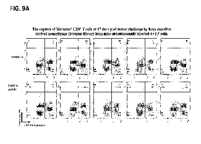
Tetramer+CD8+ T cells were detectable in mice challenge with Pan02-EV cells.
[0054] FIGs. 10A and 10B depicts the tumor growth curve of mice inoculated
with 4x10⁵
cells Pan02-EV (without neoantigen expressing Kras mutation) and Pan02-n (with
neoantigen
expressing Kras mutation) tumor cells as assessed via bioluminescence (FIG.
9A) and the
picture of mice at day 4 post tumor challenge evidencing no observation of
tumor at the site
of inoculation (red circle) (FIG. 9B).
[0055] FIGs. 11A-11C depict the presence or absence of Kras mutation
neoantigen specific
CD8+ T cells with Kras mutation specific Tetramer library in peripheral blood
of pancreatic
cancer patients (Patient It 1-13).
[0056] FIG. 12 depicts of a summary of the presence or absence of CD8+ T cells
in thirteen
confirmed pancreatic patients with a presence or absence of a Kras mutation
and a specific
HLA-A phenotype. In this experiment, twenty G12D neoantigen peptides
associated MHC
tetramers were included in the bait composition but only four G12R neoantigen
peptides
associated tetramers were included.
DETAILED DESCRIPTION
[0057] The present application provides methods of analyzing a sample of an
individual
exhibiting no pathological symptoms of a cancer. The application is based on
inventors'
unique insight that immune cells activated by tumor neoantigens can be
detectable in an
individual having cancer even before any pathological symptoms of cancer are
exhibited,
thus serving as useful markers for early cancer detection. As shown in Example
section,
exemplary Kras mutation associated neoantigen-specific T cells were
successfully detected in
mice intravenously or subcutaneously inoculated with tumor cells (of as low as
104 cells, see,
e.g., FIG. 8) expressing Kras mutation associated neoantigens and as early as
day 4 after
9
CA 03235256 2024-4- 16
WO 2023/077113
PCT/US2022/078953
inoculation (see e.g., FIG. 9A-9B), which is prior to the any detection via
bioluminescence
(see e.g., FIG. 10A-10B), or in pancreatic cancer patients harboring Kras
mutations (e.g., see
FIG. 11A-11C and 12). These data demonstrated that the provided methods can be
successfaully used for cancer detection including both early cancer detection
as well as for
monitoring cancer cells in individuals who may have residual minimal cancer
cells.
[0058] The present application provides methods of isolating and analyzing
immune cells in
individuals exhibiting no pathological symptoms of a cancer, as well as
methods of utilizing
information so obtained for cancer screening in healthy individuals, for
diagnosing or
assisting in diagnosis of individuals suspected of having cancer, and for
detecting minimal
residual cancer (MRD) in individuals who has been previously treated with an
anti-cancer
therapy and exhibits no pathological symptom of cancer after treatment. More
detailed
analysis on the isolated immune cells can be further used for classifying
cancer of the
individual.
[0059] Thus, the present application in one aspect provides methods of
analyzing a sample of
an individual exhibiting no pathological symptoms of a cancer. In some
embodiments, the
method comprises a) contacting the sample with a bait composition comprising a
display
moiety comprising a neoantigenic peptide under a condition sufficient for an
immune cell to
bind to the display moiety; b) isolating an immune cell associated with the
display moiety;
and c) analyzing the isolated immune cell. In some embodiments, the individual
has been
previously treated for cancer and exhibits no pathological symptom of a cancer
after the
treatment. In some embodiments, the method comprises a) analyzing a pre-
treatment sample
from the individual prior to anti-cancer therapy and a post-treatment sample
from the
individual; and b) identifying a difference in characteristics of the isolated
immune cell from
the pre-treatment sample and the isolated immune cell from the post-treatment
sample.
[0060] The present application in another aspect provides methods of detecting
cancer in an
individual exhibiting no pathological symptoms of a cancer, comprising:
analyzing a sample
from the individual, wherein a predetermined characteristic of the isolated
immune cell is
indicative of cancer in the individual.
[0061] The present application in another aspect provides methods of detecting
residual
cancer in an individual, wherein the individual has been previously treated
with an anti-
cancer therapy and exhibits no pathological symptom of cancer after treatment,
the method
CA 03235256 2024-4- 16
WO 2023/077113
PCT/US2022/078953
comprising analyzing a post-treatment sample from the individual, wherein a
predetermined
characterics of the isolated immune cell is indicative of residual cancer in
the individual. A
post-treatment sample refers to a sample harvested from an individual who has
been
subjected to a cancer treatment. In some embodiments, the post-treatment
sample is harvested
within about 1-3 weeks after treatment. In some embodiments, the post-
treatment sample is
harvested within about 1-3 months after treatment. In some embodiments, the
post-treatment
sample is harvested within about 6, 9, or 12 months after treatment.
[0062] The present application in another aspect provides method of treating a
cancer in an
individual, comprising a) diagnosing the individual as having cancer according
to the
diagnosis methods described herein; and b) subjecting the individual to an
anti-cancer
therapy.
Definitions
[0063] In general, terms used in the claims and the specification are intended
to be construed
as having the plain meaning understood by a person of ordinary skill in the
art. Certain terms
are defined below to provide additional clarity. In case of conflict between
the plain meaning
and the provided definitions, the provided definitions are to be used.
[0064] As used herein the term "antigen" is a substance that induces an immune
response.
[0065] As used herein the term "neoantigen" is an antigen that has at least
one alteration that
makes it distinct from the corresponding wild-type, parental antigen, e.g.,
via mutation in a
tumor cell or post-translational modification specific to a tumor cell. A
neoantigen can
include a polypeptide sequence. A mutation that results in a neoantigen can
include a
frameshift or non-frameshift indel, missense or nonsense substitution, splice
site alteration,
genomic rearrangement or gene fusion, or any genomic or expression alteration
giving rise to
a neoORF. A mutations can also include a splice variant. Post-translational
modifications
specific to a tumor cell can include aberrant phosphorylation. Post-
translational modifications
specific to a tumor cell can also include a proteasome-gencrated spliced
antigen. See Licpc et
al., A large fraction of HLA class I ligands are proteasome-generated spliced
peptides;
Science. 2016 Oct 21;354(6310):354-358.
[0066] As used herein the term "tumor neoantigen" or "cancer neoantigen" is a
neoantigen
present in a subject's tumor cell or tissue but not in the subject's
corresponding norrnal cell or
tissue.
11
CA 03235256 2024-4- 16
WO 2023/077113
PCT/US2022/078953
[0067] As used herein the term "missense mutation" is a mutation causing a
substitution from
one amino acid to another.
[0068] As used herein the term "nonsense mutation" is a mutation causing a
substitution from
an amino acid to a stop codon.
[0069] As used herein the term "frameshift mutation" is a mutation causing a
change in the
frame of the protein.
[0070] As used herein the term "indel" is an insertion or deletion of one or
more nucleic
acids.
[0071] As used herein, the term percent "identity," in the context of two or
more nucleic acid
or polypeptide sequences, refer to two or more sequences or subsequences that
have a
specified percentage of nucleotides or amino acid residues that are the same,
when compared
and aligned for maximum correspondence, as measured using one of the sequence
comparison algorithms described below (e.g., BLASTP and BLASTN or other
algorithms
available to persons of skill) or by visual inspection. Depending on the
application, the
percent "identity" can exist over a region of the sequence being compared,
e.g., over a
functional domain, or, alternatively, exist over the full length of the two
sequences to be
compared.
[0072] For sequence comparison, typically one sequence acts as a reference
sequence to
which test sequences are compared. When using a sequence comparison algorithm,
test and
reference sequences are input into a computer, subsequence coordinates are
designated, if
necessary, and sequence algorithm program parameters are designated. The
sequence
comparison algorithm then calculates the percent sequence identity for the
test sequence(s)
relative to the reference sequence, based on the designated program
parameters.
Alternatively, sequence similarity or dissimilarity can be established by the
combined
presence or absence of particular nucleotides, or, for translated sequences,
amino acids at
selected sequence positions (e.g., sequence motifs).
[0073] As used herein the term "epitope" is the specific portion of an antigen
typically bound
by an antibody or T-cell receptor.
12
CA 03235256 2024-4- 16
WO 2023/077113
PCT/US2022/078953
[0074] As used herein the term "immunogenic" is the ability to elicit an
immune response,
e.g., via T-cells, B cells, or both.
[0075] As used herein the term "HLA binding affinity" "MHC binding affinity"
means
affinity of binding between a specific antigen and a specific MHC allele.
[0076] As used herein the term "bait composition" is a composition comprising
a molecule
(e.g., a neoantigen peptide) used to enrich a cell that specifically binds to
the bait from a
sample.
[0077] As used herein the term "variant" is a difference between a subject's
nucleic acids and
the reference human genome used as a control.
[0078] As used herein the term "allele" is a version of a gene or a version of
a genetic
sequence or a version of a protein.
[0079] As used herein the term "IILA type" is the complement of IILA gene
alleles.
[0080] As used herein the term "exome" is a subset of the genome that codes
for proteins. An
exome can be the collective exons of a genome.
[0081] As used herein the term "proteome" is the set of all proteins expressed
and/or
translated by a cell, group of cells, or individual.
[0082] As used herein the term "dextramers" is a dextran-based peptide-MHC
multimers
used for antigen-specific immune-cell staining in flow cytometry.
[0083] As used herein the term "MHC multimers" is a peptide-MHC complex
comprising
multiple peptide- MHC monomer units.
[0084] As used herein the term "MHC tetramers" is a peptide-MHC complex
comprising four
peptide- MHC monomer units.
[0085] As used herein, "sample" refers to an aliquot of body fluid or a tissue
obtained from a
subject which contains an immune cell.
[0086] The term "mammal" encompasses both humans and non-humans and includes
but is
not limited to humans, non-human primates, canines, felines, murines, bovines,
equines, and
porcines.
13
CA 03235256 2024-4- 16
WO 2023/077113
PCT/US2022/078953
[0087] As used herein, "treatment" or "treating" is an approach for obtaining
beneficial or
desired results, including clinical results. For purposes of this application,
beneficial or
desired clinical results include, but are not limited to, one or more of the
following:
alleviating one or more symptoms resulting from the disease, diminishing the
extent of the
disease, stabilizing the disease (e.g., preventing or delaying the worsening
of the disease),
preventing or delaying the spread (e.g., metastasis) of the disease,
preventing or delaying the
recurrence of the disease, delaying or slowing the progression of the disease,
ameliorating the
disease state, providing a remission (partial or total) of the disease,
decreasing the dose of one
or more other medications required to treat the disease, delaying the
progression of the
disease, increasing or improving the quality of life, increasing weight gain,
and/or prolonging
survival. Also encompassed by "treatment" is a reduction of pathological
consequence of
cancer (such as, for example, tumor volume). The methods of the application
contemplate
any one or more of these aspects of treatment.
[0088] A "reference" as used herein, refers to any sample, standard, or level
that is used for
comparison purposes. A reference may he obtained from a healthy and/or non-
diseased
sample. In some examples, a reference may be obtained from an untreated
sample. In some
examples, a reference is obtained from a non-diseased or non-treated sample of
an individual.
In some examples, a reference is obtained from one or more healthy individuals
who are not
the individual or patient.
[0089] The terms "subject,- "individual,- and "patient- are used
interchangeably herein to
refer to a mammal, including, but not limited to, human, bovine, horse,
feline, canine, rodent,
or primate. In some embodiments, the individual is a human.
[0090] It is understood that embodiments of the application described herein
include
"consisting" and/or "consisting essentially of' embodiments.
[0091] Reference to "about" a value or parameter herein includes (and
describes) variations
that are directed to that value or parameter per se. For example, description
referring to
"about X- includes description of -X-.
[0092] As used herein, reference to "not" a value or parameter generally means
and describes
"other than" a value or parameter. For example, the method is not used to
treat cancer of type
X means the method is used to treat cancer of types other than X.
14
CA 03235256 2024-4- 16
WO 2023/077113
PCT/US2022/078953
[0093] The term "about X-Y" used herein has the same meaning as "about X to
about Y."
[0094] It should be noted that, as used in the specification and t e appended
claims, the
singular forms "a," "an," and "the" include plural referents unless the
context clearly dictates
otherwise.
[0095] Any terms not directly defined herein shall be understood to have the
meanings
commonly associated with them as understood within the art of the invention.
Certain terms
are discussed herein to provide additional guidance to the practitioner in
describing the
compositions, devices, methods and the like of aspects of the invention, and
how to make or
use them. It will be appreciated that the same thing may be said in more than
one way.
Consequently, alternative language and synonyms may be used for any one or
more of the
terms discussed herein. No significance is to be placed upon whether or not a
term is
elaborated or discussed herein. Some synonyms or substitutable methods,
materials and the
like are provided. Recital of one or a few synonyms or equivalents does not
exclude use of
other synonyms or equivalents, unless it is explicitly stated. Use of
examples, including
examples of terms, is for illustrative purposes only and does not limit the
scope and meaning
of the aspects of the invention herein.
Methods of analyzing a sample of an individual exhibiting no pathological
symptoms of
a cancer
[0096] In some embodiments, there is provided a method of analyzing a sample
(e.g., blood,
plasma, or a PBMC sample) of an individual exhibiting no pathological symptoms
of a
cancer. In some embodiments, the method comprises a) contacting the sample
with a bait
composition comprising a display moiety comprising a neoantigenic peptide
under a
condition sufficient for an immune cell (e.g., a T cell, a cytotoxic T cell, a
helper T cell, a
memory T cell, and/or a tumor infiltrating T cell) to bind to the display
moiety; b) isolating
an immune cell associated with the display moiety; and c) analyzing the
isolated immune
cell. In some embodiments, the method further comprises culturing the isolated
immune cell
prior to the analyzing step. In some embodiments, the display moiety comprises
a particle
(e.g., a particle selected from the group consisting of: a surface, a
nanoparticle, a bead, and a
polymer). In some embodiments, the display moiety further comprises a
detectable label. In
some embodiments, the detectable label is a fluorophore. In some embodiments,
analyzing
the sequences of the one or more nucleic acids comprises whole genome
sequencing,
CA 03235256 2024-4- 16
WO 2023/077113
PCT/US2022/078953
RNAseq sequencing, and/or subjecting the isolated immune cell to mass
spectrometry
analysis.
[0097] In some embodiments, there is provided a method of analyzing a sample
(e.g., blood,
plasma, or a PBMC sample) of an individual exhibiting no pathological symptoms
of a
cancer, wherein the method comprises a) contacting the sample with a bait
composition
comprising a display moiety comprising a neoantigenic peptide under a
condition sufficient
for an immune cell (e.g., a T cell, a cytotoxic T cell, a helper T cell, a
memory T cell, and/or
a tumor infiltrating T cell) to bind to the display moiety; b) isolating an
immune cell
associated with the display moiety; and c) analyzing the isolated immune cell,
wherein the
display moiety comprises two or more (e.g., four) neoantigenic peptides. In
some
embodiments, the two or more neoantigenic peptides in the display moiety are
the same. In
some embodiments, the neoantigenic peptide has one or more of the following
characteristics: a) having a binding affinity of about 1 nM to about 5000 nM
(e.g., about 1
nM to about 50 nM, about 50 nM to about 500 nM, about 500 nM to about 5000 nM)
to an
MHC molecule; b) having a binding affinity of about 1 nM to about 5000 nM
(e.g., about 1
nM to about 50 nM, about 50 nM to about 500 nM, about 500 nM to about 5000 nM)
to a
cognate TCR molecule; c) having a mutation relative to a wildtype peptide,
optionally at the
third amino acid position counting from the N-terminus; d) is hydrophobic; and
e) has high
content of aromatic residues. In some embodiments, the neoantigenic peptide
has low
immunogenicity. In some embodiments, the display moiety comprises an MHC
molecule
complexed with the neoantigenic peptide. In some embodiments, the MHC molecule
is a
MHC class I molecule and/or a MHC class II molecule. In some embodiments, the
isolated
immune cell is an isolated single immune cell. In some embodiments, the
isolated immune
cell is in a mixture of immune cells. In some embodiments. the mixture of
immune cells is a
mixture comprising T cells, memory T cells, macrophage cells, or dendritic
cells, or
combinations thereof. In some embodiments, analyzing the isolated immune cell
comprises
detecting and/or quantifying the isolated immune cell. In some embodiments,
analyzing the
isolated immune cell comprises sequencing one or more nucleic acids in the
isolated immune
cell, optionally further comprising analyzing the sequences of the one or more
nucleic acids
(e.g., a TCR related sequence). In some embodiments, analyzing the isolated
immune cell
further comprises identifying one or more epigenetic modifications (e.g., DNA
or RNA
methylation, hydroxymethylation, and/or histone modifications such as
acetylation.
methylation, glycosylation) in the isolated immune cell. In some embodiments,
the method
16
CA 03235256 2024-4- 16
WO 2023/077113
PCT/US2022/078953
further comprises generating a report comprising information about the cancer
status in the
individual. In some embodiments, the information about cancer status
comprises:
classification of cancer; type of cancer; nature of cancer; origin of cancer;
stage of cancer;
likelihood of cancer progression; likelihood of developing one or more cancer
symptoms;
molecular diagnosis; NGS pathology; and/or treatment options for the
individual. In some
embodiments, the bait composition comprises a plurality of different display
moieties. In
some embodiments, each of the plurality of different display moieties in the
bait composition
comprises a different neoantigenic peptide (e.g., at least about two, four,
10, 25, 50, 75, or
100 different display moieties, each comprising a different MHC molecule). In
some
embodiments, each of the different display moieties comprising different MHC
molecules
comprises a different detectable label (e.g., a fluorophore). In some
embodiments, the
isolating step comprises using fluorescence-activated cell sorting (FACS),
and/or separating
immune cells associated with each of the different display moieties comprising
different
MHC molecules into different populations, optionally further comprises
contacting each of a
plurality of different display moieties with a sample from the individual
separately and
isolating the immune cell associated with each of the different display
moiety. In some
embodiments, the method further comprises culturing the isolated immune cell
prior to the
analyzing step. In some embodiments, the display moiety comprises a particle
(e.g., a particle
selected from the group consisting of: a surface, a nanoparticle, a bead, and
a polymer). In
some embodiments, the display moiety further comprises a detectable label. In
some
embodiments, the detectable label is a fluorophore. In some embodiments,
analyzing the
sequences of the one or more nucleic acids comprises whole genome sequencing,
RNAseq
sequencing, and/or subjecting the isolated immune cell to mass spectrometry
analysis.
[0098] In some embodiments, there is provided a method of analyzing a sample
(e.g., blood,
plasma, or a PBMC sample) of an individual exhibiting no pathological symptoms
of a
cancer, wherein the method comprises a) contacting the sample with a bait
composition
comprising a display moiety comprising a neoantigenic peptide under a
condition sufficient
for an immune cell (e.g., a T cell, a cytotoxic T cell, a helper T cell, a
memory T cell, and/or
a tumor infiltrating T cell) to bind to the display moiety; b) isolating an
immune cell
associated with the display moiety; and c) analyzing the isolated immune cell,
wherein the
display moiety comprises an MHC molecule complexed with the neoantigenic
peptide. In
some embodiments, the MHC molecule is a MHC class I molecule. In some
embodiments,
the MHC class I molecule is selected from the group consisting of HLA-A, HLA-
B, and
17
CA 03235256 2024-4- 16
WO 2023/077113
PCT/US2022/078953
HLA-C. In some embodiments, the peptide complexed with a MHC class I molecule
is about
8 to about 10 amino acids long. In some embodiments, the MHC is a MHC class II
molecule.
In some embodiments, the MHC class II molecule is selected from the group
consisting of
HLA-DQ and HLA-DR. In some embodiments, the neoantigenic peptide complexed
with the
MHC class II molecule is about 10 to about 20 amino acids long. In some
embodiments, the
isolated immune cell is an isolated single immune cell. In some embodiments,
the isolated
immune cell is in a mixture of immune cells. In some embodiments, the mixture
of immune
cells is a mixture comprising T cells. memory T cells, macrophage cells, or
dendritic cells, or
combinations thereof. In some embodiments, analyzing the isolated immune cell
comprises
detecting and/or quantifying the isolated immune cell. In some embodiments,
analyzing the
isolated immune cell comprises sequencing one or more nucleic acids in the
isolated immune
cell, optionally further comprising analyzing the sequences of the one or more
nucleic acids
(e.g., a TCR related sequence). In some embodiments, analyzing the isolated
immune cell
further comprises identifying one or more epigenetic modifications (e.g., DNA
or RNA
methylation, hydroxymethylation, and/or histone modifications such as
acetylation,
methylation, glycosylation) in the isolated immune cell. In some embodiments,
the method
further comprises generating a report comprising information about the cancer
status in the
individual. In some embodiments, the information about cancer status
comprises:
classification of cancer; type of cancer; nature of cancer; origin of cancer;
stage of cancer;
likelihood of cancer progression; likelihood of developing one or more cancer
symptoms;
molecular diagnosis; NGS pathology; and/or treatment options for the
individual. In some
embodiments, the bait composition comprises a plurality of different display
moieties. In
some embodiments, each of the plurality of different display moieties in the
bait composition
comprises a different neoantigenic peptide (e.g., at least about two, four,
10, 25, 50, 75, or
100 different display moieties, each comprising a different MHC molecule). In
some
embodiments, each of the different display moieties comprising different MHC
molecules
comprises a different detectable label (e.g., a fluorophore). In some
embodiments, the
isolating step comprises using fluorescence-activated cell sorting (FACS),
and/or separating
immune cells associated with each of the different display moieties comprising
different
MHC molecules into different populations, optionally further comprises
contacting each of a
plurality of different display moieties with a sample from the individual
separately and
isolating the immune cell associated with each of the different display
moiety. In some
embodiments, the method further comprises culturing the isolated immune cell
prior to the
analyzing step. In some embodiments, the display moiety comprises a particle
(e.g., a particle
18
CA 03235256 2024-4- 16
WO 2023/077113
PCT/US2022/078953
selected from the group consisting of: a surface, a nanoparticle, a bead, and
a polymer). In
some embodiments, the display moiety further comprises a detectable label. In
some
embodiments, the detectable label is a fluorophore. In some embodiments,
analyzing the
sequences of the one or more nucleic acids comprises whole genome sequencing,
RNAseq
sequencing, and/or subjecting the isolated immune cell to mass spectrometry
analysis.
[0099] In some embodiments, there is provided a method of analyzing a sample
(e.g., blood,
plasma, or a PBMC sample) of an individual exhibiting no pathological symptoms
of a
cancer, wherein the method comprises a) contacting the sample with a bait
composition
comprising a display moiety comprising a neoantigenic peptide under a
condition sufficient
for an immune cell (e.g., a T cell, a cytotoxic T cell, a helper T cell, a
memory T cell, and/or
a tumor infiltrating T cell) to bind to the display moiety; b) isolating an
immune cell
associated with the display moiety; and c) analyzing the isolated immune cell,
wherein the
isolated immune cell comprises a mixture of immune cells. In some embodiments,
the
mixture of immune cells is a mixture comprising T cells, memory T cells,
macrophage cells,
and/or dendritic cells, or combinations thereof. In some embodiments,
analyzing the isolated
immune cell comprises detecting and/or quantifying the isolated immune cell.
In some
embodiments, analyzing the isolated immune cell comprises sequencing one or
more nucleic
acids in the isolated immune cell, optionally further comprising analyzing the
sequences of
the one or more nucleic acids (e.g., a TCR related sequence). In some
embodiments,
analyzing the isolated immune cell further comprises identifying one or more
epigenetic
modifications (e.g., DNA or RNA methylation, hydroxymethylation, and/or
histone
modifications such as acetylation, methylation, glycosylation) in the isolated
immune cell. In
some embodiments, the method further comprises generating a report comprising
information
about the cancer status in the individual. In some embodiments, the
information about cancer
status comprises: classification of cancer; type of cancer; nature of cancer;
origin of cancer;
stage of cancer; likelihood of cancer progression; likelihood of developing
one or more
cancer symptoms; molecular diagnosis; NGS pathology; and/or treatment options
for the
individual. In some embodiments, the bait composition comprises a plurality of
different
display moieties. In some embodiments, each of the plurality of different
display moieties in
the bait composition comprises a different neoantigenic peptide (e.g., at
least about two, four,
10, 25, 50, 75, or 100 different display moieties, each comprising a different
MHC molecule).
In some embodiments, each of the different display moieties comprising
different MHC
19
CA 03235256 2024-4- 16
WO 2023/077113
PCT/US2022/078953
molecules comprises a different detectable label (e.g., a fluorophore). In
some embodiments,
the isolating step comprises using fluorescence-activated cell sorting (FACS),
and/or
separating immune cells associated with each of the different display moieties
comprising
different MHC molecules into different populations, optionally further
comprises contacting
each of a plurality of different display moieties with a sample from the
individual separately
and isolating the immune cell associated with each of the different display
moiety. In some
embodiments, the method further comprises culturing the isolated immune cell
prior to the
analyzing step. In some embodiments, the display moiety comprises a particle
(e.g., a particle
selected from the group consisting of: a surface, a nanoparticle, a bead, and
a polymer). In
some embodiments, the display moiety further comprises a detectable label. In
some
embodiments, the detectable label is a fluorophore. In some embodiments,
analyzing the
sequences of the one or more nucleic acids comprises whole genome sequencing,
RNAseq
sequencing, and/or subjecting the isolated immune cell to mass spectrometry
analysis.
[0100] In some embodiments, there is provided a method of analyzing a sample
(e.g., blood,
plasma, or a PBMC sample) of an individual exhibiting no pathological symptoms
of a
cancer, wherein the method comprises a) contacting the sample with a bait
composition
comprising a display moiety comprising a neoantigenic peptide under a
condition sufficient
for an immune cell (e.g., a T cell, a cytotoxic T cell, a helper T cell, a
memory T cell, and/or
a tumor infiltrating T cell) to bind to the display moiety; b) isolating an
immune cell
associated with the display moiety; and c) analyzing the isolated immune cell,
wherein
analyzing the isolated immune cell comprises detecting and/or quantifying the
isolated
immune cell. In some embodiments, analyzing the isolated immune cell comprises
sequencing one or more nucleic acids in the isolated immune cell, optionally
further
comprising analyzing the sequences of the one or more nucleic acids (e.g., a
TCR related
sequence). In some embodiments, analyzing the isolated immune cell further
comprises
identifying one or more epigenetic modifications (e.g., DNA or RNA
methylation,
hydroxymethylation, and/or histone modifications such as acetylation,
methylation,
glycosylation) in the isolated immune cell. In some embodiments, the method
further
comprises generating a report comprising information about the cancer status
in the
individual. In some embodiments, the information about cancer status
comprises:
classification of cancer; type of cancer; nature of cancer; origin of cancer;
stage of cancer;
likelihood of cancer progression; likelihood of developing one or more cancer
symptoms;
molecular diagnosis; NGS pathology; and/or treatment options for the
individual. In some
CA 03235256 2024-4- 16
WO 2023/077113
PCT/US2022/078953
embodiments, the bait composition comprises a plurality of different display
moieties. In
some embodiments, each of the plurality of different display moieties in the
bait composition
comprises a different neoantigenic peptide (e.g., at least about two, four,
10, 25, 50, 75, or
100 different display moieties, each comprising a different MHC molecule). In
some
embodiments, each of the different display moieties comprising different MHC
molecules
comprises a different detectable label (e.g., a fluorophore). In some
embodiments, the
isolating step comprises using fluorescence-activated cell sorting (FACS),
and/or separating
immune cells associated with each of the different display moieties comprising
different
MHC molecules into different populations, optionally further comprises
contacting each of a
plurality of different display moieties with a sample from the individual
separately and
isolating the immune cell associated with each of the different display
moiety. In some
embodiments, the method further comprises culturing the isolated immune cell
prior to the
analyzing step. In some embodiments, the display moiety comprises a particle
(e.g., a particle
selected from the group consisting of: a surface, a nanoparticle, a bead, and
a polymer). In
some embodiments, the display moiety further comprises a detectable label. In
some
embodiments, the detectable label is a fluorophore. In some embodiments,
analyzing the
sequences of the one or more nucleic acids comprises whole genome sequencing,
RNAseq
sequencing, and/or subjecting the isolated immune cell to mass spectrometry
analysis.
[0101] In some embodiments, there is provided a method of analyzing a sample
(e.g., blood,
plasma, or a PBMC sample) of an individual exhibiting no pathological symptoms
of a
cancer, wherein the method comprises a) contacting the sample with a bait
composition
comprising a display moiety comprising a neoantigenic peptide under a
condition sufficient
for an immune cell (e.g., a T cell, a cytotoxic T cell, a helper T cell, a
memory T cell, and/or
a tumor infiltrating T cell) to bind to the display moiety; b) isolating an
immune cell
associated with the display moiety; and c) analyzing the isolated immune cell,
wherein
analyzing the isolated immune cell comprises sequencing one or more nucleic
acids in the
isolated immune cell, optionally wherein analyzing the isolated immune cell
further
comprises analyzing the sequences of the one or more nucleic acids (e.g., a
TCR related
sequence). In some embodiments, analyzing the sequences of the one or more
nucleic acids
comprises: a) obtaining an enriched sample from the isolated immune cell,
wherein the
enriched sample is enriched for the one or more nucleic acids; and b)
sequencing the one or
more nucleic acids in the enriched sample. In some embodiments, analyzing the
isolated
immune cell further comprises identifying one or more epigenetic modifications
(e.g., DNA
21
CA 03235256 2024-4- 16
WO 2023/077113
PCT/US2022/078953
or RNA methylation, hydroxymethylation, and/or histone modifications such as
acetylation,
methylation, glycosylation) in the isolated immune cell. In some embodiments,
the method
further comprises generating a report comprising information about the cancer
status in the
individual. In some embodiments, the information about cancer status
comprises:
classification of cancer; type of cancer; nature of cancer; origin of cancer;
stage of cancer;
likelihood of cancer progression; likelihood of developing one or more cancer
symptoms;
molecular diagnosis; NGS pathology; and/or treatment options for the
individual. In some
embodiments, the bait composition comprises a plurality of different display
moieties. In
some embodiments, each of the plurality of different display moieties in the
bait composition
comprises a different neoantigenic peptide (e.g., at least about two, four,
10, 25, 50, 75, or
100 different display moieties, each comprising a different MHC molecule). In
some
embodiments, each of the different display moieties comprising different MHC
molecules
comprises a different detectable label (e.g., a fluorophore). In some
embodiments, the
isolating step comprises using fluorescence-activated cell sorting (FACS),
and/or separating
immune cells associated with each of the different display moieties comprising
different
MHC molecules into different populations, optionally further comprises
contacting each of a
plurality of different display moieties with a sample from the individual
separately and
isolating the immune cell associated with each of the different display
moiety. In some
embodiments, the method further comprises culturing the isolated immune cell
prior to the
analyzing step. In some embodiments, the display moiety comprises a particle
(e.g., a particle
selected from the group consisting of: a surface, a nanoparticle, a bead, and
a polymer). In
some embodiments, the display moiety further comprises a detectable label. In
some
embodiments, the detectable label is a fluorophore. In some embodiments,
analyzing the
sequences of the one or more nucleic acids comprises whole genome sequencing,
RNAseq
sequencing, and/or subjecting the isolated immune cell to mass spectrometry
analysis.
[0102] In some embodiments, there is provided a method of analyzing a sample
(e.g., blood,
plasma, or a PBMC sample) of an individual exhibiting no pathological symptoms
of a
cancer, wherein the method comprises a) contacting the sample with a bait
composition
comprising a display moiety comprising a neoantigenic peptide under a
condition sufficient
for an immune cell (e.g., a T cell, a cytotoxic T cell, a helper T cell, a
memory T cell, and/or
a tumor infiltrating T cell) to bind to the display moiety; b) isolating an
immune cell
associated with the display moiety; and c) analyzing the isolated immune cell,
wherein
analyzing the isolated immune cell comprises identifying one or more
epigenetic
CA 03235256 2024-4- 16
WO 2023/077113
PCT/US2022/078953
modifications (e.g., DNA or RNA methylation, hydroxymethylation, and/or
histone
modifications such as acetylation, methylation, glycosylation) in the isolated
immune cell. In
some embodiments, the method further comprises generating a report comprising
information
about the cancer status in the individual. In some embodiments, the
information about cancer
status comprises: classification of cancer; type of cancer; nature of cancer;
origin of cancer;
stage of cancer; likelihood of cancer progression; likelihood of developing
one or more
cancer symptoms; molecular diagnosis; NGS pathology; and/or treatment options
for the
individual. In some embodiments, the bait composition comprises a plurality of
different
display moieties. In some embodiments, each of the plurality of different
display moieties in
the bait composition comprises a different neoantigenic peptide (e.g., at
least about two, four,
10, 25, 50, 75, or 100 different display moieties, each comprising a different
MHC molecule).
In some embodiments, each of the different display moieties comprising
different MHC
molecules comprises a different detectable label (e.g., a fluorophore). In
some embodiments,
the isolating step comprises using fluorescence-activated cell sorting (FACS),
and/or
separating immune cells associated with each of the different display moieties
comprising
different MHC molecules into different populations, optionally further
comprises contacting
each of a plurality of different display moieties with a sample from the
individual separately
and isolating the immune cell associated with each of the different display
moiety. In some
embodiments, the method further comprises culturing the isolated immune cell
prior to the
analyzing step. In some embodiments, the display moiety comprises a particle
(e.g., a particle
selected from the group consisting of: a surface, a nanoparticle, a bead, and
a polymer). In
some embodiments, the display moiety further comprises a detectable label. In
some
embodiments, the detectable label is a fluorophore. In some embodiments,
analyzing the
sequences of the one or more nucleic acids comprises whole genome sequencing,
RNAseq
sequencing, and/or subjecting the isolated immune cell to mass spectrometry
analysis.
[0103] In some embodiments, there is provided a method of analyzing a sample
(e.g., blood,
plasma, or a PBMC sample) of an individual exhibiting no pathological symptoms
of a
cancer. In some embodiments, the method comprises a) contacting the sample
with a bait
composition comprising a display moiety comprising a neoantigenic peptide
under a
condition sufficient for an immune cell (e.g., a T cell, a cytotoxic T cell, a
helper T cell, a
memory T cell, and/or a tumor infiltrating T cell) to bind to the display
moiety; b) isolating
an immune cell associated with the display moiety; and c) analyzing the
isolated immune
cell, wherein the method further comprises generating a report comprising
information about
23
CA 03235256 2024-4- 16
WO 2023/077113
PCT/US2022/078953
the cancer status in the individual. In some embodiments, the information
about cancer status
comprises: classification of cancer; type of cancer; nature of cancer; origin
of cancer; stage of
cancer; likelihood of cancer progression; likelihood of developing one or more
cancer
symptoms; molecular diagnosis; NGS pathology; and/or treatment options for the
individual.
In some embodiments, the bait composition comprises a plurality of different
display
moieties. In some embodiments, each of the plurality of different display
moieties in the bait
composition comprises a different neoantigenic peptide (e.g., at least about
two. four, 10, 25,
50, 75, or 100 different display moieties, each comprising a different MHC
molecule). In
some embodiments, each of the different display moieties comprising different
MHC
molecules comprises a different detectable label (e.g., a fluorophore). In
some embodiments,
the isolating step comprises using fluorescence-activated cell sorting (FACS),
and/or
separating immune cells associated with each of the different display moieties
comprising
different MHC molecules into different populations, optionally further
comprises contacting
each of a plurality of different display moieties with a sample from the
individual separately
and isolating the immune cell associated with each of the different display
moiety. In some
embodiments, the method further comprises culturing the isolated immune cell
prior to the
analyzing step. In some embodiments, the display moiety comprises a particle
(e.g., a particle
selected from the group consisting of: a surface, a nanoparticle, a bead, and
a polymer). In
some embodiments, the display moiety further comprises a detectable label. In
some
embodiments, the detectable label is a fluorophore. In some embodiments,
analyzing the
sequences of the one or more nucleic acids comprises whole genome sequencing,
RNAseq
sequencing, and/or subjecting the isolated immune cell to mass spectrometry
analysis.
[0104] In some embodiments, there is provided a method of analyzing a sample
(e.g., blood,
plasma, or a PBMC sample) of an individual exhibiting no pathological symptoms
of a
cancer. In some embodiments, the method comprises a) contacting the sample
with a bait
composition comprising a display moiety comprising a neoantigenic peptide
under a
condition sufficient for an immune cell (e.g., a T cell, a cytotoxic T cell, a
helper T cell, a
memory T cell, and/or a tumor infiltrating T cell) to bind to the display
moiety; b) isolating
an immune cell associated with the display moiety; and c) analyzing the
isolated immune
cell, wherein the bait composition comprises a plurality of different display
moieties. In some
embodiments, each of the plurality of different display moieties in the bait
composition
comprises a different neoantigenic peptide (e.g., at least about two, four,
10, 25, 50, 75, or
100 different display moieties, each comprising a different MHC molecule). In
some
24
CA 03235256 2024-4- 16
WO 2023/077113
PCT/US2022/078953
embodiments, each of the different display moieties comprising different MHC
molecules
comprises a different detectable label (e.g., a fluorophore). In some
embodiments, the
isolating step comprises using fluorescence-activated cell sorting (FACS). In
some
embodiments, the isolating step comprises separating immune cells associated
with each of
the different display moieties comprising different MHC molecules into
different
populations. In some embodiments, the method further comprises contacting each
of a
plurality of different display moieties with a sample from the individual
separately and
isolating the immune cell associated with each of the different display
moiety. In some
embodiments, the method further comprises culturing the isolated immune cell
prior to the
analyzing step. In some embodiments, the display moiety comprises a particle
(e.g., a particle
selected from the group consisting of: a surface, a nanoparticle, a bead, and
a polymer). In
some embodiments, the display moiety further comprises a detectable label. In
some
embodiments, the detectable label is a fluorophore. In some embodiments,
analyzing the
sequences of the one or more nucleic acids comprises whole genome sequencing,
RNAseq
sequencing, and/or subjecting the isolated immune cell to mass spectrometry
analysis.
[0105] In some embodiments, there is provided a method of analyzing a sample
(e.g., blood,
plasma, or a PBMC sample) of an individual exhibiting no pathological symptoms
of a
cancer, wherein the method comprises a) contacting the sample with a bait
composition
comprising a display moiety comprising a neoantigenic peptide under a
condition sufficient
for an immune cell to bind to the display moiety; b) isolating an immune cell
associated with
the display moiety; and c) analyzing the isolated immune cell, wherein the
isolated immune
cell is a T cell. In some embodiments, the T cell is a cytotoxic T cell. In
some embodiments,
the T cell is a helper T cell. In some embodiments, the T cell is a memory T
cell. In some
embodiments, the T cell is a tumor infiltrating T cell. In some embodiments,
the display
moiety comprises two or more (e.g., four) neoantigenic peptides. In some
embodiments, the
neoantigenic peptide has one or more of the following characteristics: a)
having a binding
affinity of about 1 nM to about 5000 nM (e.g., about 1 nM to about 50 nM,
about 50 nM to
about 500 nM, about 500 nM to about 5000 nM) to an MHC molecule; b) having a
binding
affinity of about 1 nM to about 5000 nM (e.g., about 1 nM to about 50 nM,
about 50 nM to
about 500 nM, about 500 nM to about 5000 nM) to a cognate TCR molecule; c)
having a
mutation relative to a wildtype peptide, optionally at the third amino acid
position counting
from the N-terminus; d) is hydrophobic; and e) has high content of aromatic
residues. In
some embodiments, the neoantigenic peptide has low immunogenicity. In some
CA 03235256 2024-4- 16
WO 2023/077113
PCT/US2022/078953
embodiments, the display moiety comprises an MHC molecule complexed with the
neoantigenic peptide. In some embodiments, the MHC molecule is a MHC class I
molecule
and/or a MHC class II molecule. In some embodiments, analyzing the isolated
immune cell
comprises detecting and/or quantifying the isolated immune cell. In some
embodiments,
analyzing the isolated immune cell comprises sequencing one or more nucleic
acids in the
isolated immune cell, optionally further comprising analyzing the sequences of
the one or
more nucleic acids (e.g., a TCR related sequence). In some embodiments,
analyzing the
isolated immune cell further comprises identifying one or more epigenetic
modifications
(e.g., DNA or RNA methylation, hydroxymethylation, and/or histone
modifications such as
acetylation, methylation, glycosylation) in the isolated immune cell. In some
embodiments,
the method further comprises generating a report comprising information about
the cancer
status in the individual. In some embodiments, the information about cancer
status comprises:
classification of cancer; type of cancer; nature of cancer; origin of cancer;
stage of cancer;
likelihood of cancer progression; likelihood of developing one or more cancer
symptoms;
molecular diagnosis; NGS pathology; and/or treatment options for the
individual. In some
embodiments, the bait composition comprises a plurality of different display
moieties. In
some embodiments, each of the plurality of different display moieties in the
bait composition
comprises a different neoantigenic peptide (e.g., at least about two, four,
10, 25, 50, 75, or
100 different display moieties, each comprising a different MHC molecule). In
some
embodiments, each of the different display moieties comprising different MHC
molecules
comprises a different detectable label (e.g., a fluorophore). In some
embodiments, the method
further comprises culturing the isolated immune cell prior to the analyzing
step. In some
embodiments, the display moiety comprises a particle (e.g., a particle
selected from the group
consisting of: a surface, a nanoparticle, a bead, and a polymer). In some
embodiments, the
display moiety further comprises a detectable label (e.g., a fluorophore). In
some
embodiments, analyzing the sequences of the one or more nucleic acids
comprises whole
genome sequencing, RNAseq sequencing, and/or subjecting the isolated immune
cell to mass
spectrometry analysis.
[0106] In some embodiments, there is provided a method of analyzing a sample
(e.g., blood,
plasma, or a PBMC sample) of an individual exhibiting no pathological symptoms
of a
cancer, wherein the method comprises a) contacting the sample with a bait
composition
comprising a display moiety comprising a neoantigenic peptide under a
condition sufficient
for an immune cell to bind to the display moiety; b) isolating an immune cell
associated with
26
CA 03235256 2024-4- 16
WO 2023/077113
PCT/US2022/078953
the display moiety; and c) analyzing the isolated immune cell, wherein the
display moiety
comprises an MHC molecule complexed with the neoantigenic peptide, wherein the
MHC
molecule is HLA-A * 24:02, HLA-A*11:01, HLA-A*02:01, or HLA-A*03:01. In some
embodiments, the display moiety comprises two or more (such as two, three and
four)
different kinds of MHC class I molecules selected from the group consisting of
HLA-A
24:02, HLA-A*11:01, HLA-A*02:01, and HLA-A*03:01. In some embodiments, the
display
moiety further comprises a MHC class II molecule. In some embodiments, the
display moiety
does not comprise a MHC class II molecule. In some embodiments, the isolated
immune cell
is an isolated single immune cell. In some embodiments, the isolated immune
cell is in a
mixture of immune cells. In some embodiments, the mixture of immune cells is a
mixture
comprising T cells, memory T cells, macrophage cells, or dendritic cells, or
combinations
thereof. In some embodiments, the display moiety comprises two or more (e.g.,
four)
neoantigenic peptides. In some embodiments, the neoantigenic peptide has one
or more of the
following characteristics: a) having a binding affinity of about 1 nM to about
5000 nM (e.g.,
about 1 nM to about 50 nM, about 50 nM to about 500 nM, about 500 nM to about
5000 nM)
to an MHC molecule; b) having a binding affinity of about 1 nM to about 5000
nM
about 1 nM to about 50 nM, about 50 nM to about 500 nM, about 500 nM to about
5000 nM)
to a cognate TCR molecule; c) having a mutation relative to a wildtype
peptide, optionally at
the third amino acid position counting from the N-terminus; d) is hydrophobic;
and e) has
high content of aromatic residues. In some embodiments, the neoantigenic
peptide has low
immunogenicity. In some embodiments, analyzing the isolated immune cell
comprises
detecting and/or quantifying the isolated immune cell. In some embodiments,
analyzing the
isolated immune cell comprises sequencing one or more nucleic acids in the
isolated immune
cell, optionally further comprising analyzing the sequences of the one or more
nucleic acids
(e.g., a TCR related sequence). In some embodiments, analyzing the isolated
immune cell
further comprises identifying one or more epigenetic modifications (e.g., DNA
or RNA
methylation, hydroxymethylation, and/or histone modifications such as
acetylation.
methylation, glycosylation) in the isolated immune cell. In some embodiments,
the method
further comprises generating a report comprising information about the cancer
status in the
individual. In some embodiments, the information about cancer status
comprises:
classification of cancer; type of cancer; nature of cancer; origin of cancer;
stage of cancer;
likelihood of cancer progression; likelihood of developing one or more cancer
symptoms;
molecular diagnosis; NGS pathology; and/or treatment options for the
individual. In some
embodiments, the bait composition comprises a plurality of different display
moieties. In
27
CA 03235256 2024-4- 16
WO 2023/077113
PCT/US2022/078953
some embodiments, each of the plurality of different display moieties in the
bait composition
comprises a different neoantigenic peptide (e.g., at least about two, four,
10, 25, 50, 75, or
100 different display moieties, each comprising a different MHC molecule). In
some
embodiments, each of the different display moieties comprising different MHC
molecules
comprises a different detectable label (e.g., a fluorophore). In some
embodiments, the method
further comprises culturing the isolated immune cell prior to the analyzing
step. In some
embodiments, the display moiety comprises a particle (e.g., a particle
selected from the group
consisting of: a surface, a nanoparticle, a bead, and a polymer). In some
embodiments, the
display moiety further comprises a detectable label (e.g., a fluorophore). In
some
embodiments, analyzing the sequences of the one or more nucleic acids
comprises whole
genome sequencing, RNAseq sequencing, and/or subjecting the isolated immune
cell to mass
spectrometry analysis.
[0107] In some embodiments, there is provided a method of analyzing a sample
(e.g., blood,
plasma, or a PBMC sample) of an individual (e.g., an individual exhibiting no
pathological
symptoms of a cancer), wherein the method comprises a) contacting the sample
with a bait
composition comprising a display moiety comprising a plurality of neoantigenic
peptides
under a condition sufficient for an immune cell to bind to the display moiety;
b) isolating an
immune cell associated with the display moiety; and c) analyzing the isolated
immune cell,
wherein the display moiety comprises one or more MHC molecule complexed with
the
plurality of neoantigenic peptides, optionally wherein the MHC molecule is HLA-
A * 24:02,
HLA-A*11:01, HLA-A*02:01, or HLA-A*03:01. In some embodiments, at least one
(e.g.,
each) of the plurality of neoantigen peptides comprises one or more known
mutations
associated with a cancer (e.g., Kras G12C, G12D, G12R, G12V. G121, and/or G12A
mutations). In some embodiments, the plurality of neoantigen peptides comprise
at least 5,
10, 15, or 20 distinct neoantigen peptides each comprising a single known
mutation
associated with a cancer (e.g., Kras G12C, G12D, G12R, G12V. G12I, and/or G12A
mutations). See FIG. 1 for exemplified design of a plurality of neoantigen
peptides associated
with a single known mutation. In some embodiments, the plurality of neoantigen
peptides
comprise at least 5, 10, 15, or 20 distinct neoantigen peptides associated
with Kras G12C
mutation (e.g., as shown in FIG. 1). In some embodiments, the plurality of
neoantigen
peptides comprise at least 5. 10, 15, or 20 distinct neoantigen peptides
associated with Kras
G12D mutation (e.g., as shown in FIG. 1). In some embodiments, the plurality
of neoantigen
peptides comprise at least 5. 10, 15, or 20 distinct neoantigen peptides
associated with Kras
28
CA 03235256 2024-4- 16
WO 2023/077113
PCT/US2022/078953
G12R mutation (e.g., as shown in FIG. 1). In some embodiments, the plurality
of neoantigen
peptides comprise at least 5. 10, 15, or 20 distinct neoantigen peptides
associated with Kras
G12V mutation (e.g., as shown in FIG. 1). In some embodiments, the plurality
of neoantigen
peptides comprise at least 5. 10, 15, or 20 distinct neoantigen peptides
associated with Kras
G12I mutation (e.g., as shown in FIG. 1). In some embodiments, the plurality
of neoantigen
peptides comprise at least 5. 10, 15, or 20 distinct neoantigen peptides
associated with Kras
G12A mutation (e.g., as shown in FIG. 1). In some embodiments, the display
moiety
comprises two or more (such as two, three and four) different kinds of MHC
class I
molecules selected from the group consisting of HLA-A * 24:02, HLA-A*11:01,
HLA-
A*02:01, and HLA-A*03:01. In some embodiments, the display moiety further
comprises a
MHC class II molecule. In some embodiments, the display moiety does not
comprise a MHC
class II molecule. In some embodiments, the isolated immune cell is an
isolated single
immune cell. In some embodiments, the isolated immune cell is in a mixture of
immune cells.
In some embodiments, the mixture of immune cells is a mixture comprising T
cells, memory
T cells, macrophage cells, or dendritic cells, or combinations thereof. In
some embodiments,
the display moiety comprises two or more (e.g., four) neoantigenic peptides.
In some
embodiments, the neoantigenic peptide has one or more of the following
characteristics: a)
having a binding affinity of about 1 nM to about 5000 nM (e.g., about 1 nM to
about 50 nM,
about 50 nM to about 500 nM, about 500 nM to about 5000 nM) to an MHC
molecule; b)
having a binding affinity of about 1 nM to about 5000 nM (e.g., about 1 nM to
about 50 nM,
about 50 nM to about 500 nM, about 500 nM to about 5000 nM) to a cognate TCR
molecule;
c) having a mutation relative to a wildtype peptide, optionally at the third
amino acid position
counting from the N-terminus; d) is hydrophobic; and e) has high content of
aromatic
residues. In some embodiments, the neoantigenic peptide has low
immunogenicity. In some
embodiments, analyzing the isolated immune cell comprises detecting and/or
quantifying the
isolated immune cell. In some embodiments, analyzing the isolated immune cell
comprises
sequencing one or more nucleic acids in the isolated immune cell, optionally
further
comprising analyzing the sequences of the one or more nucleic acids (e.g., a
TCR related
sequence). In some embodiments, analyzing the isolated immune cell further
comprises
identifying one or more epigenetic modifications (e.g., DNA or RNA
methylation,
hydroxymethylation, and/or histone modifications such as acetylation,
methylation,
glycosylation) in the isolated immune cell. In some embodiments, the method
further
comprises generating a report comprising information about the cancer status
in the
individual. In some embodiments, the information about cancer status
comprises:
29
CA 03235256 2024-4- 16
WO 2023/077113
PCT/US2022/078953
classification of cancer; type of cancer; nature of cancer; origin of cancer;
stage of cancer;
likelihood of cancer progression; likelihood of developing one or more cancer
symptoms;
molecular diagnosis; NGS pathology; and/or treatment options for the
individual. In some
embodiments, the bait composition comprises a plurality of different display
moieties. In
some embodiments, each of the plurality of different display moieties in the
bait composition
comprises a different neoantigenic peptide (e.g., at least about two, four,
10, 25, 50, 75, or
100 different display moieties, each comprising a different MHC molecule). In
some
embodiments, each of the different display moieties comprising different MHC
molecules
comprises a different detectable label (e.g., a fluorophore). In some
embodiments, the method
further comprises culturing the isolated immune cell prior to the analyzing
step. In some
embodiments, the display moiety comprises a particle (e.g., a particle
selected from the group
consisting of: a surface, a nanoparticle, a bead, and a polymer). In some
embodiments, the
display moiety further comprises a detectable label (e.g., a fluorophore). In
some
embodiments, analyzing the sequences of the one or more nucleic acids
comprises whole
genome sequencing, RNAseq sequencing, and/or subjecting the isolated immune
cell to mass
spectrometry analysis.
[0108] In some embodiments, there is provided a method of detecting (or
identifying or
assessing) the presence of cancer cells (e.g., minimal residual cancer cells,
e.g., solid tumor
cells) in an individual (e.g., an individual exhibiting no pathological
symptoms of a cancer),
wherein the method comprises a) contacting a sample (e.g., blood, plasma, or a
PBMC
sample) derived from the individual with a bait composition comprising a
display moiety
comprising a plurality of neoantigenic peptides under a condition sufficient
for an immune
cell to bind to the display moiety; b) isolating an immune cell associated
with the display
moiety; and c) analyzing the isolated immune cell, wherein the display moiety
comprises one
or more MHC molecule complexed with the plurality of neoantigenic peptides,
optionally
wherein the MHC molecule is HLA-A * 24:02, HLA-A*11:01, HLA-A*02:01, or HLA-
A*03:01. In some embodiments, the individual has at least one cancer-related
mutation. In
some embodiments, at least one (e.g., each) of the plurality of neoantigen
peptides comprise
one or more known mutations associated with a cancer (e.g., Kras G12C. G12D,
G12R,
G12V, G12I, and/or G12A mutations). In some embodiments, the plurality of
neoantigen
peptides comprise at least 5. 10, 15, or 20 distinct neoantigen peptides each
comprising a
single known mutation associated with a cancer (e.g., Kras G12C, G12D, G12R,
G12V,
G121, and/or G12A mutations). See FIG. 1 for exemplified design of a plurality
of neoantigen
CA 03235256 2024-4- 16
WO 2023/077113
PCT/US2022/078953
peptides associated with a single known mutation. In some embodiments, the
plurality of
neoantigen peptides comprise at least 5, 10, 15, or 20 distinct neoantigen
peptides associated
with Kras G12C mutation (e.g., as shown in FIG. 1). In some embodiments, the
plurality of
neoantigen peptides comprise at least 5, 10, 15, or 20 distinct neoantigen
peptides associated
with Kras G12D mutation (e.g., as shown in FIG. 1). In some embodiments, the
plurality of
neoantigen peptides comprise at least 5, 10, 15, or 20 distinct neoantigen
peptides associated
with Kras G12R mutation (e.g., as shown in FIG. 1). In some embodiments, the
plurality of
neoantigen peptides comprise at least 5, 10, 15, or 20 distinct neoantigen
peptides associated
with Kras G12V mutation (e.g., as shown in FIG. 1). In some embodiments, the
plurality of
neoantigen peptides comprise at least 5, 10, 15, or 20 distinct neoantigen
peptides associated
with Kras G12I mutation (e.g., as shown in FIG. 1). In some embodiments, the
plurality of
neoantigen peptides comprise at least 5, 10, 15, or 20 distinct neoantigen
peptides associated
with Kras G12A mutation (e.g., as shown in FIG. 1). In some embodiments, the
display
moiety comprises two or more (such as two, three and four) different kinds of
MHC class I
molecules selected from the group consisting of HLA-A * 24:02, HLA-A*11:01,
HLA-
A*02:01. and HLA-A*03:01. In some embodiments, the display moiety further
comprises a
MHC class II molecule. In some embodiments, the display moiety does not
comprise a MHC
class II molecule. In some embodiments, the isolated immune cell is an
isolated single
immune cell. In some embodiments, the isolated immune cell is in a mixture of
immune cells.
In some embodiments, the mixture of immune cells is a mixture comprising T
cells, memory
T cells, macrophage cells, or dendritic cells, or combinations thereof. In
some embodiments,
the display moiety comprises two or more (e.g., four) neoantigenic peptides.
In some
embodiments, the neoantigenic peptide has one or more of the following
characteristics: a)
having a binding affinity of about 1 nM to about 5000 nM (e.g., about 1 nM to
about 50 nM,
about 50 nM to about 500 nM, about 500 nM to about 5000 nM) to an MHC
molecule; b)
having a binding affinity of about 1 nM to about 5000 nM (e.g., about 1 nM to
about 50 nM,
about 50 nM to about 500 nM, about 500 nM to about 5000 nM) to a cognate TCR
molecule;
c) having a mutation relative to a wildtype peptide, optionally at the third
amino acid position
counting from the N-terminus; d) is hydrophobic; and e) has high content of
aromatic
residues. In some embodiments, the ncoantigcnic peptide has low
immunogenicity. In some
embodiments, analyzing the isolated immune cell comprises detecting and/or
quantifying the
isolated immune cell. In some embodiments, analyzing the isolated immune cell
comprises
sequencing one or more nucleic acids in the isolated immune cell, optionally
further
comprising analyzing the sequences of the one or more nucleic acids (e.g., a
TCR related
31
CA 03235256 2024-4- 16
WO 2023/077113
PCT/US2022/078953
sequence). In some embodiments, analyzing the isolated immune cell further
comprises
identifying one or more epigenetic modifications (e.g., DNA or RNA
methylation,
hydroxymethylation, and/or histone modifications such as acetylation,
methylation,
glycosylation) in the isolated immune cell. In some embodiments, the method
further
comprises generating a report comprising information about the cancer status
in the
individual. In some embodiments, the information about cancer status
comprises:
classification of cancer; type of cancer; nature of cancer; origin of cancer;
stage of cancer;
likelihood of cancer progression; likelihood of developing one or more cancer
symptoms;
molecular diagnosis; NGS pathology; and/or treatment options for the
individual. In some
embodiments, the bait composition comprises a plurality of different display
moieties. In
some embodiments, each of the plurality of different display moieties in the
bait composition
comprises a different neoantigenic peptide (e.g., at least about two, four,
10, 25, 50, 75, or
100 different display moieties, each comprising a different MHC molecule). In
some
embodiments, each of the different display moieties comprising different MHC
molecules
comprises a different detectable label (e.g., a fluorophore). In some
embodiments, the method
further comprises culturing the isolated immune cell prior to the analyzing
step. In some
embodiments, the display moiety comprises a particle (e.g., a particle
selected from the group
consisting of: a surface, a nanoparticle, a bead, and a polymer). In some
embodiments, the
display moiety further comprises a detectable label (e.g., a fluorophore). In
some
embodiments, analyzing the sequences of the one or more nucleic acids
comprises whole
genome sequencing, RNAseq sequencing, and/or subjecting the isolated immune
cell to mass
spectrometry analysis.
[0109] In some embodiments, the particle described herein is a dextran
particle. In some
embodiments, the particle is a magnetic nanoparticle or polystyrene
nanoparticle. In some
embodiments, the particle is an agarose bead or a sepharose bead. In some
embodiments, the
neoantigenic peptide or MHC is directly attached to the particle. In some
embodiments, the
neoantigenic peptide or MHC is attached to the particle via a binding pair
comprising a first
binding component attached to the neoantigenic peptide and a second binding
component
bound to the particle. In some embodiments, the display moiety comprises a
cell. In some
embodiments, the cell comprises a polynucleotide encoding the neoantigenic
peptide. In
some embodiments, the polynucleotide encodes a plurality of neoantigenic
peptides.
32
CA 03235256 2024-4- 16
WO 2023/077113
PCT/US2022/078953
[OHO] In some embodiments, the individual is a human. In some embodiments, the
individual is at least about 50 years old (e.g., at least 50, 60, 70, or 80
years old).
[0111] In some embodiments, the sample described herein (including this
section and any
other sections of the present application) can be any sample from the
individual. In some
embodiments, the sample described herein is a blood sample. In some
embodiments, the
sample described herein is a plasma sample. In some embodiments, the sample
described
herein comprises peripheral blood mononuclear cell (PMBC). In some
embodiments, the
sample described herein is a sample (e.g., a biopsy sample) obtained from a
tissue or organ of
the individual. In some embodiments, the sample is obtained from a lymph node
of the
individual.
Methods of detecting a cancer in an individual
[0112] In some embodiments, there is provided a method of detecting cancer in
an individual
(e.g., an individual not exhibiting a pathological symptom of cancer, e.g., an
individual who
has not been diagnosed as having a cancer, e.g., an individual being at risk
of developing a
cancer, e.g., an individual who has not been treated for cancer, e.g., an
individual who has
been treated with cancer and is suspected to have minimal residual cancer
cells), comprising:
analyzing a sample from the individual according to any of the methods
described herein,
wherein a predetermined characteristic of the isolated immune cell is
indicative of cancer in
the individual. In some embodiments, the predetermined characteristic of the
isolated
immune cell comprises the presence of the isolated immune cell. In some
embodiments, the
predetermined characteristic of the isolated immune cell comprises a quantity
of the isolated
immune cell above a threshold level. In some embodiments, the predetermined
characteristic
of the isolated immune cell comprises a gene expression profile signature, a
gene mutation
profile signature, and/or an epigenetic modification signature. In some
embodiments, the
signature epigenetic modification comprises a DNA methylation signature and a
histone
glycosylation signature.
[0113] In some embodiments, there is provided a method of detecting cancer in
an individual
(e.g., an individual who has not been diagnosed as having a cancer, e.g., an
individual being
at risk of developing a cancer, e.g., an individual who have been treated for
cancer),
comprising: a) contacting the sample with a bait composition comprising an
display moiety
comprising a cancer neoantigenic peptide under a condition sufficient for an
immune cell
(e.g., a T cell, a cytotoxic T cell, a helper T cell, a memory T cell, and/or
a tumor infiltrating
33
CA 03235256 2024-4- 16
WO 2023/077113
PCT/US2022/078953
T cell) to bind to the display moiety; b) isolating an immune cell associated
with the display
moiety; and c) analyzing the isolated immune cell, wherein a predetermined
characteristic of
the isolated immune cell is indicative of cancer in the individual, and
wherein the
predetermined characteristic of the isolated immune cell comprises the
presence of the
isolated immune cell, optionally wherein the predetermined characteristic of
the isolated
immune cell comprises a quantity of the isolated immune cell above a threshold
level. In
some embodiments, the predetermined characteristic of the isolated immune cell
further
comprises a gene expression profile signature, a gene mutation profile
signature, and/or an
epigenetic modification signature (e.g., a DNA methylation signature and a
histone
glycosylation signature). In some embodiments, the display moiety comprises
two or more
(e.g., four) neoantigenic peptides. In some embodiments, the two or more
neoantigenic
peptides in the display moiety are the same. In some embodiments, the
neoantigenic peptide
has one or more of the following characteristics: a) having a binding affinity
of about 1 nM to
about 5000 nM (e.g., about 1 nM to about 50 nM, about 50 nM to about 500 nM,
about 500
nM to about 5000 nM) to an MHC molecule; b) having a binding affinity of about
1 nM to
about 5000 nM (e.g., about 1 nM to about 50 nM, about 50 nM to about 500 nM,
about 500
nM to about 5000 nM) to a cognate TCR molecule; c) having a mutation relative
to a
wildtype peptide, optionally at the third amino acid position counting from
the N-terminus; d)
is hydrophobic; and e) has high content of aromatic residues. In some
embodiments, the
neoantigenic peptide has low immunogenicity. In some embodiments, the display
moiety
comprises an MHC molecule complexed with the neoantigenic peptide. In some
embodiments, the MHC molecule is a MHC class 1 molecule and/or a MHC class 11
molecule. In some embodiments, the isolated immune cell is an isolated single
immune cell.
In some embodiments, the isolated immune cell is in a mixture of immune cells.
In some
embodiments, the mixture of immune cells is a mixture comprising T cells,
memory T cells,
macrophage cells, or dendritic cells, or combinations thereof. In some
embodiments,
analyzing the isolated immune cell comprises detecting and/or quantifying the
isolated
immune cell. In some embodiments, analyzing the isolated immune cell comprises
sequencing one or more nucleic acids in the isolated immune cell, optionally
further
comprising analyzing the sequences of the one or more nucleic acids (e.g., a
TCR related
sequence). In some embodiments, analyzing the isolated immune cell further
comprises
identifying one or more epigenetic modifications (e.g., DNA or RNA
methylation,
hydroxymethylation, and/or histone modifications such as acetylation,
methylation,
glycosylation) in the isolated immune cell. In some embodiments, the method
further
34
CA 03235256 2024-4- 16
WO 2023/077113
PCT/US2022/078953
comprises generating a report comprising information about the cancer status
in the
individual. In some embodiments, the information about cancer status
comprises:
classification of cancer; type of cancer; nature of cancer; origin of cancer;
stage of cancer;
likelihood of cancer progression; likelihood of developing one or more cancer
symptoms;
molecular diagnosis; NGS pathology; and/or treatment options for the
individual. In some
embodiments, the bait composition comprises a plurality of different display
moieties. In
some embodiments, each of the plurality of different display moieties in the
bait composition
comprises a different neoantigenic peptide (e.g., at least about two, four,
10, 25, 50, 75, or
100 different display moieties, each comprising a different MHC molecule). In
some
embodiments, each of the different display moieties comprising different MHC
molecules
comprises a different detectable label (e.g., a fluorophore). In some
embodiments, the
isolating step comprises using fluorescence-activated cell sorting (FACS),
and/or separating
immune cells associated with each of the different display moieties comprising
different
MHC molecules into different populations, optionally further comprises
contacting each of a
plurality of different display moieties with a sample from the individual
separately and
isolating the immune cell associated with each of the different display
moiety. In some
embodiments, the method further comprises culturing the isolated immune cell
prior to the
analyzing step. In some embodiments, the display moiety comprises a particle
(e.g., a particle
selected from the group consisting of: a surface, a nanoparticle, a bead, and
a polymer). In
some embodiments, the display moiety further comprises a detectable label. In
some
embodiments, the detectable label is a fluorophore. In some embodiments,
analyzing the
sequences of the one or more nucleic acids comprises whole genome sequencing,
RNAseq
sequencing, and/or subjecting the isolated immune cell to mass spectrometry
analysis.
[0114] In some embodiments, there is provided a method of detecting cancer in
an individual
(e.g., an individual who has not been diagnosed as having a cancer, e.g., an
individual being
at risk of developing a cancer, e.g., an individual who have been treated for
cancer),
comprising: a) contacting the sample with a bait composition comprising an
display moiety
comprising a cancer neoantigenic peptide under a condition sufficient for an
immune cell
(e.g., a T cell, a cytotoxic T cell, a helper T cell, a memory T cell, and/or
a tumor infiltrating
T cell) to bind to the display moiety; b) isolating an immune cell associated
with the display
moiety; and c) analyzing the isolated immune cell, wherein a predetermined
characteristic of
the isolated immune cell is indicative of cancer in the individual, and
wherein the
predetermined characteristic of the isolated immune cell comprise a gene
expression profile
CA 03235256 2024-4- 16
WO 2023/077113
PCT/US2022/078953
signature. In some embodiments, the predetermined characteristic of the
isolated immune cell
further comprises a gene mutation profile signature, and/or an epigenetic
modification
signature (e.g., a DNA methylation signature and a histone glycosylation
signature). In some
embodiments, the display moiety comprises two or more (e.g., four)
neoantigenic peptides. In
some embodiments, the two or more neoantigenic peptides in the display moiety
are the
same. In some embodiments, the neoantigenic peptide has one or more of the
following
characteristics: a) having a binding affinity of about 1 nM to about 5000 nM
(e.g., about 1
nM to about 50 nM, about 50 nM to about 500 nM, about 500 nM to about 5000 nM)
to an
MHC molecule; b) having a binding affinity of about 1 nM to about 5000 nM
(e.g., about 1
nM to about 50 nM, about 50 nM to about 500 nM, about 500 nM to about 5000 nM)
to a
cognate TCR molecule; c) having a mutation relative to a wildtype peptide,
optionally at the
third amino acid position counting from the N-terminus; d) is hydrophobic; and
e) has high
content of aromatic residues. In some embodiments, the neoantigenic peptide
has low
immunogenicity. In some embodiments, the display moiety comprises an MHC
molecule
complexed with the neoantigenic peptide. In some embodiments, the MHC molecule
is a
MHC class I molecule and/or a MHC class II molecule. In some embodiments, the
isolated
immune cell is an isolated single immune cell. In some embodiments, the
isolated immune
cell is in a mixture of immune cells. In some embodiments, the mixture of
immune cells is a
mixture comprising T cells, memory T cells, macrophage cells, or dendritic
cells, or
combinations thereof. In some embodiments, analyzing the isolated immune cell
comprises
detecting and/or quantifying the isolated immune cell. In some embodiments,
analyzing the
isolated immune cell comprises sequencing one or more nucleic acids in the
isolated immune
cell, optionally further comprising analyzing the sequences of the one or more
nucleic acids
(e.g., a TCR related sequence). In some embodiments, analyzing the isolated
immune cell
further comprises identifying one or more epigenetic modifications (e.g., DNA
or RNA
methylation, hydroxymethylation, and/or histone modifications such as
acetylation,
methylation, glycosylation) in the isolated immune cell. In some embodiments,
the method
further comprises generating a report comprising information about the cancer
status in the
individual. In some embodiments, the information about cancer status
comprises:
classification of cancer; type of cancer; nature of cancer; origin of cancer;
stage of cancer;
likelihood of cancer progression; likelihood of developing one or more cancer
symptoms;
molecular diagnosis; NGS pathology; and/or treatment options for the
individual. In some
embodiments, the bait composition comprises a plurality of different display
moieties. In
some embodiments, each of the plurality of different display moieties in the
bait composition
36
CA 03235256 2024-4- 16
WO 2023/077113
PCT/US2022/078953
comprises a different neoantigenic peptide (e.g., at least about two, four,
10, 25, 50, 75, or
100 different display moieties, each comprising a different MHC molecule). In
some
embodiments, each of the different display moieties comprising different MHC
molecules
comprises a different detectable label (e.g., a fluorophore). In some
embodiments, the
isolating step comprises using fluorescence-activated cell sorting (FACS),
and/or separating
immune cells associated with each of the different display moieties comprising
different
MHC molecules into different populations, optionally further comprises
contacting each of a
plurality of different display moieties with a sample from the individual
separately and
isolating the immune cell associated with each of the different display
moiety. In some
embodiments, the method further comprises culturing the isolated immune cell
prior to the
analyzing step. In some embodiments, the display moiety comprises a particle
(e.g., a particle
selected from the group consisting of: a surface, a nanoparticle, a bead, and
a polymer). In
some embodiments, the display moiety further comprises a detectable label. In
some
embodiments, the detectable label is a fluorophore. In some embodiments,
analyzing the
sequences of the one or more nucleic acids comprises whole genome sequencing,
RNAseq
sequencing, and/or subjecting the isolated immune cell to mass spectrometry
analysis.
[0115] In some embodiments, there is provided a method of detecting cancer in
an individual
(e.g., an individual who has not been diagnosed as having a cancer, e.g., an
individual being
at risk of developing a cancer, e.g., an individual who have been treated for
cancer),
comprising: a) contacting the sample with a bait composition comprising an
display moiety
comprising a cancer neoantigenic peptide under a condition sufficient for an
immune cell
(e.g., a T cell, a cytotoxic T cell, a helper T cell, a memory T cell, and/or
a tumor infiltrating
T cell) to bind to the display moiety; b) isolating an immune cell associated
with the display
moiety; and c) analyzing the isolated immune cell, wherein a predetermined
characteristic of
the isolated immune cell is indicative of cancer in the individual, and
wherein the
predetermined characteristic of the isolated immune cell comprises a gene
mutation profile
signature, and/or an epigenetic modification signature (e.g., a DNA
methylation signature and
a histone glycosylation signature). In some embodiments, the display moiety
comprises two
or more (e.g., four) neoantigenic peptides. In some embodiments, the two or
more
neoantigenic peptides in the display moiety are the same. In some embodiments,
the
neoantigenic peptide has one or more of the following characteristics: a)
having a binding
affinity of about 1 nM to about 5000 nM (e.g., about 1 nM to about 50 nM,
about 50 nM to
about 500 nM, about 500 nM to about 5000 nM) to an MHC molecule; b) having a
binding
37
CA 03235256 2024-4- 16
WO 2023/077113
PCT/US2022/078953
affinity of about 1 nM to about 5000 nM (e.g., about 1 nM to about 50 nM,
about 50 nM to
about 500 nM, about 500 nM to about 5000 nM) to a cognate TCR molecule; c)
having a
mutation relative to a wildtype peptide, optionally at the third amino acid
position counting
from the N-terminus; d) is hydrophobic; and e) has high content of aromatic
residues. In
some embodiments, the neoantigenic peptide has low immunogenicity. In some
embodiments, the display moiety comprises an MHC molecule complexed with the
neoantigenic peptide. In some embodiments, the MHC molecule is a MHC class I
molecule
and/or a MHC class II molecule. In some embodiments, the isolated immune cell
is an
isolated single immune cell. In some embodiments, the isolated immune cell is
in a mixture
of immune cells. In some embodiments, the mixture of immune cells is a mixture
comprising
T cells, memory T cells, macrophage cells, or dendritic cells, or combinations
thereof. In
some embodiments, analyzing the isolated immune cell comprises detecting
and/or
quantifying the isolated immune cell. In some embodiments, analyzing the
isolated immune
cell comprises sequencing one or more nucleic acids in the isolated immune
cell, optionally
further comprising analyzing the sequences of the one or more nucleic acids
(e.g., a TCR
related sequence). In some embodiments, analyzing the isolated immune cell
further
comprises identifying one or more epigenetic modifications (e.g., DNA or RNA
methylation,
hydroxymethylation, and/or histone modifications such as acetylation,
methylation,
glycosylation) in the isolated immune cell. In some embodiments, the method
further
comprises generating a report comprising information about the cancer status
in the
individual. In some embodiments, the information about cancer status
comprises:
classification of cancer; type of cancer; nature of cancer; origin of cancer;
stage of cancer;
likelihood of cancer progression; likelihood of developing one or more cancer
symptoms;
molecular diagnosis; NGS pathology; and/or treatment options for the
individual. In some
embodiments, the bait composition comprises a plurality of different display
moieties. In
some embodiments, each of the plurality of different display moieties in the
bait composition
comprises a different neoantigenic peptide (e.g., at least about two, four,
10, 25, 50, 75, or
100 different display moieties, each comprising a different MHC molecule). In
some
embodiments, each of the different display moieties comprising different MHC
molecules
comprises a different detectable label (e.g., a fluorophorc). In some
embodiments, the
isolating step comprises using fluorescence-activated cell sorting (FACS),
and/or separating
immune cells associated with each of the different display moieties comprising
different
MHC molecules into different populations, optionally further comprises
contacting each of a
plurality of different display moieties with a sample from the individual
separately and
38
CA 03235256 2024-4- 16
WO 2023/077113
PCT/US2022/078953
isolating the immune cell associated with each of the different display
moiety. In some
embodiments, the method further comprises culturing the isolated immune cell
prior to the
analyzing step. In some embodiments, the display moiety comprises a particle
(e.g., a particle
selected from the group consisting of: a surface, a nanoparticle, a bead, and
a polymer). In
some embodiments, the display moiety further comprises a detectable label. In
some
embodiments, the detectable label is a fluorophore. In some embodiments,
analyzing the
sequences of the one or more nucleic acids comprises whole genome sequencing,
RNAseq
sequencing, and/or subjecting the isolated immune cell to mass spectrometry
analysis.
[0116] In some embodiments, there is provided a method of detecting residual
cancer in an
individual, wherein the individual has been previously treated with an anti-
cancer therapy and
exhibits no pathological symptom of cancer after treatment, the method
comprising analyzing
a post-treatment sample from the individual. In some embodiments, the method
further
comprises a) analyzing a pre-treatment sample from the individual prior to
anti-cancer
therapy and a post-treatment sample from the individual according to any of
the methods
described herein, and b) comparing the characteristics of the isolated immune
cells from the
pre-treatment sample and isolated immune cells from the post-treatment sample.
In some
embodiments, a predetermined difference in characteristics of the isolated
immune cell from
the pre-treatment sample and the isolated immune cell from the post-treatment
sample is
indicative of residual cancer in the individual. In some embodiments, the
predetermined
characteristic of the isolated immune cell comprises the presence of the
isolated immune cell,
optionally further comprising a quantity of the isolated immune cell above a
threshold level.
In some embodiments, the predetermined characteristic of the isolated immune
cell comprises
a gene expression profile signature, a gene mutation profile signature, and/or
an epigenetic
modification signature (e.g.. a DNA methylation signature and/or a histone
glycosylation
signature). In some embodiments, the method comprises: a) contacting the pre-
treatment
and/or post-treatment sample with a bait composition comprising an display
moiety
comprising a cancer neoantigenic peptide under a condition sufficient for an
immune cell
(e.g., a T cell, a cytotoxic T cell, a helper T cell, a memory T cell, and/or
a tumor infiltrating
T cell) to bind to the display moiety; b) isolating an immune cell associated
with the display
moiety; and c) analyzing the isolated immune cell. In some embodiments, the
display moiety
comprises two or more (e.g., four) neoantigenic peptides. In some embodiments,
the two or
more neoantigenic peptides in the display moiety are the same. In some
embodiments, the
neoantigenic peptide has one or more of the following characteristics: a)
having a binding
39
CA 03235256 2024-4- 16
WO 2023/077113
PCT/US2022/078953
affinity of about 1 nM to about 5000 nM (e.g., about 1 nM to about 50 nM,
about 50 nM to
about 500 nM, about 500 nM to about 5000 nM) to an MHC molecule; b) having a
binding
affinity of about 1 nM to about 5000 nM (e.g., about 1 nM to about 50 nM,
about 50 nM to
about 500 nM, about 500 nM to about 5000 nM) to a cognate TCR molecule; c)
having a
mutation relative to a wildtype peptide, optionally at the third amino acid
position counting
from the N-terminus; d) is hydrophobic; and e) has high content of aromatic
residues. In
some embodiments, the neoantigenic peptide has low immunogenicity. In some
embodiments, the display moiety comprises an MHC molecule complexed with the
neoantigenic peptide. In some embodiments, the MHC molecule is a MHC class I
molecule
and/or a MHC class II molecule. In some embodiments, the isolated immune cell
is an
isolated single immune cell. In some embodiments, the isolated immune cell is
in a mixture
of immune cells. In some embodiments, the mixture of immune cells is a mixture
comprising
T cells, memory T cells, macrophage cells, or dendritic cells, or combinations
thereof. In
some embodiments, analyzing the isolated immune cell comprises detecting
and/or
quantifying the isolated immune cell. In some embodiments, analyzing the
isolated immune
cell comprises sequencing one or more nucleic acids in the isolated immune
cell, optionally
further comprising analyzing the sequences of the one or more nucleic acids
(e.g., a TCR
related sequence). In some embodiments, analyzing the isolated immune cell
further
comprises identifying one or more epigenetic modifications (e.g., DNA or RNA
methylation,
hydroxymethylation, and/or histone modifications such as acetylation,
methylation,
glycosylation) in the isolated immune cell. In some embodiments, the method
further
comprises generating a report comprising information about the cancer status
in the
individual. In some embodiments, the information about cancer status
comprises:
classification of cancer; type of cancer; nature of cancer; origin of cancer;
stage of cancer;
likelihood of cancer progression; likelihood of developing one or more cancer
symptoms;
molecular diagnosis; NGS pathology; and/or treatment options for the
individual. In some
embodiments, the bait composition comprises a plurality of different display
moieties. In
some embodiments, each of the plurality of different display moieties in the
bait composition
comprises a different neoantigenic peptide (e.g., at least about two, four,
10, 25, 50, 75, or
100 different display moieties, each comprising a different MHC molecule). In
some
embodiments, each of the different display moieties comprising different MHC
molecules
comprises a different detectable label (e.g., a fluorophore). In some
embodiments, the
isolating step comprises using fluorescence-activated cell sorting (FACS),
and/or separating
immune cells associated with each of the different display moieties comprising
different
CA 03235256 2024-4- 16
WO 2023/077113
PCT/US2022/078953
MHC molecules into different populations, optionally further comprises
contacting each of a
plurality of different display moieties with a sample from the individual
separately and
isolating the immune cell associated with each of the different display
moiety. In some
embodiments, the method further comprises culturing the isolated immune cell
prior to the
analyzing step. In some embodiments, the display moiety comprises a particle
(e.g., a particle
selected from the group consisting of: a surface, a nanoparticle, a bead, and
a polymer). In
some embodiments, the display moiety further comprises a detectable label. In
some
embodiments, the detectable label is a fluorophore. In some embodiments,
analyzing the
sequences of the one or more nucleic acids comprises whole genome sequencing,
RNAseq
sequencing, and/or subjecting the isolated immune cell to mass spectrometry
analysis.
Methods of treatment
[0117] In some embodiments, there is provided a method of treating a cancer in
an individual
(e.g., an individual who has not been diagnosed as having a cancer, e.g., an
individual being
at risk of developing a cancer, e.g., an individual who have been treated for
cancer),
comprising a) diagnosing the individual as having cancer according to the
methods described
herein; and b) subjecting the individual to an anti-cancer therapy. In some
embodiments,
diagnosing the individual comprises a) obtaining a sample from the individual,
b) contacting
the sample with a bait composition comprising an display moiety comprising a
cancer
neoantigenic peptide under a condition sufficient for an immune cell (e.g., a
T cell, a
cytotoxic T cell, a helper T cell, a memory T cell, and/or a tumor
infiltrating T cell) to bind to
the display moiety; c) isolating an immune cell associated with the display
moiety; and d)
analyzing the isolated immune cell, wherein a predetermined characteristic of
the isolated
immune cell is indicative of cancer in the individual. In some embodiments,
the
predetermined characteristic of the isolated immune cell comprises the
presence of the
isolated immune cell. In some embodiments, the predetermined characteristic of
the isolated
immune cell comprises a quantity of the isolated immune cell above a threshold
level. In
some embodiments, the predetermined characteristic of the isolated immune cell
comprises a
gene expression profile signature, a gene mutation profile signature, and/or
an epigenetic
modification signature (e.g., a DNA methylation signature and/or a histone
glycosylation
signature). In some embodiments, the display moiety comprises two or more
(e.g., four)
neoantigenic peptides. In some embodiments, the two or more neoantigenic
peptides in the
display moiety are the same. In some embodiments, the neoantigenic peptide has
one or more
of the following characteristics: a) having a binding affinity of about 1 nM
to about 5000 nM
41
CA 03235256 2024-4- 16
WO 2023/077113
PCT/US2022/078953
(e.g., about 1 nM to about 50 nM, about 50 nM to about 500 nM, about 500 nM to
about
5000 nM) to an MHC molecule; b) having a binding affinity of about 1 nM to
about 5000 nM
(e.g., about 1 nM to about 50 nM, about 50 nM to about 500 nM, about 500 nM to
about
5000 nM) to a cognate TCR molecule; c) having a mutation relative to a
wildtype peptide,
optionally at the third amino acid position counting from the N-terminus; d)
is hydrophobic;
and e) has high content of aromatic residues. In some embodiments, the
neoantigenic peptide
has low immunogenicity. In some embodiments, the display moiety comprises an
MHC
molecule complexed with the neoantigenic peptide. In some embodiments, the MHC
molecule is a MHC class I molecule and/or a MHC class II molecule. In some
embodiments,
the isolated immune cell is an isolated single immune cell. In some
embodiments, the isolated
immune cell is in a mixture of immune cells. In some embodiments, the mixture
of immune
cells is a mixture comprising T cells. memory T cells, macrophage cells, or
dendritic cells, or
combinations thereof. In some embodiments, analyzing the isolated immune cell
comprises
detecting and/or quantifying the isolated immune cell. In some embodiments,
analyzing the
isolated immune cell comprises sequencing one or more nucleic acids in the
isolated immune
cell, optionally further comprising analyzing the sequences of the one or more
nucleic acids
(e.g., a TCR related sequence). In some embodiments, analyzing the isolated
immune cell
further comprises identifying one or more epigenetic modifications (e.g., DNA
or RNA
methylation, hydroxymethylation, and/or histone modifications such as
acetylation,
methylation, glycosylation) in the isolated immune cell. In some embodiments,
the method
further comprises generating a report comprising information about the cancer
status in the
individual. In some embodiments, the information about cancer status
comprises:
classification of cancer; type of cancer; nature of cancer; origin of cancer;
stage of cancer;
likelihood of cancer progression; likelihood of developing one or more cancer
symptoms;
molecular diagnosis; NGS pathology; and/or treatment options for the
individual. In some
embodiments, the bait composition comprises a plurality of different display
moieties. In
some embodiments, each of the plurality of different display moieties in the
bait composition
comprises a different neoantigenic peptide (e.g., at least about two, four,
10, 25, 50, 75, or
100 different display moieties, each comprising a different MHC molecule). In
some
embodiments, each of the different display moieties comprising different MHC
molecules
comprises a different detectable label (e.g., a fluorophore). In some
embodiments, the
isolating step comprises using fluorescence-activated cell sorting (FACS),
and/or separating
immune cells associated with each of the different display moieties comprising
different
MHC molecules into different populations, optionally further comprises
contacting each of a
4')
CA 03235256 2024-4- 16
WO 2023/077113
PCT/US2022/078953
plurality of different display moieties with a sample from the individual
separately and
isolating the immune cell associated with each of the different display
moiety. In some
embodiments, the method further comprises culturing the isolated immune cell
prior to the
analyzing step. In some embodiments, the display moiety comprises a particle
(e.g., a particle
selected from the group consisting of: a surface, a nanoparticle, a bead, and
a polymer). In
some embodiments, the display moiety further comprises a detectable label. In
some
embodiments, the detectable label is a fluorophore. In some embodiments,
analyzing the
sequences of the one or more nucleic acids comprises whole genome sequencing,
RNAseq
sequencing, and/or subjecting the isolated immune cell to mass spectrometry
analysis.
[0118] In some embodiments, the anti-cancer therapy is a standard or commonly
used agent
or therapy for treating cancer (e.g., a specific cancer). In some embodiments,
the anti-cancer
therapy comprises a chemotherapeutic agent. In some embodiments, the anti-
cancer therapy
comprises a surgery. In some embodiments, the anti-cancer therapy comprises a
radiation
therapy. In some embodiments, the anti-cancer therapy comprises an
immunotherapy. In
some embodiments, the anti-cancer therapy comprises a cell therapy (such as a
cell therapy
comprising an immune cell (e.g., CAR T cell)). In some embodiments, the anti-
cancer
therapy comprises an angiogenesis inhibitor.
[0119] In some embodiments, the anti-cancer therapy is not an immunotherapy.
[0120] In some embodiments, the individual has minimal residual disease (MRD).
In some
embodiments, the individual has minimal residual cancer. In some embodiments,
the minimal
residual cancer is seen after the cancer was surgical resected or cured. In
some embodiments,
the minimal residual disease is too minimal to be detected by imaging
instruments (e.g., a
routinely used or standard imaging instrument used for detection of the
cancer). In some
embodiments, the location of the minimal residual disease is diverse. In some
embodiments,
the minimal residual cancer is a result of immune escape or resistance to
treatment. In some
embodiments, the individual has been previously treated for cancer and
exhibits no
pathological symptom of a cancer after the treatment.
Bait compositions, display moieties and neoantigen peptides
[0121] In some embodiments, bait compositions described herein comprise one or
more
display moieties comprising one or more neoantigenic peptides, wherein an
immune cell
43
CA 03235256 2024-4- 16
WO 2023/077113
PCT/US2022/078953
(such as a T cell) binds (e.g., cognately binds) to the display moiety and/or
neoantigenic
peptide.
[0122] In some embodiments, the display moiety described herein comprises an
MHC
molecule complexed with a neoantigenic peptide (e.g. a truncal neoantigenic
peptide).
[0123] In some embodiments, the MHC molecule is a MHC class T molecule.
[0124] In some embodiments, the MHC class 1 molecule is selected from the
group
consisting of HLA-A, HLA-B, HLA-C, and HLA-D. In some embodiments, the MI-IC
class
molecule is selected from the group consisting of HLA-A, HLA-B, and HLA-C. In
preferred
embodiments, the MHC class I molecule is selected from the group consisting
HLA-A *
24:02, HLA-A*11:01, HLA-A*02:01, and HLA-A*03:01. In preferred embodiments,
the
MHC class I molecule comprises multiple kinds of MHC class I molecules
comprising HLA-
A * 24:02, HLA-A*11:01, HLA-A*02:01, and HLA-A*03:01.
[0125] In some embodiments, the neoantigenic peptide complexed with MI-IC T
molecule is
about 8 to about 10 amino acids long. In some embodiments, the neoantigenic
peptide is at
least 8 (e.g., 8, 9, or 10) amino acids long.
[0126] In some embodiments, the MHC molecule is a recombinant MHC T molecule.
[0127] In some embodiments, the MHC molecule is a MHC class II molecule.
[0128] In some embodiments, the MHC class II molecule is selected from the
group
consisting of HLA-DR, HLA-DQ, and HLA-DF. In some embodiments, the MHC class
II
molecule is selected from the group consisting of HLA-DQ and HLA-DR. In some
embodiments, the neoantigenic peptide that complexed with a MHC class II
molecule is
about 10 to about 20 amino acids long. In some embodiments, the neoantigenic
peptide is at
least 10 (e.g., 10, 11, 12, 13, 14, 15, 16, 17, 18, 19, or 20) amino acids
long.
[0129] In some embodiments, the MHC molecule is a recombinant MHC II molecule.
[0130] In some embodiments, the MHC molecule comprise both a MHC I molecule
and a
MHC II molecule.
[0131] In preferred embodiments, the MHC molecule matches with at least one
HLA type of
the individual from where the sample is obtained. For example, a MHC I
molecule
44
CA 03235256 2024-4- 16
WO 2023/077113
PCT/US2022/078953
comprising HLA-A*24:02 or HLA-A *11:01 is used when the individual has both
HLA-A *
24:02 and HLA-A*11:01. Patient-specific NGS data from WGS, WES, or RNA-seq can
be
used to predict HLA types with computational tools such as Optiptype and
Polysolver
(polymorphic loci resolver). See e.g., Szolet et al., Bioinformatics 30, 3310-
3316, e.g.,
Shukla et al., Nat. Biotechnol. 33, 1152-1158. Reads can be selected from the
NGS data that
potentially derived from the HLA region and then they can be fully aligned to
a full-length
genomic library of all known HLA alleles. See e.g., Nucleic Acids Res. 41,
D1222-D1227.
[0132] In some embodiments, the MHC molecule is coupled with a chaperon
molecule prior
to being complexed to the neoantigenic peptide. See e.g., Overall et al., Nat
Commun. 2020
Apr 20;11(1):1909.
[0133] In some embodiments, the display moiety comprises two or more (e.g., at
least 2, 3, 4,
5, 6, 7, 8, 9, 10, 20, 30, 40, 50, 60, 70, 80, 90, 100, 1000, 10,000, or
100,000) neoantigenic
peptides. In some embodiments, the display moiety comprises four neoantigenic
peptides. In
some embodiments, the two or more neoantigenic peptides in the display moiety
are the same
or similar. In some embodiments, the two or more neoantigenic peptides in the
display
moiety are distinct. In some embodiments, at least one of the two or more
neoantigenic
peptides is a truncal neoantigen peptide.
[0134] In some embodiments, the display moiety comprises an MHC/peptide
tetramer. In
some embodiments, MHC/peptide complexes are assembled into tetramers,
comprising 1, 2,
3, or 4 MHC/peptide complexes bound to a display moiety. In some embodiments,
the
MHC/peptide tetramer further comprises a detectable label. The detectable
label is a
fluorophore, such as phycoerythrin (PE), allophycocyanin (APC) or any
fluorophore known
in the art.
[0135] In some embodiments, the MHC/peptide complex is assembled into a
multimer (such
as, dimer, trimer, tetramer, pentamer, hexamer, or high order multimer). In
some
embodiments, the multimer can comprise at least 2, 3, 4, 5, 6, 7, 8, 9, or 10
MHC/peptide
complexes. In some embodiments, a high throughput peptide-MHC (pMHC) tetramer
library
is constructed. See e.g., Overall et al., Nat Commun. 2020 Apr 20;11(1):1909.
[0136] In some embodiments, the display moiety further comprises a barcode
(e.g., a DNA
barcode). In some embodiments, each of the one or more display moieties
comprises a unique
CA 03235256 2024-4- 16
WO 2023/077113
PCT/US2022/078953
barcode (e.g., a unique DNA barcode). See e.g., Overall et al., Nat Commun.
2020 Apr
20;11(1):1909.
[0137] In some embodiments, the display moiety comprises a particle. In some
embodiments,
the particle is selected from the group consisting of a surface, a nanop
article, a bead, and a
polymer. In some embodiments, the particle is a magnetic nanoparticle, e.g.
for isolation
using a magnet. See e.g., Peng etal., Cell Rep. 2019 Sep 3;28(10):2728-
2738.e7. In some
embodiments, the magnetic particle comprises magnetic iron oxide. In some
embodiments,
the particle is a polystyrene nanoparticle, e.g., for isolation by gravity. In
some embodiments,
the particle is an agarose bead. In some embodiments, the particle is a
sepharose bead. In
some embodiments, the particle is a dextran particle. In some embodiments, the
particle is a
biotinylated dextran or a streptavidin-coated dextran.
[0138] In some embodiments, the particle is detectable. In some embodiments,
the particle is
fluorescent. In some embodiments, the particle is attached directly or
indirectly to a
fluorophore. In some embodiments, the particle is modified with an attachment
moiety for
attaching additional molecules.
[0139] In some embodiments, the neoantigenic peptide or MHC is directly
attached to the
particle. In some embodiments, the neoantigenic peptide or MHC is attached to
the particle
via a binding pair comprising a first binding component attached to the
neoantigenic peptide
and a second binding component bound to the particle. In some embodiments, the
binding
components are any suitable moieties known in the art (such as, thiol,
maleimide,
cyclodextrin, amine, adamantine, carboxy, azide, and alkyne).
[0140] In some embodiments, multiple display moieties (e.g., MHC/peptide
complexes) are
attached to a single particle.
[0141] In some embodiments, the display moiety comprises a cell (e.g., an
antigen presenting
cell, e.g., a dendritic cell, e.g., a macrophage). In some embodiments, the
cell comprises a
polynucleotide encoding the neoantigenic peptide (e.g., a truncal neoantigen
peptide). In
some embodiments, the polynucleotide encodes a plurality of neoantigenic
peptides. In some
embodiments, the plurality of neoantigenic peptides are displayed on the
surface of the cell.
In some embodiments, the plurality of neoantigenic peptides are displayed on
the surface of
the cell in complex with an MHC molecule.
46
CA 03235256 2024-4- 16
WO 2023/077113
PCT/US2022/078953
[0142] In some embodiments, the cell is obtained from the individual. In some
embodiments,
the cell has at least one (or two) same HLA type as that of the individual.
For example, if the
individual has HLA-A*24:02, the cell in the display moiety also has HLA-
A*24:02.
[0143] In some embodiments, the display moiety further comprises a detectable
label. In
some embodiments, the detectable label is a fluorophore. In some embodiments,
the display
moiety is itself fluorescent or is attached to a fluorophore directly or
indirectly. In some
embodiments, the fluorophore is a phycoerythrin (PE), allophycocyanin (APC) Or
any
fluorophore known in the art.
Multiple display moieties
[0144] In some embodiments, the bait composition comprises a plurality of
different display
moieties. Use of the plurality of different display moieties can facilitate or
promote
identification of heterogeneous neoantigenic specific immune cells (e.g., T
cells). In some
embodiments, multiple display moieties comprise at least 2, 3, or 4 kinds of
MHC molecules.
In some embodiments, multiple display moieties comprise at least two (e.g., 2,
or 3, or 4)
different kinds of MHC class T molecules, optionally the at least two (e.g.,
2, 3, or 4) different
kinds of MHC class I molecules are selected from the group consisting of HLA-A
* 24:02,
HLA-A*11:01, HLA-A*02:01, and HLA-A*03:01. In some embodiments, multiple
display
moieties comprise HLA-A * 24:02, HLA-A*11:01, HLA-A*02:01, and HLA-A*03:01.
[0145] In some embodiments, at least two of the plurality of different display
moieties in the
bait composition comprise different neoantigenic peptides. In some
embodiments, each of the
plurality of different display moieties in the bait composition comprises a
different
neoantigenic peptide. In some embodiments, the plurality of different display
moieties
comprise a number (e.g., about 10, 20, 30, 40, 50, 60, 70, 80, 90 or 100) of
different
neoantigenic peptides. In some embodiments, at least 30%, 40%, 50%, 60%, 70%,
80%, or
90% of neoantigenic peptides for a cancer (e.g., a specific cancer) in a
neoantigen database
are included in the number of different neoantigenic peptides. In some
embodiments, at least
30%, 40%, 50%, 60%, 70%, 80%, or 90% of neoantigenic peptides for a specific
kind of
MHC molecule (e.g., HLA-A * 24:02, HLA-A*11:01, HLA-A*02:01, or HLA-A*03:01)
for
a cancer (e.g., a specific cancer) in a neoantigen database are included in
the number of
different neoantigenic peptides.
47
CA 03235256 2024-4- 16
WO 2023/077113
PCT/US2022/078953
[0146] In some embodiments, the plurality of different display moieties
comprise at least one
kind of MHC molecule that is HLA-A*24:02, wherein HLA-A*24:02 is complexed
with at
least two different neoantigenic peptides. In some embodiments, HLA-A*24:02 is
complexed
with a number (e.g., about 10, 20, 30, 40, 50, 60, 70, 80, 90 or 100) of
neoantigen peptides,
optionally wherein at least 30%, 40%, 50%, 60%, 70%, 80%, or 90% neoantigenic
peptides
for a cancer (e.g., a specific cancer) in a neoantigen database are included
in the number of
different neoantigenic peptides. In some embodiments, the plurality of
different display
moieties further comprises HLA-A*11:01, optionally wherein HLA-A*11:01 is
complexed
with at least two different neoantigenic peptides. In some embodiments, HLA-
A*11:01 is
complexed with a number (e.g., about 10,20, 30, 40, 50, 60,70, 80. 90 or 100)
of neoantigen
peptides, optionally wherein at least 30%, 40%, 50%, 60%, 70%, 80%, or 90%
neoantigenic
peptides for HLA-A*11:01 for a cancer (e.g., a specific cancer) in a
neoantigen database are
included in the number of different neoantigenic peptides. In some
embodiments, the
plurality of different display moieties further comprises HLA-A*02:01,
optionally wherein
HLA-A*02:01 is complexed with at least two different neoantigenic peptides. In
some
embodiments, HLA-A*02:01 is complexed with a number (e.g., about 10, 20, 30,
40, 50, 60,
70, 80, 90 or 100) of neoantigen peptides, optionally wherein at least 30%,
40%, 50%, 60%,
70%, 80%. or 90% neoantigenic peptides for HLA-A*02:01 for a cancer (e.g., a
specific
cancer) in a neoantigen database are included in the number of different
neoantigenic
peptides. In some embodiments, the plurality of different display moieties
further comprises
HLA-A*03:01, optionally wherein HLA-A*03:01 is complexed with at least two
different
neoantigenic peptides. In some embodiments, HLA-A*03:01 is complexed with a
number
(e.g., about 10, 20, 30, 40, 50. 60, 70, 80, 90 or 100) of neoantigen
peptides, optionally
wherein at least 30%, 40%, 50%, 60%, 70%, 80%. or 90% neoantigenic peptides
for HLA-
A*03:01 for a cancer (e.g., a specific cancer) in a neoantigen database are
included in the
number of different neoantigenic peptides.
[0147] In some embodiments, the plurality of different display moieties
comprise at least one
kind of MHC molecule that is HLA-A*11:01, wherein HLA-A*11:01 is complexed
with at
least two different neoantigenic peptides. In some embodiments, HLA-A*11:01 is
complexed
with a number (e.g., about 10, 20, 30, 40, 50, 60, 70, 80, 90 or 100) of
neoantigen peptides,
optionally wherein at least 30%, 40%, 50%, 60%, 70%, 80%, or 90% neoantigenic
peptides
for HLA-A*11:01 for a cancer (e.g., a specific cancer) in a neoantigen
database are included
in the number of different neoantigenic peptides. In some embodiments, the
plurality of
48
CA 03235256 2024-4- 16
WO 2023/077113
PCT/US2022/078953
different display moieties further comprises HLA-A*02:01, optionally wherein
HLA-
A*02:01 is complexed with at least two different neoantigenic peptides. In
some
embodiments, HLA-A*02:01 is complexed with a number (e.g., about 10, 20, 30,
40, 50, 60,
70, 80, 90 or 100) of neoantigen peptides, optionally wherein at least 30%,
40%, 50%, 60%,
70%, 80%. or 90% neoantigenic peptides for HLA-A*02:01 for a cancer (e.g., a
specific
cancer) in a neoantigen database are included in the number of different
neoantigenic
peptides. In some embodiments, the plurality of different display moieties
further comprises
HLA-A*03:01, optionally wherein HLA-A*03:01 is complexed with at least two
different
neoantigenic peptides. In some embodiments, HLA-A*03:01 is complexed with a
number
(e.g., about 10, 20, 30, 40, 50, 60, 70, 80, 90 or 100) of neoantigen
peptides, optionally
wherein at least 30%, 40%, 50%, 60%, 70%, 80%. or 90% neoantigenic peptides
for HLA-
A*03:01 for a cancer (e.g., a specific cancer) in a neoantigen database are
included in the
number of different neoantigenic peptides.
[0148] In some embodiments, the plurality of different display moieties
comprise at least one
kind of MHC molecule that is HLA-A*02:01, wherein HLA-A*02:01 is complexed
with at
least two different neoantigenic peptides. In some embodiments, HLA-A*02:01 is
complexed
with a number (e.g., about 10, 20, 30, 40, 50, 60, 70, 80, 90 or 100) of
neoantigen peptides,
optionally wherein at least 30%, 40%, 50%, 60%, 70%, 80%, or 90% neoantigenic
peptides
for HLA-A*02:01 for a cancer (e.g., a specific cancer) in a neoantigen
database are included
in the number of different neoantigenic peptides. In some embodiments, the
plurality of
different display moieties further comprises HLA-A*03:01, optionally wherein
HLA-
A*03:01 is complexed with at least two different neoantigenic peptides. In
some
embodiments, HLA-A*03:01 is complexed with a number (e.g., about 10, 20, 30,
40, 50, 60,
70, 80, 90 or 100) of neoantigen peptides, optionally wherein at least 30%,
40%, 50%, 60%,
70%, 80%. or 90% neoantigenic peptides for HLA-A*03:01 for a cancer (e.g., a
specific
cancer) in a neoantigen database are included in the number of different
neoantigenic
peptides.
[0149] In some embodiments, the plurality of different display moieties
comprise at least one
kind of MHC molecule that is HLA-A*03:01, wherein HLA-A*03:01 is complexed
with at
least two different neoantigenic peptides. In some embodiments, HLA-A*03:01 is
complexed
with a number (e.g., about 10, 20, 30, 40, 50, 60, 70, 80, 90 or 100) of
neoantigen peptides,
optionally wherein at least 30%, 40%, 50%, 60%, 70%, 80%, or 90% neoantigenic
peptides
49
CA 03235256 2024-4- 16
WO 2023/077113
PCT/US2022/078953
for HLA-A*03:01 for a cancer (e.g., a specific cancer) in a neoantigen
database are included
in the number of different neoantigenic peptides.
[0150] In some embodiments, the plurality of different display moieties
comprise at least
four kinds of MHC molecules, wherein the at least four kinds of MHC molecules
comprise
HLA-A*24:02, HLA-A*11:01, HLA-A*02:01, and HLA-A*03:01. In some embodiments,
each kind of the MHC molecules (HLA-A*24:02, HLA-A*11:01, HLA-A*02:01) are
complexed with at least two different neoantigenic peptides. In some
embodiments, each kind
of the MHC molecule is complexed with a number (e.g., about 10, 20, 30, 40,
50, 60, 70, 80,
90 or 100) of neoantigen peptides, optionally wherein at least 30%, 40%, 50%,
60%, 70%,
80%, or 90% neoantigenic peptides for the specific MHC molecule for a cancer
(e.g., a
specific cancer) in a neoantigen database are included in the number of
different neoantigenic
peptides.
[0151] In some embodiments, the method comprises separately contacting each of
a plurality
of different display moieties with a sample from the individual separately and
separately
isolating the immune cell associated with each of the different display
moiety.
[0152] In some embodiments, the method comprises contacting the plurality of
different
display moieties with a sample from the individual, and analyzing the pool of
immune cells.
For example, use of multi-color-labeled MHC tetramers for multiplex flow
cytometry, pMHC
tetramers labeled for mass cytometry analysis, and DNA-labeled tetramers
designed for
sequencing analysis have also been reported. See e.g., Andersen et at. Nat
Protoc. 2012 Apr
12;7(5):891-902.
Neoantigenic peptides
[0153] Neoantigens can be formed via various mechanisms. Non-synonymous
somatic
mutations, which can alter amino acid coding sequences, are the main cause of
neo-epitopes.
Except for somatic non-synonymous protein-altering mutations, tumor
neoantigens can be
generated from alternative splicing variations. Multiple computational methods
and databases
have been developed to identify alternative splicing events from RNA-seq data,
such as
SplAdder and CancerSplicingQTL2. See e.g.. Kahles etal., B ioinformatics 32
1840-1847;
Tian etal., Nucleic Acids Res. 47 D909¨D916. A computational strategy was
developed to
identify neoepitopes generated from intron retention events in tumor
transcriptomes and
CA 03235256 2024-4- 16
WO 2023/077113
PCT/US2022/078953
confirmed that these neoepitopes were processed and presented on MHC-I. See
Smart et al.
Nat. Biotechnol. 36 1056-1058.
Detection and Screening of Neoantigens
[0154] Various methods are available to detect and screen neoantigens.
Sandwich
immunoassays in the miniaturized system could successfully identify tumor
antigens in sera
samples extracted from patients. See e.g., Pollard etal., Proteomics Chn.
App!. 1 934-952
(2007); Yang etal., Biosens. Bioelectron. 40 385-392 (2013). Another tool
named Serologic
Proteome analysis (SERPA) or 2-D western blots, consists of the isoelectric
focusing (TEE)
gel run in the first dimension and SDS-PAGE gel run in the second dimension.
SERPA
separates the proteins in the gel by their isoelectric point (1-13) and
molecular mass and then
transfers the proteins from the gel to a carrier membrane to screen
antibodies. Finally, the
antigenic protein spots can be identified by MS. See e.g., Tjalsma et al.,
Proteomics CUM
Appl. 2 167-180 (2008). This approach has been used to identify antigens in
different tumor
types. Serological analysis of recombinant cDNA expression libraries (SEREX),
which
combines serological analysis with antigen cloning techniques, is a widely
used technique to
explore tumors' antigen repertoire. SEREX first construct a cDNA library from
cancer cell
lines or fresh tumor samples, then screen the cDNA library with autologous
sera of cancer
patients, and finally sequence the immune-reactive clones. SEREX have
identified a variety
of tumor antigens including CTAs, differentiation antigens, mutational
antigens, splice-
variant antigens and overexpressed antigens. See e.g., Chen et al., Proc.
Natl. Acad. Sci.
U.S.A. 94 1914-1918 (1997). Furthermore, other methods such as Multiple
Affinity Protein
Profiling (MAPPing) and nanoplasmonic biosensor have also been developed to
identify
tumor antigens. See e.g., Lee etal., Biosens. Bioelectron. 74 341-346 (2015).
[0155] In some embodiments, the one or more neoantigenic peptides described
herein are
obtained from a neoantigenic database (such as any of the neoantigenic
databases described
herein). For example, Tan et al constructed a manually curated database
("dbPepNeo") for
human tumor neoantigen peptides based upon the four criterias as below: (i)
peptides were
isolated from human tumor tissues or cell lines, (ii) peptides contained non-
synonymous
mutations in amino acid sequence, (iii) Peptides can be bound by HLA-I
molecules, (iv)
Peptides can induce CD8+ T cell responses. See Tan et al., Database (Oxford).
2020 Jan
1;2020:baaa004. Xia et al constructed another database, NEPdb, which provides
pan-cancer
level predicted HLA-I neoepitopes derived from 16,745 shared cancer somatic
mutations,
51
CA 03235256 2024-4- 16
WO 2023/077113
PCT/US2022/078953
using state-of-the-art predictors. See Xia et al., Front Immunol. 2021; 12:
644637. Wu et al.
developed a comprehensive tumor-specific neoantigen database (TSNAdb v1.0),
based on
pan-cancer immunogenomic analyses of somatic mutation data and human leukocyte
antigen
(HLA) allele information for 16 tumor types with 7748 tumor samples from The
Cancer
Genome Atlas (TCGA) and The Cancer Immunome Atlas (TCIA). See Wu et al.,
enomics
Proteomics Bioinformatics. 2018 Aug;16(4):276-282.
[0156] In some embodiments, the one or more neoantigenic peptides are obtained
from
analyzing the biological information of the individual (such as a patient who
had a cancer).
In some embodiments, the neoantigenic peptides are obtained from a
computational analysis
of a cancer patient's tumor genome. See e.g., Roudko et al. Front Immunol.
2020; 11:27. In
some embodiments, the neoantigenic peptides are obtained from a computational
analysis of
a cancer patient's transcriptome. See e.g., Caushi et al., Nature. 2021
Aug;596(7870):126-
132. In some embodiments, the neoantigenic peptides are obtained from a
computational
analysis of a cancer patient's proteome. See e.g., Wen et al. Nat Commun. 2020
Apr
9;11(1):1759.
[0157] In some embodiments, the neoantigenic peptides are selected from
patient data. In
some embodiments, the patient data is derived from data from a group of
patients having a
particular type of cancer (e.g., any of the cancers described here). In some
embodiments, the
patient data is derived from data from a group of patients having any cancer.
In some
embodiments, the group of patients are from the same sex (e.g., male or
female). In some
embodiments, the group of patients are from the same ethnicity. In some
embodiments, the
group of patients bear one or more biomarkers (e.g., an aberration in a
particular gene, e.g.,
KRAS, e.g., PTEN).
[0158] In some embodiments, the one or more neoantigenic peptides are derived
from any
polypeptide known to or have been found to contain a tumor specific mutation.
Suitable
polypeptides from which the neoantigenic peptides can be derived can be found
for example
in various databases available in the field (e.g., COSMIC database). These
databases curate
comprehensive information on somatic mutations in human cancer. In some
embodiments,
the peptide contains a tumor specific mutation. In some embodiments, the tumor
specific
mutation is a driver mutation for a particular cancer type.
52
CA 03235256 2024-4- 16
WO 2023/077113
PCT/US2022/078953
[0159] In some embodiments, a library of tumor specific neoantigenic peptides
are
synthesized (e.g., based upon one or a group of patients as described above).
In some
embodiments, the neoantigenic peptides are obtained by exome high throughput
sequencing
and prescreened with epitope prediction algorithms.
Selecting neoantigenic peptides
[0160] In some embodiments, the one or more neoantigenic peptides used in the
bait
compositions described herein are further optimized based upon one or more
selection
criteria.
[0161] In some embodiments, the neoantigenic peptides are further selected
based on their
likelihood to be processed and/or presented on the cell surface HLA molecules.
In some
embodiments, in silico prediction algorithms (such as any of the algorithms
described herein)
is used as the basis for the selection. In some embodiments, immunopeptidomics
analysis is
used as the basis for the selection.
[0162] Computational algorithms such as NetMHC (See e.g., Andreatta etal.,
Bioinformatics
32, 511-517, 2016), NetMHCpan (See e.g., Rammensee et al., Immunogenetics 50,
213-219,
1999), and MHCflurry (O'Donnell et at., Cell Syst. 7, 129-132.e124, 2018)
trained on large
in vitro experimental datasets can be used to prioritize candidate neoantigens
that bind to the
predicted HLA types with high affinity. For example, Neopepsee and pVAC-Seq
are
representative analysis pipelines for tumor somatic mutations (Hundal et al.,
Genome Med. 8,
11,2016; Kim et al., Ann. Oncol. 29, 1030-1036, 2018). Recently, anew
prediction model-
EDGE based on tumor HLA peptide mass spectrometry (MS) datasets has increased
the
positive predictive value up to nine-fold. (Bulik-Sullivan et at., Nat.
Biotechnol. 18:4313).
[0163] In some embodiments, the one or more neoantigenic peptides are selected
based upon
its binding affinity to a) an MHC molecule and/or h) a cognate TCR molecule.
[0164] In some embodiments, the ncoantigenic peptide has a binding affinity
that is less than
5000 nM (IC50) to an MHC molecule. In some embodiments, the neoantigenic
peptide has a
binding affinity of about 500 nM to 5000 nM (IC50) to an MHC molecule. In some
embodiments, the neoantigenic peptide has a binding affinity that is less than
500 nM (IC50)
to an MHC molecule. In some embodiments, the neoantigenic peptide has a
binding affinity
of about 250 nM to 500 nM IC50 to an MHC molecule. In some embodiments, the
neoantigenic peptide has a binding affinity that is less than 250 nM (IC50) to
an MHC
53
CA 03235256 2024-4- 16
WO 2023/077113
PCT/US2022/078953
molecule. In some embodiments, the neoantigenic peptide has a binding affinity
that is less
than 100 nM (IC50) to an MHC molecule. In some embodiments, the neoantigenic
peptide
has a binding affinity of about 50 nM to 500 nM IC50 to an MHC molecule. In
some
embodiments, the neoantigenic peptide has a binding affinity that is less than
50 nM (IC50)
to an MHC molecule. In some embodiments, the neoantigenic peptide has a
binding affinity
of about 1 nM to 50 nM IC50 to an MHC molecule.
[0165] In some embodiments, the neoantigenic peptide has a binding affinity
that is less than
5000 nM (IC50) to a cognate TCR molecule. In some embodiments, the
neoantigenic peptide
has a binding affinity of about 500 nM to 5000 nM (IC50) to a cognate TCR
molecule. In
some embodiments, the neoantigenic peptide has a binding affinity of about 50
nM to 500
nM IC50 to a cognate TCR molecule. In some embodiments, the neoantigenic
peptide has a
binding affinity of about 1 nM to 50 nM IC50 to a cognate TCR molecule.
[0166] In some embodiments, the neoantigenic peptide is selected based upon
its mutational
status. In some embodiments, the neoantigenic peptide has a mutation at the
third amino acid
position counting from the N-terminus, relative to a wildtype peptide. In some
embodiments,
the neoantigenic peptides may comprise two or more (such as at least 2, 3, 4,
or 5) somatic
mutations.
[0167] In some embodiments, the neoantigenic peptide is selected based upon
its
hydrophobic status. In some embodiments, the neoantigenic peptide is
hydrophobic. In some
embodiments, the neoantigenic peptide has a high content of aromatic residues.
In some
embodiments, the neoantigenic peptide has at least about 10%, 20%, 30%, or 40%
aromatic
residues.
[0168] In some embodiments, the neoantigenic peptide has a binding affinity of
about ltiM to
about 5000 nM (e.g., about 1nM to about 50 nM, about 50 nM to about 500 nM,
about 500
nM to about 5000 nM) to an MHC molecule, a binding affinity of about 1nM to
about 5000
nM (e.g., about 1nM to about 50 nM, about 50 nM to about 500 nM, about 500 nM
to about
5000 nM) to a cognate TCR molecule, a mutation relative to a wildtype peptide,
optionally at
the third amino acid position counting from the N-terminus, is hydrophobic,
and has high
content of aromatic residues.
[0169] In some embodiments, the neoantigenic peptide has low immunogenicity.
Immunogenicity of the neoantigenic peptide can be predicted by algorithm
developed for this
54
CA 03235256 2024-4- 16
WO 2023/077113
PCT/US2022/078953
purpose. See e.g., Riley et al., Front Immunol. 2019 Aug 28;10:2047; e.g.,
Schmidt et al.,
Cell Rep Med. 2021 Feb 6;2(2):100194.
[0170] In some embodiments, the cancer antigenic peptide may be flanked by
universal
sequences or portions thereof. In some embodiments, the universal sequences or
portions
allow for rapid, high throughput methods for replacing or inserting the
antigenic peptide
encoding nucleotide in the polynucleotide MHC template.
[0171] In some embodiments, the neoantigenic peptides further comprise a
unique defined
barcode sequence operably associated with the identity of each distinct
polypeptide. In some
embodiments, the unique defined barcodes provide an antigen-specific sequence
for
identification during the analysis of the immune cell. See e.g., Peng et al.,
Cell Rep. 2019 Sep
3;28(10):2728-2738.e7.
[0172] The size of a neoantigenic peptide can comprise, but is not limited to,
about 5, about
6, about 7, about 8, about 9, about 10, about 11, about 12, about 13, about
14, about 15, about
16, about 17, about 18, about 19, about 20, about 21, about 22, about 23,
about 24, about 25,
about 26, about 27, about 28, about 29, about 30, about 31, about 32, about
33, about 34,
about 35, about 36, about 37, about 38, about 39, about 40, about 41, about
42, about 43,
about 44, about 45, about 46, about 47, about 48, about 49, about 50, about
60, about 70,
about 80, about 90, about 100, about 110, about 120 or greater amino molecule
residues, and
any range derivable therein. In preferred embodiments, the neoantigenic
peptide molecules
are equal to or less than 50 amino acids.
[0173] In some embodiments, the neoantigenic peptide complexed with a MHC
Class I
molecule has 15 residues or less in length and usually consist of between
about 8 and about
11 residues, particularly 9 or 10 residues. In some embodiments, the
neoantigenic peptides
complexed with a MHC Class 11 molecule has 6-30 residues.
[0174] If desirable, a longer peptide can be designed in several ways. For
example, when
presentation likelihoods of peptides on HLA alleles are predicted or known, a
longer peptide
could consist of either: (1) individual presented peptides with an extensions
of 2-5 amino
acids toward the N- and C-terminus of each corresponding gene product; (2) a
concatenation
of some or all of the presented peptides with extended sequences for each.
CA 03235256 2024-4- 16
WO 2023/077113
PCT/US2022/078953
[0175] In some embodiments, the one or more neoantigenic peptides are about 8-
50 amino
acids in length. In some embodiments, it is about 8-10 amino acids in length.
In some
embodiments, it is greater than 10 amino acids in length, greater than 15
amino acids in
length, greater than 20 amino acids in length, or greater than 30 amino acids
in length. In
some embodiments, it is about 24-40 amino acids in length.
[0176] In some embodiments, the methods involve constructing a plurality of
neoantigen
peptide peptides (e.g., building a library, such as in Example 1 and FIG. 1).
In some
embodiments, the pluralirty of neoantigen peptides are based upon a known
mutation (e.g.,
Kras mutation). In some embodiments, the plurality of neoantigen peptides
based upon one
known mutation (e.g., a point mutation, e.g., Kras G12D, G12V, G12C, G12R,
G12A, or
G12I) comprises at least about 5, 10, 12, 15, 18 or 20 distinct neoantigen
peptides. In some
embodiments, the plurality of neoantigen peptides based upon one known
mutation comprise
at least about 5, 10, 12, 15, 18 or 20 distinct neoantigen peptides that a)
have a binding
affinity of about 1 nM to about 5000 nM to an MHC molecule; b) have a binding
affinity of
about 1 nM to about 5000 nM to a cognate TCR molecule; c) have a mutation
relative to a
wildtype peptide, optionally at the third amino acid position counting from
the N-terminus; d)
are hydrophobic; and/or e) have high content of aromatic residues.
Exemplary considerations for Bait Composition Design
[0177] In some embodiments, the one or more neoantigenic peptides are selected
through one
or more steps as described below.
1. Determination of a set of peptides that cover an optimized number of tumor
subclones
[0178] In some embodiments, the one or more neoantigenic peptides comprise a
truncal
peptide. Truncal peptides described herein refer to those presented by all or
most tumor
subclones, is prioritized for inclusion into the bait composition.
[0179] If there are no truncal peptides predicted to be presented with high
probability, or if
the number of truncal peptides predicted to be presented with high probability
is small
enough that additional non-truncal peptides can be included in the display
moiety, then
further peptides can be prioritized by estimating the number and identity of
tumor subclones
and choosing peptides so as to maximize the number of tumor subclones covered.
56
CA 03235256 2024-4- 16
WO 2023/077113
PCT/US2022/078953
2. Neoantigen selection
[0180] Neoantigenic peptides can be selected via various methods or processes.
In some
embodiments, an integrated multi-dimensional model described below is applied
that places
candidate neoantigens in a space with at least one or more of the following
axes and
optimizes selection using an integrative approach. See e.g., W02019050994A.
[0181] 1. Probability of sequencing artifact (lower probability of artifact is
typically
preferred).
[0182] 2. Probability of presentation (higher probability of presentation is
typically
preferred).
[0183] 3. Gene expression (higher expression is typically preferred).
[0184] 4. Coverage of HLA genes (larger number of HLA molecules involved in
the
presentation of a set of neoantigens is typically preferred).
[0185] 5. Coverage of both MHC classes (covering both MHC-I and MHC-II is
preferred).
Exemplary bait compositions, display moieties and neoantigens (e.g., for
analyzing
immune cells from an individual for the presence of a lung cancer (e.g.,
NSCLC))
[0186] Exemplary bait compositions were determined using the methodology
described
herein.
[0187] Table 1. Exemplary neoantigenic peptides
SEQ ID NO HLA type sequence
1 HLA-A*24:02 VYCEEYYLF
2 HLA-A*02:01 YQANVVWKV
3 HLA-A*03:01 ALYFNSQWK
4 HLA-A*02:01 KLLSFHSV
5 HLA-A*02:01 YLNEAVFNFV
6 HLA-A*24:02 FYMHEYPEGW
7 HLA-A*01:01 EIDLPRELEY
8 HLA-A*02:01 KQDGYDSV
9 HLA-A*11:01 LLQHYLLYR
10 HLA-A*11:01 TTARMRTMR
11 HLA-A*11:01 HTFHLQDHH
12 HLA-A*11:01 STTGATDLK
13 HLA-A*11:01 MTFAETYPA
57
CA 03235256 2024-4- 16
WO 2023/077113
PCT/US2022/078953
SEQ ID NO HLA type sequence
14 HLA-A'03:01 NLLLIRGFK
15 HLA-A'11:01 HGYFWFMGR
16 HLA-A*11:01 FVLAALMEY
17 HLA-A*03:01 RLRRLPVPR
18 HLA-A*11:01 QS LVPAHPK
19 HLA-A*11:01 RSFLSWDSR
20 HLA-A*03:01 RIQGYIIEK
21 HLA-A*03:01 ALNGMPLLK
22 HLA - A* 11:01 GTSESRETR
23 HLA-A'124:02 IFSHPLYN1
24 HLA-A*11:01 AAYLLFYQR
25 HLA-A*24:02 SYVNILRAI
26 HLA-A*11:01 RVEPVNYPK
27 HLA-A*24:02 GYMGQQNEL
28 HLA-A*11:01 RTQEARPPR
29 HLA-A*11:01 RTASEDHPR
30 HLA-A*11:01 VTIFVYDVK
31 HLA-A*11:01 VVCIDAFLK
32 HLA-A'11:01 STDQPVIPK
33 HLA-A*11:01 SVAWPQDRR
34 HLA-A'11:01 PTFGLS ILK
35 HLA-A*24:02 SYGLILLAF
36 HLA-A*24:02 VFPLLFGTF
37 HLA-A*24:02 YYLCLRHRL
38 HLA - A*11:01 TS FPLDANK
39 HLA - A*11:01 SLMVCNHDK
40 HLA-A*24:02 SYIYILIII
41 HLA-A*11:01 GVFRRCWEK
42 HLA-A*11:01 RS AAIASEK
43 HLA-A*11:01 SS THPHFVR
44 HLA-A*11:01 NVLEINFIK
45 HLA-A*11:01 SMVPVMYQK
46 HLA-A*11:01 SVYCIGQRR
47 HLA-A*11:01 YQMLSFVHK
48 HLA - A*24:02 LYS HS RFT
49 HLA-A'11:01 FS FRSCNFK
50 HLA-A*11:01 IS YAKYFPK
51 HLA-B*44:02 AEYQDMHSY
52 HLA-B *44:02 VEHINISQDW
53 HLA-B'07:02 RGRMQTASL
54 HLA-C*06:02 VRINTARPV
58
CA 03235256 2024-4- 16
WO 2023/077113
PCT/US2022/078953
SEQ ID NO HLA type sequence
55 HLA-B*27:05 RRSMLFARH
56 HLA-C*05:01 KTDTGVHATL
[0188] In some embodiments, the one or more neoantigenic peptides comprise one
or more
(e.g., at least 2, 5, 10, 15, 20, 25, 30, 35, 40, 45, or 50) sequences
selected from the group
consisting of SEQ ID NOs: 1-56.
[0189] In some embodiments, the one or more neoantigenic peptides comprise the
amino
acid sequences set forth in SEQ ID NOs: 1-5. In some embodiments, the one or
more
neoantigenic peptides further comprise one or more (e.g., at least 2, 5, 10,
15, 20, 25, 30, 35,
or 40) sequences selected from the group consisting of SEQ ID NOs: 6-50 and/or
one or more
(e.g., 1, 2, 3, 4, 5 or 6) sequences selected from the group consisting of SEQ
ID NOs: 51-56.
[0190] In some embodiments, the one or more neoantigenic peptides comprise one
or more
(e.g., at least 2, 5, 10, 15, 20, 25, 30, 35, or 40) sequences selected from
the group consisting
of SEQ ID NOs: 6-50. In some embodiments, the one or more neoantigenic
peptides
comprise the amino acid sequences set forth in SEQ ID NOs: 6-50. In some
embodiments,
the one or more neoantigenic peptides further comprise one or more (e.g., 1,
2, 3, 4, or 5)
sequences selected from the group consisting of SEQ ID NOs: 1-5 and/or one or
more (e.g.,
1, 2, 3, 4, 5 or 6) sequences selected from the group consisting of SEQ ID
NOs: 51-56.
[0191] In some embodiments, the one or more neoantigenic peptides comprise the
amino
acid sequences set forth in SEQ ID NOs: 51-56. In some embodiments, the one or
more
neoantigenic peptides further comprise one or more (e.g., at least 2, 5, 10,
15, 20, 25, 30, 35,
or 40) sequences selected from the group consisting of SEQ ID NOs: 6-50 and/or
one or more
one or more (e.g., 1, 2, 3, 4, or 5) sequences selected from the group
consisting of SEQ ID
NOs: 1-5.
[0192] In some embodiments, the one or more (e.g., 1, 2, 3, 4, 5, 6, 7, 8, 9,
or 10)
ncoantigcnic peptides comprise the amino acid sequences selected from the
group consisting
of SEQ ID NOs: 1, 6, 23, 25, 27, 35-37, 40, and 48.
[0193] In some embodiments, the one or more (e.g., 5, 10, 15, 20, 25, or 30)
neoantigenic
peptides comprise the amino acid sequences selected from the group consisting
of SEQ ID
NOs: 9-13, 15, 16, 18, 19, 22, 24, 26, 28-34, 38, 39, 41-47, 49, and 50.
59
CA 03235256 2024-4- 16
WO 2023/077113
PCT/US2022/078953
[0194] In some embodiments, the one or more (e.g., 1, 2, 3, or 4) neoantigenic
peptides
comprise the amino acid sequences selected from the group consisting of SEQ ID
NOs: 2, 4,
5, and 8.
[0195] In some embodiments, the one or more (e.g., 1, 2, 3, 4, or 5)
neoantigenic peptides
comprise the amino acid sequences selected from the group consisting of SEQ ID
NOs: 3, 14,
17, 20, and 21.
[0196] In some embodiments, the one or more (e.g., 1, 2, 3, or 4) neoantigenic
peptides
comprise the amino acid sequences selected from the group consisting of SEQ ID
NOs: 53,
55, 51, and 52.
[0197] In some embodiments, the one or more (e.g., 1 or 2) neoantigenic
peptides comprise
the amino acid sequences selected from the group consisting of SEQ ID NOs: 54
and 56.
[0198] In some embodiments, the one or more neoantigenic peptides comprise
amino acid
sequences set forth in SEQ ID NOs: 1-56.
[0199] In some embodiments, the bait composition comprises one or more display
moieties
comprising one or more (e.g., at least 2, 5, 10, 15, 20, 25, 30, 35, 40, 45,
or 50) MI-IC-peptide
complexes (i.e., pMHC) paired as shown in Table 1. In some embodiments, the
bait
composition comprises one or more display moieties comprising pMHC comprising
one or
more neoantigenic peptides set forth in SEQ ID NOs 1-5 complexed with a MI-IC
molecule
according to Table 1. In some embodiments, the bait composition comprises one
or more
display moieties comprising pMHC comprising one or more (e.g., at least 2, 5,
10, 15, 20, 25,
30, 35, or 40) neoantigenic peptides comprising amino acids selected from the
group
consisting of SEQ ID NOs: 6-50 complexed with a MHC molecule according to
Table 1. In
some embodiments, the bait composition comprises one or more display moieties
comprising
pMHC comprising one or more (e.g., 1, 2, 3, 4, 5 or 6) neoantigenic peptides
comprising
amino acids selected from the group consisting of SEQ ID NOs: 51-56 complexed
with a
MHC molecule according to Table 1.
[0200] In some embodiments, the display moieties comprise one or more MHC-
peptide
complexes comprising neoantigenic peptides comprising one or more (e.g., 1, 2,
3, 4, 5, 6, 7,
8, 9, or 10) amino acid sequences selected from the group consisting of SEQ ID
NOs: 1, 6,
23, 25, 27, 35-37, 40, and 48, wherein the MHC molecule is HLA-A*24:02. In
some
CA 03235256 2024-4- 16
WO 2023/077113
PCT/US2022/078953
embodiments, the display moieties comprise one or more MHC-peptide complexes
comprising neoantigenic peptides comprising amino acid sequences set forth in
SEQ ID NOs:
1, 6, 23, 25, 27, 35-37, 40, and 48, wherein the MHC molecule is HLA-A*24:02.
[0201] In some embodiments, the display moieties comprise one or more MHC-
peptide
complexes comprising neoantigenic peptides comprising (e.g., 5, 10, 15, 20,
25. or 30) amino
acid sequences selected from the group consisting of SEQ ID NOs: 9-13, 15, 16,
18, 19, 22,
24, 26, 28-34, 38, 39, 41-47, 49, and 50, wherein the MHC molecule is HLA-
A*11:01. In
some embodiments, the display moieties comprise one or more MHC-peptide
complexes
comprising neoantigenic peptides comprising amino acid sequences set forth in
SEQ ID NOs:
9-13, 15, 16, 18, 19, 22, 24, 26, 28-34, 38, 39, 41-47, 49, and 50, wherein
the MHC molecule
is HLA-A*11:01.
[0202] In some embodiments, the display moieties comprise one or more MHC-
peptide
complexes comprising neoantigenic peptides comprising one or more (e.g., 1, 2,
3, or 4)
amino acid sequences selected from the group consisting of SEQ ID NOs: 2, 4,
5, and 8,
wherein the MHC molecule is HLA-A*02:01. In some embodiments, the display
moieties
comprise one or more MHC-peptide complexes comprising neoantigenic peptides
comprising
amino acid sequences set forth in SEQ ID NOs: 2, 4, 5, and 8, wherein the MHC
molecule is
HLA-A*02:01.
[0203] In some embodiments, the display moieties comprise one or more MHC-
peptide
complexes comprising neoantigenic peptides comprising one or more (e.g., 1, 2,
3, 4 or 5)
amino acid sequences selected from the group consisting of SEQ ID NOs: 3, 14,
17, 20, and
21, wherein the MHC molecule is HLA-A*03:01. In some embodiments, the display
moieties
comprise one or more MHC-peptide complexes comprising neoantigenic peptides
comprising
amino acid sequences set forth in SEQ ID NOs: 3, 14, 17, 20, and 21, wherein
the MHC
molecule is HLA-A*03:01.
[0204] In some embodiments, the display moieties comprise one or more MHC-
peptide
complexes comprising neoantigenic peptides comprising one or more (e.g., 1, 2,
3, or 4)
amino acid sequences selected from the group consisting of SEQ ID NOs: 53, 55,
51, and 52,
wherein the MHC molecule is HLA-B molecule. In some embodiments, the display
moieties
comprise one or more MHC-peptide complexes comprising neoantigenic peptides
comprising
61
CA 03235256 2024-4- 16
WO 2023/077113
PCT/US2022/078953
amino acid sequences set forth in SEQ ID NOs: 53, 55, 51, and 52, wherein the
MHC
molecule is HLA-B molecule.
[0205] In some embodiments, the display moieties comprise one or more MHC-
peptide
complexes comprising neoantigenic peptides comprising one or more amino acid
sequences
selected from the group consisting of SEQ ID NOs: 54 and 56, wherein the MHC
molecule is
HLA-C molecule. In some embodiments, the display moieties comprise one or more
MHC-
peptide complexes comprising neoantigenic peptides comprising amino acid
sequences set
forth in SEQ ID NOs: 54 and 56, wherein the MHC molecule is HLA-C molecule.
[0206] In some embodiments, the display moieties comprise MHC-peptide
complexes
comprising neoantigenic peptides comprising amino acid sequences set forth in
SEQ ID NO:
1-56, wherein each of the neoantigen peptides is complexed with a MHC molecule
according
to Table 1.
Modifications of neoantigenic peptides
[0207] Neoantigenic peptides having a desired activity or property can be
modified to
provide certain desired attributes, while increasing or at least retaining
substantially all of the
biological activity of the unmodified peptide to bind the desired MHC molecule
and activate
the appropriate immune cell (e.g., a T cell). For instance, neoantigenic
peptide described
herein can be subject to various changes, such as substitutions, either
conservative or non-
conservative, where such changes might provide for certain advantages in their
use, such as
improved MHC binding, stability or presentation. By conservative substitutions
is meant
replacing an amino acid residue with another which is biologically and/or
chemically similar,
e.g., one hydrophobic residue for another, or one polar residue for another.
The substitutions
include combinations such as Gly, Ala; Val, Ile, Leu, Met; Asp, Glu; Asn, Gin;
Ser, Thr; Lys,
Arg; and Phe, Tyr. The effect of single amino acid substitutions may also be
probed using D-
amino acids. Such modifications can be made using well known peptide synthesis
procedures, as described in e_g_, Merrifield, Science 232:341-347 (1986),
Barany &
Merrifield, The Peptides, Gross & Meienhofer, eds. (N.Y., Academic Press), pp.
1-284
(1979); and Stewart & Young, Solid Phase Peptide Synthesis, (Rockford, 111.,
Pierce), 2d
Ed. (1984).
[0208] Proteins or peptides described herein can be made by any technique
known to those of
skill in the art, including the expression of proteins, polypeptides or
peptides through
62
CA 03235256 2024-4- 16
WO 2023/077113
PCT/US2022/078953
standard molecular biological techniques, the isolation of proteins or
peptides from natural
sources, or the chemical synthesis of proteins or peptides. The nucleotide and
protein,
polypeptide and peptide sequences corresponding to various genes have been
previously
disclosed, and can be found at computerized databases known to those of
ordinary skill in the
art. One such database is the National Center for Biotechnology Information's
Genbank and
GenPept databases located at the National Institutes of Health web site. The
coding regions
for known genes can be amplified and/or expressed using the techniques
disclosed herein or
as would be known to those of ordinary skill in the art. Alternatively,
various commercial
preparations of proteins, polypeptides and peptides are known to those of
skill in the art.
Isolating, culturing, and/or Analyzing the immune cells that bind to the
display moiety
[0209] In some embodiments, the methods described herein comprise isolating an
immune
cell associated with the display moiety; and analyzing the isolated immune
cell. In some
embodiments, the methods described herein further provides culturing the
isolated immune
cell prior to the analyzing step.
[0210] In some embodiments, the methods described herein does not comprise a
step of
culturing the isolated immune cell prior to the analyzing step.
[0211] In some embodiments, the isolating step comprises separating immune
cells
associated with the display moiety from the rest of the sample.
[0212] In some embodiments, the isolating step comprises using fluorescence-
activated cell
sorting (FACS). In some embodiments, the display moiety may be attached to one
or more
fluorescent attachment moieties, such as a streptavidin core attached or bound
to a fluorescent
molecule. In some embodiments, the display moiety is fluorescent or conjugated
to a
fluorophore directly. In some embodiments, multiple elements within the
display moiety
(such as the particle, attachment moiety, binding component) can he
fluorescent, including
each comprising a different fluorophore. In some embodiments, the display
moiety used is
magnetic or non-magnetic. In some embodiments, magnetic separation methods can
be used
in conjunction with FACS (e.g., before, after, or before and after FACS). In
some
embodiments, magnetic activated cell sorting, affinity chromatography, or any
of the methods
of cell sorting known in the art are used.
[0213] In some embodiments, the isolated immune cell is selected from the
group consisting
of: a cytotoxic T cell (e.g., a CD8+ T cell), a helper T cell (e.g., a CD4+ T
cell), a memory T
63
CA 03235256 2024-4- 16
WO 2023/077113
PCT/US2022/078953
cell, and a tumor infiltrating T cell. In some embodiments, the isolated
immune cell is a
helper T cell.
[0214] In some embodiments, the isolated immune cell is a B cell. In some
embodiments, the
B cell is a memory B cell.
[0215] In some embodiments, the isolated immune cell is an isolated single
immune cell. In
some embodiments, the isolated immune cell is in a mixture of immune cells.
For example,
the isolated immune cell (e.g., a T cell) can be in a mixture of immune cells
(e.g., a mixture
containing both T cells and antigen presenting cells) when contacting with the
display
moiety. In some embodiments, the mixture of immune cells is a mixture
comprising T cells,
memory T cells, macrophage cells, or dendritic cells, or combinations thereof.
[0216] In some embodiments, culturing the isolated immune cell comprises
incubating the
isolated immune cell (such as a T cell) with one or more neoantigenic peptides
described
herein. In some embodiments, culturing the isolated immune cell (such as a T
cell)
comprising incubating the isolated immune cell (such as a T cell) with one or
more
neoantigenic peptides described herein for at least 1, 2, 3, 4, 5, 6,7, 8, 9,
10, 11, 12, 13, 14,
15 days. In some embodiments, culturing the isolated immune cell (such as a T
cell)
comprising incubating the isolated immune cell (such as a T cell) with one or
more
neoantigenic peptides described herein for less than 5, 4, 3, 2, or 1 day. In
some
embodiments, culturing the isolated immune cell comprises incubating the
isolated immune
cell (such as a T cell) with one or more cytokines (e.g., IL-2, IL-7, IL-15).
[0217] In some embodiments, analyzing the isolated immune cell comprises
detecting the
isolated immune cell. In some embodiments, analyzing the isolated immune cell
comprises
quantifying the isolated immune cell. In some embodiments, analyzing the
isolated immune
cell comprises sequencing one or more nucleic acids in the isolated immune
cell. In some
embodiments, analyzing the isolated immune cell further comprises analyzing
the sequences
of the one or more nucleic acids. In some embodiments, the one or more nucleic
acids
comprises a nucleic acid sequence selected from the group consisting of a TCR
sequence. In
some embodiments, analyzing the sequences of the one or more nucleic acids
comprises
whole genome sequencing. In some embodiments, analyzing the sequences of the
one or
more nucleic acids comprises RNA sequencing. In some embodiments, analyzing
the
sequences of the one or more nucleic acids comprises single cell sequencing
(such as
64
CA 03235256 2024-4- 16
WO 2023/077113
PCT/US2022/078953
scRNAseq). In some embodiments, analyzing the sequences of the one or more
nucleic acids
comprises decoding the barcode sequences. In some embodiments, analyzing the
sequences
of the one or more nucleic acids comprises identifying the rearranged genes on
the tumor
specific neoantigen activated immune cells. In some embodiments, analyzing the
sequences
of the one or more nucleic acids comprises identifying the rearranged T cell
receptor (TCR)
genes.
[0218] In some embodiments, analyzing the isolated immune cell comprises
counting
number or percentage of immune cells that bind to the bait composition and
determining if
the number or percentage of immune cells is above a threshold level, wherein
the number or
percentage of immune cells above a threshold level is indicative of cancer in
the individual.
In some embodiments, the immune cells are T cells (e.g., total T cells). In
some
embodiments, the immune cells are CD8+ T cells. In some embodiments, the
immune cells
arc CD4+ T cells. In some embodiments, the immune cells are memory T cells. In
some
embodiments, the threshold is at least about 10, 20, 30, 40, 50, 75, 100, 150,
200, 250, 500,
1000, 2000, 3000, 4000, or 5000 immune cells that bind to the bait composition
within a total
of about 1x10⁵, 5x10⁵, 1x10⁶, 2x10⁶, 6x10⁶, 1x10⁷, 2x10⁷, 5x10⁷, or 1x10⁸
cells (e.g., immune
cells) obtained in the sample (e.g., blood) from the individual.
In some embodiments, at
least about lx l0 CD8 cells are analyzed, and cut-offs are set to more than
0.001% (such as
more than about 0.001%, 0.002%, 0.003%, 0.004%, or 0.005% of CD8 cells and
more than at
least 2, 3, 4, 5, 6, 7, 8, 9, or 10 events. In some embodiments, at least
about 1x10⁵ CD8 cells
are analyzed, and cut-offs are set to more than 0.005% of CD8 cells and more
than at least
about 10 events.
[0219] Analysis of the immune repertoire usually involves one or more
diversity indexes,
such as Shannon entropy, clonality, and high-expanded clone (HEC) ratio, which
are used to
evaluate the amplification status of different TCR sequences and determine
whether there is a
high expansion of a few T-cell clones. See e.g., Li et al., Cancer Commun
(Lond). 2020 Oct;
40(10): 473-483. It has been found that the highest diversity was found in
peripheral blood
samples of the healthy population according to the calculation results of
Shannon entropy.
Moreover, TCR repertoire diversity in sentinel lymph nodes from patients with
tumor was
higher than in tumor tissues.
[0220] In some embodiments, analyzing immune cells comprises analyzing
repertoire
diversity (e.g., Shannon diversity) of T cells obtained from the individual.
In some
CA 03235256 2024-4- 16
WO 2023/077113
PCT/US2022/078953
embodiments, analyzing repertoire diversity comprises comparing the repertoire
diversity of
T cells obtained from the individual to that of T cells from a reference
individual (such as a
healthy individual, or a group of healthy individuals) and determine if the
repertoire diversity
is below a threshold, wherein the repertoire diversity (e.g., Shannon
diversity) being below a
threshold diversity is indicative of cancer in the individual. In some
embodiments, the
threshold is a Shannon diversity that is lower (e.g., at least about 5%, 10%,
15%, 20%, 25%
or 30%) lower than that of T cells from a reference individual, it is
indicative of cancer in the
individual.
[0221] In some embodiments, analyzing the isolated immune cell comprises
generating a
signature profile. In some embodiments, the signature profile is associated
with a specific
cancer. In some embodiments, the cancer is hepatocellular carcinoma (HCC), and
the
signature profile comprises a three gene signature, wherein the three genes
are CXCR2,
CCR2 and EP400. See e.g., Shi et al., Eur J Cancer. 2014 Mar;50(5):928-36.
[0222] In some embodiments, analyzing the isolated immune cell comprises
generating a
CD8 and/or CD4 T cell signature profile (e.g., gene expression profile (RNA-
seq), gene
rearrangement, 5mC, 5hmC or 5caC profile).
[0223] In some embodiments, analyzing the isolated immune cell comprises
sequencing one
or more nucleic acids and generating a library. In some embodiments, the one
or more
nucleic acids are repertoire related nucleic acids (e.g., VDJ gene-related DNA
or RNA). In
some embodiments, analyzing the isolating immune cells further comprises
analyzing
repertoire of the T cells as described above.
[0224] In some embodiments, analyzing the sequences of the one or more nucleic
acids
comprises a) obtaining an enriched sample from the isolated immune cell,
wherein the
enriched sample is enriched for the one or more nucleic acids and b)
sequencing the one or
more nucleic acids in the enriched sample.
[0225] In some embodiments, analyzing the isolated immune cell further
comprises
subjecting the isolated immune cell to mass spectrometry analysis. In some
embodiments,
analyzing the isolated immune cell further comprises subjecting the isolated
immune cell to
Assay for Transposase-Accessible Chromatin using sequencing (ATAC)-sequencing.
In some
embodiments, analyzing the isolated immune cell further comprises subjecting
the isolated
immune cell to chromatin immunoprecipitation (ChIP) - sequencing.
66
CA 03235256 2024-4- 16
WO 2023/077113
PCT/US2022/078953
[0226] Epigenetic modifications of tumor specific immune cells (e.g., T cells,
e.g., CD8+ T
cells) have been reported. See e.g., Yang et al., Genorne Biol 21, 2 (2020);
Villanueva et al.,
Trends Immunol. 2020 Aug;41(8):676-691. In some embodiments, analyzing the
isolated
immune cell further comprises identifying one or more epigenetic modifications
in the T cell
receptor genes of the isolated immune cell. In some embodiments, the one or
more epigenetic
modifications comprises histone acetylation, histone ubiquitination, and/or
histone
methylation. In some embodiments, the one or more epigenetic modifications
comprises
DNA or RNA methylation, hydroxylation and/or histone glycosylation.
Individual
[0227] In some embodiments, the individual described herein is a mammal (such
as a human,
dog, cat, or horse). In some embodiments, the individual is a human.
[0228] In some embodiments, the individual has not previously been diagnosed
as having a
cancer (e.g. any cancer or a specific type of cancer).
[0229] In some embodiments, the individual has previously been diagnosed as
having a
cancer.
[0230] In some embodiments, the individual has minimal residual disease (MRD).
In some
embodiments, the individual has minimal residual cancer. In some embodiments,
the minimal
residual cancer is seen after the cancer was surgical resected or cured. In
some embodiments,
the minimal residual disease is too minimal to be detected by imaging
instruments (e.g., a
routinely used or standard imaging instrument used for detection of the
cancer). In some
embodiments, the location of the minimal residual disease is diverse. In some
embodiments,
the minimal residual cancer is a result of immune escape or resistance to
treatment. In some
embodiments, the individual has been previously treated for cancer and
exhibits no
pathological symptom of a cancer after the treatment.
[0231] In some embodiments, the individual is at risk of developing cancer. In
some
embodiments, the risk of having cancer is based on any one or more factors
selected from the
group consisting of family history, mutations, environmental factors, and age.
In some
embodiments, the individual is predicted by a doctor to have a risk of at
least 20%, 30%,
40%, or 50% to develop a cancer (such as any specific kind of cancer) based
upon any one or
more factors selected from the group consisting of family history, mutations,
environmental
factors, and age.
67
CA 03235256 2024-4- 16
WO 2023/077113
PCT/US2022/078953
[0232] In some embodiments, the individual is a human and is at least about 50
years old
(e.g., such as at least about 50, 60, 70, or 80 years old).
[0233] In some embodiments, the individual is no more than 14 years old.
[0234] In some embodiments, the individual is a male. In some embodiments, the
individual
is a female.
Cancer
[0235] The cancer described herein can be a cancer in general, or any type of
cancer. In some
embodiments, the cancer is a solid tumor. In some embodiments, the cancer is a
liquid
cancer.
[0236] In some embodiments, the cancer is a carcinoma, a sarcoma, a mycloma, a
leukemia,
a lymphoma, a blastoma, a germ cell tumor, or any combination thereof. In some
embodiments, the cancer is a squamous cell carcinoma or an adenocarcinoma.
[0237] In some embodiments, the cancer is selected from the group consisting
of: small cell
lung cancer, non-small-cell lung cancer, nasopharyngeal cancer, colorectal
cancer, anal
cancer, liver cancer, bladder cancer, testicular cancer, cervical cancer,
ovarian cancer, gastric
cancer, esophageal cancer, head-and-neck cancer, pancreatic cancer, prostate
cancer, renal
cancer, thyroid cancer, melanoma cancer, and breast cancer.
[0238] In some embodiments, the cancer is a recurrent cancer.
Generating report
[0239] In some embodiments, the method of analyzing a sample of an individual
further
comprises generating a report comprising information about the cancer status
in the
individual.
[0240] In some embodiments, the information about cancer status comprises
likelihood of the
presence of a cancer, classification of cancer, type of cancer, nature of
cancer, origin of
cancer, stage of cancer, likelihood of cancer progression, likelihood of
developing one or
more cancer symptoms, and/or treatment options for the individual.
[0241] The information about the cancer status is based upon the results
gathered from
analyzing the immune cells that bind to the bait composition as discussed
above. In some
68
CA 03235256 2024-4- 16
WO 2023/077113
PCT/US2022/078953
embodiments, the likelihood of the presence of a cancer is wholly or at least
partly based
upon the number and/or percentage of immune cells (e.g., T cells) that bind to
the bait
composition. For example, when the number and/or percentage of immune cells
(e.g., T cells,
e.g., CD8 T cells, e.g., CD4 T cells) that bind to the bait composition is
significantly above a
threshold level (such as a threshold level described above), the generated
report comprises the
information that the cancer is present in the individual. When the number
and/or percentage
of immune cells (e.g., T cells, e.g., CD8 T cells, e.g., CD4 T cells) is near
or above threshold
but not significantly above threshold, the generated report comprises the
information that
there is a median or high likelihood of the presence of the cancer.
[0242] In some embodiments, the likelihood of the presence of a cancer is
wholly or at least
partly based upon the repertoire diversity of T cells that bind to the bait
composition. For
example, when the repertoire diversity (e.g., Shannon diversity) of the T
cells (e.g., CD8 T
cells, e.g., CD4 T cells) that bind to the bait composition is significantly
below the threshold,
the generated report comprises the information that the cancer is present in
the individual.
When the repertoire diversity (e.g., Shannon diversity) of the T cells (e.g.,
CD8 T cells, e.g.,
CD4 T cells) that bind to the bait composition is near or below the threshold
but not
significantly below the threshold, the generated report comprises the
information that there is
a median or high likelihood of the presence of the cancer.
[0243] In some embodiments, the information about cancer status comprises gene
expression
profile signature, a gene mutation profile signature, and/or an epigenetic
modification
signature. In some embodiments, the signature epigenetic modification
comprises a DNA
methylation signature and a histone glycosylation signature.
EXAMPLES
[0244] The examples below are intended to be purely exemplary of the invention
and should
therefore not be considered to limit the invention in any way. The following
examples and
detailed description are offered by way of illustration and not by way of
limitation.
69
CA 03235256 2024-4- 16
WO 2023/077113
PCT/US2022/078953
Example 1.
A. Methods
HLA purification
[0245] HLA-A:02:01, HLA-A:24:02, HLA-A:11:01, HLA-A:03:01, and 13-2M were
expressed in Escherichia coli. The purified subunits were confirmed by
Coomassie blue
staining. See e.g., FIG. 2.
Kras mutation specific neoantigen library
[0246] Neoantigens comprising one of the four most common Kras mutations,
including Kras
G12V, Kras G12D, Kras G12R, Kras Gl2C were selected for building the
exemplified
neoantigen library. Twenty predicated neoantigens for each mutation were
designed
according to the features of neoantigens. These features include binding
affinity with MHC
molecules and/or T cells, binding stability, mutation sequence, and public
neoantigens
library. See e.g., FIG.1.
Neoantigen synthesis
[0247] The neoantigen library was synthesized by Nanjing Peptide Biotech Ltd.
The purity of
peptide was above 95%.
[0248] Tetramers for specific neoantigens were assembly in vitro. The brief
procedure was as
following.
[0249] 1. Put a 100mL beaker with sterilized magnetic pole into a 4 C
refrigerator for
precooling. Add reduced glutathione to make its final concentration 5mM; Add
oxidized
glutathione, and the final concentration is 0.5mM; The storage concentration
is 100 mM
(prepared with isopropanol) and the final concentration is 0.2 mM in the - 20
C refrigerator.
[0250] 2. Take 100 nmol MHC heavy chain and 200 nmol MHC light chain,
respectively,
and thaw them (previously dissolved with 6M guanidine hydrochloride). Add 5mL
injection
buffer (see solution configuration), and use a pipette to mix it evenly to
avoid precipitation
caused by high local protein concentration.
[0251] 3. Use 100u1 DMSO to fully dissolve the mixture of the peptides (e.g.,
mixture of
neooantigens comprising G12V, G12D, G12R, or G12C) with purity? 95%. Depending
on
CA 03235256 2024-4- 16
WO 2023/077113
PCT/US2022/078953
the solubility of the peptides, 30 mg of the peptide was dissolved in a
minimum volume (e.g.,
1 rnL) of DMSO.
[0252] 4. Put the folding system (i.e., the system for assembling MHC-peptide
tetramer
mixture) into a magnetic stirrer at 4 C and stir at high speed. Use a lmL
pipette to slowly
drop the peptides into the stirring folding system. Avoid excessive local
peptide
concentration and add it drop by drop.
[0253] 5. Put the prepared MHC heavy chains and MHC light chains on the ice to
be
precooled for use. After the preparation is completed, use a lmL sterile
pipette to add 5mL
light chain and heavy chain suspension to the high-speed stirred reaction
system. Avoid
precipitation caused by excessive local protein concentration. Use the same
method when
adding the heavy chain into the folding system, and add the folding buffer as
close to the
mixing rod as possible.
[0254] 6. Return the folding reaction system to 4 C and incubate it for 12
hours (overnight).
During this period, the magnetic stirrer was used to stir gently (the rotation
speed was
controlled at 150-200rpm/min) to make the folding reaction more complete.
[0255] 7. After incubation (i.e., assembly of MHC-peptide mixture), add 1 nlVI
MHC heavy
chain prepared as in step 2 per method described in step 5, and sufficiently
incubate for 10
hours at 4 C.
[0256] 8. After incubation (for example, before leaving at night), add an
additional 1 j. M
heavy chain as described above (step 5) and return to 10 C for at least
overnight and at most
days.
[0257] 9. Purify the assembled tetramers by anion-exchange chromatography. See
e.g., FIG.
3 for exemplified successfully assembled MHC tetramers with neoantigen
peptides
comprisning Gl2V mutation. MHC tetramers with neoantigen peptides comprising
Gl2R,
G12D or G12C were also generated. Construction of pancreatic tumor cells line
expressing
multiple Kras mutation specific neoantigens.
[0258] The five most common Kras mutation in pancreatic cancer were co-
overexpressed in
one plasmid and transfected into cells to stably expression in pancreatic
tumor cells Pan02-
Luc-GFP or 266-6-luc to produce excess Kras mutation specific neoantigens. The
expression
71
CA 03235256 2024-4- 16
WO 2023/077113
PCT/US2022/078953
of transfected Kras mutant sequence were detected by PCR and mass spectrum.
See e.g., FIG.
6.
Animal model of tumor cells Co-overexpressing five most common Kras mutations
[0259] Subcutaneous tumor model: Tumor cells Pan02-Luc-GFP (e.g., 4x10⁵) which
have
different gradient antigen peptide combinations were inoculated subcutaneously
in C57BL/6J
mice. The existence of T cells targeting neoantigens comprising Kras mutations
were
evaluated by contacting peripheral blood cells with tetramers targeting
specific antigen
library prepared as above various days post inoculation respectively to
determine the time
point of early diagnosis on tumor size and immune stimulation time. The tumor
size was
recorded by bioluminescence. See FIG. 5.
[0260] Intravenous model: Tumor cells which have different gradient
overexpression
neoantigen peptide combinations were inoculated through the tail vein. The
existence of T
cells targeting neoantigens comprising Kras mutations were evaluated by
incubating
tetramers targeting specific neoantigen library prepared as above on day 0,
day 1, day 4, day
8, day 16, and day 24 respectively, so as to determine the time point of early
diagnosis on
tumor size and immune stimulation time. The number of residual tumor cells in
the peripheral
system were monitored in real time through the autofluorescence of the
inoculated cells.
The collection of peripheral blood from pancreatic cancer patients and T cells
capture
[0261] The fresh blood was collected from pancreatic cancer patients and the
red blood cells
were lysed by ACK lysis buffer for 5min at room temperature. Then, the white
cell pellet was
washed for two times before cell staining with cool PBS on ice. Fcblock
antibody was used to
pretreated samples for 10min at 4 C, and the fluorescence conjugated primary
antibodies
targeting CD45, CD3e. or CD8 along with Tetramer were added for cell specific
staining for
30min in dark at 4 C. After staining, the cells were washed by cool PBS
(containing 2%
FBS) for two time. Finally, the cells were detected by flow cytometry
immediately and the
data was analyzed by Flow Jo V10 software.
B. Results
[0262] As shown in FIGs. 7A-7B, 8, 9A-9B, Kras mutation associated neoantigen-
specific T
cells were successfully detected in mice intravenously or subcutaneously
inoculated with
tumor cells (of as low as 104 cells, see, e.g., FIG. 8) expressing Kras
mutation associated
neoantigens as early as day 4 after inoculation (see e.g., FIG. 9A-9B) which
is prior to the
72
CA 03235256 2024-4- 16
WO 2023/077113
PCT/US2022/078953
any detection via bioluminescence (see e.g., FIG. 10A-10B), or in pancreatic
cancer patients
harboring Kras mutations (e.g., see FIG. 11A-11C and 12). These results
demonstrated
successful applications of a tetramer based screening platform for early
cancer screening.
73
CA 03235256 2024-4- 16
WO 2023/077113
PCT/US2022/078953
SEQUENCE TABLE
SEQ ID Description Nucleotide or Amino Acid Sequence
NO.
1. Exemplary NSCLC
neoantigenic peptide VYCEEYYLF
2. Exemplary NSCLC
neoantigenic peptide YQANVVWKV
3. Exemplary NSCLC
neoantigenic peptide ALYFNSQWK
4. Exemplary NSCLC
neoantigenic peptide KLLSFHSV
5. Exemplary NSCLC
neoantigenic peptide YLNEAVFNFV
6. Exemplary NSCLC
neoantigenic peptide FYMHEYPEGW
7. Exemplary NSCLC
neoantigenic peptide EIDLPRELEY
8. Exemplary NSCLC
neoantigenic peptide KQDGYDSV
9. Exemplary NSCLC
neoantigenic peptide LLQHYLLYR
10. Exemplary NSCLC
neoantigenic peptide TTARMRTMR
11. Exemplary NSCLC
neoantigenic peptide HTFHLQDHH
12. Exemplary NSCLC
neoantigenic peptide STTGATDLK
13. Exemplary NSCLC
neoantigenic peptide MTFAETYPA
14. Exemplary NSCLC
neoantigenic peptide NLLLIRGFK
15. Exemplary NSCLC
neoantigenic peptide HGYFWFMGR
16. Exemplary NSCLC
neoantigenic peptide FVLAALMEY
17. Exemplary NSCLC
neoantigenic peptide RLRRLPVPR
18. Exemplary NSCLC
neoantigenic peptide QSLVPAHPK
19. Exemplary NSCLC
neoantigenic peptide RSFLSWDSR
20. Exemplary NSCLC
neoantigenic peptide RIQGYI1EK
21. Exemplary NSCLC
neoantigenic peptide ALNGMPLLK
22. Exemplary NSCLC
neoantigenic peptide GTSESRETR
74
CA 03235256 2024-4- 16
WO 2023/077113
PCT/US2022/078953
SEQ ID Description Nucleotide or Amino Acid Sequence
NO.
23. Exemplary NSCLC
neoantigenic peptide IFSHPLYNI
24. Exemplary NSCLC
neoantigenic peptide AAYLLFYQR
25. Exemplary NSCLC
neoantigenic peptide SYVNILRAI
26. Exemplary NSCLC
neoantigenic peptide RVEPVNYPK
27. Exemplary NSCLC
neoantigenic peptide GYMGQQNEL
28. Exemplary NSCLC
neoantigenic peptide RTQEARPPR
29. Exemplary NSCLC
neoantigenic peptide RTASEDHPR
30. Exemplary NSCLC
neoantigenic peptide VTIFVYDVK
31. Exemplary NSCLC
neoantigenic peptide VVCIDAFLK
32. Exemplary NSCLC
neoantigenic peptide STDQPVIPK
33. Exemplary NSCLC
neoantigenic peptide SVAWPQDRR
34. Exemplary NSCLC
neoantigenic peptide PITGLSILK
35. Exemplary NSCLC
neoantigenic peptide SYGLILLAF
36. Exemplary NSCLC
neoantigenic peptide VFPLLFGTF
37. Exemplary NSCLC
neoantigenic peptide YYLCLRHRL
38. Exemplary NSCLC
neoantigenic peptide TSFPLDANK
39. Exemplary NSCLC
neoantigenic peptide SLMVCNHDK
40. Exemplary NSCLC
neoantigenic peptide SYIYILIII
41. Exemplary NSCLC
neoantigenic peptide GVFRRCWEK
42. Exemplary NSCLC
neoantigenic peptide RSAAIASEK
43. Exemplary NSCLC
neoantigenic peptide SSTHPHFVR
44. Exemplary NSCLC
neoantigenic peptide NVLEINFIK
45. Exemplary NSCLC
neoantigenic peptide SMVPVMYQK
CA 03235256 2024-4- 16
WO 2023/077113
PCT/US2022/078953
SEQ ID Description Nucleotide or Amino Acid Sequence
NO.
46. Exemplary NSCLC
neoantigenic peptide SVYCIGQRR
47. Exemplary NSCLC
neoantigenic peptide YQMLSFVHK
48. Exemplary NSCLC
neoantigenic peptide LYSFISRFI
49. Exemplary NSCLC
neoantigenic peptide FSFRSCNFK
50. Exemplary NSCLC
neoantigenic peptide ISYAKYFPK
51. Exemplary NSCLC
neoantigenic peptide AEYQDMHSY
52. Exemplary NSCLC
neoantigenic peptide VEHIN1SQDW
53. Exemplary NSCLC
neoantigenic peptide RGRMQTASL
54. Exemplary NSCLC
neoantigenic peptide VRINTARPV
55. Exemplary NSCLC
neoantigenic peptide RRSMLFARH
56. Exemplary NSCLC
neoantigenic peptide KTDTGVHATL
57-76 See FIG. 1 or sequence
listing
76
CA 03235256 2024-4- 16