Note: Descriptions are shown in the official language in which they were submitted.
CA 03235306 2024-04-11
WO 2023/064753
PCT/US2022/077885
DEVICES AND METHODS FOR TREATING A VIRAL INFECTION AND
SYMPTOMS THEREOF
REFERENCE TO SEQUENCE LISTING
[0001] The
present application is being filed along with a Sequence Listing in
electronic format. The Sequence Listing is provided in a file entitled
SeqListingAETH037PR2.TXT, which was created on March 21,2022, and is 687 bytes
in size.
The information in the electronic Sequence Listing is hereby expressly
incorporated by
reference in its entirety.
FIELD
[0002] The
present invention is related to the field of therapeutic methodologies
and devices for treating or inhibiting viral infections including coronavirus
infections, beta
coronavirus infections, or COVID-19 infections, including COVID-19 variant
infections, and
sequela associated with said infections. The present invention is also related
to the treatment
of Epstein-Barr Virus infection and/or reactivation, which may arise from
coinfection with a
bacteria or virus (e.g., coronavirus) or other condition.
BACKGROUND
[0003] The
ongoing coronavirus disease 2019 (COVID-19) pandemic is caused by
severe acute respiratory syndrome coronavirus 2 (SARS-CoV-2). The SARS-CoV-2
virus is
mainly spread during close contact and by small droplets produced when the
infected cough,
sneeze, or talk. People may also become infected by touching a contaminated
surface and then
their face. The virus can survive on surfaces for up to 72 hours. It is most
contagious during
the first three days after onset of symptoms, although spread may be possible
before symptoms
appear and in later stages of the disease. Common symptoms include fever,
cough, and
shortness of breath. Complications may include pneumonia, acute respiratory
distress
syndrome, cardiac complications, neurological complications, septic shock, and
death.
Vaccines have only recently been approved. The emergence of many SARS-CoV-2
variants
has led to concerns regarding the efficacy of current vaccines.
[0004] The
COVID-19 pandemic has led to significant loss of human life.
Approximately 15-20% of patients develop severe respiratory distress syndrome
or septic
shock. The treatment of these critically ill patients is particularly
difficult, requiring sedation,
-1-
CA 03235306 2024-04-11
WO 2023/064753
PCT/US2022/077885
supplemental oxygen, and life support ventilators. Furthermore, some patients
exhibit
persistent symptoms even after clearance of the virus. These patients with
Postacute Sequalae
of COVID-19 (PASC) are reported to have debilitating sequela including those
affecting the
pulmonary, cardiac, and neurological systems. Morbidity and mortality
associated with
COVID-19 are highest in the elderly and among people with comorbidities.
[0005]
Accordingly, there is an urgent need for therapeutic interventions,
particularly for patients critically ill with or at severe risk for advanced
COVID-19 disease.
SUMMARY
[0006] Aspects
of the present invention described herein include devices and
methods for the capture and removal of coronavirus, beta coronavirus, or COVID-
19 viral
particles, including COVID-19 variant viral particles, or subcellular
nanoparticles related
thereto, e.g., exosomes, or both from the circulatory system of a subject,
which is or has been
infected with such virus or that presents sequela resulting from such
infections or both, even
when circulating virus in said subject is diminished, as compared to the
initial infection of said
subject, or absent altogether. These subcellular nanoparticles may include
viral particles or
components thereof, or other molecules such as cytokines, chemokines, or
miRNAs that cause
or are associated with a coronavirus infection or a symptom or sequela thereof
such as a
coagulation disorder or hypoxia. Some of the alternatives set forth herein
directly benefit
COVID-19 patients, which have either an on-going infection or after clearance
of the infection,
by providing lectin based extracorporeal methods, which bind and physically
remove the
subcellular nanoparticles, such as exosomes, or viral particles or both, from
the patient's blood
thereby treating or inhibiting the COVID-19 infection or a symptom or sequela
thereof Some
alternatives described herein provide lectin based extracorporeal methods for
binding and
physically removing non-viral COVID-19 mediating nanoparticles, such as
exosomes, from
the circulatory system, thereby reducing exosome mediated COVID-19 infection
or sequelae
thereof and improving the levels or amounts of total lymphocyte count or to
reduce the disease
severity or the onset of lymphopenia. For patients severely affected by or at
high risk for severe
COVID-19 disease due to a SARS-CoV infection, the devices and methods
described herein
can be used to reduce time spent on mechanical ventilators, reduce the
likelihood of cardiac
complications or blood clotting, reduce the likelihood of multiorgan failure,
reduce the
likelihood of acute kidney disease, sepsis and/or other complications.
Additional embodiments
concern more generally the use of one or more of the lectin based
extracorporeal methods,
which bind and physically remove the subcellular nanoparticles, such as
exosomes, or viral
-2-
CA 03235306 2024-04-11
WO 2023/064753
PCT/US2022/077885
particles or both, from a patient's blood to treat, inhibit, reduce or improve
a coagulopathy or
hypoxia in said patient, preferably but not necessarily a patient that is
infected or has been
infected with virus e.g., COVID-19. Coagulopathy is a condition in which the
coagulation
system is activated and fibrin forms within blood vessels. This condition can
cause impaired
blood flow and oxygenation of tissues.
[0007] Aspects
of the methods described herein can be used to inhibit onset of a
coagulopathy, such as a COVID-19 associated coagulopathy, or the amount or
level of a marker
thereof, such as the amount or level of D-dimer, C-reactive protein, and/or T-
Troponin. For
example, for severely affected COVID-19 patients having systemic inflammation
and/or
compounds that contribute to coagulopathy, the devices and methods described
herein can be
used to suppress or reduce the production or presence of circulating
chemokines and/or
cytokines such as IL-1, IL-6, IL-10, IL-15, CXCL10, CCL2, Myeloperoxidase,
VCAM-1, TNF
alpha, C-reactive protein (CRP), D-dimer, or Troponin-T or any combination
thereof
Optionally, a measure of the levels or amounts of IL-1, IL-6, IL-10, IL-15,
CXCL10, CCL2,
Myeloperoxidase, VCAM-1, TNF alpha, C-reactive protein (CRP), D-dimer, or
Troponin-T or
any combination thereof in a sample of plasma or blood of the patient is made
before the lectin
based extracorporeal method is performed and after the lectin based
extracorporeal method is
completed (e.g., at 4 days or more after therapy).
[0008] The
present invention also relates to methods for using lectins (e.g.,
Galanthus nivalis agglutinin (GNA), Narcissus pseudonarcissus agglutinin (NPA)
or Nostoc
ellipsosporum cyanovirin (referred herein as cyanovirin)) that bind to
coronavirus, beta
coronavirus, or COVID-19 viral particles, including COVID-19 variant viral
particles, or
subcellular nanoparticles related thereto, e.g., exosomes, or both, in
particular SARS-CoV-2
virions, or fragments thereof, and non-viral subcellular nanoparticles, such
as exosomes,
related thereto to remove them from infected blood or plasma in an
extracorporeal setting.
Accordingly, some alternatives provide a method for treating or inhibiting a
coronavirus
infection, or a symptom or sequela thereof, in an individual comprising
obtaining blood or
plasma from the individual, passing the blood or plasma through a filter
membrane, preferably
a porous hollow fiber membrane, wherein lectin molecules (e.g., Galanthus
nivalis agglutinin
(GNA), Narcissus pseudonarcissus agglutinin (NPA) or Nostoc ellipsosporum
cyanovirin) are
immobilized within the exterior portion of the membrane, preferably at a
porous portion of the
membrane, thereby collecting pass-through blood or plasma or both and
reinfusing the pass-
through blood or plasma or both into the individual.
-3-
CA 03235306 2024-04-11
WO 2023/064753
PCT/US2022/077885
[0009] Passage
of the blood through the hollow fibers having immobilized lectin,
for example, causes the SARS-CoV-2 virions and fragments thereof and non-viral
subcellular
nanoparticles, such as exosomes, which contain glycoproteins, to bind to the
lectins thereby
reducing the viral load and the amount of non-viral subcellular nanoparticles,
such as
exosomes, in the effluent. In some embodiments, lectins that bind viral
envelope proteins e.g.,
the spike protein, of many subtypes, variants, or mutants of coronavirus are
employed in the
devices described herein. The methods described herein effectively reduce the
number of
SARS-CoV-2 virions and fragments thereof and non-viral subcellular
nanoparticles, such as
exosomes, in the blood and rapidly allow a patient to recover from the
infection or symptom
or sequela thereof
[0010] Thus, an
object of the invention is to provide a method for reducing the
COVID-19 viral load in the blood of an individual infected with COVID-19. In
one
embodiment, COVID-19 virions or protein fragments thereof or combinations
thereof are
removed from the blood of an individual infected with the virus. Optionally, a
measure of viral
exposure or the level of circulating virus in the individual (e.g., as
detected in blood or plasma)
is made such as, measuring the !SARS CoV-2 RNA levels in plasma and
nasopharyngeal
samples (e.g., isolated from a nasal swab) from the patient are detected e.g.,
before each lectin
based extracorporeal method is performed, every 2 hours during therapy and/or
after the
therapy is completed.
[0011] Another
object of the present invention is to provide a method for reducing
the COVID-19 viral load or the amount of subcellular nanoparticles, such as
exosomes, or both
in the blood or plasma of a patient that is or has been infected with COVID-19
by
extracorporeal circulation of the patient's blood through a cartridge
comprising hollow fibers
containing immobilized lectins (e.g., Galanthus nivalis agglutinin (GNA),
Narcissus
pseudonarcissus agglutinin (NPA) or Nostoc ellipsosporum cyanovirin) having
affinity for
viral COVID-19 glycoproteins or other subcellular nanoparticles, such as
exosomes. Preferably, said patient is identified, diagnosed or selected as one
having a COVID-
19 infection or having had a COVID-19 infection prior to implementation of
this method and,
optionally, the viral load of COVID-19 or a marker of COVID-19 infection is
measured in said
subject, e.g., in a biological sample such as blood, nasal fluid, or saliva,
prior to or after
implementation of the method or both. In some embodiments, the patient
receiving the method
is selected as one having a diminished, reduced, or no amount of circulating
COVID-19 viral
particles in the blood or plasma, as compared to an initial infection, yet
said patient presents
sequela resulting from COVID-19 infection, e.g., a "long hauler" patient or a
patient having
-4-
CA 03235306 2024-04-11
WO 2023/064753
PCT/US2022/077885
sequela resulting from COVID-19 infection but no or a negligible amount of
COVID-19 viral
particles in the patient's plasma or blood.
[0012] Another
object of the present invention is to provide a method for reducing
the amount or level of IL-1, IL-6, IL-10, IL-15, CXCL10, CCL2,
Myeloperoxidase, VCAM-1,
TNF alpha, C-reactive protein (CRP), D-dimer, or Troponin-T or any combination
thereof in
the blood or plasma of a patient that is preferably infected with a virus or
has been infected
with a virus e.g., COVID-19 by extracorporeal circulation of the patient's
blood through a
cartridge comprising hollow fibers containing immobilized lectins (e.g.,
Galanthus nivalis
agglutinin (GNA), Narcissus pseudonarcissus agglutinin (NPA) or Nostoc
ellipsosporum
cyanovirin) having affinity for viral COVID-19 glycoproteins or other
subcellular
nanoparticles, such as exosomes. Preferably, said patient is identified,
diagnosed or selected
as one having a COVID-19 infection or having had a COVID-19 infection prior to
implementation of this method and, optionally, the viral load of COVID-19 or a
marker of
COVID-19 infection, or the levels or amount of IL-1, IL-6, IL-10, IL-15,
CXCL10, CCL2,
Myeloperoxidase, VCAM-1, TNF alpha, C-reactive protein (CRP), D-dimer, or
Troponin-T or
any combination thereof is measured in said subject e.g., in a biological
sample such as blood,
nasal fluid (e.g., isolated from a nasal swab), or saliva, prior to or after
implementation of the
method or both. In some embodiments, the patient receiving the method is
selected as one
having a diminished, reduced, or no amount of circulating COVID-19 viral
particles in the
blood or plasma, as compared to an initial infection, yet said patient
presents sequela resulting
from COVID-19 infection, e.g., a "long hauler" patient or a patient having
sequela resulting
from COVID-19 infection but no amount or a negligible amount of COVID-19 viral
particles
in the patient's plasma or blood.
[0013] Another
object of the present invention is to provide a method for reducing
the biomarker D-dimer in the blood or plasma of a patient, preferably but not
necessarily a
patient that is or has been infected with a virus e.g., COVID-19 by
extracorporeal circulation
of the patient's blood through a cartridge comprising hollow fibers containing
immobilized
lectins (e.g., Galanthus nivalis agglutinin (GNA), Narcissus pseudonarcissus
agglutinin
(NPA) or Nostoc ellipsosporum cyanovirin) having affinity for viral COVID-19
glycoproteins
or other subcellular nanoparticles, such as exosomes. Preferably, said patient
is identified,
diagnosed or selected as one having a COVID-19 infection or having had a COVID-
19
infection or being in need for a therapy, with elevated D-dimer levels (e.g.,
a patient having a
coagulopathy or being at risk of having a coagulopathy) prior to
implementation of this method
and, optionally, the viral load of COVID-19 or a marker of COVID-19 infection,
or IL-1, IL-
-5-
CA 03235306 2024-04-11
WO 2023/064753
PCT/US2022/077885
6, IL-10, IL-15, CXCL10, CCL2, Myeloperoxidase, VCAM-1, TNF alpha, C-reactive
protein
(CRP), or Troponin-T levels, or any combination thereof is measured in said
subject, e.g., in
a biological sample such as blood, nasal fluid (e.g., an isolate from a nasal
swab), or saliva,
prior to or after implementation of the method or both. In some embodiments,
the patient
receiving the method is selected as one having a diminished, reduced, or no
amount of
circulating COVID-19 viral particles in the blood, as compared to an initial
infection, yet said
patient presents sequela resulting from COVID-19 infection, e.g., a "long
hauler" patient or a
patient having sequela resulting from COVID-19 infection but no or a
negligible amount of
COVID-19 viral particles in the patient's plasma or blood.
[0014] Another
object of the present invention is to provide a method for reducing
the biomarker Troponin T in the plasma or blood of a patient that is
preferably but not
necessarily infected with a virus or has been infected with a virus e.g.,
COVID-19 by
extracorporeal circulation of the patient's blood through a cartridge
comprising hollow fibers
containing immobilized lectins (e.g., Galanthus nivalis agglutinin (GNA),
Narcissus
pseudonarcissus agglutinin (NPA) or Nostoc ellipsosporum cyanovirin) having
affinity for
viral COVID-19 glycoproteins or other subcellular nanoparticles, such as
exosomes.
Preferably, said patient is identified, diagnosed or selected as one having a
COVID-19 infection
or having had a COVID-19 infection prior to implementation of this method and,
optionally,
the viral load of COVID-19 or a marker of COVID-19 infection, or the levels or
amount of IL-
1, IL-6, IL-10, IL-15, CXCL10, CCL2, Myeloperoxidase, VCAM-1, TNF alpha, C-
reactive
protein (CRP), or D-dimer or any combination thereof is measured in said
subject, e.g., in a
biological sample such as blood, nasal fluid (e.g., isolated from a nasal
swab), or saliva, prior
to or after implementation of the method or both. In some embodiments, the
patient receiving
the method is selected as one having a diminished, reduced, or no amount of
circulating
COVID-19 viral particles in the blood or plasma, as compared to an initial
infection, yet said
patient presents sequela resulting from COVID-19 infection, e.g., a "long
hauler" patient or a
patient having sequela resulting from COVID-19 infection but no or a
negligible amount of
COVID-19 viral particles in the patient's plasma or blood.
[0015] Another
object of the present invention is to provide a method for reducing
subcellular nanoparticles, such as exosomes, comprising miR-424-5p (miR-424),
miR-16-2-3p
(miR-16), or both in the plasma or blood of a patient that is, preferably
infected with a virus or
has been infected with virus e.g., COVID-19, by extracorporeal circulation of
the patient's
blood through a cartridge comprising hollow fibers containing immobilized
lectins (e.g.,
Galanthus nivalis agglutinin (GNA), Narcissus pseudonarcissus agglutinin (NPA)
or Nostoc
-6-
CA 03235306 2024-04-11
WO 2023/064753
PCT/US2022/077885
ellipsosporum cyanovirin) having an affinity for subcellular nanoparticles,
such as exosomes,
in particular exosomes comprising miR-424, miR-16, or both. Preferably, said
patient is
identified, diagnosed or selected as one having a COVID-19 infection or having
had a COVID-
19 infection prior to implementation of this method and, optionally, the viral
load of COVID-
19 or a marker of COVID-19 infection, or the levels or amount of IL-1, IL-6,
IL-10, IL-15,
CXCL10, CCL2, Myeloperoxidase, VCAM-1, TNF alpha, C-reactive protein (CRP), D-
dimer,
or Troponin-T or any combination thereof or the amount of exosomes comprising
miR-424,
miR-16, or both, is measured in said subject, e.g., in a biological sample
such as blood, nasal
fluid (e.g., isolated from a nasal swab), or saliva, prior to or after
implementation of the method
or both. In some embodiments, the patient receiving the method is selected as
one having a
diminished, reduced, or no amount of circulating COVID-19 viral particles in
the blood, as
compared to an initial infection, yet said patient presents sequela resulting
from COVID-19
infection, e.g., a "long hauler" patient or patient having sequela resulting
from COVID-19
infection but no or a negligible amount of COVID-19 viral particles in the
patient's plasma or
blood.
[0016] Another
object of the present invention is to provide an apparatus
comprising hollow fibers, wherein the exterior surface of the fibers is in
close proximity with
immobilized lectins (e.g., Galanthus nivalis agglutinin (GNA), Narcissus
pseudonarcissus
agglutinin (NPA) or Nostoc ellipsosporum cyanovirin) for use in removing COVID-
19 and/or
non-viral glycoproteins or other subcellular nanoparticles from a subject.
[0017]
Preferred aspects of the present invention are related to the following
numbered alternatives:
[0018] 1. A
method for reducing SARS-CoV-2 virions, or portions thereof, in a
COVID-19 patient in need thereof, comprising:
a) introducing blood or plasma from a patient infected with COVID-19 into an
extracorporeal device comprising a lectin that binds to SARS-CoV-2 virions, or
portions
thereof;
b) contacting the blood or plasma from the patient with the lectin in the
extracorporeal
device for a time sufficient to allow the SARS-CoV-2 virions, or portions
thereof, present in
the blood or plasma, to bind to said lectin;
c) reintroducing the blood or plasma obtained after (b) into said patient,
wherein the
blood or plasma obtained after (b) has a reduced amount of the SARS-CoV-2
virions, or
portions thereof, as compared to the blood or plasma of said patient prior to
(b); and
-7-
CA 03235306 2024-04-11
WO 2023/064753
PCT/US2022/077885
d) optionally, detecting or identifying SARS-CoV-2 virions, or portions
thereof, in a
sample from said patient, such as a nasal (e.g., isolated from a nasal swab),
blood, or plasma
sample, prior to (a) or after (b) or both and/or, optionally selecting or
identifying a patient
having COVID-19 to receive a therapy that reduces SARS-CoV-2 virions, or
fragments
thereof
[0019] 2. A
method for reducing COVID-19 mediating nanoparticles in a COVID-
19 patient in need thereof, comprising:
a) introducing blood or plasma from a patient infected with COVID-19 into an
extracorporeal device comprising a lectin that binds to COVID-19 mediating
nanoparticles;
b) contacting the blood or plasma from the patient with the lectin in the
extracorporeal
device for a time sufficient to allow the COVID-19 mediating nanoparticles to
bind to said
lectin;
c) reintroducing the blood or plasma obtained after (b) into said patient,
wherein the
blood or plasma obtained after (b) has a reduced amount of the COVID-19
mediating
nanoparticles, as compared to the blood or plasma of said patient prior to
(b); and
d) optionally, detecting or identifying SARS-CoV-2 virions, or portions
thereof, or
COVID-19 mediating nanoparticles in a sample from said patient, such as a
nasal (e.g., isolated
from a nasal swab), blood, or plasma sample, prior to (a) or after (b) or both
and/or, optionally
selecting or identifying a patient having COVID-19 to receive a therapy that
reduces COVID-
19 mediating nanoparticles.
[0020] 3. A
method for reducing exosomes comprising a COVID-19 antigen in a
COVID-19 patient, comprising:
a) introducing blood or plasma from a patient infected with COVID-19 into an
extracorporeal device comprising a lectin that binds to the exosomes
comprising the COVID-
19 antigen;
b) contacting the blood or plasma from the patient with the lectin in the
extracorporeal
device for a time sufficient to allow the exosomes comprising the COVID-19
antigen to bind
to said lectin;
c) reintroducing the blood or plasma obtained after (b) into said patient,
wherein the
blood or plasma obtained after (b) has a reduced amount of the exosomes
comprising the
COVID-19 antigen, as compared to the blood or plasma of said patient prior to
(b); and
d) optionally, detecting or identifying SARS-CoV-2 virions, or portions
thereof or the
exosomes comprising the COVID-19 antigen in a sample from said patient, such
as a nasal
(e.g., isolated from a nasal swab), blood, or plasma sample, prior to (a) or
after (b) or both
-8-
CA 03235306 2024-04-11
WO 2023/064753
PCT/US2022/077885
and/or, optionally selecting or identifying a patient having COVID-19 to
receive a therapy that
reduces exosomes comprising a COVID-19 antigen.
[0021] 4. A
method for reducing interleukin 6 (IL-6) in a COVID-19 patient
comprising:
a) introducing blood or plasma from a patient infected with COVID-19 into an
extracorporeal device comprising a lectin that binds to SARS-CoV-2 virions or
fragments
thereof or exosomes comprising a COVID-19 antigen;
b) contacting the blood or plasma from the patient with the lectin in the
extracorporeal
device for a time sufficient to allow the SARS-CoV-2 virions or fragments
thereof or the
exosomes comprising the COVID-19 antigen to bind to said lectin;
c) reintroducing the blood or plasma obtained after (b) into said patient,
wherein the
blood or plasma obtained after (b) has a reduced amount of the SARS-CoV-2
virions or
fragments thereof, or the exosomes comprising the COVID-19 antigen, as
compared to the
blood or plasma of said patient prior to (b); and
d) optionally, measuring the level or amount of IL-6 in a sample from said
patient, such
as a blood or plasma sample, prior to (a) or after (b) or both and, optionally
selecting or
identifying a patient having COVID-19 to receive a therapy that reduces IL-6
levels.
[0022] 5. A
method for reducing the amount of circulating D-dimer in a COVID-
19 patient comprising:
a) introducing blood or plasma from a patient infected with COVID-19 into an
extracorporeal device comprising a lectin that binds to SARS-CoV-2 virions or
fragments
thereof or exosomes comprising a COVID-19 antigen;
b) contacting the blood or plasma from the patient with the lectin in the
extracorporeal
device for a time sufficient to allow the SARS-CoV-2 virions or fragments
thereof or the
exosomes comprising the COVID-19 antigen to bind to said lectin;
c) reintroducing the blood or plasma obtained after (b) into said patient,
wherein the
blood or plasma obtained after (b) has a reduced amount of the SARS-CoV-2
virions or
fragments thereof or the exosomes comprising the COVID-19 antigen, as compared
to the
blood or plasma of said patient prior to (b); and
d) optionally, measuring the level or amount of D-dimer in a sample from said
patient,
such as a blood or plasma sample, prior to (a) or after (b) or both and,
optionally selecting or
identifying a patient having COVID-19 to receive a therapy that reduces D-
dimer.
[0023] 6. A
method for reducing the amount of Troponin T in a COVID-19 patient
comprising:
-9-
CA 03235306 2024-04-11
WO 2023/064753
PCT/US2022/077885
a) introducing blood or plasma from a patient infected with COVID-19 into an
extracorporeal device comprising a lectin that binds to SARS-CoV-2 virions or
fragments
thereof or exosomes comprising a COVID-19 antigen;
b) contacting the blood or plasma from the patient with the lectin in the
extracorporeal
device for a time sufficient to allow the SARS-CoV-2 virions or fragments
thereof or the
exosomes comprising the COVID-19 antigen to bind to said lectin;
c) reintroducing the blood or plasma obtained after (b) into said patient,
wherein the
blood or plasma obtained after (b) has a reduced amount of the SARS-CoV-2
virions or
fragments thereof or the exosomes comprising the COVID-19 antigen, as compared
to the
blood or plasma of said patient prior to (b); and
d) optionally, measuring the level or amount of Troponin T in a sample from
said
patient, such as a blood or plasma sample, prior to (a) or after (b) or both
and, optionally
selecting or identifying a patient having COVID-19 to receive a therapy that
reduces Troponin
T.
[0024] 7. A
method of treating or inhibiting a coronavirus infection, or a symptom
or sequela thereof, in a patient in need thereof, comprising:
a) introducing blood or plasma comprising coronavirus or a portion thereof
from a
patient having a coronavirus infection, or a symptom or sequela thereof, into
an extracorporeal
device comprising a lectin that binds to said coronavirus or a portion thereof
b) contacting the blood or plasma from the patient with the lectin in the
extracorporeal
device for a time sufficient to allow the coronavirus or a portion thereof
present in the blood or
plasma, to bind to said lectin;
c) reintroducing the blood or plasma obtained after (b) into said patient,
wherein the
blood or plasma obtained after (b) has a reduced amount of the coronavirus, or
portion thereof,
as compared to the blood or plasma of said patient prior to (b); and
d) optionally, detecting or identifying the coronavirus or portions thereof in
a sample
from said patient, such as a nasal (e.g., isolated from a nasal swab), blood
or plasma sample,
prior to (a) or after (b) or both and/or, optionally selecting or identifying
a patient having a
coronavirus infection, or a symptom or sequela, thereof to receive a therapy
that reduces said
coronavirus or a portion thereof
[0025] 8. A
method of treating or inhibiting a coronavirus infection, or a symptom
or sequela thereof, in a patient in need thereof, comprising:
a) introducing blood or plasma comprising exosomes associated with the
coronavirus
infection, or the symptom or sequela thereof, from a patient having a
coronavirus infection, or
-10-
CA 03235306 2024-04-11
WO 2023/064753
PCT/US2022/077885
a symptom or sequela thereof, into an extracorporeal device comprising a
lectin that binds to
said exosomes;
b) contacting the blood or plasma from the patient with the lectin in the
extracorporeal
device for a time sufficient to allow the exosomes present in the blood or
plasma to bind to said
lectin;
c) reintroducing the blood or plasma obtained after (b) into said patient,
wherein the
blood or plasma obtained after (b) has a reduced amount of the exosomes as
compared to the
blood or plasma of said patient prior to (b); and
d) optionally, detecting or identifying the exosomes in a sample from said
patient, such
as a nasal, blood or plasma sample, prior to (a) or after (b) or both and/or,
optionally selecting
or identifying a patient having a coronavirus infection, or a symptom or
sequela thereof, to
receive a therapy that reduces said exosomes.
[0026] 9. A
method of treating or inhibiting a coronavirus infection, or a symptom
or sequela thereof, in a patient in need thereof, wherein the symptom or
sequela thereof
comprises COVID-19-associated coagulopathy (CAC), comprising:
a) introducing blood or plasma comprising exosomes associated with CAC from a
patient having CAC into an extracorporeal device comprising a lectin that
binds to said
exosomes;
b) contacting the blood or plasma from the patient with the lectin in the
extracorporeal
device for a time sufficient to allow the exosomes present in the blood or
plasma to bind to said
lectin;
c) reintroducing the blood or plasma obtained after (b) into said patient,
wherein the
blood or plasma obtained after (b) has a reduced amount of the exosomes as
compared to the
blood or plasma of said patient prior to (b); and
d) optionally, detecting or identifying the exosomes in a sample from said
patient, such
as a nasal, blood or plasma sample, prior to (a) or after (b) or both and/or,
optionally selecting
or identifying a patient having CAC to receive a therapy that reduces said
exosomes.
[0027] 10. The
method of any one of alternatives 7-9, wherein the symptom or
sequela thereof comprises reactivation of Epstein-Barr Virus (EBV) in the
patient.
[0028] 11. A
method of treating reactivation of Epstein-Barr Virus (EBV) in a
patient having a coronavirus infection, comprising:
a) introducing blood or plasma comprising coronavirus or a portion thereof,
and EBV
or a portion thereof from a patient having a coronavirus infection into an
extracorporeal device
-11-
CA 03235306 2024-04-11
WO 2023/064753
PCT/US2022/077885
comprising a lectin that binds to said coronavirus or a portion thereof and
said EBV or a portion
thereof;
b) contacting the blood or plasma from the patient with the lectin in the
extracorporeal
device for a time sufficient to allow the coronavirus or a portion thereof and
the EBV or a
portion thereof present in the blood or plasma to bind to said lectin;
c) reintroducing the blood or plasma obtained after (b) into said patient,
wherein the
blood or plasma obtained after (b) has a reduced amount of the coronavirus or
a portion thereof
and the EBV or a portion thereof, as compared to the blood or plasma of said
patient prior to
(b); and
d) optionally, detecting or identifying the coronavirus or portion thereof
and/or the EBV
or portion thereof in a sample from said patient, such as a nasal (e.g.,
isolated from a nasal
swap), blood or plasma sample, prior to (a) or after (b) or both and/or
optionally selecting or
identifying a patient having a coronavirus infection and/or an EBV infection
to receive a
therapy that reduces said coronavirus or portion thereof and/or the EBV or
portion thereof
[0029] 12. The
method of any one of alternatives 1-11, wherein the patient does not
comprise a coronavirus infection prior to step (a) but exhibits symptoms or
sequela of the
coronavirus infection.
[0030] 13. The
method of any one of alternatives 1-12, wherein the patient has
cleared the coronavirus infection prior to step (a), but the patient still
exhibits symptoms or
sequela of the coronavirus infection.
[0031] 14. The
method of any one of alternatives 1-13, wherein the blood or plasma
of the patient does not comprise the coronavirus prior to step (a), but the
patient still exhibits
symptoms or sequela of the coronavirus infection.
[0032] 15. The
method of any one of alternatives 1-14, further comprising
determining whether the patient has early acute lung injury (ALT), early acute
respiratory
distress syndrome (ARDS), dyspnea, respiratory frequency > 30 breaths/min,
blood oxygen
saturation < 93%, partial pressure of arterial oxygen to fraction of inspired
oxygen ratio of
<300, lung infiltrates >50%, respiratory failure within 24 to 48 hours,
elevated ferritin, elevated
lactate, elevated lactate dehydrogenase (LDH), low absolute lymphocyte count
(ALC), low
platelet count, elevated prothrombin time/international normalized ratio
(PT/INR), septic
shock, or multiple organ dysfunction or failure, or any combination thereof
prior to (a) or after
(b) or both.
-12-
CA 03235306 2024-04-11
WO 2023/064753
PCT/US2022/077885
[0033] 16. The
method of any one of alternatives 1-15, further comprising
determining whether the patient has an elevated IL-6 level or amount either
before (a) or after
(b) or both.
[0034] 17. The
method of alternative 16, wherein the elevated serum IL-6 level is
greater than or equal to 2 pg/mL.
[0035] 18. The
method of any one of alternatives 1-17, further comprising
determining whether the patient has an elevated D-dimer level or amount either
before (a) or
after (b) or both.
[0036] 19. The
method of alternative 18, wherein the elevated serum D-Dimer level
is greater than or equal to 50Ong/mL.
[0037] 20. The
method of any one of alternatives 1-19, further comprising
determining whether the patient has an elevated Troponin T level or amount
either before (a)
or after (b) or both.
[0038] 21. The
method of alternative 20, wherein the elevated serum Troponin T
level is greater than or equal to 15 ng/L.
[0039] 22. The
method of any one of alternatives 1-21, wherein the lectin is
Galanthus nivalis agglutinin (GNA).
[0040] 23. The
method of any one of alternatives 1-22, wherein the extracorporeal
device comprises a hollow fiber cartridge comprising the lectin and wherein
the blood or
plasma flows through hollow fibers of the hollow fiber cartridge.
[0041] 24. The
method of alternative 23, wherein the hollow fibers of the hollow
fiber cartridge comprise a pore size that excludes cellular components of the
blood or plasma
from contacting the lectin.
[0042] 25. The
method of alternative 24, wherein the pore size is 200 nm or about
200 nm.
[0043] 26. The
method of any one of alternatives 20-25, wherein the lectin is
immobilized or adsorbed on to a solid support, and the hollow fiber cartridge
comprises the
lectin immobilized or adsorbed on the solid support.
[0044] 27. The
method of alternative 26, wherein the solid support comprises
diatomaceous earth.
[0045] 28. The
method of any one of alternatives 1-27, further comprising isolating
coronavirus virions, or portions thereof, bound to the lectin of the
extracorporeal device.
-13-
CA 03235306 2024-04-11
WO 2023/064753
PCT/US2022/077885
[0046] 29. The
method of any one of alternatives 1-28, further comprising isolating
exosomes associated with the coronavirus infection, or the symptom or sequela
thereof, bound
to the lectin of the extracorporeal device.
[0047] 30. The
method of alternative 29, further comprising determining the
contents of the isolated exosomes.
[0048] 31. The
method of alternative 29 or 30, wherein the exosomes associated
with the coronavirus infection, or the symptom or sequela thereof, comprise
miR-424-5p, or
miR-16-2-3p, or both.
[0049] 32. The
method of any one of alternatives 1-31, further comprising
observing or measuring a reduction in number of coronavirus virions, or
portions thereof;
number of exosomes associated with the coronavirus infection, or the symptom
or sequela
thereof; or measuring the level or amount of IL-1, IL-6, IL-10, IL-15, CXCL10,
CCL2,
Myeloperoxidase, VCAM-1, TNF alpha, C-reactive protein (CRP), D-dimer, or
Troponin-T,
or any combination thereof, in a sample of the patient's blood taken after (b)
relative to a sample
of the patient's blood taken before (b).
[0050] 33. The
method of any one of alternatives 1-32, further comprising
observing an improvement in the coronavirus infection, or the symptom or
sequela thereof, in
the patient following (b) or (c) or both.
[0051] 34. The
method of alternative 33, wherein observing the improvement in the
coronavirus infection, or the symptom or sequela thereof, comprises
determining an
improvement in early ALI, early ARDS, respiratory frequency, blood oxygen
saturation, partial
pressure of arterial oxygen to fraction of inspired oxygen ratio, lung
infiltrates, respiratory
failure, ferritin, lactate, LDH, ALC, platelet count, PT/INR, septic shock, or
multiple organ
dysfunction or failure, or any combination thereof, in the patient.
[0052] 35. The
method of alternative 33 or 34, wherein observing the improvement
in the coronavirus infection, or the symptom or sequela thereof, comprises
observing a
reduction in number of coronavirus virions, or portions thereof; exosomes
associated with the
coronavirus infection, or the symptom or sequela thereof; or measuring the
level or amount of
IL-1, IL-6, IL-10, IL-15, CXCL10, CCL2, Myeloperoxidase, VCAM-1, TNF alpha, C-
reactive
protein (CRP), D-dimer, or Troponin-T, or any combination thereof in the
patient relative to
before the treatment.
[0053] 36. The
method of any one of alternatives 7-35, wherein the coronavirus
infection is caused by a coronavirus selected from SARS-CoV-2, SARS-CoV-1,
MERS-CoV,
HCoV-229E, HCoV-0C43, HCoV NL63, or HCoV-HKUl.
-14-
CA 03235306 2024-04-11
WO 2023/064753
PCT/US2022/077885
[0054] 37. The
method of alternative 36, wherein the SARS-CoV-2 is a SARS-
CoV-2 variant.
[0055] 38. The
method of alternative 37, wherein the SARS-CoV-2 variant is
selected from 20I/501Y.V1 (Alpha, B.1.1.7), 20H/501Y.V2 (Beta, B.1.351),
20J/501Y.V3
(Gamma, P.1), B.1.617.2 (Delta), AY.1, AY.2, C.37 (Lambda), B.1.621 (Mu),
B.1.1.207, VUI-
202102/03 (B.1.525), VUI-202101/01 (P.2), VUI-202102/01 (A.23.1), VUI
202102/04
(B.1.1.318), VUI 202103/01 (B.1.324.1), B.1.427, CAL.20C (B.1.429), R.1,
B.1.466.2,
B.1.1.519, C.36.3, B.1.214.2, B.1.1.523, B.1.619, B.1.620, C.1.2, B.1.617.1,
B.1.1.529
(Omicron), or B.1.526.
[0056] 39. The
method of any one of alternatives 1-38, wherein the extracorporeal
device is primed with an anticoagulant, preferably heparin, to prevent
clotting of blood prior
to (a).
[0057] 40. The
method of any one of alternatives 1-39, wherein the blood is flowed
at a rate of about 50 to about 600 mL/min, preferably about 200 to about 400
mL/min,
preferably about 200 to about 240 ml/min through said extracorporeal device.
[0058] 41. The
method of any one of alternatives 1-40, wherein reintroducing the
blood back to the patient comprises flushing the extracorporeal device with
saline.
[0059] 42. The
method of any one of alternatives 1-41, wherein the blood or plasma
is contacted with the extracorporeal device for 0.1, 0.2, 0.3, 0.4, 0.5, 0.6,
0.7, 0.8, 0.9, 1, 2, 3,
4, 5, 6, 7, 8, 9, or 10 hours, or any amount of time within a range defined by
any two of the
aforementioned times.
[0060] 43. The
method of any one of alternatives 1-42, wherein steps (a), (b), (c),
and optionally (d) is repeated every day for 1, 2, 3, 4, 5, 6, 7, 8, 9, 10,
11, 12, 13, 14, or 15
days.
[0061] 44. An
extracorporeal device comprising a lectin for use in the treatment of
a coronavirus infection, or a symptom or sequela thereof, or to reduce the
levels or amount of
IL-1, IL-6, IL-10, IL-15, CXCL10, CCL2, Myeloperoxidase, VCAM-1, TNF alpha, C-
reactive
protein (CRP), D-dimer, or Troponin-T or any combination thereof in a patient
in need thereof
[0062] 45. An
extracorporeal device comprising a lectin for use in the treatment of
COVID-19-associated coagulopathy in a patient in need thereof
[0063] 46. An
extracorporeal device comprising a lectin for use in a method of
treating a coronavirus infection, or a symptom or sequela thereof, in a
patient in need thereof,
the method comprising flowing blood from the patient through the
extracorporeal device such
-15-
CA 03235306 2024-04-11
WO 2023/064753
PCT/US2022/077885
that the blood comes in contact with the lectin, thereby resulting in
processed blood; and
reintroducing the processed blood back to the patient.
[0064] 47. The
extracorporeal device for use of alternative 44 or 45, wherein the
symptom or sequela thereof comprises reactivation of EBV in the patient.
[0065] 48. An
extracorporeal device comprising a lectin for use in the treatment of
EBV reactivation in a patient having a coronavirus infection.
[0066] 49. The
extracorporeal device for use of any one of alternatives 44-48,
wherein the lectin is Galantus nivalis agglutinin.
[0067] 50. The
extracorporeal device for use of any one of alternatives 44-49,
wherein the extracorporeal device comprises a hollow fiber cartridge
comprising the lectin,
wherein the blood of the patient flows through hollow fibers of the hollow
fiber cartridge.
[0068] 51. The
extracorporeal device for use of alternative 50, wherein the lectin is
immobilized or adsorbed onto a solid support, and the hollow fiber cartridge
comprises the
lectin immobilized or adsorbed on the solid support.
[0069] 52. The
extracorporeal device for use of alternative 51, wherein the solid
support is diatomaceous earth.
[0070] 53. The
extracorporeal device for use of any one of alternatives 44-52,
wherein the lectin selectively binds to coronavirus virions, or portions
thereof; exosomes
associated with the coronavirus infection, or the symptoms or sequela thereof,
or any
combination thereof
[0071] 54. The
extracorporeal device for use of any one of alternatives 44-53,
wherein the coronavirus infection is caused by a coronavirus selected from
SARS-CoV-2,
SARS-CoV-1, MERS-CoV, HCoV-229E, HCoV-0C43, HCoV NL63, or HCoV-HKU1
[0072] 55. The
extracorporeal device for use of alternative 54, wherein the SARS-
CoV-2 is a SARS-CoV-2 variant.
[0073] 56. The
extracorporeal device for use of alternative 55, wherein the SARS-
CoV-2 variant is selected from 20I/501Y.V1 (Alpha, B.1.1.7), 20H/501Y.V2
(Beta, B.1.351),
20J/501Y.V3 (Gamma, P.1), B.1.617.2 (D e 1 t a) , AY.1, AY.2, C.37 (Lambda),
B.1.621 (Mu),
B.1.1.207, VUI-202102/03 (B.1.525), VUI-202101/01 (P.2), VUI-202102/01
(A.23.1), VUI
202102/04 (B.1.1.318), VUI 202103/01 (B.1.324.1), B.1.427, CAL.20C (B.1.429),
R.1,
B.1.466.2, B.1.1.519, C.36.3, B.1.214.2, B.1.1.523, B.1.619, B.1.620, C.1.2,
B.1.617.1,
B.1.1.529 (Omicron), or B.1.526.
[0074] 57. The
extracorporeal device for use of any one of alternatives 44-56,
wherein the extracorporeal device is used for 0.1, 0.2, 0.3, 0.4, 0.5, 0.6,
0.7, 0.8, 0.9, 1, 2, 3, 4,
-16-
CA 03235306 2024-04-11
WO 2023/064753
PCT/US2022/077885
5, 6, 7, 8, 9, or 10 hours at a time, or any amount of time within a range
defined by any two of
the aforementioned times.
[0075] 58. The
extracorporeal device for use of any one of alternatives 44-57,
wherein the extracorporeal device is used every day for 1, 2, 3, 4, 5, 6, 7,
8, 9, 10, 11, 12, 13,
14, or 15 days.
[0076] 59. A
method for reducing exosomes comprising miR-424-5p, or miR-16-
2-3p, or both in a patient, preferably a patient having a coronavirus
infection, such as COVID-
19, or a patient that has had a coronavirus infection, such as COVID-19,
comprising:
a) introducing blood or plasma from said patient into an extracorporeal device
comprising a lectin that binds to the exosomes, e.g., GNA, NPA, or cyanovirin;
b) contacting the blood or plasma from the patient with the lectin in the
extracorporeal
device for a time sufficient to allow the exosomes to bind to said lectin;
c) reintroducing the blood or plasma obtained after (b) into said patient,
wherein the
blood or plasma obtained after (b) has a reduced amount of the exosomes, as
compared to the
blood or plasma of said patient prior to (b); and
d) optionally, detecting or identifying exosomes having miR-424-5p, or miR-16-
2-3p,
or both in a sample from said patient, such as a nasal (e.g., isolated from a
nasal swab), blood,
or plasma sample, prior to (a) or after (b) or both and/or, optionally
selecting or identifying a
patient having COVID-19 to receive a therapy that reduces exosomes comprising
miR-424-5p,
or miR-16-2-3p, or both.
[0077] 60. The
method of alternative 59, wherein the patient does not comprise a
coronavirus infection, such as COVID-19, prior to step (a) but exhibits
symptoms or sequela
of the coronavirus infection.
[0078] 61. The
method of any one of alternatives 59 or 60, wherein the patient has
cleared the coronavirus infection, such as COVID-19, prior to step (a), but
the patient still
exhibits symptoms or sequela of the coronavirus infection.
[0079] 62. The
method of any one of alternatives 59-61, wherein the blood or
plasma of the patient does not comprise the coronavirus, such as COVID-19,
prior to step (a),
but the patient still exhibits symptoms or sequela of the coronavirus
infection.
[0080] 63. The
method of any one of alternatives 59-62, further comprising
determining whether the patient has early acute lung injury (ALT), early acute
respiratory
distress syndrome (ARDS), dyspnea, respiratory frequency > 30 breaths/min,
blood oxygen
saturation < 93%, partial pressure of arterial oxygen to fraction of inspired
oxygen ratio of
<300, lung infiltrates >50%, respiratory failure within 24 to 48 hours,
elevated ferritin, elevated
-17-
CA 03235306 2024-04-11
WO 2023/064753
PCT/US2022/077885
lactate, elevated lactate dehydrogenase (LDH), low absolute lymphocyte count
(ALC), low
platelet count, elevated prothrombin time/international normalized ratio
(PT/INR), septic
shock, or multiple organ dysfunction or failure, or any combination thereof
prior to (a) or after
(b) or both.
[0081] 64. The
method of any one of alternatives 59-63, further comprising
determining whether the patient has an elevated IL-6 level or amount either
before (a) or after
(b) or both.
[0082] 65. The
method of alternative 64, wherein the elevated serum IL-6 level is
greater than or equal to 2 pg/mL.
[0083] 66. The
method of any one of alternatives 59-65, further comprising
determining whether the patient has an elevated D-dimer level or amount either
before (a) or
after (b) or both.
[0084] 67. The
method of alternative 66, wherein the elevated serum D-Dimer level
is greater than or equal to 50Ong/mL.
[0085] 68. The
method of any one of alternatives 59-67, further comprising
determining whether the patient has an elevated Troponin T level or amount
either before (a)
or after (b) or both.
[0086] 69. The
method of alternative 68, wherein the elevated serum Troponin T
level is greater than or equal to 15 ng/L.
[0087] 70. The
method of any one of alternatives 59-69, wherein the lectin is
Galanthus nivalis agglutinin (GNA).
[0088] 71. The
method of any one of alternatives 59-70, further comprising
observing or measuring a reduction in number of coronavirus virions, or
portions thereof;
number of exosomes associated with the coronavirus infection, or the symptom
or sequela
thereof; or measuring the level or amount of IL-1, IL-6, IL-10, IL-15, CXCL10,
CCL2,
Myeloperoxidase, VCAM-1, TNF alpha, C-reactive protein (CRP), D-dimer, or
Troponin-T,
or any combination thereof, in a sample of the patient's blood taken after (b)
relative to a sample
of the patient's blood taken before (b).
[0089] 72. The
method of any one of alternatives 59-71, wherein the coronavirus
infection is caused by a coronavirus selected from SARS-CoV-2, SARS-CoV-1,
MERS-CoV,
HCoV-229E, HCoV-0C43, HCoV NL63, or HCoV-HKUl.
[0090] 73. The
method of alternative 72, wherein the SARS-CoV-2 is a SARS-
CoV-2 variant.
-18-
CA 03235306 2024-04-11
WO 2023/064753
PCT/US2022/077885
[0091] 74. The
method of alternative 73, wherein the SARS-CoV-2 variant is
selected from 20I/501Y.V1 (Alpha, B.1.1.7), 20H/501Y.V2 (Beta, B.1.351),
20J/501Y.V3
(Gamma, P.1), B.1.617.2 (Delta), AY.1, AY.2, C.37 (Lambda), B.1.621 (Mu),
B.1.1.207, VUI-
202102/03 (B.1.525), VUI-202101/01 (P.2), VUI-202102/01 (A.23.1), VUI
202102/04
(B.1.1.318), VUI 202103/01 (B.1.324.1), B.1.427, CAL.20C (B.1.429), R.1,
B.1.466.2,
B.1.1.519, C.36.3, B.1.214.2, B.1.1.523, B.1.619, B.1.620, C.1.2, B.1.617.1,
B.1.1.529
(Omicron), or B.1.526.
[0092] 75. A
method of immobilizing a SARS-CoV-2 or a SARS-CoV-2 variant
spike protein or portion thereof or a viral fragment comprising a SARS-CoV-2
or a SARS-
CoV-2 variant spike protein or portion thereof comprising contacting the SARS-
CoV-2 or a
SARS-CoV-2 variant spike protein or portion thereof or a viral fragment
comprising a SARS-
CoV-2 or a SARS-CoV-2 variant spike protein or portion thereof with a lectin,
which is
immobilized on a support, such as a bead, resin, dish, tube, or filter,
thereby providing an
immobilized SARS-CoV-2 or a SARS-CoV-2 variant spike protein or portion
thereof or a viral
fragment comprising a SARS-CoV-2 or a SARS-CoV-2 variant spike protein or
portion
thereof
[0093] 76. The
method of alternative 75, wherein the SARS-CoV-2 variant spike
protein is a spike protein from a virus selected from 20I/501Y.V1 (Alpha,
B.1.1.7),
20H/501Y.V2 (Beta, B.1.351), 20J/501Y.V3 (Gamma, P.1), B.1.617.2 (Delta),
AY.1, AY.2,
C.37 (Lambda), B.1.621 (Mu), B.1.1.207, VUI-202102/03 (B.1.525), VUI-202101/01
(P.2),
VUI-202102/01 (A.23.1), VUI 202102/04 (B.1.1.318), VUI 202103/01 (B.1.324.1),
B.1.427,
CAL.20C (B.1.429), R.1, B.1.466.2, B.1.1.519, C.36.3, B.1.214.2, B.1.1.523,
B.1.619,
B.1.620, C.1.2, B.1.617.1, B.1.1.529 (Omicron), or B.1.526.
[0094] 77. The
method of any one of alternatives 75 or 76, wherein the lectin is
GNA, NPA, or cyanovirin.
[0095] 78. The
method of any one of alternatives 75-77, wherein the immobilized
lectin is provided in a column or a cartridge, which is configured for
extracorporeal circulation
with a patient.
[0096] 79. A
method of treating an Epstein-Barr Virus (EBV) infection or
mitigating or reducing EBV infection in a patient in need thereof comprising:
a) introducing blood or plasma comprising EBV or a portion thereof from a
patient into
an extracorporeal device comprising a lectin that binds to said EBV or a
portion thereof;
-19-
CA 03235306 2024-04-11
WO 2023/064753
PCT/US2022/077885
b) contacting the blood or plasma from the patient with the lectin in the
extracorporeal
device for a time sufficient to allow the EBV or a portion thereof present in
the blood or plasma
to bind to said lectin;
c) reintroducing the blood or plasma obtained after (b) into said patient,
wherein the
blood or plasma obtained after (b) has a reduced amount of the EBV or a
portion thereof, as
compared to the blood or plasma of said patient prior to (b); and
d) optionally, detecting or identifying the EBV or portion thereof in a sample
from said
patient, such as a nasal (e.g., isolated from a nasal swap), blood or plasma
sample, prior to (a)
or after (b) or both and/or optionally selecting or identifying a patient
having an EBV infection
to receive a therapy that reduces the EBV or portion thereof
[0097] 80. The
method of alternative 79, wherein the patient comprises a latent
EBV infection that has been reactivated to an active EBV infection.
[0098] 81. The
method of alternative 79 or 80, wherein the patient exhibits
symptoms of an EBV infection prior to step (a).
[0099] 82. The
method of any one of alternatives 79-81, wherein the EBV infection
in the patient is induced by a bacterial coinfection or a viral coinfection,
optionally a
coronavirus coinfection.
[0100] 83. The
method of alternative 82, wherein the coronavirus coinfection is
caused by a coronavirus selected from SARS-CoV-2, SARS-CoV-1, MERS-CoV, HCoV-
229E, HCoV-0C43, HCoV NL63, or HCoV-HKUl.
[0101] 84. The
method of alternative 83, wherein the SARS-CoV-2 is a SARS-
CoV-2 variant.
[0102] 85. The
method of alternative 84, wherein the SARS-CoV-2 variant is
selected from 20I/501Y.V1 (Alpha, B.1.1.7), 20H/501Y.V2 (Beta, B.1.351),
20J/501Y.V3
(Gamma, P.1), B.1.617.2 (Delta), AY.1, AY.2, C.37 (Lambda), B.1.621 (Mu),
B.1.1.207, VUI-
202102/03 (B.1.525), VUI-202101/01 (P.2), VUI-202102/01 (A.23.1), VUI
202102/04
(B.1.1.318), VUI 202103/01 (B.1.324.1), B.1.427, CAL.20C (B.1.429), R.1,
B.1.466.2,
B.1.1.519, C.36.3, B.1.214.2, B.1.1.523, B.1.619, B.1.620, C.1.2, B.1.617.1,
B.1.1.529
(Omicron), or B.1.526.
[0103] 86. The
method of any one of alternatives 79-81, wherein the EBV infection
is associated with multiple sclerosis, an autoimmune disease, and/or a
malignancy in the
patient.
[0104] 87. The
method of alternative 86, wherein the malignancy comprises Burkitt
lymphoma, Hodgkin lymphoma, T/NK cell lymphoma, gastric cancer, breast cancer,
-20-
CA 03235306 2024-04-11
WO 2023/064753
PCT/US2022/077885
nasopharyngeal cancer, glioblastoma multiforme, or posttransplant
lymphoproliferative
disorder.
[0105] 88. The
method of any one of alternatives 79-87, wherein the lectin is
Galanthus nivalis agglutinin (GNA).
[0106] 89. The
method of any one of alternatives 79-88, wherein the extracorporeal
device comprises a hollow fiber cartridge comprising the lectin and wherein
the blood or
plasma flows through hollow fibers of the hollow fiber cartridge.
[0107] 90. The
method of alternative 89, wherein the hollow fibers of the hollow
fiber cartridge comprise a pore size that excludes cellular components of the
blood or plasma
from contacting the lectin.
[0108] 91. The
method of alternative 90, wherein the pore size is 200 nm or about
200 nm.
[0109] 92. The
method of any one of alternatives 79-91, wherein the lectin is
immobilized or adsorbed on to a solid support, and the hollow fiber cartridge
comprises the
lectin immobilized or adsorbed on the solid support.
[0110] 93. The
method of alternative 92, wherein the solid support comprises
diatomaceous earth.
[0111] 94. The
method of any one of alternatives 79-93, further comprising
isolating EBV virions, or portions thereof, bound to the lectin of the
extracorporeal device.
[0112] 95. The
method of any one of alternatives 79-94, further comprising
isolating exosomes associated with the EBV infection, or the symptom or
sequela thereof,
bound to the lectin of the extracorporeal device.
[0113] 96. The
method of alternative 95, further comprising determining the
contents of the isolated exosomes.
[0114] 97. The
method of any one of alternatives 79-96, further comprising
observing an improvement in the EBV infection, or the symptom or sequela
thereof, in the
patient following (b) or (c) or both.
[0115] 98. The
method of any one of alternatives 79-97, wherein the extracorporeal
device is primed with an anticoagulant, preferably heparin, to prevent
clotting of blood prior
to (a).
[0116] 99. The
method of any one of alternatives 79-98, wherein the blood is
flowed at a rate of about 50 to about 600 mL/min, preferably about 200 to
about 400 mL/min,
preferably about 200 to about 240 ml/min through said extracorporeal device.
-21-
CA 03235306 2024-04-11
WO 2023/064753
PCT/US2022/077885
[0117] 100. The
method of any one of alternatives 79-99, wherein reintroducing the
blood back to the patient comprises flushing the extracorporeal device with
saline.
[0118] 101. The
method of any one of alternatives 79-100, wherein the blood or
plasma is contacted with the extracorporeal device for 0.1, 0.2, 0.3, 0.4,
0.5, 0.6, 0.7, 0.8, 0.9,
1, 2, 3, 4, 5, 6, 7, 8, 9, or 10 hours, or any amount of time within a range
defined by any two of
the aforementioned times.
[0119] 102. The
method of any one of alternatives 79-101, wherein steps (a), (b),
(c), and optionally (d) is repeated every day for 1, 2, 3, 4, 5, 6, 7, 8, 9,
10, 11, 12, 13, 14, or 15
days.
[0120] 103. An
extracorporeal device comprising a lectin for use in the treatment
of an EBV infection in a patient in need thereof
[0121] 104. The
extracorporeal device of alternative 103, wherein the patient
comprises a latent EBV infection that has been reactivated to an active EBV
infection.
[0122] 105. The
extracorporeal device of alternative 104, wherein the reactivation
of EBV in the patient is induced by a bacterial coinfection or a viral
coinfection, optionally a
coronavirus coinfection.
[0123] 106. The
extracorporeal device of alternative 103, wherein the EBV
infection is associated with multiple sclerosis, an autoimmune disease, and/or
a malignancy in
the patient.
[0124] 107. The
method of alternative 106, wherein the malignancy comprises
Burkitt lymphoma, Hodgkin lymphoma, T/NK cell lymphoma, gastric cancer, breast
cancer,
nasopharyngeal cancer, glioblastoma multiforme, or posttransplant
lymphoproliferative
disorder.
BRIEF DESCRIPTION OF THE DRAWINGS
[0125] In
addition to the features described above, additional features and
variations will be readily apparent from the following descriptions of the
drawings and
exemplary embodiments. It is to be understood that these drawings depict
typical embodiments
and are not intended to be limiting in scope.
[0126] FIG. 1
is a schematic illustration of a longitudinal cross section of an
affinity cartridge.
[0127] FIG. 2
is a schematic illustration of a horizontal cross section at plane 2 in
FIG. 1.
-22-
CA 03235306 2024-04-11
WO 2023/064753
PCT/US2022/077885
[0128] FIG. 3
is an illustration of a channel from FIG. 2. A hollow fiber membrane
structure 40 is composed of a tubular section comprising a relatively tight
ultrafiltration
membrane 42 and relatively porous exterior portion 44 in which may be
immobilized affinity
molecules 46, such as lectins.
[0129] FIG. 4
is a graphical representation of the capture of SARS-CoV-2 spike 1
(Si) glycoproteins with a lectin affinity cartridge. In vitro experiments were
performed by
continuously circulating a solution spiked with Si glycoprotein of SARS-COV-2
over a porous
hollow fiber membrane device, wherein lectin molecules consisting of Galanthus
nivalis
agglutinin (GNA) were immobilized within the porous exterior portion of the
membrane.
Briefly, 10 mL of a 1 microgram/mL solution of SARS-COV-2 Si in phosphate
buffered saline
was circulated over a device containing 0.7g of GNA affinity resin at a flow
rate of 50 mL/min.
The rate of viral Si capture, expressed as a percentage of Si remaining in
solution vs. time,
was established by removing fluid samples at defined time intervals. The
control consisted of
Si kept on the benchtop (i.e., not run through the device). The results show
the clearance of
SARS-CoV-2 glycoprotein from the solution by the lectin affinity capture
device.
[0130] FIG. 5
depicts the absence of detectable SARS-CoV-2 RNA in a patient. A
positive control PCR using SARS-CoV-2 nucleic acid templates demonstrates
amplification of
the SARS-CoV-2 spike (S) protein, nucleocapsid (N) protein, and ORF 1 ab
sequences. The
same reaction performed with a plasma sample from the patient resulted in no
amplification
for any of the three SARS-CoV-2 genes. Concurrent amplification of an RNAse P
control gene
(either as part of the control template or as RNA isolated from the plasma
sample) confirmed
nucleic acid integrity of the constituent RNA in the samples before and during
the reaction.
Accordingly, this patient did not have circulating COVID-19 viral particles.
[0131] FIG. 6A
depicts the decrease in nanoparticle concentration in unprocessed
patient plasma after Hemopurifier0 therapy. Pre (t=0) represents sample
measurements before
therapy, and Post (t=6) represents sample measurements after therapy.
[0132] FIG. 6B
depicts particle size populations in unprocessed patient plasma,
which are generally unchanged after Hemopurifier0 therapy. Pre (t=0)
represents sample
measurements before therapy, and Post (t=6) represents sample measurements
after therapy.
[0133] FIG. 7A
depicts the decrease in circulating exosome concentration in a
fractionated sample of patient plasma after Hemopurifier0 therapy. Pre (t=0)
represents sample
measurements before therapy, and Post (t=6) represents sample measurements
after therapy.
[0134] FIG. 7B
depicts the size of exosome populations in a fractionated sample
of patient plasma, which are generally unchanged after Hemopurifier0 therapy.
Pre (t=0)
-23-
CA 03235306 2024-04-11
WO 2023/064753
PCT/US2022/077885
represents sample measurements before therapy, and Post (t=6) represents
sample
measurements after therapy.
[0135] FIG. 8
depicts qRT-PCR amplification plots of Caenorhabditis elegans cel-
miR-39-3p (ce1-39) spiked into exosome fractions as a control.
[0136] FIG. 9
depicts exemplary qRT-PCR amplification plots of the tested
miRNA in exosome fractions of the patient plasma samples. The miRNA tested are
human
miR-424-5p (miR-424) and miR-16-2-3p (miR-16).
[0137] FIG. 10
depicts differences in miRNA abundance relative to control in
exosome fractions of patient plasma samples before and after Hemopurifier0
therapy.
[0138] FIG. 11
depicts a correlation between therapy-associated exosome
depletion and miRNA reduction (days 2-4 were tested).
[0139] FIG. 12A
depicts miRNA abundance relative to control in day 4 whole
plasma samples from the COVID-19 patient.
[0140] FIG. 12B
depicts a proportional reduction of initial miRNA content that is
larger in the processed exosome fraction compared to unprocessed plasma of the
COVID-19
patient.
[0141] FIG. 13A
depicts abundance of miRNAs miR-424 and miR-16, and
exosome abundance in patient samples from days 1 and 4 of treatment.
[0142] FIG. 13B
depicts abundance of miRNAs miR-424 and miR-16, and
exosome abundance in patient samples from days 5 and 8 of treatment.
[0143] FIG. 14
depicts an exemplary schematic for eluting bound material from a
Hemopurifier0 device.
[0144] FIG. 15A
depicts amplification of SARS-CoV-2 specific genetic targets
from a template obtained from an eluate of a Hemopurifier0 device used on a
COVID-19
patient.
[0145] FIG. 15B
depicts variability in amplification of three distinct SARS-CoV-
2 genetic targets from the Hemopurifier0 eluate target.
[0146] FIG. 16
depicts Hemopurifier-mediated clearance of Middle East
respiratory syndrome coronavirus (MERS-CoV) from human serum. A Hemopurifier
column
was exposed to recirculating MERS-CoV in serum and aliquots taken at the
indicated times
were used for flow cytometry-based infectivity assays (FCIA) as readouts of
the quantities of
virus remaining in serum (expressed as a percentage) vs time. The control
consisted of a column
that lacked GNA.
-24-
CA 03235306 2024-04-11
WO 2023/064753
PCT/US2022/077885
[0147] FIG. 17A
depicts an exemplary connection diagram using Hemopurifier
alone.
[0148] FIG. 17B
depicts an exemplary connection diagram using Hemopurifer in
line with a dialyzer.
[0149] FIG. 17C
depicts an exemplary connection diagram for Hemopurifier
priming when used in line with a dialyzer.
[0150] FIG. 17D
depicts an exemplary connection diagram for a continuous renal
replacement therapy (CRRT)-type dialysis setup using Hemopurifier alone.
[0151] FIG. 17E
depicts an exemplary connection diagram for a CRRT-type
dialysis setup using Hemopurifier in line with a dialyzer.
[0152] FIG. 18A-
C depict ELISA standard curves for wild-type SARS-CoV-2, UK
and South Africa variant spike proteins in either lx PBS (FIG. 18A) or a 50%
mix of exosome
free fetal bovine serum and lx PBS (FIG. 18C). FIG. 18B shows depletion of UK
and South
Africa variant spike proteins in lx PBS by GNA lectin affinity matrix over a 0-
4 hour period.
FIG. 18D shows depletion of UK, South Africa, and India (Delta) variant spike
proteins in the
50% mix of exosome free fetal bovine serum and lx PBS by GNA lectin affinity
matrix over
a 0-4 hour period.
[0153] FIG. 19A-
B depict results of qPCR amplification detecting Epstein-Barr
Virus (EBV) DNA in samples of patient plasma and Hemopurifier eluates
following
Hemopurifier treatment of a patient.
[0154] FIG. 20
depicts graphs indicating that total circulating DNA (as measured
by control RNAse P amplification) and total circulating EBV DNA in the plasma
of one of the
patients tested ("patient 2") increased after Hemopurifier treatment.
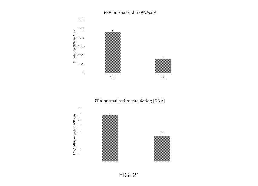
[0155] FIG. 21
depicts graphs showing total circulating EBV DNA normalized to
control RNAse P DNA or total circulating DNA indicating that EBV DNA is
relatively
depleted after Hemopurifier treatment.
DETAILED DESCRIPTION
[0156]
Disclosed herein are extracorporeal devices and their uses to treat or inhibit
a coronavirus infection, e.g., a beta corona virus infection, such as COVID-
19, or a symptom
or sequela associated with the coronavirus infection. The severe impact of the
widespread
COVID-19 pandemic has necessitated rapid development of effective and safe
therapeutics and
prophylaxes of the causative agent, SARS-CoV-2. While some vaccines and
treatments have
now been approved, it has become apparent that the emergence of SARS-CoV-2
mutants and
-25-
CA 03235306 2024-04-11
WO 2023/064753
PCT/US2022/077885
variants, including those having greater virulence and/or potential to evade
current
therapeutics, threaten to prolong the pandemic. Furthermore, many patients who
have
overcome a COVID-19 infection continue to exhibit debilitating symptoms and
sequela,
including permanent lung scarring and fibrosis, heart complications and
failure, strokes,
seizures, and immunological disorders such as Guillain-Barre syndrome. The
devices disclosed
herein function in ways that are effective against SARS-CoV-2 variants, as
well as, treating or
inhibiting the underlying causes of sequela associated with a current or past
COVID-19
infection, including within a subpopulation of patients that do not have
circulating viral
particles but continue to present sequelae associated with COVID-19 infection
e.g., the "long
hauler" patient.
Definitions
[0157] Unless
defined otherwise, all technical and scientific terms used herein have
the same meaning as is commonly understood by one of ordinary skill in the
art. All patents,
applications, published applications and other publications referenced herein
are expressly
incorporated by reference in their entireties unless stated otherwise. In the
event that there are
a plurality of definitions for a term herein, those in this section prevail
unless stated otherwise.
[0158] The
articles "a" and "an" are used herein to refer to one or to more than one
(for example, at least one) of the grammatical object of the article. By way
of example, "an
element" means one element or more than one element.
[0159] The
terms "about" or "around" as used herein refer to a quantity, level,
value, number, frequency, percentage, dimension, size, amount, weight or
length that varies by
as much as 30, 25, 20, 15, 10, 9, 8, 7, 6, 5, 4, 3, 2 or 1% to a reference
quantity, level, value,
number, frequency, percentage, dimension, size, amount, weight or length.
[0160]
Throughout this specification, unless the context requires otherwise, the
words "comprise," "comprises," and "comprising" will be understood to imply
the inclusion
of a stated step or element or group of steps or elements but not the
exclusion of any other step
or element or group of steps or elements.
[0161] By
"consisting of' is meant including, and limited to, whatever follows the
phrase "consisting of" Thus, the phrase "consisting of' indicates that the
listed elements are
required or mandatory, and that no other elements may be present. By
"consisting essentially
of' is meant including any elements listed after the phrase and limited to
other elements that
do not interfere with or contribute to the activity or action specified in the
disclosure for the
listed elements. Thus, the phrase "consisting essentially of' indicates that
the listed elements
are required or mandatory, but that other elements are optional and may or may
not be present
-26-
CA 03235306 2024-04-11
WO 2023/064753
PCT/US2022/077885
depending upon whether or not they materially affect the activity or action of
the listed
elements.
[0162] Unless
defined otherwise, all technical and scientific terms used herein have
the same meaning as is commonly understood by one of ordinary skill in the art
to which this
disclosure belongs. If there is a plurality of definitions for a term herein,
those in this section
prevail unless stated otherwise. The practice of the present disclosure will
employ, unless
indicated specifically to the contrary, conventional methods of molecular
biology and
recombinant DNA techniques within the skill of the art, many of which are
described below
for the purpose of illustration.
[0163] The
terms "individual", "subject", or "patient" as used herein, means a
human or a non-human mammal, e.g., a dog, a cat, a mouse, a rat, a cow, a
sheep, a pig, a goat,
a non-human primate, or a bird, e.g., a chicken, as well as any other
vertebrate or invertebrate.
[0164] The term
"mammal" is used in its usual biological sense. Thus, it
specifically includes, but is not limited to, primates, including simians
(chimpanzees, apes,
monkeys) and humans, cattle, horses, sheep, goats, swine, rabbits, dogs, cats,
rodents, rats,
mice, guinea pigs, or the like.
[0165] The
terms "function" and "functional" as used herein refer to a biological,
enzymatic, or therapeutic function.
[0166] The term
"isolated" as used herein refers to material that is substantially or
essentially free from components that normally accompany it in its native
state. For example,
an "isolated cell," as used herein, includes a cell that has been purified
from the milieu or
organisms in its naturally occurring state, a cell that has been removed from
a subject or from
a culture, for example, it is not significantly associated with in vivo or in
vitro substances.
[0167]
"Formulation", "pharmaceutical composition", and "composition" as used
interchangeably herein are equivalent terms referring to a composition of
matter for
administration to a subject.
[0168] The term
"pharmaceutically acceptable" means compatible with therapy for
a subject, and in particular, a human.
[0169] The
terms "agent" refers to an active agent that has biological activity and
may be used in a therapy. Also, an "agent" can be synonymous with "at least
one agent,"
"compound," or "at least one compound," and can refer to any form of the
agent, such as a
derivative, analog, salt or a prodrug thereof The agent can be present in
various forms,
components of molecular complexes, and pharmaceutically acceptable salts
(e.g.,
hydrochlorides, hydrobromides, sulfates, phosphates, nitrates, borates,
acetates, maleates,
-27-
CA 03235306 2024-04-11
WO 2023/064753
PCT/US2022/077885
tartrates, or salicylates). The term "agent" can also refer to any
pharmaceutical molecules or
compounds, therapeutic molecules or compounds, matrix forming molecules or
compounds,
polymers, synthetic molecules and compounds, natural molecules and compounds,
and any
combination thereof
[0170] The term
"purity" of any given substance, compound, or material as used
herein refers to the actual abundance of the substance, compound, or material
relative to the
expected abundance. For example, the substance, compound, or material may be
at least 80,
85, 90, 91, 92, 93, 94, 95, 96, 97, 98, 99, or 100% pure, including all
decimals in between.
Purity may be affected by unwanted impurities, including but not limited to
side products,
isomers, enantiomers, degradation products, solvent, carrier, vehicle, or
contaminants, or any
combination thereof Purity can be measured technologies including but not
limited to
chromatography, liquid chromatography, gas chromatography, spectroscopy, UV-
visible
spectrometry, infrared spectrometry, mass spectrometry, nuclear magnetic
resonance,
gravimetry, or titration, or any combination thereof
[0171] Some
embodiments disclosed herein related to selecting a subject or patient
in need for any one or more of the extracorporeal methods described herein. In
some
embodiments, a patient is selected for any one or more of the extracorporeal
methods described
herein who is in need of treatment or inhibition of a coronavirus infection,
e.g., a beta corona
virus infection, such as a SARS-CoV-2 infection. In some embodiments, a
patient is selected
for any one or more of the extracorporeal methods described herein who has
previously
received a therapy for a coronavirus infection, such as a SARS-CoV-2
infection. In some
embodiments, a patient is selected for any one or more of the extracorporeal
methods described
herein who has previously received a therapy for being at risk of a
coronavirus infection e.g.,
a beta corona virus infection, such as a SARS-CoV-2 infection. In some
embodiments, a patient
is selected for any one or more of the extracorporeal methods described herein
who has
developed a recurrence of a coronavirus infection, e.g., a beta corona virus
infection, such as a
SARS-CoV-2 infection. In some embodiments, a patient is selected for any one
or more of the
extracorporeal methods described herein who has developed resistance to
therapies for a
coronavirus infection, e.g., a beta corona virus infection, such as a SARS-CoV-
2 infection. In
some embodiments, a patient is selected for any one or more of the
extracorporeal methods
described herein who exhibits a symptom or sequela of a coronavirus infection,
e.g., a beta
corona virus infection, such as a SARS-CoV-2 infection. In some embodiments, a
patient is
selected for any one or more of the extracorporeal methods described herein
who has cleared a
coronavirus infection, e.g., a beta corona virus infection, e.g., has no
amount or a diminished
-28-
CA 03235306 2024-04-11
WO 2023/064753
PCT/US2022/077885
or reduced amount of circulating viral particles in the plasma or blood, but
continues to exhibit
a symptom or sequela of the coronavirus infection, e.g., a beta corona virus
infection, such as
a SARS-CoV-2 infection. In some embodiments, a patient is selected for any one
or more of
the extracorporeal methods described herein who has developed a coagulopathy,
such as a
COVID-19 associated coagulopathy, or who is at risk of developing a
coagulopathy (e.g., a
patient having levels or amounts of IL-1, IL-6, IL-10, IL-15, CXCL10, CCL2,
Myeloperoxidase, VCAM-1, TNF alpha, C-reactive protein (CRP), D-dimer, or
Troponin-T or
any combination thereof, that exceed levels of a control or baseline, such as
the level or amount
of IL-1, IL-6, IL-10, IL-15, CXCL10, CCL2, Myeloperoxidase, VCAM-1, TNF alpha,
C-
reactive protein (CRP), D-dimer, or Troponin-T or any combination thereof
found in a healthy
patient or a patient that does not have a coagulopathy or a patient that is
not at risk of a
coagulopathy). These patients having a coagulopathy or that are at risk of
developing a
coagulopathy may or may not have or have had a viral infection, such as COVID-
19. In some
embodiments, a patient is selected for any one or more of the extracorporeal
methods described
herein who has developed hypoxia (e.g., a patient having oxygen levels that
are reduced as
compared to a healthy patient or a patient that does not have hypoxia. These
patients having
hypoxia may or may not have or have had a viral infection, such as COVID-19.
In some
embodiments, a patient is selected for any one or more of the extracorporeal
methods described
herein who may have any combination of the aforementioned selection criteria.
Such selections
may be made by clinical or diagnostic evaluation of the subject as is routine
in the field.
[0172] The
terms "treat", "treating", "treatment", "therapeutic", or "therapy" as
used herein has its ordinary meaning as understood in light of the
specification, and do not
necessarily mean total cure or abolition of the disease or condition. The term
"treating" or
"treatment" as used herein (and as well understood in the art) also means an
approach for
obtaining beneficial or desired results in a subject's condition, including
clinical results.
Beneficial or desired clinical results can include, but are not limited to,
alleviation or
amelioration of one or more symptoms or conditions, diminishment of the extent
of a disease,
stabilizing (i.e., not worsening) the state of disease, prevention of a
disease's transmission or
spread, delaying or slowing of disease progression, amelioration or palliation
of the disease
state, diminishment of the reoccurrence of disease, and remission, whether
partial or total and
whether detectable or undetectable. "Treating" and "treatment" as used herein
also include
prophylactic treatment. Treatment methods comprise administering to a subject
a
therapeutically effective amount of an active agent or use of a therapeutic
device. The
administering step may consist of a single administration or may comprise a
series of
-29-
CA 03235306 2024-04-11
WO 2023/064753
PCT/US2022/077885
administrations. The compositions are administered or applied to the subject
in an amount, for
a duration, or for a number or repetitions sufficient to treat the patient.
The length of the
treatment period depends on a variety of factors, such as the severity of the
condition, the age
and genetic profile of the patient. It will also be appreciated that the
treatment or prophylaxis
may be modified over the course of a particular treatment or prophylaxis
regime. In some
instances, chronic administration or application may be required. The term
"prophylactic
treatment" refers to treating a subject who does not yet exhibit symptoms of a
disease or
condition, but who is susceptible to, or otherwise at risk of, a particular
disease or condition,
whereby the treatment reduces the likelihood that the patient will develop the
disease or
condition. The term "therapeutic treatment" refers to administering treatment
to a subject
already suffering from or developing a disease or condition.
[0173] The term
"inhibit" as used herein has its ordinary meaning as understood in
light of the specification, and may refer to the reduction or prevention of a
viral infection, such
as SARS-CoV-2, or a symptom or sequela thereof The reduction can be by 10%,
20%, 30%,
40%, 50%, 60%, 70%, 80%, 90%, or 100%, or an amount that is within a range
defined by any
two of the aforementioned values. As used herein, the term "delay" has its
ordinary meaning
as understood in light of the specification, and refers to a slowing,
postponement, or deferment
of an event, such as a viral infection, or a symptom or sequela thereof, to a
time which is later
than would otherwise be expected. The delay can be a delay of 0%, 10%, 20%,
30%, 40%,
50%, 60%, 70%, 80%, 90%, 100%, or an amount within a range defined by any two
of the
aforementioned values. The terms inhibit and delay may not necessarily
indicate a 100%
inhibition or delay. A partial inhibition or delay may be realized.
[0174] The
terms "nucleic acid" or "nucleic acid molecule" as used herein refers to
polynucleotides, such as deoxyribonucleic acid (DNA) or ribonucleic acid
(RNA),
oligonucleotides, fragments generated by the polymerase chain reaction (PCR),
and fragments
generated by any of ligation, scission, endonuclease action, and exonuclease
action. Nucleic
acid molecules can be composed of monomers that are naturally-occurring
nucleotides (such
as DNA and RNA), or analogs of naturally-occurring nucleotides (e.g.,
enantiomeric forms of
naturally-occurring nucleotides), or a combination of both. Modified
nucleotides can have
alterations in sugar moieties and/or in pyrimidine or purine base moieties.
Sugar modifications
include, for example, replacement of one or more hydroxyl groups with
halogens, alkyl groups,
amines, and azido groups, or sugars can be functionalized as ethers or esters.
Moreover, the
entire sugar moiety can be replaced with sterically and electronically similar
structures, such
as aza-sugars and carbocyclic sugar analogs. Examples of modifications in a
base moiety
-30-
CA 03235306 2024-04-11
WO 2023/064753
PCT/US2022/077885
include alkylated purines and pyrimidines, acylated purines or pyrimidines, or
other well-
known heterocyclic substitutes. Nucleic acid monomers can be linked by
phosphodiester bonds
or analogs of such linkages. Analogs of phosphodiester linkages include
phosphorothioate,
phosphorodithio ate, phosphoroselenoate, phosphorodiselenoate,
phosphoroanilothioate,
phosphoranilidate, or phosphoramidate. The term "nucleic acid molecule" also
includes so-
called "peptide nucleic acids," which comprise naturally-occurring or modified
nucleic acid
bases attached to a polyamide backbone. Nucleic acids can be either single
stranded or double
stranded. "Oligonucleotide" can be used interchangeable with nucleic acid and
can refer to
either double stranded or single stranded DNA or RNA. A nucleic acid or
nucleic acids can be
contained in a nucleic acid vector or nucleic acid construct (e.g. plasmid,
virus, bacteriophage,
cosmid, fosmid, phagemid, bacterial artificial chromosome (BAC), yeast
artificial
chromosome (YAC), or human artificial chromosome (HAC)) that can be used for
amplification and/or expression of the nucleic acid or nucleic acids in
various biological
systems. Typically, the vector or construct will also contain elements
including but not limited
to promoters, enhancers, terminators, inducers, ribosome binding sites,
translation initiation
sites, start codons, stop codons, polyadenylation signals, origins of
replication, cloning sites,
multiple cloning sites, restriction enzyme sites, epitopes, reporter genes,
selection markers,
antibiotic selection markers, targeting sequences, peptide purification tags,
or accessory genes,
or any combination thereof
[0175] A
nucleic acid or nucleic acid molecule can comprise one or more sequences
encoding different peptides, polypeptides, or proteins. These one or more
sequences can be
joined in the same nucleic acid or nucleic acid molecule adjacently, or with
extra nucleic acids
in between, e.g. linkers, repeats or restriction enzyme sites, or any other
sequence that is 1, 2,
3, 4, 5, 6, 7, 8, 9, 10, 11, 12, 13, 14, 15, 16, 17, 18, 19, 20, 25, 30, 35,
40, 45, 50, 55, 60, 65,
70, 75, 80, 85, 90, 95, 100, 150, 200, or 300 bases long, or any length in a
range defined by any
two of the aforementioned lengths. The term "downstream" on a nucleic acid as
used herein
refers to a sequence being after the 3'-end of a previous sequence, on the
strand containing the
encoding sequence (sense strand) if the nucleic acid is double stranded. The
term "upstream"
on a nucleic acid as used herein refers to a sequence being before the 5'-end
of a subsequent
sequence, on the strand containing the encoding sequence (sense strand) if the
nucleic acid is
double stranded. The term "grouped" on a nucleic acid as used herein refers to
two or more
sequences that occur in proximity either directly or with extra nucleic acids
in between, e.g.
linkers, repeats, or restriction enzyme sites, or any other sequence that is
1, 2, 3, 4, 5, 6, 7, 8,
9, 10, 11, 12, 13, 14, 15, 16, 17, 18, 19, 20, 25, 30, 35, 40, 45, 50, 55, 60,
65, 70, 75, 80, 85,
-31-
CA 03235306 2024-04-11
WO 2023/064753
PCT/US2022/077885
90, 95, 100, 150, 200, or 300 bases long, or any length in a range defined by
any two of the
aforementioned lengths, but generally not with a sequence in between that
encodes for a
functioning or catalytic polypeptide, protein, or protein domain.
[0176] The
terms "peptide", "polypeptide", and "protein" as used herein refers to
macromolecules comprised of amino acids linked by peptide bonds. The numerous
functions
of peptides, polypeptides, and proteins are known in the art, and include but
are not limited to
enzymes, structure, transport, defense, hormones, or signaling. Peptides,
polypeptides, and
proteins are often, but not always, produced biologically by a ribosomal
complex using a
nucleic acid template, although chemical syntheses are also available. By
manipulating the
nucleic acid template, peptide, polypeptide, and protein mutations such as
substitutions,
deletions, truncations, additions, duplications, or fusions of more than one
peptide,
polypeptide, or protein can be performed. These fusions of more than one
peptide, polypeptide,
or protein can be joined in the same molecule adjacently, or with extra amino
acids in between,
e.g. linkers, repeats, epitopes, or tags, or any other sequence that is 1, 2,
3, 4, 5, 6, 7, 8, 9, 10,
11, 12, 13, 14, 15, 16, 17, 18, 19, 20, 25, 30, 35, 40, 45, 50, 55, 60, 65,
70, 75, 80, 85, 90, 95,
100, 150, 200, or 300 bases long, or any length in a range defined by any two
of the
aforementioned lengths. The term "downstream" on a polypeptide as used herein
refers to a
sequence being after the C-terminus of a previous sequence. The term
"upstream" on a
polypeptide as used herein refers to a sequence being before the N-terminus of
a subsequent
sequence.
[0177] As used
herein, a "lectin" is a protein that bind selectively to
polysaccharides and glycoproteins. Although many are insufficiently specific
to be useful,
certain lectins are highly selective for enveloped viruses. Among lectins
which have this
property are those derived from Galanthus nivalis in the form of Galanthus
nivalis agglutinin
("GNA"), Narcissus pseudonarcissus in the form of Narcissus pseudonarcissus
agglutinin
("NPA") and a lectin derived from blue green algae Nostoc ellipsosporum called
"cyanovirin".
GNA is non-toxic and sufficiently safe that it has been incorporated into
genetically engineered
rice and potatoes (Bell et al. Transgenic Res 10(1): 35-42, 2001; Rao et al.
Plant J 15(4): 469-
477, 1998). These lectins bind to glycoproteins having a high mannose content
such as found
in HIV surface proteins (Chervenak et al. Biochemistry 34(16): 5685-5695,
1995).
[0178] As used
herein, a "high mannose glycoprotein" refers to a glycoprotein
having mannose-mannose linkages in the form of a-1->3 or a-1->6 mannose-
mannose
linkages.
-32-
CA 03235306 2024-04-11
WO 2023/064753
PCT/US2022/077885
[0179] The term
"coronavirus" as used herein refers to the family of enveloped,
positive-sense, single stranded RNA viruses belonging to the family
Coronaviridae that infect
mammals and birds. In humans, coronavirus infections can cause mild symptoms
as a common
cold, or more severe respiratory conditions such as severe acute respiratory
syndrome (SARS),
acute respiratory distress syndrome (ARDS), coughing, congestion, sore throat,
shortness of
breath, pneumonia, bronchitis, and hypoxia. Other symptoms include but are not
limited to
fever, fatigue, myalgia, and gastrointestinal symptoms such as vomiting,
diarrhea, and
abdominal pain. To infect host cells, enveloped viruses must fuse with the
host cell membrane
and deliver their genome into the cell. The viral envelope comprises spike
("S"), envelope
("E"), membrane ("M"), and hemagglutinin esterase ("HE") transmembrane
structural
proteins. Coronaviruses have average diameters of 80-120 nm and virion
surfaces that are
densely covered in projections of trimeric S glycoproteins that are decorated
with N-linked
glycosylation sequences. The S protein comprises a receptor binding domain
("RBD"), a highly
immunogenic region that determines the host receptor specificity of the virus
strain. The viral
nucleocapsid comprises multiple nucleocapsid ("N" or "NP") proteins coating
the RNA
genome. During infection, the S protein attaches to a host cell receptor and
initiate entry into
the host cell through endocytosis or fusion of the envelope membrane. The RNA
genome is
translated by the host ribosome to produce new structural proteins and RNA-
dependent RNA
polymerases, which replicate the viral genome. Viral particles are assembled
in the host
endoplasmic reticulum and are shed by Golgi-mediated exocytosis. More
information about
the structure and infection cycle of coronaviruses can be found in Fehr AR &
Perlman S.
"Coronaviruses: An Overview of Their Replication and Pathogenesis" Methods
Mol. Biol.
(2015); 1282:1-23, hereby expressly incorporated by reference in its entirety.
[0180] The
terms "SARS-CoV-2" and "2019-nCoV" as used herein refers to the
coronavirus strain or strains responsible for the human coronavirus disease
2019 (COVID-19)
pandemic. The contagiousness, long incubation period, and modern globalization
has led to
worldwide spread of the virus. Development of SARS and other respiratory
issues in infected
individuals has resulted in immense stress on medical infrastructure.
Treatments and vaccines
for SARS-CoV-2 and other coronaviruses in humans are starting to be approved,
but additional
testing is necessary. The embodiments disclosed herein can be applied to other
coronaviruses,
including but not limited to HCoV-229E, HCoV-0C43, SARS-CoV-1, HCoV NL63, HCoV-
HKU1, and MERS-CoV, as well as SARS-CoV-2 variants, including but not limited
to
20I/501Y.V1 (Alpha, B.1.1.7), 20H/501Y.V2 (Beta, B.1.351), 20J/501Y.V3 (Gamma,
P.1),
B.1.617.2 (Delta), AY.1, AY.2, C.37 (Lambda), B.1.621 (Mu), B.1.1.207, VUI-
202102/03
-33-
CA 03235306 2024-04-11
WO 2023/064753
PCT/US2022/077885
(B.1.525), VUI-202101/01 (P.2), VUI-202102/01 (A.23.1), VUI 202102/04
(B.1.1.318), VUI
202103/01 (B.1.324.1), B.1.427, CAL.20C (B.1.429), R.1, B.1.466.2, B.1.1.519,
C.36.3,
B.1.214.2, B.1.1.523, B.1.619, B.1.620, C.1.2, B.1.617.1, B.1.1.529 (Omicron),
or B.1.526. It
is envisioned that the devices and methods of use will be effective against a
coronavirus
infection caused by other SARS-CoV-2 variants that are identified or that are
currently
unknown.
[0181]
According to the National Institute for Health and Care Excellence (NICE)
of the United Kingdom, a COVID-19 infection is categorized as any of the
following: "acute
COVID-19" is associated with signs and symptoms of COVID-19 for up to 4 weeks;
"ongoing
symptomatic COVID-19" is associated with signs and symptoms of COVID-19 from 4
to 12
weeks; and "post-COVID-19 syndrome" is associated with signs and symptoms that
develop
during or after an infection consistent with COVID19, continue for more than
12 weeks and
are not explained by an alternative diagnosis.
[0182] As used
herein "COVID-19-associated coagulopathy" (CAC) refers to
thrombotic complications associated with a COVID-19 infection. As SARS-CoV-2
is able to
infect vascular endothelial cells through ACE2, significant inflammation and
damage to the
cardiovascular system may be experienced by the patient over the course of the
disease. Some
hallmarks associated with CAC involves a decrease in platelet count, increase
in the circulating
D-dimer, prolongation of the prothrombin time (PT), and the presence of macro-
thrombosis
and/or micro-thrombosis. Additional information on CAC can be found in Iba et
al. I Clin.
Med. (2021) 10;191, which is hereby incorporated by reference in its entirety.
[0183] As used
herein, a "SARS-CoV-2 derived glycoprotein" includes any
glycoprotein contained or expressed by the SARS-CoV-2 virus. For example, a
SARS-CoV-2
derived glycoprotein encompassed by the present invention is the SARS-CoV-2 S
(spike)
protein, comprising the outermost glycoprotein-decorated moieties of the viral
envelope, or
subunits thereof, including the 51, S2, and RBD subunits.
[0184] As used
herein, the "SARS-CoV-2 S spike protein" or "COVID-19 spike
protein" includes the S protein which is a class I viral fusion protein
consisting of a single chain
of approximately 1,300 amino acids that trimerizes after folding, comprising
an N-terminal Si
subunit with the receptor-binding domain, and a C-terminal S2 subunit
responsible for
membrane fusion. During viral assembly, coronavirus proteins undergo numerous
post-
translational modifications, including heavy glycosylation that has an
essential role in viral
pathogenesis. The S trimers on the coronavirus surface are extensively
decorated with N-linked
glycans that represent critical moieties for viral function. The N-linked
glycan moieties on the
-34-
CA 03235306 2024-04-11
WO 2023/064753
PCT/US2022/077885
coronavirus surfaces are critical for both viral assembly and functions. These
glycans are
needed for stability during the generation of S proteins; inhibition of N-
glycosylation by
tunicamycin resulted in the synthesis of "spikeless" virions. The coating of
the viral envelope
by N-glycans also masks immunogenic protein epitopes, forming a glycan shield
that allows
coronaviruses to evade the host immune system and host proteases. Coronavirus
glycoproteins
are therefore principal antigenic determinants that represent primary targets
of therapeutic
interventions and vaccines. These highly conserved glycoproteins on SARS-CoV-2
and other
coronaviruses, e.g., a beta corona virus such as COVID-19 and variants
thereof, are therefore
believed to be ideal targets for the lectin-based affinity devices described
herein. Accordingly,
it is contemplated that the devices and procedures described herein are useful
for the removal
of SARS-CoV-2 and other coronaviruses, e.g., a beta corona virus such as COVID-
19 and
variants thereof, as well as exosomes having antigens from said virus, even if
such virus mutate
overtime e.g., generate new variants.
[0185] As used
herein, "exosomes" are nanoparticles of 200 nm in size or less that
are a part of a communication system that conveys signals to near or distant
target cells and
reprograms their functions. The contents of exosomes vary, and can include
nucleic acids,
proteins, and lipids. They can be transferred from host to recipient cells to
alter cellular
function. They function as a mode of intercellular communication and molecular
transfer, and
facilitate the direct extracellular transfer of specific proteins, and lipids,
as well as, miRNA,
mRNA, and DNA between cells. Exosomes are present in the systemic circulation
and are
distributed throughout the body. In normal, healthy individuals, a basal level
of exosome
release aids in cell-to-cell communication and promotes elimination of
cellular debris.
However, it is contemplated that an increase in exosome quantity reflects an
altered
physiological state. Exosomes are released in abundance in pathological states
where they are
deployed by activated cells in large quantities and transfer their membrane
composition and
internal cargo to distant tissues via the circulatory system, for instance.
Additional information
about exosomes and purification of constituent material may be found in PCT
Publication WO
2016/172598, which is hereby expressly incorporated by reference in its
entirety.
[0186] As used
herein, "COVID-19 mediating nanoparticle" includes any
nanoparticle, i.e., 200nm or less in size, that contains or expresses a SARS-
CoV-2 derived
glycoprotein, or a subcellular nanoparticle associated with COVID-19, or a
symptom or
sequela thereof, which is not necessarily derived from a SARS-CoV-2 particle.
For example, a
COVID-19 mediating nanoparticle may be a SARS-CoV-2 virion, or fragments
thereof such
-35-
CA 03235306 2024-04-11
WO 2023/064753
PCT/US2022/077885
as SARS-CoV-2-derived glycoprotein, as well as, a non-viral COVID-19 mediating
nanoparticle or an exosome.
[0187] As used
herein, the term "portion" refers to an amount of a material that is
less than a whole. A minor portion refers to an amount that is, for example,
less than 50%, and
a major portion refers, for example, to an amount greater than 50%. Thus, a
unit of coronavirus
particles, COVID-19 mediating nanoparticles, or exosomes that is less than the
entire amount
of coronavirus particles, COVID-19 mediating nanoparticles, or exosomes
removed from a
subject is a portion of the removed coronavirus particles, COVID-19 mediating
nanoparticles,
or exosomes. In reference to the disclosure herein, coronavirus particles,
COVID-19 mediating
nanoparticles, or exosomes, or a unit thereof may refer to the entire amount
of the coronavirus
particles, COVID-19 mediating nanoparticles, or exosomes removed from a
subject, or an
amount that is less than the entire amount of coronavirus particles, COVID-19
mediating
nanoparticles, or exosomes removed from a subject. In some embodiments, the
subject includes
warm-blooded animals, preferably mammals, including humans. In a preferred
embodiment,
the subject is a primate. In a more preferred embodiment, the subject is a
human.
[0188] As used
herein, the term "microRNA" or "miRNA" refers to highly
conserved non-coding RNA molecules, about 21-25 nucleotides in length that
play critical roles
in regulating post-transcriptional gene expression by targeting messenger RNA
(mRNA) of
protein-coding genes. Mature miRNAs exist in the cellular cytoplasm as RNA
duplexes to
which an argonaute (Ago2) protein and a glycine-tryptophan repeat-containing
protein bind,
forming the core of a multi-subunit complex called the miRNA-mediating
silencing complex
(miRISC). The miRNA duplex contains two strands identified with either the
suffix "-5p"
(from the 5' arm of pre-miRNA) or "-3p" (from the 3' arm of the pre-miRNA).
One of the
strands of the duplex is typically discarded (passenger strand) while the
retained strand guides
mRNA target selection (guide strand). However, in some cases, two mature
miRNAs excised
from the 5' and 3' arms of the same stem-loop pre-miRNA have been reported to
be functional
and target on different mRNA. Once assembled into the miRISC through base-
pairing
interactions between nucleotides 2 and 8 of the miRNA and complementary
nucleotides
predominantly in the 3'-untranslated regions (UTR) of mRNAs, miRNAs act as
repressors of
translation. Each miRNA binds to many specific target mRNAs and often not to
those in the
same molecular pathway. miRNAs exist as both free-floating entities in plasma
and other
biofluids and also packaged into exosomes. It is believed that extracellular,
non-exosomal
miRNAs in circulation are by-products of dead cells stably bound to the Ago2
protein.
-36-
CA 03235306 2024-04-11
WO 2023/064753
PCT/US2022/077885
Exosomes, which are produced in large quantities by diseased and activated
cells, are packed
with a full complement of the parent cell's cargo, including miRNA.
[0189] Some
miRNAs have been identified as ubiquitously associated with various
diseases. Specific miRNAs have been described as "fine-tuners" of immune
responses that can
either promote an inflammatory state or have anti-inflammatory effects by
regulating toll-like
receptor signaling. miRNAs are also non-antigenic, allowing them to escape
immune
surveillance, which can be beneficial for the virus. In viral infections,
certain miRNAs may
serve both as antiviral tools that stimulate the innate and adaptive immune
systems while others
have roles in viral propagation. Host miRNAs can be regulated by viruses and
viruses can also
usurp cellular resources to produce their own miRNAs that participate in
immune evasion and
maintaining infection. The presence of the various viral and host miRNAs may
be monitored
when evaluating the outcome of a therapeutic strategy.
[0190] Exosomes
are believed to be crucial in disseminating pathogenic material as
well as host-derived molecules during viral infections, thereby contributing
to viral infectivity
and disease complications. For example, dissemination of miRNAs via exosomes
can serve as
a mechanism that viruses use to escape the immune system and allow for
continual infection
of host cells. An example is the infection of hepatocytes with hepatitis B
virus (HBV), which
leads to the release of exosomal miRNAs from virus-infected cells that
attenuate the production
of cytokines involved in anti-viral immunity. It has been hypothesized that
extracellular
vesicles produced in response to many viruses including coronaviruses
contribute to
coagulopathy as well as to maintaining the pathological state and promoting
the spread of
infection. In COVID-19, viral and/or host miRNAs transported in exosomes and
dispersed
systemically may act as mediators of thrombosis, immune dysfunction, and multi-
organ injury.
The existence of miRNA that is transferred between cells via exosomes and that
has a
distinctive pathogenic role from infectious virions is an important component
to understanding
the pathogenesis of COVID-19. The devices disclosed herein, by virtue of its
affinity for high-
mannose glycoprotein moieties, has the capacity to bind and remove SARS-CoV2
as well as
exosomes from the circulatory system.
[0191] Where a
range of values is provided, it is understood that the upper and
lower limit, and each intervening value between the upper and lower limit of
the range is
encompassed within the embodiments.
[0192] The term
"% w/w" or "% wt/wt" as used herein has its ordinary meaning as
understood in light of the specification and refers to a percentage expressed
in terms of the
weight of the ingredient or agent over the total weight of the composition
multiplied by 100.
-37-
CA 03235306 2024-04-11
WO 2023/064753
PCT/US2022/077885
The term "% v/v" or "% vol/vol" as used herein has its ordinary meaning as
understood in the
light of the specification and refers to a percentage expressed in terms of
the liquid volume of
the compound, substance, ingredient, or agent over the total liquid volume of
the composition
multiplied by 100.
COVID-19 Symptoms and Sequela
[0193] As
information concerning SARS-CoV-2 pathogenesis has emerged, it has
become apparent that this virus not only targets the respiratory tract but, in
more serious cases,
is also capable of eliciting massive systemic inflammation and exploiting the
vulnerabilities of
other organs, which may lead to respiratory failure, acute cardiac injury,
acute kidney injury,
neurological disorders, sepsis, or other complications. There is also evidence
for "RNAemia"
(i.e., the presence of viral RNA in blood) in COVID-19 patients, which
indicates that a systemic
viral load may promote inflammation and tissue injury as further described
herein.
[0194] COVID-19
Disease ¨ Cytokine Storm. Recent data suggest that SARS-
CoV-2-induced immunopathological events underlie ARDS as well as other
systemic sequelae
that occur in COVID-19. A subset of patients with COVID-19, in particular
those with severe
disease, show evidence of the "cytokine storm" in blood: unbridled and
dysregulated
inflammation that is believed to culminate in tissue damage, pulmonary edema,
and
deterioration of normal immune functions. When moderate vs. severe cases of
COVID-19 are
compared, severe cases more frequently presented with dyspnea, and
hypoalbuminemia, with
higher levels of alanine aminotransferase, lactate dehydrogenase, C-reactive
protein (CRP),
ferritin and D-dimer as well as markedly elevated systemic levels of cytokines
and receptors;
namely, IL-2R, IL-6, IL-10, and TNF-a. Due to the association that exists
between severe
inflammation and poor outcomes in COVID-19 patients, inflammatory markers may
serve as
surrogates for evaluating the outcomes of a therapeutic intervention in COVID-
19 patients by
measuring changes in specific cytokines, chemokines and combinations thereof
including IL-
1 beta, IL-6, IL-8, IL-10, granulocyte-colony stimulating factor (G-CSF),
interferon gamma-
induced protein 10 (IP-10), monocyte chemoattractant protein-1 (MCP-1), and
macrophage
inflammatory proteins (MIP-1 alpha and MIP-1 beta). Circulating cytokines such
as IL-6 have
been shown to be a biomarker for COVID-19 infection severity (Zhang et al. I
Translational
Medicine (2020) 18(1):406).
[0195] COVID-19
Disease ¨ Immune Suppression. The elevated levels of
cytokines in the pathogenesis of COVID-19 also correlate with attrition of
CD4+ T cells, CD8+
-38-
CA 03235306 2024-04-11
WO 2023/064753
PCT/US2022/077885
T cells, and natural killer (NK) cells in SARS-CoV-2 infections. The total
numbers of CD4+
and CD8+ T cells are dramatically reduced in COVID-19 patients, especially
among patients
> 60 years of age and in those requiring ICU care. The virus either directly
or indirectly leads
to lymphocyte loss and/or the inflammatory process, fueled by by-products of
the infection,
and causes lymphocyte apoptosis. There is evidence that the absolute numbers
of lymphocytes
in blood and/or percentages of lymphocytes among white blood cells are
indicative of disease
progression and outcomes for COVID-19 patients, whereby patients with moderate
to severe
disease symptoms who recover present with improvements in the lymphocyte
levels and
critically-ill patients who die do not recover from lymphopenia. Hence,
absolute counts for
lymphocytes can be used to identify the presence of lymphopenia using
laboratory reference
ranges known in the art and may be used to predict COVID-19 patients' status
and prognosis.
Additionally, the numbers of specific lymphocytes (e.g., T cells and NK cells)
and of specific
lymphocyte subsets (CD4+ T cells and CD8+ T cells) in the peripheral blood of
patients with
COVID-19 can serve to identify the status of the immune system, in particular
of cell types
that are involved in anti-viral responses.
[0196] COVID-19
Disease ¨ RNAemia. Viral RNA in plasma ("RNAemia") in
hospital-admitted patients who tested positive for COVID-19 have been
demonstrated.
RNAemia has also been observed in critically ill patients with COVID-19 and
correlated with
elevated levels of the pro-inflammatory cytokine IL-6. This indicates that
systemic SARS-
CoV-2 viral loads correlate with the severity of COVID-19. Accordingly,
reducing viral loads
or the circulating viral RNA using the methods described herein will improve
the recovery of
critically ill patients with COVID-19.
[0197] The term
"viral load" as used herein refers to the amount of viral particles,
viral RNA, or fragments thereof in a biological fluid, such as blood or
plasma. "Viral load"
encompasses all viral particles (either infectious, replicative, or non-
infective), and fragments
thereof Therefore, viral load represents the total number of viral particles
and/or fragments
thereof circulating in the biological fluid. Viral load can therefore be a
measure of any of a
variety of indicators of the presence of a virus, such as viral copy number
per unit of blood or
plasma or units of viral proteins or fragments thereof per unit of blood or
plasma. The presence
of SARS-CoV-2 viral RNA in circulation correlates with poor outcomes. The
changes in
circulating viral burden can be evaluated by RT-PCR. Viral clearance may be
quantitated by
elution of viral particles bound to lectin, including those disclosed herein.
[0198] COVID-19
Disease ¨ Cardiac Complications. Myocardial injury is
significantly associated with fatal outcome of COVID-19, while the prognosis
of patients with
-39-
CA 03235306 2024-04-11
WO 2023/064753
PCT/US2022/077885
underlying cardiovascular disease (CVD) but without myocardial injury is
relatively favorable.
Myocardial injury is associated with cardiac dysfunction and arrhythmias.
Inflammation may
be a potential mechanism for myocardial injury. Use of any one or more of the
methods
described herein may be considered for patients at high risk of myocardial
injury and such
methods can be employed multiple times (e.g., one, two, three, four, five,
six, seven, eight,
nine, or ten times) over a therapy period.
[0199] As
disclosed herein, "troponin" is a type of protein found in the muscles of
your heart. Troponin, and subunits thereof (e.g., troponin C, troponin I,
troponin T) is not
normally found in the blood. When heart muscles become damaged, troponin is
sent into the
bloodstream. As heart damage increases, greater amounts of troponin are
released in the blood.
[0200] Among
COVID-19 patients, those who are at risk of myocardial injury, as
assessed by elevated troponin T levels, are older and have a higher prevalence
of hypertension,
coronary artery disease, heart failure, and diabetes. Patients with myocardial
injury also have
evidence of more severe systemic inflammation, including greater leukocyte
counts and higher
levels of C-reactive protein and procalcitonin as well as high levels of other
biomarkers of
myocardial injury and stress, such as elevated creatine kinase, myoglobin, and
N-terminal pro-
B-type natriuretic peptide (NT-proBNP). These patients also have a higher
incidence of
systemic inflammation as well as a need for assisted ventilation than COVID-19
patients
without myocardial injury. Troponins may include and/or be referred as:
cardiac troponin I
(cTnI), cardiac troponin T (cTnT), cardiac troponin (cTN), cardiac-specific
troponin I and
troponin T. Circulating troponin T has been shown to be a biomarker for COVID-
19 infection
severity (Gaze. Ann. Clin. Biochem. (2020) 57(3):202-205).
[0201] COVID-19
Disease ¨ Multiorgan Failure, Sepsis, Acute Kidney Disease,
Neurological Disorders, Olfactory Disorders, Hyperinflammation & Other
Complications. For critically ill patients with COVID-19, improvements in
markers of
systemic inflammation and/or injury to organs may serve as measurements of a
clinically
effective therapeutic intervention. The markers that are expected to be
reduced in response to
a therapeutic intervention may include C-reactive protein (CRP), ferritin,
lactate
dehydrogenase, alanine aminotransferase (ALT), interleukin-6 (IL-6), IL-1
beta, tumor
necrosis factor-alpha (TNF-a), macrophage inflammatory protein 1-alpha,
granulocyte-colony-
stimulating factor, interferon-gamma inducible protein 10 and/or monocyte
chemoattractant
protein 1. To evaluate clinical outcomes related to a therapeutic intervention
for COVID-19,
evaluations of survival, the duration and need for assisted ventilation, the
multiorgan systems
failure, and cardiac complications may be monitored.
-40-
CA 03235306 2024-04-11
WO 2023/064753
PCT/US2022/077885
[0202] COVID-19
Disease ¨ Coagulation. A D-dimer test looks for D-dimer in
blood. D-dimer is a protein fragment produced by the degradation of cross-
linked fibrin, which
is the major component of blood clotting. During blood clotting, thrombin
activates Factor
XIII, which then crosslinks fibrin at their D regions. The activity of the
serine protease plasmin
degrades the crosslinked fibrin, producing circulating D-dimer. SARS-CoV-2
infection has
been attributed to dysregulation of blood clotting in patients, resulting in
potentially lethal
thrombosis, stroke, and pulmonary embolism. Circulating D-dimer has been shown
to be a
biomarker for COVID-19 infection severity (Yao et al. I Intensive Care. (2020)
8:49). Other
names: fragment D-dimer, fibrin degradation fragment.
Exemplary Lectin-based Hemofiltration Devices
[0203]
Disclosed herein are extracorporeal devices and methods of use for the
treatment of viral diseases, such as a coronavirus infection, or a symptom or
sequela associated
with the disease, including long-term sequela that a patient may experience
even after clearance
of the viral infection, such as those seen in recovering COVID-19 patients.
The extracorporeal
devices comprise a lectin that binds to various glycoprotein-containing
biological components,
such as exosomes. When used for hemofiltration, the extracorporeal device with
the lectin is
able to filter, for example, viral particles having glycoproteins (including
SARS-CoV-2 and
constituent subcomponents), non-viral COVID-19 mediating nanoparticles, and
circulating
glycoprotein-laden exosomes.
[0204] In some
embodiments, the devices, systems and methods of the invention
comprise one or more hollow fiber cartridges containing an affinity agent that
is a lectin, which
preferably is GNA. Other lectins include NPA, Concanavalin A and cyanovirin.
Examples of
extracorporeal devices comprising lectins that can be used in the methods
disclosed herein may
be found in WO 2007/103572, WO 2009/023332, and WO 2010/065765, each of which
is
hereby expressly incorporated by reference in its entirety.
[0205] The
extracorporeal devices disclosed herein are useful for capturing
circulating viral particles comprising glycoproteins, including enveloped
viral particles that,
during replication, incorporate host cell membrane that include a rich set of
glycoproteins and
other molecules. In the case of SARS-CoV-2 and other coronaviruses, the S
glycoprotein is
also expressed and decorates the viral envelope.
[0206]
Throughout the COVID-19 pandemic, it has become apparent that many
patients may experience long-term side effects from a SARS-CoV-2 infection, or
post-COVID-
19 syndrome. The inflammatory process that the body undergoes to fight against
the virus may
-41-
CA 03235306 2024-04-11
WO 2023/064753
PCT/US2022/077885
lead to severe complications affecting a wide range of systemic organs. In
some cases, the
damage done by inflammation may be more severe than the infection itself As
shown herein,
the extracorporeal devices disclosed herein are also useful in treating or
inhibiting these long-
term sequelae of a coronavirus infection, resulting in improved prognosis of
chronic issues
caused by the infection. This therapy may involve the depletion of exosomes
from the patient,
which may comprise one or more miRNAs that negatively impact the patient, even
if the patient
no longer has an active viral infection.
[0207]
Accordingly, in some embodiments, the present invention relates to
extracorporeal devices comprising a lectin for removing pathogenic organisms,
fragments
thereof, or other biological components from blood or plasma from a patient.
In some
embodiments, the extracorporeal device comprises one or more hollow fiber
cartridges
comprising the lectin. In accordance with hollow fiber membrane technology
provided herein
or otherwise known in the art, embodiments of the invention involves a size
exclusion
mechanism for subcellular nanoparticles (including but not limited to viral
particles, COVID-
19 mediating nanoparticles, exosomes, and the like) to contact the affinity
matrix, wherein
larger blood components (including cells) are restricted from passing through
the pores of the
hollow fibers into the extra-capillary space of the device where the affinity
agent resides. In
some embodiments, the pore sizes range from 20-500 nanometers. In some
embodiments, the
pore sizes are 200 nm or about 200 nm.
[0208] By way
of example, blood or plasma is run through an extracorporeal
circulation circuit that uses a hollow fiber cartridge with the membranes of
said hollow fibers
having sufficient permeability for the subcellular nanoparticles found in the
blood or plasma to
be removed through the membrane of the hollow fibers and into an area outside
of the fibers
containing a substrate that is bound to a single or plurality of agents (e.g.
lectins) capable of
adhering to said subcellular nanoparticles in a manner such that said
subcellular nanoparticles
are attached to said agent and do not substantially re-enter the hollow
fibers. Within the
knowledge of one skilled in the art are available numerous types of hollow
fiber systems.
Selection of said hollow fiber system is dependent on the desired blood or
plasma volume and
rate of passage of said blood or plasma volume through the hollow fiber
system. Specifically,
hollow fiber cartridges may be used having lengths of 250 mm and containing
535 hollow
fibers supplied by Amicon, and having the fiber dimensions: I.D. 180 micron
and O.D. 360
micron, and the total contact surface area in the cartridge is 750 cm2.
Alternatively, the
"Plasmaflux P2" hollow fiber filter cartridge (sold by Fresenius) or Plasmart
PS60 cartridges
(sold by Medical srl) may be used.
-42-
CA 03235306 2024-04-11
WO 2023/064753
PCT/US2022/077885
[0209]
Regardless of the hollow fiber system used, the concept needed for
application of the present invention is that said hollow fiber filters are
required to allow passage
of blood cells through the interior of said hollow fiber and allow diffusion
of subcellular
nanoparticles to the exterior. In order to allow such diffusion, the pores on
the membrane of
the hollow fiber need to be of a diameter sufficient to allow particles
ranging from the size of
20 nanometers to 500 nanometers in diameter, depending on the particles of
interest. In some
embodiments, the pores on the membrane of the hollow fiber need to be of a
diameter sufficient
to allow particles ranging from the size of 50 nanometers to 300 nanometers in
diameter. In
some embodiments, the pores on the membrane of the hollow fiber need to be of
a diameter
sufficient to allow particles ranging from the size of 80 nanometers to 200
nanometers in
diameter. During experimentation with different hollow fibers, one skilled in
the art would find
it useful to utilize particles of similar size ranges as the subcellular
nanoparticles in order to
calibrate and quantitate the ability of various pore sizes of hollow filters.
One method of
performing this is through the utilization of commercially available MACSTM
Beads (Milteny
Biotech), which have a size of 60 nanometers. Fluorescent, spherical latex
beads ranging in
size from 25 to 1000 nm are also available for this purpose (e.g., from Duke
Scientific (Palo
Alto, Calif)).
[0210] The
substrate or matrix to be used in practicing the present invention needs
to allow sufficient permeation of flow so that non-cellular blood components
that enter the
space exterior to the hollow fiber are distributed throughout the substrate or
matrix material,
so that substantial contact is made between the subcellular nanoparticles
permeating the hollow
fiber filter and the binding agent that is attached to the substrate or
matrix. Suitable substrates
or matrices are known to one skilled in the art. Said substrates or matrices
include silica gel,
dextran, agarose, nylon polymers, polymers of acrylic acid, co-polymers of
ethylene and maleic
acid anhydride, aminopropylsilica, aminocelite, glass beads, diatomaceous
earth, silicate
containing diatomaceous earth or other substrates or matrices known in the
art. Examples of
such are described in the following patents, each of which are incorporated by
reference herein
in their entirety: Lentz U.S. Pat. No. 4,708,713, Motomura U.S. Pat. No.
5,667,684, Takashima
et al U.S. Pat. No. 5,041,079, and Porath and Janson U.S. Pat. No. 3,925,152.
The agents that
are attached to said substrate may be chosen based on known affinity to
subcellular
nanoparticles.
[0211] In some
embodiments, methods of the present invention are carried out by
using an affinity cartridge using the device illustrated in FIG. 1. In this
device, blood or plasma
is passed through the lumen of a hollow fiber ultrafiltration membrane that is
in intimate
-43-
CA 03235306 2024-04-11
WO 2023/064753
PCT/US2022/077885
contact, on the non-blood wetted side of the membrane, with immobilized
lectins, which form
a means to accept and immobilize viruses and other subcellular nanoparticles.
Thus, the device
retains intact glycoproteins (which may be a part of a larger structure) bound
by lectin while
allowing other components to pass through the lumen.
[0212] SARS-CoV-
2 is the prototypic virus for which this invention is described,
but the invention can be adapted to the removal of any coronavirus or other
virus. An exemplary
device, described in detail in FIGS. 1-3, includes multiple channels of hollow
fiber
ultrafiltration membrane that forms a filtration chamber. An inlet port and an
effluent port are
in communication with the filtration chamber. The ultrafiltration membrane is
preferably an
anisotropic membrane with the tight or retention side facing the bloodstream.
The membrane
is conveniently formed of any number of polymers known to the art, for
example, polysulfone,
polyethersulfone, polyamides, polyimides, cellulose acetate, and
polyacrylamide. Preferably,
the membrane has pores 200-700 nm in diameter, which will allow passage of
subcellular
nanoparticles, e.g., SARS-CoV-2 virions, or fragments thereof, such as SARS-
CoV-2-derived
glycoproteins, (e.g., SARS-CoV-2 virions of 110 nm diameter), and non-viral
COVID-19
mediating nanoparticles (e.g., exosomes) but not most blood cells (red blood
cells, 2,000 nm
diameter; lymphocytes, 7,000-12,000 nm diameter; macrophages, 10,000-18,000 nm
diameter). A diagram of an exemplary device is shown in FIG. 1. The device
comprises a
cartridge 10 comprising a blood-processing chamber 12 formed of suitable
material such as
polycarbonate 14. Around chamber 12 is an optional exterior chamber 16. A
temperature
controlling fluid can be circulated into chamber 16 through port 18 and out of
port 20. The
device includes an inlet port 32 for the blood and an outlet port 34 for the
effluent. The device
also provides one or more ports 48 and 50, for accessing the extra-channel
space in the
cartridge. As shown in FIGS. 1 and 2, chamber 12 contains a plurality of
ultrafiltration
membranes 22. These membranes preferably have a 0.3 mm inside diameter and 0.5
min
outside diameter. FIG. 3 is a cross sectional representation of a channel 22
and shows the
anisotropic nature of the membrane. As shown in FIG. 3, a hollow fiber
membrane structure
40 is composed of a single polymeric material which is formed into a tubular
section
comprising a relatively tight ultrafiltration membrane 42 and relatively
porous exterior portion
44 in which may be immobilized lectins 46. During the operation of the device,
a solution
containing the lectins is loaded on to the device through port 48. The lectins
are allowed to
immobilize to the exterior 22 of the membrane in FIG. 2. Unbound lectins can
be collected
from port 50 by washing with saline or other solutions. The cartridge housing
is made of
polycarbonate and the fibers are held in place with polyurethane potting
material. The lectins
-44-
CA 03235306 2024-04-11
WO 2023/064753
PCT/US2022/077885
are found in the extra-lumen (or extra-capillary or extra-channel) covalently
bound to a solid
resin material 150-300 microns (or wider) in diameter. The resin is made of a
porous silicon
dioxide material (SiO2), preferably diatomaceous earth. Approximately 35-45
grams of lectin
bound resin is loaded into the extra-lumen space around the fibers through the
polycarbonate
side ports of the cartridge. During operation, blood runs along the length of
the fibers and the
plasma exits the pores and comes into contact with the lectin that is bound to
the solid resin
substrate.
[0213] For
binding of lectins to the ultrafiltration membrane, the polymers of the
ultrafiltration membrane are first activated, e.g., made susceptible for
combining chemically
with proteins, by using processes known in the art. Any number of different
polymers can be
used. To obtain a reactive polyacrylic acid polymer, for example,
carbodiimides can be used
(Valuev et al., 1998, Biomaterials, 19:41-3). Once the polymer has been
activated, the lectins
can be attached directly or via a linker to form in either case an affinity
matrix. Suitable linkers
include, but are not limited to, avidin, strepavidin, biotin, protein A, or
protein G. The lectins
may also be directly bound to the polymer of the ultrafiltration membrane
using coupling
agents such as bifunctional reagents, or may be indirectly bound. In some
embodiments, GNA
covalently coupled to agarose can be used to form an affinity matrix.
[0214]
Accordingly, one aspect of the invention provides a lectin affinity
hemodialysis cartridge, comprising: a filtration chamber configured to receive
blood or plasma
; a lectin, optionally coupled to agarose, diatomaceous earth, or aminocelite
disposed within
said filtration chamber; and a porous hollow fiber membrane, wherein said
membrane has pores
of 200-500 nm in diameter; wherein the lectin is selected from the group
consisting of:
Galanthus nivalis agglutinin (GNA), Narcissus pseudonarcissus agglutinin
(NPA), cyanovirin,
and Concanavalin A, and mixtures thereof wherein the cartridge is configured
to remove
subcellular nanoparticles from the blood or plasma.
[0215]
Processes that can be used to isolate lectins such as GNA are generally
known in the art. For example, Van Damme et al. demonstrate isolating the
lectin from
Galantus nivalis (snowdrop) bulbs by affinity purification with mannose or
other sugars (Van
Damme et al. FEBS Letters (1987) 215(1):140-144). Use of purified GNA for
affinity
purification purposes have been previously demonstrated, such as for isolating
glycoproteins
like immunoglobulins (Shibuya et al. Archives Biochem. Biophys. (1988)
267(2):676-680).
Each of the references above are hereby expressly incorporated by reference in
its entirety.
[0216] The
present invention also provides a device with a filtration chamber
further comprises an inlet port and an outlet port; wherein a channel of said
hollow fiber
-45-
CA 03235306 2024-04-11
WO 2023/064753
PCT/US2022/077885
membrane is in fluidic communication with said inlet and said outlet ports;
said cartridge
having an extra-channel space within said chamber which surrounds said hollow
fiber
membrane; and wherein said lectin is, optionally, covalently coupled to
agarose, diatomaceous
earth or aminocelite that is disposed within said extra-channel space
proximate to an exterior
surface of said membrane.
[0217] For some
methods of the present invention, blood or plasma having
subcellular nanoparticles (which may or may not contain SARS-CoV-2 viral
particles) is
withdrawn from a patient and contacted with an ultrafiltration membrane. In
some
embodiments, the blood is first separated into its plasma and cellular
components. The blood
or plasma is then contacted with the lectins to remove the subcellular
nanoparticles by binding
between glycoproteins and lectins. The plasma can then be recombined with the
cellular
components and returned to the patient. Alternatively, the cellular components
may be returned
to the patient separately. The therapy can be repeated periodically until a
desired response has
been achieved. In some embodiments, the therapy can be carried out for
0.1,0.2, 0.3, 0.4, 0.5,
0.6, 0.7, 0.8, 0.9, 1, 2, 3, 4, 5, 6, 7, 8, 9, 10, 11, 12, 13, 14, 15, 16, 17,
18, 19, 20, 21, 22, 23, or
24 hours within a 24 hour period, or any amount of time within a range defined
by any two of
the aforementioned times. In some embodiments, the therapy can be repeated
every day for 1,
2, 3, 4, 5, 6, 7, 8, 9, 10, 11, 12, 13, 14, or 15 days.
[0218] In some
embodiments, the methods and devices of the present invention
additionally comprise affinity agents that are monoclonal antibodies that bind
to SARS-CoV-
19 derived glycoproteins, such as the Si spike protein described herein, in
the extracorporeal
circuit. In certain embodiments of the invention, the methods and devices of
the present
invention comprise a GNA affinity agent and a monoclonal antibody affinity
agent.
[0219] One
skilled in the art will recognize that a biological sample can be taken
from, but not limited to the following bodily fluids: peripheral blood,
plasma, serum, ascites,
cerebrospinal fluid (CSF), sputum, saliva, bone marrow, synovial fluid,
aqueous humor,
amniotic fluid, cerumen, breast milk, broncheoalveolar lavage fluid, semen
(including prostatic
fluid), Cowper's fluid or pre-ejaculatory fluid, female ejaculate, sweat,
fecal matter, hair, tears,
cyst fluid, pleural and peritoneal fluid, pericardial fluid, lymph, chyme,
chyle, bile, interstitial
fluid, menses, pus, sebum, vomit, vaginal secretions, mucosal secretion, nasal
fluid (e.g., a
nasal swab isolate) stool water, urine, pancreatic juice, lavage fluids from
sinus cavities,
bronchopulmonary aspirates or other lavage fluids. A biological sample may
also include the
blastocyst cavity, umbilical cord blood, or maternal circulation that may be
of fetal or maternal
origin. The biological sample may also be a tissue sample or biopsy.
-46-
CA 03235306 2024-04-11
WO 2023/064753
PCT/US2022/077885
Methods of Therapy or Use For Treatment of COVID-19
[0220] The
present invention relates to methods for using lectins for hemofiltration
of blood or plasma in an extracorporeal setting. Accordingly, the present
invention provides
methods for reducing subcellular nanoparticles, such as those associated with
COVID-19 or a
symptom or sequela thereof, from the circulatory system of an individual
comprising the steps
of obtaining blood or plasma from the individual, passing the blood or plasma
through a porous
hollow fiber membrane where lectin molecules are immobilized within the porous
exterior
portion of the membrane, collecting pass-through blood or plasma, and
reinfusing the pass-
through blood or plasma into the individual.
[0221] In some
non-limiting embodiments, the devices and methods of the present
invention have the ability to capture and physically remove the SARS-CoV-2 Si
(spike) protein
with a high efficiency. Accordingly, the present invention provides methods
for capturing and
physically removing COVID-19 mediating nanoparticles from the circulatory
system of an
individual comprising the steps of obtaining blood or plasma from the
individual, passing the
blood or plasma through a porous hollow fiber membrane wherein lectin
molecules are
immobilized within the porous exterior portion of the membrane, collecting
pass-through blood
or plasma and reinfusing the pass-through blood or plasma into the individual.
However, in
some embodiments, the devices and methods disclosed herein can be used for a
patient who no
longer has an active viral infection, but still exhibits a symptom or sequela
thereof
[0222] Once a
subject in need is identified, for example, a subject with severe
COVID-19 disease, at risk for severe COVID-19 disease (e.g., a subject in need
of oxygen
therapy), or has overcome COVID-19 but still has one or more symptoms or
sequelae, a method
of depleting subcellular nanoparticles that may be associated with COVID-19
may include the
following steps: a) providing a hollow fiber cartridge comprising a lectin or
other affinity
binding agent that selectively binds to the outer surfaces of the subcellular
nanoparticles; b)
removing a biological sample, e.g., blood or plasma, from a subject using the
system, the
biological sample having a concentration of the subcellular nanoparticles; c)
processing the
biological sample using the hollow fiber cartridge such that the affinity
agents are in contact
with the biological sample; d) capturing at least a portion of the subcellular
nanoparticles from
the biological sample such that said portion of the subcellular nanoparticles
is retained in the
hollow fiber cartridge; and e) reintroducing the biological sample without
said portion of
captured subcellular nanoparticles to the patient without removing the
biological sample from
the system before the biological sample is ready to be administered to the
patient. Optionally,
-47-
CA 03235306 2024-04-11
WO 2023/064753
PCT/US2022/077885
a biological sample from said subject, such as a nasal fluid (e.g., an isolate
from a nasal swab),
blood or plasma, from said subject is obtained before or after the therapy or
both and said
biological sample is analyzed for the level or amount of subcellular
nanoparticles.
[0223] For use
in critically-ill patients with COVID-19, the capture of SARS-CoV-
2 virions from the circulatory system may have several positive benefits as
follows: (1)
Diminishing the systemic load of SARS-CoV-2; (2) Reducing the severity of the
systemic
inflammatory response (e.g., cytokine storm) occurring during the infection;
(3) Improving the
functions of immune cells including cells with anti-viral functions; and (4)
Reduction of
continuous cellular infection, progressive damage to affected organs, and/or
disease-related
symptoms due to the virus itself and/or the inflammatory response.
[0224] As
described herein, it has demonstrated that the extracorporeal devices are
able to capture exosomes from the blood or plasma from a patient. In some
embodiments, the
patient may have an on-going coronavirus infection, such as COVID-19. In other
embodiments,
the patient may no longer have an active coronavirus infection (e.g., has a
reduced amount or
no amount of circulating virus that is detected by conventional approaches),
but the patient still
exhibits a symptom or sequela of the coronavirus infection. In some
embodiments, the
exosomes depleted by the extracorporeal devices may comprise miR-424-5p, miR-
16-2-3p, or
both. These miRNAs may be involved in negative effects of the coronavirus
symptom or
sequela on the patient, even if the patient no longer has an active
coronavirus infection.
[0225]
Disclosed herein in some embodiments are methods for reducing SARS-
CoV-2 virions, or portions thereof, in a COVID-19 patient in need thereof In
some
embodiments, the methods comprise (a) introducing blood or plasma from a
patient infected
with COVID-19 into an extracorporeal device comprising a lectin (e.g.,
Galanthus nivalis
agglutinin (GNA), Narcissus pseudonarcissus agglutinin (NPA) or Nostoc
ellipsosporum
cyanovirin) that binds to SARS-CoV-2 virions, or portions thereof (b)
contacting the blood or
plasma from the patient with the lectin in the extracorporeal device for a
time sufficient to
allow the SARS-CoV-2 virions, or portions thereof, present in the blood or
plasma, to bind to
said lectin; (c) reintroducing the blood or plasma obtained after (b) into
said patient, wherein
the blood or plasma obtained after (b) has a reduced amount of the SARS-CoV-2
virions, or
portions thereof, as compared to the blood or plasma of said patient prior to
(b); and (d)
optionally, detecting or identifying SARS-CoV-2 virions, or portions thereof,
in a sample from
said patient, such as a nasal, blood, or plasma sample, prior to (a) or after
(b) or both and/or,
optionally selecting or identifying a patient having COVID-19 to receive a
therapy that reduces
SARS-CoV-2 virions, or fragments thereof, as compared to a control level or
amount (e.g., a
-48-
CA 03235306 2024-04-11
WO 2023/064753
PCT/US2022/077885
level or amount found in a sample from healthy patient or a patient not
experiencing
inflammation or COVID-19 infection or a sequela associated therewith). In some
embodiments, the patient does not comprise a coronavirus infection prior to
step (a) but exhibits
symptoms or sequela of the coronavirus infection. In some embodiments, the
patient has
cleared the coronavirus infection prior to step (a), but the patient still
exhibits symptoms or
sequela of the coronavirus infection. In some embodiments, the blood or plasma
of the patient
does not comprise the coronavirus prior to step (a), but the patient still
exhibits symptoms or
sequela of the coronavirus infection. In some embodiments, the methods further
comprise
determining whether the patient has early acute lung injury (ALT), early acute
respiratory
distress syndrome (ARDS), dyspnea, respiratory frequency? 30 breaths/min,
blood oxygen
saturation < 93%, partial pressure of arterial oxygen to fraction of inspired
oxygen ratio of
<300, lung infiltrates >50%, respiratory failure within 24 to 48 hours,
elevated ferritin, elevated
lactate, elevated lactate dehydrogenase (LDH), low absolute lymphocyte count
(ALC), low
platelet count, prolonged prothrombin time/international normalized ratio
(PT/INR), septic
shock, or multiple organ dysfunction or failure, or any combination thereof
prior to (a) or after
(b) or both. In some embodiments, the methods further comprise determining
whether the
patient has an elevated IL-6 level or amount in a blood or plasma sample, as
compared to a
control level or amount (e.g., a level or amount found in a blood or plasma
sample from a
healthy patient or a patient not experiencing inflammation or COVID-19
infection or a sequela
associated therewith) either before (a) or after (b) or both. In some
embodiments, the elevated
serum IL-6 level is greater than equal to 2 pg/mL. In some embodiments, the
methods further
comprise determining whether the patient has an elevated D-dimer level or
amount in a blood
or plasma sample, as compared to a control level or amount (e.g., a level or
amount found in a
blood or plasma sample from a healthy patient or a patient not experiencing
inflammation or
COVID-19 infection or a sequela associated therewith) either before (a) or
after (b) or both. In
some embodiments, the elevated serum D-Dimer level is greater than or equal to
50Ong/mL.
In some embodiments, the methods further comprise determining whether the
patient has an
elevated Troponin T level or amount in a blood or plasma sample, as compared
to a control
level or amount (e.g., a level or amount found in a blood or plasma sample
from a healthy
patient or a patient not experiencing inflammation or COVID-19 infection or a
sequela
associated therewith) either before (a) or after (b) or both. In some
embodiments, the elevated
serum Troponin T level is greater than or equal to 15 ng/L. In some preferred
embodiments,
the lectin is Galanthus nivalis agglutinin (GNA). In some embodiments, the
extracorporeal
device comprises a hollow fiber cartridge comprising the lectin and wherein
the blood or
-49-
CA 03235306 2024-04-11
WO 2023/064753
PCT/US2022/077885
plasma flows through hollow fibers of the hollow fiber cartridge. In some
embodiments, the
hollow fibers of the hollow fiber cartridge comprise a pore size that excludes
cellular
components of the blood or plasma from contacting the lectin. In some
embodiments, the pore
size is 20-500 nm or about 20-500 nm. In some embodiments, the pore size is
200 nm or about
200 nm. In some embodiments, the lectin is immobilized or adsorbed on to a
solid support, and
the hollow fiber cartridge comprises the lectin immobilized or adsorbed on the
solid support.
In some embodiments, the solid support comprises agarose, diatomaceous earth,
or
aminocelite. In some embodiments, the solid support comprises diatomaceous
earth. In some
embodiments, the methods further comprise isolating coronavirus virions, or
portions thereof,
bound to the lectin of the extracorporeal device. In some embodiments, the
methods further
comprise isolating exosomes associated with the coronavirus infection, or the
symptom or
sequela thereof, bound to the lectin of the extracorporeal device. In some
embodiments, the
methods further comprise determining the contents of the isolated exosomes. In
some
embodiments, the exosomes associated with the coronavirus infection, or the
symptom or
sequela thereof, comprise miR-424-5p, or miR-16-2-3p, or both. In some
embodiments, the
methods further comprise observing or measuring a reduction in number of
coronavirus virions,
or portions thereof; number of exosomes associated with the coronavirus
infection, or the
symptom or sequela thereof; measuring the levels or amount of IL-1, IL-6, IL-
10, IL-15,
CXCL10, CCL2, Myeloperoxidase, VCAM-1, TNF alpha, C-reactive protein (CRP), D-
dimer,
or Troponin-T or any combination thereof, in a sample of the patient's blood
or plasma taken
after (b) relative to a sample of the patient's blood or plasma taken before
(b). In some
embodiments, the methods further comprise observing an improvement in the
coronavirus
infection, or the symptom or sequela thereof, in the patient following (b) or
(c) or both. In some
embodiments, observing the improvement in the coronavirus infection, or the
symptom or
sequela thereof, comprises determining an improvement in early ALI, early
ARDS, respiratory
frequency, blood oxygen saturation, partial pressure of arterial oxygen to
fraction of inspired
oxygen ratio, lung infiltrates, respiratory failure, ferritin, lactate, LDH,
ALC, platelet count,
PT/INR, septic shock, or multiple organ dysfunction or failure, or any
combination thereof, in
the patient. In some embodiments, observing the improvement in the coronavirus
infection, or
the symptom or sequela thereof, comprises observing a reduction in number of
coronavirus
virions, or portions thereof; or exosomes associated with the coronavirus
infection, or the
symptom or sequela thereof; or measuring the level or amount of IL-1, IL-6, IL-
10, IL-15,
CXCL10, CCL2, Myeloperoxidase, VCAM-1, TNF alpha, C-reactive protein (CRP), D-
dimer,
or Troponin-T or any combination thereof in a biological sample such as blood
from the patient
-50-
CA 03235306 2024-04-11
WO 2023/064753
PCT/US2022/077885
before or after the therapy or both. In some embodiments, the COVID-19 is
caused by a SARS-
CoV-2 variant. In some embodiments, the variant is selected from 20I/501Y.V1
(Alpha,
B.1.1.7), 20H/501Y.V2 (Beta, B.1.351), 20J/501Y.V3 (Gamma, P.1), B.1.617.2
(Delta), AY.1,
AY.2, C.37 (Lambda), B.1.621 (Mu), B.1.1.207, VUI-202102/03 (B.1.525), VUI-
202101/01
(P.2), VUI-202102/01 (A.23.1), VUI 202102/04 (B.1.1.318), VUI 202103/01
(B.1.324.1),
B.1.427, CAL.20C (B.1.429), R.1, B.1.466.2, B.1.1.519, C.36.3, B.1.214.2,
B.1.1.523,
B.1.619, B.1.620, C.1.2, B.1.617.1, B.1.1.529 (Omicron), or B.1.526. In some
embodiments,
the extracorporeal device is primed with an anticoagulant, preferably heparin,
to prevent
clotting of blood prior to (a). In some embodiments, the blood is flowed at a
rate of about 50
to about 600 mL/min, preferably about 200 to about 400 mL/min through said
extracorporeal
device, preferably about 200 to about 240 mL/min or 200 to 240 mL/min or most
preferably
240mL/min. In some embodiments, the flow of blood is started at an initial
flow rate of
100m1/min and increased gradually to 200m1/min (e.g., in a stepwise increase
over a five-
minute period). In some embodiments, reintroducing the blood back to the
patient comprises
flushing the extracorporeal device with saline. In some embodiments, the blood
or plasma is
contacted with the extracorporeal device for 0.1, 0.2, 0.3, 0.4, 0.5, 0.6,
0.7, 0.8, 0.9, 1, 2, 3, 4,
5, 6, 7, 8, 9, or 10 hours, or any amount of time within a range defined by
any two of the
aforementioned times. In some embodiments, steps (a), (b), (c), and optionally
(d) is repeated
every day for 1, 2, 3, 4, 5, 6, 7, 8, 9, 10, 11, 12, 13, 14, or 15 days. In
some embodiments, the
methods further comprise administering an additional antiviral therapy to the
patient. In some
embodiments, the additional antiviral therapy comprises administration of
favipiravir,
favilavir, remdesivir, tocilizumab, galidesivir, sarilumab, lopinavir,
ritonavir, darunavir,
ribavirin, interferon-a, pegylated interferon-a, interferon alfa-2b,
convalescent serum, or any
combination thereof
[0226] Also
disclosed herein some embodiments are methods for reducing COVID-
19 mediating nanoparticles in a COVID-19 patient in need thereof In some
embodiments, the
methods comprise (a) introducing blood or plasma from a patient infected with
COVID-19 into
an extracorporeal device comprising a lectin that binds to COVID-19 mediating
nanoparticles
(e.g., Galanthus nivalis agglutinin (GNA), Narcissus pseudonarcissus
agglutinin (NPA) or
Nostoc ellipsosporum cyanovirin); (b) contacting the blood or plasma from the
patient with the
lectin in the extracorporeal device for a time sufficient to allow the COVID-
19 mediating
nanoparticles to bind to said lectin; (c) reintroducing the blood or plasma
obtained after (b)
into said patient, wherein the blood or plasma obtained after (b) has a
reduced amount of the
COVID-19 mediating nanoparticles, as compared to the blood or plasma of said
patient prior
-51-
CA 03235306 2024-04-11
WO 2023/064753
PCT/US2022/077885
to (b); and (d) optionally, detecting or identifying SARS-CoV-2 virions, or
portions thereof, or
COVID-19-mediating nanoparticles in a sample from said patient, such as a
nasal (e.g.,
obtained from a nasal swab), blood, or plasma sample, prior to (a) or after
(b) or both and/or,
optionally selecting or identifying a patient having COVID-19 to receive a
therapy that reduces
COVID-19-mediating nanoparticles, as compared to a control level or amount
(e.g., a level or
amount found in a sample from a healthy patient or a patient not experiencing
inflammation or
COVID-19 infection or a sequela associated therewith). In some embodiments,
the patient does
not comprise a coronavirus infection prior to step (a) but exhibits symptoms
or sequela of the
coronavirus infection. In some embodiments, the patient has cleared the
coronavirus infection
prior to step (a), but the patient still exhibits symptoms or sequela of the
coronavirus infection.
In some embodiments, the blood or plasma of the patient does not comprise the
coronavirus
prior to step (a), but the patient still exhibits symptoms or sequela of the
coronavirus infection.
In some embodiments, the methods further comprise determining whether the
patient has early
acute lung injury (ALI), early acute respiratory distress syndrome (ARDS),
dyspnea,
respiratory frequency > 30 breaths/min, blood oxygen saturation < 93%, partial
pressure of
arterial oxygen to fraction of inspired oxygen ratio of <300, lung infiltrates
>50%, respiratory
failure within 24-48 hours, elevated ferritin, elevated lactate, elevated
lactate dehydrogenase
(LDH), low absolute lymphocyte count (ALC), low platelet count, elevated
prothrombin
time/international normalized ratio (PT/INR), septic shock, or multiple organ
dysfunction or
failure, or any combination thereof prior to (a) or after (b) or both. In some
embodiments, the
methods further comprise determining whether the patient has an elevated IL-6
level or amount
in a blood or plasma sample either before (a) or after (b) or both, as
compared to a control level
or amount (e.g., a level or amount found in a blood or plasma sample from a
healthy patient or
a patient not experiencing inflammation or COVID-19 infection or a sequela
associated
therewith). In some embodiments, the elevated serum IL-6 level is greater than
equal to 2
pg/mL. In some embodiments, the methods further comprise determining whether
the patient
has an elevated D-dimer level or amount in a blood or plasma sample, as
compared to a control
level or amount (e.g., a level or amount found in a blood or plasma sample
from a healthy
patient or a patient not experiencing inflammation or COVID-19 infection or a
sequela
associated therewith) either before (a) or after (b) or both. In some
embodiments, the elevated
serum D-Dimer level is greater than or equal to 50Ong/mL. In some embodiments,
the methods
further comprise determining whether the patient has an elevated Troponin T
level or amount
in a blood or plasma sample, as compared to a control level or amount (e.g., a
level or amount
found in a blood or plasma sample from a healthy patient or a patient not
experiencing
-52-
CA 03235306 2024-04-11
WO 2023/064753
PCT/US2022/077885
inflammation or COVID-19 infection or a sequela associated therewith) either
before (a) or
after (b) or both. In some embodiments, the elevated serum Troponin T level is
greater than or
equal to 15 ng/L. In some preferred embodiments, the lectin is Galanthus
nivalis agglutinin
(GNA). In some embodiments, the extracorporeal device comprises a hollow fiber
cartridge
comprising the lectin and wherein the blood or plasma flows through hollow
fibers of the
hollow fiber cartridge. In some embodiments, the hollow fibers of the hollow
fiber cartridge
comprise a pore size that excludes cellular components of the blood or plasma
from contacting
the lectin. In some embodiments, the pore size is 20-500 nm or about 20-500
nm. In some
embodiments, the pore size is 200 nm or about 200 nm. In some embodiments, the
lectin is
immobilized or adsorbed on to a solid support, and the hollow fiber cartridge
comprises the
lectin immobilized or adsorbed on the solid support. In some embodiments, the
solid support
comprises agarose, diatomaceous earth, or aminocelite. In some embodiments,
the solid
support comprises diatomaceous earth. In some embodiments, the methods further
comprise
isolating coronavirus virions, or portions thereof, bound to the lectin of the
extracorporeal
device. In some embodiments, the methods further comprise isolating exosomes
associated
with the coronavirus infection, or the symptom or sequela thereof, bound to
the lectin of the
extracorporeal device. In some embodiments, the methods further comprise
determining the
contents of the isolated exosomes. In some embodiments, the exosomes
associated with the
coronavirus infection, or the symptom or sequela thereof, comprise miR-424-5p,
or miR-16-2-
3p, or both. In some embodiments, the methods further comprise observing or
measuring a
reduction in number of coronavirus virions, or portions thereof; number of
exosomes associated
with the coronavirus infection, or the symptom or sequela thereof; or
measuring the level or
amount of IL-1, IL-6, IL-10, IL-15, CXCL10, CCL2, Myeloperoxidase, VCAM-1, TNF
alpha,
C-reactive protein (CRP), D-dimer, or Troponin-T or any combination thereof in
a biological
sample such as blood or plasma from the patient before or after the therapy or
both. In some
embodiments, the methods further comprise observing an improvement in the
coronavirus
infection, or the symptom or sequela thereof, in the patient following (b) or
(c) or both. In some
embodiments, observing the improvement in the coronavirus infection, or the
symptom or
sequela thereof, comprises determining an improvement in early ALI, early
ARDS, respiratory
frequency, blood oxygen saturation, partial pressure of arterial oxygen to
fraction of inspired
oxygen ratio, lung infiltrates, respiratory failure, ferritin, lactate, LDH,
ALC, platelet count,
PT/INR, septic shock, or multiple organ dysfunction or failure, or any
combination thereof, in
the patient. In some embodiments, observing the improvement in the coronavirus
infection, or
the symptom or sequela thereof, comprises observing a reduction in number of
coronavirus
-53-
CA 03235306 2024-04-11
WO 2023/064753
PCT/US2022/077885
virions, or portions thereof; exosomes associated with the coronavirus
infection, or the
symptom or sequela thereof or measuring the level or amount of IL-1, IL-6, IL-
10, IL-15,
CXCL10, CCL2, Myeloperoxidase, VCAM-1, TNF alpha, C-reactive protein (CRP), D-
dimer,
or Troponin-T or any combination thereof in a biological sample such as blood
or plasma from
the patient before or after the therapy or both. In some embodiments, the
COVID-19 is caused
by a SARS-CoV-2 variant. In some embodiments, the variant is selected from
20I/501Y.V1
(Alpha, B.1.1.7), 20H/501Y.V2 (Beta, B.1.351), 20J/501Y.V3 (Gamma, P.1),
B.1.617.2
(Delta), AY.1, AY.2, C.37 (Lambda), B.1.621 (Mu), B.1.1.207, VUI-202102/03
(B.1.525),
VUI-202101/01 (P.2), VUI-202102/01 (A.23.1), VUI 202102/04 (B.1.1.318), VUI
202103/01
(B.1.324.1), B.1.427, CAL.20C (B.1.429), R.1, B.1.466.2, B.1.1.519, C.36.3,
B.1.214.2,
B.1.1.523, B.1.619, B.1.620, C.1.2, B.1.617.1, B.1.1.529 (Omicron), or
B.1.526. In some
embodiments, the extracorporeal device is primed with an anticoagulant,
preferably heparin,
to prevent clotting of blood prior to (a). In some embodiments, the blood is
flowed at a rate of
about 50 to about 600 mL/min, preferably about 200 to about 400 mL/min,
preferably about
200 to about 240 mL/min, through said extracorporeal device. In some
embodiments, the flow
of blood is started at an initial flow rate of 100m1/min and increased
gradually to 200m1/min
(e.g., in a stepwise increase over a five-minute period). In some embodiments,
reintroducing
the blood back to the patient comprises flushing the extracorporeal device
with saline. In some
embodiments, the blood or plasma is contacted with the extracorporeal device
for 0.1, 0.2, 0.3,
0.4, 0.5, 0.6, 0.7, 0.8, 0.9, 1, 2, 3, 4, 5, 6, 7, 8, 9, or 10 hours, or any
amount of time within a
range defined by any two of the aforementioned times. In some embodiments,
steps (a), (b),
(c), and optionally (d) is repeated every day for 1, 2, 3, 4, 5, 6, 7, 8, 9,
10, 11, 12, 13, 14, or 15
days. In some embodiments, the methods further comprise administering an
additional antiviral
therapy to the patient. In some embodiments, the additional antiviral therapy
comprises
favipiravir, favilavir, remdesivir, tocilizumab, galidesivir, sarilumab,
lopinavir, ritonavir,
darunavir, ribavirin, interferon-a, pegylated interferon-a, interferon alfa-
2b, convalescent
serum, or any combination thereof
[0227] Also
disclosed herein in some embodiments are methods for reducing
exosomes comprising a COVID-19 antigen in a COVID-19 patient. In some
embodiments, the
methods comprise a) introducing blood or plasma from a patient infected with
COVID-19 into
an extracorporeal device comprising a lectin that binds to the exosomes
comprising the
COVID-19 antigen (e.g., Galanthus nivalis agglutinin (GNA), Narcissus
pseudonarcissus
agglutinin (NPA) or Nostoc ellipsosporum cyanovirin); (b) contacting the blood
or plasma from
the patient with the lectin in the extracorporeal device for a time sufficient
to allow the
-54-
CA 03235306 2024-04-11
WO 2023/064753
PCT/US2022/077885
exosomes comprising the COVID-19 antigen to bind to said lectin; (c)
reintroducing the blood
or plasma obtained after (b) into said patient, wherein the blood or plasma
obtained after (b)
has a reduced amount of the exosomes comprising the COVID-19 antigen, as
compared to the
blood or plasma of said patient prior to (b); and (d) optionally, detecting or
identifying SARS-
CoV-2 virions, or portions thereof or the exosomes comprising the COVID-19
antigen in a
sample from said patient, such as a nasal, blood, or plasma sample, prior to
(a) or after (b) or
both and/or, optionally selecting or identifying a patient having COVID-19 to
receive a therapy
that reduces exosomes comprising a COVID-19 antigen, as compared to a control
level or
amount (e.g., a level or amount found in a blood or plasma sample from a
healthy patient or a
patient not experiencing inflammation or COVID-19 infection or a sequela
associated
therewith). In some embodiments, the patient does not comprise a coronavirus
infection prior
to step (a) but exhibits symptoms or sequela of the coronavirus infection. In
some
embodiments, the patient has cleared the coronavirus infection prior to step
(a), but the patient
still exhibits symptoms or sequela of the coronavirus infection. In some
embodiments, the
blood or plasma of the patient does not comprise the coronavirus prior to step
(a), but the patient
still exhibits symptoms or sequela of the coronavirus infection. In some
embodiments, the
methods further comprise determining whether the patient has early acute lung
injury (ALT),
early acute respiratory distress syndrome (ARDS), dyspnea, respiratory
frequency > 30
breaths/min, blood oxygen saturation < 93%, partial pressure of arterial
oxygen to fraction of
inspired oxygen ratio of <300, lung infiltrates >50%, respiratory failure
within 24-48 hours,
elevated ferritin, elevated lactate, elevated lactate dehydrogenase (LDH), low
absolute
lymphocyte count (ALC), low platelet count, elevated prothrombin
time/international
normalized ratio (PT/INR), septic shock, or multiple organ dysfunction or
failure, or any
combination thereof prior to (a) or after (b) or both, as compared to a
control level or amount
(e.g., a level or amount found in a sample from a healthy patient or a patient
not experiencing
inflammation or COVID-19 infection or a sequela associated therewith ). In
some
embodiments, the methods further comprise determining whether the patient has
an elevated
IL-6 level or amount in a blood or plasma sample, as compared to a control
level or amount
(e.g., a level or amount found in a blood or plasma sample from a healthy
patient or a patient
not experiencing inflammation or COVID-19 infection or a sequela associated
therewith),
either before (a) or after (b) or both. In some embodiments, the elevated
serum IL-6 level is
greater than equal to 2 pg/mL. In some embodiments, the methods further
comprise
determining whether the patient has an elevated D-dimer level or amount in a
blood or plasma
sample, as compared to a control level or amount (e.g., a level or amount
found in a blood or
-55-
CA 03235306 2024-04-11
WO 2023/064753
PCT/US2022/077885
plasma sample from a healthy patient or a patient not experiencing
inflammation or COVID-
19 infection or a sequela associated therewith), either before (a) or after
(b) or both. In some
embodiments, the elevated serum D-Dimer level is greater than or equal to
500ng/mL. In some
embodiments, the methods further comprise determining whether the patient has
an elevated
Troponin T level or amount in a blood or plasma sample, as compared to a
control level or
amount (e.g., a level or amount found in a blood or plasma sample from a
healthy patient or a
patient not experiencing inflammation or COVID-19 infection or a sequela
associated
therewith), either before (a) or after (b) or both. In some embodiments, the
elevated serum
Troponin T level is greater than or equal to 15 ng/L. In some preferred
embodiments, the lectin
is Galanthus nivalis agglutinin (GNA). In some embodiments, the extracorporeal
device
comprises a hollow fiber cartridge comprising the lectin and, wherein the
blood or plasma flows
through hollow fibers of the hollow fiber cartridge. In some embodiments, the
hollow fibers of
the hollow fiber cartridge comprise a pore size that excludes cellular
components of the blood
or plasma from contacting the lectin. In some embodiments, the pore size is 20-
500 nm or about
20-500 nm. In some embodiments, the pore size is 200 nm or about 200 nm. In
some
embodiments, the lectin is immobilized or adsorbed on to a solid support, and
the hollow fiber
cartridge comprises the lectin immobilized or adsorbed on the solid support.
In some
embodiments, the solid support comprises agarose, diatomaceous earth, or
aminocelite. In
some embodiments, the solid support comprises diatomaceous earth. In some
embodiments,
the methods further comprise isolating coronavirus virions, or portions
thereof, bound to the
lectin of the extracorporeal device. In some embodiments, the methods further
comprise
isolating exosomes associated with the coronavirus infection, or the symptom
or sequela
thereof, bound to the lectin of the extracorporeal device. In some
embodiments, the methods
further comprise determining the contents of the isolated exosomes. In some
embodiments, the
exosomes associated with the coronavirus infection, or the symptom or sequela
thereof,
comprise miR-424-5p, or miR-16-2-3p, or both. In some embodiments, the methods
further
comprise observing or measuring a reduction in number of coronavirus virions,
or portions
thereof; number of exosomes associated with the coronavirus infection, or the
symptom or
sequela thereof; or measuring the level or amount of IL-1, IL-6, IL-10, IL-15,
CXCL10, CCL2,
Myeloperoxidase, VCAM-1, TNF alpha, C-reactive protein (CRP), D-dimer, or
Troponin-T or
any combination thereof in a biological sample such as blood or plasma from
the patient before
or after the therapy or both. In some embodiments, the methods further
comprise observing an
improvement in the coronavirus infection, or the symptom or sequela thereof,
in the patient
following (b) or (c) or both. In some embodiments, observing the improvement
in the
-56-
CA 03235306 2024-04-11
WO 2023/064753
PCT/US2022/077885
coronavirus infection, or the symptom or sequela thereof, comprises
determining an
improvement in early ALT, early ARDS, respiratory frequency, blood oxygen
saturation ,
partial pressure of arterial oxygen to fraction of inspired oxygen ratio, lung
infiltrates,
respiratory failure, ferritin, lactate, LDH, ALC, platelet count, PT/INR,
septic shock, or
multiple organ dysfunction or failure, or any combination thereof, in the
patient. In some
embodiments, observing the improvement in the coronavirus infection, or the
symptom or
sequela thereof, comprises observing a reduction in number of coronavirus
virions, or portions
thereof; exosomes associated with the coronavirus infection, or the symptom or
sequela
thereof; or measuring the level or amount of IL-1, IL-6, IL-10, IL-15, CXCL10,
CCL2,
Myeloperoxidase, VCAM-1, TNF alpha, C-reactive protein (CRP), D-dimer, or
Troponin-T or
any combination thereof in a biological sample such as blood or plasma from
the patient before
or after the therapy or both. In some embodiments, the COVID-19 is caused by a
SARS-CoV-
2 variant. In some embodiments, the variant is selected from 20I/501Y.V1
(Alpha, B.1.1.7),
20H/501Y.V2 (Beta, B.1.351), 20J/501Y.V3 (Gamma, P.1), B.1.617.2 (Delta),
AY.1, AY.2,
C.37 (Lambda), B.1.621 (Mu), B.1.1.207, VUI-202102/03 (B.1.525), VUI-202101/01
(P.2),
VUI-202102/01 (A.23.1), VUI 202102/04 (B.1.1.318), VUI 202103/01 (B.1.324.1),
B.1.427,
CAL.20C (B.1.429), R.1, B.1.466.2, B.1.1.519, C.36.3, B.1.214.2, B.1.1.523,
B.1.619,
B.1.620, C.1.2, B.1.617.1, B.1.1.529 (Omicron), or B.1.526. In some
embodiments, the
extracorporeal device is primed with an anticoagulant, preferably heparin, to
prevent clotting
of blood prior to (a). In some embodiments, the blood is flowed at a rate of
about 50 to about
600 mL/min, preferably about 200 to about 400 mL/min, more preferably about
200 to about
240 mL/min or 200 to 240mL/min, or 240mL/min through said extracorporeal
device. In some
embodiments, the flow of blood is started at an initial flow rate of 100m1/min
and increased
gradually to 200m1/min (e.g., in a stepwise increase over a five-minute
period). In some
embodiments, reintroducing the blood back to the patient comprises flushing
the extracorporeal
device with saline. In some embodiments, the blood or plasma is contacted with
the
extracorporeal device for 0.1, 0.2, 0.3, 0.4, 0.5, 0.6, 0.7, 0.8, 0.9, 1, 2,
3, 4, 5, 6, 7, 8, 9, or 10
hours, or any amount of time within a range defined by any two of the
aforementioned times.
In some embodiments, steps (a), (b), (c), and optionally (d) is repeated every
day for 1, 2, 3, 4,
5, 6,7, 8, 9, 10, 11, 12, 13, 14, or 15 days. In some embodiments, the methods
further comprise
administering an additional antiviral therapy to the patient. In some
embodiments, the
additional antiviral therapy comprises favipiravir, favilavir, remdesivir,
tocilizumab,
galidesivir, sarilumab, lopinavir, ritonavir, darunavir, ribavirin, interferon-
a, pegylated
interferon-a, interferon alfa-2b, convalescent serum, or any combination
thereof
-57-
CA 03235306 2024-04-11
WO 2023/064753
PCT/US2022/077885
[0228] Also
disclosed herein in some embodiments are methods for reducing
circulating interleukin 6 (IL-6) levels or amounts in a subject e.g., a COVID-
19 patient, as
compared to a healthy patient or a patient not experiencing inflammation or
COVID-19
infection or a sequela associated therewith. In some embodiments, the methods
comprise (a)
introducing blood or plasma from a patient infected with COVID-19 into an
extracorporeal
device comprising a lectin (e.g., Galanthus nivalis agglutinin (GNA),
Narcissus
pseudonarcissus agglutinin (NPA) or Nostoc ellipsosporum cyanovirin) that
binds to SARS-
CoV-2 virions or fragments thereof or exosomes comprising a COVID-19 antigen;
(b)
contacting the blood or plasma from the patient with the lectin in the
extracorporeal device for
a time sufficient to allow the SARS-CoV-2 virions or fragments thereof or the
exosomes
comprising the COVID-19 antigen to bind to said lectin; (c) reintroducing the
blood or plasma
obtained after (b) into said patient, wherein the blood or plasma obtained
after (b) has a reduced
amount of the SARS-CoV-2 virions or fragments thereof, or the exosomes
comprising the
COVID-19 antigen, as compared to the blood or plasma of said patient prior to
(b); and (d)
optionally, measuring the level or amount of IL-6 in a sample from said
patient, such as a blood
or plasma sample, prior to (a) or after (b) or both and, optionally selecting
or identifying a
patient having COVID-19 to receive a therapy that reduces IL-6 levels or
amount in a plasma
or blood sample, as compared to a control level or amount (e.g., a level or
amount found in a
blood or plasma sample from a healthy patient or a patient not experiencing
inflammation or
COVID-19 infection or a sequela associated therewith). In some embodiments,
the patient does
not comprise a coronavirus infection prior to step (a) but exhibits symptoms
or sequela of the
coronavirus infection. In some embodiments, the patient has cleared the
coronavirus infection
prior to step (a), but the patient still exhibits symptoms or sequela of the
coronavirus infection.
In some embodiments, the blood or plasma of the patient does not comprise the
coronavirus
prior to step (a), but the patient still exhibits symptoms or sequela of the
coronavirus infection.
In some embodiments, the methods further comprise determining whether the
patient has early
acute lung injury (ALI), early acute respiratory distress syndrome (ARDS),
dyspnea,
respiratory frequency > 30 breaths/min, blood oxygen saturation < 93%, partial
pressure of
arterial oxygen to fraction of inspired oxygen ratio of <300, lung infiltrates
>50%, respiratory
failure within 24-48 hours, septic shock, or multiple organ dysfunction or
failure, or any
combination thereof prior to (a) or after (b) or both. In some embodiments,
the methods further
comprise determining whether the patient has an elevated IL-6 level or amount
in a blood or
plasma sample, as compared to a control level or amount (e.g., a level or
amount found in a
blood or plasma sample from a healthy patient or a patient not experiencing
inflammation or
-58-
CA 03235306 2024-04-11
WO 2023/064753
PCT/US2022/077885
COVID-19 infection or a sequela associated therewith), either before (a) or
after (b) or both.
In some embodiments, the elevated serum IL-6 level is greater than equal to2
pg/mL. In some
embodiments, the methods further comprise determining whether the patient has
an elevated
D-dimer level or amount in a blood or plasma sample, as compared to a control
level or amount
(e.g., a level or amount found in a blood or plasma sample from a healthy
patient or a patient
not experiencing inflammation or COVID-19 infection or a sequela associated
therewith),
either before (a) or after (b) or both. In some embodiments, the elevated
serum D-Dimer level
is greater than or equal to 500 mg/mL. In some embodiments, the methods
further comprise
determining whether the patient has an elevated Troponin T level or amount in
a blood or
plasma sample, as compared to a control level or amount (e.g., a level or
amount found in a
blood or plasma sample from a healthy patient or a patient not experiencing
inflammation or
COVID-19 infection or a sequela associated therewith), either before (a) or
after (b) or both.
In some embodiments, the elevated serum Troponin T level is greater than or
equal to 15 ng/L.
In some preferred embodiments, the lectin is Galanthus nivalis agglutinin
(GNA). In some
embodiments, the extracorporeal device comprises a hollow fiber cartridge
comprising the
lectin and wherein the blood or plasma flows through hollow fibers of the
hollow fiber
cartridge. In some embodiments, the hollow fibers of the hollow fiber
cartridge comprise a
pore size that excludes cellular components of the blood or plasma from
contacting the lectin.
In some embodiments, the pore size is 20-500 nm or about 20-500 nm. In some
embodiments,
the pore size is 200 nm or about 200 nm. In some embodiments, the lectin is
immobilized or
adsorbed on to a solid support, and the hollow fiber cartridge comprises the
lectin immobilized
or adsorbed on the solid support. In some embodiments, the solid support
comprises agarose,
diatomaceous earth, or aminocelite. In some embodiments, the solid support
comprises
diatomaceous earth. In some embodiments, the methods further comprise
isolating coronavirus
virions, or portions thereof, bound to the lectin of the extracorporeal
device. In some
embodiments, the methods further comprise isolating exosomes associated with
the
coronavirus infection, or the symptom or sequela thereof, bound to the lectin
of the
extracorporeal device. In some embodiments, the methods further comprise
determining the
contents of the isolated exosomes. In some embodiments, the exosomes
associated with the
coronavirus infection, or the symptom or sequela thereof, comprises miR-424-
5p, or miR-16-
2-3p, or both. In some embodiments, the methods further comprise observing or
measuring a
reduction in number of coronavirus virions, or portions thereof; number of
exosomes associated
with the coronavirus infection, or the symptom or sequela thereof; or
measuring the level or
amount of IL-1, IL-6, IL-10, IL-15, CXCL10, CCL2, Myeloperoxidase, VCAM-1, TNF
alpha,
-59-
CA 03235306 2024-04-11
WO 2023/064753
PCT/US2022/077885
C-reactive protein (CRP), D-dimer, or Troponin-T or any combination thereof in
a biological
sample such as blood from the patient before or after the therapy or both. In
some embodiments,
the methods further comprise observing an improvement in the coronavirus
infection, or the
symptom or sequela thereof, in the patient following (b) or (c) or both. In
some embodiments,
observing the improvement in the coronavirus infection, or the symptom or
sequela thereof,
comprises determining an improvement in early ALT, early ARDS, respiratory
frequency,
blood oxygen saturation, partial pressure of arterial oxygen to fraction of
inspired oxygen ratio,
lung infiltrates, respiratory failure, septic shock, or multiple organ
dysfunction or failure, or
any combination thereof, in the patient. In some embodiments, observing the
improvement in
the coronavirus infection, or the symptom or sequela thereof, comprises
observing a reduction
in number of coronavirus virions, or portions thereof; exosomes associated
with the
coronavirus infection, or the symptom or sequela thereof; or measuring the
level or amount of
IL-1, IL-6, IL-10, IL-15, CXCL10, CCL2, Myeloperoxidase, VCAM-1, TNF alpha, C-
reactive
protein (CRP), D-dimer, or Troponin-T or any combination thereof in a
biological sample such
as blood or plasma from the patient before or after the therapy or both. In
some embodiments,
the COVID-19 is caused by a SARS-CoV-2 variant. In some embodiments, the
variant is
selected from 20I/501Y.V1 (Alpha, B.1.1.7), 20H/501Y.V2 (Beta, B.1.351),
20J/501Y.V3
(Gamma, P.1), B.1.617.2 (Delta), AY.1, AY.2, C.37 (Lambda), B.1.621 (Mu),
B.1.1.207, VUI-
202102/03 (B.1.525), VUI-202101/01 (P.2), VUI-202102/01 (A.23.1), VUI
202102/04
(B.1.1.318), VUI 202103/01 (B.1.324.1), B.1.427, CAL.20C (B.1.429), R.1,
B.1.466.2,
B.1.1.519, C.36.3, B.1.214.2, B.1.1.523, B.1.619, B.1.620, C.1.2, B.1.617.1,
B.1.1.529
(Omicron), or B.1.526. In some embodiments, the extracorporeal device is
primed with an
anticoagulant, preferably heparin, to prevent clotting of blood prior to (a).
In some
embodiments, the blood is flowed at a rate of about 50 to about 600 mL/min,
preferably about
200 to about 400 mL/min or 200 to 240mL/min or most preferably 240mL/min
through said
extracorporeal device. In some embodiments, the flow of blood is started at an
initial flow rate
of 100m1/min and increased gradually to 200m1/min (e.g., in a stepwise
increase over a five-
minute period). In some embodiments, reintroducing the blood back to the
patient comprises
flushing the extracorporeal device with saline. In some embodiments, the blood
or plasma is
contacted with the extracorporeal device for 0.1, 0.2, 0.3, 0.4, 0.5, 0.6,
0.7, 0.8, 0.9, 1, 2, 3, 4,
5, 6, 7, 8, 9, or 10 hours, or any amount of time within a range defined by
any two of the
aforementioned times. In some embodiments, steps (a), (b), (c), and optionally
(d) is repeated
every day for 1, 2, 3, 4, 5, 6, 7, 8, 9, 10, 11, 12, 13, 14, or 15 days. In
some embodiments, the
methods further comprise administering an additional antiviral therapy to the
patient. In some
-60-
CA 03235306 2024-04-11
WO 2023/064753
PCT/US2022/077885
embodiments, the additional antiviral therapy comprises favipiravir,
favilavir, remdesivir,
tocilizumab, galidesivir, sarilumab, lopinavir, ritonavir, darunavir,
ribavirin, interferon-a,
pegylated interferon-a, interferon alfa-2b, convalescent serum, or any
combination thereof
[0229] Also
disclosed herein in some embodiments are methods for reducing the
level or amount of circulating D-dimer in a subject e.g., a COVID-19 patient,
as compared to
a healthy patient or a patient not experiencing inflammation or COVID-19
infection or a
sequela associated therewith. In some embodiments, the methods comprise (a)
introducing
blood or plasma from a patient infected with COVID-19 into an extracorporeal
device
comprising a lectin (e.g., Galanthus nivalis agglutinin (GNA), Narcissus
pseudonarcissus
agglutinin (NPA) or Nostoc ellipsosporum cyanovirin) that binds to SARS-CoV-2
virions or
fragments thereof or exosomes comprising a COVID-19 antigen; (b) contacting
the blood or
plasma from the patient with the lectin in the extracorporeal device for a
time sufficient to
allow the SARS-CoV-2 virions or fragments thereof or the exosomes comprising
the COVID-
19 antigen to bind to said lectin; (c) reintroducing the blood or plasma
obtained after (b) into
said patient, wherein the blood or plasma obtained after (b) has a reduced
amount of the SARS-
CoV-2 virions or fragments thereof or the exosomes comprising the COVID-19
antigen, as
compared to the blood or plasma of said patient prior to (b); and (d)
optionally, measuring the
level or amount of D-dimer in a sample from said patient, such as a blood or
plasma sample,
prior to (a) or after (b) or both and, optionally selecting or identifying a
patient having COVID-
19 to receive a therapy that reduces the level or amount of D-dimer in a blood
or plasma sample,
as compared to a control level or amount (e.g., a level or amount found in a
blood or plasma
sample from a healthy patient or a patient not experiencing inflammation or
COVID-19
infection or a sequela associated therewith). In some embodiments, the patient
does not
comprise a coronavirus infection prior to step (a) but exhibits symptoms or
sequela of the
coronavirus infection. In some embodiments, the patient has cleared the
coronavirus infection
prior to step (a), but the patient still exhibits symptoms or sequela of the
coronavirus infection.
In some embodiments, the blood or plasma of the patient does not comprise the
coronavirus
prior to step (a), but the patient still exhibits symptoms or sequela of the
coronavirus infection.
In some embodiments, the methods further comprise determining whether the
patient has early
acute lung injury (ALI), early acute respiratory distress syndrome (ARDS),
dyspnea,
respiratory frequency > 30 breaths/min, blood oxygen saturation < 93%, partial
pressure of
arterial oxygen to fraction of inspired oxygen ratio of <300, lung infiltrates
>50%, respiratory
failure within 24-48 hours, elevated ferritin, elevated lactate, elevated
lactate dehydrogenase
(LDH), low absolute lymphocyte count (ALC), low platelet count, elevated
prothrombin
-61-
CA 03235306 2024-04-11
WO 2023/064753
PCT/US2022/077885
time/international normalized ratio (PT/INR), septic shock, or multiple organ
dysfunction or
failure, or any combination thereof prior to (a) or after (b) or both. In some
embodiments, the
methods further comprise determining whether the patient has an elevated IL-6
level or amount
in a blood or plasma sample, as compared to a control level or amount (e.g., a
level or amount
found in a blood or plasma sample from a healthy patient or a patient not
experiencing
inflammation or COVID-19 infection or a sequela associated therewith) either
before (a) or
after (b) or both. In some embodiments, the elevated serum IL-6 level is
greater than equal to
2 pg/mL. In some embodiments, the methods further comprise determining whether
the patient
has an elevated D-dimer level or amount in a blood or plasma sample, as
compared to a control
level or amount (e.g., a level or amount found in a blood or plasma sample
from a healthy
patient or a patient not experiencing inflammation or COVID-19 infection or a
sequela
associated therewith) either before (a) or after (b) or both. In some
embodiments, the elevated
serum D-Dimer level is greater than or equal to 50Ong/mL. In some embodiments,
the methods
further comprise determining whether the patient has an elevated Troponin T
level or amount
in a blood or plasma sample, as compared to a control level or amount (e.g., a
level or amount
found in a blood or plasma sample from a healthy patient or a patient not
experiencing
inflammation or COVID-19 infection or a sequela associated therewith) either
before (a) or
after (b) or both. In some embodiments, the elevated serum Troponin T level is
greater than or
equal to 15 ng/L. In some preferred embodiments, the lectin is Galanthus
nivalis agglutinin
(GNA). In some embodiments, the extracorporeal device comprises a hollow fiber
cartridge
comprising the lectin and wherein the blood or plasma flows through hollow
fibers of the
hollow fiber cartridge. In some embodiments, the hollow fibers of the hollow
fiber cartridge
comprise a pore size that excludes cellular components of the blood or plasma
from contacting
the lectin. In some embodiments, the pore size is 20-500 nm or about 20-500
nm. In some
embodiments, the pore size is 200 nm or about 200 nm. In some embodiments, the
lectin is
immobilized or adsorbed on to a solid support, and the hollow fiber cartridge
comprises the
lectin immobilized or adsorbed on the solid support. In some embodiments, the
solid support
comprises agarose, diatomaceous earth, or aminocelite. In some embodiments,
the solid
support comprises diatomaceous earth. In some embodiments, the methods further
comprise
isolating coronavirus virions, or portions thereof, bound to the lectin of the
extracorporeal
device. In some embodiments, the methods further comprise isolating exosomes
associated
with the coronavirus infection, or the symptom or sequela thereof, bound to
the lectin of the
extracorporeal device. In some embodiments, the methods further comprise
determining the
contents of the isolated exosomes. In some embodiments, the exosomes
associated with the
-62-
CA 03235306 2024-04-11
WO 2023/064753
PCT/US2022/077885
coronavirus infection, or the symptom or sequela thereof, comprises miR-424-
5p, or miR-16-
2-3p, or both. In some embodiments, the methods further comprise observing or
measuring a
reduction in number of coronavirus virions, or portions thereof; number of
exosomes associated
with the coronavirus infection, or the symptom or sequela thereof; or
measuring the level or
amount of IL-1, IL-6, IL-10, IL-15, CXCL10, CCL2, Myeloperoxidase, VCAM-1, TNF
alpha,
C-reactive protein (CRP), D-dimer, or Troponin-T or any combination thereof in
a biological
sample such as blood from the patient before or after the therapy or both. In
some embodiments,
the methods further comprise observing an improvement in the coronavirus
infection, or the
symptom or sequela thereof, in the patient following (b) or (c) or both. In
some embodiments,
observing the improvement in the coronavirus infection, or the symptom or
sequela thereof,
comprises determining an improvement in early ALI, early ARDS, respiratory
frequency,
blood oxygen saturation, partial pressure of arterial oxygen to fraction of
inspired oxygen ratio,
lung infiltrates, respiratory failure, ferritin, lactate, LDH, ALC, platelet
count, PT/INR, septic
shock, or multiple organ dysfunction or failure, or any combination thereof,
in the patient. In
some embodiments, observing the improvement in the coronavirus infection, or
the symptom
or sequela thereof, comprises observing a reduction in number of coronavirus
virions, or
portions thereof; exosomes associated with the coronavirus infection, or the
symptom or
sequela thereof; or measuring the level or amount of IL-1, IL-6, IL-10, IL-15,
CXCL10, CCL2,
Myeloperoxidase, VCAM-1, TNF alpha, C-reactive protein (CRP), D-dimer, or
Troponin-T or
any combination thereof in a biological sample such as blood from the patient
before or after
the therapy or both. In some embodiments, the COVID-19 is caused by a SARS-CoV-
2 variant.
In some embodiments, the variant is selected from 20I/501Y.V1 (Alpha,
B.1.1.7),
20H/501Y.V2 (Beta, B.1.351), 20J/501Y.V3 (Gamma, P.1), B.1.617.2 (Delta),
AY.1, AY.2,
C.37 (Lambda), B.1.621 (Mu), B.1.1.207, VUI-202102/03 (B.1.525), VUI-202101/01
(P.2),
VUI-202102/01 (A.23.1), VUI 202102/04 (B.1.1.318), VUI 202103/01 (B.1.324.1),
B.1.427,
CAL.20C (B.1.429), R.1, B.1.466.2, B.1.1.519, C.36.3, B.1.214.2, B.1.1.523,
B.1.619,
B.1.620, C.1.2, B.1.617.1, B.1.1.529 (Omicron), or B.1.526. In some
embodiments, the
extracorporeal device is primed with an anticoagulant, preferably heparin, to
prevent clotting
of blood prior to (a). In some embodiments, the blood is flowed at a rate of
about 50 to about
600 mL/min, preferably about 200 to about 400 mL/min, preferably about 200 to
about 240
mL/min or 200 to 240mL/min, most preferably 240 mL/min, through said
extracorporeal
device. In some embodiments, the flow of blood is started at an initial flow
rate of 100m1/min
and increased gradually to 200m1/min (e.g., in a stepwise increase over a five-
minute period).
In some embodiments, reintroducing the blood back to the patient comprises
flushing the
-63-
CA 03235306 2024-04-11
WO 2023/064753
PCT/US2022/077885
extracorporeal device with saline. In some embodiments, the blood or plasma is
contacted with
the extracorporeal device for 0.1, 0.2, 0.3, 0.4, 0.5, 0.6, 0.7, 0.8, 0.9, 1,
2, 3, 4, 5, 6, 7, 8, 9, or
hours, or any amount of time within a range defined by any two of the
aforementioned times.
In some embodiments, steps (a), (b), (c), and optionally (d) is repeated every
day for 1, 2, 3, 4,
5, 6,7, 8, 9, 10, 11, 12, 13, 14, or 15 days. In some embodiments, the methods
further comprise
administering an additional antiviral therapy to the patient. In some
embodiments, the
additional antiviral therapy comprises favipiravir, favilavir, remdesivir,
tocilizumab,
galidesivir, sarilumab, lopinavir, ritonavir, darunavir, ribavirin, interferon-
a, pegylated
interferon-a, interferon alfa-2b, convalescent serum, or any combination
thereof
[0230] Also
disclosed herein in some embodiments are methods for reducing the
level or amount of circulating Troponin T in a COVID-19 patient, as compared
to a healthy
patient or a patient not experiencing inflammation or COVID-19 infection or a
sequela
associated therewith. In some embodiments, the methods comprise (a)
introducing blood or
plasma from a patient infected with COVID-19 into an extracorporeal device
comprising a
lectin (e.g., Galanthus nivalis agglutinin (GNA), Narcissus pseudonarcissus
agglutinin (NPA)
or Nostoc ellipsosporum cyanovirin) that binds to SARS-CoV-2 virions or
fragments thereof
or exosomes comprising a COVID-19 antigen; (b) contacting the blood or plasma
from the
patient with the lectin in the extracorporeal device for a time sufficient to
allow the SARS-
CoV-2 virions or fragments thereof or the exosomes comprising the COVID-19
antigen to bind
to said lectin; (c) reintroducing the blood or plasma obtained after (b) into
said patient, wherein
the blood or plasma obtained after (b) has a reduced amount of the SARS-CoV-2
virions or
fragments thereof or the exosomes comprising the COVID-19 antigen, as compared
to the
blood or plasma of said patient prior to (b); and (d) optionally, measuring
the level or amount
of Troponin T in a sample from said patient, such as a blood or plasma sample,
prior to (a) or
after (b) or both and, optionally selecting or identifying a patient having
COVID-19 to receive
a therapy that reduces Troponin T levels or amount in a blood or plasma
sample, as compared
to a control level or amount (e.g., a level or amount found in a blood or
plasma sample from a
healthy patient or a patient not experiencing inflammation or COVID-19
infection or a sequela
associated therewith). In some embodiments, the patient does not comprise a
coronavirus
infection prior to step (a) but exhibits symptoms or sequela of the
coronavirus infection. In
some embodiments, the patient has cleared the coronavirus infection prior to
step (a), but the
patient still exhibits symptoms or sequela of the coronavirus infection. In
some embodiments,
the blood or plasma of the patient does not comprise the coronavirus prior to
step (a), but the
patient still exhibits symptoms or sequela of the coronavirus infection. In
some embodiments,
-64-
CA 03235306 2024-04-11
WO 2023/064753
PCT/US2022/077885
the methods further comprise determining whether the patient has early acute
lung injury
(ALT), early acute respiratory distress syndrome (ARDS), dyspnea, respiratory
frequency? 30
breaths/min, blood oxygen saturation < 93%, partial pressure of arterial
oxygen to fraction of
inspired oxygen ratio of <300, lung infiltrates >50%, respiratory failure
within 24-48 hours,
septic shock, or multiple organ dysfunction or failure, or any combination
thereof prior to (a)
or after (b) or both. In some embodiments, the methods further comprise
determining whether
the patient has an elevated IL-6 level or amount in a blood or plasma sample
either before (a)
or after (b) or both, as compared to a control level or amount (e.g., a level
or amount found in
a blood or plasma sample from a healthy patient or a patient not experiencing
inflammation or
COVID-19 infection or a sequela associated therewith). In some embodiments,
the elevated
serum IL-6 level is greater than equal to 2 pg/mL In some embodiments, the
methods further
comprise determining whether the patient has an elevated D-dimer level or
amount in a blood
or plasma sample either before (a) or after (b) or both. In some embodiments,
the elevated
serum D-Dimer level is greater than or equal to 500ng/mL. In some embodiments,
the methods
further comprise determining whether the patient has an elevated Troponin T
level or amount
in a blood or plasma sample either before (a) or after (b) or both. In some
embodiments, the
elevated serum Troponin T level is greater than or equal to 15 ng/L. In some
preferred
embodiments, the lectin is Galanthus nivalis agglutinin (GNA). In some
embodiments, the
extracorporeal device comprises a hollow fiber cartridge comprising the lectin
and wherein the
blood or plasma flows through hollow fibers of the hollow fiber cartridge. In
some
embodiments, the hollow fibers of the hollow fiber cartridge comprise a pore
size that excludes
cellular components of the blood or plasma from contacting the lectin. In some
embodiments,
the pore size is 20-500 nm or about 20-500 nm. In some embodiments, the pore
size is 200 nm
or about 200 nm. In some embodiments, the lectin is immobilized or adsorbed on
to a solid
support, and the hollow fiber cartridge comprises the lectin immobilized or
adsorbed on the
solid support. In some embodiments, the solid support comprises agarose,
diatomaceous earth,
or aminocelite. In some embodiments, the solid support comprises diatomaceous
earth. In some
embodiments, the methods further comprise isolating coronavirus virions, or
portions thereof,
bound to the lectin of the extracorporeal device. In some embodiments, the
methods further
comprise isolating exosomes associated with the coronavirus infection, or the
symptom or
sequela thereof, bound to the lectin of the extracorporeal device. In some
embodiments, the
methods further comprise determining the contents of the isolated exosomes. In
some
embodiments, the exosomes associated with the coronavirus infection, or the
symptom or
sequela thereof, comprises miR-424-5p, or miR-16-2-3p or both. In some
embodiments, the
-65-
CA 03235306 2024-04-11
WO 2023/064753
PCT/US2022/077885
methods further comprise observing or measuring a reduction in number of
coronavirus virions,
or portions thereof; number of exosomes associated with the coronavirus
infection, or the
symptom or sequela thereof or measuring the level or amount of IL-1, IL-6, IL-
10, IL-15,
CXCL10, CCL2, Myeloperoxidase, VCAM-1, TNF alpha, C-reactive protein (CRP), D-
dimer,
or Troponin-T or any combination thereof in a biological sample such as blood
or plasma from
the patient before or after the therapy or both. In some embodiments, the
methods further
comprise observing an improvement in the coronavirus infection, or the symptom
or sequela
thereof, in the patient following (b) or (c) or both. In some embodiments,
observing the
improvement in the coronavirus infection, or the symptom or sequela thereof,
comprises
determining an improvement in early ALT, early ARDS, respiratory frequency,
blood oxygen
saturation, partial pressure of arterial oxygen to fraction of inspired oxygen
ratio, lung
infiltrates, respiratory failure, septic shock, or multiple organ dysfunction
or failure, or any
combination thereof, in the patient. In some embodiments, observing the
improvement in the
coronavirus infection, or the symptom or sequela thereof, comprises observing
a reduction in
number of coronavirus virions, or portions thereof; exosomes associated with
the coronavirus
infection, or the symptom or sequela thereof; or measuring the level or amount
of IL-1, IL-6,
IL-10, IL-15, CXCL10, CCL2, Myeloperoxidase, VCAM-1, TNF alpha, C-reactive
protein
(CRP), D-dimer, or Troponin-T or any combination thereof in a biological
sample such as
blood or plasma from the patient before or after the therapy or both. In some
embodiments, the
COVID-19 is caused by a SARS-CoV-2 variant. In some embodiments, the variant
is selected
from 20I/501Y.V1 (Alpha, B.1.1.7), 20H/501Y.V2 (Beta, B.1.351), 20J/501Y.V3
(Gamma,
P.1), B.1.617.2 (Delta), AY.1, AY.2, C.37 (Lambda), B.1.621 (Mu), B.1.1.207,
VUI-
202102/03 (B.1.525), VUI-202101/01 (P.2), VUI-202102/01 (A.23.1), VUI
202102/04
(B.1.1.318), VUI 202103/01 (B.1.324.1), B.1.427, CAL.20C (B.1.429), R.1,
B.1.466.2,
B.1.1.519, C.36.3, B.1.214.2, B.1.1.523, B.1.619, B.1.620, C.1.2, B.1.617.1,
B.1.1.529
(Omicron), or B.1.526. In some embodiments, the extracorporeal device is
primed with an
anticoagulant, preferably heparin, to prevent clotting of blood prior to (a).
In some
embodiments, the blood is flowed at a rate of about 50 to about 600 mL/min,
preferably about
200 to about 400 mL/min or 200mL/min to 240mL/min, most preferably 240mL/min
through
said extracorporeal device. In some embodiments, the flow of blood is started
at an initial flow
rate of 100m1/min and increased gradually to 200m1/min (e.g., in a stepwise
increase over a
five-minute period). In some embodiments, reintroducing the blood back to the
patient
comprises flushing the extracorporeal device with saline. In some embodiments,
the blood or
plasma is contacted with the extracorporeal device for 0.1, 0.2, 0.3, 0.4,
0.5, 0.6, 0.7, 0.8, 0.9,
-66-
CA 03235306 2024-04-11
WO 2023/064753
PCT/US2022/077885
1, 2, 3, 4, 5, 6, 7, 8, 9, or 10 hours, or any amount of time within a range
defined by any two of
the aforementioned times. In some embodiments, steps (a), (b), (c), and
optionally (d) is
repeated every day for 1, 2, 3, 4, 5, 6, 7, 8, 9, 10, 11, 12, 13, 14, or 15
days. In some
embodiments, the methods further comprise administering an additional
antiviral therapy to
the patient. In some embodiments, the additional antiviral therapy comprises
favipiravir,
favilavir, remdesivir, tocilizumab, galidesivir, sarilumab, lopinavir,
ritonavir, darunavir,
ribavirin, interferon-a, pegylated interferon-a, interferon alfa-2b,
convalescent serum, or any
combination thereof
[0231] Also
disclosed herein in some embodiments are methods for treating or
inhibiting a coronavirus infection, or a symptom or sequela thereof, in a
patient in need thereof
In some embodiments, the methods comprise (a) introducing blood or plasma
comprising
coronavirus or a portion thereof from a patient having a coronavirus
infection, or a symptom
or sequela thereof, into an extracorporeal device comprising a lectin that
binds to said
coronavirus or a portion thereof (e.g., Galanthus nivalis agglutinin (GNA),
Narcissus
pseudonarcissus agglutinin (NPA) or Nostoc ellipsosporum cyanovirin); (b)
contacting the
blood or plasma from the patient with the lectin in the extracorporeal device
for a time sufficient
to allow the coronavirus or a portion thereof present in the blood or plasma,
to bind to said
lectin; (c) reintroducing the blood or plasma obtained after (b) into said
patient, wherein the
blood or plasma obtained after (b) has a reduced amount of the coronavirus, or
portion thereof,
as compared to the blood or plasma of said patient prior to (b); and (d)
optionally, detecting or
identifying the coronavirus or portions thereof in a sample from said patient,
such as a nasal
(e.g., a nasal swab isolate), blood or plasma sample, prior to (a) or after
(b) or both and/or,
optionally selecting or identifying a patient having a coronavirus infection,
or a symptom or
sequela, thereof to receive a therapy that reduces said coronavirus or a
portion thereof In some
embodiments, the patient does not comprise a coronavirus infection prior to
step (a) but exhibits
symptoms or sequela of the coronavirus infection. In some embodiments, the
patient has
cleared the coronavirus infection prior to step (a), but the patient still
exhibits symptoms or
sequela of the coronavirus infection. In some embodiments, the blood or plasma
of the patient
does not comprise the coronavirus prior to step (a), but the patient still
exhibits symptoms or
sequela of the coronavirus infection. In some embodiments, the methods further
comprise
determining whether the patient has early acute lung injury (ALI), early acute
respiratory
distress syndrome (ARDS), dyspnea, respiratory frequency? 30 breaths/min,
blood oxygen
saturation < 93%, partial pressure of arterial oxygen to fraction of inspired
oxygen ratio of
<300, lung infiltrates >50%, respiratory failure within 24-48 hours, elevated
ferritin, elevated
-67-
CA 03235306 2024-04-11
WO 2023/064753
PCT/US2022/077885
lactate, elevated lactate dehydrogenase (LDH), low absolute lymphocyte count
(ALC), low
platelet count, elevated prothrombin time/international normalized ratio
(PT/INR), septic
shock, or multiple organ dysfunction or failure, or any combination thereof
prior to (a) or after
(b) or both. In some embodiments, the methods further comprise determining
whether the
patient has an elevated IL-6 level or amount in a blood or plasma sample
either before (a) or
after (b) or both, as compared to a control level or amount (e.g., a level or
amount found in a
blood or plasma sample from a healthy patient or a patient not experiencing
inflammation or
COVID-19 infection or a sequela associated therewith). In some embodiments,
the elevated
serum IL-6 level is greater than equal to 2 pg/mL. In some embodiments, the
methods further
comprise determining whether the patient has an elevated serum D-dimer level
or amount in a
blood or plasma sample either before (a) or after (b) or both, as compared to
a control level or
amount (e.g., a level or amount found in a blood or plasma sample from a
healthy patient or a
patient not experiencing inflammation or COVID-19 infection or a sequela
associated
therewith). In some embodiments, the elevated serum D-Dimer level is greater
than or equal to
50Ong/mL. In some embodiments, the methods further comprise determining
whether the
patient has an elevated Troponin T level or amount in a blood or plasma sample
either before
(a) or after (b) or both, as compared to a control level or amount (e.g., a
level or amount found
in a blood or plasma sample from a healthy patient or a patient not
experiencing inflammation
or COVID-19 infection or a sequela associated therewith). In some embodiments,
the elevated
serum Troponin T level is greater than or equal to 15 ng/L. In some preferred
embodiments,
the lectin is Galanthus nivalis agglutinin (GNA). In some embodiments, the
extracorporeal
device comprises a hollow fiber cartridge comprising the lectin and wherein
the blood or
plasma flows through hollow fibers of the hollow fiber cartridge. In some
embodiments, the
hollow fibers of the hollow fiber cartridge comprise a pore size that excludes
cellular
components of the blood or plasma from contacting the lectin. In some
embodiments, the pore
size is 20-500 nm or about 20-500 nm. In some embodiments, the pore size is
200 nm or about
200 nm. In some embodiments, the lectin is immobilized or adsorbed on to a
solid support, and
the hollow fiber cartridge comprises the lectin immobilized or adsorbed on the
solid support.
In some embodiments, the solid support comprises agarose, diatomaceous earth,
or
aminocelite. In some embodiments, the solid support comprises diatomaceous
earth. In some
embodiments, the methods further comprise isolating coronavirus virions, or
portions thereof,
bound to the lectin of the extracorporeal device. In some embodiments, the
methods further
comprise isolating exosomes associated with the coronavirus infection, or the
symptom or
sequela thereof, bound to the lectin of the extracorporeal device. In some
embodiments, the
-68-
CA 03235306 2024-04-11
WO 2023/064753
PCT/US2022/077885
methods further comprise determining the contents of the isolated exosomes. In
some
embodiments, the exosomes associated with the coronavirus infection, or the
symptom or
sequela thereof, comprises miR-424-5p, or miR-16-2-3p, or both. In some
embodiments, the
methods further comprise observing or measuring a reduction in number of
coronavirus virions,
or portions thereof; number of exosomes associated with the coronavirus
infection, or the
symptom or sequela thereof; or measuring the level or amount of IL-1, IL-6, IL-
10, IL-15,
CXCL10, CCL2, Myeloperoxidase, VCAM-1, TNF alpha, C-reactive protein (CRP), D-
dimer,
or Troponin-T or any combination thereof, in a sample of the patient's blood
or plasma taken
after (b) relative to a sample of the patient's blood or plasma taken before
(b). In some
embodiments, the methods further comprise observing an improvement in the
coronavirus
infection, or the symptom or sequela thereof, in the patient following (b) or
(c) or both, as
compared to a control level or amount (e.g., a level or amount found in a
blood or plasma
sample from a healthy patient or a patient not experiencing inflammation or
COVID-19
infection or a sequela associated therewith). In some embodiments, observing
the improvement
in the coronavirus infection, or the symptom or sequela thereof, comprises
determining an
improvement in early ALI, early ARDS, respiratory frequency, blood oxygen
saturation, partial
pressure of arterial oxygen to fraction of inspired oxygen ratio, lung
infiltrates, respiratory
failure, ferritin, lactate, LDH, ALC, platelet count, PT/INR, septic shock, or
multiple organ
dysfunction or failure, or any combination thereof, in the patient. In some
embodiments,
observing the improvement in the coronavirus infection, or the symptom or
sequela thereof,
comprises observing a reduction in number of coronavirus virions, or portions
thereof;
exosomes associated with the coronavirus infection, or the symptom or sequela
thereof; or
measuring the level or amount of IL-1, IL-6, IL-10, IL-15, CXCL10, CCL2,
Myeloperoxidase,
VCAM-1, TNF alpha, C-reactive protein (CRP), D-dimer, or Troponin-T or any
combination
thereof in a biological sample such as blood or plasma from the patient before
or after the
therapy or both. In some embodiments, the coronavirus infection is caused by a
coronavirus
selected from SARS-CoV-2, SARS-CoV-1, MERS-CoV, HCoV-229E, HCoV-0C43, HCoV
NL63, or HCoV-HKUl. In some embodiments, the SARS-CoV-2 is a SARS-CoV-2
variant.
In some embodiments, the variant is selected from 20I/501Y.V1 (Alpha,
B.1.1.7),
20H/501Y.V2 (Beta, B.1.351), 20J/501Y.V3 (Gamma, P.1), B.1.617.2 (Delta),
AY.1, AY.2,
C.37 (Lambda), B.1.621 (Mu), B.1.1.207, VUI-202102/03 (B.1.525), VUI-202101/01
(P.2),
VUI-202102/01 (A.23.1), VUI 202102/04 (B.1.1.318), VUI 202103/01 (B.1.324.1),
B.1.427
CAL.20C (B.1.429), R.1, B.1.466.2, B.1.1.519, C.36.3, B.1.214.2, B.1.1.523,
B.1.619,
B.1.620, C.1.2, B.1.617.1, B.1.1.529 (Omicron), or B.1.526. In some
embodiments, the
-69-
CA 03235306 2024-04-11
WO 2023/064753
PCT/US2022/077885
symptom or sequela comprises reactivation of EBV in the patient. In some
embodiments, the
extracorporeal device is primed with an anticoagulant, preferably heparin, to
prevent clotting
of blood prior to (a). In some embodiments, the blood is flowed at a rate of
about 50 to about
600 mL/min, preferably about 200 to about 400 mL/min, preferably about 200 to
about 240
mL/min or 200mL/min to 240mL/min, most preferably 240mL/min, through said
extracorporeal device. In some embodiments, reintroducing the blood back to
the patient
comprises flushing the extracorporeal device with saline. In some embodiments,
the blood or
plasma is contacted with the extracorporeal device for 0.1, 0.2, 0.3, 0.4,
0.5, 0.6, 0.7, 0.8, 0.9,
1, 2, 3, 4, 5, 6, 7, 8, 9, or 10 hours, or any amount of time within a range
defined by any two of
the aforementioned times. In some embodiments, steps (a), (b), (c), and
optionally (d) is
repeated every day for 1, 2, 3, 4, 5, 6, 7, 8, 9, 10, 11, 12, 13, 14, or 15
days. In some
embodiments, the methods further comprise administering an additional
antiviral therapy to
the patient. In some embodiments, the additional antiviral therapy comprises
favipiravir,
favilavir, remdesivir, tocilizumab, galidesivir, sarilumab, lopinavir,
ritonavir, darunavir,
ribavirin, interferon-a, pegylated interferon-a, interferon alfa-2b,
convalescent serum, or any
combination thereof
[0232] Also
disclosed herein in some embodiments are methods for treating or
inhibiting a coronavirus infection, or a symptom or sequela thereof, in a
patient in need thereof
In some embodiments, the methods comprise (a) introducing blood or plasma
comprising
exosomes associated with the coronavirus infection, or the symptom or sequela
thereof, from
a patient having a coronavirus infection, or a symptom or sequela thereof,
into an
extracorporeal device comprising a lectin that binds to said exosomes; (b)
contacting the blood
or plasma from the patient with the lectin in the extracorporeal device for a
time sufficient to
allow the exosomes present in the blood or plasma to bind to said lectin; (c)
reintroducing the
blood or plasma obtained after (b) into said patient, wherein the blood or
plasma obtained after
(b) has a reduced amount of the exosomes as compared to the blood or plasma of
said patient
prior to (b); and (d) optionally, detecting or identifying the exosomes in a
sample from said
patient, such as a nasal (e.g., isolated from a nasal swab), blood or plasma
sample, prior to (a)
or after (b) or both and/or, optionally selecting or identifying a patient
having a coronavirus
infection, or a symptom or sequela thereof, to receive a therapy that reduces
said exosomes. In
some embodiments, the patient does not comprise a coronavirus infection prior
to step (a) but
exhibits symptoms or sequela of the coronavirus infection. In some
embodiments, the patient
has cleared the coronavirus infection prior to step (a), but the patient still
exhibits symptoms
or sequela of the coronavirus infection. In some embodiments, the blood or
plasma of the
-70-
CA 03235306 2024-04-11
WO 2023/064753
PCT/US2022/077885
patient does not comprise the coronavirus prior to step (a), but the patient
still exhibits
symptoms or sequela of the coronavirus infection. In some embodiments, the
methods further
comprise determining whether the patient has early acute lung injury (ALT),
early acute
respiratory distress syndrome (ARDS), dyspnea, respiratory frequency? 30
breaths/min, blood
oxygen saturation < 93%, partial pressure of arterial oxygen to fraction of
inspired oxygen ratio
of <300, lung infiltrates >50%, respiratory failure, elevated ferritin,
elevated lactate, elevated
lactate dehydrogenase (LDH), low absolute lymphocyte count (ALC), low platelet
count,
elevated prothrombin time/international normalized ratio (PT/INR), septic
shock, or multiple
organ dysfunction or failure, or any combination thereof prior to (a) or after
(b) or both. In
some embodiments, the methods further comprise determining whether the patient
has an
elevated IL-6 level or amount in a blood or plasma sample either before (a) or
after (b) or both,
as compared to a control level or amount (e.g., a level or amount found in a
blood or plasma
sample from a healthy patient or a patient not experiencing inflammation or
COVID-19
infection or a sequela associated therewith). In some embodiments, the
elevated serum IL-6
level is greater than equal to 2 pg/mL. In some embodiments, the methods
further comprise
determining whether the patient has an elevated D-dimer level or amount in a
blood or plasma
sample either before (a) or after (b) or both, as compared to a control level
or amount (e.g., a
level or amount found in a blood or plasma sample from a healthy patient or a
patient not
experiencing inflammation or COVID-19 infection or a sequela associated
therewith). In some
embodiments, the elevated serum D-Dimer level is greater than or equal to
50Ong/mL In some
embodiments, the methods further comprise determining whether the patient has
an elevated
Troponin T level or amount in a blood or plasma sample either before (a) or
after (b) or both,
as compared to a control level or amount (e.g., a level or amount found in a
blood or plasma
sample from a healthy patient or a patient not experiencing inflammation or
COVID-19
infection or a sequela associated therewith). In some embodiments, the
elevated serum
Troponin T level is greater than or equal to 15 ng/L. In some preferred
embodiments, the lectin
is Galanthus nivalis agglutinin (GNA). In some embodiments, the extracorporeal
device
comprises a hollow fiber cartridge comprising the lectin and wherein the blood
or plasma flows
through hollow fibers of the hollow fiber cartridge. In some embodiments, the
hollow fibers of
the hollow fiber cartridge comprise a pore size that excludes cellular
components of the blood
or plasma from contacting the lectin. In some embodiments, the pore size is 20-
500 nm or about
20-500 nm. In some embodiments, the pore size is 200 nm or about 200 nm. In
some
embodiments, the lectin is immobilized or adsorbed on to a solid support, and
the hollow fiber
cartridge comprises the lectin immobilized or adsorbed on the solid support.
In some
-71-
CA 03235306 2024-04-11
WO 2023/064753
PCT/US2022/077885
embodiments, the solid support comprises agarose, diatomaceous earth, or
aminocelite. In
some embodiments, the solid support comprises diatomaceous earth. In some
embodiments,
the methods further comprise isolating coronavirus virions, or portions
thereof, bound to the
lectin of the extracorporeal device. In some embodiments, the methods further
comprise
isolating exosomes associated with the coronavirus infection, or the symptom
or sequela
thereof, bound to the lectin of the extracorporeal device. In some
embodiments, the methods
further comprise determining the contents of the isolated exosomes. In some
embodiments, the
exosomes associated with the coronavirus infection, or the symptom or sequela
thereof,
comprises miR-424-5p, or miR-16-2-3p, or both. In some embodiments, the
methods further
comprise observing or measuring a reduction in number of coronavirus virions,
or portions
thereof; number of exosomes associated with the coronavirus infection, or the
symptom or
sequela thereof; or measuring the level or amount of IL-1, IL-6, IL-10, IL-15,
CXCL10, CCL2,
Myeloperoxidase, VCAM-1, TNF alpha, C-reactive protein (CRP), D-dimer, or
Troponin-T,
or any combination thereof, in a sample of the patient's blood or plasma taken
after (b) relative
to a sample of the patient's blood taken before (b). In some embodiments, the
methods further
comprise observing an improvement in the coronavirus infection, or the symptom
or sequela
thereof, in the patient following (b) or (c) or both. In some embodiments,
observing the
improvement in the coronavirus infection, or the symptom or sequela thereof,
comprises
determining an improvement in early ALI, early ARDS, respiratory frequency,
blood oxygen
saturation, partial pressure of arterial oxygen to fraction of inspired oxygen
ratio, lung
infiltrates, respiratory failure, ferritin, lactate, LDH, ALC, platelet count,
PT/INR, septic shock,
or multiple organ dysfunction or failure, or any combination thereof, in the
patient. In some
embodiments, observing the improvement in the coronavirus infection, or the
symptom or
sequela thereof, comprises observing a reduction in number of coronavirus
virions, or portions
thereof; exosomes associated with the coronavirus infection, or the symptom or
sequela
thereof; or measuring the level or amount of IL-1, IL-6, IL-10, IL-15, CXCL10,
CCL2,
Myeloperoxidase, VCAM-1, TNF alpha, C-reactive protein (CRP), D-dimer, or
Troponin-T,
or any combination thereof in the patient relative to before the treatment. In
some
embodiments, the coronavirus infection is caused by a coronavirus selected
from SARS-CoV-
2, SARS-CoV-1, MERS-CoV, HCoV-229E, HCoV-0C43, HCoV NL63, or HCoV-HKU1 . In
some embodiments, the SARS-CoV-2 is a SARS-CoV-2 variant. In some embodiments,
the
variant is selected from 20I/501Y.V1 (Alpha, B.1.1.7), 20H/501Y.V2 (Beta,
B.1.351),
20J/501Y.V3 (Gamma, P.1), B.1.617.2 (Delta), AY.1, AY.2, C.37 (Lambda),
B.1.621 (Mu),
B.1.1.207, VUI-202102/03 (B.1.525), VUI-202101/01 (P.2), VUI-202102/01
(A.23.1), VUI
-72-
CA 03235306 2024-04-11
WO 2023/064753
PCT/US2022/077885
202102/04 (B.1.1.318), VUI 202103/01 (B.1.324.1), B.1.427, CAL.20C (B.1.429),
R.1,
B.1.466.2, B.1.1.519, C.36.3, B.1.214.2, B.1.1.523, B.1.619, B.1.620, C.1.2,
B.1.617.1,
B.1.1.529 (Omicron), or B.1.526. In some embodiments, the symptom or sequela
comprises
reactivation of EBV in the patient. In some embodiments, the extracorporeal
device is primed
with an anticoagulant, preferably heparin, to prevent clotting of blood prior
to (a). In some
embodiments, the blood is flowed at a rate of about 50 to about 600 mL/min,
preferably about
200 to about 400 mL/min, preferably about 200 to about 240 mL/min or 20mL/min
to
240mL/min, most preferably 240mL/min, through said extracorporeal device. In
some
embodiments, reintroducing the blood back to the patient comprises flushing
the extracorporeal
device with saline. In some embodiments, the blood or plasma is contacted with
the
extracorporeal device for 0.1, 0.2, 0.3, 0.4, 0.5, 0.6, 0.7, 0.8, 0.9, 1, 2,
3, 4, 5, 6, 7, 8, 9, or 10
hours, or any amount of time within a range defined by any two of the
aforementioned times.
In some embodiments, steps (a), (b), (c), and optionally (d) is repeated every
day for 1, 2, 3, 4,
5, 6,7, 8, 9, 10, 11, 12, 13, 14, or 15 days. In some embodiments, the methods
further comprise
administering an additional antiviral therapy to the patient. In some
embodiments, the
additional antiviral therapy comprises favipiravir, favilavir, remdesivir,
tocilizumab,
galidesivir, sarilumab, lopinavir, ritonavir, darunavir, ribavirin, interferon-
a, pegylated
interferon-a, interferon alfa-2b, convalescent serum, or any combination
thereof
[0233] Also
disclosed herein in some embodiments are methods for treating or
inhibiting a coronavirus infection, or a symptom or sequela thereof, in a
patient in need thereof,
wherein the symptom or sequela thereof comprises COVID-19-associated
coagulopathy
(CAC). In some embodiments, the methods are more generally for treating or
inhibiting a
coagulopathy (CAC) in a patient in need thereof In some embodiments, the
methods comprise
(a) introducing blood or plasma comprising exosomes associated with a viral or
bacterial
infection (e.g., COVID-19), or the symptom or sequela thereof, such as CAC,
from a patient
having a viral or bacterial infection (e.g., COVID-19), or a symptom or
sequela thereof, such
as CAC, into an extracorporeal device comprising a lectin (e.g., GNA, NPA, or
cyanovirin)
that binds to said exosomes; (b) contacting the blood or plasma from the
patient with the lectin
in the extracorporeal device for a time sufficient to allow the exosomes
present in the blood or
plasma to bind to said lectin; (c) reintroducing the blood or plasma obtained
after (b) into said
patient, wherein the blood or plasma obtained after (b) has a reduced amount
of the exosomes
as compared to the blood or plasma of said patient prior to (b); and (d)
optionally, detecting or
identifying the exosomes in a sample from said patient, such as a nasal (e.g.,
isolated from a
nasal swab), blood or plasma sample, prior to (a) or after (b) or both and/or,
optionally selecting
-73-
CA 03235306 2024-04-11
WO 2023/064753
PCT/US2022/077885
or identifying a patient having a viral or bacterial infection (e.g., COVID-
19), or a symptom or
sequela thereof, such as CAC, to receive a therapy that reduces said exosomes.
In some
embodiments, the patient does not comprise a viral or bacterial infection
(e.g., COVID-19)
prior to step (a) but exhibits symptoms or sequela of the infection, such as
CAC. In some
embodiments, the patient has cleared the infection prior to step (a), but the
patient still exhibits
symptoms or sequela of the infection, such as CAC. In some embodiments, the
blood or plasma
of the patient does not comprise the virus or bacteria (e.g., COVID-19) prior
to step (a), but the
patient still exhibits symptoms or sequela of the infection, such as CAC. In
some embodiments,
the methods further comprise determining whether the patient has CAC, early
acute lung injury
(ALT), early acute respiratory distress syndrome (ARDS), dyspnea, respiratory
frequency? 30
breaths/min, blood oxygen saturation < 93%, partial pressure of arterial
oxygen to fraction of
inspired oxygen ratio of <300, lung infiltrates >50%, respiratory failure
within 24 to 48 hours,
elevated ferritin, elevated lactate, elevated lactate dehydrogenase (LDH), low
absolute
lymphocyte count (ALC), low platelet count, elevated prothrombin
time/international
normalized ratio (PT/INR), septic shock, or multiple organ dysfunction or
failure, or any
combination thereof prior to (a) or after (b) or both. In some embodiments,
the methods further
comprise determining whether the patient has CAC prior to (a) or after (b), or
both. In some
embodiments, the methods further comprise determining whether the patient has
an elevated
IL-6 level or amount in a blood or plasma sample either before (a) or after
(b) or both, as
compared to a control level or amount (e.g., a level or amount found in a
blood or plasma
sample from a healthy patient or a patient not experiencing inflammation or
COVID-19
infection or a sequela associated therewith). In some embodiments, the
elevated serum IL-6
level is greater than equal to 2 pg/mL. In some embodiments, the methods
further comprise
determining whether the patient has an elevated D-dimer level or amount in a
blood or plasma
sample either before (a) or after (b) or both, as compared to a control level
or amount (e.g., a
level or amount found in a blood or plasma sample from a healthy patient or a
patient not
experiencing inflammation or COVID-19 infection or a sequela associated
therewith). In some
embodiments, the elevated serum D-Dimer level is greater than or equal to
50Ong/mL. In some
embodiments, the methods further comprise determining whether the patient has
an elevated
Troponin T level or amount in a blood or plasma sample either before (a) or
after (b) or both,
as compared to a control level or amount (e.g., a level or amount found in a
blood or plasma
sample from a healthy patient or a patient not experiencing inflammation or
COVID-19
infection or a sequela associated therewith). In some embodiments, the
elevated serum
Troponin T level is greater than or equal to 15 ng/L. In some preferred
embodiments, the lectin
-74-
CA 03235306 2024-04-11
WO 2023/064753
PCT/US2022/077885
is Galanthus nivalis agglutinin (GNA). In some embodiments, the extracorporeal
device
comprises a hollow fiber cartridge comprising the lectin and wherein the blood
or plasma flows
through hollow fibers of the hollow fiber cartridge. In some embodiments, the
hollow fibers of
the hollow fiber cartridge comprise a pore size that excludes cellular
components of the blood
or plasma from contacting the lectin. In some embodiments, the pore size is 20-
500 nm or about
20-500 nm. In some embodiments, the pore size is 200 nm or about 200 nm. In
some
embodiments, the lectin is immobilized or adsorbed on to a solid support, and
the hollow fiber
cartridge comprises the lectin immobilized or adsorbed on the solid support.
In some
embodiments, the solid support comprises agarose, diatomaceous earth, or
aminocelite. In
some embodiments, the solid support comprises diatomaceous earth. In some
embodiments,
the methods further comprise isolating coronavirus virions, or portions
thereof, bound to the
lectin of the extracorporeal device. In some embodiments, the methods further
comprise
isolating exosomes associated with the coronavirus infection, or the symptom
or sequela
thereof, such as CAC, bound to the lectin of the extracorporeal device. In
some embodiments,
the methods further comprise determining the contents of the isolated
exosomes. In some
embodiments, the exosomes associated with the coronavirus infection, or the
symptom or
sequela thereof, such as CAC, comprises miR-424-5p, or miR-16-2-3p, or both.
In some
embodiments, the methods further comprise observing or measuring a reduction
in number of
coronavirus virions, or portions thereof; number of exosomes associated with
the coronavirus
infection, or the symptom or sequela thereof; or measuring the level or amount
of IL-1, IL-6,
IL-10, IL-15, CXCL10, CCL2, Myeloperoxidase, VCAM-1, TNF alpha, C-reactive
protein
(CRP), D-dimer, or Troponin-T, or any combination thereof, in a sample of the
patient's blood
or plasma taken after (b) relative to a sample of the patient's blood taken
before (b). In some
embodiments, the methods further comprise observing an improvement in the
coronavirus
infection, or the symptom or sequela thereof, such as CAC, in the patient
following (b) or (c)
or both. In some embodiments, observing the improvement in the coronavirus
infection, or the
symptom or sequela thereof, such as CAC, comprises determining an improvement
in the CAC,
early ALI, early ARDS, respiratory frequency, blood oxygen saturation ,
partial pressure of
arterial oxygen to fraction of inspired oxygen ratio, lung infiltrates,
respiratory failure, ferritin,
lactate, LDH, ALC, platelet count, PT/INR, septic shock, or multiple organ
dysfunction or
failure, or any combination thereof, in the patient. In some embodiments,
observing the
improvement in the coronavirus infection, or the symptom or sequela thereof,
such as CAC,
comprises observing a reduction in number of coronavirus virions, or portions
thereof;
exosomes associated with the coronavirus infection, or the symptom or sequela
thereof; or
-75-
CA 03235306 2024-04-11
WO 2023/064753
PCT/US2022/077885
measuring the level or amount of IL-1, IL-6, IL-10, IL-15, CXCL10, CCL2,
Myeloperoxidase,
VCAM-1, TNF alpha, C-reactive protein (CRP), D-dimer, or Troponin-T, or any
combination
thereof in the patient relative to before the treatment. In some embodiments,
the coronavirus
infection is caused by a coronavirus selected from SARS-CoV-2, SARS-CoV-1,
MERS-CoV,
HCoV-229E, HCoV-0C43, HCoV NL63, or HCoV-HKUl. In some embodiments, the SARS-
CoV-2 is a SARS-CoV-2 variant. In some embodiments, the variant is selected
from
20I/501Y.V1 (Alpha, B.1.1.7), 20H/501Y.V2 (Beta, B.1.351), 20J/501Y.V3 (Gamma,
P.1),
B.1.617.2 (Delta), AY.1, AY.2, C.37 (Lambda), B.1.621 (Mu), B.1.1.207, VUI-
202102/03
(B.1.525), VUI-202101/01 (P.2), VUI-202102/01 (A.23.1), VUI 202102/04
(B.1.1.318), VUI
202103/01 (B.1.324.1), B.1.427, CAL.20C (B.1.429), R.1, B.1.466.2, B.1.1.519,
C.36.3,
B.1.214.2, B.1.1.523, B.1.619, B.1.620, C.1.2, B.1.617.1, B.1.1.529 (Omicron),
or B.1.526. In
some embodiments, the symptom or sequela comprises reactivation of EBV in the
patient. In
some embodiments, the extracorporeal device is primed with an anticoagulant,
preferably
heparin, to prevent clotting of blood prior to (a). In some embodiments, the
blood is flowed at
a rate of about 50 to about 600 mL/min, preferably about 200 to about 400
mL/min, preferably
about 200 to about 240 mL/min or 200mL/min to 240mL/min, most preferably
240mL/min,
through said extracorporeal device. In some embodiments, reintroducing the
blood back to the
patient comprises flushing the extracorporeal device with saline. In some
embodiments, the
blood or plasma is contacted with the extracorporeal device for 0.1, 0.2, 0.3,
0.4, 0.5, 0.6, 0.7,
0.8, 0.9, 1, 2, 3, 4, 5, 6, 7, 8,9, or 10 hours, or any amount of time within
a range defined by
any two of the aforementioned times. In some embodiments, steps (a), (b), (c),
and optionally
(d) is repeated every day for 1, 2, 3, 4, 5, 6, 7, 8, 9, 10, 11, 12, 13, 14,
or 15 days. In some
embodiments, the methods further comprise administering an additional
antiviral therapy to
the patient. In some embodiments, the additional antiviral therapy comprises
favipiravir,
favilavir, remdesivir, tocilizumab, galidesivir, sarilumab, lopinavir,
ritonavir, darunavir,
ribavirin, interferon-a, pegylated interferon-a, interferon alfa-2b,
convalescent serum, or any
combination thereof
[0234] Also
disclosed herein are methods for reducing the amount of SARS-CoV-
2 virions, or fragments thereof, in a COVID-19 patient. In some embodiments,
the methods
comprise (a) providing an extracorporeal device comprising a hollow fiber
cartridge
comprising a lectin that selectively binds to the outer surfaces of SARS-CoV-2
virions, or
fragments thereof; (b) removing blood from a COVID-19 patient; (c) processing
the blood from
the hollow fiber cartridge such that the lectin is in contact with the blood;
(d) reducing at least
a portion of SARS-CoV-2 virions, or fragments thereof such that said portion
of SARS-CoV-
-76-
CA 03235306 2024-04-11
WO 2023/064753
PCT/US2022/077885
2 virions, or fragments thereof is retained in the hollow fiber cartridge; and
(e) reintroducing
the blood without said portion of SARS-CoV-2 virions, or fragments thereof to
the patient. In
some embodiments, the lectin is GNA.
[0235] Also
disclosed herein are methods for reducing the amount of COVID-19
mediating nanoparticles in a COVID-19 patient. In some embodiments, the
methods comprise
(a) providing an extracorporeal device comprising a hollow fiber cartridge
comprising a lectin
that selectively binds to the outer surfaces of COVID-19 mediating
nanoparticles; (b) removing
blood from a COVID-19 patient; (c) processing the blood from the hollow fiber
cartridge such
that the lectin is in contact with the blood; (d) reducing at least a portion
of COVID-19
mediating nanoparticles such that said portion of COVID-19 mediating
nanoparticles is
retained in the hollow fiber cartridge; and (e) reintroducing the blood
without said portion of
COVID-19 mediating nanoparticles to the patient. In some embodiments, the
lectin is GNA.
[0236] Also
disclosed herein are methods for reducing the amount of COVID-19
mediating exosomes in a COVID-19 patient. In some embodiments, the methods
comprise (a)
providing an extracorporeal device comprising a hollow fiber cartridge
comprising a lectin that
selectively binds to the outer surfaces of COVID-19 mediating exosomes; (b)
removing blood
from a COVID-19 patient; (c) processing the blood from the hollow fiber
cartridge such that
the lectin is in contact with the blood; (d) reducing at least a portion of
COVID-19 mediating
exosomes such that said portion of COVID-19 mediating exosomes is retained in
the hollow
fiber cartridge; and (e) reintroducing the blood without said portion of COVID-
19 mediating
exosomes to the patient. In some embodiments, the lectin is GNA.
[0237] Also
disclosed herein are methods for reducing the amount of IL-6 in a
COVID-19 patient. In some embodiments, the methods comprise (a) providing an
extracorporeal device comprising a hollow fiber cartridge comprising a lectin
that selectively
binds to the outer surfaces SARS-CoV-2 virions or fragments thereof or COVID-
19 mediating
exosomes; (b) removing blood from a COVID-19 patient; (c) measuring the levels
of IL-6 in
the blood; (d) processing the blood from the hollow fiber cartridge such that
the lectin is in
contact with the blood; (e) reducing at least a portion of COVID-19 mediating
exosomes such
that said portion of SARS-CoV-2 virions or fragments thereof or COVID-19
mediating
exosomes is retained in the hollow fiber cartridge; (0 measuring the levels of
IL-6 in the
blood; and (g) reintroducing the blood without said portion of surfaces SARS-
CoV-2 virions
or fragments thereof or COVID-19 mediating exosomes to the patient. In some
embodiments,
the lectin is GNA.
-77-
CA 03235306 2024-04-11
WO 2023/064753
PCT/US2022/077885
[0238] Also
disclosed herein are methods for reducing the amount of D-dimer in a
COVID-19 patient. In some embodiments, the methods comprise (a) providing an
extracorporeal device comprising a hollow fiber cartridge comprising a lectin
that selectively
binds to the outer surfaces SARS-CoV-2 virions or fragments thereof or COVID-
19 mediating
exosomes; (b) removing blood from a COVID-19 patient; (c) measuring the levels
of D-dimer
in the blood; (d) processing the blood from the hollow fiber cartridge such
that the lectin is in
contact with the blood; (e) reducing at least a portion of COVID-19 mediating
exosomes such
that said portion of SARS-CoV-2 virions or fragments thereof or COVID-19
mediating
exosomes is retained in the hollow fiber cartridge; (0 measuring the levels of
D-dimer in the
blood; and (g) reintroducing the blood without said portion of surfaces SARS-
CoV-2 virions
or fragments thereof or COVID-19 mediating exosomes to the patient. In some
embodiments,
the lectin is GNA.
[0239] Also
disclosed herein are methods for reducing the amount of Troponin T
in a COVID-19 patient. In some embodiments, the methods comprise (a) providing
an
extracorporeal device comprising a hollow fiber cartridge comprising a lectin
that selectively
binds to the outer surfaces SARS-CoV-2 virions or fragments thereof or COVID-
19 mediating
exosomes; (b) removing blood from a COVID-19 patient; (c) measuring the levels
of Troponin
T in the blood; (d) processing the blood from the hollow fiber cartridge such
that the lectin is
in contact with the blood; (e) reducing at least a portion of COVID-19
mediating exosomes
such that said portion of SARS-CoV-2 virions or fragments thereof or COVID-19
mediating
exosomes is retained in the hollow fiber cartridge; (0 measuring the levels of
Troponin T in
the blood; and (g) reintroducing the blood without said portion of surfaces
SARS-CoV-2
virions or fragments thereof or COVID-19 mediating exosomes to the patient. In
some
embodiments, the lectin is GNA.
[0240] Also
disclosed herein are extracorporeal devices comprising a lectin for use
in the treatment or inhibition of a coronavirus infection, or a symptom or
sequela thereof, or to
reduce the levels or amounts of IL-1, IL-6, IL-10, IL-15, CXCL10, CCL2,
Myeloperoxidase,
VCAM-1, TNF alpha, C-reactive protein (CRP), D-dimer, or Troponin-T or any
combination
thereof in a patient in need thereof Also disclosed herein are extracorporeal
devices comprising
a lectin for use in the treatment or inhibition of COVID-19-associated
coagulopathy in a patient
in need thereof Also disclosed herein are extracorporeal devices comprising a
lectin for use in
a method of treating or inhibiting a coronavirus infection, or a symptom or
sequela thereof, in
a patient in need thereof, the method comprising flowing blood from the
patient through the
extracorporeal device such that the blood comes in contact with the lectin,
thereby resulting in
-78-
CA 03235306 2024-04-11
WO 2023/064753
PCT/US2022/077885
processed blood; and reintroducing the processed blood back to the patient. In
some preferred
embodiments, the lectin is Galantus nivalis agglutinin. In some embodiments,
the
extracorporeal device comprises a hollow fiber cartridge comprising the
lectin, wherein the
blood of the patient flows through hollow fibers of the hollow fiber
cartridge. In some
embodiments, the lectin is immobilized or adsorbed onto a solid support, and
the hollow fiber
cartridge comprises the lectin immobilized or adsorbed on the solid support.
In some
embodiments, the solid support is agarose, diatomaceous earth, or aminocelite.
In some
embodiments, the solid support is diatomaceous earth. In some embodiments, the
lectin
selectively binds to coronavirus virions, or portions thereof exosomes
associated with the
coronavirus infection, or the symptoms or sequela thereof, or any combination
thereof In some
embodiments, the coronavirus infection is caused by a coronavirus selected
from SARS-CoV-
2, SARS-CoV-1, MERS-CoV, HCoV-229E, HCoV-0C43, HCoV NL63, or HCoV-HKUl. In
some embodiments, the SARS-CoV-2 is a SARS-CoV-2 variant. In some embodiments,
the
SARS-CoV-2 variant is selected from 20I/501Y.V1 (Alpha, B.1.1.7), 20H/501Y.V2
(Beta,
B.1.351), 20J/501Y.V3 (Gamma, P.1), B.1.617.2 (Delta), AY.1, AY.2, C.37
(Lambda),
B.1.621 (Mu), B.1.1.207, VUI-202102/03 (B.1.525), VUI-202101/01 (P.2), VUI-
202102/01
(A.23.1), VUI 202102/04 (B.1.1.318), VUI 202103/01 (B.1.324.1), B.1.427,
CAL.20C
(B.1.429), R.1, B.1.466.2, B.1.1.519, C.36.3, B.1.214.2, B.1.1.523, B.1.619,
B.1.620, C.1.2,
B.1.617.1, B.1.1.529 (Omicron), or B.1.526. In some embodiments, the symptom
or sequela
comprises reactivation of EBV in the patient. In some embodiments, the
extracorporeal device
is used for 0.1, 0.2, 0.3, 0.4, 0.5, 0.6, 0.7, 0.8, 0.9, 1, 2, 3, 4, 5, 6,7,
8,9, or 10 hours at a time,
or any amount of time within a range defined by any two of the aforementioned
times. In some
embodiments, the extracorporeal device is used every day for 1, 2, 3, 4, 5, 6,
7, 8, 9, 10, 11, 12,
13, 14, or 15 days. In some embodiments, the extracorporeal device is used
with an additional
antiviral therapy. In some embodiments, the additional antiviral therapy
comprises favipiravir,
favilavir, remdesivir, tocilizumab, galidesivir, sarilumab, lopinavir,
ritonavir, darunavir,
ribavirin, interferon-a, pegylated interferon-a, interferon alfa-2b,
convalescent serum, or any
combination thereof
Methods of Therapy or Use for Treatment of EBV Reactivation
[0241] More
than half of all COVID-19 patients are found positive for Epstein-Barr
Virus (EBV) reactivation, which is associated with a wide range of adverse
clinical
manifestations in both acute and postacute sequelae of COVID-19. EBV viremia
during acute
COVID-19 infcetion was one of 4 factors associated with development of
postacute sequelae
-79-
CA 03235306 2024-04-11
WO 2023/064753
PCT/US2022/077885
of COVID-19. It has been hypothesized that EBV reactivation may be one of the
main factors
underlying COVID diseases severity, and patients with EBV/COVID-19 co-
infections have
been found to have significantly increased levels of infection and fever. In
hospitalized patients,
EBV reactivation was detected in over 80% of COVID patients after ICU
admission and was
associated with a longer ICU length-of-stay. Circulating EBV virion DNA can be
detected in
the serum of COVID-19 patients and is a more reliable marker of reactivation
than EBV IgM
antibody detection.
[0242]
Disclosed herein in some embodiments are methods for treating reactivation
of Epstein-Barr Virus (EBV) or mitigating or reducing EBV infection in a
patient having a
coronavirus infection. In some embodiments, the methods comprise (a)
introducing blood or
plasma comprising coronavirus or a portion thereof, and EBV or a portion
thereof from a
patient having a coronavirus infection into an extracorporeal device
comprising a lectin that
binds to said coronavirus or a portion thereof and said EBV or a portion
thereof (e.g., Galanthus
nivalis agglutinin (GNA), Narcissus pseudonarcissus agglutinin (NPA) or Nostoc
ellipsosporum cyanovirin); (b) contacting the blood or plasma from the patient
with the lectin
in the extracorporeal device for a time sufficient to allow the coronavirus or
a portion thereof
and the EBV or a portion thereof present in the blood or plasma to bind to
said lectin; (c)
reintroducing the blood or plasma obtained after (b) into said patient,
wherein the blood or
plasma obtained after (b) has a reduced amount of the coronavirus or a portion
thereof and the
EBV or a portion thereof, as compared to the blood or plasma of said patient
prior to (b); and
(d) optionally, detecting or identifying the coronavirus or portion thereof
and/or the EBV or
portion thereof in a sample from said patient, such as a nasal (e.g., isolated
from a nasal swap),
blood or plasma sample, prior to (a) or after (b) or both and/or optionally
selecting or
identifying a patient having a coronavirus infection and/or an EBV infection
to receive a
therapy that reduces said coronavirus or portion thereof and/or the EBV or
portion thereof In
some embodiments, the patient does not comprise a coronavirus infection and/or
EBV infection
prior to step (a) but exhibits symptoms or sequela of the coronavirus
infection and/or EBV
infection. In some embodiments, the patient has cleared the coronavirus
infection and/or EBV
infection prior to step (a), but the patient still exhibits symptoms or
sequela of the coronavirus
infection and/or EBV infection. In some embodiments, the blood or plasma of
the patient does
not comprise the coronavirus and/or EBV prior to step (a), but the patient
still exhibits
symptoms or sequela of the coronavirus infection and/or EBV infection. In some
embodiments,
the methods further comprise determining whether the patient has early acute
lung injury
(ALI), early acute respiratory distress syndrome (ARDS), dyspnea, respiratory
frequency? 30
-80-
CA 03235306 2024-04-11
WO 2023/064753
PCT/US2022/077885
breaths/min, blood oxygen saturation < 93%, partial pressure of arterial
oxygen to fraction of
inspired oxygen ratio of <300, lung infiltrates >50%, respiratory failure
within 24-48 hours,
elevated ferritin, elevated lactate, elevated lactate dehydrogenase (LDH), low
absolute
lymphocyte count (ALC), low platelet count, elevated prothrombin
time/international
normalized ratio (PT/INR), septic shock, or multiple organ dysfunction or
failure, or any
combination thereof prior to (a) or after (b) or both. In some embodiments,
the methods further
comprise determining whether the patient has an elevated IL-6 level or amount
in a blood or
plasma sample either before (a) or after (b) or both, as compared to a control
level or amount
(e.g., a level or amount found in a blood or plasma sample from a healthy
patient or a patient
not experiencing inflammation or COVID-19 infection or a sequela associated
therewith). In
some embodiments, the elevated serum IL-6 level is greater than equal to 2
pg/mL In some
embodiments, the methods further comprise determining whether the patient has
an elevated
serum D-dimer level or amount in a blood or plasma sample either before (a) or
after (b) or
both, as compared to a control level or amount (e.g., a level or amount found
in a blood or
plasma sample from a healthy patient or a patient not experiencing
inflammation or COVID-
19 infection or a sequela associated therewith). In some embodiments, the
elevated serum D-
Dimer level is greater than or equal to 50Ong/mL. In some embodiments, the
methods further
comprise determining whether the patient has an elevated Troponin T level or
amount in a
blood or plasma sample either before (a) or after (b) or both, as compared to
a control level or
amount (e.g., a level or amount found in a blood or plasma sample from a
healthy patient or a
patient not experiencing inflammation or COVID-19 infection or a sequela
associated
therewith). In some embodiments, the elevated serum Troponin T level is
greater than or equal
to 15 ng/L. In some preferred embodiments, the lectin is Galanthus nivalis
agglutinin (GNA).
In some embodiments, the extracorporeal device comprises a hollow fiber
cartridge comprising
the lectin and wherein the blood or plasma flows through hollow fibers of the
hollow fiber
cartridge. In some embodiments, the hollow fibers of the hollow fiber
cartridge comprise a
pore size that excludes cellular components of the blood or plasma from
contacting the lectin.
In some embodiments, the pore size is 20-500 nm or about 20-500 nm. In some
embodiments,
the pore size is 200 nm or about 200 nm. In some embodiments, the lectin is
immobilized or
adsorbed on to a solid support, and the hollow fiber cartridge comprises the
lectin immobilized
or adsorbed on the solid support. In some embodiments, the solid support
comprises agarose,
diatomaceous earth, or aminocelite. In some embodiments, the solid support
comprises
diatomaceous earth. In some embodiments, the methods further comprise
isolating coronavirus
virions or portions thereof and/or EBV or portions thereof bound to the lectin
of the
-81-
CA 03235306 2024-04-11
WO 2023/064753
PCT/US2022/077885
extracorporeal device. In some embodiments, the methods further comprise
isolating exosomes
associated with the coronavirus infection and/or EBV infection bound to the
lectin of the
extracorporeal device. In some embodiments, the methods further comprise
determining the
contents of the isolated exosomes. In some embodiments, the exosomes
associated with the
coronavirus infection and/or EBV infection comprises miR-424-5p, or miR-16-2-
3p, or both.
In some embodiments, the methods further comprise observing or measuring a
reduction in
number of coronavirus virions or portions thereof and/or EBV or portions
thereof; number of
exosomes associated with the coronavirus infection and/or EBV infection; or
measuring the
level or amount of IL-1, IL-6, IL-10, IL-15, CXCL10, CCL2, Myeloperoxidase,
VCAM-1,
TNF alpha, C-reactive protein (CRP), D-dimer, or Troponin-T or any combination
thereof, in
a sample of the patient's blood or plasma taken after (b) relative to a sample
of the patient's
blood or plasma taken before (b). In some embodiments, the methods further
comprise
observing an improvement in the coronavirus infection and/or EBV infection in
the patient
following (b) or (c) or both, as compared to a control level or amount (e.g.,
a level or amount
found in a blood or plasma sample from a healthy patient or a patient not
experiencing
inflammation or COVID-19 infection and/or EBV infection or a sequela
associated therewith).
In some embodiments, observing the improvement in the coronavirus infection,
or the
symptom or sequela thereof, comprises determining an improvement in early ALI,
early
ARDS, respiratory frequency, blood oxygen saturation, partial pressure of
arterial oxygen to
fraction of inspired oxygen ratio, lung infiltrates, respiratory failure,
ferritin, lactate, LDH,
ALC, platelet count, PT/INR, septic shock, or multiple organ dysfunction or
failure, or any
combination thereof, in the patient. In some embodiments, observing the
improvement in the
coronavirus infection, or the symptom or sequela thereof, comprises observing
a reduction in
number of coronavirus virions, or portions thereof; exosomes associated with
the coronavirus
infection, or the symptom or sequela thereof; or measuring the level or amount
of IL-1, IL-6,
IL-10, IL-15, CXCL10, CCL2, Myeloperoxidase, VCAM-1, TNF alpha, C-reactive
protein
(CRP), D-dimer, or Troponin-T or any combination thereof in a biological
sample such as
blood or plasma from the patient before or after the therapy or both. In some
embodiments, the
coronavirus infection is caused by a coronavirus selected from SARS-CoV-2,
SARS-CoV-1,
MERS-CoV, HCoV-229E, HCoV-0C43, HCoV NL63, or HCoV-HKUl. In some
embodiments, the SARS-CoV-2 is a SARS-CoV-2 variant. In some embodiments, the
variant
is selected from 20I/501Y.V1 (Alpha, B.1.1.7), 20H/501Y.V2 (Beta, B.1.351),
20J/501Y.V3
(Gamma, P.1), B.1.617.2 (Delta), AY.1, AY.2, C.37 (Lambda), B.1.621 (Mu),
B.1.1.207,
VUI-202102/03 (B.1.525), VUI-202101/01 (P.2), VUI-202102/01 (A.23.1), VUI
202102/04
-82-
CA 03235306 2024-04-11
WO 2023/064753
PCT/US2022/077885
(B.1.1.318), VUI 202103/01 (B.1.324.1), B.1.427 CAL.20C (B.1.429), R.1,
B.1.466.2,
B.1.1.519, C.36.3, B.1.214.2, B.1.1.523, B.1.619, B.1.620, C.1.2, B.1.617.1,
B.1.1.529
(Omicron), or B.1.526. In some embodiments, the symptom or sequela comprises
reactivation
of EBV in the patient. In some embodiments, the extracorporeal device is
primed with an
anticoagulant, preferably heparin, to prevent clotting of blood prior to (a).
In some
embodiments, the blood is flowed at a rate of about 50 to about 600 mL/min,
preferably about
200 to about 400 mL/min, preferably about 200 to about 240 mL/min or 200mL/min
to
240mL/min, most preferably 240mL/min, through said extracorporeal device. In
some
embodiments, reintroducing the blood back to the patient comprises flushing
the extracorporeal
device with saline. In some embodiments, the blood or plasma is contacted with
the
extracorporeal device for 0.1, 0.2, 0.3, 0.4, 0.5, 0.6, 0.7, 0.8, 0.9, 1, 2,
3, 4, 5, 6, 7, 8, 9, or 10
hours, or any amount of time within a range defined by any two of the
aforementioned times.
In some embodiments, steps (a), (b), (c), and optionally (d) is repeated every
day for 1, 2, 3, 4,
5, 6,7, 8, 9, 10, 11, 12, 13, 14, or 15 days. In some embodiments, the methods
further comprise
administering an additional antiviral therapy to the patient. In some
embodiments, the
additional antiviral therapy comprises favipiravir, favilavir, remdesivir,
tocilizumab,
galidesivir, sarilumab, lopinavir, ritonavir, darunavir, ribavirin, interferon-
a, pegylated
interferon-a, interferon alfa-2b, convalescent serum, or any combination
thereof
[0243] Also
disclosed herein are extracorporeal devices comprising a lectin for use
in the treatment or inhibition of EBV reactivation in a patient having a
coronavirus infection.
In some preferred embodiments, the lectin is Galantus nivalis agglutinin. In
some
embodiments, the extracorporeal device comprises a hollow fiber cartridge
comprising the
lectin, wherein the blood of the patient flows through hollow fibers of the
hollow fiber
cartridge. In some embodiments, the lectin is immobilized or adsorbed onto a
solid support,
and the hollow fiber cartridge comprises the lectin immobilized or adsorbed on
the solid
support. In some embodiments, the solid support is agarose, diatomaceous
earth, or
aminocelite. In some embodiments, the solid support is diatomaceous earth. In
some
embodiments, the lectin selectively binds to coronavirus virions or portions
thereof and/or EBV
or portions thereof; exosomes associated with the coronavirus infection and/or
EBV infection,
or any combination thereof In some embodiments, the coronavirus infection is
caused by a
coronavirus selected from SARS-CoV-2, SARS-CoV-1, MERS-CoV, HCoV-229E, HCoV-
0C43, HCoV NL63, or HCoV-HKUl. In some embodiments, the SARS-CoV-2 is a SARS-
CoV-2 variant. In some embodiments, the SARS-CoV-2 variant is selected from
20I/501Y.V1
(Alpha, B.1.1.7), 20H/501Y.V2 (Beta, B.1.351), 20J/501Y.V3 (Gamma, P.1),
B.1.617.2
-83-
CA 03235306 2024-04-11
WO 2023/064753
PCT/US2022/077885
(Delta), AY.1, AY.2, C.37 (Lambda), B.1.621 (Mu), B.1.1.207, VUI-202102/03
(B.1.525),
VUI-202101/01 (P.2), VUI-202102/01 (A.23.1), VUI 202102/04 (B.1.1.318), VUI
202103/01
(B.1.324.1), B.1.427, CAL.20C (B.1.429), R.1, B.1.466.2, B.1.1.519, C.36.3,
B.1.214.2,
B.1.1.523, B.1.619, B.1.620, C.1.2, B.1.617.1, B.1.1.529 (Omicron), or
B.1.526. In some
embodiments, the symptom or sequela comprises reactivation of EBV in the
patient. In some
embodiments, the extracorporeal device is used for 0.1, 0.2, 0.3, 0.4, 0.5,
0.6, 0.7, 0.8, 0.9, 1,
2, 3, 4, 5, 6, 7, 8, 9, or 10 hours at a time, or any amount of time within a
range defined by any
two of the aforementioned times. In some embodiments, the extracorporeal
device is used every
day for 1, 2, 3, 4, 5, 6, 7, 8, 9, 10, 11, 12, 13, 14, or 15 days. In some
embodiments, the
extracorporeal device is used with an additional antiviral therapy. In some
embodiments, the
additional antiviral therapy comprises favipiravir, favilavir, remdesivir,
tocilizumab,
galidesivir, sarilumab, lopinavir, ritonavir, darunavir, ribavirin, interferon-
a, pegylated
interferon-a, interferon alfa-2b, convalescent serum, or any combination
thereof
[0244] As
further disclosed herein, the extracorporeal devices disclosed herein may
be used to treat EBV caused by other reasons, not just coronavirus infection.
[0245] Provided
herein are methods for treating an EBV infection or mitigating or
reducing EBV infection in a patient in need thereof In some embodiments, the
methods
comprise a) introducing blood or plasma comprising EBV or a portion thereof
from a patient
into an extracorporeal device comprising a lectin that binds to said EBV or a
portion thereof;
b) contacting the blood or plasma from the patient with the lectin in the
extracorporeal device
for a time sufficient to allow the EBV or a portion thereof present in the
blood or plasma to
bind to said lectin; c) reintroducing the blood or plasma obtained after (b)
into said patient,
wherein the blood or plasma obtained after (b) has a reduced amount of the EBV
or a portion
thereof, as compared to the blood or plasma of said patient prior to (b); and
d) optionally,
detecting or identifying the EBV or portion thereof in a sample from said
patient, such as a
nasal (e.g., isolated from a nasal swap), blood or plasma sample, prior to (a)
or after (b) or both
and/or optionally selecting or identifying a patient having an EBV infection
to receive a therapy
that reduces the EBV or portion thereof In some embodiments, the patient
comprises a latent
EBV infection that has been reactivated to an active EBV infection. In some
embodiments, the
patient exhibits symptoms of an EBV infection prior to step (a). In some
embodiments, the
EBV infection in the patient is induced by a bacterial coinfection or a viral
coinfection,
optionally a coronavirus coinfection. In some embodiments, the coronavirus
coinfection is
caused by a coronavirus selected from SARS-CoV-2, SARS-CoV-1, MERS-CoV, HCoV-
229E, HCoV-0C43, HCoV NL63, or HCoV-HKUl. In some embodiments, the SARS-CoV-2
-84-
CA 03235306 2024-04-11
WO 2023/064753
PCT/US2022/077885
is a SARS-CoV-2 variant. In some embodiments, the SARS-CoV-2 variant is
selected from
20I/501Y.V1 (Alpha, B.1.1.7), 20H/501Y.V2 (Beta, B.1.351), 20J/501Y.V3 (Gamma,
P.1),
B.1.617.2 (Delta), AY.1, AY.2, C.37 (Lambda), B.1.621 (Mu), B.1.1.207, VUI-
202102/03
(B.1.525), VUI-202101/01 (P.2), VUI-202102/01 (A.23.1), VUI 202102/04
(B.1.1.318), VUI
202103/01 (B.1.324.1), B.1.427, CAL.20C (B.1.429), R.1, B.1.466.2, B.1.1.519,
C.36.3,
B.1.214.2, B.1.1.523, B.1.619, B.1.620, C.1.2, B.1.617.1, B.1.1.529 (Omicron),
or B.1.526. In
some embodiments, the EBV infection is associated with multiple sclerosis, an
autoimmune
disease, or a malignancy in the patient. In some embodiments, the malignancy
comprises
Burkitt lymphoma, Hodgkin lymphoma, T/NK cell lymphoma, gastric cancer, breast
cancer,
nasopharyngeal cancer, glioblastoma multiforme, or posttransplant
lymphoproliferative
disorder. In some embodiments, the methods further comprise isolating EBV
virions, or
portions thereof, bound to the lectin of the extracorporeal device. In some
embodiments, the
methods further comprise isolating exosomes associated with the EBV infection,
or the
symptom or sequela thereof, bound to the lectin of the extracorporeal device.
In some
embodiments, the methods further comprise determining the contents of the
isolated exosomes.
Any embodiments of extracorporeal devices disclosed herein as well as the
methods of using
said extracorporeal devices may be used for these methods.
[0246] Also
disclosed herein are extracorporeal devices comprising a lectin for use
in the treatment of an EBV infection in a patient in need thereof In some
embodiments, the
patient comprises a latent EBV infection that has been reactivated to an
active EBV infection
(i.e. the EBV infection is caused by reactivation of EBV). In some
embodiments, the
reactivation of EBV in the patient is induced by a bacterial coinfection or a
viral coinfection,
optionally a coronavirus coinfection. In some embodiments, the EBV infection
is associated
with multiple sclerosis, an autoimmune disease, and/or a malignancy in the
patient. In some
embodiments, the malignancy comprises Burkitt lymphoma, Hodgkin lymphoma, T/NK
cell
lymphoma, gastric cancer, breast cancer, nasopharyngeal cancer, glioblastoma
multiforme, or
posttransplant lymphoproliferative disorder. Any embodiments of extracorporeal
devices
disclosed herein as well as the methods of using said extracorporeal devices
may be used for
these uses for treatment.
EXAMPLES
[0247] Some
aspects of the embodiments discussed above are disclosed in further
detail in the following examples, which are not in any way intended to limit
the scope of the
-85-
CA 03235306 2024-04-11
WO 2023/064753
PCT/US2022/077885
present disclosure. Those in the art will appreciate that many other
embodiments also fall
within the scope of the invention, as it is described herein above and in the
claims.
Example 1: Preparation of an exemplary lectin agarose affinity matrix
[0248] This
example demonstrates the preparation of an affinity matrix using GNA
covalently coupled to Agarose using Cyanogen Bromide. Cyanogen bromide (CNBr)
activated
agarose was used for direct coupling essentially according to Cuatrecasas, et
al (Cuatracasas et
al. Proc Natl Acad Sci USA 61(2): 636-643, 1968). In brief, 1 ml of GNA at a
concentration
of 10 mg/ml in 0.1M NaHCO3 pH 9.5 is added to 1 ml CNBr activated agarose
(Sigma, St.
Louis, Mo.) and allowed to react overnight in the cold. When the reaction is
complete,
unreacted materials are aspirated and the lectin coupled agarose washed
extensively with sterile
cold PBS. The lectin agarose affinity matrix is then stored cold until ready
for use.
Alternatively, GNA agarose is available commercially from Vector Labs
(Burlingame, Calif).
Example 2: Preparation of an exemplary lectin silica affinity matrix
[0249] This
example demonstrates preparation of the lectin affinity matrix using
GNA covalently coupled to glass beads via Schiffs base and reduction with
cyanoborohydride.
The lectin silica affinity matrix was prepared by a modification of the method
of Hermanson
(Hermanson. Bioconjugate Techniques: 785, 1996). GNA lectin was dissolved to a
final protein
concentration of 10 mg/ml in 0.1M sodium borate pH 9.5 and added to aldehyde
derivatized
silica glass beads (BioConnexant, Austin Tex.). The reaction is most efficient
at alkaline pH
but will go at pH 7-9 and is normally done at a 2-4 fold excess of GNA over
coupling sites. To
this mixture was added 10 ill 5M NaCNBH3 in 1N NaOH (Aldrich, St Louis, Mo.)
per ml of
coupling reaction and the mixture allowed to react for 2 hours at room
temperature. At the end
of the reaction, remaining unreacted aldehyde on the glass surfaces are capped
with 20 ill 3M
ethanolamine pH 9.5 per ml of reaction. After 15 minutes at room temperature,
the reaction
solution was decanted and the unbound proteins and reagents removed by washing
extensively
in PBS. The matrix was the stored in the refrigerator until ready for use.
Example 3: Preparation of an exemplary lectin aminocelite affinity matrix
[0250] This
example demonstrates preparation of GNA covalently coupled to
aminocelite using glutaraldehyde. Aminocelite was prepared by reaction of
celite (silicate
containing diatomaceous earth) by overnight reaction in a 5% aqueous solution
of aminopropyl
triethoxysilane. The aminated celite was washed free of excess reagent with
water and ethanol
-86-
CA 03235306 2024-04-11
WO 2023/064753
PCT/US2022/077885
and dried overnight to yield an off-white powder. One gram of the powder was
then suspended
in 5 ml 5% glutaraldehyde (Sigma) for 30 minutes. Excess glutaraldehyde was
then removed
by filtration and washing with water until no detectable aldehyde remained in
the wash using
Schiff s reagent. The filter cake was then resuspended in 5 ml of Sigma
borohydride coupling
buffer containing 2-3 mg/ml GNA and the reaction allowed to proceed overnight
at room
temperature. At the end of the reaction, unreacted GNA was washed off and the
unreacted
aldehyde aminated with ethanolamine as described. After final washing in
sterile PBS, the
material was stored cold until ready for use.
Example 4: Preparation of an exemplary lectin diatomaceous earth affinity
matrix
[0251] A lectin
affinity viral hemodialysis device is made by pouring a dry powder
consisting of GNA immobilized on diatomaceous earth (CHROMOSORB GAW 60/80;
Celite
Corp, Lompoc, Calif) into the outside compartment of a hollow-fiber
plasmapheresis column
(PLASMART 60; Medica, srl, Medollo Italy) using a funnel attached to the
outlet ports of the
column. The powder (40 grams) is introduced under gravity flow with shaking to
fill the
available extrafiber space. For therapeutic use, the cartridges containing the
affinity resin is
heat sealed in TYVEK shipping pouches and sterilized with 25-40 kGy gamma
irradiation.
Samples of the product are then tested for sterility and endotoxin and found
to meet FDA
standards. The finished product can be stored for at least 6 months at room
temperature in a
cool dry place until ready for use.
Example 5: Preparation of an exemplary lectin affinity matrix cartridge
[0252] This
example demonstrates preparation of a GNA lectin affinity
hemodialysis device. The viral device was made by pumping a slurry of
particulate
immobilized GNA on agarose beads or celite in sterile PBS buffer into the
outside compartment
of a hollow-fiber dialysis column using a syringe. For blood samples up to 15
mls, Microkros
polyethersulfone hollow-fiber dialysis cartridge equipped with Luer fittings
(200 p.m ID, 240
p.m OD, pore diameter 200-500 nm, approximately 0.5 ml internal volume)
obtained from
Spectrum Labs (Rancho Dominguez, Calif.) were used. Cartridges containing the
affinity resin
were equilibrated with 5-10 column volumes sterile PBS.
Example 6: The lectin GNA binds to SARS-CoV-2 spike protein
[0253] The
devices and methods of the present invention capture SARS-CoV-2
spike 1 (51) glycoproteins and deplete them from a sample. Experiments were
performed by
-87-
CA 03235306 2024-04-11
WO 2023/064753
PCT/US2022/077885
continuously circulating a solution spiked with Si glycoprotein of SARS-COV-2
over the
mini-device column in vitro. Briefly, 10 mL of a 1 pg/mL solution of SARS-COV-
2 Si (i.e.,
SARS-CoV-2 (2019-nCoV) Spike Si-His Recombinant Protein (HPLC-verified) Sino
Biological Catalog Number: 40591-VO8H) in phosphate buffered saline was
circulated over a
Hemopurifier0 containing 0.7g of affinity resin (comprising GNA and CHROMOS
ORB GAW
60/80) at a flow rate of 50 mL/min. The rate of viral Si capture, expressed as
a percentage of
Si remaining in solution vs. time, was established by removing fluid samples
at defined time
intervals. The control consisted of Si kept on the benchtop (i.e., not run
through the device).
As seen in FIG. 4, over 90% of the Si protein in the test sample is depleted
by the first time
point at 15 minutes, and no Si is detected following 60 minutes of flow
through the column.
Example 7: Treatment of COVID-19 with a GNA lectin affinity hemofiltration
device
[0254] This
example pertains to methods of use of a clinical hemofiltration device
for treatment of COVID-19. A clinical study will be performed to evaluate the
use of an
extracorporeal lectin affinity hemofiltration device to capture and remove
COVID-19
mediating nanoparticles for the treatment of SARS-CoV-2 Virus Disease (COVID-
19).
[0255] The
device of the present invention is a single-use hollow-fiber
plasmapheresis cartridge that is modified to contain an affinity matrix
consisting of the lectin
Galanthus nivalis agglutinin (GNA), which is incorporated between hollow
fibers running the
length of the cartridge. As blood enters the device, enveloped viruses in the
blood are
transported via convection and diffusion through pores in the hollow fibers
having nominal
pore sizes of 200 nm where they contact the affinity matrix. The viruses are
captured by GNA
and prevented from re-entry into the circulation. Meanwhile, the cellular
components of the
blood remain within the lumen of the fibers and are excluded from contact with
the affinity
matrix. The device is operated by establishing access to a subject's
circulatory system with a
dual lumen central catheter and utilizing standard dialysis infrastructure to
achieve
hemofiltration.
[0256] The
objectives of the study will be as follows: Assessment of safety of the
hemofiltration device. Evaluating the changes in circulating viral load in
blood by RT-PCR.
Elution of viral particles from used hemofiltration cartridges and measuring
viral load.
Evaluating clinical outcomes include assessing survival rate, time on
ventilator, incidence of
multiorgan systems failure, and measuring markers of inflammation,
coagulation, and tissue
damage.
-88-
CA 03235306 2024-04-11
WO 2023/064753
PCT/US2022/077885
[0257] The following subjects will be enrolled in the study: Subjects
who have been
diagnosed with COVID-19 with any of the following disease characteristics:
Early acute lung
injury (ALI)/early acute respiratory distress syndrome (ARDS); and/or severe
COVID-19
disease or at risk for severe COVID-19 disease as defined as: dyspnea,
respiratory frequency
> 30/min, blood oxygen saturation < 93%, partial pressure of arterial oxygen
to fraction of
inspired oxygen ratio of <300 and/or lung infiltrates >50% within 24 to 48
hours; and/or Life-
threatening disease, defined as: respiratory failure, septic shock, and/or
multiple organ
dysfunction or failure.
[0258] Procedures for patients prior to treatment with the
hemofiltration device:
Blood samples will be collected for pre-treatment assessment of
cytokines/inflammatory and
coagulation markers and other blood biomarkers of organ damage, as well as for
blood cell
counts. Also, a pre-treatment blood sample will be used for detection of viral
material. For
example, SARS-CoV-2 RNA may be evaluated by RT-PCR. An additional blood sample
may
be used for evaluation of exosomes present in the patient's circulatory system
prior to treatment
for later comparison to the post-treatment exosomes. In preparation for
treatment, a
hemodialysis catheter will be placed into the patient.
[0259] Preparation of the Device for Hemopurification:
[0260] The extracorporeal circuit is to be connected;
[0261] The extracorporeal circuit is primed and rinsed with a minimum of
two liters
of priming solution;
[0262] An anticoagulant such as heparin will be added per liter of
priming solution
if needed to prevent dotting of the blood circuit;
[0263] The initial flow rate for priming will be 200-250 mL/min and up
to 400-500
ml/min for several minutes to increase the shear forces inside the fibers to
encourage the
dislodgement of microbubbles. During this procedure, all bubbles are removed
from the tubing
and the cartridge by gentle tapping.
[0264] Therapy:
[0265] For use on a patient with established vascular access, the
patient will be
connected to the dialysis machine, which pumps blood from the patient through
the cartridge
and returns the purified blood to the patient. Blood flow rates are typically
maintained at 200
to 400 ml/min at the discretion of the attending physician. Heparin injections
are most often
used to prevent blood clotting. Typical treatment times are up to 4 hours for
dialysis patients.
Longer times may be used to increase the effectiveness of the treatment. At
the end of the
treatment, the blood in the tubing and cartridge is washed back into the
patient using sterile
-89-
CA 03235306 2024-04-11
WO 2023/064753
PCT/US2022/077885
saline. The machine is then disconnected from the patient and the contaminated
cartridge and
blood tubing properly disposed.
[0266] During the therapy with the device, testing will be performed to
measure the
activated clotting times (ACT) for monitoring anticoagulation.
[0267] Procedures after Therapy:
[0268] The used hemofiltration device will be removed from the circuit,
flushed
with physiologic saline and as much of the fluid from the cartridge as
possible will be
evacuated.
[0269] The used hemofiltration device should be placed into a clear
plastic pouch
and sealed after which it should be stored in a freezer until being shipped to
the appropriate
laboratory facility for analysis.
[0270] Blood sample will be collected from the patient for post-
treatment
assessment of cytokines/inflammatory markers, other blood biomarkers of organ
damage, and
blood cell counts.
[0271] Results:
[0272] Pre- and post-treatment blood samples from a COVID-19 patient
that will
receive the therapy above will have a significantly reduced load of viral RNA
in the blood post-
therapy. SARS-CoV-2 may be detected based on viral RNA using RT-PCR. Enzyme-
linked
immunosorbent assays (ELISA) may be used to detect and/or quantify viral
proteins (for
example, the spike ("S") glycoproteins or the nucleocapsid protein).
Serological assays to
detect viral proteins may utilize antibodies that exhibit specificity for one
or more conserved
epitopes on SARS-CoV-2.
[0273] For analysis of viral material captured by the hemofiltration
device, one of
the abovementioned techniques for measurement of viral material (e.g. RNA by
RT-PCR) will
be performed to detect SARS-CoV-2 that was removed from the patient's
circulatory system.
[0274] The results of analysis of the used hemofiltration device will
show the
capture of SARS-CoV-2 from the circulation by the hemofiltration device.
[0275] Blood samples collected from a COVID-19 patient at various
intervals after
treatment with a hemofiltration device will show reduced levels of D-dimer, a
fibrin
degradation product.
[0276] Blood samples collected from a COVID-19 patient at various
intervals after
therapy with a hemofiltration device will show reduced levels of Troponin T
after treatment
with the hemofiltration device vs. pre-treatment.
-90-
CA 03235306 2024-04-11
WO 2023/064753
PCT/US2022/077885
[0277]
Laboratory analysis of pre-therapy blood samples may show evidence of
lymphopenia and, specifically, may show the presence of abnormally low
concentrations of T
cells and NK cells in the peripheral blood. Post-therapy blood samples will
show a partial or
full restoration of the concentrations of total lymphocytes, T cells, and NK
cells in the days
and weeks following treatment with the device.
[0278] Serum
samples will be subjected to laboratory analysis using ELISA to
quantify concentrations of markers such as IL-6, IL-10, IL-15, CXCL-10, and/or
CCL-2.
Laboratory evaluations will include the following: complete blood count with
differential;
comprehensive metabolic panel including LDH, ferritin, and C-reactive protein
(CRP);
concentrations of inflammatory cytokines and chemokines (IL-6, IL-10, IL-15,
CXCL10,
CCL2; Myeloperoxidase; VCAM-1; LDH; D-dimer and PT-INR; nasopharyngeal sample
for
SARS-CoV-2; viral (SARS-CoV-2) RNA quantification from plasma; viral (SARS-CoV-
2)
RNA quantification from post-treatment Hemopurifier0 cartridges. A COVID-19
patient will
demonstrate reduced IL-6, IL-10, IL-15, CXCL-10, and/or CCL-2 concentrations
in serum
following treatment with the hemofiltration device.
[0279] Serum
samples will be subjected to laboratory analysis using ELISA to
quantify C-reactive protein (CRP) concentrations. A COVID-19 patient will
present with
reduced CRP concentrations in serum following treatment with the
hemofiltration device.
[0280] Clinical
outcomes in COVID-19 patients that received the aforementioned
therapy with the hemofiltration device will show improvements in clinical
parameters, which
may include a reduction or resolution in pulmonary lesions based on chest CT
scans and
reduced time spent on a ventilator and improvement of multi-organ failure.
[0281] The
devices and methods of the present invention can be used to reduce the
time spent on mechanical ventilators, reduce the likelihood of acute
respiratory distress
syndrome, reduce the likelihood of cardiac complications including arrhythmias
and heart
failure, reduce the likelihood of multi-organ failure, reduce the likelihood
of acute kidney
disease, sepsis and/or other complications. For example, for severely affected
COVID-19
patients with systemic inflammation, the devices and methods of the present
invention can
suppress or reduce the production or presence of cytokines such as IL-6.
[0282] The
devices and methods of the present invention can be used to improve
coagulopathy in patients with COVID-19, as indicated by, the reduction in D-
dimer levels in
blood, shortening of the prothrombin time and international normalized ratio
(PT/INR), and
increase in the platelet count.
-91-
CA 03235306 2024-04-11
WO 2023/064753
PCT/US2022/077885
Example 8: Hemofiltration of a post-infection patient with a lectin affinity
matrix resulted in
significant improvement in clinical status
[0283] A
patient presented with severe persistent respiratory decline and an 02
saturation of 40%-50% following a COVID-19 infection. However, the patient
displayed no
improvement in condition, and remained on 100% 02 ventilation. Emergency use
of the GNA
lectin affinity matrix cartridge disclosed herein (using a CHROMOSORB GAW
60/80 support)
was authorized for the patient. The patient underwent whole blood hemodialysis
with the lectin
cartridge at a flow rate of 200 mL/min for 6 hours a day for a total of 8
days, using a fresh
cartridge every day. Pre-therapy and post-therapy blood draw samples were
taken. Pre-therapy
blood draw samples collected on day 1 did not contain any detectable
circulating COVID-19
viremia as tested by qPCR of patient plasma (FIG. 5). On day 1, the therapy
was interrupted
due to blood clotting issues, but no issues arose in the remaining days. After
the hemofiltration
therapy, the patient made a remarkable recovery, exhibiting partial pressure
of oxygen (Pa02)
of 105 mmHg and no longer requiring pure oxygen.
[0284]
Specimens of the patient blood draws and the contents of the Hemopurifier0
cartridges were examined to determine the components that were isolated from
the patient by
hemodialysis. 700 [IL each of 8 pre-treatment and 8 post-treatment plasma
samples (with
EDTA anticoagulant) were processed in buffer AVL (Qiagen) to isolate nucleic
acids for
detection of viral genome and miRNA. An additional 1 mL each of 8 pre-therapy
and 8 post-
therapy plasma samples (with EDTA anticoagulant) were stored unprocessed for
detection of
intact exosomes and exosome cargo (including protein and miRNA). Used
Hemopurifiers0
were processed with 1) 1 M alpha-methylmannoside (a-MM; a lectin binding
competitor) and
subsequently 2) TRIzol0 reagent (Thermo Fisher) (to liberate remaining nucleic
acids) to elute
blood components bound to the GNA lectin. Approximately 200 mL of eluate was
obtained
from each step and for each of the 4 Hemopurifiers0.
[0285] An
overview of the outcome of the patient and results of the analysis of
exosomes from patient samples is provided in this example. Additional
information regarding
the exosome analysis is provided in Examples 9-10. Table 1 summarizes this
overview.
[0286] 1) 8
days prior to therapy (July 30, 2020), the patient had evidence of tissue
injury with an LDH of 2370 U/L, and evidence of systemic inflammation with
ferritin of
3599.5ng/ml.
[0287] 2) On
August 1, 2020 (6 days prior to therapy), the patient had evidence of
endothelial damage/coagulopathy with a D-dimer > 7650 ng/ml .
-92-
CA 03235306 2024-04-11
WO 2023/064753
PCT/US2022/077885
[0288] 3) On August 3, 2020 (4 days prior to therapy), the patient had
evidence of
tissue hypoxia with a lactate of 3.6 mmol/L.
[0289] 4) Total exosomal concentration decreased pre- to post-therapy
over days 2-
4 after the therapy was given.
[0290] 5) Exosomal miRNA miR-424 and miR-16 decreased over the first 4
days
after therapy was given.
[0291] 6) On August 7, 2020 (Day #1, the first day of HP treatment), the
patient
had ongoing evidence of endothelial damage/coagulopathy with a platelet count
that was low
at 115,000/mcl, as well as a prolonged PT-INR at 1.2 (13.6 seconds). The
patient had evidence
of ARDS with a Pa02/FI02 ratio of 93 (Pa02 65 mmHg on an FI02 of 0.70). The
patient also
had evidence of systemic inflammation with an IL-6 level of 641.7 pg/ml, total
WBC of
15,500/mcl and lymphopenia with an absolute lymphocyte count of 780/mcl.
[0292] 7) On August 8, 2020 (Day #2, the second day of HP treatment),
the
Pa02/FI02 ratio was 98 (Pa02 98 mmHg on an FI02 of 1.0). The patient's lactate
has
decreased to 2.3 mmo1/1.
[0293] 8) On August 9, 2020 (Day #3, the third day of HP treatment), the
Pa02/FI02 was 75.5 (Pa02 of 68 mmHg with an FI02 of 0.90).
[0294] 9) On August 10, 2020 (Day #4, the fourth day of HP treatment),
Pa02/FI02
was 88.57 (Pa02 of 62mmHg with an FI02 of 0.70).
[0295] 10) On August 12, 2020, prior to Day #5 of treatment, the patient
had
evidence of improvement in COVID-19 induced coagulopathy with D-dimer
decreasing to
3703ng/ml, PT dropping to 11.3 seconds (PT-INR 1.0) and platelet count
improving to
162,000/mcl. The decrease in exosomal miR-424 by the Hemopurifier0 is thought
to have
played a role in this improvement, as miR-424 levels have been increased in
thrombosis
associated with COVID-19. The patient also had an improvement in pulmonary
function with
the Pa02/FI02 rising to 136.25 (Pa02 of 109 mmHg with FI02 requirement of
0.80).
Increased miR-16 has been associated with LPS-induced acute lung injury and
increased miR-
424 has been associated with ARDS. Decreases in these two exosomal miRNAs by
the therapy
is thought to have played a role in the patient's improvement in oxygenation.
Systemic
inflammation had improved with ferritin decreasing to 622.4 ng/ml and
lymphopenia resolved
with an ALC up to 1180/mcl. Tissue injury had improved with LDH decrease to
978 U/L.
Tissue hypoxia improved with lactate that was normal at 0.8 mmol/L.
[0296] 11) Level of exosomal miR-424 and miR-16 decreased by August 12,
2020.
[0297] 12) Total exosomal concentration went up pre- to post-therapy.
-93-
CA 03235306 2024-04-11
WO 2023/064753
PCT/US2022/077885
[0298] 13) Over
post-therapy days 6-8 (August 13 ¨ August 15, 2020), a change in
Pa02/FI02 ratio was not observed, with it being 117.14, 126.6, and 120,
respectively with the
patient remaining on an FI02 of 0.70 on August 15, 2020.
[0299] 14)
Levels of exosomal miRNAs decreased post-therapy on Day #8, eight
days after the therapy was given.
[0300] 15) On
August 19, 2020, the Pa02/FI02 ratio was up to 149.23 (Pa02 of
92mmHg on FI02 of 0.65).
[0301] 16) On
August 20, 2020, the Pa02/FI02 ratio had risen to 175 (Pa02 of
105mmHG on FI02 of 0.60).
[0302] 17) Labs
on August 24, 2020 indicated the presence of significant
inflammation with ferritin back up to 1583.8ng/m1 and a CRP > 270 mg/L.
Additionally, the
coagulopathy had again worsened with a D-dimer of 5595 ng/ml and a PT-INR of
1.3. Of note,
the patient's Procalcitonin had risen to 2.11ng/m1 on this day after having
been normal at
0.19ng/m1 on August 12, 2020. This raised the possibility of a bacterial
superinfection being
present and explains the patient's clinical worsening.
Table 1: Summary of COVID-19 patient data
ALC (absolute
D- Platelet Pa02
Ferritin Lactate lymphocyte LDH
Date dimer (cells/m PT/INR /F102
(ng/ml) (mmo1/1) count) (U/L)
(ng/ml) cl) ratio
(cells/mcl)
7/30/20 3599.5 2370
(8 days prior (systemic (tissue
to therapy) inflammation) injury)
8/1
(6 days prior >7650
to therapy)
8/3
3.6 (tissue
(4 days prior 115,000
hypoxia)
to therapy)
8/7 1.2
93 780
(Day 1 (13.65ec,
(lymphopenia)
therapy) prolonged)
8/8
(Day 2 2.3 98
therapy)
8/9
(Day 3 75.5
therapy)
8/10
(Day 4 88.57
therapy)
8/12 1.0
0.8 136.25 978
(Day 5 of 3703 162,000 (11.35ec, 622.4 1180
(normal) (improved)
therapy) improved)
-94-
CA 03235306 2024-04-11
WO 2023/064753
PCT/US2022/077885
8/13 - 8/15
(Days 6 - 8 >117
of therapy)
8/20/20
(5 Days after
175
completion
of therapy)
Example 9: Exosomes were depleted from the patient by hemofiltration
[0303] The
effects of Hemopurifier0 treatments on circulating exosome quantities
and cargo in the COVID-19 patient treated for 8 days (6 hours/treatment) of
Example 8 was
determined. The analysis was done on patient plasma sets collected on days 1-4
of therapy.
Pre-therapy plasma was collected before Hemopurifier0 therapy. Post-therapy
plasma was
collected after the 6 hour Hemopurifier0 treatment. On day 1, Hemopurifier0
therapy was
interrupted due to blood clotting issues, and more than 13 hours lapse between
pre- and post-
treatment blood draws. To briefly summarize the methods, after an initial
particle
characterization analysis of the unprocessed plasma, isolated exosomes are
purified from the
rest of the plasma components using a mini-size exclusion column (mini-SEC)
procedure that
removes other similarly sized particles and abundant protein contaminants.
Using the mini-
SEC methodology, purified exosome samples are collected in the Fraction #4
eluent and used
for comparative analysis. Results presented represent yields from 1 mL of the
COVID-19
patient plasma. Table 2 depicts the COVID-19 clinical samples that were
collected.
Table 2: Summary of COVID-19 plasma samples collected
Pre-treatment Post-treatment
(t=0) (t=6 hours) Time lapse Comments
Initial 25 min treatment.
Blood clot problems. Clot
Da 0 5 mL (1130 13 hr 30 reducing therapy needed. A
y .
1 mL (10 AM) : second Hemopurifier0
1 PM) mi, n
cartridge was used to re-
initiate the 6 hour treatment
at 4 PM.
Day . 08 mL (6:10
0.8 mL (11 AM) 7 hr, 10 min No known issues
2 PM)
Day 1 mL (10:30
1 mL (5:45 PM) 7 hr, 30 min No known issues
3 AM)
Day 1 mL (10:30
1 mL (5:45 PM) 7 hr, 15 min No known issues
4 AM)
Day 0.8 mL (2:45 0.4 mL (8:45
6 hr, 0 min No known issues
PM) PM)
Day 0.8 mL (11:30 0.8 mL (7:00
7 hr, 30 mm 6 AM) PM) n No known issues
-95-
CA 03235306 2024-04-11
WO 2023/064753
PCT/US2022/077885
Patient received a blood
Day 1 mL (11:00 0.8 mL (8:00 transfusion during therapy.
7 AM) PM) 9 hr, 0 min A second Hemopurifier0
cartridge was used to
complete the therapy.
Day 0.75 mL (11:45 0.6 mL (6:15
8 AM) PM) 6 hr, 30 min No known issues
[0304]
Isolation of Exosomes from Patient Plasma: Exosomes were purified from
patient plasma using an established methodology in the art (Ludwig et al.
Curr. Protoc.
Immunol. (2019) 127:e91, which is hereby expressly incorporated by reference
in its entirety).
1 mL of patient plasma was precleared through a two-step centrifugation
process to remove
larger plasma particles, then filtered through a 0.22 1.1M PES membrane, and
loaded onto a 10
mL Sepharose0 column. Exosomes were isolated from the rest of the plasma
components
through size exclusion chromatography by adding 1 mL increments of PBS to the
Sepharose0
column until the Fraction #4 eluent, containing plasma exosomes, is collected.
[0305] In order
to obtain reliable quantification measurements, plasma exosome
samples had to be diluted in 0.22p,M filtered PBS to a concentration of
approximately 108-109
exosomes/mL. Approximately 20-100 particles could be observed in the Nanosight
field of
view once exosome samples had been diluted to the appropriate concentration
range. To
improve detection of any smaller exosome populations that may be present in
the plasma
sample, nanoparticle tracking measurements were collected using a Camera Level
of 12 and a
Detection Threshold of 3. Three 30 second capture videos of different segments
of the
homogenous exosome sample were evaluated with the NTA 3.3 software in order to
determine
particle quantification and sizing measurements.
[0306]
Nanoparticle counts in unprocessed patient plasma was assessed for each
pre- and post-therapy samples. Typically, overall nanoparticle counts
decreased after treatment
(FIG. 6A). Day 1 was suspected to be an outlier due to therapy interruption.
However, relative
particle sizes in the unprocessed plasma samples were unchanged by treatment
(FIG. 6B).
[0307] Plasma
samples were processed by mini-SEC and eluted in 8 fractions.
Table 3 depicts the relative protein concentration (mg/mL) of each fraction by
BCA assay.
Fraction 4 was considered to contain purified exosomes. Day 1 post-therapy
plasma had a
protein content greater than the typical 60-80 mg/mL reported in the art
(Leeman et al. Anal.
Bioanal. Chem. (2018); 410:4867-73). Fraction 4 isolated exosomes represent
about 0.1% of
the total plasma proteins, which is consistent with exosome protein quantities
reported in the
-96-
CA 03235306 2024-04-11
WO 2023/064753
PCT/US2022/077885
art (Shtam et al. I Hematol. (2018); 7:149-53). This data suggests that
Hemopurifier0 therapy
has only a minor effect on overall plasma protein levels.
Table 3: Protein content of mini-SEC fractions of patient plasma (mg of
protein/mL of plasma)
Day 1 Day 1 Day 2 Day 2 Day 3 Day 3 Day 4 Day 4
TO T6 TO T6 TO T6 TO T6
Unprocessed
62.5 103 52.5 48.75 56 50 63 59
plasma
Fraction 3 0.002 0.002 0 0 0.002 0 0.003
0.007
Fraction 4 0.063 0.077 0.0575 0.04 0.047 0.046
0.067 0.051
Fraction 5 0.335 0.354 0.2125 0.16375 0.28 0.275
0.341 0.31
Fraction 6 1.5 1.38 1.025 0.6375 1.02 1.15
1.36 1.2
Fraction 7 4.1 3.76 2.7 1.85 2.94 3.12 3.77
2.99
Fraction 8 8.1 7.4 36.25 28.75 8.36 8.59 9.24 9.99
[0308] Exosome counts were quantified in Fraction 4 of each plasma
sample. FIG.
7A shows that generally, exosome abundance remaining in the plasma was reduced
following
treatment. Day 1 was considered to be an outlier, possibly due to therapy
interruption. This
suggests that while Hemopurifier0 therapy does not substantially reduce
overall plasma
protein content, it does deplete exosomes to a significant extent.
[0309] The relative sizes of the exosomes in Fraction 4 of each plasma
sample was
assessed. FIG. 7B demonstrates that the relative sizes of the circulating
exosome populations
are not altered by Hemopurifier0 treatment.
Example 10: Purified exosomes contained miRNA that may be associated with
disease
phenotypes
[0310] As miRNAs are known to be associated with inflammation and
disease, the
miRNA content of the exosomes purified by mini-SEC from patient plasma samples
(of
Example 9) were assessed, both comparing pre- and post-therapy samples, as
well as, to normal
human plasma. The normal human plasma was processed in the same manner as the
patient
samples by mini-SEC and fraction 4 containing exosomes were analyzed. Table 4
identifies
the miRNAs that were tested. miRNA was isolated from the plasma exosomes using
a Qiagen
-97-
CA 03235306 2024-04-11
WO 2023/064753
PCT/US2022/077885
miRNA easy isolation kit and incorporating an exogenous miRNA spike-in
control. miRNA
was reverse transcribed to a cDNA template using the TaqMan Advanced miRNA
cDNA
synthesis kit. Specific miRNA targets were amplified on a Quant 3 qPCR machine
using
specific TaqMan Advanced miRNA primer/probe sets (Thermo Fisher #A25576).
Quantification of miRNA sequences was done by normalization to an exogenous
spike-in cel-
miR-39-3p miRNA control.
Table 4: Tested miRNAs (2 endogenous targets and 1 exogenous spike-in control)
miRNA Mature miRNA sequence Comments
Inflammatory response,
has-miR-424- CAGCAGCAAUUCAUGUUUUGAA targets immune checkpoints,
5p (SEQ ID NO: 1) inversely associated with
PD-Li
hsa-miR-16-2- CCAAUAUUACUGUGCUGCUUUA Upregulated in serum from
3p (SEQ ID NO: 2) patients with COVID-19
cel-miR-39-3p UC AC C GGGUGUAAAUCAGC UUG Spike-in exogenous
(control) (SEQ ID NO: 3) normalization control
[0311]
Exogenous spike-in cel-miR-39-3p miRNA (ce1-39) was added to every
sample to control for variability introduced by the miRNA isolation process
and subsequent
synthesis of the cDNA template. The ce1-39 control was used at 5.6 x 10⁸
copies per sample.
FIG. 8 shows the qRT-PCR amplification plots of the ce1-39 spike-in control,
and the limited
range of inter-sample variability that must be controlled. Mean Ct value of
all ce1-39 amplicons
was used to normalize the signal of the miRNA targets in each sample. The 2-
AACt method
(Livak & Schmittgen, Methods (2001) 25(4):402-8) was used to calculate the
quantity of each
miRNA relative to the spike-in ce1-39 target. The quantity of miRNA measured
in the
exosomes was further normalized to reflect a starting sample volume of 1 mL of
plasma.
[0312] Each of
the tested miRNA in fraction 4 of the COVID-19 patient and human
control plasma samples were quantified by qRT-PCR. FIG. 9 shows exemplary qRT-
PCR
amplification plots for the miRNA, and how their signal intensity reflecting
abundance can
vary in samples collected at distinct time points of the therapy. The relative
abundance of the
miRNA for samples of days 1-4 are shown in FIG. 10. In general, a decrease in
abundance of
the tested miRNA is observed in the post-treatment exosome samples relative to
pre-treatment
-98-
CA 03235306 2024-04-11
WO 2023/064753
PCT/US2022/077885
exosomes samples, suggesting that Hemopurifier0 therapy depletes exosomes
containing these
miRNAs. FIG. 11 shows that miRNA abundance reduction is directly proportional
to exosome
depletion in the samples during some of the initial days of therapy.
[0313] To
compare the relative abundance and depletion of the tested miRNA in
the exosome fractions and unprocessed plasma, miRNA of the day 4 whole plasma
samples of
the COVID-19 patient was quantified (FIG. 12A). Depletion of the miRNA was
similarly
observed in the post-treatment whole plasma samples, and both miR-424-5p and
miR-16-2-3p
were depleted to a greater extent in the exosome fraction compared to whole
plasma (FIG.
12B). Overall, this indicates that miRNAs are found in various forms (e.g. as
exosome cargo,
associated with other biological structures, or as free floating nucleic
acids) in plasma, and the
Hemopurifier0 cartridge with GNA lectin is able to deplete all types to an
extent. Yet, the
greater depletion observed in the exosome fraction indicates that these miRNAs
are more
selectively eliminated through the GNA lectin's ability to capture exosomes
with pathological
characteristics.
[0314] After
analysis of the days 1-4 samples, exosomes were isolated and analyzed
for the day 5 and day 8 plasma samples, as well. There was a two day gap in
between day 4
and day 5 of therapy during which a second emergency use authorization was
obtained. FIG.
13A shows abundance of 1) miR-424 and miR-16 found in exosomes isolated from
plasma,
and 2) exosomes in plasma for the day 1 (August 7, 2020) and day 4 (August 10,
2020) plasma
samples. FIG. 13B shows the same for the day 5 (August 12, 2020) and day 8
(August 15,
2020) plasma samples. A consistent decrease in miR-424 and miR-16 abundance is
observed
following Hemopurifier0 therapy, as evident by the pre- and post-treatment
samples from each
day.
[0315] In
summary, the Hemopurifier0 device is capable of removing pathological
miRNAs through the capture of disease promoting exosomes, regardless of the
overall exosome
counts pre- and post-treatment. The miRNAs miR-424 and miR-16, which have been
associated with COVID-19-associated coagulopathy and acute lung injury, are
able to be
depleted from the circulating blood of a patient with acute COVID-19.
Example 11: Isolation of bound material from Hemopurifier0 cartridges
[0316]
Disclosed in this example are additional details describing methods for
eluting a Hemopurifier0 cartridge of bound virus and exosomes, and extracting
material such
as proteins and nucleic acids following treatment of a human subject. In the
case of viral
genomic material, the samples can then be processed using qPCR to assess viral
concentration.
-99-
CA 03235306 2024-04-11
WO 2023/064753
PCT/US2022/077885
[0317] Used
Hemopurifiers0 may be stored on ice or at -20 C until processing.
However, immediate shipping and processing is preferable. Devices are to be
shipped and
handled in a labeled biohazard bag that is inside a larger secondary bag. Upon
receipt, the
sealed devise should immediately be placed into refrigerated storage at 2 C to
8 C.
[0318] When the
Hemopurifier0 is ready for processing, 250-300 mL of sterile
saline or filtered PBS is prepared for rinsing fluid. The device is placed
vertically into a clamp
on a ring stand or other stabilizing apparatus. The top twist lock cap from
the top blood port of
the Hemopurifier0 device is disconnected. MPC-850-16 and MPC-865 tubing is
attached. A
syringe is filled with the rinsing fluid and the syringe is connected to the
open end of the MPC-
865 tubing. The Hemopurifier0 is then rotated so that the other end is facing
up, and the twist
lock cap is disconnected from the other blood port. MPC-875 tubing is
attached. The open end
of the MPC-875 tubing is placed into the proper biohazard waste receptacle,
and the device is
reoriented so that the open end of the tubing can remain in the waste
receptacle while rinsing
the device. The rinsing fluid is slowly pushed through the Hemopurifier0 and
into the waste
receptacle. This is repeated 2-3 times. Fluid exiting the device should not be
red but may still
have a slight pink color. Then, the syringe is filled with air, which is
pushed through the device,
forcing residual fluid out of the Hemopurifier0 and into the waste receptacle.
Repeat 2-3 times
to remove as much fluid as possible prior to storing the device. When
finished, the tubing is
disconnected and disposed in the proper biohazard container. The blood port
caps are
reattached and the device can be stored at -20 C
[0319] Elution
circuit set-up: The Hemopurifier0 is removed from the biohazard
bag and placed on an adsorbent towel or pad. The device is allowed to
equilibrate to room
temperature (15-20 minutes). Both twist lock caps are unscrewed from the blood
ports; one of
the luer lock caps from the side dialysate ports is also unscrewed. All
removed caps should be
kept for reattachment following the elution procedure. Tubing with a twist
lock is attached to
the end port of the cartridge. The other end of this tubing is placed into a
glass flask or bottle,
making sure that the tube reaches near the bottom. Another piece of tubing
with a twist lock is
attached to the other end-port of the cartridge, placing its other tubing end
in the glass container.
A drain tube with a male luer fitting is attached to the open side-port of the
device, placing the
other tubing end in the glass container. Once all tubes are securely attached
and in place, tube
clamps or hemostats are attached to all of the tubing. The Hemopurifier0 is
then mounted in
vertical position using a ring stand/holder, ensuring that the open side port
is on top. See FIG.
14 for an exemplary schematic of this set up.
-100-
CA 03235306 2024-04-11
WO 2023/064753
PCT/US2022/077885
[0320] Alpha-
methylmannoside (a-MM) elution: A 200 mL solution of 1M alpha-
methylmannoside (a-MM) in lx PBS is prepared. The a-MM solution is added to
the glass
container holding the tubing. A pump is started to flow the a-MM solution at a
rate of 50
mL/min through the Hemopurifier0 cartridge. The solution should drain out of
one or both
upper drain ports. Once the cartridge is filled with a-MM solution, the tubing
attached to the
outlet blood port is clamped ("A clamp site") and the a-MM solution is allowed
to flow through
the fibers and extra-lumen space of the cartridge for 20 minutes. After this,
the clamp is
removed, and the side port tubing is clamped ("B clamp site") to allow the a-
MM solution to
flow through the fiber lumen of the cartridge for 20 minutes. When the
circulation is finished,
the remaining a-MM solution is drained from both the fibers and extra-lumen
space of the
cartridge. The eluate can be quantified or stored frozen at -20 C for later
use.
[0321] TRI
reagentO/TRIzol0 extraction: Immediately following the a-MM
elution, all of the tubing ends are placed into a glass container containing
200 mL of TRI
reagent or TRIzo10. This process should be performed in a fume hood or
appropriate
biosafely cabinet. Once all of the tubes are securely in place, a pump is
started to flow the TRI
reagent at a rate of 50 mL/min through the Hemopurifier0 cartridge. The
solution should
drain out of one or both upper drain ports. Once the cartridge is filled with
the solution, the
lumen outlet drain tube is clamped ("A clamp site") and the solution is
allowed to flow through
the fibers and extra-lumen space of the cartridge for 20 minutes. The TRIO
reagent solution
will begin to melt the fibers in the cartridge, and some of the tubing
connectors. The system
should be observed frequently for leaks. The resin material can clog the
tubing. Circulation
should be checked often to ensure consistent flow throughout the system. The
tubing path
should be adjusted to avoid clogging of the inlet tubing. If a clog occurs,
the pump should be
stopped and the clog should be cleared, replacing the tubing if necessary. The
A clamp site
clamp can be removed, and the B clamp site can be clamped to allow the
solution to flow
through the lumen of the cartridge for 20 minutes. When circulation is
finished, the remaining
reagent should be drained from both the fibers and extra-lumen space of the
cartridge. The
eluate can be quantified or stored at -20 C for later use.
[0322] Final
rinse: Immediately following the TRI reagent step, all tubing ends
are placed into a glass container containing 200 mL of fresh lx PBS. This
rinse is flowed
through the cartridge at a rate of 50 mL/min for 5 minutes. When circulation
is finished, the
remaining buffer solution is drained from both the fibers and extra-lumen
space of the cartridge.
A sample of the rinse buffer is stored at -20 C. All tubing is removed and all
ports of the
cartridge are capped. Everything is discarded in an appropriate biohazard
waste receptacle.
-101-
CA 03235306 2024-04-11
WO 2023/064753 PCT/US2022/077885
Example 12: Overview of use of the Hemopurifier0 for a second acute COVID-19
patient
[0323] On
January 14, 2021, the Hemopurifier0 device was approved for a single
patient under emergency use. The subject was a 67 year old male with a history
of Tetralogy
of Fallot repair, coronary artery disease, and newly diagnosed diabetes
mellitus. He presented
to the hospital with a 1 week history of cough and shortness of breath. He was
found to be
COVID-19 positive by PCR and was admitted to the hospital. The patient was
also noted to
have acute kidney injury. Despite treatment with remdesivir, dexamethasone,
baricitinib,
convalescent plasma, and full dose anticoagulation, the patient developed
worsening multiple
organ system failure. He was on mechanical ventilation with a fraction of
inspired oxygen
(FI02) of 100% and positive end-expiratory pressure (PEEP) of 12 cmH20, a
single
vasopressor for hypotension and CRRT for acute renal failure. Given the
patient's
deterioration, the Hemopurifier0 device was approved for emergency use for
"filtration of viral
components as well as exosomes in the bloodstream".
[0324] The
subject completed one 6 hour and 15 minute Hemopurifier0 therapy.
A total of 4 investigational devices were originally provided to the hospital.
Of the 4 devices
provided, 1 device was used and subsequently processed.
[0325] As shown
in FIG. 15A, positive amplification of 3 distinct SARS-CoV-2
viral genomic regions is observed using the TRIzol0 eluted Hemopurifier0
contents. Table 5
shows the cycle threshold (Ct) quantification. Ct values <37 are considered
positive for the
presence of the SARS-CoV-2 virus.
Table 5: SARS-CoV-2 amplification Ct values
Sample from Hemopurifier0
Viral gene Control (50 copies/reaction)
after COVID patient treatment
N protein 34.2 0.07 32.4 0.04
S protein 31.8 0.48 28.8 0.71
ORF lab 34.9 0.3 35.5 0.51
[0326] The 2-
AACt method (Livak & Schmittgen, Methods (2001) 25(4):402-8)
was used to calculate the quantity of each viral gene target relative to a
positive control
containing a known copy number. As shown in FIG. 15B, each of the three SARS-
CoV-2
targets amplified from the Hemopurifier0 eluate at different ratios relative
to the control. While
the N protein and S protein targets amplified at higher levels than the
control, suggesting a
-102-
CA 03235306 2024-04-11
WO 2023/064753
PCT/US2022/077885
higher abundance of these SARS-CoV-2 viral targets in the isolated sample, the
ORF lab target
did not amplify as well.
[0327] In summary, the second COVID-19 patient described in this example
may
have had intact SARS-CoV-2 virions circulating through their blood stream
based on the
positive amplification of SARS-CoV-2 genes from samples eluted from the
Hemopurifier0
device used to treat the patient. This demonstrates that either SARS-CoV-2
viral particles, or
fragments containing the RNA genetic material were adsorbed onto the GNA
lectin affinity
resin, and the TRIzol0 flush eluted the captured contents. It was possible to
detect the presence
of the SARS-CoV-2 genome in RNA purified from 1 mL of the eluate. Distinct
quantities of
the three viral genomic targets detected in amplification could be result of
viral or genomic
fragmentation, capture of other circulating nanoparticles containing viral
genomic contents,
distinct GNA lectin adsorption or elution profiles, or the presence of PCR
inhibitors in the
purified RNA samples.
Example 13: Study Protocol for Treatment of SARS-CoV-2 Virus Disease (COVID-
19) in
Humans with Hemopurifier0 Device
[0328] Primary Objective: The goal of the study herein is to evaluate
the use of the
Hemopurifier0 (device) in the treatment of SARS-CoV-2 Virus Disease (COVID-
19). The
primary objective will be the assessment of the safety of the device in
patients with COVID-
19 based on the following assessments: Procedure-related adverse effects,
Device-related
adverse effects, and Serious adverse effects.
[0329] Secondary Objective(s): The secondary objective is to evaluate
the efficacy
of the Hemopurifier0 in patients with COVID-19 based on in-hospital morbidity
and mortality.
[0330] Primary Safety Endpoints: The proportion of patients with grade 2
or greater
adverse events deemed possibly related to the procedure or the device from the
date of fully
executed informed consent through day 28 or hospital discharge and/or the
proportion of
patients with acute reactions during the treatment period.
[0331] Secondary Efficacy Endpoints: The following measures will be
evaluated:
[0332] 1) 28-day all-cause mortality
[0333] 2) ICU, vasopressor, ventilator and dialysis-free days (days 0-
28)
[0334] 3) Severity of disease (SOFA) at 0, 48 hours, 96 hours and 7 days
[0335] 4) Clinical status at day 15 by 6 point ordinal scale created by
WHO
[0336] 5) Laboratory evaluations including Complete Blood Count with
differential; Comprehensive Metabolic Panel including LDH, ferritin, and C-
reactive protein;
-103-
CA 03235306 2024-04-11
WO 2023/064753
PCT/US2022/077885
Concentrations of inflammatory cytokines and chemokines (e.g., IL-6, IL-10, IL-
15, CXCL10,
CCL2); D-dimer and PT-INR; Nasopharyngeal Sample for SARS-CoV-2; Viral (SARS-
CoV-
2) RNA quantification from plasma; Viral (SARS-CoV-2) RNA quantification from
post-
treatment Hemopurifier0 cartridges.
[0337]
Investigational Device: The Hemopurifier0 is a single-use hollow-fiber
plasmapheresis cartridge that is modified to contain an affinity matrix
consisting of the lectin
Galanthus nivalis agglutinin (GNA), which is incorporated between hollow
fibers running the
length of the cartridge. GNA has broad-spectrum avidity for enveloped viruses
due to its
selective binding to high-mannose glycoproteins expressed on viral surfaces.
As blood enters
the Hemopurifier0, enveloped viruses in the blood are transported via
convection and diffusion
through pores in the hollow fibers having nominal pore sizes of 200 nm where
they contact the
affinity matrix. The viruses are captured by GNA and prevented from re-entry
into the
circulation. Meanwhile, the cellular components of the blood remain within the
lumen of the
fibers and are excluded from contact with the affinity matrix. The
Hemopurifier0 is operated
via established central access to a patient's circulatory system and utilizing
standard dialysis
infrastructure to achieve hemofiltration.
[0338]
Intervention: Once patients are identified and consented, either a double-
lumen hemodialysis catheter, an arteriovenous fistula or graft must be present
for treatment.
Patients will receive a four hour daily treatment with the Hemopurifier0
extracorporeal therapy
daily for up to four treatments or until discontinued because of clinical
improvement or
deterioration or upon decision by an investigator.
[0339]
Treatment: The Hemopurifier0 is placed within an extracorporeal circuit
and with all connections secured, treatment utilizing a blood pump at an
initial flow rate of
100mL/min. The blood flow rate is to be increased gradually in a stepwise
fashion over the
first minutes to a maximum blood flow rate of 200mL/min. The circuit must be
continually
monitored for blood leaks and blood clotting within the device.
[0340] If the
treatment is halted before 4 hours for blood leak, blood clotting or
other reason related to the filter, the initial filter can be replaced with
another. The filter can be
replaced once during the treatment day. If the device shows continuing signs
of clotting or
blood leaks, the treatment must be paused, the blood will be returned to the
patient and a new
filter will be placed into the circuit. The therapy can then be resumed with
consideration to
altering the level of anticoagulation. The treatment may be restarted with a
goal of completing
at least 4 hours of therapy but no longer than 6 hours.
-104-
CA 03235306 2024-04-11
WO 2023/064753
PCT/US2022/077885
[0341] Study Population: The study population includes patients with
COVID-19
who are severely affected or at high risk for severe disease. Positive COVID-
19 infection will
be confirmed by SARS-CoV-2 RT-PCR.
[0342] Entry Criteria:
[0343] Inclusion:
[0344] 1) Laboratory confirmed diagnosis of COVID-19 infection with any
of the
following disease characteristics:
[0345] i) Early acute lung injury (ALI)/early acute respiratory distress
syndrome
(ARDS).
[0346] ii) Severe disease, defined as one of the following: dyspnea;
respiratory
frequency greater or equal to 30/min; blood oxygen saturation less than or
equal to 93%; partial
pressure of arterial oxygen to fraction of inspired oxygen ratio of less than
300; and/or lung
infiltrates greater than 50% within 24 to 48 hours.
[0347] iii) Life-threatening disease, defined as one of the following:
respiratory
failure; septic shock; and/or multiple organ dysfunction or failure.
[0348] 2) Admission to the ICU or area of a hospital repurposed to
function as an
ICU for surge capacity management
[0349] 3) Established central access and has tolerated recent
hemodialysis
[0350] 4) Informed consent from the patient or legal representative
[0351] 5) Age greater than or equal to 18.
[0352] Exclusion:
[0353] 1) Stroke (known or suspected) within the last 3 months
[0354] 2) Severe congestive heart failure (NYHA III and IV classes)
[0355] 3) Biopsy proven cancer not in remission
[0356] 4) Acute (an international normalized ratio (INR) of greater than
1.5, and
any degree of mental alteration (e.g., encephalopathy) in a patient without
preexisting cirrhosis
and with an illness of less than 26 weeks' duration) or chronic (e.g., Child
Pugh C) liver disease
[0357] 5) Known pre-existing non-COVID-19 related hypercoagulability or
other
coagulopathy
[0358] 6) Inability to maintain a minimum mean arterial pressure of 65
mm Hg
despite vasopressors and fluid resuscitation
[0359] 7) Terminal illness with a life expectancy of less than 28 days
or for whom
a decision of withdrawal of care is in place or imminently anticipated
-105-
CA 03235306 2024-04-11
WO 2023/064753
PCT/US2022/077885
[0360] 8) Patients with known hypersensitivity to any of the components
of the
Hemopurifier0
[0361] 9) Presence of an advance directive to withhold life-sustaining
treatment
(except Cardiopulmonary Resuscitation)
[0362] 10) Contraindications to extracorporeal blood purification
therapy such as:
i) clinically relevant bleeding disorder; ii) contraindication to anti-
coagulation; iii) pregnancy;
iv) inability to establish functional vascular access; v) participation in
another competing
investigational drug, device or vaccine trial; vi) administration of an
angiotensin converting
enzyme (ACE) inhibitor in the previous 14 days, vii) platelet count less than
50,000
cells/microliter.
[0363] 11) Recent history of unstable or untreated intradialytic
hypotension
[0364] Study procedures, frequency, and timing are provided in Table 6.
Assessments are performed for each Hemopurifier0 treatment session. Each
subject may
receive up to 4 Hemopurifier0 treatments on separate days.
[0365] Treatment windows for these assessments during Hemopurifier0
therapy
are as follows: For activities to be performed before and after Hemopurifier0
therapy: within
60 minutes prior to or following therapy; for all other activities: at the
time point indicated in
Table 6 +/- minutes.
[0366] Assessments after Hemopurifier0 therapy will be performed for 28
days
after the last Hemopurifier0 treatment session, as applicable.
[0367] Brief physical exam includes a cardiac, lung, abdominal and
extremity exam
with examination of IV access site, plus any other examination deemed
necessary by treatment
providers. Any change in routine physical exam conditions over the four days
of treatment are
documented as an adverse event.
[0368] Severity of disease by SOFA at 0, 48 hours, 96 hours, and 7 days.
The
following data are required to derive a SOFA score: partial pressure of oxygen
(Pa02)/fraction
of inspired 02 (Fi02) (5p02/Fi02 can be used if Pa02 is not available);
mechanically ventilated
or not; platelets 103/4; bilirubin mg/dL, mean arterial pressure; if dopamine,
epinephrine or
norepinephrine are required; Glasgow Coma score; creatinine mg/dL or urine
output.
[0369] Each Hemopurifier0 treatment may be between 4 and 6 hours long.
Specimens will be taken at 1 hour long intervals during the course of the
Hemopurifier0
treatment session.
[0370] Adverse Events Monitoring should include documentation of
hemolysis,
blood leaks, or clotting in the Hemopurifier0 device occurring at any time
during the treatment
-106-
CA 03235306 2024-04-11
WO 2023/064753 PCT/US2022/077885
session. Any of these events may prompt adjustment of anticoagulation
treatment, blood flow
or even interruption of the treatment based on the treating physician's
clinical judgement.
[0371] Heparin
as a component of Hemopurifier0 therapy and other anticoagulants
administered to the patient should be recorded prior to connecting the patient
to the
extracorporeal circuit, during therapy, and following therapy.
[0372] Post-
treatment Hemopurifiers0 are placed in biohazard bags and stored in
the refrigerator. If Hemopurifiers0 are stored longer than 2 hours, they
should be placed in a -
20 C freezer prior to shipment to an analysis site.
[0373]
Hemopurifier0 monitoring includes: record arterial negative pressure,
arterial positive pressure, venous return pressure, blood flow rates, evidence
of hemolysis
assessment, reasons for hemolysis cause, occurrence of Hemopurifier0 leaks.
Table 6: Schedules of Activities
Hemopurifier Treatment Days: For
Each Treatment Session
Treatment or
Day 0 non-treatment
Activity Before During After
screen days (as
applicable)
Informed Consent X
Demographics X
Inclusion/Exclusion
X
Criteria
Medical History X
Start of COVID-19
X
Symptoms
Pregnancy Test (serum) X
Brief Physical Exam X
Concomitant
X X
Medications
Temperature X X Every 15 min X
Pulse Rate X X Every 15 min X
Respiration Rate X X Every 15 min X
Blood Pressure X X Every 15 min X
Pulse Oximetry (If no
X X Every 30 min X
Ventilatory or ABG)
Cardiac Rhythm Record
Monitoring abnormalities
SOFA score X X
Arterial Blood Gas
X X
(ABG)
Complete Blood Count
X X
(CBC) and differential
-107-
CA 03235306 2024-04-11
WO 2023/064753
PCT/US2022/077885
Comprehensive
48 hour, 96 hour,
Metabolic Panel (CMP)
X X and 7 days after
including LDH, ferritin
each treatment
and C-reactive protein
Myeloperoxidase X X
VCAM-1
LDH X X
Cytokine and Chemokine
Assays (IL-6, IL-10, IL- X X
15, CXCL-10, CCL-2)
D-dimer and PT-INR X X
SARS-CoV-2 by RT- Every 48 hours
PCR in plasma for 14 days and
X Every hour X
weekly until
hospital discharge
Nasopharyngeal SARS-
X
CoV-2
Adverse Events
X
Monitoring
Anticoagulants and
X X X
Doses
Activated Clotting Time 0, 15, 30, 45, and
(ACT) X 60 min then every
30 min
Hemopurifier monitoring 0, 15, 30, 45, and
650 min then
every 30 min
SARS-CoV-2 Viral Load
from Filter (preparation X
and ship for analysis)
ICU status-free days
X
(days 0-28)
Dialysis status-free days
X
(days 0-28)
Vasopressor status-free
X X X
days (days 0-28)
Ventilator status-free Record any
days (days 0-28), record X changes from pre- X X
settings of ventilator treatment
[0374]
Embodiments of the Hemopurifier0 device disclosed herein are single-use
hollow-fiber plasmapheresis cartridges that are modified to contain an
affinity matrix
consisting of the lectin Galanthus nivalis agglutinin (GNA), which is
incorporated between
hollow fibers running the length of the cartridges. GNA has broad-spectrum
avidity for
enveloped viruses due to selective binding to high-mannose glycoproteins
expressed on viral
surfaces. As blood enters the Hemopurifier0, enveloped viruses in the blood
are transported
via convection and diffusion through pores in the hollow fibers having nominal
pore sizes of
200 nm where they contact the affinity matrix. The viruses are captured by GNA
and prevented
-108-
CA 03235306 2024-04-11
WO 2023/064753
PCT/US2022/077885
from re-entry into the circulation. Meanwhile, the cellular components of the
blood remain
within the lumen of the fibers and are excluded from contact with the affinity
matrix. The
Hemopurifier0 is operated via established access to a patient's circulatory
system with a
central catheter, an arteriovenous fistula or graft and utilizing standard
dialysis infrastructure
to achieve hemofiltration. The Hemopurifier0 provides an advanced approach to
potentially
treating a broad-spectrum of life-threatening viruses that are not addressed
with FDA-approved
antiviral drugs. The Hemopurifier has been the subject of several small, ex-
U.S. clinical
studies, a U.S. Early Feasibility Study (EFS), and individual anecdotal
treatments, which have
demonstrated the safety and performance of the device.
[0375] The
Hemopurifier0 can be used to capture and remove SARS-CoV-2 from
the circulatory system of patients with COVID-19. As information concerning
SARS-CoV-2
pathogenesis has emerged, it has become apparent that this virus not only
targets the respiratory
tract but, in more serious cases, is also capable of eliciting massive
systemic inflammation and
exploiting the vulnerabilities of other organs, which may lead to acute
cardiac injury, acute
kidney injury, sepsis, or other complications. Coronaviruses have average
diameters of 80-120
nm and virion surfaces that are densely covered in projections of trimeric
spike (S)
glycoproteins that are decorated with N-linked glycosylation sequences. There
is also evidence
for "RNAemia" (i.e., the presence of viral RNA in blood) in COVID-19 patients,
which
suggests that a systemic viral load may underly the inflammation and tissue
injury.
[0376] Taking
all of these data into account, SARS-CoV-2 is a prime target for
physical removal using the Hemopurifier0. As more direct support for this
assertion, a
benchtop version of the Hemopurifier0 (i.e., the mini-Hemopurifier0) has been
shown to
capture enveloped viruses and viral glycoproteins from a number of diverse
virus families in
vitro including the MERS-CoV, another member of the beta-coronavirus family.
Most
significantly, it has been shown that the SARS-CoV-2 51 (spike) protein,
comprising the
outermost glycoprotein-decorated moieties of the viral envelope, can be
cleared from buffer
with a high efficiency by the mini-Hemopurifier0 in vitro.
[0377]
Accordingly, the Hemopurifier0 can be used for critically ill patients with
COVID-19. The capture of SARS-CoV-2 from the circulatory system of patients
may have
several positive benefits such as: diminishing systemic load of SARS-CoV-2;
diminishing
severity of the systemic inflammatory response (e.g., cytokine storm)
occurring during the
infection; improving functions of immune cells; and/or slowing or diminishing
of continuous
cellular infection, progressive damage to affected organs, and/or disease-
related symptoms due
to the virus itself and/or inflammation.
-109-
CA 03235306 2024-04-11
WO 2023/064753
PCT/US2022/077885
[0378] Lectins
are a class of proteins, isolated from higher plants, fungi, bacteria,
and animals, that bind carbohydrates, and by doing so, agglutinate cells or
precipitate
polysaccharides and glycoproteins. This is mainly due to the fact that lectins
are polyvalent,
meaning that each lectin molecule has at least two carbohydrate binding sites
to allow cross-
linking between cells (by combining with sugars on their surfaces) or between
sugar containing
macromolecules. GNA (commonly known as snowdrop lectin) was first discovered
in
snowdrop bulbs and was isolated by performing affinity chromatography on
immobilized
mannose. GNA is part of the "monocot mannose-binding" family of lectins
because of its
specificity towards binding mannose. The GNA tetramer has 12 mannose-binding
sites, with 3
sites located on each of the polypeptides.
[0379] The
Hemopurifier0 comprises a single-use hollow-fiber plasmapheresis
cartridge that is modified to contain a GNA affinity matrix, which is
incorporated in the
extraluminal space outside the hollow fibers. The device is operated via
established access to
a patient's circulatory system with a central venous catheter, an
arteriovenous fistula or graft
and utilizing standard dialysis infrastructure to achieve hemofiltration.
Enveloped viruses have
highly conserved, surface-expressed high mannose glycoprotein structures,
which allows them
to move through nano-sized pores in the hollow fibers to contact the affinity
matrix as the
patient's blood is recirculated through the device. The viruses are captured
by the lectin and
prevented from re-entry into the circulation. Meanwhile, the cellular
components of the blood
remain within the lumen of the fibers and are excluded from contact with the
affinity matrix
and are returned to the circulatory system.
[0380]
Coronaviruses are enveloped, positive-sense, single stranded RNA viruses
belonging to the family Coronaviridae in the order Nidovirales . To infect
host cells, enveloped
viruses must fuse with the host cell membrane and deliver their genome into
the cell. The three
highly pathogenic zoonotic viruses belonging to the beta-coronavirus family,
severe acute
respiratory syndrome coronavirus (SARS-CoV), Middle East respiratory syndrome
coronavirus (MERS-CoV), and SARS-CoV-2, the virus responsible for COVID-19,
share
common structural elements for mediating host cell infection. Coronaviruses
use
transmembrane S glycoproteins that are arranged as homotrimers on the viral
surface for
binding to host cells and fusing with the host cellular membranes. The S
protein is a class I
viral fusion protein consisting of a single chain of approximately 1,300 amino
acids that
trimerizes after folding, comprising an N-terminal Si subunit with the
receptor-binding
domain, and a C-terminal S2 submit responsible for membrane fusion. During
viral assembly,
coronavirus proteins undergo numerous post-translational modifications,
including heavy
-110-
CA 03235306 2024-04-11
WO 2023/064753
PCT/US2022/077885
glycosylation that has an essential role in viral pathogenesis. The S trimers
on the coronavirus
surface are extensively decorated with N-linked glycans that represent
critical moieties for viral
function. The N-linked glycan moieties on the coronavirus surfaces are
critical for both viral
assembly and functions. These glycans are needed for stability during the
generation of S
proteins; inhibition of N glycosylation by tunicamycin resulted in the
synthesis of "spikeless"
virions. The coating of the viral envelope by N-glycans also masks immunogenic
protein
epitopes, forming a glycan shield that allows coronaviruses to evade the host
immune system
and host proteases. Coronavirus glycoproteins are therefore principal
antigenic determinants
that represent primary targets of therapeutic interventions and vaccines.
These highly
conserved glycoproteins on SARS-CoV-2 are therefore also ideal targets for the
GNA affinity
mechanism of the Hemopurifier0.
[0381] Data
suggest that SARS-CoV-2-induced immunopathological events
underlie ARDS as well as other systemic sequelae that occur in COVID-19. A
subset of patients
with COVID-19, in particular those with severe disease, show evidence of the
"cytokine storm"
in blood: unbridled and dysregulated inflammation that is believed to
culminate in tissue
damage, pulmonary edema, and deterioration of normal immune functions. In a
study
comparing the clinical presentation of moderate vs. severe cases of COVID-19,
severe cases
more frequently presented with dyspnea, and hypoalbuminemia, with higher
levels of alanine
aminotransferase, lactate dehydrogenase, C-reactive protein, ferritin and D-
dimer as well as
markedly elevated systemic levels of cytokines and receptors; namely, IL-2R,
IL-6, IL-10, and
TNF-a. The elevated levels of cytokines also correlate with attrition of CD4+
and CD8+ T cells
in SARS-CoV2 infections. Another study corroborated that the total numbers of
CD4+ and
CD8+ T cells were dramatically reduced in COVID-19 patients, especially among
patients >60
years of age and in those requiring ICU care. Statistical analyses revealed
that T cell numbers
were negatively correlated with serum IL-6, IL-10 and TNF-a concentration. On
this basis,
SARS-CoV-2 may follow the playbook of the other highly pathogenic
coronaviruses, SARS-
CoV and MERS-CoV, where high viral loads (as measured by viral RNA) in
patients are
associated both with massive inflammation and higher morbidity and mortality
rates.
Importantly, viral RNA in plasma ("RNAaemia") was demonstrated in 15% (n=41)
of hospital-
admitted patients who tested positive for COVID-19 although infectious virus
was not
measured. These results were expanded by demonstrating that RNAaemia was
exclusively
confirmed in critically ill patients with COVID-19 and was correlated with
elevated levels of
the pro-inflammatory cytokine IL-6. In sum, although more data are needed
describing the
viremia profile of SARS-CoV-2, the information thus far suggests that systemic
SARS-CoV-2
-111-
CA 03235306 2024-04-11
WO 2023/064753
PCT/US2022/077885
viral loads correlate with the severity of COVID-19. Accordingly, we
hypothesize that
reducing the viral loads using the Hemopurifier may improve the recovery of
critically ill
patients with COVID-19.
[0382] In vitro
experiments using the mini-Hemopurifier have been performed
using MERS-CoV pseudovirus and SARS-CoV-2 spike glycoprotein. These
experiments were
performed using mini-Hemopurifier columns that are operated in vitro using a
peristaltic pump
to recirculate blood, plasma/serum and/or cell culture fluids through the
device. At defined
time points during the fluid recirculation, aliquots of fluid are taken to
quantify the uncaptured
virus remaining in the fluid.
[0383] The data
pertaining to the capture and removal of MERS-CoV psuedovirus
from serum are depicted in FIG. 16. These data show a time-dependent reduction
in MERS-
CoV psuedovirus by the Hemopurifier with the majority of the device-related
pseudovirus
clearance occurring in the first hour.
[0384] An
experiment assessing the capture of the Si glycoprotein of SARS-CoV-
2 by the Hemopurifier is shown in FIG. 4. The solution circulated over the
mini-Hemopurifier
contained 10 mL of Si at a concentration of 1 pg /mL. According to previous
literature on
SARS-COV-1, each virion has an average of 65 spikes with each spike containing
three Si
glycoproteins. Based on the molecular weight of the Si provided by the
manufacturer of the
recombinant protein, 1 pg of Si would represent approximately 4x101 virions.
Therefore,
these data show the near-compete clearance of SARS-CoV-2 Si glycoprotein,
equivalent to
4x10¹¹ virions, by the Hemopurifier within 30 minutes of running the device.
In sum, these
data provide support for the rapid capture and removal of coronaviruses from
solution by the
Hemopurifier.
[0385] Previous
nonclinical investigations of the Hemopurifier have also evaluated
numerous other viral indications. The experiments pertaining to viral capture
were performed
in collaboration with government and non-government research institutes using
the laboratory-
grade, mini-Hemopurifier. The data revealed that the Hemopurifier can rapidly
capture a broad
spectrum of enveloped viruses for which no approved antiviral therapies or
vaccines exist. The
rapid reduction of numerous viruses as well as shed viral glycoproteins from
blood, plasma,
and/or cell culture fluids is summarized in Tables 7 and 8, respectively.
Viruses in the
indicated fluid types (whole blood, plasma, serum or culture medium) were
recirculated
through single-use mini-Hemopurifier columns for a minimum of 6 hours. Over
the course of
these experiments, aliquots of recirculating fluid were removed for analysis
of viral particles
remaining in the fluid (i.e. uncaptured virus). In each experiment, one of the
following
-112-
CA 03235306 2024-04-11
WO 2023/064753
PCT/US2022/077885
detection means was used for quantifying viral titers remaining in the fluid;
1) Plaque assays
wherein the number of plaque forming units (PFU) in a sample was determined;
2) 50% Tissue
Culture Infectious Dose (TCID50) wherein the amount of virus required to
produce a
cytopathic effect in 50% of inoculated tissue culture cells was determined; 3)
RNA or DNA
quantification by polymerase chain reaction (PCR) or, 4) Flow cytometric
immunobead assay
(FCIA) to quantify virus. Duration for experiments in which the Hemopurifier
was run for <6
hours are indicated in parentheses.
[0386] Viral
glycoproteins present in the indicated fluid types (whole blood,
plasma, serum or buffer) were recirculated through single-use mini-
Hemopurifier cartridges
for a minimum of 6 hours. Duration for experiments in which the Hemopurifier
was run for <6
hours are indicated in parentheses. Aliquots of recirculating fluid were
removed for analysis of
viral glycoproteins by ELISA. HIV-GP120: HIV outer envelope glycoprotein
gp120. Ebola-
Zaire-sGP: Soluble glycoprotein (sGP) released from infected cells. Eboal-
Zaire-GP/GPA1,2:
Soluble glycoprotein and metalloprotease-cleaved viral spike proteins from
Ebola.
[0387]
HIV=Human Immunodeficiency Virus; HCV=hepatitis C Virus; H1N1=an
Influenza A virus also known as "swine flu"; H5N1=an Influenza A virus,
commonly known
as avian influenza or "bird flu"; EboVZ wild-type=Ebola, species Zaire
ebolavirus; EboVZ
mutant=a more cytopathic mutant strain of Zaire ebolavirus; HSV-1=Herpes
Simplex Virus
Type 1; CMV=Cytomegalovirus; MERSCoV= Middle East Respiratory Syndrome
Coronavirus.
Table 7: Summary of Non-Clinical Virus Removal from Fluids Using the Mini-
Hemopurifier
Virus Family Virus (fluid type) % Removal Time for 50% Detection
at 6 hours Removal Means
(hours)
Retroviridae HIV (plasma) 96 (4 hours) <1 PCR
Retroviridae HIV (blood) 63 (4 hours) 1.5 PCR
Flaviviridae HCV (plasma) 93 (3 hours) <1 PCR
Flaviviridae Dengue (plasma) 85 1.75 PCR
Flaviviridae Dengue (plasma) 91 <1 PFU
Flaviviridae West Nile (culture) 79 3.25 PCR
Orthomyxoviridae H1N1 (culture) 80 (4 hours) <1 PCR
Orthomyxoviridae H5N1 (culture) 99 1.25 TCID50
Orthomyxoviridae Reconstructed 1918 Spanish 85 <1 CPR
Flu (culture)
Orthomyxoviridae Reconstructed 1918 Spanish 26 N/A TCID50
Flu (culture)
Orthomyxoviridae Reconstructed 1918 Spanish 93 <1 PCR
Flu (culture)
Poxviridae Monkeypox (culture) 82 1.5 PFU
-113-
CA 03235306 2024-04-11
WO 2023/064753
PCT/US2022/077885
Poxviridae Monkeypox (culture) 90 1.75 PCR
Filoviridae EboVZ wild-type (culture) 78 (5 hours) 1.5 PCR
Filoviridae EboVZ mutant (culture) 79 (4 hours) 1.5 PCR
Filoviridae Ebola (culture) 52 5.75 PCR
Filoviridae Ebola (culture) 65 3.5 PFU
Filoviridae Chikungunya (serum) 77 4.75 PFU
Herpesviridae HSV-1 (serum) 80 2.75 PCR
Herpesviridae HSV-1 (serum) 70 1.75 PFU
Herpesviridae EBV (serum) 75 1.2 PFU
Herpesviridae CMV (serum) 95 <1 PCR
Herpesviridae CMV (serum) 90 <1 PFU
Arenaviridae Lassa (culture) 36 22 PCR
Coronaviridae MERS-CoV (serum) 60 (3 hours) <1 FCIA
Table 8: Summary of Non-Clinical Viral Glycoprotein Removal from Fluids Using
the Mini-
Hemopurifier
Virus Family Viral Glycoprotein (culture % Removal Time for 50% Detection
medium) at 6 hours Removal (hours) Means
Retroviridae HIV-GP120 (plasma) 90 <1 ELISA
Retroviridae HIV-GP120 (blood) 97 <1 ELISA
Filoviridae Ebola-Zaire-sGP (buffer) 97 <1 ELISA
Filoviridae Ebola-Zaire-GP/GPA1,2 ELISA
97 <1
(buffer)
Filoviridae Marburg-GP (serum) 60 (3 hours) <1 ELISA
[0388]
Hemopurifier treatment was well tolerated, with the primary safety and side
effect observations being mild to moderate constitutional symptoms, which have
been
generally attributed to the dialysis procedure itself rather than Hemopurifier
treatment.
Hemolysis events were associated with operation of the Hemopurifier with
higher blood flow
rates, which were reduced to 200 ml/min for operating the device to mitigate
recurrence of the
problem.
[0389] Based on
the clinical history of use of the Hemopurifier in health-
compromised individuals and for several different viral indications, there is
a strong rationale
that the Hemopurifier will be safe and could be effective for reducing SARS-
CoV-2 loads in
COVID-19.
[0390]
Anticipated Adverse Events: In the long history of hemodialysis, as well as
in the safety studies performed using the Hemopurifier, several adverse device
events have
been identified that may be anticipated. These may include hypotension,
headache, nausea,
muscle cramps, itching, hemorrhage, air embolism, blood loss, acid-base
imbalance,
hypercoagulability, hypertension, fluid imbalance, complement activation and
inflammatory
responses (e.g., chest pain, back pain, shortness of breath, hypotension).
Additional adverse
-114-
CA 03235306 2024-04-11
WO 2023/064753
PCT/US2022/077885
events that are possible for the Hemopurifier may include: hemolysis,
formation of blood clots
in the device, loss of blood if the filter clots and the blood cannot be
returned, blood leak in the
cartridge, leakage of components from the affinity matrix, decrease in white
blood cells and
platelets, anaphylaxis.
[0391] COVID-19
viral infection is associated with a high case mortality.
Currently, remdesivir is available under an EUA, but there are no approved
therapies for
COVID-19. In addition, in the remdesivir trial justifying the EUA, the impact
was entirely seen
in the two least severely affected groups and no benefit was seen in the
target patient population
for this study. Since no approved alternative therapies exist for COVID-19,
COVID-19 has
been associated with a high risk of mortality and Hemopurifier treatment has
previously been
shown to be well tolerated, the probable risk of using the Hemopurifier
extracorporeal therapy
is expected to be no greater than the probable risk from the disease in high
risk and critically
ill patients.
[0392] Each
patient will receive one four to six hour treatment session daily for up
to 4 treatments with the Hemopurifier. Patients will participate in the study
approximately 28
days or for the duration of their stay in the ICU.
[0393] During
Hemopurifier treatment, in-use monitoring procedures will be
performed that will determine whether the entire treatment session is
completed or whether
device therapy should be prematurely suspended, as follows: Clinically
significant and
sustained changes in vital signs; Clinically significant and sustained changes
in pulse oximetry
or EKG monitoring; Post pump, pre-Hemopurifier pressures seen rising above 300
mmHG; If
the plasma becomes light red in color, the patient's red blood cell count and
potassium levels
should be measured. If the labs suggest significant abnormalities and/or the
discoloration
within the filter persists, the treatment should be discontinued.
[0394] For this
protocol, a prescription medication is defined as a medication that
can be prescribed only by a properly authorized/licensed clinician.
Medications to be reported
in the CRF are concomitant prescription medications. Patients will be allowed
all treatments
that the investigator considers necessary for the patient's well-being at the
discretion of the
investigator unless prohibited in exclusion criteria. However, patients will
be excluded if
treated with angiotensin converting enzyme (ACE) inhibitors within 14 days of
device
treatment or any time during treatment.
[0395]
Enrollment is limited to subjects with already established central access and
requirement for hemodialysis. Subjects may be treated either in conjunction
with hemodialysis
-115-
CA 03235306 2024-04-11
WO 2023/064753
PCT/US2022/077885
or with the Hemopurifier alone as long as they have completed a successful
hemodialysis
session prior to treatment with the Hemopurifier.
[0396] Preparation for Hemopurification alone using Dialysis Machine:
[0397] If possible, use the Fresenius CombiSet True Flow Bloodline set
or
equivalent 510k approved tubing. The intake blood line is to be connected to
the Hemopurifier
using sterile technique as shown in FIG. 17A. Connect the Hemopurifier to the
return blood
line, again using sterile technique. The filter should be oriented vertically
to allow air to escape
as the priming solution is perfused through the filter, from bottom to top.
For safety reasons, a
supplemental pressure monitor will be added to allow continuous monitoring of
pressure
between the blood pump and the Hemopurifier cartridge to allow early detection
of any clotting
that may be developing in the Hemopurifier. To accomplish this, use the pre-
attached heparin
infusion line. The female Luer connecter on the heparin infusion line is used
to attach to a 3-
way stopcock. The 3-way stopcock is inserted in a sterile fashion between the
heparin infusion
line and the heparin syringe. The 3rd stopcock port is connected via standard
sterile tubing to
a standard arterial pressure transducer (conventionally used to monitor intra-
arterial blood
pressure). Output from the transducer is displayed continuously on a bedside
monitor. This
addition will have no effect on the standard operation of the hemodialysis
machine. Dialysate
tubing lines which are connected to dialysate ports on a conventional
hemodialysis artificial
kidney are to remain in place on the hemodialysis machine and the machine will
be set to
"ultrafiltration" mode.
[0398] Priming Procedure for Hemopurification alone using Dialysis
Machine:
[0399] The extracorporeal circuit is to be primed and rinsed with a
minimum of 2
liters of priming solution. It is recommended that Normosol-R be used for
priming, but normal
saline is also acceptable. Heparin, 300 IU may be added per liter of priming
solution if needed
to prevent clotting of the blood circuit. The initial flow rate for priming
should be 200-250
mL/min. Great care should be taken to ensure that all the air is completely
eliminated from the
Hemopurifier prior to initiation of treatment. In order to accomplish this, it
is recommended
that once the priming solution begins exiting the Hemopurifier, the blood pump
should be set
to run at a high rate such as 400-500 ml/min for several minutes. This high
flow rate increases
the shear forces inside the fibers and encourages the dislodgement of
microbubbles. While the
priming solution is flowing at this high rate, an instrument such as a rubber
reflex hammer or
the heel of the hand should be used to tap on the side of the Hemopurifier for
1 to 2 minutes in
order to further encourage microbubbles that are adhering to the surfaces of
the fibers or plastic
housing by surface tension to be dislodged.
-116-
CA 03235306 2024-04-11
WO 2023/064753
PCT/US2022/077885
[0400]
Preparation for Hemopurification in line with a dialyzer using Dialysis
Machine:
[0401] Save the
twist lock blood port caps to seal the Hemopurifier after use. After
removing the Hemopurifier from its pouch, orient and insert the Hemopurifier
vertically into
the dialyzer clamp/holder attached to the pole on the dialysis machine. Place
the dialyzer
vertically into a separate clamp/holder. The Hemopurifier will be upstream of
the dialyzer in
the extracorporeal circuit. The vertical orientation of both filters allows
air to escape as the
priming solution is perfused through the filter, from bottom to top. Unscrew
the Twist Lock
cap from the blood port at the bottom of the Hemopurifier and connect the
arterial blood line
to the Hemopurifier using sterile technique as shown in FIG. 17B. Unscrew the
Twist Lock
cap from the blood port at the top of the Hemopurifier and connect one end of
the DIN
connector tubing (MPC-850-16). The other end of the DIN connector tubing will
then be
connected to the custom prime tubing (MPC-865 and, if needed, MPC-870) and
drain bag to
allow the Hemopurifier to be initially flushed/primed prior to connecting the
dialyzer. Connect
the venous blood line to the blood port at the top of the dialyzer, again
using sterile technique.
Keep the lower blood port cap attached to the dialyzer until the Hemopurifier
is fully primed.
For safety reasons, a supplemental pressure monitor will be added to allow
continuous
monitoring of pressure between the blood pump and the Hemopurifier cartridge.
This will
provide early detection of any clotting that may be developing in the
Hemopurifier. To
accomplish this, use the pre-attached heparin infusion line. The female Luer
connecter on the
heparin infusion line is used to attach to a 3-way stopcock. The 3-way
stopcock is inserted in
a sterile fashion between the heparin infusion line and the heparin syringe.
The 3rd stopcock
port is connected via standard sterile tubing to a standard arterial pressure
transducer
(conventionally used to monitor intra-arterial blood pressure). Output from
the transducer is
displayed continuously on a bedside monitor. This addition will have no effect
on the standard
operation of the hemodialysis machine. Set up dialyzer for the corresponding
dialysis treatment
per the appropriate internal clinical protocol.
[0402] Priming
Procedure for Hemopurification in line with a dialyzer using
Dialysis Machine:
[0403] The
extracorporeal circuit is to be primed and rinsed with a minimum of 2
liters of priming solution. The first liter of priming solution will flow
through Hemopurifier to
the drain bag (see FIG. 17C) and the second will flow through the entire
circuit, including the
dialyzer and venous blood lines, with the custom prime lines disconnected. It
is recommended
that Normosol-R be used for priming, but normal saline is also acceptable.
Heparin, 300 IU,
-117-
CA 03235306 2024-04-11
WO 2023/064753
PCT/US2022/077885
may be added per liter of priming solution if needed to prevent clotting of
the blood circuit.
Make sure the patient ends of the arterial and venous blood lines are secure
and oriented toward
a waste container for collecting the solution used for priming the
Hemopurifier and
extracorporeal circuit. Aseptically insert the spike at the end of the priming
infusion line
(connected to the arterial blood line) into the bag containing the priming
solution. Ensure the
line is clamped until ready to prime. Close clamps on all monitor lines and
side arm tubing on
the drip chambers. The main line clamp on the arterial bloodline should be
open. Open the
clamp on the priming line and allow the solution to gravity prime all the way
to the patient end
of the arterial bloodline. Once gravity primed, clamp the main line on the
arterial line near the
patient end. Ensure the DIN connector tubing, custom prime tubing, and drain
bag are all
securely connected and open. Turn the blood pump speed to 200-250 mL/min and
press Start
on the dialysis machine to initiate the pump. Prime the arterial blood line
and Hemopurifier.
Ensure no leaks are seen from the connection between the blood lines and
Hemopurifier. Great
care should be taken to ensure that all the air is completely eliminated from
the Hemopurifier
prior to initiation of treatment. In order to accomplish this, it is
recommended that once the
priming solution begins exiting the Hemopurifier, the blood pump should be set
to run at a high
rate such as 400-500 ml/min. This high flow rate increases the shear forces
inside the fibers
and encourages the dislodgement of microbubbles. While the priming solution is
flowing at
this high rate, an instrument such as a rubber reflex hammer or the heel of
the hand should be
used to tap on the side of the Hemopurifier in order to further encourage
microbubbles that are
adhering to the surfaces of the fibers or plastic housing by surface tension
to be dislodged.
After the first liter of priming solution is passed through the Hemopurifier
and into the drain
bag, press Stop on the dialysis machine. Using sterile technique, disconnect
the DIN connector
line from the custom prime tubing and reattach to the bottom blood port of the
dialyzer,
completing connection between the Hemopurifier and dialyzer. Restart the pump
at the slower
setting and again ramp up to 400-500 mL/min. Allow at least one additional
liter of priming
solution to pass through the dialyzer and venous blood lines. Discard the
custom prime tubing
and drain bag in an appropriate waste receptacle. Continue to examine the
blood tubing,
Hemopurifier, and dialyzer for air bubbles during the priming procedure. If
air bubbles are still
detected in the blood lines or Hemopurifier, use an additional liter of
priming solution to
continue priming the extracorporeal circuit until air/bubbles are no longer
visible. The "pinch
and release" technique can also be used on the arterial blood line between the
blood pump and
the Hemopurifier to facilitate air removal from the device/circuit. Once
priming is complete,
-118-
CA 03235306 2024-04-11
WO 2023/064753
PCT/US2022/077885
press the Stop button on the dialysis machine to cease the blood pump and then
clamp both the
priming infusion line and main venous blood line near the patient end.
[0404] Extracorporeal Pressure Monitoring Operation:
[0405] After the extracorporeal circuit is primed, the heparin infusion
line and the
pressure monitoring set will be flushed, and the pressure transducer leveled
and zeroed. After
treatment begins a baseline pressure measurement will be obtained, and serial
measurements
obtained every 15 minutes. Any increase of pressure > 50 mm Hg will be
interpreted as initial
clogging of Hemopurifier fibers and may prompt adjustment of anticoagulation
treatment,
blood flow or even interruption of the treatment based on the nephrologist's
clinical judgement.
Post hoc correlation of Hemopurifier pressure readings with anticoagulation
treatment and
blood samples for thrombosis and hemolysis will be performed.
[0406] Hemopurification:
[0407] Using aseptic technique, connect catheter limbs to the blood
lines of the
extracorporeal circuit. Then, unclamp the catheter limbs and blood lines.
Verify that all
connections are secure and apply blood line securing device. Ensure that air
leak detector is
armed, and lines are open. To begin treatment, the blood pump should be
started at an initial
blood flow rate of 100mL/min. The blood flow rate is to be increased gradually
in a stepwise
fashion over the first 20 minutes of treatment, until the maximum recommended
blood flow
rate of 200 mL/min is reached. Do not let the blood flow rate exceed 240
mL/min. Continuous
monitoring of system blood flow rate, pressure, and anticoagulant flow rate is
required.
Possible complications to watch for in the circuit include air, obstructions,
or hemolysis in the
plasma compartment of the Hemopurifier. If any of these are visualized,
immediately stop the
pump on the dialysis machine and clamp the blood lines. The Hemopurifier
treatment should
last for at least 4 hours but no longer than 6 hours. Hemopurifier treatments
may be
administered once each day and on subsequent days up to 4 days in a row.
Intravenous
medications should be provided per internal protocol during the treatment.
[0408] Preparation for Hemopurification alone with NxStage System One:
[0409] Save the twist lock blood port caps to seal the Hemopurifier
after use. Open
the NxStage door and insert the tubing cartridge into the opening. Proper
orientation of the
cartridge shows a Smiling Face in the upper left-hand corner. Push the tubing
securely into the
three insert slots. Close and latch the NxStage door, making sure all side
tubing is clear of the
door/latch. Connect the Access Pressure Pod monitoring line to the port on the
lower right side
of the NxStage System One. Find the Priming Spike with Red, White, and Blue
clamps attached
to it. Close all three clamps, and then insert the Priming Spike into the
saline bag. Close the
-119-
CA 03235306 2024-04-11
WO 2023/064753
PCT/US2022/077885
white clamps that lead to the two other spikes. Find the Red to Green
connection at the
Warming Bag Inlet. Disconnect this Red connector and reposition the tubing to
connect to the
Red connection on the Priming Spike (now Red to Red). Find the Waste Line T
tubing junction
with the Blue tubing connection. Disconnect these two tubes and reconnect the
Blue luer
connector to the tubing on the Priming Spike with the blue clamp (blue to
blue). Find the
Priming line connection at the Saline T Junction. Clamp both the White and Red
clamps and
disconnect the luer connection. Find the Warming Bag Outlet and disconnect the
tubing at the
luer connection. Connect the available end of the Priming Line to the Therapy
Fluid Inlet Line.
Warming Bag is unneeded in this configuration and can be discarded. Place the
Hemopurifier
on the dialyzer holder to the left of the NxStage machine with the label
reading left to right.
Attach the red DIN connector on the Arterial Blood Line to the inlet (bottom
left) of the
Hemopurifier and the blue DIN on the Venous Blood Line to the outlet (top
right) of the
Hemopurifier. Remove the Red and Blue Hansen connectors from the Effluent Line
and clamp
(Yellow Clamp). Attach the Effluent Line to one of the two post-Hemopurifier T
lines just
downstream of the Hemopurifier. Dispose of the Hansen connectors. An exemplary
connection
diagram is provided in FIG. 17D.
[0410] Priming
Procedure for Hemopurification alone using NxStage System One:
[0411] The
extracorporeal circuit is to be primed and rinsed with a minimum of 2
liters of priming solution. It is recommended that Normosol-R be used for
priming, but normal
saline is also acceptable. Open all clamps that do not expose ports to the
outside environment.
Ensure that all ports (T junctions) that are exposed to the outside
environment are capped and
clamped. Press the Add Fluid Button on the NxStage Control Panel. Fluid will
begin to
circulate through the tubing and Hemopurifier. During the priming sequence,
remove the
Hemopurifier from the holder and firmly tap the outside to encourage trapped
air bubbles out
of the fibers and air pockets in the affinity resin. Place the Hemopurifier
back in the holder.
Slowly loosen the upper side port luer cap of the Hemopurifier. This should
help purge any air
trapped in the extra-capillary space outside the fibers. Close the port cap
after air has been
removed. Perform setup of the therapy bags on the pole and priming of the
Warming Bag.
When circuit priming is complete, the Control Panel light up all 8's. Press
Mute to continue.
The Control Panel will then display 1234ABCDEFGH, 987654321. Press Mute to
continue.
The number 23 will appear on the Control Panel, and the pump will continue
circulating saline
until the Stop button is pressed. Continue encouraging bubbles out of both the
Hemopurifier
and tubing lines. If priming is set up in an alternate location, the NxStage
System can be
unplugged and moved without losing the progress in the system priming process.
After moving
-120-
CA 03235306 2024-04-11
WO 2023/064753
PCT/US2022/077885
the NxStage System into position at the patient bedside, plug the unit back
in. The Control
Panel should now show "40". Connect the Waste Line Extension tubing to the
open end of the
Effluent line. Place the end of this tubing into a sink or toilet. A Heparin
syringe and
supplemental pressure monitor can now be connected to the Pre-Hemopurifier T
line. The
female luer connecter on the Pre-Hemopurifier T line will be used to attach a
3-way stopcock.
The 3-way stopcock will be inserted in a sterile fashion between the Pre-
Hemopurifier T tubing
and a heparin syringe. The 3rd stopcock port will be connected via standard
sterile tubing to a
standard arterial pressure transducer (conventionally used to monitor intra-
arterial blood
pressure. Output from the transducer will be displayed continuously on a
bedside monitor.
Press the green kidney shaped button on the NxStage pump to reinitiate Stage
23. Check the
Hemopurifier and blood lines again for any bubbles. Once the circuit is
effectively primed,
press the Stop Button on the Control Panel. Clamp the white Priming line and
Therapy Inlet
Line. Connect the outlet tubing of the Warming Bag to the Therapy Fluid Inlet
tubing. Ensure
therapy bags are connected to inlet tubing of the Warming Bag and all therapy
lines are
unclamped. Prime the Pre-Pump T and Saline T ports as necessary. Ensure caps
are replaced
on all open lines. Close the main Arterial Blood Line and Venous Blood Line
clamps (two red
and two blue clamps on the main blood line). Carefully disconnect the Arterial
Blood Line and
aseptically connect the tubing to the appropriate catheter port on the
patient. Repeat for the
Venous Blood Line and other catheter port. Open the Arterial and Venous Blood
Line clamps.
Patient is now ready for Treatment. Connect the Effluent Line to the free
Priming Spike and
insert the spike into the new 1L Saline Bag. Cap the Post-Hemopurifier "T"
port. Open the
clamp on the Effluent Line to allow saline to flow. Close the main Arterial
Blood Line and
Venous Blood Line clamps (two red and two blue clamps on the main blood line).
Carefully
disconnect the Arterial Blood Line and aseptically connect the tubing to the
appropriate
catheter port on the patient. Repeat for the Venous Blood Line and other
catheter port. Open
the Arterial and Venous Blood Line clamps. Patient is now ready for Treatment.
The following
rates should be programmed on the NxStage Pump Control Panel: Therapy Rate
(Green): 0
L/hr, Effluent Rate (Yellow): 25 ml/hr, Initial Blood Flow Rate (Red): 120
ml/min. Go to the
Home screen to monitor the pressures. Use the bulb attached to the IV Bag to
pressurize it until
the Effluent Pressure reaches at least 100mmHg. Lock the pressure using the
stop cock. Press
the green kidney-shaped button to start the pump and begin blood circulation
for the treatment.
Once blood is seen steadily exiting the Hemopurifier, gradually increase the
blood flow rate by
10mL/min increments up to a final rate of 200 ml/min over the course of the
initial 20 minutes
of treatment. The Hemopurifier treatment should last at least 4 hours but no
longer than 6 hours.
-121-
CA 03235306 2024-04-11
WO 2023/064753
PCT/US2022/077885
[0412]
Preparation for Hemopurification using NxStage System One and In-Line
Dialyzer:
[0413] Save the
twist lock blood port caps to seal the Hemopurifier after use. Open
the NxStage door and insert the tubing cartridge into the opening. Proper
orientation of the
cartridge shows a Smiling Face in the upper left-hand corner. Push the tubing
securely into the
three insert slots. Close and latch the NxStage door, making sure all side
tubing is clear of the
door/latch. Connect the Access Pressure Pod monitoring line to the port on the
lower right side
of the NxStage System One. Find the Priming Spike with Red, White, and Blue
clamps attached
to it. Close all three clamps, and then insert the Priming Spike into the
saline bag. Close the
white clamps that lead to the two other spikes. Find the Red to Green
connection at the
Warming Bag Inlet. Disconnect this Red connector and reposition the tubing to
connect to the
Red connection on the Priming Spike (now Red to Red). Find the Waste Line T
tubing junction
with the Blue tubing connection. Disconnect these two tubes and reconnect the
Blue luer
connector to the tubing on the Priming Spike with the blue clamp (blue to
blue). Find the
Priming line connection at the saline T junction. Clamp both the White and Red
clamps and
disconnect the luer connection. Find the Warming Bag Outlet and disconnect the
tubing at the
luer connection. Connect the available end of the Priming Line to the Therapy
Fluid Inlet Line.
Place the Hemopurifier vertically in a dialyzer holder on the pole behind the
NxStage machine
with the label reading bottom to top. Attach the red DIN connector on the
Arterial Blood Line
to the inlet (bottom) of the Hemopurifier. Place the dialyzer on the stand to
the left of the
NxStage so the arterial inlet is lower, and the venous outlet is higher.
Attach the DIN to DIN
connector tubing from the outlet of the Hemopurifier (top) to the arterial end
of the dialyzer.
Then connect the venous end of the dialyzer to the blue DIN connector on the
Venous Blood
Line. Attach both Hansen connectors to the appropriate port (red and blue) on
the dialyzer.
Ensure the effluent line is attached to the Red Hansen connector. Make sure
Effluent line
clamps are open. Locate the tubing connection between the Check Valve and Pre-
Hemopurifier
T. Disconnect the luer connection, clamp the Pre-Hemopurifier T line, and
connect the Therapy
Outlet line to the Blue Hansen on the dialyzer. Make sure both clamps near the
Hansen
connections are open. An exemplary connection diagram is provided in FIG. 17E.
[0414] Priming
Procedure for Hemopurification using NxStage System One and
In-Line Dialyzer:
[0415] The
extracorporeal circuit is to be primed and rinsed with a minimum of 2
liters of priming solution. It is recommended that Normosol-R be used for
priming, but normal
saline is also acceptable. Open all clamps that do not expose ports to the
outside environment.
-122-
CA 03235306 2024-04-11
WO 2023/064753
PCT/US2022/077885
Ensure that all ports (T junctions) that are exposed to the outside
environment are capped and
clamped. Press the Add Fluid Button on the NxStage Control Panel. Fluid will
begin to
circulate through the tubing and Hemopurifier. During the priming sequence,
remove the
Hemopurifier from the holder and firmly tap the outside to encourage trapped
air bubbles out
of the fibers and air pockets in the affinity resin. Place the Hemopurifier
back in the holder.
Slowly loosen the upper side port luer cap of the Hemopurifier. This should
help purge any air
trapped in the extra-capillary space outside the fibers. Close the port cap
after air has been
removed. Perform setup of the therapy bags on the pole and priming of the
Warming Bag.
When circuit priming is complete, the Control Panel light up all 8's. Press
Mute to continue.
The Control Panel will then display 1234ABCDEFGH, 987654321. Press Mute to
continue.
The number 23 will appear on the Control Panel, and the pump will continue
circulating saline
until the Stop button is pressed. Continue encouraging bubbles out of both the
Hemopurifier
and tubing lines. If priming is set up in an alternate location, the NxStage
System can be
unplugged and moved without losing the progress in the system priming process.
After moving
the NxStage System into position at the patient bedside, plug the unit back
in. The Control
Panel should now show "40". Connect the Waste Line Extension tubing to the
open end of the
Effluent line. Place the end of this tubing into a sink or toilet. A Heparin
syringe and
supplemental pressure monitor can now be connected to the Pre-Hemopurifier T
line. The
female luer connecter on the Pre-Hemopurifier T line will be used to attach a
3-way stopcock.
The 3-way stopcock will be inserted in a sterile fashion between the Pre-
Hemopurifier T tubing
and a heparin syringe. The 3rd stopcock port will be connected via standard
sterile tubing to a
standard arterial pressure transducer (conventionally used to monitor intra-
arterial blood
pressure. Output from the transducer will be displayed continuously on a
bedside monitor.
Press the green kidney shaped button on the NxStage pump to reinitiate Stage
23. Check the
Hemopurifier and blood lines again for any bubbles. Once the circuit is
effectively primed,
press the Stop Button on the Control Panel. Clamp the white Priming line and
Therapy Inlet
Line. Connect the outlet tubing of the Warming Bag to the Therapy Fluid Inlet
tubing. Ensure
therapy bags are connected to inlet tubing of the Warming Bag and all therapy
lines are
unclamped. Prime the Pre-Pump T and Saline T ports as necessary. Ensure caps
are replaced
on all open lines. Close the main Arterial Blood Line and Venous Blood Line
clamps (two red
and two blue clamps on the main blood line). Carefully disconnect the Arterial
Blood Line and
aseptically connect the tubing to the appropriate catheter port on the
patient. Repeat for the
Venous Blood Line and other catheter port. Open the Arterial and Venous Blood
Line clamps.
Patient is now ready for Treatment. The programmed settings on the NxStage
Control Panel
-123-
CA 03235306 2024-04-11
WO 2023/064753
PCT/US2022/077885
for the Therapy Rate (Green) and Effluent Rate (Yellow) should be the same as
those
prescribed for the patient by the attending physician. The initial Blood Flow
Rate (Red) should
be set to 120 ml/min. Press the green kidney-shaped button to start the pump
and begin blood
circulation for the treatment. Once blood is seen steadily exiting the
Hemopurifier, gradually
increase the blood flow rate by 10mL/min increments up to a final rate of 200
ml/min over the
course of the initial 20 minutes of treatment. The Hemopurifier treatment
should last at least 4
hours but no longer than 6 hours.
[0416] Blood Leak:
[0417] If a blood leak is detected in the column, discontinue the
treatment.
Treatment may be started with a new Hemopurifier. Exchange the leaking
Hemopurifier with
a new Hemopurifier by following the installation and priming instructions
herein.
[0418] In Use Monitoring:
[0419] Hemopurification, like any form of extracorporeal blood
purification,
requires specific parameters to be monitored during treatment. The
instrumentation system
used must be have the ability to detect the presence of air in the venous
blood tubing segment,
along with a corresponding clamping safety mechanism to prevent air from
entering the
patient's vascular space.
[0420] Monitoring of the pressure in the venous blood tubing segment is
mandatory. It is recommended that the pressure in the intake blood tubing
segment also be
monitored post-pump and pre-Hemopurifier since this is a more sensitive
indicator of forming
clots. Monitoring of the transmembrane pressure is not possible, since the
plasma ports on the
Hemopurifier cartridge remain capped during treatment this results in an
equilibration of
pressure across the membrane on average. Since there is no external flow of
plasma out of the
Hemopurifier, there is no mechanism for automatically monitoring blood leaks
caused by
broken fibers. Consequently, the attending clinical personnel must visually
inspect the
Hemopurifier periodically to assess whether any blood has entered the plasma
case. All lines
and the entire circumference of the Hemopurifier cartridge must be visually
inspected for
evidence of hemolysis and clotting. Plasma is normally a straw color. If the
plasma becomes
light red in color, the patient's red blood cell count and potassium levels
should be measured.
If the labs suggest significant abnormalities and/or the discoloration within
the filter persists,
the treatment should be discontinued. If a blood leak is detected in the
column, discontinue the
treatment. Treatment may be started with a new Hemopurifier. Vital signs (body
temperature,
pulse, respiratory rate, blood pressure) will be monitored prior to treatment,
and every 15
minutes during treatment and again post treatment. Pulse oximetry, EKG rhythm
and pre-
-124-
CA 03235306 2024-04-11
WO 2023/064753
PCT/US2022/077885
Hemopurifier pressure will be monitored continuously. Anti-coagulation will be
monitored
pre-treatment, then hourly during treatment and post treatment. Blood will be
drawn (5mL)
before, at 2 hours and after Hemopurifier treatment.
[0421] After Treatment:
[0422] After the Hemopurifier treatment is complete, incrementally
reduce the
blood flow rate back down to 100 mL/min. Spike a new bag of sterile saline
solution and open
the clamp on the priming line. The blood is to be rinsed back to the patient
using between 300
and 1000 ml of sterile normal saline. Once rinse back is complete, the
arterial and venous
patient connectors of the blood tubing sets are to be disconnected from the
patient's blood
access devices. With the device disconnected from the patient, remove the
blood lines and
dialyzer (if present) pre- and post- Hemopurifier and discard in an
appropriate, labeled
biohazard waste container. If possible, place parafilm over the blood ports
and then place the
twist lock blood port caps over the parafilm. If no parafilm is available,
screw on the twist lock
blood port caps only. Following termination of the Hemopurifier treatment, an
attending
physician should perform a post-treatment assessment. The Hemopurifier should
be placed into
a clear plastic pouch and sealed (detailed packaging instructions provided
separately), after
which it should be stored in a freezer until being shipped toa BSL-4
laboratory facility.
Hemopurifiers will be shipped for each individual patient as soon as possible.
If the device
cannot be packaged and shipped within 2 hours, store the device packaged at -
20 C until
shipped. Pack and ship the devices as UN 3373 Biological Substance, Category
B, in
accordance with the current edition of the International Air Transport
Association (IATA)
Dangerous Goods Regulation. Personnel must be trained to ship according to the
regulations.
These devices will be subjected to an elution procedure aimed at quantifying
the number of
viral particles captured in each device.
[0423] Vital Signs:
[0424] Vital signs (temperature, pulse, respiratory rate, blood
pressure) is
documented 60 minutes prior to treatment, every 15 minutes during treatment
and at 60 minutes
post treatment. This data will be recorded. Clinically significant and
sustained changes in vital
signs will be recorded and documented as adverse events (as indicated).
[0425] Body temperature is recorded 60 minutes prior to hemopurification
treatment start on each day of treatment. This data is required as a component
of the SOFA
scoring components.
[0426] If a clinically significant abnormality is found during the study
or if the
investigator feels that there has been a clinically significant change from
screening, it should
-125-
CA 03235306 2024-04-11
WO 2023/064753
PCT/US2022/077885
be recorded as an adverse event and the patient will be followed until the
vital sign has
normalized or stabilized.
[0427] Mean Arterial Pressure: This data is required as a component of
the SOFA
and scoring components and is derived from the blood pressure assessments.
[0428] Weight: A weight is obtained on Day 1 of the hemopurifier
treatment at
approximately 60 minutes prior to treatment start.
[0429] Pulse Oximetry and Cardiac Monitoring: Regular pulse oximetry
measurements (if no ventilator or ABG) are required to be recorded at
approximately 60
minutes before, every 30 minutes during and at approximately 60 minutes after
end of
treatment. These are recorded every day of treatment. This data is also
required to calculate the
SOFA score. Continuous cardiac (ECG) monitoring is required during all aspects
of treatment
as described previously.
[0430] Vasopressor Support: All use of vasopressor support is recorded.
[0431] Anti-Coagulation (Specifically Activated Clotting Time (ACT)):
Anti-
coagulation is monitored regularly during the treatment to ensure that
coagulation in the
Hemopurifier is prevented. ACT values are recorded if sample collection is
clinically indicated.
The specific method and monitoring of anticoagulation during use are left to
the discretion of
the treating physician. At any time during therapy, if the post pump, pre-
Hemopurifier
pressures are seen rising above 200 mmHG, anticoagulation status must be
checked. If heparin
is used, the literature regarding heparin anticoagulation of plasma filters
suggests that the
patient be administered a loading dose of 75 IU/kg body weight and that this
dose be allowed
to circulate systemically for no less than 5 minutes prior to connecting the
patient to the
extracorporeal circuit. If heparin is used, continuous infusion or repeated
bolus injections will
be required based upon ACT measurements. ACTs are recorded before, every 15
minutes for
the first hour and then every 30 minutes for the remaining therapy and after
end of treatment.
[0432] Arterial Blood Gas (ABG ¨ as required for SOFA): Blood gas
analysis is
performed to measure changes in arterial oxygenation at approximately 60
minutes before and
at approximately 60 minutes after end of treatment, unless samples have been
drawn within 2
hours of treatment start or stop. These data are recorded on every day of
treatment. This data
is also required to calculate the SOFA score. If no arterial blood gas is
available, then
respiratory SOFA can be derived by 5p02/FI02 using a pulse oximetry
measurement.
[0433] Ventilator Settings: Ventilator settings are recorded at
approximately 60
minutes before, every 30 minutes during and at approximately 60 minutes after
end of
treatment. These are recorded every day of treatment. The following
information needs to be
-126-
CA 03235306 2024-04-11
WO 2023/064753
PCT/US2022/077885
recorded if the patient is on mechanical ventilation: Date of intubation and
placement on a
ventilator, Day 1 only, closest to treatment start; Current Fi02; Current
Prescribed Volume
Setting; If on PEEP, record PEEP level; If on assist/control or SIMV at what
rate; If the patient
is prone at time of assessment; Date of ex-intubation and/or removal of the
ventilator.
[0434] SOFA:
The following data are required to derive a SOFA score: Partial
pressure of oxygen (Pa02)/ Fraction of inspired 02 (Fi02) or 5p02/Fi02 using
pulse oximetry
measurement, Mechanically Ventilated or not; Platelets 103/pL, Bilirubin
mg/dL, Mean
Arterial Pressure, if Dopamine, Epinephrine or Norepinephrine are required,
Glasgow Coma
Score, Creatinine mg/dL or urine output. Partial pressure of oxygen (Pa02)/
Fraction of
inspired 02 (Fi02) will be determined from arterial blood gas analysis and/or
ventilator
settings.
[0435]
Laboratory Assessments: All clinically relevant laboratory values should be
recorded on the case report form with source document attached as applicable.
Any value
outside the normal range will be flagged for the attention of the investigator
or designee at the
site. The investigator or designee will indicate whether the value is of
clinical significance.
Additional testing during the study may be done if clinically indicated. If a
clinically significant
abnormality is found in the samples taken, they should be recorded as an
adverse events and
the study patient will be followed until the test(s) has (have) normalized or
stabilized. If a
treatment emergent, clinically significant laboratory abnormality is found in
the samples taken
during the study they will be recorded as an Adverse Event or Adverse Device
Effect.
[04361 The
following study specific tests should be performed; all others will be
recorded if clinically indicated: Complete Blood Count and differential;
Myeloperoxidase;
Comprehensive Metabolic Panel; LDH; ferritin; Inflammatory markers: C-reactive
protein
(CRP), IL-6, IL-10, IL-15, CXCL-10, CCL-2; D-dimer and PT-INR; VCAM-1;
Nasopharyngeal Sample for SARS-CoV-2; Viral (SARS-CoV-2) RNA from plasma;
Viral
(SARS-CoV-2) RNA quantification from post-treatment Hemopurifier cartridges.
[0437] Brief
Physical Examination: A brief physical examination is performed
during screening. The examination will include an assessment of the following
body systems:
General Appearance, EENT & Head/Neck, Cardiovascular, Respiratory,
Gastrointestinal,
Neurological, and Musculoskeletal. If a clinically significant abnormality is
found during any
physical exam during the study or if the investigator feels that there has
been a clinically
significant change from screening, it should be recorded as an adverse event
and the study
patient will be followed until the findings have normalized or stabilized.
-127-
CA 03235306 2024-04-11
WO 2023/064753
PCT/US2022/077885
[0438] Required Monitoring During Treatment: Hemopurification, like any
form
of extracorporeal blood purification, requires specific parameters to be
monitored during
treatment. The system used must be have the ability to detect the presence of
air in the venous
blood tubing segment, along with a corresponding clamping safety mechanism to
prevent air
from entering the patient's vascular space. After treatment begins pressure
measurements will
be obtained, and serial measurements obtained every 15 minutes for the first
hour and then
every 30 minutes for the duration of therapy. The following will need to be
recorded at the
various timepoints: Heparin Dose; Blood Flow Rate; Arterial Negative Pressure;
Post Pump
Pre-Hemopurifier Pressure; Venous Return Pressure; Visible Blood Clotting/
Evidence of
Clotting (Record on Adverse Event CRF); Hemolysis Event (Record on Adverse
Event CRF);
Assessment of Filter Leaking (Record on Adverse Event CRF); Adverse Event
(Record on
Adverse Event CRF); Additional Medications Given including Vasopressor Support
(Record
on Con-Med CRF)
[0439] Other Monitoring Information: It is recommended that the pressure
in the
intake blood tubing segment also be monitored since this is a more sensitive
indicator of
forming clots. Monitoring of the transmembrane pressure is not possible, since
the plasma ports
on the Hemopurifier cartridge remain capped during treatment this results in
an equilibration
of pressure across the membrane on average. Since there is no external flow of
plasma out of
the Hemopurifier, there is no mechanism for automatically monitoring blood
leaks caused by
broken fibers. Consequently, the attending clinical personnel must visually
inspect the
Hemopurifier periodically to assess whether any blood has entered the plasma
case. All lines
and the entire circumference of the Hemopurifier cartridge must be visually
inspected for
evidence of hemolysis and clotting. Plasma is normally a straw color. Possible
complications
to watch for in the circuit include air, obstructions, or hemolysis in the
plasma compartment of
the Hemopurifier. If any of these are visualized, immediately stop the pump on
the dialysis
machine and clamp the blood lines.
[0440] Safety Assessments
[0441] Hemolysis Event: Any hemolysis event will be assessed during the
treatment and will be recorded as an adverse event. These events usually occur
during active
dialysis treatment. The most frequent causes of hemodialysis-associated
hemolysis are
increased chloramine in the water used for dialysis; nitrate contamination of
the dialysate,
formaldehyde residue left after dialyzer reprocessing or water treatment
system disinfection,
use of hypotonic dialysate or dialysate exceeding 108 F (42 C), or
mechanical injury of RBCs
from occluded or kinked hemodialysis blood lines.
-128-
CA 03235306 2024-04-11
WO 2023/064753
PCT/US2022/077885
[0442] Blood leak - Filter: If a blood leak is detected in the column,
discontinue the
treatment. Treatment may be restarted once with a new Hemopurifier filter
based on clinical
j udgement.
[0443] Blood Clotting ¨ Filter: Any increase of post-pump, pre-
Hemopurifier
pressure > 50 mm Hg or any increase of post-pump, pre-Hemopurifier pressure
above 200
mmHg will be interpreted as initial clogging of Hemopurifier fibers and may
prompt
adjustment of anticoagulation treatment, blood flow or even interruption of
the treatment based
on the clinical judgement. Treatment may be restarted once with a new
Hemopurifier filter
based on clinical judgement.
[0444] Pregnancy: Pregnancy Test: 13-human chorionic gonadotropin (r3-
HCG)
using a serum sample, will be done at screening for women of childbearing
potential
(WOCBP). Patients will use effective contraception for two weeks after
hemofiltration is
completed. Patients will be required to report pregnancy during from signing
of consent until
2 weeks post discharge from the hospital. Women of child-bearing potential and
men that have
fathered a child during this time will contact the Clinical staff and
documentation of the event
will be completed.
[0445] Screening Period: The following activities will occur during the
screening
period: Confirm that patient meets all inclusion and no exclusion criteria;
Fully execute
informed consent; Begin adverse event/concomitant medication monitoring;
Demographics:
race, ethnicity, sex, date of birth; Medical History; Brief physical exam:
noting all abnormal
findings.
[0446] After Treatment (Shipping Filters for Analysis): Following
termination of
the Hemopurifier treatment, an attending physician should perform a post-
treatment
assessment. The Hemopurifier should be placed into a clear plastic pouch and
sealed. If the
filter cannot be packaged and shipped within 2 hours, store the filter
packaged at -20 C until
shipped to a BSL-4 laboratory facility. Hemopurifiers should be shipped for
each individual
patient as soon as possible. These filters will be subjected to an elution
procedure aimed at
quantifying the number of viral particles captured in each filter.
[0447] Adverse Events and Serious Adverse Events:
[0448] A study-treatment related adverse event which fits any of the
criteria below
is considered a serious adverse event (SAE): Results in death; Is life-
threatening (meaning that
the patient was at risk of death at the time of the event; this does not refer
to an event which
might have caused death if it had occurred in a more severe form); Requires in-
patient
hospitalization or prolongs the existing hospitalization; Is a persistent
disability/incapacity; Is
-129-
CA 03235306 2024-04-11
WO 2023/064753
PCT/US2022/077885
a congenital anomaly or birth defect; Is considered an important medical event
by the
Investigator (e.g., surgery, return to ICU, emergency procedures, etc.)
[0449] Adverse
effect (AE) severity will be graded according to the National
Cancer Institute Common Terminology Criteria for Adverse Events (NCI CTCAE
v5.0), which
is available on the world wide web at
ctep.cancer.gov/protocoldevelopment/electronic applications/docs/CTCAE v5
Quick Refer
ence 8.5x11.pdf.
[0450] Study
treatment of any patient is to be discontinued upon the occurrence of
any of the following conditions: Individual treatment will be stopped for any
grade 3 or greater
adverse event considered possibly related to treatment or for acute reactions
during treatment.
The study will be stopped for any grade 4 adverse reaction deemed related to
treatment or a
grade 3 or greater adverse reaction that occurs in at least 2 patients.
Example 14: The Hemopurifier0 Device Binds to SARS-CoV-2 Variants and their
Glycoproteins
[0451] The
emergence of SARS-CoV-2 variants, including Variants of Interest
(VOI) and Variants of Concern (VOC) have led to a significant medical burden
beyond what
was initially estimated for the original SARS-CoV-2 virus. These variants may
exhibit
enhanced viral load in patients, increased viral transmission, and decreased
efficacy of
available treatments and/or prophylaxes such as vaccines, anti-SARS-CoV-2
antibodies, or
other therapeutics. These phenotypes arise from mutations in the genome of the
virus.
Commonly, these mutations are found in the spike glycoprotein, which the virus
uses to engage
cell surface markers and invade host cells, although mutations in other genes
such as the viral
RNA-dependent RNA polymerase are also prevalent.
[0452]
Embodiments of the Hemopurifier0 device disclosed herein loaded with
lectin are able to deplete SARS-CoV-2 variants from a sample, such as the
blood or plasma of
a patient having a SARS-CoV-2 variant infection. As the data provided herein
indicates that
the lectin of the column is able to bind to SARS-CoV-2 spike glycoproteins and
SARS-CoV-2
variant spike glycoproteins, the devices disclosed herein are also able to or
configured to
deplete SARS-COV-2 particles and variants thereof, free SARS-CoV-2 spike
glycoprotein and
free SARS-CoV-2 variant spike glycoprotein (e.g., not assembled as a full
viral particle, or part
of a partial viral particle), as well as, fragments of virus that comprise a
SARS-CoV-2 spike
glycoprotein or a SARS-CoV-2 variant spike glycoprotein and fragments of the
SARS-CoV-2
-130-
CA 03235306 2024-04-11
WO 2023/064753
PCT/US2022/077885
spike glycoprotein or a SARS-CoV-2 variant spike glycoprotein themselves.
Accordingly, the
devices disclosed herein are useful for the treatment or amelioration of COVID-
19 caused by
SARS-CoV-2 or a SARS-CoV-2 variant with the same or similar efficacy as seen
with
infections caused by the original SARS-CoV-2 virus. The devices disclosed
herein are also
useful for the treatment or amelioration of symptoms or sequela stemming from
a SARS-CoV-
2 or SARS-CoV-2 variant infection even when circulating virus is not present
in a subject by
e.g., removing fragments of SARS-CoV-2 or SARS-CoV-2 variants, which comprise
spike
proteins or SARS-CoV-2 or SARS-CoV-2 variant spike proteins themselves or
portions
thereof
[0453]
Infections caused by SARS-CoV-2 variants that can be treated with
embodiments of the devices provided herein may include but are not limited to
Alpha (B.1.1.7),
Beta (B.1.351), Gamma (P.1), Delta (B.1.617.2), AY.1, AY.2, Lambda (C.37), Mu
(B.1.621),
B.1.427, B.1.429, R.1, B.1.446.2, B.1.1.318, B.1.1.519, C.36.3, B.1.214.2,
B.1.1.523, B.1.619,
B.1.620, C.1.2., B.1.617.1, B.1.1.529 (Omicron), B.1.526, B.1.525, B.1.1.207,
VUI-202101/01
(P.2), VUI-202102/01 (A.23.1), VUI 202103/01 (B.1.324.1), and/or CAL.20C
(B.1.429)
variants, or any other VOI or VOC of SARS-CoV-2 known in the art. A current
list of SARS-
CoV-2 VOI and VOC may be found in publicly available resources, such as those
provided by
the WHO, including the list available on the world wide web at
www.who.int/en/activities/tracking-SARS-CoV-2-variants/.
[0454] In some
embodiments, embodiments of the Hemopurifier0 devices
provided herein are able to deplete levels of a SARS-CoV-2 variant or a
glycoprotein thereof
(which may or may not be part of a viral particle) in a sample, including but
not limited to
blood or plasma of a patient having a SARS-CoV-2 infection, by at least 10%,
15%, 20%, 25%,
30%, 35%, 40%, 45%, 50%, 55%, 60%, 65%, 70%, 75%, 80%, 85%, 90%, 95%, 96%,
97%,
98%, 99%, or 100%, or any percentage within a range defined by any two of the
aforementioned percentages.
Example 15: GNA Lectin Affinity Matrix Depletes SARS-CoV-2 Variant Viral
Particles
[0455] Provided
herein, a GNA lectin column was demonstrated to be able to
deplete SARS-CoV-2 variant viral particles.
[0456] Columns
packed with GNA lectin affinity resin were prepared. A separate
column without GNA lectin affinity resin was prepared as a negative control.
Columns were
placed vertically using a ring stand and deionized water or sterile saline was
flowed through to
wash down any statically held resin and pack the resin bed. A collection
vessel was placed
-131-
CA 03235306 2024-04-11
WO 2023/064753
PCT/US2022/077885
under the bottom outlet of the column. The columns were inspected for any air
pockets, and if
present, the columns were tapped while fluid was dripping through the resin
bed to dislodge
the air pockets.
[0457] 10 mL of
viral solution per column was run. The viral solution was also run
through the empty control column. A time 0 sample was taken for baseline
concentration
measurements. When the viral solution was ready to be tested, a clean sterile
collection tube
was placed under the outlet of the column. The 10 mL of viral solution was
added dropwise to
the resin bed. All of the fluid was allowed to pass through the resin bed and
into the collection
vessel. The contents of the collection vessel were passed through the column 2
additional times.
The final collected solution was analyzed for viral concentrations.
[0458] Viral
solutions containing SARS-CoV-2 variants Alpha (UK; B.1.1.7), Beta
(South Africa, B.1.351), or Gamma (Brazil, P.1) were tested. The results of
viral depletion by
the GNA lectin affinity column for each variant are shown in Tables 9-12. Dose
control
indicates the initial unprocessed sample. Column control indicates output
viral solution passed
through an empty column. For passage 2 of Beta variant (experiment 1), the
solution passed
through after than other samples. For passage 2 of Beta variant (experiment
2), there was no
reduction compared to the control.
[0459] These
data demonstrate that a GNA lectin affinity resin, which is used in
embodiments of the Hemopurifier0 is able to bind to and deplete SARS-CoV-2
variant viral
particles.
Table 9: Depletion of Alpha Variant
Condition PFU/mL PFU total Percent reduction
Dose control 1.05E+04 5.27E+04
Column control 1.27E+04 6.33E+04
Passage 1 3.87E+03 1.93E+04 69%
Passage 2 4.40E+03 2.20E+04 65%
Passage 3 3.20E+03 1.60E+04 75%
Table 10: Depletion of Beta Variant (Experiment 1)
Condition PFU/mL PFU total Percent reduction
Column control 2.97E+04 1.48E+05 NA
Passage 1 7.00E+03 3.50E+04 76%
Passage 2 1.30E+04 6.50E+04 56%
Passage 3 7.33E+03 3.67E+04 75%
-132-
CA 03235306 2024-04-11
WO 2023/064753
PCT/US2022/077885
Table 11: Depletion of Beta Variant (Experiment 2)
Condition PFU/mL PFU total Percent reduction
Column control 1.40E+04 7.00E+04
Passage 1 1.17E+04 5.83E+04 17%
Passage 2 2.00E+04 1.00E+05 -43%
Passage 3 1.00E+04 5.00E+04 29%
Table 12: Depletion of Gamma Variant
Condition PFU/mL PFU total Percent reduction
Dose control 4.47E+03 2.23E+04
Column control 4.93E+03 2.47E+04
Passage 1 6.20E+02 3.10E+03 87%
Passage 2 6.73E+02 3.37E+03 86%
Passage 3 3.33E+02 1.67E+03 93%
Example 16: GNA Lectin Affinity Matrix Depletes SARS-CoV-2 Variant Spike
Proteins
[0460] Small-
scale Hemopurifiers0 were used to determine if the Hemopurifier0
could successfully remove COVID-19 spike protein variants, as it is
contemplated that the
presence of COVID-19 spike protein variants or fragments of viral particles
having COVID-
19 spike protein variants in humans can be pathogenic or contribute to the
sequala associated
with COVID-19 infection. The following methods were used for setup, sample
collection and
binding determination. Empty Repligen MicroKros Hollow Fiber Modules (0.65 p.m
pore size
Mini Hemopurifier0) were filled with approximately 0.75 g of affinity resin.
These Mini
Hemopurifiers0 were connected to tubing that was placed in the KrosFlo pump
system. This
pump flow system was used to pump the spike protein variant solutions over the
Mini
Hemopurifier0 at a rate of 20 mL/min. 10 mL solutions of lx PBS were made for
both the UK
(Alpha) and South Africa ("SA", Beta) variants at a concentration of 0.5
pg/mL. 10 mL
solutions of 5 mL exosome free fetal bovine serum and 5 mL lx PBS were made
for the UK,
South Africa, and India (Delta) variants at a concentration of 0.4 [i.g/mL.
These solutions were
circulated for 4 hours over the Mini Hemopurifier0, with 200 [IL samples being
taken at Tc
(control), 0, 0.25, 0.5, 1, 2, 3, and 4 hours. Spike protein variant sample
concentrations were
determined using the Sino Biological COVID-19 Spike protein ELISA assay.
[0461] The
ELISA assay was used to determine a standard curve for the COVID-
19 spike protein data. Standards were then made for the UK and South Africa
spike protein
variants for the lx PBS solutions (FIG. 18A). Standards were made for the UK
and South
Africa spike protein variants for the exosome free fetal bovine serum/lx PBS
solutions (FIG.
18C). These standard curves were used in determining their respective spike
protein
-133-
CA 03235306 2024-04-11
WO 2023/064753
PCT/US2022/077885
concentrations for the experiment (the South Africa Standard in exosome free
fetal bovine
serum/lx PBS was used to determine the India spike protein concentration.
[0462] Using
the standard curves derived from the ELISA assay, the spike protein
concentration was determined for the various experiments. It was determined
that the UK and
South Africa spike protein variants bound >99% over the 4 hours of the
experiment when
placed in a lx PBS solution (FIG. 18B). The spike proteins had various binding
results when
placed in a solution that was 5 mL lx PBS and 5 mL exosome free fetal bovine
serum (FIG.
18D). The South Africa spike variant bound approximately 31% over the 4 hour
experiment.
The UK spike variant bound approximately 54% over the 4 hour experiment.
Finally, the India
spike variant bound approximately 42% over the 4 hour experiment. These data
provide
evidence that the Hemopurifier0 device effectively binds to COVID-19 spike
protein variants
and fragments of viral particles having COVID-19 spike protein variants, as
well as COVID-
19 variant viral particles themselves. These data also provide evidence that
the Hemopurifier0
can be used to effectively remove COVID-19 spike protein variants and
fragments of viral
particles having COVID-19 spike protein variants, as well as COVID-19 variant
viral particles
themselves from patients that have been infected with COVID-19.
Example 17: GNA Lectin Affinity Matrix Depletes Additional SARS-CoV-2 Variants
[0463] The
ability of the Hemopurifier0 (with Galanthus nivalis agglutinin [GNA]
lectin) to remove SARS-CoV-2 virus of different variants from the fluidic
matrix was
determined. The capture efficiency of the resin column using seven SARS-CoV-2
variants was
experimentally tested. Testing was performed within a CDC-permitted Biosafety
Level 3
facility, and all work was performed in accordance with external regulatory
requirements and
following approved internal safety and technical protocols.
[0464] Seven
different variants of SARS-CoV-2 were prepared in Eagle's
Minimum Essential Medium (EMEM) and 2% exosome-free FBS at target viral
concentrations
of approximately 1 x 10⁴ PFU/mL. The viral titer (i.e., concentration) of each
challenge
suspension was verified by plaque assay. Each trial included four columns,
comprising three
resin-containing columns (i.e., the replicate test samples) and one positive
control column
containing no resin ("column control"). The challenge virus suspensions were
allowed to pass
through each column using gravity flow and were collected into separate
conical tubes. The
effluent suspension in each conical tube was collected, transferred, and
allowed to pass through
the column two additional times. After the three passages, the collected
samples were analyzed
-134-
CA 03235306 2024-04-11
WO 2023/064753
PCT/US2022/077885
for presence of viable virus using plaque assay. The amount of viable virus
collected was
compared to that of the column control to calculate capture efficiency.
[0465] Seven SARS-CoV-2 variants were procured (the data provided in
this
Example adhere to this variant nomenclature):
[0466] Variant 1: SARS-CoV-2 Isolate hCoV-19 South Africa
[0467] Variant 2: SARS-CoV-2 Brazil (P.1 lineage)
[0468] Variant 3: SARS-CoV-2 hCov-19 Eng (UK) (B.1.1.7)
[0469] Variant 4: SARS-CoV-2, Isolate hCoV-19U5APHC6582021 (Lineage
B.1.617.2 Delta Variant)
[0470] Variant 5: SARS-CoV-2, Isolate hCoV-19/USA/CA-VRLC086/2021
(Delta Variant) AY.1
[0471] Variant 6: SARS-CoV-2, Isolate hCoV-19/Peru/un-CDC-2-4069945/2021
(Lambda Variant) C.37
[0472] Variant 7: SARS-CoV-2, Isolate hCoV-19/USA/MD-HP20874/2021
(Lineage B.1.1.529; Omicron Variant)
[0473] A single test trial containing three replicate test samples was
conducted for
each SARS-CoV-2 variant. A total of seven trials were performed. Each test
trial generated
three types of samples:
[0474] 1) Dose Control Sample: the viral load (i.e., viral concentration
in PFU/mL)
of the starting challenge suspension;
[0475] 2) Column Control Sample: generated by passing challenge virus
samples
through an empty column without resin; and
[0476] 3) Test Sample: the effluent suspension that was collected after
passing
through the active resin column.
[0477] Test Procedures
[0478] Virus Propagation Method Development: Propagation for each
variant was
performed as follows. Variants 1-3 were propagated using Vero E6 cells for
three passages.
Variants 4-7 were propagated using Calu-3 cells for two passages. All were
propagated in
EMEM supplemented with 2% FBS. Cells were infected at an MOI of 0.001 for one
hour with
gentle agitation every 15 minutes and further incubated with additional medium
for two days
at 37 C with 5% CO2. The supernatants containing viral particles and scraped
adherent-
infected cells were collected, clarified by centrifugation at 1500g for 10
minutes at 4 C,
aliquoted and frozen to storage at -80 C. When necessary, the supernatant was
concentrated
after clarification using centrifugal filter units to increase concentration.
Titrations of viral
-135-
CA 03235306 2024-04-11
WO 2023/064753
PCT/US2022/077885
stocks by plaque assay were performed using Vero E6 cells for all variants at
each passage.
Incubation time ranged from four to six days and overlay concentrations (0.4%
to 0.75%) were
adapted for each variant for optimal formation and visualization of plaques.
For the last
passage, infections were performed in EMEM supplemented with 2% exosome-free
FBS.
[0479] Plaque
Assay Preparation: One day before the test trial, one 12-well place
was prepared for each test sample by seeding each well with Vero E6 cells and
incubating
overnight to produce host cell monolayers in each well at approximately 90%
confluency. On
each plate, three wells were dedicated to controls and the remaining nine
wells were used for
triplicate analyses of each 10-fold diluted test ample, 100-fold diluted test
sample and 1,000-
fold diluted test sample. Five 12-well plates were prepared for each trial to
accommodate the
three replicates of the test sample, the column control sample, and the dose
control sample.
[0480] Column
Preparation: Prior to teach test, the test columns (three containing
the affinity resin and one control column without resin) were prepared for the
experiment. The
columns containing the resin were held vertically and the sides were tapped to
allow the resin
to settle to the bottom. The four columns were places vertically into a clamp
attached to a ring
stand. The lids of the columns were removed, and 10 mL of PBS was slowly
poured around
the inner walls to wash any statically held resin and to allow the resin to
pack into a bed. After
the PBS was added, a 50 mL conical tube was placed under each column to
collect the effluent.
The bottom tabs of the columns were removed, and the PBS flowed through the
columns into
the conical tubes. An additional 5 mL of PBS was added dropwise to the inner
walls of the
columns using a transfer pipette. This PBS flowed through the columns and into
the conical
collection tubes. At this time, the sides of the resin beds were examined for
air pockets. If an
air pocket was visible, the side of the column was tapped while the PBS
dripped through the
resin bed. This process allowed the resin to pack and settle into the pockets.
Once all of the
PBS passed through the column, both ends of each column were capped. The
conical collection
tubes and the effluent PBS were discarded.
[0481]
Challenging the Affinity Resin Columns: The challenge viral solution was
prepared in EMEM and 2% exosome-free FBS to achieve a concentration of 1.0 x
104 PFU
such that a 5 mL aliquot would provide a total challenge of 5.0 x 10⁴
PFU/column. A 500 pi
aliquot of this challenge suspension was set aside to serve as the dose
control sample. A clean
sterile collection tube was placed under each column and caps were removed
from the columns.
Using a serological pipette, 5 mL of viral suspension was transferred dropwise
to the top and
side of the column containing the affinity resin bed. Once the challenge
suspension passed
through the column into the collection vessel, the effluent was collected by
pipette and passed
-136-
CA 03235306 2024-04-11
WO 2023/064753
PCT/US2022/077885
through each column two additional times using the same process. Each 5 mL
aliquot took
approximately 60 seconds to pass through the column. Simultaneously, the
control column
received three sequential 5 mL passages of challenge suspension using the same
procedures.
The conical collection tubes were capped and immediately process for analysis.
[0482] Sample Analysis by Plaque Assay: Samples of the suspension from
the
conical collection tubes were serially diluted (10, 100 and 1000-fold) and
triplicate aliquots of
each dilution were transferred onto the appropriate 12-well plate containing
the confluent
monolayers of host cells as described above. The plates were incubated at 37 C
for one hour
with CO2 and gently rocked every 15 minutes to promote virus adsorption. After
the initial 1
hour incubation, the dilution aliquots were removed from each well and an
overly of
microcrystalline cellulose was added to each well. The plates were incubated
at 37 C for 96 to
144 hours, depending on the variant tested.
[0483] After incubation, the microcrystalline cellulose overlays were
removed, and
formalin was added to each well. The plates were incubated for one hour to
allow for cell
fixation and virus inactivation. The formalin was removed, and each well was
washed with
water, stained with crystal violet, and incubated for 15 minutes. After
incubation, the crystal
violet was removed, each well was washed with water, and the plates were
allowed to dry.
Once the plates were dry, the plaques (indicating the presence of live virus)
were counted in
each well.
[0484] Results
[0485] Calculations of the Affinity Resin Efficacy: The number of viable
organisms
in the suspension after passages over the resin bed were used to perform
calculations of resin
efficacy. Percent reductions were calculated as follows:
[0486] Percent reduction = (1- (B/A)) x 100%
[0487] where A is the number of viable organisms per milliliter
recovered from the
column control sample and B is the number of viable organisms per milliliter
recovered from
the test samples.
[0488] Summary Test Results: The resin column technology (treated with
Galanthus nivalis agglutinin lectin) disclosed herein demonstrated capture
efficiencies ranging
from 53.2% to 89.9% for the seven SARS-CoV-2 variants tested. The resin
columns were
successful at removing greater than 70% of the viral load in a single pass for
five of the seven
variants. Variant 1 was removed at 69.3% and Variant 5 was removed at 53.2%.
Table 13
summarizes these results.
-137-
CA 03235306 2024-04-11
WO 2023/064753
PCT/US2022/077885
Table 13: Average Column Capture Efficiency for SARS-CoV-2 Variants
Variant Capture Efficiency (%)
1 69.3 11.4
2 69.8 4.7
3 89.0 3.7
4 78.8 1.9
70.5 3.6
6 53.2 11.6
7 89.9 2.1
[0489] Detailed
Test Results by Variant: The following summary tables present the
detailed data sets for each variant. The tables present the concentration in
PFU/mL and
calculated percent capture efficiency for the column control (without resin)
and each of the
three test samples collected during the experiments.
[0490] During
the Variant 1 test, the 5 mL challenge aliquot for Sample 2 passed
through the column in almost half the time for Samples 1 and 3. While no air
pockets in the
resin bed were visible, it is possible that some channeling of the challenge
suspension occurred.
This may account for the lower capture efficiency observed for Sample 2.
Table 14: Results for Variant 1: Isolate hCoV-19 South Africa
Sample Description Passage Time Concentration Reduction (%)
(sec) (PFU/mL)
Column Control 90 2.97 x 10⁴
Test Sample 1 85 7.00 x 10³ 76.4
Test Sample 2 49 1.30x 10⁴ 56.2
Test Sample 3 90 7.33 x 10³ 75.3
Average 71.7 27.5 69.3 11.4
Table 15: Results for Variant 2: Brazil (P.1 lineage)
Sample Description Passage Time Concentration Reduction (%)
(sec) (PFU/mL)
Column Control 55 4.93 x 10³
Test Sample 1 90 6.20 x 10² 87.4
Test Sample 2 93 6.73 x 10² 86.4
Test Sample 3 105 3.33 x 10² 93.2
Average 96.0 7.9 89.0 3.7
Table 16: Results for Variant 3: hCov-19 Eng (UK) (B.1.1.7)
Sample Description Passage Time Concentration Reduction (%)
(sec) (PFU/mL)
Column Control 55 1.27 x 10⁴
-138-
CA 03235306 2024-04-11
WO 2023/064753 PCT/US2022/077885
Test Sample 1 80 3.87 x 10³ 69.5
Test Sample 2 82 4.40 x 10³ 65.3
Test Sample 3 83 3.20 x 10³ 74.7
Average 81.7 1.5 69.8 4.7
Table 17: Results for Variant 4: Isolate hCoV-19U5APHC6582021 (Lineage
B.1.617.2.Delta
Variant)
Sample Description Passage Time Concentration Reduction (%)
(sec) (PFU/mL)
Column Control 45 2.47 x 10⁴ -
Test Sample 1 54 5.73 x 10³ 76.8
Test Sample 2 71 5.13x 10³ 79.2
Test Sample 3 108 4.80 x 10³ 80.5
Average 77.7 27.6 78.8 1.9
Table 18: Results for Variant 5: Isolate hCoV-19/USA/CA-VRLC086/2021 (Delta
Variant)
AY. 1
Sample Description Passage Time Concentration Reduction (%)
(sec) (PFU/mL)
Column Control 24 5.27 x 10³ -
Test Sample 1 65 2.33 x 10³ 55.7
Test Sample 2 55 3.13x 10³ 40.5
Test Sample 3 61 1.93 x 10³ 63.3
Average 60.3 5 53.2 11.6
Table 19: Results for Variant 6: Isolate hCoV-19/Peru/un-CDC-2-4069945/2021
(Lambda
Variant) C.37
Sample Description Passage Time Concentration Reduction (%)
(sec) (PFU/mL)
Column Control 42 1.67 x 10³ -
Test Sample 1 61 4.87 x 10² 70.8
Test Sample 2 70 4.33 x 10² 74.0
Test Sample 3 72 5.53x 10² 66.8
Average 67.7 5.9 70.5 3.6
Table 20: Results for Variant 7: Isolate hCoV-19/USA/MD-HP20874/2021 (Lineage
B.1.1.529; Omicron Variant)
Sample Description Passage Time Concentration Reduction (%)
(sec) (PFU/mL)
Column Control 27 6.00 x 10³ -
Test Sample 1 65 7.07 x 10² 88.2
Test Sample 2 70 7.67 x 10² 87.2
Test Sample 3 63 5.20x 10² 91.3
-139-
CA 03235306 2024-04-11
WO 2023/064753
PCT/US2022/077885
Average 66.0 3.6 88.9 2.1
Example 18: Hemopurifier0 Capture of the EBV Virus in COVID Patients
[0491] It was
evaluated if the Hemopurifier0 can capture circulating Epstein-Barr
Virus (EBV) DNA in COVID patients treated under an emergency use protocol.
[0492]
Hemopurifier0 Viral DNA Isolation: A Trizol solution was used to flush
used Hemopurifiers0 after emergency-use COVID treatments. The following
protocol was
developed to isolated EBV viral DNA from these samples. First, 1 mL of a
frozen Trizol eluent
was thawed at room temperature (RT) and mixed with 200 pi of chloroform,
vortexed for 15
seconds, and incubated at RT for 2-3 minutes. Then, the mixture was
centrifuged at 12,000g at
4 C for 15 minutes, resulting in a clearly defined 3-phase separation of the
liquid contents.
Carefully, 850 1,it of the upper aqueous phase (containing the RNA) was
removed and
discarded. The remaining 350 pi, consisting of a protein-rich interphase and a
lower phenol
phase, was mixed thoroughly with 450 1,it of 100% ethanol to precipitate the
DNA. The
mixture was then centrifuged at 2000g at 4 C for 6 minutes to pellet the
precipitated DNA. The
phenol-ethanol supernatant was carefully removed without disturbing the DNA
pellet, which
was then further processed using a QiaAMP DNA Blood mini kit (Qiagen). The
samples were
then stored at -80 C until qPCR analyses were performed to measure their EBV
DNA levels.
[0493] Plasma
Viral DNA Isolation: In addition to isolating DNA from
Hemopurifier0 eluent samples, circulating DNA (which may contain EBV DNA) was
also
isolated from 200 pi of patient plasma using the QiaAMP DNA Blood mini kit
(Qiagen). The
concentration of the isolated DNA was measured, and then the samples were
stored at -80 C
until qPCR analyses were performed to measure their EBV DNA levels.
[0494]
Quantification of EBV DNA by qPCR: To determine if the presence of EBV
DNA could be detected in the purified DNA samples (plasma and Hemopurifier0
eluted
DNA), a qPCR analysis was performed using the QuantStudio3 instrument and
Taqman
reagents. PCR reactions were prepared in duplicate, in a 204 total volume
using the Taqman
Fast Advanced Master Mix (Applied Biosystems) and approximately 10-20% of the
total
isolated DNA as a template. For detection of EBV, Taqman specific EBV primers
were used
in a multiplex amplification with RNAse P control primers as a measure of the
total DNA in
the reaction. A positive control EBV reference standard (Zepto Matrix) was
included in each
PCR plate in order to calculate the EBV content of the plasma and
Hemopurifier0 eluent, using
the 2.-AAcT method (Rao et al. An improvement of the 2^(-delta delta CT)
method for
-140-
CA 03235306 2024-04-11
WO 2023/064753
PCT/US2022/077885
quantitative real-time polymerase chain reaction data analysis. Biostat
Bioinforma Biomath
(2013) 3:71-85).
[0495] Samples were isolated from two patients ("patient 1" and "patient
2").
[0496] FIG. 19A-B shows the qPCR amplification plot and Ct values of DNA
samples isolated from Hemopurifier0 eluate from patient 1 (day 1 and day 2 of
treatment) and
patient 2 (day 1 of treatment). Amplification of EBV DNA did not occur in
samples from
patient 1, indicating that EBV was not reactivated. However, EBV DNA was
detected in eluate
from patient 2, suggesting reactivation of EBV during the course of COVID
infection.
[0497] Notably, in patient 2, an increase in total circulating DNA is
observed in the
plasma following Hemopurifier0 therapy. As detected with qPCR, an
approximately 6.5x
increase in total circulating genomic RNAse P control and 2x increase in total
circulating EBV
genome is observed (FIG. 20). It is possible that this patient may have been
experiencing some
type of systemic necrotic event involving cell degradation that released an
abundant number of
intracellular contents into the plasma.
[0498] Nevertheless, when the concentration of EBV DNA is normalized to
either
RNAse P control or total DNA in patient 2 plasma samples after Hemopurifier0
therapy, a
decrease in relative EBV copies is observed, indicating that treatment
successfully depletes
EBV genome from the patient blood (FIG. 21). An approximately 50% reduction in
plasma
EBV content was observed.
[0499] It is to be understood that this invention is not limited to
particular
formulations or process parameters, as these may, of course, vary. It is also
to be understood
that the terminology used herein is for the purpose of describing particular
embodiments of the
invention only, and is not intended to be limiting. Further, it is understood
that a number of
methods and materials similar or equivalent to those described herein can be
used. Any feature
or combination of features described herein are included within the scope of
the present
invention provided that the features included in any such combination are not
mutually
inconsistent as will be apparent from the context, this specification, and the
knowledge of one
of ordinary skill in the art. The above-described embodiments have been
provided by way of
example, and the present invention is not limited to these examples. Multiple
variations and
modification to the disclosed embodiments will occur, to the extent not
mutually exclusive, to
those skilled in the art upon consideration of the foregoing description.
Additionally, other
combinations, omissions, substitutions and modifications will be apparent to
the skilled artisan
in view of the disclosure herein. Accordingly, the present invention is not
intended to be limited
by the disclosed embodiments, but is to be defined by reference to the
appended claims.
-141-
CA 03235306 2024-04-11
WO 2023/064753
PCT/US2022/077885
[0500] With
respect to the use of substantially any plural and/or singular terms
herein, those having skill in the art can translate from the plural to the
singular and/or from the
singular to the plural as is appropriate to the context and/or application.
The various
singular/plural permutations may be expressly set forth herein for sake of
clarity.
[0501] It will
be understood by those within the art that, in general, terms used
herein, and especially in the appended claims (e.g., bodies of the appended
claims) are
generally intended as "open" terms (e.g., the term "including" should be
interpreted as
"including but not limited to," the term "having" should be interpreted as
"having at least," the
term "includes" should be interpreted as "includes but is not limited to,"
etc.). It will be further
understood by those within the art that if a specific number of an introduced
claim recitation is
intended, such an intent will be explicitly recited in the claim, and in the
absence of such
recitation no such intent is present. For example, as an aid to understanding,
the following
appended claims may contain usage of the introductory phrases "at least one"
and "one or
more" to introduce claim recitations. However, the use of such phrases should
not be construed
to imply that the introduction of a claim recitation by the indefinite
articles "a" or "an" limits
any particular claim containing such introduced claim recitation to
embodiments containing
only one such recitation, even when the same claim includes the introductory
phrases "one or
more" or "at least one" and indefinite articles such as "a" or "an" (e.g., "a"
and/or "an" should
be interpreted to mean "at least one" or "one or more"); the same holds true
for the use of
definite articles used to introduce claim recitations. In addition, even if a
specific number of an
introduced claim recitation is explicitly recited, those skilled in the art
will recognize that such
recitation should be interpreted to mean at least the recited number (e.g.,
the bare recitation of
"two recitations," without other modifiers, means at least two recitations, or
two or more
recitations). Furthermore, in those instances where a convention analogous to
"at least one of
A, B, and C, etc." is used, in general such a construction is intended in the
sense one having
skill in the art would understand the convention (e.g., "a system having at
least one of A, B,
and C" would include but not be limited to systems that have A alone, B alone,
C alone, A and
B together, A and C together, B and C together, and/or A, B, and C together,
etc.). In those
instances where a convention analogous to "at least one of A, B, or C, etc."
is used, in general
such a construction is intended in the sense one having skill in the art would
understand the
convention (e.g., "a system having at least one of A, B, or C" would include
but not be limited
to systems that have A alone, B alone, C alone, A and B together, A and C
together, B and C
together, and/or A, B, and C together, etc.). It will be further understood by
those within the
art that virtually any disjunctive word and/or phrase presenting two or more
alternative terms,
-142-
CA 03235306 2024-04-11
WO 2023/064753
PCT/US2022/077885
whether in the description or claims, should be understood to contemplate the
possibilities of
including one of the terms, either of the terms, or both terms. For example,
the phrase "A or
B" will be understood to include the possibilities of "A" or "B" or "A and B."
[0502] In
addition, where features or aspects of the disclosure are described in terms
of Markush groups, those skilled in the art will recognize that the disclosure
is also thereby
described in terms of any individual member or subgroup of members of the
Markush group.
[0503] As will
be understood by one skilled in the art, for any and all purposes,
such as in terms of providing a written description, all ranges disclosed
herein also encompass
any and all possible sub-ranges and combinations of sub-ranges thereof Any
listed range can
be easily recognized as sufficiently describing and enabling the same range
being broken down
into at least equal halves, thirds, quarters, fifths, tenths, etc. As a non-
limiting example, each
range discussed herein can be readily broken down into a lower third, middle
third and upper
third, etc. As will also be understood by one skilled in the art all language
such as "up to," "at
least," "greater than," "less than," and the like include the number recited
and refer to ranges
which can be subsequently broken down into sub-ranges as discussed above.
Finally, as will
be understood by one skilled in the art, a range includes each individual
member. Thus, for
example, a group having 1-3 articles refers to groups having 1, 2, or 3
articles. Similarly, a
group having 1-5 articles refers to groups having 1, 2, 3, 4, or 5 articles,
and so forth.
[0504] The
contents of all cited references, including literature references, issued
patents, published patent applications, and co-pending patent applications,
cited throughout
this application are hereby expressly incorporated by reference in their
entirety. Those skilled
in the art will recognize, or be able to ascertain using no more than routine
experimentation,
many equivalents to the specific embodiments of the invention described
herein. Such
equivalents are intended to be encompassed by the appended claims.
[0505] While
various aspects and embodiments have been disclosed herein, other
aspects and embodiments will be apparent to those skilled in the art. The
various aspects and
embodiments disclosed herein are for purposes of illustration and are not
intended to be
limiting, with the true scope and spirit being indicated by the following
claims.
References
Alenquer M, Amorim MJ. Exosome Biogenesis, Regulation, and Function in Viral
Infection. Viruses 2015; 7(9): 5066-83.
Ambros V. microRNAs: tiny regulators with great potential. Cell 2001; 107(7):
823-6.
-143-
CA 03235306 2024-04-11
WO 2023/064753
PCT/US2022/077885
Amundson DE et al. Removal of COVID-19 Spike Protein Whole Virus, Exosomes,
and Exosomal MicroRNAs by the Hemopurifier Lectin-Affinity Cartridge in
Critically Ill
Patients With COVID-19 Infection. Front. Med. 8:744141.
Baptista R, Marques C, Catarino S, et al. MicroRNA-424(322) as a new marker of
disease progression in pulmonary arterial hypertension and its role in right
ventricular
hypertrophy by targeting SMURF1. Cardiovasc Res 2018; 114(1): 53-64.
Barberis et al. Circulating Exosomes Are Strongly Involved in SARS-CoV-2
Infection.
Front. Mol. Biosci. 2021; 8:632290.
Beige! JH, Tomashek KM, Dodd LE, et al. Remdesivir for the Treatment of Covid-
19
¨ Final Report. N Engl J Med. 2020. 383(19):1813-1826.
Beniac DR, Andonov A, Grudeski E, Booth TF: Architecture of the SARS
coronavirus
prefusion spike. Nat Struct Mol Biol 2006, 13:751-752.
Bosch BJ, van der Zee R, de Haan CA, Rottier PJ: The coronavirus spike protein
is a
class I virus fusion protein: structural and functional characterization of
the fusion core
complex. J Virol 2003, 77:8801-8811.
Brennecke J, Stark A, Russell RB, Cohen SM. Principles of microRNA-target
recognition. PLoS Biol 2005; 3(3): e85.
Burkard C, Verheije MH, Wicht 0, van Kasteren SI, van Kuppeveld FJ, Haagmans
BL,
Pelkmans L, Rottier PJ, Bosch BJ, de Haan CA: Coronavirus cell entry occurs
through the
endo-/lysosomal pathway in a proteolysis-dependent manner. PLoS Pathog 2014,
10:e1004502.
Buttner S, Koch B, Dolnik 0, Eickmann M, Freiwald T, Rudolf S, Engel J, Becker
S,
Ronco C, Geiger H: Extracorporeal virus elimination for the treatment of
severe Ebola virus
disease¨first experience with lectin affinity plasmapheresis. Blood Purif
2014, 38:286-291.
Chamorro-Jorganes A, Araldi E, Penalva LO, Sandhu D, Fernandez-Hernando C,
Suarez Y. MicroRNA-16 and microRNA-424 regulate cell-autonomous angiogenic
functions
in endothelial cells via targeting vascular endothelial growth factor receptor-
2 and fibroblast
growth factor receptor-1. Arterioscler Thromb Vasc Biol 2011; 31(11): 2595-
606.
Channappanavar R, Perlman S: Pathogenic human coronavirus infections: causes
and
consequences of cytokine storm and immunopathology. Semin Immunopathol 2017,
39:529-
539.
Chen G, Wu D, Guo W, Cao Y, Huang D, Wang H, Wang T, Zhang X, Chen H, Yu H,
et al: Clinical and immunologic features in severe and moderate Coronavirus
Disease 2019. J
Clin Invest 2020.
-144-
CA 03235306 2024-04-11
WO 2023/064753
PCT/US2022/077885
Chen T, Wu D, Chen H, Yan W, Yang D, Chen G, Ma K, Xu D, Yu H, Wang H, et al:
Clinical characteristics of 113 deceased patients with coronavirus disease
2019: retrospective
study. BMJ 2020, 368:m1091.
Chen T et al. Positive Epstein-Barr virus detection in coronavirus diseases
2019
(COVID-19) patients. Sci. Rep. (2021) 11:10902.
Chen X, et al. Detectable serum SARS-CoV-2 viral load (RNAaemia) is closely
correlated with drastically elevated interleukin-6 (IL-6) level in critically
ill COVID-19
patients. Clin Infect Dis. 2020: ciaa449.
Cordes KR, Sheehy NT, White MP, et al. miR-145 and miR-143 regulate smooth
muscle cell fate and plasticity. Nature 2009; 460(7256): 705-10.
Cullen BR. MicroRNAs as mediators of viral evasion of the immune system. Nat
Immunol 2013; 14(3): 205-10.
Diao B WC, et al.: Reduction and functional exhaustion of T cells in patients
with
Coronavirus Disease 2019 (COVID-19). BMJ, Yale and Cold Spring Harbor
Laboratory 2020,
MedRxiv Preprint
Diener C, Hart M, Kehl T, et al. Quantitative and time-resolved miRNA pattern
of early
human T cell activation. Nucleic Acids Res 2020; 48(18): 10164-83.
Drosten C, Gunther S, Preiser W, van der Werf S, Brodt HR, Becker S, Rabenau
H,
Panning M, Kolesnikova L, Fouchier RA, et al: Identification of a novel
coronavirus in patients
with severe acute respiratory syndrome. N Engl J Med 2003, 348:1967-1976.
Fichtlscherer S, De Rosa S, Fox H, et al. Circulating microRNAs in patients
with
coronary artery disease. Circ Res 2010; 107(5): 677-84.
Fung TS, Liu DX: Post-translational modifications of coronavirus proteins:
roles and
function. Future Virol 2018, 13:405-430.
Gambardella J et al. Exosomal microRNAs drive thrombosis in COVID-19. medRxiv;
2020.
Gardiner C et al. Extracellular vesicle sizing and enumeration by nanoparticle
tracking
analysis. I Extracellular Vesicles (2013) 2:19671.
Ghosh G, Subramanian IV, Adhikari N, et al. Hypoxia-induced microRNA-424
expression in human endothelial cells regulates HIF-alpha isoforms and
promotes
angiogenesis. J Clin Invest 2010; 120(11): 4141-54.
Gold JE et al. Investigation of Long COVID Prevalence and Its Relationship to
Epstein-
Barr Virus Reactivation. Pathogens (2021) 10(6):763.
-145-
CA 03235306 2024-04-11
WO 2023/064753
PCT/US2022/077885
Goshua G, Pine AB, Meizlish ML, et al. Endotheliopathy in COVID-19-associated
coagulopathy: evidence from a single-centre, cross-sectional study. Lancet
Haematol 2020;
7(8): e575-e82.
Guo L, Zhou L, Gao Q, et al. MicroRNA-144-3p inhibits autophagy activation and
enhances Bacillus Calmette-Guerin infection by targeting ATG4a in RAW264.7
macrophage
cells. PLoS One 2017; 12(6): e0179772.
Guo T, Fan Y, Chen M, Wu X, Zhang L, He T, Wang H, Wan J, Wang X, Lu Z:
Cardiovascular Implications of Fatal Outcomes of Patients With Coronavirus
Disease 2019
(COVID-19). JAMA Cardiol 2020.
Guo Y, Huang N, Tian M, et al. Integrated Analysis of microRNA-mRNA Expression
in Mouse Lungs Infected With H7N9 Influenza Virus: A Direct Comparison of Host-
Adapting
PB2 Mutants. Front Microbiol 2020; 11: 1762.
Haasnoot J, Berkhout B. RNAi and cellular miRNAs in infections by mammalian
viruses. Methods Mol Biol 2011; 721: 23-41.
Hu YW, Hu YR, Zhao JY, et al. An agomir of miR-144-3p accelerates plaque
formation
through impairing reverse cholesterol transport and promoting pro-inflammatory
cytokine
production. PLoS One 2014; 9(4): e94997.
Huang C, Wang Y, Li X, Ren L, Zhao J, Hu Y, Zhang L, Fan G, Xu J, Gu X, et al:
Clinical features of patients infected with 2019 novel coronavirus in Wuhan,
China. Lancet
2020, 395:497-506.
Huang J, Sun Z, Yan W, et al. Identification of microRNA as sepsis biomarker
based
on miRNAs regulatory network analysis. Biomed Res Int 2014; 2014: 594350.
Hukowska-Szematowicz B, Maciejak-Jastrzebska A, Blatkiewicz M, et al. Changes
in
MicroRNA Expression during Rabbit Hemorrhagic Disease Virus (RHDV) Infection.
Viruses
2020; 12(9).
Iba T et al. Proposal of the Definition for COVID-19-Associated Coagulopathy.
I Clin.
Med. 2021; 10(191).
Jams T, Janas MM, Sapon K, Jams T. Mechanisms of RNA loading into exosomes.
FEBS Lett 2015; 589(13): 1391-8.
Jeon JS, Kim E, Bae YU, et al. microRNA in Extracellular Vesicles Released by
Damaged Podocytes Promote Apoptosis of Renal Tubular Epithelial Cells. Cells
2020; 9(6).
Jin Q et al. Extracellular Vesicles: Novel Roles in Neurological Disorders.
Stem Cells
International. 2021: 6640836
-146-
CA 03235306 2024-04-11
WO 2023/064753
PCT/US2022/077885
Keller A, Leidinger P, Steinmeyer F, et al. Comprehensive analysis of microRNA
profiles in multiple sclerosis including next-generation sequencing. Mult
Scler 2014; 20(3):
295-303.
Khan MA, Sany MRU, Islam MS, Islam A. Epigenetic Regulator miRNA Pattern
Differences Among SARS-CoV, SARS-CoV-2, and SARS-CoV-2 World-Wide Isolates
Delineated the Mystery Behind the Epic Pathogenicity and Distinct Clinical
Characteristics of
Pandemic COVID-19. Front Genet 2020; 11: 765.
Koch B et al. Lectin Affinity Plasmapheresis for Middle East Respiratory
Syndrome-
Coronavirus and Marburg Virus Glycoprotein Elimination. Blood Purif 2018;
46:126-133.
Kosaka N, Yoshioka Y, Hagiwara K, Tominaga N, Katsuda T, Ochiya T. Trash or
Treasure: extracellular microRNAs and cell-to-cell communication. Front Genet
2013; 4: 173.
Kouwaki T, Okamoto M, Tsukamoto H, Fukushima Y, Oshiumi H. Extracellular
Vesicles Deliver Host and Virus RNA and Regulate Innate Immune Response. Int J
Mol Sci
2017; 18(3).
Ksiazek TG, Erdman D, Goldsmith CS, Zaki SR, Peret T, Emery S, Tong S, Urbani
C,
Comer JA, Lim W, et al: A novel coronavirus associated with severe acute
respiratory
syndrome. N Engl J Med 2003, 348:1953-1966.
Li C, Hu X, Li L, Li JR. Differential microRNA expression in the peripheral
blood
from human patients with COVID-19. J Clin Lab Anal 2020: e23590.
Li RD, Shen CH, Tao YF, et al. MicroRNA-144 suppresses the expression of
cytokines
through targeting RANKL in the matured immune cells. Cytokine 2018; 108: 197-
204.
Lodge R, Bellini N, Laporte M, et al. Interleukin-lbeta Triggers p53-Mediated
Downmodulation of CCR5 and HIV-1 Entry in Macrophages through MicroRNAs 103
and
107. mBio 2020; 11(5).
Ludwig N et al. Isolation and Analysis of Tumor-Derived Exosomes. Curr. Prot.
Immunol. (2019) 127, e91.
Masters PS: The molecular biology of coronaviruses. Adv Virus Res 2006, 66:193-
292.
McGowan K, Simpson KJ, Petrik J. Expression Profiles of Exosomal MicroRNAs
from
HEV- and HCV-Infected Blood Donors and Patients: A Pilot Study. Viruses 2020;
12(8).
Mehta P, McAuley DF, Brown M, Sanchez E, Tattersall RS, Manson JJ, Hlh Across
Speciality Collaboration UK: COVID-19: consider cytokine storm syndromes and
immunosuppression. Lancet 2020, 395:1033-1034.
-147-
CA 03235306 2024-04-11
WO 2023/064753
PCT/US2022/077885
Meng C, Guo Z, Li D, et al. miR-183 and miR-141 in lesion tissues are
potential risk
factors for poor prognosis in patients with infected abdominal aortic
aneurysm. Exp Ther Med
2018; 16(6): 4695-9.
Min CK, Cheon S, Ha NY, Sohn KM, Kim Y, Aigerim A, Shin HM, Choi JY, Inn KS,
Kim JH, et al: Comparative and kinetic analysis of viral shedding and
immunological responses
in MERS patients representing a broad spectrum of disease severity. Sci Rep
2016, 6:25359.
Mishra R, Kumar A, Ingle H, Kumar H. The Interplay Between Viral-Derived
miRNAs
and Host Immunity During Infection. Front Immunol 2019; 10: 3079.
Nersisyan S, Engibaryan N, Gorbonos A, Kirdey K, Makhonin A, Tonevitsky A.
Potential role of cellular miRNAs in coronavirus-host interplay. Peed 2020; 8:
e9994.
Nik Mohamed Kamal N, Shahidan WNS. Non-Exosomal and Exosomal Circulatory
MicroRNAs: Which Are More Valid as Biomarkers? Front Pharmacol 2019; 10: 1500.
Nomura S, Taniura T, Ito T. Extracellular Vesicle-Related Thrombosis in Viral
Infection. Int J Gen Med 2020; 13: 559-68.
Oh MD, Park WB, Choe PG, Choi SJ, Kim JI, Chae J, Park SS, Kim EC, Oh HS, Kim
EJ, et al: Viral Load Kinetics of MERS Coronavirus Infection. N Engl J Med
2016, 375:1303-
1305.
O'Neill LA, Sheedy FJ, McCoy CE. MicroRNAs: the fine-tuners of Toll-like
receptor
signalling. Nat Rev Immunol 2011; 11(3): 163-75.
Qi H, Ren J, E M, et al. MiR-103 inhibiting cardiac hypertrophy through
inactivation
of myocardial cell autophagy via targeting TRPV3 channel in rat hearts. J Cell
Mol Med 2019;
23(3): 1926-39.
Rech M, Kuhn AR, Lumens J, et al. AntagomiR-103 and -107 Treatment Affects
Cardiac Function and Metabolism. Mol Ther Nucleic Acids 2019; 14: 424-37.
Richardsen E, Andersen S, Al-Saad S, et al. Low Expression of miR-424-3p is
Highly
Correlated with Clinical Failure in Prostate Cancer. Sci Rep 2019; 9(1):
10662.
Roderburg C, Benz F, Koch A, et al. A Combined Score of Circulating miRNAs
Allows
Outcome Prediction in Critically Ill Patients. J Clin Med 2019; 8(10).
Ronco C et al. Coronavirus Epidemic and Extracorporeal Therapies in Intensive
Care:
si vis pacem para bellum. Blood Purif 2020; 49-255-258.
Rosenberger CM, Podyminogin RL, Diercks AH, et al. miR-144 attenuates the host
response to influenza virus by targeting the TRAF6-IRF7 signaling axis. PLoS
Pathog 2017;
13(4): e1006305.
-148-
CA 03235306 2024-04-11
WO 2023/064753
PCT/US2022/077885
Rossen JW, de Beer R, Godeke GJ, Raamsman MJ, Horzinek MC, Vennema H, Rottier
PJ: The viral spike protein is not involved in the polarized sorting of
coronaviruses in epithelial
cells. J Virol 1998, 72:497-503.
Sheraz M, Kanak M, Hasan M, et al. Use of Flaviviral genetic fragments as a
potential
prevention strategy for HIV-1 Silencing. J Infect Dev Ctries 2016; 10(8): 870-
9.
Sherman Horev H, Rabinowitz KM, Elad H, et al. Increase in Processing Factors
Is
Involved in Skewed MicroRNA Expression in Patients with Ulcerative Colitis Who
Develop
Small Intestine Inflammation after Pouch Surgery. Inflamm Bowel Dis 2018;
24(5): 1045-54.
Shimizu C, Kim J, Stepanowsky P, et al. Differential expression of miR-145 in
children
with Kawasaki disease. PLoS One 2013; 8(3): e58159.
Simonnet A et al. High incidence of Epstein-Barr virus, cytomegalovirus, and
human-
herpes virus-6 reactivation in critically ill patients with COVID-19. Infect.
Dis. Now.
51(3): 296-299.
Singaravelu R, Ahmed N, Quan C, et al. A conserved miRNA-183 cluster regulates
the
innate antiviral response. J Biol Chem 2019; 294(51): 19785-94.
Su Y et al. Multiple Early Factors Anticipate Post-Acute COVID-19 Sequelae.
Cell
(2022) 5(3):881-895.
Sun B, Shan Z, Sun G, Wang X. MicroRNA-183-5p acts as a potential diagnostic
biomarker for atherosclerosis and regulates the growth of vascular smooth
muscle cell. J Chin
Med Assoc 2020.
Tortorici MA, Walls AC, Lang Y, Wang C, Li Z, Koerhuis D, Boons GJ, Bosch BJ,
Rey FA, de Groot RJ, Veesler D: Structural basis for human coronavirus
attachment to sialic
acid receptors. Nat Struct Mol Biol 2019, 26:481-489.
Trobaugh DW, Klimstra WB. MicroRNA Regulation of RNA Virus Replication and
Pathogenesis. Trends Mol Med 2017; 23(1): 80-93.
Turchinovich A, Weiz L, Langheinz A, Burwinkel B. Characterization of
extracellular
circulating microRNA. Nucleic Acids Res 2011; 39(16): 7223-33.
Valadi H, Ekstrom K, Bossios A, Sjostrand M, Lee JJ, Lotvall JO. Exosome-
mediated
transfer of mRNAs and microRNAs is a novel mechanism of genetic exchange
between cells.
Nat Cell Biol 2007; 9(6): 654-9.
Walls AC, Tortorici MA, Bosch BJ, Frenz B, Rottier PJM, DiMaio F, Rey FA,
Veesler
D: Cryo-electron microscopy structure of a coronavirus spike glycoprotein
trimer. Nature 2016,
531:114-117.
-149-
CA 03235306 2024-04-11
WO 2023/064753
PCT/US2022/077885
Walls AC, Tortorici MA, Frenz B, Snijder J, Li W, Rey FA, DiMaio F, Bosch BJ,
Veesler D: Glycan shield and epitope masking of a coronavirus spike protein
observed by cryo-
electron microscopy. Nat Struct Mol Biol 2016, 23:899-905.
Wang H, Zhang P, Chen W, Feng D, Jia Y, Xie L. Serum microRNA signatures
identified by Solexa sequencing predict sepsis patients' mortality: a
prospective observational
study. PLoS One 2012; 7(6): e38885.
Wang H, Zhang P, Chen W, Feng D, Jia Y, Xie LX. Evidence for serum miR-15a and
miR-16 levels as biomarkers that distinguish sepsis from systemic inflammatory
response
syndrome in human subjects. Clin Chem Lab Med 2012; 50(8): 1423-8.
Wang Q, Feng Q, Zhang Y, Zhou S, Chen H. Decreased microRNA 103 and microRNA
107 predict increased risks of acute respiratory distress syndrome and 28-day
mortality in
sepsis patients. Medicine (Baltimore) 2020; 99(25): e20729.
Wang X, Chen QZ, Zan YX, et al. Exosomal miR-145-5p derived from
orthohantavirus-infected endothelial cells inhibits HTNV infection. FASEB J
2020.
Wang Y, Song X, Li Z, et al. MicroRNA-103 Protects Coronary Artery Endothelial
Cells against H202-Induced Oxidative Stress via BNIP3-Mediated End-Stage
Autophagy and
Antipyroptosis Pathways. Oxid Med Cell Longev 2020; 2020: 8351342.
Wang Y, Song Y, Pang Y, et al. miR-183-5p alleviates early injury after
intracerebral
hemorrhage by inhibiting heme oxygenase-1 expression. Aging (Albany NY) 2020;
12(13):
12869-95.
Wu D, Yang XO: TH17 responses in cytokine storm of COVID-19: An emerging
target
of JAK2 inhibitor Fedratinib. J Microbiol Immunol Infect 2020.
Xiong X, Tortorici MA, Snijder J, Yoshioka C, Walls AC, Li W, McGuire AT, Rey
FA, Bosch BJ, Veesler D: Glycan Shield and Fusion Activation of a
Deltacoronavirus Spike
Glycoprotein Fine-Tuned for Enteric Infections. J Virol 2018, 92.
Xu S, Tao Z, Hai B, et al. miR-424(322) reverses chemoresistance via T-cell
immune
response activation by blocking the PD-Li immune checkpoint. Nat Commun 2016;
7: 11406.
Yang M, Zhao L, Sun M. Diagnostic Value of miR-103 in Patients with Sepsis and
Noninfectious SIRS and Its Regulatory Role in LPS-Induced Inflammatory
Response by
Targeting TLR4. Int J Genomics 2020; 2020: 2198308.
Yao Y et al. D-dimer as a biomarker for disease severity and mortality in
COVID-19
patients: a case control study. I Intensive Care. 2020; 8(49).
-150-
CA 03235306 2024-04-11
WO 2023/064753
PCT/US2022/077885
Yuan Y, Liu X, Hao S, He Q, Shen Z. Plasma levels of miR-143 and miR-145 are
associated with coronary in-stent restenosis within 1 year of follow-up after
drug-eluting stent
implantation. Ann Transl Med 2020; 8(12): 756.
Zaki AM, van Boheemen S, Bestebroer TM, Osterhaus AD, Fouchier RA: Isolation
of
a novel coronavirus from a man with pneumonia in Saudi Arabia. N Engl J Med
2012,
367:1814-1820.
Zhang T, Guo J, Gu J, et al. Identifying the key genes and microRNAs in
colorectal
cancer liver metastasis by bioinformatics analysis and in vitro experiments.
Oncol Rep 2019;
41(1): 279-91.
Zhang X, Gu H, Wang L, Huang F, Cai J. MiR-885-3p is down-regulated in
peripheral
blood mononuclear cells from T1D patients and regulates the inflammatory
response via
targeting TLR4/NF-kappaB signaling. J Gene Med 2020; 22(1): e3145.
Zhao X, Jia Y, Chen H, Yao H, Guo W. Plasma-derived exosomal miR-183
associates
with protein kinase activity and may serve as a novel predictive biomarker of
myocardial
ischemic injury. Exp Ther Med 2019; 18(1): 179-87.
Zhu L, Zhou X, Li S, et al. miR1835p attenuates cerebral ischemia injury by
negatively
regulating PTEN. Mol Med Rep 2020; 22(5): 3944-54.
Zhu N, Zhang D, Wang W, Li X, Yang B, Song J, Zhao X, Huang B, Shi W, Lu R, et
al: A Novel Coronavirus from Patients with Pneumonia in China, 2019. N Engl J
Med 2020,
382:727-733.
Zhu Z, Qi Y, Ge A, et al. Comprehensive characterization of serum microRNA
profile
in response to the emerging avian influenza A (H7N9) virus infection in
humans. Viruses 2014;
6(4): 1525-39.
-151-