Note: Descriptions are shown in the official language in which they were submitted.
CA 03236725 2024-04-25
WO 2023/076896
PCT/US2022/078651
Technical Fields
[0001] This disclosure concerns cosmetic treatments of oral conditions,
more
specifically, a method to facilitate treatments of the gums and structures
surrounding
teeth to enhance their appearance using mechanical vibration.
Background
[0002] When a tooth is extracted, the extraction socket that held the tooth
is filled
with blood from the surrounding boney socket walls and soft tissue (e.g., the
gums).
Hemorrhage due to tooth extraction leads to the formation of a blood clot
filling the
entire socket. Formation of granulation tissue begins to occur under the
influence of
the patient's inflammatory response, which further stimulates the recruitment
of
inflammatory and immune cells. Over time, depending on size, the clot, exposed
to
the oral environment and other factors, is converted to host bone. Starting
from the
base of the socket, granulation tissue begins to infiltrate the clot.
Epithelial and
connective tissue begins to form at the periphery, and the host tissue begins
to form
new capillaries into the clot from the periphery (i.e., angiogenesis). This
process
allows the migration of osteoblasts (which form new bone), fibroblasts and
other host
cells, which further serve to organize and convert the clot to immature
osteoid bone
such as unmineralized spicules that will over time become more organized and
denser
bone through mineralization and increased epithelialization. Frequently, bone
graft
materials are placed into an extraction or excision site to increase bone
volume
intraorally. The site may be an extraction socket or a site from other
surgical
procedures resulting in a void or defect in the bone, or the removal of
damaged or
diseased bone, trauma, or other endodontic or periodontal condition. This is
typically
1
CA 03236725 2024-04-25
WO 2023/076896
PCT/US2022/078651
the case where, after extraction of a tooth, the volume of a defect in need of
repair is
larger, or where according to a clinical plan, quickly filling the defect is
needed in
order to place an implant or other prosthodontic device at the site.
[0003] With regards to osseous grafting, the graft material can be an
autograft,
allograft, synthetic or even a xenograft. The graft material acts as a
scaffold
maintaining the defect or void volume longer than would be observed with just
a clot
alone. The peripheral tissue may bleed into the graft material, forming a clot
around
it, stabilizing the graft material to help contain it in the site and to help
introduce host
cells into the mixture. Some practitioners are using blood drawn from the
patient and
processed into autogenous blood concentrates, such as Platelet Rich Plasma
(PRP) or
Platelet Rich Fibrin (PRF) and mixed with the graft particles to form a stable
mass,
which is then packed into site. Autogenous blood concentrates have, as the
name
implies, a concentration of those stimulatory cells from the host, and have
been shown
to clinically improve bone grafting outcomes. The oral grafting process is
often
accompanied by pain and inflammation.
[0004] Unlike natural teeth, implants have no native periodontal ligament
(PDL)
between the implant and the bone to which the implant is anchored. As a
result,
further bone loss and receding of the PDL can result in widening gaps post
extraction
and result in unwanted mobility of implanted prosthetic teeth. Mobility of an
implant
is an indication of failure, and without intervention could lead to loss of
osseointegration between the implant and adjacent bone. Effective treatments
have
been demonstrated to be able to treat bone loss around implants when no
mobility is
present. When mobility already presents with an implant, however, no
documented
2
CA 03236725 2024-04-25
WO 2023/076896
PCT/US2022/078651
treatment has been proven effective. Therefore, it is desired to have faster
high-quality
grafting around an implant area after the implant is planted.
[0005] Another recognized problem with implanted prosthetic teeth is that
over time,
inflammation is commonly found around dental implants, a condition known as
peri-
implantitis. Peri-implantitis is cosmetically unattractive, with symptoms
including
redness, inflammation, and bleeding of the gum tissue, deepening of
periodontal
pockets around the implant resulting in exposure and visibility of the
underlying
implant threads and pus discharging from the tissues around the implant. Peri-
implantitis initiates in the soft periodontal tissue and spreads to the
underlying bone
surrounding the implant, and ultimately results in bone loss. The inflammation-
initiated bone loss leads to decreases in bone density and osteoblast cells,
and an
increase in osteoclast cells (for bone resorbing), all of which could
contribute to bone
resorption from the alveolar crest (under the gum tissue) down to the implant.
Often
visibly recognizable by gingival inflammation and bleeding in the soft tissue
around
the implant, treatment for the inflammation-initiated bone loss may include
cleaning
the area with scalers and other methods to resolve the inflammation. Following
mechanical cleaning of the soft tissue around the implant, a clot forms in
this area.
The goal of treatment is for the bone and soft periodontal tissue to stabilize
around the
implant to avoid possible loss of the implant and the resulting cosmetic
impacts.
These treatments can be performed when minimal crestal bone loss has occurred.
When more significant bone loss presents, surgical intervention is required,
which
includes flapping the soft tissue to expose the portion of the implant that
has lost
bone, cleaning that area, placing graft material, and repositioning the flap
to regrow
3
CA 03236725 2024-04-25
WO 2023/076896
PCT/US2022/078651
the bone. Therefore, it is also desired to have faster grafting after the
procedures, so
the treatment outcome could be better secured.
Summary
[0006] According to an exemplary embodiment of the present disclosure, a
cosmetic
method for accelerating intraoral graft conversion is described. The method
includes
identifying a patient having bone graft material in a tooth extraction socket
and one or
more teeth comprising the patient's dentition, providing to the patient a
vibrational
dental device having a mouthpiece for contacting the dentition, and providing
instructions for using the vibrational dental device. The instruction includes
placing
the mouthpiece over the dentition and applying a vibratory force during a
predetermined number of sessions throughout a predetermined treatment period.
The
graft material can be converted to mature bone faster than without vibratory
treatment.
[0007] According to yet another exemplary embodiment of the present
disclosure, a
cosmetic method for accelerating graft conversion to alveolar bone is
described. The
method includes identifying a patient having bone graft material placed around
an
exposed portion of a dental implant, and one or more teeth comprising the
patient's
dentition, providing to the patient a vibrational dental device having a
mouthpiece for
contacting the dentition and/or the dental implant, and providing instructions
for using
the vibrational dental device. The instruction includes placing the mouthpiece
over the
dentition and applying a vibratory force during a predetermined number of
sessions
4
CA 03236725 2024-04-25
WO 2023/076896
PCT/US2022/078651
throughout a predetermined treatment period. The graft material can be
converted to
mature bone faster than without vibratory treatment.
[0008] Additional features and advantages of the disclosed embodiments will
be set
forth in part in the description that follows, and in part will be obvious
from the
description, or may be learned by practice of the disclosed embodiments. The
features
and advantages of the disclosed embodiments will be realized and attained by
the
elements and combinations particularly pointed out in the appended claims.
[0009] It is to be understood that both the foregoing general description
and the
following detailed description are examples and explanatory only and are not
restrictive of the disclosed embodiments as claimed.
Brief Description of the Drawings
[0010] The accompanying drawings constitute a part of this specification.
The
drawings illustrate several embodiments of the present disclosure and,
together with
the description, serve to explain the principles of the disclosed embodiments
as set
forth in the accompanying claims. The patent or application file contains at
least one
drawing executed in color.
[0011] The drawings are not necessarily to scale or exhaustive. Instead,
emphasis is
generally placed upon illustrating the principles of the inventions described
herein.
The accompanying drawings, which are incorporated in and constitute a part of
this
specification, illustrate several embodiments consistent with the disclosure
and
together with the description, serve to explain the principles of the
disclosure. In the
drawings:
CA 03236725 2024-04-25
WO 2023/076896
PCT/US2022/078651
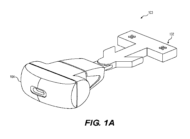
[0012] FIG. 1A depicts an illustrative cosmetic vibrational dental device
according to
one aspect of the disclosure;
[0013] FIG. 1B depicts an illustrative cosmetic vibrational dental device,
such as that
depicted in FIG 1A placed in the mouth of a user, according to one aspect of
the
disclosure;
[0014] FIG. 2A is a cone-beam computed tomography (CBCT) view of an implant
placed four months post-grafting according to one example of the present
disclosure;
[0015] FIG. 2B is a CBCT view of the implant of FIG. 2A following four
months of
integration with an exemplary use of a vibration device for five minutes
daily;
[0016] FIG. 3A depicts a CBCT cross section of the maxillary molar area
pretreatment demonstrating periapical pathology associated with failed
endodontics;
[0017] FIG. 3B depicts a periapical radiograph demonstrating failed
endodontics with
associated osseous destruction;
[0018] FIG. 4A depicts a radiograph of a maxillary molar exhibiting grade
2+
mobility;
[0019] FIG. 4B depicts a radiograph of the maxillary molar of FIG. 4A
following an
exemplary use of a vibration device according to one aspect of the present
disclosure;
[0020] FIG. 5A is an image of extraction sockets following curettage prior
to socket
grafting according to an example;
[0021] FIG. 5B is a radiograph of a grafted socket following extraction of
bridge
abutments;
6
CA 03236725 2024-04-25
WO 2023/076896
PCT/US2022/078651
[0022] FIGs. 6A-6B depict CBCT cross sections of grafted sockets four
months post
treatment following use of an illustrative device;
[0023] FIG. 7A is a panoramic CBCT view of an implant site demonstrating
osseous
graft maturation at four months according to an example;
[0024] FIG. 7B is an image of an exposed implant site showing the grafted
area at
four months post extraction;
[0025] FIG. 8A depicts a plan view of a dental mouthpiece according to an
exemplary
embodiment;
[0026] FIG. 8B is a side view of an illustrative intraoral dental device
according to an
exemplary embodiment;
[0027] FIG. 8C is a partial side view of an illustrative dual-arch dental
device
according to an exemplary embodiment;
[0028] FIG. 8D is a side view of a further illustrative intraoral dental
device
according to an exemplary embodiment;
[0029] FIG. 8E depicts exemplary pillar shapes according to further
exemplary
embodiments of the disclosure;
[0030] FIGs. 9A-9B are front and schematic cross-sectional views
respectively of an
upper dental arch engaging exemplary embodiments of the disclosure;
[0031] FIGs. 9C-9D are top and schematic cross-sectional views respectively
of an
upper dental arch engaging exemplary embodiments of the disclosure;
7
CA 03236725 2024-04-25
WO 2023/076896
PCT/US2022/078651
[0032] FIG. 10 depicts an illustrative dental device according to a further
exemplary
embodiment of the disclosure;
[0033] FIG. 11A is a CBCT image of a mandibular implant presenting with a
radiolucent area on the mesial aspect of the implant with no clinical mobility
or
patient stated sensitivity;
[0034] FIG. 11B is a CBCT image of a mandibular implant following four
months of
daily use of the appliance showing that the radiolucency has resolved and
increased
osseous density is noted;
[0035] FIG. 11C is a CBCT image of a cross section before treatment
demonstrating
bone level on buccal/lingual of the implant and the density of the cancellous
bone in
contact with the implant;
[0036] FIG. 11D is a CBCT image of a cross section following treatment with
the
appliance demonstrating bone level on buccal/lingual of the implant and the
increase
in density of the cancellous bone in contact with the implant;
[0037] FIGs. 12A-B are images of a mandibular implant where the patient
presented
with bone loss as evidenced by decreased bone density adjacent to the implant
in the
absence of mobility (purple = very low density, blue = low density, green =
high
density, yellow = very high density);
[0038] FIGs. 12C-D are images of the mandibular implant of FIGs. 12A-B
immediately following graft placement demonstrating the graft material filling
the
osseous void that resulted by peri-implantitis;
8
CA 03236725 2024-04-25
WO 2023/076896
PCT/US2022/078651
[0039] FIGs. 12E-F are images of the mandibular implant of FIGs. 12C-D two
months post graft repair of peri-implantitis associated bone loss with daily
use of low-
magnitude high-frequency vibration (LMHFV) by the patient demonstrating
increased
density of the grafted area to blend with the native bone adjacent to it and
an increase
in adjacent bone density related to vibration transfer throughout the maxilla;
[0040] FIG. 13A is a chart showing comparison of PDL fibroblast between non-
vibrated control and LMHFV 120 Hz over a 3-day period demonstrating
statistically
significant increases with the LMHFV;
[0041] FIG. 13B is a chart showing comparison of osteoblasts between non-
vibrated
control and LMHFV 120 Hz over a 3-day period demonstrating statistically
significant increases with the LMHFV;
[0042] Reference will now be made in detail to exemplary embodiments.
Unless
otherwise defined, technical or scientific terms have the meaning commonly
understood by one of ordinary skill in the art. The disclosed embodiments are
described in sufficient detail to enable those skilled in the art to practice
the disclosed
embodiments. It is to be understood that other embodiments may be utilized and
that
changes may be made without departing from the scope of the disclosed
embodiments. Thus, the materials, methods, and examples are illustrative only
and are
not intended to be necessarily limiting.
9
CA 03236725 2024-04-25
WO 2023/076896
PCT/US2022/078651
Detailed Description
[0043] It is to be understood that both the foregoing general description
and the
following detailed description are exemplary and explanatory only and are not
restrictive of the claims.
[0044] The disclosed embodiments relate to cosmetic devices, systems, and
methods
for accelerating graft conversion to alveolar bone. Advantageously,
embodiments of
the present disclosure can be implemented to convert graft material to mature
bone
more quickly than without. This is surprising in light of the generally held
view that
mechanical disruption of the graft site is detrimental to osseointegration.
[0045] When applied in immediate implant loading LMHFV can additionally
advantageously accelerate bone density surrounding the implants improving the
expected cosmetic outcome in a shorter period than traditionally observed.
This is
surprising in light of the generally held view that motion, including
micromotion. of
the implant after implantation is detrimental to the osseointegration of the
implant.
The application of vibration according to aspects of the current disclosure
also has
osseous stimulatory affects in cases where the implant will not be immediately
loaded
and allowed to heal before initiation of the restorative phase. Compared to
without
treatment, LMHFV can increase the speed and/or quality of the process of
osseointegration of a bone graft including the infiltration of granulation
tissue into a
blood clot at an extraction site, the proliferation of epithelium into the
extraction site
and graft, the formation of bone spicules, and the mineralization of these
spicules into
mature bone. LMHFV can also increase the speed and/or quality of
mineralization of
immature bone spicules into mature bone.
CA 03236725 2024-04-25
WO 2023/076896
PCT/US2022/078651
[0046] LMHFV, as indicated may be utilized immediately following implant
placement when insertion torque so dictates, for example when sufficient
torque to
immediately load, or when clinical circumstances will not permit immediate
loading,
for example, insufficient insertion torque. In an aspect, with reference to
FIGs. 1A
and 1B, use of the device 100 providing LMHFV for 5 minutes daily accelerates
osseous healing through osteogenic cell stimulation, with increased growth
factor
expression and angiogenesis stimulation permitting earlier loading. Further
advantageously, bone density improvement is observed contributing to implant
stability and better overall oral health.
[0047] LMHFV therapy according to the present disclosure is also
advantageously
configured to enhance and accelerate bone remodeling by improving bone density
and
mineral content of the bone around teeth, implants, and within grafted implant
sites.
In an aspect, LMHFV therapy is configured to increase bone mineral density
(BMD)
and improve localized osseous circulation. In an aspect, the increase in bone
density
improves the periodontal status of those involved teeth and contributes to a
subsequent decrease in tooth mobility. This correlates to implant applications
with
improvement in both the BMD and circulation when utilized after implant
placement
or with sites that are being grafted in anticipation of later implant
placement. In an
aspect, LMHFV therapy advantageously contributes to the release of growth
factor
such as BMP2, PDGFa, and TGF 131 among others. In another aspect, LMHFV
therapy is configured to increase osteoblast and PDL cell proliferation
stimulation.
LMHFV may also regulate gene expression-enhancing callus formation,
mineralization, and remodeling of bone.
11
CA 03236725 2024-04-25
WO 2023/076896
PCT/US2022/078651
[0048] Described herein are LMHFV dental devices, which in certain
embodiments
include a mouthpiece configured to transmit vibration to all or a portion of
the
patient's teeth.
[0049] Referring to FIGS. 1A-1B, an exemplary cosmetic dental device 100
includes
a mouthpiece 102 operatively connected to a housing 104. The mouthpiece 102
can be
separable from the housing 104 for interchangeability between users or for
ease of
cleaning. The mouthpiece 102 can include one or more oral tissue-contacting
portion,
such as a biteplate or probe for contacting teeth, gums or other oral tissues.
As shown,
in FIGS. 1A, the mouthpiece can include a biteplate which can be appropriately
shaped to cover occlusal surfaces of some or all of a patient's dentition.
Other shapes
for the mouthpiece are possible. For example, the mouthpiece can be configured
to
abut the lingual and buccal lateral sides of the alveolar ridge either with or
without
occlusal contact or, when no teeth are present, contact with gums overlying
the
alveolar ridge. A vibration generator can be located in the mouthpiece 102 or
the
housing 104 to vibrate the mouthpiece 102. The housing 104 can also include
the
electronics to run the motor the vibrator, collect usage and device operation
data,
collect data from sensors in the mouthpiece or base, and store data in memory.
The
housing 104 can include a data interface which can be wired or wireless to
allow a
data connection to other devices. The housing 104 can also include a power
interface
to allow charging of any onboard power sources, such as batteries or capacitor
banks.
The mouthpiece 102 can be electrically interconnected to the housing 104. FIG.
1B
depicts an illustrative dental device 100, such as that described above with
reference
to FIG. 1A, inserted in the mouth of a human user 106 and engaging the
occlusal
surfaces of the molars. The mouthpiece of the dental device 100 can, as
described
12
CA 03236725 2024-04-25
WO 2023/076896
PCT/US2022/078651
above, be sized and shaped to contact any dental tissue, including some or all
of the
teeth, specific regions of the gums, or both.
[0050] As is known in the art, the vibration generator can include an
electric motor
connected to an eccentric weight, or can be a piezo generator, as well as
other known
expedients. Accordingly, when the mouthpiece 102 is placed in a patient's
mouth and
the dental device is 100 turned on, the vibration of the mouthpiece 102 will
place
vibratory force repetitively on the teeth and/or other oral tissues.
[0051] FIG. 2A is a CBCT (also referred to as C-arm CT, cone beam volume
CT, flat
panel CT or Digital Volume Tomography (DVT)) view showing an implant placed
four months post-grafting according to an example. The implants were placed
into
extraction sockets into which bone had been grafted with the use of LMHFV to
increase bone density and integrity and to provide a more stable foundation in
which
to locate the implants at initial placement. Advantageously, implants such as
threaded
posts can be driven with a higher insertion torque than would be normally
possible in
type 3 or type 4 bone normally found in a long-healed posterior maxilla or 4
months
post-grafting when LMHFV was not used during healing. Once loading is
initiated,
continued use of LMFHV will continue to further increase bone density around
the
implants improving their long-term prognosis through better load handling. In
an
example, the appliance may be used long-term as an at home therapy to preserve
bone
to implant contact (BIC) and potentially prevent peri-implantitis. FIG. 2B is
a view of
the implant in FIG. 2A following an additional four months of integration and
with
use of an illustrative dental device according to the present disclosure for
five minutes
daily. The density of the BIC during the integration period has improved,
demonstrating blending of the graft with the surrounding host bone.
13
CA 03236725 2024-04-25
WO 2023/076896
PCT/US2022/078651
[0052] LMHFV also stimulates bone progenitor cells as well as increase
angiogenesis
resulting in acceleration of maturation of the clot in the site.
[0053] In one aspect, LMHFV has demonstrated improvement with mobility of
natural teeth by its stimulation leading to improvement in the bond density
making it
more stable and a decrease in the PDL width.
[0054] In another aspect, as with just a clot, LMHFV offers the same
effects of
stimulating and accelerating conversion of the material to denser mature bone
so that
a dental implant may be placed into higher quality bone sooner than when LMHFV
is
not utilized. As a consequence, superior aesthetic results can be obtained.
[0055] In yet another aspect, LMHFV can be performed as an extraction
sockets aid
after a nonsurgical or surgical approach to peri-implantitis is performed.
LMHFV can
also be performed when any oral or facial procedure or surgery is performed
and
results a need for grafting, such as root canals, scaling and planning, etc.
LMHFV
stimulates organization of the clot to improve soft tissue reattachment,
accelerates
angiogenesis, and therefore improves bone formation. LMHFV also has an anti-
inflammatory effect on the soft and hard tissue by accelerating and
stimulating host
factors to improve organization, and by depressing factors that may cause
unsightly
inflammation. In some embodiments, apart from the aesthetic or cosmetic
advantages
discussed herein, accelerated healing and organization may result better pain
management of the patient.
[0056] Clinically, a tooth that due to clinical issues that will not permit
long-term
maintenance of that tooth will be indicated for extraction. Teeth requiring
extraction
frequently have less dense bone surrounding them or defects related to
negative
14
CA 03236725 2024-04-25
WO 2023/076896
PCT/US2022/078651
biological effects such as infection. Turning to FIGs. 3A and 3B, examples of
instances where bone formation is required include presentations of failed
endodontics. FIG. 3A shows an image of a cross section of the maxillary molar
area
pretreatment demonstrating periapical pathology associated with failed
endodontics
that will in this instance necessitate extraction of the molar. FIG. 3B
depicts a
periapical radiograph demonstrating failed endodontics with associated osseous
destruction that will necessitate extraction of the bridge abutment teeth.
[0057] Turning to FIGs. 4A and 4B, teeth presenting with mobility will
usually have
a widened PDL space and lower bone density surrounding that tooth. FIG. 4A is
a
radiograph of a maxillary molar exhibiting grade 2+ mobility that has widened
periodontal ligament space surrounding the tooth, as referenced by arrows A,
and a
possible lesion on the distal buccal apical area. Such widened periodontal
ligament
space is indicative of increased tooth mobility. FIG. 4B is a radiograph of
the
maxillary molar of FIG. 4A following use of the appliance for 5 minutes daily
for 4
months showing that mobility has resolved, and apical area has disappeared
with a
normal PDL space radiographically. LMHFV may be administered using
illustrative
device 100 (FIGs. 1A and 1B) to stimulate the bone and increase the bone
density
while decreasing the PDL space and associated improvement in the mobility
returning
to a healthy periodontal state. In an example, a patient can stimulate the
bone using
the appliance for five minutes daily.
[0058] Following extraction of a problematic tooth or teeth, curettage of
extraction
sockets can be performed to remove any residual unhealthy or pathologic
tissue. (FIG.
5A) The extraction sockets can be packed with a clinically indicated amount of
an
appropriate graft material and the implantation site can be closed with or
without a
CA 03236725 2024-04-25
WO 2023/076896
PCT/US2022/078651
membrane. FIG. 5B is a radiograph of a grafted socket following extraction of
bridge
abutments, socket curettage and socket grafting demonstrating a granular
appearance
of the graft material according to an example. The granular appearance of the
graft
material reveals a lower tissue density within the socket than the host's
adjacent
native bone.
[0059] In some embodiments, the patient can be instructed to use the
appliance for a
prescribed time and duration to augment a grafted implant site. In an example,
the
patient can be instructed to use the appliance for five minutes daily over a
four-month
period. Turning to FIGs. 6A and 6B, when radiographically examined after a
four-
month healing period, the grafted site demonstrates more rapid conversion of
the graft
particles to blend with the surrounding host bone with similar radiographic
density
and trabeculation, appearing ready for implant placement. FIGs. 6A and 6B
depict
CBCT cross sections of grafted sockets four months post treatment following
use of
an illustrative device for five minutes daily demonstrating increased density
of the
grafted sites, which are approximately delimited by dashed lines B. FIG. 7A is
a
panoramic CBCT view of an implant site demonstrating osseous graft maturation
at
four months and ready for implant placement. Following socket grafting of the
extraction sockets and use of LMHFV for 4 months, the graft sites,
approximately
delimited by dashed lines C, have radiographically blended with the
surrounding host
bone and are indistinguishable therefrom. Density improvement was accelerated
with
LMHFV that would not be otherwise observed. FIG. 7B is an image of an exposed
implant site showing the grafted area at four months post extraction and
socket
preservation with daily use of an exemplary device demonstrating that the
osseous
graft has organized to blend with the surrounding host bone.
16
CA 03236725 2024-04-25
WO 2023/076896
PCT/US2022/078651
[0060] The vibration can be applied along multiple axes or selected to be
primarily on
a single axis. The primary anatomic reference directions with reference to a
standing
human are superior-inferior (up and down), anterior-posterior (front to back),
medial-
lateral (side to side). Because mastication places loading on oral structures
primarily
in the superior-inferior direction through mandibular action, it may be
advantageous
to choose vibrational loading along other axes either separately or in
combination.
Vibrational Cosmetic Dental Devices
[0061] According to an aspect of the present disclosure, a vibrational
cosmetic dental
device that vibrates at one or more predetermined frequencies is provided. In
some
embodiments the vibrational frequency is fixed within a lower bound and an
upper
bound. The lower bound can be greater than about 110 Hz, 105 Hz, 100 Hz, 95
Hz, 90
Hz, 85 Hz, 80 Hz, 75 Hz, 70 Hz, 65 Hz, 60 Hz, 55 Hz, 50 Hz, 45 Hz, or less.
The
upper bound can be greater than about 115 Hz, 120 Hz, 125 Hz, 130 Hz, 135 Hz,
140
Hz, 145 Hz, 150 Hz, or more. In some embodiments, the frequency varies within
a
lower and an upper bound. In some embodiments two or more frequencies, fixed
or
varying, are employed.
[0062] In some embodiments the duration of a treatment session can be
specified to
be greater than about 30 seconds, 1 min, 2 min, 3 min, 4 min, 5 min, 6 min, 7
min, 8
min, 9 min, 10 min, 11 min, 12 min, 13 min, 14 min, 15 min, 16 min, 17 min, 18
min,
19 min, 20 min, or more; or specified to be less than about 20 min, 19 min, 18
min, 17
min, 16 min, 15 min, 14 min, 13 min, 12 min, 10 min, 9 min, 8 min, 7 min, 6
min, 5
min, 4 min, 3 min, 2 min, 1 min, 30 seconds, or less.
17
CA 03236725 2024-04-25
WO 2023/076896
PCT/US2022/078651
[0063] FIG. 1 depicts a cosmetic vibrational dental device according to an
example.
The vibrational dental device can include a mouthpiece and a vibrational
source
connected to each other. The mouthpiece is configured to be provided between
the
occlusal surfaces of a user's teeth, and to be bite down by the user to
contact the
user's dentition during the treatment. The mouthpiece can cover at least the
teeth or
implant around which accelerating graft conversion is desired. The vibrational
source
is configured to provide vibration to the mouthpiece at a preset frequency and
acceleration.
[0064] To achieve the maximum desired cosmetic results of accelerating
graft
material conversion, further studies are still needed to optimize the
parameters of
LMHFV. Such parameters may include frequency, acceleration, and dosage. Dosage
may include duration per use, number of uses per day, or number of days of
use,
either consecutively or at a certain schedule.
[0065] In some embodiments, the vibrational source may be connected to the
mouthpiece in such way that the vibration provided is in the sagittal plane of
a user's
mouth. A motor may be included in the vibrational source to provide such
vibration.
The motor may be of any suitable type known in the art. The motor, when in
use, may
be configured to provide vibration at a frequency as disclosed herein. The
motor,
when in use, may be further configured to provide vibration at an acceleration
magnitude. In some embodiments the mouthpiece of a dental vibration device can
have an acceleration within a lower bound and an upper bound. The lower bound
can
be greater than about 0.010 G, 0.015 G, 0.020 G, 0.025 G, 0.030 G, 0.035 G,
0.040
G, 0.045 G, 0.050 G, 0.055 G, 0.060 G, or more; or less than about 0.060 G,
0.055 G,
0.050 G, 0.045 G, 0.040 G, 0.035 G, 0.030 G, 0.025 G, 0.020 G, 0.015 G, 0.010
G, or
18
CA 03236725 2024-04-25
WO 2023/076896
PCT/US2022/078651
less. The upper bound can be greater than about 0.07 G, 0.08 G, 0.09 G, 0.10
G, 0.11
G, 0.12 G, 0.13 G, 0.14 G, 0.15 G, or more; or less than about 0.15 G, 0.14 G,
0.13 G,
0.12 G, 0.11 G, 0.10 G, 0.09 G, 0.08 G, 0.07 G, or less.
[0066] The motor may be assembled into the vibrational source in an
orientation that
may provide vibration in such ways.
[0067] In some embodiments, sensors may be added to the vibrational dental
device,
either on the vibrational device, or on the mouthpiece. The sensors may be
configured
to detect and monitor the parameters of the vibration, for example,
frequencies and
acceleration magnitudes. The sensors may also be configured to detect if the
user has
bitten down on the mouthpiece correctly. The sensors may be accelerometers,
gyroscopes, proximity sensors, pressure sensors, humidity sensors, temperature
sensors, or any combinations of them.
[0068] In some embodiments, the mouthpiece could be in contact with at
least the
teeth or implant near which graft conversion acceleration is needed. The
mouthpiece
may be configured to be placed in contact with a user's dentition, between and
clamped down by both occlusal surfaces of the dentition. The mouthpiece can
include
ridges or be without ridges. The mouthpiece can cover the entire dentition, or
only a
part of the dentition. The shape of the mouthpiece can be customized to cover
only
selected teeth or implants.
[0069] Turning to FIGs. 8A-8E, a further exemplary dental appliance 200 is
depicted.
The illustrative device 200 can include a base 210 and an array of bristles or
pillars
220 covering the base. In an aspect, the array of pillars 220 are configured
to
substantially envelope one or more teeth according to an example. In some
19
CA 03236725 2024-04-25
WO 2023/076896
PCT/US2022/078651
embodiments, dental appliance 200 can include a first set of pillars 222
configured to
interface with a first set of teeth and a second set of pillars 224 configured
to interface
with a second set of teeth. In some embodiments, the array of pillars can
protrude
substantially parallel and vertically from the base. Subsets of pillars may
also be non-
parallel and apply angular stresses on the teeth. In some embodiments, each
pillar can
be movable with a spring 230 configured to retract when engaged with a tooth
(Fig.
8D, see also FIGs. 9A-9D). Fig. 8E depicts examples of pillar shapes, having
one or
more materials, and configured to modify torsion on the teeth and gums and
selectively enhance and/or dampen vibrations.
[0070] In some embodiments, the appliance can be configured to engage with
a
patient's teeth alone (FIGs. 8A-8B) or can be configured to engage with a
patient's
teeth and gums (FIGs. 9C-9D). As shown in FIG. 9C, the array of pillars 220,
222,
224 may gently engage with the graft site and/or the future implant site to
provide
stimulation to the soft tissue. Such gentle stimulation can help to increase
blood flow
and other cells of repair to the site, in addition to that provided by
vibration conducted
through neighboring teeth and tissue structures.
[0071] Turning to FIG. 10, in some embodiments, a granular dental appliance
or
appliance 400 can be configured to isolate one or more teeth or implant sites
for
stimulation therapy. In an example, the appliance 400 can be configured to
control
stimulation energy to a subset of the array of pillars. In an example, a first
set of
pillars 422 and a second set of pillars 424 can be configured to immobilize or
isolate
stimulation from at least one tooth 432 and 436 while stimulation energy is
being
applied to an active set of pillars 426 directed at engaging a tooth 434 or
implant site.
CA 03236725 2024-04-25
WO 2023/076896
PCT/US2022/078651
[0072] In some embodiments, a granular dental appliance 400 can include a
base
portion 410 including a stimulation source such as a vibration source, a
plurality of
pillars 420 in communication with the base and configured to engage with at
least one
tooth 432, 434, and 436 and at least a portion of a gum, where a first set of
pillars of
the plurality of pillars is configured to immobilize or dampen vibration of at
least a
first tooth 432 or portion of gum, and a second set of pillars of the
plurality of pillars
is configured to mobilize or enhance vibration of at least a second tooth 434
or
portion of gum, which can also be seen in FIGs. 9C to 9D.
[0073] According to yet another aspect of the present disclosure, a method
for
accelerating graft conversion to alveolar bone is described. The method
including
providing a vibratory dental appliance, comprising a base including a
vibration
source, and a plurality of pillars extending from the base and configured to
engage
with at least one tooth and at least a portion of a gum, determining at least
one of an
orientation of at least one tooth and a gum line, controlling a first
vibration to a first
set of pillars of the plurality of pillars, the first vibration is configured
to immobilize
or dampen vibration of at least a first tooth or portion of gum, and
controlling a
second vibration to a second set of pillars of the plurality of pillars, the
second
vibration is configured to mobilize or enhance vibration of at least a second
tooth or
portion of gum.
Method For Accelerating Graft Material Conversion
[0074] According to yet another aspect of the present disclosure, a method
for
accelerating graft material conversion is described. The method includes
providing
the mouthpiece of the vibrational dental device to a user and providing
instructions to
21
CA 03236725 2024-04-25
WO 2023/076896
PCT/US2022/078651
the user. The instruction may include placement guidelines and dosage
information.
The dosage information may include duration of each treatment session, number
of
sessions in a day, number of days, etc. For example, the instruction may
instruct a
user to use the vibrational dental device for number of times per day. In some
embodiments the treatment frequency can be specified to be once per day, twice
per
day, 3 times per day, 4 times per day, 5 times per day, 6 times per day, 7
times per
day, 8 times per day, 9 times per day, or more. In some embodiments the
duration of
treatment can be specified to be about 1 day, 1 week, 2 weeks, 3 weeks, 1
month, 2
months, 3 months, 4 months, 5 months, 6 months, 7 months, 8 months, 9 months,
10
months, 11 months, 1 year, or more.
[0075] In some embodiments, the method may further include configuring the
vibrational source providing an axial vibratory force to the mouthpiece. The
axial
vibratory force may be eventually applied to the dentition through the
mouthpiece,
which is clamped down by the teeth. The vibratory force (e.g., acceleration
magnitudes, frequencies, etc.) can be adjusted by selecting preset values, or
fine-tuned
by users, technicians, or healthcare professionals.
[0076] According to yet another aspect of the present disclosure, a method
for
detecting graft material conversion is described. The method includes steps of
identifying an implant site, providing a graft at the implant site, applying a
stimulus to
a portion of the implant site, sensing a baseline response at the implant
site, applying
one or more vibration sessions over a period of time, sensing at least one
second
response at the implant site, and determining an osseous status based on a
comparison
between the baseline response and one or more second responses. In some
embodiments, the method may further include implanting a dental implant at the
22
CA 03236725 2024-04-25
WO 2023/076896
PCT/US2022/078651
implant site based on the osseous status along with aesthetic factors. In some
embodiments, the method may further include applying a stimulus to a portion
of the
implant site with the dental implant.
[0077] In some embodiments, the stimulus applied can be one or electrical
energy,
light energy, and a mechanical dynamic load that is either isotonic or
isometric. In
addition, the stimulus can be applied to a portion of the implant site
symmetrically or
asymmetrically on one side of the implant site or across the implant site such
as
across a facial side and lingual side or mesial side and distal side (see FIG.
3A). In
some embodiments, sensing a baseline response can include information
informing an
osseous density at the implant site.
Examples
[0078] FIG. 11A is a CBCT image of a mandibular implant presenting with a
radiolucent area on the mesial aspect of the implant with no clinical mobility
or
patient stated sensitivity. Radiographic evidence of a space present between
the
implant and bone on the mesial, indicated by arrows D, indicate peri-
implantitis.
LMHFV was utilized by the patient daily, increasing bone density and
eliminating the
mesial space and the peri-implantitis. The patient reported no pain or
mobility during
or following treatment. FIG. 11B is a CBCT image of a mandibular implant
following
four months of daily use of the appliance showing that the radiolucency has
resolved
and increased osseous density is noted. The implant was rescued without the
need for
surgical intervention.
[0079] FIG. 11C is a CBCT image of a cross section before treatment
demonstrating
bone level on buccal/lingual of the implant and the density of the cancellous
bone in
23
CA 03236725 2024-04-25
WO 2023/076896
PCT/US2022/078651
contact with the implant. This posterior mandibular implant presented with
radiographic evidence of a space present between the implant and bone on the
mesial,
as indicated by arrows E, indicating peri-implantitis with no mobility of the
implant or
pain noted by the patient. FIG. 11D is a CBCT image of a cross section
following
daily LMHFV treatment with the appliance demonstrating bone level on
buccal/lingual of the implant and the increase in density of the cancellous
bone in
contact with the implant. As a result of the treatment, the patient eliminated
the peri-
implantitis and rescued the implant without the need for surgical
intervention.
[0080] FIGs. 12A (unretouched) and 12B (colorized) are radiographic images
of a
mandibular implant where the patient presented with bone loss as evidenced by
decreased bone density adjacent to the implant in the absence of mobility
(purple =
very low density, blue = low density, green = high density, yellow = very high
density). The patient was subject to surgical intervention to debride the area
and clean
the exposed threads plus place osseous graft material to fill in the defect
caused by the
inflammation associated with the peri-implantitis. FIGs. 12C and 12D are
images of
the mandibular implant of FIGs. 12A and 12B immediately following graft
placement
demonstrating the graft material filling the osseous void that resulted by
peri-
implantitis. FIGs. 12E-F are images of the mandibular implant of FIGs. 12C and
12D
two months post-graft repair of peri-implantitis associated bone loss with
daily use of
LMHFV by the patient demonstrating a disappearance of peri-implantitis,
increased
density of the grafted area to blend with the native bone adjacent to it and
an increase
in adjacent bone density related to vibration transfer throughout the maxilla.
Two
months post treatment after daily LMHFV use, and improvements is seen in the
affect
(grafted) implant, as well as high density bone along the entire length.
Additionally,
24
CA 03236725 2024-04-25
WO 2023/076896
PCT/US2022/078651
comparing distant bone (to the left of the implants) were no teeth or implants
were
present initially, the density is of type 4 quality typically found in the
posterior
maxilla. Following LMHFV and its transmission throughout the bone, a distance
from
the implants being treated, a significant increase in bone density raising
that bone to at
least type 2, demonstrating LMHFV transmission greatly improved bone quality
at
and adjacent to the area being treated. Bone density improvement to this
degree is not
observed with graft placement without the use of LMHFV.
[0081] The distal maxillary implant presented with 50% bone loss and very
low bone
density surrounding the implant, indicative of peri-implantitis. When compared
to the
mesial implant that had no bone loss, a significant deterioration on the
affected
implant is evident. The patient utilized LMHFV daily for 5 minutes and the
radiograph taken at 2 months demonstrates successful graft integration into
native
bone and a rescue of the implant.
[0082] FIG. 13A is a chart showing comparison of PDL fibroblast between non-
vibrated control and LMHFV 120 Hz over a 3-day period demonstrating
statistically
significant increases with the LMHFV. FIG. 13B is a chart showing comparison
of
osteoblasts between non-vibrated control and LMHFV 120 Hz over a 3-day period
demonstrating statistically significant increases with the LMHFV.
[0083] The foregoing descriptions have been presented for purposes of
illustration.
They are not exhaustive and are not limited to precise forms or embodiments
disclosed. Modifications and adaptations of the embodiments will be apparent
from
consideration of the specification and practice of the disclosed embodiments.
For
example, the described implementations include hardware, but systems and
methods
CA 03236725 2024-04-25
WO 2023/076896
PCT/US2022/078651
consistent with the present disclosure can be implemented with hardware and
software. In addition, while certain components have been described as being
coupled
to one another, such components may be integrated with one another or
distributed in
any suitable fashion.
[0084] Moreover, while illustrative embodiments have been described herein,
the
scope includes any and all embodiments having equivalent elements,
modifications,
omissions, combinations (e.g., of aspects across various embodiments),
adaptations or
alterations based on the present disclosure. The elements in the claims are to
be
interpreted broadly based on the language employed in the claims and not
limited to
examples described in the present specification or during the prosecution of
the
application, which examples are to be construed as nonexclusive. Further, the
steps of
the disclosed methods can be modified in any manner, including reordering
steps or
inserting or deleting steps.
[0085] It should be noted that, the relational terms herein such as "first"
and "second"
are used only to differentiate an entity or operation from another entity or
operation,
and do not require or imply any actual relationship or sequence between these
entities
or operations. Moreover, the words "comprising," "having," "containing," and
"including," and other similar forms are intended to be equivalent in meaning
and be
open ended in that an item or items following any one of these words is not
meant to
be an exhaustive listing of such item or items, or meant to be limited to only
the listed
item or items.
[0086] The features and advantages of the disclosure are apparent from the
detailed
specification, and thus, it is intended that the appended claims cover all
systems and
26
CA 03236725 2024-04-25
WO 2023/076896
PCT/US2022/078651
methods falling within the true spirit and scope of the disclosure. As used
herein, the
indefinite articles "a" and "an" mean "one or more." Similarly, the use of a
plural
term does not necessarily denote a plurality unless it is unambiguous in the
given
context. Further, since numerous modifications and variations will readily
occur from
studying the present disclosure, it is not desired to limit the disclosure to
the exact
construction and operation illustrated and described, and accordingly, all
suitable
modifications and equivalents may be resorted to, falling within the scope of
the
disclosure.
[0087] As used herein, unless specifically stated otherwise, the terms
"and/or" and
"or" encompass all possible combinations, except where infeasible. For
example, if it
is stated that a database may include A or B, then, unless specifically stated
otherwise
or infeasible, the database may include A, or B, or A and B. As a second
example, if it
is stated that a database may include A, B, or C, then, unless specifically
stated
otherwise or infeasible, the database may include A, or B, or C, or A and B,
or A and
C, or B and C, or A and B and C.
[0088] It is appreciated that the above-described embodiments can be
implemented by
hardware, or software (program codes), or a combination of hardware and
software. If
implemented by software, it may be stored in the above-described computer-
readable
media. The software, when executed by the processor can perform the disclosed
methods. The computing units and other functional units described in this
disclosure
can be implemented by hardware, or software, or a combination of hardware and
software. One of ordinary skill in the art will also understand that multiple
ones of the
above-described modules/units may be combined as one module/unit, and each of
the
27
CA 03236725 2024-04-25
WO 2023/076896
PCT/US2022/078651
above-described modules/units may be further divided into a plurality of sub-
modules/sub-units.
[0089] In the foregoing specification, embodiments have been described with
reference to numerous specific details that can vary from implementation to
implementation. Certain adaptations and modifications of the described
embodiments
can be made. Other embodiments can be apparent to those skilled in the art
from
consideration of the specification and practice of the disclosure disclosed
herein. It is
intended that the specification and examples be considered as exemplary only,
with a
true scope and spirit of the disclosure being indicated by the following
claims. It is
also intended that the sequence of steps shown in figures are only for
illustrative
purposes and are not intended to be limited to any particular sequence of
steps. As
such, those skilled in the art can appreciate that these steps can be
performed in a
different order while implementing the same method.
28