Note: Descriptions are shown in the official language in which they were submitted.
PROSTHETIC HEART VALVE DELIVERY APPARATUS
FIELD
[001] The present invention concerns embodiments of a prosthetic valve (e.g.,
prosthetic heart
valve) and a delivery apparatus for implanting a prosthetic valve.
BACKGROUND
[002] Prosthetic cardiac valves have been used for many years to treat cardiac
valvular
disorders. The native heart valves (such as the aortic, pulmonary and mitral
valves) serve critical
functions in assuring the forward flow of an adequate supply of blood through
the cardiovascular
system. These heart valves can be rendered less effective by congenital,
inflammatory or
infectious conditions. Such damage to the valves can result in serious
cardiovascular
compromise or death. For many years the definitive treatment for such
disorders was the surgical
repair or replacement of the valve during open heart surgery, but such
surgeries are prone to
many complications. More recently a transvascular technique has been developed
for introducing
and implanting a prosthetic heart valve using a flexible catheter in a manner
that is less invasive
than open heart surgery.
[003] In this technique, a prosthetic valve is mounted in a crimped state on
the end portion of a
flexible catheter and advanced through a blood vessel of the patient until the
prosthetic valve
reaches the implantation site. The prosthetic valve at the catheter tip is
then expanded to its
functional size at the site of the defective native valve such as by inflating
a balloon on which the
prosthetic valve is mounted. Alternatively, the prosthetic valve can have a
resilient, self-
expanding stent or frame that expands the prosthetic valve to its functional
size when it is
advanced from a delivery sheath at the distal end of the catheter.
[004] Balloon-expandable prosthetic valves typically are preferred for
replacing calcified native
valves because the catheter balloon can apply sufficient expanding force to
anchor the frame of
the prosthetic valve to the surrounding calcified tissue. On the other hand,
self-expanding
prosthetic valves sometimes are preferred for replacing a defective, non-
stenotic (non-calcified)
native valve, although they also can be used to replace stenotic valves. One
drawback associated
with implanting a self-expanding prosthetic valve is that as the operator
begins to advance the
1
Date Recue/Date Received 2024-05-29
prosthetic valve from the open end of the delivery sheath, the prosthetic
valve tends to "jump"
out very quickly from the end of the sheath; in other words, the outward
biasing force of the
prosthetic valve's frame tends to cause the prosthetic valve to be ejected
very quickly from the
distal end of the delivery sheath, making it difficult to deliver the
prosthetic valve from the
sheath in a precise and controlled manner and increasing the risk of trauma to
the patient.
[005] Another problem associated with implanting a percutaneous prosthetic
valve in a
non-stenotic native valve is that the prosthetic valve may not be able to
exert sufficient force
against the surrounding tissue to resist migration of the prosthetic valve.
Typically, the stent of
the prosthetic valve must be provided with additional anchoring or attachment
devices to assist in
anchoring the prosthetic valve to the surrounding tissue. Moreover, such
anchoring devices or
portions of the stent that assist in anchoring the prosthetic valve typically
extend into and
become fixed to non-diseased areas of the vasculature, which can result in
complications if
future intervention is required, for example, if the prosthetic valve needs to
be removed from the
patient.
SUMMARY
[006] Certain embodiments of the present disclosure provide a prosthetic valve
(e.g., a
prosthetic heart valve) and a valve delivery apparatus for delivery of the
prosthetic valve to a
native valve site via the human vasculature. The delivery apparatus is
particularly suited for
advancing a prosthetic valve through the aorta (i.e., in a retrograde
approach) for replacing a
diseased native aortic valve. The delivery apparatus in particular embodiments
is configured to
deploy a prosthetic valve from a delivery sheath in a precise and controlled
manner at the target
location within the body.
[007] In an aspect, a delivery assembly comprises a prosthetic valve, an
elongate shaft located
proximal to the prosthetic valve, a suture-retention member located distal to
the shaft, a slidable
release member, and an outer sheath. The prosthetic valve can comprise a self-
expandable stent
having a plurality of apices spaced circumferentially around a first end
portion of the stent,
wherein each apex has an aperture. The suture-retention member can comprise a
proximal
portion and a distal portion spaced from the proximal portion, the proximal
portion being
coupled to the shaft. The at least one slidable release member can extend
through the proximal
portion and the distal portion of the suture-retention member and a plurality
of suture loops
2
Date Recue/Date Received 2024-05-29
extending from the proximal portion of the suture-retention member. The
plurality of suture
loops can extend through the apertures in the apices of the stent and around
the release member
at a location between the proximal and distal portions of suture-retention
member, such that at
least one of the suture loops extends through the aperture of every apex. The
outer sheath can be
advanced over the prosthetic valve to retain the prosthetic valve in a
radially compressed state,
and can be retracted relative to the prosthetic valve to permit radial
expansion of the prosthetic
valve while the stent remains connected to the suture-retention member via the
suture loops.
After the entirety of the prosthetic valve is deployed from the sheath, the
sheath can be advanced
distally back over the prosthetic valve to cause the prosthetic valve to
radially collapse as it is
recaptured by the sheath.
[008] In some embodiments, the suture loops are formed from a single length of
suture
material.
[009] In some embodiments, the shaft comprises a first shaft and the assembly
further
comprises a second shaft extending at least partially through the first shaft,
wherein the outer
sheath can be advanced or retracted relative to the prosthetic valve by
rotating the second shaft
relative to the first shaft.
[010] In some embodiments, the at least one release member is slidable
relative to the suture-
retention member, and when the release member is retracted proximally such
that a distal end of
the release member is proximal to the distal portion of the suture-retention
member, the suture
loops can slide off the distal end of the release member, thereby releasing
the prosthetic valve
from the suture-retention member.
[011] In some embodiments, at least a portion of the outer sheath comprises a
slotted metal
tube.
[012] In some embodiments, a distal end portion of the outer sheath comprises
a delivery
capsule connected to a distal end of the slotted metal tube, the delivery
capsule configured to
extend over and retain the prosthetic valve in the radially compressed state.
[013] In some embodiments, the delivery capsule comprises a polymer sleeve.
[014] In some embodiments, the sheath is at least about 3-10 cm is length and
no greater than
about 40 cm in length.
3
Date Recue/Date Received 2024-05-29
[015] In some embodiments, at least one of the suture loops has a greater
thickness than others
of the suture loops.
[016] In another aspect, a delivery apparatus for implanting a prosthetic
valve comprises a first
elongated shaft having a proximal end portion and a distal end portion, a
second elongated shaft
extending through the first shaft and having a proximal end portion and a
distal end portion, and
a delivery sheath having a distal end portion configured to receive and retain
a prosthetic valve in
a compressed delivery state and a proximal end portion connected to the distal
end portion of the
second elongated shaft. The second shaft can be rotatable relative to the
first shaft but fixed
against axial movement relative to the first shaft. The proximal end portion
of the delivery sheath
can be more flexible than the distal end portion of the sheath. The delivery
sheath can be,
without limitation, at least about 3-5 cm in length and no greater than about
40 cm in length. The
second shaft can be configured to be rotatable relative to the first shaft
such that rotation of the
second shaft causes the delivery sheath to move axially relative to the first
and second shafts.
[017] In some embodiments, the delivery apparatus further comprises a screw
connected to a
distal end of the second shaft, and a nut mounted on the screw and connected
to the delivery
sheath such that rotation of the second shaft and the screw causes axial
movement of the nut
relative to the screw, thereby producing axial movement of the delivery
sheath.
[018] In some embodiments, the proximal end portion of the delivery sheath is
between about
cm and about 30 cm in length.
[019] In some embodiments, the distal end portion of the first shaft extends
through the
delivery sheath and comprises a slotted metal tube.
[020] In some embodiments, the delivery apparatus further comprises a suture-
retention
member connected to the distal end portion of the first shaft, a plurality of
suture loops extending
from the suture-retention member and configured to extend through openings in
a frame of the
prosthetic valve, and at least one slidable release member configured to
extend through the
suture-retention member and the suture loops to releasably secure the
prosthetic valve to the
suture-retention member.
[021] In some embodiments, the suture-retention member comprises a proximal
portion and a
distal portion spaced axially apart from the first portion and the release
member is slidable
4
Date Recue/Date Received 2024-05-29
relative to the suture-retention member, between a first position extending
through the proximal
and distal portions of the suture-retention member and a second position in
which the release
member is retracted to a location proximal of the distal portion of the suture-
retention member.
When the release member is in the first position and the suture loops extend
through the
openings of the frame and around the release member at a location between the
proximal and
distal portions, the prosthetic valve is secured to the suture-retention
member. When the release
member is in the second position, the suture loops can slide off a distal end
of the release
member to release the prosthetic valve from the suture-retention member.
[022] In some embodiments, the at least one release member comprises a
plurality of release
members extending through the suture-retention member.
[023] In some embodiments, the proximal portion of the outer sheath comprises
a slotted metal
tube.
[024] In some embodiments, the distal end portion of the outer sheath
comprises a delivery
capsule connected to a distal end of the slotted metal tube. The delivery
capsule can be
configured to extend over and retain the prosthetic valve in the compressed
delivery state. In
some embodiments, the delivery capsule comprises a polymer sleeve.
[025] In another aspect, a method for delivering a prosthetic valve to the
aortic annulus of the
heart comprises inserting an elongated delivery apparatus into a femoral
artery of a patient, the
delivery apparatus comprising a delivery sheath containing the prosthetic
valve in a radially
compressed state. The method can further comprise advancing the delivery
apparatus through the
aorta until the prosthetic valve is at an implantation location within the
aortic annulus, wherein
when the prosthetic valve is at the implantation location, the delivery sheath
extends through the
ascending aorta and the aortic arch, and a proximal end of the delivery sheath
is within the
descending aorta. The method can further comprise retracting the delivery
sheath relative to the
prosthetic valve to deploy the prosthetic valve from a distal end of the
delivery sheath.
[026] In some embodiments, the delivery sheath is at least about 3-5 cm and no
greater than 40
cm in length.
[027] In some embodiments, the delivery sheath comprises a distal end portion
and a proximal
end portion that is more flexible than the distal end portion. The distal end
portion of the sheath
Date Recue/Date Received 2024-05-29
can extend over and retain the prosthetic valve in the radially compressed
state during the acts of
the inserting and advancing the delivery apparatus, and the proximal end
portion can extend
through the ascending aorta, the aortic arch and into the descending aorta
when the prosthetic
valve is at the implantation location.
[028] In some embodiments, the prosthetic valve is releasably secured to the
delivery apparatus
via a plural of suture loops.
[029] In some embodiments, the act of retracting the delivery sheath comprises
deploying the
entire prosthetic valve from the delivery sheath and allowing the prosthetic
valve to radially
expand while still secured to the delivery apparatus via the suture loops.
[030] In some embodiments, the method further comprises, after deploying the
entire prosthetic
valve from the delivery sheath, recapturing the prosthetic valve by advancing
the delivery sheath
distally back over the prosthetic valve.
[031] In another aspect, a method for delivering a prosthetic valve to a
native valve annulus of
the heart comprises inserting an elongated delivery apparatus into the
vasculature of a patient,
the delivery apparatus comprising a delivery sheath containing the prosthetic
valve in a radially
compressed state, wherein the prosthetic valve is releasably secured to the
delivery apparatus via
a plural of suture loops. The method can further comprise retracting the
delivery sheath relative
to the prosthetic valve to deploy the entire prosthetic valve from the
delivery sheath, allowing the
prosthetic valve to radially expand while still secured to the delivery
apparatus via the suture
loops. The method can further comprise, after deploying the entire prosthetic
valve from the
delivery sheath, recapturing the prosthetic valve by advancing the delivery
sheath distally back
over the prosthetic valve.
BRIEF DESCRIPTION OF THE DRAWINGS
[032] FIG. 1 is a perspective view of a prosthetic valve that can be used to
replace the native
aortic valve of the heart, according to one embodiment.
[033] FIG. 2 is a perspective view of a portion of the prosthetic valve of
FIG. 1 illustrating the
connection of two leaflets to the support frame of the prosthetic valve.
[034] FIG. 3 is side elevation view of the support frame of the prosthetic
valve of FIG. 1.
6
Date Recue/Date Received 2024-05-29
[035] FIG. 4 is a perspective view of the support frame of the prosthetic
valve of FIG. 1.
[036] FIG. 5A is a cross-sectional view of the heart showing the prosthetic
valve of FIG. 1
implanted within the aortic annulus.
[037] FIG. 5B is an enlarged view of Fig. 5A illustrating the prosthetic valve
implanted within
the aortic annulus, shown with the leaflet structure of the prosthetic valve
removed for clarity.
[038] FIG. 6 is a perspective view of the leaflet structure of the prosthetic
valve of FIG. 1
shown prior to being secured to the support frame.
[039] FIG. 7 is a cross-sectional view of the prosthetic valve of FIG. 1.
[040] FIG. 8 is a cross-sectional view of an embodiment of a delivery
apparatus that can be
used to deliver and implant a prosthetic valve, such as the prosthetic valve
shown in FIG. 1.
FIGS. 8A-8C are enlarged cross-sectional views of sections of FIG. 8.
[041] FIG. 9 is an exploded view of the delivery apparatus of FIG. 8.
[042] FIG. 10 is a side view of the guide catheter of the delivery apparatus
of FIG. 8.
[043] FIG. 11 is a perspective, exploded view of the proximal end portion of
the guide catheter
of FIG. 10.
[044] FIG. 12 is a perspective, exploded view of the distal end portion of the
guide catheter
of FIG. 10.
[045] FIG. 13 is a side view of the torque shaft catheter of the delivery
apparatus of FIG. 8.
[046] FIG. 14 is an enlarged side view of the rotatable screw of the torque
shaft catheter
of FIG. 13.
[047] FIG. 15 is an enlarged perspective view of a coupling member disposed at
the end of the
torque shaft.
[048] FIG. 16 is an enlarged perspective view of the threaded nut used in the
torque shaft
catheter of FIG. 13.
[049] FIG. 17 is an enlarged side view of the distal end portion of the nose
cone catheter of the
delivery apparatus of FIG. 8.
7
Date Recue/Date Received 2024-05-29
[050] FIG. 17A is an enlarged, cross-sectional view of the nose cone of the
catheter
shown FIG. 17.
[051] FIG. 17B is an enlarged cross-sectional view of the distal end portion
of the delivery
apparatus of FIG. 8 showing the stent of a prosthetic valve retained in a
compressed state within
a delivery sheath.
[052] FIG. 18 is an enlarged side view of the distal end portion of the
delivery apparatus of
FIG. 8 showing the delivery sheath in a delivery position covering a
prosthetic valve in a
compressed state for delivery into a patient.
[053] FIG. 19 is an enlarged cross-sectional view of a section of the distal
end portion of the
delivery apparatus of FIG. 8 showing the valve-retaining mechanism securing
the stent of a
prosthetic valve to the delivery apparatus.
[054] FIG. 20 is an enlarged cross-sectional view similar to FIG. 19, showing
the inner fork of
the valve-retaining mechanism in a release position for releasing the
prosthetic valve from the
delivery apparatus.
[055] FIGS. 21 and 22 are enlarged side views of distal end portion of the
delivery apparatus of
FIG. 8, illustrating the operation of the torque shaft for deploying a
prosthetic valve from a
delivery sheath.
[056] FIGS. 23-26 are various views of an embodiment of a motorized delivery
apparatus that
can be used to operate the torque shaft of the delivery apparatus shown in
FIG. 8.
[057] FIG. 27 is a perspective view of an alternative motor that can be used
to operate the
torque shaft of the delivery apparatus shown in FIG. 8.
[058] FIG. 28A is an enlarged view of a distal segment of the guide catheter
shaft of FIG. 10.
[059] FIG. 28B shows the cut pattern for forming the portion of the shaft
shown in FIG. 28A,
such as by laser cutting a metal tube.
[060] FIG. 29A is an enlarged view of a distal segment of a guide catheter
shaft, according to
another embodiment.
[061] FIG. 29B shows the cut pattern for forming the shaft of FIG. 29A, such
as by laser
cutting a metal tube.
8
Date Recue/Date Received 2024-05-29
[062] FIG. 30 is a side elevation view of a support stent for use in a
prosthetic valve.
[063] FIG. 31A is an enlarged view an exemplary delivery assembly having a
plurality of
suture loops for reversibly engaging the support stent of FIG. 30.
[064] FIG. 31B is a side view of an exemplary suture-retention member for use
in the delivery
assembly of FIG. 31A.
[065] FIG. 31C is a proximal end view of the suture-retention member of FIG.
31B, showing a
proximal end of a first (proximal) disc member with suture loops extending
distally outward.
[066] FIG. 31D, is a distal end view of the suture-retention member of FIG.
31B, showing a
distal end view of the first disc member with suture loops extending distally
outward. The second
(distal) disc member and the shaft member of the suture-retention member are
omitted from FIG.
31D for clarity.
[067] FIG. 32 is a side elevation view of an exemplary delivery assembly
comprising the
delivery catheter of FIG. 31A, with a suture loop shown engaging the stent of
FIG. 30.
[068] FIG. 33 is an enlarged view of the delivery assembly of FIG. 32 engaging
the stent of
FIG. 30.
[069] FIG. 34 is a side elevation view of the delivery assembly of FIG. 32
holding the stent of
FIG. 30, with suture loops engaging each apex of the stent.
[070] FIG. 35 is a side elevation view of the delivery assembly of FIG. 32,
with a sheath
component of the delivery assembly advanced over a portion of the stent of
FIG. 30.
[071] FIG. 36 is a side elevation view of the delivery assembly of FIG. 32,
with a sheath
component of the delivery assembly fully advanced over the stent of FIG. 30.
[072] FIG. 37 is a side elevation view of the delivery catheter of FIG. 32,
with suture loops
disengaged from the stent of FIG. 30.
[073] FIG. 38 is a top view of another exemplary delivery assembly, showing a
delivery
cylinder and a screw mechanism. The delivery assembly can have a single,
continuous outer
sleeve portion (not shown) covering the components.
9
Date Recue/Date Received 2024-05-29
[074] FIG. 39A is a top view of a delivery cylinder and screw mechanism for
use in the
delivery assembly of FIG. 38. The screw mechanism can be used to advance and
retract the
delivery cylinder. The delivery cylinder, screw member and nut are shown
separately.
[075] FIG. 39B is a top view of the delivery cylinder and screw mechanism of
FIG. 39A, with
the nut is mounted on the screw member and the delivery cylinder shown
separately.
[076] FIG. 39C is a top view of the delivery cylinder and screw mechanism of
FIG. 39A, with a
tab portion of the nut (mounted on the screw member) extending through a
proximally-located
window of the delivery cylinder.
[077] FIG. 40 is a top view of the distal end portion of the first catheter of
the delivery
apparatus of FIG. 38.
[078] FIG. 41 is a top view of a section of the delivery apparatus of FIG. 38,
showing a screw
mechanism coupled to a delivery sheath.
[079] FIG. 42 is a top view of the distal end portion of the delivery
apparatus of FIG. 38,
showing the delivery sheath retracted to a proximal position.
[080] FIG. 43 is a detailed view of the distal end portion of the delivery
apparatus of FIG. 38,
with the delivery sheath advanced to its distal-most position for delivery of
a prosthetic valve.
[081] FIG. 44 is a front elevation view of a wire coil and washer assembly
that can be
incorporated in a torque shaft in place of the screw and nut assembly shown in
FIG. 13 or the
screw and nut assembly shown in FIG. 39A.
[082] FIG. 45 is a side view of the wire coil and washer assembly of FIG. 44
shown partially in
section.
[083] FIG. 46 is an enlarged, cross-sectional view of the distal end portion
of a delivery sheath,
according to one embodiment.
[084] FIG. 47 is a side view of an alternative slotted metal tube that can be
used in the delivery
apparatus of FIG. 38
[085] FIG. 48 is a side view of a portion of a delivery apparatus
incorporating the slotted metal
tube shown in FIG. 47.
Date Recue/Date Received 2024-05-29
[086] FIG. 49 is an enlarged view of the distal end portion of the slotted
metal tube of FIG. 47,
shown connected to a suture-retention member.
DETAILED DESCRIPTION
[087] Referring first to FIG. 1, there is shown a prosthetic aortic heart
valve 10, according to
one embodiment. The prosthetic valve 10 includes an expandable frame member,
or stent, 12
that supports a flexible leaflet section 14. The prosthetic valve 10 is
radially compressible to a
compressed state for delivery through the body to a deployment site and
expandable to its
functional size shown in FIG. 1 at the deployment site. In certain
embodiments, the prosthetic
valve 10 is self-expanding; that is, the prosthetic valve can radially expand
to its functional size
when advanced from the distal end of a delivery sheath. Apparatuses
particularly suited for
percutaneous delivery and implantation of a self-expanding prosthetic valve
are described in
detail below. In other embodiments, the prosthetic valve can be a balloon-
expandable prosthetic
valve that can be adapted to be mounted in a compressed state on the balloon
of a delivery
catheter. The prosthetic valve can be expanded to its functional size at a
deployment site by
inflating the balloon, as known in the art.
[088] The illustrated prosthetic valve 10 is adapted to be deployed in the
native aortic annulus,
although it also can be used to replace the other native valves of the heart.
Moreover, the
prosthetic valve 10 can be adapted to replace other valves within the body,
such venous valves.
[089] FIGS. 3 and 4 show the stent 12 without the leaflet section 14 for
purposes of illustration.
As shown, the stent 12 can be formed from a plurality of longitudinally
extending, generally
sinusoidal shaped frame members, or struts, 16. The struts 16 are formed with
alternating bends
and are welded or otherwise secured to each other at nodes 18 formed from the
vertices of
adjacent bends so as to form a mesh structure. The struts 16 can be made of a
suitable shape
memory material, such as the nickel titanium alloy known as Nitinol, that
allows the prosthetic
valve to be compressed to a reduced diameter for delivery in a delivery
apparatus (such as
described below) and then causes the prosthetic valve to expand to its
functional size inside the
patient's body when deployed from the delivery apparatus. If the prosthetic
valve is a balloon-
expandable prosthetic valve that is adapted to be crimped onto an inflatable
balloon of a delivery
11
Date Recue/Date Received 2024-05-29
apparatus and expanded to its functional size by inflation of the balloon, the
stent 12 can be made
of a suitable ductile material, such as stainless steel.
[090] The stent 12 has an inflow end 26 and an outflow end 27. The mesh
structure formed by
struts 16 comprises a generally cylindrical "upper" or outflow end portion 20,
an outwardly
bowed or distended intermediate section 22, and an inwardly bowed "lower" or
inflow end
portion 24. The intermediate section 22 desirably is sized and shaped to
extend into the Valsalva
sinuses in the root of the aorta to assist in anchoring the prosthetic valve
in place once implanted.
As shown, the mesh structure desirably has a curved shape along its entire
length that gradually
increases in diameter from the outflow end portion 20 to the intermediate
section 22, then
gradually decreases in diameter from the intermediate section 22 to a location
on the inflow end
portion 24, and then gradually increases in diameter to form a flared portion
terminating at the
inflow end 26.
[091] When the prosthetic valve is in its expanded state, the intermediate
section 22 has a
diameter Di, the inflow end portion 24 has a minimum diameter D2, the inflow
end 26 has a
diameter D3, and the outflow end portion 20 has a diameter D4, where D2 is
less than Di and D3,
and D4 is less than D2. In addition, Di and D3 desirably are greater than the
diameter of the native
annulus in which the prosthetic valve is to be implanted. In this manner, the
overall shape of the
stent 12 assists in retaining the prosthetic valve at the implantation site.
More specifically, and
referring to FIGS. 5A and 5B, the prosthetic valve 10 can be implanted within
a native valve (the
aortic valve in the illustrated example) such that the lower section 24 is
positioned within the
aortic annulus 28, the intermediate section 24 extends above the aortic
annulus into the
Valsalva's sinuses 56, and the lower flared end 26 extends below the aortic
annulus. The
prosthetic valve 10 is retained within the native valve by the radial outward
force of the lower
section 24 against the surrounding tissue of the aortic annulus 28 as well as
the geometry of the
stent. Specifically, the intermediate section 24 and the flared lower end 26
extend radially
outwardly beyond the aortic annulus 28 to better resist against axial
dislodgement of the
prosthetic valve in the upstream and downstream directions (toward and away
from the aorta).
Depending on the condition of the native leaflets 58, the prosthetic valve
typically is deployed
within the native annulus 28 with the native leaflets 58 folded upwardly and
compressed between
the outer surface of the stent 12 and the walls of the Valsalva sinuses, as
depicted in FIG. 5B. In
12
Date Recue/Date Received 2024-05-29
some cases, it may be desirable to excise the leaflets 58 prior to implanting
the prosthetic
valve 10.
[092] Known prosthetic valves having a self-expanding frame typically have
additional
anchoring devices or frame portions that extend into and become fixed to non-
diseased areas of
the vasculature. Because the shape of the stent 12 assists in retaining the
prosthetic valve,
additional anchoring devices are not required and the overall length L of the
stent can be
minimized to prevent the stent upper portion 20 from extending into the non-
diseased area of the
aorta, or to at least minimize the extent to which the upper portion 20
extends into the non-
diseased area of the aorta. Avoiding the non-diseased area of the patient's
vasculature helps
avoid complications if future intervention is required. For example, the
prosthetic valve can be
more easily removed from the patient because the stent is primarily anchored
to the diseased part
of the native valve. Furthermore, a shorter prosthetic valve is more easily
navigated around the
aortic arch.
[093] In particular embodiments, for a prosthetic valve intended for use in a
22-mm to 24-mm
annulus, the diameter D1 is about 28 mm to about 32 mm, with 30 mm being a
specific example;
the diameter D2 is about 24 mm to about 28 mm, with 26 mm being a specific
example; the
diameter D3 is about 28 mm to about 32 mm, with 30 mm being a specific
example; and the
diameter D4 is about 24 mm to about 28 mm, with 26 mm being a specific
example. The length
L in particular embodiments is about 20 mm to about 24 mm, with 22 mm being a
specific example.
[094] Referring to FIG. 1, the stent 12 can have a plurality of angularly
spaced retaining arms,
or projections, in the form of posts 30 (three in the illustrated embodiment)
that extend from the
stent upper portion 20. Each retaining arm 30 has a respective aperture 32
that is sized to receive
prongs of a valve-retaining mechanism that can be used to form a releasable
connection between
the prosthetic valve and a delivery apparatus (described below). In
alternative embodiments, the
retaining arms 30 need not be provided if a valve-retaining mechanism is not
used.
[095] As best shown in FIGS. 6 and 7, the leaflet assembly 14 in the
illustrated embodiment
comprises three leaflets 34a, 34b, 34c made of a flexible material. Each
leaflet has an inflow end
portion 60 and an outflow end portion 62. The leaflets can comprise any
suitable biological
material (e.g., pericardial tissue, such as bovine or equine pericadium), bio-
compatible synthetic
13
Date Recue/Date Received 2024-05-29
materials, or other such materials, such as those described in U.S. Patent No.
6,730,118. The
leaflet assembly 14 can include an annular reinforcing skirt 42 that is
secured to the outer
surfaces of the inflow end portions of the leaflets 34a, 34b, 34c at a suture
line 44 adjacent the
inflow end of the prosthetic valve. The inflow end portion of the leaflet
assembly 14 can be
secured to the stent 12 by suturing the skirt 42 to struts 16 of the lower
section 24 of the stent
(best shown in FIG. 1). As shown in FIG. 7, the leaflet assembly 14 can
further include an inner
reinforcing strip 46 that is secured to the inner surfaces of the inflow end
portions 60 of the
leaflets.
[096] Referring to FIGS. 1 and 2, the outflow end portion of the leaflet
assembly 14 can be
secured to the upper portion of the stent 12 at three angularly spaced
commissure attachments of
the leaflets 34a, 34b, 34c. As best shown in FIG. 2, each commissure
attachment can be formed
by wrapping a reinforcing section 36 around adjacent upper edge portions 38 of
a pair of leaflets
at the commissure formed by the two leaflets and securing the reinforcing
section 36 to the edge
portions 38 with sutures 48. The sandwiched layers of the reinforcing material
and leaflets can
then be secured to the struts 16 of the stent 12 with sutures 50 adjacent the
outflow end of the
stent. The leaflets therefore desirably extend the entire length or
substantially the entire length of
the stent from the inflow end 26 to the outflow end 27. The reinforcing
sections 36 reinforces the
attachment of the leaflets to the stent so as to minimize stress
concentrations at the suture lines
and avoid "needle holes" on the portions of the leaflets that flex during use.
The reinforcing
sections 36, the skirt 42, and the inner reinforcing strip 46 desirably are
made of a bio-
compatible synthetic material, such as polytetrafluoroethylene (PTFE), or a
woven fabric
material, such as woven polyester (e.g., polyethylene terephtalate) (PET)).
[097] FIG. 7 shows the operation of the prosthetic valve 10. During diastole,
the leaflets 34a,
34b, 34c collapse to effectively close the prosthetic valve. As shown, the
curved shape of the
intermediate section 22 of the stent 12 defines a space between the
intermediate section and the
leaflets that mimics the Valsalva sinuses. Thus, when the leaflets close,
backflow entering the
"sinuses" creates a turbulent flow of blood along the upper surfaces of the
leaflets, as indicated
by arrows 52. This turbulence assists in washing the leaflets and the skirt 42
to minimize
clot formation.
14
Date Recue/Date Received 2024-05-29
[098] The prosthetic valve 10 can be implanted in a retrograde approach where
the prosthetic
valve, mounted in a crimped state at the distal end of a delivery apparatus,
is introduced into the
body via the femoral artery and advanced through the aortic arch to the heart,
as further
described in U.S. Patent Publication No. 2008/0065011.
[099] FIGS. 8 and 9 show a delivery apparatus 100, according to one
embodiment, that can be
used to deliver a self-expanding prosthetic valve, such as prosthetic valve 10
described above,
through a patient's vasculature. The delivery apparatus 100 comprises a first,
outermost or main
catheter 102 (shown alone in FIG. 10) having an elongated shaft 104, the
distal end of which is
coupled to a delivery sheath 106 (FIG. 18; also referred to as a delivery
cylinder). The proximal
end of the main catheter 102 is connected to a handle of the delivery
apparatus. FIGS. 23-26
show an embodiment of a handle mechanism having an electric motor for
operating the delivery
apparatus. The handle mechanism is described in detail below. During delivery
of a prosthetic
valve, the handle can be used by a surgeon to advance and retract the delivery
apparatus through
the patient's vasculature. Although not required, the main catheter 102 can
comprise a guide
catheter that is configured to allow a surgeon to guide or control the amount
the bending or
flexing of a distal portion of the shaft 104 as it is advanced through the
patient's vasculature,
such as further described below. Another embodiment of a guide catheter is
disclosed in U.S.
Patent Publication No. 2008/0065011.
101001 As best shown in FIG. 9, the delivery apparatus 100 also includes a
second, intermediate
catheter 108 (also referred to herein as a torque shaft catheter) having an
elongated shaft 110
(also referred to herein as a torque shaft) and an elongated screw 112
connected to the distal end
of the shaft 110. The shaft 110 of the intermediate catheter 108 extends
coaxially through the
shaft 104 of the main catheter 102. The delivery apparatus 100 can also
include a third, nose-
cone catheter 118 having an elongated shaft 120 and a nose piece, or nose
cone, 122 secured to
the distal end portion of the shaft 120. The nose piece 122 can have a tapered
outer surface as
shown for atraumatic tracking through the patient's vasculature. The shaft 120
of the nose-cone
catheter extends through the prosthetic valve 10 (not shown in FIGS. 8-9) and
the shaft 110 of
the intermediate catheter 108. In the illustrated configuration, the innermost
shaft 120 is
configured to be moveable axially and rotatably relative to the shafts 104,
110, and the torque
shaft 110 is configured to be rotatable relative to the shafts 104, 120 to
effect valve deployment
and release of the prosthetic valve from the delivery apparatus, as described
in detail below.
Date Recue/Date Received 2024-05-29
Additionally, the innermost shaft 120 can have a lumen for receiving a guide
wire so that the
delivery apparatus can be advanced over the guide wire inside the patient's
vasculature.
[0101] As best shown in FIG. 10, the outer catheter 102 can comprise a flex
control mechanism
168 at a proximal end thereof to control the amount the bending or flexing of
a distal portion of
the outer shaft 104 as it is advanced through the patient's vasculature, such
as further described
below. The outer shaft 104 can comprise a proximal segment 166 that extends
from the flex
control mechanism 168 and a distal segment 126 that comprises a slotted metal
tube that
increases the flexibility of the outer shaft at this location. The distal end
portion of the distal
segment 126 can comprises an outer fork 130 of a valve-retaining mechanism 114
that is
configured to releasably secure a prosthetic valve 10 to the delivery
apparatus 100 during valve
delivery, as described in detail below.
[0102] FIG. 28A is an enlarged view of a portion of the distal segment 126 of
the outer shaft
104. FIG. 28B shows the cut pattern that can be used to form the distal
segment 126 by laser
cutting the pattern in a metal tube. The distal segment 126 comprises a
plurality of
interconnected circular bands or links 160 forming a slotted metal tube. A
pull wire 162 can be
positioned inside the distal segment 126 and can extend from a location 164 of
the distal segment
126 (FIGS. 10 and 12) to the flex control mechanism. The distal end of the
pull wire 162 can be
secured to the inner surface of the distal segment 126 at location 164, such
as by welding. The
proximal end of the pull wire 162 can be operatively connected to the flex
control mechanism
168, which is configured to apply and release tension to the pull wire in
order to control bending
of the shaft, as further described below. The links 160 of the shaft and the
gaps between adjacent
links are shaped to allow bending of the shaft upon application of light
pulling force on the pull
wire 162. In the illustrated embodiment, as best shown in FIG. 12, the distal
segment 126 is
secured to a proximal segment 166 having a different construction (e.g., one
or more layers of
polymeric tubing). In the illustrated embodiment, the proximal segment 166
extends from the
flex control mechanism 168 to the distal segment 126 and therefore makes up
the majority of the
length of the outer shaft 104. In alternative embodiments, the entire length
or substantially the
entire length of the outer shaft 104 can be formed from a slotted metal tube
comprising one or
more sections of interconnected links 160. In any case, the use of a main
shaft having such a
construction can allow the delivery apparatus to be highly steerable,
especially when use in
16
Date Recue/Date Received 2024-05-29
combination with a torque shaft having the construction shown in FIGS. 40 and
41 (described
below).
[0103] The width of the links 160 can be varied to vary the flexibility of the
distal segment along
its length. For example, the links within the distal end portion of the
slotted tube can be relatively
narrower to increase the flexibility of the shaft at that location while the
links within the
proximal end portion of the slotted tube can be relatively wider so that the
shaft is relatively less
flexible at that location.
[0104] FIG. 29A shows an alternative embodiment of a distal segment, indicated
at 126', which
can be formed, for example, by laser cutting a metal tube. The segment 126'
can comprise the
distal segment of an outer shaft of a delivery apparatus (as shown in FIG. 12)
or substantially the
entire length of an outer shaft can have the construction shown in FIG. 29A.
FIG. 29B shows the
cut pattern for forming the segment 126'. In another embodiment, a delivery
apparatus can
include a composite outer shaft comprising a laser-cut metal tube laminated
with a polymeric
outer layer that is fused within the gaps in the metal layer. In one example,
a composite shaft can
comprise a laser cut metal tube having the cut pattern of FIGS. 29A and 29B
and a polymeric
outer layer fused in the gaps between the links 160 of the metal tube. In
another example, a
composite shaft can comprise a laser cut metal tube having the cut pattern of
FIGS. 28A and 28B
and a polymeric outer layer fused in the gaps between the links 160 of the
metal tube. A
composite shaft also can include a polymeric inner layer fused in the gaps
between the links 160
of the metal tube.
[0105] Referring to FIGS. 8A and 11, the flex control mechanism 168 can
comprise a rotatable
housing, or handle portion, 186 that houses a slide nut 188 mounted on a rail
192. The slide nut
188 is prevented from rotating within the housing by one or more rods 192,
each of which is
partially disposed in a corresponding recess within the rail 192 and a slot or
recess on the inside
of the nut 188. The proximal end of the pull wire 162 is secured to the nut
188. The nut 188 has
external threads that engage internal threads of the housing. Thus, rotating
the housing 186
causes the nut 188 to move axially within the housing in the proximal or
distal direction,
depending on the direction of rotation of the housing. Rotating the housing in
a first direction
(e.g., clockwise), causes the nut to travel in the proximal direction, which
applies tension to the
pull wire 162, which causes the distal end of the delivery apparatus to bend
or flex. Rotating the
17
Date Recue/Date Received 2024-05-29
housing in a second direction (e.g., counterclockwise), causes the nut to
travel in the distal
direction, which relieves tension in the pull wire 162 and allows the distal
end of the delivery
apparatus to flex back to its pre-flexed configuration under its own
resiliency.
[0106] As best shown in FIG. 13, the torque shaft catheter 108 includes an
annular projection in
the form of a ring 128 (also referred to as an anchoring disc) mounted on the
distal end portion of
the torque shaft 110 adjacent the screw 112. The ring 128 is secured to the
outer surface of the
torque shaft 110 such that it cannot move axially or rotationally relative to
the torque shaft. The
inner surface of the outer shaft 104 is formed with a feature, such as a slot
or recess, that receives
the ring 128 in such a manner that the ring and the corresponding feature on
the inner surface of
the outer shaft 104 allow the torque shaft 110 to rotate relative to the outer
shaft 104 but prevent
the torque shaft from moving axially relative to the outer shaft. The
corresponding feature on the
outer shaft 104 that receives the ring 128 can be inwardly extending tab
portions formed in the
distal segment 126, such as shown at 164 in FIG. 12. In the illustrated
embodiment (as best
shown in FIG. 14), the ring 128 is an integral part of the screw 112 (i.e.,
the screw 112 and the
ring 128 are portions of single component). Alternatively, the screw 112 and
the ring are
separately formed components but are both fixedly secured to the distal end of
the torque
shaft 110.
[0107] The torque shaft 110 desirably is configured to be rotatable relative
to the delivery sheath
106 to effect incremental and controlled advancement of the prosthetic valve
10 from the
delivery sheath 106. To such ends, and according to one embodiment, the
delivery apparatus 100
can include a sheath retaining ring in the form of a threaded nut 150 mounted
on the external
threads of the screw 112. As best shown in FIG. 16, the nut 150 includes
internal threads 152
that engage the external threads of the screw and axially extending legs 154.
Each leg 154 has a
raised distal end portion that extends into and/or forms a snap fit connection
with openings 172
in the proximal end of the sheath 106 (as best shown in FIG. 18) so as to
secure the sheath 106 to
the nut 150. As illustrated in FIGS. 17B and 18, the sheath 106 extends over
the prosthetic valve
and retains the prosthetic valve in a radially compressed state until the
sheath 106 is retracted
by the user to deploy the prosthetic valve.
[0108] As best shown in FIGS. 21 and 22, the outer fork 130 of the valve-
retaining mechanism
comprises a plurality of prongs 134, each of which extends through a region
defined between
18
Date Recue/Date Received 2024-05-29
two adjacent legs 154 of the nut so as to prevent rotation of the nut relative
to the screw 112
upon rotation of the screw. As such, rotation of the torque shaft 110 (and
thus the screw 112)
causes corresponding axial movement of the nut 150. The connection between the
nut 150 and
the sheath 106 is configured such that axially movement of the nut along the
screw 112 (in the
distal or proximal direction) causes the sheath 106 to move axially in the
same direction relative
to the screw and the valve-retaining mechanism. FIG. 21 shows the nut 150 in a
distal position
wherein the sheath 106 (not shown in FIG. 21) extends over and retains the
prosthetic valve 10 in
a compressed state for delivery. Movement of the nut 150 from the distal
position (FIG. 21) to a
proximal position (FIG. 22) causes the sheath 106 to move in the proximal
direction, thereby
deploying the prosthetic valve from the sheath 106. Rotation of the torque
shaft 110 to effect
axial movement of the sheath 106 can be accomplished with a motorized
mechanism (such as
shown in FIGS. 23-26 and described below) or by manually turning a crank or
wheel.
[0109] FIG. 17 shows an enlarged view of the nose cone 122 secured to the
distal end of the
innermost shaft 120. The nose cone 122 in the illustrated embodiment includes
a proximal end
portion 174 that is sized to fit inside the distal end of the sheath 106. An
intermediate section 176
of the nose cone is positioned immediately adjacent the end of the sheath in
use and is formed
with a plurality of longitudinal grooves, or recessed portions, 178. The
diameter of the
intermediate section 176 at its proximal end 180 desirably is slightly larger
than the outer
diameter of the sheath 106. The proximal end 180 can be held in close contact
with the distal end
of the sheath 106 to protect surrounding tissue from coming into contact with
the metal edge of
the sheath. The grooves 178 allow the intermediate section to be compressed
radially as the
delivery apparatus is advanced through an introducer sheath. This allows the
nose cone to be
slightly oversized relative to the inner diameter of the introducer sheath.
FIG. 17B shows a cross-
section the nose cone 122 and the sheath 106 in a delivery position with the
prosthetic valve
retained in a compressed delivery state inside the sheath 106 (for purposes of
illustration, only
the stent 12 of the prosthetic valve is shown). As shown, the proximal end 180
of the
intermediate section 176 can abut the distal end of the sheath 106 and a
tapered proximal surface
182 of the nose cone can extend within a distal portion of the stent 12.
[0110] As noted above, the delivery apparatus 100 can include a valve-
retaining mechanism 114
(FIG. 8B) for releasably retaining a stent 12 of a prosthetic valve. The valve-
retaining
mechanism 114 can include a first valve-securement component in the form of an
outer fork 130
19
Date Recue/Date Received 2024-05-29
(as best shown in FIG. 12) (also referred to as an "outer trident" or "release
trident"), and a
second valve-securement component in the form of an inner fork 132 (as best
shown in FIG. 17)
(also referred to as an "inner trident" or "locking trident"). The outer fork
130 cooperates with
the inner fork 132 to form a releasable connection with the retaining arms 30
of the stent 12.
101111 The proximal end of the outer fork 130 is connected to the distal
segment 126 of the outer
shaft 104 and the distal end of the outer fork is releasably connected to the
stent 12. In the
illustrated embodiment, the outer fork 130 and the distal segment 126 can be
integrally formed as
a single component (e.g., the outer fork and the distal segment can be laser
cut or otherwise
machined from a single piece of metal tubing), although these components can
be separately
formed and subsequently connected to each other. The inner fork 132 can be
mounted on the
nose catheter shaft 120 (as best shown in FIG. 17). The inner fork 132
connects the stent to the
distal end portion of the nose catheter shaft 120. The nose catheter shaft 120
can be moved
axially relative to the outer shaft 104 to release the prosthetic valve from
the valve-retaining
mechanism, as further described below.
[0112] As best shown in FIG. 12, the outer fork 130 includes a plurality of
angularly-spaced
prongs 134 (three in the illustrated embodiment) corresponding to the
retaining arms 30 of the
stent 12, which prongs extend from the distal end of distal segment 126. The
distal end portion of
each prong 134 includes a respective opening 140. As best shown in FIG. 17,
the inner fork 132
includes a plurality of angularly-spaced prongs 136 (three in the illustrated
embodiment)
corresponding to the retaining arms 30 of the stent 12, which prongs extend
from a base portion
138 at the proximal end of the inner fork. The base portion 138 of the inner
fork is fixedly
secured to the nose catheter shaft 120 (e.g., with a suitable adhesive) to
prevent axial and
rotational movement of the inner fork relative to the nose catheter shaft 120.
[0113] Each prong of the outer fork cooperates with a corresponding prong of
the inner fork to
form a releasable connection with a retaining arm 30 of the stent. In the
illustrated embodiment,
for example, the distal end portion of each prong 134 is formed with an
opening 140. When the
prosthetic valve is secured to the delivery apparatus (as best shown in FIG.
19), each retaining
arm 30 of the stent 12 extends inwardly through an opening 140 of a prong 134
of the outer fork
and a prong 136 of the inner fork is inserted through the opening 32 of the
retaining arm 30 so as
to retain the retaining arm 30 from backing out of the opening 140. FIG. 42
also shows the
Date Recue/Date Received 2024-05-29
prosthetic valve 10 secured to the delivery apparatus by the inner and outer
forks before the
prosthetic valve is loaded into the sheath 106. Retracting the inner prongs
136 proximally (in the
direction of arrow 184 in FIG. 20) to remove the prongs from the openings 32
is effective to
release the prosthetic valve 10 from the retaining mechanism. When the inner
fork 132 is moved
to a proximal position (FIG. 20), the retaining arms 30 of the stent can move
radially outwardly
from the openings 140 in the outer fork 130 under the resiliency of the stent.
In this manner, the
valve-retaining mechanism 114 forms a releasable connection with the
prosthetic valve that is
secure enough to retain the prosthetic valve relative to the delivery
apparatus to allow the user to
fine tune or adjust the position of the prosthetic valve after it is deployed
from the delivery
sheath. When the prosthetic valve is positioned at the desired implantation
site, the connection
between the prosthetic valve and the retaining mechanism can be released by
retracting the nose
catheter shaft 120 relative to the outer shaft 104 (which retracts the inner
fork 132 relative to the
outer fork 130).
[0114] Techniques for compressing and loading the prosthetic valve 10 into the
sheath 106 are
described below. Once the prosthetic valve 10 is loaded in the delivery sheath
106, the delivery
apparatus 100 can be inserted into the patient's body for delivery of the
prosthetic valve. In one
approach, the prosthetic valve can be delivered in a retrograde procedure
where delivery
apparatus is inserted into a femoral artery and advanced through the patient's
vasculature to the
heart. Prior to insertion of the delivery apparatus, an introducer sheath can
be inserted into the
femoral artery followed by a guide wire, which is advanced through the
patient's vasculature
through the aorta and into the left ventricle. The delivery apparatus 100 can
then be inserted
through the introducer sheath and advanced over the guide wire until the
distal end portion of the
delivery apparatus containing the prosthetic valve 10 is advanced to a
location adjacent to or
within the native aortic valve.
[0115] Thereafter, the prosthetic valve 10 can be deployed from the delivery
apparatus 100 by
rotating the torque shaft 110 relative to the outer shaft 104. As described
below, the proximal end
of the torque shaft 110 can be operatively connected to a manually rotatable
handle portion or a
motorized mechanism that allows the surgeon to effect rotation of the torque
shaft 110 relative to
the outer shaft 104. Rotation of the torque shaft 110 and the screw 112 causes
the nut 150 and the
sheath 106 to move in the proximal direction toward the outer shaft (FIG. 22),
which deploys the
prosthetic valve from the sheath. Rotation of the torque shaft 110 causes the
sheath to move
21
Date Recue/Date Received 2024-05-29
relative to the prosthetic valve in a precise and controlled manner as the
prosthetic valve
advances from the open distal end of the delivery sheath and begins to expand.
Hence, unlike
known delivery apparatuses, as the prosthetic valve begins to advance from the
delivery sheath
and expand, the prosthetic valve is held against uncontrolled movement from
the sheath caused
by the expansion force of the prosthetic valve against the distal end of the
sheath. In addition, as
the sheath 106 is retracted, the prosthetic valve 10 is retained in a
stationary position relative to
the ends of the inner shaft 120 and the outer shaft 104 by virtue of the valve-
retaining
mechanism 114. As such, the prosthetic valve 10 can be held stationary
relative to the target
location in the body as the sheath is retracted. Moreover, after the
prosthetic valve is partially
advanced from the sheath, it may be desirable to retract the prosthetic valve
back into the sheath,
for example, to reposition the prosthetic valve or to withdraw the prosthetic
valve entirely from
the body. The partially deployed prosthetic valve can be retracted back into
the sheath by
reversing the rotation of the torque shaft, which causes the sheath 106 to
advance back over the
prosthetic valve in the distal direction.
[0116] In known delivery devices, the surgeon must apply push-pull forces to
the shaft and/or
the sheath to unsheathe the prosthetic valve. It is therefore difficult to
transmit forces to the distal
end of the device without distorting the shaft (e.g., compressing or
stretching the shaft axially),
which in turn causes uncontrolled movement of the prosthetic valve during the
unsheathing
process. To mitigate this effect, the shaft and/or sheath can be made more
rigid, which is
undesirable because the device becomes harder to steer through the
vasculature. In contrast, the
manner of unsheathing the prosthetic valve described above eliminates the
application of push-
pull forces on the shaft, as required in known devices, so that relatively
high and accurate forces
can be applied to the distal end of the shaft without compromising the
flexibility of the device. In
certain embodiments, as much as 20 lbs. of force can be transmitted to the end
of the torque shaft
without adversely affecting the unsheathing process. In contrast, prior art
devices utilizing push-
pull mechanisms typically cannot exceed about 5 lbs. of force during the
unsheathing process.
[0117] After the prosthetic valve 10 is advanced from the delivery sheath and
expands to its
functional size (the expanded prosthetic valve 10 secured to the delivery
apparatus is depicted in
FIG. 42), the prosthetic valve remains connected to the delivery apparatus via
the retaining
mechanism 114. Consequently, after the prosthetic valve is advanced from the
delivery sheath,
the surgeon can reposition the prosthetic valve relative to the desired
implantation position in the
22
Date Recue/Date Received 2024-05-29
native valve such as by moving the delivery apparatus in the proximal and
distal directions or
side to side, or rotating the delivery apparatus, which causes corresponding
movement of the
prosthetic valve. The retaining mechanism 114 desirably provides a connection
between the
prosthetic valve and the delivery apparatus that is secure and rigid enough to
retain the position
of the prosthetic valve relative to the delivery apparatus against the flow of
the blood as the
position of the prosthetic valve is adjusted relative to the desired
implantation position in the
native valve. Once the surgeon positions the prosthetic valve at the desired
implantation position
in the native valve, the connection between the prosthetic valve and the
delivery apparatus can be
released by retracting the innermost shaft 120 in the proximal direction
relative to the outer shaft
104, which is effective to retract the inner fork 132 to withdraw its prongs
136 from the openings
32 in the retaining arms 30 of the prosthetic valve (FIG. 20). Slightly
retracting of the outer shaft
104 allows the outer fork 130 to back off the retaining arms 30 of the
prosthetic valve, which
slide outwardly through openings 140 in the outer fork to completely
disconnect the prosthetic
valve from the retaining mechanism 114. Thereafter, the delivery apparatus can
be withdrawn
from the body, leaving the prosthetic aortic valve 10 implanted within the
native valve (such as
shown in FIGS. 5A and 5B).
[0118] The delivery apparatus 100 has at its distal end a semi-rigid segment
comprised of
relatively rigid components used to transform rotation of the torque shaft
into axial movement of
the sheath. In particular, this semi-rigid segment in the illustrated
embodiment is comprised of
the prosthetic valve and the screw 112. An advantage of the delivery apparatus
100 is that the
overall length of the semi-rigid segment is minimized because the nut 150 is
used rather than
internal threads on the outer shaft to affect translation of the sheath. The
reduced length of the
semi-rigid segment increases the overall flexibility along the distal end
portion of the delivery
catheter. Moreover, the length and location of the semi-rigid segment remains
constant because
the torque shaft does not translate axially relative to the outer shaft. As
such, the curved shape of
the delivery catheter can be maintained during valve deployment, which
improves the stability of
the deployment. A further benefit of the delivery apparatus 100 is that the
ring 128 prevents the
transfer of axial loads (compression and tension) to the section of the torque
shaft 110 that is
distal to the ring.
[0119] In an alternative embodiment, the delivery apparatus can be adapted to
deliver a
balloon-expandable prosthetic valve. As described above, the valve retaining
mechanism 114 can
23
Date Recue/Date Received 2024-05-29
be used to secure the prosthetic valve to the end of the delivery apparatus.
Since the stent of the
prosthetic valve is not self-expanding, the sheath 106 can be optional. The
retaining mechanism
114 enhances the pushability of the delivery apparatus and prosthetic valve
assembly through an
introducer sheath.
[0120] FIGS. 23-26 illustrate the proximal end portion of the delivery
apparatus 100, according
to one embodiment. The delivery apparatus 100 can comprise a handle 202 that
is configured to
be releasably connectable to the proximal end portion of a catheter assembly
204 comprising
catheters 102, 108, 118. It may be desirable to disconnect the handle 202 from
the catheter
assembly 204 for various reasons. For example, disconnecting the handle can
allow another
device to be slid over the catheter assembly, such as a valve-retrieval device
or a device to assist
in steering the catheter assembly. It should be noted that any of the features
of the handle 202
and the catheter assembly 204 can be implemented in any of the embodiments of
the delivery
apparatuses disclosed herein.
[0121] FIGS. 23 and 24 show the proximal end portion of the catheter assembly
204 partially
inserted into a distal opening of the handle 202. The proximal end portion of
the main shaft 104
is formed with an annular groove 212 (as best shown in FIG. 24) that
cooperates with a holding
mechanism, or latch mechanism, 214 inside the handle. When the proximal end
portion of the
catheter assembly is fully inserted into the handle, as shown in FIGS. 25 and
26, an engaging
portion 216 of the holding mechanism 214 extends at least partially into the
groove 212. One
side of the holding mechanism 214 is connected to a button 218 that extends
through the housing
of the handle. The opposite side of the holding mechanism 214 is contacted by
a spring 220 that
biases the holding mechanism to a position engaging the main shaft 104 at the
groove 212. The
engagement of the holding mechanism 214 within the groove 212 prevents axial
separation of
the catheter assembly from the handle. The catheter assembly can be released
from the handle by
depressing button 218, which moves the holding mechanism 214 from locking
engagement with
the main shaft. Furthermore, the main shaft 104 can be formed with a flat
surface portion within
the groove 212. The flat surface portion is positioned against a corresponding
flat surface portion
of the engaging portion 216. This engagement holds the main shaft 104
stationary relative to the
torque shaft 110 as the torque shaft is rotated during valve deployment.
24
Date Recue/Date Received 2024-05-29
[0122] The proximal end portion of the torque shaft 110 can have a driven nut
222 (FIG. 26) that
is slidably received in a drive cylinder 224 (FIG. 25) mounted inside the
handle. The nut 222 can
be secured to the proximal end of the torque shaft 100 by securing the nut 222
over a coupling
member 170 (FIG. 15). FIG. 26 is a perspective view of the inside of the
handle 202 with the
drive cylinder and other components removed to show the driven nut and other
components
positioned within the drive cylinder. The cylinder 224 has a through opening
(or lumen)
extending the length of the cylinder that is shaped to correspond to the flats
of the nut 222 such
that rotation of the drive cylinder is effective to rotate the nut 222 and the
torque shaft 110. The
drive cylinder can have an enlarged distal end portion 236 that can house one
or more seals (e.g.,
0-rings 246) that form a seal with the outer surface of the main shaft 104
(FIG. 25). The handle
can also house a fitting 238 that has a flush port in communication with the
lumen of the torque
shaft and/or the lumen of the main shaft.
[0123] The drive cylinder 224 is operatively connected to an electric motor
226 through
gears 228 and 230. The handle can also house a battery compartment 232 that
contains batteries
for powering the motor 226. Rotation of the motor in one direction causes the
torque shaft 110 to
rotate, which in turn causes the sheath 106 to retract and uncover a
prosthetic valve at the distal
end of the catheter assembly. Rotation of the motor in the opposite direction
causes the torque
shaft to rotate in an opposite direction, which causes the sheath to move back
over the prosthetic
valve. An operator button 234 on the handle allows a user to activate the
motor, which can be
rotated in either direction to un-sheath a prosthetic valve or retrieve an
expanded or partially
expanded prosthetic valve.
[0124] As described above, the distal end portion of the nose catheter shaft
120 can be secured to
an inner fork 132 that is moved relative to an outer fork 130 to release a
prosthetic valve secured
to the end of the delivery apparatus. Movement of the shaft 120 relative to
the main shaft 104
(which secures the outer fork 130) can be effected by a proximal end portion
240 of the handle
that is slidable relative to the main housing 244. The end portion 240 is
operatively connected to
the shaft 120 such that movement of the end portion 240 is effective to
translate the shaft 120
axially relative to the main shaft 104 (causing a prosthetic valve to be
released from the inner
and outer forks). The end portion 240 can have flexible side panels 242 on
opposite sides of the
handle that are normally biased outwardly in a locked position to retain the
end portion relative
to the main housing 244. During deployment of the prosthetic valve, the user
can depress the side
Date Recue/Date Received 2024-05-29
panels 242, which disengage from corresponding features in the housing and
allow the end
portion 240 to be pulled proximally relative to the main housing, which causes
corresponding
axial movement of the shaft 120 relative to the main shaft. Proximal movement
of the shaft 120
causes the prongs 136 of the inner fork 132 to disengage from the apertures 32
in the stent 12,
which in turn allows the retaining arms 30 of the stent to deflect radially
outwardly from the
openings 140 in the prongs 134 of the outer fork 130, thereby releasing the
prosthetic valve.
[0125] FIG. 27 shows an alternative embodiment of a motor, indicated at 400,
that can be used
to drive a torque shaft (e.g., torque shaft 110). In this embodiment, a
catheter assembly can be
connected directly to one end of a shaft 402 of the motor, without gearing.
The shaft 402
includes a lumen that allows for passage of an innermost shaft (e.g., shaft
120) of the catheter
assembly, a guide wire, and/or fluids for flushing the lumens of the catheter
assembly.
[0126] Alternatively, the power source for rotating the torque shaft 110 can
be a hydraulic power
source (e.g., hydraulic pump) or pneumatic (air-operated) power source that is
configured to
rotate the torque shaft. In another embodiment, the handle can have a manually
movable lever or
wheel that is operable to rotate the torque shaft 110.
[0127] In another embodiment, a power source (e.g., an electric, hydraulic, or
pneumatic power
source) can be operatively connected to a shaft, which is turn is connected to
a prosthetic valve
10. The power source is configured to reciprocate the shaft longitudinally in
the distal direction
relative to a valve sheath in a precise and controlled manner in order to
advance the prosthetic
valve from the sheath. Alternatively, the power source can be operatively
connected to the sheath
in order to reciprocate the sheath longitudinally in the proximal direction
relative to the
prosthetic valve to deploy the prosthetic valve from the sheath.
[0128] FIGS. 44-45 show an alternative configuration for the screw 112 and nut
150 of the
delivery apparatus 100 or delivery apparatus 600 (described below). In this
embodiment, the
screw 112 is replaced with a helical coil 700 (which can be, for example, a
metal compression or
tension spring), and the nut 150 is replaced with a sheath retaining ring in
the form of a washer,
or blade, 702 mounted on the coil 700. The proximal end of the coil is fixedly
secured to the
distal end of the torque shaft 110 (for example by welding or a suitable
adhesive). The coil 700
can be made of any of various suitable metals (e.g., stainless steel, Nitinol,
etc.) or polymeric
materials.
26
Date Recue/Date Received 2024-05-29
[0129] The washer 702 has a central aperture 704 that receives the coil 700
and an internal tooth
706 that engages the grooves defined on the outer surface of the coil and
desirably extends
radially inwardly between adjacent turns or loops of the coil. The outer
circumferential edge of
the washer 702 can be formed with a plurality of recesses, or grooves, 708,
each of which is
sized to receive a prong 134 of the outer fork 130, which prevents rotation of
the washer upon
rotation of the torque shaft 110. The sheath 106 can be secured to the outer
circumferential edge
of the washer 702 in any convenient manner. For example, the portions between
recesses 708
can extend into the openings 172 of the sheath (FIG. 18) to fix the sheath
axially and rotationally
relative to the washer. Alternatively, the washer can be welded or adhesively
secured to the
sheath.
[0130] When incorporated in the delivery apparatus 100, the coil 700 and
washer 702 operate in
a manner similar to the screw 112 and nut 150. Thus, when the torque shaft 110
is rotated, the
washer 702 is caused to move axially along the length of the coil 700 to
effect corresponding
axial movement of the sheath, either to deploy a prosthetic valve or recapture
a prosthetic valve
back into the sheath. An advantage of the coil and washer configuration is
that it allows the distal
portion of the delivery apparatus occupied by the coil to bend or flex to
facilitate tracking
through the patient's vasculature, especially in patients with relatively
small aortic arches and
short ascending aortas. The coil also allows the sheath to be moved
(proximally or distally) upon
rotation of the torque shaft when the coil is in a flexed or curved state
inside the patient's
vasculature. In particular embodiments, the distal portion of the delivery
apparatus occupied by
the coil can be flexed from a straight configuration to a curved configuration
having a radius of
curvature of about 1 cm. In addition, the coil can change its pitch under
dynamic loading
(compression or tension), which reduces the build-up of tensile forces along
the length of the
delivery apparatus and avoids galling of the washer when subjected to bending
forces.
[0131] The coil and washer configuration can be implemented in other delivery
apparatuses that
are used to implant various other types of prosthetic implants within body
ducts. For example,
the coil and washer configuration can be incorporated in a delivery apparatus
used to implant
stents or similar implants within the coronary sinus. The coil and washer
configuration can also
be utilized in various non-medical applications to replace a screw and nut
assembly where the
screw is subjected to bending forces.
27
Date Recue/Date Received 2024-05-29
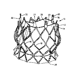
[0132] FIG. 30 shows another exemplary stent 300, for use in a prosthetic
heart valve. For
purposes of illustration, only the bare stent 300 is shown while the other
components of the
prosthetic valve, including the leaflets and the skirt, are omitted. However,
it should be
understood that the prosthetic valve can include leaflets 34a, 34b, 34c and a
skirt 42 mounted to
the stent 300, as described above in connection with the prosthetic valve 10.
The stent 300 can
have the same overall shape and configuration as the stent 12 of prosthetic
valve 10 described
above, except that all apices 302 at the outflow end of the stent 300 have
respective apertures
304. The stent 300 can further comprise three commissure posts 306 (which are
also referred to
as "apices" herein) with eyelets 308, also at the outflow end. The delivery
apparatuses 500, 600
(described below for use with stent 300) can be used to deliver the stent 10
(or any other stent
with apices that lack apertures). In this case, the delivery apparatus can
engage the stent by
wrapping the suture loops around the apices at one end of the stent (e.g., the
outflow end). In
some embodiments, the stent can have notches, channels or other narrowed
portions formed in or
adjacent to the apices, for stably holding the suture loops against their
respective apices.
[0133] FIGS. 31A-37 show an exemplary delivery apparatus 500 for delivering
the stent 300.
The delivery apparatus 500 is similar to the delivery apparatus 100 except
that the delivery
apparatus 500 includes a different mechanism for releasably securing a
prosthetic valve to the
delivery apparatus. The delivery apparatus 500 in the illustrated embodiment
comprises a main
shaft 502, a sheath 504 mounted to the distal end of the shaft 502, an inner
shaft 506 that extends
co-axially through the main shaft 502, and a nose cone 508 mounted to the
distal end of the inner
shaft 506. The inner shaft 506 can have a guidewire lumen configured to
receive a guidewire
509. As best shown in FIGS. 31A, a suture-retention member 510 can extend
distally from the
distal end of the main shaft 502. The inner shaft 506 can extend co-axially
through the suture-
retention member 510.
[0134] Although not shown, the delivery apparatus 500 can also include a
torque shaft that is
effective to move the sheath 504 in the proximal and distal directions
relative to the main shaft
502 and relative to a prosthetic valve secured to the distal end of the
delivery apparatus. The
distal end portion of the main shaft 502 can have the same configuration as
the distal segment
126 of the shaft 104 of the delivery apparatus 100 described above.
28
Date Recue/Date Received 2024-05-29
[0135] The suture-retention member 510 comprises a proximal disc member 512, a
distal disc
member 516, and a shaft 514 extending between and connecting the proximal and
distal disc
members 512, 516, respectively. As best shown in FIG. 33, the proximal disc
member 512 can
be fixed inside of the main shaft 502. Each disc member 512, 516 is formed
with one or more
axially extending openings 518 (FIGS. 31C and 31D), each of which is sized to
receive the distal
end portion of a suture release member 520 (FIG. 32). The release member 520
can be, for
example, a stiff wire, and therefore is referred to below as a release wire.
In the illustrated
embodiment, the delivery apparatus includes a single release wire 520 that
extends distally
through corresponding openings 518 in the disc members 512, 516 and proximally
through the
main shaft 502 along the length of the delivery apparatus toward a handle (not
shown) of the
delivery apparatus. The proximal end of the release wire (not shown) can be
exposed at the
proximal end of the delivery apparatus for being manipulated by a user or can
be coupled to an
actuator on the handle of the delivery apparatus that can control axial
movement of the release
wire.
[0136] The release wire 520 is slidable in the proximal and distal directions
relative to the
suture-retention member 510 to secure the stent 300 to the suture-retention
member 510 via a
plurality of suture loops 522 and to release the stent 300 from the suture-
retention member, as
further described below. In some embodiments, the delivery apparatus can
include a plurality of
such release wires 520 (such as two or three release wires 520), each of which
extends through
corresponding openings 518 in the disc members 512, 516. These release wires
520 can each
interact with one or more suture loops 522, and can aid in balancing load
distribution.
[0137] As noted above, the stent 300 can be releasably connected to the suture-
retention member
510 using a plurality of suture loops 522. For that purpose, the proximal disc
member 512 can
include a plurality of openings 528 and 530 (in addition to opening 518 for
the release wire) for
threading the suture loops through the proximal disc member (FIGS. 31A and
31B). The suture
loops 522 can be formed from a single piece of suture material that is folded
multiple times so as
to form multiple loops 522 extending distally from openings in the proximal
disc member 512, as
depicted in FIG. 31A. In alternative embodiments, each loop 522 can be formed
from a separate
piece of suture material. In some cases, each suture loop 522 consists
entirely of a loop of suture
material, whereas in other cases, one or more of the suture loops 522 can
comprise a non-looped
portion (such as a linear segment of suture material) proximal to the looped
portion. As such, a
29
Date Recue/Date Received 2024-05-29
"suture loop" can be characterized as "extending from" a given location, even
if the looped
portion itself does not originate or extend through that location, so long as
the suture material
comprising the looped portion extends from that location. However, where a
"suture loop" is
described as wrapping or extending around a given structure and/or residing at
a given location,
this specifically refers to the looped portion of the suture loop.
[0138] As shown in FIGS. 31A-31D, multiple loops 522 (e.g., two or three
loops) can extend
outwardly from each opening in the proximal disc member 512, although in other
embodiments
each suture loop 522 can extend from a separate opening. As shown in FIG. 31D,
the proximal
disc member 512 can have six openings. Three suture loops 522 can extend from
each of four
openings 528 in the proximal disc member 512, and through apertures 304 in the
apices 302. A
fifth opening 530 can have one or more suture loops extending therefrom (such
as three suture
loops) to engage the commissure post eyelets 308 of the stent 300. Finally,
the release wire 520
can extend distally from out of the sixth opening 518, toward the second disc
member 516. In the
illustrated embodiment, a single suture loop extends from the fifth opening
530 through the
eyelets 308 of each of the commissure posts. In some cases, having a single
suture loop
extending through the commissure post eyelets 308 provides better tension
control, resulting in
more controlled release and/or recapture of the prosthetic valve. The suture
loop(s) extending
through the commissure post eyelets 308 may be thicker than the suture loops
522 that extend
through the other stent apices 302. For example, in one embodiment, the suture
loop(s) extending
through the commissure posts are 4-0 sutures, whereas the suture loops
extending through the
apices are 3-0 sutures. The six openings can be arranged in an annular pattern
as shown (FIG.
31D), and the suture loops 522 can be configured to extend outward to engage
stent apices 302 in
accordance with their relative positions within this annular pattern, such
that the suture loops 522
do not cross past one another to reach their respective stent apices. In other
embodiments, the
suture loops can be configured to engage stent apices 302, such that the
suture loops cross one
another to reach respective stent apices.
[0139] Referring to FIGS. 32 and 33, when loading the stent 300 onto the
delivery apparatus, the
apices 302 of the stent 300 are placed adjacent the distal disc member 516,
and each suture loop
522 is threaded through a respective aperture 304 in one of the apices 302. By
having a
respective suture loop 522 extend through every apex 302 (including every
commissure post
306), the prosthetic valve may be fully retrievable (while connected to the
delivery apparatus), as
Date Recue/Date Received 2024-05-29
the apices 302, 306 can be collapsed radially inward using the sutures 522. In
certain
embodiments, the functioning of the prosthetic valve can be assessed after
deploying the valve
from the sheath 504 and prior its recapture. In various embodiments, the
number of apices and
corresponding suture loops can vary, so long as a sufficient number of apices
are connected such
that the end of the prosthetic valve is collapsed when the apices are
collapsed radially inward. In
the embodiment shown, there are twelve suture loops threaded through twelve
respective apices.
The end 524 of each suture loop 522 is then placed in the area between the
proximal and distal
disc members 512, 516 and the release wire 520 is slid axially through the
loop and a respective
opening 519 in the distal disc member 516 (FIG. 31A) so as to retain the end
524 of the loop on
the release wire, as depicted in FIG. 33. For purposes of illustration, FIGS.
32 and 33 show just a
single suture loop 522 releasably connecting one of the apices 302 of the
stent to the release wire
520. Desirably, a suture loop 522 is inserted through each of the
apertures/eyelets 304, 308 in the
apices 302, 306 of the stent and retained by the release wire 520. FIG. 34
shows the stent 300
after suture loops 522 are inserted through all of the apices of the stent and
retained on the
release wire 520. As noted above, while only one release wire 520 is shown in
the illustrated
embodiment, the delivery apparatus can be provided with a plurality of release
wires 520 for
retaining the suture loops 522.
[0140] When threading the suture loops 522 through the openings 304, 308 of
the apices, the
suture loops 522 can be threaded sequentially through each of the openings
304, 308 moving in a
circumferential direction around the stent. In another embodiment, the suture
loops 522 can be
inserted through every second or third or fourth opening 304, 308 and placed
on the release wire
520, moving in a circumferential direction around the stent several times
until a suture loop is
inserted through each of the openings, so as to balance the stent attachment
relative to the release
wire 520.
[0141] After the stent 300 is connected to the suture-retention member 510
(FIG. 34), the sheath
504 is advanced distally (e.g., by rotating the torque shaft of the delivery
apparatus) to load the
prosthetic valve into the sheath. As the sheath 504 is advanced over the
suture loops 522, tension
in the suture loops causes the apices 302, 306 to collapse radially inward
toward the main shaft
502. The sheath 504 is further advanced, causing the sheath 504 to extend over
and collapse the
stent 300 (as shown in FIG. 35), until the distal end of the sheath 504 abuts
the nose cone 508
(FIG. 36). As best shown in FIG. 33, the apices 302 can bear against the
distal surface of the
31
Date Recue/Date Received 2024-05-29
distal disc member 516, which prevents the prosthetic valve from sliding
proximally and
maintains tension in the suture loops 522 as the sheath is retracted.
[0142] When the prosthetic valve is delivered to the desired implantation site
within the body,
the sheath 504 is retracted (e.g., by rotating the toque shaft) to deploy the
prosthetic valve. After
the prosthetic valve is fully deployed from the sheath, the stent 300 is still
connected to the stent-
retention member 510 by the suture loops 522, as depicted in FIG. 34. Thus, if
it becomes
necessary to retrieve the prosthetic valve such as for removal or re-
positioning, the sheath 504 is
advanced distally to draw the prosthetic valve back into the sheath. On the
other hand, if it is
determined that the prosthetic valve is accurately positioned at the desired
implantation site, the
release wire 520 can be pulled proximally to release the ends 524 of the
suture loops 522. Slight
retraction of the main shaft 502 is effective to pull the suture loops out of
the openings 302 in the
stent 300, as depicted in FIG. 37. The proximal end of the release wire 520
can be exposed at the
proximal end of the delivery apparatus so that the user can manually pull the
release wire to
release the prosthetic valve. Alternatively, the handle can have an actuator
or switch that is
configured to effect proximal movement of the release wire.
[0143] The sheath 504 can be made of a polymeric material, such as PEEK or
nylon-12, and can
have a reinforced distal tip portion, such as by securing a metal ring to the
distal end portion of
the sheath, to better resist the expansion force of the stent as it is drawn
into the sheath.
Alternatively, the sheath 504 can comprise a metal cylinder having a polymeric
soft tip portion
reflowed or molded to the distal end portion of the cylinder.
[0144] FIGS. 38-43 shows another delivery apparatus 600 generally comprising a
first catheter
602 and a second catheter 604 extending coaxially through the first catheter
602, and a delivery
sheath or cylinder 612 coupled to the distal ends of the catheters 602, 604.
The proximal ends of
the catheters 602, 604 can be coupled to a handle (e.g., a handle 202 such as
shown in FIG. 23).
As best shown in FIG. 40, the first catheter 602 comprises an elongated shaft
606 that extends
distally from the handle, an intermediate section 608 extending distally from
the distal end of the
shaft 606, and a distal end portion 610 extending distally from the
intermediate section 608. The
intermediate section 608 comprises a plurality of angularly spaced rails 613
that extend
longitudinally from the shaft 606 to the distal end portion 610. The rails 613
cooperate with a nut
640 to inhibit rotation of the nut yet allow longitudinal movement of the nut
upon rotation of the
32
Date Recue/Date Received 2024-05-29
second catheter 604. In this manner, the rails 613 serve the same purpose of
the prongs 134 in
preventing rotation of the nut 150. The distal end portion 610 in the
illustrated embodiment
comprises a slotted metal tube to enhance the flexibility of this section of
the first catheter 602.
[0145] As best shown in FIGS. 39A-39C, the second catheter 604 can comprise a
elongated
shaft 614 (which can be referred to as a "torque shaft"), a coupling member
616 connected to the
proximal end of the shaft 614, and a threaded screw 618 connected to the
distal end of the shaft
614. The coupling member 616 is configured to be connected to a handle as
described above
(e.g., a handle 202). The screw 618 has external threads that engage internal
threads of the nut
640. As best shown in FIG. 41, when the apparatus is assembled, the elongated
shaft 614 of the
second catheter 604 extends coaxially through the elongated shaft 606 of the
first catheter 602,
and the screw 618 extends coaxially through the railed section 608 of the
first catheter 602. The
nut 640 is mounted on the screw 618 and is connected to the proximal end
portion of delivery
cylinder 612. The distal end portion 610 of the first catheter 602 extends
coaxially through the
delivery cylinder 612.
[0146] As best shown in FIGS. 40-42, a suture-retention member 626 can be
connected to the
distal end of the slotted tube 610. The suture-retention member 626 can have
features similar to
as described above for suture-retention member 510, including a proximal disc
member 638
connected to the distal end of the slotted tube 610, a distal disc member 636,
and at least one
release member or release wire 628 extending through the proximal and distal
disc members for
interacting with one or more suture loops 522.
[0147] Returning to FIGS. 39A-39C, the delivery cylinder 612 in the
illustrated embodiment
comprises a relatively more flexible proximal portion 630 and a relatively
less flexible distal end
portion 632. The proximal portion 630 can comprise a slotted metal tube or
cylinder to enhance
the flexibility of this section of the delivery cylinder 612. The distal end
portion 632 comprises a
sleeve or sheath (also referred to as a "valve holding portion") that is
configured to extend over
and retain a prosthetic valve in a radially compressed state during delivery.
In some
embodiments, the sheath 632 can extend over the prosthetic valve and the
suture-retention
member 626 during delivery (FIG. 43). Alternatively, the suture-retention
member 626 can be (at
least partially) housed within the proximal portion 630 during delivery. The
sheath 632 can be
made of a suitable polymeric material, such as PEEK, nylon-12, and/or PEBAX,
or a metal
33
Date Recue/Date Received 2024-05-29
having a polymeric inner liner. When made of polymeric materials, the sheath
632 can be
thermally bonded to the slotted tube 630. A distal end segment 634 of the
sheath 632 can be
flared radially outward to enhance recapturability of the prosthetic valve.
The distal end segment
634 can comprise a polymeric and/or elastomeric material, such as PEEK, nylon,
and/or
PEBAX. In particular embodiments, the distal end segment 634 is more flexible
and/or
elastomeric than the remaining section of the distal end portion. In a working
embodiment, the
distal end segment 634 comprises PEBAX and the remaining portion of the distal
end portion
632 comprises nylon. As shown in FIG. 46, the distal end segment 634 can
include a radially
projecting, annular bump 650 to facilitate loading and recapturing of a
prosthetic valve. During
recapture, the bump 650 presses the sutures 522 inwardly, which causes the
apices 302, 306 to
collapse inwardly, allowing the sheath to slide over the frame.
[0148] As shown in FIG. 42, the delivery apparatus 600 can further comprise a
nose cone 620
connected to the distal end of a nose cone shaft 622, which extends through
the distal shaft
portion 610 of the first catheter 602, the suture retention member 626, and
the screw 618 and the
shaft 614 of the second catheter 604. The nose cone shaft 622 can include a
guidewire lumen and
can extend proximally to the handle of the delivery apparatus.
[0149] The delivery cylinder 612 cooperates with the screw 618 and the nut 640
to allow for
longitudinal (i.e., proximal and/or distal) movement of the delivery cylinder
612 relative to the
distal shaft portion 610 and the suture-retention member 626. Rotational
motion of the screw 618
(initiated by the user rotating the torque shaft 614) can be converted into
translational movement
of the delivery cylinder 612 via the nut 640 positioned along external threads
of the screw 618
(FIG. 39B). The nut 640 can have internal threading configured to compatibly
engage the
external threads of the screw member 618. The nut 640 can further comprise one
or more tabs
642 protruding radially outward, and the delivery cylinder 612 can comprise
one or more
receiving areas (such as one or more windows 644) adjacent a proximal end of
the cylinder 612
for engaging with these tabs 642. In particular, upper portions of the tab(s)
642 can extend
through the window(s) 644 to produce a secure fit (e.g., a snap fit) with the
delivery
cylinder 612.
[0150] As noted above, the first catheter 602 includes a section 608 that
includes a plurality of
angularly spaced rails 613, which cooperate with the tab(s) 642 of the nut. As
best shown in FIG.
34
Date Recue/Date Received 2024-05-29
41, the screw 618 extends coaxially through the rails 613 and the nut 640 is
disposed on the
screw 618 with each tab 642 positioned in the space between two adjacent rails
613. To produce
movement of the delivery cylinder 612, the screw 618 can be rotated using a
torque shaft 614, as
described above with respect to delivery apparatus 100. Placement of the
tab(s) 642 between the
rails 613 prevents the nut 640 from rotating along with the screw 618. With
rotation of the nut
640 restricted, rotation of the screw 618 produces translational movement of
the nut 640 along
the screw 618. Axial movement of the nut 640 along the screw 618 (in the
distal or proximal
direction) causes the cylinder 612 to also move axially, and in the same
direction as the nut 640
(relative to the screw 618). Thus, as the nut 640 moves along the screw 618
longitudinally, the
delivery cylinder 612 (connected to the nut at windows 644) is carried along-
with.
[0151] An outer sleeve portion 648 can be positioned over the first and second
catheters 602,
604 (FIG. 41) and the delivery cylinder 612, and thereby form an outermost
layer of the delivery
apparatus 600. This sleeve portion 648 allows a user to effectively flush the
delivery apparatus
600 with fluid to, for example, eliminate air bubbles. In some embodiments,
the sleeve portion
648 can comprise an elastomeric material and/or may be affixed to the delivery
cylinder 612 at
one or more locations. In particular, a sleeve portion 648 having elastomeric
properties can be
affixed to both the delivery cylinder 612 and the elongated shaft 606 of the
first catheter 602
(proximal to the intermediate section 608). In this case, the sleeve portion
648 can stretch,
between the cylinder 612 and the shaft 606, as the nut 640 and delivery
cylinder 612 are
advanced, and relax when these components are retracted. In some embodiments,
the sleeve
portion 648 is substantially rigid and/or is only affixed to the delivery
cylinder 612. In such
cases, the entire sleeve portion 648 can be advanced distally or retracted
proximally along with
the delivery cylinder 612 relative to the first catheter.
[0152] In the case of a screw 618 and a nut 640 with standard-type threading,
clockwise rotation
of the screw 618 can result in proximal movement of the nut 640 along the
screw 618.
Conversely, counter-clockwise rotation of the standard screw 618 can result in
distal movement
of the nut 640. In this manner, rotation of the screw 618 can cause proximal
or distal movement
of the delivery cylinder 612 connected to the nut 640. Alternatively, the
threads of the screw can
be reversed such that counter-clockwise rotation of the screw causes proximal
movement of the
nut and clockwise movement of the nut causes distal movement of the nut.
Date Recue/Date Received 2024-05-29
[0153] FIG. 43 shows the delivery cylinder 612 advanced forward to its distal-
most position for
delivery. In the delivery configuration, the distal end portion 632 extends
over a prosthetic valve
(not shown), which is retained in a radially compressed state and releasably
connected to the
suture retention member 626 with a plurality of sutures 522. The distal end of
the delivery
cylinder 612 can abut an annular shoulder of the nose cone 620 (as shown in
FIG. 43) when the
delivery cylinder is in the delivery configuration. FIG. 42 shows the delivery
cylinder 612 in a
deployment configuration, with the delivery cylinder 612 retracted to a
proximal position. In this
position, the distal end portion 632 is retracted proximally past the
prosthetic valve (allowing the
prosthetic valve to expand) and the distal disc member 636 of the suture
retention member 626.
To release the prosthetic valve from the suture retention member 626, the
release wire 628 is
retracted such that its distal end is proximal to the second disc member 636,
thereby freeing the
distal ends 524 of the suture loops 522 from the prosthetic valve.
[0154] Replacing the metal-metal connection between the stent and the delivery
apparatus with
suture loops allows for lower deployment and recapture torques. These reduced
torques allow for
relocation of the screw mechanism further away from the distal end of the
delivery apparatus.
Increasing the spacing between the screw 618 and the prosthetic valve
advantageously decreases
the relatively stiff section of the delivery apparatus occupied by the
prosthetic valve at the distal
end of the delivery apparatus. Referring to FIG. 43, the portion of the
delivery cylinder 612
extending over the prosthetic valve has a length L3 and the overall relatively
stiff section of the
delivery system 600 (which does not include the length of the nose cone) has a
length L4, which
in this embodiment corresponds to the length of the delivery cylinder 612
extending over the
prosthetic valve and the suture-retention member 626. For example, in some
embodiments, L4 is
about 1.3x the length of L3. In various other embodiments, the ratio of L4 to
L3 is about 1.6 or
less, about 1.5 or less, or about 1.4 or less.
[0155] Referring to FIGS. 40-41 and 43, the portion of the delivery apparatus
600 extending
from the proximal end of the suture retention member 626 to the distal end of
the screw 618
(which is equal to the length L2 of the distal shaft portion 610) can be more
flexible than the stiff
section housing the prosthetic valve (which is equal to the length L4 of the
delivery cylinder 612)
Desirably, the relatively more flexible section is long enough such that when
the delivery system
600 is advanced through the aorta to implant a prosthetic valve at the aortic
valve of a subject,
the relatively stiff section is positioned in the ascending aorta, the screw
618 is positioned in the
36
Date Recue/Date Received 2024-05-29
descending aorta, and the relatively more flexible portion extending
therebetween is positioned
in the aortic arch. This greatly facilitates steering of the delivery
apparatus through the aortic
arch and proper positioning of the prosthetic valve at the aortic annulus.
[0156] In various embodiments, for example, a distal end of the screw 618 can
be located at least
about 5 cm, at least about 10 cm, at least about 15 cm, at least about 20 cm,
or at least about 30
cm away from the distal end of the suture-retention member 626 (and a
prosthetic valve
releasably connected to the suture-retention member 626). In various
embodiments, the delivery
cylinder 612 can have an overall length Li between about 3 cm and about 40 cm,
between about
cm and about 40 cm, between about 10 cm and about 35 cm, between about 15 cm
and about
30 cm, or between about 18 cm and about 25 cm. In various embodiments, the
distal shaft
portion 610 can have an overall length L2 between about 0 cm and about 30 cm,
between about 5
cm and about 25 cm, between about 10 cm and about 22 cm, or between about 15
cm and about
20 cm.
[0157] In alternative embodiments, the length Li of the deliver cylinder 612
can be longer than
40 cm, and in some embodiments, it can extend proximally to the handle of the
delivery
apparatus.
[0158] FIG. 47 shows an alternative slotted tube 652 that can be used in place
of slotted tube 610
in the delivery apparatus 600. The slotted tube 652 has a plurality of teeth
or projections 654
formed in each turn or coil that extend into respective recesses in adjacent
coils to increase
torque resistance. A distal end of the tube can be formed with one or more
longitudinal openings
656, forming rails 658 between adjacent openings for cooperating with the
projections 642 of the
nut 640. At the distal and proximal ends of the rails 658, the tube can be
formed with openings
660 to allow a pull wire 662 to extend through the openings and alongside the
screw 618 on the
outside of the slotted tube 652. A proximal end of the tube 652 can be formed
with a plurality of
inwardly projecting tabs 664. As shown in FIG. 49, the tabs 664 can engage an
annular recessed
portion 666 on the outer surface of the proximal member 638 of the suture-
retention member
626. The tabs 664 can be configured to form a snap-fit connection with the
proximal member
638 sufficient to secure the suture-retention member to the slotted tube.
[0159] In alternative embodiments, the slotted tube 610 and the slotted tube
630 can have other
patterns or configurations, such as any of those shown in FIGS. 12, 28A, 28B,
29A, or 29B.
37
Date Recue/Date Received 2024-05-29
General Considerations
[0160] For purposes of this description, certain aspects, advantages, and
novel features of the
embodiments of this disclosure are described herein. The disclosed methods,
devices, and
systems should not be construed as limiting in any way. Instead, the present
disclosure is
directed toward all novel and nonobvious features and aspects of the various
disclosed
embodiments, alone and in various combinations and sub-combinations with one
another. The
methods, devices, and systems are not limited to any specific aspect or
feature or combination
thereof, nor do the disclosed embodiments require that any one or more
specific advantages be
present or problems be solved.
[0161] Features, integers, characteristics, compounds, chemical moieties or
groups described in
conjunction with a particular aspect, embodiment or example of the invention
are to be
understood to be applicable to any other aspect, embodiment or example
described herein unless
incompatible therewith. All of the features disclosed in this specification
(including any
accompanying claims, abstract and drawings), and/or all of the steps of any
method or process so
disclosed, may be combined in any combination, except combinations where at
least some of
such features and/or steps are mutually exclusive. The invention is not
restricted to the details of
any foregoing embodiments. The invention extends to any novel one, or any
novel combination,
of the features disclosed in this specification (including any accompanying
claims, abstract and
drawings), or to any novel one, or any novel combination, of the steps of any
method or process
so disclosed.
[0162] Although the operations of some of the disclosed methods are described
in a particular,
sequential order for convenient presentation, it should be understood that
this manner of
description encompasses rearrangement, unless a particular ordering is
required by specific
language. For example, operations described sequentially may in some cases be
rearranged or
performed concurrently. Moreover, for the sake of simplicity, the attached
figures may not show
the various ways in which the disclosed methods can be used in conjunction
with other methods.
As used herein, the terms "a", "an" and "at least one" encompass one or more
of the specified
element. That is, if two of a particular element are present, one of these
elements is also present
38
Date Recue/Date Received 2024-05-29
and thus "an" element is present. The terms "a plurality of' and "plural" mean
two or more of
the specified element.
[0163] As used herein, the term "and/or" used between the last two of a list
of elements means
any one or more of the listed elements. For example, the phrase "A, B, and/or
C" means "A,"
"B," "C," "A and B," "A and C," "B and C" or "A, B and C."
[0164] As used herein, the term "coupled" generally means physically coupled
or linked and
does not exclude the presence of intermediate elements between the coupled
items absent
specific contrary language.
[0165] In view of the many possible embodiments to which the principles of the
disclosed
invention may be applied, it should be recognized that the illustrated
embodiments are only
preferred examples of the invention and should not be taken as limiting the
scope of the
invention.
[0166] In view of the many possible embodiments to which the principles of the
disclosed
invention may be applied, it should be recognized that the illustrated
embodiments are only
preferred examples of the invention and should not be taken as limiting the
scope of the
invention. Moreover, additional embodiments are disclosed in U.S. Patent
Application
Publication No. 2010/0049313 (U.S. Application No. 12/429,040). Accordingly,
the scope of the
invention is defined by the following claims. We therefore claim as our
invention all that comes
within the scope and spirit of these claims.
39
Date Recue/Date Received 2024-05-29