Note: Descriptions are shown in the official language in which they were submitted.
WO 2023/102661 PCT/CA2022/051795
1
STEROID ACID-PEPTIDE BASED INTRACELLULAR CARGO DELIVERY
The present description relates to intracellular delivery of biologically
active cargoes. More
specifically, the present description relates to compositions comprising a
steroid acid-peptide conjugate
covalently linked to, and/or admixed with, a cargo for improved intracellular,
cytosolic and/or nuclear
delivery, as well as to the use of such compositions for example in genome
editing and the manufacture of
cell-based vaccines. The present description further relates to methods of
increasing the stability of
cargoes via their covalent conjugation to one or more steroid acid-peptide
moieties. The present
description refers to a number of documents, the contents of which is herein
incorporated by reference in
their entirety.
BACKGROUND
Intracellular delivery of biological cargoes such as peptides, proteins, and
polynucleotides
generally rely on the endocytic pathway as the major uptake mechanism,
resulting in a large fraction of
the cargoes being trapped inside endosomes/lysosomes. Such trapped cargoes
often remain sequestered
from their intended targets and may be degraded. Thus, improved strategies for
increasing intracellular
delivery and avoiding endosomal entrapment would be highly desirable.
SUMMARY
In a first aspect, described herein is a composition comprising a steroid acid-
peptide conjugate
covalently linked to, and/or admixed with, a cargo for intracellular delivery.
In some embodiments,
covalently linking the cargo to the steroid acid-peptide conjugate increases
intracellular delivery and/or
cytosolic/nuclear delivery of the cargo, as compared to a corresponding
composition lacking the steroid
acid-peptide conjugate. In some embodiments, the cargo is admixed with a
sufficient concentration of the
steroid acid-peptide conjugate to increase intracellular delivery and/or
cytosolic/nuclear delivery of the
cargo, as compared to a corresponding composition lacking admixture with the
steroid acid-peptide
conjugate. In particular embodiments, the steroid acid may be a bile acid and
the peptide may comprise a.
functional nuclear localization signal (NLS) or other subcellular targeting
domain.
In a further aspect, described herein is a method for delivering a cargo
intracellularly, the method
comprising providing a composition as defined herein, and administering the
composition to target cells
in vitro or in vivo.
In a further aspect, described herein is a method for preparing a cargo for
intracellular delivery
having increased stability, the method comprising covalently linking the cargo
to a sufficient number of
CA 03240433 2024- 6-7
WO 2023/102661 PC T/CA2022/051795
2
steroid acid-peptide moieties to produce a covalently-modified cargo that
exhibits greater stability (e.g.,
thermal stability) than the corresponding unmodified cargo.
In a further aspect, described herein is a composition comprising an antigen
covalently linked to
and/or admixed with a steroid acid-peptide conjugate in an amount sufficient
to improve presentation of
the antigen upon administration of the composition to non-professional antigen
presenting cells (e.g.,
mesenchymal stromal cells [MSCs]), as compared to administration of a
corresponding composition
lacking the steroid acid-peptide conjugate.
In a further aspect, described herein is a cell culture comprising non-antigen
presenting cells
pulsed with an antigen covalently linked to and/or admixed with a steroid acid-
peptide conjugate.
In a further aspect, described herein is a vaccine comprising a composition as
described herein, or
comprising cells produced using a cell culture as described herein.
In a further aspect, described herein is a method for enhancing presentation
of an antigen of
interest in a subject or cells, the method comprising administering to the
subject or in non-antigen
presenting cells a composition as described herein, or cells produced using a
cell culture as described
herein.
In a further aspect, described herein is a method for vaccinating a subject
against an infectious
disease, the method comprising administering to the subject a composition as
described herein or cells
produced using a cell culture as described herein, wherein the antigen
comprises an antigenic fragment of
a pathogen (e.g., virus, bacteria, fungus) causing the infectious disease.
In a further aspect, described herein is a method for treating cancer in a
subject, the method
comprising administering to the subject a composition as described herein, or
cells produced using a cell
culture as described herein, wherein the antigen is an overexpressed or
aberrantly expressed in cells
causing the cancer.
General Definitions
Headings, and other identifiers, e.g., (a), (b), (i), (ii), etc., are
presented merely for ease of reading
the specification and claims. The use of headings or other identifiers in the
specification or claims does
not necessarily require the steps or elements be performed in alphabetical or
numerical order or the order
in which they are presented.
The use of the word "a" or "an" when used in conjunction with the term
"comprising" in the
claims and/or the specification may mean "one," but it is also consistent with
the meaning of "one or
more", "at least one", and "one or more than one".
As used in this specification and claim(s), the words "comprising" (and any
form of comprising,
such as "comprise" and "comprises"), "having" (and any form of having, such as
"have" and -has-),
CA 03240433 2024- 6-7
WO 2023/102661 PC T/CA2022/051795
3
"including" (and any form of including, such as "includes" and "include") or
"containing" (and any form
of containing, such as "contains" and "contain") are inclusive or open-ended
and do not exclude
additional, unrecited elements or method steps.
The term -about" is used to indicate that a value includes the standard
deviation of error for the
device or method being employed in order to determine the value. In general,
the terminology "about" is meant
to designate a possible variation of up to 10%. Therefore, a variation of 1,
2, 3, 4, 5, 6, 7, 8, 9 and 10% of a
value is included in the term -about-. Unless indicated otherwise, use of the
term -about" before a range
applies to both ends of the range.
Other objects, advantages and features of the present description will become
more apparent upon
reading of the following non-restrictive description of specific embodiments
thereof, given by way of
example only with reference to the accompanying drawings.
BRIEF DESCRIPTION OF THE DRAWINGS
In the appended drawings:
Fig. 1 shows representative fluorescence microscopy images for HEK293 cells
incubated for 3 or
6 hours with a Cas9-NLS cargo, followed by fixation, permeabilization and
labeling with Hoescht nuclear
stain ("nuclei-) and a fluorescent anti-Cas9 antibody (-Cas9-NLS cargo-).
Fig. 2 shows representative fluorescence microscopy images for HEK293 cells
incubated for 3 or
6 hours with a [CDCA-SV401-Cas9-NLS cargo, followed by fixation,
permeabilization and labeling with
Hoescht nuclear stain ("nuclei") and a fluorescent anti-Cas9 antibody ("[CDCA-
SV40]-Cas9-NLS
cargo").
Fig. 3 shows a representative experiment of GFP-Ga13-expressing DC2.4 cells
treated with OVA
cargo for 3 hours, followed by visualization by fluorescence microscopy. The
diffuse cytosolic pattern of
GFP-Gal3 is indicative of intact endosomal membranes.
Fig. 4 shows a representative experiment of GFP-Ga13-expressing DC2.4 cells
treated with
[CA-SV401-OVA cargo for 3 hours, followed by visualization by fluorescence
microscopy. The punctate
pattern of GFP-Gal3 is indicative of disrupted endosomal membranes (arrows).
Fig. 5 shows the results of a representative flow-cytometry experiment
investigating the
intracellular degradation and processing in primary dendritic cells of DQTM
OVA cargo (grey peak)
versus [CA-SV401- DQTM OVA cargo (red peak) after 3 or 6 hours via flow
cytometry.
Fig. 6 shows increased intracellular delivery of OVA-AF647 cargo in
mesenchymal stromal cells
upon coincubation with CA-hnRPA1 M9 NLS conjugate as evaluated by flow
cytometry.
CA 03240433 2024- 6- 7
WO 2023/102661 PC
T/CA2022/051795
4
Fig. 7 shows increased intracellular degradation/processing in mesenchymal
stromal cells of
DQTM OVA cargo upon coincubation with CA-hnRPA1 M9 NLS conjugate as evaluated
by flow
cytometry.
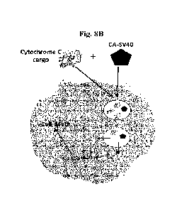
Fig. 8A and 8B show schematic representations of the study design in which
Cytochrome C
cargo is delivered intracellularly in the absence (Fig. 8A) or presence (Fig
8B) of CA-SV40 NLS
conjugate. Fig. 8C shows a representative flow cytometry assessment of EL4
cell death when treated with
CA-SV40 (471AM) admixed with Cytochrome C.
Fig. 9 shows an intrinsic tryptophan fluorescence (ITF) analysis of
unconjugated OVA ("nOVA")
or [CA-SV40]-0VA ("cOVA") at various CA-SV40 to OVA ratios in response to
thermal stress.
Fig. 10A shows a representative gel image of Genomic Cleavage Detection Assay
using Control
Template & Primers and stained with ethidium bromide. After re-annealing,
samples were treated with
and without Detection Enzyme and separated on a 2% agarose gel. Fig.10B shows
the cleavage efficiency
calculated by determining the relative proportion of DNA contained in each
band (parental and cleaved
bands) using desired gel analysis software and following the next equations:
Cleavage Efficiency = 1 ¨
[(1¨fraction cleaved) 1/2] ; Fraction Cleaved= sum of cleaved band
intensities/(sum of the cleaved and
parental band intensities).
Fig. 11 shows the effect of different bile acids on the intracellular delivery
and subsequent
antigen presentation activity of bile acid-SV40 NLS conjugates. For this
experiment, BMDCs were used
as the antigen presenting cells (n=6) and the molar ratio (bile
acid/peptide/conjugate) : antigen was 4:1.
Controls tested included no antigen ("PBS"), antigen alone ("OVA alone"),
unconjugated NLS peptide
("SV4ONLS"), unconjugated cholic acid mixed with OVA ("CA"), and the positive
control peptide
SIINFEKL (SEQ ID NO: 16) mixed with OVA ("SIINFEKL"). The dashed line
represents the signal
obtained with the OVA cargo alone. Bile acids: cholic acid (CA);
glycodeoxycholic acid (GDCA);
glvcochenodeoxycholic acid (GCDCA); ursodeoxycholic acid (UDCA); and
lithocholic acid (LCA).
Fig. 12 shows the effect of different NLS peptides on the intracellular
delivery and subsequent
antigen presentation activity of cholic acid-NLS peptide conjugates. The
dashed line represents the signal
obtained with the OVA cargo alone. The readout was taken after 24 h of
incubation and error bars
represent SD (n=6). For this experiment, BMDCs were used as the antigen
presenting cells.
Fig. 13 shows the effect of different NLS peptides on the intracellular
delivery and subsequent
antigen presentation activity of cholic acid-NLS peptide conjugates. The
dashed line represents the signal
obtained with the OVA cargo alone. Readout was taken after 24 h of incubation
from a single experiment.
The molar ratio of CA-peptide conjugate : OVA was 22:1. For this experiment,
BMDCs were used as the
antigen presenting cells.
CA 03240433 2024- 6-7
WO 2023/102661 PCT/CA2022/051795
Fig. 14 shows the effect of different NLS peptides on the intracellular
delivery and subsequent
antigen presentation activity of cholic acid-NLS peptide conjugates. For this
experiment, BMDCs were
used as the antigen presenting cells. The dashed line represents the signal
obtained with the OVA cargo
alone. Readout was taken after 24 h of incubation from a single experiment.
The molar ratio of CA-
peptide conjugate: OVA was as follows: CA-GWG-SV4ONLS (12:1); CA-hnRNP M NLS
(12:1); CA-
NLS2-RPS17 NLS (22:1); CA-HuR NLS (22:1); CA-cMyc NLS (2:1); CA-NLS3-RPS17 NLS
(22:1);
CA-NLS2-RG-RPS17 NLS (2:1); CA-PQBP1 NLS (8:1); CA-hnRNPA1 M9 NLS (22:1); and
CA-SV40
NLS (2:1).
Fig. 15 shows the effect of different NLS peptides on the intracellular
delivery and subsequent
antigen presentation activity of cholic acid-NLS peptide conjugates. For this
experiment, a cross-
presentation mesenchymal stromal cell (MSC) line was used as the antigen
presenting cells. The dashed
line represents the signal obtained with the OVA cargo alone. Readout was
taken after 24 h of incubation.
The molar ratio of CA-peptide conjugate : OVA was as follows: CA-GWG-SV4ONLS
(2:1); CA-hnRNP
M NLS (8:1); CA-hnRNP D NLS (12:1); CA-NLS2-RG-RPS17 (4:1); CA-cMyc NLS
(12:1); CA-HuR
NLS (12:1). CA-Tus NLS (2:1); CA-NLS2-RPS17 NLS (4:1); CA-PQBP1 NLS (12:1); CA-
hnRNPA1
M9 NLS (2:1); and CA-SV40 NLS (2:1).
Fig. 16 shows the effect of different NLS peptides on the intracellular
delivery of cholic acid-
NLS peptide conjugates. For this experiment, a cross-presenting mesenchymal
stromal cell line (cpMSC)
was used as the antigen presenting cells, which were pulsed with the OVA cargo
labelled with Alexa
Fluor 647 (i.e., 0VA647)TM. OVA' fluorescence was measured by flow cytometry.
Different ratios of CA
(NLS1 RPS17 [Fig. 16A1; NLS3 RPS17 [Fig. 16B1; and PQBP-1 [Fig. 16C1 to
antigen (CA: OVA =
22:1, 12:1, 8:1,4:1, and 2:1) were tested (hnR1NPA1 M9 NLS at 2:1).
Fig. 17 shows the effect of different NLS peptides on intracellular delivery
and subsequent
antigen processing activity of cholic acid-NLS peptide conjugates. For this
experiment, a cross-presenting
mesenchymal stromal cell line (cpMSC) was used as the antigen presenting
cells, which were pulsed with
DQTM Ovalbumin (i.e., OVADQ). OVADQ fluorescence was measured by flow
cytometry. Different ratios
of CA (NLS1 RSP17 [Fig. 17A1; NLS3 RPS17 [Fig. 17B1; and PQBP-1 [Fig. 17C1 to
antigen (CA:
OVA = 22:1, 12:1, 8:1. 4:1, and 2:1) were tested (hnRNPA1 M9 NLS at 2:1).
Fig. 18 shows the characterization of the cross-presentation capacity WT MSCs
treated with CA-
hnRNPA1 (SEQ ID NO: 4). Fig. 18A and Fig. 18B show the results of antigen
cross-presentation assay
conducted using CA-hnRNPA1 admixed with OVA compared to controls and CA-SV40.
Fig. 18C shows
the effect of CA-hnRNPA1 on fluorescent OVA uptake by MSCs. Fig. 18D shows the
effect of CA-
hnRNPA1 on fluorescent OVA processing by MSCs. Fig. 18E shows the results of
the antigen cross-
presentation assay conducted using different pulsing time points. Fig. 18F
shows the results of the antigen
CA 03240433 2024- 6-7
WO 2023/102661 PC T/CA2022/051795
6
cross-presentation assay conducted using CA-hnRNPA1 diluted in PBS or WO. Fig.
18G shows a
representative flow cytometry analysis of H2-Kb expression. Fig. 18H shows a
representative flow-
cytometry analysis of I-Ab expression. Fig. 181 shows the phenotype
characterization of CA-luiRNPA1-
treated MSCs. For Figs. 18B, 18E and 18F, n=6/group with *P<0.05, **P<0.01 and
***P<0.001.
Fig. 19 shows the antigen cross-presentation capacity WT MSCs treated with CA-
hiJRNPA1
requires reactive oxygen species (ROS) production. Fig. 19A shows the flow
cytometry assessment of
ROS production by MSCs in response to CA-hnRNPA1. Dp44mt was used as a
positive control. Fig. 19B
shows the antigen cross-presentation capacity of CA-1-mRNPA1 in the presence
of a-tocopherol,
MitoTempoTm, and N¨acetylcysteine (NAC). Fig. 19C shows the antigen cross-
presentation capacity of
CA-hnRNPA1 can be neutralized using NOX inhibitors diphenyleneiodonium
chloride (DPI) and
ML171. Fig. 19D shows the endosomal damaging properties of CA-hnRNPA1 on MSCs
co-treated with
recombinant cytochrome C. For Figs. 19A-19C, 11=6/group with *P<0.05, **P<0.01
and ***P<0.001.
Fig. 20 shows the molecular characterization of the impact of CA-hnRNPA1 on WT
MSCs. List
of top reactome pathways that are enriched for both up-regulated (Fig. 20A)
and down-regulated (Fig.
20B) genes in CA-hnRNPA1 treated group versus control MSCs. Coloured circles
intensity corresponds
to adjusted p-values; size of circles is the ratio of genes in the tested set.
Fig. 20C shows a representative
unfolded-protein response heatmap displaying the genes most contributing to
the pathway enrichment and
modulated in response to CA-hnRNPA1 treatment (FDR < 5%); gene expression is
sealed between -1 and
+1. Fig. 20D shows the bile acid heatmap depicting genes that are modulated by
CA-hnRNPA1
treatment. Fig. 20E shows the cholesterol heatmap depicting genes that are
modulated by CA-hnRNPA1
treatment. Genes showing in heatmap Fig. 20D and Fig. 20E were also
contributing to significant
statistics form both differential expression and pathway analyses (FDR < 5%).
Fig. 20F shows the IL-12
heatmap depicting genes that are modulated by CA-hnRNPA1 treatment. Gene
expression is scaled to -1
and I range. Fig. 20G shows a LuminexTM analysis of various cvtokines in
response to CA-hnRNPA1
treatment (in grey). For this figure, n=6/group. Fig. 20H shows the similarity
of gene expression patterns
between the CA-hnRNPA1 and CA-hnRNPA1 +OVA groups compared to control MSCs.
Correlation
plot showing the speamian's rank correlation coefficient of DEGs (log2 fold
changes). Fig. 201 shows a
volcano plot representing differentially expressed genes in response to CA-
hnRNPAl. Fig. 20J shows a
volcano plot depicting some important biological processes modulated in MSCs
in response to CA-
hnRNPAl. All genes from corresponding reactome analyses and showing a log2FC
greater or equal to 0.5
are labelled for further investigation. Fig. 20K shows a turbidity assay
reflecting the CA-hnRNPA1
capacity to form protein aggregation mixed with the OVA protein.
Fig. 21 shows the validation of the antigen cross-presentation properties of
CA-hnRNPA1 on
human WT MSCs. Fig. 21A shows the representative flow cytometry analysis of
OVA uptake by CA-
CA 03240433 2024- 6-7
WO 2023/102661 PC
T/CA2022/051795
7
hnRNPAl-treated human MSCs. Fig. 21B shows the signal quantification of the
results presented in Fig.
21A. Fig. 21C shows the representative flow cytometry analysis of OVA
processing by CA-hnRNPA1-
treated human MSCs. Fig. 21D shows the signal quantification of the results
presented in Fig. 21C.
Fig. 22 shows the working model of CA-hnRNPA1-mediated enhancement of antigen
cross-
presentation in WT MSCs and WT MSC vaccination.
Fig. 23 shows the therapeutic vaccination using the WT MSC vaccine with CA-
hnR1NPA1 in established
lymphoma tumors. Fig. 23A shows the timeline representing the steps used for
therapeutic vaccination.
Fig. 23B shows tumor growth in response to syngeneic MSC vaccination. Fig. 23C
shows the Kaplan-
Meier survival curve of the experiment shown in Fig. 23B. Fig. 23D shows the
tumor growth in response
to allogeneic MSC vaccination. Fig. 23E shows Kaplan-Meier survival curve of
the experiment shown in
Fig. 23D. For Figs. 23B-23E, n=5/group.
SEQUENCE LISTING
This application contains a Sequence Listing in computer readable form
December 8, 2022. The
computer readable form is incorporated herein by reference.
SEQ ID NO: Description
1 CA-SV40
2 NLS from SV-40 large T-antigen
3 GWG-SV4ONLS
4 hnRNPA1 M9 NLS
hnRNP D NLS
6 hnRNP M NLS
7 PQBP-1 NLS
8 NLS2-RG Domain RPS17
9 NLSI RPS17
NLS2 RPS17
11 NLS3 RPS17
12 cMye NLS
13 HuR NLS
14 Tus NLS
Nucleoplasmin NLS
16 SIINFEKL peptide
DETAILED DESCRIPTION
Described herein arc compositions and methods relating to cargoes for improved
intracellular
delivery. In some aspects, the present invention stems from the demonstration
herein that total
intracellular delivery, cytosolic delivery, and/or nuclear delivery of cargoes
may be enhanced by
admixture with, or covalent linkage to, a variety of steroid acid-peptide
conjugates. In further aspects, the
present invention stems from the demonstration herein that covalent
conjugation with steroid acid-peptide
CA 03240433 2024- 6-7
WO 2023/102661 PC T/CA2022/051795
8
moieties may improve cargo stability. In further aspects, the present
invention stems from the
demonstration herein that steroid acid-peptide conjugates described herein may
be used for the generation
of cell-based vaccines, including in some non-professional antigen-presenting
cells that have been
previously shown to be immunosuppressive. In a first aspect, described
herein is a composition
comprising a steroid acid-peptide conjugate covalently linked to and/or
admixed with a cargo for
intracellular delivery. In some embodiments, covalently linking the cargo to
the steroid acid-peptide
conjugate increases intracellular delivery and/or cytosolic/nuclear delivery
of the cargo, as compared to a
corresponding composition lacking the steroid acid-peptide conjugate. In some
embodiments, the cargo is
admixed with a sufficient concentration of the steroid acid-peptide conjugate
to increase intracellular
delivery and/or cytosolic/nuclear delivery of the cargo, as compared to a
corresponding composition
lacking admixture with the steroid acid-peptide conjugate. In some
embodiments, steroid acid-peptide
conjugate conjugates described herein may increase presentation of an
antigenic polypeptide cargo by
target cells. In some embodiments, steroid acid-peptide conjugate conjugates
described herein may
increase intracellular reactive oxygen species production in target cells. In
some embodiments, the target
cells comprise or consist of professional anti-presenting cells (e.g.,
dendritic cells, macrophages, B cells,
or non-immune cells engineered for overexpression of an immunoproteasome). In
some embodiments, the
target cells comprise or consist of non-professional antigen-presenting cells
(e.g., wild-type, engineered,
primary, and/or cultured non-immune cells, such as mcsenchymal stromal cells
[MCSs; also known as
mesenchymal stem cells]. In some embodiments, steroid acid-peptide conjugate
conjugates described
herein may transform immunosuppressive cells (e.g., immunosuppressive MS Cs)
into immunostimulatory
and/or proinflammatory MSCs, which may then be used, for example, in cell-
based immunostimulatory
compositions and/or cell-based vaccines.
In some embodiments, the cargo may be or may comprise a protein, peptide,
polynucleotide (e.g.,
DNA, RNA, shRNA, siRNA, antisense oligonucleotides), polynucleotide analog
(having cationic,
anionic, or charge-neutral backbones), polysaccharide, drug, or any
combination thereof. In some
embodiments, the cargo for intracellular delivery is a cargo that does not
bind specifically to a cell surface
receptor or ligand such that increased intracellular delivery is not
predominantly the result of receptor- or
ligand-mediated internalization/endocytosis (e.g., as is the case with
antibody-drug conjugates). In some
embodiments, the cargo is not an antibody (e.g., an antibody that binds to a
cell surface epitope). In some
embodiments, the cargo may comprise an antibody or fragment thereof that
specifically binds to an
intracellular target or epitope. In some embodiments, the cargo is not an
antigen against which an immune
response is to be mounted. In some embodiments, compositions described herein
do not comprise an
adjuvant and/or are not formulated as immunogenic or vaccine compositions.
CA 03240433 2024- 6-7
WO 2023/102661 PC T/CA2022/051795
9
In some embodiments, the compositions described herein may be for use in
genome editing, base
editing, transcription control/regulation, o diagnostic compositions. In some
embodiments, cargoes
described herein may include RNA- or DNA guided nucleases having or lacking
DNA/RNA cleavage
activity. In some embodiments, cargoes described herein may comprise a CRISPR-
Cas nuclease, such as
a class 2 CRISPR-Cas nuclease. In some embodiments, the cargoes described
herein may comprise Cas9
or Cas12a.
In some embodiments, the cargo described herein may be covalently linked to a
sufficient number
of steroid acid-peptide moieties such that the cargo exhibits greater
stability (e.g., thermal stability) than
the unmodified cargo.
In some embodiments, the steroid acid in the steroid acid-peptide conjugates
or moieties
described herein may enhance endocytosis and/or endosomal escape when
internalized. Without being
bound by theory, steroid acids (e.g., bile acids and bile acid analogs) have
been shown to be
utilized/exploited by viruses to facilitate their infection of host cells,
such as by increasing their endocytic
uptake and/or endosomal escape to gain access to the cytosol (Shivanna et al.,
2014; Shivanna et al.,
2015; Murakami et al., 2020). For example, bile acids have been shown to
trigger the enzyme acid
sphingomyelinase (ASM) to cleave sphingomyelin to ceramide on the inner
leaflet of endosomes.
Increased amounts of ceramide destabilize membranes and facilitate endosomal
escape. In some
embodiments, steroid acids described herein may comprise those that trigger
ceramide accumulation on
the inner leaflet of endosomes, thereby destabilizing endosomal membranes and
facilitating endosomal
escape of the steroid acid upon intracellular delivery. In some embodiments,
steroid acids described
herein comprise those that trigger increased acid sphingomyelinase (ASM)-
mediated cleavage of
sphingomyelin to form ceramide.
In some embodiments, the steroid acid described herein may comprise or consist
of a bile acid
(e.g., a primary bile acid or a secondary bile acid). In some embodiments, the
steroid acid may be or
comprise: cholic acid (CA), chenodeoxycholic acid (CDCA), deoxycholic acid
(DCA), lithocholic acid
(LCA), glycodeoxycholic acid (GDCA), glycocholic acid (GCA), taurocholic acid
(TCA),
glycodeoxycholic acid (CDCA), glycochenodeoxycholic acid (GCDCA),
taurodeoxycholic acid (TDCA),
glycolithocholic acid (GLCA), taurolithocholic acid (TLCA),
taurohyodeoxycholic acid (THDCA),
taurochenodeoxycholic acid (TCDCA), ursocholic acid (UCA),
tauroursodeoxycholic acid (TUDCA),
ursodeoxycholic acid (UDCA), glycoursodcoxycholic acid (GUDCA), or any analog
thereof that: induces
endocytosis; triggers ceramide accumulation on the inner leaflet of endosomes;
triggers increased acid
sphingomyelinase (ASM)-mediated cleavage of sphingomyelin to form ceramide;
and/or has a
hydrophobicity greater than that of cholic acid.
CA 03240433 2024- 6-7
WO 2023/102661 PC T/CA2022/051795
Hydrophobic bile acids such as GCDCA, TCA, GCA, and CA (but not hydrophilic
bile acids
such as UDCA) were shown to increase GII.3 human norovirus infection and
replication in host intestinal
cells by enhancing endosomal uptake and endosomal escape via A SM-mediated
ceramide accumulation
on the apical membrane (Murakami et al., 2020). In some embodiments, the
steroid acid described herein
comprises or consists of a bile acid or bile acid analog that is more
hydrophobic than cholic acid. In some
embodiments, the steroid acid described herein comprises or consists of a bile
acid or bile acid analog that
is more hydrophobic than cholic acid (e.g., CDCA, DCA, LCA, TCA, TDCA, TCDCA,
GCA, GDCA, or
GCDCA; Hana-Fi et al., 2018).
In some embodiments, the cargoes described herein are covalently linked to at
least 1, 2, 3, 4, 5,
6, 7, 8, 9, 10, 11, 12, 13, 14, 15, 16, 17, 18, 19, 20, 21, 22, 23, 24, 25,
26, 27, 28, 29, 30, 31, 32, 33, 34,
35, 36, 37, 38, 39, 40, 41, 42, 43, 44, 45, 46, 47, 48, 49, or 50 steroid acid-
peptide moieties. Covalent
modification of proteinaceous antigens with different steroid acid-NLS
peptides has been shown in
previous studies to enhance intracellular delivery and antigen presentation in
a variety of antigen-
presenting cells (US patent no. 11,291,717).
In some embodiments, the steroid acid-peptide conjugate is covalently linked
to the cargo via a
linker (e.g., bifunctional, trifunctional linker, or multi-functional linker).
In some embodiments, the linker
may be a cleavable or non-cleavable linker.
In some embodiments, the steroid acid may be conjugated to the peptide, for
example at or
towards a free N-terminal or C-terminal amino group of the peptide or at some
other functional group
within the peptide.
In some embodiments, the peptide may be a non-immunogenic peptide. In some
embodiments,
the peptide may be a water-soluble peptide, wherein conjugation of the peptide
to the steroid acid
increases the water solubility of the steroid acid-peptide moiety as compared
to the steroid acid moiety
alone. In some embodiments, the peptide may be a cationic peptide. In some
embodiments, the
peptide may comprise one or more domains that impart an additional
functionality to the modified
polypeptide antigen. As used herein, a "domain" generally refers to a part of
a protein having a particular
functionality. Some domains conserve their function when separated from the
rest of the protein, and thus can
be used in a modular fashion. The modular characteristic of many protein
domains can provide flexibility in
terms of-their placement within the peptides described herein. However, some
domains may perform better
when engineered at certain positions of the peptide (e.g., at the N- or C-
terminal region, or therebetwecn). The
position of the domain within its endogenous protein may be an indicator of
where the domain should be
engineered within the peptide.
In some embodiments where non-specific delivery may be desired, the peptide
may comprise a
protein transduction domain (PTD) that stimulates endocytosis, endosomal
formation, or intracellular
CA 03240433 2024- 6-7
WO 2023/102661 PC T/CA2022/051795
11
delivery in a non-cell-specific manner. In some embodiments, the peptide may
comprise a subcellular
targeting signal promoting targeting of the modified polypeptide antigen to a
specific subcellular
compartment. In some embodiments, the peptide may comprise a nuclear
localization signal (NLS) that
targets the modified polypeptide antigen to the nucleus.
In some embodiments, the nuclear localization signals described herein may
comprise or be
derived from the NLS from SV-40 large T-antigen (e.g., PKKKRKV; SEQ ID NO: 1
or 2) or from other
classical NLSs. In some embodiments, the nuclear localization signals
described herein may comprise or
be derived from non-classical NLS (e.g.; acidic M9 domain in the hnRNP Al
protein; the sequence
KIPIK in yeast transcription repressor Mata2; PY-NLS; ribosomal NLS; or the
complex signals of U
snRNPs). In some embodiments, the nuclear localization signal described herein
comprises or consists
essentially of the amino acid sequence of any one of SEQ ID NOs: 1 to 15, or
any portion thereof. In
some embodiments, the nuclear localization signal described herein comprises
or consists essentially of a
nuclear localisation signal which is SV40 NLS (e.g., comprised in SEQ ID NO: 1
or 2), GWG-SV40
NLS (e.g., comprised in SEQ ID NO: 3), hnRNPA1 M9 NLS (e.g., comprised in SEQ
ID NO: 4),
hnRNP D NLS (e.g., comprised in SEQ ID NO: 5), hnRNP M NLS (e.g., comprised in
SEQ NO: 6),
PQBP-1 NLS (e.g., comprised in SEQ ID NO: 7), NLS2-RG Domain RPS17 (e.g.,
comprised in SEQ ID
NO: 8), NLS1 RPS17 (e.g., comprised in SEQ ID NO: 9), NLS2 RPS17 (e.g.,
comprised in SEQ ID
NO: 10), NLS3 RPS17 (e.g., comprised in SEQ ID NO: 11), cMyc NLS (e.g.,
comprised in SEQ ID
NO: 12), HuR NLS (e.g., comprised in SEQ ID NO: 13), Tus NLS (e.g., comprised
in SEQ ID NO: 14),
or Nucleoplasmin NLS (e.g., comprised in SEQ ID NO: 15). In some instances,
the SEQ ID NOs
referred to above comprise an N-terminal cysteine residue that was or that may
be used to facilitate
conjugation to the polypeptide antigen (e.g., the thiol group of the N-
terminal cysteine residue). Thus, in
some embodiments, the NLS sequences referred to herein may exclude the N-
terminal cysteine residue
comprised in any one of SEQ ID NOs: 1 to 15. In some embodiments, other
functional groups added or
inserted (e.g., towards the N or C terminal portions of the peptides described
herein) to facilitate steroid
acid-peptide conjugation to a given polypeptide antigen are also envisaged
(e.g., carboxyl groups,
synthetic amino acids, etc.). For example, the peptide may include a C-term
amide and/or an N-term
cysteine.
In some embodiments, the peptide describe herein may comprise one or more
cysteine residues
(e.g., at or towards the peptide's N- and/or C terminus) having a free thiol
group (-SH) or a thiol group
that is protected in a cleavable manner (e.g., by a pharmaceutically
acceptable protecting group). Such
modifications may be introduced, for example, during chemical synthesis of the
peptides or via chemical
modification with one or more functional or protecting groups following
peptide synthesis. In some
embodiments, free thiol groups facilitate further conjugations and/or
reactivities in reducing
CA 03240433 2024- 6-7
WO 2023/102661 PC T/CA2022/051795
12
environments, such as the pen-cellular and/or intracellular environments
(e.g., of cancer cells). In some
embodiments, the free thiol group may be conjugated or protected by
conjugation to a different peptide or
to the same peptide (e.g., via a disulphide bridge between two cysteine-
comprising peptides). In some
embodiments, the steroid acid-peptide conjugates described herein may be
comprised in oligomer form
(e.g., dimer, trimer, tetramer, pentamer, etc.). In some embodiments, the
steroid acid-peptide conjugates
described herein may be comprised in oligomer form via cleavable linkages
(e.g., disulphide or other
linkages cleavable in pen-cellular and/or intracellular environments). In some
embodiments, the cleavable
linkages maybe motifs recognizable by intracellular proteases.
In some embodiments, peptides described herein do not comprise an endosomal
escape motif, or
protein transduction, or cell penetrating motif.
In some embodiments, the nuclear localization signals described herein may
comprise the general
consensus sequence: (i) K(K/R)X(K/R); (ii) (K/R)(K/R)X10_12(K/R)3/5, wherein
(K/R)3/5 represents three
lysine or arginine residues out of five consecutive amino acids; (iii)
KRXio_i2KRRK; (iv) KRXio_
12K(K/R)(K/R); or (v) KRXio-i2K(K/R)X(K/R), wherein X is any amino acid (Sun
et al., 2016).
In some embodiments, the peptide does not include an endosomal escape motif
(e.g. -GFFG, -
GWG, -GFWG, -GFWFG, -GWWG, -GWGGWG, and -GWWWG), or protein transduction, or
cell
penetrating motif (such as a cell penetrating peptide).
In some embodiments, the steroid acid described herein is not or does not
comprise cholic acid;
the NLS peptide is not or does not comprise an SV40 NLS; and/or the steroid
acid-peptide conjugate is
not or does not comprise CA-SV40.
In some embodiments, the composition may further comprise any pharmaceutically
or
physiologically acceptable carrier and/or excipient. In some embodiments, the
compositions described
herein may be formulated within a hydrogel, liposome, lipid-based transfection
agent, or nanoparticle
(e.g., lipid nanoparticle).
In some embodiments, the compositions, methods and uses described herein may
be formulated
or adapted for any route of administration, such as but not limited to oral,
intravenous, intranasal,
intramuscular, subcutaneous, intradennal, intratumoral, intracranial, topical,
and intrarectal
administration.
In some embodiments, the compositions described herein may be for use in
increasing the
intracellular, cytosolic, and/or nuclear delivery of a biologically active
cargo (e.g., therapeutic cargo or
diagnostic cargo) in vitro or in vivo.
In a further aspect, described herein is a method for delivering a cargo
intracellularly, the method
comprising providing a composition as defined herein, and administering the
composition to target cells
in vitro or in vivo.
CA 03240433 2024- 6-7
WO 2023/102661 PC T/CA2022/051795
13
In a further aspect, described herein is a method for preparing a cargo for
intracellular delivery
having increased the stability, the method comprising covalently linking the
cargo to a sufficient number
of steroid acid-peptide moieties to produce a covalently-modified cargo that
exhibits greater stability
(e.g., thermal stability) than the corresponding unmodified cargo. In some
embodiments, cargo may be
reacted with between a 2-fold and 100-fold molar excess of the steroid acid-
peptide conjugate; between a
2-fold and 50-fold molar excess of the steroid acid-peptide conjugate; or
between a 5-fold and 25-fold
molar excess of the steroid acid-peptide conjugate. In some embodiments, the
mean number of steroid
acid-peptide moieties conjugated per cargo is at least about 1, 2, 3, 4, 5, 6,
7, 8, 9, 10, 11, 12, 13, 14, 15,
16, 17, 18, 19, 20, 21, 22, 23, 24, 25, 26, 27, 28, 29, 30, 31, 32, 33, 34,
35, 36, 37, 38, 39, 40, 41, 42, 43,
44, 45, 46, 47, 48, 49, or 50; or is between about 1, 2, 3, 4, 5, 6, 7, 8, 9,
or 10 and n, wherein n is the total
number of accessible sites on the cargo available for conjugation.
In another aspect, described herein is a composition comprising non-antigen
presenting cells or
non-professional antigen presenting cells and an antigen covalently linked to
or admixed with an enhancer
of antigen-presentation (e.g., the steroid acid-peptide conjugate defined
herein). As used herein, the term
"admixture" or "admixing" refers to the combination of two separate components
into a single
composition, wherein the components are not covalently conjugated or otherwise
reacted together. In
some embodiments, the enhancer may comprise a steroid acid or steroid acid-
peptide conjugate in an
amount sufficient to improve presentation (e.g., cross presentation or
classical antigen presentation) of the
antigen upon administration of the composition to non-antigen-presenting cells
(e.g., in vitro, ex vivo, or
in vivo), as compared to administration of a corresponding composition lacking
the enhancer. In some
embodiments, the enhancer may comprise a steroid acid-peptide conjugate in an
amount sufficient to
improve presentation (e.g., cross presentation or classical antigen
presentation) of the antigen upon
administration of the composition to antigen-presenting cells (e.g., in vitro,
ex vivo. or in vivo), as
compared to administration of a corresponding composition lacking the
enhancer.
As used herein, the term "non-antigen presenting cells (APCs)" or "non-
professional antigen
presenting cells" refer to cells that do not efficiently present antigen or
possess the ability/machinery to
efficiently present antigen when unstimulated, untreated, or unmodified (e.g.,
genetically). For example,
untreated wild type mesenchymal stem cells do not efficiently present antigen
to T cells, whereas they can
be genetically engineered to possess specific machinery (e.g., proteasomes or
immunoproteosomes) to
enhance their antigen presentation capabilities. Professional APCs generally
refer to dendritic cells (DCs),
macrophages; and B cells, which express high levels of MHC-II and express
sufficient levels of the
proteins/machinery involved in efficient antigen presentation.
CA 03240433 2024- 6-7
WO 2023/102661 PC T/CA2022/051795
14
As used herein, the term "antigen presentation" may refer to the classical
antigen presentation
pathways of extracellular (via MHC class II) and/or intracellular antigens
(via MHC class I), as well as
the cross presentation pathway (presentation of extracellular antigen via MHC
class I).
Polypeptide antigens arc normally captured by antigen-presenting cells (e.g.,
dendritic cells) but
are initially entrapped in endosomes. Endosomal maturation towards lysosomes
results in a decrease in
pH and an activation of proteolytic enzymes that mediate non-specific antigen
degradation. As a result,
some of the antigen fragments generated may then pass through endosomal pores
to reach the cytosol
where further antigen degradation takes place by the proteasomal machinery
prior to MHC class
presentation. Although this process occurs naturally, the generated antigen
fragments that ultimately leave
the endosomes may be small and/or damaged, rendering them unsuitable for
proteasomal degradation,
thereby precluding their MHC class I presentation and thus cellular immunity
based thereon. Without
being bound by theory, admixture of antigens with immunogen enhancers
described herein may facilitate
intemalization/endosomal escape of the antigens, allowing them (or larger
antigen fragments) to reach the
cytosol in a more native conformation and/or in greater quantities. As a
result, proteasomal degradation of
these more native antigens may result in a higher amount and/or variety of
immunogenic and/or stable
peptides presented via MHC class 1 at the surface of antigen-presenting cells,
thereby eliciting potent T-
cell activation.
As used herein, "polypeptide antigen" refers to an immunogenic peptide-linked
chain of amino
acids of any length, but generally at least 8, 9, 10, 11, or 12 amino acids
long. For greater clarity,
polypeptide antigens referred to herein exclude antigen-binding antibodies or
fragments thereof. As used
herein, a "protein antigen" refers to a polypeptide antigen having a length of
at least 50 amino acid
residues, while a "peptide antigen" refers to a polypeptide antigen having a
length of less than 50 amino
acid residues. For greater clarity, polypeptides, proteins, and peptides
described herein may or may not
comprise any type of modification (e.g., chemical or post-translational
modifications such as acetylation,
phosphorylation, glycosylation, sulfatation, sumoylation, prenylation,
ubiquitination, etc.) or incorporate
one or more synthetic or non-natural amino acids, to the extent that the
modification or synthetic or non-
natural amino acids does not destroy the antigenicity of the polypeptide
antigen or the desired
functionality of the peptide (or domain comprised therein).
ITEMS
1. A composition comprising a steroid acid-peptide conjugate covalently
linked to and/or admixed
with a cargo for intracellular delivery.
2. The composition of item 1, wherein the cargo is or comprises a protein,
peptide, polynucleotide,
polynucleotide analog, polysaccharide, drug, or any combination thereof.
CA 03240433 2024- 6-7
WO 2023/102661 PC T/CA2022/051795
3. The composition of item 1 or 2, wherein: (a) the cargo does not bind
specifically to a cell surface
receptor or ligand; (b) the cargo is not an antibody (e.g., an antibody that
binds to a cell surface
epitope); (c) the cargo is not or does not comprise an antigen; or (d) any
combination of (a) to (c).
4. The composition of any one of items Ito 3, wherein the cargo is or
comprises a nuclease, such as a
CR1SPR-Cas nuclease (e.g., a class 2 CR1SPR-Cas nuclease, such as Cas9 or
Cas12a).
5. The composition of any one of items 1 to 4, wherein: (a) covalently
linking the cargo to the steroid
acid-peptide conjugate increases intracellular delivery and/or
cytosolic/nuclear delivery of the
cargo, as compared to a corresponding composition lacking the steroid acid-
peptide conjugate; or
(b) the cargo is admixed with a sufficient concentration of the steroid acid-
peptide conjugate to
increase intracellular delivery and/or cytosolic/nuclear delivery of the
cargo, as compared to a
corresponding composition lacking admixture with the steroid acid-peptide
conjugate.
6. The composition of any one of items 1 to 5, wherein the cargo is
covalently linked to a sufficient
number of steroid acid-peptide moieties such that the cargo exhibits greater
stability (e.g., thermal
stability) than the unmodified cargo.
7. The composition of any one of items 1 to 6, wherein the steroid acid is
or comprises a bile acid
(e.g., a primary bile acid or a secondary bile acid).
8. The composition of any one of items 1 to 7, wherein the steroid acid is
or comprises: (a) a bile acid
which is: cholic acid (CA), chcnodcoxycholic acid (CDCA), dcoxycholic acid
(DCA), lithocholic
acid (LCA), glycodeoxycholic acid (GDCA), glycocholic acid (GCA), taurocholic
acid (TCA),
glycodeoxycholic acid (CDCA), glycochenodeoxycholic acid (GCDCA),
taurodeoxycholic acid
(TDCA), glycolithocholic acid (GLCA), taurolithocholic acid (TLCA),
taurohyodeoxycholie acid
(THDCA), taurochenodeoxycholic acid (TCDCA), ursocholic acid (UCA),
tauroursodeoxycholic
acid (TUDCA), ursodeoxycholic acid (UDCA), or glycoursodeoxycholic acid
(GUDCA); (b) an
analog of the bile acid of (a) that: induces endocytosis; triggers ceramide
accumulation on the inner
leaflet of endosomes; triggers increased acid sphingomyelinase (ASM)-mediated
cleavage of
sphingomyelin to form ceramide; and/or has a hydrophobicity greater than that
of cholic acid; (c) a
bile acid or bile acid analog that is more hydrophobic than cholic acid (e.g.
CDCA, DCA, LCA,
TCA, TDCA, TCDCA, GCA, GDCA, or GCDCA); or (d) any combination of (a) to (c).
9. The composition of any one of items 1 to 8, wherein each cargo molecule
is covalently linked to at
least 1, 2, 3, 4, 5, 6, 7, 8, 9, 10, 11, 12, 13, 14, 15, 16, 17, 18, 19, 20,
21, 22, 23, 24, 25, 26, 27, 28,
29, 30, 31, 32, 33, 34, 35, 36, 37, 38, 39, 40, 41, 42, 43, 44, 45, 46, 47,
48, 49, or 50 steroid acid-
peptide moieties.
CA 03240433 2024- 6-7
WO 2023/102661 PC T/CA2022/051795
16
10. The composition of any one of items 1 to 9, wherein the steroid acid-
peptide conjugate is covalently
linked to the cargo via a cleavable or non-cleavable linker (e.g.,
bifunctional, trifunctional linker, or
multi-functional linker).
11. The composition of any one of items Ito 10, wherein the steroid acid-
peptide conjugate is
covalently linked to the cargo via said peptide (e.g., the steroid acid is
conjugated at or towards the
N- or C-terminus of the peptide).
12. The composition of any one of items Ito 11, wherein the peptide: (i)
comprises a protein
transduction domain that stimulates endocytosis and/or endosomal formation;
(ii) comprises a
subcellular targeting signal; (iii) is a cationic peptide (e.g., a non-cell-
penetrating cationic peptide);
(iv) is a non-immunogenic peptide; (v) comprises at least one cysteine residue
(e.g., at or towards the
peptide's N- and/or C terminus) having a free thiol group or a thiol group
that is protected in a
cleavable manner (e.g., by a pharmaceutically acceptable protecting group); or
(vi) any combination
of (i) to (v).
13. The composition of any one of items 1 to 12, wherein: the steroid acid
is not or does not comprise
cholic acid; the NLS peptide is not or does not comprise an SV40 NLS; and/or
the steroid acid-
peptide conjugate is not or does not comprise CA-SV40.
14. The composition of any one of items 1 to 13, wherein the peptide is or
comprises a nuclear
localization signal which is a classical NLS (e.g., NLS from SV-40 large T-
antigen (e.g.,
PKKKRKV; SEQ ID NO: 1 or 2) or from other classical NLSs) or a non-classical
NLS (e.g.,
acidic M9 domain in the hnRNP Al protein; the sequence KIPIK in yeast
transcription repressor
Mata2; PY-NLS; ribosomal NLS; and the complex signals of U snRNPs).
15. The composition of any one of item 1 to 14, wherein the peptide is or
comprises a nuclear
localization signal which is a/an: SV40 NLS (e.g., comprised in SEQ ID NO: 1
or 2), GWG-
SV4ONLS (e.g., comprised in SEQ ID NO: 3), hnRNPA1 M9 NLS (e.g., comprised in
SEQ ID
NO: 4), hnRNP D NLS (e.g., comprised in SEQ ID NO: 5), hnRNP M NLS (e.g.,
comprised in
SEQ ID NO: 6), PQBP-1 NLS (e.g., comprised in SEQ ID NO: 7), NLS2-RG Domain
RPS17
(e.g., comprised in SEQ ID NO: 8), NLS1 RPS17 (e.g., comprised in SEQ ID NO:
9), NLS2
RPS17 (e.g., comprised in SEQ ID NO: 10), NLS3 RPS17 (e.g., comprised in SEQ
ID NO: 11),
cMyc NLS (e.g., comprised in SEQ ID NO: 12), HuR NLS (e.g., comprised in SEQ
ID NO: 13),
Tus NLS (e.g., comprised in SEQ ID NO: 14), or Nucicoplasmin NLS (e.g.,
comprised in SEQ ID
NO: 15), or is a variant of an NLS having nuclear localization activity, the
NLS comprising or
consisting of the amino acid sequence of any one of SEQ ID NOs: 1 to 15.
CA 03240433 2024- 6-7
WO 2023/102661 PC T/CA2022/051795
17
16. The composition of any one of items 1 to 15, wherein the steroid acid
comprises CA or DCA, and
the peptide comprises an hnRNPA1 M9 NLS or a variant thereof having nuclear
localization
activity.
17. The composition of any one of item 1 to 16, wherein the peptide does
not comprise an endosomal
escape motif, or protein transduction motif, or cell penetrating motif.
18. The composition of any one of items 1 to 17, wherein the composition or
conjugate is formulated
within a hydrogel, liposome, lipid-based transfection agent, or nanopartiele
(e.g., lipid
nanoparticle).
19. The composition of any one of items 1 to 18, further comprising
pharmaceutically or
physiologically acceptable carrier and/or excipient.
20. The composition of any one of items 1 to 19, for use in: (a) increasing
the intracellular, cytosolic,
and/or nuclear delivery of a biologically active cargo (e.g., therapeutic
cargo or diagnostic cargo) in
vitro or in vivo, as compared to a corresponding composition lacking the
steroid acid-peptide
conjugate; (b) increasing presentation of an antigenic polypeptide cargo by
target cells, such as by
professional anti-presenting cells (e.g., dendritic cells, macrophages, B
cells, or non-immune cells
engineered for overexpression of an immunoproteasome), or by non-professional
antigen-
presenting cells (e.g., wild-type, engineered, primary, and/or cultured non-
immune cells, such as
mesenchymal stromal cells (MCSs)); (c) increasing intracellular reactive
oxygen species production
in target cells, such as by professional anti-presenting cells (e.g.,
dendritic cells, macrophages, B
cells, or non-immune cells engineered for overexpression of an
immunoproteasome), or by non-
professional antigen-presenting cells (e.g., wild-type, engineered, primary,
and/or cultured non-
immune cells, such as MCSs); (d) transforming immunosuppressive cells (e.g.,
immunosuppressive
MSCs) into immunostimulatory and/or proinflammatory MSCs: or (e) any
combination of (a) to
(d).
21. The composition for use of item 20, wherein the composition is adapted
or formulated for oral,
intravenous, intranasal, intramuscular, subcutaneous, intradermal,
intratumoral, intracranial, topical,
intrarectal administration, or any other route of administration.
22. A method for delivering a cargo intracellularly, the method comprising
providing a composition as
defined in any one of items 1 to 21, and administering the composition to
target cells in vitro or in
vivo.
23. A method for preparing a cargo for intracellular delivery having
increased stability, the method
comprising covalently linking the cargo to a sufficient number of steroid acid-
peptide moieties to
produce a covalently-modified cargo that exhibits greater stability (e.g.,
thermal stability) than the
corresponding unmodified cargo.
CA 03240433 2024- 6-7
WO 2023/102661 PC T/CA2022/051795
18
24. The method of item 23, wherein the cargo and/or the steroid acid-
peptide is as defined in any one of
items 1 to 17.
25. The method of item 23 or 24, wherein the cargo is reacted or admixed
with between a 2-fold and
1000-fold, 2-fold and 500-fold, 2-fold and 200-fold, 2-fold and 100-fold molar
excess of the
steroid acid-peptide conjugate; between a 2-fold and 50-fold molar excess of
the steroid acid-
peptide conjugate; or between a 5-fold and 25-fold molar excess of the steroid
acid-peptide
conjugate.
26. The method of any one of items 23 to 25, wherein the mean number of
steroid acid-peptide
moieties conjugated per cargo, or the molar ratio of cargo: steroid acid-
peptide conjugate admixed,
is at least about 1, 2, 3, 4, 5, 6, 7, 8, 9, 10, 11, 12, 13, 14, 15, 16, 17,
18, 19, 20, 21, 22, 23, 24, 25,
26, 27, 28, 29, 30, 31, 32, 33, 34, 35, 36, 37, 38, 39, 40, 41, 42, 43, 44,
45, 46, 47, 48, 49, or 50; or
wherein the mean number of steroid acid-peptide moieties conjugated per cargo
is between about 1,
2, 3, 4, 5, 6, 7, 8, 9, or 10 and n, wherein n is the total number of
accessible sites on the cargo
available for conjugation.
27. A composition comprising an antigen covalently linked to and/or admixed
with a steroid acid-
peptide conjugate in an amount sufficient to improve presentation of the
antigen upon
administration of the composition to non-antigen presenting cells (e.g.,
mesenchymal stromal cells
[MSCs_1), as compared to administration of a corresponding composition lacking
the steroid acid-
peptide conjugate.
28. The composition of item 27, wherein the steroid acid or peptide is as
defined in any one of items 7,
8, or 10 to 17.
29. The composition of item 27 or 28, wherein the molar ratio of steroid
acid-peptide conjugate to
antigen in the composition is at least 0.01:1, 0.05:1, 0.1:1, 0.2:1, 0.5:1,
1:1, 2:1, 3:1, 4:1, 5:1, 6:1,
7:1, 8:1, 9:1, 10:1, 15:1,20:1; is no more than 2:1, 3:1, 4:1, 5:1, 6:1, 7:1,
8:1, 9:1, 10:1, 15:1, 20:1,
50:1, 100:1, 250:1, 500:1, 1000:1, and/or is between 1:1 to 1000:1; 1:1 to
500:1, 1:1 to 250:1, 1:1 to
200:1.
30. The composition of any one of items 27 to 29, wherein the steroid acid
is conjugated to the peptide:
(a) at a molar ratio of steroid acid : peptide of 1:1, 2:1, 3:1, 4:1, 5:1,
6:1, 7:1, 8:1, 9:1, or 10:1, or
between 1:1 to 10:1; (b) at a free amino group and/or a free thiol group
(e.g., of a lysine or cysteine)
of the peptide; (c) at or towards the N-terminal end of the peptide (e.g., at
the free amino group of N
terminal residue and/or at the thiol group of an N-terminal cysteine residue);
or (d) any combination
of (a) to (c).
31. The composition of any one of items 27 to 30, wherein the antigen is a
polypeptide antigen
comprising one or more MEC class I epitopes and/or MHC class II epitopes.
CA 03240433 2024- 6-7
WO 2023/102661 PCT/CA2022/051795
19
32. The composition of any one of items 27 to 31, wherein the antigen is or
comprises: (a) a tumor-
associated antigen (TAA), tumor-specific antigen (TSA), tumor lysate, a
neoantigen, a viral
antigen, a bacterial antigen, a fungal antigen, an antigen associated with a
disease or disorder
amenable to treatment by vaccination and/or immunotherapy; or any antigenic
fragment thereof; or
(b) a corona viral antigen (e.g., SARS-CoV-2 Spike protein, SARS-CoV Spike
protein, or an
antigenic fragment thereof; or a cancer antigen, such as a single-nucleotide
variant antigen, a
mutational frameshift antigen, splice variant antigen, a gene fusion antigen,
an endogenous
retroelement antigen, or another class of antigen, such as a human leukocyte
antigen (HLA)-
somatic mutation-derived antigen or a post-translational TSA, a viral-derived
cancer antigen (e.g.,
from human papillomavirus (HPV), cytomegalovirus, or Epstein¨Barr virus
(EBV)), a cancer-testis
antigen, HER2, PSA, TRP-1, TRP-2, EpCAM, GPC3, CEA, MUC1, MAGE-Al, NY-ESO-1,
SSX-
2, mesothelin (MSLN), EGFR, cell lysates or other material derived from a
tumor (e.g., tumor-
derived exosomes).
33. The composition of any one of items 27 to 32, further comprising a
pharmaceutically acceptable
excipient and/or adjuvant.
34. A cell culture comprising non-antigen presenting cells (e.g.,
mesenchymal stromal cells [MSCs])
and the composition as defined in any one of items 27 to 33.
35. A cell culture comprising non-antigen presenting cells (e.g.,
mesenchymal stromal cells [MSCs])
pulsed with an antigen covalently linked to and/or admixed with a steroid acid-
peptide conjugate.
36. A vaccine comprising the composition as defined in any one of items 27
to 32, or comprising cells
produced using the cell culture as defined in item 34 or 35.
37. The vaccine of item 36, which is a therapeutic or prophylactic vaccine
(e.g., anti-cancer vaccine,
anti-viral vaccine, or anti-bacterial vaccine).
38. A method for enhancing presentation of an antigen of interest in a
subject or cells, the method
comprising administering to the subject or in non-antigen presenting cells
(e.g., mesenchymal
stromal cells [MSCs]) the composition as defined in any one of items 27 to 33,
or cells produced
using the cell culture as defined in item 34 or 35.
39. A method for vaccinating a subject against an infectious disease, the
method comprising
administering to the subject the composition as defined in any one of items 27
to 33 or cells
produced using the cell culture as defined in item 34 or 35, wherein the
antigen comprises an
antigenic fragment of a pathogen (e.g., virus, bacteria, fungus) causing the
infectious disease.
40. A method for treating cancer in a subject, the method comprising
administering to the subject the
composition as defined in any one of items 27 to 33 or cells produced using
the cell culture as
CA 03240433 2024- 6-7
WO 2023/102661 PC T/CA2022/051795
defined in item 34 or 35, wherein the antigen is an overexpressed or
aberrantly expressed in cells
causing the cancer.
41. The composition as defined in any one of items 27 to 33, or the
cell culture as defined in item 34 or
35, for use in: (i) generating enhancing presentation of an antigen of
interest in a subject or in non-
professional antigen presenting cells (e.g., mesenchymal stromal cells
[MSCs]); (ii) the
manufacture of a medicament (e.g., vaccine) for generating an immune response
in a subject; (iii)
increasing presentation of an antigenic polypeptide cargo by non-professional
antigen-presenting
cells (e.g., wild-type, engineered, primary, and/or cultured non-immune cells,
such as mesenchymal
stromal cells [MCSs]); (iv) increasing intracellular reactive oxygen species
production in by non-
professional antigen-presenting cells (e.g., wild-type, engineered, primary,
and/or cultured non-
immune cells, such as MCSs); (v) transforming immunosuppressive cells (e.g.,
immunosuppressive
MSCs) into immunostimulatory and/or proinflammatory MSCs; or (vi) any
combination of (i) to
(v).
EXAMPLES
Example 1: General Materials and Methods
Cell lines and reagents
All cell culture media and reagents were purchased from Wisent Bioproducts (St-
Bruno, QC,
Canada) unless otherwise indicated. All flow cytometry antibodies were
purchased from BD Biosciences
(San Jose, CA, USA) unless otherwise indicated. The albumin from chicken egg
white (ovalbumin; OVA)
and LPS was purchased from Sigma-Aldrich (St-Louis, MI, USA). DQTM OVA was
purchased from
ThermoFisher (Waltham, MA, USA). Recombinant GM-CSF was purchased from
Peprotech (Rocky Hill,
NJ, USA).
Cargo delivery fluorescence microscopy assay
Sterilized microscopy coverslips were placed in 24-well cell culture plates
and seeded overnight
with 25,000 cells per well. The following day, cells were treated with a cargo
solution (e.g., containing
unconjugated cargo; cargo conjugated to one or more steroid acid-peptide
moieties; or unconjugated
cargo mixed with steroid acid-peptide moieties) in a final volume of 250 tit
per well for a specified
incubation time. Following incubation, cells were washed three times with PBS
and then fixed for 30
minutes in a 1% paralonnaldehyde/suctose solution on ice. The fixed cells were
permeabilized with
0.05% Triton X-100/PBS, washed three times with PBS, and then blocked with a
10% normal goat
serum/PBS solution for 1 h in a humidified chamber. For Cas9 cargoes, cells
were then treated with an
anti-Cas9-AF488 antibody and incubated at room temperature in the dark for 1
h, washed three times in
CA 03240433 2024- 6-7
WO 2023/102661 PC T/CA2022/051795
21
PBS, and then incubated with Hoescht nuclear stain diluted in PBS for 15
minutes. After a final washing
step, the cells were mounted on microscopy slides with a drop of SlowFadeTM
reagent and sealed.
Generation of bone marrow derived DCs
Mouse bone marrow derived Des (RMDes) were generated by flushing the whole
marrow from
mouse femurs using RPMITm 1640 supplemented with 10% fetal bovine serum (FBS),
50 U/mL
Penicillin-Streptomycin, 2 mM L-glutamine, 10 mM HEPES, 1% MEM Non-essential
Amino Acids,
1 mM Sodium Pyruvate, 0.5 mM beta-mercaptoethanol. Following red blood cell
lysis, cells were then
cultured in media supplemented with 50 ng/mL murine recombinant GM-CSF. The
media was changed
on days 2, 4, 6 and 8. On day 9, the media was replaced to include recombinant
murine GM-CSF and LPS
from Escherichia colt 0111(1 ng/mL) to stimulate DC maturation. Mature DCs
were assessed by flow
cytometry for their surface expression of CD3, CD19, NK1.1, CD1 1 c, CD80.
CD86, and I-Ab.
Phenotypic assessment of generated BMDCs by flow cytometry
To assess the expression of cell surface markers, 13MDCs were incubated with
various antibodies
diluted according to manufacturer's instructions using the staining buffer
(PBS containing 2% FBS) for
30 mM at 4 C in the dark. After extensive washing using the staining buffer,
the cells were re-suspended
in 400 f.t1_, of staining buffer. The samples were acquired by BD FACSDivaTM
on CANTOIITm, then
analyzed using Flow.lo TM V 1 0 .
Generation of the steroid acid-peptide conjugates
Steroid acid-peptide conjugates (e.g., CA-SV40 NLS) were synthesized as
previously described
in Beaudoin et al., 2016, in US patent no. 11,291,717, and in PCT application
publication number
WO/2022/232945, unless otherwise indicated. For example, for CA-SV40 NLS,
cholic acid was
conjugated to the free amino group of the N-terminal cysteine residue of a 13-
mer peptide
(CGYGPKKKRKVGG ; SEQ ID NO: 1) that comprises a nuclear localization signal
from SV40 large T-
antigen (SEQ ID NO: 2) flanked by linker amino acids. For cargo conjugations,
cargoes were solubilized
at 1-10 mg/mL in sterile PBS with or without other formulation components, but
free of amine (NH3) or
sulfhydryl (SH) groups. The SM(PEG)4 cross-linker was added to the reaction
for lh using different
molar excess ratios (10x for Cas9-NLS or Cas9-GFP cargoes; 50x for OVA
cargoes). The free SM(PEG)4
cross-linker was discarded by CentriconTM filtration and SephadexTM column.
Steroid acid-peptide
conjugates were added in the same molar excess ratios and incubated for lh to
obtain different amounts of
steroid acid-peptide moieties per cargo. Free unconjugated steroid acid-
peptide conjugates were removed
by CentriconTM filtration and SephadexTM column. Steroid acid-peptide-cargo
conjugates were
concentrated in sterile PBS to obtain final concentration 5-10 mg/mL as
determined by UV absorbance.
CA 03240433 2024- 6-7
WO 2023/102661 PC T/CA2022/051795
22
To evaluate steroid acid-peptide-cargo loading, 10 jug of unconjugated or
conjugated cargoes
were separated under reducing conditions on a 12% polyacrylamide gel and
stained with Coomassie
brilliant blue R250TM (Bio-Rad, Mississauga, ON, Canada). The migration
distance in the gel relative to
the blue dye front (Rf) was measured and the numbers of steroid acid-peptide
moieties conjugated per
cargo were estimated by reference to a logarithm plot of molecular weight
versus 1/Rf for Kaleidoscope
pre-stained standards (Bio-Rad) electrophoresed under identical conditions. In
addition, Western blot
analysis against the cargoes were performed to confirm the Coomassie results.
DC2.4 transfection and assessment of damaged endosomes by microscopy
For this assay, 15 x 10 DC2.4 cells were seeded on a sterile cover slide in a
24-well plate. Two
days following transfection of DC2.4 cells with the eGFP-hGal3 mammalian
expression vector, 0.1
mg/mL of cargo was added to cells then incubated for 3h at 37 C. The cells
were then washed twice to
remove excess protein prior to being mounted on a slide. The slides were
viewed by fluorescent
microscopy (Nikon, EclipseTM Ti2-U) and the results analyzed using the
ImageJTm software.
Monitoring intracellular cargo degradation/processing
To evaluate OVA degradation/processing, cells were incubated with 10 ug/mL
DQTM OVA (with
or without steroid acid-peptide modification) at 37 C. 30 minutes later,
cells were washed, and regular
media was added. At the end of the indicated incubation time, cells were
collected and washed with cold
PBS containing 2% FBS. Fluorescence was monitored by analyzing the cells by
flow cytometry.
Assessment of Intrinsic Tryptophan Fluorescence (ITF)
An Applied Photophysics (Leatherhead, Surrey, UK) ChirascanTM Q100 circular
dichroism (CD)
spectrometer was used for intrinsic tryptophan fluorescence (ITF) analysis and
a VVVR digital heatblock
(Radnor, PA) was used for dry block temperature incubations. The ChirascanTM
Q100 autosampler rack
cooling system was used for all 4 C incubations. Data was analyzed using
MATLABTm software (Natick,
MA). Briefly, samples were removed from storage at -20 'V and allowed to
equilibrate to room
temperature. Samples were then diluted to 0.8 mg/mL in PBS from stock
concentrations in the range of 4
to 5 mg/mL. Diluted samples were then analyzed for ITF without exposure to
thermal stress (Native) or
after ten minutes of thermal stress by dry block incubation. An aliquot of
each diluted sample was
incubated at 4 C, a second aliquot was incubated at 37 C, while a third
aliquot was incubated at 80 C.
BSA, diluted to 0.8 mg/mL, was included with the samples under each of the
thermal conditions
described above. All samples were re-equilibrated to room temperature after
incubation. ITF Analysis
was performed in 8 triplicates by excitation at 280 nm with an emission scan
range of 200-600 nm with a
bandwidth of 1.0 nm, a Time-per point of 1 s, and a Step of 0.5. The
triplicate spectra were blank
CA 03240433 2024- 6-7
WO 2023/102661
PC T/CA2022/051795
23
subtracted, averaged, and converted from units of mdeg to relative
fluorescence intensity using MATLAB
software. Diluted BSA solutions were assayed as controls preceding and
following the sample sequence.
Antigen cross-presentation assay
To evaluate antigen cross-presentation, cells were seeded at 25 x 103 cells
per well in 24-well
plates (Corning; Massachusetts, United States), then pulsed with antigens or
antigen-containing mixtures
at different concentrations for 3 h. At the end of the pulsing period, the
cells were washed to remove
excess antigen and co-cultured with 106/mL CD8 T-cells purified from the
spleens of OT-I mice using T-
cell isolation kits according to the manufacturer's protocol. After 72 hours,
supernatants were collected
and used to quantify cytokine production by commercial enzyme-linked
immunosorbent assays (ELISAs).
Antigen-presentation assay using the B3Z reporter system
Various bile acid-NLS conjugates were screened using the B3Z reporter system.
The B3Z cell
line is a T-cell hybridoma specific for the H2-K1'-SIINFEKL complex. Once
activated via its TCR, the
LacZ reporter gene (under the NFAT promoter control) is expressed. Briefly,
1.5 >< 105 BMDCs or 2.5 x
MSCs were co-cultured with 5 >< 104 B3Z cells treated with the mixing
conditions of ovalbumin
(OVA) and bile acid-NLS conjugates for overnight at 37 C with 5% CO,. The
following day, all cells
were washed twice with PBS (pH 7.4), and the cell pellets were lysed by adding
100 L of a lysis buffer
containing 0.15 mM chlorophenol red-beta-D-galactopyranoside (CPRG) substrate
(Calbiochem, La Jolla,
CA), 0.125% NP40 (EMD Sciences, La Jolla. CA). 9 mM MgCl2 (Aldrich, USA) and
100 mM 2-
mercaptoethanol in PBS. After a 5- or 24-h incubation at 37 C, absorbance was
taken at 570 nm with 636
am as the reference wavelength. For these experiments, OVA was re-suspended in
PBS (pH 7.3) at 5-10
mg/mL. The different bile acid-NLS conjugates were re-suspended in H20 at 10
mg/mL. Bile acid-NLS
conjugate : antigen mixtures were prepared at different molar ratios according
to Table 1.
Table 1: Molar Ratios of Bile Acid-NLS conjugate:OVA
Molar ratio (bile acid- 2.2 :1
22 :1
4 :1 8 :1 12 :1
NLS conjugate :OVA) (50 jamol/L)
(00 jamol/L)
2.27273 x 2.27273 x 2.27273 x
2.27273 x 10-5 2.27273 x 10⁻⁵ (1
OVA (mmol)
10-5 (1 mg) 10-5 (1 mg) 10-5(1 mg)
(1 mg) mg)
Bile acid-NLS 9.09091 x
0.00005 0.00018182
0.000272727 0.0005
conjugate (mmol) 10-5
Animals and Ethics
All female Balb/c and C57BL/6 (6-8 weeks old) mice were purchased from the
Jackson Laboratory (Bar
Harbor, ME, USA) and housed in a pathogen-free environment at the animal
facility located at the
Institute for Research in Immunology and Cancer (IRIC). All experimental
procedures and protocols were
approved by the Animal Ethics Committee (CDEA) of Universite de Montreal.
CA 03240433 2024- 6-7
WO 2023/102661 PC
T/CA2022/051795
24
Antibodies and Reagents
The flow-cytometry antibodies (CD44, CD45, CD73, CD90, H2-Kb, and I-Ab) were
purchased from BD
Biosciences (San Jose, CA, USA). OVA-AF647 and OVA-DQ were purchased from Life
Technologies
(Waltham, MA USA) and used according to manufacturer's instructions. The
annexin-V staining kit was
purchased from Cedarlane (Burlington, ON, CANADA). Recombinant Cytochorme C
was purchased
from Sigma Aldrich (Oakville, ON, CANADA).
Cell lines
The EG.7 cell line used in this study was obtained from ATCC. The B3Z cells
were maintained in
Roswell Park Memorial Institute (RPMI) 1640 Medium supplemented with 10% fetal
bovine serum
(FBS). EG.7 cells were cultured RPMI 16400 supplemented with 2 g/L Glucose,
10% FBS, 50 U/mL
Penicillin-Streptomycin, 2 mM L-glutamine, 10mM HEPES, 1mM Sodium Pyruvate,
and 0.5 mM 13-
Mereaptoethanol, and kept under selection using 80 mg/mL of G418. All cells
were maintained at 37 C in
a 5% CO2 incubator. All cell culture media and reagents were purchased from
Wisent Bioproducts (St-
Bruno, QC, Canada).
Generation of BM-Derived MSCs
In order to generate Bone marrow (BM)-derived mouse MSCs, the femurs of 6-8-
week-old female Balb/c
or C57BL/6 mice were isolated and flushed with Alpha Modification of Eagle's
Medium (AMEM)
supplemented with 10% FBS and 50 U/mL Penicillin¨Streptomycin in a 10 cm cell
culture dish, then
incubated at 37 'C. Two days later, non-adherent cells were removed and the
media replaced every 3 to 4
days until plastic-adherent cells reached 80% confluency. The generated cells
were detached using 0.05%
trypsin and expanded until a uniform MSC population was obtained. The
generated MSCs were validated
for their innate phenotype by flow-cytometry for the expression of CD44, CD45,
CD73, and CD90. The
cells were frozen in liquid nitrogen until use.
Antigen cross-presentation screening assay performed on wild-type MSCs
To assess cross-presentation assay, 25 x 103 cells MSCs were seeded per well
in 24-well plate then pulsed
with steroid acid peptide-conjugated for 6 h admixed with OVA. At the end of
the pulsing period, the
cells were washed to remove excess antigen then 5 x 10⁴B3Z cells. The cells
were incubated for 17-19
hours prior to their lysis and incubation for another 4-6 hours at 37 C with a
CPRG solution. The optical
density signal was detected at wavelength 570 using a SynergyHlTM microplate
reader (Biotek,
Winooski, VT, United States).
Monitoring antigen uptake and processing
To evaluate OVA uptake, MSCs were first treated with 1 jig/mL of OVA-AF647
admixed with CA-
hnRNPA1 for 1 hour at 37 C then assessed for their fluorescence intensity by
flow-cytometry. To
CA 03240433 2024- 6-7
WO 2023/102661 PC T/CA2022/051795
evaluate antigen processing, MSCs were incubated with 10 iig/mL OVA-DQ
admixed with Al at 37 C.
Half an hour later, cells were washed, and regular media added. At the end of
the indicated incubation
time, cells were collected and washed with cold PBS containing 2% FBS.
Fluorescence was monitored by
flow cytoinctry.
Assessing Endosomal Escape
Endosomal leakage was assessed using an apoptosis assay. Briefly, 105 MSCs
were first supplemented
with 10 mg/mL of exogenous rCyt-C for 6 h at 37 C in the presence or absence
of CA-huRNPA1 (50
IIM). Once the incubation period completed, the cells were collected using
Accutase0, washed with ice
cold PBS, then stained for Annexin-V according to manufacturer's instructions
prior to analysis using BD
FACS DivaTM on CANTOIITm.
Turbidity Assay
For the turbidity assay to track protein aggregation, OVA (1 mg/mL) and CA-
hnRNPA1 (50 !LIM) were
diluted in scrum-free AMEM. 100 pl., of each sample were added to a
polystyrene flat bottom 96-well
plate (Corning). The wavelength for measurement was defined according to
examination of the
absorbance spectra of the buffer (serum-free AMEM) in which no significant
peak was observed_ Thus,
turbidity was assessed at 420 nm using a Synergy H1 microplate reader
(BioTek). Plates were incubated
at 37 C and shaken for 5s before each reading, that was taken in every 15
minutes. The experiment was
conducted 4 times and in each 6 technical replicates were performed.
Cytokine and Chemokine Analysis
For cytokine and chemokine profiling, 15 cm cell culture dishes containing 80-
90% confluent MSCs
were grown in serum-free AMEM for 24 h at 37 C and 5% CO2. MSCs were then
treated with 50 viM of
CA-hnRNPA1 in serum-free AMEM for 6 h. The supernatant was collected and fresh
serum-free AMEM
was replenished, without CA-linRNPAl. After 2411 of the initial CA-hnRNPA1
treatment, the
supernatant was collected and gathered with the previous one collected.
Collected supernatants were then
concentrated using the Amicon U1tra4TM centrifugal filters (3000 NMWL) for 1 h
at 4 C. Collected
concentrates (80x) were then frozen at ¨80 C until shipped to
EveTechnologiesTm (Calgary, AB,
Canada) for cytokine/chemokine assessment by CommercTM.
Therapeutic vaccination
For therapeutic vaccination, female C57BL/6 mice (n=10/group) received a SC
injection of 5 x 10⁵ EG.7
cells at day 0. Five days later (appearance of palpable tumors ¨ 35-50 mm3),
mice were SC-injected with
5 x 10⁵ CA-hnRNPAl+OVA-pulsed MSCs (two injections 1 week apart). Control
animals received 5 x
1W tumor cells alone. Treated animals were followed thereafter for tumor
growth. For therapeutic
vaccination in combination with the immune-checkpoint inhibitors (anti-PD-1),
mice received SC-
CA 03240433 2024- 6-7
WO 2023/102661 PCT/CA2022/051795
26
injections of the antibody or its isotype at 200 ng/per dose every 2 days for
a total of 6 doses over two
weeks. A similar approach was conducted for allogeneic dosing vaccination in
C57BL/6 mice but using
Balb/c-derived MSCs.
RNA-seq and Rioinformatic analysis
For RNA-seq, control MSCs or MSCs treated with CA-hnRNPA1 alone or CA-hnRNPA1
+ OVA for 6h
were used to extract RNA a commercial RNA extraction kit. Quantification of
total RNA was made by
QuBitTM (ABI) and 500 ng of total RNA was used for library preparation.
Quality of total RNA was
assessed with the BioAnalvzerTM Nano (Agilent) and all samples had a RIM above
8. Library preparation
was done with the KAPATM mRNA seq stranded kit (KAPA, Cat no. KK8420).
Ligation was made with 9
nM final concentration of Illumina index and 10 PCR cycles was required to
amplify cDNA libraries.
Libraries were quantified by QuBit and BioAnalyzer. All libraries were diluted
to 10 nM and normalized
by qPCR using the KAPA library quantification kit (KAPA; Cat no. KK4973).
Libraries were pooled to
equimolar concentration. Sequencing was performed with the Illumina Hiseq2000
using the HiseqTM
Reagent Kit v3 (200 cycles, paired-end) using 1.7 nM of the pooled library.All
Fastq files (strand-specific
sequencing, N=4 per group) were aligned to GRCm38 (mouse genome Ensembl
release 102) with STAR
(v2.7). Raw reads mapping to genomic features (summarized per gene) were
extracted with featureCounts
(strand specific option). Expression matrices were filtered, genes with very
low counts were removed and
protein coding genes were kept for further analyses. Gene expression in both
CA-hnRNPA1 ¨ and CA-
hnRNPA1 + OVA-treated MSCs were compared to BM-Derived MSC controls with
DEScq2TM to
generate a ranked list of differentially expressed genes based on the 1og2
fold change. Gene set
enrichment on either ranked lists of genes, or a number of significantly up-or
down-unregulated genes
perturbed by CA-hnRNPA I alone or admixed with CA-hnRNPA1 variant compared to
MSC controls
were performed using the Reactome collection of pathways. The variance
stabilizing transformation was
applied to gene expression matrices prior to visualization. If not mentioned
in the text, significance
threshold is set to 5% after p-value adjustment with the Benjamini¨Hochberg
method to control for false
positives among differentially expressed genes (DEGs). All custom scripts
including prediction of
putative targets were written in R programming and statistical language. Data
visualization was made
with ggp1ot2, enrichplot, Upset plots and Pheatmap R functions.
Statistical Analyses
p-values were calculated using one-way analysis of variance (ANOVA). Results
are represented as
average mean with standard deviation (S.D.) error bars and statistical
significance is represented with
asterisks: * p< 0.05, **p <0.01, *** p < 0.001.
CA 03240433 2024- 6-7
WO 2023/102661 PC T/CA2022/051795
27
Example 2: Coniugation of Cas9-NLS cargo with CDCA-SV40 results in robust
nuclear delivery in
IIEK293 cells
Recombinant Cas9-NLS protein conjugated with a molar excess of CDCA-SV40 NLS
steroid
acid-peptide moieties (1CDCA-SV401-Cas9-NLS) was produced as described in
Example 1. HEK293
cells were incubated with 2 uM of either unconjugated Cas9-NLS cargo or [CDCA-
SV401-Cas9-NLS
cargo for 0, 3 or 6 hours at 37 C, and then intracellular Cas9-NLS delivery
was assessed by fluorescence
microscopy as described in Example 1. Representative microscopy images for
cells incubated with
unconjugated Cas9-NLS or conjugated 1CDCA-SV401-Cas9-NLS as cargo are shown in
Fig. 1 and Fig.
2, respectively. The results in Fig. 1 show that incubation of HEK293 cells
with unconjugated Cas9-NLS
as cargo resulted in only low levels of intracellular cargo delivery
observable by fluorescence
microscopy, even after 3 and 6 h. The mostly punctate pattern observed for the
Cas9-NLS cargo
suggested that the intracellular delivery was mainly endosomal. In contrast,
the results in Fig. 2 show that
incubation of HEK293 cells with [CDCA-SV40]-Cas9-NLS resulted in a strikingly
high level of
intracellular delivery at 3 and 6 h. The pattern was mostly nuclear,
suggesting that conjugated cargo
avoided endosomal entrapment and was able to reach the nucleus.
Example 3: Intracellular delivery of ICA-SV401-OVA is associated with
endosomal membrane
disruption in DC2.4 cells
A GFP-galectin-3 (GFP-Ga13) based detection system was utilized to explore the
effect on
endosomal membranes following intracellular delivery of steroid acid-peptide-
conjugated cargoes.
Briefly, Gal3 is a cytosolic protein that exhibits high affinity towards 13-
galactoside sugars, which are
normally present on the cell surface, Golgi apparatus, and in the lumen of
endocytic compartments (i.e.,
compartments sequestered from the cytosol). When expressed under normal
conditions, Ga13 is evenly
distributed across the cytosol but disruption of endosomal membranes allows
Gal3 to access and bind
luminal glycoproteins. We thus transiently transfected the murine dendritic
cell line DC2.4 with a
construct expressing Gal3 fused to enhanced green fluorescent protein (eGFP-
Gal3). Two days later,
DC2.4 cells were incubated with 0.1 mg/mL of either unconjugated OVA cargo or
ICA-SV40]-OVA
conjugated cargo for 3 h at 37 C and the cells were observed by fluorescent
microscopy. As shown in
Fig. 3, the eGFP-Gal3 marker remained cytosolic following incubation with
unconjugated OVA cargo. In
contrast, the eGFP-Gal3 marker became punctatc following intracellular
delivery of the [CA-SV401-OVA
conjugated cargo, suggesting that the avoidance of endosomal entrapment by
conjugated cargoes is
associated with disruption of endosomal membranes (Fig. 4).
CA 03240433 2024- 6-7
WO 2023/102661 PC
T/CA2022/051795
28
Example 4: ICA-SV401-OVA cargoes are metabolizable by intracellular proteases
in primary
dendritic cells
The fluorogenic substrate DQ' ovalbumin (DQTM OVA) was employed to study the
intracellular
fate of steroid acid-peptide-conjugated cargoes. Briefly, while a strong
fluorescence quenching effect is
observed when the DQTM OVA substrate remains intact, hydrolysis of DQTM OVA
into single dye-labeled
peptides by proteases relieves this quenching, thereby producing brightly
fluorescent products.
Recombinant DQTM OVA conjugated with a molar excess of CA-SV40 NLS steroid
acid-peptide moieties
(ICA-SV4O1-DQTM OVA) was produced as described in Example 1. Primary bone
marrow-derived DCs
were incubated with either DQTM OVA unconjugated cargo or [CA-SV4OI-DQTM OVA
conjugated cargo
for 3 or 6 h at 37 C. Cells were then collected and fluorescence was
monitored by flow eytometry. As
shown in Fig. 5, a sharp increase (i.e., right shift) in fluorescence was
observed at six hours for cells
incubated with the [CA-SV4OI-DQTM OVA cargo, which was not observed for cells
incubated with the
unconjugated DQTM OVA cargo. These results suggest that conjugation of
proteinaceous cargoes with
steroid acid-peptide moieties are metabolizable by intracellular proteases.
Example 5: Coincubation of unconiugated OVA cargo with CA-HnRPA1 steroid acid-
peptide is
associated with increased intracellular delivery and endosomal escape in
mesenchymal stromal cells
Mesenchymal stromal cells (MSCs) were incubated with either unconjugated
fluorescently
labeled OVA-AF647 alone or mixed with 45 jaM of CA-HnRPA1 NLS steroid acid-
peptide
for nine hours and then intracellular OVA-AF647 delivery was assessed by flow
cytometry as described
in Example 1. As shown in Fig. 6, coincubation of the unconjugated OVA-AF647
with CA-HnRPA1
resulted in increased intracellular cargo delivery as compared to incubation
with the cargo alone. The
experiment was repeated with DQTM OVA as cargo to evaluate the intracellular
fate of the cargo delivered
upon coincubation with CA-HnRPA1 NLS, as described in Example 4. As shown in
Fig. 7, a sharp
increase (i.e., right shift) in fluorescence was observed at nine hours for
cells coincubated with both
unconjugated cargo and CA-HnRPA1 NLS, consistent with endosomal escape and
eventual processing of
the cargo by intracellular proteases.
Example 6: Coincubation of Cvtochrome C cargo with CA-SV40 NLS steroid acid-
peptide is
associated enhanced cytosolic delivery and induction of apoptosis in EL4 cells
Cytochrome C is a protein that is normally entrapped in the mitochondria but
its release into the
cytosol in known to induce cell death. An experiment was performed in which
EL4 cells were incubated
with recombinant cytochrome C either alone (Fig. 8A) or mixed with CA-SV40 NLS
(47 iuM) (Fig. 8B).
Successful delivery of cytochrome C to the cytosol in its active form was
assessed by measuring cell
CA 03240433 2024- 6-7
WO 2023/102661 PC T/CA2022/051795
29
death (annexin V binding) via flow cytometry. interestingly, incubation with
cytochrome C alone did not
trigger cell death, whereas coincubation with CA-SV40 increased cell death by
more than three-fold (19%
to 61%, Fig. 8C). These results suggest that the steroid acid-peptide
conjugate, CA-SV40 NLS, facilitates
cargo delivery to the cytosol in its biologically active form. The observation
that the CA-SV40 NLS
conjugate alone resulted in 19% cell death suggests that other steroid acid-
peptide conjugates having
reduced cytotoxicity may be advantageously considered.
Example 7: Biochemical characterization of 1CA-SV401-OVA
[CA-SV401-0VA prepared as described in Example 1 using different molar excess
ratios of
OVA cargo to CA-SV40 NLS conjugate. SDS-PAGE followed by Coomassie staining
revealed that
[CA-SV401-OVA prepared using a 25x molar excess of CA-SV40 NLS had an average
of about four
[CA-SV40] moieties conjugated per OVA, corresponding to a MW increase of about
8.6 kDa compared
to unconjugated OVA. [CA-SV401-OVA prepared using a 50x molar excess of CA-
SV40 NLS had an
average of about eight [CA-SV40] moieties conjugated per OVA, corresponding to
a MW of about 19.2
kDa. Furthermore, to assess the overall stability of [CA-SV401-0VA, ITF
analysis was conducted to
measure its unfolding following thermal stress. In this assay, changes in peak
shifts or intensities are
indicative of unfolding as polypeptide residues may become solvent-exposed and
undergo change in
orientation (Fig. 9). When different OVA: CA-SV40 NLS ratios were assayed
under native or thermally
variable conditions, naked or unconjugated OVA (Fig. 9, "nOVA") underwent
complete denaturation at
80 C along with partial reduction in peak intensity observed for OVA
conjugated with a 50x molar
excess of CA-SV40 NLS (Fig. 9, "50x-cOVA"). No changes in ITF spectral
measures were observed for
the other conjugated OVA samples suggesting that conjugation with the [CA-
SV401 moieties greatly
increased cargo stability.
Example 8: Bile acid-NLS moieties increase intracellular delivery of Cas9-GFP
in JIMT-1 cells in
the presence or absence of a lipid-based transfection agent
A Cas9-GFP fusion cargo protein was conjugated to (e.g., [CA-SV40]-Cas9-GFP),
or mixed with
(e.g., Cas9-GFP + [Bile acid-NLS J), different bile acid-NLS moieties and then
evaluated for intracellular
delivery as described in Example 1. Delivery experiments were performed in
JIMT-1 cells, which are
generally considered difficult to transfect, and intracellular cargo delivery
was measured via flow
cytometry based on GFP fluorescence. Briefly, JIMT-1 cells were co-incubated
with 5 pg (0.0257 nmol)
Cas9-GFP cargo and 0.275 mnol different bile acid-NLS moieties for 48 hours
and then intracellular
Cas9-GFP delivery was assessed with a Biotek Spectrometer (final volume 2 mL).
The results are shown
in Table 2 with GFP fluorescence values being normalized to that of cells
incubated with the
CA 03240433 2024- 6-7
WO 2023/102661 PC T/CA2022/051795
unconjugated cargo alone (Cas9-GFP alone). The results in Table 2 suggest that
bile acid-NLS moieties
can increase intracellular delivery of proteinaceous cargoes, even in some
cells/cell lines traditionally
considered difficult to transfect. The increase in intracellular delivery was
observed with the moieties
being conjugated to or simply mixed with the cargoes. Fluorescence microscopy
experiments in live cells
confirmed that the Cas9-GFP cargoes (which were engineered to contain an NLS)
were successfully
delivered to the nucleus of JIMT-1 cells (data not shown).
Table 2: Cas9-GFP delivery by bile acid-NLS moieties
Cargo / bile acid-NLS moiety Relative intracellular GFP fluorescence
Cas9-GFP alone 1.0
[CA-SV40[-Cas9-GFP 1.3
Cas9-GFP + [CA-GWG-SV401 1.5
Cas9-GFP + [CA-cMyc] 4.1
To assess whether the enhanced intracellular delivery conferred by bile acid-
NLS moieties is
compatible with other delivery technologies, a delivery experiment in JIMT-1
cells was performed using
LipofectamineTM CRISPRMAXTm Cas9 Transfection Reagent (Thermo Fisher
Scientific) according to the
manufacturer's protocol, except that the cargo was Cas9-GFP alone or
conjugated to/mixed with different
bile acid-NLS moieties. The results are shown in Table 3 with GFP fluorescence
values being normalized
to that of cells incubated with the unconjugated cargo/transfection reagent
alone (Cas9-GFP alone). The
results in Table 3 suggest that bile acid-NLS moieties can increase
intracellular delivery of proteinaceous
cargoes in the context of lipid-based delivery systems. The increase in
intracellular delivery was observed
with the moieties being conjugated to or simply mixed with the cargoes before
formulation with the
transfection reagent. Fluorescence microscopy experiments in live cells
confirmed that the Cas9-GFP
cargoes were successfully delivered to the nucleus of JIMT-1 cells (data not
shown). Strikingly, mixture
with the moiety CA-HuR resulted in a 17-fold increase in intracellular Cas9-
GFP delivery as compared to
Cas9-GFP/transfection reagent alone.
Table 3: Cas9-GFP delivery by bile acid-NLS moieties in the presence of Lip
ofectamine
Cargo / bile acid-NLS moiety Relative intracellular GFP
fluorescence
Cas9-GFP alone 1.0
[CA-SV40[-Cas9-GFP 2.2
Cas9-GFP + [CA-GWG-SV401 5.0
Cas9-GFP + [CDCA-NLS1 RPS17] 1.5
Cas9-GFP + [CDCA-NLS3 RPS17] 1.4
Cas9-GFP + [CA-cMycl 1.9
Cas9-GFP + [CA-cMyc] 2.4
Cas9-GFP + CA-HuR 17.0
CA 03240433 2024- 6-7
WO 2023/102661 PC T/CA2022/051795
31
To assess whether Cas9 retains endonuclease activity following conjugation
with bile acid-NLS
moieties, a delivery experiment was performed by treating JIMT-1 cells with
Cas9 complexed with
TrueGuideT" CDK4 gRNA (Invitrogen). Endonuclease activity was assessed
following delivery using the
GencArtTM Gcnomic Detection Kit (Cat. No: A24372, Life Technologies), with
positive endonucicase
activity being observable by the detection of genomic DNA cleavage products,
as shown using the
manufacturer's positive (+) and negative (-) controls in Fig. 10A. The results
shown in Fig. 10B compare
the cleavage activities of unconjugated Cas9 with ICA-SV401-Cas9-GFP, with or
without
LipofectamineTM CRISPRMAXTm Cas9 Transfection Reagent. The ratios in
parentheses refer to the
molar ratios between Cas9 and gRNA, wherein "1:1" means that the
concentrations recommended by the
manufacturer were used, while "0.5:0.5" means that half of the manufacturer's
recommended
concentrations were used. Cleavage efficiency (%) was calculated according to
the manufacturer's
instructions. The results in Fig. 10B show that [CA-SV40]-Cas9 RNP (striped
bars) not only retained
cleavage activity, but resulted in increased cleavage over their corresponding
Cas9 RNP controls (solid
bars). Interestingly, transfection with [CA-SV40]-Cas9 RNP in the absence of
transfection reagent
yielded cleavage efficiencies higher than those obtained from transfection
with unconjugated Cas9 RNP
in the presence of transfection reagent.
Example 9: Bile acid-NLS moieties increase nuclear delivery of plasmid DNA
Further delivery experiments were performed to assess the impact of bile acid-
NLS moieties on
cytosolic/nuclear delivery of polynucleotide cargoes. Poly-D-lysine ("poly-K";
MW: 110 kDa, Thermo
Fisher Scientific) was conjugated with a 10-fold molar excess of [CA-SV40]
moieties as described in
Example 1. Plasmid DNA encoding GFP was used as cargo, with cytosolic/nuclear
delivery being
evaluated by flow cytometry based on GFP fluorescence. Briefly, plasmid DNA
complexes were prepared
by adding, dropwise and with constant mixing, 7.5 vtg poly-K or 5 lug of ICA-
SV401-poly-K in 0.3 mL of
serum-free DMEM to 8 jig plasmid DNA in 0.7 mL of serum-free DMEM
(NH3:phosphate = 2:1) . The
mixed solutions were kept for 30 min at 20 'V before use. HEK293 cells were
seeded (day 0) into wells
of culture plates. On day 1, the medium was removed and 1 mL of a solution
containing a plasmid/poly-K
complexes in serum-free DMEM was added. After 6 h and 24 h incubation at 37 C
in a humidified
atmosphere (95% air, 5% CO,), 1 mL of complete medium was added and cells were
further incubated at
37 C in 0.5 mL of the relevant complete culture medium for another 24 h
before the cells were collected
and subjected to flow cytometry analysis. The results shown in Table 4
demonstrate the ability of bile
acid-NLS moieties to delivery polynucleotide cargoes intracellularly to the
cytosol/nucleus.
Table 4: Nuclear delivery of plasmid DNA by bile acid-NLS moieties
CA 03240433 2024- 6-7
WO 2023/102661 PC T/CA2022/051795
32
Cargo GFP fluorescence (geometric mean:
GFP-A)
GFP plasmid alone 17.7
GFP plasmid/poly-K 19.3
GFP plasmid/lCA-SV401-poly-K 53.6
Example 10: Enhanced intracellular delivery and antigen presentation in BMDCs
by SV4ONLS
conjugated to different bile acids
Variants of CA-SV4ONLS were synthesized in order to explore structure-activity
relationships
relating to the antigen cross-presentation enhancing activity observed for
this conjugate, and therefore a
measure of antigen/cargo delivery. More particularly, conjugates having
different bile acids conjugated to
the SV4ONLS peptide (SEQ ID NO: 1) were synthesized and their effect on
antigen presentation was
evaluated by using the B3Z reporter system with the OVA antigen as described
in Example 1. The results
in Fig. 11 show that increased antigen cross-presentation was observed in
BMDCs when OVA was mixed
with the CA-SV4ONLS conjugate as compared to the OVA antigen alone (-OVA
alone"; dashed line).
These results were consistent with those observed using an OT-I CD8 T cell-
based assay (data not
shown). Interestingly, comparable or higher antigen cross-presentation to CA-
SV4ONLS was observed
when cholic acid was replaced with the bile acids: glycodeoxycholic acid
(GDCA),
glycochenodeoxycholic acid (GCDCA), ursodeoxycholic acid (UDCA), and
lithocholic acid (LCA). In
Fig. 11, no increase in antigen cross-presentation in BMDCs over the antigen
alone (-OVA") was
observed when OVA was mixed with either unconjugated cholic acid ("CA-) or
SV4ONLS peptide
(-SV40NLS"), although lower sensitivity of the B3Z reporter system as compared
to the OT-I CD8 T
cell-based assay may have been a factor. Interestingly, subsequent assays
using the same B3Z reporter
system revealed up to about a 30% increase in B3Z response (0D570) when OVA
was mixed with
unconjugated glycoursodeoxycholic acid (GUDCA; 22:1) over the OVA alone (data
not shown).
Furthermore, the immunogen enhancer activity of GUDCA was observed at all
GUDCA: OVA molar
ratios tested (i.c., 2:1, 4:1, 8:1, 12:1 and 22:1).
Example 11: Enhanced intracellular delivery and antigen presentation in anti-
presenting cells upon
admixture with different NLS peptides conjugated to cholic acid
Further variants of CA-SV4ONLS were synthesized in which the SV4ONLS peptide
was replaced
with peptides comprising other NLS's (Table 5) and the antigen presentation
activities of the CA-NLS
peptide conjugates were evaluated using the B3Z reporter system as described
in Example 1. The
following conjugate : antigen molar ratios were tested for each conjugate:
2:1, 4:1, 8:1, 12:1 and 22:1.
The results in Figs. 12-15 compare the antigen presentation activities of
different conjugates at the
conjugate : antigen ratio that yielded the highest B3Z response (013570) for
that conjugate.
CA 03240433 2024- 6-7
WO 2023/102661 PCT/CA2022/051795
33
Table 5: NLS peptides characterized in Figs. 12-17
SEQ ID
NLS type Peptide name Peptide sequence
NO:
Classical NLS SV4ONLS CGYGP KKKRKVGG 1
(K-K/R-X-K/R) GWG-SV4ONLS CGWWGYGPKKKRKVGGWWG 3
luaNPA1 M9 NLS CSNFGPMKGGNEGGRS SGPY
hnRNP D NLS CSGYGKVSRRGGHQNSYKPY 5
PQBP-1 NLS CADREEGKERRHHRREELAPY 7
PY/G-NLS hnRNP M NLS CNENRKEKN I KRGGNRDEPY 6
(hydrophobic & cMyc NLS CGYGPAAKRVKLDGG 12
basic) HuR NLS CGRFS PMGVDHMSGLSGVNVPG 13
Tus NLS CGYGKLKI KRPVKGG 14
CNKRVCEEIAI I P SKKLRNK
NLS2-RG Domain RRPS17 8
GSGRI QRGPVRGI S
NLS1 RPS17 CMGRVRT KTVKKAAGG 15
Ribosomal NLS NLS2 RPS17 CNKRVCEEIAI I P SKKLRNK 10
NLS3 RPS17 SKKLRNKIAGYVTHLMKRI 11
The results in Figs. 12-15 generally show that increased antigen presentation
can be achieved by
exposing antigen-presenting cells to the antigen in the presence of cholic
acid conjugated to peptides
comprising nuclear localisation signals of different types and having
different amino acid sequences.
Using BMDCs as antigen presenting cells, the glutamate-rich peptide PQBP-1 NLS
was
associated with strikingly high antigen-presentation activity (Figs. 12 and
14). Furthermore, NLS2-RG
Domain RPS17, NLS3-RPS17, cMyc NLS, and HuRNLS peptides were also associated
with high
antigen presentation activity. Interestingly, the peptide GWG-SV4ONLS was
associated with higher
antigen-presentation activity than SV4ONLS, suggesting that the addition of
flanking aromatic amino
acids (WW or GWWG) was beneficial for activity (see Figs. 12-14). Similar
results were observed using
a DC cell line (DC2.4) as antigen presenting cells.
Using a cross-presenting cell line of MSCs (i.e., immortalized MSCs
genetically engineered to
possess cross-presenting capabilities, "cpMSCs") as antigen presenting cells,
various cholic acid peptide
conjugates enhanced antigen presentation of OVA (Fig. 15). Similar to BMDCs,
cholic acid-peptide
conjugates comprising PQBP-1 NLS, HuR NLS, and GWG-SV4ONLS were associated
with strikingly
high antigen-presentation activity, as compared to OVA alone or OVA mixed with
CA-SV4ONLS. CA-
hnRNPA1 M9 NLS ("CA-hnR1NPA1-) was also shown to highly enhance cross
presentation in the MSC
cell line.
To further dissect the effect of bile acid peptide conjugates on antigen
presentation, antigen
internalization and processing were evaluated. cpMSCs were pulsed with OVA-
labelled with AF647 in
the presence of various molar ratios of different bile acid peptide
conjugates, NLS1-RPS17 [Fig. 16A];
NLS3 RPS17 [Fig. 16B1; PQBP-1 [Fig. 16C1; and hnRNPA1 M9 NLS [Fig. 6]) and
fluorescence was
CA 03240433 2024- 6-7
WO 2023/102661 PC T/CA2022/051795
34
assessed by flow cytometry. Bile acid conjugates were shown to enhance OVA
internalization, generally
with increasing ratios. OVA processing was assessed by pulsing cpMSCs with
DQTm-Ovalbumin (OVA-
DQ) in the presence of the same bile acid peptide conjugates as in Figs. 6 and
16. Bile acid conjugates
NLSI-RPS17 [Fig. 17A1; NLS3 RPSI7 [Fig. 17B1; PQBP-1 [Fig. 17C1; and hnRNPA1
M9 NLS [Fig.
7]) were shown to enhance OVA processing, generally with increasing ratios.
In summary, these data demonstrate the versatility and capability of bile acid
peptide conjugates
in enhancing cargo/antigen delivery, processing, and presentation.
Example 12: Enhanced intracellular delivery and antigen cross presentation in
wild-type MSCs
upon admixture with steroid acid-peptide conjugates
In Fig. 15, several steroid acid-peptide conjugates were shown to enhance
cross presentation in an
MSC cell line that was specifically engineered to express non-native cellular
machinery required for cross
presentation. To determine whether a similar effect occurs in wild type (WT)
(i.e., non-engineered)
MSCs, BM-derived MSCs (Example 1) were used instead in the same cross
presentation assay with B3Z
responder cells. As CA-hnRNPA1 (SEQ ID NO: 4) was shown to be amongst the best
enhancers of cross
presentation of OVA in the MSC cell line (Fig. 15), it was selected for
subsequent studies in WT MSCs.
As shown in Fig 18A and Fig. 18B, untreated WT MSCs were capable of presenting
the processed
peptide ("SIINFEKL") but poorly cross presented the complete OVA antigen
("Ova"). However, in the
presence of CA-hnRNPA1, OVA cross presentation was significantly enhanced (Fig
18A and Fig. 18B),
even in comparison to CA-SV40 (Fig. 18A).
Fig. 18C shows the increase in OVA uptake by WT MSCs in the presence of CA-
hnRNPAl.
Fig. 180 shows the increase in OVA processing by WT MSCs in the presence of CA-
linRNPAl. Fig.
18E shows the results of the antigen cross-presentation assay conducted using
different pulsing time
points. Fig. 18F shows the enhancement of antigen cross-presentation assay in
WT MSCs in the presence
of CA-hnRNPA1 diluted in either PBS or H20. Fig. 18G and Fig. 18H show the
increase in H2-Kb and I-
Ab expression in WT MSCs treated with CA-hnRNPA1, respectively. Fig. 181 shows
the phenotype
characterization of CA-hnRNPAl-treated WT MSCs.
Next, the mechanism involved in cross presentation enhancement of steroid acid-
peptide
conjugates was further dissected. Fig. 19A-190 shows that the antigen cross-
presentation capacity of WT
MSCs treated with CA-hnRNPA1 requires reactive oxygen species (ROS)
production. Fig. 19A shows
increase in ROS production by MSCs in response to CA-hnRNPA1, as compared to
Dp44mt (positive
control). Fig. 19B shows the neutralization of the antigen cross-presentation
capacity of CA-hnRNPA1 by
a-tocopherol (lipid peroxidation inhibitor) and N¨acetylcysteine (NAC; ROS
inhibitor). Inhibition of
mitochondrial-derived ROS, via MitoTempoTm, did not neutralize the cross-
presentation effect of CA-
CA 03240433 2024- 6-7
WO 2023/102661 PC T/CA2022/051795
hnRNPA 1. Fig. 19C shows the neutralization of the antigen cross-presentation
capacity of CA-hnRNPA 1
by NOX inhibitors diphenyleneiodonium chloride (DPI) and ML171. Fig. 19D shows
the endosomal
membrane damaging properties of CA-hnRNPA 1 on MSCs co-treated with
recombinant cytochrome C to
increase antigen release and enhance cross-presentation.
Fig. 20 shows the molecular characterization of the impact of CA-hnRNPA1 on WT
MSCs. List
of top reactome pathways that are enriched for both up-regulated (Fig. 20A)
and down-regulated (Fig.
20B) genes in CA-hnRNPA1 treated group versus control MSCs. Coloured circles
intensity corresponds
to adjusted p-values; size of circles is the ratio of genes in the tested set.
Fig. 20C shows a representative
unfolded-protein response heatmap displaying the genes most contributing to
the pathway enrichment and
modulated in response to CA-hnRNPA1 treatment (FDR < 5%); gene expression is
scaled between -1 and
+1. Fig. 20D shows the bile acid heatmap depicting genes that are modulated by
CA-hnRNPA1
treatment. Fig. 20E shows the cholesterol heatmap depicting genes that are
modulated by CA-hnRNPA1
treatment. Genes showing in heatmap Fig. 20D and Fig. 20E were also
contributing to significant
statistics form both differential expression and pathway analyses (FDR < 5%).
Fig. 20F shows the IL-12
heatmap depicting genes that are modulated by CA-hnRNPA 1 treatment. Gene
expression is scaled to -1
and 1 range. Fig. 20G shows a Luminex'm analysis showing increases in various
cytokines in response to
CA-hnRNPA1 treatment (in grey). For this figure, n=6/group. Fig. 20H shows the
similarity of gene
expression patterns between the CA-hnRNPA1 and CA-hnRNPA1 +OVA groups compared
to control
MSCs. Correlation plot showing the spearman's rank correlation coefficient of
DEGs (1og2 fold changes).
Fig. 201 shows a volcano plot representing differentially expressed genes in
response to CA-hnRNPAl.
Fig. 20J shows a volcano plot depicting some important biological processes
modulated in MSCs in
response to CA-hnRNPAl. All genes from corresponding reactome analyses and
showing a log2FC
greater or equal to 0.5 are labelled for further investigation. Fig. 20K shows
a turbidity assay reflecting
the CA-hnRNPA1 capacity to form protein aggregation mixed with the OVA
protein.
Fig. 21 shows the validation of the antigen cross-presentation properties of
CA-hnRNPA 1 on
human WT MSCs. Fig. 21A shows the increase of OVA uptake by CA-hnRNPAl-treated
human MSCs.
Fig. 21B shows the signal quantification of the results presented in Fig. 21A.
Fig. 21C shows the increase
of OVA processing by CA-hnRNPAl-treated human MSCs. Fig. 21D shows the signal
quantification of
the results presented in Fig. 21C.
Fig. 22 shows a model of CA-hnRNPA1-mediated enhancement of antigen cross-
presentation in
WT human and murine MSCs. These data suggest that the mechanism involved in
the enhancement of
cross presentation by steroid acid-peptides involves increased endosomal ROS
production, NOX activity,
and lipid peroxidation, as well as increased released of aggregated antigen.
CA 03240433 2024- 6-7
WO 2023/102661 PC T/CA2022/051795
36
A variant of CA-hnRNPA1 in which the bile acid CA was replaced with the bile
acid DCA (i.e.,
DCA-hnRNPA1) yielded similar results as CA-hnRNPA1 in terms of antigen cross-
presentation in WT
MSCs and induction of intracellular ROS.
These data demonstrate the striking enhancement of cross presentation of
antigen by steroid
acid-peptide conjugates in non-professional cross-presenting cells.
Example 13: In vivo therapeutic vaccination against T-cell lymphoma using an
MSC-based vaccine
previously pulsed with an admixture of antigen and steroid acid-peptide
conjugates
To determine the effectiveness of steroid acid peptide-conjugates in cell-
based therapeutic
vaccines, mice were first implanted with EC :7 lymphoma cells then immunized
with WT MSCs (that
were previously pulsed with OVA in the presence or absence of CA-hnRNPA1)
and/or treated with the
immune checkpoint inhibitor/anti-cancer agent, anti-PD-1 antibody. The
immunization scheme is shown
in Fig. 23A.
Mice immunized with syngeneic WT MSCs previously pulsed with OVA and CA-
hnRNPA1 had
significantly smaller tumors (Fig. 23B) and increased survival rates (Fig.
23C) as compared to mice
immunized with anti-PD-1 antibody alone, or to MSCs pulsed with OVA alone in
the presence or absence
of anti-PD1 Ab. Strikingly, mice treated with a combination therapy of anti-PD-
1 Ab and WT MSCs
previously pulsed with OVA and CA-hnRNPA1 showed synergistic efficacy in
treating T-cell lymphoma
in mice, as shown by the decrease in tumor volumes and increase in survival
rates.
Even stronger positive results were observed after immunization of allogeneic
WT MSCs in EG.7
implanted mice, whereby mice immunized with WT MSCs previously pulsed with OVA
and CA-
hnRNPA1 had strikingly lower tumor volumes (Fig. 23D) and enhanced survival
rates (Fig. 23E), as
compared to controls. This effect was enhanced by the addition of anti-PD-1
Ab.
Overall, these findings suggest that "off-the-shelf' allogeneic or syngeneic
MSCs previously
pulsed with tumor antigens in the presence of steroid acid-peptide conjugates
may be effectively exploited
as universal vaccines to trigger potent anti-tumoral responses.
REFERENCES
Beaudoin et al., (2016). ChAcNLS, a novel modification to antibody-conjugates
permitting target cell-
specific endosomal escape, localization to the nucleus and enhanced total
intracellular
accumulation. Molecular Pharmaceutics. 13(6): 1915-26.
Hanafi et at, (2018). Overview of Bile Acids Signaling and Perspective on the
Signal of Ursodeoxycholic
Acid, the Most Hydrophilic Bile Acid, in the Heart. Biomolecules, 8(4): 159.
CA 03240433 2024- 6-7
WO 2023/102661 PC T/CA2022/051795
37
Murakami et al., (2020). Bile acids and ceramide overcome the entry
restriction for GIT.3 human
norovirus replication in human intestinal enteroids. Proceedings of the
National Academy of
Sciences USA. 117(3) : 1700-1710 .
Shivanna et al., (2014) The crucial role of bile acids in the entry of porcine
enteric calicivirus. Virology
456-457, 268-278.
Shivanna et al., (2015). Ceramide formation mediated by acid sphingomyelinase
facilitates endosomal
escape of caliciviruses. Virology, 483, 218-228.
Sun et al., (2016). Factors influencing the nuclear targeting ability of
nuclear localization signals. Journal
of Drug Targeting, 24(10): 927-933.
US patent number 11,291,717.
CA 03240433 2024- 6-7