Note: Descriptions are shown in the official language in which they were submitted.
WO 2023/114414
PCT/US2022/053034
BENZOIC ACID SALTS FOR TREATMENT OF NERVOUS SYSTEM INJURIES
AND DISORDERS
CROSS-REFERENCE TO RELATED APPLICATION
[0001] This application claims priority to U.S. Provisional Application No.
63/290,633 filed
December 16, 2021, which is incorporated by reference in its entirety.
BACKGROUND
[0002] Annually, about 1.7 million of people in the U.S. and 10 million people
globally
suffer from traumatic brain injury (TBI). TBI is a major cause of death and
disability in the
U.S. and contributes to about 30% of all injury-related deaths. Although all
people are at risk,
military personnel are at greater risk of TBI due to the nature of their
profession. During TBI,
chronic neuroinflammation and demyelination and/or remyelination failure are
important
contributors of disability among military personnel. Although there are
treatments to reduce
or eliminate certain physical, emotional, and cognitive problems associated
with TBI, an
effective neuroprotective therapy is needed for a person to recover from TBI.
[0003] Following TBI and other nervous system injuries, a series of complex
pathophysiological events occurs, causing both structural damage and
functional deficits.
Activation of glial cells and associated upregulation of proinflammatory
molecules in the
nervous system participate in the pathogenesis of a number of
neurodegenerative and
neuroinflammatory diseases. Accordingly, one of the main hallmarks of both
acute and
chronic TBI is also neuroinflammation, which is evidenced within minutes of
TBI. Studies
from laboratory animals of focal and diffuse TBI have shown the involvement of
various
proinflammatory molecules such as IL-1(3, 'TNF-a and inducible nitric oxide
synthase (iNOS)
in the pathogenesis of TBI. Many clinical studies demonstrated the increases
in 1L-113 and
INF-a in CSF and serum of TBI patients as compared to healthy controls.
Upregulation of
broad-spectrum proinflammatory molecules in the brain causes edema, blood
brain barrier
(BBB) leakage, neuronal apoptosis, and atrophy, eventually leading to
functional
impairments. A need exists for more effective treatments for injuries to the
nervous system,
including TBI.
1
CA 03241282 2024- 6- 17
WO 2023/114414
PCT/US2022/053034
SUMMARY
[0004] In one aspect, the method of slowing the progression of or reducing the
severity of a
symptom associated with a nervous system injury in a subject in need thereof
comprises
administering to the subject an effective amount of a benzoic acid salt or a
prodrug thereof,
thereby slowing the progression of or reducing the severity of the symptom
associated with
the nervous system injury.
[0005] In another aspect, a method of slowing the progression of or reducing
the severity of a
symptom associated with a nervous system injury in a subject in need thereof
comprises
administering to the subject an effective amount of sodium benzoate, thereby
slowing the
progression of or reducing the severity of the symptom associated with the
nervous system
injury.
BRIEF DESCRIPTION OF THE DRAWINGS
[0006] The foregoing summary, as well as the following description of the
disclosure, is
better understood when read in conjunction with the appended drawings. For the
purpose of
illustrating the disclosure, the drawings illustrate some, but not all,
alternative embodiments.
This disclosure is not limited to the precise arrangements and
instrumentalities shown. The
following figures, which are incorporated into and constitute part of the
specification, assist
in explaining the principles of the disclosure.
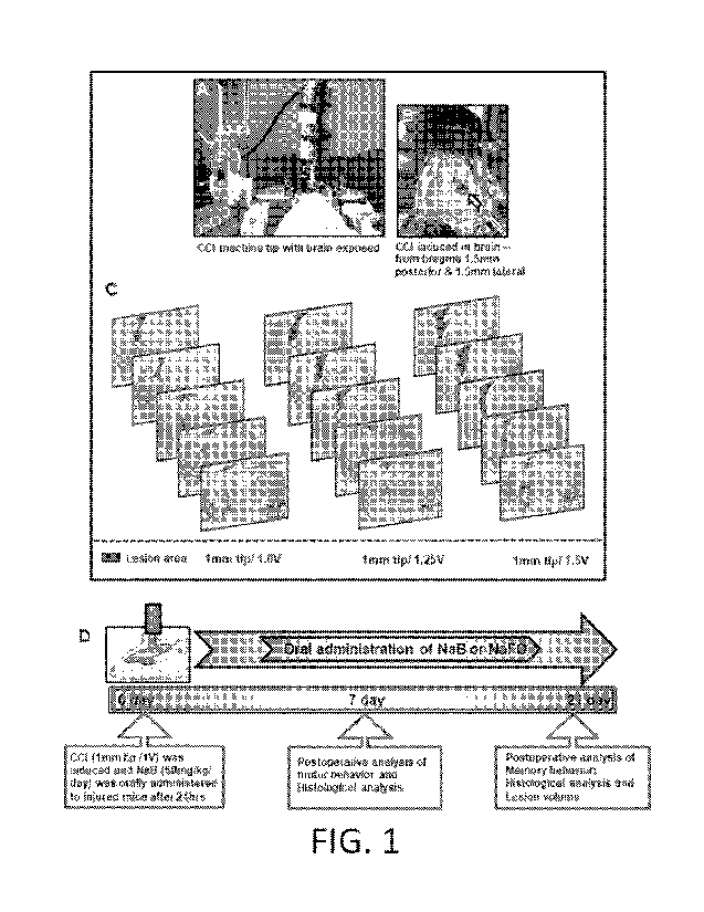
[0007] FIG. IA is a photograph of the CCI machine tip with mouse brain
exposed. Using the
CCI technique, brain injury was gently induced onto the exposed brain region
of anesthetized
mice.
[0008] FIG. 1B is a photograph showing CCI induced in the brain. Blood clots
and tissue
damage in burr hole (stereotactic coordinates ¨ from bregma 1.5 mm posterior
and 1.5 mm
lateral) were seen in the injured brain region of mice after CCI injury.
[0009] FIG. 1C are images showing the induction of mild, moderate, and severe
CCI injury
using a 1 mm tip with three different velocities, viz. 1.0V, 1.25V and 1.5V
respectively. After
one-week post-injury, mice (n=3) were perfused with 4% paraformaldehyde
followed by
removal of brains and staining the brain sections with cresyl violet.
2
CA 03241282 2024- 6- 17
WO 2023/114414
PCT/US2022/053034
[0010] FIG. 1D is a schematic of the experimental design showing the time
course of
treatment, behavioral, and histological analysis following CCI injury (1 mm
tip/1.0V).
100111 FIG. 2A are images of double-label immunofluorescence for GFAP and iNOS
in
brain sections for the control. Mice were treated with 50 mg/kg/day of NaB or
NaF0 via oral
administration after the in of CCI injury. After 7 days of NaB
treatment, brain
sections were analyzed by double-label immunofluorescence for GFAP and iNOS.
These
results show that oral treatment of NaB attenuates the activation of
astrocytes in vivo in the
cortex and hippocampus region of mice with CCI injury.
[0012] FIG. 2B are images of double-label immunofluorescence for GFAP and iNOS
in brain
sections for CCI injury. Mice were treated with 50 mg/kg/day of NaB or NaF0
via oral
administration after the induction of CCI injury. After 7 days of NaB
treatment, brain
sections were analyzed by double-label immunofluorescence for GFAP and iNOS.
These
results show that oral treatment of NaB attenuates the activation of
astrocytes in vivo in the
cortex and hippocampus region of mice with CCI injury.
[0013] FIG. 2C are images of double-label immunofluorescence for GFAP and iNOS
in brain
sections for CCI + NaB. Mice were treated with 50 mg/kg/day of NaB or NaF0 via
oral
administration after the induction of CCI injury. After 7 days of NaB
treatment, brain
sections were analyzed by double-label immunofluorescence for GFAP and iNOS.
These
results show that oral treatment of NaB attenuates the activation of
astrocytes in vivo in the
cortex and hippocampus region of mice with CCI injury.
100141 FIG. 2D are images of double-label immunofluorescence for GFAP and iNOS
in
brain sections for CCI + NaFO. Mice were treated with 50 mg/kg/day of NaB or
NaF0 via
oral administration after the induction of CCI injury. After 7 days of NaB
treatment, brain
sections were analyzed by double-label immunofluorescence for GFAP and iNOS.
These
results show that oral treatment of NaB attenuates the activation of
astrocytes in vivo in the
cortex and hippocampus region of mice with CCI injury.
[0015] FIG. 2E is a histogram of cells positive for GFAP counted in the cortex
region.
Results represent analysis of six sections of each of six mice per group. ap <
0.001 vs control;
bp <- 0.001 vs CCI injury.
CA 03241282 2024- 6- 17
WO 2023/114414
PCT/US2022/053034
[0016] FIG. 2F is a histogram of cells positive for GFAP counted in the CA1
region. Results
represent analysis of six sections of each of six mice per group. ap <0.001 vs
control; bp <
0.001 vs CCI injury.
[0017] FIG. 2G is a histogram of cells positive for iNOS counted in the cortex
region.
Results represent analysis of six sections of each of six mice per group. ap <
0.001 vs control;
bp < 0.001 vs CCI injury.
[0018] FIG. 2H is a histogram of cells positive for iNOS counted in the CA1
region. Results
represent analysis of six sections of each of six mice per group. ap <0.001 vs
control; hp <
0.001 vs CCI injury.
[0019] FIG. 21 is an immunoblot image of tissue extracts of hippocampal region
from all
groups of mice (n = 4 per group) for GFAP. Actin was run as a loading control.
[0020] FIG. 2J is a plot showing the values of GFAP/Actin relative to control
as obtained by
immunoblot band scanning. ap < 0.001 vs control; bp < 0.001 VS CCI injury.
[0021] FIG. 3A are images of double-label fluorescence for Ibal and iNOS for
the control.
Mice were treated with 50 mg/kg/day of NaB or NaF0 from 24hrs after the
induction of CCI
injury. After 7 days of treatment, brain sections were analyzed by double-
label fluorescence
for Ibal and iNOS. These results show that NaB treatment inhibits microglial
activation in
vivo in the cortex and hippocampus of mice with CCI injury.
100221 FIG. 3B are images of double-label fluorescence for Ibal and iNOS for
CCI injury.
Mice were treated with 50 mg/kg/day of NaB or NaF0 from 24hrs after the
induction of CCI
injury. After 7 days of treatment, brain sections were analyzed by double-
label fluorescence
for Ibal and iNOS. These results show that NaB treatment inhibits microglial
activation in
vivo in the cortex and hippocampus of mice with CCI injury.
[0023] FIG. 3C are images of double-label fluorescence for Ibal and iNOS for
CCI + NaB.
Mice were treated with 50 mg/kg/day of NaB or NaF0 from 24hrs after the
induction of CCI
injury. After 7 days of treatment, brain sections were analyzed by double-
label fluorescence
for Ibal and iNOS. These results show that NaB treatment inhibits microglial
activation in
vivo in the cortex and hippocampus of mice with CCI injury.
4
CA 03241282 2024- 6- 17
WO 2023/114414
PCT/US2022/053034
[0024] FIG. 3D are images of double-label fluorescence for Ibal and iNOS for
CCI + NaFO.
Mice were treated with 50 mg/kg/day of NaB or NaF0 from 24hrs after the
induction of CCI
injury. After 7 days of treatment, brain sections were analyzed by double-
label fluorescence
for Ibal and iNOS. These results show that NaB treatment inhibits microglial
activation in
vivo in the cortex and hippocampus of mice with CCI injury.
[0025] FIG. 3E is a histogram of cells positive for Ibal counted in the cortex
region. Results
represent analysis of six sections of each of six mice per group. ap <0.001 vs
control; bp <
0.001 vs CCI injury.
[0026] FIG. 3F is a histogram of cells positive for Ibal counted in the CA1
region. Results
represent analysis of six sections of each of six mice per group. ap <0.001 vs
control; bp <
0.001 vs CCI injury.
[0027] FIG. 3G is an immunoblot image of tissue extracts of hippocampal region
from all
groups of mice (n = 4 per group) for Ibal. Actin was run as a loading control.
100281 FIG. 3H is a plot showing the values of Ibal/Actin relative to control
as obtained by
immunoblot band scanning. ap < 0.001 vs control; hp <0.001 vs CCI injury.
[0029] FIG. 31 is an immunoblot image of tissue extracts of hippocampal region
from all
groups of mice (n = 4 per group) for iNOS. Actin was run as a loading control.
[0030] FIG. 3J is a plot showing the values of iNOS/Actin relative to control
as obtained by
immunoblot band scanning. ap < 0.001 vs control; bp < 0.001 vs CCI injury.
[0031] FIG. 4 are images of the corpus collosum in mice showing the levels of
proteolipid
protein (PLP) and A2B5, a marker of oligodendroglial progenitor cells (OPC),
in the
experimental conditions after 21 days. Oral administration of sodium benzoate
(NaB), but not
sodium formate (NaF0), stimulates remyelination in mice with traumatic brain
injury (TBI).
Mice (n=6 per group) were induced moderate TBI by controlled cortical impact
(CCI).
Starting from 2 hours after TBI, mice were treated with NaB and NaF0 (50 mg/kg
body
weight/day; mixed with water) orally via gavage. After 21 d of treatment,
brain sections were
double-labeled for PLP and A2B5. Results represent analysis of two sections of
each of six
mice per group.
CA 03241282 2024- 6- 17
WO 2023/114414
PCT/US2022/053034
[0032] FIG. 5A are images showing representative cresyl violet sections of
mouse brain
arranged in series of hippocampal region shows the volume of lesion cavity in
different
groups. NaB treatment reduces the lesion volume in mice with CCI injury.
[0033] FIG. 5B are illustrative images of cresyl violet section. Note the
extent of damage
induced in brain was found to be reduced in NaB treated mice when compared to
CCI-mice
without treatment and NaFo treated CCI injury mice.
[0034] FIG. 5C is a plot showing lesion size in the experimental conditions.
Lesion size was
quantitatively measured in control mice, untreated CCI injured mice, NaB
treated CCI-mice
and NaF0-treated CCI-mice at 21 days post-injury. Statistical analyses were
performed with
Student's t-test in the injured side of the brain [ap < 0.001 (5.623x10⁻⁷) vs
control; bp < 0.001
(=0.001) vs CCI injury].
[0035] FIG. 6A shows a heat map analysis for mice (n=6 per group) with TBI
induced by
CCI in open field activities after 7 days of treatment. Open field activities
were monitored by
the Ethovision XT 13.0 Open Field Activity System (Noldus). Starting from 2
hours after
TBI, mice were treated with NaB and NaF0 (50 mg/kg body weight/day; mixed with
water)
orally via gavage. Oral administration of sodium benzoate (NaB) but not sodium
formate
(NaF0) improves open field activities in mice with TBI.
[0036] FIG. 613 is a bar graph showing the distance traveled by mice (n-6 per
group) with
TBI induced by CCI in open field activities after 7 days of treatment.
Statistical analyses
were conducted with Student t-test for distance moved rp < 0.001 (=0.0001) vs
control; bp <
0.001 (=0.0029) vs CCI injury]; velocity [ap < 0.001 (=0.0001) vs control; bp
< 0.001
(=0.0078) vs CCI injury]; center frequency [ap < 0.001 (=0.0001) vs control;
bp < 0.001
(=0.0036) vs CCI injury] and rearing behavior rp < 0.001 (=9.498x10⁻⁶) vs
control; bp <
0.001 (-0.0081) vs CCI injury].
[0037] FIG. 6C is a bar graph showing the velocity of mice (n=6 per group)
with TBI
induced by CCI in open field activities after 7 days of treatment. Statistical
analyses were
conducted with Student t-test for distance moved [ap < 0.001 (=0.0001) vs
control; bp < 0.001
(=0.0029) vs CCI injury]; velocity [ap < 0.001 (=0.0001) vs control; bp <
0.001 (=0.0078) vs
CCI injury]; center frequency [ap < 0.001 (=0.0001) vs control; bp < 0.001
(=0.0036) vs CCI
injury] and rearing behavior rp < 0.001 (=9.498x10⁻⁶) vs control; bp < 0.001
(=0.0081) vs
CCI injury].
6
CA 03241282 2024- 6- 17
WO 2023/114414
PCT/US2022/053034
[0038] FIG. 6D is a bar graph showing the center frequency of mice (n=6 per
group) with
TBI induced by CCI in open field activities after 7 days of treatment.
Statistical analyses
were conducted with Student t-test for distance moved [ap < 0.001 (=0.0001) vs
control; bp <
0.001 (=0.0029) vs CCI injury]; velocity rp < 0.001 (=0.0001) vs control; bp <
0.001
(=0.0078) vs CCI injury]; center frequency [ap < 0.001 (=0.0001) vs control; b
< 0.001
(=0.0036) vs CCI injury] and rearing behavior rp < 0.001 (=9.498x10⁻⁶) vs
control; hp <
0.001 (-0.0081) vs CCI injury].
[0039] FIG. 6E is a bar graph showing the rearing behavior of mice (n=6 per
group) with TBI
induced by CCI in open field activities after 7 days of treatment. Statistical
analyses were
conducted with Student t-test for distance moved [ap < 0.001 (-0.0001) vs
control; hp < 0.001
(=0.0029) vs CCI injury]; velocity [ap < 0.001 (=0.0001) vs control; bp <
0.001 (=0.0078) vs
CCI injury]; center frequency rp < 0.001 (=0.0001) vs control; bp < 0.001
(=0.0036) vs CCI
injury] and rearing behavior [ap < 0.001 (=9.498x10⁻⁶) vs control; bp < 0.001
(=0.0081) vs
CCI injury].
[0040] FIG. 6F shows heat map analysis for mice (n=6 per group) with TBI
induced by CCI
in open field activities after 21 days of treatment. Open field activities
were monitored by the
Ethovision XT 13.0 Open Field Activity System (Noldus). Starting from 2 hours
after TBI,
mice were treated with NaB and NaF0 (50 mg/kg body weight/day; mixed with
water) orally
via gavage.
[0041] FIG. 6G is a bar graph showing the distance traveled by mice (n=6 per
group) with
TBI induced by CCI in open field activities after 21 days of treatment.
Statistical analyses
were conducted with Student t-test for distance moved 1-ap < 0.001 (=0.0001)
vs control; bp <
0.001 (=0.0029) vs CCI injury]; velocity rp < 0.001 (=0.0001) vs control; hp <
0.001
(=0.0078) vs CCI injury]; center frequency [ap < 0.001 (=0.0001) vs control;
bp < 0.001
(=0.0036) vs CCI injury] and rearing behavior rp < 0.001 (=9.498x10⁻⁶) vs
control; hp <
0.001 (=0.0081) vs CCI injury].
[0042] FIG. 6H is a bar graph that shows the velocity of mice (n=6 per group)
with TBI
induced by CCI in open field activities after 21 days of treatment.
Statistical analyses were
conducted with Student t-test for distance moved [ap < 0.001 (=0.0001) vs
control; hp < 0.001
(=0.0029) vs CCI injury]; velocity [ap < 0.001 (=0.0001) vs control; hp <
0.001 (=0.0078) vs
CCI injury]; center frequency rp < 0.001 (=0.0001) vs control; bp < 0.001
(=0.0036) vs CCI
7
CA 03241282 2024- 6- 17
WO 2023/114414
PCT/US2022/053034
injury] and rearing behavior [ap < 0.001 (-9.498x10⁻⁶) vs control, b < 0.001 (-
0.0081) vs
CCI injury].
100431 FIG. 61 is a bar graph showing the center frequency of mice (n=6 per
group) with TBI
induced by CCI in open field activities after 21 days of treatment.
Statistical analyses were
conducted with Student t-test for distance moved [ap < 0.001 (=0.0001) vs
control; hp <0.001
(=0.0029) vs CCI injury]; velocity [ap < 0.001 (=0.0001) vs control; bp <
0.001 (=0.0078) vs
CCI injury]; center frequency [ap < 0.001 (=0.0001) vs control; bp < 0.001
(=0.0036) vs CCI
injury] and rearing behavior rp < 0.001 (=9.498x10⁻⁹) vs control; b < 0.001
(=0.0081) vs
CCI injury].
[0044] FIG. 6J is a bar graph showing the rearing of mice (n=6 per group) with
TBI induced
by CCI in open field activities after 21 days of treatment. Statistical
analyses were conducted
with Student t-test for distance moved Pp < 0.001 (=0.0001) vs control; bp <
0.001 (=0.0029)
vs CCI injury]; velocity [ap < 0.001 (=0.0001) vs control; bp < 0.001
(=0.0078) vs CCI
injury]; center frequency [ap < 0.001 (=0.0001) vs control; bp < 0.001
(=0.0036) vs CCI
injury] and rearing behavior [ap < 0.001 (=9.498x10⁻⁶) vs control; bp < 0.001
(=0.0081) vs
CCI injury].
[0045] FIG. 6K is a bar graph showing the results for tail suspension test.
Following the NaB
treatment, mice with CCI-injury showed significant improvement in tail
suspension test on 7-
day post-injury [ap < 0.001 (-5.725x10⁻⁸) vs control; hp < 0.001 (-0.0001) vs
CCI injury] and
21-day post-injury [ap < 0.001 (=3.995x10⁵) vs control; hp < 0.001 (=0.0003)
vs CCI injury].
100461 FIG. 6L is a bar graph showing the results for rotarod test. The
performance of mice
with NaB treatment has significantly improved in the rotarod test on 7-day
post-injury [ap <
0.001 (=9.5998x10⁻⁹) vs control; bp <0.001 (-0.0003) vs CCI injury] and 21-day
post-injury
[ap < 0.001 (=1.2133x10⁻⁹) vs control; bp < 0.001 (-0.0010) vs CCI injury].
[0047] FIG. 6M is a bar graph showing the number of steps on beam runway. Oral
treatment
of NaB also improved the performance of CCI-injured mice on beam runway.
Statistical
significance was performed using the Student t-test on 7-day post-injury
[Steps: ap < 0.001
(=2.258x10⁻⁶) vs control; bp < 0.001 (=0.0027) vs CCI injury; Time taken: ap <
0.001
(=2.979x10⁻⁷) vs control; bp < 0.001 (=0.0039) vs CCI injury; Foot slipping:
ap < 0.001
(=1.567x10⁻⁷) vs control; bp < 0.001 (= 0.0001) vs CCI injury] and on 21-day
post-injury
[Steps: ap < 0.001 (=0.0051) vs control; ns (=0.2533) vs CCI injury; Time
taken: ap <0.001
8
CA 03241282 2024- 6- 17
WO 2023/114414
PCT/US2022/053034
(-0.0003) vs control; ns (-0.1228) vs CCI injury; Foot slipping: "p <0.001 (-
0.0001) vs
control; bp < 0.05 (=0.0736) vs CCI injury].
100481 FIG. 6N is a bar graph showing the time taken on beam runway. Oral
treatment of
NaB also improved the performance of CCI-injured mice on beam runway.
Statistical
significance was performed using the Student t-test on 7-day post-injury
[Steps: ap < 0.001
(=2.258x10⁻⁶) vs control; bp < 0.001 (=0.0027) vs CCI injury; Time taken: ap <
0.001
(=2.979x10⁻⁷) vs control; bp < 0.001 (=0.0039) vs CCI injury; Foot slipping:
ap < 0.001
(=1.567x10⁻⁷) vs control; bp < 0.001 (= 0.0001) vs CCI injury] and on 21-day
post-injury
[Steps: ap <0.001 (=0.0051) vs control; ns (=0.2533) vs CCI injury; Time
taken: ap < 0.001
(=0.0003) vs control; ns (=0.1228) vs CCI injury; Foot slipping: ap <0.001
(=0.0001) vs
control; bp < 0.05 (=0.0736) vs CCI injury].
[0049] FIG. 60 is a bar graph showing the foot-slipping on beam runway. Oral
treatment of
NaB also improved the performance of CCI-injured mice on beam runway.
Statistical
significance was performed using the Student t-test on 7-day post-injury
[Steps: ap < 0.001
(=2.258x10⁻⁶) vs control; bp < 0.001 (=0.0027) vs CCI injury; Time taken: ap <
0.001
(=2.979x10⁻⁷) vs control; bp < 0.001 (=0.0039) vs CCI injury; Foot slipping:
ap < 0.001
(=1.567x10⁻⁷) vs control; bp <0.001 (= 0.0001) vs CCI injury] and on 21-day
post-injury
[Steps: ap <0.001 (=0.0051) vs control; ns (=0.2533) vs CCI injury; Time
taken: ap <0.001
(=0.0003) vs control; ns (=0.1228) vs CCI injury; Foot slipping: ap <0.001
(=0.0001) vs
control; bp < 0.05 (-0.0736) vs CCI injury].
[0050] FIG. 6P is a bar graph showing number of steps in grid runway. CCI
injured mice
with NaB treatment exhibited improvements in grid runway. Using Student t-
test, statistical
significance were performed on 7-day post-injury [Steps: ap < 0.001 (=9.364x10-
9) vs control;
hp <0.001 (=3.394x10⁻⁵) vs CCI injury; Time taken: Up <0.001 (=1.3770x10⁻⁷) vs
control; hp
<0.001 (-3.3886x10⁻⁵) vs CCI; Foot-misplacement: "p < 0.001 (-2.737x10⁸) vs
control; hp <
0.001 (=5.954x10⁻⁶) vs CCI injury] and on 21-day post-injury (Steps: Up <
0.001 (=0.0014) vs
control; hp < 0.05 (=0.0718) vs CCI injury; Time taken: "p < 0.001 (=5.562x10)
vs control;
bp < 0.05 (=0.072) vs CCI injury; Foot-misplacement: Up < 0.001 (=1.079x10⁻⁵)
vs control; ns
(=0.2465) vs CCI injury. ns - Non-significant.
[0051] FIG. 6Q is a bar graph showing time taken in grid runway. CCI injured
mice with
NaB treatment exhibited improvements in grid runway. Using Student t-test,
statistical
9
CA 03241282 2024- 6- 17
WO 2023/114414
PCT/US2022/053034
significance were performed on 7-day post-injury [Steps: ap <0.001 (-9.364x10-
9) vs
control; bp <0.001 (=3.394x10⁻⁵) vs CCI injury; Time taken: ap <0.001
(=1.3770x10⁻⁷) vs
control; bp < 0.001 (=3.3886x10⁻⁵) vs CCI; Foot-misplacement: ap < 0.001
(=2.737x10⁻⁸) vs
control; bp <0.001 (=5.954x10⁻⁶) vs CCI injury] and on 21-day post-injury
(Steps: ap <
0.001 (=0.0014) vs control; bp < 0.05 (=0.0718) vs CCI injury; Time taken: ap
<0.001
(=5.562x10⁻⁵) vs control; bp < 0.05 (=0.072) vs CCI injury; Foot-misplacement:
ap < 0.001
(-1.079x10⁻⁵) vs control; ns (-0.2465) vs CCI injury. ns -Non-significant.
[0052] FIG. 6R is a bar graph showing foot misplacement in grid runway. CCI
injured mice
with NaB treatment exhibited improvements in grid runway. Using Student t-
test, statistical
significance were performed on 7-day post-injury [Steps: ap <0.001 (=9.364x10-
9) vs
control; bp < 0.001 (=3.394x10⁻⁵) vs CCI injury; Time taken: ap < 0.001
(=1.3770x10⁻⁷) vs
control; bp < 0.001 (=3.3886x10⁻⁵) vs CCI; Foot-misplacement: ap < 0.001
(=2.737x10⁻⁸) vs
control; bp < 0.001 (=5.954x10⁻⁶) vs CCI injury] and on 21-day post-injury
(Steps: ap <
0.001 (=0.0014) vs control; bp <0.05 (=0.0718) vs CCI injury; Time taken: ap
<0.001
(=5.562x10⁻⁵) vs control; bp < 0.05 (=0.072) vs CCI injury; Foot-misplacement:
ap < 0.001
(=1.079x10⁻⁵) vs control; ns (=0.2465) vs CCI injury. ns -Non-significant.
[0053] FIG. 7A shows heat maps demonstrating the novel object recognition of
mice (n=6
per group) with TBI induced by CCI 21 days post-operation. Starting from 2
hours after TBI,
mice were treated with NaB and NaF0 (50 mg/kg body weight/day; mixed with
water) orally
via gavage. Statistical analyses were performed by Student I-test for Novel
object recognition
test [Exploration time: ap < 0.001 (=2.5989x10⁻⁵) vs control; bp < 0.001
(=0.0003) vs CCI
injury]; Barnes maze test [Time taken: ap <0.001 (=1.7509x10⁻⁵) vs control; bp
< 0.001
(=4.8824x10⁻⁵) vs CCI injury and Number of errors: ap < 0.001 (=3.234x10⁻⁵) vs
control; bp <
0.001 (=0.0001) vs CCI injury] ; T-maze [Positive turns: ap < 0.001
(=3.3524x10⁻⁵) vs
control; bp <0.001 (=0.0004) vs CCI injury and Negative turns: ap <0.001
(=3.3924x10⁻⁵) vs
control; bp < 0.001 (=0.0005) vs CCI injury]. ns - Non-significant.
[0054] FIG. 7B shows heat maps demonstrating the Barnes circular maze test
results of mice
(n=6 per group) with TBI induced by CCI 21 days post-operation. Statistical
analyses were
performed by Student t-test for Novel object recognition test [Exploration
time: ap < 0.001
(=2.5989x10⁻⁵) vs control; bp < 0.001 (=0.0003) vs CCI injury]; Barnes maze
test [Time
taken: ap < 0.001 (=1.7509x10⁻⁵) vs control; bp < 0.001 (=4.8824x10⁻⁵) vs CCI
injury and
Number of errors: ap < 0.001 (=3.234x10⁻⁵) vs control; bp <0.001 (=0.0001) vs
CCI injury] ;
CA 03241282 2024- 6- 17
WO 2023/114414
PCT/US2022/053034
T-maze [Positive turns: ap <0.001 (-3.3524x10⁵) vs control; bp < 0.001 (-
0.0004) vs CCI
injury and Negative turns: ap < 0.001 (=3.3924x10⁵) vs control; bp <0.001
(=0.0005) vs CCI
injury]. ns - Non-significant.
[0055] FIG. 7C is a bar graph showing the exploration time of mice (n=6 per
group) with
TBI induced by CCI 21 days post-operation during novel object recognition
test. Statistical
analyses were performed by Student t-test for Novel object recognition test
[Exploration
time: ap < 0.001 (=2.5989x10⁻⁵) vs control; bp < 0.001 (=0.0003) vs CCI
injury]; Barnes maze
test [Time taken: ap < 0.001 (=1.7509x10⁵) vs control; bp < 0.001
(=4.8824x10⁵) vs CCI
injury and Number of errors: ap <0.001 (=3.234x10⁻⁵) vs control; bp <0.001
(=0.0001) vs
CCI injury] ; T-maze [Positive turns: ap < 0.001 (=3.3524x10⁵) vs control; bp
<0.001
(=0.0004) vs CCI injury and Negative turns: ap < 0.001 (=3.3924x10⁵) vs
control; bp < 0.001
(=0.0005) vs CCI injury]. ns -Non-significant.
[0056] FIG. 7D is a bar graph showing the latency time of mice (n=6 per group)
with TBI
induced by CCI 21 days post-operation during the Barnes maze test. Statistical
analyses were
performed by Student t-test for Novel object recognition test [Exploration
time: ap < 0.001
(=2.5989x10⁵) vs control; bp < 0.001 (=0.0003) vs CCI injuryl; Barnes maze
test [Time
taken: ap <0.001 (=1.7509x10⁻⁵) vs control; bp < 0.001 (=4.8824x10⁵) vs CCI
injury and
Number of errors: ap < 0.001 (=3.234x10⁵) vs control: bp <0.001 (=0.0001) vs
CCI injury] ;
T-maze [Positive turns: ap <0.001 (=3.3524x10⁵) vs control; bp < 0.001
(=0.0004) vs CCI
injury and Negative turns: ap < 0.001 (-3.3924x10⁵) vs control; bp < 0.001 (-
0.0005) vs CCI
injury]. ns - Non-significant.
100571 FIG. 7E is a bar graph showing the number of errors of mice (n=6 per
group) with
TBI induced by CCI 21 days post-operation during the Barnes maze test.
Statistical analyses
were performed by Student t-test for Novel object recognition test
[Exploration time: ap <
0.001 (-2.5989x10⁵) vs control; bp < 0.001 (=0.0003) vs CCI injury]; Barnes
maze test
[Time taken: ap < 0.001 (=1.7509x10⁻⁵) vs control; bp < 0.001 (=4.8824x10⁻⁵)
vs CCI injury
and Number of errors: ap <0.001 (=3.234x10⁻⁵) vs control; bp < 0.001 (=0.0001)
vs CCI
injury] ; T-maze [Positive turns: ap < 0.001 (=3.3524x10⁻⁵) vs control; bp <
0.001 (=0.0004)
vs CCI injury and Negative turns: ap < 0.001 (=3.3924x10⁵) vs control; bp
<0.001 (=0.0005)
vs CCI injury]. ns Non-significant.
11
CA 03241282 2024- 6- 17
WO 2023/114414
PCT/US2022/053034
100581 FIG. 7F is a bar graph showing the number of positive turns of mice
(n=6 per group)
with TBI induced by CCI 21 days post-operation during the T maze. Statistical
analyses were
performed by Student t-test for Novel object recognition test [Exploration
time: ap < 0.001
(-2.5989x10⁻⁵) vs control; bp <0.001 (-0.0003) vs CCI injury]; Barnes maze
test [Time
taken: ap < 0.001 (-1.7509x10⁻⁵) vs control; bp < 0.001 (-4.8824x10⁻⁵) vs CCI
injury and
Number of errors: ap <0.001 (=3.234x10⁻⁵) vs control; bp <0.001 (=0.0001) vs
CCI injury] ;
T-maze [Positive turns: ap <0.001 (-3.3524x10⁻⁵) vs control; bp < 0.001 (-
0.0004) vs CCI
injury and Negative turns: an < 0.001 (-3.3924x10⁻⁵) vs control; bp < 0.001 (-
0.0005) vs CCI
injury]. ns ¨ Non-significant.
[0059] FIG. 7G is a bar graph showing the number of negative turns of mice
(n=6 per group)
with TBI induced by CCI 21 days post-operation during the T maze. Statistical
analyses were
performed by Student t-test for Novel object recognition test [Exploration
time: ap < 0.001
(=2.5989x10⁻⁵) vs control; bp < 0.001 (-0.0003) vs CCI injury]; Barnes maze
test [Time
taken: ap <0.001 (-1 .7509x10-5) vs control; bp <0.001 (-4.8824x10⁻⁵) vs CCI
injury and
Number of errors: ap < 0.001 (=3.234x10⁻⁵) vs control: bp <0.001 (-0.0001) vs
CCI injury] ;
T-maze [Positive turns: ap <0.001 (=3.3524x10⁻⁵) vs control; bp < 0.001
(=0.0004) vs CCI
injury and Negative turns. ap <0.001 (-3.3924x10⁻⁵) vs control, bp <0.001 (-
0.0005) vs CCI
injury]. ns ¨ Non-significant.
[0060] FIGs. 8A-8D are images of double-label immunofluorescence for GFAP and
iNOS in
brain sections for the control (FIG. 8A), CCI (FIG. 8B), CCI + GTB (FIG. 8C)
and CCI +
Vehicle (FIG. 8D. Mice were treated with 50 mg/kg/day of GTB via oral gavage
after the
induction of CCI injury. After 7 days of GTB treatment, brain sections were
analyzed by
double-label immunofluorescence for GFAP and iNOS. Cells positive for GFAP
were
counted in cortex (FIG. 8E) and CA1 region of hippocampus (FIG. 8F).
Similarly, cells
positive for iNOS were also counted in cortex (FIG. SG) and CAI region (FIG.
8H). Results
represent analysis of six sections of each of six mice per group. Tissue
extracts of
hippocampal region from all groups of mice (n= 4 per group) were immunoblotted
for GFAP
(FIG. 81) and iNOS (FIG. 8K). Actin was run as a loading control. Bands were
scanned, and
values (GFAP/Actin) (FIG. 8J) and (iNOS/Actin) (FIG. 8L) presented as relative
to control.
These results show that oral administration of GTB inhibits astroglial
inflammation in vivo in
the cortex and hippocampus of mice with TBI.
12
CA 03241282 2024- 6- 17
WO 2023/114414
PCT/US2022/053034
[0061] FIGs. 9A-9H. Oral GTB decreases microglial activation in vivo in the
cortex and
hippocampus of mice with TBI. TBI was induced in mice by CCI injury and after
24 h of
injury, mice were treated with 50 mg/kg/day of GTB via oral gavage. Seven days
after GTB
treatment, brain sections were double-labeled for Thal and iNOS (FIG. 9A,
control; FIG. 9B,
CCI; FIG. 9C, CCI+GTB; FIG. 9D, CCI+Vehicle). Cells positive for Ibal were
counted in
cortex (FIG. 9E) and CA1 region of hippocampus (FIG. 9F). Results represent
analysis of
two sections of each of six mice per group. Tissue extracts of hippocampal
region from all
groups of mice (n= 4 per group) were immunoblotted for Ibal (FIG. 9G). Actin
was run as a
loading control. Bands were scanned, and values (Ibal/Actin) (FIG. 9H)
presented as relative
to control.
[0062] FIGs. 10A-10C. Decrease in lesion volume in TBI mice by GTB treatment.
TBI was
induced in mice by CCI injury and after 24 h of injury, mice were treated with
50 mg/kg/day
of GTB via oral gavage. FIG. 10A. Twenty-one days after injury, brain sections
were stained
with H&E and H&E stained sections were arranged in a series demonstrating the
volume of
lesion cavity in different groups. FIG. 10B shows illustrative images of H&E
stained
sections. FIG. 10C. Lesion volume was quantified in all groups of mice.
Statistical analyses
were performed with two way ANOVA and expressed as mean SD to compare the
lesion
volume between unlesioned and lesioned side of the brain.
[0063] FIGs. 11A-11H. Restoration of PSD-95, NR2A and GluR1 in the hippocampus
of
TBI mice by oral administration of GTB. TBI was induced in mice by CCI injury
and after 24
h of injury, mice were treated with 50 mg/kg/day of GTB via oral gavage.
Twenty-one days
after CCI injury, brain sections were double-labeled for NeuN and PSD-95 (FIG.
11A,
control; FIG. 11B, CCI; FIG. 11C, CCI+GTB; FIG. 11D, CCI Vehicle). Results
represent
analysis of one section of each of six mice per group. Hippocampal tissue
extracts from all
groups of mice (n= 4 per group) were immunoblotted for PSD-95, NR2A and GluR1
. FIG.
11E. Actin was run as a loading control. Bands were scanned, and values
(Ibal/Actin, FIG.
11F; NR2A/Actin, FIG. 11G; GluRl/Actin, FIG. 11H) presented as relative to
control. Data
are expressed as mean + SD. Statistical analyses were performed with one way
ANOVA.
[0064] FIGs. 12A-12G. Effect of GTB on spatial learning and memory in TBI
mice. TBI was
induced in mice by CCI injury and after 24 h of injury, mice were treated with
50 mg/kg/day
of GTB via oral gavage. Twenty-one days after CCI injury, mice were tested by
Novel object
recognition test (FIG. 12A, Heat map; FIG. 12C, Exploration time), Barnes maze
(FIG. 12B,
13
CA 03241282 2024- 6- 17
WO 2023/114414
PCT/US2022/053034
Heat map; FIG. 12D, number of errors; FIG. 12E, latency pr time taken) and T-
maze (FIG.
12F, positive turns; FIG. 12G, Negative turns). Six mice were used in each
group. Statistical
analyses were performed by one way ANOVA followed by Tukey's post hoc test.
[0065] FIGs. 13A-13M. GTB treatment recovers motor functions in TBI mice. TBI
was
induced in mice by CCI injury and after 24 h of injury, mice were treated with
50 mg/kg/day
of GTB via oral gavage. Seven days after CO injury, mice were tested for open-
field
behavior (FIG. 13A, heat map analysis monitored by using the Noldus system;
FIG. 13B,
distance moved; FIG. 13C, velocity; FIG. 13D, center frequency; FIG. 13E,
rearing), roto rod
(FIG. 13F, latency), tail suspension test (FIG. 13G, immobility time), beam
walking (FIG.
13H, number of steps; FIG. 131, time taken; FIG. 131, slips), and grid runway
(FIG. 13K,
number of steps; FIG. 13L, time taken; FIG. 13M, misplacements). Six mice were
used in
each group. Statistical analyses were performed by one way ANOVA followed by
Tukey's
posthoc test.
[0066] FIGs. 14A-14M. Effect of GTB on motor functions in TBI mice on 21st day
of CCI
injury. TBI was induced in mice by CCI injury and after 24 h of injury, mice
were treated
with 50 mg/kg/day of GTB via oral gavage. Twenty-one days after CCI injury,
mice were
tested for open-field behavior (FIG. 14A, heat map analysis monitored by using
the Noldus
system; FIG. 14B, distance moved; FIG. 14C, velocity; FIG. 14D, center
frequency; FIG.
14E, rearing), rotorod (FIG. 14F, latency), tail suspension test (FIG. 14G,
immobility time),
beam walking (FIG. 14H, number of steps; FIG. 141, time taken; FIG. 14J,
slips), and grid
runway (FIG. 14K, number of steps; FIG. 14L, time taken; FIG. 14M,
misplacements). Six
mice were used in each group. Statistical analyses were performed by one way
ANOVA
followed by Tukey's post hoc test.
[0067] FIGs. 15A-15L: Effect of sodium benzoate (NaB) on the maturation of
oligodendroglial progenitor cells (OPCs) into oligodendrocytes. OPCs were
isolated from
P1-P2 neonatal mouse pups, cultured in OPC media for 4 days in vitro (DIV)
followed by
treatment with 100pM NaB and sodium formate (NaF0) in the absence of FGF and
PDGF.
FIG. 15A) After 18 h of 100 iuM NaB treatment in serum-free condition, cells
were fixed and
then dual immuno-stained with MBP (red) and OPC marker NG2 (green). Nuclei
were
stained with DAPI (blue). FIG. 15B) Similarly, OPCs were stained with PLP
(red) and A2B5
(green) under similar treatment condition. Quantification of MBP+ (FIG. 15C),
NG2+ (FIG.
15D), PLP+ (FIG. 15E), and A2B5+ (FIG. 15F) cells as a percentage of total
cells (DAPI+).
14
CA 03241282 2024- 6- 17
WO 2023/114414
PCT/US2022/053034
Average 5 fields per slide, total 3 slides. Results are mean + SEM. ***p
<0.001. FIG. 15G)
For protein expression, cells were treated with different doses of NaB for 18
h under serum
free condition and then immunoblotted with PLP and MOG. FIG. 15H)
Densitometric
analyses of the bands relative to beta actin were done for PLP and MOG.
ap<0.001 vs.
control PLP; bp<0.005 vs. control MOG. FIG. 151) Cells were treated with NaB
(100uM) and
NaF0 (100uM) for 4 h under serum-free condition followed by monitoring the
mRNA
expression of myelin-specific genes by real-time PCR (ap<0.01 vs. control PLP;
bp<0.01 vs.
control MOG; cp<0.01 vs. control MBP; dp<0.01 vs. control CNPase). OPCs were
cultured
on the top of randomly-oriented polycaprolactone nanofibers (Nanofiber
Solutions; Cat #
7694576) for 7 days. After that, these cells were treated with 100 iaM NaB
(FIG. 15J) and
NaF0 (K) for additional 2 days followed by immunofluorescence analysis for MBP
(red)
(FIG. 15J, control; FIG. 15K, NaB; FIG. 15L, NaF0). Images were displayed in a
single red
channel as well as merged with phase-contrast image of nanofibers.
Representative 3D
constructed images of OPCs adhered to nanofibers were also shown at the right
side.
[0068] FIGs. 16A-16C: Oral NaB stimulates the maturation of OPCs in vivo in
the corpus
callosum of cuprizone-intoxicated mice. C57/BL6 mice (8-10 week old; male)
were fed
cuprizone-containing diet (Envigo) for 5 weeks followed by treatment with NaB
(50 mg/kg
body wt/d) orally via gavage. FIG. 16A) After 3 weeks of treatment with NaB,
corpus
callosum sections were double-labeled for PLP and A2B5. Mean fluorescence
intensity
(MFI) of A2B5 (FIG. 16B) and PLP (FIG. 16C) were quantified from one section
(two
images per section) of each of 5 mice group. Results are mean + SEM of 5 mice
per group.
***p <0.001; **p < 0.01.
100691 FIGs. 17A-17H: Effect of NaB on myelination in vivo in the corpus
callosum of
cuprizone-intoxicated mice. C57/BL6 mice (8-10 week old; male; n=5) were fed
cuprizone-
containing diet (Envigo) for 5 weeks followed by treatment with NaB (50 mg/kg
body wt/d)
orally via gavagc. After 3 weeks of treatment with NaB, corpus callosum
sections were
immunostained MBP (FIG. 17A) and PLP (FIG. 17B). Mean fluorescence intensity
(MFI) of
MBP (FIG. 17C) and PLP (FIG. 17D) were quantified from one section (two images
per
section) of each of 5 mice group. Results are mean + SEM of 5 mice per group.
**p < 0.01;
*p <0.05. FIG. 17E) Corpus callosum sections were stained for luxol fast blue
(LFB). FIG.
17F) For electron microscopic studies, 50 tm thick sagittal sections were
prepared and
stained followed by analysis of corpus callosum sections for different
parameters to evaluate
CA 03241282 2024- 6- 17
WO 2023/114414
PCT/US2022/053034
axonal ultrastructures. FIG. 17G) G score was calculated in 75 axons per group
for all three
groups. FIG. 17H) Percentage of myelinated axons was calculated in 7 randomly
selected
corpus callosum sections of 5 mice per group. ***p <0.001.
[0070] FIGs. 18A-18C. Cinnamein inhibits the induction of NO production from
LPS- and
IFNy-stimulated mouse RAW 264.7 macrophages. FIG. 1RA) Cells preincubated with
different concentrations of cinnamein for 6 h were stimulated with 1 mg/m1LPS
under serum-
free condition. After 24 h of stimulation, the level of nitrite was measured
in supernatants by
Griess reagent. FIG. 18B) Cells preincubated with 400 i.t.M cinnamein for
different hours
were stimulated with 1 vtg/m1LPS under serum-free condition. After 24 h of
stimulation, the
level of nitrite was measured in supernatants. FIG. 18C) Cells preincubated
with different
concentrations of cinnamein for 6 h were stimulated with 25 U/ml IFNy under
serum-free
condition. After 24 h of stimulation, the level of nitrite was measured in
supernatants. Results
are mean + SD of three independent experiments. *p < 0.05, **p <0.01, ***p <
0.001, NS,
not significant.
[0071] FIGs. 19A-19B. Cinnamein inhibits LPS- and polyIC-induced production of
TNFa in
primary mouse microglia. Microglia isolated from 2d old mouse pups were
incubated with
different concentrations of cinnamein for 6 h followed by stimulation with
either 11.1g/m1
LPS (FIG. 19A) or 50 1.1g/m1 polyIC (FIG. 19B) under serum-free condition.
After 24 h of
stimulation, the level of TNFa was measured in supernatants by ELISA. Results
are mean +
SD of three independent experiments. *p <0.05; **p <0.01; ***p <0.001; NS, not
significant.
100721 FIGs. 20A-20B. Cinnamein suppresses the production of IL-113 from LPS-
and poly
IC-stimulated primary mouse microglia Cells preincubated with different
concentrations of
cinnamein for 6 h were stimulated with either 1 1,1g/m1LPS (FIG. 20A) or 50
vtg/m1 polyIC
(FIG. 20B) under serum-free condition. After 24 h of stimulation, the level of
1L-113 was
measured in supematants by ELISA. Results are mean + SD of three independent
experiments. 1-1-1-p <0.001.
[0073] FIGs. 21A-21B. Cinnamein decreases LPS- and polyIC-induced production
of 1L-6 in
primary mouse microglia. Microglia were incubated with different
concentrations of
cinnamein for 6 h followed by stimulation with either 1 vtg/m1LPS (FIG. 21A)
or 50 ug/m1
polyIC (FIG. 21B) under serum-free condition. After 24 h of stimulation, the
level of IL-6
16
CA 03241282 2024- 6- 17
WO 2023/114414
PCT/US2022/053034
was measured in supernatants by ELISA. Results are mean + SD of three
independent
experiments. -4-4-p <0.001.
100741 FIGs. 22A-22B. Cinnamein inhibits the production of proinflammatory
cytokines
from polyIC-stimulated primary mouse astrocytes. Astrocytes preincubated with
different
concentrations of cinnamein for 6 h were stimulated with 50 vig/m1 poly IC
under serum-free
condition. After 24 h of stimulation, levels of TNFa (FIG. 22A) and 1L-6 (FIG.
22B) were
measured in supernatants by ELISA. Results are mean + SD of three independent
experiments. *p < 0.05; **p < 0.01; ***p < 0.001.
DETAILED DESCRIPTION
A. Definitions
[0075] "Nervous system injury," including central or peripheral nervous system
injuries,
refers to any injury to the nervous system caused by trauma and/or disease.
[0076] The "central nervous system" (CNS) includes the brain, spinal cord,
optic, olfactory,
and auditory systems. The CNS comprises both neurons and glial cells
(neuroglia), which are
support cells that aid the function of neurons. Oligodendrocytes, astrocytes,
and microglia are
glial cells within the CNS. Oligodendrocytes myelinate axons in the CNS, while
astrocytes
contribute to the blood-brain barrier, which separates the CNS from blood
proteins and cells,
and perform a number of supportive functions for neurons. Microglial cells
serve immune
system functions.
[0077] "Central nervous system injury" refers to any injury to the central
nervous system
caused by trauma instead of disease. The term encompasses injuries to the
central nervous
system that result in loss or impairment of motor function, sensory function,
or a combination
thereof
[0078] The "peripheral nervous system" (PNS) includes the cranial nerves
arising from the
brain (other than the optic and olfactory nerves), the spinal nerves arising
from the spinal
cord, sensory nerve cell bodies, and their processes, i.e., all nervous tissue
outside of the
CNS. The PNS comprises both neurons and glial cells (neuroglia), which are
support cells
that aid the function of neurons. Glial cells within the PNS are known as
Schwann cells, and
serve to myelinate axons by providing a sheath that surrounds the axons.
17
CA 03241282 2024- 6- 17
WO 2023/114414
PCT/US2022/053034
[0079] "Peripheral nervous system injury" refers to any injury to a peripheral
nerve caused
by trauma instead of disease. The term encompasses all degrees of nerve
injury, including the
lowest degree of nerve injury in which the nerve remains intact but signaling
ability is
damaged, known as neurapraxia. The term also includes the second degree in
which the axon
is damaged but the surrounding connecting tissue remains intact, known as
axonotmesis.
Finally, the term encompasses the last degree in which both the axon and
connective tissue
are damaged, known as neurotmesis.
[0080] "Traumatic brain injury" or "TBI" refers to an acquired brain injury or
head injury in
which trauma damages the brain. The damage can be localized, i.e., limited to
one area of the
brain, or diffuse, affecting one or more areas of the brain.
[0081] "Spinal cord injury- means any injury to the spinal cord that is caused
by trauma
instead of disease. Depending on where the spinal cord and nerve roots are
damaged, the
symptoms can vary widely, for example from pain to paralysis to incontinence.
Spinal cord
injuries are described at various levels of "incomplete," which can vary from
having no effect
on the subject to a -complete" injury which means a total loss of function.
Spinal cord
injuries have many causes, but are typically associated with major trauma from
motor vehicle
accidents, falls, sports injuries, and violence. The abbreviation -SCI" means
spinal cord
injury.
[0082] "Spinal cord contusion" refers to an injury caused by trauma instead of
disease in
which part of the spinal cord is crushed with part of its tissue spared,
particularly the ventral
nerve fibers connecting the spinal cord rostral and caudal to the injury.
[0083] "Nerve crush injury" refers to traumatic compression of the nerve from
a blunt object,
such as a bat, surgical clamp or other crushing object that does not result in
a complete
transection of the nerve.
[0084] "Administering- means any method used to deliver the compounds, salts,
or
compositions to the subject. These include oral routes, intraduodenal routes,
parenteral
injection (including intravenous, subcutaneous, intraperitoneal,
intramuscular, intravascular
or infusion), topical, and rectal administration. Those of skill in the art
are familiar with
administration techniques that can be used, e.g., as discussed in Goodman and
Gilman, The
Pharmacological Basis of Therapeutics, current ed.; Pergamon; and Remington's,
18
CA 03241282 2024- 6- 17
WO 2023/114414
PCT/US2022/053034
Pharmaceutical Sciences (current edition), Mack Publishing Co., Easton, Pa. In
some aspects,
the compounds and compositions are administered orally.
100851 "Effective amount" refers to a sufficient amount of at least one agent
or compound
being administered which will relieve or prevent to some extent one or more of
the symptoms
of the injury being treated. The result can be reduction and/or alleviation of
the signs,
symptoms, or causes of an injury, or any other desired alteration of a
biological system. For
example, an -effective amount" for therapeutic uses is the amount of a
compound required to
provide a clinically significant decrease in the progression or severity of a
symptom
associated with an injury being treated. An appropriate "effective" amount in
any individual
case may be determined using techniques such as a dose escalation study.
[0086] "Subject- can be any living subject, including mammalian subjects such
as a human.
[0087] "Prodrug" refers to any pharmaceutically acceptable compound or salt,
which, upon
administration to the subject, is capable of providing, either directly or
indirectly, a benzoic
acid salt, e.g., through metabolism in the body.
[0088] "Pharmaceutically acceptable" refers to a material, such as a carrier,
diluent, or
excipient, which does not abrogate the biological activity or properties of
the active
ingredient, and is relatively nontoxic, i.e., the material may be administered
to a subject
without causing undesirable biological effects or interacting in a deleterious
manner with any
of the components of the composition in which it is contained.
100891 "Pharmaceutical composition" refers to a composition comprising a
biologically
active compound, optionally mixed with at least one pharmaceutically
acceptable component,
such as carriers, stabilizers, diluents, dispersing agents, suspending agents,
thickening agents,
or excipients.
B. Treatment Methods
[0090] In one aspect, the method of slowing the progression of or reducing the
severity of a
symptom associated with a nervous system injury in a subject in need thereof
comprises
administering to the subject an effective amount of a benzoic acid salt or a
prodrug thereof,
thereby slowing the progression of or reducing the severity of the symptom
associated with
the nervous system injury.
19
CA 03241282 2024- 6- 17
WO 2023/114414
PCT/US2022/053034
[0091] In one aspect, the benzoic acid salt, when used, is sodium benzoate,
potassium
benzoate, calcium benzoate, 2-aminobenzoate, 3-aminobenzoate, 4-aminobenzoate,
or any
combination thereof
[0092] In one aspect, the prodrug of the benzoic acid salt, when used, is
benzyl cinnamate,
glyceryl tribenzoate, cinnamic acid, benzyl acetate, benzyl alcohol, benzoic
acid, quinic acid,
phenylalanine, tyrosine, or any combination thereof
[0093] A variety of nervous system disorders can be treating using the
disclosed method. In
one aspect, the nervous system injury in the subject is a central nervous
system (CNS) injury
or a peripheral nerve injury. In a further aspect, the nervous system injury
is a spinal cord
injury (SCI), spinal cord contusion, or nerve crush injury. For example, when
the injury to the
nervous system is a spinal cord injury (SCI), the benzoic acid salt or the
prodrug thereof can
improve nervous system dysfunction caused by trauma to the cervical, thoracic,
lumbar or
sacral segments of the spinal cord, including without limitation dysfunction
caused by trauma
to one or more of dermatomes Cl, C2, C3, C4, C5, C6, C7, Ti, T2, T3, T4, T5,
T6, T7, T8,
T9, T10, T11, T12, Li, L2, L3, L4 or L5.
[0094] In one aspect, the nervous system injury is traumatic brain injury
(TBI). In various
aspects, TBI can be an injury to the frontal lobe, parietal lobe, occipital
lobe, temporal lobe,
brain stem, or cerebellum. In some aspects, the TBI is a mild TBI. In a
further aspect, the TBI
is a moderate to severe TBI. The benzoic acid salts and prodrugs thereof can,
in various
aspects, cause a detectable improvement in, or a reduction in the progression
of, one or more
of the following symptoms of TBI: headache, memory problems, attention
deficits, mood
swings and frustration, fatigue, visual disturbances, memory loss, poor
attention or
concentration, sleep disturbances, dizziness or loss of balance, irritability,
emotional
disturbances, feelings of depression, seizures, nausea, loss of smell,
sensitivity to light and
sounds, mood changes, getting lost or confused, or slowness in thinking.
[0095] In another aspect, the nervous system injury is demyelinating disorder.
The
demyelinating disorder for example can be optic neuritis, X-
Adrenoleukodystrophy, Krabbe
disease, progressive multifocal leucoencephalopathy, adrenomyeloneuropathy,
acute-
disseminated encephalomyelitis, acute haemorrhagic leucoencephalitis, multiple
sclerosis,
Balo's disease (concentric sclerosis), Charcot-Marie-Tooth disease, Guillain-
Barre syndrome,
CA 03241282 2024- 6- 17
WO 2023/114414
PCT/US2022/053034
HTLV-I associated myelopathy, neuromyelitis optica (Devic's disease),
Schilder's disease,
transverse myelitis, or a combination thereof
100961 In general, the injuries that can be treated with the disclosed method
can result in a
number of symptoms which can be alleviated, slowed, or prevented using the
benzoic acid
salt or the prodrug thereof. In one aspect, administering the effective amount
of the benzoic
acid salt or the prodrug thereof results in a reduction of glial inflammation,
improvement in
motor function or coordination, or an improvement in learning or memory
dysfunction. In a
further aspect, particularly when the injury being treated is an injury to the
CNS,
administering the effective amount of the benzoic acid salt or the prodrug
thereof prevents or
reduces the severity of a symptom associated with mental depression. One non-
limiting
example of a symptom of mental depression is the level of physical activity
the subject is
motivated to engage in.
[0097] In one aspect, it can be useful to administer the benzoic acid salts or
prodrugs thereof
before a nervous system injury has significantly progressed. For example, the
effective
amount of the benzoic acid salt or the prodrug thereof can be administered
within 24 hours
after the nervous system injury, e.g., within 23, 22, 21, 20, 19, 18, 17, 16,
15, 14, 13, 12, 11,
10, 9, 8, 7, 6, 5, 4, 3, 2, or 1 hour after the nervous system injury. In
another aspect, the
effective amount of the benzoic acid salt or the prodrug thereof can be
administered 24 hours
or longer after the nervous system injury. In a further aspect, the effective
amount of the
benzoic acid salt or the prodrug thereof can be administered within 24 hours
after the nervous
system injury, and administration of the benzoic acid salt or the prodrug
thereof can continue
for a period of time, e.g., days, weeks, months, or years after injury.
[0098] In one aspect, the subject has been diagnosed with the nervous system
injury prior to
the administering step. In a further aspect, the subject has been diagnosed
with a central
nervous system (CNS) injury or a peripheral nerve injury prior to the
administering step. In a
further aspect, the subject has been diagnosed with a spinal cord injury
(SCI), spinal cord
contusion, or nerve crush injury prior to the administering step. In a still
further aspect, the
subject has been diagnosed with traumatic brain injury (TBI) prior to the
administering step.
[0099] In one aspect, prior to the administering step, the subject being
treated has not been
diagnosed with or treated for a urea cycle disorder, glycine encephalopathy,
multiple
sclerosis, Parkinson's disease, Alzheimer's disease, Huntington disease, or an
autism
21
CA 03241282 2024- 6- 17
WO 2023/114414
PCT/US2022/053034
spectrum disorder. Examples of autism spectrum disorders include Asperger's
syndrome,
childhood disintegrative disorder, and pervasive developmental disorder.
1001001 In one aspect, the subject being treated is older than
12 years old. In a further
aspect, the subject being treated is at least 18 years old. In a still further
aspect, the subject
being treated is at least 21 years old. In other aspects, the subject can be
any age, including
younger than 12 years old.
[00101] In one aspect, the benzoic acid salt or the prodrug
thereof can be administered
as a pharmaceutical composition comprising the benzoic acid salt or the
prodrug thereof and
a pharmaceutically acceptable excipient, with the composition comprising
greater than 0.1%
of the benzoic acid salt or the prodrug thereof by weight of the composition,
e.g., greater than
0.5%, greater than 1%, greater than 2%, greater than 5%, greater than 10%,
greater than 15%,
greater than 20%, greater than 30%, greater than 40%, or greater than 50% by
weight of the
benzoic acid salt or the prodrug thereof, up to 99% by weight of the benzoic
acid salt or the
prodrug thereof, based on the total weight of the pharmaceutical composition.
For instance,
the pharmaceutical composition can comprise benzoic acid or the prodrug
thereof in an
amount ranging from 1.1% to 50% or more, by weight of the composition.
[00102] In a further aspect, the pharmaceutical composition can
comprise only the
benzoic acid salt or the prodrug thereof as the active ingredient for treating
the injury. In
other words, in one aspect, the benzoic acid salt or the prodrug thereof can
serve as the only
active ingredient. In a further aspect, the pharmaceutical composition of the
present
disclosures comprises an active pharmaceutical ingredient that consists
essentially of, or in
other aspects, consists of, the benzoic acid salt or the prodrug thereof
[00103] In one aspect, the total daily dose of the benzoic acid
salt or the prodrug
thereof is 100 mg/day, 120 mg/day, 150 mg/day, 180 mg/day, 200 mg/day, 225
mg/day, 250
mg/day, 300 mg/day, 400 mg/day, 500 mg/day, 1000 mg/day, 1500 mg/day, 2000
mg/day,
2500 mg/day, 3000 mg/day, 3500 mg/day, or 4000 mg/day, administered to the
subject as a
single or multiple doses depending on various factors such as the route of
administration. In a
further aspect, the total daily dose of the benzoic acid salt or the prodrug
thereof is 1000
mg/day to 4000 mg/day, e.g., 1000 mg/day to 3000 mg/day, or 1000 mg/day to
2000 mg/day.
[00104] In one specific aspect, these total daily doses of the
benzoic acid salt or the
prodrug thereof can be administered orally as a single or multiple doses. In
one aspect, the
22
CA 03241282 2024- 6- 17
WO 2023/114414
PCT/US2022/053034
composition administered to the subject can be formulated in a form suitable
for oral
administration. For example, the composition can be formulated in a form of
dry powder, a
tablet, a lozenge, a capsule, granule, or a pill. The pharmaceutically
acceptable excipient
includes, but is not limited to, a filler, a binder, a preservative, a
disintegrating agent, a
lubricant, a suspending agent, a wetting agent, a solvent, a surfactant, an
acid, a flavoring
agent, polyethylene glycol (PEG), alkylene glycol, sebacic acid, dimethyl
sulfoxide, an
alcohol, or any combination thereof.
C. Examples
[00105] The following examples further illustrate this
disclosure. The scope of the
disclosure and claims is not limited by the scope of the following examples.
1. Materials and Methods
a. Animals
[00106] Male C57BL6 mice (7-8 weeks old) purchased from Harlan,
Indianapolis, IN
were used for this study. Animal maintenance and surgical procedure were
conducted in
compliance with NIH guidelines for the Care and Use Committee and were
approved by the
Rush University Animal Care and Use Committee. Animals were housed in an
environment
with stable temperature and 12h light-dark cycle. Water and food were provided
ad libituin.
b. Controlled Cortical impact Procedure
[00107] To induce brain injury in mice, the controlled cortical
impact (CCI) injury
technique was applied. Adult C57BL6 mice were anesthetized with 2% isoflurane
and
allowed to breathe normally without tracheal intubation. Body temperature was
maintained at
37 C on a heating pad and monitored by a rectal probe during the surgery. The
depth of
anesthesia was observed by a gentle toe pinch without causing any injury. The
heads of
anesthetized mice were shaved with sterile electric shaver and skin was
cleaned with betadine
solutions. Then, the animal's head was fixed in stereotaxic frame and TB1 was
induced by
using the CCI technique (FIG. 1A-D). Initially, a midline skin incision was
performed to
expose the skull and 4 mm diameter craniotomy was made in the right side of
exposed skull
with the coordinates -1.5 mm AP and -1.5 mm ML using the stereotaxic
apparatus. Then the
brain was exposed in this burr-hole with intact dura. Under surgical
microscope control, the
23
CA 03241282 2024- 6- 17
WO 2023/114414
PCT/US2022/053034
Leica Impact One Stereotaxic Impactor (Leica Mi-crosystems, Buffalo Grove, IL)
attached
with 1.0 mm rounded metal tip was angled vertically towards the brain surface
with intact
dura. Subsequently, a mild injury was unilaterally induced with a strike
velocity of 1.0 m/s in
the right side of exposed brain region. A sterile sponge immobilization board
was used to
support below the head to act as a support like cushion during the induction
of brain injury.
After impact injury, the damage was produced in the cerebral cortex, causing
extensive
structural damage in the surrounding region. Sham group animals underwent the
similar
surgical procedure but without CCI injury. Then, the operated animal was
removed from the
stereotaxic holder and the skin incision was lightly sutured to close the
incised region. All
operated animals were placed in a thermal blanket for the maintenance of body
temperature
within the normal limits. These animals were monitored until the recovery from
anesthesia
and over the next three consecutive postoperative days.
[00108] Using small laboratory animals, like mice, for
producing a clinically related
TBI model is a challenging task in TBI research. The effect of TBI may vary in
physical and
psychological outcomes depending on the extent of damage to the brain. Some
symptoms
may appear immediately after the injury, while others may appear days or weeks
later.
Therefore, a fixed 1 min rounded tip with different velocity was used for the
standardization
purpose. In this study, mice were randomly divided into three groups and CCI
was applied
with a 1 mm rounded tip with three velocities, viz. 1.0V, 1.25V and 1.50V for
the induction
of mild, moderate and severe injuries, respectively (FIG. IC). At the end of
one week post-
operative period, all three groups of animals were perfused with 4%
paraformaldehyde to
remove the brain. Subsequently, the brain sections were made at 40 vim
thickness. Using
cresyl violet staining, the histopathological features of brain damage that
revealed prominent
tissue damage in cortex and hippocampus region in the mild injury group were
studied.
However, no noticeable damage was seen in the contralateral side of the brain
in this group of
mice. In moderate injury group, more damage was found in tissues in the
ipsilateral cortex
and hippocampus region of mice brain and recovery of mice after surgery was
found
extremely slow and fatal in some cases. Further, serious tissue damage was
noticed in both
cortex and hippocampus of the ipsilateral side of brain after surgery in
severe injury group
(FIG. IC). Recovery of mice was minimal and the injury produced became fatal
in many
cases in this group of mice. Therefore, based on the histopathological
observations of three
types of injury groups, the mild type of CCI injury (1mm tip and 1.0 V) was
selected to
24
CA 03241282 2024- 6- 17
WO 2023/114414
PCT/US2022/053034
delineate the beneficial effects of cinnamon metabolite NaB in the improvement
of cognitive
and motor functions after brain injury (FIG. 1C).
c. Treatment with sodium benzoate or sodium formate
[00109] Sodium benzoate ("NaB") and sodium formate ("NaF0")
were solubilized in
0.1% methyl cellulose solution. Starting from 24 hours of CCI injury, mice
were orally
treated with NaB or NaF0 (50 mg/kg/day) once daily for 7 postoperative days.
Later, the oral
treatment was continued every alternate days till 21 postoperative days and
following
behavior analysis the mice were sacrificed for histological and biochemical
studies.
d. Experimental groups and NaB/NaF0 Treatment
[00110] FIG. 1D shows the experimental design used in this
study. All mice were
randomized into the following groups:
[00111] Group 1: Control/Sham group (n=6 per group): Mice
underwent surgery
without any in-jury and treatment.
[00112] Group 2: CCI group (n=6 per group): Mice underwent CCI
injury and no
treatment was carried out.
[00113] Group 3: CCI+NaB treatment (n=6 per group): Mice were
subjected to CCI
and NaB (50 mg/kg/day) treatment orally was started 24 hours after the
induction of injury.
[00114] Group 4: CCI+NaF0 treatment (n=6 per group). Mice were
subjected to brain
injury and NaF0 (50 mg/kg/day) treatment orally was started 24 hours after the
induction of
injury.
e. Western Blotting
[00115] Western blotting was performed as described in earlier
studies. Equal amount
of proteins were electrophoresed in 10% or 12% SDS-PAGE and transferred onto
nitrocellulose membrane. The blot was probed with primary antibodies overnight
at 4 C. The
following are the primary antibodies used in this study and are detailed in
Table 1 below:
anti-iNOS (1:1000, BD Bio-sciences), anti-Ibal (1:1000, Abcam), anti-GFAP
(1:1000, Santa
Cruz Biotechnology, Dallas, TX), and anti-I3-actin (1:5000, Abeam). Following
the overnight
incubation, primary antibodies were removed and the blots were washed with
phosphate
CA 03241282 2024- 6- 17
WO 2023/114414 PCT/US2022/053034
buffer saline containing 0.1% Tween-20 (PBST) and corresponding infrared
fluorophore
tagged secondary antibodies (1:10,000, Jackson Immuno-Research) were added at
room
temperature. The blots were then incubated with secondary antibodies for 1
hour. Later, blots
were scanned with an Odyssey infrared scanner (Li-COR, Lincoln, NE). Band
intensities
were quantified using the ImageJ software (NIH, USA).
TABLE 1.
Antibody Manufacturer Catalog Host Application/Dilution
GFAP Dako Z0334 Rabbit IF/1:2000
iNOS BD 610432 Mouse IF/1:500
Biosciences
Ibal Abcam ab5076 Goat IF/1:500
GFAP Dako Z0334 Rabbit WB/1:1000
iNOS BD 610432 Mouse WB/1:1000
Biosciences
Ibal Abcam ab5076 Goat WB/1:1000
Actin Abcam ab1801 Mouse WB/1:5000
f. Immunohistochemistry
[00116] Mice were anesthetized with ketamine-xylazine mix
solutions and perfused
with PBS and then with 4% paraformaldehyde (w/v) in PBS, followed by
dissection of the
brain for immunofluorescence microscopic examination. Briefly, the dissected
brains were
incubated in 10% sucrose for 3 hours and then followed by 30% sucrose
overnight at 4 C.
Then the brains were embedded in optimal cutting temperature medium (Tissue
Tech) at -
80 C and processed for conventional cryosectioning. Frozen sections (40 p.m
thickness) were
treated with cold ethanol (-20 C), washed with PBS, blocked with 2% BSA in
PBST, and
double labeled with two primary antibodies (Table-1). After three washes with
PBST,
sections were incubated with Cy2 and Cy5 (Jackson ImmunoResearch
Laboratories). The
sections were mounted and observed under an Olympus IX81 fluorescence
microscope.
Counting analysis was performed using Olympus Microsuite V software with the
help of a
touch counting module.
g. Quantification of lesion volume using stereological techniques
[00117]
The estimation of lesion volume was performed based on the Cavalieri
method
of unbiased stereology using the Stereolnvestigator software (MicroBright
Biosciences,
26
CA 03241282 2024- 6- 17
WO 2023/114414
PCT/US2022/053034
USA). Both the ipsilateral and contralateral hemisphere of brain volumes were
determined
using the Cavalieri estimator with a 1 mm grid spacing 1 mm. Every fourth
section was
analyzed beginning from a random start point. Lesion volume was estimated by
subtracting
the volume of the ipsilateral hemisphere from that of the contralateral hemi-
sphere. Then the
volume of lesion cavity estimated in brain section of untreated mice was
compared with
lesion volume of brain sections of drug treated mice.
h. Behavioral analysis
[00118] Analysis of behaviors in animals were conducted on the
7th and 21st
postoperative days after CC1 injury. These time-points for behavioral testing
were selected
based upon earlier studies with these animal models where behavioral
abnormalities were
seen at these time points.
i. Open field behavior
[00119] The performance of animals in open field test was
analyzed as described in
earlier studies. Briefly, each animal was allowed to move freely to explore an
open field
arena designed with a square shaped wooden floor measuring 40 x 40cm, with
walls 30 cm
high for 5 min. A video computer 6 (Basler Gen I Cam Basler acA 1300-60)
connected to a
Noldus computer system was fixed in top facing-down on the open field arena.
Each mouse
was placed individually on center of the arena and the performance was
monitored by the live
video tracking system. The central area was arbitrarily defined as a square of
20 x 20cm (half
of the total area).
j. Rotarod
[00120] The forehindlimb motor coordination and balance in
animals was observed
using the rotarod test as described in earlier studies. Briefly, each mouse
was placed on the
confined section of the rod and trial was initiated with a smooth increase in
speed from 4 rpm
to 40 rpm for 5 mins. If the mouse did not fall from the rod, it was removed
from the rod after
mins. The latency to fall was measured in seconds and used for the analysis.
Following the
CCI injury, each mouse performed the task three trials during the testing
sessions and the
average score on these three trials was used as the individual rotarod score.
Each trial on the
rod was terminated when the mice fell off the rod or held on to the rod by
hanging and
completed improper revolutions.
27
CA 03241282 2024- 6- 17
WO 2023/114414
PCT/US2022/053034
k. Tail suspension test
[00121] Mice were subjected to the tail suspension test using a
methodology as
described in earlier studies. The mice were gently hung upside down by the
tail using the
non-toxic adhesive tape 50 cm above the floor for 6 mins. Immobility time was
defined as the
period of time during which the mice only hung passively, without any active
movements. An
increased immobility time is defined as a depression-like behavior.
1. Nesting behavior
[00122] A nestlet consisting of a 5 cm x 5 cm pressed cotton
square was kept inside the
cage be-tween 5 pm. and 6 pm. Next morning between 9 am. to 10 am, two
observers blind to
the experimental procedures scored the quality of nest built by the mice using
a 5-point scale
as follows: Score 1 (>90% of nestlet intact), Score 2 (50% to 90% of nestlet
intact), Score 3
(10% to 50% of nestlet intact but no recognizable nest site), Score 4 (<10% of
nestlet intact,
nest is recognizable but flat), Score 5 (<10% of the nestlet intact, nest is
recognizable with
walls higher than the mouse body).
m. Beam runway
[00123] The beam runway was made of smooth wooden material and
measured 65 cm
length x 0.7 cm breadth x 4 cm height. A black box with an opening was fixed
at one end and
an aversive stimulus (bright lamp) at the other end of beam. This test was
used to evaluate the
complex coordination and balance of mice while traversing the beam and we
performed the
procedure as described in earlier studies. The mouse was placed on the beam
near the light
source and the light was turned 'on'. This makes the animal move into the box
to avoid the
aversive stimulus, which was then turned off. Six repetitions were performed
with a 2 mins
resting period inside the box. The parameters measured were the time taken
(sec) to reach the
box and the number of steps with contralateral limb drag/slips. An error was
considered
whenever the paw slipping on the beam and the number of slips were counted.
The beam
walk analysis was performed by an observer blinded to the treatment at 7th and
21st
postoperative day.
28
CA 03241282 2024- 6- 17
WO 2023/114414
PCT/US2022/053034
n. Grid runway
[00124] The grid runway (65cm length x 8 cm breadth x 1 cm
intervals) made of
parallel grid bars with interbar intervals of 1 cm apart and grid were kept
above the surface
on a table during the testing session. The soft padding was positioned under
the grid run-way
in the event for protection to avoid serious injury, if the animal falls from
the grid. Each
mouse was allowed to walk freely on grid and the time taken and number of
steps to cross the
runway was noted. Each successful foot placement on grid was recorded as a
step. However,
an error was considered whenever the paw slips through the grid or the paw
misses a bar and
extends downwards through the plane of bars. The locomotor behavior of animal
on grid was
evaluated by an observer blinded to the treatment on 7th and 21st day after
CC1 injury.
o. Barnes maze test
[00125] The Barnes maze test was performed as described in our
earlier studies [44,
49, 581. Briefly, the mice were initially trained for 2 consecutive days
followed by
examination on day 3. After each training session, maze and escape tunnel were
thoroughly
cleaned with a mild detergent to avoid instinctive odor avoidance due to
mouse's odor from
the familiar object. On day 3, a video camera (Basler Gen I Cam ¨ Basler acA
1300-60)
connected to a Noldus computer system was placed above the maze and was
illuminated with
high voltage light that generated enough light and heat to motivate animals to
enter into the
escape tunnel. The performance was monitored by the video tracking system
(Noldus
System). Cognitive behavior parameters were examined by measuring latency
(duration
before all four paws were on the floor of the escape box) and errors
(incorrect response
before all four paws were on the floor of the escape box).
p. T-maze
1001261 Mice were initially habituated in the T-maze for 2 days
under food-deprived
conditions. Food reward was provided for at least 5 times over a 10 mins
period of training.
T-maze was cleaned with mild detergent solution between each testing session,
so as to
minimize the animal's ability to use any olfactory clues. The food-reward side
was always
associated with a visual cue. Each time the animal consumed food-reward and it
was
considered as a positive turn.
29
CA 03241282 2024- 6- 17
WO 2023/114414
PCT/US2022/053034
q. Novel object recognition (NOR) test
[00127] This test evaluates the animal's ability to recognize
the novel object in the
environment and monitor short-term memory. Initially, the mice were placed in
a square
novel box (20 in. long x 8 in. high) surrounded with an infrared sensor. Two
plastic toys (2.5-
3 in. size) that varied in color, shape, and texture were placed in specific
locations in the
environment 18in. away from each other. The mice were able to freely explore
the
environment and objects for 15 mins and were then placed back into their
individual home
cages. After 30 min intervals, the mice were placed back into the environment,
with the 2
objects in the same locations, but now one of the familiar objects was
replaced with a third
novel object. The mice were again allowed to freely explore both objects for
15 min. The
familiar and novel objects were thoroughly cleaned with a mild detergent after
each testing
session.
r. Statistical analysis
[00128] Based on previous studies of similar type and
complexity, six mice are
expected to give > 80% power for all behavioral experiments. Statistical
analyses were
performed with Student's t-test for two-group comparison and One-way ANOVA
followed
Tukey's multiple comparison tests as appropriate for multiple comparison by
using GraphPad
Prism 7. Data are represented as mean SD or mean SEM as stated figure
legends.
Statistical significance was determined at the level of p<0.05.
2. NaB treatment attenuates glial activation in CC!-
induced TB! mice
[00129] Recent findings have established microglial and
astroglial activation and
associated neuroinflammati on as important pathological events in different
neuroinflammatory and neurodegenerative disorders, including brain injury.
Immediately
after the initial CCI injury, tissue environment modifies to activate glial
cells. Accordingly,
following CCI insult (FIG. 1), a marked increase in the number of GFAP-
positive astrocytes
(FIG. 2A, FIG. 2B, FIG. 2E, and FIG. 2F) and Ibal -positive microglia (FIG.
3A, FIG. 3B,
FIG. 3E, and FIG. 3F) in cortex and hippocampus region of mice on day 7 post-
injury was
observed as compared to sham control. Western blot analysis of hippocampal
extracts also
corroborated this increase in GFAP (FIG. 21 and FIG. 2J) and Ibal (FIG. 31 and
FIG. 3J).
However, oral treatment of CCI-insulted TBI mice with NaB led to decrease in
both GFAP-
positive astrocytes (FIG. 2A-2F) and Ibal-positive microglia (FIG. 3A-3F).
This result was
CA 03241282 2024- 6- 17
WO 2023/114414
PCT/US2022/053034
specific as sodium formate (NaFO) remained unable to inhibit glial activation
in the
hippocampus of TBI nice (FIG. 2A, FIG. 2B, FIG. 2E, and FIG. 2F and FIG. 3A,
FIG. 3B,
FIG. 3E, and FIG. 3F).
[00130] Decrease and/or normalization of protein levels of GFAP
(FIG. 21- 2J) and
Ibal (FIG. 31-3J) in the hippocampus of NaB-treated TBI mice is also evident
from Western
blots. Activated glial cells are known to express inducible nitric oxide
synthase (iNOS) that
produce excessive nitric oxide to cause nitrosative stress in a
neuroinflammatory milieu.
Correspondingly, the level of iNOS was higher in cortex and hippocampus of TBI
mice on
day 7 post-injury in comparison to sham control (FIG. 2 and FIG. 3). Double-
label
immunofluorescence analysis revealed that increased iNOS was present in both
GFAP-
expressing astrocytes (FIG. 2A-2H) and Ibal -positive microglia (FIG. 3A-3F).
However,
treatment of TBI mice with NaB, but not NaFO, led to inhibition of iNOS in
both cortex and
hippocampus (FIG. 2C-2D and FIG. 3C-3D). These findings were confirmed by
quantitative
analyses (FIG. 2G-2H) and Western blot (FIG. 3I-3J). Collectively, these
results denote that
NaB is capable of reducing the glial inflammation in vivo in the CNS of CCI-
induced TBI
mice.
3. Oral NaB stimulated remyelination in mice with TB!
[00131] Proteolipid protein (PLP) is a marker of
oligodendrocytes and A2B5 is a
marker of oligodendroglial progenitor cells (OPC). After 21 days of treatment,
brain sections
were double-labeled with antibodies against PLP and A2B5. The images in FIG. 4
show that,
as expected, the level of PLP was very low in the corpus callosum of mice with
TBI and
many A2B5 positive OPCs were localized in the demyelinated area. However, NaB
treatment
markedly increased the level of PLP in corpus callosum of mice with TBI.
Accordingly, NaB
treatment also decreased the number of OPCs in the corpus callosum of TBI
mice. These
results were specific to NaB, as NaFO, a molecule structurally similar to NaB
without the
benzene ring, remained unable to restore the level of PLP and decrease the
number of OPCs
in the corpus callosum of TBI mice.
4. NaB treatment reduced the lesion volume in CCI-induced mice
[00132] Since oral NaB reduced glial inflammation in the CNS of
TBI mice, next, it
was examined whether NaB treatment was capable of reducing lesion volume.
Therefore,
lesion volume was measured in cresyl-violet stained sections and compared
between
31
CA 03241282 2024- 6- 17
WO 2023/114414
PCT/US2022/053034
untreated and treated groups. FIG. 5A shows cresyl violet stained brain
sections arranged
serially to evidence the volume of lesion cavity from different groups of
mice. After 21 days
post-injury, typical lesion was observed, including the enlarged cavity,
originating from
cortex through hippocampus and connecting to lateral ventricle in CCI-induced
TBI mice as
compared to none in sham control (FIG. 5B). On the other hand, oral
administration of NaB,
but not NaFO, reduced the size of lesion cavity in CCI-induced mice.
Quantitative analysis of
lesion volume using the Cavalieri Stereological techniques revealed that total
lesion volume
in the whole hemisphere was significantly reduced after oral treatment of NaB
when
compared to either untreated or NaF0-treated TBI-mice (FIG. 5C).
5. Oral NaB improved open field behaviors and locomotor
activities in mice
with TBI
[00133] The foremost therapeutic objective of neuroprotection
research is to limit
secondary tissue loss and to preserve or improve the behavioral functions.
Therefore, to
analyze whether oral administration of NaB protected not only the
organizational damage but
also functional shortages caused by CCI insult, the overall gait activities
were examined. A
video camera 6 (Basler Gen I Cam ¨ Basler acA 1300-60) connected to a Noldus
computer
system remained stationary on top facing-down on the open-field arena for
recording general
locomotor behaviors. FIG. 6A and FIG. 6F represent heat maps summarizing the
overall
activity of mice in the open field test at 7 day and 21 day post-injury,
respectively. As
compared to either untreated or NaF0-treated TBI mice, the general locomotor
activity
showed a significant improvement in NaB treated TBI-mice at 7 day post-injury
(FIG 6A-
6E). Functional upgrading was clearly visible from distance traveled (FIG.
6B), velocity
(FIG. 6C), center frequency (FIG. 6D) and rearing behavior (FIG. 6E). On the
other hand,
significant differences in overall movements were not observed between treated
and
untreated TBI-groups at 21 day post-injury (FIG. 6).
[00134] Subsequently, the recovery of motor coordination and
balance activity in all
group of CCI-insulted mice using the rotarod test at 7 day and 21 day post-
injury was also
examined. Following CCI injury, mice without treatment showed a significant
decrease in
latency to fall at 7 day post-injury and this motor activity remained impaired
on rotarod
throughout the 21 days post-injury as compared to sham-control group. However,
treatment
of CCI-injured mice with NaB, but not NaFO, resulted in prolonged latencies by
maintaining
the proper body movements and balancing functions on the rotarod test (FIG.
6L).
32
CA 03241282 2024- 6- 17
WO 2023/114414
PCT/US2022/053034
[00135] Depression is a common symptoms noticed during the
initial stage of brain
injury. Therefore, depression-like behavior in CCI-injured mice was monitored.
Previous
studies in TBI research have demonstrated that depression in mice can be
analyzed by an
increase in duration of immobility. Hence, this test to was performed to
examine the
neuroprotective effect of NaB on depression like behavior in CCI-insulted
mice. At 7 days
post-injury, CCI-mice without any treatment showed significantly longer
immobility time
than sham controls (FIG. 6K). On the other hand, CCI-mice treated with NaB
exhibited
significantly less immobility time compared to either untreated or NaF0-
treated ones. Upon
NaB treatment, the duration of immobility was close to the normal level. These
results
suggest that NaB is capable of controlling the depression-like behavior in CCI-
insulted mice_
[00136] TBI-induced damage always impairs the connection
between brain and
muscles, ultimately affecting gait movements. Consequently, gait-related
impairments in
CCI-mice on beam and grid were analyzed as these two multifaceted runways
appeared to
divulge different patterns of movement than the ones on the open-field
behavior test. Earlier
studies have revealed that these beam and grid runways are particularly useful
in models of
unilateral TBI because it allows scientists the opportunity to analyze and
compare the
contralateral-versus-ipsilateral limb movement. Hence, the neuroprotective
role of NaB on
recovery of gait functions in the unilateral CCI model using beam and grid
runways was
examined. CCI-mice had a tendency to drag the contralateral pelvic limb while
walking. This
type of behavior was not seen in sham controls. Further, sham controls did not
show
significant changes in the latency or number of foot-steps to cross the beam
after surgery.
[00137] However, none of the CCI-mice were able to cross the
beam on the day of
surgery and the day after surgery (FIG. 6M-60). On the 7 day post-injury, CCI-
mice without
treatments showed significant deficits to balance the body on the beam or paw
slipping
through the grid. CCI-mice without treatments showed poor performance in gait
behavior
exhibiting more latency, steps and foot-fault, or foot misplacement while
crossing the beam
as compared to sham controls. Similar results were seen for grid analysis
(FIG. 6P-6R).
However, upon treatment with NaB, but not NaFO, CCI-injured mice demonstrated
significant improvement in gait movement at beam and grid runways.
[00138] NaB-treated CCI mice also exhibited significant upgrade
in latency, foot-
steps, foot-slips, and foot-misplacement as compared to either untreated or
NaF0-treated CCI
mice (FIG. 6M-6R). On the other hand, at 21 day post-injury, CCI-mice
recovered
33
CA 03241282 2024- 6- 17
WO 2023/114414
PCT/US2022/053034
considerably to the near normal-level as we did not see significant changes in
these
parameters with respect to sham controls. As a result, NaB treatment also did
not display
significant protection at either beam walking or grid runways of CCI-mice at
21 days post-
injury.
6. Oral NaB protected spacial learning and memory in mice with TBI
1001391 TBI survivors often suffer from problems with learning
and memory
throughout the rest of their lives. Therefore, to examine whether oral NaB
protects memory
and cognitive function in TBI mice, mouse performance on novel object
recognition (NOR),
Barnes maze, and T maze masks was monitored. FIG. 7A shows heat maps
demonstrating the
novel object recognition of mice after 21 days of treatment. FIG. 7C shows the
exploration
time results for this same test.
[00140] The Barnes circular maze test is a hippocampus-
dependent cognitive task
which requires spatial reference memory. FIG. 7B shows heat maps demonstrating
the Barnes
circular test results of TBI mice after 21 days of treatment, and FIG. 7D
shows the latency
time, and FIG. 7E shows the number of errors made. TBI mice did not find the
reward hole
easily, required more time (latency), and made more errors. On the other hand,
NaB-treated
TBI mice were as capable as healthy control mice in finding the target hole
with less latency
and fewer errors.
[00141] Similar results were found in the T maze test. FIG. 5F
shows the number of
positive turns, and FIG. 5G shows the number of negative turns made during
this test after 21
days of treatment. TBI mice displayed fewer number of positive turns and a
higher number of
negative turns than the sham control. Once again, NaB treatment significantly
improved the
hippocampus dependent memory performance in TBI mice as exhibited by a higher
number
of positive turns and a lower number of negative turns.
[00142] These results were specific to NaB. NaFO, a negative
control of NaB,
remained unable to improve hippocampus-dependent behaviors in TBI mice.
7. Discussion
[00143] Although TBI is a major cause of death and disability
in US, despite intense
investigation, no effective treatment is available until today to improve the
quality of life in
34
CA 03241282 2024- 6- 17
WO 2023/114414
PCT/US2022/053034
patients with TBI except for regular medical evaluation and care. Therefore,
describing a safe
and effective therapy to modulate the pathological process of TBI, resulting
in improvement
in behavioral outcome is an important area of research. Several pieces of
evidence outlined in
this study clearly support the conclusion that NaB is capable of suppressing
the disease
process of TBI in a CCI-induced mouse model. While the TBI caused a massive
lesion
cavity, oral NaB treatment started from 24 h after the CCI decreased the
lesion volume and
restored the structural-tissue integrity of damaged hippocampus. In contrast,
treatment with
NaFO, a NaB analog without the benzene ring, remained unable to exhibit such
protection.
NaB treatment also reduced the depression-like behavior, attenuated motor
dysfunction and
enhanced cognitive performance in mice with Tat_ Furthermore, consistent to
its safety track
record, oral NaB did not cause any side effects (for example, decrease in body
weight, loss of
hair, fecal boli, infection, untoward behavior, etc.). These results suggest
that oral NaB may
be beneficial for treatment of TBI and that NaB should not be toxic for TBI
patients.
[00144] Glial activation and upregulation of proinflammatory
molecules in the CNS
participate in the pathogenesis of a number of neurodegenerative diseases
including TBI. It is
known that immediately after TBI, microglia and astroglia in the brain are
activated to
produce proinflammatory cy tokines (e.g. IL-113, TNFu, etc.), proinflanunatory
enzymes (e.g.
inducible nitric oxide synthase or iNOS), reactive oxygen species, etc., in
toxic amounts for a
prolonged time period to ultimately cause axonal damage. Here, it has been
demonstrated that
NaB treatment reduces the level of microglial marker Ibal and astroglial
marker GFAP and
decreases the expression of iNOS in the hippocampus of mice with TBI.
Therefore, although
NaB treatment started from 24 h after TBI in a therapeutic mode, it is capable
of reducing
and/or normalizing glial inflammation in TBI mice.
[00145] The signaling mechanisms by which glial cells are
activated are poorly
understood. It is reported that NaB inhibits LPS-induced expression of iNOS
and
proinflammatory cytokincs in microglia. TLR4 is a prototype receptor for LPS.
However,
NaB has no effect on the level of TLR4 in LPS-stimulated microglia, indicating
that NaB
deters LPS-induced expression of proinflammatory molecules without involving
its receptor
TLR4. Interestingly, intermediates (HMG-CoA, mevalonate and farnesyl
pyrophosphate), but
not the end products (cholesterol and coenzyme Q), of the mevalonate pathway
reverse the
anti-inflammatory effect of NaB in microglia. Suppression of LPS-induced
activation of NF-
id3 and expression of iNOS in glial cells by farnesyltransferase inhibitor
proposes an
CA 03241282 2024- 6- 17
WO 2023/114414
PCT/US2022/053034
important role of famesylation reaction in the upregulation of iNOS gene.
Consistent to a role
of farnesylation in the activation of p2lras, it is seen that p2lras signaling
plays an important
role in the expression of proinflammatory molecules in glial cells. Therefore,
suppression of
p2lras activation in microglial cells by NaB indicates that NaB attenuates
glial inflammation
via suppression of p2lras activation.
[00146] Until now, no effective interdictive therapy is
available for stopping the
progression of TBI. Although anticoagulants are there to prevent blood clots
and improve
blood flow, anti-anxiety medications for reducing fear and nervousness,
antidepressants to
treat symptoms of depression and mood instability, anticonvulsants for
preventing seizures,
muscle relaxants to decrease muscle spasms, except anticoagulants others are
peripheral
treatments. Moreover, some of these medications show limited symptomatic
relief exhibiting
a number of side effects. On the other hand, there are several advantages of
NaB over
available TBI therapies. First, NaB is objectively safe. It is water soluble
and if consumed in
excess, it is secreted through the urine. Second, NaB can be taken orally, the
least painful
route of drug treatment. Oral NaB reduced glial activation in vivo in the
hippocampus and
improved cognitive performance in TBI mice. Third, NaB is economical compared
to the
other existing anti-TBI therapies. Fourth, entry of drugs through the blood-
brain barrier
(BBB) is an important issue for the treatment of CNS disorders. Although in
the early phase
of TBI, the BBB remains compromised, with time, the integrity of BBB improves
and
therefore, BBB-permeable drugs will be helpful for neuroprotection in TBI
patients. NaB has
also been detected in the brain of mice that were treated with cinnamon
orally. Therefore,
after oral treatment NaB enters into the brain.
8. Protection of Mice from Controlled Cortical Impact
Injury By Food
Additive Glyceryl Tribenzoate
1001471 Here, we examined the neuroprotective effect of GTB in
controlled cortical
impact (CCI) mouse model of TBI. We demonstrate that after oral administration
GTB is
capable of attenuating glial activation, reducing the level of pro-
inflammatory molecules,
decreasing lesion volume, and improving synaptic structure in CCI-induced TBI
mice.
Functionally, oral GTB restored locomotor performance and improved learning
and memory
in TBI mice, high-lighting possible therapeutic application of GTB in TBI.
36
CA 03241282 2024- 6- 17
WO 2023/114414
PCT/US2022/053034
9. Attenuation of astroglial and microglial activation in
CCI-induced TBI
mice by oral GTB
1001481 Astrocytes and microglia are two important cell types
of the central nervous
system. However, studies over the last three decades have revealed that upon
activation, these
cells release different proinflammatory molecules to participate in the
pathogenesis of
different neuroinflammatory and neurodegenerative disorders, including TBI.
[00149] Therefore, we examined the effect of oral GTB on glial
activation in the CNS
of TBI mice. At first, we monitored astroglial activation and as expected, CCI
insult induced
astroglial activation in cortex and hippocampus as revealed by enhanced GFAP
expression on
day 7 post-injury as compared to sham control (FIG. 8A-B). This finding was
corroborated
by counting of GFAP positive cells in both cortex (FIG. 8E) and hippocampus
(FIG. 8F).
Increase in GFAP following TBI was further confirmed by Western blot analysis
of
hippocampal extracts (FIG. 8I-J). Recently we have seen that oral
administration of GTB at a
dose of 50 mg/kg body wt/d alleviates Huntington pathology in mice and
inhibits the
adoptive transfer of experimental allergic encephalomyelitis (EAE), an animal
model of
multiple sclerosis (MS), in mice. Therefore, here, CCI-insulted mice were
treated with GTB
orally via gavage at a dose of 50 mg/kg body wt/d and we observed decrease in
GFAP
positive astro cytes (FIG. 8A-F) and the level of GFAP protein (FIG. 8I-J) in
the
hippocampus of TBI mice upon GTB treatment. This result was specific as we did
not find
such change with vehicle treatment (FIG. 8A-F & I-J). Activated astro cytes
express different
proinflammatory molecules including inducible nitric oxide synthase (iNOS),
which is
known to produce excessive nitric oxide to cause nitrosative stress in a
neuroinflammatory
milieu.
[00150] Therefore, we examined the status of iNOS in the
hippocampus and cortex of
GTB-treated and untreated TBI mice. As expected, we also found increase in
iNOS-positive
cells (FIG. 8A, B, G, 8z H) and the level of iNOS protein (FIG. 8K-L) in the
brain of TBI
mice as compared to sham control. Many GFAP-positive astrocytes colocalized
with iNOS
(FIG. 8A-D). However, similar to the suppression of astroglial activation,
oral GTB also
decreased iNOS-positive cells (FIG. 8A, B, G, & H) and the level of iNOS
protein (FIG. 8K-
L) in the brain of TBI mice.
37
CA 03241282 2024- 6- 17
WO 2023/114414
PCT/US2022/053034
[00151] Next, we investigated microglial activation and found
marked increase in
Ibal-posi-tive microglia in cortex and hippocampus of TBI mice as compared to
sham
control (FIG. 9A, B. E. & F).
[00152] This result was confirmed by Western blot of Ibal in
hippocampal extracts
(FIG. 9G-H). Double labeling experiment also showed colocalization of Ibal -
positive
microglia with iNOS (FIG. 9A-D). However, similar to the attenuation of
astroglial
activation, oral adminis-tration of GTB, but not vehicle, reduced the number
of Ibal-positive
astrocytes (FIG. 9A-F) and the level of Ibal protein (FIG. 9G-H) in the brain
of TBI mice.
Together, these results suggest that oral GTB is capable of decreasing both
astroglial and
microglial activation in the hippocampus of TBI mice.
10. Oral administration of GTB reduces the lesion volume in the CCI model
of TBI
[00153] Since GTB treatment inhibited astroglial and microglial
activation in the brain
of TBI mice, next, we decided to monitor whether oral GTB could reduce the
lesion volume
after 21 days post-injury. For measuring lesion volume, brain sections were
stained with
hematoxylin and eosin (H&E). Figure 10A displays H&E-stained brain sections
arranged
serially to show the volume of lesion cavity from different groups of mice. As
anticipated, we
found typical lesion with the distended cavity, originating from cortex
through hippocampus
and involving to the lateral ventricle in TBI mice as compared to no lesion in
sham control
(FIG. 10B). However, consistent to the suppression of astroglial and
microglial inflammation,
treatment with GTB, but not vehicle, reduced the size of lesion cavity in TBI
mice (FIG.
10A-B). This was also corroborated by quantitative analysis of lesion volume
using the
Cavalieri Stereological techniques, which revealed the decrease in total
lesion volume in the
whole hemisphere upon GTB treatment as corn-pared to either un-treated or
vehicle-treated
TBI mice (FIG. 10C).
11. GTB treatment resto-res synapse maturation in the brain of CCI-insulted
mice
[00154] Recent studies have shown that TBI has a major impact
on synapse structure
and function via a combination of the instant mechanical insult and the
resultant secondary
injury processes (e.g. inflammation), ultimately leading to synapse loss. For
example,
38
CA 03241282 2024- 6- 17
WO 2023/114414
PCT/US2022/053034
according to Witcher et al, TBI causes chronic cortical inflammation mediated
by activated
microglia, ultimately leading to synaptic dysfunction.
1001551 Therefore, since GTB treatment reduces glial
inflammation, we examined
whether GTB could protect the synapse in TBI mice. PSD-95 is involved in
synapse
development and maturation. Double labeling of brain sections for NeuN and PSD-
95
indicated loss of synaptic maturation in cortex and hippocampus of TBI mice as
indicated by
decrease in PSD-95 after 21 days post-injury in comparison to sham control
mice (FIG. 11A-
B). On the other hand, we did not observe such loss of NeuN in cortex and
hippocampus of
TBI mice (FIG. 11A-B). Western blot analysis of hippocampal tissues also
confirmed a
marked decrease in PSD-95 in the hippocampus of TBI mice as compared to sham
mice
(FIG. 11E-F). However, consistent to the suppression of astroglial and
microglial
inflammation, treatment with GTB, but not vehicle, upregulated the level of
PSD-95 in the
brain of TBI mice (FIG. 11A-F).
[00156] In addition to PSD-95, other molecules such as NR2A and
GluR1 are also
involved in synapse maturation. Therefore, we also monitored the levels of
NR2A and GluR1
and found significant decrease in both NR2A (FIG. 11E & G) and GluR1 (FIG. 11E
& H) in
the hippocampus of TBI mice after 21 days post-injury in comparison to sham
control mice.
Similar to the upregulation and/or restoration of PSD-95. GTB treatment
increased the level
of NR2A (FIG. 11E & G) and GluR1 (FIG. 11E & H) in the hippocampus of TBI
mice.
These results were specific as we did not observe any such increase in NR2A
and GluR1 by
vehicle treatment (FIG. 11E, G & H). These results suggest that oral GTB is
capable of
restoring synapse maturation in the hippocampus of TBI mice.
12. Oral GTB protects cognitive functions in TBI mice
[00157] Many TBI survivors suffer from cognitive deficits
throughout the rest of their
lives. It has been reported that impaired synaptic alterations are implicated
in contributing to
cognitive defects in TBI. Since GTB treatment protected and/or improved
synapse
development and maturation in hippocampus and cortex of TRI mice, we examined
whether
GTB could protect cognitive functions in TBI mice after 21 days post-injury.
While to
monitor short term memory, we employed novel object recognition (NOR) test,
for spatial
learning and memory, mouse behaviors were analyzed on Barnes maze and T-maze.
39
CA 03241282 2024- 6- 17
WO 2023/114414
PCT/US2022/053034
[00158] As evident from NOR task, TBI mice spent less time with
novel object as
compared to sham control mice (FIG. 12A & C). On the other hand, upon
treatment with
GTB, but not vehicle. TBI mice spent significantly more time with novel object
(FIG. 12A &
C), indicating improvement in short term memory by oral GTB. Barnes maze is a
hippocampus-dependent memory task that requires spatial reference memory. It
showed that
TBI mice without treatments did not find the reward hole easily (FIG. 12B),
made more
errors (FIG. 12D) and required greater time (latency) (FIG. 12E) as compared
to sham control
mice. However, GTB-treated, but not vehicle-treated, TBI mice performed much
better on
Barnes maze (FIG. 12B), made less errors (FIG. 12D), and took less time (FIG.
12E) to find
the target hole as compared to untreated TBI mice. In T-maze as well, TBI mice
without
treatments exhibited less number of positive turns (FIG. 12F) and greater
number of negative
turns (FIG. 12G) than sham control mice. Consistent to NOR task and Barnes
maze, oral
administration of GTB, but not vehicle, considerably enhanced the hippocampus-
dependent
memory performance in TBI mice as exhibited by a higher number of positive
turns (FIG.
12F) and a lower number of negative turns (FIG. 12G) than untreated TBI mice.
13. GTB treatment improves locomotor functions in TBI mice
after 7 days of
CCI injury
[00159] The principal therapeutic aim of TBI research is to
preserve or recover the
behavioral functions. Since GTB treatment protected cognitive functions in TBI
mice, next,
we investigated whether GTB also protected overall locomotor activities. For
recording
general locomotor behaviors, we employed the Noldus computer system connected
to a video
camera 6 (Basler Gen I Cam ¨ Basler acA 1300-60) that remained stationary on
top facing
down on the open field arena. Figure 13A represents heat maps summarizing the
overall
movement of mice in the open field arena after 7 day of CCI injury.
[00160] As expected, TBI mice exhibited decreased open field
activity in comparison
to sham control with respect to heat map (FIG. 13A), distance travelled (FIG.
13B), velocity
(FIG. 13C), center frequency (FIG. 13D), and rearing (FIG. 13E) on 7th day
post CCI injury.
However, treatment of TBI mice with GTB, but not vehicle, led to significant
increase in
open-field behavior (FIG. 13A-E).
[00161] Next, we used rotorod test to examine motor
coordination and balance activity
of mice. Similar to open field activity. TBI mice exhibited significant
decrease in latency to
CA 03241282 2024- 6- 17
WO 2023/114414
PCT/US2022/053034
fall at 7 day post CCI injury as compared to sham control (FIG. 13F). On the
other hand, oral
administration of GTB, but not vehicle, improved rotorod performance as seen
by increase in
latency (FIG. 13F).
[00162] Depression is a noticeable symptom of TBI particularly
during the initial stage
of brain injury, which can be monitored in mice by tail suspension test.
Therefore, we
performed this test to monitor the effect of GTB treatment on depression like
behavior in TBI
mice. As evident from Figure 13G, TBI mice on 7th day of CCI insult exhibited
significantly
higher immobility time than sham control, indicating more depressive behavior
in TBI mice
than sham mice. However, GTB-treated TBI mice displayed significantly less
immobility
time during tail suspension test than either untreated or vehicle-treated TBI
mice (FIG. 13G),
suggesting inhibition of depressive behavior by GTB.
[00163] TBI is known to damage the connection between brain and
muscles, thereby
impairing gait movements. Therefore, we employed beam walking to monitor gait
behavior
and observed poor gait movement of TBI mice as compared to sham control (FIG.
13H-J).
TBI mice used more steps (FIG. 13H), took more time (FIG. 131) and made more
slips (FIG.
13J) than sham control mice while crossing the beam. However, oral
administration of GTB,
but not vehicle, improved beam walking of TBI mice (FIG. 13H-J). To further
confirm the
results, we also used grid runway that allows scientists the opportunity to
analyze and
compare gait activities.
[00164] Similar to that found with beam walking, TBI mice also
performed poorly in
comparison to sham control on grid runway in terms of number of steps (FIG.
13K), time
taken (FIG. 13L) and misplacement (FIG. 13M). In this case as well, GTB
treatment
improved the performance of TBI mice on grid runway (FIG. 13K-M). Together,
these results
indicate improved locomotor performance of TBI mice on 7th day of CCI injury
upon GTB
treatment.
[00165] On the other hand, many of the locomotor parameters
improved spontaneously
on 21st day of CCI injury and we also did not observe any significant change
after GTB
treatment (FIG. 14A-M). For example, no significant change was seen in all
parameters
tested for open-field behavior (FIG. 14A, heat map; FIG. 14B, distance
traveled; FIG. 14C,
velocity; FIG. 14D, center frequency; FIG. 14E, rearing) and some parameters
tested for
beam walking (FIG. 14H, number of steps; FIG. 141, time taken) and grid runway
(FIG. 14K,
41
CA 03241282 2024- 6- 17
WO 2023/114414
PCT/US2022/053034
number of steps). Only on tail suspension test, significant impairment was
seen in TBI mice
as compared to untreated TBI mice and GTB treatment also led to significantly
less
immobility time during tail suspension test than either untreated or vehicle-
treated TBI mice
on 21st day of CCI injury (FIG. 14G), suggesting that GTB can inhibit
depressive behavior
even in the later phase of TBI.
14. Conclusions
[00166] In summary, we have demonstrated that oral GTB, a
flavoring ingredient,
reduces glial activation, decreases lesion cavity, and protects cognitive and
motor behaviors
in a preclinical model of TBI. Our results decipher an important
neuroprotective effect of
GTB, suggesting that GTB may be repurposed for therapeutic intervention in
TBI.
15. Sodium benzoate (NaB) stimulates the maturation of oligodendroglial
progenitor cells (OPCs) into oligodendrocytes
[00167] Downregulation of myelin proteins and subsequent loss
of myelin sheath are
considered to be pathological features of multiple sclerosis as well as
neurological conditions
such as traumatic brain injury. Therefore, we wanted to explore the effect of
NaB on
remyelination. Oligodendrocytes are generated from OPCs as a result of a
reduction of
precursor markers such as NG2 and A2B5 with subsequent induction of
myelinating proteins
such as myelin basic protein (MBP) and proteolipid protein (PLP).
Interestingly, we found
that NaB stimulated the differentiation of OPCs into oligodendrocytes (FIGs.
15A-F). On the
other hand, NaFO, a structural analog of NaB, did not promote the maturation
of OPCs to
oligodendrocytes (FIGs. 15A-F), indicating the specificity of OPC maturation
effect of NaB.
Accordingly, NaB treatment increased the level of PLP and myelin
oligodendrocyte
glycoprotein (MOG) in OPCs (FIGs. 15G-H). These results were corroborated by
mRNA
analysis of MBP, PLP, MOG, and CNPase (FIG. 151). To understand the functional
significance of this finding, we examined the effect of NaB on myelination of
synthetic fibers
and found stimulation of myelination by NaB, but not NaF0 (FIGs. 15J-L).
16. Effect of NaB on the remyelination of corpus callosum in cuprizone-
intoxicated mouse model of demyelination
[00168] Next, we examined the effect of NaB on the
remyelination in vivo in mouse
brain corpus callosum. Corpus callosum is a region, which is primarily
affected in different
42
CA 03241282 2024- 6- 17
WO 2023/114414
PCT/US2022/053034
inflammatory demyelinating diseases including MS. As expected, we found
decrease in
myelin protein PLP and increase in OPC marker A2B5 in the corpus callosum of
cuprizone-
intoxicated mice as compared to control mice (FIGs. 16A-C). However, NaB
treatment
increased the level of PLP and decreased the level of A2B5 (FIGs. 16A-C),
suggesting that
NaB can promote remyelination in the corpus callosum of cuprizone-intoxicated
mice. Since,
MBP is the marker of myelin integrity, we stained corpus callosum sections
with MBP and
found loss of MBP in cuprizone-intoxicated mice that increased after NaB
treatment (FIGs.
17A 8z C). Similar results were found in case of PLP, another stability marker
of myelin
fibers (FIGs. 17B & D). These results were confirmed by LFB staining (FIG..
17E) as well as
ultrastructural details by electron microscopy (FIGs. 17F-H).
17. Cinnamein: An anti-inflammatory agent
[00169] Chronic inflammation driven by macrophages, microglia
and astrocytes plays
an important role in the pathogenesis of several autoimmune, inflammatory as
well as
neurodegenerative disorders. Upon activation, macrophages, microglia and
astrocytes
produce proinflammatory cytokines (tumor necrosis factor a or TNFa,
interleukin 113 or IL-
113, interleukin-6 or IL-6, etc.), and nitric oxide (NO) that ultimately
participate in
autoimmune, inflammatory as well as neurodegenerative disorders like
rheumatoid arthritis,
multiple sclerosis, Alzheimer's disease, Parkinson's disease, Huntington's
disease, traumatic
brain injury, etc. Therefore, identification of nontoxic anti-inflammatory
drugs may be
beneficial for these autoimmune, inflammatory and neurodegenerative disorders.
Cinnamein,
an ester derivative of cinnamic acid and benzyl alcohol, is used as a
flavoring agent and for
its antifungal and antibacterial properties. Here, we demonstrate anti-
inflammatory properties
of cinnamein in RAW 264.7 macrophages and primary mouse microglia and
astrocytes.
Stimulation of RAW 264.7 macrophages with lipopolysaccharide (LPS) and
interferon y
(IFNy) led to marked production of NO (FIGs. 18A-C). However, cinnamein
pretreatment for
6 h significantly inhibited LPS- and IFNy-induced production of NO in RAW
264.7
macrophages (FIGs. 18A-C). Accordingly, LPS and viral double-stranded RNA
mimic
polyinosinic:polycytidylic acid (polyIC) stimulated the production of TNFa
(FIGs. 19A-B),
IL-113 (FIGs. 20A-B) and IL-6 (FIGs. 21A-B) in primary mouse microglia, which
was
strongly inhibited by cinnamein pretreatment. Similarly, cinnamein also
inhibited polyIC-
induced production of TNFa and IL-6 in primary mouse astrocytes (Fig. 22A-B).
These
43
CA 03241282 2024- 6- 17
WO 2023/114414
PCT/US2022/053034
results suggest that cinnamein may be used to control inflammation in
different autoimmune,
inflammatory and neurodegenerative disorders.
1001701 Features and advantages of this disclosure are apparent
from the detailed
specification, and the claims cover all such features and advantages. Numerous
variations
will occur to those skilled in the art, and any variations equivalent to those
described in this
disclosure fall within the scope of this disclosure. Those skilled in the art
will appreciate that
the conception upon which this disclosure is based may be used as a basis for
designing other
methods and systems for carrying out the several purposes of this disclosure.
As a result, the
claims should not be considered as limited by the description or examples.
44
CA 03241282 2024- 6- 17