Note : Les descriptions sont présentées dans la langue officielle dans laquelle elles ont été soumises.
t317172
~5C~
~ he present invention has for its object to pr~vide an apparatus
for the stabilization of bone fractures.
It is Xnown at this time that the problem of stabilization with
mechanical means of bone fractures, as a substitute for the more cumbersome,
obsolete and non functional plaster casting, has been tackled by various
surgical teams in the principal ccuntries of the World.
At the end of 1942 Otto Stader in the United States (US-A
2,393,831) proposed to fix the fracture of long bones by means of
transosseous nails which however had a considerable weight and a lack of
utility in use, in that they did not pe~mit to reduce the fracture in
separate frontal and sagittal planes to put in correct alignment the two
damaged bones to be joined together. lhis brought about the necessity for
surgical operations dangerous for ~he patient and with long use of radio-
logical apparatus for the verification of the two osseous stu~ps.
In 1945 Roger Anderson in the United States (US-A 2,477,562)
proposed an operating table in association wi~h a device having p ms going
~hrough for the reduction of the frac~ure, which however constrained the
patient to a long pexiod of immobility, with all the undesirable resulting
consequences.
In 1976 a group of researchers of the French Institute ISERM &
CERCA (FR-A 2,338,692) proposed an apparatus pr~vided with movable clamps
along two parallel guides and with jaws with a spr mg held by friction.
~lowever, the apparatus w~s cumbersome and complex, requiring complex
investigations and mu mpulations to reduce the fracture contemporaneously in
the tow vertical and horizontal planes.
In 1978 the American Richard Frederick Kronner (FR-A 2,439,002)
prcposed an apparatus to reduoe and immobilise the fracture which however
MLS/lcm 2
~ 1317172
was very heavy, of great erx~ ~ rance and diffic~ty to set up.
In 1979 a grcup of Czechoslovakian researchers (OEl-A 738,390)
proposed a device to reduce ~he fracture pravided with elements slidable on
parallel guides. The locking clamps are very simple and do not permit
variations in inclination and length, as a result of which there is little
possibility of reduc mg the fracture.
In 1981 Rudolf Kleining (DE-~ 3,118,397) propcsed an over-
simplified device in which there was complete lack of a pivot for external
adjustment, as a result of which it was Lmpossible to reduce a fracture
after having placed the apparatus in operation. Because of this, it could
only be used for fractur~s already reduced, whioh did not require
interventions in the apparatus after it was inserted.
Also in 1981 Juan Lazo Zbihowski (FR-A 2,517,535) proposed a
device with scrEws which were absorbable and aligned, held by clamps of
considerable weight and encumbrance, which did not have a very great
stability of retention at a distanoe, did not permit to reduce the fracture
at the same time in two planes, with great harm for the patient an~
operators exposed for much time to radiological emissio~s for verifying the
alignment of the skeletal stumps.
In 1981 the presPnt applicants Castaman and Boryhettini (IT-A
1,148,100) proposed a device for the stabilization of the fracture of long
bones which offers an easy possibility of reduction in v æious planes, an
optimum mechanical stability, a wide versatility resulting at the same time
in a low weight and lcw ercumbrance, with notable advantages for the patient
and for the surgeon.
The present application describes a devic~ which is improved with
respect to the previous invention, bo~h with respect to the manner of fixing
the bone-traversing p ms to the structure of the carrier bar, and with
~` MIS/lcm 3
1317172
respect to the c~mponent parts o the bar itself, in such a manner as to
increase the staoility of the apparatus ~uring use and the possibility of
adjustment of the parts of the same dur~ng the aperation of applying it to
the limb of the patient.
One of the inconveniences which occur in practice resides in the
difficulty of guaranteeing a secure position at the time of positioning of
the various metal pins, which may be threaded, at the time of inserting them
into the individual fragmented bones, for the purpose of holding them in
position, up to the time when they are joined. A second inconvenience,
which occurs sometimes in the use of the above~Rntioned locking apparatus
(IT-A 1,148,100) is due to the fact that at the point of connection of the
parallel portion with the threaded portion, the bar carrying the pin which
has passed through the bone is rotated, with grave conseque~ces on the
healing of the patient.
All of these inconveniences are eliminated with the adoption of
the apEaratus improved in aocordance with the invention, because the system
for locking of the bearing elements, which flx the pins passing through the
bones to the carrier bar of the apparatus is made by means of a stirrup and
wafiher both having a deformable lining. Further, the parallel portion of
the track of the carrier bar has a r~N3h surfa oe which guarantees an almost
mmovable fixture of the p ms.
The curvilinear con~ection between the tracXed portion and the
threaded portion of the carrier bar almost ccmpletely eliminates the
possibility of rotation, following the usual mampulations which occur once
the apparatus is put to use, guaranteeing the maximum security during use of
the apparatus.
Other improvements relate to the particular ovoidal shape of the
threaded portion of the carrier bar which permlts to orient, in torsional
~IS/lcm 4
1 3 1 7 1 72
m~nner, and in the most sui~able way, the two parts which constitute the
carrier bar itself, as required by the Fkarticular exigencies of the patient.
A further feature of novelty is constitut~d by the fact that only
one of the two carrier bars is fixed to the mtermediate pivot by an
adjustable telescopic system, while the other forms a unitary body with the
intermediate stirrupo mis considerably sLmplifies the manipulation of
adapting the apparatus to the specific case of the patie~t on which it is
applied and also increases the entire stability of the apparatus.
Accordingly, an external device for use in orthopedic surgery for
fixing pins adapted to be secured to bone fragments is provided comprising
a pivot block, at least one carrier bar fixed to the pivot block~ bone-
traversing pins adapted for insertion into bone fragments, and at least one
first element for securing the pins to the carrier ~ar, the car~ier bar
having in part a rough surface. Means for locking the first element to the
carrier bar are provided consisting of a stirrup and a washer located on
either side of the carrier bar. The stirrup and the washer have at least
the surface in contact with the rough surfaoe of the bar covered with a
deform~ble lining. A second element is also provided for clamping the
stirrup and the washer to the first element.
In another embodirent the apparatus is constructed with a series
of pins disposed on one plane perpendicular to a second series of pins,
which lends itself to the employment of the apparatus in epiphytal
fractures, because it permits the placing of a series of pins, traversing
the epiphysis, in a plane which is substantially perpendicular to that of
the series of pins penetrating through the diaphyses.
~hese and other features of the invention will be better
described in the follcwing and illustrated in the acccmpanying set of
drawings, wherein:
.~
~ /lcm 5
131717~
Fig. 1 (sheet 1) represents a front elevational view of an
apparatus with two serie~ of aligned p ms;
Fig. 2 represents a profile view of the same;
Fig. 3 is a front view of the portion of the poLnt of connection
between the thr~aded portion and the guidLng tracked portion of the barrier
carrying the pins;
Fig. 4 is a cross section taken along the line IV-IV of Fig. 3;
Fig. 5 represents a side view, with the parts shown separately, of
the locking screw of a pin.
Fig. 6 (sheet 2) represents, an elevational view of a second form
of construction o~ the apparatus of the invention, intended for epiphytal
joints;
Fig. 7 represents a side view of the same;
Fig. 8 (sheet 3) represents schematically a bone joint with
indication of the points of insertion of the pins in the apparatus for
epiphytal joints according to Figs. 6 and 7;
Fig. 9 represents a side view of the same;
Fig. 10 represents schematically and in side view a bony structure
with indica~ion of the points of Fassage of the pins, in the case of
intermediate bone fracture.
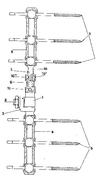
The apparatus illustrated m Figs. 1 to 4 is particularly adapted
to be employed in central fractures of long bones.
This comprises a pivot block 1 which is connected, ~y means of the
screw 2 to the bracket 3, with which is integral the tracked bar 4 carrying
the pins 5.
On the side opposite to the block 1 there is fixed the threaded
socket 6, on which in its turn there is screwed the threaded stud 7,
integral with the second tracked bar 8 carryLng the pins 9.
MIS/lcm 6
I 3 1 7 1 72
It will be seen that the screw 7 (Figs. 3 and 4) has an cvoidal
section in which the threads are limited to the two opposed sections 10 and
101, while on one of the twv curved s~faces, but not threaded, there rest
the headless threaded screw 10" and 10"' having a shaping of the head which
does not permit locking.
Two screws with mutually convergent axes guarantee alone an
optimNm lockmg, and the two opposite screws can be omitted when not needed.
Analogously the screw 11 guarantees an optLmum pressure on the
flat central non threaded portion of the socket 6, which in this manner
remains rigidly fix~d to the pivot block 1.
On the flat roughened surfaces of the carrier bars 4 and 8 there
are fixed, as has been mentioned, the respective pins 5 and 9, by means of
the screw 12 fixed to the respective bar by means of the nuts 13, by means
of the stirrup 14 and th~ washer 15, both provided with a layer of
deformable material, respectively 14' and 15', which co~es to bear on the
roughened surfaces 4 and 8 of the slotted portions of the carrier bars.
Ihe normal metallic washer 16 bears directly on the underside of
the nu~ 13, assuring a firm locking of the latter on the stirrup 14.
In Figs 6 and 7 it will be s~en that the series of pins 17 is
disposed in a plane which is pexpendicular with respect to the series of
pins 18, because this second form of c~nstruction of the apparatus is
intended for the stabilization of epiphytal osseous fractures.
In this case, the carrier bar of tracked construction is single
and is ~hown with the reference numeral 18. This bar is intended to carry
the pins 18 for insertion into the osseous diaphyses, while the pins 17
intended for insertion into osseous epiphyses are simply fixed to the
drilled shank 20, provided with a nut 21 and washer 22, ~lich locks the pin
17 to the socket 23 of the threaded portion 24 of the carrier bar, in its
MLS/lcm 7
1317172
turn fixed by ~he socket 25 and pivot 26, to the tracked portion 19 of the
carrier bar.
In tha first cæ e there ara provided simple channels, with parts
of circular section, form~d on the block 27, on the washex 28 and on the
block 29 dispcsed facing the pins 17. Ihe pins 18 c~re, on ~he contrary,
fixed to the tracked portion 19 oE the carrier bar, by means of bolts with
nuts, washers and stirrups, provided with a deformable coating, analogously
to what has been describ0d in the first form of construction of the
apparatus illustrated in Figs. 1 to 5.
Also in this case however, while the pins 17 are connected to the
pivoting element 26, by m~ans of the socket 25 of adjustable position, so as
to permit a lengthening of the distance betwaen epiphysis and diaphysis, the
tracked portion 19 is connected to the stirrup 30, which is m its turn
fixed by means of the nut 31 to the pivot 26, through the neck 32, of
curvilinear shape, the shaping of which is s~ch as to eliminate the
possibility of rotation which w~uld be extremely dangerous for the recovery
of the patient.
In Figs. 9 and 10 there are shown the points of insertion of the
pins 17, into the epiphysis 33, while the pins 18 penetrate into the
diaphysis 34 of the bone, the line of fracture of which is shown
schematically by the line 35.
In the case of fracture of a central portion of a long bone~ the
pins 5 and 9 (Fig. 10) are inserted at opposite parts with respect to the
line of intermediate fracture 36.
It is to be noted that the apparatus now described could be
usefully employed also for artificial lengthening of the joints, by neans of
periodical adjustment of the pcsition of the respective threaded sockets 6
and 25, which brings about the resultant increase of the distanoe between
MLS/lcm 8
- 1317172
the truncated bones and successive regrowths of the same, in accordance with
the by now well kncwn treatments in the Eield.
Naturally this will be facilitated by the presence of means of
adjustable 10", 10"' and 11 (Fig. 1~ which, when loosened, permit the
lengthening of the apparatus by simple r~tation of rotating neans or by
rotation in its turn of the socket 6 and successive stabilization for a
successive period of regrowth of the oss~cus callus.
Naturally, the constructive features of details of the apparatus,
such as for example the number of pins or the position thereof, the
dimensions of the varicus parts and the particular finish, can assume
various shapes and aspects with re~ Gt to those now described and
illustrated on the set of accompanying drawings, provided that the essential
features, set out in the successive claims, remain the same, without thereby
exceeding the scope of the inYention.
MIS/lcm g