Note : Les descriptions sont présentées dans la langue officielle dans laquelle elles ont été soumises.
2176619
ULTRASONIC BONE TESTING APPARATUS WITH REPEATABLE POSITIONING
AND REPEATABLE COUPLING
R~ QrND OF THE INVENTION
Field of the Invention
This invention relates to the field of ultrasonic analysis
of bone tissue in humans, and more particularly to an apparatus
using novel techniques for reproducibly measuring certain
properties of the heel bone or os calcis using transmission and
reflection of ultrasonic energy.
Description of Related Art
Certain known techniques for measuring properties of the
heel bone or os calcis have required that the foot is held
between a pair of ultrasonic transducers in a jig or clamp while
the foot and the ultrasonic transducers are immersed in a water
bath to couple the ultrasonic energy between the transducers and
the foot. These immersing procedures reduce interference with
the coupling of ultrasonic energy which is caused by air or other
gas present between the transducer and the object to be tested.
However, the techniques are relatively time consuming and can be
inconvenient.
Other previous designs of ultrasonic bone testing apparatus
include a support behind the leg in the calf muscle area. The
use of a footrest allows some tolerance for the positioning of
the foot. However, the approach has the disadvantage that the
footrest does not facilitate consistent measurement because the
size and location of calf muscles can vary greatly. In addition,
the patient's calf muscle tends to be flaccid while the patient
2176619
is sitting, and therefore does not provide a fixed reference
surface even for the same person during subsequent measurements.
OBJ~CT8 AND 8UMMARY OF THE INVENTION
Accordingly, it is an object of this invention to provide
an improved ultrasonic bone analysis apparatus.
Another object of this invention is to provide an ultrasonic
bone analysis apparatus which omits the water bath and replaces
the coupling function of the water with a system that includes
soft elastomer pads, delay lines, and a mechanism and controller
that causes the transducers to couple to the foot in the
desirable manner.
A further object of this invention is to provide an
ultrasonic bone analysis apparatus that achieves repeatable
results by employing both repeatable positioning and repeatable
coupling of the transducers with respect to the foot.
In one example of the invention, the repeatable positioning
of the foot is accomplished by a mechanism that locates certain
anatomical points of the lower leg and restrains motion during
the measurement process by using the located anatomical points.
The shin bone or tibia is used as one principal reference surface
for the lower leg. The tibia typically has only a thin and
uniform covering of skin in the anterior direction, no variable
muscle tissue, and provides a hard reference surface even on
fairly obese persons. The inferior aspect of the foot and the
posteria aspect of the os calcis, at the point just below the
lower attachment of the Achilles tendon, provide two other
reference surfaces for immobilizing the foot at a specified
21 7~fil 9
angle. In order to restrain the foot from lateral-medial
rotation, the foot instep is restrained and pressed down and to
the rear at an angle of about 55 degrees.
The repeatable coupling of the transducers to the foot can
be accomplished by controlling the pressure applied between the
transducer and the foot, and monitoring the quality of the signal
received by the transducer. The quality of the transducer signal
is used as feedback information to modulate the pressure applied
via a motor. An acoustical delay line is provided to allow the
transducer's wavefronts to evolve from the granular near field
pattern to a smoother far field pattern before entering the foot.
The acoustical and mechanical properties of the elastomer
coupling pad are inherently critical to the operation of the
inventlon .
BRIEF DE8CRIPTION OF THE DRA~ING8
FIG. 1 is a side view of a foot restraint device of the
present invention.
FIG. 2 is a perspective view of a foot well assembly of the
present invention.
FIG. 3 is a front view of a molded form in a shin guide
assembly of the present invention.
FIG. 4 is a top view of the molded form.
FIG. 5 is a side view of the molded form.
FIG. 6A, FIG. 6B, FIG. 6C, FIG. 6D and FIG. 6E are sectional
views of the molded form taken essentially on the lines A--A, B--
B, C--C, D--D and E--E, respectively, of FIG. 5.
FIG. 7 is a sectional view showing the interaction between
21 76b! 9
bridge brackets with channels of the foot well assembly and slide
blocks of an instep support guide.
FIG. 8 is a sectional view of a transducer drive mechanism
of the present invention.
FIG. 9A and FIG. 9B are front and side views of a position
encoder of the present invention.
FIG. 10 is a block diagram showing automatic positioning by
a transducer drive mechanism of the present invention.
FIG. llA and FIG. llB are front and side views of a
pad/delay unit of one embodiment of the present invention.
FIG. llC is a contour diagram of an end of the pad/delay
unit.
DFT~TT~ ~F8CRIPTION
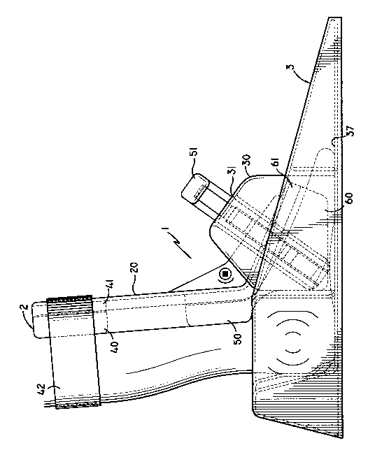
Referring to FIG. 1, an ultrasonic bone analysis apparatus
according to one embodiment of the invention combines the
mechanisms to position and restrain the foot and lower leg into
a single foot restraint device 1. The foot restraint device 1
comprises two assemblies, a shin guide assembly 2 and a foot well
assembly 3.
As seen in FIG. 2, the foot well assembly 3 comprises a box
cover 38 having a foot support 39, and foot well bottom 37. The
foot support 39 has an area slightly larger than a human foot
such that even a large foot can fit comfortably.
Transducer ports 36 are located on the sides of the foot
support 39, towards the rear. Bridge brackets 30 with channels
31 are located along the sides of the foot support 39, and are
arranged at a predefined angle, preferably 55 degrees, with
2 1 766 1 9
respect to the foot well bottom 37. The bridge brackets 30 with
channels 31 facilitate the mounting of the shin guide assembly
2.
Referring back to FIG. 1, the shin guide assembly 2 includes
a plastic molded form 20 lined with contoured foam lining 41.
The molded form 20 is a combination of restraints for the shin,
instep, and front of the foot into a single piece.
The molded form 20 includes shin restraint section 40 which
restrains, supports, and centers the tibia against contoured foam
lining 41 with the help of a flexible strap 42 placed around the
calf. The flexible strap 42 can be adjusted to secure the molded
form 20 comfortably around the patient's leg. The shin restraint
section 40 of the shin guide assembly 2 extends upward from an
instep support section 50 at an angle of about 95 degrees with
respect to the foot well bottom 37 of the foot well assembly 3.
FIG. 3 and FIG. 4 illustrate front and top views of the
molded form 20, respectively. The shin restraint section 40
tapers from an upper portion to a lower portion to adapt to the
tapering generally found in a human leg from the shin region to
the ankle region. For example, referring to FIG. 6A and FIG. 6B
which are cross-sectional views of the slices A--A and B--B in
FIG. 5, respectively, a cross-section of the shin restraint
section 40 near the upper portion has a greater radius than a
cross-section of the shin restraint section 40 near the lower
portion.
Referring again to FIG. 1, the front of the foot is
restrained from lateral rotation by the foot restraint section
2176b~9
60 extending from the lower part of the instep support section
50 towards the toes. As shown in FIG. 3, the foot restraint
section 60 has an inverted "U" or "V" shape and includes a
contoured foam lining 61 to properly center the front of the foot
as the molded form 20 is lowered to match up with the correct
width of the foot. The side wall of the foot restraint section
60 and the foot well bottom 37 form a predefined angle which is
preferably 60 degrees. Such an arrangement along with contoured
foam lining 61 facilitates a comfortable fit over both a large
foot 65 and a small foot 66, as shown in FIG. 6C, FIG. 6D and
FIG. 6E.
Referring again to FIG. 1, the instep support section 50
includes instep support guide 51. The instep support guide 51
is mounted on opposing sides of the molded form 20, and includes
slide blocks 21. The shin guide assembly 2 is attached to the
foot well assembly 3 by inserting slide blocks 21 into
corresponding channels 31 of the foot well assembly 3. The 55
degree angle of the channels 31 facilitates a proper contact
between the instep support guide 51 and the instep area of
different size feet, as well as sufficient differential vertical
displacement to allow the V-shape of the foot restraint section
60 to match and center varying widths of the lower foot.
Referring now to FIG. 7, the channels 31 are lined with
strips of repeating triangular ratchet teeth 32, facing downward.
The slide blocks 21 have matching ratchet teeth 22 facing upward.
When the slide blocks 21 are inserted into the respective
channels 31 of the respective bridge brackets 30, the ratcheting
action allows the slide block 21 to latch at one of multiple
2176~9
levels to the bridge brackets 30, and thereby the shin guide
assembly 2 can be adjusted to fit and restrain comfortably and
securely any size foot.
To facilitate release of the mating ratchet teeth 22 and 32
from each other, the ratchet teeth 22 are attached to leaf
springs 23 mounted to the base of the slide blocks 21. The
operator squeezes together two rigid brackets 24 attached to the
free ends of the springs 23, thus retracting the ratchet teeth
22. When the teeth 22 are clear of the teeth 32 inside the
channels 31, the operator can pull the slide blocks 21 out of the
channels 31 to allow the patient to remove her foot from the foot
well 3. The use of ratchet teeth 22 mounted into a spring
assembly 25 allows independent optimization of the materials used
to provide the spring action, and the materials used to provide
the sliding and ratchet action.
The shin guide assembly 2 is conveniently stored for
transport of the foot restraint device 1 by sliding the slide
blocks 21 into a lowest position in the channels 31.
Referring now to FIG. 8, one embodiment of a transducer
drive mechanism of the present invention includes a pair of
transducer assemblies 110. The transducer assembly 110 includes
transducer 101, acoustical delay line 109 and coupling pad 102.
The transducers 101 are mounted to respective carriages 103
that slide along a lateral-medial axis. Respective compression
springs 104 attached to the carriages 103 apply opposing lateral
forces towards the center of the foot. The carriage/spring
assembly is free floating and will center itself on the foot with
2 1 766 1 ~ ~
,, ;'
equal pressure on both sides.
An extension spring 105 applies the initial pressure when
the coupling pads 102 reach the patient's foot. To adjust the
pressure in small increments, a stepper motor with rack and
pinion mechanism 106 will move a finite number of steps and
compress the compression springs 104 that are attached to the
respective carriages 103. The compression springs 104 will pull
the respective transducers 101 and pads 102 inward at a force
proportional to the spring rate and distance translated.
The distance between the transducers 101 is continuously
measured by means of a position encoder 120 that is mechanically
linked to the motion of the transducers 101. Referring to FIG.
9A and FIG. 9B, front and side views of the position encoder 120,
respectively, a preferred encoder uses a code strip 121 mounted
onto one of the carriages 103 along with an optical encoder
reader 122 mounted on the other of the carriages 103. As the
distance between the transducers 101 changes, the code strip 121
moves between the slot of the optical encoder reader 122, and the
optical reader 122 reads lines 123 of the code strip 121 as the
lines 123 are traversed.
The transducer drive mechanism 100 automatically positions
transducer assemblies 110 against the patient's heel with
sufficient pressure to insure ultrasonic coupling. The automatic
positioning will be explained by referring to FIG. 10. Signals
received by the receiving transducer 101 are supplied to
controller 200. The controller 200 is preferably a
microprocessor-based controller having memory 201 (e.g. RAM and
ROM) for storing system and application software and input/output
21766tq
circuitry.
The controller 200 determines the quality of the signals
received by the receiving transducer 101 at least in part
according to the attenuation of the signals. The controller 200
controls the operations of the stepper motor 106 according to the
quality of the signals received by the receiving transducer 101
and positional data supplied by the position encoder 120. The
coupling pressure thereby is modified under control of the
controller 200 based on the quality of the signals received by
the receiving transducer 101. These steps are repeated by the
controller 200 until the signals received by the receiving
transducer 101 achieve a predetermined quality. Accordingly, the
transducer drive mechanism 100 under the control of the
controller 200 provides automatic positioning.
The controller 200 determines other parameters of interest,
including broadband ultrasound attenuation and bone velocity.
Also, the controller 200 calculates a speed of the ultrasonic
signals through the foot using the distance between the
transducers determined by the position encoder 120. An apparatus
for measuring bone characteristics by means of ultrasound is
well-known in the art. Such an apparatus is disclosed for
example in United States Patent 4,774,959 issued to Palmer et al.
on October 4, 1988 which is hereby incorporated by reference.
The controller 200 uses temperature readings from
temperature sensor 250 to improve the accuracy of the position
encoder measurements and correct for temperature dependent
inaccuracy in the ultrasound measurement. For example, the
controller 200 accounts for linear expansion of the encoder strip
21 76~T 5
121 by applying a temperature dependent term to the data supplied
by the position encoder 120. Additionally, the controller 200
applies a temperature dependent term to correct an estimation of
the time delay through the delay line 109 and the coupling pad
102. Furthermore, the controller 200 uses the temperature
reading to determine if the apparatus is operating within the
specified environmental range allowed, and if not, the operator
is informed that the apparatus is not ready to be used.
In addition, guided by operator input 300, the following are
examples of additional selectable functions provided by the
transducer drive mechanism 100 under the control of controller
200: (1) separate the transducers 101 to allow the foot to be
moved to and from a position between the transducers 101 without
interference from the transducers; (2) move the position encoder
120 to a known transducer separation zero; (3) extend the
transducers 101 to a cleaning or standby position; and (4) secure
the transducers 101 in an off or shipping position. The operator
input 300 can be any one of the conventional input devices such
as pre-allocated buttons, keyboard/keypad device, etc.
Several features of the coupling pads 102 are important to
the operation of the described invention. The acoustic impedance
of the material of the pads 102 is matched to the acoustic
impedance of human skin to provide a minimal loss of power and
reduce extraneous reflections. Preferably, the coupling pads are
elastomer coupling pads.
The coupling pads 102 also provide a waveguide function to
collimate the acoustic beam a sufficient distance along the
propagation axis to allow the wavefronts to evolve onto a more
21 7661 9
uniform intensity pattern. To this end, the acoustical delay
lines 109 are provided to allow the wavefronts to evolve from the
granular near field pattern to a smoother far field pattern
before entering the foot.
The pads 102 are chosen to have a durometer corresponding
to a sufficiently flexible waveguide that can partially conform
to the shape of a foot and provide some comfort to the patient.
The shape of the pads 102 conforms to the heel to eliminate any
gaps between the foot and pad. The surfaces of the pads 102
which contact the transducers 101, the delay line 109, or the
patent's skin is shaped at an angle to the propagation axis to
reduce the acoustic reflection at the pad-to-skin interface by
spreading the reflected energy over time and position.
The coupling pad 102 and the delay line 109 are preferably
integrated into a single pad/delay unit 150 to reduce an
extraneous reflection between a pad-to-delay-line interface.
FIG. llA and FIG. llB illustrate top and side vlews of the
pad/delay unit 150. The surface of the pad that contacts the
patient's skin is shaped to expel air bubbles from the contact
area when pressure is applied. ~IG. llC shows the contours of
the surface of the pad/delay unit 150 which contacts the
patient's skin. The surface preferably forms a 25 degree angle
with respect to a vertical axis.
The material of the coupling pad is required to be
compatible with coupling gel and non-irritating to the skin. One
preferred material is CIBA polyurethane (TDT 178-34) mixed with
an additive to provide a cured durometer of approximately 10 to
15 Shore A.
217661~
While the elastomer coupling pad is preferred, the coupling
pads may be a homogeneous material, a gel pad, or a liquid or
gel-filled bladder. The shape of the bladder may be conical
whereby air bubbles are expelled when the pad engages the heel.
In a known system, commercially available coupling gel is
commonly used between the skin and coupling pads. The
commercially available coupling gel is typically water-based.
While such water-based gels can be used, a non-aqueous jelly is
preferred in this invention. One implementation of the invention
uses petroleum jelly as a coupling gel.
The ultrasound coupling gel that is commonly used to
efficiently couple ultrasonic energy between the skin and
transducers also may be eliminated by using a self-wetting
material such as Parker Laboratory Aquaflex pads. In one
implementation of the design, self-wetting coupling pads can be
used as a disposable, or single use device, eliminating concerns
about sanitation.
Having described a preferred embodiment of the invention
with reference to the accompanying drawings, it is to be
understood that the invention is not limited to that precise
embodiment and that various changes and modifications thereof
could be effected by one skilled in the art without departing
from the spirit or scope of the novel concepts of the invention,
as defined in the appended claims.