Note : Les descriptions sont présentées dans la langue officielle dans laquelle elles ont été soumises.
WO 95/27438 "' ~ ~ ~'~ ' PCT/US95104175
,.,.
'",~,
MAGNETIC RESONANCE IMAGING
USING HYPERPOLARIZED NOBLE GASES
BACKGROUND OF THE INVENTION
The present invention relates generally to techniques of
nuclear magnetic resonance imaging. In particular, the
present invention relates to, among other things, the
detection and imaging of a noble gas by nuclear magnetic
resonance spectrometry.
Current views as to the molecular basis of anesthetic
action are mostly derived from experimental work carried out
'fin vitro. Interpretation of many of the results of these
studies are extremely controversial, e.g., changes in lipid
structure are observed at exceedingly high, indeed toxic,
concentrations of anesthetic. Changes observed 'fin vitro, from
animals whose physiology has been altered, or from animals
administered non-clinical doses of anesthetics might not
reflect the effects of these agents clinically. It is
believed that significant progress can be made by employing
direct non-invasive methods for the detection and
characterization of anesthetics in living animals. Both lipid
solubility and protein binding undoubtedly do play a role, but
new ideas are now needed.
Attempts have been made to bring powerful nuclear
magnetic resonance (N1~) techniques to bear on this problem.
(References 1-3). Wyrwicz and co-workers pioneered the use of
fluorine-19 (19F) NI~t spectroscopy to observe fluorinated
anesthetics in intact tissues and recorded the first I9F Nl~t
spectra from the brain of a live anesthetized rabbit.
(References 1, 4). These early studies demonstrated the
feasibility of studying the fate of anesthetics in live
mammals. Burt and collaborators also used halothane and other
fluorinated anesthetics for monitoring membrane alterations
1
~~~~~ao
WO 95/27438 " - - PCT/US95/04175
in tumors by 19F NMR. (References 5-6). In recent years,
several groups have conducted 19F NMR studies which have shed
light on the molecular environment of anesthetics in the
brains of rabbits and rats. (References 3, 7). Using a
surface coil placed on top of the calvarium during halothane
inhalation, two overlapping spectral features observed by
d'Avignon and coworkers, perhaps 0.1-0.2 ppm apart, could be
resolved through their different transverse relaxation times
(T2). (Reference 3). The biexponential dependence of the spin-
echo amplitude on echo delay reported in this study
demonstrated that anesthetics in different molecular
environments could be discerned in the brain in vivo using '9F
NMR. Such environments, separated by chemical shifts of only
about 0.1 ppm, had previously been reported by Wyrwicz et al.
in high resolution studies of excised neural tissue.
(Reference 4).
Notwithstanding such attempts to use other compounds for
NMR imaging, state-of-the-art biological magnetic resonance
imaging (MRI) has remained largely restricted to the water
proton, 1H20, NMR signal. The natural abundance of water
protons, about 80-100 M in tissue, and its large magnetic
moment make it ideal for most imaging applications. Despite
its tremendous value as a medical diagnostic tool, however,
proton MRI does suffer several limitations. Most notably, the
water protons in lung tissue, and the protons in lipids of all
interesting biological membranes, are notoriously NMR
invisible as a result of the short T2 in such environments.
(References 8-9). Other 1H signals and signals from other
biologically interesting nuclides are either present in too
low a concentration (10'3 to 10'' M, compared to ca. 100 M for
H20) or have undesirable NMR characteristics. In studying
dynamic processes with 'HZO, one must sacrif ice much of the
proton signal to exploit differences in effective spin density
resulting from T1 and/or TZ spatial variation. (Reference 10).
2
WO 95/27438 PCT/US95/04175
Various noble gases are known to be effective
anesthetic agents. For example, Xenon is approved for use in
humans, and its efficacy as a general anesthetic has been
shown. Attempts have previously been made to take advantage
of the properties of Xenon for purposes of medical imaging,
but success has heretofore been extremely limited, and
techniques have been impractical at best. For example, the
I~Xe isotope was used in early ventilation studies of the lung.
(References 11-12). Unfortunately, the poor image quality
attained limited its clinical use. Xenon has, however, been
used as a contrast enhancement agent in computed tomography
(CT) studies of the brain, (References 13-14), and as a tracer
for regional cerebral blood flow (rCBF) measurements.
(Reference 15).
An isotope of Xenon, Xenon-129 (~Z9Xe), has non-zero
nuclear spin (i.e., 1/2) and therefore is a nucleus which, in
principle, is suited to study by nuclear magnetic resonance
techniques. Despite the apparent potential for use of Xenon
in magnetic resonance imaging, its small magnetic moment, and
the low number densities of gas generally achievable, have
heretofore been insuperable obstacles to practicable magnetic
resonance (lit) imaging of ~29Xe at normal, equilibrium (also
known as "Boltzmann") polarizations, P (P-10-5 in 0.5-1.5 Tesla
(T) clinical imaging systems). However, unlike the water
proton ('H) employed as the nucleus in conventional NMR
techniques, the nuclear magnetic resonance signals obtainable
from iz9Xe are extraordinarily sensitive to local environment
and therefore very specific to environment.
Certain aspects of the behavior of ~~Xe, and other
noble gas isotopes having nuclear spin, in various
environments have been studied and described. For example,
Albert et al. have studied the chemical shift and transverse
and longitudinal relaxation times of Boltzmann polarized 129Xe
. ~ in several chemical solutions. (Reference 16). Albert et al.
and others have also shown that Oxygen can affect longitudinal
3
WO 95/27438 PCT/US95/04175
relaxation time Tl of '29Xe. (References 17-18) . Miller et a~,_
.;
have also studied the chemical.: shits of 'Z9Xe and '3'Xe in
solvents, proteins, and memb'r~anes. (Reference 2). However,
none of these publications provides any indication of a method
by which '~Xe could be used for nuclear magnetic resonance
imaging.
It is known in the art that the polarization of
certain nuclei, such as noble gas nuclei having nuclear spin,
may be enhanced over the equilibrium or Boltzmann
polarization, i.e., hyperpolarized. Such techniques include
spin exchange with an optically pumped alkali metal vapor and
metastability exchange.
The physical principles underlying the
hyperpolarization of noble gases have been studied.
(Reference 19). For example, Happer et al. have studied the
physics of spin exchange between noble gas atoms, such as
Xenon, with alkali metals, such as Rubidium. (Reference 20).
Others have studied spin exchange between Helium and alkali
metals. (References 21-22, 49). Other publications have
described physical aspects of spin exchange between alkali
metals and noble gases. (References 23-24). The technique of
using metastability exchange to hyperpolarize noble gases has
been studied by Schearer et al. and by Hadeishi et al.
(References 26-31).
Other publications, by Gates et al. and
Gatzke et al., describe certain behaviors of frozen,
crystalline 129Xe that has been hyperpolarized. (References 32-
33). Gates et al. and others describe spin-exchange rates
between Rubidium and 'z9Xe at high Xenon pressures as measured
by magnetic resonance apparatus. (References 34-35). These
publications, however, relate to 'Z9Xe behavior in highly
controlled physical systems and provide no description
concerning how '29Xe might be used to produce images by nuclear
magnetic resonance.
4
WO 95/27438 PCT/US95/04175
- Raftery ~t al. have described optically pumped '29Xe
as an adsorption probe for the study of surface structure by
analysis of NI~2 spectra. (References 36-37). Long et al. have
also observed the chemical shift of laser polarized Xenon
adsorbed to a polymer surface. (Reference 38).
U.S. Patent Nos. 4,856,511 and 4,775,522 to Clark
describe a nuclear magnetic resonance technique for detecting
certain dissolved gases in an animal subject. Gas
compositions described as useful for this technique include
fluorine compounds such as perfluorocarbons. Other gases
suggested to be potentially useful for the technique of Clark
include 129Xe, but Clark fails to recognize any of the
difficulties which have heretofore rendered use of l~Xe for
magnetic resonance imaging of biological subjects
impracticable.
Therefore, it would be a significant advance in the
art to overcome the above-described difficulties and
disadvantages associated with nuclear magnetic resonance
imaging, in a manner which would permit the imaging of noble
gases, especially the imaging of noble gases in biological
systems, without requiring excessively long image acquisition
times and without being limited to systems and environments
previously imageable only by 'H NMR.
SUMMARY OF THE INVENTION
In accordance with the present invention, there is
provided a method of performing nuclear magnetic resonance
imaging which includes detecting the spatial distribution of
at least one noble gas by nuclear magnetic resonance (NMR),
and generating a representation of the noble gas spatial
distribution.
5
~1~~'~~b
WO 95/27438 PCT/US95/04175
In a preferred embodiment, there is also provided a -
method of performing nuclear magnetic resonance imaging of an
animal or human subject by administering an imageable amount
of at least one noble gas to the subject, employing an NMR
imaging spectrometer to detect radio-frequency signals derived
from the magnetic resonance of at least one noble gas,
processing the detected signals to obtain an NMR parameter
data set as a function of the spatial distribution of at least
one noble gas, and further processing the data set to generate
a representation of at least one dimension of the spatial
distribution of at least one noble gas.
In another preferred embodiment, the method of the
invention further includes detecting and imaging at least one
hyperpolarized noble gas. The hyperpolarized noble gas is
preferably hyperpolarized by laser polarization through spin
exchange with an alkali metal or by metastability exchange.
The noble gas is preferably selected from among Helium-3,
Neon-21, Krypton-83, Xenon-129, Xenon-131 and mixtures
thereof. Most preferably, the noble gas is Helium-3 or Xenon-
129. Combinations of noble gases and/or noble gas isotopes
are contemplated, as are combinations of hyperpolarized and
non-hyperpolarized noble gases and/or noble gas isotopes.
When the noble gas is laser polarized through spin exchange
with an alkali metal, preferably an alkali metal selected from
among Sodium-23, Potassium-39, Cesium-133, Rubidium-85, and
Rubidium-87. Most preferably, the alkali metal is Rubidium-85
or Rubidium-87.
The method of the invention preferably includes
detecting and imaging at least one physical dimension of the
spatial distribution of at least one noble gas, more
preferably including detecting and imaging two or three
physical dimensions. The method of the invention may also
include detecting and imaging alterations of the spatial
distribution of the noble gas as a function of time.
6
WO 95/27438 PCT/US95/04175
The generating of a representation of a noble gas
preferably includes generating a representation of at least
one physical dimension of the spatial distribution of the
noble gas, more preferably including generating a
representation of two or three physical dimensions of the
noble gas. The generating of the representation may also
include generating a representation of one or more physical
dimensions of the spatial distribution of the noble gas as a
function of time, including such N1~2 parameters as chemical
shift, T1 relaxation, T2 relaxation, and TlP relaxation.
Preferably, the method of the invention includes generating a
visual representation.
The noble gas being imaged is preferably distributed
spatially in at least one physical phase such as a gas,
liquid, gel, or solid. The noble gas may be imaged as
distributed in two or more physical phases in one sample. The
noble gas being imaged may be distributed on a solid surface.
The noble gas may be imaged in association with various
materials or environments.
The sample being imaged using a noble gas may include
an in vitro chemical, ~ vitro biological or in vivo
biological, system. When the noble gas distribution in an in
vivo biological system is imaged, the system may include one
or more human or animal subjects. The noble gas is preferably
distributed in an organ or body system of the human or animal
subject. Alternatively, the noble gas may be distributed in
an anatomical space of the subject.
In another embodiment of the invention, there is
provided a medical composition which includes a medically
acceptable bifunctional gas effective for in vivo
p anesthesiological and nuclear magnetic resonance imaging
functions. In a preferred embodiment, the gas composition
includes at least one noble gas, preferably selected from
among Helium-3~, Neon-21, Krypton-83, Xenon-129, and Xenon-131.
7
WO 95/27438 PCTIUS95/04175
More preferably, the noble gas is Helium-3 or Xenon-129. Thc..
noble gas is preferably hyperpolarized, more preferably
through spin exchange with an alkali metal or through
metastability exchange. Combinations of hyperpolarized and
non-hyperpolarized noble gases and noble gas isotopes are
possible.
Also in accordance with the present invention, there
is provided apparatus for nuclear magnetic resonance imaging
which includes NMR imaging means, for detecting and imaging at
least one noble gas, and means for providing imageable
quantities of the noble gas. In a preferred embodiment, the
apparatus includes means for providing imageable quantities of
a hyperpolarized noble gas. The apparatus of this embodiment
includes hyperpolarizing means, preferably means for
hyperpolarizing a noble gas through spin exchange with an
alkali metal or through metastability exchange. The noble gas
may be provided in continuous, discontinuous, and/or quasi-
continuous mode, and when more than one noble gas is provided,
noble gases may be provided as a mixture or individually, and
may be provided either together or by separate routes and/or
at separate times and durations.
The noble gas may be contacted with the sample to be
imaged in gaseous, liquid, or solid form, either alone or in
combination with one or more other components in a gaseous,
liquid, or solid composition. The noble gas may be combined
with other noble gases and/or other inert or active
components. The noble gas may be delivered as one or more
boluses or by continuous or quasi-continuous delivery.
Also in accordance with the invention there is
provided a method of performing nuclear magnetic resonance
imaging of a human or animal subject. In this embodiment, the
method includes administering to a subject an imageable amount
of a hyperpolarized noble gas, generating radio-frequency
signals from the nuclear magnetic resonance of the
8
2~.~~~
WO 95/27438 PCT/US95/04175
- hyperpolarized noble gas by means of a nuclear magnetic
resonance imaging spectrometer, detecting the generated radio-
frequency signals, processing the detected radio-frequency
signals to derive a nuclear magnetic resonance parameter data
set as a function of a spatial distribution of the
~' i
hyperpolarized noble gas in the subject, and further
processing said nuclear magnetic resonance parameter data set
to derive a representation corresponding to at least one
spatial dimension of the spatial distribution of the
hyperpolarized noble gas in the subject.
The noble gas may be administered to a human or
animal subject as a gas or in a liquid, either alone or in
combination with other noble gases and/or other inert or
active components. The noble gas may be administered as a gas
by either passive or active inhalation or by direct injection
into an anatomical space such as lung or gastrointestinal
tract. The noble gas may be administered as a liquid by
enteral or parenteral injection. The preferred method of
parenteral administration includes intravenous administration,
optionally by contacting blood with the noble gas
extracorporeally and reintroducing the noble gas-contacted
blood by intravenous means.
The present invention solves the disadvantages
inherent in the prior art by providing a method for imaging at
least one noble gas by nuclear magnetic resonance. The
present method provides a new and unexpectedly powerful method
of Nl~t imaging of noble gas spatial and temporal distribution
in non-biological as well as in in v'tro and in v'vo
biological systems. The present invention also permits the
acquisition of images of high signal to noise ratio, in
unexpectedly short acquisition periods. In addition, the
present invention provides a method for imaging biological
phenomena of short duration as well as for imaging systems
previously not amenable to imaging by conventional 'H NMR
techniques.
9
WO 95127438 7 PCT/US95/04175
Other objects and advantages of the present inventic.._
will become more fully apparent from the following disclosure,
figures, and appended claims.
BRIEF DESCRIPTION OF THE DRAWINGS
Figure la shows a nuclear magnetic resonance spectrum
of 'Z9Xe in a rat brain synaptosome suspension; Figure ib shows
a nuclear magnetic resonance spectrum of '29Xe in a homogenate
of rat brain tissue; Figure lc shows a nuclear magnetic
resonance spectrum of '29Xe in a whole rat brain preparation.
l0 Figure 2a shows a graphical representation of a glass
sphere 20 mm in diameter; and Figure 2b shows a nuclear
magnetic resonance image of Boltzmann polarized '29Xe gas in a
20 mm diameter glass sphere.
Figure 3a shows a graphical representation of a glass
sphere, 20 mm in diameter, containing octanol (shaded region)
and Xenon gas (unshaded region); Figure 3b shows a nuclear
magnetic resonance spectrum illustrating NNBt signals obtained
from '~Xe in gas phase and in octanol; Figure 3c shows a
nuclear magnetic resonance image of '~Xe in octanol in 20 mm
glass sphere; and Figure 3d shows a nuclear magnetic resonance
image of '29Xe in gas phase in a 20 mm glass sphere.
Figure 4 shows a series of nuclear magnetic resonance
images of a hyperpolarized '~Xe gas phantom, representing
different mutually parallel planes.
Figures 5a-5c show a sequence of nuclear magnetic
resonance images of a mouse lung inflated with hyperpolarized
'29Xe gas; and Figure 5d shows a nuclear magnetic resonance
image of 'H in a mouse heart.
CA 02183740 2001-07-17
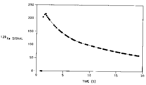
Figure 6 shows a graph illustrating a decrease in '29Xe magnetic
resonance signal intensity, obtained from mouse lung inflated by
hyperpolarized 'z9Xe, as a function of time.
Figure 7 shows a longitudinal section view of a noble gas delivery device
for nuclE~ar magnetic rE~sonance imaging of noble gases.
DETAILED DESCRIPTION OF THE PREFERRED EMBODIMENTS
Nuclear magnetic resonance spectroscopy is a technique which is well
known inn a wide variety of scientific disciplines. Basic considerations
regarding the conventional practice of nuclear magnetic resonance imaging,
especially as applied i:o biological systems, are found in Rinck et al., An
Introduction to Magnetic Resonance in Medicine (1990), especially Chapters
1-4 (Reference 39); and Wehrli, F.W., "Principles of Magnetic Resonance",
Chapter 1, and Wood, M.L., "Fourier Imaging", Chapter 2, in Magnetic
Resonance Imaging, Vol. 1, 2d ed., Stark et al., eds. (1992) (Reference 40).
In certain disciplines, an adaptation of NMR spectroscopy, involving the
generation of images from NMR data has found increasing popularity. In
medicine, certain MF;I techniques have become fairly commonplace,
principally employing the water proton ('H20) for the imaging of certain
regions in the body.
Nonetheless, certain other magnetically susceptible nuclei are desired
to be adapted for MRI for various reasons. In particular, the physical
characteristics of other' elements may predispose the nuclei to the imaging
of other kinds of physical and biological systems. In medicine, other nuclei
are desired which Can enable the imaging of regions of the body which are
difficultly accessible by currently available NMR probes. Prior to the
unexpected observations of the
-11-
~~ ~3'~4p
WO 95/27438 PCT/US95/04175
utility of noble gases for I~tI applications, as described
herein, acceptable alternative nuclear probes have been
unavailable. ~'
Noble gas isotopes having non-zero nuclear spin have
now been discovered to offer vast possibilities for use in
I~tI. For example, the l~Xe isotope is, in principle, suited to
NI~t uses, but is 26% naturally abundant and has a sensitivity
relative to 1H (in conventional NMR) of 2.12 x 10'2. The
resonance frequency of 129Xe spans an enormous range (0-30o ppm)
over the gas and condensed phase, and is exceptionally
sensitive to chemical environment. (Reference 2). Its
longitudinal relaxation time, T" is huge (practically at least
3000 s in the pure gas phase, and theoretically perhaps as
long as 56 hrs at 1 atm), (References 32, 41), and is
particularly sensitive to chemical environment, OZ
concentration, (References 17-18), and the effects of other
relaxation promoters. (References 2, 42, 16). Its transverse
relaxation time is also susceptible to relaxation promoters.
(References 16, 18, 43).
The longitudinal and transverse relaxation times, T1
and T2, respectively, are also indicative of the environment
surrounding the 129Xe atom, e.g., whether the atom is bound to a
protein, dissolved in a lipid, or constrained in some other
way. Thus a combination of chemical shift, T1, and TZ data can
provide a basis for distinguishing the presence or absence of
the nucleus in a particular environment as well as for
identifying the nature of the environment in question.
Elemental Xenon is a benign and effective anesthetic,
(Reference 44), which is not metabolized by the body. Xenon
has an essentially Raoult's Law solubility in non-polar
solvents. (Reference 45). Inhaled into the lungs, Xenon
equilibrates quickly with the pulmonary circulation, reaching
a steady state with the entire blood volume in one blood
circuit, (Reference 13), on average, about 1 s or 1-2 breaths
12
WO 95/27438 PCT/US95/04175
- in the mouse, about 18 s in humans. (Reference 46). Xenon is
known to accumulate rapidly in highly-vascularized tissue.
For example, in the brain, which contains 10% lipid and 10%
protein, (Reference 10), one can expect steady-state
concentrations (for 0.5-1.0 atm lung Xenon) of 5-10 mM in the
membrane bilayers, 2-4 mM in water, and about 1-5 mM bound to
proteins. (References 45, 47-48). Xenon is also approximately
twice as soluble in white matter as in gray matter.
(Reference 13 ) . The Nl~t resonance frequency of 129Xe is
different in each of the above sites, and exchange between
compartments is slow on the chemical shift Nl~t timescale.
(References 2, 16-17). The potential usefulness of
hyperpolarized 'Z9Xe as a contrast agent in biological systems
is therefore apparent.
The total Xenon concentration in materials of
biological interest will typically range between about two and
about five times its solubility in pure water. The problem
with any attempt to image Boltzmann polarized Xenon in such a
system is that many samples are required in order to determine
a solution parameter. These difficulties stem in large part
from the lower concentrations of I~Xe, its smaller magnetic
moment, and its lower natural abundance, as compared with 1H20.
Similar considerations apply with regard to other noble gases
which are generally less soluble in water as well as in non-
polar media.
For example, the spectrum of Figure lc, obtained in 8
hrs from in vitro brain samples, taken from rats anesthetized
with Xenon gas, has significantly less signal to noise (S/N)
than a spectrum of 'Z9Xe in a synaptosomal suspension shown in
Fig. la, obtained in 27 hrs under a Xe pressure of 3 atm.
The difficulties which have heretofore prevented the
development of noble gas MRI are clear: typically long
'longitudinal relaxation times and low signal strength require
signal averaging of exceedingly many free induction decays
13
WO 95/27438 PCT/US95/04175
(FIDs) over long periods of time..- It is clear that to condw_
in vivo NMR experiments, extraordinary enhancement of the
noble gas signal is necessary. The total accumulation times
for Boltzmann noble gas spectra is prohibitively long in such
biological samples.
The ability to use noble gases for Nl~t imaging, then,
is directly and profoundly limited by the average signal
intensity and the signal acquisition ability of the
spectrometer. Given current NI~t spectrometer technology, it
is reasonable to conclude that on the order of a 10,000 fold
increase in sensitivity, e.g., that increase necessary to
render Xenon imaging possible using Boltzmann polarized Xenon,
could take years if not decades to develop, assuming it is
feasible at all. The required sensitivity increase is more
practicably attained through hyperpolarization, for example,
through the use of optical pumping and spin exchange,
(References 21-22, 32, 36), or metastability exchange.
(References 26-31). This method of enhancing the noble gas
signal can be used to create noble gas nuclear polarizations
which are on the order of 104 - 106 larger than typical thermal
equilibrium polarizations. Nuclear polarizations attained
using these techniques are easily of order 0.25, (Reference
32), and can approach 1.0, making the product of spin density
and polarization at least an order of magnitude larger than
for the proton ('H) in typical imaging situations. Thus, it
has now been unexpectedly found that the hyperpolarization of
noble gases permits a spectacular new means of producing
magnetic resonance images.
while the extraordinary property of
hyperpolarizability of noble gases, especially '29Xe and ~Ie, is
of great importance in rendering the imaging of biological
systems possible, other factors play a role in developing such
images. For example, noble gases exhibit other unusual
properties, including distinctly different behavior compared
to 'H20 in (a) cell and tissue compartmentalization; (b)
14
WO 95/27438 ~ PCT/US95/04175
__dramatically time-dependent distribution; and (c) response of
resonance frequency, T1, and TZ to environment, Oz concentration
and subcellular exchange kinetics. The combination of
hyperpolarizability of noble gases and these other unusual
properties enables the use of noble gases as a new and
qualitatively different source of NMR image contrast. For
example, as opposed to water protons, t~Xe is not omnipresent;
its space and time distribution in the body depends entirely
on the anatomy and physiology of Xenon transport. (Reference
l0 13). This permits its use in magnetic resonance imaging (MRI)
and magnetic resonance spectrometry (MRS) studies of soft-
tissue anatomy, physiology (e. g., cerebral blood flow,
cerebral activity) and pathology (e. g., demyelination, early
detection of tumors or other foci of changed or anomalous
metabolism). Moreover, the large MR signal strengths
obtainable using hyperpolarized noble gases permit the use of
the high-speed imaging protocols, which have heretofore been
possible only with 1H20.
The imaging method of the invention is preferably
performed using the l~Xe and/or the 3He nuclei. However, the
method of the invention may also be performed with other noble
gases, i.e., other noble gas isotopes having nuclear spin.
3He, '29Xe and the other noble gases may be preferred in
different applications because of their different physical and
magnetic resonance properties. A list of noble gas nuclei
useful according to the invention is provided below in Table
I. This list is intended to be illustrative and non-limiting.
WO 95!27438 PCT/US95/04175
TABLE I
H~~perpolarizable f~oble Gases
Naturai~ Nuclear
Isotope Abundance (%) Spin
3He ~ 10~ 1/2
ziNe 0 . 2 7 3 / 2
SKr 11.5 g/2
iz9Xe 2 6 . 4 1 / 2
~3iXe 21. 2 3 / 2
While each of the noble gas isotopes listed in Table I,
alone or in combination, may be used for nuclear magnetic
resonance imaging according to the invention, it is known that
the degree of polarization of the gases in equilibrium
(Boltzmann) state is prohibitively low, preventing high speed
image acquisition. The various parameters governing signal
decay such as T1 and Tz relaxation and the local environment of
the nucleus will also determine whether high speed images can
be effectively acquired. These limitations become of great
importance in acquisition of images from in vitro and in vivo
biological systems since the time course of events desired to
be imaged often requires data acquisition periods of less than
one second. Enhancement of the NI~t signal is, therefore,
highly desirable. Accordingly, the noble gas is preferably
hyperpolarized relative to its normal Boltzmann polarization.
Such hyperpolarization is preferably induced prior to data
acquisition by an NMR spectrometer and may be induced by any
of the techniques known in the art.
Further enhancement of the noble gas magnetic resonance
signal may be obtained, independently of, or together with,
hyperpolarization, by increasing the proportion of the
16
WO 95/27438 PCTIUS95/04175
imageable isotope in each noble gas to a level above the
natural abundance of such imageable isotopes in the noble gas.
In the case of '29Xe, which has a natural isotopic abundance of
about 26%, this amounts to enhancement by no more than a
factor of four, even in a gas which is enriched to 100% ~29Xe.
Other considerations, such as the hyperpolarizability of the
noble gas, usually play a much larger role in signal
enhancement, but isotopic enrichment can provide a significant
contribution to the ultimate efficacy of the present
invention. This is especially true in the case of 3He which
has a natural abundance of on the order of 10'x. Even the
hyperpolarizability of 3Iie and its very large magnetic
resonance signal could be considerably offset by the low
natural abundance of this isotope. Despite its low natural
abundance, however, 3He is readily available in very pure form
as a result of industrial use of tritium (3H), which decays
exclusively to 3He. The ready availability of artificial
sources of 3He eliminates concerns regarding its low natural
abundance and associated expensive enrichment processes.
Noble gases may be hyperpolarized for use according to the
invention through any of various means known in the art, such
as spin-exchange interactions with optically pumped alkali
metal vapor. (References 34-35, 49-50). The optical pumping
and spin-exchange can be performed in the absence of an
applied magnetic field, but is preferably performed using
modest fields of about 1 G or larger. Pumping in the Nl~t
magnet bore at fields of several Tesla is also possible. The
maximum steady state ~~Xe nuclear polarization achievable
depends on the time constant characterizing the spin exchange
with the alkali metal and the time constant characterizing the
relaxation (T1) due, for example, to contact with the surfaces
of the pumping cell. For instance, with T~ ~ 20 min,
polarizations of 20-40% are quite practicable, (Reference 32),
and polarizations of 90% or more should be attainable. The
long T1 of the gas also allows samples to be manipulated, even
stored as Xe ice, (Reference 32), and transported on time
17
WO 95/27438 PCT/US95/04175
scales of hours or even days, without serious loss of
magnetization.
The art of hyperpolarizing.rioble gases through spin
exchange with an optically pumped alkali-metal vapor starts
with the irradiation of the alkali-metal vapor with circularly
polarized light at the wavelength of the first principal (D1)
resonance of the alkali metal (e. g. 795 nm for Rb). The ZSIn
ground state atoms are thus excited to the 2P1~ state and
subsequently decay back to the ground state. If performed in
a modest (10 Gauss) magnetic field aligned along the axis of
incident D1 light, this cycling of atoms between the ground and
first excited states leads to nearly 100% polarization of the
atoms in a few microseconds. This polarization is carried
mostly by the lone valence electron characteristic of all
alkali metals; this essentially means that all of these
electrons have their spin either aligned or anti-aligned to
the magnetic field depending upon the helicity (right- or
left-handed circular polarization state) of the pumping light.
If a noble gas with non-zero nuclear spin is also present, the
alkali-metal atoms can undergo collisions With the noble gas
atoms in which the polarization of the valence electrons is
transferred to the noble-gas nuclei through a mutual spin
flip. This spin exchange results from the Fermi-contact
hyperfine interaction between the electron and the noble-gas
nucleus. By maintaining the alkali-metal polarization at
nearly 100% with the pumping light, large non-equilibrium
polarizations (5% - 80%) are currently achievable in large
quantities of a variety of noble gases through this spin-
exchange process. For example, one currently available
Titanium: Sapphire-laser could theoretically provide 1 g/hr
(200 cc-atm/hr) of highly polarized '29Xe.
The alkali metals capable of acting as spin exchange
partners in optically pumped systems include any of the alkali
metals. Preferred alkali metals for this hyperpolarization
technique include Sodium-23, Potassium-39, Rubidium-85,
18
WO 95/27438 PCT/US95/04175
Rubidium-87, and Cesium-133. Alkali metal isotopes, useful
according to the invention, and their relative abundance and
nuclear spins are listed in Table II, below. This list is
intended to be illustrative and non-limiting.
TABLE II
Alkali Metals Capable of Spin Exchange
Natural Nuclear
Isotope Abundance (%) Spin
~Na 100 3/2
93.3 3/2
~Rb 72.2 5/2
s~~ 2 7 . 8 3 / 2
~33Cs 100 7 / 2
Alternatively, the noble gas may be hyperpolarized using
metastability exchange. (References 28, 51). The technique
of metastability exchange involves direct optical pumping of,
for example, 3He, without need for an alkali metal
intermediary. The method of metastability exchange usually
involves the excitation of ground state 3He atoms (l~So) to a
metastable state (2351) by weak radio frequency discharge. The
23S1 atoms are then optically pumped using circularly polarized
light having a wavelength of 1.08 ~,m in the case of 3He. The
light drives transitions up to the 23P states, producing high
polarizations in the metastable state to which the 23P atoms
. then decay. The polarization of the 23S1 states is rapidly
transferred to the ground state through metastability exchange
collisions between metastable and ground state atoms.
Metastability exchange optical pumping will work in the same
19
2t8~'~4~
WO 95/27438 PCT/US95/04175
low magnetic fields in which spin exchange pumping works.
Similar polarizations are achievable, but generally at lower
pressures, e.g., about 0-10 Torr.
The method of the invention preferably includes detecting
anal imaging at least one physical dimension of the spatial
distribution of at least one noble gas, more preferably
including detecting and imaging two or three physical
dimensions. The method of the invention may also include
detecting and imaging alterations in the spatial distribution
of the noble gas as a function of time.
The generating of a representation of a noble gas
preferably includes generating a representation of at least
one physical dimension of the spatial distribution of the
noble gas, more preferably including generating a
representation of two or three physical dimensions of the
noble gas. The generating of the representation may also
include generating a representation of one or more physical
dimensions of the spatial distribution of the noble gas as a
function of time, including such NNHt parameters as chemical
shift, T1 relaxation, TZ relaxation and TlP relaxation.
Preferably, the method of the invention includes generating a
visual representation.
Representations of the spatial distribution of a noble gas
may be generated by any of the methods known in the art,
subject to the type of information desired to be represented.
These techniques employ various means for collecting and
manipulating nuclear magnetic resonance data for the-
generation of images. Such methods are described in the
literature available in the art and include, without
limitation, Fourier imaging, planar imaging, echo planar
imaging, projection-reconstruction imaging, spin-warp Fourier
imaging, gradient recalled acquisition in the steady state
(GRASS) imaging also known as fast low angle shot (FLASH)
imaging, and hybrid imaging.
CA 02183740 2001-07-17
Such imaging methods are described in, for example, Ernst et al.,
Principles of Nuclear IVlagnetic Resonance in One and Two Dimensions
(1987) (Reference 52), particularly Chapter 10, "Nuclear Magnetic
Resonance Imaging", pages 539-564; Shaw, D.D., "The Fundamental
Principles of Nuclear Magnetic Resonance", Chapter 1 in Biomedical
Magnetic Resonance Ilma in , S.W. Wehrli et al. eds. (1988) (Reference
53); and Stark et al. e~ds., Magnetic Resonance Imaging, Vol. 1, 2d ed.
(1992) (Reference 40).
The selection of imaging method will depend on the behavior of the noble
gas nucleus under investigation, the nature of the sample and the degree of
interaction of the nucleus with the sample. The selection of imaging method
will also depend on whether one or more spatial dimensions of the spatial
distribution of the noble gas is desired to be represented and whether a
temporal or time-dependent dimension is desired to be represented. When
a multidimensional representation is desired such representation may be
generated by, for example, multi-slice imaging or volume imaging.
It is generally preferred that the image or representation be generated by
a method which is as fast and as sensitive as possible. Preferred imaging
methods include the FLASH or GRASS imaging method and the echo-planar
imaging (EPI) method. These methods are preferred for their capacity to
generate images through fast data acquisition, thereby conserving
polarization of the noble gas. EPI is especially preferred because it is a
relatively fast method and requires only one radio-frequency (RF) pulse per
image. It thus permits maximum utilization of the available polarization.
These preferred methods also permit fast time resolution of time-dependent
phenomena in human and animal subjects. Such applications include, for
example, magnetic resonance angiography (MRA) studies, functional
imaging of the nervous system (e.g.,
-21 -
WO 95/27438 PCT/US95/04175
brain), as well as studies of variations in cardiopulmonary
and circulatory physiological states.
The nuclear magnetic resonance imaging method of the
invention also includes the registration of multiple imaging
modalities. For example, using coils tunable to l~Xe
frequencies and the frequencies of one or more other magnetic
probes permits enhanced data interpretation. Such combined
multiple imaging approaches would include, for example the
combined imaging of '29Xe with 'H, and the imaging of more than
one noble gas, such as imaging of ~~Xe with 3He. In this
embodiment, geometric image registry and overlay are possible,
including the generation of false-color images, in which
distinct colors would represent distinct probes. Image
subtraction techniques would also be possible using
combinations of l~Xe with other probes, or combinations of
noble gas probes.
The noble gas being imaged is preferably distributed
spatially in at least one physical phase such as a gas,
liquid, gel, or solid. The noble gas may be imaged as
distributed in two or more physical phases in one sample. The
noble gas being imaged may be distributed on a solid surface.
The noble gas may be imaged in association with various
materials or environments such as, without limitation,
zeolites, xenon clathrates, xenon hydrates, and polymers.
The sample being imaged using a noble gas may include an
in vitro chemical, in vitro biological or in vivo biological,
system. When the noble gas distribution in an in vivo
biological system is imaged, the system may include one or
more human or animal subjects. The noble gas is preferably
distributed in an organ or body system of the human or animal
subject, including, without limitation, lung tissue, nervous
tissue, brain tissue, gastrointestinal tissue or
cardiovascular tissue or combinations thereof. Alternatively,
the noble gas~may be distributed in an anatomical space such
22
21 ~ 3 ~~ p
WO 95/27438 PCT/US95l04175
as, without limitation, lung space, gastrointestinal tract
space, peritoneal space, bladder space or combinations
thereof .
The noble gas may be contacted with the sample to be
imaged in gaseous or liquid form, either alone or in
combination with other components in a gaseous or liquid
composition. The noble gas may be combined with other noble
gases and/or other inert or active components. The noble gas
may be delivered as one or more boluses or by continuous or
quasi-continuous delivery.
In a preferred embodiment, there is also provided a method
of performing nuclear magnetic resonance imaging of an animal
or human subject by administering an imageable amount of a
hyperpolarized noble gas to the subject, employing an Nl~t
spectrometer to generate and detect radio-frequency signals
derived from the magnetic resonance of the noble gas,
processing the detected signals to obtain an Nl~t parameter
data set as a function of the spatial distribution of the
noble gas, and further processing the data set to generate a
representation corresponding to at least one dimension of the
spatial distribution of the noble gas.
The noble gas may be administered to a human or animal
subject as a gas or as a liquid, either alone or in
combination with other noble gases and/or other inert or
active components. The noble gas may be administered as a .gas
by either passive or active inhalation or by direct injection
into an anatomical space such as lung or gastrointestinal
tract. The noble gas may be administered as a liquid by
enteral or parenteral injection. The preferred method of
parenteral administration includes intravenous administration,
optionally by contacting blood with the noble gas
extracorporeally and reintroducing the noble gas-contacted
blood by intravenous means.
23
WO 95/27438 PCT/US95104175
The cost of a purified noble gas tends to be relatively
high as compared to the cost of common gases such as nitrogen
or carbon dioxide. The cost is especially high in the case of
Xenon which has been enriched to, for example, 70% 'z9Xe.
However, being inert, the noble gas is not metabolized in
biological systems and can be recovered. For example, Xenon
can be recovered from the exhaled breath of human subjects
over about a 20 minute period. Such apparatus for noble gas
recovery and repurification would include, for example, a cold
trap and/or a zirconium getter apparatus, such as are known in
the art. Other apparatus for recovery of noble gases may be
employed.
It is preferred that, because of the high cost of the
noble gas, the gas be maintained in a system which is
substantially sealed to prevent loss to the atmosphere.
Sealed containment apparatus would include a noble gas source,
such as a gas canister or compressed gas tank, conduits to and
away from a sample, as well as recovery apparatus.
The noble gas source may include a permanent or semi-
permanent canister or pressurized containment apparatus.
Alternatively, the noble gas may be supplied in disposable or
refillable one-use containers such as pressurized gas ampoules
or cylinders. The noble gas source may be integrated with a
sealed noble gas supply and recovery system or may be stored
separately and affixed to and opened to the supply and
recovery system on a periodic or as-needed basis.
The sample to be studied, whether a physical structure, a
chemical system, an in vitro system, a living animal or human
host, or other suitable sample, is preferably imaged using
apparatus which substantially prevents loss of Xenon to the
environment, although the invention may be practiced without
such apparatus. Thus, a sample may be imaged while maintained
in a sample chamber substantially suffused or suffusable with
the noble gas. Alternatively, for human or animal subjects,
24
WO 95/27438 PCT/US95104175
_ the subject may be fitted with an administration device, such
as a sealed mask, for administration of the noble gas. In
such cases, the sample chamber or noble gas administration
device preferably communicates with a noble gas source and/or
a noble gas recovery apparatus.
A hyperpolarized noble gas may be stored for extended
periods of time in a hyperpolarized state. Storage systems
capable of cryogenic storage of a hyperpolarized noble gas are
preferably able to maintain temperatures such that noble gas
is stored in frozen state. Frozen ~~Xe can be reasonably
maintained at fields of >_ 500 Gauss at temperatures ranging
from 4.2K (liquid helium temperature), for which T1 is about a
million seconds (10 days), to 77K (liquid nitrogen
temperature), for which T~ is about 10 thousand seconds. The
fields necessary here may be provided by a small permanent
magnet or by a larger electromagnet typically carrying on the
order of ten or more amperes of current. For 3He, things are
quite different. Relaxation rates are such that low 10-20
Gauss fields can be used to hold it at room temperature-a few
atmospheres will live for days under these conditions. The
field here could also be a permanent magnet or a Helmholtz
pair of coils carrying about one ampere of current. The
conditions required for maintaining other hyperpolarized noble
gases may be determined by those skilled in the art.
A noble gas which has been hyperpolarized by spin exchange
with an alkali metal may be stored either before or after
removal of any alkali metal used in spin exchange
hyperpolarization techniques. In all cases in which rubidium
or other alkali metal would interfere with the behavior of the
system the alkali metal is removed before introduction of the
noble gas to the sample. This removal of toxic alkali metal
is important in biological samples and is especially critical
in cases in which the sample is a living human or animal
subject.
WO 95/27438 PCT/US95/04175
An alkali metal removal device may be employed either
distant from the imaging site or proximally thereto. For
example, the alkali metal removal device may be incorporated
in a sealed noble gas administration system at a point prior
to a conduit to a sample chamber or other administration
device.
An alkali metal removal device would generally include a
conduit for conducting the noble gas to a region or chamber
which is cooler than the pumping region. At room temperature,
the saturated vapor pressure of Rubidium, i.e., the pressure
in an enclosure in the presence of a pool of liquid Rubidium,
is about 10'9 atm. By moving the noble gas away from any
macroscopic pools of liquid Rubidium, any remaining vapor is
likely to plate out onto a cool (e. g., room temperature)
surface, thereby never reaching an experimental subject. It
is preferred, however, that a cold trap, such as is known in
the art, be used.
The delivery of the noble gas to a sample may be performed
as single or multiple bolus delivery. Such delivery would
ordinarily be suited to the study of systems in which
observations of the change in noble gas distribution is
important. Such systems would include, inter alia, human or
animal subjects in which an anatomical or physiological event
or events are being examined as a function of time.
Alternatively, the delivery of the noble gas to a sample may
be performed as a continuous or quasi-continuous delivery.
Such delivery would ordinarily be desired when steady state
analyses of samples are desired. For example, high resolution
imaging of human yr animal organ systems would be possible by
sequential imaging of steady state Xenon concentrations by
data processing, e.g., image subtraction or signal averaging.
Hyperpolarized Xenon or other noble gas could also be used as
a marker or for contrast enhancement in whole body 'HZO NMR
imaging in which the. noble gas NI~t signal could be digitally
subtracted from the ~H20 NMR image. For example,
26
~1~~7~a
WO 95!27438 PCT/US95/04175
ayperpolarized Xenon could be introduced in the gastro-
intestinal tract of a subject to inflate the regions therein
and to provide contrast enhancement when digital subtraction
of signals is performed.
Comparative data have been obtained which illustrate the
NMR behavior of i~Xe in various environments. For example,
various groups have determined chemical shift and relaxation
rates (T1 and TZ) for l~Xe in environments such as n-octanol,
benzene, water and myoglobin. (See References 2, 16).
l0 Octanol represents a relatively non-polar lipid-like
environment resembling the interior of the cell membrane,
water models aqueous regions, and the myoglobin solution
represents a protein to which Xenon is known to bind.
(Reference 54). The measured range of resonance frequencies
for Xenon extends approximately 300 ppm over the gas and
condensed phase. (Reference 2). Although the range of
chemical shifts observed in these model biological systems is
not as large as that in other solvents, it is large compared
to the relevant 19F brain resonance values that have been
reported. (Reference 3).
. Moreover, the huge range of T~ values is extraordinary.
Table III lists some reported values of T1 and TZ for 129Xe in
octanol, water and aqueous Fe(III) metmyoglobin (Reference
54); models representing two major cell compartments, lipid
membrane and cytosol. The values for T, in octanol, 80 s, and
water, 130 s, provide an indication of the extraordinarily
long lifetimes of l~Xe polarization (anoxic tissue with no
other relaxers). In other biological environments, longer T~
values are possible. The lower limit is unknown: The 5 ms T1
in 10% Fe(III) metMb (a strong relaxer) implies a
physiological lower limit much higher than this. The
extremely short T, and TZ values found for the protein solution
certainly occur because Xenon binds very near the paramagnetic
center of metmyoglobin. (Reference 54).
27
WO 95/27438 PCT/US95/04175
TAHLB III
ENVIRONMENT T1 ( s ) T= ( a ) om9ge ( ppm )
Octanol 78.5 5.3 204.6
Water 131.3 5.3 195.3
Myoglobin 5.2 x 10'3 0.57 x 10'3 199.4
Benzene 160.5 0.88 196.4
Pure Gas Phase 56 hrs <- 56 hrs 0.4
(1 atm)
io
* Shift relative to shift observed in pure gas at 0 atm.
The value of T1 in benzene at 300 °K, i.e., T1 - 160 s,
agrees well with that of Diehl and Jokisaari, i.e., T1= 155.0
~ 6.2 s, at 9.4 T and 300 °K, (Reference 43), rather than with
the value of T1 = 240 s obtained by Moschos and Reisse.
(Reference 55) . Measurements of T1 and T2 for ~29Xe are
difficult to obtain, hence scarce. The values quoted here
represent a significant fraction of the known list. The
difficulties are obvious: typically, longitudinal relaxation
times ale long; low signal strength requires signal averaging
of many free induction decay (FID) traces, hence very long
overall accumulation times. The problem is particularly acute
in aqueous systems: as noted above, the solubility of Xenon at
°C, 0.5 atm, is 48 mM in octanol, but only 2.4 mM in water.
25 It would be desirable to investigate the possibility of
observing multiple l~Xe resonances within brain tissue, but the
small signal from the small, largely aqueous brain volume of a
live mouse, breathing an atmosphere of 50-70% normal
Boltzmann-polarization l~Xe, would require an enormous time
30 interval of data collection for adequate signal averaging.
Seeking a system that would be tolerably stable for the
necessary time interval, capable of being sealed with Xenon at
28
WO 95/27438 PCT/US95/04175
2-3 atm, but close enough to functioning brain cells, the
behavior of Xenon in a synaptosome suspension has been
studied. (Reference 16). Synaptosomes are presynaptic nerve
terminals sheared away from their attachments to form resealed
subcellular pseudocells that retain the morphology and
chemical composition of the terminal nerve cell region, and
much of the membrane functionality. Synaptosomes are rich in
postsynaptic adhesions and constitute a source for
postsynaptic membranes, synaptosomal mitochondria, transmitter
l0 receptors, and cleft material.
Figure la shows a smooth, high S/N spectrum of 3 atm Xenon
in equilibrium over a 10% (wet weight) rat brain synaptosome
suspension as described by Albert et al. (Reference 16).
This spectrum is resolution-enhanced with Gaussian broadening
of 0.01 Hz and line broadening of -5.0 Hz. Two peaks can be
seen; a broad resonance of about 3.4 ppm to higher frequency
of a narrow component. The narrow peak appeared 0.33 ppm to
higher frequency of that of 129Xe in pure water, and is likely
due to bulk magnetic susceptibility shift effects. Although
collected using a simple one-pulse sequence, the spectrum
required 27 hours of signal averaging to obtain the degree of
signal strength and resolution shown.
An alternative model for investigating 'Z9Xe behavior in
brain tissue has also been tested. Figure lb shows a l~Xe
spectrum obtained from a sample of rat brain homogenate as
described by Albert et al. (See Reference 16). This spectrum
also shows two resolved peaks; indicating that slow-exchange
compartmentalization of '~Xe in complex biological systems can
also be observed. The decrease in high-field signal (aqueous
l~Xe) as compared to the synaptosomal spectrum (Fig. la)
reflects a decrease in water content in the preparation. The
spectrum of Fig. lb required 8 hours of data accumulation,
reflecting the difficulties inherent in attempting to examine
'~Xe in biological systems.
29
WO 95/27438 ~~ PCT/US95104175
The behavior of l~Xe in brain tissue has been studied by
investigating whether any signal could be obtained from 'Z9Xe in
whole rat brains. (See Reference 16). Fig. lc shows a
spectrum of ~29Xe obtained from a whole rat brain preparation,
again showing two resolved peaks~~but obtained with further
decreased S/N. The two resolved peaks provide further
evidence that l~Xe is slow-exchange compartmentalized in
complex biological systems. A further decrease in the
proportion of high-field signal (aqueous iZ9Xe) as compared to
Figs. la and lb, reflects a further decrease in water content
in this sample preparation. The spectrum required 8 hours of
data accumulation, again illustrating the difficulty of
obtaining Nl~t data from l~Xe in biological systems.
It is known that ~~Xe, which has a long longitudinal
relaxation time in the gas phase, can be relaxed by magnetic
dipole-dipole interaction and/or Fermi-Contact interaction
with the unpaired electron spins of dioxygen. (Reference 18).
The solubility of Xenon (and also of dioxygen) in water is
low. Due to the low sensitivity of the l~Xe signal, the time
required for determining the relaxivity of 02 toward '~Xe with
a series of T1 determinations over a range of 02 concentrations
in water would be prohibitively long.
The relaxivity of OZ toward I~Xe has been measured in only
one liquid, i.e., octanol, which models an amphipathic
membrane lipid. (Reference 17). The observed relaxivity,
0.029 s'lmMl, is about three times larger than that estimated
from previous reports for gas-phase relaxation, i.e.,
(Reference 18) , 0.0087 s'1mM'', as might be expected for
encounters in the condensed phase. The dioxygen relaxivity
for '~Xe is constant over the concentration range studied, and
thus 1/T1 will be a linear function of OZ concentration over
the entire physiological range (0-0.2 atm, 0-0.2 mM). This
translates into a T1 value of 18 s in air-saturated lipid, and
80 s in anaerobic lipid, in the absence of other relaxers.
This is the first reported value for the OZ relaxivity toward
21 ~ ~'~~ t~
WO 95/27438 PCT/US95/04175
iz9Xe in a condensed phase. TZValues over these
concentrations have been determined to range from 0.5 to
5.0 s. These results indicate that the range of T1 to be
expected in tissue in vivo is about 1-20 s. In fact, given
the relative inefficiency of the known non-paramagnetic
relaxation mechanisms, it is suspected that T~ in many tissues
will not fall below seconds or even tens of seconds. These
results are of critical importance to physiological studies
using '~Xe magnetic resonance spectroscopy.
Using Boltzmann polarization '~Xe, data have been obtained
which allow estimation of T1 = 38 s (~8 s, SD) for '29Xe.
dissolved in rat blood at 293 °K. (Reference 17). However,
since 12 hours were required to obtain this
data set, the result serves only to estimate what the normal
physiological T1 might be in vivo.
This estimate of T1 ~ 38 s for '29Xe dissolved in rat blood
at 293 °K is very encouraging. Although this result, obtained
over a 12 hr period (using Boltzmann '~Xe), might not be
representative of physiological blood, the changes likely to
occur in blood maintained at room temperature for long
periods, e.g., methemoglobin formation, would tend to decrease
the value observed for T~. One can also estimate T~ values for
other model systems. The T1 of l~Xe in water has been measured
at 300 °K to be 130 s. (Reference 16). '~Xe exchange with
protein binding sites will lower this value, (Reference 16),
but the contribution from aqueous 02 should be minimal. T1 for
l~Xe in octanol, a classic membrane phase model, is 80 s.
(References 16-17). Since membrane bilayers sequester both Xe
and 02, it should be possible to use the values for Xenon and
Oxygen distribution ratios, (Reference 45), between octanol
and water of 20:1 and 6:1, respectively, and of the OZ
relaxivity in octanol of 0.029 s''mM' at 300 °K, (Reference 17),
to estimate the T1 value for '~Xe in fully oxygenated membranes
to be >15 s. While the actual values of T, in each tissue must
be, and remain to be, determined, it is expected that the
31
WO 95/27438 ~ PCT/US95/04175
minimum value will fall above 15 s, a duration sufficient to -
enable significant accumulation of polarized I~Xe in major
tissues.
The unusual and extraordinary properties of hyperpolarized
noble gases permit imaging of a wide variety of organs, body
systems, and anatomical structures. Such structures can be
imaged in live or deceased subjects, depending on application,
and such subjects can include human as well as animal
subjects. For example, hyperpolarized Xenon will have
particular clinical importance in providing nuclear magnetic
resonance imaging of neural tissue diseases, vascular plaques,
compromised blood flow, tumors, as well as functional imaging
of the brain's response to sensory stimuli. The properties of
other noble gases will render them useful in a variety of
other situations. For example, it is expected that because of
its low solubility, 3He will be of major clinical importance in
imaging anatomical spaces such as lung or other artificially
inflated organs.
The differential solubility of Xenon and other lipid
soluble, hyperpolarizable noble gas isotopes would permit
noble gas NMR differentiation between white and gray matter in
brain tissue, while lipid membranes are essentially invisible
to 'HZo I~tI. For example, with respect to neural tissue
disease, in white matter regions of the lower medulla and the
spinal cord 'HZO l~tI contrast is poor, while the high lipid
solubility of Xenon and other noble gas anesthetics will
permit imaging of hyperpolarized isotopes. Such imaging would
have diagnostic importance for patients suffering from nerve
tissue demyelination. Hyperpolarized noble gas I~tI would be
of use for imaging of subdural hematomas as well as cystic and
necrotic changes. Indications of low noble gas uptake in
avascular regions would be valuable in demonstrating isodense
fluid collections. (Reference 56). With respect to
differentiation between tumors and infarcts, in ischemic
lesions, noble gas washin/washout is delayed and blood flow is
32
~1~~7~~
WO 95/27438 PCT/US95/04175
- diminished, while in infarcted tissue, only the noble gas
equilibrium level is diminished. In cases of multiple
sclerosis, 'HZO I~tI often cannot provide useful images of
plaques, while differential noble gas uptake (high in normal
tissue vs. low in demyelinated plaques) would permit effective
Xenon images. Similarly, in cerebral vascular and peripheral
blood vessel plaques, the plaques have little or no noble gas
uptake and would appear dark in a noble gas image.
(Reference 57).
Images of Xenon (and other noble gas anesthetics) would
also indicate cerebral, coronary and peripheral vessel
defects; providing obvious indications of blood vessel
constrictions and aneurysms. In particular, measurements of
regional cerebral blood flow would be possible with greater
exactness than is possible with other techniques. Also, study
of the effects of spasms on blood flow in cases of
subarachnoid hemorrhage would be rendered possible.
Functional study of brain tissue is also expected to be
dramatically enhanced by the imaging of hyperpolarized noble
gas anesthetics, especially Xenon, according to the invention.
For example, changes in local blood flow caused by visual,
tactile, and other stimuli should produce dramatic
fluctuations in I~Xe signal intensity. In addition, the
elucidation of the precise relationships between neurological
changes and psychological states has been a major goal of
neurobiologists. Electroencephalography, positron emission
tomography (PET) , and recently, 1H20 I~tI, have been used in
this field. Hyperpolarized Xenon I~tI, with its high
sensitivity, as exploited through fast electronics, has the
potential to make huge contributions to this area. Disease
states such as epilepsy, schizophrenia, depression and bipolar
illness can be studied.
33
WO 95/27438 PCT/US95/04175
Clearly, hyperpolarized noble gas MRI has essentially
unlimited potential application in medical settings.
Hyperpolarized noble gas MRI could displace or supplement
conventional MRI, and even the ubiquitous but intrusive X-ray
CT scan, in at least several large areas: (1) the lung,
heart, and cardiovascular systems; (2) the brain, especially
since brain membrane lipids are invisible using current
techniques; (3) brain function, since the l~Xe signal will
respond directly and strongly to metabolic changes in neural
tissue.
Noble gas MRI promises to complement 'H2o-based imaging in
a dramatic way. The near million-fold enhancement in
sensitivity to noble gases enabled by hyperpolarization should
result in temporal and spatial resolution in imaging superior
to that achievable with 1H20. In addition, the solubility of,
for example, Xenon in lipids should permit imaging of organs
that currently require far more intrusive techniques such as
X-ray computerized tomography scanning.
The following non-limiting Examples are intended to
further illustrate the present invention. In the Examples
provided below, the experimental conditions were as follows
unless otherwise noted: magnetic resonance spectra were
obtained using a Bruker MSL 400 spectrometer equipped with a
9.4 T widebore vertical magnet, an ASPECT 3000 computer, a BVT
1000 variable temperature control unit, and employing a high-
gradient Bruker micro-imaging probe and solenoidal transceiver
coils of 13.3 and 20 mm diameter, operating at 110.7 MHz for
~~Xe and 400 MHz for iH. The spectrometer was not field
frequency locked during the image acquisitions.
34
WO 95/27438 PCT/US95/04175
__ EBAMpLE 1
Xenon-Oxygen and Xenon-Oxygen-octanol "Boltzmann" imaging
phantoms were prepared by standard quantitative high-vacuum
gas-transfer techniques. Xenon gas, enriched to 70% '~Xe, was
obtained from Isotec Inc., of Miamisburg, OH.
Image acquisition made use of a Fast-Low-Angle-SHot
(FLASH) phase refocused, free-precession, fast gradient-echo
imaging sequence as described by Haase et al. (Reference 58).
This sampling-pulse technique was originally introduced by
Look et al. (Reference 59). Standard proton microimaging
gradients of 100 mT/m yielded a 50 x 50 mm2 field of view for
~29Xe. A 128 x 64 encoding matrix was used, which set the
spatial resolution to 0.8 x 0.8 x 8 mm3.
Figure 2b illustrates an image of a 20 mm lz9Xe glass
phantom containing 5 atm Xe at Boltzmann equilibrium
polarization (2 atm OZ was used to reduce T,). This image may
be compared to those images in Figure 3c and 3d. Figure 3
illustrates the spectrum and images of a l~Xe gas-octanol glass
phantom containing ca. 5 atm Xe at Boltzmann equilibrium
polarization (2 atm OZ was used to reduce T~). The observed
resolution of 1 x 2 x 20 mm3 per volumetric picture element
(voxel) was achieved by accumulating 64 replicate FLASH
imaging sequences over 7 min. Note that, as shown in Figure
3b, the '~'Xe signals from the gas and octanol phases are
separated by 186 ppm: this implies that the imaging gradients
produce no overlap.
EBAMPhE 2
Images of hyperpolarized '~Xe in glass sphere phantoms were
obtained as follows. Optical pumping cells were constructed
of 13-18 mm diameter Pyrex~ spheres. Before filling, the
cells were coated with a siliconizing agent Surfasil obtained
from Pierce, of Rockford, IL, attached to a high vacuum
WO 95/27438 PCT/US95/04175
manifold, evacuated to -10'~ Torr, and baked at 150°C for abo~...
24 hours. The silicone coating apparently reduces relaxation
of i29Xe on the walls of the glass sphere, permitting creation
of larger polarizations. The spheres were then filled with
400-1800 Torr Xe, 75 Torr N2 and a few milligrams of Rubidium
metal. Once filled with the test gas or gas/liquid, the glass
cells were flame sealed.
Optical polarization was performed generally in accordance
with techniques known in the art, in particular the methods of
Cates et al., (Reference 35), as follows. The cells were
heated to 85°C. The entire volume of the cell was exposed to
2-4 W of 795 nm Rb D1 laser light from a Spectra Physics 39005
Titanium-Sapphire laser, which was itself pumped by a Spectra
Physics 171 Argon-Ion laser operating at 18-23 W. Both lasers
were obtained from Spectra Physics of Mountain View, CA. The
laser illumination of the cells was performed in the bore of
the 9.4 T magnet described above, at a field strength of
9.4 T. After 15-20 min. of optical pumping, the cells were
cooled to room temperature and employed for MR experiments.
Image acquisition made use of a Fast-Low-Angle-SHot
(FLASH) phase refocused, free-precession, fast gradient-echo
imaging sequence as described by Haase et al. (Reference 49).
This sampling-pulse technique was originally introduced by
Look et al. (Reference 50). This technique takes advantage of
the fact that, for small B, the transverse projection, i.e.,
sin B, allows substantial signal strength, while the loss in
longitudinal projection, i.e., 1-cos B, permits only a
small loss in Z-magnetization per pulse. Standard proton
microimaging gradients of 100 mT/m yielded a 50 x 50 mmz field
of view for 129Xe. A 128 x 128 encoding matrix was used, which
set the spatial resolution to 0.37 x 0.37 x 1 mm3.
Figure 4 illustrates a series of images obtained
from slices in the plane defined by the Y and Z axes through a
13 mm diameter cell containing 400 Torr of laser-polarized
36
2~~~~~~
WO 95/27438 PCT/US95/04175
- Xenon. The laser-polarization was performed within the bore
of the 9.4 T magnet. Each image was collected in a single
FLASH sequence lasting 600 msec., with 0.37 x 0.37 x 1 mm3
resolution. Figure 4d displays the variation in 129Xe intensity
characteristic of an image slice through a domed end of the
sphere. The other slices were obtained from sections closer
to the center of the spherical phantom and are more
homogeneous and uniformly bright. For this experiment the lz9Xe
polarization was estimated to be 25-30% by signal comparison
to a cell of identical dimensions containing Xenon at a
higher pressure but at Boltzmann polarization (illustrated in
Figure 3b).
EgAMPLE 3
Nuclear magnetic resonance images of mouse lungs were
obtained using hyperpolarized 129Xe according to the following
method.
In order to deliver a quantity of hyperpolarized l~Xe to a
biological specimen, several obstacles must be overcome. To
date, 129Xe has only been successfully hyperpolarized in very
pristine environments such as sealed glass cells. Such purity
is essential because any paramagnetic impurities will greatly
reduce the longitudinal relaxation time T~ of the gas and thus
lower the achievable polarizations. To preserve the
successful sealed-cell polarization techniques and still
deliver the polarized gas to an external specimen, cells
equipped with thin break seals were developed. A glass
delivery tube, equipped with a piston, was devised so that,
once the '~'Xe was polarized, the cells could be sealed into the
delivery tube, their break seals broken by the action of the
piston, and the polarized gas freed to expand into the
biological specimen.
Figure 7 shows a delivery tube device 10 developed for the
delivery of a noble gas, e.g., ~z9Xe, from a sealed cell 16 to a
37
WO 95127438 PCT/US95104175
sample within the bore of'an NI~t spectrometer. The delivery-_
device l0 includes a cylinder 12 within which a piston 14 can
be controllably displaced in an axial direction. The cylinder
12 is threaded on an external surface at one end. The
cylinder threads match threads on the internal surface of a
control handle 22 which is rotatably attached to the piston
14. The device also includes at least one O-ring 24, 26
providing a gas tight seal between the internal surface of the
piston 14, while permitting axial movement of the piston
relative to the cylinder. At the other end of the cylinder
12, i.e., opposite the threads adapted for receiving the
control handle 22, is sealable inlet port 20 adapted for
receiving a breakable neck 18 of the sealed cell 16 containing
pressurized noble gas. The inlet port 20 is sealed with a
glass-sealing wax around the breakable neck 18 of the sealed
cell 16 containing pressurized noble gas. The delivery device
10 also includes an outlet 28 communicating with the inlet
port 20 connected to a conduit 30 to a medical sample and
through which a noble gas can be delivered to the sample. The
dead volume 32 in the delivery device is preferably as small
as possible to minimize dilution of the noble gas as it passes
from the cell 16 to the medical sample during operation of the
delivery device ~. The O-rings 24 are therefore also
preferably positioned as close to the break point of the
sealed neck 18 as possible.
The device 10 is preferably operated in situ, i.e., inside
the NMIt spectrometer used for imaging the noble gas in the
sample, and is designed so that the seal of cell 16 can be
broken by remote manipulation of the control handle 22, which
when rotated displaces the piston 14 toward the neck 16 until
contact with neck 16 is made sufficient to break neck 16 and
release the pressurized noble gas.
Mouse lungs, intact with trachea and heart were freshly
excised from 30-35 g Swiss-Webster mice which had been freshly
euthanized with 100 mg/kg sodium pentobarbital. The trachea
38
WO 95/27438 ~ PCT/US95/04175
._-was intubated with 1 mm OD Silastic medical grade tubing and
the heart-lung preparation was placed in a 10 mm internal
diameter glass cylinder, inserted into a 13.3 mm imaging coil
and flushed with one inflation of Nz. Polarized Xenon gas was
prepared as described in Example 2 except that the cells were
illuminated away the bore of the 9.4 T magnet at a low field
strength (approximately 10 mT). The hyperpolarized 'z9Xe was
delivered through the use of 18 mm OD Pyrex spheres provided
with break-seal stems which had been sealed into a vacuum
l0 tight glass delivery tube (illustrated in Fig. 7) suspended in
the bore of the magnet. The tubing from the mouse trachea was
attached to the end of the delivery tube. Once the break-seal
had been fractured, the 13-20 atm/cm3 Xenon was free to expand
into the lung. Gas pressures and volumes were adjusted to
inflate the lung to approximately 1 atm of gas within one
second, during which time only a minimal amount of relaxation
of the polarization could take place.
Images were obtained using the NI~t protocol described in
Example 2 above. Figure 5 presents a sequence of images
illustrating the time-evolution (t=0-10 s) of the distribution
of hyperpolarized ~z9Xe entering the lung of a heart
preparation. The images represent 1.0 mm thick slices through
mouse lung inflated with laser-polarized lz9Xe gas. The plane
of the slices is perpendicular to the (absent) vertebral
column (i.e., anatomical cross section). Voxel size is 0.37 x
0.37 x 1 mm3, and specimen diameter is 10 mm.
Figure 5a shows a lz9Xe image of lung obtained immediately
after inflation (i.e., t=0 s), such that the lung is
completely expanded to fill the glass cylindrical enclosure.
At this point, the lung still largely contains the Nz from the
dead volume of the delivery system. Only the trachea, hints
of the bronchi, and some of the lung periphery have received
'z9Xe at this point.
39
WO 95/27438 PCT/US95/04175
Figure 5b is an image obtained about 1 s later than the
image in Fig. 5a (i.e., t=1 s). At this time the maximally
inflated lung has received substantial ~~Xe. Both lobes of the
lung can be seen with significant contrast variation and a
small darker central region where the heart excludes the Xenon
gas. Note that the lobes of the lung have expanded to press
against the interior surface of the 10 mm diameter glass tube
in which they are contained.
Figure 5c is an image obtained seven seconds later than
the image obtained in Fig. 5b, (i.e., t=8 s), showing that the
lung has partially deflated. The lobes are more clearly
delineated and the central heart space is more apparent. The
y-axis resolution of this image is lower because it was
anticipated, incorrectly, that the 'Z9Xe magnetization remaining
after the image in Fig. 5b would necessitate the use of larger
voxels and fewer slice selection pulses. Thus not all
imaging parameters were optimized in acquiring these images.
Optimization would likely have produce resolution between 2
and 4 times better than that achieved.
Finally, Figure 5d shows a iH image of the same slice of
the heart-lung preparation. The heart, just below center, is
the primary source of intensity, while a drop of saline
delineates the upper left boundary, as confirmed by visual
observation of the sample.
Thus, the ~29Xe lung image is an excellent complement to
standard proton Nl~t imaging. The '~Xe image is clearly bright
where the 'H image is dark, and vice versa. Lung tissue is not
readily seen in water proton images; only at magnified
intensity does one see a faint trace of the lobes. It is
believed that this phenomenon is not the result of a relative
lack of protons, but is almost certainly due to the extreme
local variation in bulk magnetic susceptibility at the highly
complex gas-tissue interface which causes extremely short TZ
2~~~74Ø
WO 95/27438 PCT/US95/04175
values. (See Reference 8). This is, evidently, not a problem
for gas phase ~~Xe.
Figure 6 shows the time variation of '~Xe magnetization in
the same lung as that imaged in Fig. 5 after another bolus of
hyperpolarized 129Xe. The decrease in 1~''Xe magnetization
following the rapid influx of '~''Xe into the lung is distinctly
not monoexponential. The curve, decomposed into a sum of two
exponentials, allows a value of T1 of approximately 28 s to be
extracted from the trailing edge of the decay. The early
decrease in intensity probably reflects bulk transfer of '~Xe
out of the lung (deflation to resting volume) rather than
magnetization decay. This is evident from the difference
between the turgid lung in Fig. 5b (ca. 1 s after Xe release)
and the lung in Fig. 5c (7 s later): the lobes have shrunk
and the bright trachea has descended. This effect was
confirmed visually using boli of N2.
The 129Xe images shown in Fig. 5 were obtained in
600 ms using a Xenon concentration of approximately 40 mM,
a concentration which is tiny compared to the 80-100 M
concentrations of proton typical of 1H20 imaging. Nonetheless,
the signal intensities, spatial resolution (<0.3 mm3), and data
acquisition rates all exceed those obtained in conventional
clinical 1H20-MRI. Moreover, the magnetization densities are
so large that several images can be generated in rapid
succession, allowing for real-time tracking of physiological
processes.
It is believed that these images are the first reported
for either Boltzmann or laser polarized i~Xe. While Fig. 5
demonstrates quite clearly the power of this technique for
imaging the lungs, it may turn out that 3He, which has a larger
magnetic moment, longer gas phase T1 values, (References 60-
61), and which is significantly less expensive than '~Xe, may
be the nucleus of choice for lung imaging. However, the
features of Xenon which are unmatched by the lighter noble
41
WO 95/27438 PCT/US95/04175
gases, include its good solubility in non-polar solvents and -
its high electronic polarizability, (Reference 47), which is
responsible for the extreme sensitivity of the '~Xe resonance
frequency and relaxation time vales to environment.
(References 16-17).
Such applications, however, do require that the
longitudinal relaxation time of polarized '~Xe be long compared
to the time scales of the processes being studied. The
question that immediately arises is whether the T~ of polarized
'Z9Xe in the lung is long enough to permit transport of
sufficient magnetized probe to the various tissues, and
whether T1 in these tissues will allow survival of adequate
signal for imaging.
While long relaxation times can be attained by laboriously
constructing pristine environments such as pumping cells (T~ >
30 min), biological situations pose a marked departure from
such ideal conditions. For instance, as noted above,
paramagnetic OZ in the gas phase has been shown to relax '~Xe
with a relaxivity of 0.0087 s''mM''. (Reference 18) .
Measurements showing that T~ ~ 28 s in the nitrogen flushed
lung indicate that this is quite sufficient for lung imaging
applications. This is demonstrated by the fact that two
images, 7 seconds apart, could be acquired with a single bolus
of Xenon. For the case of a live, breathing animal, we can
use the OZ relaxivity data to estimate the contribution to
relaxation for the component of Oxygen contained in alveolar
air (--110 Torr, 5.7 mM). For an animal breathing 40-75% Xenon
and 20% Oz, we estimate T~to be on the order of 10-15 s, which
is clearly adequate for lung imaging (Fig. 9.3c). Moreover,
12 s represents 5-10 blood circuits in a mouse, (Reference
62), and nearly a full circuit in a human. (Reference 63).
Pulmonary blood should receive adequate concentrations of
polarized '~Xe.
42
~~ ~e~~~~
WO 95/27438 PCT/US95/04175
The high Iz9Xe polarizations attained permit the
use of high-speed imaging protocols hitherto limited to iH20.
We note that our field gradients and acquisition programs,
conservatively chosen to match standard iH20 protocols, waste
both time and ~~Xe magnetization sampling empty voxels.
Without any optimization of parameters, the contrast and
resolution are already quite adequate. Future optimization of
imaging parameters should easily improve upon these early
images. Moreover, typical voxel sizes for human specimens,
especially under the more exigent restraints of functional
imaging, are 3 x 3 x 8 mm3, or larger. (Reference 64). This
represents a voxel that is 500 times larger than those
displayed in Fig. 5. This represents, of course, either 500
fold more l~Xe spins per voxel or the feasibility of 500 fold
dilution of the IZ9Xe for equivalent signal intensity.
Though our studies made use of relatively expensive
isotopically enriched Xenon (70% '~Xe), a sacrifice of a factor
of only 3 in MR signal would result from the use of
inexpensive natural abundance Xenon (26% ~~Xe). Because the
polarizations achieved through optical techniques are entirely
field-independent, (References 32, 20), MR signals scale only
linearly with field. Thus, MRI using laser-polarized gases
can be performed at lower magnetic fields with only linear
sacrifices in signal intensity (as opposed to the quadratic
loss with Boltzmann polarization MR). In fact, the ratio of
hyperpolarized to Boltzmann spin excess increases as
magnetic field decreases; thus, in a 1 T clinical magnet the
ratio is 106.
If the actual relaxation times in different physiological
environments turn out to be close to those estimated above,
the extension of '~Xe imaging to other parts of the body should
prove to be limitless.
43
WO 95/27438 PCT/US95/04175
APPENDIUM OF REFERENCES -.
1, Wyrwicz, A.M., Schofield, J.C., Tillman, P.C.,
Gordon, R.E., and Martin, P.A., Science, 222, 428
(1983).
2. Miller, K.W., Reo, N.V., Uiterkamp, A.J.M.S.,
Stengle, D.P., Stengle, T.R., and Williamson, K.L.,
Proc. Natl. Acad. Sci. USA, 78, 4946 (1981).
3. Ewers, A.S., Berkowitz, B.A., and d'Avignon, D.A.,
ature, ~, 157 (1987).
4. Wyrwicz, A.M., Li, Y.-E., and Schofield, J.C., FEBS
Lett., 162, 334 (1983).
5. Burt, C.T., Moore, R.R., Roberts, M.F., and Brady,
T.J., Biochim. Biophys. Acta., 805, 375 (1984).
6. Burt, C.T., Moore, R.R., and Roberts, M.F.,
J. Maan. Reson., 53, 163 (1983).
7. Lockhart, S.H., Cohen, Y., Yasuda, N., Kim, F., Litt,
L., Eger, E.I., Chang, L.-H., and James, T.,
Anesthesioloav, 73, 455 (1990).
8. Mason, J., in Multinuclear NMR, eds. Mason, J.,
p. 606-607, Plenum Press, New York (1987).
9. Barany, M., Spigos, D.G., Mok, E.,
Venkatasubramanian, P.N., Wilbur, A.C., and Langer,
B.G., Maan. Reson. Imaaina, 5_, 393 (1987).
10. Fullerton, G.D., and Cameron, I.L., in Biomedical
Magwetic Resonance Imaqina: Principles. Methodoloav,
and Annlications, eds. Wehrli, F.W., Shaw, D.S., and
Kneeland, J.B., p. 115-155, VCH Publishers, New York
(1988) .
il. Susskind, H., Atkins, H.L., Klopper, J.K., Ansari,
A.N., Richards, P., and Fairchild, R.G., Prog~. Nucl.
Med., 5, 144 (1978).
12. Susskind, H., Ellis, K.J., Atkins, H.L., Cohn, S.H.,
and Richards, P., Proa. Nucl. Med., 5, 13 (1978).
13. Kendall, B.E., and Moseley, I.F., J. Neuroradioloav,
8, 3 (1981).
14. Imai, A., Meyer, J.S., Kobari, M., Ichijo, M.,
Shinohara, T., and Oravez, W.T., Neuroradioloav, 30,
463 (1988).
15. Yonas, H., Sekhar, L., Johnson, D.W., and Gur, D.,
Neurosuraery, 24, 368 (1989).
44
21~~'~~~
WO 95/27438 PCT/US95/04175
°~16. Albert, M.S., Springer, C.S., Murphy, R., and
Wishnia, A., Abs.. 11th Ann. Mtg~. Soc Maan Reson
Med., 2104 (1992).
17. Albert, M.S., Springer, C.S., and Wishnia, A., Abs..
11th Ann. Mta. Soc. Maan. Reson Med , 4710 (1992).
18. Jameson, C.J., Jameson, A.K., and Hwang, J.K., J.
Chem. Phys., 89, 4074 (1988).
19. Carver, T.R. Science, 141, 599 (1963).
20. Happen, W., Miron, E., Schaefer, S., Schreiber, D.,
van Wijngaarder, W.A., and Zeng, X., Phvs. Rev. A,
29, 3092 (1984).
21. Wagshul, M.E. and Chupp, T.E., Phys. Rev. A,
40, 4447 (1989).
22. Wagshul, M. E., Thesis, The Department of
Physics, Harvard University (1991).
23. Grower, B.C., Phys. Rev. Lett., 40, 391 (1978).
24. Schaefer, S.R., Gates, G.D., Chien, T.-R., Gonatas,
D., Happen, W., and Walker, T.G., Phvs. Rev. A.,
~, 5613 (1989).
25. Schaefer, S.R., Gates, G.D., and Happen, W.,
Phys. Rev. A., ~, 6063 (1990).
26. Schearer, L.D., in Phvs. Rev. Lett., ~1, 660 (1968).
27. Schearer, L.D., in Phys. Rev., 188, 505 (1969).
28. Schearer, L.D., in Phvs. Rev., 180, 83 (1969).
29. Colegrove, F.D., Schearer, L.D., and Walters, G.K.,
Phys. Rev., 132, 2561 (1963).
30. Hadeishi, T., and Liu, C.-H., Phvs. Rev. Lett., 19,
211 (1967).
31. Schearer, L.D., Phvs. Lett., 28A, 660 (1969).
32. Gates, G.D., Benton, D.R., Gatzke, M., Happen, W.,
Hasson, K.C., and Newbury, N.R., Phvs. Rev. Lett.,
65, 2591 (1990).
33. Gatzke, M., Gates, G.D., Driehuys, B., Fox, D.,
Happen, W., and Saam, B., Phvs. Rev. Lett., 70,
690 (1993).
34. Bhaskar, N.D., Happen, W., and McClelland, T.,
Phys. Rev. Lett., 49, 25 (1982).
WO 95/27438 PCT/US95/04175
35. Cates, G.D., Fitzgerald, R.J., Barton, A.S., Bogorad __
P., Gatzke, M., Newbury, N:R., and Saam, B., Phys.
Rev. A., 45, 4631 (1992).
36. Raftery, D., Long, H., Meersmann, T., Grandinetti,
P.J., Reven, L., and Pines, A., Phvs. Rev. Lett., 66,
584 (1991) .
37. Raftery, D:, Long, H., Reven, L., Tang, P., and
Pines, A., Chem. Phys. Lett., 191, 385 (1992).
38. Long, H.W. Gaede, H.C., Shore, J., Reven, L.,
Bowers, C.R., Kritzenberger, J., Pietrass, T.,
and Pines, A., J. Am. Chem. Soc., 15, 8491 (1993).
39. Rinck et al., An Introduction to Mactnetic Resonance
in Medicine (1990).
40. Stark et al., eds., Magnetic Resonance Imaging, Vol.
l, 2d ed. (1992).
41. Hunt E.R., and Carr, H.Y., Phys. Rev., 130, 2302
(1963) .
42. Tilton, R.F., and Kuntz, I.D., Biochemistrv, 21, 6850
(1982).
43. Diehl, P., and Jokisaari, J., J. Mactn. Reson., 88,
669 (1990).
44. Cullin, S.C., and Gross, E.G., Science, 113, 580
(1951) .
45. Wilcock, R.J., Battino, R., Danforth, W.F., and
Wilhelm, E., J. Chem. Thermodvn., 10, 317 (1978).
46. Blumgart, H.L., and Weiss, S., J. Clip. Invest., 4,
339-425 (1927).
47. Pollack, G.L., Himm, J.F., and Enyeart, J.J.,
J. Chem. Phys., 81, 3239 (1984).
48. Wishnia, A., Biochemistry, 8, 5064 (1969).
49. Bouchiat M.A., Carver T.R., and Varnum C.M., Phys.
Rev. Lett., 5_, 373 (1960).
50. Zeng, X., Wu, Z., Call, T., Miron, E., Schreiber, D.,
and Rapper, W., Phys. Rev. A, 31, 260 (1985).
51. Laloe, F., Nacher, P.J., Leduc, M., and Schearer,
L.D., AIP Conf. Proc. #131 (Workshop on Polarized
3He Beams and Targets) (1984).
52. Ernst et al., Principles of Nuclear Magnetic
Resonance in One and Two Dimensions (1987).
46
218x740
WO 95/27438 PCT/US95/04175
53. Wehrli, S.W., et al., eds., Biomedical Magnetic
Resonance Imaainq (1988).
54. Schoenborn, B.P., Nature, 08, 760 (1965).
55. Moschos, A., and Reisse, J., J. Maa. Reson., 95, 603
(1991).
56. Yonas, H., Laligam, S., Johnson, D.W., and Gur, D.,
Neurosurgerv, 24, 368 (1989).
57. Kendall, B.E., and Moseley, I.F., J. Neuroradioloav,
8, 3 (1981).
58. Haase, A., Frahm, J., Matthaei, D., Hanicke, W., and
Merboldt, K.D., J. Maan. Reson., 67, 217 (1986).
59. Look, D.C., and Locker, D.R., Rev. Sci. Instrum., 41,
250-251 (1970).
60. Norberg, R.E., in Rare Gas Solids, eds. Hohler, G.,
Springer-Verlag, New York (1984).
61. Yen, W.M., and Norberg, R.E., Phys. Rev., 131, 269
(1963).
62. Kaplan, H.M., Brewer, N.R., and Blair, W.H., in
The Mouse in Biomedical Research, eds. Foster, H.L.,
Small, J.D., and Fox, J.G., p. 248-278, Academic
Press, New York (1983).
63. Knudsen, G.M., Pettigrew, K.D., Patlak, C.S., and
Paulson, O.B., Am. J. Physiol., In Press.
64. Kanal, E., and Wehrli, F.W., in Biomedical Magnetic
Imaging, eds. Wehrli, F.W., Shaw, D., and Kneeland,
J.B., p. 47-112, VCH Publishers, New York (1988).
While there have been described what are presently
believed to be the preferred embodiments of the invention,
those skilled in the art will realize that changes and
modifications may be made thereto without departing from the
spirit of the invention, and it is intended to claim all such
changes and modifications as fall within the true scope of the
invention.
47